Note: Descriptions are shown in the official language in which they were submitted.
CA 02730278 2011-01-07
WO 2010/006186 PCT/US2009/050132
NEEDLE FOR SUBCUTANEOUS PORT
CROSS-REFERENCE TO RELATED APPLICATIONS
[0001] The present patent application claims priority from and the benefit of
U.S.
Provisional Patent Application No. 61/079,238, filed July 9, 2008, entitled
Needle for
Subcutaneous Port, which prior application is hereby incorporated herein by
reference, and
U.S. Provisional Patent Application No. 61/091,044, filed on August 22, 2008,
also entitled
Needle for Subcutaneous Port, which is also hereby incorporated herein by
reference.
FIELD OF THE DISCLOSURE
[0002] This disclosure relates to a needle for use with a subcutaneous port
with a
membrane, to minimize damage caused to blood cells as a result of rapid
circulation of blood
at the tip of the needle, and more particularly, to a needle for a
subcutaneous port with
openings and edges capable of protecting blood cells via a reduced local
velocity, a reduced
friction, and a controlled direction of the flow.
BACKGROUND
[0003] During medical interventions, tubes or catheters are used in a wide
variety of
applications in conjunction with different medical devices. Small, hollow
tubes are
introduced within a patient's body to remove bodily fluids, circulate them
through external
equipment, or to provide access to bodily fluids for equipment. These tubes
are often
equipped with end needles, also called high flow and low resistance needles,
that puncture
and are passed through a regenerating layer of skin or into a surface to
connect with an
internal volume where the fluid is found. Even when sharpened hollow tubes cut
the skin or
the surface, a rip is made in the shape of a small circle around the periphery
of the tube. As a
result, part of the surface is cut away or damaged. The removed portion can
also become a
loose particle entering the fluid to be collected. When punctured, skin also
requires additional
care and attention to heal properly.
CA 02730278 2011-01-07
WO 2010/006186 PCT/US2009/050132
[0004] In 1952, U.S. Patent No. 2,717,600 to Huber first described what is now
known in
the art as the Huber needle. A hollow cylinder is cut in the shape of a
pointed knife where the
center circular opening is angled as part of the bladed surface. As a result,
the Huber needle
creates a small, linear incision as it is inserted and does not remove part of
the skin into
which it is inserted as long as the medium is allowed to deform plastically
around the external
body of the Huber needle. FIG. 1 illustrates several Huber needles as
contemplated by U.S.
Patent No. 2,717,600.
[0005] While Huber needles are designed to minimize the residual trace, their
heads are
not optimized to limit the pressure drop created in a fluid moving in the
Huber needle. For
example, in the vicinity of the tip, blood is accelerated locally into a
narrow tip and enters the
needle head around an edged rim before it must change direction and travel
alongside the
needle stem. A blood cell hitting the edge of the needle may be damaged.
Therefore, a
medical device, such as a pump, connected ultimately to a Huber needle
requires more energy
to operate than if no needle is placed at the tip. Using a Huber needle also
results in a need to
increase the power at the pump, and thus subject the blood to greater pressure
gradients and
greater exit velocities as it travels through the length of the needle.
[0006] Human blood, unlike a pure liquid, is a bodily fluid composed of
different types of
cells suspended in a liquid called blood plasma. These cells are fragile and
can be damaged
easily as they travel up a needle, and more precisely as they enter the tip of
a needle. Blood
plasma is 90% water and 10% dissolved proteins, glucose, mineral ions,
hormones, or
different soluble gases such as carbon dioxide. These parts constitute 55% of
blood fluid. The
remainder of human blood is made of red blood cells and different types of
white blood cells,
such as neutrophil, eosinophil, basophil, lymphocyte, monocyte, and macrophage
cells. The
red and white blood cells are not rigid entities floating in the plasma but
are viscous bodies
having a good degree of flexibility. As the distance between adjacent cells in
the blood
2
CA 02730278 2011-01-07
WO 2010/006186 PCT/US2009/050132
decreases, the blood increases in viscosity. As the plasma changes
consistency, the blood
viscosity also increases.
[0007] When viscosity of a fluid transported in a tube increases, the force
needed to move
the fluid also increases since these forces must compensate for contact
friction with the
internal surface of the tube. Such increased force can result in damage to the
fluid. The
average viscosity of blood at 37 C is 0.0027 Ns/m2. Many factors can change
the viscosity of
blood over time, factors such as hemodialysis. As the blood is filtered during
dialysis,
unwanted waste, generally a portion of the liquid in the blood is removed.
Accordingly, the
remaining portion of the blood is thickened (i.e., the cells grow closer) in
the volume. Plasma
viscosity and whole blood viscosity rises with hemodialysis with the degree of
ultrafiltration
(i.e., weight loss). See The Effect of Hemodialysis on Whole Blood, Plasma and
Erythrocyte
Viscosity by Wink J., Vaziri ND., Barker S., Hyatt J., and Ritchie C., at Int.
J. Artif. Organs.,
1988 Sept; 11(5):340-2.
[0008] If 5% of the volume of a patient's blood is removed during
hemodialysis, the
Wink research approximates the increase in viscosity of the blood by the same
amount, or
about 5%. Patients on hemodialysis sit for long periods of time and may be
connected to a
machine for up to 8 hours. Their blood can be circulated many times through an
artificial
kidney. As a result, a large fraction of the blood is removed and the blood is
often thickened
significantly. Accordingly, the damage on the blood cells at the needle
increases as the
dialysis time increases unless the needle is designed to protect the blood.
Multiple passages
of blood at a needle tip, even if damage is minimum for each single passage
can result in
undesired side effects to the patient.
[0009] The average size of the erythrocyte disk in a red blood cell is 6 to 8
m where 1
m corresponds to 1 x 10⁻⁶ m or 0.40 x 1e in. The average size of the different
human white
blood cells ranges from 7 to 17 m for lymphocytes and monocytes,
respectively. Since about
3
CA 02730278 2011-01-07
WO 2010/006186 PCT/US2009/050132
50% of the volume of blood is made of blood cells, the average distance
between adjacent
cells can also be taken to be around 7 to 17 m (for a total cross-section of
34 m
corresponding to the sum of a cell and the surrounding plasma). To better
understand the
dynamics at the tip of a high flow/low resistance needle, an average needle
opening of 1 mm
in size with an opening hole of about 0.75 mm in radius, or 750 m, is about
20 times the size
of the cross-section of the cell moving through the opening hole.
[0010] The dynamics of a flow of liquid in an opening differs from the
dynamics of a
flow of particles through the same opening. For example, sand in an hourglass
must have a
precise maximum ratio over the size of the opening between the upper and lower
cavity to
flow freely as a semi-liquid. When blood cells are pushed through an opening
having a radius
of relative importance compared to the size of the cells, these cells can be
damaged if the
passage is too narrow, if the passage is too rapid or if the change in
direction is abrupt. In
addition, the reduced section of the needle tip increases locally the velocity
of the cells at the
opening, thereby increasing the energy available to damage the cells when they
come into
contact with the edge of the high flow/low resistance needle opening.
[0011] If blood is moved too rapidly, moved repetitively past sharp edges, or
pressurized
in a choked area of the needle tip, damage to the blood can occur, which may
lead to a
plurality of unwanted medical conditions. In the case of cyclical and
repetitive blood
circulating conditions, such as the dialysis treatment of blood where the
fluid is passed
repeated through a filtering machine, different elements of the blood can be
progressively
damaged with each passage.
[0012] U.S. Patent No. 5,041,098 ("Loiterman et al."), which is incorporated
herein by
reference and is a prior art device co-invented by the inventor of the present
disclosure,
describes a subcutaneous device used in the dialysis process that must be
accessed a plurality
of times as the patient undergoes repetitive treatments. The Huber needle
described above,
4
CA 02730278 2011-01-07
WO 2010/006186 PCT/US2009/050132
while adapted to preserve the silicone-based plenum surface shown as element
20 of FIG. 2
taken from Loiterman et al., results in the creation of a needle only capable
of drawing blood
near the bottom of the blood-filled cavity 14 at an angle from the bottom of
the blood flow in
the cavity. The Huber needle is unsuited for this use.
[0013] In FIG. 2, Loiterman et al. teaches the use of a sharp needle point
with a cpointed
tip and a lateral circular opening to draw blood at a mid height of the cavity
in a
perpendicular flow. In FIG. 3, Loiterman et al. shows the proportion of the
size of the needle
compared to the blood cavity and illustrates how a bent tip can be used to
position the end
portion of the needle within the cavity 14. What is needed is a new type of
needle designed
for repetitive use on an internal port for access of an external device to the
blood stream that
can be inserted and withdrawn without damage and that is capable of promoting
undamaged
flow of blood after repetitive passages through the needle opening(s) when the
blood is
circulated and changes consistency during the process of circulation.
SUMMARY
[0014] This disclosure relates to a new type of needle for a subcutaneous port
or for any
use where blood is recycled, and more precisely to a needle with reduced
friction openings
for easing blood and its elements along a passageway made of a through bore in
the body of a
needle. The needle includes an oval shape opening for increased mechanical
resistance of the
needle while allowing a greater passage curvature of the blood cells at the
greatest zone of
passage. In other embodiments, a plurality of staggered openings is used to
reduce the flow
through any single opening where damage occurs, the openings can be made in a
curved area,
or a plurality of smaller openings or a grid made of openings can be used to
further reduce the
interference of the needle tip on the blood.
CA 02730278 2011-01-07
WO 2010/006186 PCT/US2009/050132
BRIEF DESCRIPTION OF THE DRAWINGS
[0015] Certain embodiments are shown in the drawings. However, it is
understood that
the present disclosure is not limited to the arrangements and instrumentality
shown in the
attached drawings.
[0016] FIG. 1 is taken from the prior art and illustrates several Huber
syringe needles.
[0017] FIG. 2 is taken from the prior art and illustrates a port with one type
of known
needle.
[0018] FIG. 3 is taken from the prior art and illustrates the port of FIG. 2
shown three
dimensionally with a bent needle.
[0019] FIG. 4 is a port from the prior art with a needle having an oval
opening according
to a first embodiment of the present disclosure.
[0020] FIG. 5A is a detailed front view of the needle of FIG. 4.
[0021] FIG. 5B is a detailed cut view of the needle of FIG. 5A along cut line
5B-5B.
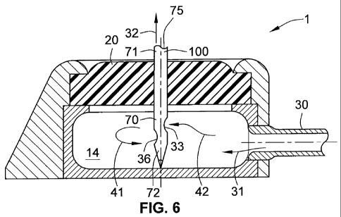
[0022] FIG. 6 is a port from the prior art with a needle with two staggered
openings
according to another embodiment of the present disclosure.
[0023] FIG. 7 is a port from the prior art with a bent needle according to
another
embodiment of the present disclosure.
[0024] FIG. 8 is a port from the prior art with a needle with a grid portion
according to
another embodiment of the present disclosure.
DETAILED DESCRIPTION OF THE INVENTION
[0025] For the purposes of promoting and understanding the principles
disclosed herein,
reference is now made to the preferred embodiments illustrated in the
drawings, and specific
language is used to describe the same. It is nevertheless understood that no
limitation of the
scope of the invention is hereby intended. Such alterations and further
modifications in the
illustrated devices and such further applications of the principles disclosed
and illustrated
6
CA 02730278 2011-01-07
WO 2010/006186 PCT/US2009/050132
herein are contemplated as would normally occur to one skilled in the art to
which this
disclosure relates.
[0026] Needles are long, hollow tubes used when placed at one end in a fluid
such as a
biologic or physiologic fluid to draw the said fluid from the dipped end to
the opposite end by
applying a pressure differential. Within the scope of this disclosure, the
word fluid includes
any biologic or physiologic fluid such as, for example, blood or urine.
Needles have tips
designed to puncture or cut into a solid to reach a destination generally
below the surface
where the fluid is found. The long axis of the needle contains a hollow
tubular channel (or
through bore) extending from a proximal end that may be connected to a machine
or volume
where fluid can be stored. The distal end includes at least one or more
orifices. Orifices can
be located at various distances along the body of the needle and may be placed
in different
orientations.
[0027] Blood cells are damaged when they travel in the blood and encounter an
obstacle.
Blood cells can also be damaged if the serum in which they float is placed
under a pressure
differential that results in the creation of shearing forces within a single
blood cell. For
example, in a machine a pump can be used to suck blood from a patient. If the
needle is
connected to a long tube, the pressure at the pump must be sufficient to
compensate the
pressure drop over the length of the tube. A powerful pump may result locally
in damage to
the cells.
[0028] For damage to the blood to be minimized, the pressure drop in the
needle tip must
be lowered. For example, keeping the blood in a laminar flow while it enters
and travels
along the length of the needle reduces the pressure drop compared to any
turbulent flow of
blood. Another method of reducing the pressure loss through the needle is to
change the
geometrical parameters of the opening or the bore to prevent friction. For
example, if the
needle's internal surface area is A, and opening area is a fraction of A, the
speed of the fluid
7
CA 02730278 2011-01-07
WO 2010/006186 PCT/US2009/050132
through the opening will be a multiple of the speed in the needle body. This
change in
velocity may result in turbulent flow if the Reynolds number of the blood
reaches a certain
fixed value based on fluid viscosity. In addition, the blood located in the
cavity or fluid
reservoir 14 must change direction, velocity, and travel upwards through the
needle as shown
by arrow 32 on FIG. 4.
[0029] FIG. 4 illustrates a needle 100 with a single oval opening 33. FIG. 6
shows a
needle 100 with two staggered oval openings 33, 36, each for collecting a
fraction of the fluid
from the cavity 14. Returning to FIG. 4, the needle is shown in greater detail
in FIGS. 5A-5B
and includes a pointed tip 62 with an end tip 61 of 0.06 inch in length in one
preferred
embodiment. The pointed tip 62 in another embodiment is a 20 cone. The inside
portion of
the cone shown in FIG. 5B includes a bottom resting place 63 shown as a
semicircular
surface to help stabilize the inner flow in the needle 100. What is
contemplated is the use of a
resting place 63 of such geometry to help with manufacturing while providing
the greatest
laminar flow within the main body of the needle 100.
[0030] The use of a vertical oval needle tip allows the creation of a greater
opening
surface than a regular or circular hole without weakening the body of the
needle 100 at any
portion of the needle along its vertical axis by not removing any metal in the
radial
orientation. FIG. 6 is another configuration where no portion of the needle
100 is weakened
by placing two different openings along a single longitudinal radius. Two
successive
openings are staggered at different radial positions, shown to be at 180
degree or on opposite
side of the needle. FIG. 8 shows a configuration where a grid of smaller holes
47 can be used
and placed in a radial staggered configuration to draw in blood. In one
preferred embodiment,
the smaller holes 47 cannot be made to a size smaller than 5 to 10 times the
total cross-
section of 34 gm of the cells in the blood, or a size of 170 to 340 m (0.0068
to 0.0136 in.).
8
CA 02730278 2011-01-07
WO 2010/006186 PCT/US2009/050132
In yet another preferred embodiment, the circular opening diameter is 0.042
inch and is offset
from the cone by 0.035 inch.
[0031] These needle configurations with multiple openings can be flow
calibrated either
by inserting the needle partly into the port plenum so only a portion of the
openings is in
contact with the blood flow, or by using a partial and movable cover.
[0032] For each of the embodiments shown, the edges of the different openings
are
rounded as shown with greater detail as 34 and 35 in FIG. 5A. What is also
contemplated is
the use of internal edges to direct the incoming flow in a selected direction
to prevent the
formation of vortices within the needle. What is also contemplated is the use
of different
walls or separations within the needle 100 to further direct the flow.
[0033] In one preferred embodiment, the internal diameter (d) of the needle
100 is taken
to be 0.0525 to 0.0545 in. The external diameter of the needle 100 is taken to
be 0.0645 to
0.0655 in. This corresponds to a minimum passage section of 0.0021 sq. in. (S
= it(d/2)2). The
surface of a circular opening of diameter 0.042 in. on the lateral wall of a
needle is 0.0014 sq.
in. (S = it(0.042/2)2) but for an oval opening made on a cylinder having a
principal axis of
0.042 in. and a secondary axis of 1.5 times the principal axis 0.063 in., the
surface can be
approximated to 0.0021 sq. in. (S = itAB). The use of an opening with a
passage area equal to
the passage area of the needle 100 to prevent locally an increase in velocity
in the blood is
contemplated. As shown in FIG. 7, the use of a circular hole 37 placed on a
bent needle or the
use of two holes 37, 38 to regulate the flow of fluid through the needle is
also contemplated.
[0034] What is also contemplated is the use of a permanent or a temporary
coating placed
on the needle to improve the flow inside of the needle, such as for example an
anti-clouting
coating like heparin, a bio-compatible coat like polished titanium oxide
coatings, or even
polymer coating such as, for example, Teflon or PTFE. In one embodiment, the
coating is
placed inside of the needle to facilitate the flow of blood. In another
embodiment, the coating
9
CA 02730278 2011-01-07
WO 2010/006186 PCT/US2009/050132
is place at the edges of the openings on the needle to reduce friction. In yet
another
embodiment (not shown), a sliding cover in the shape of a metallic shell can
be retracted over
a portion or the totality of the body of the needle. The placement of the
cover allows for the
control of the flow and the protection of the needle. In yet another
embodiment, instead of a
Huber needle, a regular needle with a cylindrical entry surface can be used in
tandem with a
pull out rod with pointed tip (not shown). In a first step of a method of use,
the pointed rod is
pushed passed the tip of the needle and enters the skin until the external
perimeter of the
needle contacts with the outer layer of the skin. The needle is then pushed
in, and finally, the
pull out rod is pulled out leaving the needle in place and allowing the flow
of blood in the
needle to start.
[0035] In yet another embodiment, as shown in FIG. 8, an intermediate portion
of the
needle can be manufactured of an array of small rounded strings of metal
formed into a
cylindrical mesh for allowing the passage of blood and welded to the end of
the needle in the
shape of a Huber tip. In yet another embodiment, the mesh is not angled and a
Huber shape
tip is connected to the mesh.
[0036] What is described is a needle 100 for a subcutaneous port 1 adapted to
reduce the
damage to the floating particles, such as blood cells a fluid at the inlet of
the needle, the
needle 100 having a needle shaft 70 with a bore 75 along a longitudinal axis
of the needle
shaft 70 with a proximal end 71 and a distal end 72 in opposition thereof as
shown on FIG. 4,
a pointed tip 62 at the distal end 72 with a pointed end tip 61 for the entry
of at least a portion
of the needle shaft shown as FIGS. 5A-B into a fluid reservoir 14 in the
subcutaneous port 1.
In addition, at least an inlet orifice or opening 33 along the needle shaft 70
between the
proximal end 71 and the distal end 72 and in fluidic contact as shown by
arrows 31, 32, with
the fluid reservoir 14 and adjacent to the pointed tip 62. The inlet orifice
33 communicates
with the bore 75 for the passage of the fluid from the fluid reservoir 14
through the inlet
CA 02730278 2011-01-07
WO 2010/006186 PCT/US2009/050132
orifice 33 and through the bore 75 as shown by arrow 32. Further, the inlet
orifice 33 has at
least a rounded edge 34 or 35.
[0037] The inlet orifice may be of different shapes as shown including oval
shape as
shown on FIG. 4, and where oval shape has a long axe along the longitudinal
axis of the shaft
70. The needle shaft 70 may have a thickness in the range of 0.001 to 0.003
inch. While some
ranges and dimensions are given, one of ordinary skill in the art will
recognize that any
thickness is contemplated. In the embodiment shown as FIG. 7, the needle shaft
70 along the
longitudinal axis is curved adjacent to the pointed tip 62. The plurality of
orifices 47 or the
grid of small holes are along the needle shaft 70 between the proximal end 71
and the distal
end 72 and in fluidic contact with the fluid reservoir 14 and adjacent to the
pointed tip 62,
and where each of the plurality of inlet orifices as shown communicate with
the bore 75 for
the passage of fluid as shown by the arrows 31, 32 from the fluid reservoir 14
through the
inlet orifice 33 and through the bore 75.
[0038] What is also contemplated is a method of protecting blood cells from
damage
during a medical treatment with a subcutaneous port 1, where blood is
circulated through a
needle 31, 32, the method having the steps of connecting (not shown) a needle
100 to a
medical treatment device such as a hemodialysis machine for conducting a
treatment using
multiple circulation of blood through the needle 100, the needle 100 having a
needle shaft 70
with a bore 75 along a longitudinal axis shown by the dashed line on FIGS. 4
to 6, and 8 of
the needle shaft 70 and a proximal end 71 and a distal end 72 in opposition
thereof, a pointed
tip 62 at the distal end 72 with a pointed end tip 61, and at least an inlet
orifice 33 along the
needle shaft between the proximal end 71 and the distal end72, and where the
inlet orifice 33
has at least a rounded edge 34, 35 for the protection of blood cells. In a
subsequent step, the
plenum surface 20 as shown on FIG. 6 is punched for entry of at least a
portion of the needle
shaft 70 and the inlet orifice 33 into a fluid reservoir 14 in the
subcutaneous port 1. The inlet
11
CA 02730278 2011-01-07
WO 2010/006186 PCT/US2009/050132
orifice 33 is then placed in fluidic contact as shown by arrows 31, 41, 42,
and ultimately 32
on FIG. 6 with blood in the fluid reservoir for the passage of the blood from
the fluid
reservoir 14 through the inlet orifice 33 and through the bore 75. Finally,
the machine is then
put on for the circulation of the blood so the flow of blood circulates around
the rounded edge
33. In addition, openings are designed so the flow is not accelerated in the
vicinity of the
edges by having a plurality of openings in a single needle.
[0039] It is understood that the preceding is merely a detailed description of
some
examples and embodiments of the present invention and that numerous changes to
the
disclosed embodiments can be made in accordance with the disclosure made
herein without
departing from the spirit or scope of the invention. The preceding
description, therefore, is
not meant to limit the scope of the invention but to provide sufficient
disclosure to one of
ordinary skill in the art to practice the invention without undue burden.
12