Note: Descriptions are shown in the official language in which they were submitted.
CA 02730531 2011-01-10
WO 2010/006809 PCT/EP2009/005217
Medical use of the radical scavenger and antioxidant alpha-1-microglobulin
Field of the invention
The present invention relates to medical use of alpha-l-microglobulin (AIM) in
the
treatment or prophylaxis of diseases wherein oxidative stress is a responsible
factor in
the progress of the disease. Notably, the present invention relates to the
medical use of
alpha-1-microglobulin in the treatment or prophylaxis of diseases or
conditions
associated with the presences of free radicals and/or free haemoglobin in the
subject.
The inventors of present invention have found that alpha-l -microglobulin,
which is a
small protein found e.g. in humans, shows extraordinary properties as
antioxidant and
radical scavenger. Particularly, it is disclosed that the antioxidative
properties of Al M
are of particularly relevance for minimizing oxidative stress in
physiologically impaired
cells.
Background of the invention
Many diseases involve unwanted oxidation of cells and molecules in the tissues
and
lead to formation of extremely reactive free radicals, which in turn may lead
to tissue
damage. Drugs with anti-oxidant properties have been developed during the last
decades, but there is still a need for developing safe drugs with a broad
therapeutic
potential for the treatment or prophylaxis of diseases or conditions that have
an
oxidative stress element.
Oxidative stress
Oxidation is a chemical process which involves loss of electrons, i.e. a
compound is
oxidized when one or more electrons are removed from it. The opposite chemical
process is called reduction. Oxidative stress in the human organism is defined
as an
increased, unwanted oxidation of cells and molecules in the tissues (reviewed
in 1). It
arises from an imbalance between oxidants, mediators of oxidative stress, and
antioxidants, agents that can either prevent oxidation, detoxify oxidants or
repair
oxidized molecules (Fig. 1). The most important oxidants in humans and animals
are
reactive oxygen species (ROS) which include hydrogen peroxide, superoxide and
the
hydroxyl radical. The latter two belong to a group of compounds called free
radicals.
Free radicals are extremely reactive compounds due to the presence of unpaired
electrons in their outer electron shells. ROS- and free radical-formation can
be induced
by, for example, metals and the oxygen-binding organic compound heme. Herne is
an
iron-containing component of haemoglobin and cytochromes, which are proteins
that
CONFIRMATION COPY
CA 02730531 2011-01-10
WO 2010/006809 PCT/EP2009/005217
2
participate in the utilization of oxygen (see below). ROS, oxidants, and free
radicals
react with proteins, DNA and other molecular cell- and tissue components,
which leads
to unwanted modifications of the target molecules and ultimately loss of
cellular
functions.
Free radicals and oxidants are constantly introduced to the human body, both
exogenously via the environment (food, air, smoke, etc) and endogenously as by-
products of normal metabolism (Fig. 1). Endogenous free radicals and oxidants
are
important components of the metabolism in the animal organism. A certain
amount is
necessary for "house-keeping" cellular processes. For example, physiological
cell-
signalling is dependent on a continuous production of cellular free radicals
(reviewed in
2), controlled by an intricate system of cellular antioxidants. Thus, cells
need to
maintain a normal, well-controlled reduction/oxidation (redox)-balance both
intra- and
extracellularly. Oxidative stress will result when the redox balance is upset.
Free
radicals and the strong oxidant hypochlorite (HOCI) are also produced in white
blood
cells during bacterial and fungal infections as weapons to kill the pathogens
(reviewed
in 3). This also leads to oxidative stress.
Haemoglobin and other heme-containing proteins
Haemoglobin is one of the most common proteins in the human body. It is found
in
enormous quantities in the red blood cells and its function is to carry oxygen
from the
lungs to all cells. The oxygen is bound to the iron-containing heme-group,
which gives
the haemoglobin molecule its red colour. All haemoglobin is normally kept
inside the
red blood cells and thus prevented from contact with other cells and
extracellular
components. This is important because haemoglobin is toxic due to strong
oxidant
properties. When the red blood cells break (haemolysis) in diseases like
autoimmune
haemolytic anemia, sickle cell anemia and malaria or in iatrogen situations
including
mismatched blood transfusion, stem cell and solid organ transplantation and
major
surgery, oxy-haemoglobin (haemoglobin plus oxygen) is released from the red
blood
cells. Oxy-haemoglobin spontaneously reacts with itself by rearranging
electrons in a
process called auto-oxidation, forming the free radical superoxide and
methaemoglobin, an oxidized form of the protein. Methaemoglobin continues to
decompose, ultimately forming free globin, heme and iron. The products are
oxidative
as described above. Free heme, being a hydrophobic molecule, can enter cells
by
diffusion over the cell membrane or dissolving the membranes. Free haemoglobin
(located outside the red blood cells) is therefore an inducer of tissue damage
during
CA 02730531 2011-01-10
WO 2010/006809 PCT/EP2009/005217
3
many diseases and other pathological conditions. In addition, free oxy-
haemoglobin is
indirectly a vasoconstrictor because it binds nitric oxide (NO) strongly, one
of the most
important dilators of small blood vessels and capillaries. NO-scavenging by
free oxy-
haemoglobin leads to consumption of NO and subsequent constriction of the
capillaries
resulting in high blood pressure.
Other heme-containing proteins include NADPH-oxidase, myeloperoxidase (MPO)
and
mitochondrial cytochromes. The enzymes NADPH-oxidase and myeloperoxidase are
found in monocytes and neutrophil granulocytes, two subsets of white blood
cells. In a
process called oxidative burst, these enzymes produce superoxide radicals and
hypochlorite, respectively, both of which are involved in the defense against
microbial
infection. The most important of the mitochondrial cytochromes are cytochrome
c and
NADH-dehydrogenase. These enzymes are components of the respiratory complexes
I-IV which convert oxygen to water by using electrons from nutrients, stored
fats, etc. In
this process, large amounts of free radicals, mostly superoxide anions, are
produced
as intermediary metabolites by the mitochondrial enzymes.
Antioxidants
Normally, oxidant activity is balanced by the activity of antioxidants,
protective factors
that eliminate oxidants or prevent their oxidation reactions. During
conditions of
extreme oxidative stress, however, the antioxidants may be overwhelmed,
leading to
oxidative damage to molecules and/or cells and tissues.
Both endogenous and exogenous antioxidants are described. Twenty years ago,
the
prevailing view was that human homeostasis was dependent on externally added
antioxidants, for instance via food intake. Today, an increasing number of
human
antioxidants have been discovered and shown to be produced constitutively
within the
body, i.e. under normal, unstressed conditions. Antioxidants operate by
elimination of
free radicals and oxidants. They can achieve this by three major mechanisms
(see Fig.
2A and figure legends for details): 1) enzymatic addition of electrons derived
from
cellular aerobic metabolism or other sources to the oxidants, 2) non-enzymatic
addition
of electrons from the antioxidant molecule itself to the oxidant, and 3)
binding
(scavenging) of the radicals/oxidants to the antioxidant. Examples of the
first category
are the enzymes superoxide dismutase (SOD), catalase, glutathione peroxidase
and
heme oxygenase. Examples of the second category are thioredoxin, glutathione
and
alpha-lipoic acid. Vitamins C and E, unsaturated fatty acids and plant
flavonoids are
CA 02730531 2011-01-10
WO 2010/006809 PCT/EP2009/005217
4
exogeneous category 2 antioxidants that are not produced in the body but can
be
found in food. Some of the antioxidants of the second category, for example
thioredoxin and glutathione, can be re-generated by reduction of electrons
from other
sources (Fig. 2A). Most antioxidants in the food are poorly re-generated after
reacting
with their targets. Thus, the consumed (=oxidized) vitamins C and E, etc,
present
oxidative stress to the tissues unless quickly removed.
Electrons which are produced by cellular aerobic metabolism (ultimately
derived from
nutrients, e.g. glucose, fat, proteins via the electron-carrier NADH) provide
the reducing
equivalents to the antioxidants of category 1, when re-generating antioxidants
of
category 2 and in the scavenging process (category 3) (Fig. 2A). Therefore,
most
antioxidants are dependent on an intact cell metabolism and only operate intra-
cellularly. In fact, most antioxidants, being intracellular, are part of the
normal cellular
"house-keeping" machinery.
Several antioxidants are specialized against haemoglobin-induced oxidative
stress.
The plasma proteins haptoglobin, hemopexin and transferrin bind free,
extracellular
haemoglobin, heme and iron, respectively, in the blood. The cellular protein
ferritin
binds and stores free, cellular iron. Heme oxygenase-1 (HO-1) is produced in
most
cells in response to increased concentrations of haemoglobin, heme and free
radicals
and eliminates heme by degradation into bilirubin, carbon monoxide and free
iron.
However, none of the above-mentioned antioxidants act by all three mechanisms
and,
accordingly, a general therapeutic use of such an antioxidant is limited. An
antioxidant
having all mechanisms of action would be advantageous as it will have a much
more
general use and be less dependent on the cellular homeostasis for functioning.
Abbreviations
ABTS, 2,2'-azino-bis(3-ethylbenzo-thiazoline-6-sulphonic acid)diammonium salt
al M or Al M, al-microglobulin or alpha- 1 -microglobulin
t-a1 M; truncated al-microglobulin or alpha-l-glycoprotein
AGP, al-acid glycoprotein
DTT, dithiothreitol
G3DPH, glyceraldehyd-3-phosphate dehydrogenase
Hb, haemoglobin
H2DCFDA, 2',7'-dichlorodihydrofluorescein diacetate
CA 02730531 2011-01-10
WO 2010/006809 PCT/EP2009/005217
IVF, in vitro fertilization
IVH, intra ventricular brain haemorrhage
NEM, N-ethylmaleimide
PE, preeclampsia
5 PI, propidium iodide
ROS, reactive oxygen species;
5-IAF, 5-iodoacetamide-fluorescein
RIA, radio immuno assay
MPO, myeloperoxidase
Definitions
In describing and claiming the disclosed subject matter, the following
terminology will
be used in accordance with the definitions set forth below. Unless defined
otherwise, all
technical and scientific terms used herein have the same meaning as commonly
understood by one of ordinary skill in the art to which this disclosure
belongs. In some
cases, terms with commonly understood meanings are defined herein for clarity
and/or
for ready reference, and the inclusion of such definitions herein should not
necessarily
be construed to represent a substantial difference over what is generally
understood in
the art. The techniques and procedures described or referenced herein are
generally
well understood and commonly employed using conventional methodology by those
skilled in the art. As appropriate, procedures involving the use of
commercially
available kits and reagents are generally carried out in accordance with
manufacturer
defined protocols and/or parameters unless otherwise noted. Although any
methods
and materials similar or equivalent to those described herein can also be used
in the
practice or testing of the present disclosure, the preferred methods and
materials are
described herein.
In this specification, unless otherwise specified, "a" or "an" means "one or
more".
There exist different forms of haemoglobin. Adult haemoglobin (Haemoglobin A)
consists of two alpha and two beta polypeptide chains (Hba, Hb(3), each
containing a
non-peptide heme group that reversibly binds a single oxygen molecule.
Haemoglobin
A2, another adult haemoglobin component is composed of two alpha-chains and
two
delta chains (Hba, Hbb). Fetal haemoglobin (Haemoglobin F) on the other hand
is the
major component of haemoglobin in the fetus. This haemoglobin has two alpha-
and
two gamma polypeptide chains (Hba, Hby).
CA 02730531 2011-01-10
WO 2010/006809 PCT/EP2009/005217
6
The term "free haemoglobin", in this specification refers to free haemoglobin
generally
and includes total free haemoglobin, free haemoglobin A, free haemoglobin A2,
free
haemoglobin F, any free haemoglobin subunit (e.g. an Hba, Hb1, Hb6 or Hby
chain), or
any combination thereof. It further includes these haemoglobin entities in
either a
polypeptide (protein) or nucleotide (RNA) form, except when applied as a
target for
treatment. The term "free fetal haemoglobin" refers to free haemoglobin F or
any
subunit of haemoglobin F and includes the haemoglobin F entities in a
polypeptide
(protein) or nucleotide (RNA) form, except when applied as a target for
treatment.
In this specification, the term "free" as used, inter alia, in the expressions
"free
haemoglobin" or "free haemoglobin subunits (e.g. Hba, Hbf3, Hba or Hby
chains)" refer
to haemoglobin or haemoglobin subunits freely circulating in a biological
fluid, as
opposed to cellular haemoglobin which refers to the molecules residing inside
cells.
The term "free" in this sense is thus mainly used to distinguish free
haemoglobin from
haemoglobin which is present in intact erythrocytes.
The terms "treatment or prophylaxis" in their various grammatical forms in
relation to
the present invention refer to preventing, curing, reversing, attenuating,
alleviating,
ameliorating, inhibiting, minimizing, suppressing, or halting (1) the
deleterious effects of
a disorder, (2) disorder progression, or (3) disorder causative agent.
The term "effective amount' in relation to the present invention refers to
that amount
which provides a therapeutic effect for a given condition and administration
regimen.
This is a predetermined quantity of active material calculated to produce a
desired
therapeutic effect in association with the required additives and diluents;
i.e., a carrier,
or administration vehicle. Further, it is intended to mean an amount
sufficient to reduce
and most preferably prevent a clinically significant deficit in the activity
and response of
the host. Alternatively, a therapeutically effective amount is sufficient to
cause an
improvement in a clinically significant condition in a host. As is appreciated
by those
skilled in the art, the amount of a compound may vary depending on its
specific activity.
Suitable dosage amounts may contain a predetermined quantity of active
composition
calculated to produce the desired therapeutic effect in association with the
required
diluents; i.e., carrier, or additive. Further, the dosage to be administered
will vary
depending on the active principle or principles to be used, the age, weight
etc. of the
patient to be treated but will generally be within the range from 0,001 to
1000 mg/kg
CA 02730531 2011-01-10
WO 2010/006809 PCT/EP2009/005217
7
body weight/day. Moreover, the dose depends on the administration route.
The term "polypeptides" includes proteins and fragments thereof. Polypeptides
are
disclosed herein as amino acid residue sequences. Those sequences are written
left to
right in the direction from the amino to the carboxy terminus. In accordance
with
standard nomenclature, amino acid residue sequences are denominated by either
a
three letter or a single letter code as indicated as follows: Alanine (Ala,
A), Arginine
(Arg, R), Asparagine (Asn, N), Aspartic Acid (Asp, D), Cysteine (Cys, C),
Glutamine
(GIn, Q), Glutamic Acid (Glu, E), Glycine (GIy, G), Histidine (His, H),
Isoleucine (lie, I),
Leucine (Leu, L), Lysine (Lys, K), Methionine (Met, M), Phenylalanine (Phe,
F), Proline
(Pro, P), Serine (Ser, S), Threonine (Thr, T), Tryptophan (Trp, W), Tyrosine
(Tyr, Y),
and Valine (Val, V).
"Variant" refers to a polypeptide or polynucleotide that differs from a
reference
polypeptide or polynucleotide, but retains essential properties. A typical
variant of a
polypeptide differs in amino acid sequence from another, reference
polypeptide.
Generally, differences are limited so that the sequences of the reference
polypeptide
and the variant are closely similar overall (homologous) and, in many regions,
identical.
A variant and reference polypeptide may differ in amino acid sequence by one
or more
modifications (e.g., substitutions, additions, and/or deletions). A
substituted or inserted
amino acid residue may or may not be one encoded by the genetic code. A
variant of a
polypeptide may be naturally occurring such as an allelic variant, or it may
be a variant
that is not known to occur naturally.
"Identity"as known in the art, is a relationship between two or more
polypeptide
sequences, as determined by comparing the sequences. In the art, "identity"
also
refers to the degree of sequence relatedness between polypeptide as determined
by
the match between strings of such sequences. "Identity" and "similarity" can
be readily
calculated by known methods.
The term "substantially similar" as used herein generally refers to a
function, activity, or
behavior that is close enough to the natural, expected, or average, so as to
be
considered, for all practical purposes, interchangeable. For instance, a
protein with
substantially similar activity would be one that has an activity level that
would not be
considered to be substantially more or less active than the native protein.
The term "prodrug" refers to an agent, including nucleic acids and proteins,
which is
CA 02730531 2011-01-10
WO 2010/006809 PCT/EP2009/005217
8
converted into a biologically active form in vivo. For instance, Prodrugs are
often useful
because, in some situations, they may be easier to administer than the parent
compound. They may, for instance, be bioavailable by oral administration
whereas the
parent compound is not. The prodrug may also have improved solubility in
pharmaceutical compositions over the parent drug. A prodrug may be converted
into
the parent drug by various mechanisms, including enzymatic processes and
metabolic
hydrolysis.
As used herein "functional variant" refers to a variant of a protein or
polypeptide (e.g., a
circularly permuted protein, with or without additional sequence alterations)
that can
perform the same functions or activities as the original protein or
polypeptide, although
not necessarily at the same level (e.g., the variant may have enhanced,
reduced or
changed functionality, so long as it retains the basic function).
Detailed description of the invention
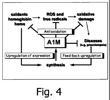
In present invention, it is disclosed that Al M possesses broad antioxidant
properties
suitable to avoid or minimize injuries caused by oxidative stress. The current
concept is
that the physiological function of Al M is to continuously "vacuum-clean"
tissues from
free radicals and oxidants, perhaps especially heme, and deliver the products
to the
kidneys for degradation and/or excretion. A second physiological function is
to reduce
oxidants and oxidized cell components and tissue molecules. An important
property of
Al M, adding to its value as an antioxidant, is that the protein, after
binding a maximum
load of radicals, and/or reducing oxidants or oxidation products, does not
present
oxidative stress to tissue components. In other words, ROS, radicals and other
oxidants are eliminated by Al M, hence Al M may be considered as a radical
"sink".
This feature may be of particular importance in damaged cells or other cells
in which
intracellular homeostatic processes are impaired and thus incapable of
removing the
oxidative stress that other antioxidants, as eg Vitamin E and D, impose on the
cell after
action.
Alpha- 1-micrwlobulin
Al M is synthesized in the liver at a high rate, secreted into the blood
stream and
transported across the vessel walls to the extravascular compartment of all
organs.
The protein is also synthesized in other tissues (blood cells, brain, kidney,
skin) but at a
lower rate. Due to the small size, free Al M is rapidly filtered from blood in
the kidneys.
AIM has excellent anti-oxidative properties in general and specifically
towards free
CA 02730531 2011-01-10
WO 2010/006809 PCT/EP2009/005217
9
haemoglobin; properties that makes it suitable for use in the treatment or
prophylaxis of
a variety of diseases that involves oxidative stress or wherein the presence
of free
haemoglobin induces or aggravates a disease or condition.
Alpha-1-microglobulin (AIM) is an endogenous antioxidant that provides
antioxidation
in several ways (Fig. 2B and figure legends hereto). Thus, the present
invention relates
to Al M which has been found to combine enzymatic reductase (category 1), non-
enzymatic reduction (category 2) and radical-scavenging (category 3)
properties. In
addition, the non-enzymatic reduction mechanism (category 2) can be employed
repeatedly with several cycles of electron-donation. Furthermore, the radical-
scavenger
mechanism (category 3) result in a net production of electrons further
increasing the
antioxidation capacity of the protein. In other words, the protein carries its
own supply
of electrons, is independent on cellular metabolism, and can operate both
intra- and
extracellularly. In addition, Al M can repair oxidative damage that has been
inflicted to
tissue components (a unique property assigned category 4). See also below for
a
detailed description of the radical scavenging mechanism.
Al M is a member of the lipocalin superfamily, a group of proteins from
animals, plants
and bacteria with a conserved three-dimensional structure but very diverse
functions.
Each lipocalin consists of a 160-190-amino acid chain that is folded into a R-
barrel
pocket with a hydrophobic interior. Twelve human lipocalin genes are known.
Among
the human lipocalins, Al M is a 26 kDa plasma and tissue protein that so far
has been
identified in mammals, birds, fish and frogs. A model of the three-dimensional
structure
of Al M is shown in Fig. 3. Al M is synthesized in the liver at a high rate,
secreted into
the blood stream and rapidly (T'h = 2-3 min) transported across the vessel
walls to the
extravascular compartment of all organs. The protein is also synthesized in
other
tissues (blood cells, brain, kidney, skin) but at a lower rate. Al M is found
both in a
free, monomeric form and as covalent complexes with larger molecules (IgA,
albumin,
prothrombin) in blood and interstitial tissues. Due to the small size, free Al
M is rapidly
filtered from blood in the kidneys. The major portion is then readsorbed, but
significant
amounts are excreted to the urine.
Sequence and structural properties of A 1 M
The full sequence of human Al M was first reported by Kaumeyer et al. (5). The
protein
was found to consist of 183 amino acid residues. Since then, ten additional
AlM
cDNAs and/or proteins have been detected, isolated and/or sequenced from other
CA 02730531 2011-01-10
WO 2010/006809 PCT/EP2009/005217
mammals, birds, amphibians, and fish. The length of the peptide chain of Al M
differs
slightly among species, due mainly to variations in the C-terminus. Alignment
comparisons of the different deduced amino acid sequences show that the
percentage
of identity varies from approximately 75-80% between rodents or ferungulates
and
5 man, down to approximately 45% between fish and mammals. A free cysteine
side-
chain at position 34 is conserved. This group has been shown to be involved in
redox
reactions (see below), in complex formation with other plasma proteins and in
binding
to a yellow-brown chromophore. Computerised 3D models based on the known X-ray
crystallographic structures of other lipocalins suggest that Cys34 is solvent
exposed
10 and located near the opening of the lipocalin pocket (see Fig. 3).
Complement factor
C8y, another lipocalin, also carries an unpaired Cys in position 34 that is
involved in the
formation of the active C8 complex.
In the present context the term "alpha-l-microglobulin" intends to cover alpha-
l-
microglobulin as identified in SEQ ID NO: 1 (human AIM) as well as SEQ ID NO:
2
(human recombinant AIM) as well as homologues, fragments or variants thereof
having similar therapeutic activities. In a preferred aspect, the alpha-1 -
microglobulin is
in accordance with SEQ ID NO: 1 or 2 as identified herein. In Fig. 19 is given
the
sequence listing of the amino acid sequence of human Al M and human
recombinant
Al M (SEQ ID NOs 1 and 2, respectively) and the corresponding nucleotide
sequences
(SEQ ID NOs 3 and 4, respectively).
As mentioned above homologues of Al M can also be used in accordance with the
description herein. In theory AIM from all species can be used including the
most
primitive found so far, which is from fish (plaice). Al M is also available in
isolated form
from human, rat, mouse, rabbit, guinea pig, cow and plaice.
Considering homologues, variants and fragments of Al M, the following has been
identified as important parts of the protein for the anti-oxidative effect:
Y22 (Tyrosine, pos 22, basepairs 64-66)
C34 (Cystein, position 34, basepairs 100-102)
K69 (Lysine, pos 69, basepairs 205-207)
K92 (Lysine, pos 92, basepairs 274-276)
K118 (Lysine, pos 118, basepairs 352-354)
K130 (Lysine, pos 130, basepairs 388-390)
Y132 (Tyrosine, pos 132, basepairs 394-396)
CA 02730531 2011-01-10
WO 2010/006809 PCT/EP2009/005217
11
L180 (Leucine, pos 180, basepairs 538-540)
1181 (Isoleucine, pos 181, basepairs 541-543)
P182 (Proline, pos 182, basepairs 544-546)
R183 (Arginine, pos 183, basepairs 547-549)
(Numbering of amino acids and nucleotides throughout the document refers to
SEQ ID
1 and 3, see also Figs 3 and 6; if other Al M from other species, A 1 M
analogs or
recombinant sequences thereof are employed, a person skilled in the art will
know how
to identify the amino acids of the active site(s) or site(s) responsible for
the enzymatic
activity.)
Human Al M is substituted with oligosaccharides in three positions, two
sialylated
complex-type, probably diantennary carbohydrated linked to Asn17 and Asn96 and
one
more simple oligosaccharide linked to Thr5. The carbohydrate content of Al M
proteins
from different species varies greatly, though, ranging from no glycosylation
at all in
Xenopus leavis over a spectrum of different glycosylation patterns. However,
one
glycosylation site, corresponding to Asn96 in man, is conserved in mammals,
suggesting that this specific carbohydrate may be functionally important.
Al M is yellow-brown-coloured when purified from plasma or urine. The colour
is
caused by heterogeneous compounds covalently bound to various amino acid side
groups mainly located at the entrance to the pocket. These modifications
probably
represent the oxidized degradation products of organic oxidants covalently
trapped by
Al M in vivo, for example heme, kynurenin and tyrosyl radicals (6-8, 10).
Al M is also charge- and size-heterogeneous and more highly brown-coloured Al
M-
molecules are more negatively charged. The probable explanation for the
heterogeneity is that different side-groups are modified to a varying degree
with
different radicals, and that the modifications alter the net charge of the
protein.
Covalently linked coloured substances have been localized to Cys34, and Lys92,
Lysl 18 and Lysl 30, the latter with molecular masses between 100 and 300 Da.
The
tryptophan metabolite kynurenine was found covalently attached to lysyl
residues in
Al M from urine of haemodialysis patients and appears to be the source of the
brown
colour of the protein in this case (6). Oxidized fragments of the synthetic
radical ABTS
(2,2'-azino-di-(3-ethylbenzothiazoline)-6-sulfonic acid) was bound to the side-
chains of
Y22 and Y132 (10).
CA 02730531 2011-01-10
WO 2010/006809 PCT/EP2009/005217
12
C34 is the reactive center of Al M (9). It becomes very electronegative,
meaning that it
has a high potential to give away electrons, by the proximity of the
positively charged
side-chains of K69, K92, K118 and K 130, which induce a deprotonization of the
C34
thiol group which is a prerequisite of oxidation of the sulphur atom.
Preliminary data
shows that C34 is one of the most electronegative groups known.
Theoretically, the amino acids that characterize the unique enzymatic and non-
enzymatic redox properties of Al M (C34, Y22, K92, K118, K130, Y132, L180,
1181,
P182, R183), which will be described in more detail below, can be arranged in
a similar
three-dimensional configuration on another frame-work, for instance a protein
with the
same global folding (another lipocalin) or a completely artificial organic or
inorganic
molecule such as a plastic polymer, a nanoparticle or metal polymer.
The three-dimensional arrangement of some of these amino acids (blue ovals,
the
lysines are depicted by a õ+"), the Al M-framework (barrel), the electron-flow
and the
radical-trapping, are illustrated in Fig. 6.
Accordingly, homologues, fragments or variants comprising a structure
including the
reactive center and its surroundings as depicted above, are preferred.
Modifications and changes can be made in the structure of the polypeptides of
this
disclosure and still result in a molecule having similar characteristics as
the polypeptide
(e.g., a conservative amino acid substitution). For example, certain amino
acids can be
substituted for other amino acids in a sequence without appreciable loss of
activity.
Because it is the interactive capacity and nature of a polypeptide that
defines that
polypeptide's biological functional activity, certain amino acid sequence
substitutions
can be made in a polypeptide sequence and nevertheless obtain a polypeptide
with like
properties.
In making such changes, the hydropathic index of amino acids can be
considered. The
importance of the hydropathic amino acid index in conferring interactive
biologic
function on a polypeptide is generally understood in the art. It is known that
certain
amino acids can be substituted for other amino acids having a similar
hydropathic
index or score and still result in a polypeptide with similar biological
activity. Each
amino acid has been assigned a hydropathic index on the basis of its
hydrophobicity
and charge characteristics. Those indices are: isoleucine (+4.5); valine
(+4.2); leucine
CA 02730531 2011-01-10
WO 2010/006809 PCT/EP2009/005217
13
(+3.8); phenylalanine (+2.8); cysteine/cysteine (+2.5); methionine (+1.9);
alanine
(+1.8); glycine (-0.4); threonine (-0.7); serine (-0.8); tryptophan (-0.9);
tyrosine (- 1.3);
proline (-1.6); histidine (-3.2); glutamate (-3.5); glutamine (-3.5);
aspartate (-3.5);
asparagine (-3.5); lysine (-3.9); and arginine (-4.5).
It is believed that the relative hydropathic character of the amino acid
determines the
secondary structure of the resultant polypeptide, which in turn defines the
interaction of
the polypeptide with other molecules, such as enzymes, substrates, receptors,
antibodies, antigens, and the like. It is known in the art that an amino acid
can be
substituted by another amino acid having a similar hydropathic index and still
obtain a
functionally equivalent polypeptide. In such changes, the substitution of
amino acids
whose hydropathic indices are within 2 is preferred, those within 1 are
particularly
preferred, and those within 0.5 are even more particularly preferred.
Substitution of like amino acids can also be made on the basis of
hydrophilicity,
particularly where the biologically functional equivalent polypeptide or
peptide thereby
created is intended for use in immunological embodiments. The following
hydrophilicity
values have been assigned to amino acid residues: arginine (+3.0); lysine
(+3.0);
aspartate (+3.0 1); glutamate (+3.0 1); serine (+0.3); asparagine (+0.2);
glutamnine
(+0.2); glycine (0); proline (-0.5 1); threonine (-0.4); alanine (-0.5);
histidine (-0.5);
cysteine (-1.0); methionine (-1.3); valine (-1.5); leucine (-1.8); isoleucine
(-1.8); tyrosine
(-2.3); phenylalanine (-2.5); tryptophan (-3.4). It is understood that an
amino acid can
be substituted for another having a similar hydrophilicity value and still
obtain a
biologically equivalent, and in particular, an immunologically equivalent
polypeptide. In
such changes, the substitution of amino acids the hydrophilicity values of
which are
within 2 is preferred, those within 1 are particularly preferred, and
those within 0.5
are even more particularly preferred.
As outlined above, amino acid substitutions are generally based on the
relative
similarity of the amino acid side-chain substituents, for example, their
hydrophobicity,
hydrophilicity, charge, size, and the like. Exemplary substitutions that take
one or more
of the foregoing characteristics into consideration are well known to those of
skill in the
art and include, but are not limited to (original residue: exemplary
substitution): (Ala:
Gly, Ser), (Arg: Lys), (Asn: GIn1 His), (Asp: Glu, Cys, Ser), (GIn: Asn),
(Glu: Asp), (Gly:
Ala), (His: Asn, GIn), (Ile: Leu, Val), (Leu: Ile, Val), (Lys: Arg), (Met:
Leu, Tyr), (Ser:
Thr), (Thr: Ser), (Trp: Tyr), (Tyr: Trp, Phe), and (Val: Lle, Leu).
Embodiments of this
disclosure thus contemplate functional or biological equivalents of a
polypeptide as set
CA 02730531 2011-01-10
WO 2010/006809 PCT/EP2009/005217
14
forth above. In particular, embodiments of the polypeptides can include
variants having
about 50%, 60%, 70%, 80%, 90%, and 95% sequence identity to the polypeptide of
interest.
In the present context, the homology between two amino acid sequences or
between
two nucleic acid sequences is described by the parameter "identity".
Alignments of
sequences and calculation of homology scores may be done using a full Smith-
Waterman alignment, useful for both protein and DNA alignments. The default
scoring
matrices BLOSUM50 and the identity matrix are used for protein and DNA
alignments
respectively. The penalty for the first residue in a gap is -12 for proteins
and -16 for
DNA, while the penalty for additional residues in a gap is -2 for proteins and
-4 for
DNA. Alignment may be made with the FASTA package version v20u6.
Multiple alignments of protein sequences may be made using "ClustalW".
Multiple
alignments of DNA sequences may be done using the protein alignment as a
template,
replacing the amino acids with the corresponding codon from the DNA sequence.
Alternatively different software can be used for aligning amino acid sequences
and
DNA sequences. The alignment of two amino acid sequences is e.g. determined by
using the Needle program from the EMBOSS package (http://emboss.org) version
2.8Ø The Needle program implements the global alignment algorithm described
in.
The substitution matrix used is BLOSUM62, gap opening penalty is 10, and gap
extension penalty is 0.5.
The degree of identity between an amino acid sequence; e.g. SEQ ID NO: 1 and a
different amino acid sequence (e.g. SEQ ID NO: 2) is calculated as the number
of
exact matches in an alignment of the two sequences, divided by the length of
the "SEQ
ID NO: 1" or the length of the " SEQ ID NO: 2 ", whichever is the shortest.
The result is
expressed in percent identity.
An exact match occurs when the two sequences have identical amino acid
residues in
the same positions of the overlap.
If relevant, the degree of identity between two nucleotide sequences can be
determined by the Wilbur-Lipman method using the LASER- GENET"' MEGALIGNT ^
software (DNASTAR, Inc., Madison, WI) with an identity table and the following
multiple
CA 02730531 2011-01-10
WO 2010/006809 PCT/EP2009/005217
alignment parameters: Gap penalty of 10 and gap length penalty of 10. Pairwise
alignment parameters are Ktuple=3, gap penalty=3, and windows=20.
In a particular embodiment, the percentage of identity of an amino acid
sequence of a
polypeptide with, or to, amino acids of SEQ ID NO: 1 is determined by i)
aligning the
5 two amino acid sequences using the Needle program, with the BLOSUM62
substitution
matrix, a gap opening penalty of 10, and a gap extension penalty of 0.5; ii)
counting the
number of exact matches in the alignment; iii) dividing the number of exact
matches by
the length of the shortest of the two amino acid sequences, and iv) converting
the
result of the division of iii) into percentage. The percentage of identity to,
or with, other
10 sequences of the invention is calculated in an analogous way.
By way of example, a polypeptide sequence may be identical to the reference
sequence, that is be 100% identical, or it may include up to a certain integer
number of
amino acid alterations as compared to the reference sequence such that the %
identity
15 is less than 100%. Such alterations are selected from: at least one amino
acid deletion,
substitution (including conservative and non-conservative substitution), or
insertion,
and wherein said alterations may occur at the amino- or carboxy-terminus
positions of
the reference polypeptide sequence or anywhere between those terminal
positions,
interspersed either individually among the amino acids in the reference
sequence, or in
one or more contiguous groups within the reference sequence.
Conservative amino acid variants can also comprise non-naturally occurring
amino acid
residues. Non-naturally occurring amino acids include, without limitation,
trans-3-
methylproline, 2,4-methanoproline, cis-4-hydroxyproline, trans-4-
hydroxyproline, N-
methyl-glycine, allo-threonine, methylthreonine, hydroxy-ethylcysteine,
hydroxyethylhomocysteine, nitro-glutamine, homoglutamine, pipecolic acid,
thiazolidine
carboxylic acid, dehydroproline, 3- and 4-methylpr6line, 3,3-dimethylproline,
tert-
leucine, norvaline, 2-azaphenyl-alanine, 3-azaphenylalanine, 4-
azaphenylalanine, and
4- fluorophenylalanine. Several methods are known in the art for incorporating
non-
naturally occurring amino acid residues into proteins. For example, an in
vitro system
can be employed wherein nonsense mutations are suppressed using chemically
aminoacylated suppressor tRNAs. Methods for synthesizing amino acids and
aminoacylating tRNA are known in the art. Transcription and translation of
plasmids
containing nonsense mutations is carried out in a cell-free system comprising
an E. coli
S30 extract and commercially available enzymes and other reagents. Proteins
are
purified by chromatography. In a second method, translation is carried out in
Xenopus
CA 02730531 2011-01-10
WO 2010/006809 PCT/EP2009/005217
16
oocytes by microinjection of mutated mRNA and chemically aminoacylated
suppressor
tRNAs. Within a third method, E. coli cells are cultured in the absence of a
natural
amino acid that is to be replaced (e.g., phenylalanine) and in the presence of
the
desired non-naturally occurring amino acid(s) (e.g., 2-azaphenylalanine, 3-
azaphenylalanine, 4-azaphenylalanine, or 4-fluorophenylalanine). The non-
naturally
occurring amino acid is incorporated into the protein in place of its natural
counterpart.
Naturally occurring amino acid residues can be converted to non-naturally
occurring
species by in vitro chemical modification. Chemical modification can be
combined with
site-directed mutagenesis to further expand the range of substitutions.
Alternative
chemical structures providing a 3-dimensional structure sufficient to support
the
antioxidative properties of Al M may be provided by other technologies e.g.
artificial
scaffolds, amino-acid substitutions and the like. Furthermore, structures
mimicking the
active sites of Al M as listed above and depicted in figure 3 and 6 are
contemplated as
having the same function as Al M.
Diseases associated with oxidative stress
In the following diseases or conditions are described which involve oxidative
stress. It
is contemplated that Al M can be used in the treatment of any of the diseases
mentioned in the following.
Oxidative stress has been reported in a variety of diseases. As mentioned
above,
oxidative stress is a situation when there is an imbalance between free
radicals and the
protective antioxidants. Oxidative stress can induce a wide range of acute or
long-term
physiological reactions releasing various bio-active factors. These in turn
can promote
additional oxidation/free radical-formation which further accelerate the
oxidative stress,
etc. Thus, the physiological reactions and oxidative stress interact with each
other as
gears and together they make the oxidative stress machinery spin faster and
faster
(Fig. 5). Some of the more important gears are inflammation, ischemia and
reperfusion,
blood haemoglobin and environmental/food-derived factors, which will be
discussed
below.
A) Infection and inflammation
Inflammation is a collective term for a wide range of secondary immune
reactions to
infections of all kinds, and that also characterizes several other diseases
such as
autoimmune diseases. The body responds to bacterial infections by recruiting
white
blood cells (monocytes and granulocytes) to the infection site. As described
above,
CA 02730531 2011-01-10
WO 2010/006809 PCT/EP2009/005217
17
white blood-cells produce superoxide anions and hypochlorite. To obtain iron,
many
bacteria are hemolytic, i.e. they produce molecules, which induce rupture of
red blood
cells and exposure of haemoglobin to bystander tissue components. Furthermore,
the
inflammation is characterized by necrosis, i.e. cells at the infection site
rupture and die.
This leads to exposure of, for example, the mitochondrial respiratory enzymes
that
produce free radicals. Pro-inflammatory cytokines such as TNF-alpha, impair
intracellular antioxidants, superoxide dismutase and glutathione peroxidase.
In these
ways, many factors contribute to oxidative stress during infection and
inflammation.
An example of the diseases in this group is chronic obstructory pulmonary
disease
(COPD), an inflammatory lung disease. Inflammatory diseases of lungs and
airways
are associated with strong pathological oxidation of extravascular tissue.
This is
mediated by activation of neutrophil and eosinophil granulocytes and their
secretion of
peroxidases, as well as the challenge from molecular oxygen.
Arthritis is a group of diseases in which the joints are damaged in a way that
involves
inflammation. Arthritis can have many causes, for example forced trauma,
bacterial
infection, gout and autoimmune attack. The inflammation of the joints is
associated with
high levels of oxidative stress and oxidative modification of cartilage,
connective tissue
and cells.
Other examples of conditions with high levels of inflammation are
= Autoimmune diseases (rheumatoid arthritis, thyroid diseases, etc)
= Infectious diseases
= Neurodegenerative diseases (Alzheimer's, Parkinson's, ALS, Huntingtons
Disease, and Multiple sclerosis MS)
= Inflammatory bowel diseases
= Arthritis
B) Ischemia- and reperfusion-related diseases
When the blood vessels are occluded or damaged, either permanent or
intermittent,
there is an increased formation of free radicals due to the cell-death
resulting from the
decreased blood-flow and hypoxia (ischemia). When blood-flow is restored
(reperfusion) a sudden elevation of the local oxygen supply leads to a
dramatic
increase of ROS from reactions between cell-components and oxygen. For
instance,
the enzyme xanthine dehydrogenase, an essential and abundant component of DNA-
CA 02730531 2011-01-10
WO 2010/006809 PCT/EP2009/005217
18
metabolism, uses blood oxygen to form superoxide anions. If reperfusion is
sustained
over a long period, the formation of ROS exceeds the capacity of the
endogenous
antioxidants and oxidative stress occurs. Endothelial injury is induced by
oxidative
stress which in turn activates platelets and induce thrombus formation that
further
threatens to occlude the vessels.
Stroke is an ischemia-reperfusion-related condition of the brain caused by
obstruction
of the blood-flow, due to thrombosis, embolism, haemorrhage, etc. Infarction
of an
organ, e.g. heart, is a condition with tissue necrosis due to occlusion of the
blood and
ischemia-reperfusion effects. Atherosclerosis is a disease affecting arterial
blood
vessels. It is a chronic inflammatory response in the walls of arteries,
partly due to the
accumulation of macrophage type white blood cells and promoted by low density
lipoproteins (LDL). Oxidative stress is a strong component in the development
of
atherosclerosis. Thus, oxidants and free radicals participate in oxidative
modification of
LDL, endothelial cell membranes and other components of the blood vessels.
Oxidized
LDL (ox-LDL) binds to specific receptors in the endothelium and local
accumulation of
ox-LDL leads to recruitment of monocytes which differentiate to macrophages at
the
specific site. This leads to inflammation, attraction of granulocytes and an
increased
local production of ROS from NADPH-oxidase, MPO and other sources. The local
endothelial damage, resulting in atherosclerotic plaques, ultimately occludes
the blood
flow.
= Arteriosclerosis
Ischemic heart disease
= Stroke and other conditions secondary to ischemia
= Hypertensive disorders
= Metabolic disorders (diabetes, dyslipedemia, hypercholesterolemia)
latrogen ischemia- and reperfusion-related damages are developed secondary to
the
treatment of an underlying disease. During the treatment the different gears
shown in
Fig. 5 may drive the oxidative stress.
For example, several methods of dialysis are in practice to replace lost
kidney function.
The very complex functions of the kidneys can be summarized as maintaining
water
and salt balance of the body and removing harmful and toxic degradation
products.
The kidneys operate by continuous filtration of the blood, followed by an
active
CA 02730531 2011-01-10
WO 2010/006809 PCT/EP2009/005217
19
reabsorbtion of most components, including adequate amounts of water, and
excretion
of excess water, salt and toxic degradation products. Free radicals and ROS,
especially small free organic radicals, such as urate and 3-hydroxy-
kynurenine, are
examples of toxic substances normally cleared from blood by the kidneys.
Dialysis is
far from a perfect replacement of the kidneys, and dialysis patients thus
suffer from
oxidative stress. In addition, it has been shown that the dialysis process
itself induces
an inflammatory response also contributing to the oxidative stress.
Hypothetically, the
higher incidence of atherosclerosis among dialysis patients may therefore be
explained
by the oxidative stress associated with kidney failure and the dialysis
process.
Ischemic heart patients undergo by-pass operation on a routinely basis. During
surgery, a heart-lung machine pumps the blood. During this time, many of the
red
blood cells are being destroyed which results in free haemoglobin, a potent
oxidizer as
described above. Furthermore, coronary heart surgery as well as other vascular
surgery, requires that the blood flow is stopped during the procedure. When
the blood
flow again is established, reperfusion damage occurs.
Cell and organ transplantation. One of the problems encountered in cell and
organ
transplantation is that ROS are being formed in organs and cells during
storage. The
situation resembles the problems encountered during ischemia-reperfusion. This
oxidative stress can be prevented or at least decreased in animal models by
use of
ROS scavengers. Much effort has been spent on trying to optimize the medium
used to
store and transport solid organs used for transplantation. Cold salt solutio
ns with
nutrients designed specifically are being used today for this purpose.
However, allowed
ischemia time (time without oxygen) is still very limited for organs like
heart, lungs,
kidneys and liver. Increasingly, pancreatic islet cells and different kinds of
so-called
stem cells (hematopoietic and mesenchymal dito, for instance) are also
transported
around the world for transplantation purposes. Along the same lines,
transplantation of
retinal tissue is a future potential treatment currently tried in animals.
Diseases
characterized by oxidative damage of the retina are often indications to
retina
transplantation. It was recently shown that pre-conditioning of the retinal
tissue can
protect it against oxidative cell death.
The examples thus include:
= Use of kidney dialysis
= Use of heart and lung machine
= Vascular surgery
CA 02730531 2011-01-10
WO 2010/006809 PCT/EP2009/005217
= Cell and organ transplantation
C) Oxidative stress as a result of free haemoglobin, heme and iron ions
5 As described previously, free haemoglobin and its metabolites are among the
strongest endogenous oxidants. Several protective antioxidative enzymes and
protein
systems exist naturally to prevent oxidation. In many diseases bleeding is
part of the
pathophysiology, enhancing the oxidative stress. latrogenic causes are also
common.
Bleeding can occur either systematically or within closed compartments, i.e.
bleedings
10 intra-cranially, within joints, in the gastrointestinal tract and within
capsulated organs.
Haemolysis, uncontrolled destruction of red blood cells, may lead to
haemoglobinemia
and haemoglobinuria, i.e. elevated concentrations of haemoglobin in the blood
and
urine, respectively. Plasma haemoglobin is filtered through the glomeruli of
the kidneys
15 and re-absorbed by tubular cells where it may lead to formation of
precipitates called
hemosiderin during conditions with haemoglobin overload, causing oxidative
damage.
If the haemoglobin overload is too high and no treatment available, the
kidneys are
irreversibly damaged and dialysis or kidney transplantation required.
20 Diseases in which red blood cells lyse are categorized by the location of
the lytic event,
either inside the blood vessels or outside them. Both intra- and extravascular
hemolysis
causes anemia that can be divided into different categories depending on the
cause of
lysis. Thus, three main groups of autoimmune haemolytic anaemia (AIHA) are
warm
IgG-mediated AIHA, cold IgM-mediated AIHA or cold agglutinin syndrome and drug-
induced immune haemolytic anaemia. In all of these diseases antibodies made by
the
patient coat and destroy the red blood cells. Another special form of antibody-
mediated
hemolytic anemia is found in fetuses/newborns to mothers who have been
immunized
to make blood group antibodies against the paternal antigens on the red cell
surface in
the offspring. Hemolytic disease of the newborn is a potentially lethal
disease since it
can lead to dangerously low haemoglobin values and eventually hydrops fetalis,
a
critical state in which the baby accumulates water because of the relative
lack of red
blood cells able to circulate haemoglobin.
Many different enzyme defects of the red blood cell metabolism can also be
associated
with hemolytic syndromes. Mechanical hemolysis and paroxysmal nocturnal
haemoglobinuria (PNH), an acquired intravascular hemolytic disease due to lack
of
CA 02730531 2011-01-10
WO 2010/006809 PCT/EP2009/005217
21
g lycosyl phosphatid yli nos itol-a nchored complement regulatory proteins,
represent other
less common forms of intravascular hemolysis. Furthermore, immediate or
delayed
hemolysis may occur as adverse events following clinical transfusion or
transplantation
(see below). Patients with myelodysplastic syndrome and dyserythropoeitic
anemia
may also suffer from ineffective RBC production and subsequent lysis. In
addition to
conditions associated with an excess of free haemoglobin, patients with iron
metabolism disorders including hemochromatosis suffer from increased oxidative
stress and like patients with hemolytic disease may benefit from upregulated
antioxidative mechanisms.
Both heme and free haemoglobin resulting from these hemolytic conditions are
associated with generation of various reactive oxygen species (ROS) which can
induce
oxidative damage to matrix molecules, cell membranes and other tissue
components
as outlined above. Especially intravascular hemolysis results in unacceptably
high
concentrations of free haemoglobin in plasma which can lead to hypertension,
kidney
damage and circulatory collapse. This can be seen in its most dramatic and
potentially
lethal form as part of acute hemolytic transfusion reactions following
administration of a
blood unit with the wrong ABO group. The whole situation is characterized by
an
immunological induction of an oxidative stress response. For instance, the
supply of
haemoglobin-binding haptoglobin in plasma is rapidly superseded since
complexes
between haemoglobin and haptoglobin are rapidly cleared from the circulation
by the
CD163 receptor. Accordingly, lowered or absent haptoglobin in plasma is used
as a
diagnostic marker for hemolysis. Similarly, free heme is bound by hemopexin in
plasma
from which this complex is then removed by interaction with the CD91 receptor.
Infections are yet another type of diseases that can lead to hemolysis. In
malaria, red
blood cells are invaded by the parasite Plasmodium, which feed, multiply and
intermittently cause the cells to rupture. Parvovirus B19 attaches via the P
blood group
antigen to erythroid progenitor cells and infects them preferentially. In
children, such an
infection is not seldomly characterized by production of auto-anti-P which
causes red
blood cell destruction. Similarly, a Mycoplasma infection raises an antibody
response
that often cross-reacts both with the pathogen and the I blood group antigen,
thus
causing intravascular lysis that can sometimes be life-threatening.
Another group of diseases with a hemolytic component is found in large numbers
of
people around the globe. There are multiple variants of genetic disorders of
CA 02730531 2011-01-10
WO 2010/006809 PCT/EP2009/005217
22
haemoglobin but a few of the most important will be mentioned here: Sickle
cell
anemia, in which a mutation of haemoglobin resulting in the HbS variant, is
associated
with malformation and destabilization of red blood cells, especially if the
oxygen tension
drops. Thalassemia is another collection of genetic disorders reducing
synthesis of
haemoglobin and sometimes leading to hemolysis. Both sickle cell patients and
thalassemics have been shown to suffer from iron overload, inflammation and
oxidative
stress. Interestingly, another lipocalin member, NGAL, has recently been shown
to be
upregulated in thalassemic patients in which oxidative stress is known to be
increased.
These kinds of disorders lead to exposure of haemoglobin and the downstream
events
described above: formation of ROS, free heme, free iron, oxidative stress and
vasoconstriction.
latrogenic conditions associated with cell-free haemoglobin.
Blood transfusion. When red cells from a blood donor are transfused to a
patient,
storage of the blood unit has taken place at 4 degrees for a maximum of 42
days. This
results in suboptimal function and stability of the cellular elements in the
plastic bag.
For instance, even if there are regulations to ensure an optimal quality of
the blood
components transfused, a certain amount of haemoglobin leakage from the cells
is
unavoidable and expected. There is also a progressive oxidation of
cytoskeletal
proteins and accumulation of denatured haemoglobin in stored red cells. I n
addition, a
fair percentage of damaged cells will be lysed or cleared immediately from the
circulation of the recipient upon infusion. Thus, even if the best possible
matching of
blood groups is ensured by crossmatching etc at the blood center, the
recipient will
suffer an increased load of free haemoglobin following transfusion and
consequently
also the negative effects of oxidative stress.
Despite this, almost half a million blood units are given in Sweden annually
and
accordingly millions around the globe. Today transfusions are considered a
prerequisite for modern medicine, including surgical procedures, safe
obstetrical
activities, hematopoietically suppressing aggressive chemotherapy treatment
for
cancer, as well as stem cell or organ transplantation.
Blood substitutes. Since the need for transfusible blood continously is
exceeding the
actual supply, there is a constant demand for alternative sources of blood.
Several
blood substitute products are in clinical trial today, in North America as
well as Europe
and Sweden. A major group of blood substitutes are the haemoglobin-based
oxygen
CA 02730531 2011-01-10
WO 2010/006809 PCT/EP2009/005217
23
carriers (HBOCs). These consist of concentrated solutions of cell-free
haemoglobin,
modified in one way or another to minimize the adverse effects of the
haemoglobin
molecule (see above). Although no HBOC is used clinically today, it is
believed that it is
only a question of time before treatment with blood substitutes is a reality
for
indications such as haemorrhagic shock induced by trauma and prevention of
hypotension.
In addition to the above-described situation of blood transfusion at its best,
there are
three situations in which the burden of free haemoglobin and he me (and
consequently
oxidative stress) risks to be further increased:
1) Current statistics from haemovigilance (i.e. blood surveillance) systems in
most
developed countries today show clearly that the most frequent serious
incidents
associated with blood transfusion are blood-group-related. For instance, this
applied to
>80% of all serious incidents in the Serious Hazards of Transfusion (SHOT)
database
in the UK. In addition, blood transfusions that are ABO-mismatched by mistake
can
cause intravascular hemolysis and lethal adverse reactions due to acute
overload of
free haemoglobin in plasma and all its downstream effects. This tragic
complication has
been shown to account for as much as 50% of all transfusion-related
fatalities. If the
patient makes it through the acute phase of the haemoglobin overload reaction,
it is not
unlikely that permanent kidney damage persists and may be a reason for kidney
transplantation later on. All these reactions are due to lack of appropriate
ways to take
care of the excessive amounts of free haemoglobin, heme and cell membranes
left
behind following this kind of lytic episode due to the naturally-occurring
anti-A and/or -B
present in the plasma depending on the recipients ABO blood group. Even if
less
common, also other mismatched blood group combinations outside the ABO system
can cause intravascular or extravascular hemolytic events that put the patient
at risk.
Most importantly, blood units are only matched for ABO and RhD status today
which
leaves the other approximately 300 blood groups un matched. If the patient has
or
mounts an immune response against any of those structures, then hemolysis may
occur without the need for a mistake to have happened in the transfusion
process.
2) Patients with chronic transfusion needs will eventually suffer from iron
overload
since the transfused red cells will have a shorter half-life in the
circulation of the
recipient. This is due to many factors but the storage lesion is typically
considered
important and the disease for which the patient was transfused can also cause
CA 02730531 2011-01-10
WO 2010/006809 PCT/EP2009/005217
24
increased turnover of red cells in general. Thus, these patients are treated
with
chelating agents with the capacity to bind iron ions, thereby lowering the
oxidative
stress. However, there is no specific treatment to take care of the increased
load of
heme and haemoglobin associated with chronic transfusion, nor the oxidative
stress in
general caused by the combination of a lytic disorder like e.g. thalassemia
major and
the chronic transfusion need it creates.
3) Finally, patients requiring irradiated blood components receive red cell
units that are
further damaged beyond the standard storage lesion. Typically, a dose of gamma-
irradiation at 25 Gray is delivered to each blood unit to ensure that all
cellular
components are inactivated, i.e. not able to divide and proliferate. This is
critically
important for any patient whose immune system is seriously suppressed or non-
funcational. Accordingly, common patient categories receiving this kind of
blood
includes stem cell transplant recipients, patients treated with certain
chemotherapeutic
agents, fetuses transfused in utero and patients with serious congenital
immune
defects.
The examples thus include:
= Hemolytic disorders
= Infections disease (malaria, shigella, hemorragic fevers etc)
= Metabolic disease (sickle cell anemia, thalassemia, hemolytic uremic
syndrome etc, hemophilia)
= Blood transfusion
= Treatment with blood substitute
= Anti-coagulation therapy (detta berors ej)
= Per- and post-operative complications (detta berors ej, kan vara per och
postoperative blodningar som hindrar eller fordrojer lakning)
D. Oxidative stress as a result of environmental and food derived factors
Ultraviolet (UV) light, or photon-irradiation, has been known for a long time
to induce
free radicals and oxidative stress, and thus damage of the tissues (ie skin)
exposed to
UV-light. The mechanisms include direct damage by the UV-irradiation of
cellular DNA
and indirect damage via formation of ROS that cause tissue damage by oxidative
modification. The latter is called photo-oxidative stress.
latrogenic causes are again important in this respect. Treatment of infections
with
antibacterial and viral therapies can cause inflammation and formation of
oxidative
CA 02730531 2011-01-10
WO 2010/006809 PCT/EP2009/005217
products that tip the balance (Fig. 1). Moreover, aggressive cytostatic cancer
therapies
induce massive cell-death which in turn drain the endogenous ant ioxidative
systems.
Furthermore, radiation therapies induce large amounts of free radicals.
Likewise,
charged particle irradiation of living tissues can induce biological responses
ranging
5 from necrotic cell-death, apoptosis or cell-cycle arrest to oxidative stress
induced by
ROS-formation. Ion-irradiation therapy, or charge particle microbeam
irradiation, is a
particular form used for treatment of cancer. For example, in proton-
irradiation therapy
an irradiation dose is targeted to a tumour, and the irradiation doses used
are high
enough to kill the tumour cells but low enough to minimize oxidative damage to
10 surrounding tissue via ROS-formation. The examples include:
= UV light irradiation
= Anti-infection therapies (anti-bacterial, viral and parasites)
= Cytostatics
15 = Radiation therapy
= X-ray
Environmental pollutions and toxins have a general negative effect on all
living
creatures. Depending on the antioxidative capacity of an individual, it has a
varying
20 degree of natural resistance to oxidative stress. Rats and cockroaches have
extremely high antioxidative capacity, therefore they have a high predictive
survival
rate in extreme events like post nuclear war situations etc.
The majority of the human antioxidative capacity is endogenous, however the
25 different systems depend on co-factors such as vitamins and minerals,
provided by
food intake. The nutritional status of an individual is therefore important to
counteract
oxidative stress. Antioxidative therapy, with supplementation of vitamin C and
E, have
been evaluated in many situations but since the effects requires that the
reduced
products are removed from the body, there are no studies that support their
use in
situations with high oxidative stress (e.g. preeclampsia).
E. Oxidative stress-related disorders of the skin.
The skin is the largest organ of the body and provides a physical barrier that
protects
the human organism from the environment. Pathological conditions involving
disruption of the barrier function easily develops inflammation due to oxygen-
exposure, UV-light irradiation, microbial invasion, etc. Furthermore,
inflammation and
CA 02730531 2011-01-10
WO 2010/006809 PCT/EP2009/005217
26
other oxidative stress-related disorders of the skin have characteristic
features due to
its high content of ECM components, for example collagen fibers. Collagen is
especially sensitive to oxidative damage since this molecule has an extremely
slow
turn-over rate. In fact, the collagen fibres of the skin are made to last a
life-time. Thus,
the number of oxidative modifications in skin tissue increases over time, with
age.
Atopic dermatitis is a chronic (relapsing) inflammatory condition of the skin
caused by
physical and chemical irritation (e.g. allergy) leading to flaky skin and
eczema.
Psoriasis is a similar condition but is caused by autoimmunity instead of
outer irritants.
Chronic leg wounds and other chronic ulcers are characterized by a persistent
inflammation due to impaired blood flow, commonly seen in diabetic patients,
bleeding
and/or microbial infections. Several mechanisms are believed to cause the
defective
healing. Haemoglobin, heme and free iron, originating from red blood cells,
migrating
from blood to the wound tissue, as well as from extravascular necrosis, are
important
pathogenic factors. The ROS and free radicals induced by the haemoglobin
degradation components present strong oxidative stress that leads to tissue
damage
and cell destruction and therefore prevents normal healing.
= UV-light irradiation
= Age-related modifications
= Acute wounds
= Chronic skin wounds
= Atopic dermatitis
= Psoriasis
F. Oxidative stress and reproduction
The female reproductive tract is of particular interest from an oxidative
stress point of
view. During the normal menstruation cycle, there is a monthly bleeding,
discharging
the endometrium. Many women experience pain in this process, so called
dysmenorrea. We have recently been able to detect high levels of marker for
oxidative
stress in plasma from these women (unpublished data).
Dysmenorrea can also be a symptom of endometriosis, still an enigma within the
field
of gynecology. In this condition, there is an ectopic endometrial tissue
spreading as
islands in the abdominal cavity. These islands react to the systemic hormone
levels
CA 02730531 2011-01-10
WO 2010/006809 PCT/EP2009/005217
27
and consequently bleed during menstruation. The intra-abdominal blood cause
pain
and later also bried formation, strings may occlude the intestine as well as
the uterine
tubes causing infertility and gastrointestinal problems.
Implantation is when the fertilized egg establishes contact with the pregnant
endometrium, thedecidua. Regulation of the uterine blood flow is important
both during
both menstruation, implantation and during pregnancy. Monoamines are potent
vasoactive mediators that regulate blood flow and, in the case of histamine,
capillary
permeability. Serotonin and histamine play a role in decidualization,
implantation and,
in the case of histamine, also in immuno-modulation. It has been reported that
local
injury to the endometrium, caused by taking a biopsy, increased the incidence
of
implantation in IVF (in vitro fertilization) patients. Thus, it is likely that
inflammatory
mediators, including histamine, which are normally released during tissue
repair and
remodelling function as mediators of decidualization and implantation.
Implantation in
rats was also induced by histamine when combined with suboptimal doses of
estrogen
while intrauterine application of inhibitors or antagonists to histamine
receptors inhibits
decidua formation. The oxidative stress that follow the regulated
inflammation, may in
the case of infertility exceed the antioxidative system and thereby cause
miscarriage.
Preeclampsia (PE) is a two-stage disease. The first stage, implantation and
placentation, is characterized by a defect invasion of the placental cells,
trophoblasts,
into the muscle layers of the spiral arteries of the endometrium. This
contributes to a
reduced utero-placental blood flow that results in reduced oxygen delivery and
intra
uterine growth restrictions (IUGR) seen in one of four PE cases. A growing
body of
evidence suggests that this oxidative stress causes release of placental
factors that in
stage two give rise to general endothelial damage and inflammation. We have
shown
involvement of genes in both oxidative stress and inflammation. Of particular
interest is
Hb a2 and 7 transcripts that were significantly over-expressed in placentas
from women
with PE versus normotensive pregnancies. In fact, we have recently been able
to show
significantly increased levels of free haemoglobin, in maternal plasma and
urine from
PE patients (not shown). Our working hypothesis is that the local HbF-
upregulation in
placenta is an oxidative insult that triggers leakage of the placenta barrier
and
hemolysis of maternal erythrocytes. Once the blood-placenta barrier is
damaged, fetal
cells and fetal haemoglobin may enter the maternal circulation, causing
vascular
inflammation that characterize stage two in PE. The resulting increase of
maternal free
CA 02730531 2011-01-10
WO 2010/006809 PCT/EP2009/005217
28
haemoglobin is a major cause of hypertension, kidney failure and ecclampsia,
the
hallmarks of PE that includes all the gears in the oxidative stress machinery
(Fig. 5).
Premature contractions and delivery are common obstetrical problems. The
essential
mechanism that triggers premature cervical ripening and uterine contractions
is
inflammation. The inflammation can be induced by infections and bleedings.
Oxidative
stress is likely the main culprit also in these situations.
= Dysmenorrea
= Endometriosis
= Preeclampsia
= Premature labour
G. Oxidative stress in neonatal medicine
A high percentage of all deliveries are premature, i.e. before gestational
week 34.
Extreme prematurity (gestational week 23-28) is often complicated with severe
organ
damage. Dominating problems in premature babies are lung damage, necrosis of
the
gastrointestinal tract, cerebral hemorrhages and infections, situations
characterized by
high oxidative stress. Accordingly, oxidation is the damaging denominator. The
endogenous antioxidative systems are not fully developed and/or mature to
handle the
oxidative stress occurring outside the womb. In normal pregnancies the lungs,
skin
and the gastrointestinal tract of the baby, are all protected by the amniotic
fluid
surrounding the fetus from both the inside and outside, i.e. to the skin.
Intraventricular haemorrhage (IVH). Severe cerebral IVH occurs in about 15% of
preterm infants delivered below 28 gestational weeks. Over 50% of infants with
IVH
develop post-hemorrhagic hydrocephalus and 40% develop severe neurological
impairment (mainly cerebral palsy) as detected at 2 y of age. There is no
available
therapy to prevent infants from developing either hydrocephalus or serious
neurological disability. Haemolysis of extravasated blood causes release of
free
haemoglobin in to the intraventricular cerebro-spinal fluid (CSF). Free
haemoglobin
and its degradation products heme, CO and free iron are highly capable of
inducing
oxidative stress and pro-inflammation. The pre-oligodendrocytes populating the
periventricular white matter are extremely vulnerable to inflammation and
oxidative
stress resulting in damage to cortico-spinal white matter tracts leading to
development
of cerebral palsy. Secondary oxidative stress arises from the cell death
induced by
CA 02730531 2011-01-10
WO 2010/006809 PCT/EP2009/005217
29
haemolysis products and leads to release of oxidants and free radicals mainly
from
damaged mitochondria, adding to the oxidative stress caused by free
haemoglobin
itself. The examples include:
= Respiratory distress
= Intraventricular brain haemorrhage
= Necrotic enterocolites (NEC)
= Infections
= Hemolysis
Principles of administration of alpha-1-microglobulin
From the description above, it follows that Al M must be present for a
sufficient period
of time in a fluid, organ, buffer, etc, to prevent, or inhibit, the actions of
an oxidant
present in the same biological specimen. On the other hand, Al M can also be
used at
a single occasion for cleaning-up purposes, i.e. to remove an oxidant or an
oxidation
product. Therefore, medical applications of Al M can be of two major types, 1)
continuous in a defined period of time or 2) single/multi-dose. Mostly, single-
dose
applications will be used for practical reasons but continuous administration
is also
possible, for instance by using recombinant Al M-producing vehicles (i.e.
cells).
A second categorization is related to the location of the Al M administration,
i.e. Al M
can be added 1) globally/systemically or 2) locally. Examples of category 1)
is artificial
dialysis, where Al M can be used for radical scavenging as an addition to the
full
volume of dialysis fluid, or atherosclerosis treatment, where Al M can be
added to the
blood to repair MPO-induced endothelial damage. Examples of category 2) is
treatment
of chronic wound legs, where Al M can be added in a single-dose locally to the
wound
to "clean up" deposited heme and radicals, and to repair oxidation products.
The administration of Al M may be once daily or divided in multiple doses
daily
dependent on the particular disease to be treated, the condition of the
subject to be
treated (age, weight, severity of the disease). A daily dosage of Al M is
normally from
0.5 mg/kg body weight to 100 mg/kg body weight . The dosage regime depends on
the
disease or condition to treat and may involve treatment for 1 minute up to
life-long
treatment.
A composition typically comprises from 10-99% w/w of Al M but may be less than
10%
CA 02730531 2011-01-10
WO 2010/006809 PCT/EP2009/005217
as low as trace amounts, if pharmaceutically feasible. For additional details
and
examples of formulations, please see below under formulation examples.
Al M is preferably administered in the form of a pharmaceutical composition.
Due to the
5 polypeptide nature of Al M the preferred compositions are designed for
parenteral use,
but Al M may also be applied locally e.g. on the skin in connection with
healing of
wounds, in joints in connection with arthritis, or in the brain cavities when
treating
intraventricular haemorrhages. Moreover, as it appears from the description
herein
Al M can also be added to blood intended for transfusion or to cells or organs
to be
10 transplanted into a subject. Accordingly, Al M can be formulated in a
liquid, e.g. in a
solution, a dispersion, an emulsion, a suspension etc., or it may be in a
formulation
suitable for administration to the skin such as, e.g., a lotion, a cream, an
ointment, a
suspension, an emulsion, a paste, a powder, a patch, a plaster, a dressing, a
soap, a
shampoo, sun protection lotion etc. Moreover, Al M may be included in medical
devices
15 or equipment, e.g. as a releasable coating on catheters etc.
Alternatively and in addition, specific carriers to target the active
substance to a specific
part of the body can be included. For example an antibody-Al M complex where
the
antibody is targeted to the locality of choice ("homing") by its specificity
for a certain
20 epitope; a stem cell or a recombinant cell with such homing properties,
e.g. integrin-
receptors specific for a tissue and with the artificial or natural capacity to
secrete large
amounts of Al M. The treatment would be more efficient since the drug would be
concentrated to the site of inflammation, bleeding, etc, and less Al M would
be
required.
For parenteral use suitable solvents include water, vegetable oils, propylene
glycol and
organic solvents generally approved for such purposes. In general, a person
skilled in
the art can find guidance in "Remington's Pharmaceutical Science" edited by
Gennaro
et al. (Mack Publishing Company), in "Handbook of Pharmaceutical Excipients"
edited
by Rowe et al. (PhP Press) and in official Monographs (e.g. Ph.Eur. or USP)
relating to
relevant excipients for specific formulation types and to methods for
preparing a
specific formulation.
Examples of possible uses for alpha-1-microglobulin as therapeutics
Examples presented below are solely included for inspiration and shall not in
any way
be considered as limiting.
CA 02730531 2011-01-10
WO 2010/006809 PCT/EP2009/005217
31
Due to its unique properties as discussed herein, A1M may be significantly
superior to
presently known alternatives as treatment in a number of applications. The
main
principle for the below listed applications is that Al M is added to a
clinical situation
either before an expected increase in oxidative stress, during or after a
situation with
high oxidative stress, in order to counterbalance the oxidation. Many clinical
situations
characterized by high oxidative stress are listed under the bullets above,
only a
selection of diseases are used in the examples below, but the treatment
principles
apply to all. An overview is given in the following Table 1.
Table 1. Conditions with oxidative stress that can be treated with Al M
GROUP DISEASES/ IATROGEN
CONDITIONS CONDITIONS
A. Infection Lung (COPD)
and inflammation Autoimmune inflammations
Neurodegenerative inflam
Inflam. bowel disease
Arthritis, arthrosis
B. Ischemia- and Atherosclerosis Dialysis
reperfusion-related Stroke Organ transplantation
Myocardial infarction Stem cell transplantation
Heart-lung machine stress
C. Oxidative stress Hemolytic diseases Blood transfusion
as a result of free haemo- Hemochromatosis Treatment with HBOCs
globin, heme, and iron Malaria inf.
ions Shigella inf.
Hemorragic fevers
Sickle-cell
Thalassemias
D. Environmental UV-light irradiation Cancer radiotherapy
or food-derived Radiation therapy
factors Anti-infection therapy
X-ray investigation
E. Skin UV-light irradiation of skin
Atopic dermatitis Wound healing after
Psoriasis surgery, including
Chronic leg wounds plastic surgery
Acute wounds
Ageing
F. Reproduction Dysmenorrea
Endometriosis
CA 02730531 2011-01-10
WO 2010/006809 PCT/EP2009/005217
32
Infertility
Preeclampsia
Pre-term deliveries
G. Neonatal conditions Brain hemorrhages
including IVH
Hemolytic conditions
Respiratory distress
Necrotic enterocolitis
A) Infection and inflammation
Al M could be given in combination with conventional drugs against infection
and anti-
inflammatory drugs in order to counteract the oxidative stress seen in for
example
COPD. In this disease, Al M could be used as an addition to lung lavage fluid
or as
inhalated aerosols. The effect of Al M in this application would be to
scavenge radicals
and reduce/repair oxidative modifications in lung tissue. The treatment can be
of
single- or multidose type and local and the effect pro- and interactive.
B) lschemia- and reperfusion-related diseases
Atherosclerosis. As describe above, it is well established that oxidants and
oxidative
stress are central in the development of atherosclerosis, and that oxidation
of LDL is a
common intermediate leading to atherosclerotic plaques. Although most studies
of
antioxidants have failed to show protective effects against the disease, we
see Al M as
a potential therapeutic agent for several reasons: 1) it has a broad
antioxidant arsenal:
enzymatic and non-enzymatic reduction of oxidants and radical scavenging; 2)
it has
an oxidation repair capacity of potential importance to atherosclerotic
lesions; 3) Al M
can inhibit oxidation of LDL by heme and ROS (not shown); 4) Al M can reduce
heme-
and ROS-induced oxidative modification of LDL (not shown); 5) Al M is present
in
endogeneous LDL-particles (not shown), suggesting that it has a role as an
antioxidant
in LDL already and that atherosclerosis is a result of pathological oxidation
in excess of
the capacity of Al M; 6) AlM participates in de-activation of MPO (not shown).
Application of Al M for prevention and/or treatment of atherosclerosis can be
continuous and global, using for instance venous infusion or transplantation
of a
cellular vector with high Al M-producing capacity. Alternatively, the
treatment may be of
single-dose type and local, for example by direct infusion into a cardiac
coronary artery.
The treatment can be pro- or interactive, inhibiting LDL-oxidation and
atherosclerosis
formation, or therapeutic repairing/re moving atherosclerotic plaques.
CA 02730531 2011-01-10
WO 2010/006809 PCT/EP2009/005217
33
Stroke and heart infarction are indications where Al M can be used to inhibit
oxidative
stress and repair lesions induced by the ischemia-reperfusion events. Al M
could be
added systemically or locally. The treatment can be of single-dose, multi-dose
type or
continuous and the effect post-active. In the case of myocardial infarction
the Al M -
treatment could be combined with surgical invasive procedure associated with
arterial
distension, etc.
Arthritis. In these inflammatory conditions, treatment of Al M is favourable
because of
the natural limitation of the disease site(s). Mechanistically, the anti-
inflammatory,
repair and ECM-promoting properties would be especially valuable in the
treatment.
The treatment can be of single-dose, multi-dose type or continuous and the
effect post-
active.
Dialysis. Oxidative stress induced as a result of dialysis can be prevented by
Al M-
treatment and oxidative damage induced by dialysis is repaired by Al M. Two
major
types of clinical dialysis exist: hemodialysis and peritoneal dialysis. In
hemodialysis, the
patient's blood is circulated extracorpo really through a dialysis apparatus
where it is
equilibrated with a dialysis fluid over a semipermeable membrane. Al M can be
added
to the dialysis fluid in a way that does not allow it to enter the blood. The
Al M will
reduce and bind oxidants/radicals, and thus function as an oxidant- or
"radical sink"
that traps and eliminates the radicals and oxidants, increasing the rate of
elimination of
radicals ten-fold. Alternatively, the blood can be passed through a column of
insolubilized Al M, arranged on-line with the dialysis apparatus. In
peritoneal dialysis,
the dialysis fluid is injected into the peritoneal cavity of the patient
(surrounding the
intestines), and left there for a time-period. The peritoneum acts as the
dialysis
membrane allowing small molecules (water, salts, small organic solutes
including
radicals) to equilibrate. The dialysis fluid is then drained and discarded.
Also in this
case, Al M can be added to the dialysis fluid in a way that does not allow it
to enter the
blood and Al M will function as an oxidant- or radical='sink". Thus, the mode
of
application of Al M is single-dose (although repeated for each round of
dialysis), local
and pro-active.
Organ transplantation. Since oxidative cell and organ damage is known to be a
limiting
factor among others in the field of transplantation, we propose to add Al M to
rinsing
solutions and cold storage media to increase the viability and transportation
time of
such cells and organs. This would include all organs being used routinely for
CA 02730531 2011-01-10
WO 2010/006809 PCT/EP2009/005217
34
transplantation (e.g. heart, lung, liver, intestines, pancreas, kidney, skin,
bone, retinas)
and also cells (e.g. pancreatic cell islands, mesenchymal stem cells,
haematopoietic
stem cells, dendritic cells and leukocytes for donor lymphocyte infusions to
treat graft-
versus-host disease).
C) Oxidative stress as a result of free haemoglobin, heme and iron ions
Hemolytic diseases. As described above there are multiple different diseases
characterized by uncontrolled lysis of red blood cells, intra- or
extravascularly.
Especially those involving intravascular hemolysis would be an interesting
target for
Al M therapy in that the problem includes free haemoglobin and heme in plasma.
These diseases include autoimmune hemolytic anemia of the cold IgM-mediated
type,
paroxysmal nocturnal haemoglobinuria, and paroxysmal cold haemoglobinuria. The
treatment is envisioned to be single or multiple (possibly continuous)
intravenous
administration of Al M in a therapeutic fashion. These patients often have no
remaining
haptoglobin levels and therefore have no buffering defense to bind the free
haemoglobin.
At the same time, we know that patients with extravascular hemolysis, e.g.
diagnoses
including autoimmune hemolytic anemia of the warm IgG-mediated type and drug-
induced hemolytic anemia, have impairment of their kidney functions. It is
likely that
they also suffer from increased oxidative stress based on the large amounts of
haemoglobin and heme they have to process when their reticuloendothelial
system
including macrophages in the spleen phagocytose billions of extra red blood
cells
compared to steady state. It is possible that also these patients could
benefit from
antioxidative therapy by Al M.
Blood transfusion. Our preliminary data show that Al M prevents hemolysis
(Fig. 9) and
protects red blood cells from already formed free heme/haemoglobin in vitro.
At the
same time, today's storage solutions for red blood cells for transfusion do
not take into
account the problem with oxidative damage during storage. We have therefore
performed experiments in which Al M was added to blood tubes or even to the
storage
medium of whole blood units (not shown).
Improvement of the quality of red cell units: As a preventive measure to
increase the
quality of red blood cells for transfusion we therefore propose to add Al M
before
and/or after storage, depending on what kind of blood component it is. This
would be a
CA 02730531 2011-01-10
WO 2010/006809 PCT/EP2009/005217
pro-active, single-dose scheme for each blood unit. Optimally, Al M would be
added to
the storage medium although this is currently difficult due to the heating
procedures
which are used to sterilize it. Thus, Al M could be added separately 1) before
storage
period is started (prophylactic); 2) as a rejuvenation agent after a certain
time period
5 had passed (interactive); or 3) prior to issuing the blood unit to the ward
("clean-
up"/"therapeutic" approach). The purpose would be to be able to supply
patients with a
better blood component by the addition of suitable doses of a non-immunogenic,
endogenous protein. It would be especially appealing in the situation of gamma
irradiation of blood since those units tend to be more damaged than common
blood for
10 transfusion.
Therapy in the event of an acute or delayed hemolytic transfusion reaction:
Yet another
application of Al M in relation to transfusion of different blood components
(red cells,
platelets and plasma) is to treat patients who have suffered a hemolytic
transfusion
15 reactions following the administration of one or more incompatible unit(s).
Currently,
there is no specific treatment to avoid the toxic effects of free haemoglobin
which result
from antibody-mediated lysis of red blood cells. All available therapies are
unspecific,
broad and not very successful due to the fact that they do not target the
problems, free
haemoglobin and heme in plasma as well as the general oxidative stress they
create.
20 Instead, the traditional pharmaceutical approaches have been to counteract
the
biological effects of these substances. We propose to use Al M as an addition
to
steroids, adrenaline and other agents in the current therapeutic arsenal.
The lysis can be of one of two types: 1) active which means that incompatible
donor
red blood cells are transfused, get coated with the recipients antibodies and
lysed,
25 either intra- or extravascularly; and 2) passive which occurs when the
patient's own red
blood cells are lysed due to the administration of incompatible plasma
containing
naturally-occurring anti-A or -B. This scenario is less dangerous than 1) but
can occur
after transfusion of both plasma units and platelets. Whilst plasma is always
supposed
to be given in a compatible way mistakes can occur here as well as in 1).
However, for
30 platelets it is common to give 0 platelets to patients of all blood groups
which means
that high-titer anti-A and -B can be transferred to the patient and cause
hemolysis even
if this is not due to a mistake.
Blood substitute treatment. Al M can be administered as an addition to the
HBOC itself,
35 either in free form or as a fusion-protein with the HBOC to decrease
further any
possible adverse effects of extracellular haemoglobin.
CA 02730531 2011-01-10
WO 2010/006809 PCT/EP2009/005217
36
Thus, we propose to give Al M to patients as a means of coping with the
negative
consequences and oxidative stress induced by free haemoglobin and heme. A
preferable method is intravenous administration of protein as soon as possible
after the
incident (when the mistake has been noted, or if the patient starts passing
red urine or
complains about symptoms like back pain, fever, sweating and nausea). This
would be
a single or multiple dose therapeutic application. Other non-specific
treatments like
steroids, adrenaline and forced diuresis would still have to be tried.
Al M may be applied in therapy to prevent oxidative stress due to iron
overload
following transfusion or in the case of hemochromatosis, Wilson's disease
(copper ion
over load) and other similar diseases in which metal ions are being
accumulated in the
patient's body. In chronically transfused patients or other states
accumulating metal
ions, we propose to supplement the chelation therapy (by which divalent ions
are
bound be an infused substance) with Al M infusions to decrease the oxidative
stress
that has been documented in these patients.
D) Oxidative stress as a result of environmental and food-derived factors
Targeted cancer radiotherapy. Cancer can be treated with irradiation the
purpose of
which is to kill the tumour cells. The irradiation can be targeted, i.e. more
or less
focused on the tumours. A problem with this is, of course, that healthy tissue
is also
affected. For this reason, the dose has to be kept low, limiting the effect of
radiotherapy. Here, Al M can be used to prevent destruction of bystander cells
and
tissue components, allowing a much higher irradiation dose to be used. The
application
of Al M is single-dose, local and pro-active.
E) Oxidative stress-related disorders of the skin
UV-irradiation. Al M can be used to prevent UV-irradiation-induced tissue
damage.
Since parts of the damage are mediated by oxidative stress, the combination of
antioxidation-, radical scavenging-, oxidation repair- and "radical sink"-
properties of
Al M makes the protein a unique and powerful agent to minimize tissue damage
during
UV-light irradiation. For example, Al M can be employed as a "sun-screening"
substance of the skin to protect against acute effects of UV-light as well as
long-term
effects such as development of skin cancer. Al M can be added to existing skin
protection agents such as zinc oxide. The application of Al M is single-dose,
local and
pro-active.
CA 02730531 2011-01-10
WO 2010/006809 PCT/EP2009/005217
37
Age-related modification of skin. Several properties of Al M, as described
above,
suggest that it may promote repair of the extracellular components collagen
and ECM,
including age-induced oxi dative modifications (pigmentation, carbonylation,
hydroxylation, cross-linking, etc). The protein may be added to the otherwise
healthy
skin in several injection sites or as local application on intact skin. The
application of
Al M can be repeated local single doses.
Atopic dermatitis and psoriasis. AIM may be used to inhibit the oxidative
stress
associated with the inflammation in skin in these conditions. This includes
protection of
the cells and repair of cells, collagen and ECM. The application of Al M can
be
repeated local single doses.
Chronic leg wounds. Chronic wounds, of which most are localized on legs,
represent a
major health problem and a substantial, increasing burden to health care
providers and
their financers. Al M is found in chronic leg wounds actively binding to heme,
and co-
localized to heme mostly around blood vessels. A continuous production of t-Al
M in
ulcer fluid is also seen. This suggests that Al M is an activated defence
mechanism in
endogenous wound healing. Excessive haemoglobin, heme and iron, not bound or
eliminated by Al M, may be an important component of the chronic inflammation
by
induction of ROS. Therefore, treatment of the chronic wounds with Al M is
proposed to
provide an approach for quicker and more efficient ulcer-healing than
presently
available treatments. The combination of antioxidation-, radical scavenging-,
oxidation
repair- and "radical sink"-properties of Al M makes the protein a unique and
powerful
agent for treatment of chronic leg wounds. The application of Al M can consist
of
several single-doses, should be locally applied in the wound, or even be
provided as an
intrinsic part of the bandage as a therapeutic agent.
Improved acute wound healing including surgical wound healing. The ECM-
promoting
properties (molecular and genetic) as well as the anti-inflammatory and repair
effects of
Al M suggest that the protein may be used in wound healing, for example post-
operatively. In addition, it may reduce the negative effects of the blood left
from the
operation and also prevent infection by binding free iron etc.
F) Oxidative stress and reproduction
Through an administration of Al M in a cyclic fashion, either systemically or
as vaginal
CA 02730531 2011-01-10
WO 2010/006809 PCT/EP2009/005217
38
suppositories, dysmenorrea could be treated. Infertility is a common problem
that could
be an important indication for treatment with Al M. This could be done in a
cyclic
fashion or as aid in assisted pregnancies, ie. IVF, embryo transfer,
insemination, ICSI
etc.
Today, only symptomatic treatment is available for PE, delivery remains the
only causal
treatment. In accordance with the findings reported herein that free fetal
haemoglobin
(HbF) is an indicator of PE and that a reduction in the HbF level (or Hb level
in general)
is likely to reduce any progression of the disease, it is contemplated that
any substance
that has the ability to i) inhibit formation of free Hb, ii) bind free Hb,
and/or iii) reduce
the concentration of circulating free Hb would be a potential substance for
effective
treatment and/or prevention of PE.
Preterm deliveries may be an important indication for Al M use. Again,
administration
systematically or as vaginal suppositories may be an effective way of stopping
the
oxidative stress which triggers the premature contractions and ripening of
cervix.
G) Oxidative stress in neonatal medicine
All the components of the oxidative stress machinery (Fig. 5), are represented
in the
patophysiology of most neonatal complications. Al M could be used in the
respirators to
protect the immature lungs and in the milk replacement formulas to protect the
intestine.
Brain hemorragies including intraventricular haemorrhage (IVH) are indications
for
Al M-treatment. Firstly, the anti-haemolytic effects described above (Fig. 9)
will lead to
a decrease of free haemoglobin-concentrations after bleeding incidents.
Secondly, the
reduction, scavenger and repair properties of Al M will achieve an immediate
scavenging and protecting effect against haemoglobin, heme- and ROS-induced
cell
and tissue damage. Al M can be injected directly into the ventricles when
indications of
bleeding are obtained by routine ultrasound scanning of prematurely newborn in
the
risk-zone.
Hemolytic disease of the fetus/newborn is always mediated via immunoglobulin G
(IgG), since IgM molecules do not pass the placenta from the mother to the
fetus but
only IgG does. This situation is thus similar to the warm autoimmune hemolytic
anemias discussed in the previous paragraph. We propose that Al M treatment
can be
CA 02730531 2011-01-10
WO 2010/006809 PCT/EP2009/005217
39
used to help the sensitive fetus cope with the high oxidative stress level
caused by the
maternal allo-antibodies (often Rh antibodies) destroying the fetal red blood
cells and
releasing their haemoglobin content. This treatment could be antenatal (by
infusion
post-cordocentesis) or postnatal as a continuous infusion or together with the
blood
exchange during which group 0 RhD neg blood is transfused together with AB RhD
neg plasma. The latter could be spiked with high levels of Al M to help bring
down the
oxidative stress level in these babies.
In vitro models to investigate the effect of Al M
Placenta perfusion model
Today, there are no adequate animal models for PE (preeclamsia). In order to
study
the effects of free haemoglobin we are using the dual placental perfusion
model. The
dual-placenta perfusion is a well-established model to study the placental
blood flow in-
vitro (Fig. 18). Recently, the model was used to mimic PE by inducing ROS
formation
with xanthine and xanthine oxidase. Our own very recent data indicate that
placenta,
perfused with xanthine have a gene profile similar to PE placentas indicating
that the
model is suitable for studying PE in vitro. Furthermore, we have profiled
placentas that
have been perfused with media containing whole red blood cells. Due to
hemolysis of
the red blood cells, the level of free haemoglobin increased during perfusion
causing
oxidative damage, reflected by up-regulation of heme oxygenase and superoxide
dismutase gene expression. In a recent experiment, we aimed to mimic the
situation
seen in PE, by perfusing the fetal circuit with free fetal haemoglobin (2 and
4pg/ml).
Interestingly, the perfusion pressure (blood pressure), increased in a dose
dependent
manner and haemoglobin gradually leaked into the maternal circulation. We will
continue to use the model to study the specific gene and protein expression
after
perfusion with haemoglobin and evaluate the protective role of Al M (Fig. 17).
In vivo models to investigate the effect of Al M
We will use a ewe model to mimic PE by infusing free haemoglobin in pregnant
ewes.
In a pilot study we infused pregnant ewes with free hemoglobin, causing a
tendency
towards increased blood pressure. We will continue to use the model to study
perfusion
with haemoglobin and evaluate the protective role of Al M.
The Piglet model will be used for evaluation of IVH-treatment with AIM. The
piglet is an
ideal animal for study of the human neonatal brain. In both the human and
piglet brain,
the growth velocity is greatest from a few weeks before birth to a few weeks
after birth.
CA 02730531 2011-01-10
WO 2010/006809 PCT/EP2009/005217
The piglet thus replicates the vulnerable state of the human during the brain
growth
spurt.
CA 02730531 2011-01-10
WO 2010/006809 PCT/EP2009/005217
41
Examples:
Antioxidation properties of alpha-1-micro-globulin
The physiological role of Al M is to protect cells and tissues against heme-
haemoglobin- and ROS-induced damage (Fig. 4). Below is a description of
results
supporting this concept.
1. Reduction. Al M has enzymatic reductase and dehydrogenase properties with a
wide spectrum of organic and inorganic substrates. The protein reduces heme
proteins,
free iron and the synthetic compound nitroblue tetrazolium (NBT), using the
biological
electron donors ascorbate and NADH/NADPH as co-factors (9). The thiol group of
Cys34 and the three lysyl residues of K92, K118 and K130 are found in the
active site
(9). Al M also rapidly reduces the synthetic radical ABTS (10). Al M, added to
the
culture medium of cells, reduces the cell cytosol and thiol groups on cytosol
protein
(not shown). Al M also reduces the oxidation products formed on collagen,
lipoproteins,
and erythrocyte membranes by heme, haemoglobin, hydrogen peroxide and hydroxyl
radicals.
Example 1.A
To measure the intracellular oxidation in cells, the redox-sensitive probe
H2DCFDA was
added to 0.5 - 1.0x10⁶ K562 cells/ml in serum-free medium to a final
concentration of 3
M. After 30 min, the cells were washed twice in phosphate buffered saline
(PBS, 10
mM Na-phosphate pH 7.4, 125 mM NaCl) and suspended in fresh medium. Heme,
hydrogen peroxide, ascorbate, Al M or AGP was added as indicated in the figure
legends to figure 10 and the cells were incubated for various times. After
incubation the
fluorescence intensity of the suspension was quantified using flow cytometry
(BD
FACSAriaTM, BD Biosciences, Palo Alto, CA, USA). The analysis was performed on
10000 cells using a Coherent SapphireTM Solid State Laser (excitation: 488
nm,
emission: band pass filter 530/30 nm).
K562 cells were cultured with different concentrations of heme (5-20 M) for 2
h, and
the generation of ROS was evaluated by measuring the amount of oxidized
cytosolic
H2DCFDA (Figure 10A). A slight but significant increase of the relative
fluorescence
intensity was seen with 5 pM heme, and a clear increase was seen with 10 and
20 NM.
The time-dependence of the cytosol oxidation was studied using 10 .tM heme
(Figure
10B). The addition of heme induced a rapid increase in the relative
fluorescence
intensity, which was sustained throughout the incubation period. From the dose
and
CA 02730531 2011-01-10
WO 2010/006809 PCT/EP2009/005217
42
time experiments 10 M heme for 2 h was chosen for further oxidation
experiments.
The effects of Al M were examined by adding 2, 5 or 10 M Al M to the K562
cells,
prior to the addition of heme (Figur 10C). Ten micromolar heme was then added
and
the cells were incubated for 2 h. A dose-dependent reduction in relative
fluorescence
by up to approximately 90 % was seen when Al M was added. In control
experiments,
no inhibition of the fluorescence was seen with the lipocalin AGP at the same
concentrations as Al M, and 10 M ascorbate reduced the fluorescence by
approximately 50 % (Figure 10C).
The results demonstrate that Al M inhibits cytosol oxidation by heme. We also
investigated the repair effect of Al M, i.e. whether Al M could reduce the
cytosol in cells
that had been preincubated with heme (Figure 10D). The cells were incubated
with 10
M heme for 30 min and all the unbound heme was washed away. Al M (2, 5 or 10
M)
was added and the cells were incubated for 2 h. The results revealed that Al M
was
able to significantly reduce cytosol oxidation in a dose-dependent manner. To
test
whether Al M could reduce the cytosol of "resting", non-stimulated cells,
these were
incubated with Al M (10 M) for a period of 2 h (Figure 1 OE). Addition of Al
M to the
cells resulted in a clear reduction in the fluorescence intensity as compared
to cells
without Al M. Heme, on the other hand, increased the fluorescence as expected.
The
reduction of unspecific background oxidation seen with Al M was not observed
when
adding the control protein, AGP.
We investigated whether the anti-oxidative effects of Al M were restricted to
cells
oxidized with heme, or could also be directed against other oxidants. H202 (50-
250 M)
was used to induce oxidation for a period of 0-20 h (Figure 11A). H202 induced
elevated levels of H2DCFDA up to a peak at 6 h after which the levels
decreased.
Simultaneous incubation of cells with Al M (10 or 20 M) and 50 M H202 for 6
h
showed a dose-dependent reduction in the fluorescence induced by H202,
demonstrating the inhibitory effects of Al M (Figure 11 B).
Example 1. B
To measure the reduction potential of Al M on thiol groups, experiments were
undertaken with oxidized thiol proteins.
Fluorescent labelling of oxidized thiol proteins was performed by washing and
suspending K564 cells in PBS to 0.5 - 1.0x10⁶ cells/ml and incubated with
heme,
NH4Fe(SO4)2, hydrogen peroxide, ascorbate or Al M as indicated in the figure
legends.
CA 02730531 2011-01-10
WO 2010/006809 PCT/EP2009/005217
43
Reversibly oxidized thiol proteins were then monitored as described in
litterature.
Briefly, protein thiols in their reduced state were blocked by resuspending in
a buffer
containg 100 mM NEM (N-ethylmaleimide) and incubating at room temperature for
15
min. After lysing the cells, excess NEM was removed by desalting through a
Micro Bio-
Spin 6 Chromatography Column (Bio-Rad Laboratories, Hercules, CA). The
oxidized
thiols were then reduced with 1 mM DTT (dithiothreitol) and the resulting free
protein
thiols were labelled by adding 200 M 5-IAF. Excess 5-IAF was removed by
desalting
through a Micro Bio-Spin 6 Chromatography Column and samples containing 60 g
protein were run on a 10% SDS-PAGE. The gel-electrophoresis was carried out as
described by Laemmli [25] in the dark, with a constant voltage of 200 V. After
completion of electrophoresis, gels were scanned using a Molecular Imager FX
(Bio-
Rad, excitation: 488 nm, emission: 530 nm). Oxidized thiol proteins were
quantified by
measuring the pixel density of relevant bands, using Adobe Photoshop CS3.
As described above, the anti-oxidative effects of Al M on intra-cellular
protein thiol
oxidation was measured. Two, five and ten micromolar Al M was added prior to
adding
10 M heme to K562 cells and these were then incubated for 6 h. Intracellular
proteins
labelled with the disulfide-specific fluorescent probe 5-IAF were separated by
SDS-
PAGE. The oxidized thiols were visualized by fluorimetry (Figure 1 IC and 11
D) and the
strongest bands quantified by measuring pixel density (Figure 11 E and 11 F).
Herne induced an increased protein thiol oxidation (Figure 11C,E) and the
thiol label
intensity of the four strongest bands (migrating as 50, 55, 60 and 66 kDa;
marked by
arrows) was inhibited by Al M in a dose-dependent manner down to the level of
resting
cells. Henc Al M show strong reducing properties on oxidized thiol groups. The
weakly
stained bands showed less upregulation by heme and less inhibition by Al M
(not
shown), perhaps because of non-specific binding of the probe. Furthermore, Al
M also
inhibited non-he me induced protein thiols: a mixture of Fe3+, ascorbate and
hydrogen
peroxide (10 M, 100 M and 20 M, respectively) was used to generate hydroxyl
radicals by the Fenton-reaction. The mixture resulted in an increased level of
oxidized
protein thiols (Figure 11 D,F) and the thiol label intensity of the five
strongest bands
(migrating as 50, 60, 66, 80 and 120 kDa; marked by arrows) decreased by
addition of
AIM.
2. Radical scavenging. Al M can react with small organic radicals by using a
combination of enzymatic reductase activity (see above) and covalent binding
to amino
CA 02730531 2011-01-10
WO 2010/006809 PCT/EP2009/005217
44
acid side-chains ("trapping"). Heme, kynurenin and the tyrosyl-radical are
physiological
examples of radicals scavenged by AiM (6-8,10). Thus, the A1M-chromophores can
be heme- and kynurenin degradation products, and perhaps also other similar
products. The heme-binding and degradation is much enhanced by proteolytic
cleavage of Al M, a reaction which is induced by haemoglobin and results in
elimination
of the four C-terminal aminoacids of Al M, Leucine-Isoleucine-Proline-A
rginine (7). The
shorter (truncated) form, which thus has enhanced heme-degrading properties,
is
called t-Al M. Antioxidation by heme-scavenging is probably not restricted to
haemoglobin. A reaction between MPO and Al M results in formation of t-A1 M
and
transfer of the heme-group from MPO to Al M (not shown). Therefore, Al M may
function as an inhibitor of the damaging effects of the oxidative burst of
neutrophil
granulocytes (see above) on by-stander tissue components.
Example 2.A
ABTS assay: The reductase activity of Al M was analysed by reaction with the
2,2'-
azinobis-(3-ethylbenzothiazoline-6-sulfonic acid)-radical (ABTS-radical) as
described
previously. Briefly, ABTS-radical was prepared by oxidation of 7 mM ABTS with
2.45
mM potassium disulfite in water for at least 5 h in the dark, and using the
resulting
ABTS-radical solution within 24 h. Heme (50 NM) was incubated with 0.5 -
I.0x106
K562 cells, the cells were washed and then incubated 2 h with 10 pM Al M as
indicated
in the figure legends. Supernatant aliquots (> 5 l) were then added to a 35
pM solution
of ABTS-radical in 25 mM Na-phosphate, pH 8, giving a final concentration of 3
pM
Al M. The absorbance of the ABTS-radical was read every 10 s at 735 nm, fora
total of
3 min (Fig. 12B). The rate of reduction of the ABTS-radical was estimated by
linear
regression of the first five points, i.e. 40 s of the reaction, including time-
point zero.
Absorbance scanning of solubilized K562 cells was performed by washing and
suspending the cells in PBS to 0.5 - 1.0x10⁶ cells/ml and incubated with heme
or
proteins in various steps as indicated in the figure legends. After
incubation, the
medium was saved and the cells were washed and solubilized with buffer
containing 50
mM Tris-HCI, pH 8.0; 2 mM EDTA; 1 % NP-40; 1 g/ l pepstatin; 5 g/ l
antipain; 10
g/ l leupeptin. Both solubilized cells and medium were then analysed visually
and
spectrophotometrically by reading the absorbance spectra (300-700 nm).
Gel chromatography: K562 cells were washed, and incubated (0.5 - 1.0x10⁶
cells/ml)
with buffer or 50 pM heme, then washed and incubated 2 h with buffer or 10 pM
AIM
CA 02730531 2011-01-10
WO 2010/006809 PCT/EP2009/005217
as indicated in the figure legends. After incubation, cells and culture medium
were
separated by centrifugation and culture medium was analysed by gel-
chromatography
as discussed in subsequent paragraphs below. This was performed on a 25 ml-
Superose 12 HR 10/30 column (GE Healthcare) using a Fast Performance Liquid
5 Chromatography (FPLC) apparatus (Bio-Rad) equipped with a 0.5 ml sample
injection
loop, monitoring the eluate at 280 and 405 nm and collecting 0.5 ml fractions.
The
column was equilibrated and eluated with 20 mM Tris-HCI, pH 8.5; 0.1 M NaCl;
0.02 %
NaN3.
10 It was shown recently that Al M can reduce the ABTS-radical, a stable
organic radical,
in a semi-catalytic manner, leading to formation of free, reduced ABTS and
covalent
attachment to side-groups on the protein [10]. We investigated this property
of the
protein after incubation with cells and/or heme. The disappearance of the ABTS-
radical
(abs 735 nm) was seen with Al M alone, or Al M incubated with heme or heme
plus
15 cells (Figure 12B). The reduction (reaction) rate was slightly, but
significantly increased
when Al M was incubated with heme-loaded cells or with 10 pM heme (P< 0.05)
(Figure 12C). Background activity, i.e. a very slow disappearance of the ABTS-
radical,
was seen with the controls.
20 Most of the heme (abs 405 nm) was co-eluted with the protein (abs 280 nm),
suggesting that it is bound to Al M. Furthermore, increased amounts of dimeric
and
higher-order aggregates of AIM were seen after heme-binding, as compared to
AIM
incubated with cells but without heme (Figure 12A).
25 To study the radical scavenging mechanism, the reaction between Al M and
the
synthetic, model free radical ABTS was characterized in detail. It was shown
that Al M
reduced 5-6 molecules of the ABTS-radical in a rapid semi-catalytic reaction,
and
covalently trapped 3 additional ABTS-radicals by binding on tyrosyl side-
chains in a
modified, oxidized form giving the protein a purple colour (10). Two of the
modified Tyr-
30 residues were identified (Y22, Y132) and localized on the Al M-molecule by
mass-
spectrometry (Fig. 6).
It is important in this context that Al M, after binding a maximum load of
radicals does
not present any oxidative stress to tissue components, i.e. both the radicals
and the
35 Al M-protein itself are electroneutral. In other words, ROS, radicals and
other oxidants
are completely eliminated by Al M, hence the metaphor "radical sink".
CA 02730531 2011-01-10
WO 2010/006809 PCT/EP2009/005217
46
Example 2. B
Heme scavenging mechanisms of Al M were further studied by analyzing the
interactions between the protein and cell-bound heme. Cells were incubated
with 10
pM heme for 30 min, excess heme washed off and Al M or the control protein AGP
added to a concentration of 2 or 10 pM and incubated for 2 h. The culture
media were
saved, cells washed and solubilized, and both media and solubilized cells
analyzed by
spectrophotometry (Figure 7A) and visually (Figure 7B). He me incorporated to
the cells
was seen as a strong brown-colouring of the cells; the typical absorbance
spectrum
with a non-distinct peak was detected around 400 nm. When adding Al M, the
heme
was almost completely removed from the cells and instead found in the medium.
The
control protein AGP at the same molar concentrations had much less effect on
the cell-
bound heme (Figure 7B).
3. Inhibition of oxidative damage to biological tissue
Example 3.A
Al M was also shown to inhibit the propagation of cell-death induced by
charged
particle irradiation of HepG2 cell monolayers. The cells were irradiated with
a low dose
of alpha-particles at an area of approximately 50 pmt. The directly hit cells
were killed
and the cells peripheral to the irradiation area, not directly hit by the
particles
(=bystander cells), showed a delayed and slowly accumulating necrosis up to 5
days
after the irradiation. Furthermore, a significant increase in apoptosis,
protein carbonyl
groups, and expression of the stress response genes heme oxygenase-1, was
observed in the whole cell population (Fig. 8 A-C). Addition of Al M reduced
the amount
of dead cells by approximately 50 % in irradiated cells and 100 % in bystander
cells,
and completely inhibited the irradiation-induced apoptosis, formation of
carbonyl
groups and upregulation of heme oxygenase-1, (Fig. 8 A-C). Irradiation induced
an
upregulation of endogeneous synthesis and secretion of Al M and an increased
uptake
of Al M from the medium. A possible mechanism for the bystander cell killing
is by
oxidant- and ROS-production, and a possible mechanism for the inhibition by Al
M is
by antioxidation and radical-scavenging.
Example 3. B
Al M inhibited the oxidation of low density lipoprotein (LDL) by heme,
hydrogen
peroxide and hydroxyl radicals, the oxidation of erythrocyte membranes by
hydroxyl
radicals, and the oxidation of collagen monomers by heme and hydrogen peroxide
(not
CA 02730531 2011-01-10
WO 2010/006809 PCT/EP2009/005217
47
shown). Oxidative modification of collagen and low-density lipoproteins (LDL)
is
involved in the pathogenesis of arthritis, diabetes and cardiovascular
diseases.
Collagen is especially sensitive to oxidative stress because the molecules
have a very
slow turnover-rate, i.e. the fibrils can last a life-time. Therefore,
oxidation of collagen is
involved in the pathogenesis of arthritis, diabetes and cardiovascular
diseases.
Collagen is also a major constituent of the basement membranes of blood
vessels,
glomerular filtration barrier and blood-brain barrier and oxidative damage to
collagen
therefore affects the function of these barriers (see below).
To investigate the cell protective properties of Al M to exogenous oxidative
stress, in
human cell lines, the erythroid cell line K562 was incubated in serum free
growth
medium and exposed to exogenous oxidative stress by culturing the cells with
different
concentrations of heme (5-2OpM) (Fig. 10 A, B). As shown in figure 10 C,
addition of
Al M to the growth medium exhibits an inverse dose-dependent antoxidation
effect
where the oxidative stress diminishes with increasing amounts of Al M. The
results
demonstrate that Al M is able to not only inhibit oxidative stress during its
formation,
but also (as shown in Fig. 10 D) remove the oxidative stress after incubation
of the
cells under oxidative stress in heme-containing medium.
To illustrate the severity on cell viability, the effect of heme and Al M was
tested by
adding 20-500p M heme to K562 cells for 4hours and subsequently testing the
viability
by staining with PI (propidium iodide) and FACS-analysis. To assay the
viability of the
K562 cells, the cells were washed and suspended in PBS to 0.5 - 1.0x10⁶
cells/ml and
incubated with heme, human Al M and/or AGP as indicated in the figure legends.
After
incubation the nucleus-staining dye PI was added to a final concentration of
10 M and
the fluorescence intensity of the suspension was quantified using flow
cytometry (BD
FACSAriaTM, BD Biosciences, Palo Alto, CA, USA). The analysis was performed on
10000 cells using a Coherent SapphireTM Solid State Laser (PE-chanel, filter-
setting
556 LP and 576/26 BP).
A clear dose-dependence of the cell death was observed (Figure 13B) up to
almost
100 % at 500 pM heme. No effect was seen using corresponding amounts of the
heme
solvent, DMSO (not shown). The ability of Al M to rescue cells from death was
examined by adding Al M (2, 5 or 10 M) or AGP (10 M) to the cells, prior to
the
addition of 200 pM heme and incubating for 4 h. Approximately 70 % of the dead
cells
could be rescued with the addition of Al M at the highest concentrations
(Figure 13C).
No significant effect was observed with AGP.
CA 02730531 2011-01-10
WO 2010/006809 PCT/EP2009/005217
48
Example 3. C
To test the role of intracellular Al M, three siRNA-targeting Al M nucleotides
were
purchased from Sigma-Aldrich and evaluated for their ability to inhibit/
silence Al M
expression in human K562 cells. The best results, evaluated by real time PCR
analysis
of the Al M/glyceraldehyde-3-phosphate dehydrogenase (G3DPH) mRNA ratio (see
below), were obtained with the Al M siRNA pair (NM_001633/1): 5'-
CCUAUGUGGUCCACACCAA -3' and 5'-UUGGUGUGGACCACAUAGG -3'. This
siRNA species was subsequently used for all experiments. The transfection of
siRNA
was conducted according to the protocol from OZ Biosciences (Marseille,
France).
Briefly, siRNA was diluted in culture medium, containing Lullaby -siRNA
transfection
reagent (OZ Biosciences), to a final concentration of 5 nM. This solution was
incubated
for 20 min in room temperature and added to a pellet of 2x10⁶ cells drop by
drop. The
cells were then cultivated under standard conditions. After 24 h, the cells
were washed,
loaded with H2DCFDA, oxidized with heme and analyzed with flow cytometry as
described above. Alternatively, cells were resuspended in serum free medium
with or
without heme, according to the figure legends, and analyzed with real time
PCR.
The Al M gene was silenced by adding Al M-specific siRNA and the cells
challenged
with 20 pM heme. The AIM mRNA was partially silenced (Figure 13E, left panel),
resulting in a significant increase of cytosol oxidants as measured by the
H2DCFDA
probe (Figure 13E, right panel). Moreover, AIM added to the culture medium
inhibited
cytosol oxidation and heme oxygenase-1 expression by heme, hydrogen peroxide
and
hydroxyl radicals (not shown).
Example 3. D
Fluorescence microscopy of the K562 cells were washed and suspended in PBS to
0.5
- 1.0 x 10⁶ cells/ml and incubated with human Al M as indicated in the figure
legends.
The cells were washed, placed on ice and resuspended in blocking solution (5.4
mM
KCI; 1.2mM KH2PO4; 0.8 mM MgSO4; 5.6 mM D-glucose; 127 mM NaCl; 10 mM
Hepes; 1.8 mM CaCl2; pH 7.3; 1 % BSA; 5 % goat serum) for 15 min. First, cell
surface
staining was carried out by incubating for 15 min on ice with mouse monoclonal
antibodies against Al M (5 g/ml). This was followed by washing and incubating
for 15
min on ice with goat anti-mouse IgG F(ab' )2 fragments (Alexa Fluor 594;
Invitrogen,
Eugene, OR, USA). After washing, total staining of the cells (cell surface +
cytosol) was
performed by suspending in ice-cold Na-medium, fixation with 1 % BD CeIIFIX
(BD
CA 02730531 2011-01-10
WO 2010/006809 PCT/EP2009/005217
49
Biosciences, Belgium) on ice for 15 min and at room temperature for 45 min,
followed
by permeabilization in 0.02 % Triton-X and blocking in 1 % BSA, 5 % goat
serum, 0.2
% Tween-20. The cells were then stained at 4 C overnight with mouse monoclonal
antibodies against AIM at 5 g/ml. Subsequently, goat anti-mouse IgG F(ab')2
fragments (Alexa Fluor 488; Invitrogen, Eugene, OR, USA), was applied for 1h
at
room temperature. Cells were mounted using ProLong Gold AntiFade Reagent with
DAPI (Invitrogen). Visual inspection and recording of images was performed
using a
Nikon Eclipse TE300 inverted fluorescence microscope equipped with a Hamamatsu
C4742-95 cooled CCD camera, using a Plan Apochromat 100 x objective.
Since endogeneousl y produced Al M is secreted from the cells, both this and
exogeneously added Al M is found in the cell-medium, outside the cells. As
described
above, by using fluorescence microscopy, we investigated the cellular
localization of
Al M. Most of the endogenous protein was detected as a patchy staining on the
surface
of the cells, as shown by incubation with anti-A1 M before and after
permeabilization
and fixation. Weak staining of intra-cellular compartments was also observed
after
permeabilization. Approximately 60 % of the cells were positive for Al M
staining.
However, a much stronger staining was obtained after cells had been incubated
with
exogeneous Al M, and also in this case most of the staining was located at the
cell
surface (approximately 90 % of the cells) in a patchy pattern, as shown by
incubation
with anti-A1 M before and after permeabilization and fixation.
Statistical analysis of the results from triplicate experiments are presented
as mean
SD. Statistical analysis was performed in the computer program Origin
(Microcal
Software, Inc., Version 6), comparing groups with Student's t test.
4. Inhibition of cell lysis and cell repair
Example 4.A
Cell death of irradiated HepG2 cells was inhibited by Al M, as described above
(exemplified in Fig. 8A). Al M also inhibited heme-induced lysis of K562
cells. For
example, 200 pM heme killed 50% of the cells but adding 2 pM Al M lowered the
number of dead cells to 15%, i.e. by approximately 70% (exemplified in Fig. 13
B,C).
Al M can also inhibit lysis of red blood cells by ROS (hydrogen peroxide and
hydroxyl
radicals). This is exemplified in Fig. 9A. Hydroxyl radicals, generated by the
Fenton
reaction, induced lysis of red blood cells and the lysis could be inhibited by
Al M.
CA 02730531 2011-01-10
WO 2010/006809 PCT/EP2009/005217
5. Repair of oxidative modification
As mentioned above, Al M can reduce oxidative modifications on cells and
molecules
after their formation and removal (washing) of the oxidation agents. This may
be a
5 result of reduction, radical scavenging or both. For example, free heme,
which is a
hydrophobic molecule that can pass through cell membranes, is absorbed by
cells and
is found mostly in the cytosol (Fig. 7C, D) where it induces oxidation. Adding
Al M to
heme-loaded cells effectively removes the heme (Fig. 7C-D) and the cytosol
oxidation
(Fig. 7A).
Example 5.A
As another example of such reparation, Al M reversed the inhibition of
collagen fibril
formation (measured by EM) induced by heme and ROS (Fig. 14). Al M binds to
collagen (not shown) and our results shows that it is probably involved in
physiological
protection of the fibrils against oxidation. These results also suggest that
Al M may be
used in therapeutic applications to restore, or repair tissues damaged by
haemoglobin-
or ROS-induced oxidation.
6. Up-regulation of gene expression in vitro and in vivo
Example 6.A
To investigate the transcriptional regulation of Al M, Real-Time PCR was
applied.
For Real-Time PCR, total RNA was isolated from K562 cells using the acid
guanidinium phenol chloroform method supplied by QIAGEN Sciences (Maryland,
USA). The OD ratio (optical density at 260 nm/280 nm) of RNA was always
greater
than 1.8. Reverse transcription was performed on 3 g total RNA at 42 C for 60
min in
the presence of 0.5 g oligo(dT)18 primer, 200 U reverse transcriptase and 20
U
RiboLockTM Ribonuclease inhibitor in reaction buffer (RevertAidTM H Minus
First Strand
cDNA Synthesis Kit, Fermentas GMBH, St. Leon-Rot, Germany). Real-time PCR was
used to examine the expression of the Al M and heme oxygenase-1 (HO-1) mRNA in
K562 cells exposed to heme, hydrogen peroxide or a mixture of (NH4)Fe(SO4)2,
hydrogen peroxide and ascorbate. Human G3DPH was used to normalize the
expression of Al M and HO-1 which are depicted in the figure as AACt. Primers
were
designed accordingly: Al M forward primer 5'-CACTCGTTGGCGGAAAGG-3', reverse
primer 5'-ACTCATCATAGTTGGTGTGGAC-3'; HO-1 forward primer 5'-
CAACAAAGTGCAAGATTCTG-3', reverse primer 5'-AAAGCC-CTACAGCAACTG-3';
G3DPH forward primer 5'-TGGTATCGTGGAAGGACTC-3', reverse primer 5'-
CA 02730531 2011-01-10
WO 2010/006809 PCT/EP2009/005217
51
AGTAGAGGCAGGGATGATG-3'. The expression was analyzed using iQ SYBR Green
Supermix (Bio-Rad). Amplification was performed at 55 C for 40 cycles in
iCycler
Thermal Cycler (Bio-Rad) and data analyzed using iCycler iQ Optical System
Software.
The heme oxygenase 1 (HO-1)-gene is up-regulated in K562 cells as a result of
heme
exposure. We analyzed the effect of Al M on oxidant-induced HO-1 expression
(Figure
13A). As expected, HO-1 was up-regulated by 10 pM heme, 50 pM H2O2 or a
mixture
of 10 pM Fe3+, 20 pM H2O2 and 100 pM ascorbate, and the up-regulation was
reversed by 10 pM Al M. The expression of the house-keeping gene G3DPH was not
affected by any of the additions.
It was shown previously that small amounts of Al M are secreted from K562
cells, and
that the secretion was increased after incubation with haemoglobin or ROS.
Figure 13D
shows real time PCR-analysis of AlM-mRNA in K562 cells incubated with medium,
10
pM heme, 50 pM H2O2 or a mixture of 10 pM Fe 3+, 20 pM H202 and 100 pM
ascorbate.
All these oxidants induced an increase in the Al M mRNA levels.
The expression of Al M is increased by exposure of cells to haemoglobin, heme
and
ROS in hepatocyte-, histiocyte- and erythrocyte cell lines. An upregulation of
Al M is
also seen in keratinocytes and fibroblasts in response to haemoglobin and ROS
(not
shown). This may be important for the antioxidation defense in the skin where
keratinocytes constitute the major cell type. Furthermore, endogenously
produced A 1 M
is localized on the surface of K562 cells, probably after secretion and uptake
by cell
surface receptors (not shown), and exogeneously added Al M is also found
mainly on
the cell surface (not shown). Thus, peripheral cells may be protected both by
plasma
Al M produced by the liver and locally synthesized Al M.
Example 6.B
New results show a correlation between the concentrations of Al M and
haemoglobin,
and between Al M and protein carbonyl groups, respectively, in the human
disease
(PE). PE is a disease of pregnancy with the clinical symptoms high blood
pressure and
proteinuria. It is known that PE is a disease with a certain degree of
oxidative stress.
Our results show that the concentrations of plasma haemoglobin concentrations
and
plasma protein carbonyl group, a biomarker of oxidative stress are correlated
to the
disease (not shown). We also found that the concentrations of Al M in plasma
and
placenta tissue extracts were significantly elevated in PE-patients and
correlated to
CA 02730531 2011-01-10
WO 2010/006809 PCT/EP2009/005217
52
plasma haemoglobin and protein carbonyl group concentrations (Fig. 15 and 16A-
D).
These results indicate that the synthesis of Al M are upregulated in vivo in
diseases
with oxidative stress and elevated concentrations of haemoglobin.
7. Stimulation of extracellular matrix growth
As described above Al M has protective and repair effects on collagen during
oxidative
stress. Collagen is a major molecular component of extracellular matrix (ECM)
in for
example basal membranes and skin tissue. New experimental results also
indicate that
Al M have positive effects on ECM growth (Table 3). Human placentas were
perfused
in vitro with Al M or with buffer only and the tissue then sampled for
investigation of
mRNA-levels using gene array technique (Table 3) and ultrastructure using
electron
microscopy (Fig. 14). As shown in the Table 3, many extracellular matrix gene
groups
were upregulated by Al M perfusion. Electron microscopy showed a clear
difference in
collagen structure in the two groups. Perfusion with buffer resulted in
breakdown of
collagen bundles, thinner fibrils and an abundance of scattered monomers,
whereas
perf usion with Al M gave the opposite result - an increase in the thickness
and number
of fibrils (Fig. 14). It is therefore contemplated, that Al M may be applied
locally by a
formulation in which Al M is included in a patch, impregnated into bandages or
included
in lotions.
Table 3. Gene groups with increased expression levels in perfusion with Al M
compared to medium only.
Category Name of group Count P-value Fold change
00:0005581 Collagen 4 0.00035 28.49
00:0044421 Extracellular region part 10 0.00124 3.65
00:0006817 Phosphate transport 4 0.00416 12.10
00:0006820 Anion transport 5 0.00479 7.18
00:0005201 ECM structural constituent 4 0.00504 11.33
G0:0005578 Proteinaceous ECM 6 0.00526 5.22
00:0031012 ECM 6 0.00566 5.13
00:0044420 ECM part 4 0.00619 10.50
00:0005576 Extracellular region 11 0.01127 2.438
00:0003886 DNA (cytosine-5)-methyl- 2 0.01290 151.5
transferase activity
G0:0005496 steroid binding 3 0.01568 15.41
G0:0009008 DNA-methyltransferase activity 2 0.01610 121.1
G0:0015698 Inorganic anion transport 4 0.01836 7.02
G0:0005887 Integral to plasma membrane 10 0.01890 2.401
00:0031226 Intrinsic to plasma membrane 10 0.02038 2.371
00:0005615 Extracellular space 6 0.02749 3.45
00:0006807 Nitrogen compound metabolic process 6 0.02770 3.44
00:0007423 Sensory organ development 3 0.04335 8.89
CA 02730531 2011-01-10
WO 2010/006809 PCT/EP2009/005217
53
GO:0006783 Heme biosynthetic process 2 0.04726 40.6
Placenta perfusions, extractions and measurements were carried out on term
placenta tissues from healthy
individuals as described (Centlow M, Junus K, Nystrom H, May K, Larsson I,
Olsson MG, Akerstrom B,
Sager R, Schneider H, Hansson SR. Perfusion of the human placenta with red
blood cells mimics
preeclampsia in vitro. Z Geburtshilfe and Neonatologie, in press) with 10 M
AIM in tissue culture
medium NCTC-135, diluted with Earle's buffer containing 40 g/1 bovine serum
albumin, 10 g/1 dextran
40, 1.33 g/1 glucose, 2500 IU/I heparin and 250 mg/I clamoxyl, or medium only.
AIM was prepared as
described (Kwasek A, Osmark P, Allhorn M, Lindqvist A, Akerstrom B, Wasylewski
Z. Production of
recombinant human AIM and mutant forms involved in chromophore formation.
Protein Expr Purif
2007;53(l):145-52.). The values compare placenta tissue extracts after the
perfusion. Count: total number
of Al M- and control perfusions analyzed, p-value: significance of the
difference, fold change: values of
Al M-perfusions divided by values of control-perfusions.
Example 7.A
To investigate the potential for use of microglobulins in general and A1M in
particular,
for the treatment of inflammatory dermatological diseases, a skin penetration
experiment was undertaken. The dermatological diseases of interest are
inflammatory
diseases involving impaired barrier function such as atopic dermatitis and
psoriasis.
The model used takes into consideration the impaired barrier function and is
described
in the literature, U. Jacobi and K. Engel, et.al. in "Penetration of Pollen
Proteins into the
Skin" Skin Pharmacol Physiol 2007;20:297-, as a relevant model for
determination of
protein delivery through skin in atopic dermatitis patients.
A bronaugh cell diffusion unit is used. The equipment consists of 14 cells and
each cell
has lower part where the receptor medium, 20mM TRIS, 0,1 N NaCl at pH 8, is
pumped through at a rate of 1,4 ml/hour and an upper part where the product,
in this
case a 3 w/w % solution of Al M, is administered. The upper and lower parts
are
separated by a pig ear skin membrane.
The membranes used are skin from the inner ear of domestic pig. The reason is
a long
experience giving rise to small variations in penetration behaviour and the
fact that
porcine Al M is separable from human ditto by radio immuno assay, RIA. The
membranes are tape stripped 25 or 50 times respectively prior to the
penetration
experiment.
For cells having membranes that are tape stripped 25 times the penetration
data is
demonstrated in tablet while in table 2 the penetration data of membranes that
has
been tape stripped 50 times. The experiment was terminated after 24 hours. The
amount absorbed, the amount passing through skin and tissue concentration,
what is
found in the tissue, is listed in table 2 and 3.
CA 02730531 2011-01-10
WO 2010/006809 PCT/EP2009/005217
54
Table 2
Cell number 1 2 3 4 5 6 7 Average
% absorbed 1,658 0,863 1,015 0,136 0,217 0,131 0,431
Membrane thickness 0,851 0,734 0,658 0,955 0,896 0,727 0,514
(mm)
Membrane volume mL 0,054 0,046 0,041 0,060 0,056 0,046 0,032
Amount absorbed (Ng) 3,015 1,568 1,846 0,246 0,395 0,238 0,783
Tissue conc (Ng/ml) 56,23 33,91 44,52 4,10 7,00 5,21 24,17 25,02
Table 2.
Cell number 8 9 10 11 12 13 14 Average
% absorbed 0,45 1,20 0,76 0,84 0,86 0,47 0,19
Membrane thickness 0,851 0,734 0,658 0,955 0,896 0,727 0,514
(mm)
Membrane volume mL 0,054 0,046 0,041 0,060 0,056 0,046 0,032
Amount absorbed (Ng) 0,823 2,173 1,377 1,536 1,556 0,847 0,337
Tissue conc (Ng/ml) 15,35 46,99 33,23 25,53 27,57 18,50 10,40 25,37
Although the variation is large, which is normal in this type of single
administration
experiments, the results indicates that a tissue concentration of about 25
pg/ml, or 1
pM is generated after a single administration. This concentration has
previously been
shown to have a biological effect and is comparable to the concentration in
human
blood plasma. Further administration of Al M will generated higher tissue
concentrations. It can therefore be concluded that Al M will be delivered to
the tissue
and the effect can be utilised in the treatment of inflammatory skin diseases
involving
an impaired barrier function.
Examples of pharmaceutical compositions comprising AIM
Examples below are solely included for inspiration and may not be considered
limiting
in any matter. Formulations may be combined, adjusted and applied by the user
in any
way that supports the optimal treatment and patient compliance.
Examples include formulations for topical or mucosal application, formulations
for
parenteral application and examples for alternative routes of administration.
CA 02730531 2011-01-10
WO 2010/006809 PCT/EP2009/005217
Example 1
Topical composition
Compositions comprising the following ingredients will be made
Ingredient Function Concentration range
Al M Drug substance 10-40% w/w
Penetration enhancer(s) 0-10% w/w
Solubilizer(s) 2.5-20% w/w
Ointment base, 30-87.5% w/w
Suspension base or
Emulsion base
5
Example 2
Parenteral composition
Compositions comprising the following ingredients will be made
Ingredient Function Concentration range
Al M Drug substance 5-40% w/w
Solubilizer(s) 0-10% w/w
Solvent(s) up to 100 % w/w
Example 3
Ointment, Hydrophilic Petrolatum
Ingredient Amount
Al M 5-30% w/w, notably 10-20% w/w
Cholesterol 2-10% w/w, notably 3% w/w
Stearyl alcohol 2-10% w/w, notably 3% w/w
White wax 5-15% w/w, notably 8% w/w
White petroleum 70-90% w/w, notably 86% w/w
Example 4
Hydrophilic ointment
Ingredient Amount
Al M 5-30% w/w, notably 10-20% w/w
Methylparaben 0,01-0,05% w/w, notably 0,025% w/w
Propylbaraben 0,005-0,03% w/w, notably 0,015% w/w
Sodium Lauryl Sulfate 0,5-5% w/w, notably 1 % w/w
Propylene glycol 5-20% w/w, notably 12% w/w
Stearyl alcohol 10-50% w/w, notably 25% w/w
White petroleum 10-50% w/w, notably 25% w/w
Purified water 25-75% w/w, notably 37% w/w
CA 02730531 2011-01-10
WO 2010/006809 PCT/EP2009/005217
56
Example 5
Liquid for parenteral administration (e.g. intramuscular-, intravenous-,
subcutaneous- or intradermal administration). Concentration ranges are
recommendations and may exceded if relevant.
Function Example Normal amount
Active drug AIM 1-50%
Solvent e.g. Ethanol, Glycerol, 0-30%
Pro len I col or Macro of 400
Solubilizer e.g. Polysorbat 80 0-5%
Emulsifier e. g. Lecithin 0-2%
Isotoni giving compounds e.g. NaCl or Glucose 0-0,9%
Complex-former e.g. Sodiomedetat 0-0,1%
pH adjustment e. g. HCI or NaOH -
Buffer e.g. Citrate-, Acetate- or 0-5%
Phosphate buffer
Preservatives e.g. Phenol, Chlorcresol, 0-2%
Parabene(s), Benzylalcohol or
Thiomersal
Viscosity adjustment e.g. Methyl cellulose, 0-1%
Sodiumcarboxymethylcellulose,
Glycerol or Macro of
CA 02730531 2011-01-10
WO 2010/006809 PCT/EP2009/005217
57
Legends to figures
Fig. 1. Human health is dependent on a balance between oxidants and
antioxidants.
Oxidants are produced continuously within the organism as a result of normal
metabolism and are introduced from outside via food, air, etc. Antioxidants
are
produced by the body and, less importantly, via food. Imbalance between
oxidants and
antioxidants can be caused by increased production or intake of exogeneous
oxidants
or decreased production of endogeneous antioxidants. This result in oxidative
stress
and various diseases as described in the text.
Fig. 2. A. Major antioxidation mechanisms. 1. Catalytic reduction of oxidant
using
electrons derived from cellular metabolism or other sources. 2. Non-catalytic
reduction
of oxidants using electrons derived from the antioxidant itself, but re-
generating the
antioxidant with electrons derived from cellular metabolism or other sources.
3.
Scavenging of oxidant by covalent binding to the antioxidant. When the
scavenging
reaction is oxidative, electrons are derived from cellular metabolism or other
sources.
Large circle: antioxidant; small amoeba: deleterious oxidant (radical); small
circle: de-
toxified oxidant (radical). B. Antioxidation mechanisms of AIM. 1. Catalytic
reduction of
oxidant using electrons derived from cellular metabolism or other sources. 2.
Non-
catalytic reduction of oxidants using electrons derived from the Al M itself.
3.
Scavenging of oxidant by covalent binding to the antioxidant. The scavenging
reaction
is reductive, i.e. electrons are produced by the reaction. 4. Repair of
oxidative
modifications.
Large circle: Al M; small amoeba: deleterious oxidant (radical) or oxidative
modification
of cell or molecule; small circle: de-toxified oxidant (radical) or reduced
modification of
cell or molecule.
Fig. 3. Model of the structure of A 1M. The model was prepared as described
(ref 29).
The eight (3-strands, shown as ribbons, form a slightly cone-shaped cylinder
with a
hydrophobic interior: the "lipocalin pocket". One side of the lipocalin pocket
is open
(shown by the arrow), i.e. it permits entrance of small molecules. The
opposite side is
closed. Two a-helices are shown as cylinders. The positions of three
carbohydrate
groups (T5; N17; N96) and four side-chains involved in reductase avtivity
(C34; K92;
K118; K130) are shown.
Fig. 4. Antioxidant properties of AIM. Oxidants, exemplified by heme and
hemoglobin,
induce formation of free radicals and reactive oxygen species (ROS). These
cause
CA 02730531 2011-01-10
WO 2010/006809 PCT/EP2009/005217
58
oxidative damage by harmful oxidation reactions with tissue components. A1M
interferes with this process = Antioxidation. Haemoglobin and oxidants
stimulate the
synthesis of Al M. Oxidative damage is involved in disease development,
exemplified
by preeclampsia. Al M is involved in the diseases by antioxidation and its
synthesis is
stimulated by oxidants during the disease. Red lines: inhibitions; blue
arrows: positive
effects.
Fig. 5.The oxidative stress machinery shown as a red wheel, turned by
different gears,
blood metabolites (in red), ischemia (green), inflammation (yellow) and
environmental
factors (blue).
Fig. 6. Tentative mechanism of the reactions between AIM and ABTS-radical. The
figure illustrates schematically the electron-flow, reaction-mechanism and
structural
relationship between reactive amino acid side-chains of the radical-scavenging
mechanism of Al M. Al M is represented by a "lipocalin" barrel (see Fig. 2)
with Y132
and the thiolate group of C34 high-lighted. The latter is located on a large,
flexible loop.
The C34 thiolate group reacts with ABTS-radical, and a thiolyl radical and
reduced
ABTS are formed. The thiolate is then regenerated by intramolecular reaction
with
Y132, producing a tyrosyl radical. Subsequently, the tyrosyl radical reacts
with another
ABTS-radical, forming a stable purple tyr-ABTS adduct. The same reaction is
also
proposed for Y22, and an additional tyrosine with an unknown location.
Fig. 7. AIM cleans K562 cells from deposited heme. A: K562 cells were cultured
with
buffer or 10 pM heme for 30 minutes, washed, and incubated with Al M for 2
hours,
washed and solubilized by suspending in 1% NP-40. The culture medium and
cellsuspension were then analysed by reading the absorbance spectra. B: Cells
were
incubated with buffer or 10 pM heme for 30 min (Step 1), washed and then
incubated
with buffer, 10 pM Al M or 10 pM AGP for 2 hours (Step 2). Supernatants and
cells,
solubilized in 1%NP-40, were photographed separately.
Fig. 8. Inhibition of irradiation-induced cell death. A: Human hepatoma
(HepG2) cells
were grown to confluency and irradiated with 1.3 Gy alpha-particles and then
cultured
for 3 days. The irradiation area was 50 pm. The cell death at the irradiation
spot
("irradiated cells") or 0.5 cm from the irradiation spot ("bystander cells")
was measured
by uptake of propidium iodide, using fluorescence microscopy at various time-
points
after the irradiation. Irradiation was performed on cells cultured in medium
(=), on cells
CA 02730531 2011-01-10
WO 2010/006809 PCT/EP2009/005217
59
cultured in medium + 5 pM Al M (A). Control cells (^) cultured in medium were
not
irradiated. B and C: Human hepatoma (HepG2) cells were grown to confluency and
irradiated with 0.2 Gy alpha-particles. Fresh medium with 0 or 10 pM Al M was
added
and the cells incubated for 0-72 hours. At the end-point of all incubations
cells were
harvested and the protein carbonyl group concentrations (B) and HO-1 mRNA
levels
(C) were analyzed by ELISA and real-time PCR, respectively. All steps, except
the
irradiation, were performed with identical control cultures. Cultures
incubated with 10
pM Al M (Y) or medium only (o) and non-irradiated cultures with 10 pM Al M (A)
were
investigated. Non-irradiated cell cultures were set to zero. HO-1 threshold
cycle values
were normalized against G3DPH and AACt was calculated by normalizing against
non-
irradiated cell cultures. Results from triplicate experiments are presented as
mean
SD. Statistical comparison between groups was made using Student's t test. * P
<
0.05; *** P < 0.001.
Fig. 9. Antihemolytic effects of AIM. Red blood cells were prepared by density
gradient
centrifugation, washed with PBS and suspended to 1% in PBS. A and B: Aliquots
were
incubated 20 h at 37 C with buffer (control), 50 pM Fe3+, 20 pM H202 and 250
pM
ascorbate (Fenton-reaction reagents), Fenton-reaction reagents + 2 pM Al M, or
Fenton-reaction reagents + 10 pM Al M. After centrifugation, the light
absorbance of
the supernatant was read by scanning spectrophotometry (A) and the LDH
concentration determined by a colorimetric assay (B). C: Cells were incubated
for 5
min, 1 h and 20 h at 37 C with buffer ("control"), 50 pM Fe3+, 20 pM H2O2 and
250 pM
ascorbate ("Fenton"), Fenton-reaction reagents + 5 pM recombinant Al M ("a,-
microglobulin"), or Fenton-reaction reagents + 5 pM AGP ("orosomucoid"). After
centrifugation, the light absorbance of the supernatant was read by scanning
spectrophotometry and the absorbance at 415 nm plotted against time.
Fig. 10. Heme-induced intracellular oxidation. K562 cells were labelled with 3
M of
the oxidation-sensitive probe H2DCFDA for 30 min, washed, and resuspended in
fresh
medium. A: The cells were cultured with heme (5-20 M) for 2 h and analysed
with flow
cytometry. B: Cells were incubated in buffer only (A) or with 10 M heme (o).
The cell
suspension was collected after 0, 2, 6 or 20 h and analysed with flow
cytometry. C:
Al M (2, 5 or 10 M), AGP (2, 5 or 10 M) or ascorbate (10 M) were added to
the cells
prior to the addition of 10 M heme. The cells were incubated for 2 h and
analysed with
flow cytometry. D: Cells were incubated with 10 .tM heme for 30 min, washed
twice
CA 02730531 2011-01-10
WO 2010/006809 PCT/EP2009/005217
with PBS and then incubated with Al M (2, 5 or 10 M) for 2 h and analysed
with flow
cytometry. E: The cells were cultured for a period of 2 h with either culture
medium
only, heme (10 M), Al M (10 M) or AGP (10 M). The cell suspension was
collected
and analysed with flow cytometry. The relative fluorescence intensity of 10000
cells
5 (excitation 488 nm, emission 530 nm) was plotted as mean of triplicates +/-
sd, 100 %
defined as the mean fluorescence intensity (MFI), induced by 10 M heme.
Statistical
analysis was performed in the computer program Origin (Microcal Software,
Inc.,
Version 6), comparing groups with Student's t test. * P < 0.05; ** P < 0.01;
*** P <
0.001.
Fig. 11 Inhibition of H202- and Fenton reaction-induced intracellular
oxidation. A and
B: K562 cells were labelled with 3 M of the oxidation-sensitive probe H2DCFDA
for 30
min, washed, and resuspended in fresh medium. The cells were cultured with
H202
(50-250 M) for a period of 0-20 h and the cell suspension analysed with flow
cytometry (A). Ten or twenty micromolar Al M were added to the cells prior to
the
addition of 50 M H202. The cells were cultured for 6 h and the cell
suspension
analysed with flow cytometry (B). The relative fluorescence intensity of 10000
cells
(excitation 488 nm, emission 530 nm) was plotted as mean of triplicates +/-
sd, 100 %
is defined as the mean fluorescence intensity (MFI), induced by 250 M (A) or
50 M
(B) H202. Statistical analysis in Figure A and B was performed in the computer
program Origin (Microcal Software, Inc., Version 6), comparing groups with
Student's t
test. * P < 0.05; ** P < 0.01. C to F: Intracellular protein thiol-oxidation
was measured
by SDS-PAGE according to Baty et al (7), described in Materials and Methods.
AIM, 2-
10 M, was added to cells prior to the addition of 10 M heme and visualized
by
fluorimetry (C) and quantified by pixel intensity analysis (E). Al M, 2-10 M,
was added
prior to the addition of a mixture containing 10 M (NH4)Fe(SO4)2 + 100 M
ascorbate
+ 20 M H202 (shown as Fe in the figure) and visualized by fluorimetry (D) and
quantified by pixel intensity analysis (F). One representative experiment is
shown in
Figure C and E, and D and F, respectively.
Figure 12. Biochemical and redox properties of Al M-heme complex. K562-cells
(0.5-1
x 106) were incubated with buffer or 50 pM heme for 30 min, washed and then
incubated with 10 M Al M for 2 h. After centrifugation, the supernatants were
analysed. A: Gel-filtration was performed on a 25 ml-Superose 12 HR 10/30
column
using a Fast Performance Liquid Chromatography (FPLC) apparatus equipped with
a
CA 02730531 2011-01-10
WO 2010/006809 PCT/EP2009/005217
61
0.5 ml sample injection loop, monitoring the eluate at 280 (solid line) and
405 nm (dot
line) and collecting 0.5 ml fractions. The column was equilibrated and eluated
with 20
mM Tris-HCI, pH 8.5; 0.1 M NaCI; 0.02 % NaN3. B: ABTS-radical reduction
activity was
measured by mixing cell supernatants (0: cells+heme; =: cells+A1 M; o:
cells+heme+A1 M), giving a final concentration of 3 pM Al M, with 35 ^M ABTS-
radical
in 25 mM Na-phosphate, pH 8, reading the absorbance at 735 nm at regular
intervals.
Control reaction: 3 M AIM + 10 M heme without cells (^). C: The reaction
rates were
calculated as the absolute values of the slopes of a line drawn by regression
analysis
of the points during the first 40 s, including time-point zero. Identical
numbers of cells
were used for the comparison of the ABTS-reduction rates. All values represent
the
mean and SD from three separate experiments. Statistical analysis was
performed in
the computer program Origin (Microcal Software, Inc., Version 6), comparing
groups
with Student's t test. * P < 0.05.
Figure 13. A: Inhibition of oxidant-induced HO-1-expression. Real-time PCR was
used to examine the expression of the HO-1 mRNA in K562 cells exposed to heme,
hydrogen peroxide or a mixture of (NH4)Fe(SO4)2, hydrogen peroxide and
ascorbate
(Fenton reaction). The HO-1 expression was also analysed with the addition of
Al M to
all the conditions. B-C: Inhibition of heme-induced cell death. K562 cells
were cultured
with heme, with or without Al M or AGP for 4 h. The cell suspension was
collected,
mixed with 10 M PI (final concentration) and analysed with flow cytometry.
The
percentage PI-positive cells (= dead cells) of 10000 cells (PE-chanel,
filtersetting 556
LP and 576/26 BP) was plotted as mean of triplicates +/- sd. D: Up-regulation
of Al M.
Real-time PCR was used to examine the expression of the Al M mRNA in K562
cells
exposed to heme, hydrogen peroxide or a mixture of (NH4)Fe(SO4)2, hydrogen
peroxide and ascorbate (Fenton reaction). E: Silencing of Al M-expression.
K562 cells
were transfected with 5 nM Al M-specific siRNA, cultured for 24 h, washed,
loaded with
H2DCFDA as described in Materials and Methods (right panel) and exposed to 20
M
heme for 2 h. Cells were then analysed by real time-PCR (left panel) or flow
cytometry
(right panel). RNA-extraction, cDNA-preparation and PCR-amplification was
performed
as described in Materials and Methods. . The relative fluorescence intensity
of 10000
cells (excitation 488 nm, emission 530 nm) was plotted as mean of triplicates
+/- sd,
100 % is defined as the mean fluorescence intensity (MFI), induced by Al M in
cells
challenged by 20 pM heme. All expression levels were normalized against G3DPH
and
are depicted in the figure as AACt. Statistical analysis was performed in the
computer
CA 02730531 2011-01-10
WO 2010/006809 PCT/EP2009/005217
62
program Origin (Microcal Software, Inc., Version 6), comparing groups with
Student's t
test. * P < 0.05; ** P < 0.01.
Fig. 14. Repair of oxidized collagen fibrils visualized by negative staining
and
transmission EM. Fibrils were formed by incubation of collagen 1 (0.4 mg/ml)
for 24 h at
37 C. The fibrils were then incubated for 24 h at 37 C with buffer (A) or
hydroxyl-
radicals generated by the Fenton-reaction: 100 pM Fe3+, 200 pM H202, 1 mM
ascorbate (Fenton-reaction) (B). AIM was then added (7pM) and incubated for 24
h at
37 C (C).
Fig. 15. Quantification ofA1M in plasma and urine. Samples were from
uncomplicated
normal pregnancies (C), and women diagnosed with preeclampsia (PE). The Al M
concentrations in plasma (top) and urine (bottom) were determined by RIA. The
results
from the analysis are plotted as a scatter of individual patient data and as
mean
SEM. *** P<0.001, * P<0.05.
Fig. 16. AIM in placenta and its correlation with free haemoglobin. Samples
were from
uncomplicated normal pregnancies (C, o) and women diagnosed with preeclampsia
(PE, .). The Al M protein concentration in placenta (A) was determined by RIA.
Total
RNA was extracted from homogenized cells, cDNA was prepared using reverse
transcription and mRNA expression of Al M was analysed using Real-Time PCR
(B).
Amplification was performed as described in the description. The mean
normalized Ct
values are shown for each group. To investigate the correlation between
placental/plasma Al M and plasma haemoglobin, placental Al M (C) and plasma Al
M
(D) concentration of each patient sample was plotted against the plasma
haemoglobin
concentration (determined as described in materials and methods). The results
from
the analysis are plotted as a scatter of individual patient data and as mean
SEM. *
P<0.05.
Fig. 17AIM stimulates ECM growth. A: shows well organized collagen bundles
(arrows) in a control placenta. B: shows a placenta perfused with free Hb,
note the
scattered collagen bundles. C: shows how Al M perfusion induces collagen
production.
Fig. 18 illustrates the dual placenta perfusion system. The system consists of
a
placenta (oval) with a maternal (right) and a fetal (left) circulation.
CA 02730531 2011-01-10
WO 2010/006809 PCT/EP2009/005217
63
Fig. 19 shows the sequence listing of the sequences mentioned herein.
References
1. Halliwell B, Gutteridge JMC (eds.) Free radicals in biology and medicine.
Oxford
University Press, 2002.
2. Droge W. 2002. Free radicals in the physiological control of cell function.
Physiol
Rev 82, 47-95.
3. Segal AW. 2005. How neutrophils kill bacteria. Annu Rev Immunol 23, 197-
223.
4. Halliwell B, Gutteridge JM. 1996. The definition and measurement of
antioxidants
in biological systems. Free Rad Biol Med 18, 125-126.
5. Kaumeyer JF, Polazzi JO, Kotick MP. The mRNA for a proteinase inhibitor
related
to the HI-30 domain of inter-a-trypsin inhibitor also encodes a,-microglobulin
(protein HC). Nucleic Acids Res 1986;14(20):7839-50.
6. Sala A, Campagnoli M, Perani E, Romano A, Labo S, Monzani E, Minchiotti L,
Galliano M. 2004. Human a,-microglobulin is covalently bound to kynurenine-
derived chromophores. J Biol Chem 279, 51033-41.
7. Allhom M, Berggard T, Nordberg J, Olsson ML, Akerstrom B. 2002. Processing
of
the lipocalin a,-microglobulin by haemoglobin induces heme-binding and heme-
degradation properties. Blood 99, 1894-1901.
8. Larsson J, Allhom M, Akerstrom B. 2004. The lipocalin a,-microglobulin
binds
heme in different species. Arch. Biophys. Biochem. 432, 196-204.
9. Allhom M, Klapyta A, Akerstrom B. 2005. Redox properties of the lipocalin
a,-
microglobulin: reduction of cytochrome c, haemoglobin, and free iron. Free
Radic
Biol Med 38, 557-67.
10. Akerstrom B, Maghzal G, Winterboum CC, Kettle AJ. 2007. The lipocalin a,-
microglobulin has radical scavenging activity. J Biol Chem 282, 31493-31503.