Note: Descriptions are shown in the official language in which they were submitted.
CA 02730614 2011-01-12
WO 2010/009337 PCT/US2009/050885
SIGNATURES AND PCDETERMINANTS ASSOCIATED WITH
PROSTATE CANCER AND METHODS OF USE THEREOF
RELATED APPLICATION
[0001] This application claims the benefit of U.S.S.N. 61/081,286, filed July
16, 2008, the
contents of which are incorporated herein by reference in its entirety.
FIELD OF THE INVENTION
[0002] The present invention relates generally to the identification of
biological signatures
associated with and genetic PCDETERMINANTS effecting cancer metastasis and
methods of
using such biological signatures and PCDETERMINANTS in the screening,
prevention,
diagnosis, therapy, monitoring, and prognosis of cancer. The invention further
relates to a
genetically engineered mouse model of metastatic prostate cancer.
BACKGROUND OF THE INVENTION
[0003] Prostate cancer (PCA) is the most frequent male cancer and a leading
cause of cancer
death in US. Most elderly men harbor prostatic neoplasia with the vast
majority of cases
remaining localized and indolent without need for therapeutic intervention.
There are however a
subset of early stage PCAs "hardwired" for aggressive malignant behavior stage
and, if left
untreated, will spread beyond the prostate and progress relentlessly to
metastatic disease and
ultimately death. The current inability to accurately distinguish indolent and
aggressive disease
has subjected many men with potentially indolent disease to unnecessary
therapeutic interventions
with high morbidity.
[0004] Current methods of stratifying tumors to predict outcome are based on
clinicopathological factors including Gleason grade, PSA, and tumor stage.
Although these
formulae are helpful, they do not fully predict outcome and importantly are
not reliably linked to
the most meaningful clinical endpoints of risk of metastatic disease and PCA-
specific death. This
unmet medical need has fueled efforts to define the genetic and biological
bases of PCA
progression with the goals of identifying biomarkers capable to assigning
progression risk and
providing opportunities for targeted interventional therapies. Genetic studies
of human PCA has
identified a number of signature events including PTEN tumor suppressor
inactivation and ETS
CA 02730614 2011-01-12
WO 2010/009337 PCT/US2009/050885
family translocation and dysregulation, as well as many other important
genetic and/or epigenetic
alterations including Nkx3.1, c-Myc and SPINK. Global molecular analyses have
also identified
an array of potential recurrence/metastasis biomarkers, such as ECAD, AIPC,
Pim-1 Kinase,
hepsin, AMACR, and EZH2. However, the intense heterogeneity of human PCA has
limited the
utility of single biomarkers in the clinical setting, thus prompting more
comprehensive
transcriptional profiling studies to define prognostic multi-gene biomarker
panels or signatures.
These predictive signatures appear to be more robust; however their clinical
utility has remained
uncertain due to the inherent noise and context-specific nature of
transcriptional networks and the
extreme instability of cancer genomes with myriad bystander genetic and
epigenetic events
producing significant disease heterogeneity. These factors have conspired to
impede the
identification of biomarkers capable of accurately assigning risk of disease
progression.
Accordingly, a need exists for more accurate models of human cancer that can
be used together
with complex human datasets to identify robust biomarkers that can be used to
predict the
occurrence and the behavior of cancer, particularly at an early stage.
SUMMARY OF THE INVENTION
[0005] The present invention relates in part to the discovery that certain
biological markers
(referred to herein as "PCDETERMINANTS "), such as proteins, nucleic acids,
polymorphisms,
metabolites, and other analytes, as well as certain physiological conditions
and states, are present
or altered in early stage cancers which endow these neoplasm with an increased
risk of recurrence
and progression to metastatic cancer. The cancer is for example prostate
cancer or breast cancer.
[0006] Accordingly, in one aspect the invention provides a method with a
predetermined level
of predictability for assessing a risk of development of metastatic cancer in
a subject. Risk of
developing metastatic prostate cancer is determined by measuring the level of
a
PCDETERMINANT in a sample from the subject. An increased risk of developing
metastatic
cancer in the subject is determined by measuring a clinically significant
alteration in the level of
the PCDETERMINANT in the sample. Alternatively, an increased risk of
developing metastatic
cancer in the subject is determined by comparing the level of the effective
amount
PCDETERMINANT to a reference value. In some aspects the reference value is an
index.
[0007] In another aspect, the invention provides a method with a predetermined
level of
predictability for assessing the progression of a tumor in a subject by
detecting the level of
PCDETERMINANTS in a first sample from the subject at a first period of time,
detecting the
2
CA 02730614 2011-01-12
WO 2010/009337 PCT/US2009/050885
level of PCDETERMINANTS in a second sample from the subject at a second period
of time and
comparing the level of the PCDETERMINANTS detected to a reference value. In
some aspects
the first sample is taken from the subject prior to being treated for the
tumor and the second
sample is taken from the subject after being treated for the tumor.
[0008] In a further aspect, the invention provides a method with a
predetermined level of
predictability for monitoring the effectiveness of treatment or selecting a
treatment regimen for
metastatic cancer by detecting the level of PCDETERMINANTS in a first sample
from the subject
at a first period of time and optionally detecting the level of an effective
amount of
PCDETERMINANTS in a second sample from the subject at a second period of time.
The level
of the effective amount of PCDETERMINANTS detected at the first period of time
is compared
to the level detected at the second period of time or alternatively a
reference value. Effectiveness
of treatment is monitored by a change in the level of the effective amount of
PCDETERMINANTS from the subject.
[0009] A PCDETERMINANT includes for example DETERMINAT 1-372 described herein.
One, two, three, four, five, ten or more PCDETERMINANTS are measured. In some
embodiments least two PCDETERMINANTS selected from the PCDETERMINANTS listed
on
Table 2, 3, 4, 5, 6, or 7 are measured. Preferably, PTEN, SMAD4, cyclin D1 and
SPP1 are
measured. Optionally, the methods of the invention further include measuring
at least one
standard parameters associated with a tumor. A standard parameter is for
example Gleason Score.
[00010] The level of a PCDETERMINANT is measured electrophoretically or
immunochemically. For example the level of the PCDETERMINANT is detected by
radioimmunoassay, immunofluorescence assay or by an enzyme-linked
immunosorbent assay.
Optionally, the PCDETERMINANT is detected using non-invasive imaging
technology.
[00011] The subject has a primary tumor, a recurrent tumor, or metastatic
cancer. In some
aspects the sample is taken for a subject that has previously been treated for
the tumor.
Alternatively, the sample is taken from the subject prior to being treated for
the tumor. The
sample is a tumor biopsy such as a core biopsy, an excisional tissue biopsy or
an incisional tissue
biopsy. The sample is blood or a circulating tumor cell in a biological fluid.
[00012] Also included in the invention is metastatic prostate cancer reference
expression profile
containing a pattern of marker levels of an effective amount of two or more
markers selected from
PCDETERMINANTS 1-372. Preferably, the profile contains a pattern of marker
levels of the
PCDETERMINANTS listed on any one of Tables IA, 1B, 2, 3, 4, 5, 6, or 7. Also
included is a
3
CA 02730614 2011-01-12
WO 2010/009337 PCT/US2009/050885
machine readable media containing one or more metastatic tumor reference
expression profiles
and optionally, additional test results and subject information. In another
aspect the invention
provides a kit comprising a plurality of PCDETERMINANT detection reagents that
detect the
corresponding PCDETERMINANTS. For example, the kit includes PTEN, SMAD4,
cyclin D1
and SPP1 detection reagents. The detection reagent is for example antibodies
or fragments
thereof, oligonucleotides or aptamers.
[00013] In a further aspect the invention provides a PCDETERMINANT panel
containing one
or more PCDETERMINANTS that are indicative of a physiological or biochemical
pathway
associated metastasis or the progression of a tumor. The physiological or
biochemical pathway
includes for example, P13K, RAC-RHO, FAK, and RAS signaling pathways.
[00014] In yet another aspect, the invention provides a method of identifying
a biomarker that
is prognostic for a disease by identifying one or more genes that are
differentially expressed in the
disease compared to a control to produce a gene target list; and identifying
one or more genes on
the target list that is associated with a functional aspect of the progression
of the disease. The
functional aspect is for example, cell migration, angiogenesis, distal
colonization, extracellular
matrix degradation or anoikis. Optionally, the method includes identifying one
or more genes on
the gene target list that comprise an evolutionarily conserved change to
produce a second gene
target list. The disease is for example cancer such as invasive or metastatic
cancer.
[00015] Compounds that modulates the activity or expression of a PCDETERMINANT
are
identified by providing a cell expressing the PCDETERMINANT, contacting (e.g.,
in vivo, ex vivo
or in vitro) the cell with a composition comprising a candidate compound; and
determining
whether the substance alters the expression of activity of the PCDETERMINANT.
If the
alteration observed in the presence of the compound is not observed when the
cell is contacted
with a composition devoid of the compound, the compound identified modulates
the activity or
expression of a PCDETERMINANT.
[00016] Cancer is treated in a subject by administering to the subject a
compound that
modulates the activity or expression of a PCDETERMINANT or by administering to
the subject
an agent that modulates the activity or expression of a compound that is
modulated by a
PCDETERMINANT.
[00017] Cancer is treated by providing a subject whose cancer cells have
clinically significant
alteration in the level of the two or more of PCDETERMINANTS 1-372 and
treating the subject
with adjuvant therapy in addition to surgery or radiation. The alteration in
the level of the
4
CA 02730614 2011-01-12
WO 2010/009337 PCT/US2009/050885
PCDETERMINANTS indicates an increased risk of cancer recurrence or developing
metastatic
cancer in the subject. Additionally, prostate cancer is treated in a subject
in need thereof by
obtaining information on the expression levels of PTEN, SMAD4, CYCLIN D1 and
SPP1 in a
sample from prostate cancer tissue in the subject; and administering an SPP1
inhibitor, a CD44
inhibitor, or both. The subject is one identified as being at risk for
recurrence of prostate cancer or
development of metastatic cancer based on expression levels of PTEN, SMAD4,
CYCLIN D1
and SPP1.
[0001] In one aspect the invention provide a method of selecting a tumor
patient in need of
adjuvant treatment by assessing the risk of metastasis in the patient by
measuring an effective
amount of PC DETERMINANTS where a clinically significant alteration two or
more
PCDETERMINANTS in a tumor sample from the patient indicates that the patient
is in need of
adjuvant treatment. For example, the methods describes herein are useful in
determining
whether as particular subject is suitable for a clinical trial.
[0002] In a further aspect the invention provides a method of informing a
treatment decision
for a tumor patient by obtaining information on an effective amount of
PCDETERMINANTS in
a tumor sample from the patient, and selecting a treatment regimen that
prevents or reduces
tumor metastasis in the patient if two or more PCDETERMINANTS are altered in a
clinically
significant manner.
[00018] In various embodiments the assessment/monitoring is achieved with a
predetermined
level of predictability. By predetermined level of predictability is meant
that that the method
provides an acceptable level of clinical or diagnostic accuracy. Clinical and
diagnostic accuracy is
determined by methods known in the art, such as by the methods described
herein.
[00019] The invention further provides a transgenic double knockout mouse
whose genome
contains genetic modification that enables a homozygous disruption of both the
endogenous Pten
gene and Smad4 gene in the prostate epithelium. One skilled in the art would
recognize that this
disruption can be achievement by recombinase-mediated excision of Pten or Smad
genes with
embedded LoxP site (i.e., the current strain) or by for example mutational
knock-in, and RNAi-
mediated extinction of these genes either in a germline configuration or in
somatic transduction of
prostate epithelium in situ or in cell culture followed by reintroduction of
these primary cells into
the renal capsule or orthotopically. Other engineering strategies are also
obvious including
chimera formation using targeted ES clones that avoid germline transmission.
The transgenic
mouse exhibits an increased susceptibility to formation of prostate tumors as
compared to a wild
CA 02730614 2011-01-12
WO 2010/009337 PCT/US2009/050885
type mouse. The mouse also exhibits an increased susceptibility to formation
of metastatic
prostate cancer as compared to a Pten-only single knockout transgenic mouse.
Also includes are
cells from the mouse. Preferably, the cells are epithelial cells such as
prostate epithelial cells,
breast epithelial cells, lung epithelial cells or colon epithelial cells.
[00020] Unless otherwise defined, all technical and scientific terms used
herein have the same
meaning as commonly understood by one of ordinary skill in the art to which
this invention
pertains. Although methods and materials similar or equivalent to those
described herein can be
used in the practice of the present invention, suitable methods and materials
are described below.
All publications, patent applications, patents, and other references mentioned
herein are expressly
incorporated by reference in their entirety. In cases of conflict, the present
specification, including
definitions, will control. In addition, the materials, methods, and examples
described herein are
illustrative only and are not intended to be limiting.
[00021] Other features and advantages of the invention will be apparent from
and encompassed
by the following detailed description and claims.
BRIEF DESCRIPTION OF THE DRAWINGS
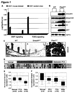
[00022] Figure 1 demonstrates that the loss of Pten prostate upregulated the
level of p-
Smad2/Smad3 and Smad4 expression. (A) Ingenuity Canonical Pathway Analysis of
differentially expressed genes between Ptenp /- mice (3331 probe sets, in
blue) were compared to
randomly drawn gene sets of equal size. (B) Western blot analysis of AP tissue
from each
genotype at 15 weeks shows pSmad2/3 level enhanced, Smad4 upregulation, and
Idl induction in
Ptenp -/- mice compared to control mice. (C) Immunohistochemistry analysis of
15-week-old APs
for Smad4 is performed demonstrating upregulation in Ptenp /- mice (Panel c)
compared to control
mice (Panel a). Smadp -/- mice used as negative control (Panel b). Scale bars,
50 gm. (D,E)
Onconmine analysis (http://www.oncomine.org/) of Smad4 expression between
human PCA and
metastasis. Heatmap of Smad4 differentially expressed in Yu et al prostate
expression dataset
(D). Boxed plot of Smad4 expression between human PCA and metastasis in Yu et
al prostate
expression dataset and Dhanasekaran et al (2001) prostate expression dataset
(E).
[00023] Figure 2 demonstrate that the loss of Smad4 does not initiate prostate
tumors but
renders Pten-deficient carcinomas lethal. (A) Histopathological analysis
(haematoxylin/eosin
staining) of anterior prostates (AP) in WT, Smad4 and Pten single and double
mutants at 9 weeks
of age reveals normal glands in WT and Smadp /- mice but PIN lesions in Ptenp -
/- mice and
6
CA 02730614 2011-01-12
WO 2010/009337 PCT/US2009/050885
invasion (arrow) in Ptenp -'-; Smadp /- mice. Scale bars, 50 gm. (B) Kaplan-
Meier overall
cumulative survival analysis. A statistically significant decrease in lifespan
(P<0.0001) compared
with the Ptenp /- cohort (n=28) was found for the Ptenp /-; Smadp /- cohort
(n=26) (asterisk). (C)
Gross anatomy of representative WT, Smadp /-, Ptenp /-, and Ptenp -/-; Smadp /-
anterior prostate
or prostate tumor at 22 weeks of age. Scale bars, 0.5 cm.
[00024] Figure 3 demonstrates that the loss of Smad4 enhanced proliferation
and circumvented
Pten-loss-induced cellular senescence. (A) Histopathological and proliferation
analysis of 15-
week-old Al's demonstrated increase in proliferation at some invasion foci
(arrow, panel e) in
Ptenp -/-; Smadp -/- double mutants (panel j). Tunel analysis of 15-week-old
Al's showed no
significant difference in Ptenp -/-; Smadp /- double mutants (panel i,j) and
Ptenp /- prostate tumors
(panel h). H&E, haematoxylin/eosin. Scale bars, 50 gm. (B) Loss of Smad4
circumvented Pten-
loss-induced cellular senescence. 3-Gal staining analysis of 15-week-old APs.
Scale bars, 100
gm. (C) Quantification of brdu pulse labeling of 15-week-old APs done as in
(A,f-j).
Representative sections from three mice were counted for each genotype. (D)
Quantification of
TUNEL assay for apoptosis in the AP at 15 weeks. Representative sections from
three mice were
counted for each genotype. (E) Quantification of the 3-Gal staining seen on AP
sections at 15
weeks done as in (B). Representative sections from three mice were counted for
each genotype.
Error bars in C-E represent s.d. for a representative experiment performed in
triplicate. Asterisk
indicates statistical significance between Ptenp /-; Smadp /- double mutants
and Ptenp /- (P< 0.05).
[00025] Figure 4 demonstrates that the loss of Smad4 leads to Pten-deficient
carcinomas
progress to metastasis to lymph nodes and lung with complete penetrance. (A)
Metastasis-free
survival curve (Kaplan-Meier plot) of prostate cancer. Metastasis foci in
lumbar lymph nodes
and/or lungs was found only in the Ptenp /-; Smadp /- cohort from 16 to 32
weeks of age. A
statistically significant (P<0.0001) compared with the Ptenp /- cohort (n=25)
was found for the
Ptenp -/-; SmadpG/- cohort (n=25) (asterisk) which with complete penetrance of
metastasis. (B)
Gross anatomy of representative lumbar lymph modes (dashed circle) and lung
with metastasis
foci (dark arrows). Scale bars, 0.5 cm. (C) H&E stained sections show
metastatic prostate cancer
cells in the lymph node (dark arrows) and lung. Immunohistochemical analyses
show that
metastatic cells in lymph node and lung are CK8 positive and AR positive
(prostate epithelial
markers). Scale bars, 50 gm. Mets, metastasis; LN, lymph node.
[00026] Figure 5 demonstrates that the 284 PCDETERMINANTS from Table IA
predict
human prostate cancer aggressiveness and metastasis. In this particular
experiment, the 284
7
CA 02730614 2011-01-12
WO 2010/009337 PCT/US2009/050885
PCDETERMINTS listed on Table IA were derived from a comparison of 3 tumor
samples from
Pten and 3 tumor samples form Pten Smad4. The 284 PCDETERMINANTS from Table IA
were
evaluated for prognostic utility from the Glinsky et al (2004) prostate cancer
gene expression data
set. Biochemical recurrence (BCR) was defined by PSA levels (>0.2 ng/ml).
Patient samples were
categorized into two major clusters (High-risk and Low-Risk group) defined by
the 284
PCDETERMINANTS listed on Table IA.
[00027] Figure 6 illustrates that Cell Movement genes are differentially
expressed in the
metastastic Smad4/Pten prostate tumors compares to indolent Pten tumors.
Ingenuity Pathway
Analysis (IPA) analysis on molecular functions of the differential expressed
genes revealed that
the cell movement genes ranks #18 vs. #1 for the Smad4/Pten prostate tumors
when either are
compared to Pten tumors. (A) IPA on molecular functions of differentially
expressed genes
between Ptenp'-I ; Smad4p'-I double mutants and Ptenp'-I mice reveals that
those genes have roles
in cell movement, Cell Death, Cellular Growth and Proliferation, Cell-To-Cell
Signaling and
Interaction, Cellular Development, Cell Morphology, Cell Cycle, Cell
Signaling, Post-
Translational Modification, Lipid Metabolism, Small Molecule Biochemistry,
Drug Metabolism,
Vitamin and Mineral Metabolism, Cellular Function and Maintenance, Molecular
Transport, Gene
Expression, DNA Replication and Repair. Cell movement genes ranks #1. (B) IPA
analysis on
molecular functions of the differential expressed genes expressed between
Ptenp'-I; p53p'-I- double
mutants and Ptenp'-I mice reveals that those genes have roles in Cell Death,
Gene Expression,
Cellular Growth and Proliferation, Cellular Development, Amino Acid
Metabolism, Post-
Translational Modification, Small Molecule Biochemistry, Cellular Function and
Maintenance,
Cell Morphology, Cellular Assembly and Organization, Cell Cycle, Cell-To-Cell
Signaling and
Interaction, Drug Metabolism, Lipid Metabolism, Molecular Transport, Cellular
Compromise,
Antigen Presentation, Cellular Movement, Carbohydrate Metabolism, RNA Damage
and Repair,
DNA Replication, and Repair, Nucleic Acid Metabolism, Cell Signaling, Protein
Synthesis. In
contrast to the Ptenpc-l ; Smad4' tumors, the IPA of Ptenpc-l ; p53p'" tumors
show that cell
movement genes ranks #18.
[00028] Figure 7 illustrates gene profiling and promoter analysis reveals a
subset of 66 putative
Smad4 target genes differentially expressed between Ptenp -/-; Smadp /- double
mutants and Ptenp
i- mice. (A) 66 genes differentially expressed between Ptenp /-; Smadp -/-
double mutants and
Ptenp -/- mice. (B) Ingenuity Pathway Analysis (IPA) on molecular functions
reveals that these 66
genes have roles in cell movement, cancer, cellular growth and proliferation,
and ell death.
8
CA 02730614 2011-01-12
WO 2010/009337 PCT/US2009/050885
[00029] Figure 8 illustrates a 17 Smad-target gene signature can predictor
cancer
aggressiveness and metastasis. (A) A diagram- representation of the
development of 17 Smad
target gene signature. Computer analysis reveal that there are 66 putative
Snnad-target gene
among 284 genes differentially expressed between Ptenp /-; Smadp /- double
mutants and Ptenp /-
mice. A 17 gene signature was developed based on the overlap with a human
metastatic PCA
dataset (B) 17 genes differentially expressed between Ptenp /-; Smadp -/-
double mutants and
Ptenp -/- mice. (C) The 17 putative Smad target genes were subsequently
evaluated for prognostic
utility on a prostate cancer gene expression data set. Hierarchical clustering
of the tumor samples
(columns) and genes (rows) is provided. Red indicates high relative levels of
gene expression,
while green represents low relative levels of gene expression. Horizontal bars
above the heat maps
indicate the recurrence status of each patient (1, biochemical or tumor
recurrence; 0, recurrence-
free). Patients were categorized into two major clusters defined by the 17-
gene signature. Lymph
node and other distal metastasis are indicated by arrow in red. (D) Kaplan-
Meier survival analysis
based on the groups defined by the 17-gene cluster. (E, F) Same as C, 17-gene
signature was
evaluated in a breast adenocarcinoma dataset. Kaplan-Meier analysis was
conducted for survival
probability (E) and metastasis-free survival (F) based on the groups defined
by the 17-gene
cluster.
[00030] Figure 9 illustrates that loss of Smad4 does not initiate prostate
tumors up to 2 years
age. Histopathological analysis (haematoxylin/eosin staining) of anterior
prostates (AP) in Smad4
single mutants at one year (A) and two year of age (B) reveals normal glands
in Smadp /- mice.
Scale bars, 50 gm.
[00031] Figure 10 shows histopathological analysis of representative
hydronephrosis in Ptenp -/-
; Smadp -/- mice. (A) Gross anatomy of representative Ptenp /-; Smadp /- with
prostate tumor at 26
weeks of age with a huge prostate tumor (dashed circle). Scale bars, 2 cm.
(B,C)
Histopathological analysis of representative kidney from Ptenp /- mice (B) and
Ptenp -/-; Smadp -/-
mice with hydronephrosis (arrow) (C). Stained with hematoxylin and eosin
(H&E). Scale bars, 1
mm.
[00032] Figure 11 shows Microarray analysis of a subset of 284 (See Table IA)
cancer biology
related genes differentially expressed between Ptenp /-; Smadp -/- double
mutants and Ptenp /-
mice. (A) 284 genes differentially expressed between Ptenp -/-; Smadp /-
double mutants and
Ptenp -/- mice. (B) Ingenuity Pathway Analysis (IPA) on molecular functions
reveals that these
9
CA 02730614 2011-01-12
WO 2010/009337 PCT/US2009/050885
284 genes have roles in cellular movement, cancer, cellular growth and
proliferation, and cell
death.
[00033] Figure 12 (A) The 66 putative Smad target genes were subsequently
evaluated for
prognostic utility on a prostate cancer gene expression data set. Hierarchical
clustering of the
tumor samples (columns) and genes (rows) is provided. Red indicates high
relative levels of gene
expression, while green represents low relative levels of gene expression.
Horizontal bars above
the heat maps indicate the recurrence status of each patient (1, biochemical
or tumor recurrence; 0,
recurrence-free). Patients were categorized into two major clusters defined by
the 66-gene
signature. Lymph node and other distal metastasis are indicated by arrow in
red. (B) Kaplan-
Meier survival analysis based on the groups defined by the 66-gene cluster.
[00034] Figure 13 shows that Smad4 loss can circumvent cellular senescence
elicited by Pten
loss in primary mouse embryonic fibroblasts (MEFs) through p53-dependent
pathway. (A)
senescence staining of WT (Panel a), Smad-/- (Panel b), Pten i- (Panel c), and
Pten /-; Smad-/- (Panel
d) MEFs. Representative sections from three independent MEFs of each genotype.
(B)
Quantification of the 3-Gal staining. Error bars represent s.d. for a
representative experiment
performed in triplicate. Asterisk indicates statistical significance between
Ptenp /- and Ptenp /-;
Smadp /- double mutants (P< 0.05). (C) Western blot analysis of MEFs from each
genotype
shows p53 expression level for a representative experiment performed in
duplicate (of more than
four mice per genotype). Actin was used as an internal loading control.
[00035] Figure 14 shows prostate epithelial cells from Ptenp /-; Smadp -/-
double mutants form
orthotopic metastatic tumors with prostate epithelial cell markers in nude
mice. (A) Orthotopic
injection of prostate epithelial cells from Ptenp -/-; Smadp /- double mutants
form tumor in prostate
(dashed circle) and form lung metastasis (arrows). Scale bars, 1 cm. (B)
Immunohistochemical
analyses show that orthotopic tumors and lung metastasis are CK8 positive and
#AR positive
(prostate epithelial markers). Scale bars, 50 gm.
[00036] Figure 15 shows Prostate epithelial cells from Ptenp /-; Smadp -/-
double mutants form
orthotopic metastatic tumors with prostate epithelial cell markers in nude
mice. (A) Kidney
implantation of prostate epithelial cells from Ptenp -/-; Smadp /- double
mutants form tumor in
prostate (dashed circle) and form lung metastasis (arrows). Scale bars, 1 cm.
(B)
Immunohistochemical analyses show that kidney tumors and lung metastasis are
CK8 positive and
#AR positive (prostate epithelial markers). Scale bars, 50 gm
CA 02730614 2011-01-12
WO 2010/009337 PCT/US2009/050885
[00037] Figure 16 shows that restoration of Smad4 in Pten-Smad4 double null
prostate tumor
cells decreases cell viability when treated with TGF(31. (A) The restoration
of Smad4 in Smad4-
deficient prostate cancer cells decreases cell viability upon treatment with
TGF(31. Parental
control cells (Contl) and Smad4-Tet on cells (Smad4) were treated with
0.016ng/mL, 0.031ng/mL,
0.063ng/mL, 0.125ng/mL, 0.25ng/mL,0.5ng/mL TGF(31in the presence or absence of
1 gg/mL
doxycycline (Dox) in 5% charcoal-stripped FBS -containing medium, and then
cell viability was
assayed by adenosine triphosphate quantitation. Error bars represent s.d. for
a representative
experiment performed in triplicate. Black bars, control parental line without
Dox; blue bars,
control parental line with Dox; red bars, Smad4 tet-on line without Dox; green
bars, Smad4 tet on
line with Dox. (B) Western blot analysis of Smad4 expression upon Dox
treatment shows Smad4
expression in Smad4 tet-on line with treatment of Dox or without the treatment
of Dox. Ran was
used as an internal loading control. (C) Morphology of cells with or without
TGF(31 treatmnent.
The cells were photographed after 4 d of treatment with TGF(31 or vehicle.
[00038] Figure 17 shows loss of Smad4 circumvented Pten-loss-induced
autophagy. (A)
Morphology of cells with or without TGF(31 treatment. The cells were
photographed after 3 days
of treatment with TGF(31 or vehicle. (B) Transmission electron microscopy of
prostate tumor
cells from Ptenp -/-; Smadp /- double mutants and Ptenp /- mouse at 15weeks of
age.
[00039] Figure 18 demonstrates that Pten/Smad4 double mutant mice with hormone
ablation
via castration developed hormone-refractory metastatic PCA. (A) Kaplan-Meier
overall
cumulative survival analysis of castrated animals. A statistically significant
extension in lifespan
(P<0.0001) compared with the castration-free Ptenp -/-; Smadp /- cohort (n=20)
was found for the
castrated Ptenp /-; Smadp /- cohort (n=22) (asterisk). The arrow indicates the
castration at 15
weeks of age. (B) Castration did not block metastasis of prostate cancer in
Ptenp /-; Smadp -/-
double mutants. A higher magnified picture (boxed region) is shown on the
right (panel b).
Histopathological analysis of representative lymph node metastasis. Scale
bars, 200 gm for panel a
and 50 gm for panel b. (C) Histopathological and proliferation analysis
revealed high proliferation
(brown staining) in castrated Ptenp /-;Smadp -/- double mutants, compared with
castrated WT and
Ptenp -/- mice. H&E, haematoxylin/eosin. Scale bars, 50 gm. Analysis was
performed on 23-
week-old mice which were castrated at 15-week-old. (D) Quantification of brdu
pulse labeling of
23-week-old mice which were castrated at 15-week-old. Representative sections
from three mice
were counted for each genotype. Asterisk indicates statistical significance
between Ptenp -/-;
Smadp /- double mutants and Ptenp /- (P< 0.05).
11
CA 02730614 2011-01-12
WO 2010/009337 PCT/US2009/050885
[00040] Figure 19 illustrates the model of how Pten and Smad4 cooperate to
control prostate
cancer initiation and progression. Pten loss in prostate result in the
development of prostate
tumor, but further progression was suppressed by proliferative
block/senescence induced by Pten
loss. Both Pten and Smad4 loss circumvent the Pten-loss-induced proliferative
block/senescence
and possibly other cellular and intracellular suppression mechanisms such as
those impeding
cellular movement through PCDETERMINANTS 1-372 or a subset of PCDETERMINANTS 1-
372, and eventually led to the prostate tumor cells to progress to metastasis.
[00041] Figure 20 demonstrates cross-species triangulated differentially
expressed genes
between Ptenp'--; Smad4' double mutants and Pten' mice are linked to clinical
outcome in
human PCA. (A) A diagram representation of the development of a 56 gene set
based on the
overlap of differentially expressed genes between Ptenp'-I ; Smad4p'-I double
mutants and Ptenpc-l
mice (Table 1B) with a human metastatic PCA dataset 19. (B) The 56 gene set
(TABLE 7) was
subsequently evaluated for prognostic utility on a prostate cancer gene
expression data set. Patient
samples were categorized into two major clusters (low risk group and high risk
group) defined by
the 56-gene signature. Kaplan-Meier analysis of biochemical recurrence (BCR)
PSA level (>0.2
ng/ml) based on the groups defined by the 56-gene cluster. A statistically
significant for BCR
PSA recurrence-free survival (P=0.0018) compared with the "low-risk" cohort
was found for the
"high-risk" cohort.
[00042] Figure 21 illustrates approaches to identify PCDETERMINANTS that
functionally
drive or inhibit invasion in vitro.
[00043] Figure 22 demonstrates use of the invasion assay to functionally
validate candidate
genes. A representative Boyden chamber invasion assay with PC3 cells
overexpressing SPP1 and
or GFP control in triplicates. (A) Enforced expression of SPP1 confirmed its
capability to
significantly enhance invasive activity of human PCA PC3 cells by invasion
assay. (B) Bar graph
indicates statistical significance between enforced SPP1 and GFP control(P<
0.05). (C) The table
confirms the assay identifies invasion-promoting genes that are annotated as
being involved in
cellular movement, but also genes not classified as being involved in movement
yet drive invasive
and metastatic properties in vitro. A significantly higher frequency (P=0.02,
Fisher's Exact Test)
of invasion-validated PCDETERMINANTS are annotated as cellular movement genes
compared
to those not from the cellular movement annotated genes.
[00044] Figure 23 demonstrates a FOUR (4) PCDETERMINANT gene signature PTEN-
SMAD4- Cyclin D1-SPP1 which was informed by the Pten/Smad4 transcriptome data,
the
12
CA 02730614 2011-01-12
WO 2010/009337 PCT/US2009/050885
histopathological data and invasion validation data is linked to clinical
outcome in human PCA.
(A) Dysregulated Pten and Smad4 expression together with the related Cyclin D1
(proliferation/senescence) and SPP1 (motility network) was subsequently shown
to be correlated
with the human prostate cancer progression on a prostate cancer gene
expression data set. Patient
samples were categorized into two major clusters by K-mean (High-risk and Low
risk groups)
defined by the PTEN, SMAD4, Cyclin D1, and SPP1 signature. High-risk group
patient showed
statistically significant in biochemical recurrence (BCR) PSA level (>0.2
ng/ml) by Kaplan-Meier
analysis. (B) The significant correlation of PTEN, SMAD4, Cyclin D1, and SPP1
signature in
PCA progression was validated in an independent Physicians' Health Study (PHS)
dataset with c-
statistic. The PTEN, SMAD4, Cyclin D1, and SPP1 show similar power to Gleason
score in the
prediction of lethal outcomes. The addition of PTEN, SMAD4, Cyclin D1, and
SPP1 genes to
Gleason significantly improves prediction of lethal outcomes over the model of
Gleason alone in
PHS. Moreover, PTEN, SMAD4, Cyclin D1, and SPP1 4-gene set ranked as the most
enriched
among 244 bidirectional signatures curated in the Molecular Signature
Databases of the Broad
Institute (MSigDB, version 2.5), indicating the robust significance of this 4
gene signature in
prediction of lethal outcome.
[00045] Figure 24 demonstrates cross-species triangulated differentially
expressed genes
between Ptenp'-I ; Smad4' double mutants and Pten' mice are linked to clinical
outcome in
human breast cancer. (A) The 56 gene set (TABLE 7) was subsequently evaluated
for
prognostic utility on a breast adenocarcinoma dataset. Patient samples were
categorized into two
major clusters (low risk group and high risk group) defined by the 56-gene
signature. Kaplan-
Meier analysis was conducted for survival probability (p= 0.00358) (A) and
metastasis-free
survival (p= 00492) (B) based on the groups defined by the 56-gene cluster.
[00046] Figure 25 demonstrates that both prostate and breast cancer
progression correlated
PCDETERMINANTS are highly linked to clinical outcome in human breast cancer.
(A) The 20
PCDETERMINANTS exhibiting progression correlated expression in both prostate
cancer and
breast cancer (Table 6) was evaluated for prognostic utility on a breast
adenocarcinoma dataset.
Patient samples were categorized into two major clusters (low risk group and
high risk group)
defined by the 20 progression correlated-gene signature. Kaplan-Meier analysis
was conducted
for survival probability (p= 2.93e-11) (A) and metastasis-free survival (p=
4.62e-10) (B) based on
the groups defined by the 20 PCDETERMINANTS .
13
CA 02730614 2011-01-12
WO 2010/009337 PCT/US2009/050885
DETAILED DESCRIPTION OF THE INVENTION
[00047] The present invention relates to the identification of signatures
associated with and
PCDETERMINANTS conferring subjects with metastatic prostate cancer or are at
risk for
developing metastatic prostate cancer. The invention further provides a murine
mouse model for
invasive and metastatic prostate cancer, where the mouse prostate epithelium
sustains deletion, or
other means of mutational or epigenetic extinction of an initiating lesion
such as the Pten and
Smad4 gene. It would be recognized by one skilled in the art that other
initiating lesion, including
over-expression of oncogene trangenes could be coupled to the Smad4 deletion
to enable
malignant progression. This mouse model can be used to identify cancer
detection biomarkers.
[00048] Human cancers harbor innumerable genetic and epigenetic alterations
presenting
formidable challenges in deciphering those changes that drive the malignant
process and dictate a
given tumor's clinical behavior. The need for accurately predictive biomarkers
reflective of a
tumor's malignant potential is evident across many cancer types, particularly
prostate cancer,
where current management algorithms result in either under-treatment with
consequent risk of
death or exposure to unnecessary morbid treatments.
[00049] Genetically engineered mouse models have been shown to be tremendously
powerful
as "filters" to mine highly complex genomic datasets in human. In particular,
these refined
genetically engineered mouse models of human cancers have been documented in
high-resolution
comparative oncogenomic analyses to harbor substantial overlap in cancer-
associated
transcriptional and chromosomal DNA aberrations patterns - the latter
resulting in the rapid and
efficient identification of many novel cancer genes. Similar cross-species
comparisons of the
serum proteome have also proven effective in the identification of early
detection biomarkers for
pancreas cancer in humans. Thus, it stands to reasons that development of a
valid mouse model
recapitulating the disease state of metastasis driven by bona fide human
prostate cancer genes will
greatly facilitate our efforts to develop prognostic and early detection
biomarkers and possible
therapeutic targets.
[00050] Global transcriptome analyses of indolent Pten deficient prostate PIN
lesions inferred
the presence of a Smad4-dependent checkpoint which induces a senescence
response in setting of
Pten inactivation, blocking progression beyond PIN. Concomitant Smad4 deletion
in the mouse
prostate epithelium along with Pten deletion indeed generated a fulminant
metastatic prostate
model with short latency, providing unequivocal genetic proof of this
hypothesis. That this is a
mouse model of metastatic prostate cancers driven by bona fide prostate tumor
suppressors is
14
CA 02730614 2011-01-12
WO 2010/009337 PCT/US2009/050885
supported by the demonstration of consistent Smad4 downregulation during
progression from
primary to metastatic PCA in human. The validity of this model was further re-
enforced by
demonstration that the 17 predicted direct targets of Smad4 conserved across
two species are
capable of stratifying human prostate and breast adenocarcinomas into two
groups with significant
differences in outcome as measured by recurrence or survivals. Therefore, the
inventors have
established a bona fide genetically engineered mouse model of metastatic PCA,
enabling future
mechanistic studies as well as comparative genomic and proteomic analyses in
searches for
prognostic and early-detection biomarkers.
[00051] It has been established that loss of Pten function is one of the most
significant genetic
events in prostate carcinogenesis. Loss of Pten results in prostate
tumorigenesis in the mouse
prostate; however, it also provokes cellular senescence which may function as
a further level of
tumor suppressor to block the tumor cells progression to an invasive stage.
Overriding
senescence induced by Pten through inactivation of p53 contributes to the
progression of prostate
tumors from an indolent lesion to an invasive tumor. The inventors have
discovered that Smad4
loss also can circumvent cellular senescence elicited by Pten loss. Overriding
senescence by loss
of Smad4 is cooperative to Pten loss and may contribute its role in the
promotion of tumor cells.
This is also in agreement with the previous report that circumvention of
cellular senescence by
p53 loss is cooperative to Pten loss and contributes to the prostate tumor
progression to a modestly
invasive but non-metastatic lesion.. This unique Pten/Smad4 model system
therefore provides a
tool to further dissect the molecular events for this important tumor
biological process in the
future.
[00052] Although circumvention of senescence results in Pten/Smad4 double
mutant mouse
prostate tumor cell progression to an invasive and metastatic state,
circumvention of senescence in
mouse model with Pten/p53 inactivation does not result in metastasis.
Inactivation of Pten alone
in mouse prostate can generate some feeble metastasis phenotype at very old
age (more than one
year) in a small portion of Pten mice (2 in 8). These observations indicated
that additional genetic
or epigenetic alterations besides Pten loss are needed for the prostate tumor
cells to achieve a
metastatic state. Circumvention of cellular senescence may be a pre-requisite
for progression but
other biological processes are likely needed such as deactivation of autophagy
to achieve a robust
metastatic state. In support of the presence of other biological processes, we
observed that
reconstitution of Smad4 in the Pten/Smad deficient tumor cells does not
reinstate senescence yet
renders cells non-metastatic. Specifically, we established an inducible Smad4
tet-on system to
CA 02730614 2011-01-12
WO 2010/009337 PCT/US2009/050885
restore Smad4 expression in a time-dependent and dose dependent manner. It was
found that
restoration of Smad4 can sensitize the tumor to cell death in response to the
treatment of TGF(3.
[00053] The canonical TGF(3-Smad pathway starts from the ligand-receptor
complex and ends
in the nucleus. Upon TGF(3 superfamily ligand binding, receptor-phosporylated
R-Smads
oligomerizes with Smad4 and translocate to the nucleus and bind directly to
Smad-binding
elements on DNA where they can induce or repress a diverse array of genes. In
benign prostatic
epithelia, by eliciting differentiation, inhibiting proliferation, and
inducing apoptosis, TGF-13
provides a mechanism to maintain homeostasis in the prostate. Thus, it was
speculated that this
major arm of the TGF(3 plays a critical role in the prostate tumor progression
suppression.. The
tumor suppressor role of TGF(3 signaling is underscored by the presence of
inactivating TGF(3
receptor mutations and the extinction of Smad2, Smad3, and Smad4 proteins in
multiple cancers
including prostate cancer. Although TGF(3 was shown to inhibit many normal
cell types and
tumor cell growth, TGF(3 was also reported to enhance malignant potential of
epithelial tumors,
including proliferation, migration, and epithelial-to-mesenchymal transition
(EMT)-a process by
which advanced carcinomas acquire a highly invasive, undifferentiated and
metastatic phenotype.
Most recently, it has been demonstrated that TGF(3 in the breast tumor
microenvironment can
prime cancer cells for metastasis to the lungs though induction of
angiopoietin-like 4 (ANGPTL4)
by TGF(3 via the Smad signaling pathway. These paradoxical activities of tumor
suppression and
promotion are probably dependent on the activities of other signaling pathways
in given cells,
which are dictated by the different cell contexts as well as the interplay
with other tissue. The
Pten/Smad4 model has now clarified the role of the TGFb pathway in prostate
cancer by clearly
showing that Smad4 loss is not sufficient alone to initiate the development of
prostate lesion, but
promotes acceleration and progression of prostate tumor to metastasis with
complete penetrance,
at least on the background of Pten deficiency (Figure 3). The Pten/Smad4 model
study clearly
demonstrated that Smad4 loss can override the senescence induced by Pten loss.
Since override
senescence by p53 loss in Pten deficiency background result in progression of
indolent prostate
tumor to invasive lesion, but not to metastasis. Senescence is thus considered
to be an early
barrier during the prostate tumorigenesis from indolent to invasive status. As
restoration of
Smad4 back into the Pten/Smad4 double mutant prostate tumor cells did not
restore the
senescence (data not shown). However, restoration of Smad4 decreased the
viability of the cells
upon the treatment of TGF(31. The senescence barrier may be, therefore, a
transient cellular
response to the oncogenic signal(s) to block tumor progression.
16
CA 02730614 2011-01-12
WO 2010/009337 PCT/US2009/050885
[00054] Additionally, molecularly comparative transcriptomic analyses of
equivalent early
stage Pten and Pten/Smad null prostate tumors (n=5 for each genotype) revealed
differential
expression of 372 genes of which at least 66 genes contain Smad binding
elements in their
promoters. Through cross-species integration with copy number profiles of
human metastatic
prostate tumors, we identified 17 of these Smad4 targets that are strongly
associated with risk of
recurrence in human prostate cancer and with metastasis risk and survival in
breast cancer, thereby
supporting the human relevance of this novel metastatic prostate model and its
use in the
discovery of genetic PCDETERMINANTS governing disease progression across many
tumor
types through comparative oncogenomics.
[00055] Accordingly, the invention provides an animal model for metastatic
prostate cancer.
The animal model of the instant invention thus finds particular utility as a
screening tool to
elucidate the mechanisms of the various genes involved in both normal and
diseased patient
populations.
[00056] The invention also provides methods for identifying subjects who have
metastatic
prostate cancer, or who at risk for experiencing metastatic prostate cancer by
the detection of
PCDETERMINANTS associated with the metastatic tumor, including those subjects
who are
asymptomatic for the metastatic tumor. These signatures and PCDETERMINANTS are
also
useful for monitoring subjects undergoing treatments and therapies for cancer,
and for selecting or
modifying therapies and treatments that would be efficacious in subjects
having cancer, wherein
selection and use of such treatments and therapies slow the progression of the
tumor, or
substantially delay or prevent its onset, or reduce or prevent the incidence
of tumor metastasis.
[00057] Definitions
[00058] "Accuracy" refers to the degree of conformity of a measured or
calculated quantity (a
test reported value) to its actual (or true) value. Clinical accuracy relates
to the proportion of true
outcomes (true positives (TP) or true negatives (TN) versus misclassified
outcomes (false
positives (FP) or false negatives (FN)), and may be stated as a sensitivity,
specificity, positive
predictive values (PPV) or negative predictive values (NPV), or as a
likelihood, odds ratio, among
other measures.
[00059] "PCDETERMINANTS in the context of the present invention encompasses,
without
limitation, proteins, nucleic acids, and metabolites, together with their
polymorphisms, mutations,
variants, modifications, subunits, fragments, protein-ligand complexes, and
degradation products,
17
CA 02730614 2011-01-12
WO 2010/009337 PCT/US2009/050885
protein-ligand complexes, elements, related metabolites, and other analytes or
sample-derived
measures. PCDETERMINANTS can also include mutated proteins or mutated nucleic
acids.
PCDETERMINANTS also encompass non-blood borne factors or non-analyte
physiological
markers of health status, such as "clinical parameters" defined herein, as
well as "traditional
laboratory risk factors", also defined herein. PCDETERMINANTS also include any
calculated
indices created mathematically or combinations of any one or more of the
foregoing
measurements, including temporal trends and differences. Where available, and
unless otherwise
described herein, PCDETERMINANTS which are gene products are identified based
on the
official letter abbreviation or gene symbol assigned by the international
Human Genome
Organization Naming Committee (HGNC) and listed at the date of this filing at
the US National
Center for Biotechnology Information (NCBI) web site
(http://www.ncbi.nlm.nih.gov/sites/entrez?db=gene ), also known as Entrez
Gene.
[00060] "PCDETERMINANT" OR "PCDETERMINANTS " encompass one or more of all
nucleic acids or polypeptides whose levels are changed in subjects who have
metastatic prostate
cancer or are predisposed to developing metastatic prostate cancer, or at risk
of metastatic prostate
cancer. Individual PCDETERMINANTS are summarized in Table 1B and are
collectively
referred to herein as, inter alia, "metastatic tumor-associated proteins",
"PCDETERMINANT
polypeptides", or "PCDETERMINANT proteins". The corresponding nucleic acids
encoding the
polypeptides are referred to as "metastatic tumor-associated nucleic acids",
"metastatic tumor-
associated genes", "PCDETERMINANT nucleic acids", or "PCDETERMINANT genes".
Unless
indicated otherwise, "PCDETERMINANT", "metastatic tumor -associated proteins",
"metastatic
tumor -associated nucleic acids" are meant to refer to any of the sequences
disclosed herein. The
corresponding metabolites of the PCDETERMINANT proteins or nucleic acids can
also be
measured, as well as any of the aforementioned traditional risk marker
metabolites.
[00061] Physiological markers of health status (e.g., such as age, family
history, and other
measurements commonly used as traditional risk factors) are referred to as
"PCDETERMINANT
physiology". Calculated indices created from mathematically combining
measurements of one or
more, preferably two or more of the aforementioned classes of PCDETERMINANTS
are referred
to as "PCDETERMINANT indices".
[00062] "Clinical parameters" encompasses all non-sample or non-analyte
biomarkers of
subject health status or other characteristics, such as, without limitation,
age (Age), ethnicity
(RACE), gender (Sex), or family history (FamHX).
18
CA 02730614 2011-01-12
WO 2010/009337 PCT/US2009/050885
[00063] "Circulating endothelial cell" ("CEC") is an endothelial cell from the
inner wall of
blood vessels which sheds into the bloodstream under certain circumstances,
including
inflammation, and contributes to the formation of new vasculature associated
with cancer
pathogenesis. CECs may be useful as a marker of tumor progression and/or
response to
antiangiogenic therapy.
[00064] "Circulating tumor cell" ("CTC") is a tumor cell of epithelial origin
which is shed
from the primary tumor upon metastasis, and enters the circulation. The number
of circulating
tumor cells in peripheral blood is associated with prognosis in patients with
metastatic cancer.
These cells can be separated and quantified using immunologic methods that
detect epithelial
cells, and their expression of PCDETERMINANTS can be quantified by qRT-PCR,
immunofluorescence, or other approaches.
[00065] "FN" is false negative, which for a disease state test means
classifying a disease subject
incorrectly as non-disease or normal.
[00066] "FP" is false positive, which for a disease state test means
classifying a normal subject
incorrectly as having disease.
[00067] A "formula," "algorithm," or "model" is any mathematical equation,
algorithmic,
analytical or programmed process, or statistical technique that takes one or
more continuous or
categorical inputs (herein called "parameters") and calculates an output
value, sometimes referred
to as an "index" or "index value." Non-limiting examples of "formulas" include
sums, ratios, and
regression operators, such as coefficients or exponents, biomarker value
transformations and
normalizations (including, without limitation, those normalization schemes
based on clinical
parameters, such as gender, age, or ethnicity), rules and guidelines,
statistical classification
models, and neural networks trained on historical populations. Of particular
use in combining
PCDETERMINANTS and other PCDETERMINANTS are linear and non-linear equations
and
statistical classification analyses to determine the relationship between
levels of
PCDETERMINANTS detected in a subject sample and the subject's risk of
metastatic disease. In
panel and combination construction, of particular interest are structural and
synactic statistical
classification algorithms, and methods of risk index construction, utilizing
pattern recognition
features, including established techniques such as cross-correlation,
Principal Components
Analysis (PCA), factor rotation, Logistic Regression (LogReg), Linear
Discriminant Analysis
(LDA), Eigengene Linear Discriminant Analysis (ELDA), Support Vector Machines
(SVM),
Random Forest (RF), Recursive Partitioning Tree (RPART), as well as other
related decision tree
19
CA 02730614 2011-01-12
WO 2010/009337 PCT/US2009/050885
classification techniques, Shrunken Centroids (SC), StepAIC, Kth-Nearest
Neighbor, Boosting,
Decision Trees, Neural Networks, Bayesian Networks, Support Vector Machines,
and Hidden
Markov Models, among others. Other techniques may be used in survival and time
to event
hazard analysis, including Cox, Weibull, Kaplan-Meier and Greenwood models
well known to
those of skill in the art. Many of these techniques are useful either combined
with a
PCDETERMINANT selection technique, such as forward selection, backwards
selection, or
stepwise selection, complete enumeration of all potential panels of a given
size, genetic
algorithms, or they may themselves include biomarker selection methodologies
in their own
technique. These may be coupled with information criteria, such as Akaike's
Information
Criterion (AIC) or Bayes Information Criterion (BIC), in order to quantify the
tradeoff between
additional biomarkers and model improvement, and to aid in minimizing overfit.
The resulting
predictive models may be validated in other studies, or cross-validated in the
study they were
originally trained in, using such techniques as Bootstrap, Leave-One-Out (LOO)
and 10-Fold
cross-validation (10-Fold CV). At various steps, false discovery rates may be
estimated by value
permutation according to techniques known in the art. A "health economic
utility function" is a
formula that is derived from a combination of the expected probability of a
range of clinical
outcomes in an idealized applicable patient population, both before and after
the introduction of a
diagnostic or therapeutic intervention into the standard of care. It
encompasses estimates of the
accuracy, effectiveness and performance characteristics of such intervention,
and a cost and/or
value measurement (a utility) associated with each outcome, which may be
derived from actual
health system costs of care (services, supplies, devices and drugs, etc.)
and/or as an estimated
acceptable value per quality adjusted life year (QALY) resulting in each
outcome. The sum,
across all predicted outcomes, of the product of the predicted population size
for an outcome
multiplied by the respective outcome's expected utility is the total health
economic utility of a
given standard of care. The difference between (i) the total health economic
utility calculated for
the standard of care with the intervention versus (ii) the total health
economic utility for the
standard of care without the intervention results in an overall measure of the
health economic cost
or value of the intervention. This may itself be divided amongst the entire
patient group being
analyzed (or solely amongst the intervention group) to arrive at a cost per
unit intervention, and to
guide such decisions as market positioning, pricing, and assumptions of health
system acceptance.
Such health economic utility functions are commonly used to compare the cost-
effectiveness of
the intervention, but may also be transformed to estimate the acceptable value
per QALY the
CA 02730614 2011-01-12
WO 2010/009337 PCT/US2009/050885
health care system is willing to pay, or the acceptable cost-effective
clinical performance
characteristics required of a new intervention.
[00068] For diagnostic (or prognostic) interventions of the invention, as each
outcome (which
in a disease classifying diagnostic test may be a TP, FP, TN, or FN) bears a
different cost, a health
economic utility function may preferentially favor sensitivity over
specificity, or PPV over NPV
based on the clinical situation and individual outcome costs and value, and
thus provides another
measure of health economic performance and value which may be different from
more direct
clinical or analytical performance measures. These different measurements and
relative trade-offs
generally will converge only in the case of a perfect test, with zero error
rate (a.k.a., zero predicted
subject outcome misclassifications or FP and FN), which all performance
measures will favor over
imperfection, but to differing degrees.
[00069] "Measuring" or "measurement," or alternatively "detecting" or
"detection," means
assessing the presence, absence, quantity or amount (which can be an effective
amount) of either a
given substance within a clinical or subject-derived sample, including the
derivation of qualitative
or quantitative concentration levels of such substances, or otherwise
evaluating the values or
categorization of a subject's non-analyte clinical parameters.
[00070] "Negative predictive value" or "NPV" is calculated by TN/(TN + FN) or
the true
negative fraction of all negative test results. It also is inherently impacted
by the prevalence of the
disease and pre-test probability of the population intended to be tested.
[00071] See, e.g., O'Marcaigh AS, Jacobson RM, "Estimating The Predictive
Value Of A
Diagnostic Test, How To Prevent Misleading Or Confusing Results," Clin. Ped.
1993, 32(8): 485-
491, which discusses specificity, sensitivity, and positive and negative
predictive values of a test,
e.g., a clinical diagnostic test. Often, for binary disease state
classification approaches using a
continuous diagnostic test measurement, the sensitivity and specificity is
summarized by Receiver
Operating Characteristics (ROC) curves according to Pepe et al, "Limitations
of the Odds Ratio in
Gauging the Performance of a Diagnostic, Prognostic, or Screening Marker," Am.
J. Epidemiol
2004, 159 (9): 882-890, and summarized by the Area Under the Curve (AUC) or c-
statistic, an
indicator that allows representation of the sensitivity and specificity of a
test, assay, or method
over the entire range of test (or assay) cut points with just a single value.
See also, e.g., Shultz,
"Clinical Interpretation Of Laboratory Procedures," chapter 14 in Teitz,
Fundamentals of Clinical
Chemistry, Burtis and Ashwood (eds.), 4th edition 1996, W.B. Saunders Company,
pages 192-199;
and Zweig et al., "ROC Curve Analysis: An Example Showing The Relationships
Among Serum
21
CA 02730614 2011-01-12
WO 2010/009337 PCT/US2009/050885
Lipid And Apolipoprotein Concentrations In Identifying Subjects With Coronory
Artery Disease,"
Clin. Chem., 1992, 38(8): 1425-1428. An alternative approach using likelihood
functions, odds
ratios, information theory, predictive values, calibration (including goodness-
of-fit), and
reclassification measurements is summarized according to Cook, "Use and Misuse
of the Receiver
Operating Characteristic Curve in Risk Prediction," Circulation 2007, 115: 928-
935.
[00072] Finally, hazard ratios and absolute and relative risk ratios within
subject cohorts
defined by a test are a further measurement of clinical accuracy and utility.
Multiple methods are
frequently used to defining abnormal or disease values, including reference
limits, discrimination
limits, and risk thresholds.
[00073] "Analytical accuracy" refers to the reproducibility and predictability
of the
measurement process itself, and may be summarized in such measurements as
coefficients of
variation, and tests of concordance and calibration of the same samples or
controls with different
times, users, equipment and/or reagents. These and other considerations in
evaluating new
biomarkers are also summarized in Vasan, 2006.
[00074] "Performance" is a term that relates to the overall usefulness and
quality of a
diagnostic or prognostic test, including, among others, clinical and
analytical accuracy, other
analytical and process characteristics, such as use characteristics (e.g.,
stability, ease of use),
health economic value, and relative costs of components of the test. Any of
these factors may be
the source of superior performance and thus usefulness of the test, and may be
measured by
appropriate "performance metrics," such as AUC, time to result, shelf life,
etc. as relevant.
[00075] "Positive predictive value" or "PPV" is calculated by TP/(TP+FP) or
the true positive
fraction of all positive test results. It is inherently impacted by the
prevalence of the disease and
pre-test probability of the population intended to be tested.
[00076] "Risk" in the context of the present invention, relates to the
probability that an event
will occur over a specific time period, as in the conversion to metastatic
events, and can mean a
subject's "absolute" risk or "relative" risk. Absolute risk can be measured
with reference to either
actual observation post-measurement for the relevant time cohort, or with
reference to index
values developed from statistically valid historical cohorts that have been
followed for the relevant
time period. Relative risk refers to the ratio of absolute risks of a subject
compared either to the
absolute risks of low risk cohorts or an average population risk, which can
vary by how clinical
risk factors are assessed. Odds ratios, the proportion of positive events to
negative events for a
22
CA 02730614 2011-01-12
WO 2010/009337 PCT/US2009/050885
given test result, are also commonly used (odds are according to the formula
p/(1-p) where p is the
probability of event and (1- p) is the probability of no event) to no-
conversion.
[00077] "Risk evaluation," or "evaluation of risk" in the context of the
present invention
encompasses making a prediction of the probability, odds, or likelihood that
an event or disease
state may occur, the rate of occurrence of the event or conversion from one
disease state to
another, i.e., from a primary tumor to metastatic prostate cancer or to one at
risk of developing a
metastatic, or from at risk of a primary metastatic event to a more secondary
metastatic event.
Risk evaluation can also comprise prediction of future clinical parameters,
traditional laboratory
risk factor values, or other indices of cancer, either in absolute or relative
terms in reference to a
previously measured population. The methods of the present invention may be
used to make
continuous or categorical measurements of the risk of metastatic prostate
cancer thus diagnosing
and defining the risk spectrum of a category of subjects defined as being at
risk for metastatic
tumor. In the categorical scenario, the invention can be used to discriminate
between normal and
other subject cohorts at higher risk for metastatic tumors. Such differing use
may require different
PCDETERMINANT combinations and individualized panels, mathematical algorithms,
and/or
cut-off points, but be subject to the same aforementioned measurements of
accuracy and
performance for the respective intended use.
[00078] A "sample" in the context of the present invention is a biological
sample isolated from
a subject and can include, by way of example and not limitation, tissue
biopsies, whole blood,
serum, plasma, blood cells, endothelial cells, circulating tumor cells,
lymphatic fluid, ascites fluid,
interstitial fluid (also known as "extracellular fluid" and encompasses the
fluid found in spaces
between cells, including, inter alia, gingival cevicular fluid), bone marrow,
cerebrospinal fluid
(CSF), saliva, mucous, sputum, sweat, urine, or any other secretion,
excretion, or other bodily
fluids.
[00079] "Sensitivity" is calculated by TP/(TP+FN) or the true positive
fraction of disease
subjects.
[00080] "Specificity" is calculated by TN/(TN+FP) or the true negative
fraction of non-disease
or normal subjects.
[00081] By "statistically significant", it is meant that the alteration is
greater than what might
be expected to happen by chance alone (which could be a "false positive").
Statistical significance
can be determined by any method known in the art. Commonly used measures of
significance
include the p-value, which presents the probability of obtaining a result at
least as extreme as a
23
CA 02730614 2011-01-12
WO 2010/009337 PCT/US2009/050885
given data point, assuming the data point was the result of chance alone. A
result is often
considered highly significant at a p-value of 0.05 or less.
[00082] A "subject" in the context of the present invention is preferably a
mammal. The
mammal can be a human, non-human primate, mouse, rat, dog, cat, horse, or cow,
but are not
limited to these examples. Mammals other than humans can be advantageously
used as subjects
that represent animal models of tumor metastasis. A subject can be male or
female. A subject can
be one who has been previously diagnosed or identified as having primary tumor
or a metastatic
tumor, and optionally has already undergone, or is undergoing, a therapeutic
intervention for the
tumor. Alternatively, a subject can also be one who has not been previously
diagnosed as having
metastatic prostate cancer. For example, a subject can be one who exhibits one
or more risk
factors for metastatic prostate cancer.
[00083] "TN" is true negative, which for a disease state test means
classifying a non-disease or
normal subject correctly.
[00084] "TP" is true positive, which for a disease state test means correctly
classifying a
disease subject.
[00085] "Traditional laboratory risk factors" correspond to biomarkers
isolated or derived from
subject samples and which are currently evaluated in the clinical laboratory
and used in traditional
global risk assessment algorithms. Traditional laboratory risk factors for
tumor metastasis include
for example Gleason score, depth of invasion, vessel density, proliferative
index, etc.. Other
traditional laboratory risk factors for tumor metastasis are known to those
skilled in the art.
[00086] Methods and Uses of the Invention
[00087] The methods disclosed herein are used with subjects at risk for
developing metastatic
prostate cancer, or other cancer subjects, such as those with breast cancer
who may or may not
have already been diagnosed with metastatic prostate cancer or other cancer
types and subjects
undergoing treatment and/or therapies for a primary tumor or metastatic
prostate cancer and other
cancer types. The methods of the present invention can also be used to monitor
or select a
treatment regimen for a subject who has a primary tumor or metastatic prostate
cancer and other
cancer types, and to screen subjects who have not been previously diagnosed as
having metastatic
prostate cancer and other cancer types, such as subjects who exhibit risk
factors for metastasis.
Preferably, the methods of the present invention are used to identify and/or
diagnose subjects who
are asymptomatic for metastatic prostate cancer and other cancer types.
"Asymptomatic" means
not exhibiting the traditional signs and symptoms.
24
CA 02730614 2011-01-12
WO 2010/009337 PCT/US2009/050885
[00088] The methods of the present invention may also used to identify and/or
diagnose
subjects already at higher risk of developing metastatic prostate cancer and
other metastatic
cancer types based on solely on the traditional risk factors.
[00089] A subject having metastatic prostate cancer and other metastatic
cancer types can be
identified by measuring the amounts (including the presence or absence) of an
effective number
(which can be two or more) of PCDETERMINANTS in a subject-derived sample and
the
amounts are then compared to a reference value. Alterations in the amounts and
patterns of
expression of biomarkers, such as proteins, polypeptides, nucleic acids and
polynucleotides,
polymorphisms of proteins, polypeptides, nucleic acids, and polynucleotides,
mutated proteins,
polypeptides, nucleic acids, and polynucleotides, or alterations in the
molecular quantities of
metabolites or other analytes in the subject sample compared to the reference
value are then
identified.
[00090] A reference value can be relative to a number or value derived from
population studies,
including without limitation, such subjects having the same cancer, subject
having the same or
similar age range, subjects in the same or similar ethnic group, subjects
having family histories of
cancer, or relative to the starting sample of a subject undergoing treatment
for a cancer. Such
reference values can be derived from statistical analyses and/or risk
prediction data of populations
obtained from mathematical algorithms and computed indices of cancer
metastasis. Reference
PCDETERMINANT indices can also be constructed and used using algorithms and
other methods
of statistical and structural classification.
[00091] In one embodiment of the present invention, the reference value is the
amount of
PCDETERMINANTS in a control sample derived from one or more subjects who are
not at risk
or at low risk for developing metastatic tumor. In another embodiment of the
present invention,
the reference value is the amount of PCDETERMINANTS in a control sample
derived from one
or more subjects who are asymptomatic and/or lack traditional risk factors for
metastatic prostate
cancer. In a further embodiment, such subjects are monitored and/or
periodically retested for a
diagnostically relevant period of time ("longitudinal studies") following such
test to verify
continued absence of metastatic prostate cancer (disease or event free
survival). Such period of
time may be one year, two years, two to five years, five years, five to ten
years, ten years, or ten or
more years from the initial testing date for determination of the reference
value. Furthermore,
retrospective measurement of PCDETERMINANTS in properly banked historical
subject
CA 02730614 2011-01-12
WO 2010/009337 PCT/US2009/050885
samples may be used in establishing these reference values, thus shortening
the study time
required.
[00092] A reference value can also comprise the amounts of PCDETERMINANTS
derived
from subjects who show an improvement in metastatic risk factors as a result
of treatments and/or
therapies for the cancer. A reference value can also comprise the amounts of
PCDETERMINANTS derived from subjects who have confirmed disease by known
invasive or
non-invasive techniques, or are at high risk for developing metastatic tumor,
or who have suffered
from metastatic prostate cancer.
[00093] In another embodiment, the reference value is an index value or a
baseline value. An
index value or baseline value is a composite sample of an effective amount of
PCDETERMINANTS from one or more subjects who do not have metastatic tumor, or
subjects
who are asymptomatic a metastatic. A baseline value can also comprise the
amounts of
PCDETERMINANTS in a sample derived from a subject who has shown an improvement
in
metastatic tumor risk factors as a result of cancer treatments or therapies.
In this embodiment, to
make comparisons to the subject-derived sample, the amounts of PCDETERMINANTS
are
similarly calculated and compared to the index value. Optionally, subjects
identified as having
metastatic tumor, or being at increased risk of developing metastatic prostate
cancer are chosen to
receive a therapeutic regimen to slow the progression the cancer, or decrease
or prevent the risk of
developing metastatic prostate cancer.
[00094] The progression of metastatic prostate cancer, or effectiveness of a
cancer treatment
regimen can be monitored by detecting a PCDETERMINANT in an effective amount
(which may
be two or more) of samples obtained from a subject over time and comparing the
amount of
PCDETERMINANTS detected. For example, a first sample can be obtained prior to
the subject
receiving treatment and one or more subsequent samples are taken after or
during treatment of the
subject. The cancer is considered to be progressive (or, alternatively, the
treatment does not
prevent progression) if the amount of PCDETERMINANT changes over time relative
to the
reference value, whereas the cancer is not progressive if the amount of
PCDETERMINANTS
remains constant over time (relative to the reference population, or
"constant" as used herein).
The term "constant" as used in the context of the present invention is
construed to include changes
over time with respect to the reference value.
26
CA 02730614 2011-01-12
WO 2010/009337 PCT/US2009/050885
[00095] For example, the methods of the invention can be used to discriminate
the
aggressiveness/ and or accessing the stage of the tumor (e.g. Stage I, II, II
or IV). This will allow
patients to be stratified into high or low risk groups and treated
accordingly.
[00096] Additionally, therapeutic or prophylactic agents suitable for
administration to a
particular subject can be identified by detecting a PCDETERMINANT in an
effective amount
(which may be two or more) in a sample obtained from a subject, exposing the
subject-derived
sample to a test compound that determines the amount (which may be two or
more) of
PCDETERMINANTS in the subject-derived sample. Accordingly, treatments or
therapeutic
regimens for use in subjects having a cancer, or subjects at risk for
developing metastatic tumor
can be selected based on the amounts of PCDETERMINANTS in samples obtained
from the
subjects and compared to a reference value. Two or more treatments or
therapeutic regimens can
be evaluated in parallel to determine which treatment or therapeutic regimen
would be the most
efficacious for use in a subject to delay onset, or slow progression of the
cancer.
[00097] The present invention further provides a method for screening for
changes in marker
expression associated with metastatic prostate cancer, by determining the
amount (which may be
two or more) of PCDETERMINANTS in a subject-derived sample, comparing the
amounts of the
PCDETERMINANTS in a reference sample, and identifying alterations in amounts
in the subject
sample compared to the reference sample.
[00098] The present invention further provides a method of treating a patient
with a tumor, by
identifying a patient with a tumor where an effective amount of PCDETERMINANTS
are altered
in a clinically significant manner as measured in a sample from the tumor, an
treating the patient
with a therapeutic regimen that prevents or reduces tumor metastasis.
[00099] Additionally the invention provides a method of selecting a tumor
patient in need of
adjuvant treatment by assessing the risk of metastasis in the patient by
measuring an effective
amount of PCDETERMINANTS where a clinically significant alteration two or more
PCDETERMINANTS in a tumor sample from the patient indicates that the patient
is in need of
adjuvant treatment.
[000100] Information regarding a treatment decision for a tumor patient by
obtaining
information on an effective amount of PCDETERMINANTS in a tumor sample from
the patient,
and selecting a treatment regimen that prevents or reduces tumor metastasis in
the patient if two
or more PCDETERMINANTS are altered in a clinically significant manner.
27
CA 02730614 2011-01-12
WO 2010/009337 PCT/US2009/050885
[000101] If the reference sample, e.g., a control sample, is from a subject
that does not have a
metastatic cancer, or if the reference sample reflects a value that is
relative to a person that has a
high likelihood of rapid progression to metastatic prostate cancer, a
similarity in the amount of the
PCDETERMINANT in the test sample and the reference sample indicates that the
treatment is
efficacious. However, a difference in the amount of the PCDETERMINANT in the
test sample
and the reference sample indicates a less favorable clinical outcome or
prognosis.
[000102] By "efficacious", it is meant that the treatment leads to a decrease
in the amount or
activity of a PCDETERMINANT protein, nucleic acid, polymorphism, metabolite,
or other
analyte. Assessment of the risk factors disclosed herein can be achieved using
standard clinical
protocols. Efficacy can be determined in association with any known method for
diagnosing,
identifying, or treating a metastatic disease.
[000103] The present invention also provides PCDETERMINANT panels including
one or more
PCDETERMINANTS that are indicative of a general physiological pathway
associated with a
metastatic lesion. For example, one or more PCDETERMINANTS that can be used to
exclude or
distinguish between different disease states or squeal associated with
metastasis. A single
PCDETERMINANT may have several of the aforementioned characteristics according
to the
present invention, and may alternatively be used in replacement of one or more
other
PCDETERMINANTS where appropriate for the given application of the invention.
[000104] The present invention also comprises a kit with a detection reagent
that binds to two or
more PCDETERMINANT proteins, nucleic acids, polymorphisms, metabolites, or
other analytes.
Also provided by the invention is an array of detection reagents, e.g.,
antibodies and/or
oligonucleotides that can bind to two or more PCDETERMINANT proteins or
nucleic acids,
respectively. In one embodiment, the PCDETERMINANT are proteins and the array
contains
antibodies that bind two or more PCDETERMINANTS 1-372 sufficient to measure a
statistically
significant alteration in PCDETERMINANT expression compared to a reference
value. In
another embodiment, the PCDETERMINANTS are nucleic acids and the array
contains
oligonucleotides or aptamers that bind an effective amount of PCDETERMINANTS 1-
372
sufficient to measure a statistically significant alteration in PCDETERMINANT
expression
compared to a reference value.
[000105] In another embodiment, the PCDETERMINANT are proteins and the array
contains
antibodies that bind an effective amount of PCDETERMINANTS listed on any one
of Tables 1-7
sufficient to measure a statistically significant alteration in PCDETERMINANT
expression
28
CA 02730614 2011-01-12
WO 2010/009337 PCT/US2009/050885
compared to a reference value. In another embodiment, the PCDETERMINANTS are
nucleic
acids and the array contains oligonucleotides or aptamers that bind an
effective amount of
PCDETERMINANTS listed on any one of Tables 1-7 sufficient to measure a
statistically
significant alteration in PCDETERMINANT expression compared to a reference
value.
[000106] Also provided by the present invention is a method for treating one
or more subjects at
risk for developing a metastatic tumor by detecting the presence of altered
amounts of an effective
amount of PCDETERMINANTS present in a sample from the one or more subjects;
and treating
the one or more subjects with one or more cancer-modulating drugs until
altered amounts or
activity of the PCDETERMINANTS return to a baseline value measured in one or
more subjects
at low risk for developing a metastatic disease, or alternatively, in subjects
who do not exhibit any
of the traditional risk factors for metastatic disease.
[000107] Also provided by the present invention is a method for treating one
or more subjects
having metastatic tumor by detecting the presence of altered levels of an
effective amount of
PCDETERMINANTS present in a sample from the one or more subjects; and treating
the one or
more subjects with one or more cancer-modulating drugs until altered amounts
or activity of the
PCDETERMINANTS return to a baseline value measured in one or more subjects at
low risk for
developing metastatic tumor.
[000108] Also provided by the present invention is a method for evaluating
changes in the risk of
developing metastatic prostate cancer in a subject diagnosed with cancer, by
detecting an effective
amount of PCDETERMINANTS (which may be two or more) in a first sample from the
subject
at a first period of time, detecting the amounts of the PCDETERMINANTS in a
second sample
from the subject at a second period of time, and comparing the amounts of the
PCDETERMINANTS detected at the first and second periods of time.
[000109] Diagnostic and Prognostic Indications of the Invention
[000110] The invention allows the diagnosis and prognosis of a primary,
locally invasive and/or
metastatic tumor such as prostate, breast, among cancer types. The risk of
developing metastatic
prostate cancer can be detected by measuring an effective amount of
PCDETERMINANT
proteins, nucleic acids, polymorphisms, metabolites, and other analytes (which
may be two or
more) in a test sample (e.g., a subject derived sample), and comparing the
effective amounts to
reference or index values, often utilizing mathematical algorithms or formula
in order to combine
information from results of multiple individual PCDETERMINANTS and from non-
analyte
clinical parameters into a single measurement or index. Subjects identified as
having an increased
29
CA 02730614 2011-01-12
WO 2010/009337 PCT/US2009/050885
risk of a metastatic prostate cancer or other metastatic cancer types can
optionally be selected to
receive treatment regimens, such as administration of prophylactic or
therapeutic compounds to
prevent or delay the onset of metastatic prostate cancer or other metastatic
cancer types.
[000111] The amount of the PCDETERMINANT protein, nucleic acid, polymorphism,
metabolite, or other analyte can be measured in a test sample and compared to
the "normal control
level," utilizing techniques such as reference limits, discrimination limits,
or risk defining
thresholds to define cutoff points and abnormal values. The "normal control
level" means the
level of one or more PCDETERMINANTS or combined PCDETERMINANT indices
typically
found in a subject not suffering from a metastatic tumor. Such normal control
level and cutoff
points may vary based on whether a PCDETERMINANT is used alone or in a formula
combining
with other PCDETERMINANTS into an index. Alternatively, the normal control
level can be a
database of PCDETERMINANT patterns from previously tested subjects who did not
develop a
metastatic tumor over a clinically relevant time horizon.
[000112] The present invention may be used to make continuous or categorical
measurements of
the risk of conversion to metastatic prostate cancer, or other metastatic
cancer types thus
diagnosing and defining the risk spectrum of a category of subjects defined as
at risk for having a
metastatic event. In the categorical scenario, the methods of the present
invention can be used to
discriminate between normal and disease subject cohorts. In other embodiments,
the present
invention may be used so as to discriminate those at risk for having a
metastatic event from those
having more rapidly progressing (or alternatively those with a shorter
probable time horizon to a
metastatic event) to a metastatic event from those more slowly progressing (or
with a longer time
horizon to a metastatic event), or those having metastatic cancer from normal.
Such differing use
may require different PCDETERMINANT combinations in individual panel,
mathematical
algorithm, and/or cut-off points, but be subject to the same aforementioned
measurements of
accuracy and other performance metrics relevant for the intended use.
[000113] Identifying the subject at risk of having a metastatic event enables
the selection and
initiation of various therapeutic interventions or treatment regimens in order
to delay, reduce or
prevent that subject's conversion to a metastatic disease state. Levels of an
effective amount of
PCDETERMINANT proteins, nucleic acids, polymorphisms, metabolites, or other
analytes also
allows for the course of treatment of a metastatic disease or metastatic event
to be monitored. In
this method, a biological sample can be provided from a subject undergoing
treatment regimens,
CA 02730614 2011-01-12
WO 2010/009337 PCT/US2009/050885
e.g., drug treatments, for cancer. If desired, biological samples are obtained
from the subject at
various time points before, during, or after treatment.
[000114] By virtue of some PCDETERMINANTS' being functionally active, by
elucidating its
function, subjects with high PCDETERMINANTS, for example, can be managed with
agents/drugs that preferentially target such pathways, functioning through
TGF(3 signaling, thus,
subjects can be treated with agents that enhance of block various components
of the TGF(3
signaling pathway.
[000115] The present invention can also be used to screen patient or subject
populations in any
number of settings. For example, a health maintenance organization, public
health entity or school
health program can screen a group of subjects to identify those requiring
interventions, as
described above, or for the collection of epidemiological data. Insurance
companies (e.g., health,
life or disability) may screen applicants in the process of determining
coverage or pricing, or
existing clients for possible intervention. Data collected in such population
screens, particularly
when tied to any clinical progression to conditions like cancer or metastatic
events, will be of
value in the operations of, for example, health maintenance organizations,
public health programs
and insurance companies. Such data arrays or collections can be stored in
machine-readable
media and used in any number of health-related data management systems to
provide improved
healthcare services, cost effective healthcare, improved insurance operation,
etc. See, for
example, U.S. Patent Application No. 2002/0038227; U.S. Patent Application No.
US
2004/0122296; U.S. Patent Application No. US 2004/ 0122297; and U.S. Patent
No. 5,018,067.
Such systems can access the data directly from internal data storage or
remotely from one or more
data storage sites as further detailed herein.
[000116] A machine-readable storage medium can comprise a data storage
material encoded
with machine readable data or data arrays which, when using a machine
programmed with
instructions for using said data, is capable of use for a variety of purposes,
such as, without
limitation, subject information relating to metastatic disease risk factors
over time or in response
drug therapies.. Measurements of effective amounts of the biomarkers of the
invention and/or the
resulting evaluation of risk from those biomarkers can implemented in computer
programs
executing on programmable computers, comprising, inter alia, a processor, a
data storage system
(including volatile and non-volatile memory and/or storage elements), at least
one input device,
and at least one output device. Program code can be applied to input data to
perform the functions
described above and generate output information. The output information can be
applied to one or
31
CA 02730614 2011-01-12
WO 2010/009337 PCT/US2009/050885
more output devices, according to methods known in the art. The computer may
be, for example, a
personal computer, microcomputer, or workstation of conventional design.
[000117] Each program can be implemented in a high level procedural or object
oriented
programming language to communicate with a computer system. However, the
programs can be
implemented in assembly or machine language, if desired. The language can be a
compiled or
interpreted language. Each such computer program can be stored on a storage
media or device
(e.g., ROM or magnetic diskette or others as defined elsewhere in this
disclosure) readable by a
general or special purpose programmable computer, for configuring and
operating the computer
when the storage media or device is read by the computer to perform the
procedures described
herein. The health-related data management system of the invention may also be
considered to be
implemented as a computer-readable storage medium, configured with a computer
program, where
the storage medium so configured causes a computer to operate in a specific
and predefined
manner to perform various functions described herein.
[000118] Levels of an effective amount of PCDETERMINANT proteins, nucleic
acids,
polymorphisms, metabolites, or other analytes can then be determined and
compared to a
reference value, e.g. a control subject or population whose metastatic state
is known or an index
value or baseline value. The reference sample or index value or baseline value
may be taken or
derived from one or more subjects who have been exposed to the treatment, or
may be taken or
derived from one or more subjects who are at low risk of developing cancer or
a metastatic event,
or may be taken or derived from subjects who have shown improvements in as a
result of exposure
to treatment. Alternatively, the reference sample or index value or baseline
value may be taken or
derived from one or more subjects who have not been exposed to the treatment.
For example,
samples may be collected from subjects who have received initial treatment for
caner or a
metastatic event and subsequent treatment for cancer or a metastatic event to
monitor the progress
of the treatment. A reference value can also comprise a value derived from
risk prediction
algorithms or computed indices from population studies such as those disclosed
herein.
[000119] The PCDETERMINANTS of the present invention can thus be used to
generate a
"reference PCDETERMINANT profile" of those subjects who do not have cancer or
are not at
risk of having a metastatic event, and would not be expected to develop cancer
or a metastatic
event. The PCDETERMINANTS disclosed herein can also be used to generate a
"subject
PCDETERMINANT profile" taken from subjects who have cancer or are at risk for
having a
metastatic event. The subject PCDETERMINANT profiles can be compared to a
reference
32
CA 02730614 2011-01-12
WO 2010/009337 PCT/US2009/050885
PCDETERMINANT profile to diagnose or identify subjects at risk for developing
cancer or a
metastatic event, to monitor the progression of disease, as well as the rate
of progression of
disease, and to monitor the effectiveness of treatment modalities. The
reference and subject
PCDETERMINANT profiles of the present invention can be contained in a machine-
readable
medium, such as but not limited to, analog tapes like those readable by a VCR,
CD-ROM, DVD-
ROM, USB flash media, among others. Such machine-readable media can also
contain additional
test results, such as, without limitation, measurements of clinical parameters
and traditional
laboratory risk factors. Alternatively or additionally, the machine-readable
media can also
comprise subject information such as medical history and any relevant family
history. The
machine-readable media can also contain information relating to other disease-
risk algorithms and
computed indices such as those described herein.
[000120] Differences in the genetic makeup of subjects can result in
differences in their relative
abilities to metabolize various drugs, which may modulate the symptoms or risk
factors of cancer
or metastatic events. Subjects that have cancer, or at risk for developing
cancer or a metastatic
event can vary in age, ethnicity, and other parameters. Accordingly, use of
the
PCDETERMINANTS disclosed herein, both alone and together in combination with
known
genetic factors for drug metabolism, allow for a pre-determined level of
predictability that a
putative therapeutic or prophylactic to be tested in a selected subject will
be suitable for treating or
preventing cancer or a metastatic event in the subject.
[000121] To identify therapeutics or drugs that are appropriate for a specific
subject, a test
sample from the subject can also be exposed to a therapeutic agent or a drug,
and the level of one
or more of PCDETERMINANT proteins, nucleic acids, polymorphisms, metabolites
or other
analytes can be determined. The level of one or more PCDETERMINANTS can be
compared to
sample derived from the subject before and after treatment or exposure to a
therapeutic agent or a
drug, or can be compared to samples derived from one or more subjects who have
shown
improvements in risk factors (e.g., clinical parameters or traditional
laboratory risk factors) as a
result of such treatment or exposure.
[000122] A subject cell (i.e., a cell isolated from a subject) can be
incubated in the presence of a
candidate agent and the pattern of PCDETERMINANT expression in the test sample
is measured
and compared to a reference profile, e.g., a metastatic disease reference
expression profile or a
non- disease reference expression profile or an index value or baseline value.
The test agent can
be any compound or composition or combination thereof, including, dietary
supplements. For
33
CA 02730614 2011-01-12
WO 2010/009337 PCT/US2009/050885
example, the test agents are agents frequently used in cancer treatment
regimens and are described
herein.
[000123] The aforementioned methods of the invention can be used to evaluate
or monitor the
progression and/or improvement of subjects who have been diagnosed with a
cancer, and who
have undergone surgical interventions.
[000124] Performance and Accuracy Measures of the Invention
[000125] The performance and thus absolute and relative clinical usefulness of
the invention
may be assessed in multiple ways as noted above. Amongst the various
assessments of
performance, the invention is intended to provide accuracy in clinical
diagnosis and prognosis.
The accuracy of a diagnostic or prognostic test, assay, or method concerns the
ability of the test,
assay, or method to distinguish between subjects having cancer, or at risk for
cancer or a
metastatic event, is based on whether the subjects have , a "significant
alteration" (e.g., clinically
significant "diagnostically significant) in the levels of a PCDETERMINANT. By
"effective
amount" it is meant that the measurement of an appropriate number of
PCDETERMINANTS
(which may be one or more) to produce a "significant alteration," (e.g. level
of expression or
activity of a PCDETERMINANT) that is different than the predetermined cut-off
point (or
threshold value) for that PCDETERMINANT(S) and therefore indicates that the
subject has
cancer or is at risk for having a metastatic event for which the
PCDETERMINANT(S) is a
determinant. The difference in the level of PCDETERMINANT between normal and
abnormal is
preferably statistically significant. As noted below, and without any
limitation of the invention,
achieving statistical significance, and thus the preferred analytical,
diagnostic, and clinical
accuracy, generally but not always requires that combinations of several
PCDETERMINANTS be
used together in panels and combined with mathematical algorithms in order to
achieve a
statistically significant PCDETERMINANT index.
[000126] In the categorical diagnosis of a disease state, changing the cut
point or threshold value
of a test (or assay) usually changes the sensitivity and specificity, but in a
qualitatively inverse
relationship. Therefore, in assessing the accuracy and usefulness of a
proposed medical test,
assay, or method for assessing a subject's condition, one should always take
both sensitivity and
specificity into account and be mindful of what the cut point is at which the
sensitivity and
specificity are being reported because sensitivity and specificity may vary
significantly over the
range of cut points. Use of statistics such as AUC, encompassing all potential
cut point values, is
preferred for most categorical risk measures using the invention, while for
continuous risk
34
CA 02730614 2011-01-12
WO 2010/009337 PCT/US2009/050885
measures, statistics of goodness-of-fit and calibration to observed results or
other gold standards,
are preferred.
[000127] By predetermined level of predictability it is meant that the method
provides an
acceptable level of clinical or diagnostic accuracy. Using such statistics, an
"acceptable degree of
diagnostic accuracy", is herein defined as a test or assay (such as the test
of the invention for
determining the clinically significant presence of PCDETERMINANTS , which
thereby indicates
the presence of cancer and/or a risk of having a metastatic event) in which
the AUC (area under
the ROC curve for the test or assay) is at least 0.60, desirably at least
0.65, more desirably at least
0.70, preferably at least 0.75, more preferably at least 0.80, and most
preferably at least 0.85.
[000128] By a "very high degree of diagnostic accuracy", it is meant a test or
assay in which the
AUC (area under the ROC curve for the test or assay) is at least 0.75, 0.80,
desirably at least 0.85,
more desirably at least 0.875, preferably at least 0.90, more preferably at
least 0.925, and most
preferably at least 0.95.
[000129] Alternatively, the methods predict the presence or absence of a
cancer, metastatic
cancer or response to therapy with at least 75% accuracy, more preferably 80%,
85%, 90%, 95%,
97%, 98%, 99% or greater accuracy.
[000130] The predictive value of any test depends on the sensitivity and
specificity of the test,
and on the prevalence of the condition in the population being tested. This
notion, based on
Bayes' theorem, provides that the greater the likelihood that the condition
being screened for is
present in an individual or in the population (pre-test probability), the
greater the validity of a
positive test and the greater the likelihood that the result is a true
positive. Thus, the problem with
using a test in any population where there is a low likelihood of the
condition being present is that
a positive result has limited value (i.e., more likely to be a false
positive). Similarly, in
populations at very high risk, a negative test result is more likely to be a
false negative.
[000131] As a result, ROC and AUC can be misleading as to the clinical utility
of a test in low
disease prevalence tested populations (defined as those with less than 1% rate
of occurrences
(incidence) per annum, or less than 10% cumulative prevalence over a specified
time horizon).
Alternatively, absolute risk and relative risk ratios as defined elsewhere in
this disclosure can be
employed to determine the degree of clinical utility. Populations of subjects
to be tested can also
be categorized into quartiles by the test's measurement values, where the top
quartile (25% of the
population) comprises the group of subjects with the highest relative risk for
developing cancer or
metastatic event, and the bottom quartile comprising the group of subjects
having the lowest
CA 02730614 2011-01-12
WO 2010/009337 PCT/US2009/050885
relative risk for developing cancer or a metastatic event. Generally, values
derived from tests or
assays having over 2.5 times the relative risk from top to bottom quartile in
a low prevalence
population are considered to have a "high degree of diagnostic accuracy," and
those with five to
seven times the relative risk for each quartile are considered to have a "very
high degree of
diagnostic accuracy." Nonetheless, values derived from tests or assays having
only 1.2 to 2.5
times the relative risk for each quartile remain clinically useful are widely
used as risk factors for a
disease; such is the case with total cholesterol and for many inflammatory
biomarkers with respect
to their prediction of future metastatic events. Often such lower diagnostic
accuracy tests must be
combined with additional parameters in order to derive meaningful clinical
thresholds for
therapeutic intervention, as is done with the aforementioned global risk
assessment indices.
[000132] A health economic utility function is an yet another means of
measuring the
performance and clinical value of a given test, consisting of weighting the
potential categorical
test outcomes based on actual measures of clinical and economic value for
each. Health economic
performance is closely related to accuracy, as a health economic utility
function specifically
assigns an economic value for the benefits of correct classification and the
costs of
misclassification of tested subjects. As a performance measure, it is not
unusual to require a test
to achieve a level of performance which results in an increase in health
economic value per test
(prior to testing costs) in excess of the target price of the test.
[000133] In general, alternative methods of determining diagnostic accuracy
are commonly used
for continuous measures, when a disease category or risk category (such as
those attic risk for
having a metastatic event) has not yet been clearly defined by the relevant
medical societies and
practice of medicine, where thresholds for therapeutic use are not yet
established, or where there is
no existing gold standard for diagnosis of the pre-disease. For continuous
measures of risk,
measures of diagnostic accuracy for a calculated index are typically based on
curve fit and
calibration between the predicted continuous value and the actual observed
values (or a historical
index calculated value) and utilize measures such as R squared, Hosmer-
Lemeshow P-value
statistics and confidence intervals. It is not unusual for predicted values
using such algorithms to
be reported including a confidence interval (usually 90% or 95% CI) based on a
historical
observed cohort's predictions, as in the test for risk of future breast cancer
recurrence
commercialized by Genomic Health, Inc. (Redwood City, California).
[000134] In general, by defining the degree of diagnostic accuracy, i.e., cut
points on a ROC
curve, defining an acceptable AUC value, and determining the acceptable ranges
in relative
36
CA 02730614 2011-01-12
WO 2010/009337 PCT/US2009/050885
concentration of what constitutes an effective amount of the PCDETERMINANTS of
the
invention allows for one of skill in the art to use the PCDETERMINANTS to
identify, diagnose,
or prognose subjects with a pre-determined level of predictability and
performance.
[000135] Risk Markers of the Invention (PCDETERMINANTS)
[000136] The biomarkers and methods of the present invention allow one of
skill in the art to
identify, diagnose, or otherwise assess those subjects who do not exhibit any
symptoms of cancer
or a metastatic event, but who nonetheless may be at risk for developing
cancer or a metastatic
event.
[000137] We provides a murine mouse model for invasive and metastatic prostate
cancer, where
the mouse prostate epithelium sustains deletion of Pten and Smad4 gene. Table
1 A comprises
two hundred and eighty-four (284) overexpressed/amplified or
downregulated/deleted genes.
Table lB comprises the three hundred and seventy-two (372)
overexpressed/amplified or
downregulated/deleted phentotype correlated human homologue PCDETERMINANTS of
the
present invention.
[000138] Table 1 A
Gene Name
Up-Regulated Genes
Ab12: v-abl Abelson murine leukemia viral
oncogene 2 (arg, Abelson-related gene)
Actn l : actinin, alpha 1
Adaml9: a disintegrin and metallopeptidase
domain 19 (meltrin beta)
Adam8: a disintegrin and metallopeptidase
domain 8
Adamts 12: a disintegrin-like and
metallopeptidase (reprolysin type) with
thrombospondin type 1 motif, 12
Adcy7: adenylate cyclase 7
Agtrll: angiotensin receptor-like 1
Akl: adenylate kinase 1
Aldhla2: aldehyde dehydrogenase family 1,
subfamily A2
Aldhla3: aldehyde dehydrogenase family 1,
subfamily A3
Angptl4: angiopoietin-like 4
Antxr2: anthrax toxin receptor 2
Ar 1: arginase 1, liver
37
CA 02730614 2011-01-12
WO 2010/009337 PCT/US2009/050885
Axl: AXL receptor tyrosine kinase
B4galt5: UDP-Gal:betaGlcNAc beta 1,4-
galactosyltransferase, of e tide 5
Bcl10: B-cell leukemia/lymphoma 10
Birc5: baculoviral IAP repeat-containing 5
Bm l : bone mo ho enetic protein I
Bnip2: BCL2/adenovirus E1B interacting
protein 1, NIP2
4632434I11Rik: RIKEN cDNA 4632434I11
gene
6330406I15Rik: RIKEN cDNA 6330406I15
gene
Clgb: complement component 1, q
subcomponent, beta polypeptide
1500015010Rik: RIKEN cDNA
1500015010 gene
11 10032E23Rik: RIKEN cDNA
1110032E23 gene
Cc120: chemokine (C-C motif) ligand 20
Ccndl : cyclin D 1
Ccnd2: cyclin D2
Ccrl: chemokine (C-C motif) receptor I
Cd200: Cd200 antigen
Cd248: CD248 antigen, endosialin
Cd44: CD44 antigen
Cd53: CD53 antigen
Cd93: CD93 antigen
Cdc2a: cell division cycle 2 homolog A (S.
pombe)
Cdca8: cell division cycle associated 8
Cdhll: cadherin 11
Cdkn2b: cyclin-dependent kinase inhibitor
2B 15, inhibits CDK4)
Cebpb: CCAAT/enhancer binding protein
(C/EBP), beta
Cenpa: centromere protein A
Chll : cell adhesion molecule with homology
to L1CAM
Chstl 1: carbohydrate sulfotransferase 11
Clec4n: C-type lectin domain family 4,
member n
Clec7a: C-type lectin domain family 7,
member a
Clic4: chloride intracellular channel 4
(mitochondrial)
Cnn2: calponin 2
38
CA 02730614 2011-01-12
WO 2010/009337 PCT/US2009/050885
Coll Oal: procollagen, type X, alpha 1
Coll2al: procollagen, type XII, alpha 1
Coil 8a1: rocolla en, type XVIII, alpha 1
Coll al: rocolla en, type I, alpha 1
Coll a2: rocolla en, type I, alpha 2
Col3al: procollagen, type III, alpha 1
Col4al: rocolla en, type IV, alpha 1
Co14a2: procollagen, type IV, alpha 2
Col5al: procollagen, type V, alpha 1
Co15a2: procollagen, type V, alpha 2
Col8al: procollagen, type VIII, alpha 1
Coro 1 a: coronin, actin binding protein IA
Cotll: coactosin-like 1 (Dictyostelium)
Cp: ceruloplasmin
Crlfl: cytokine receptor-like factor 1
Csrpl: cysteine and glycine-rich protein 1
Cthrcl: collagen triple helix repeat
containing 1
Ctsz: cathepsin Z
Cxcl2: chemokine (C-X-C motif) ligand 2
Cxcl5: chemokine (C-X-C motif) ligand 5
Cxcr4: chemokine (C-X-C motif) receptor 4
Cybb: cytochrome b-245, beta of e tide
C r61: cysteine rich protein 61
Ddahl: dimethylarginine
dimeth laminoh drolase 1
Dpysl3: dihydropyrimidinase-like 3
Dsc2: desmocollin 2
Dusp4: dual specificity phosphatase 4
Dusp6: dual specificity phosphatase 6
1110006017Rik: RIKEN cDNA
1110006017 gene
Emilin2: elastin microfibril interfacer 2
Emp 1: epithelial membrane protein 1
Endodl: endonuclease domain containing 1
Etsl: E26 avian leukemia oncogene 1, 5'
domain
Fbln2: fibulin 2
Fbn 1: fibrillin 1
Fcerlg: Fc receptor, IgE, high affinity I,
gamma of e tide
Fcgr3: Fc receptor, IgG, low affinity III
Fcgr2b: Fc receptor, IgG, low affinity Ilb
F fl3: fibroblast growth factor 13
39
CA 02730614 2011-01-12
WO 2010/009337 PCT/US2009/050885
Fgfbpl: fibroblast growth factor binding
protein I
Fkb 10: FK506 binding protein 10
Flnb: Filamin, beta
Fnl: fibronectin 1
Fos: FBJ osteosarcoma oncogene
Frzb: frizzled-related protein
Fscnl: fascin homolog 1, actin bundling
protein (Strongylocentrotus purpuratus)
Fstll: follistatin-like 1
Gatm: glycine amidinotransferase (L-
arginine:glycine amidinotransferase)
Gjal: gap junction membrane channel
protein alpha 1
Gjb2: gap junction membrane channel
protein beta 2
Gliprl: GLI pathogenesis-related 1 (glioma)
Gpm6b: l co rotein m6b
G r124: G protein-coupled receptor 124
Gpx2: glutathione peroxidase 2
H : ha to lobin
I fl: insulin-like growth factor 1
Igj: immuno globulin 'oinin chain
Illb: interleukin 1 beta
I14ra: interleukin 4 receptor, alpha
Inhbb: inhibin beta-B
Itgam: integrin alpha M
Itgax: integrin alpha X
Itgb2: integrin beta 2
Jagl: jagged 1
Jub: ajuba
2810417H 13Rik: RIKEN cDNA
2810417H13 gene
Kpna3: karyopherin (importin) alpha 3
Krt l 4: keratin 14
Krt l 7: keratin 17
Krt5: keratin 5
Krt6a: keratin 6A
Lamb 1-1: laminin B 1 subunit 1
Lbh: limb-bud and heart
L gals 1: lectin, galactose binding, soluble 1
Lgals7: lectin, galactose binding, soluble 7
Lgmn: legumain
Lh : lipoma HMGIC fusion partner
Lox: l s l oxidase
CA 02730614 2011-01-12
WO 2010/009337 PCT/US2009/050885
Loxl2: l s l oxidase-like 2
Mcm5: minichromosome maintenance
deficient 5, cell division cycle 46 (S.
cerevisiae)
Mind: monocyte to macrophage
differentiation-associated
Mmp 13: matrix metallopeptidase 13
Mmp 14: matrix metallopeptidase 14
(membrane-inserted)
Mmp3: matrix metallopeptidase 3
Mrc2: mannose receptor, C type 2
Ms4a6b: membrane-spanning 4-domains,
subfamily A, member 6B
Msn: moesin
Msrb3: methionine sulfoxide reductase B3
Myolb: myosin 1B
Naplll: nucleosome assembly protein 1-like
1
Ncf4: neutrophil cytosolic factor 4
Nidl: nidogen 1
N 1: neuropilin 1
Olfml2b: olfactomedin-like 2B
Osmr: oncostatin M receptor
Palld: palladin, cytoskeletal associated
protein
Pcdhl9: protocadherin 19
Pdgfb: platelet derived growth factor, B
polypeptide
Pdgfrb: platelet derived growth factor
receptor, beta polypeptide
Pdpn: podoplanin
P1a2g7: phospholipase A2, group VII
(platelet-activating factor acetylhydrolase,
plasma)
Plek: pleckstrin
Plod2: procollagen lysine, 2-oxoglutarate 5-
dioxygenase 2
Postn: periostin, osteoblast specific factor
Pic: e tid l rol l isomerase C
Ptgs2: prostaglandin-endoperoxide synthase
2
Ptprc: protein tyrosine phosphatase, receptor
type, C
Pxdn: peroxidasin homolog (Drosophila)
Rbpl : retinol binding protein 1, cellular
Rftnl : raftlin lipid raft linker 1
41
CA 02730614 2011-01-12
WO 2010/009337 PCT/US2009/050885
Rgs4: regulator of G-protein signaling 4
C79267: expressed sequence C79267
Rrm2: ribonucleotide reductase M2
Serpine1: serine (or cysteine) peptidase
inhibitor, Glade E, member 1
Serpinfl: serine (or cysteine) peptidase
inhibitor, Glade F, member 1
Serpinhl: serine (or cysteine) peptidase
inhibitor, Glade H, member 1
Sfn: stratifin
Sfrpl: secreted frizzled-related sequence
protein 1
Sh3pxd2b: SH3 and PX domains 2B
S1c15a3: solute carrier family 15, member 3
Slcl6al: solute carrier family 16
(monocarboxylic acid transporters), member
1
Slc20al: solute carrier family 20, member 1
Slpi: secretory leukocyte peptidase inhibitor
Socs2: suppressor of cytokine signaling 2
Socs3: suppressor of cytokine signaling 3
Socs6: suppressor of cytokine signaling 6
Sparc: secreted acidic cysteine rich
1 co rotein
Sfpi1: SFFV proviral integration 1
Sponl: spondin 1, (f-spondin) extracellular
matrix protein
Sppl : secreted phosphoprotein 1
St3gal4: ST3 beta-galactoside alpha-2,3-
sialyltransferase 4
Steap4: STEAP family member 4
Stom: stomatin
Svepl: sushi, von Willebrand factor type A,
EGF and pentraxin domain containing I
Trf: transferrin
Tgfb3: transforming growth factor, beta 3
Tgfbi: transforming growth factor, beta
induced
Tgfbr2: transforming growth factor, beta
receptor II
Thbs2: thrombospondin 2
Timpl: tissue inhibitor of metalloproteinase
1
Timp3: tissue inhibitor of metalloproteinase
3
42
CA 02730614 2011-01-12
WO 2010/009337 PCT/US2009/050885
Tm4sfl: transmembrane 4 superfamily
member 1
Tnc: tenascin C
Tnfaip2: tumor necrosis factor, alpha-
induced protein 2
Tnfaip3: tumor necrosis factor, alpha-
induced protein 3
Tnfrsfl2a: tumor necrosis factor receptor
superfamily, member 12a
Top2a: topoisomerase (DNA) II alpha
Tpm4: tropomyosin 4
Tubb6: tubulin, beta 6
Tyrobp: TYRO protein tyrosine kinase
binding protein
Ube2c: ubiquitin-conjugating enzyme E2C
Uck2: uridine-cytidine kinase 2
Uhrfl: ubiquitin-like, containing PHD and
RING finger domains, 1
Vcl: vinculin
Vim: vimentin
Down-Regulated Genes
A4 alt: alpha 1 ,4- alactos ltransferase
Abcc3: ATP-binding cassette, sub-family C
CFTR/MRP , member 3
Abcg5: ATP-binding cassette, sub-family G
(WHITE), member 5
Abhdl2: abhydrolase domain containing 12
Adhl: alcohol dehydrogenase 1 (class I)
Aldhlal: aldehyde dehydrogenase family 1,
subfamily Al
Anxa13: annexin A13
Ap 1 s3: adaptor-related protein complex AP-
1, sigma 3
Arhgef4: Rho guanine nucleotide exchange
factor (GEF) 4
Atohl : atonal homolog 1 (Drosophila)
Atrn: attractin
AA986860: expressed sequence AA986860
2310007B03Rik: RIKEN cDNA
2310007B03 gene
Camkld: calcium/calmodulin-dependent
protein kinase ID
Capnl3: calpain 13
Chka: choline kinase alpha
Crym: crystallin, mu
Ctse: cathepsin E
43
CA 02730614 2011-01-12
WO 2010/009337 PCT/US2009/050885
Cyb5b: cytochrome b5 type B
Degs2: degenerative spermatocyte homolog
2 (Drosophila), lipid desaturase
Dgat2: diac 1 l cerol O-ac ltransferase 2
Epb4.114b: erythrocyte protein band 4.1-like
4b
Fmo2: flavin containing monooxygenase 2
Fmo3: flavin containing monooxygenase 3
Gata2: GATA binding protein 2
Gata3: GATA binding protein 3
Gpldl: glycosylphosphatidylinositol specific
phospholipase D1
Gsn: gelsolin
Gsto 1: glutathione S-transferase omega 1
Hmgcs2: 3 -hydroxy-3-methylglutaryl-
Coenzyme A synthase 2
Hmgn3: high mobility group nucleosomal
binding domain 3
Hpgd: hydroxyprostaglandin dehydrogenase
15 (NAD)
4632417NO5Rik: RIKEN cDNA
4632417N05 gene
Idl : inhibitor of DNA binding I
Id2: inhibitor of DNA binding 2
Id3: inhibitor of DNA binding 3
Id4: inhibitor of DNA binding 4
Ihh: Indian hedgehog
Iggap2: IQ motif containing GTPase
activating protein 2
Kbtbdl 1: kelch repeat and BTB (POZ)
domain containing 11
2310057J16Rik: RIKEN cDNA 2310057J16
gene
Krt l 5 : keratin 15
Krt4: keratin 4
Ltb4dh: leukotriene B4 12-
h drox deh droenase
Mal: myelin and lymphocyte protein, T-cell
differentiation protein
Mettl7a: methyltransferase like 7A
Midl: midline 1
AA536749: Expressed sequence AA536749
Ms4a8a: membrane-spanning 4-domains,
subfamily A, member 8A
Ncoa4: nuclear receptor coactivator 4
Nnat: neuronatin
44
CA 02730614 2011-01-12
WO 2010/009337 PCT/US2009/050885
Padi1: e tid l arginine deiminase, type I
Papss2: 3'-phosphoadenosine 5'-
phosphosulfate synthase 2
Pdk2: pyruvate dehydrogenase kinase,
isoenzyme 2
Pfn2: profilin 2
Pink 1: PTEN induced putative kinase 1
Pllp: plasma membrane proteolipid
Pparg: peroxisome proliferator activated
receptor gamma
Psca: prostate stem cell antigen
Ptgsl: prostaglandin-endoperoxide synthase
1
Rab17: RAB17, member RAS oncogene
family
Rab27b: RAB27b, member RAS oncogene
family
Gm106: gene model 106, (NCBI)
Rtn4rll : reticulon 4 receptor-like 1
Scnnl a: sodium channel, nonvoltage-gated,
type I, alpha
S1c12a7: solute carrier family 12, member 7
Sord: sorbitol deh dro enase
Sprr2a: small proline-rich protein 2A
Stardl0: START domain containing 10
Stat5a: signal transducer and activator of
transcription 5A
Tbx3: T-box 3
Tesc: tescalcin
Tff3: trefoil factor 3, intestinal
Timp4: tissue inhibitor of metalloproteinase
4
Tmeml59: transmembrane protein 159
Tmem45b: transmembrane protein 45b
Trim2: tripartite motif protein 2
Tspan8: tetraspanin 8
Ttr: transthyretin
Ugt2b35: UDP glucuronosyltransferase 2
family, of e tide B35
Upkla: uroplakin IA
Upklb: uroplakin lB
Zbtb16: zinc finger and BTB domain
containing 16
Zdhhcl4: zinc finger, DHHC domain
containing 14
CA 02730614 2011-01-12
WO 2010/009337 PCT/US2009/050885
Table 1B PC PCDETERMINANTS (372 genes)
Name Description Fold PCDeterminant
Change in No:
Ex resion
Up-Regulated Genes
ABL2 Ab12: v-abl Abelson murine leukemia viral oncogene 2 2.73 1
(arg, Abelson-related gene)
ACTN1 Actnl: actinin, alpha 1 2.01 2
ADAM19 Adam19: a disintegrin and metallopeptidase domain 19 2.69 3
(meltrin beta)
ADAM8 Adam8: a disintegrin and metallopeptidase domain 8 2.42 4
ADAMTS12 Adamtsl2: a disintegrin-like and metallopeptidase 4.84 5
(reprolysin type) with thrombospondin type 1 motif, 12
ADCY7 Adcy7: adenylate cyclase 7 2.75 6
AGTRL1 Agtrll: angiotensin receptor-like 1 3.25 7
AK1 Ak1: adenylate kinase 1 2.47 8
ALDH1A2 Aldhla2: aldehyde dehydrogenase family 1, subfamily 3.62 9
A2
ALDH1A3 Aldhla3: aldehyde dehydrogenase family 1, subfamily 10.58 10
A3
ANGPTL4 Angptl4: angiopoietin-like 4 8.58 11
ANTXR2 Antxr2: anthrax toxin receptor 2 2.59 12
ARG1 Argl: arginase 1, liver 3.08 13
AXL Axl: AXL receptor tyrosine kinase 2.27 14
B4GALT5 B4galt5: UDP-Gal:betaGlcNAc beta 1,4- 2.69 15
galactosyltransferase, polypeptide 5
BCL10 Bcll0: B-cell leukemia/lymphoma 10 2.10 16
BIRC5 Birc5: baculoviral IAP repeat-containing 5 2.99 17
BMP1 Bmpl: bone morphogenetic protein 1 2.46 18
BNC1 basonuclin 1 3.383 19
BNIP2 Bnip2: BCL2/adenovirus E1B interacting protein 1, 2.71 20
NIP2
BRCA1 breast cancer 1, early onset 3.225 21
BST1 bone marrow stromal cell antigen 1 4.903 22
C11 orf82 4632434111 Rik: RIKEN cDNA 4632434111 gene 4.49 23
C13orf33 6330406115Rik: RIKEN cDNA 6330406115 gene 3.15 24
C1QB Clqb: complement component 1, q subcomponent, 2.31 25
beta polypeptide
C2orf4O 150001501 ORik: RIKEN cDNA 1500015010 gene 6.79 26
C4orf18 1110032E23Rik: RIKEN cDNA 1110032E23 gene 3.14 27
CCDC99 coiled-coil domain containing 99 4.627 28
CCL2 chemokine (C-C motif) ligand 2 2.107 29
CCL20 Cc120: chemokine (C-C motif) ligand 20 10.18 30
CCND1 Ccndl: cyclin D1 2.43 31
CCND2 Ccnd2: cyclin D2 3.13 32
CCR1 Ccrl: chemokine (C-C motif) receptor 1 3.59 33
46
CA 02730614 2011-01-12
WO 2010/009337 PCT/US2009/050885
CD200 Cd200: Cd200 antigen 2.20 34
CD248 Cd248: CD248 antigen, endosialin 2.34 35
CD44 Cd44: CD44 antigen 2.94 36
CD53 Cd53: CD53 antigen 2.59 37
CD93 Cd93: CD93 antigen 2.59 38
CDC2 Cdc2a: cell division cycle 2 homolog A (S. pombe) 2.87 39
CDCA2 cell division cycle associated 2 4.298 40
CDCA8 Cdca8: cell division cycle associated 8 3.43 41
CDH11 Cdhl1: cadherin 11 4.24 42
CDKN2B Cdkn2b: cyclin-dependent kinase inhibitor 2B (p15, 3.14 43
inhibits CDK4)
CEBPB Cebpb: CCAAT/enhancer binding protein (C/EBP), 2.43 44
beta
CENPA Cenpa: centromere protein A 2.90 45
CEP55 centrosomal protein 55kDa 2.268 46
CHL1 Chll: cell adhesion molecule with homology to L1CAM 5.68 47
CHST1 1 Chstl 1: carbohydrate sulfotransferase 11 3.55 48
CLEC6A Clec4n: C-type lectin domain family 4, member n 4.28 49
Clec7a Clec7a: C-type lectin domain family 7, member a 2.37 50
CLIC4 Clic4: chloride intracellular channel 4 (mitochondrial) 2.06 51
CNN2 Cnn2: calponin 2 2.49 52
COL1OA1 CollOal: procollagen, type X, alpha 1 32.71 53
COL12A1 Coll2al: procollagen, type XII, alpha 1 5.19 54
COL18A1 Coll8al: procollagen, type XVIII, alpha 1 3.31 55
COL1A1 Collal: procollagen, type I, alpha 1 4.56 56
COL1A2 Colla2: procollagen, type I, alpha 2 3.48 57
COL3A1 Col3al: procollagen, type III, alpha 1 3.75 58
COL4A1 Col4al: procollagen, type IV, alpha 1 3.69 59
COL4A2 Co14a2: procollagen, type IV, alpha 2 3.07 60
COL5A1 Col5al: procollagen, type V, alpha 1 3.98 61
COL5A2 Co15a2: procollagen, type V, alpha 2 5.19 62
COL5A3 collagen, type V, alpha 3 2.169 63
COL8A1 Col8al: procollagen, type VIII, alpha 1 5.26 64
CORO1A Corola: coronin, actin binding protein 1A 3.14 65
COTL1 Cotll: coactosin-like 1 (Dictyostelium) 2.01 66
CP Cp: ceruloplasmin 4.66 67
CRH corticotropin releasing hormone 11.092 68
CRLF1 Crlf1: cytokine receptor-like factor 1 5.47 69
CSF2RB colony stimulating factor 2 receptor, beta, low- 3.114 70
affinity (granulocyte-macrophage)
CSRP1 Csrpl: cysteine and glycine-rich protein 1 2.16 71
CTHRCI Cthrcl: collagen triple helix repeat containing 1 7.81 72
CTSZ Ctsz: cathepsin Z 2.11 73
CXCL1 chemokine (C-X-C motif) ligand 1 (melanoma 4.704 74
growth stimulating activity, alpha)
CXCL2 chemokine (C-X-C motif) ligand 2 5.666 75
CXCL3 Cxcl2: chemokine (C-X-C motif) ligand 2 13.11 76
CXCL6 Cxcl5: chemokine (C-X-C motif) ligand 5 11.02 77
CXCR4 Cxcr4: chemokine (C-X-C motif) receptor 4 3.19 78
47
CA 02730614 2011-01-12
WO 2010/009337 PCT/US2009/050885
CYBB Cybb: cytochrome b-245, beta polypeptide 2.03 79
CYP7B1 cytochrome P450, family 7, subfamily B, 4.543 80
polypeptide 1
CYR61 Cyr6l: cysteine rich protein 61 3.68 81
DDAH1 Ddahl: dimethylarginine dimethylaminohydrolase 1 4.10 82
DMBX1 diencephalon/mesencephalon homeobox 1 3.067 83
DPYSL3 Dpysl3: dihydropyrimidinase-like 3 2.69 84
DSC2 Dsc2: desmocollin 2 2.19 85
DSC3 desmocollin 3 2.319 86
DUSP4 Dusp4: dual specificity phosphatase 4 6.26 87
DUSP6 Dusp6: dual specificity phosphatase 6 4.42 88
ECSM2 1110006017Rik: RIKEN cDNA 1110006017 gene 2.36 89
EMILIN2 Emilin2: elastin microfibril interfacer 2 2.37 90
EMP1 Emp1: epithelial membrane protein 1 2.21 91
ENDODI Endod1: endonuclease domain containing 1 2.52 92
ETS1 Ets1: E26 avian leukemia oncogene 1, 5' domain 2.46 93
FAP fibroblast activation protein, alpha 3.121 94
FBLN2 Fbln2: fibulin 2 3.16 95
FBN1 Fbnl: fibrillin 1 3.65 96
FCERIG Fcerlg: Fc receptor, IgE, high affinity I, gamma 2.14 97
polypeptide
FCGR2A Fcgr3: Fc receptor, IgG, low affinity III 2.02 98
FCGR2B Fcgr2b: Fc receptor, IgG, low affinity Ilb 3.63 99
FERMT3 fermitin family homolog 3 (Drosophila) 2.338 100
FGF13 Fgfl3: fibroblast growth factor 13 3.14 101
FGFBPI Fgfbpl: fibroblast growth factor binding protein 1 2.87 102
FKBP10 FkbplO: FK506 binding protein 10 4.85 103
FLNB Flnb: Filamin, beta 2.10 104
FN1 Fnl: fibronectin 1 5.01 105
FOS Fos: FBJ osteosarcoma oncogene 2.57 106
FPR2 formyl peptide receptor 2 7.272 107
FRZB Frzb: frizzled-related protein 4.30 108
FSCN1 Fscnl: fascin homolog 1, actin bundling protein 7.57 109
(Strongylocentrotus purpuratus)
FSTL1 Fstll: follistatin-like 1 2.87 110
FSTL3 follistatin-like 3 (secreted glycoprotein) 6.314 111
GATM Gatm: glycine amidinotransferase (L-arginine:glycine 2.23 112
amidinotransferase)
GCNT2 glucosaminyl (N-acetyl) transferase 2, I-branching 2.049 113
enzyme (I blood group)
GJA1 Gjal: gap junction membrane channel protein alpha 1 3.67 114
GJB2 Gjb2: gap junction membrane channel protein beta 2 2.35 115
GLIPR1 Glipr1: GLI pathogenesis-related 1 (glioma) 2.29 116
GPM6B Gpm6b: glycoprotein m6b 2.16 117
GPR124 Gpr124: G protein-coupled receptor 124 2.51 118
GPX2 Gpx2: glutathione peroxidase 2 3.70 119
HMGB2 high-mobility group box 2 2.024 120
HPR Hp: haptoglobin 10.62 121
ICAM1 intercellular adhesion molecule 1 2.594 122
48
CA 02730614 2011-01-12
WO 2010/009337 PCT/US2009/050885
ID11 isopentenyl-diphosphate delta isomerase 1 2.528 123
IGF1 Igfl: insulin-like growth factor 1 2.37 124
IGJ Igj: immunoglobulin joining chain 4.44 125
IL1 B Ill b: interleukin 1 beta 3.94 126
M RAP interleukin 1 receptor accessory protein 3.072 127
IL4R 114ra: interleukin 4 receptor, alpha 3.04 128
INHBB Inhbb: inhibin beta-B 3.72 129
ITGAM Itgam: integrin alpha M 4.09 130
ITGAX Itgax: integrin alpha X 4.25 131
ITGB2 Itgb2: integrin beta 2 2.78 132
JAG1 Jag 1: jagged 1 2.64 133
JUB Jub: ajuba 2.27 134
KIAA0101 2810417H13Rik: RIKEN cDNA 28104171-113 gene 3.30 135
KIF22 kinesin family member 22 2.257 136
KLHL6 kelch-like 6 (Drosophila) 4.358 137
KLK7 kallikrein-related peptidase 7 7.652 138
KPNA3 Kpna3: karyopherin (importin) alpha 3 2.13 139
KRT14 Krtl4: keratin 14 8.90 140
KRT17 Krtl7: keratin 17 18.65 141
KRT5 KrtS: keratin 5 2.53 142
KRT6A Krt6a: keratin 6A 13.37 143
LAMB1 Lambl-1: laminin B1 subunit 1 2.28 144
LBH Lbh: limb-bud and heart 5.00 145
LGALS1 Lgalsl: lectin, galactose binding, soluble 1 3.55 146
LGALS7 Lgals7: lectin, galactose binding, soluble 7 2.35 147
LGMN Lgmn: legumain 2.32 148
LHFP Lhfp: lipoma HMGIC fusion partner 3.03 149
LOX Lox: lysyl oxidase 3.74 150
LOXL2 Loxl2: lysyl oxidase-like 2 3.96 151
LRIG1 leucine-rich repeats and immunoglobulin-like 5.601 152
domains 1
MAP3K8 mitogen-activated protein kinase kinase kinase 8 2.454 153
MOMS McmS: minichromosome maintenance deficient 5, cell 2.48 154
division cycle 46 (S. cerevisiae)
MCM6 minichromosome maintenance complex component 2.596 155
6
MK167 antigen identified by monoclonal antibody Ki-67 2.024 156
MMD Mmd: monocyte to macrophage differentiation- 2.01 157
associated
MMP13 Mmpl3: matrix metallopeptidase 13 20.59 158
MMP14 Mmpl4: matrix metallopeptidase 14 (membrane- 2.09 159
inserted)
MMP3 Mmp3: matrix metal lopeptidase 3 11.48 160
MRC2 Mrc2: mannose receptor, C type 2 4.01 161
MS4A6A Ms4a6b: membrane-spanning 4-domains, subfamily A, 2.23 162
member 6B
MSN Msn: moesin 3.44 163
MSRB3 Msrb3: methionine sulfoxide reductase B3 2.28 164
MYO1 B Myolb: myosin IB 2.32 165
NAP1 L1 Nap111: nucleosome assembly protein 1-like 1 2.08 166
49
CA 02730614 2011-01-12
WO 2010/009337 PCT/US2009/050885
NCF1 neutrophil cytosolic factor 1 2.218 167
NCF4 Ncf4: neutrophil cytosolic factor 4 3.51 168
NID1 Nidl: nidogen 1 2.26 169
NKD2 naked cuticle homolog 2 (Drosophila) 2.027 170
NRP1 Nrpl: neuropilin 1 2.63 171
OLFML2B Olfml2b: olfactomedin-like 2B 9.97 172
OSMR Osmr: oncostatin M receptor 3.05 173
PALLD Palld: palladin, cytoskeletal associated protein 2.23 174
PCDH19 Pcdh19: protocadherin 19 2.65 175
PDGFB Pdgfb: platelet derived growth factor, B polypeptide 2.99 176
PDGFRB Pdgfrb: platelet derived growth factor receptor, beta 4.45 177
polypeptide
PDPN Pdpn: podoplanin 2.50 178
PLA2G7 Pla2g7: phospholipase A2, group VII (platelet- 4.76 179
activating factor acetylhydrolase, plasma)
PLEK Plek: pleckstrin 2.95 180
PLOD2 Plod2: procollagen lysine, 2-oxoglutarate 5- 2.74 181
dioxygenase 2
POSTN Postn: periostin, osteoblast specific factor 5.24 182
PPIC Ppic: peptidylprolyl isomerase C 2.99 183
PTGS2 Ptgs2: prostaglandin-endoperoxide synthase 2 14.78 184
PTPRC Ptprc: protein tyrosine phosphatase, receptor type, C 2.88 185
PXDN Pxdn: peroxidasin homolog (Drosophila) 4.76 186
RBP1 Rbpl: retinol binding protein 1, cellular 2.59 187
RFTN1 Rftnl: raftlin lipid raft linker 1 3.20 188
RGS16 regulator of G-protein signaling 16 14.021 189
RGS4 Rgs4: regulator of G-protein signaling 4 21.97 190
RP1-93P18.1 C79267: expressed sequence C79267 7.21 191
RRM2 Rrm2: ribonucleotide reductase M2 2.77 192
SAA1 serum amyloid Al 5.722 193
SERPINE1 Serpinel: serine (or cysteine) peptidase inhibitor, Glade 5.56 194
E, member 1
SERPINF1 Serpinfl: serine (or cysteine) peptidase inhibitor, Glade 2.44 195
F, member 1
SERPINH1 Serpinhl: serine (or cysteine) peptidase inhibitor, Glade 3.83 196
H, member 1
SFN Sfn: stratifin 4.34 197
SFRP1 Sfrpl: secreted frizzled-related sequence protein 1 3.15 198
SH3PXD2B Sh3pxd2b: SH3 and PX domains 2B 2.47 199
SLC15A3 SIcl5a3: solute carrier family 15, member 3 3.02 200
SLC16A1 SIcl6al: solute carrier family 16 (monocarboxylic acid 5.13 201
transporters), member 1
SLC20A1 Slc20al: solute carrier family 20, member 1 2.76 202
SLC5A8 solute carrier family 5 (iodide transporter), member 3.799 203
8
SLC5A9 solute carrier family 5 (sodium/glucose 4.382 204
cotransporter), member 9
SLPI Slpi: secretory leukocyte peptidase inhibitor 4.74 205
SOCS2 Socs2: suppressor of cytokine signaling 2 2.22 206
CA 02730614 2011-01-12
WO 2010/009337 PCT/US2009/050885
SOCS3 Socs3: suppressor of cytokine signaling 3 3.51 207
SOCS6 Socs6: suppressor of cytokine signaling 6 2.20 208
SPARC Sparc: secreted acidic cysteine rich glycoprotein 3.97 209
SPI1 Sfpil: SFFV proviral integration 1 2.49 210
SPON1 Spon1: spondin 1, (f-spondin) extracellular matrix 8.24 211
protein
SPP1 Sppl: secreted phosphoprotein 1 23.53 212
ST3GAL4 St3gal4: ST3 beta-galactoside alpha-2,3- 2.93 213
sialyltransferase 4
STEAP3 STEAP family member 3 3.367 214
STEAP4 Steap4: STEAP family member 4 2.31 215
STOM Stom: stomatin 2.21 216
SVEP1 Svepl: sushi, von Willebrand factor type A, EGF and 3.04 217
pentraxin domain containing 1
TF Trf: transferrin 4.57 218
TGFB3 Tgfb3: transforming growth factor, beta 3 2.64 219
TGFBI Tgfbi: transforming growth factor, beta induced 5.70 220
TGFBR2 Tgfbr2: transforming growth factor, beta receptor II 4.91 221
THBS1 thrombospondin 1 4.036 222
THBS2 Thbs2: thrombospondin 2 9.19 223
TIMP1 Timp1: tissue inhibitor of metalloproteinase 1 4.27 224
TIMP3 Timp3: tissue inhibitor of metalloproteinase 3 2.06 225
TM4SF1 Tm4sfl: transmembrane 4 superfamily member 1 5.35 226
TNC Tnc: tenascin C 11.41 227
TNF tumor necrosis factor (TNF superfamily, member 2) 3.124 228
TNFAIP2 Tnfaip2: tumor necrosis factor, alpha-induced protein 2 3.32 229
TNFAIP3 Tnfaip3: tumor necrosis factor, alpha-induced protein 3 2.69 230
TNFAIP8L2 tumor necrosis factor, alpha-induced protein 8-like 3.879 231
2
TNFRSF12A Tnfrsfl2a: tumor necrosis factor receptor superfamily, 2.76 232
member 12a
TOP2A Top2a: topoisomerase (DNA) II alpha 2.16 233
TPM4 Tpm4: tropomyosin 4 2.71 234
TTC9 tetratricopeptide repeat domain 9 7.031 235
TUBB6 Tubb6: tubulin, beta 6 4.24 236
TYROBP Tyrobp: TYRO protein tyrosine kinase binding protein 2.65 237
UBE2C Ube2c: ubiquitin-conjugating enzyme E2C 3.45 238
UCK2 Uck2: uridine-cytidine kinase 2 2.33 239
UHRF1 Uhrfl: ubiquitin-like, containing PHD and RING finger 3.85 240
domains, 1
VCAN versican 3.006 241
VCL Vcl: vinculin 2.60 242
VIM Vim: vimentin 2.44 243
WISP1 WNT1 inducible signaling pathway protein 1 7.770 244
ZEB2 zinc finger E-box binding homeobox 2 2.832 245
Down-Regulated Genes
A4GALT A4galt: alpha 1,4-galactosyltransferase -4.445274 246
51
CA 02730614 2011-01-12
WO 2010/009337 PCT/US2009/050885
ABCA5 ATP-binding cassette, sub-family A (ABC1), -2.306 247
member 5
ABCC3 Abcc3: ATP-binding cassette, sub-family C -2.434092 248
(CFTR/MRP), member 3
ABCG5 Abcg5: ATP-binding cassette, sub-family G (WHITE), -8.156716 249
member 5
ABHD12 Abad12: abhydrolase domain containing 12 -2.824131 250
ADH1C Adhl: alcohol dehydrogenase 1 (class I) -3.563348 251
AHCYL2 S-adenosylhomocysteine hydrolase-like 2 -2.142 252
ALDH1A1 Aldhlal: aldehyde dehydrogenase family 1, subfamily -3.198218 253
Al
ANXA13 Anxal3: annexin A13 -2.689684 254
AP1S3 Apls3: adaptor-related protein complex AP-1, sigma 3 -4.036778 255
ARHGEF4 Arhgef4: Rho guanine nucleotide exchange factor -2.231166 256
(GEF) 4
ATOH1 Atohl: atonal homolog 1 (Drosophila) -3.063348 257
ATP6V1 C2 ATPase, H+ transporting, lysosomal 42kDa, V1 -7.509 258
subunit C2
ATRN Atrn: attractin -2.669374 259
BEST2 bestrophin 2 -19.994 260
BEX4 brain expressed, X-linked 4 -3.94 261
BMP15 bone morphogenetic protein 15 -6.201 262
Clorfl16 AA986860: expressed sequence AA986860 -2.311741 263
C2orf54 2310007B03Rik: RIKEN cDNA 2310007B03 gene -2.42381 264
CAMK1 D Camk1 d: calcium/calmodulin-dependent protein kinase -2.303511 265
ID
CAPN13 Capn13: calpain 13 -2.458414 266
CHKA Chka: choline kinase alpha -2.592185 267
CLDN8 claudin 8 -2.234 268
CRYM Crym: crystallin, mu -4.068841 269
CTSE Ctse: cathepsin E -4.859607 270
CYB5B Cybsb: cytochrome b5 type B -2.48918 271
DEGS2 Degs2: degenerative spermatocyte homolog 2 -3.330377 272
(Drosophila), lipid desaturase
DGAT2 Dgat2: diacylglycerol O-acyltra nsfe rase 2 -2.217621 273
DNPEP aspartyl aminopeptidase -2.009 274
EPB41 L4B Epb4.114b: erythrocyte protein band 4.1-like 4b -2.840452 275
EPS8L3 EPS8-like 3 -2.465 276
FMO2 Fmo2: flavin containing monooxygenase 2 -2.195393 277
FMO3 Fmo3: flavin containing monooxygenase 3 -4.598326 278
FMOD fibromodulin -2.332 279
FOXQ1 forkhead box Q1 -2.224 280
GATA2 Gata2: GATA binding protein 2 -2.734637 281
GATA3 Gata3: GATA binding protein 3 -2.699067 282
GLB1L2 galactosidase, beta 1-like 2 -4.154 283
GPLD1 Gpldl: glycosylphosphatidylinositol specific -2.639069 284
phospholipase D1
GSN Gsn: gelsolin -2.747031 285
GSTM5 glutathione S-transferase mu 5 -2.062 286
52
CA 02730614 2011-01-12
WO 2010/009337 PCT/US2009/050885
GSTO1 Gstol: glutathione S-transferase omega 1 -2.043964 287
HDAC11 histone deacetylase 11 -2.077 288
HMGCS2 Hmgcs2: 3-hydroxy-3-methylglutaryl-Coenzyme A -9.204545 289
synthase 2
HMGN3 Hmgn3: high mobility group nucleosomal binding -4.078795 290
domain 3
HPGD Hpgd: hydroxyprostaglandin dehydrogenase 15 (NAD) -3.769384 291
HSD11B2 hydroxysteroid (11-beta) dehydrogenase 2 -4.061 292
HSPC105 4632417N05Rik: RIKEN cDNA 4632417N05 gene -2.404494 293
ID1 Id1: inhibitor of DNA binding 1 -7.414017 294
ID2 Id2: inhibitor of DNA binding 2 -2.378587 295
ID3 Id3: inhibitor of DNA binding 3 -4.716649 296
ID4 Id4: inhibitor of DNA binding 4 -2.177835 297
IHH Ihh: Indian hedgehog -10.58065 298
IQGAP2 Iggap2: IQ motif containing GTPase activating protein -2.998478 299
2
KBTBD11 Kbtbdl 1: kelch repeat and BTB (POZ) domain -2.23538 300
containing 11
KIAA1543 2310057J16Rik: RIKEN cDNA 2310057J16 gene -2.32299 301
KRT15 Krt15: keratin 15 -2.63679 302
KRT4 Krt4: keratin 4 -2.228175 303
KRT78 keratin 78 -2.88 304
LASS4 LAG1 homolog, ceramide synthase 4 -2.836 305
LPHN1 latrophilin 1 -2.412 306
LTB4DH Ltb4dh: leukotriene B4 12-hydroxydehydrogenase -2.383255 307
LY6K lymphocyte antigen 6 complex, locus K -5.539 308
MAL Mal: myelin and lymphocyte protein, T-cell -2.911572 309
differentiation protein
METTL7A Mettl7a: methyltransferase like 7A -2.749635 310
MID1 Midi: midline 1 -3.369582 311
M-RIP AA536749: Expressed sequence AA536749 -2.086553 312
MS4A8B Ms4a8a: membrane-spanning 4-domains, subfamily A, -4.763975 313
member 8A
MSMB microseminoprotein, beta- -54.942 314
NCOA4 Ncoa4: nuclear receptor coactivator 4 -4.371086 315
NKX3-1 NK3 homeobox 1 -5.818 316
NLRP10 NLR family, pyrin domain containing 10 -3.205 317
NNAT Nnat: neuronatin -5.353293 318
ONECUT2 one cut homeobox 2 -16.394 319
PAD11 Padil: peptidyl arginine deiminase, type I -3.112583 320
PAPSS2 Papss2: 3'-phosphoadenosine 5'-phosphosulfate -3.043293 321
synthase 2
PDK2 Pdk2: pyruvate dehydrogenase kinase, isoenzyme 2 -2.090604 322
PEX1 peroxisomal biogenesis factor 1 -2.268 323
PFN2 Pfn2: profilin 2 -2.213251 324
PINK1 Pink1: PTEN induced putative kinase 1 -2.017223 325
PITX2 paired-like homeodomain 2 -4.344 326
PLLP Pllp: plasma membrane proteolipid -3.416169 327
PM20D1 peptidase M20 domain containing 1 -6.322 328
53
CA 02730614 2011-01-12
WO 2010/009337 PCT/US2009/050885
PPARG Pparg: peroxisome proliferator activated receptor -3.063091 329
gamma
PPFIBP2 PTPRF interacting protein, binding protein 2 (liprin -2.063 330
beta 2)
PRLR prolactin receptor -5.992 331
PSCA Psca: prostate stem cell antigen -44.76312 332
PTEN phosphatase and tensin homolog Knockout 333
PTGS1 Ptgsl: prostaglandin-endoperoxide synthase 1 -2.729186 334
PTPRZI protein tyrosine phosphatase, receptor-type, Z -5.826 335
polypeptide 1
RAB17 Rabl7: RAB17, member RAS oncogene family -2.637571 336
RAB27B Rab27b: RAB27b, member RAS oncogene family -2.252252 337
REG3G regenerating islet-derived 3 gamma -12.093 338
RNASEI ribonuclease, RNase A family, 1 (pancreatic) -8.629 339
RPESP Gm106: gene model 106, (NCBI) -2.493949 340
RTN4RL1 Rtn4rll: reticulon 4 receptor-like 1 -2.303763 341
SATB1 SATB homeobox 1 -2.993 342
SCNN1A Scnnla: sodium channel, nonvoltage-gated, type I, -3.184111 343
alpha
SEMA4G sema domain, immunoglobulin domain (Ig), -2.695 344
transmembrane domain (TM) and short cytoplasmic
domain, (semaphorin) 4G
SLC12A7 Slc12a7: solute carrier family 12, member 7 -2.507681 345
SLC16A7 solute carrier family 16, member 7 (monocarboxylic -7.11 346
acid transporter 2)
SLC25A26 solute carrier family 25, member 26 -5.572 347
SMAD4 SMAD family member 4 Knockout 348
SORD Sord: sorbitol dehydrogenase -2.372807 349
SPINTI serine peptidase inhibitor, Kunitz type 1 -2.05 350
SPRR2G Sprr2a: small proline-rich protein 2A -3.415109 351
STARD10 Stardl0: START domain containing 10 -2.280847 352
STAT5A Stat5a: signal transducer and activator of transcription -2.794118 353
5A
SUOX sulfite oxidase -3.275 354
TBX3 Tbx3: T-box 3 -2.020364 355
TESC Tesc: tescalcin -5.666667 356
TFF3 Tff3: trefoil factor 3, intestinal -13.59246 357
TGM4 transglutaminase 4 (prostate) -31.185 358
TIMP4 Timp4: tissue inhibitor of metalloproteinase 4 -2.755187 359
TMEM159 Tmem159: transmembrane protein 159 -2.956762 360
TMEM45B Tmem45b: transmembrane protein 45b -9.007153 361
TMEM56 transmembrane protein 56 -2.609 362
TOX3 TOX high mobility group box family member 3 -2.982 363
TRIM2 Trim2: tripartite motif protein 2 -2.312697 364
TSPAN8 Tspan8: tetraspanin 8 -2.449973 365
TTR Ttr: transthyretin -160.1633 366
TYRO3 TYRO3 protein tyrosine kinase -2.026 367
UGT2B15 Ugt2b35: UDP glucuronosyltransferase 2 family, -14.95495 368
polypeptide B35
UPK1A Upkla: uroplakin 1A -5.459103 369
54
CA 02730614 2011-01-12
WO 2010/009337 PCT/US2009/050885
UPK1B Upk1 b: uroplakin 1 B -2.546784 370
ZBTB16 Zbtbl6: zinc finger and BTB domain containing 16 -3.264302 371
ZDHHC14 Zdhhcl4: zinc finger, DHHC domain containing 14 -2.030303 372
[000139] One skilled in the art will recognize that the PCDETERMINANTS
presented
herein encompasses all forms and variants, including but not limited to,
polymorphisms,
isoforms, mutants, derivatives, precursors including nucleic acids and pro-
proteins, cleavage
products, receptors (including soluble and transmembrane receptors), ligands,
protein-ligand complexes, and post-translationally modified variants (such as
cross-linking or
glycosylation), fragments, and degradation products, as well as any multi-unit
nucleic acid,
protein, and glycoprotein structures comprised of any of the PCDETERMINANTS as
constituent sub-units of the fully assembled structure.
[000140] One skilled in the art will note that the above listed PCDETERMINANTS
come from
a diverse set of physiological and biological pathways, including many which
are not commonly
accepted to be related to metastatic disease. These groupings of different
PCDETERMINANTS,
even within those high significance segments, may presage differing signals of
the stage or rate of
the progression of the disease. Such distinct groupings of PCDETERMINANTS may
allow a
more biologically detailed and clinically useful signal from the
PCDETERMINANTS as well as
opportunities for pattern recognition within the PCDETERMINANT algorithms
combining the
multiple PCDETERMINANT signals.
[000141] The present invention concerns, in one aspect, a subset of
PCDETERMINANTS ;
other PCDETERMINANTS and even biomarkers which are not listed in the above
Table 1, but
related to these physiological and biological pathways, may prove to be useful
given the signal
and information provided from these studies. To the extent that other
biomarker pathway
participants (i.e., other biomarker participants in common pathways with those
biomarkers
contained within the list of PCDETERMINANTS in the above Table 1) are also
relevant pathway
participants in cancer or a metastatic event, they may be functional
equivalents to the biomarkers
thus far disclosed in Table 1. These other pathway participants are also
considered
PCDETERMINANTS in the context of the present invention, provided they
additionally share
certain defined characteristics of a good biomarker, which would include both
involvement in the
herein disclosed biological processes and also analytically important
characteristics such as the
bioavailability of said biomarkers at a useful signal to noise ratio, and in a
useful and accessible
sample matrix such as blood serum or a tumor biopsy. Such requirements
typically limit the
CA 02730614 2011-01-12
WO 2010/009337 PCT/US2009/050885
diagnostic usefulness of many members of a biological pathway, and frequently
occurs only in
pathway members that constitute secretory substances, those accessible on the
plasma membranes
of cells, as well as those that are released into the serum upon cell death,
due to apoptosis or for
other reasons such as endothelial remodeling or other cell turnover or cell
necrotic processes,
whether or not they are related to the disease progression of cancer or
metastatic event. However,
the remaining and future biomarkers that meet this high standard for
PCDETERMINANTS are
likely to be quite valuable.
[000142] Furthermore, other unlisted biomarkers will be very highly correlated
with the
biomarkers listed as PCDETERMINANTS in Table 1 (for the purpose of this
application, any two
variables will be considered to be "very highly correlated" when they have a
Coefficient of
Determination (R2) of 0.5 or greater). The present invention encompasses such
functional and
statistical equivalents to the aforementioned PCDETERMINANTS. Furthermore, the
statistical
utility of such additional PCDETERMINANTS is substantially dependent on the
cross-correlation
between multiple biomarkers and any new biomarkers will often be required to
operate within a
panel in order to elaborate the meaning of the underlying biology.
[000143] One or more, preferably two or more of the listed PCDETERMINANTS can
be
detected in the practice of the present invention. For example, two (2), three
(3), four (4), five (5),
ten (10), fifteen (15), twenty (20), forty (40), fifty (50), seventy-five
(75), one hundred (100), one
hundred and twenty five (125), one hundred and fifty (150), one hundred and
seventy-five (175),
two hundred (200), two hundred and ten (210), two hundred and twenty (220),
two hundred and
thirty (230), two hundred and forty (240), two hundred and fifty (250), two
hundred and sixty
(260) or more, two hundred and seventy (270) or more, two hundred and eighty
(280) or more,
two hundred and ninety (290) or more, three hundred (300) or more, three
hundred and ten (310)
or more, three hundred and twenty (320) or more, three hundred and thirty
(330) or more, three
hundred and forty (340) or more, three hundred and fifty (350) or more, three
hundred and sixty
(360) or more, three hundred and seventy (370) or more PCDETERMINANTS can be
detected.
[000144] In some aspects, all 372 PCDETERMINANTS listed herein can be
detected. Preferred
ranges from which the number of PCDETERMINANTS can be detected include ranges
bounded
by any minimum selected from between one and 372, particularly two, four,
five, ten, twenty,
fifty, seventy-five, one hundred, one hundred and twenty five, one hundred and
fifty, one hundred
and seventy-five, two hundred, two hundred and ten, two hundred and twenty,
two hundred and
thirty, two hundred and forty, two hundred and fifty, paired with any maximum
up to the total
56
CA 02730614 2011-01-12
WO 2010/009337 PCT/US2009/050885
known PCDETERMINANTS , particularly four, five, ten, twenty, fifty, and
seventy-five.
Particularly preferred ranges include two to five (2-5), two to ten (2-10),
two to fifty (2-50), two to
seventy-five (2-75), two to one hundred (2-100), five to ten (5-10), five to
twenty (5-20), five to
fifty (5-50), five to seventy-five (5-75), five to one hundred (5-100), ten to
twenty (10-20), ten to
fifty (10-50), ten to seventy-five (10-75), ten to one hundred (10-100),
twenty to fifty (20-50),
twenty to seventy-five (20-75), twenty to one hundred (20-100), fifty to
seventy-five (50-75), fifty
to one hundred (50-100), one hundred to one hundred and twenty-five (100-125),
one hundred and
twenty-five to one hundred and fifty (125-150), one hundred and fifty to one
hundred and seventy
five (150-175), one hundred and seventy-five to two hundred (175-200), two
hundred to two
hundred and ten (200-210), two hundred and ten to two hundred and twenty (210-
220), two
hundred and twenty to two hundred and thirty (220-230), two hundred and thirty
to two hundred
and forty (230- 240), two hundred and forty to two hundred and fifty (240-
250), two hundred and
fifty to two hundred and sixty (250-260).
[000145] Construction of PCDETERMINANT Panels
[000146] Groupings of PCDETERMINANTS can be included in "panels." A "panel"
within the
context of the present invention means a group of biomarkers (whether they are
PCDETERMINANTS, clinical parameters, or traditional laboratory risk factors)
that includes
more than one PCDETERMINANT. A panel can also comprise additional biomarkers,
e.g.,
clinical parameters, traditional laboratory risk factors, known to be present
or associated with
cancer or cancer metastasis, in combination with a selected group of the
PCDETERMINANTS
listed in Table 1.
[000147] As noted above, many of the individual PCDETERMINANTS, clinical
parameters,
and traditional laboratory risk factors listed, when used alone and not as a
member of a multi-
biomarker panel of PCDETERMINANTS , have little or no clinical use in reliably
distinguishing
individual normal subjects, subjects at risk for having a metastatic event,
and subjects having
cancer from each other in a selected general population, and thus cannot
reliably be used alone in
classifying any subject between those three states. Even where there are
statistically significant
differences in their mean measurements in each of these populations, as
commonly occurs in
studies which are sufficiently powered, such biomarkers may remain limited in
their applicability
to an individual subject, and contribute little to diagnostic or prognostic
predictions for that
subject. A common measure of statistical significance is the p-value, which
indicates the
probability that an observation has arisen by chance alone; preferably, such p-
values are 0.05 or
57
CA 02730614 2011-01-12
WO 2010/009337 PCT/US2009/050885
less, representing a 5% or less chance that the observation of interest arose
by chance. Such p-
values depend significantly on the power of the study performed.
[000148] Despite this individual PCDETERMINANT performance, and the general
performance
of formulas combining only the traditional clinical parameters and few
traditional laboratory risk
factors, the present inventors have noted that certain specific combinations
of two or more
PCDETERMINANTS can also be used as multi-biomarker panels comprising
combinations of
PCDETERMINANTS that are known to be involved in one or more physiological or
biological
pathways, and that such information can be combined and made clinically useful
through the use
of various formulae, including statistical classification algorithms and
others, combining and in
many cases extending the performance characteristics of the combination beyond
that of the
individual PCDETERMINANTS . These specific combinations show an acceptable
level of
diagnostic accuracy, and, when sufficient information from multiple
PCDETERMINANTS is
combined in a trained formula, often reliably achieve a high level of
diagnostic accuracy
transportable from one population to another.
[000149] The general concept of how two less specific or lower performing
PCDETERMINANTS are combined into novel and more useful combinations for the
intended
indications, is a key aspect of the invention. Multiple biomarkers can often
yield better
performance than the individual components when proper mathematical and
clinical algorithms
are used; this is often evident in both sensitivity and specificity, and
results in a greater AUC.
Secondly, there is often novel unperceived information in the existing
biomarkers, as such was
necessary in order to achieve through the new formula an improved level of
sensitivity or
specificity. This hidden information may hold true even for biomarkers which
are generally
regarded to have suboptimal clinical performance on their own. In fact, the
suboptimal
performance in terms of high false positive rates on a single biomarker
measured alone may very
well be an indicator that some important additional information is contained
within the biomarker
results - information which would not be elucidated absent the combination
with a second
biomarker and a mathematical formula.
[000150] Several statistical and modeling algorithms known in the art can be
used to both assist
in PCDETERMINANT selection choices and optimize the algorithms combining these
choices.
Statistical tools such as factor and cross-biomarker correlation/covariance
analyses allow more
rationale approaches to panel construction. Mathematical clustering and
classification tree
showing the Euclidean standardized distance between the PCDETERMINANTS can be
58
CA 02730614 2011-01-12
WO 2010/009337 PCT/US2009/050885
advantageously used. Pathway informed seeding of such statistical
classification techniques also
may be employed, as may rational approaches based on the selection of
individual
PCDETERMINANTS based on their participation across in particular pathways or
physiological
functions.
[000151] Ultimately, formula such as statistical classification algorithms can
be directly used to
both select PCDETERMINANTS and to generate and train the optimal formula
necessary to
combine the results from multiple PCDETERMINANTS into a single index. Often,
techniques
such as forward (from zero potential explanatory parameters) and backwards
selection (from all
available potential explanatory parameters) are used, and information
criteria, such as AIC or BIC,
are used to quantify the tradeoff between the performance and diagnostic
accuracy of the panel
and the number of PCDETERMINANTS used. The position of the individual
PCDETERMINANT on a forward or backwards selected panel can be closely related
to its
provision of incremental information content for the algorithm, so the order
of contribution is
highly dependent on the other constituent PCDETERMINANTS in the panel.
[000152] Construction of Clinical Algorithms
[000153] Any formula may be used to combine PCDETERMINANT results into indices
useful
in the practice of the invention. As indicated above, and without limitation,
such indices may
indicate, among the various other indications, the probability, likelihood,
absolute or relative risk,
time to or rate of conversion from one to another disease states, or make
predictions of future
biomarker measurements of metastatic disease. This may be for a specific time
period or horizon,
or for remaining lifetime risk, or simply be provided as an index relative to
another reference
subject population.
[000154] Although various preferred formula are described here, several other
model and
formula types beyond those mentioned herein and in the definitions above are
well known to one
skilled in the art. The actual model type or formula used may itself be
selected from the field of
potential models based on the performance and diagnostic accuracy
characteristics of its results in
a training population. The specifics of the formula itself may commonly be
derived from
PCDETERMINANT results in the relevant training population. Amongst other uses,
such
formula may be intended to map the feature space derived from one or more
PCDETERMINANT
inputs to a set of subject classes (e.g. useful in predicting class membership
of subjects as normal,
at risk for having a metastatic event, having cancer), to derive an estimation
of a probability
function of risk using a Bayesian approach (e.g. the risk of cancer or a
metastatic event), or to
59
CA 02730614 2011-01-12
WO 2010/009337 PCT/US2009/050885
estimate the class-conditional probabilities, then use Bayes' rule to produce
the class probability
function as in the previous case.
[000155] Preferred formulas include the broad class of statistical
classification algorithms, and in
particular the use of discriminant analysis. The goal of discriminant analysis
is to predict class
membership from a previously identified set of features. In the case of linear
discriminant analysis
(LDA), the linear combination of features is identified that maximizes the
separation among
groups by some criteria. Features can be identified for LDA using an eigengene
based approach
with different thresholds (ELDA) or a stepping algorithm based on a
multivariate analysis of
variance (MANOVA). Forward, backward, and stepwise algorithms can be performed
that
minimize the probability of no separation based on the Hotelling-Lawley
statistic.
[000156] Eigengene-based Linear Discriminant Analysis (ELDA) is a feature
selection technique
developed by Shen et al. (2006). The formula selects features (e.g.
biomarkers) in a multivariate
framework using a modified eigen analysis to identify features associated with
the most important
eigenvectors. "Important" is defined as those eigenvectors that explain the
most variance in the
differences among samples that are trying to be classified relative to some
threshold.
[000157] A support vector machine (SVM) is a classification formula that
attempts to find a
hyperplane that separates two classes. This hyperplane contains support
vectors, data points that
are exactly the margin distance away from the hyperplane. In the likely event
that no separating
hyperplane exists in the current dimensions of the data, the dimensionality is
expanded greatly by
projecting the data into larger dimensions by taking non-linear functions of
the original variables
(Venables and Ripley, 2002). Although not required, filtering of features for
SVM often improves
prediction. Features (e.g., biomarkers) can be identified for a support vector
machine using a non-
parametric Kruskal-Wallis (KW) test to select the best univariate features. A
random forest (RF,
Breiman, 2001) or recursive partitioning (RPART, Breiman et al., 1984) can
also be used
separately or in combination to identify biomarker combinations that are most
important. Both
KW and RF require that a number of features be selected from the total. RPART
creates a single
classification tree using a subset of available biomarkers.
[000158] Other formula may be used in order to pre-process the results of
individual
PCDETERMINANT measurement into more valuable forms of information, prior to
their
presentation to the predictive formula. Most notably, normalization of
biomarker results, using
either common mathematical transformations such as logarithmic or logistic
functions, as normal
or other distribution positions, in reference to a population's mean values,
etc. are all well known
CA 02730614 2011-01-12
WO 2010/009337 PCT/US2009/050885
to those skilled in the art. Of particular interest are a set of
normalizations based on Clinical
Parameters such as age, gender, race, or sex, where specific formula are used
solely on subjects
within a class or continuously combining a Clinical Parameter as an input. In
other cases,
analyte-based biomarkers can be combined into calculated variables which are
subsequently
presented to a formula.
[000159] In addition to the individual parameter values of one subject
potentially being
normalized, an overall predictive formula for all subjects, or any known class
of subjects, may
itself be recalibrated or otherwise adjusted based on adjustment for a
population's expected
prevalence and mean biomarker parameter values, according to the technique
outlined in
D'Agostino et al, (2001) JAMA 286:180-187, or other similar normalization and
recalibration
techniques. Such epidemiological adjustment statistics may be captured,
confirmed, improved and
updated continuously through a registry of past data presented to the model,
which may be
machine readable or otherwise, or occasionally through the retrospective query
of stored samples
or reference to historical studies of such parameters and statistics.
Additional examples that may
be the subject of formula recalibration or other adjustments include
statistics used in studies by
Pepe, M.S. et al, 2004 on the limitations of odds ratios; Cook, N.R., 2007
relating to ROC curves.
Finally, the numeric result of a classifier formula itself may be transformed
post-processing by its
reference to an actual clinical population and study results and observed
endpoints, in order to
calibrate to absolute risk and provide confidence intervals for varying
numeric results of the
classifier or risk formula. An example of this is the presentation of absolute
risk, and confidence
intervals for that risk, derived using an actual clinical study, chosen with
reference to the output of
the recurrence score formula in the Oncotype Dx product of Genomic Health,
Inc. (Redwood City,
CA). A further modification is to adjust for smaller sub-populations of the
study based on the
output of the classifier or risk formula and defined and selected by their
Clinical Parameters, such
as age or sex.
[000160] Combination with Clinical Parameters and Traditional Laboratory Risk
Factors
[000161] Any of the aforementioned Clinical Parameters may be used in the
practice of the
invention as a PCDETERMINANT input to a formula or as a pre-selection criteria
defining a
relevant population to be measured using a particular PCDETERMINANT panel and
formula. As
noted above, Clinical Parameters may also be useful in the biomarker
normalization and pre-
processing, or in PCDETERMINANT selection, panel construction, formula type
selection and
61
CA 02730614 2011-01-12
WO 2010/009337 PCT/US2009/050885
derivation, and formula result post-processing. A similar approach can be
taken with the
Traditional Laboratory Risk Factors, as either an input to a formula or as a
pre-selection criterium.
[000162] Measurement of PCDETERMINANTS
[000163] The actual measurement of levels or amounts of the PCDETERMINANTS can
be
determined at the protein or nucleic acid level using any method known in the
art. For example, at
the nucleic acid level, Northern and Southern hybridization analysis, as well
as ribonuclease
protection assays using probes which specifically recognize one or more of
these sequences can be
used to determine gene expression. Alternatively, amounts of PCDETERMINANTS
can be
measured using reverse-transcription-based PCR assays (RT-PCR), e.g., using
primers specific for
the differentially expressed sequence of genes or by branch-chain RNA
amplification and
detection methods by Panomics, Inc. Amounts of PCDETERMINANTS can also be
determined
at the protein level, e.g., by measuring the levels of peptides encoded by the
gene products
described herein, or subcellular localization or activities thereof using
technological platform such
as for example AQUA (HistoRx, New Haven, CT) or US Patent No. 7,219,016. Such
methods
are well known in the art and include, e.g., immunoassays based on antibodies
to proteins encoded
by the genes, aptamers or molecular imprints. Any biological material can be
used for the
detection/quantification of the protein or its activity. Alternatively, a
suitable method can be
selected to determine the activity of proteins encoded by the marker genes
according to the
activity of each protein analyzed.
[000164] The PCDETERMINANT proteins, polypeptides, mutations, and
polymorphisms
thereof can be detected in any suitable manner, but is typically detected by
contacting a sample
from the subject with an antibody which binds the PCDETERMINANT protein,
polypeptide,
mutation, or polymorphism and then detecting the presence or absence of a
reaction product. The
antibody may be monoclonal, polyclonal, chimeric, or a fragment of the
foregoing, as discussed in
detail above, and the step of detecting the reaction product may be carried
out with any suitable
immunoassay. The sample from the subject is typically a biological fluid as
described above, and
may be the same sample of biological fluid used to conduct the method
described above.
[000165] Immunoassays carried out in accordance with the present invention may
be
homogeneous assays or heterogeneous assays. In a homogeneous assay the
immunological
reaction usually involves the specific antibody (e.g., anti- PCDETERMINANT
protein antibody),
a labeled analyte, and the sample of interest. The signal arising from the
label is modified, directly
or indirectly, upon the binding of the antibody to the labeled analyte. Both
the immunological
62
CA 02730614 2011-01-12
WO 2010/009337 PCT/US2009/050885
reaction and detection of the extent thereof can be carried out in a
homogeneous solution.
Immunochemical labels which may be employed include free radicals,
radioisotopes, fluorescent
dyes, enzymes, bacteriophages, or coenzymes.
[000166] In a heterogeneous assay approach, the reagents are usually the
sample, the antibody,
and means for producing a detectable signal. Samples as described above may be
used. The
antibody can be immobilized on a support, such as a bead (such as protein A
and protein G
agarose beads), plate or slide, and contacted with the specimen suspected of
containing the antigen
in a liquid phase. The support is then separated from the liquid phase and
either the support phase
or the liquid phase is examined for a detectable signal employing means for
producing such signal.
The signal is related to the presence of the analyte in the sample. Means for
producing a
detectable signal include the use of radioactive labels, fluorescent labels,
or enzyme labels. For
example, if the antigen to be detected contains a second binding site, an
antibody which binds to
that site can be conjugated to a detectable group and added to the liquid
phase reaction solution
before the separation step. The presence of the detectable group on the solid
support indicates the
presence of the antigen in the test sample. Examples of suitable immunoassays
are
oligonucleotides, immunoblotting, immunofluorescence methods,
immunoprecipitation,
chemiluminescence methods, electrochemiluminescence (ECL) or enzyme-linked
immunoassays.
[000167] Those skilled in the art will be familiar with numerous specific
immunoassay formats
and variations thereof which may be useful for carrying out the method
disclosed herein. See
generally E. Maggio, Enzyme-Immunoassay, (1980) (CRC Press, Inc., Boca Raton,
Fla.); see also
U.S. Pat. No. 4,727,022 to Skold et al. titled "Methods for Modulating Ligand-
Receptor
Interactions and their Application," U.S. Pat. No. 4,659,678 to Forrest et al.
titled "Immunoassay
of Antigens," U.S. Pat. No. 4,376,110 to David et al., titled "Immunometric
Assays Using
Monoclonal Antibodies," U.S. Pat. No. 4,275,149 to Litman et al., titled
"Macromolecular
Environment Control in Specific Receptor Assays," U.S. Pat. No. 4,233,402 to
Maggio et al.,
titled "Reagents and Method Employing Channeling," and U.S. Pat. No. 4,230,767
to Boguslaski
et al., titled "Heterogenous Specific Binding Assay Employing a Coenzyme as
Label."
[000168] Antibodies can be conjugated to a solid support suitable for a
diagnostic assay (e.g.,
beads such as protein A or protein G agarose, microspheres, plates, slides or
wells formed from
materials such as latex or polystyrene) in accordance with known techniques,
such as passive
binding. Antibodies as described herein may likewise be conjugated to
detectable labels or groups
such as radiolabels (e.g., 355 1251 131I), enzyme labels (e.g., horseradish
peroxidase, alkaline
63
CA 02730614 2011-01-12
WO 2010/009337 PCT/US2009/050885
phosphatase), and fluorescent labels (e.g., fluorescein, Alexa, green
fluorescent protein,
rhodamine) in accordance with known techniques.
[000169] Antibodies can also be useful for detecting post-translational
modifications of
PCDETERMINANT proteins, polypeptides, mutations, and polymorphisms, such as
tyrosine
phosphorylation, threonine phosphorylation, serine phosphorylation,
glycosylation (e.g., 0-
G1cNAc). Such antibodies specifically detect the phosphorylated amino acids in
a protein or
proteins of interest, and can be used in immunoblotting, immunofluorescence,
and ELISA assays
described herein. These antibodies are well-known to those skilled in the art,
and commercially
available. Post-translational modifications can also be determined using
metastable ions in
reflector matrix-assisted laser desorption ionization-time of flight mass
spectrometry (MALDI-
TOF) (Wirth, U. et al. (2002) Proteomics 2(10): 1445-5 1).
[000170] For PCDETERMINANT proteins, polypeptides, mutations, and
polymorphisms known
to have enzymatic activity, the activities can be determined in vitro using
enzyme assays known in
the art. Such assays include, without limitation, kinase assays, phosphatase
assays, reductase
assays, among many others. Modulation of the kinetics of enzyme activities can
be determined by
measuring the rate constant KM using known algorithms, such as the Hill plot,
Michaelis-Menten
equation, linear regression plots such as Lineweaver-Burk analysis, and
Scatchard plot.
[000171] Using sequence information provided by the database entries for the
PCDETERMINANT sequences, expression of the PCDETERMINANT sequences can be
detected
(if present) and measured using techniques well known to one of ordinary skill
in the art. For
example, sequences within the sequence database entries corresponding to
PCDETERMINANT
sequences, or within the sequences disclosed herein, can be used to construct
probes for detecting
PCDETERMINANT RNA sequences in, e.g., Northern blot hybridization analyses or
methods
which specifically, and, preferably, quantitatively amplify specific nucleic
acid sequences. As
another example, the sequences can be used to construct primers for
specifically amplifying the
PCDETERMINANT sequences in, e.g., amplification-based detection methods such
as reverse-
transcription based polymerase chain reaction (RT-PCR). When alterations in
gene expression are
associated with gene amplification, deletion, polymorphisms, and mutations,
sequence
comparisons in test and reference populations can be made by comparing
relative amounts of the
examined DNA sequences in the test and reference cell populations.
[000172] Expression of the genes disclosed herein can be measured at the RNA
level using any
method known in the art. For example, Northern hybridization analysis using
probes which
64
CA 02730614 2011-01-12
WO 2010/009337 PCT/US2009/050885
specifically recognize one or more of these sequences can be used to determine
gene expression.
Alternatively, expression can be measured using reverse-transcription-based
PCR assays (RT-
PCR), e.g., using primers specific for the differentially expressed sequences.
RNA can also be
quantified using, for example, other target amplification methods (e.g., TMA,
SDA, NASBA), or
signal amplification methods (e.g., bDNA), and the like.
[000173] Alternatively, PCDETERMINANT protein and nucleic acid metabolites can
be
measured. The term "metabolite" includes any chemical or biochemical product
of a metabolic
process, such as any compound produced by the processing, cleavage or
consumption of a
biological molecule (e.g., a protein, nucleic acid, carbohydrate, or lipid).
Metabolites can be
detected in a variety of ways known to one of skill in the art, including the
refractive index
spectroscopy (RI), ultra-violet spectroscopy (UV), fluorescence analysis,
radiochemical analysis,
near-infrared spectroscopy (near-IR), nuclear magnetic resonance spectroscopy
(NMR), light
scattering analysis (LS), mass spectrometry, pyrolysis mass spectrometry,
nephelometry,
dispersive Raman spectroscopy, gas chromatography combined with mass
spectrometry, liquid
chromatography combined with mass spectrometry, matrix-assisted laser
desorption ionization-
time of flight (MALDI-TOF) combined with mass spectrometry, ion spray
spectroscopy combined
with mass spectrometry, capillary electrophoresis, NMR and IR detection. (See,
WO 04/056456
and WO 04/088309, each of which are hereby incorporated by reference in their
entireties) In this
regard, other PCDETERMINANT analytes can be measured using the above-mentioned
detection
methods, or other methods known to the skilled artisan. For example,
circulating calcium ions
(Ca +) can be detected in a sample using fluorescent dyes such as the Fluo
series, Fura-2A, Rhod-
2, among others. Other PCDETERMINANT metabolites can be similarly detected
using reagents
that are specifically designed or tailored to detect such metabolites.
[000174] Kits
[000175] The invention also includes a PCDETERMINANT-detection reagent, e.g.,
nucleic
acids that specifically identify one or more PCDETERMINANT nucleic acids by
having
homologous nucleic acid sequences, such as oligonucleotide sequences,
complementary to a
portion of the PCDETERMINANT nucleic acids or antibodies to proteins encoded
by the
PCDETERMINANT nucleic acids packaged together in the form of a kit. The
oligonucleotides
can be fragments of the PCDETERMINANT genes. For example the oligonucleotides
can be 200,
150, 100, 50, 25, 10 or less nucleotides in length. The kit may contain in
separate containers a
nucleic acid or antibody (either already bound to a solid matrix or packaged
separately with
CA 02730614 2011-01-12
WO 2010/009337 PCT/US2009/050885
reagents for binding them to the matrix), control formulations (positive
and/or negative), and/or a
detectable label such as fluorescein, green fluorescent protein, rhodamine,
cyanine dyes, Alexa
dyes, luciferase, radiolabels, among others. Instructions (e.g., written,
tape, VCR, CD-ROM, etc.)
for carrying out the assay may be included in the kit. The assay may for
example be in the form of
a Northern hybridization or a sandwich ELISA as known in the art.
[000176] For example, PCDETERMINANT detection reagents can be immobilized on a
solid
matrix such as a porous strip to form at least one PCDETERMINANT detection
site. The
measurement or detection region of the porous strip may include a plurality of
sites containing a
nucleic acid. A test strip may also contain sites for negative and/or positive
controls.
Alternatively, control sites can be located on a separate strip from the test
strip. Optionally, the
different detection sites may contain different amounts of immobilized nucleic
acids, e.g., a higher
amount in the first detection site and lesser amounts in subsequent sites.
Upon the addition of test
sample, the number of sites displaying a detectable signal provides a
quantitative indication of the
amount of PCDETERMINANTS present in the sample. The detection sites may be
configured in
any suitably detectable shape and are typically in the shape of a bar or dot
spanning the width of a
test strip.
[000177] Alternatively, the kit contains a nucleic acid substrate array
comprising one or more
nucleic acid sequences. The nucleic acids on the array specifically identify
one or more nucleic
acid sequences represented by PCDETERMINANTS 1-372. In various embodiments,
the
expression of 2, 3, 4, 5, 6, 7, 8, 9, 10, 15, 20, 25, 40, 50, 100, 125, 150,
175, 200, 250, 275 or more
of the sequences represented by PCDETERMINANTS 1-372 can be identified by
virtue of
binding to the array. The substrate array can be on, e.g., a solid substrate,
e.g., a "chip" as
described in U.S. Patent No.5,744,305. Alternatively, the substrate array can
be a solution array,
e.g., xMAP (Luminex, Austin, TX), Cyvera (Illumina, San Diego, CA), CellCard
(Vitra
Bioscience, Mountain View, CA) and Quantum Dots' Mosaic (Invitrogen, Carlsbad,
CA).
[000178] Suitable sources for antibodies for the detection of PCDETERMINANTS
include
commercially available sources such as, for example, Abazyme, Abnova, Affinity
Biologicals,
AntibodyShop, Biogenesis, Biosense Laboratories, Calbiochem, Cell Sciences,
Chemicon
International, Chemokine, Clontech, Cytolab, DAKO, Diagnostic BioSystems,
eBioscience,
Endocrine Technologies, Enzo Biochem, Eurogentec, Fusion Antibodies, Genesis
Biotech,
GloboZymes, Haematologic Technologies, Immunodetect, Immunodiagnostik,
Immunometrics,
Immunostar, Immunovision, Biogenex, Invitrogen, Jackson ImmunoResearch
Laboratory, KMI
66
CA 02730614 2011-01-12
WO 2010/009337 PCT/US2009/050885
Diagnostics, Koma Biotech, LabFrontier Life Science Institute, Lee
Laboratories, Lifescreen,
Maine Biotechnology Services, Mediclone, MicroPharm Ltd., ModiQuest, Molecular
Innovations,
Molecular Probes, Neoclone, Neuromics, New England Biolabs, Novocastra, Novus
Biologicals,
Oncogene Research Products, Orbigen, Oxford Biotechnology, Panvera,
PerkinElmer Life
Sciences, Pharmingen, Phoenix Pharmaceuticals, Pierce Chemical Company,
Polymun Scientific,
Polysiences, Inc., Promega Corporation, Proteogenix, Protos Immunoresearch,
QED Biosciences,
Inc., R&D Systems, Repligen, Research Diagnostics, Roboscreen, Santa Cruz
Biotechnology,
Seikagaku America, Serological Corporation, Serotec, SigmaAldrich, StemCell
Technologies,
Synaptic Systems GmbH, Technopharm, Terra Nova Biotechnology, TiterMax,
Trillium
Diagnostics, Upstate Biotechnology, US Biological, Vector Laboratories, Wako
Pure Chemical
Industries, and Zeptometrix. However, the skilled artisan can routinely make
antibodies, nucleic
acid probes, e.g., oligonucleotides, aptamers, siRNAs, antisense
oligonucleotides, against any of
the PCDETERMINANTS in Table 1.
[000179] METHODS OF TREATING OR PREVENTING CANCER
[000180] The invention provides a method for treating, preventing or
alleviating a symptom of
cancer in a subject by decreasing expression or activity of PCDETERMINANTS 1-
245 or
increasing expression or activity of PCDETERMINANTS 246-272 Therapeutic
compounds are
administered prophylactically or therapeutically to subject suffering from at
risk of (or susceptible
to) developing cancer. Such subjects are identified using standard clinical
methods or by detecting
an aberrant level of expression or activity of (e.g., PCDETERMINANTS 1-372).
Therapeutic
agents include inhibitors of cell cycle regulation, cell proliferation, and
protein kinase activity.
[000181] The therapeutic method includes increasing the expression, or
function, or both of one
or more gene products of genes whose expression is decreased ("underexpressed
genes") in a
cancer cell relative to normal cells of the same tissue type from which the
cancer cells are derived.
In these methods, the subject is treated with an effective amount of a
compound, which increases
the amount of one of more of the underexpressed genes in the subject.
Administration can be
systemic or local. Therapeutic compounds include a polypeptide product of an
underexpressed
gene, or a biologically active fragment thereof a nucleic acid encoding an
underexpressed gene
and having expression control elements permitting expression in the cancer
cells; for example an
agent which increases the level of expression of such gene endogenous to the
cancer cells (i.e.,
which up-regulates expression of the underexpressed gene or genes).
Administration of such
67
CA 02730614 2011-01-12
WO 2010/009337 PCT/US2009/050885
compounds counter the effects of aberrantly-under expressed of the gene or
genes in the subject's
cells and improves the clinical condition of the subject
[000182] The method also includes decreasing the expression, or function, or
both, of one or
more gene products of genes whose expression is aberrantly increased
("overexpressed gene") in
cancer cells relative to normal cells. Expression is inhibited in any of
several ways known in the
art. For example, expression is inhibited by administering to the subject a
nucleic acid that
inhibits, or antagonizes, the expression of the overexpressed gene or genes,
e.g., an antisense
oligonucleotide which disrupts expression of the overexpressed gene or genes.
[000183] Alternatively, function of one or more gene products of the
overexpressed genes is
inhibited by administering a compound that binds to or otherwise inhibits the
function of the gene
products. For example, the compound is an antibody which binds to the
overexpressed gene
product or gene products.
[000184] These modulatory methods are performed ex vivo or in vitro (e.g., by
culturing the cell
with the agent) or, alternatively, in vivo (e.g., by administering the agent
to a subject). The
method involves administering a protein or combination of proteins or a
nucleic acid molecule or
combination of nucleic acid, molecules as therapy to counteract aberrant
expression or activity of
the differentially expressed genes.
[000185] Diseases and disorders that are characterized by increased (relative
to a subject not
suffering from the disease or disorder) levels or biological activity of the
genes may be treated
with therapeutics that antagonize (i.e., reduce or inhibit) activity of the
overexpressed gene or
genes. Therapeutics that antagonize activity are administered therapeutically
or prophylactically.
(e.g. vaccines)
[000186] Therapeutics that may be utilized include, e.g., (i) a polypeptide,
or analogs,
derivatives, fragments or homologs thereof of the overexpressed or
underexpressed sequence or
sequences; (ii) antibodies to the overexpressed or underexpressed sequence or
sequences; (iii)
nucleic acids encoding the over or underexpressed sequence or sequences; (iv)
antisense nucleic
acids or nucleic acids that are "dysfunctional" (i.e., due to a heterologous
insertion within the
coding sequences of coding sequences of one or more overexpressed or
underexpressed
sequences); or (v) modulators (i.e., inhibitors, agonists and antagonists that
alter the interaction
between an over/underexpressed polypeptide and its binding partner. The
dysfunctional antisense
molecule are utilized to "knockout" endogenous function of a polypeptide by
homologous
recombination (see, e.g., Capecchi, Science 244: 1288-1292 1989)
68
CA 02730614 2011-01-12
WO 2010/009337 PCT/US2009/050885
[000187] Diseases and disorders that are characterized by decreased (relative
to a subject not
suffering from the disease or disorder) levels or biological activity may be
treated with
therapeutics that increase (i.e., are agonists to) activity. Therapeutics that
upregulate activity may
be administered in a therapeutic or prophylactic manner. Therapeutics that may
be utilized
include, but are not limited to, a polypeptide (or analogs, derivatives,
fragments or homologs
thereof) or an agonist that increases bioavailability.
[000188] GENERATION OF TRANSGENIC ANIMALS
[000189] Transgenic animals of the invention have one or both endogenous
alleles of the Pten
and Smad4 genes in nonfunctional form. Inactivation can be achieved by
modification of the
endogenous gene, usually, a deletion, substitution or addition to a coding
region of the gene. The
modification can prevent synthesis of a gene product or can result in a gene
product lacking
functional activity. Typical modifications are the introduction of an
exogenous segment, such as a
selection marker, within an exon thereby disrupting the exon or the deletion
of an exon.
[000190] Inactivation of endogenous genes in mice can be achieved by
homologous
recombination between an endogenous gene in a mouse embryonic stem (ES) cell
and a targeting
construct. Typically, the targeting construct contains a positive selection
marker flanked by
segments of the gene to be targeted. Usually the segments are from the same
species as the gene to
be targeted (e.g., mouse). However, the segments can be obtained from another
species, such as
human, provided they have sufficient sequence identity with the gene to be
targeted to undergo
homologous recombination with it. Typically, the construct also contains a
negative selection
marker positioned outside one or both of the segments designed to undergo
homologous
recombination with the endogenous gene (see U.S. Pat. No. 6,204,061).
Optionally, the construct
also contains a pair of site-specific recombination sites, such as frt,
position within or at the ends
of the segments designed to undergo homologous recombination with the
endogenous gene. The
construct is introduced into ES cells, usually by electroporation, and
undergoes homologous
recombination with the endogenous gene introducing the positive selection
marker and parts of the
flanking segments (and frt sites, if present) into the endogenous gene. ES
cells having undergone
the desired recombination can be selected by positive and negative selection.
Positive selection
selects for cells that have undergone the desired homologous recombination,
and negative
selection selects against cells that have undergone negative recombination.
These cells are
obtained from preimplantation embryos cultured in vitro. Bradley et al.,
Nature 309, 255 258
(1984) (incorporated by reference in its entirety for all purposes).
Transformed ES cells are
69
CA 02730614 2011-01-12
WO 2010/009337 PCT/US2009/050885
combined with blastocysts from a non-human animal. The ES cells colonize the
embryo and in
some embryos form or contribute to the germline of the resulting chimeric
animal. See Jaenisch,
Science, 240, 1468 1474 (1988) (incorporated by reference in its entirety for
all purposes).
Chimeric animals can be bred with nontransgenic animals to generate
heterozygous transgenic
animals. Heterozygous animals can be bred with each other to generate
homozygous animals.
Either heterozygous or homozygous animals can be bred with a transgenic animal
expressing the
flp recombinase. Expression of the recombinase results in excision of the
portion of DNA between
introduced frt sites, if present.
[000191] Functional inactivation can also be achieved for other species, such
as rats, rabbits and
other rodents, bovines such as sheep, caprines such as goats, porcines such as
pigs, and bovines
such as cattle and buffalo, are suitable. For animals other than mice, nuclear
transfer technology is
preferred for generating functionally inactivated genes. See Lai et al.,
Sciences 295, 1089 92
(2002). Various types of cells can be employed as donors for nuclei to be
transferred into oocytes,
including ES cells and fetal fibrocytes. Donor nuclei are obtained from cells
cultured in vitro into
which a construct has been introduced and undergone homologous recombination
with an
endogenous gene, as described above (see WO 98/37183 and WO 98/39416, each
incorporated by
reference in their entirety for all purposes). Donor nuclei are introduced
into oocytes by means of
fusion, induced electrically or chemically (see any one of WO 97/07669, WO
98/30683 and WO
98/39416), or by microinjection (see WO 99/37143, incorporated by reference in
its entirety for all
purposes). Transplanted oocytes are subsequently cultured to develop into
embryos which are
subsequently implanted in the oviducts of pseudopregnant female animals,
resulting in birth of
transgenic offspring (see any one of WO 97/07669, WO 98/30683 and WO
98/39416). Transgenic
animals bearing heterozygous transgenes can be bred with each other to
generate transgenic
animals bearing homozygous transgenes.
[000192] Some transgenic animals of the invention have both an inactivation of
one or both
alleles of Pten and Smad4 genes and a second transgene that confers an
additional phenotype
related to prostate cancer, its pathology or underlying biochemical processes.
This disruption can
be achievement by recombinase-mediated excision of Pten or Smad genes with
embedded LoxP
site (i.e., the current strain) or by for example mutational knock-in, and
RNAi-mediated extinction
of these genes either in a germline configuration or in somatic transduction
of prostate epithelium
in situ or in cell culture followed by reintroduction of these primary cells
into the renal capsule or
CA 02730614 2011-01-12
WO 2010/009337 PCT/US2009/050885
arthotopically. Other engineering strategies are also obvious including
chimera formation
using targeted ES clones that avoid germline transmission.
EXAMPLES
[000193] Ex mPLE 1: GENERAL METHOD
[0001941 Pten and Smad4 conditional alleles, genotyping and expression
analysis.
[000195] The PtenIOXP and Smad4' " P conditional knockout alleles have been
described
elsewhere. Prostate epithelium-specific deletion was effected by the PB-Cre4
25. The PCR
genotyping strategy for (1) Pten utilizes primers 1 (5'-
CTTCGGAGCATGTCTGOCAATGC -
3'; SEQ ID NO: 1), 2 (5'-CTGCACGAGACTAGTGAGACGTGC-3'; SEQ ID NO: 2), and 3
(5'-AAGGAAGAGGGTGGGGATAC-3'; SEQ ID NO: 3) and (ii) Smad4 utilizes primers I
(5'-GGGAACAGAGCACAGGCCTCTGTGACAG-3'; SEQ ID NO: 4) and 2 (5'-
TTCACTGTGTAGCCCCGCCTGTCCTGGA-3'; SEQ ID NO: 5). To detect the Smad4
deleted allele, primers 2 and 3 (5'- TGCTCTGAGCTCACAATTCTCCT-3' ; SEQ ID NO:
6)
were used.
(000196] For Western blot, analysis, tissues and cells were lysed with RIPA
buffer (20 mM
Tris pH 7.5, 150 mM NaCl, 1% Nonidet P-40,0.5% Sodium Deoxycholate, 1 mM EDTA,
0.1% SDS) containing complete mini protease inhibitors (Roche) and phosphotase
inhibitor.
Western blots were obtained utilizing 20-50 g of lysate protein, and were
incubated with the
antibodies against Smad4, p53 (1C12), pSmad2/3, pSmadl/5/8. (Cell Signaling
Technology),
p21c1P' (M-19) and PTEN (A2B1) (Santa Cruz Biotechnology).
(000197] Tissue analysis.
[0001981 Normal and tumor tissues were fixed in 10% neutral-buffered formalin
(Sigma)
overnight, washed once with IX PBS, transferred into 70% ethanol, and stored
at 4 C. Tissues
were processed by ethanol dehydration and embedded in paraffin by Histoserv
Inc.
(Gaithersburg, MD) according to standard protocols, Sections (5 pm) were
prepared for
antibody detection and hematoxylin and eosin (H&E) staining. For
immuno=histochemical
studies, formalin-fixed paraffin-embedded sections were incubated overnight
with rabbit
polyclonal anti-PTEN or anti-p53 antibodies, followed by incubation with HRP-
conjugated
goat anti-rabbit IgG(H+L) secondary antibody (Vector), and visualized by
incubating sections
with DAB (Vector) and counterstained with hematoxylin and eosin. For
immunofluorescence
studies, prostate tumor cells were seeded on Lab-Tee 8 well slides at 5,000
cells/wel1, fixed
with methanol at -20 C for 10 min, stained with anti-CK8 and CK18 antibodies
(CM5, Vector
Laboratories), and visually
71
RECTIFIED SHEET (RULE 91) ISA/EP
CA 02730614 2011-01-12
WO 2010/009337 PCT/US2009/050885
processed via Image J (vi .38). Statistical significance was determined by
Student's t-test. To
assay senescence in prostate tissue of the various genotypes, frozen 6 m
sections were stained for
SA-(3-Gal as described elsewhere.
[000199] Establishment ofprimary and tet-inducible cell lines.
[000200] Prostate cancer tissue was dissected from Ptenlo-pll xp;Smad4l xpll
xp;PB-Cre4+ mouse,
minced, and digested with 0.5% type I collagenase (Invitrogen) as described
previously. After
filtering through a 40-gm mesh, the trapped fragments were plated in tissue
culture dishes coated
with type I collagen (BD Pharmingen). Cells with typical epithelial morphology
were collected,
and single cells were seeded into each well of a 96-well plate. Three
independence cell lines
(3132-1, -2, and -3) were established and maintained in DMEM plus 10% fetal
bovine serum
(FBS; Omega Scientific), 25 gg/mL bovine pituitary extract, 5 gg/mL bovine
insulin, and 6 ng/mL
recombinant human epidermal growth factor (Sigma-Aldrich). To establish the
Smad4 inducible
cell lines, the mouse Pten/Smad4 null prostate tumor cell lines were
transduced with pTRE-Tight
vector (Clontech) containing the human SMAD4 coding region and tet-on stable
cell lines were
established according to the manufacturer's protocol. SMAD4 induction was
achieved with 1
gg/ml doxycycline (dox) and verified by Western blot analysis.
[000201] Cell culture-based assays.
[000202] For cell viability assays, prostate epithelial cells were plated in
96-well plates at 5000
cells/well in 100 l of 5% charcoal-stripped FBS-containing medium. After 2
days incubation, the
medium was replaced. Cells viability was measured on day 4 using CellTiter-Glo
Luminescent
Cell Viability Kit (Promega, Madison, WI) according to the manufacturer's
protocol.
[000203] Transcriptomic, genomic and in silico promoter analyses.
[000204] For transcriptomic analyses, localized primary Ptenp'-I and Ptenp'-I
Smad4p'-I mouse
prostate tumors of comparable size and stage were isolated and total mRNA
extracted, labeled and
hybridized to Affymetrix GeneChip Mouse Genome 430 2.0 Arrays by the Dana-
Farber Cancer
Institute Microarray Core Facility according to the manufacturer's protocol.
Affymetrix mouse
MOE430 raw data (CEL files) were pre-processed using robust multi-array
analysis (RMA) of the
affy package of Bioconductor The background-corrected and normalized intensity
data were then
analyzed using significance analysis of microarrays (SAM) to identify
differentially expressed
genes. Using a two-fold cut-off, we generated a supervised gene list that
distinguishes Ptenp'-I
Smad4p'-I versus Ptenp'-I samples. Intersection of the murine list with the
human gene list
produced a Pten/Smad4 orthologous set of 284 (200 up-regulated and 84 down-
regulated) genes.
72
CA 02730614 2011-01-12
WO 2010/009337 PCT/US2009/050885
[000205] For in silico promoter analyses, the positional frequency matrices
(PFM) for
vertebrate-conserved binding sites were extracted from TRANSFAC Professional.
The positional
weight matrices (PWM) were constructed from PFM using the TFBS module. The
TFBS module
was also used to scan for binding sites within the 3-kb promoter sequences,
which were
downloaded from Ensembl via Biomart. The observed transcription factor binding
sites in the
target gene set were compared to those in a randomly selected background
(mouse genome) gene
set. A z-score and p-value (Statistics:: Distributions from CPAN) were
calculated to determine if a
given binding site was over-represented in the target gene set.
[000206] To determine whether murine Pten/Smad4 are targeted for copy number
alterations in
human prostate cancer, we used resident genes in minimum common regions (MCRs)
of
metastatic human prostate cancer ACGH profiles, GSE$026 that were processed by
circular
binary segmentation as described previously. Common orthologous genes showing
significant
differential expression between Ptenp'-I and Ptenp'-I Smad4p'-I mouse prostate
tumors as well as
copy number alteration in metastatic human prostate tumors were selected for
further
computational analysis of clinically-annotated samples.
[000207] The Ingenuity Pathways Analysis pro ream
(http://www.ingenuity.com/index.html) was
used to further analyze the cellular functions and pathways that were
significantly regulated in the
Ptenp'-I and Ptenp'-I Smad4p'_1_ PCA models.
[000208] Clinical outcomes analysis.
[000209] We implemented a "cross-species expression module comparison"
approach (Figure
7A) using 66 Smad target gene list emerging from the murine Pten/Smad4
transcriptome signature
or its intersection with the metastatic human prostate ACGH dataset 27.
Prostate cancer and breast
cancer expression profiles were used to evaluate the prognostic value of these
gene sets. The
Spearman's rank correlation was used to identify two main clusters of
clinically localized prostate
cancer samples based on the 66-gene and 17-gene mRNA expression. To
demonstrate statistic
significance, we also selected 10 groups of random sets of 17 genes from the
Glinsky prostate
cancer or the Chang breast cancer profiling studies (refs).
[000210] Statistical Analysis.
[000211] Invasiveness-free and cumulative survival curves were obtained with
Kaplan-Meier
analysis as described previously. Statistical analyses were done by using
GraphPad Prism 4
(GraphPadSoftware, San Diego, CA). Tumor incidence was plotted by using the
Kaplan-Meier
analysis. Statistical significance was measured by using the log-rank test.
73
CA 02730614 2011-01-12
WO 2010/009337 PCT/US2009/050885
[000212] EXAMPLE 2: PTEN NULL PROSTATE TUMORS EXHIBIT MARKED TGFB-SMAD4
PATHWAY ACTIVATION.
[000213] Prostate-specific deletion of the Pten tumor suppressor results in
prostate intraepithelial
neoplasia (PIN) and, following a long latency, occasional lesions can progress
to adenocarcinoma,
albeit with minimally invasive and metastatic features. To define checkpoints
activated in Pten
deficient PIN that might constrain progression to invasive and metastatic
adenocarcinoma, we
conducted an unbiased search using knowledge-based pathway analysis of
differentially expressed
genes in the anterior prostate high grade PIN disease arising in Pteni "P/i "P
Pb-Cre4 tumors versus
antierior prostate epithelium from Pb-Cre4 mice at 15 weeks of age. This
pathway analysis
revealed hepatic steatosis, BMP and TGF(3 as the top three networks enriched
above that observed
with randomly generated gene lists (Figure IA).
[000214] TGF(3 superfamily of ligands, comprising of TGF(3, bone morphogenetic
proteins
(BMPs), and activins families, bind to a type II receptor, which recruits and
phosphorylates a type
I receptor. The type I receptor in turn phosphorylates receptor-regulated
SMADs QZ-,S ADs).
Upon activation of Smad2/3 by TGFb and Smadl/5/8 by BMPs, these receptor-
activated R-Smads
bind to common co-mediator Smad4 to form functional protein complexes which
migrate to the
nucleus to regulate diverse cancer-relevant gene targets. The enrichment of
both BMP and TGF(3
signaling networks in the differentially expressed gene list prompted direct
molecular vfalidation
of their common co-mediator Smad4. To this end, Western blot and IHC assays
documented
marked up-regulation of Smad4 expression, phosphor-activated Smad2/3, and the
Smad-
responsive target, ID 1, in the Pten-/- PIN disease relative to wildtype
prostate tissue (Figure 1B
and Q. In comparison, constitutively expressed pSmadl/5/8 showed only
marginally increases in
Pten-/- tumors relative to wildtype prostate tissue (Figure 1B). In other
words, these indolent
Pten-/- prostate tumors had marked activation of the BMP/TGF(3-Smad4 signaling
pathway,
suggesting possible involvement of Smad4 in blocking prostate cancer
progression. This
hypothesis is in line with the observation that Smad4 expression in human PCA
is significantly
downregulated during progression from primary to metastatic disease (Figure 1D-
F).
[000215] EXAMPLE 3: SMAD4 CONSTRAINS PROGRESSION OF PTEN DEFICIENT PROSTATE
TUMORS.
[000216] To genetically address this hypothetical Smad4-dependent progression
block and its
consequent inactivation in advanced disease, we utilized the prostate-specific
deletor, Pb-Cre4, to
specifically delete Pten and/or Smad4 in the prostate epithelium. The Pteni
"P/i "P Pb-Cre4 and
74
CA 02730614 2011-01-12
WO 2010/009337 PCT/US2009/050885
Smad410"P/10xP Pb-Cre4 mice (hereafter Pte "-l and Smad4p'-) showed robust Cre-
mediated
recombination only in the prostate, specifically the anterior prostate,
ventral prostate and
dorsolateral prostate lobes (data not shown) as reported previously 18'20. In
line with previous Pten
studies 18,20, the Ptenp'-I mice consistently developed high-grade PIN in all
three lobes as early as
9 weeks of age, in contrast, PB-Cre4 (hereafter WT) and Smad4p'-I littermates
exhibited normal
prostate histology (Figure 2A). Notably, through 2 years of age (Figure 9A and
B), Smad4
deficiency had no discernable impact on prostate histology which remained
tumor-free (n=15; data
not shown).
[000217] The Ptenp'-I model shows a slowly progressive neoplastic phenotype
with invasive
features emerging after 17 to 24 weeks of age; most mice are alive at 1 year
of age (Figure 2B). In
sharp contrast, Ptenp'-I Smad4p'-I mice developed highly aggressive invasive
PCA by 9 weeks of
age (Figure 2A,d), culminating in death by 32 weeks of age in all cases
(Figure 2B,C). These
large prostate tumors produce bladder outlet obstruction and hydronephrosis -
distention of the
kidney due to outflow obstruction with consequent renal failure as a likely
cause of mortality
(Figure 10).
[000218] To begin to understand the tumor biological basis for the Ptenp'-I
Smad4p'-I
progression phenotype, we assessed the impact of Smad4 status on the levels of
proliferation,
apoptosis and senescence in the developing prostate tumors. We observed
markedly increased
proliferation in the Ptenp'-I Smad4p'-I tumors, particularly along invasive
tumor fronts; while the
Ptenp'-I tumors showed more modest proliferative activity (Figure 3A, Q. Also,
consistent with
these distinct proliferative profiles, we observe a marked decrease in SA-(3-
Gal activity in Ptenp'-I
Smad4p'-I tumors relative to Ptenpc-l tumors (Figure 3B, E), consistent with
deactivation of the
oncogene induced senescence (OIS) checkpoint. Finally, Ptenpc-l Smad4' and
Ptenpc-l tumors
showed no differences in apoptotic cell death as measured by TUNEL assays
(Figure 3A,D).
[000219] EXAMPLE 4: Loss OF SMAD4 DRIVES A FULLY PENETRANT INVASIVE AND
METASTATIC PHENOTYPE.
[000220] An obligate feature of lethal PCA in humans is progression to
invasive and metastatic
disease, prompting detailed serial and endpoint histopathological surveys of
the Ptenpc-l Smad4'
tumors. The Ptenpc-l Smad4p`l-tumors showed penetration through the basement
membrane as
early as 9 weeks (n=7 examined); whereas during the same period, all Ptenpc-l
neoplasms (n=7
examined) were confined by the basement membrane (data not shown). Notably, in
terminal
endpoint surveys, all 25 tumor-bearing Ptenpc-l Smad4' mice showed metastatic
spread to
CA 02730614 2011-01-12
WO 2010/009337 PCT/US2009/050885
draining lymph nodes and 2 of these mice also possessed lung metastasis
(Figure 4A, 4B, a,b).
The prostatic epithelial origin of the documented metastatic disease was
confirmed by positive
staining for cytokeratin (CK)8 and androgen receptor (AR) (Figure 4C,e,f,). It
is worth noting that
none of the 25 Ptenp'-~- Smad4p'-~- mice showed bone metastasis which may
relate to rapid demise
due to urinary obstruction and/or the need for genetic events beyond Smad4
loss to enable this key
feature in human PCA (Figure 10). In contrast, none of the 25 Ptenp'-~- tumor-
bearing mice
developed metastatic lesions up to 1 year of age (Figure 4A), although 1
lumbar lymph node and 1
lung metastases were documented in 8 mice older than 1.5 years - an
observation consistent with
previous reports. To our knowledge, this is the first fully penetrant
metastatic prostate
adenocarcinoma model that, similar to human PCA, retains the prostate markers
of CK and AR.
[000221] EXAMPLE 5: IDENTIFICATION OFPCDETERMINANTS AND THEIR PROGNOSTIC
UTILITY IN HUMAN PROSTATE CANCER
[000222] The strikingly different progression phenotypes of the Ptenp'-~- and
Ptenpc-'Smad4pc_1-
PCA models and the salient function of Smad4 as a sequence-specific
transcription factor
provided an ideal framework for comparative transcriptomic analysis to uncover
how Smad4
might function to constrain malignant progression, specifically in prostate
cancer. To that end, we
obtained comparably sized early stage primary anterior lobe prostate tumors
from both models at
approximately 15 weeks of age - histological surveys documented the lack of
metastatic disease in
these mice (data not shown). Tumor samples were processed for histology,
immunohistochemistry and RNA extraction for gene expression profiling. Initial
comparative
analysis with three tumors from each genotype identified 284 differentially
expressed
PCDETERMINANTS (Table IA). Subsequent analysis with an expanded group of five
tumors
from each genotype identified an expanded group of 372 differentially
expressed
PCDETERMINANTS (Table 1B). Not surprisingly, unsupervised classification
readily separated
the Pten';Smad4' and Ptenpc-- tumors (data not shown). Considering the
phenotypic
difference between these two models, it was gratifying that knowledge-based
pathway analysis of
the 284 differentially expressed genes (200 up- and 84 down-regulated)
pinpointed cell movement
as the most significant functional category, followed by cancer, cell death,
and cell growth and
proliferation enriched in these pro-metastatic Ptenpc-l Smad412 primary tumors
(Figure 11).
[000223] Next, we sought to confirm that PCDETERMINANTS discovered through
comparison
of murine prostate tumor expression profiles were relevant to human cancer, To
this end, we
utilized a human PCA gene expression dataset by Glinsky and colleagues i,
consisting of 79
76
CA 02730614 2011-01-12
WO 2010/009337 PCT/US2009/050885
clinically localized specimens annotated with time to PSA recurrence (so-
called biochemical
recurrence). Unsupervised classification by hierarchical clustering using the
284
PCDETERMINANTS listed on Table IA stratified clinical patient samples into
subgroups with
significant clinical outcome for recurrence (Figure 5, p<0.0001). -
[000224] EXAMPLE 6: INTEGRATIVE ANALYSES DEFINE A SET OF PREDICTED SMAD4
TARGETS
IN METASTATIC-CAPABLE PRIMARY TUMORS.
[000225] Next, we scanned the promoters of 284 PCDETERMINANTSfor
evolutionarily
conserved Smad binding elements, identifying 66 predicted direct Smad4
transcriptional targets
(Figure 7A; see Table 2 for complete list). The knowledge-based pathway
analysis of this 66-
Smad4 transcriptional targets (45 up- and 21 down-regulated) pinpointed cell
movement again as
the most significant functional category (p=2.46x10⁻¹²), followed by cancer
(p=3.77x10⁻¹⁰), cell
growth and proliferation (p=4.14x10⁻⁸), and cell death (p=5.75x10⁻⁷) enriched
in these pro-
metastatic Ptenp'-I Smad4l'-I primary tumors (Figure 7B). Strikingly, 28 of 66
genes are
functionally annotated as cell movement genes. This 66 gene list was further
intersected with
array-CGH profiles of human metastatic PCA 19, reasoning that key Smad4-
dependent progression
driver events would themselves be targeted for genomic alterations in advanced
disease, i.e., genes
up-regulated upon loss of Smad4 would themselves be targeted for
amplification, while down-
regulated genes would be deleted. This cross-species yielded 17 genes (Figure
8A) of which 5
have known links to cell movement (FSCN1, ID3, KRT6A, SPP1, and ZBTB16).
Interestingly,
comparative oncogenomics analyses in melanoma has recently identified FSCN1 as
a key
metastasis and prognosis PCdeterminant (data not shown), raising the
possibility that our gene
signature is relevant to invasion and metastatic processes and clinical
outcomes across multiple
tumor types.
[000226] EXAMPLE 7: CROSS-SPECIES TRIANGULATED SMAD4 TRANSCRIPTIONAL TARGETS
ARE LINKED TO CLINICAL OUTCOME
[000227] To garner evidence of human relevance for these evolutionarily-
conserved predicted
Smad4 targets and further credential this novel model of metastatic PCA, we
assessed the ability
of the 17 cross-species triangulated genes to stratify PSA-recurrence in human
PCA relative to the
murine-only 66 gene list. To this end, we utilized a human PCA gene expression
dataset by
Glinsky and colleagues 15, consisting of 79 clinically localized specimens
annotated with time to
PSA recurrence (so-called biochemical recurrence). Unsupervised classification
by hierarchical
clustering using the 17-gene list assigned these patient tumors to one of two
main branches (Figure
77
CA 02730614 2011-01-12
WO 2010/009337 PCT/US2009/050885
7C). Albeit too small in sample size for statistical significance, 4 of 5
metastatic specimens in this
cohort clustered in the high-risk group defined by these 17 genes (Figure 7B).
Moreover, Kaplan-
Meier analysis of the two subclasses stratified by this 17-gene list showed
significant differences
in time-to-recurrence (p = 0.0086) (Figure 7D), while randomly selected lists
(n=10) of 17 gene
sets from the Glinsky profiling study 15 failed to generate statistically
significant separation
(P=0.8610; 0.6086; 0.1827; 0.8338; 0.6391; 0.7918; 0.1814; 0.9851; 0.3946;
0.9201;). In
comparison, the 66-gene set was not able to stratify patients into
differential outcome subclasses
(p = 0.0626), substantiating that the cross-species filter has effectively
culled noisy bystanders
from the 66 genes list (Figure 12).
[000228] Next, to assess whether the 17-gene list is specific to prostate, we
performed similar
analyses using outcome annotated expression data from 295 primary breast
cancers 28. As shown
in Figure 8E, unsupervised clustering with the 17 genes subclassified these
breast tumor samples
into two groups with significant difference in overall survival (p <0.0001)
and metastasis-free
survival (p = 0.0005; Figure 8F). Randomly selected 17-gene lists (n=10) again
failed to achieve
any significant separation of the Kaplan-Meier curves (Supp info or fig).
Whereas the 66-gene set
was borderline performer in this task - overall survival (p = 0.0263) and
metastasis-free survival
(p=0.0886).
[000229] Taken together, these correlative analyses demonstrating the power of
these
evolutionarily conserved Smad4 targets to classify human prostate and breast
adenocarcinomas
into good and poor outcome subclasses, along with the frequent and significant
downregulation of
Smad4 during progression (Oncomine data, show boxplots) in multiple human
tumor types, serve
to validate the Pten' Smad4' mouse as a highly relevant metastatic prostate
model driven by
signature events present in human PCA and support our integrative cross-
species analytical
approach..
[000230] EXAMPLE 8: IN SILICO ANALYSIS REVEALS CELL MOVEMENT GENES ARE
DIFFERENTIALLY EXPRESSED IN METASTATIC PTEN/SMAD4 TUMORS COMPARED TO INDOLENT
PTEN TUMORS.
[000231] The strikingly different progression phenotypes of the Ptenp'-I and
Ptenpc-1 Smad4pc_1_
PCA models and the ability of the 284 gene panel to stratify human PCA patient
populations
underscore that the PCDETERMINANTS are functionally driving metastatic
progression. To
glean early insight into the types of biological activities conferred by these
genes, we performed
knowledge-based pathway analysis using Ingenuity Pathway Analysis (IPA)
(Ingenuity Systems
78
CA 02730614 2011-01-12
WO 2010/009337 PCT/US2009/050885
Inc., Redwood City, CA) (Figure 6). Whereas the cell movement category ranked
#18 in the
invasive but not metastatic Ptenpc-l; p53pC_1_ tumors (Figure 6B), cell
movement genes ranked #1
for for the metastasis-prone Ptenpc-l ; Smad4' tumors (Figure 6A).
[000232] EXAMPLE 9: PCDETERMINANTS EXHIBIT PROGRESSION CORRELATED
EXPRESSION IN HUMAN PROSTATE CANCER
[000233] It is well established that genomic instability drives tumorigenesis,
creating primary
tumors comprised of heterogeneous subpopulations of cells with common and
distinct genetic
profiles. It thus stands to reason that, if a PCDETERMINANT-expressing sub-
population within a
primary tumor is endowed with a proliferative advantage and ultimately
disseminates, the
expression of the PCDETERMINANT would increase due to enriched representation
in the more
homogeneous derivative metastatic lesions. To assess for such progression-
associated expression,
the 372 PCDETERMINANTS were examined in the large compendium of prostate
cancer
expression profiling data on Oncomine. SEVENTY-FOUR (74) PCDETERMINANTS were
found to exhibit progression-correlated expression in human prostate cancer
(Table 4), further
underscoring the relevance of PCDETERMINANTS to human cancer.
[000234] EXAMPLE 10: CROSS-SPECIES AND CROSS-PLATFORM TRIANGULATED
PCDETERMINANTS ARE PROGNOSTIC IN HUMAN PROSTATE CANCER
[000235] This metastasis signature comprising of 372 PCDETERMINANTS
differentially
expressed at the RNA level in metastatic-prone versus indolent mouse tumors
was next interfaced
with a large compendium of genes that reside in copy number aberrations (CNAs)
in a human
metastatic prostate cancer dataset 19. We used resident genes in minimum
common regions
(MCRs) of metastatic human prostate cancer ACGH profiles, 1iSE8026 19 that
were processed by
circular binary segmentation as described previously 24. Common orthologous
genes showing
significant differential expression between Ptenpc-l and Ptenpc-l Smad4' mouse
prostate tumors
as well as copy number alteration in metastatic human prostate tumors were
selected for further
computational analysis. This analysis identified 56 PCDETERMINANTS (Table 7
which are
differentially expressed at the RNA level in metastasis-prone mouse tumors and
the DNA level in
metastatic human prostate cancer (Figre 6A).
[000236] The 56 gene set (Table 7) was subsequently evaluated for prognostic
utility on a
prostate cancer gene expression data set. Patient samples were categorized
into two major clusters
(low risk group and high risk group) defined by the 56-gene signature. Kaplan-
Meier analysis of
biochemical recurrence (BCR) PSA level (>0.2 ng/ml) based on the groups
defined by the 56-gene
79
CA 02730614 2011-01-12
WO 2010/009337 PCT/US2009/050885
cluster. A statistically significant for BCR PSA recurrence-free survival
(P=0.0018) compared
with the "low-risk" cohort was found for the "high-risk" cohort (Figure 21B).
[000237] EXAMPLE 11 : GENETIC SCREENS TO IDENTIFY PCDETERMINANTS FUNCTIONALLY
INVOLVED IN INVASION.
[000238] Genetic screens are useful to identify the subset of PCDETERMINANTS
that
functionally drive metastasis (Figure 22). Heterologous overexpression of
certain
PCDETERMINANTS (in particular PCDETERMINANTS 1-245) increases invasive
activity of
human cells. Similarly, downregulation of certain PCDETERMINANTS (in
particular,
PCDETERMINANTS 246-372) results in enhanced invasion.
[000239] EXAMPLE 12: PCDETERMINANTS DIRECTLY DRIVE INVASION IN VITRO
[000240] cDNA clones representing up- and down-regulated PCDETERMINANTS were
expressed in a pMSCV retroviral system. Human prostate cancer cell line PC3
was individually
transduced with retroviral supernatants and assayed in triplicate for invasion
using standard 24-
well matrigel invasion chambers. Invasiveness of each gene was compared to GFP
controls
(Table 5). A representative Boyden chamber invasion assay with PC3 cells
overexpressing SPP1
and or GFP control in triplicates is shown (Figure 23A). Enforced expression
of SPP1 confirmed
its capability to significantly enhance invasive activity of human PCA PC3
cells by invasion
assay. The differential level of invasion was statistically significant (P<
0.05) (Figure 23B).
Certain invasion-promoting PCDETERMINANTS are annotated as cellular movement
genes,
whereas others are not (Table 5, Figure 23C). Interestingly, we found there
were 12 hits from
those 28 cell movement genes in PC3 cells (43% hits); while there were only 6
hits from 38 genes
that were in other functional categories (16% hits). Thus, these functional
validation results
confirm the veracity of the in silico annotation of the genes are cell
movement enabling genes.
These functional data documenting pro-invasion activity of putative Smad4-Pten
targets, against
the backdrop of the in vivo progressive Pten' Smad4' tumor phenotype and the
in silico cell
motility molecular profile, indicate that this invasion block is a major
mechanism of progression
inhibition by the TGF(3/BMP-Smad4 signaling network, and can be utilized for
prioritization of
the further clinical validation.
[000241] EXAMPLE 13: SMALL PANELS OF PCDETERMINANTS ARE PROGNOSTIC IN HUMAN
PROSTATE CANCER.
[000242] In certain embodiments, it is advantageous to measure 10, 20, 30, 40,
50, 60, 70,
80, 90, 100, 110, 150, 200, 250, 300, 350, or all 372 PCDETERMINANTS to
provide prognostic
CA 02730614 2011-01-12
WO 2010/009337 PCT/US2009/050885
information concerning the propensity of an individual tumor to metastasize.
In other
embodiments, it is advantageous to leverage small panels PCDETERMINANTS to
provide such
prognostic information. Figures 5, 8, 12, and 21 identify panels comprised of
>16
PCDETERMINANTs which stratify human PCA or breast outcome. We next explored
the utility
of smaller panels of PCDETERMINANTS (Figure 24). Dysregulated Pten and Smad4
expression
together with the related Cyclin D1 (proliferation/senescence) and SPP1
(motility network) was
subsequently shown to be correlated with the human prostate cancer progression
on a prostate
cancer gene expression data set. (Figure 24A). Patient samples were
categorized into two major
clusters by K-mean (High-risk and Low risk groups) defined by the PTEN, SMAD4,
Cyclin D1,
and SPP1 signature. High-risk group patient showed statistically significant
in biochemical
recurrence (BCR) PSA level (>0.2 ng/ml) by Kaplan-Meier analysis. , The
significant correlation
of PTEN, SMAD4, Cyclin D1, and SPP1 signature in PCA progression was validated
in an
independent Physicians' Health Study (PHS) dataset with c-statistic. The PTEN,
SMAD4, Cyclin
D1, and SPP1 show similar power to Gleason score in the prediction of lethal
outcomes. The
addition of PTEN, SMAD4, Cyclin D1, and SPP1 genes to Gleason significantly
improves
prediction of lethal outcomes over the model of Gleason alone in PHS (Figure
24B). Moreover,
PTEN, SMAD4, Cyclin D1, and SPP1 4-gene set ranked as the most enriched among
244
bidirectional signatures curated in the Molecular Signature Databases of the
Broad Institute
(MSigDB, version 2.5), indicating the robust significance of this 4 gene
signature in prediction of
lethal outcome (Figure 24C).
[000243] EXAMPLE 14: PCDETERMINANTS ARE PROGNOSTIC IN BREAST
[000244] While discovered in the context of prostate cancer, PCDETERMINANTS
likely
regulate core metastatic processes relevant to multiple cancer types. To
explore this possibility, we
evaluated the56 cross-species/cross-platform-filtered PCDETERMINANTS (Table 7)
for
prognostic utility on a breast adenocarcinoma dataset. Patient samples were
categorized into two
major clusters (low risk group and high risk group) defined by the 56-gene
signature. Kaplan-
Meier analysis was conducted for survival probability (p= 0.00358) (Figure
25A) and metastasis-
free survival (p= 00492) (Figure 25B) based on the groups defined by the 56-
gene cluster. In
addition, we next examined the 74 PCDETERMINANTS exhibiting progression
correlated
expression in prostate cancer (Table 4) and identified 20 PCDETERMINANTS that
also exhibit
progression-correlated expression in breast cancer. The 20 PCDeterminants
exhibiting progression
correlated expression in both prostate cancer and breast cancer (Table 6) was
evaluated for
81
CA 02730614 2011-01-12
WO 2010/009337 PCT/US2009/050885
prognostic utility on a breast adenocarcinoma dataset. Patient samples were
categorized into two
major clusters (low risk group and high risk group) defined by the 20
progression correlated-gene
signature. Kaplan-Meier analysis was conducted for survival probability (p=
2.93e-11) (Figure
26A) and metastasis-free survival (p= 4.62e-10) (Figure 26B) based on the
groups defined by the
20 PCDeterminants.
82
CA 02730614 2011-01-12
WO 2010/009337 PCT/US2009/050885
Table 2 Putative SMAD4 Targets
Name Description
ARG1 Ar 1: arginase 1, liver
ABHD12 Abhdl2: abhydrolase domain containing 12
ALDHIAI Aldhlal: aldehyde deh dro enase family 1, subfamily Al
CCND2 Ccnd2: cyclin D2
CD44 Cd44: CD44 antigen
COL12A1 Coll2al: procollagen, type XII, alpha 1
COL18A1 Coll8al: procollagen, type XVIII, alpha 1
COL1A1 Collal: procollagen, type I, alpha 1
COL1A2 Colla2: procollagen, type I, alpha 2
COL3A1 Col3al: procollagen, type III, alpha 1
COL4A1 Col4al: procollagen, type IV, alpha 1
COL4A2 Co14a2: procollagen, type IV, alpha 2
COL5A1 Col5al: procollagen, type V, alpha 1
COL5A2 Co15a2: procollagen, type V, alpha 2
CID C : ceruloplasmin
CRLF1 CrIfl: cytokine receptor-like factor 1
CTSE Ctse: cathepsin E
DEGS2 De s2: degenerative s ermatoc to homolog 2 (Drosophila), lipid
desaturase
FBLN2 Fbln2: fibulin 2
FBN1 Fbnl: fibrillin 1
FN1 Fnl: fibronectin 1
FSCN1 Fscnl: fascin homolog 1, actin bundling protein (Strongylocentrotus
purpuratus)
FSTL1 Fstll: follistatin-like 1
GJA1 Gjal: gap junction membrane channel protein alpha 1
GPX2 Gpx2: glutathione peroxidase 2
GSN Gsn: gelsolin
ID1 Idl: inhibitor of DNA binding 1
ID3 Id3: inhibitor of DNA binding 3
IGJ Igj: immunoglobulin joining chain
INHBB Inhbb: inhibin beta-B
KRT14 Krtl4: keratin 14
KRT17 Krtl7: keratin 17
KRT6A Krt6a: keratin 6A
LGALS1 Lgalsl: lectin, galactose binding, soluble 1
LHFP Lhf : lipoma HMGIC fusion partner
LOX Lox: I s l oxidase
METTL7A Mettl7a: methyltransferase like 7A
MID1 Midl: midline 1
MSN Msn: moesin
NCOA4 Ncoa4: nuclear receptor coactivator 4
OSMR Osmr: oncostatin M receptor
PLLP PlI plasma membrane proteolipid
83
CA 02730614 2011-01-12
WO 2010/009337 PCT/US2009/050885
PLOD2 Plod2: procollagen lysine, 2-oxoglutarate 5-dioxygenase 2
POSTN Postn: periostin, osteoblast specific factor
PSCA Psca: prostate stem cell antigen
SCNNIA Scnnla: sodium channel, nonvolta a ated, type I, alpha
SERPINH1 Ser inhl: serine (or c steine peptidase inhibitor, Glade H, member 1
SFRP1 Sfr l : secreted frizzled-related sequence protein 1
SLPI SI is secretory leukocyte peptidase inhibitor
SPARC Sparc: secreted acidic cysteine rich I co rotein
SPON1 S onl: spondin 1, f-s ondin extracellular matrix protein
SPP1 Sppl: secreted phosphoprotein 1
STAT5A Stat5a: signal transducer and activator of transcription 5A
STEAP4 Steap4: STEAP family member 4
TESC Tesc: tescalcin
TFF3 Tff3: trefoil factor 3, intestinal
TGFBI Tgfbi: transforming growth factor, beta induced
THBS2 Thbs2: thrombospondin 2
TIMP1 Tim 1: tissue inhibitor of metal lo roteinase 1
TM4SF1 Tm4sfl : transmembrane 4 su erfamil member 1
TMEM45B Tmem45b: transmembrane protein 45b
TNC Tnc: tenascin C
TTR Ttr: transthyretin
UPK1A Upkla: uroplakin 1A
UPK1 B U kl b: uroplakin 1 B
ZBTB16 Zbtbl6: zinc finger and BTB domain containing 16
Table 3. This represents the 17 SMAD4 targets
Name Description
ALDHIAI Aldhlal: aldehyde deh dro enase family 1, subfamily Al
CP C : ceruloplasmin
FBN1 Fbnl: fibrillin 1
FSCN1 Fscnl: fascin homolog 1, actin bundling protein (Strongylocentrotus
purpuratus)
GPX2 Gpx2: glutathione peroxidase 2
ID3 Id3: inhibitor of DNA binding 3
KRT14 Krtl4: keratin 14
KRT17 Krtl7: keratin 17
KRT6A Krt6a: keratin 6A
LHFP Lhf : lipoma HMGIC fusion partner
OSMR Osmr: oncostatin M receptor
PLOD2 Plod2: procollagen lysine, 2-oxoglutarate 5-dioxygenase 2
PSCA Psca: prostate stem cell antigen
SPP1 Sppl: secreted phosphoprotein 1
84
CA 02730614 2011-01-12
WO 2010/009337 PCT/US2009/050885
TM4SF1 Tm4sfl : transmembrane 4 su erfamil member 1
UPK1B U k1 b: uroplakin 1 B
ZBTB16 Zbtbl6: zinc finger and BTB domain containing 16
Table 4 PCDETERMINANTS exhitint Progression-Correlated Expression patterns in
prostate cancer within the Oncomine database
Name Description
Up-Regulated Genes
ADAM8 Adam8: a disintegrin and metallopeptidase domain 8
AK1 Ak1: adenylate kinase 1
ANGPTL4 Angptl4: angiopoietin-like 4
B4GALT5 B4galt5: UDP-Gal:betaGlcNAc beta 1,4-galactosyltransferase,
polypeptide 5
BIRC5 Birc5: baculoviral IAP repeat-containing 5
BST1 bone marrow stromal cell antigen 1
CCND1 Ccndl: cyclin D1
CDC2 Cdc2a: cell division cycle 2 homolog A (S. pombe)
CDCA8 Cdca8: cell division cycle associated 8
CENPA Cenpa: centromere protein A
COL18A1 Coll8al: procollagen, type XVIII, alpha 1
COL1A1 Collal: procollagen, type I, alpha 1
COL3A1 Col3al: procollagen, type III, alpha 1
COL5A2 Co15a2: procollagen, type V, alpha 2
ETS1 Ets1: E26 avian leukemia oncogene 1, 5' domain
FSCN1 Fscn1: fascin homolog 1, actin bundling protein (Strongylocentrotus
purpuratus)
HMGB2 high-mobility group box 2
ITGB2 ltgb2: integrin beta 2
KIAA0101 28104171-113Rik: RIKEN cDNA 28104171-113 gene
KLK7 kallikrein-related peptidase 7
KRT6A Krt6a: keratin 6A
LAMB1 Lambl-1: laminin B1 subunit 1
LRIG1 leucine-rich repeats and immunoglobulin-like domains 1
MOMS McmS: minichromosome maintenance deficient 5, cell division cycle 46 (S.
cerevisiae)
MK167 antigen identified by monoclonal antibody Ki-67
NCF4 Ncf4: neutrophil cytosolic factor 4
OLFML2B Olfml2b: olfactomedin-like 2B
PDPN Pdpn: podoplanin
PLOD2 Plod2: procollagen lysine, 2-oxoglutarate 5-dioxygenase 2
SLC16A1 Slcl6al : solute carrier family 16 (monocarboxylic acid transporters),
member 1
SP11 Sfpil: SFFV proviral integration 1
SPP1 Sppl: secreted phosphoprotein 1
CA 02730614 2011-01-12
WO 2010/009337 PCT/US2009/050885
STEAP3 STEAP family member 3
THBS2 Thbs2: thrombospondin 2
TNFRSF12A Tnfrsfl2a: tumor necrosis factor receptor superfamily, member 12a
TOP2A Top2a: topoisomerase (DNA) II alpha
UBE2C Ube2c: ubiquitin-conjugating enzyme E2C
VCAN Versican
Down-Regulated Genes
ALDH1A1 Aldhlal: aldehyde dehydrogenase family 1, subfamily Al
ATRN Atm: attractin
BEX4 brain expressed, X-linked 4
CYB5B Cyb5b: cytochrome b5 type B
FMOD fibromodulin
GSN Gsn: gelsolin
GSTM5 glutathione S-transferase mu 5
GSTO1 Gstol: glutathione S-transferase omega 1
ID1 Idl: inhibitor of DNA binding 1
ID2 Id2: inhibitor of DNA binding 2
IQGAP2 Iggap2: IQ motif containing GTPase activating protein 2
KRT15 Krt15: keratin 15
LASS4 LAG1 homolog, ceramide synthase 4
METTL7A Mettl7a: methyltransferase like 7A
MID1 Midl: midline 1
MSMB microseminoprotein, beta-
NCOA4 Ncoa4: nuclear receptor coactivator 4
ONECUT2 one cut homeobox 2
PEX1 peroxisomal biogenesis factor 1
PINK1 Pinkl: PTEN induced putative kinase 1
PTEN phosphatase and tensin homolog
PTGS1 Ptgsl: prostaglandin-endoperoxide synthase 1
RAB27B Rab27b: RAB27b, member RAS oncogene family
SATB1 SATB homeobox 1
SCNN1A Scnnla: sodium channel, nonvoltage-gated, type I, alpha
SLC25A26 solute carrier family 25, member 26
SMAD4 SMAD family member 4
SPINTI serine peptidase inhibitor, Kunitz type 1
STAT5A Stat5a: signal transducer and activator of transcription 5A
SUOX sulfite oxidase
TBX3 Tbx3: T-box 3
TFF3 Tff3: trefoil factor 3, intestinal
TGM4 transglutaminase 4 (prostate)
TMEM45B Tmem45b: transmembrane protein 45b
TRIM2 Trim2: tripartite motif protein 2
UPK1A Upkla: uroplakin 1A
86
CA 02730614 2011-01-12
WO 2010/009337 PCT/US2009/050885
Table 5 PCDETERMINANTS that functionally impact invasion in vitro
Name Description Result (Fold Annotation
change)
GSN Gsn: gelsolin 0.1 Cell Movement
ID4 Id4: inhibitor of DNA binding 4 0.1 Other
ID1 Idl : inhibitor of DNA binding 1 0.2 Cell Movement
ZBTB16 Zbtb16: zinc finger and BTB domain 0.2 Cell Movement
containing 16
PINK1 Pinkl : PTEN induced putative kinase 1 0.4 Other
TTR Ttr: transthyretin 0.4 Other
UGT2B15 Ugt2b35: UDP glucuronosyltransferase 2 0.4 Other
family, polypeptide B35
CTSE Ctse: cathepsin E 0.5 Cell Movement
MIDI Midi: midline 1 0.5 Other
CD53 Cd53: CD53 antigen 1.8 Cell Movement
SLPI Slpi: secretory leukocyte peptidase 2.2 Cell Movement
inhibitor
CD44VE Cd44VE: CD44 antigen isoform contains 2.4 Cell Movement
eight out of the ten variable
CD44 exons (v3-vl 0)
LOX Lox: lysyl oxidase 2.6 Cell Movement
TM4SF1 Tm4sfl : transmembrane 4 superfamily 2.64 Other
member 1
FSCN1 Fscnl: fascin homolog 1, actin bundling 3.1 Cell Movement
protein (Strongylocentrotus purpuratus)
LGALSI Lgalsl: lectin, galactose binding, soluble 1 3.3 Cell Movement
SPP1 Sppl: secreted phosphoprotein 1 3.3 Cell Movement
KRT6A Krt6a: keratin 6A 6.5 Cell Movement
ABHD12 Abad12: abhydrolase domain containing Not Hit Other
12
ADAM19 Adam19: a disintegrin and Not Hit Other
metallopeptidase domain 19 (meltrin beta)
ALDHIAI Aldhlal : aldehyde dehydrogenase family Not Hit Other
1, subfamily Al
ARG1 Argl: arginase 1, liver Not Hit Other
BIRC5 Birc5: baculoviral IAP repeat-containing 5 Not Hit Other
C4orfl8 1110032E23Rik: RIKEN cDNA Not Hit Other
1110032E23 gene
CCND2 Ccnd2: cyclin D2 Not Hit Other
CDCA8 Cdca8: cell division cycle associated 8 Not Hit Other
COL3A1 Col3al: procollagen, type III, alpha 1 Not Hit Other
DDAH1 Ddahl: dimethylarginine Not Hit Other
dimethylaminohydrolase 1
87
CA 02730614 2011-01-12
WO 2010/009337 PCT/US2009/050885
FKBP10 FkbplO: FK506 binding protein 10 Not Hit Other
FSTL1 Fstll: follistatin-like 1 Not Hit Cell Movement
GJA1 Gjal: gap junction membrane channel Not Hit Cell Movement
protein alpha 1
ID3 Id3: inhibitor of DNA binding 3 Not Hit Cell Movement
IGF1 lgfl: insulin-like growth factor 1 Not Hit Cell Movement
IL4R 114ra: interleukin 4 receptor, alpha Not Hit Cell Movement
INHBB lnhbb: inhibin beta-B Not Hit Other
ITGAX Itgax: integrin alpha X Not Hit Cell Movement
ITGB2 ltgb2: integrin beta 2 Not Hit Cell Movement
JUB Jub: ajuba Not Hit Cell Movement
KRT14 Krt14: keratin 14 Not Hit Other
KRT17 Krt17: keratin 17 Not Hit Other
LGALS7 Lgals7: lectin, galactose binding, soluble 7 Not Hit Other
LHFP Lhfp: lipoma HMGIC fusion partner Not Hit Other
LOXL2 Loxl2: lysyl oxidase-like 2 Not Hit Other
METTL7A Mettl7a: methyltransferase like 7A Not Hit Other
MSN Msn: moesin Not Hit Cell Movement
NCOA4 Ncoa4: nuclear receptor coactivator 4 Not Hit Cell Movement
OLFML2B Olfml2b: olfactomedin-like 2B Not Hit Other
OSMR Osmr: oncostatin M receptor Not Hit Other
PLLP Plip: plasma membrane proteolipid Not Hit Other
PLOD2 Plod2: procollagen lysine, 2-oxoglutarate Not Hit Other
5-dioxygenase 2
PSCA Psca: prostate stem cell antigen Not Hit Other
PTGS1 Ptgsl: prostaglandin-endoperoxide Not Hit Other
synthase 1
PXDN Pxdn: peroxidasin homolog (Drosophila) Not Hit Other
SERPINHI Serpinh1: serine (or cysteine) peptidase Not Hit Other
inhibitor, Glade H, member 1
SH3PXD2B Sh3pxd2b: SH3 and PX domains 2B Not Hit Other
SPARC Sparc: secreted acidic cysteine rich Not Hit Cell Movement
glycoprotein
SP11 Sfpil: SFFV proviral integration 1 Not Hit Cell Movement
SPON1 Spon1: spondin 1, (f-spondin) extracellular Not Hit Other
matrix protein
SPRR2G Sprr2a: small proline-rich protein 2A Not Hit Other
STAT5A Stat5a: signal transducer and activator of Not Hit Cell Movement
transcription 5A
TESC Tesc: tescalcin Not Hit Other
TFF3 Tff3: trefoil factor 3, intestinal Not Hit Cell Movement
TGFBI Tgfbi: transforming growth factor, beta Not Hit Cell Movement
induced
TIMPI Timp1: tissue inhibitor of metalloproteinase Not Hit Cell Movement
1
TMEM45B Tmem45b: transmembrane protein 45b Not Hit Other
UPK1 B Upk1 b: uroplakin 1 B Not Hit Other
88
CA 02730614 2011-01-12
WO 2010/009337 PCT/US2009/050885
Table 6 PCDETERMINANTS Exhibiting Progression Correlated Expression in Both
Human Prostate and Breast Cancers
Name Description
ADAM8 Adam8: a disintegrin and metallopeptidase domain 8
ANGPTL4 Angptl4: angiopoietin-like 4
BIRC5 Birc5: baculoviral IAP repeat-containing 5
CCND1 Ccndl: cyclin D1
CDC2 Cdc2a: cell division cycle 2 homolog A (S. pombe)
CDCA8 Cdca8: cell division cycle associated 8
CENPA Cenpa: centromere protein A
KIAA0101 2810417H1 3Rik: RIKEN cDNA 2810417H1 3 gene
MCM5 Mcm5: minichromosome maintenance deficient 5, cell division cycle 46 (S.
cerevisiae)
PLOD2 Plod2: procollagen lysine, 2-oxoglutarate 5-dioxygenase 2
SLC16A1 Slcl6al : solute carrier family 16 (monocarboxylic acid transporters),
member 1
SPP1 Sppl: secreted phosphoprotein 1
TOP2A Top2a: topoisomerase (DNA) II alpha
UBE2C Ube2c: ubiquitin-conjugating enzyme E2C
MK167 antigen identified by monoclonal antibody Ki-67
SMAD4 SMAD family member 4
TFF3 Tff3: trefoil factor 3, intestinal
PTEN phosphatase and tensin homolog
FMOD fibromodulin
SUOX sulfite oxidase
Table 7 56 PCDETERMINANTS with Altered DNA Copy Number Alterations in Human
Metastatic PCA a CGH dataset
Name Description
Up-Regulated Genes
ADAM19 Adaml9: a disintegrin and metallopeptidase domain 19 (meltrin beta)
ANTXR2 Antxr2: anthrax toxin receptor 2
C1QB Clqb: complement component 1, q subcomponent, beta polypeptide
CD200 Cd200: Cd200 antigen
CD248 Cd248: CD248 antigen, endosialin
COL8A1 Col8al: procollagen, type VIII, alpha 1
CP Cp: ceruloplasmin
89
CA 02730614 2011-01-12
WO 2010/009337 PCT/US2009/050885
FBN1 Fbn1: fibrillin 1
FKBP10 FkbplO: FK506 binding protein 10
FRZB Frzb: frizzled-related protein
FSCN1 Fscn1: fascin homolog 1, actin bundling protein (Strongylocentrotus
purpuratus)
GCNT2 glucosaminyl (N-acetyl) transferase 2, I-branching enzyme (I blood
group)
GPX2 Gpx2: glutathione peroxidase 2
HPR Hp: haptoglobin
JAG1 Jagl: jagged 1
KLHL6 kelch-like 6 (Drosophila)
KRT14 Krt14: keratin 14
KRT17 Krt17: keratin 17
KRT5 Krt5: keratin 5
KRT6A Krt6a: keratin 6A
LGMN Lgmn: legumain
LHFP Lhfp: lipoma HMGIC fusion partner
MK167 antigen identified by monoclonal antibody Ki-67
MSRB3 Msrb3: methionine sulfoxide reductase B3
NID1 Nidl: nidogen 1
OSMR Osmr: oncostatin M receptor
PDPN Pdpn: podoplanin
PLA2G7 Pla2g7: phospholipase A2, group VII (platelet-activating factor
acetylhydrolase,
plasma)
PLOD2 Plod2: procollagen lysine, 2-oxoglutarate 5-dioxygenase 2
PPIC Ppic: peptidylprolyl isomerase C
RBP1 Rbpl: retinol binding protein 1, cellular
RGS4 Rgs4: regulator of G-protein signaling 4
SPP1 Sppl: secreted phosphoprotein 1
TM4SF1 Tm4sf1: transmembrane 4 superfamily member 1
TOP2A Top2a: topoisomerase (DNA) II alpha
WISP1 WNT1 inducible signaling pathway protein 1
Down-Regulated Genes
ALDH1A1 Aldhlal: aldehyde dehydrogenase family 1, subfamily Al
ARHGEF4 Arhgef4: Rho guanine nucleotide exchange factor (GEF) 4
EPS8L3 EPS8-like 3
GPLD1 Gpldl: glycosylphosphatidylinositol specific phospholipase D1
HSPC105 4632417NO5Rik: RI KEN cDNA 4632417N05 gene
ID3 Id3: inhibitor of DNA binding 3
KBTBD11 Kbtbdl 1: kelch repeat and BTB (POZ) domain containing 11
KRT4 Krt4: keratin 4
LY6K lymphocyte antigen 6 complex, locus K
M-RIP AA536749: Expressed sequence AA536749
PAPSS2 Papss2: 3'-phosphoadenosine 5'-phosphosulfate synthase 2
CA 02730614 2011-01-12
WO 2010/009337 PCT/US2009/050885
PEX1 peroxisomal biogenesis factor 1
PITX2 paired-like homeodomain 2
PSCA Psca: prostate stem cell antigen
PTEN phosphatase and tensin homolog
SLC16A7 solute carrier family 16, member 7 (monocarboxylic acid transporter 2)
TMEM56 transmembrane protein 56
UPK1B Upk1 b: uroplakin 1 B
ZBTB16 Zbtbl6: zinc finger and BTB domain containing 16
ZDHHC14 Zdhhcl4: zinc finger, DHHC domain containing 14
91
CA 02730614 2011-01-12
WO 2010/009337 PCT/US2009/050885
References
Reference List
1. Jemal,A. et al. Cancer statistics, 2008. CA Cancer J. Clin. 58, 71-96
(2008).
2. Walsh,P.C., DeWeese,T.L. & Eisenberger,M.A. Clinical practice. Localized
prostate
cancer. N. Engl. J. Med. 357, 2696-2705 (2007).
3. Li,J. et al. PTEN, a putative protein tyrosine phosphatase gene mutated in
human brain,
breast, and prostate cancer. Science 275, 1943-1947 (1997).
4. Tomlins,S.A. et al. Recurrent fusion of TMPRSS2 and ETS transcription
factor genes in
prostate cancer. Science 310, 644-648 (2005).
5. Rubin,M.A. Targeted therapy of cancer: new roles for pathologists--prostate
cancer. Mod.
Pathol. 21 Suppl 2, S44-S55 (2008).
6. Abate-Shen,C., Shen,M.M. & Gelmann,E. Integrating differentiation and
cancer: The
Nkx3.1 homeobox gene in prostate organogenesis and carcinogenesis.
Differentiation
(2008).
7. Tomlins,S.A. et al. The role of SPINKI in ETS rearrangement-negative
prostate cancers.
Cancer Cell 13, 519-528 (2008).
8. Jenkins,R.B., Qian,J., Lieber,M.M. & Bostwick,D.G. Detection of c-myc
oncogene
amplification and chromosomal anomalies in metastatic prostatic carcinoma by
fluorescence in situ hybridization. Cancer Res. 57, 524-531 (1997).
9. Rubin,M.A. et al. E-cadherin expression in prostate cancer: a broad survey
using high-
density tissue microarray technology. Hum. Pathol. 32, 690-697 (2001).
10. Chaib,H. et al. Activated in prostate cancer: a PDZ domain-containing
protein highly
expressed in human primary prostate tumors. Cancer Res. 61, 2390-2394 (2001).
11. Dhanasekaran,S.M. et al. Delineation of prognostic biomarkers in prostate
cancer. Nature
412, 822-826 (2001).
12. Rubin,M.A. et al. alpha-Methylacyl coenzyme A racemase as a tissue
biomarker for
prostate cancer. JAMA 287, 1662-1670 (2002).
13. Rhodes,D.R., Sanda,M.G., Otte,A.P., Chinnaiyan,A.M. & Rubin,M.A. Multiplex
biomarker approach for determining risk of prostate-specific antigen-defined
recurrence of
prostate cancer. J. Natl. Cancer Inst. 95, 661-668 (2003).
92
CA 02730614 2011-01-12
WO 2010/009337 PCT/US2009/050885
14. Varambally,S. et al. The polycomb group protein EZH2 is involved in
progression of
prostate cancer. Nature 419, 624-629 (2002).
15. Glinsky,G.V., Glinskii,A.B., Stephenson,A.J., Hoffman,R.M. & Gerald,W.L.
Gene
expression profiling predicts clinical outcome of prostate cancer. J. Clin.
Invest 113, 913-
923 (2004).
16. Varambally,S. et al. Integrative genomic and proteomic analysis of
prostate cancer reveals
signatures of metastatic progression. Cancer Cell 8, 393-406 (2005).
17. Tomlins,S.A. et al. Integrative molecular concept modeling of prostate
cancer progression.
Nat. Genet. 39, 41-51 (2007).
18. Yu,Y.P. et al. Gene expression alterations in prostate cancer predicting
tumor aggression
and preceding development of malignancy. J. Clin. Oncol. 22, 2790-2799 (2004).
19. Kim,J.H. et al. Integrative analysis of genomic aberrations associated
with prostate cancer
progression. Cancer Res. 67, 8229-8239 (2007).
20. Chang,H.Y. et al. Robustness, scalability, and integration of a wound-
response gene
expression signature in predicting breast cancer survival. Proc. Natl. Acad.
Sci. U. S. A
102, 3738-3743 (2005).
21. Kim,M. et al. Comparative oncogenomics identifies NEDD9 as a melanoma
metastasis
gene. Cell 125, 1269-1281 (2006).
22. Sweet-Cordero,A. et al. An oncogenic KRAS2 expression signature identified
by cross-
species gene-expression analysis. Nat. Genet. 37, 48-55 (2005).
23. Zender,L. et al. Identification and validation of oncogenes in liver
cancer using an
integrative oncogenomic approach. Cell 125, 1253-1267 (2006).
24. Maser,R.S. et al. Chromosomally unstable mouse tumours have genomic
alterations similar
to diverse human cancers. Nature 447, 966-971 (2007).
25. Faca,V.M. et al. A mouse to human search for plasma proteome changes
associated with
pancreatic tumor development. PLoS. Med. 5, e123 (2008).
26. Chen,Z. et al. Crucial role of p53-dependent cellular senescence in
suppression of Pten-
deficient tumorigenesis. Nature 436, 725-730 (2005).
27. Wang,S. et al. Prostate-specific deletion of the murine Pten tumor
suppressor gene leads to
metastatic prostate cancer. Cancer Cell 4, 209-221 (2003).
28. Massague,J., Seoane,J. & Wotton,D. Smad transcription factors. Genes Dev.
19, 2783-28 10
(2005).
29. Lee,C. et al. Transforming growth factor-beta in benign and malignant
prostate. Prostate
39, 285-290 (1999).
93
CA 02730614 2011-01-12
WO 2010/009337 PCT/US2009/050885
30. Pardali,K. & Moustakas,A. Actions of TGF-beta as tumor suppressor and pro-
metastatic
factor in human cancer. Biochim. Biophys. Acta 1775, 21-62 (2007).
31. Bierie,B. & Moses,H.L. Tumour microenvironment: TGFbeta: the molecular
Jekyll and
Hyde of cancer. Nat. Rev. Cancer 6, 506-520 (2006).
32. Bardeesy,N. et al. Smad4 is dispensable for normal pancreas development
yet critical in
progression and tumor biology of pancreas cancer. Genes Dev. 20, 3130-3146
(2006).
33. Ao,M., Williams,K., Bhowmick,N.A. & Hayward,S.W. Transforming growth
factor-beta
promotes invasion in tumorigenic but not in nontumorigenic human prostatic
epithelial
cells. Cancer Res. 66, 8007-8016 (2006).
34. Zavadil,J. & Bottinger,E.P. TGF-beta and epithelial-to-mesenchymal
transitions. Oncogene
24, 5764-5774 (2005).
35. Padua,D. et al. TGFbeta primes breast tumors for lung metastasis seeding
through
angiopoietin-like 4. Cell 133, 66-77 (2008).
36. Zheng,H. et al. Cooperative actions of p53 and Pten in normal and
neoplastic
stem/progenitor cell differentiation and in primary glioblastoma. Nature
Submitted.,
(2008).
37. Wu,X. et al. Generation of a prostate epithelial cell-specific Cre
transgenic mouse model
for tissue-specific gene ablation. Mech. Dev. 101, 61-69 (2001).
38. Watson,P.A. et al. Context-dependent hormone-refractory progression
revealed through
characterization of a novel murine prostate cancer cell line. Cancer Res. 65,
11565-11571
(2005).
39. Irizarry,R.A. et al. Summaries of Affymetrix GeneChip probe level data.
Nucleic Acids
Res. 31, e15 (2003).
40. Gentleman,R.C. et al. Bioconductor: open software development for
computational biology
and bioinformatics. Genome Biol. 5, R80 (2004).
41. Tusher,V.G., Tibshirani,R. & Chu,G. Significance analysis of microarrays
applied to the
ionizing radiation response. Proc. Natl. Acad. Sci. U. S. A 98, 5116-5121
(2001).
42. Matys,V. et al. TRANSFAC: transcriptional regulation, from patterns to
profiles. Nucleic
Acids Res. 31, 374-378 (2003).
43. Lenhard,B. & Wasserman,W.W. TFBS: Computational framework for
transcription factor
binding site analysis. Bioinformatics. 18, 1135-1136 (2002).
44. Birney,E. et al. Ensembl 2006. Nucleic Acids Res. 34, D556-D561 (2006).
45. Ho Sui,S.J. et al. oPOSSUM: identification of over-represented
transcription factor binding
sites in co-expressed genes. Nucleic Acids Res. 33, 3154-3164 (2005).
94
CA 02730614 2011-01-12
WO 2010/009337 PCT/US2009/050885
46. Khoo,C.M., Carrasco,D.R., Bosenberg,M.W., Paik,J.H. & DePinho,R.A.
Ink4a/Arf tumor
suppressor does not modulate the degenerative conditions or tumor spectrum of
the
telomerase-deficient mouse. Proc. Natl. Acad. Sci. U. S. A 104, 3931-3936
(2007).
47. Trotman,L.C. et al. Pten Dose Dictates Cancer Progression in the Prostate.
PLoS. Biol. 1,
E59 (2003).