Note: Descriptions are shown in the official language in which they were submitted.
CA 02730769 2011-01-13
WO 2010/011583 PCT/US2009/051097
METHOD FOR CLASSIFYING AND COUNTING BACTERIA IN BODY FLUIDS
BACKGROUND OF THE INVENTION
1. Field of the Invention
This invention relates to a method for classifying and counting white blood
cells, erythroblasts, and bacteria in body fluids by means of an automated
hematology analyzer. More particularly, this invention relates to a method for
simultaneously differentiating and counting white blood cell populations,
erythroblasts, and bacteria in body fluids by means of multi-angle light
scatter,
fluorescence, and triple triggering circuitry in a three-dimensional space.
2. Discussion of the Art
Examination of various body fluids is critical for the diagnosis of bacterial
meningitis, bacterial pneumonia or lung abscess, infection of the peritoneal
cavity,
and septic arthritis. The conventional method of analyzing body fluids in
order to
determine the presence of a bacterial infection, which involves dilution of
biological
samples, counting cells by means of a hemocytometer, preparing cell cultures,
Gram
staining, and microscopic examination, is tedious, time-consuming, and labor-
intensive, and some clinical cases, such as bacterial meningitis, require
immediate
treatment because an untreated case can be lethal. Thus, the ability to
analyze
body fluids on a rapid hematology analyzer would be extremely useful.
Analysis of most body fluids drawn from hospitalized patients must be carried
out in the hospital as soon as possible because such body fluids are not very
stable
and can be expected to deteriorate within approximately two hours.
Cerebrospinal
fluid can be expected to deteriorate within one hour. See, for example, Body
Fluids:
Laboratory examination of amniotic, cerebrospinal, seminal, serous, & synovial
fluids: a text book atlas/ C. Kjeldsberg and J. Knight, eds. 3rd ed. ASCP
Press, 1993,
incorporated herein by reference. Thus, analyzing body fluids rapidly on an
automated hematology analyzer would be desirable in hospital laboratories.
A number of manufacturers of hematology analyzers have systems that use
the analysis of body fluids for cell counting. The Beckman-Coulter LH 750
1
CA 02730769 2011-01-13
WO 2010/011583 PCT/US2009/051097
hematology analyzer uses VCS technology (Volume by impedance, Complexity by
radio-frequency, and laser light Scatter) for analysis of white blood cell
differential.
However, VCS technology cannot discriminate signals generated by bacterial
cells
from signals generated by other cell debris. The Bayer ADVIA 2120 hematology
analyzer uses myelo-peroxidase staining and light scatter to differentiate
white blood
cells. In the basophil channel, also known as the Lobularity/Nuclear density
channel,
a hypotonic surfactant solution is used to strip the cytoplasmic membrane from
all
leukocytes, except basophils. Neither the myeloperoxidase channel nor the
basophil
channel of the ADVIA 2120 hematology analyzer is capable of distinguishing the
signals generated by bacteria from signals generated by erythroblast nuclei or
other
cell debris.
The Sysmex XE-2100 hematology analyzer uses forward light scatter and
side light scatter for counting white blood cells and nuclear staining, and
side light
scatter and fluorescence for differential analysis. However, the analyzer
cannot
distinguish the small noise signals generated by cell debris from those
generated by
bacteria. U. S. Patent No. 5,325,168 describes a method and apparatus for
analyzing cells in urine using both light scatter for determining size and
fluorescence
for determining differential DNA-staining intensity. This patent does not
disclose how
signals generated by small bacteria can be distinguished from noise signals
generated by cell debris or from erythroblast nuclei. The cytograms of light
scatter
vs. fluorescence, i.e., FIGS. 14A, 14B, and 14C of U. S. Patent No. 5,325,168,
show
no noticeable separation of noise signals from small bacterial signals.
To resolve the problems stated above, a rapid analysis of body fluids by
means of an automated hematology analyzer, available in most clinical
laboratories,
is highly desirable to save the lives of infected patients by the rapid
diagnosis of the
medical condition of the patients and the subsequent treatment of the
patients.
SUMMARY OF THE INVENTION
It has been discovered that the signatures of erythroblasts from certain
automated hematology analyzers, such as, for example, the CELL-DYN 4000
automated hematology analyzer and the CELL-DYN SapphireTM automated
2
CA 02730769 2011-01-13
WO 2010/011583 PCT/US2009/051097
hematology analyzer, are readily distinguishable from the signatures of
bacteria, i.e.,
bacterial cells scatter light and fluoresce differently than do red blood
cells, white
blood cells, erythroblast nuclei, and platelets. The CELL-DYN SapphireTM
automated hematology analyzer, as well as the CELL-DYN 4000 automated
hematology analyzer, both of which are commercially available from Abbott
Laboratories, are equipped with an optical bench that can measure multi-angle
light
scatter and fluorescence, as described in U. S. Patent Nos. 5,631,165 and
5,939,326, both of which are incorporated herein by reference. Furthermore, U.
S.
Patent Nos. 5,516,695 and 5,648,225, both of which are incorporated herein by
reference, describe a reagent suitable for lysing red blood cells and staining
nuclear
DNA of membrane lysed erythroblasts to discriminate white blood cells from
erythroblasts. Membrane lysed erythroblasts are erythroblasts wherein the
membrane thereof has undergone lysis. U. S. Patent No. 5,559,037, incorporated
herein by reference, describes the simultaneous detection of erythroblasts and
white
blood cell differential by means of a triple triggering circuitry (AND/OR),
which is
used to eliminate noise signals from cell debris, such as, for example,
membranes of
lysed red blood cells, which are located below the lymphocyte cluster along
the Axial
Light Loss (ALL) axis of a cytogram.
Most hematology analyzers are not capable of distinguishing signals
generated by bacteria from other components of a sample of a body fluid, such
as,
for example, red blood cell ghosts, platelets, and other cell debris.
This invention provides a method of differentiating and counting bacteria
(microorganisms) in body fluids. In one aspect, the method comprises the steps
of:
(a) providing an automated hematology analyzer capable of measuring
multi-angle light scatter and fluorescence, the automated hematology analyzer
having a triple-triggering system;
(b) providing a reagent capable of lysing red blood cells, the reagent also
capable of preserving morphology of white blood cells;
(c) providing a sample of a body fluid;
(d) mixing the reagent and the sample of the body fluid;
(e) simultaneously lysing red blood cells and membranes of erythroblasts,
if red blood cells and erythroblasts are present in the body fluid;
3
CA 02730769 2011-01-13
WO 2010/011583 PCT/US2009/051097
(f) staining erythroblast nuclei with a nuclear stain, if erythroblasts are
present in the body fluid;
(g) differentiating white blood cells by means of multi-angle light scatter;
(h) detecting erythroblast nuclei by means of at least one of multi-angle
light scatter and fluorescence, if erythroblasts are present in the body
fluid; and
(i) differentiating and counting bacteria by circuitry comprising detectors
that measure fluorescence and multi-angle light scatter.
The method described herein can include the step of diluting a portion of the
sample of the body fluid with a diluent to enable a minimal number of cells to
pass
through a counting aperture at the same time. The diluent is typically used
for the
channel that counts red blood cells. The method can also include the step of
detecting and counting red blood cells, typically, but not necessarily, by an
impedance measurement. The lysed sample of the body fluid is transported
through a flow cell to measure multi-angle light scatter and fluorescence. The
method described herein can further include the steps of (a) storing data for
the
analysis of the sample of the body fluid, (b) reporting results for the
analysis of the
sample of the body fluid, and (c) analyzing the sample of the body fluid by at
least
one algorithm to differentiate white blood cells, erythrocytes, and bacteria.
Signals generated by bacteria are distinguishable from those of erythroblasts
because the signals generated by erythroblast nuclei are sufficiently unique
that
erythroblast nuclei can be distinguished from signals generated by bacteria.
Signals
generated by platelets, lysed red blood cell ghosts, and other cell debris are
blocked
by the triple-trigger circuitry of the hematology analyzer, because all of the
signals
generated by noise are below the AND/OR thresholds. Algorithm(s) in the
software
of the system detect and count signals generated by bacteria by means of the
location and the shape of the signals generated by bacteria and calculate the
concentration of bacteria per unit of body fluid.
In one embodiment, body fluids are analyzed without any manual preparation
in the Open Mode of the hematology analyzer. The reagent system was originally
developed to preserve white blood cells and their cell surface antigens
thereof for
immuno-phenotyping and, at the same time, lyse red blood cells and the
membranes
of erythroblasts and stain their nuclei for the detection of erythroblasts, as
described
in U. S. Patent Nos. 5,516,695 and 5,648,225, both of which are incorporated
herein
4
CA 02730769 2011-01-13
WO 2010/011583 PCT/US2009/051097
by reference. In order to use this reagent system for analysis of bacteria,
the
samples prepared for the hematology analyzer by the reagent system are passed
through the electro-optical system described in U. S. Patent No. 5,656,499,
incorporated herein by reference, in single file, whereupon the electronic
logic of the
system, triple-triggering circuitry, and the algorithm(s) of the system
differentiate
each cell population based on volume of the cells, complexity of the cells,
lobularity
of the cells, refractive index of the cells, fluorescence intensity of the
cells, and the
location and pattern of each cluster of cells. The triple-triggering circuitry
eliminates
signals from the cell debris and qualifies signals from white blood cells,
erythroblasts,
and bacteria. To be qualified as a valid bacterial signal, i.e., a signal
generated by
bacteria, the amplitude of the signal must be below the OR gate, ALL trigger,
but
above the AND gate, FL3 and IAS triggers; the algorithm(s) of the system carry
out
the function of differentiating bacterial signals from signals generated by
erythroblasts by the size of the ALL signal, the intensity of the FL3+ signals
from
bacteria, and the shape and the number of FL3 clusters, i.e., the
characteristic two
clusters for erythroblasts, which stand in contrast to a single loosely
distributed
cluster for bacterial signals.
Although the apparatus and method described in U.S. Patent Nos. 5,516,695
and 5,559,037 were originally designed to perform analysis of white blood cell
differential and erythroblasts in blood samples simultaneously, it has been
discovered that the same apparatus and method can also be utilized in
analyzing
particles even smaller than nuclei of erythroblasts, such as, for example,
those
containing the genetic material DNA or RNA, which are found in bacteria in
body
fluids, such as, for example, cerebrospinal fluid, pleural fluid, peritoneal
fluid,
pericardial fluid, synovial fluid, ascites fluid, drain fluid, and dialysate
fluid. It is also
contemplated that the method described herein can also be used to detect and
count
bacteria in blood, i.e., blood is deemed to be a member of the class of body
fluids.
In another embodiment, samples of certain body fluids, such as, for example,
synovial fluid, can be pretreated with a viscosity reducing agent, such as,
for
example, hyaluronidase, for a short period of time to reduce the viscosity of
the
sample of the body fluid prior to analyzing the sample by an automated
hematology
analyzer, such as the analyzer described in U. S. Patent No. 5,939,326,
incorporated
herein by reference. To be qualified as a valid bacterial signal, the
amplitude of the
signal must be below the OR gate, ALL trigger, but above the AND gate, FL3 and
5
CA 02730769 2011-01-13
WO 2010/011583 PCT/US2009/051097
IAS triggers; and the algorithm(s) of the system carries out the function of
differentiating bacterial signals from signals generated by erythroblasts by
the size of
the ALL signals, the intensity of the FL3+ signals from bacteria, and the
shape and
the number of FL3 clusters, i.e., the characteristic two clusters for
erythroblasts,
which stand in contrast to a single loosely distributed cluster for bacterial
signals.
Patterns for ALL, IAS, PSS, and FL3 signals and the location of bacterial
signals are different from those of white blood cell subsets or erythroblasts.
Accordingly, bacterial signals are easily identified by the algorithm(s) of
the system
by using appropriate logic for the sizes of the cells, fluorescence intensity,
and the
pattern and location of the clusters to differentiate bacterial signals from
those of
erythroblasts or white blood cells.
It is preferred that clusters of bacteria be clearly identifiable by both
light
scatter at specifically selected angles and fluorescence axis and that noise
signals
from debris be blocked by a triple-triggering circuitry that qualifies valid
signals, such
as those generated by white blood cells and erythroblasts, as described in U.
S.
Patent No. 5,559,037, incorporated herein by reference. In addition, it is
preferred
that bacterial clusters be distinguishable from those of erythroblasts;
otherwise,
erythroblast nuclei would appear as bacteria and be counted as such, thereby
yielding false positive results for bacteria. Software algorithm(s) for
analyzing
signals determine where each cluster lies and then determines where the
bacterial
cluster resides, and then counts the number of events accordingly. The signals
from
erythroblast nuclei always form two distinct clusters along the FL3 axis, one
large
and one small, whereas FL3 signals from bacteria always have higher FL3+
signal
amplification than those from erythroblasts and form a loosely distributed
single
cluster, not two distinct clusters, which characterize the distribution of
erythroblasts.
BRIEF DESCRIPTION OF THE DRAWINGS
FIG. 1 is a schematic diagram illustrating the illumination and detection
optics
of an apparatus suitable for generating three-dimensional signals from white
blood
cells, erythroblasts, and bacteria for differential analysis.
6
CA 02730769 2011-01-13
WO 2010/011583 PCT/US2009/051097
FIG. 2 is a block diagram illustrating the triple-trigger circuitry suitable
for use
in the apparatus described herein. This circuitry eliminates signals from
debris and
qualifies signals form white blood cells, erythroblast nuclei, and bacteria.
FIG. 3A is a cytogram of the white blood cells of a sample of normal blood,
wherein the X-axis corresponds to intermediate angle light scatter (IAS)
signals from
3 to 100, and the Y-axis corresponds to axial light loss (ALL) signals, as
measured
by the apparatus depicted in FIGS. 1 and 2. FIG. 3B is a cytogram of the white
blood cells of a sample of the same blood as in FIG. 3A, except that the X-
axis
corresponds to red fluorescent (FL3) signals, and the Y-axis corresponds to
ALL
signals, as measured by the apparatus depicted in FIGS. 1 and 2. FIG. 3C is a
cytogram of the white blood cells of a sample of the same blood as in FIG. 3A,
except that the X-axis corresponds to 90 polarized side scatter (PSS) signals
and
the Y-axis corresponds to 90 depolarized side scatter (DSS) signals, as
measured
by the apparatus depicted in FIGS. 1 and 2. FIG. 3D is a cytogram of a sample
of
the same blood as in FIG. 3A, except that the scatter signals in this cytogram
are
from a different electronic scale, which uses much higher electronic gains,
and is
designed to measure platelets. A cytogram of the platelet (PLT) channel is
included
because light scatter signals from bacteria also appear in this cytogram,
thereby
providing a cross-check capability for the presence of bacteria in the sample.
The X-
axis corresponds to IAS signals and the Y-axis corresponds to PSS signals, as
measured by the apparatus depicted in FIGS. 1 and 2.
FIG. 4A is a cytogram of the white blood cells of a clinical blood sample
containing erythroblasts, wherein the X-axis corresponds to IAS signals and
the Y-
axis corresponds to ALL signals, as measured by the apparatus depicted in
FIGS. 1
and 2. FIG. 4B is a cytogram of the white blood cells of a clinical sample of
the
same blood as in FIG. 4A, except that the X-axis corresponds to FL3 signals
and the
Y-axis corresponds to ALL signals, as measured by the apparatus depicted in
FIGS.
1 and 2. FIG. 4C is a cytogram of the white blood cells of a clinical sample
of the
same blood as in FIG. 4A, except that the X-axis corresponds to ALL signals
and the
Y-axis corresponds to PSS signals, as measured by the apparatus depicted in
FIGS.
1 and 2. FIG. 4D is a cytogram of a clinical sample of the same blood as in
FIG. 4A,
except that the signals are from the PLT channel. The X-axis corresponds to
IAS
7
CA 02730769 2011-01-13
WO 2010/011583 PCT/US2009/051097
signals and Y-axis corresponds to PSS signals, as measured by the apparatus
depicted in FIGS. 1 and 2.
FIG. 5A is a cytogram of white blood cells of a clinical blood sample
containing a very high concentration of erythroblasts, wherein the X-axis
corresponds to FL3 signals and the Y-axis corresponds to ALL signals, as
measured
by the apparatus depicted in FIGS. 1 and 2. FIG. 5B is a cytogram of the same
blood as in FIG. 5A, except that the X-axis corresponds to ALL signals and the
Y-
axis corresponds to PSS signals.
FIG. 6A is a cytogram of a clinical sample of cerebrospinal fluid (CSF), not
suspected of carrying any infection, wherein the X-axis corresponds to IAS
signals
and the Y-axis corresponds to ALL signals, as measured by the apparatus
depicted
in FIGS. 1 and 2. FIG. 6B is a cytogram of a clinical sample of the same CSF
as in
FIG. 6A, except that the X-axis corresponds to FL3 signals and the Y-axis
corresponds to ALL signals, as measured by the apparatus depicted in FIGS. 1
and
2. FIG. 6C is a cytogram of a clinical sample of the same CSF as in FIG. 6A,
except
that the X-axis corresponds to ALL signals and the Y-axis corresponds to PSS
signals, as measured by the apparatus depicted in FIGS. 1 and 2. FIG. 6D is a
cytogram of a clinical sample of the same CSF as in FIG. 6A, except that the
signals
are from the PLT channel, wherein the X-axis corresponds to IAS signals and
the Y-
axis corresponds to PSS signals, as measured by the apparatus depicted in
FIGS. 1
and 2.
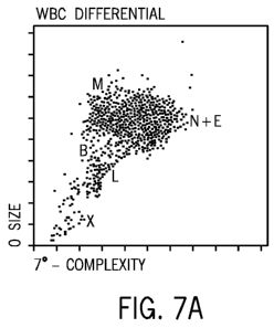
FIG. 7A is a cytogram of a clinical sample of CSF from a patient having a
diagnosis of meningococcal sepsis. The X-axis corresponds to IAS signals and
the
Y-axis corresponds to ALL signals, as measured by the apparatus depicted in
FIGS.
1 and 2. FIG. 7B is a cytogram of a clinical sample from the same CSF as in
FIG.
7A, except that the X-axis corresponds to FL3 signals and the Y-axis
corresponds to
ALL signals, as measured by the apparatus depicted in FIGS. 1 and 2. FIG. 7C
is a
cytogram of a clinical sample from the same CSF as in FIG. 7A, except that the
X-
axis corresponds to ALL signals and the Y-axis corresponds to PSS signals, as
measured by the apparatus depicted in FIGS. 1 and 2. FIG. 7D is a cytogram of
a
clinical sample from the same CSF as in FIG. 7A, except that the signals are
from
8
CA 02730769 2011-01-13
WO 2010/011583 PCT/US2009/051097
the PLT channel, wherein the X-axis corresponds to IAS signals and the Y-axis
corresponds to PSS signals, as measured by the apparatus depicted in FIGS. 1
and
2.
FIG. 8A is a cytogram of a clinical sample of a body fluid, intraperitoneal
dialysate, from a male patient having a diagnosis of peritonitis. The X-axis
corresponds to IAS signals and the Y-axis corresponds to ALL signals, as
measured
by the apparatus depicted in FIGS. 1 and 2. FIG. 8B is a cytogram of a
clinical
sample from the same body fluid as in FIG. 8A, except that the X-axis
corresponds
to FL3 signals and the Y-axis corresponds to ALL signals, as measured by the
apparatus depicted in FIGS. 1 and 2. FIG. 8C is a cytogram of a clinical
sample
from the same body fluid as in FIG. 8A, except that the X-axis corresponds to
ALL
signals and the Y-axis corresponds to PSS signals, as measured by the
apparatus
depicted in FIGS. 1 and 2. FIG. 8D is cytogram of a clinical sample of the
same
body fluid as in FIG. 8A, except that the signals are from the PLT channel,
wherein
the X-axis corresponds to IAS signals and the Y-axis corresponds to PSS
signals, as
measured by the apparatus depicted in FIGS. 1 and 2.
FIG. 9A is a cytogram of a clinical sample of a body fluid, intraperitoneal
dialysate, from a female patient having a diagnosis of Actinobacterial
infection. The
X-axis corresponds to IAS signals and the Y-axis corresponds to ALL signals,
as
measured by the apparatus depicted in FIGS. 1 and 2. FIG. 9B is a clinical
sample
of the same body fluid as in FIG. 9A, except that the X-axis corresponds to
FL3
signals and the Y-axis corresponds to ALL signals, as measured by the
apparatus
depicted in FIGS. 1 and 2. FIG. 9C is a clinical sample of the same body fluid
as in
9A, except that the X-axis corresponds to ALL signals and the Y-axis
corresponds to
PSS signals, as measured by the apparatus depicted in FIGS. 1 and 2. FIG. 9D
is a
clinical sample of the same body fluid as in FIG. 9A, except that the signals
are from
the PLT channel, wherein the X-axis corresponds to IAS signals and the Y-axis
corresponds to PSS signals, as measured by the apparatus depicted in FIGS. 1
and
2.
FIG. 10A is a cytogram of a clinical sample of CSF from a patient having a
diagnosis of complicated pancreatitis due to coagulase-negative streptococcus.
The
9
CA 02730769 2011-01-13
WO 2010/011583 PCT/US2009/051097
X-axis corresponds to IAS signals and the Y-axis corresponds to ALL signals,
as
measured by the apparatus depicted in FIGS. 1 and 2. FIG. 1 OB is a cytogram
of a
clinical sample of the same CSF as in FIG. 1 OA, except that the X-axis
corresponds
to FL3 signals and the Y-axis corresponds to ALL signals, as measured by the
apparatus depicted in FIGS. 1 and 2. FIG. 10C is a cytogram of a clinical
sample of
the same CSF as in FIG. 10A, except that the X-axis corresponds to ALL signals
and
Y-axis corresponds to PSS signals, as measured by the apparatus depicted in
FIGS.
1 and 2. FIG. 10D is a cytogram of a clinical sample of the same CSF as in
FIG.
1 OA, except that the signals are from the PLT channel, wherein the X-axis
corresponds to IAS signals and the Y-axis corresponds to PSS signals, as
measured
by the apparatus depicted in FIGS. 1 and 2.
DETAILED DESCRIPTION
As used herein the expression "axial light loss" and "ALL" refer to the
measurement of the total light lost from the laser beam at from about 0 to
about 1
when a particle passes through the beam. This parameter relates to measurement
of the size of cells or particles passing through the optical detection
system. As used
herein, the expressions "intermediate angle scatter" and "IAS" refer to the
measurement of forward light scatter at intermediate angle from 3 to 100.
This
parameter relates to measurement of complexity of a cell. As used herein, the
term
"complexity" refers to the composition of a cell. Some cells have
mitochondria,
ribosomes, nucleus, while other cells lack one or more of the foregoing
components.
The measured intensity of IAS depends to some degree on the heterogeneity of
the
contents of a cell (or particle) passing through the illumination beam of a
cytometer.
The density of "IAS" signals can be thought of as a measure of the complexity
of the
contents of the cell, i.e., the presence of organelles, such as, for example,
nuclei,
vacuoles, endoplasmic reticula, mitochondria, etc. As used herein, the
expressions
"polarized side scatter" and "PSS" refer to polarized light scatter at the
angle of 90 .
This parameter relates to measurement of lobularity. The nuclei of cells have
various
shapes that may result in one to five lobules, inclusive. A representative
example of
a cell with multi-lobed nucleus is a segmented neutrophil. As used herein, the
expressions "depolarized side scatter" and "DSS" refer to depolarized light
scatter at
CA 02730769 2011-01-13
WO 2010/011583 PCT/US2009/051097
the angle of 900. This parameter relates to measurement of subpopulations of
blood
cells. Blood cells have various numbers of subpopulations within the membranes
of
the cell. Examples of these subpopulations, for white blood cells, are
eosinophils,
neutrophils, basophils, monocytes and lymphocytes. As used herein, the
expression
"FL1" refers to fluorescence measurement at an emission signal wavelength of
530
nanometers, i.e., green fluorescence. As used herein, the expression "FL2"
refers to
fluorescence measurement at an emission signal wavelength of 580 nanometers,
i.e., yellow to orange fluorescence. As used herein, the expression "FL3"
refers to
fluorescence measurement at an emission signal wavelength of 630 nanometers,
i.e., red fluorescence. This parameter relates to measurement of DNA or RNA
stained by a nuclear stain used in the reagent system.
As used herein, the term "trigger" means the minimum electrical voltage that
an electrical signal must exceed to be considered valid. As used herein, the
expression "triple-trigger" refers to a circuitry processing signals based on
AND/OR
logic wherein a qualified signal must be greater than the second scatter
signal
threshold, while at the same time it must be greater than either the first
scatter signal
threshold or the FL3 threshold.
As used herein, the term "erythroblast" means any of the nucleated cells in
bone marrow that develop into erythrocytes. As used herein, the term
"erythrocyte"
means the yellowish, non-nucleated, disk-shaped blood cell that contains
hemoglobin and is responsible for the color of blood. As used herein, the
expression
"erythroblast nuclei" refers to the nuclei of erythroblasts.
One or more detectors are preferably placed in the light path for measuring
forward intermediate angle scattering (IAS) and either small angle forward
scattering
(SAS) or axial light loss (ALL). The light loss is generally due to scattering
and
defined as the decrease in light energy reaching a detector in the path of a
laser
beam due to the passage of a cell through that beam. Generally ALL is detected
at
an angle of from about 0 to about 1 . SAS is light energy that reaches a
detector
outside, but within a narrow angle of about 1 to about 3 , the incident
laser beam
due to scattering from a cell passing through the beam. A beam stop is
generally
provided to keep the laser beam from getting into the detector. ALL measuring
systems collect light within the incident cone of laser illumination, while
small angle
scatter systems collect light outside this cone. In ALL measuring systems, the
signal
of interest is a negative signal subtracted from the steady state laser
signal, whereas
11
CA 02730769 2011-01-13
WO 2010/011583 PCT/US2009/051097
in the small angle forward scatter measurement, the signal is a small positive
signal
imposed on a very low background light level. Intermediate angle forward
scattering
(IAS) is similar to small angle forward scattering, except the light is
scattered at a
larger angle from the incident laser beam. More specifically, IAS relates to
light
scattered in a ring between 3 and 10 away from the incident or centerline of
a laser
beam. In a preferred embodiment, ALL is collected in the angles less than 0.3
horizontally and less than 1.2 vertically from the laser axis, and IAS is
collected at
angles between 3 and 100 from the laser axis.
As used herein, the term "drain" means drainage, the systematic withdrawal of
fluids and discharges from wound of body cavity.
As used herein, the expression "Open Mode" means that the sample is
presented directly to the automated instrument by a human operator. As used
herein, the expression "Closed Mode" means that the sample is presented
directly to
the automated instrument by a robotic mechanism.
As used herein, the expression "measuring cells" refers to enumerating cells
by means of light scattering techniques to determine, size, granularity,
lobularity, and
fluorescence when the cells are stained with a particular dye of fluorochrome.
As used herein, the expression "cell surface antigen" means a substance that
promotes the generation of antibodies. The cell surface antigens are
endogenous
antigens that have been generated within the cell, as a result of normal cell
metabolism, or because of viral or intracellular bacterial infection. The
fragments are
then presented on the cell surface in the complex with MHC class I molecules.
The expression "red blood cell ghost" refers to the red blood cell membrane
remaining after a red blood cell is lysed either by hypotonic medium or by a
lysing
reagent.
The symbol "(s)" following the name of an object indicates that either the
object alone or a plurality of the objects is being referred to, depending
upon the
context of the statement surrounding the mention of the object or objects.
Automated hematology analyzers are discussed in WHITNEY WILLIAMS.
Hem I Automated Cell Counting and Evaluation. Educational publication
[online],
[retrieved on 2008-07-15]. Retrieved from the Internet: <URL:
http://www.clt.astate.edu/wwilliams/new-page_4.html>, incorporated herein by
reference.
12
CA 02730769 2011-01-13
WO 2010/011583 PCT/US2009/051097
The method described herein involves an automated method for simultaneous
analysis of white blood cell differential, erythroblasts, and bacteria in body
fluids,
such as, for example, blood, cerebrospinal fluid, ascites fluid, pleural
fluid, peritoneal
fluid, pericardial fluid, synovial fluid, dialysate fluid, and drain fluid, on
a hematology
analyzer by means of the same reagent system and optical detection system
designed for analysis of blood.
Referring now to FIG. 1, an apparatus 10 comprises a source of light 12, a
front mirror 14 and a rear mirror 16 for beam bending, a beam expander module
18
containing a first cylindrical lens 20 and a second cylindrical lens 22, a
focusing lens
24, a fine beam adjuster 26, a flow cell 28, a forward scatter lens 30, a
bulls-eye
detector 32, a first photomultiplier tube 34, a second photomultiplier tube
36, and a
third photomultiplier tube 38. The bullseye detector 32 has an inner detector
32a for
0 light scatter and an outer detector 32b for 7 light scatter.
The source of light 12 can be a vertically polarized 488 nm air-cooled argon-
ion laser or a linearly polarized blue (488 nm) diode-pumped solid-state
(DPSS)
laser. Additional details relating to the laser, the flow cell, the lenses,
the focusing
lens, the fine-beam adjust mechanism and the laser focusing lens can be found
in U.
S. Patent No. 5,631,165, incorporated herein by reference, particularly at
column 41,
line 32 through column 43, line 11.
The preferred forward optical path system shown in FIG. 1 includes a
spherical piano-convex lens 30 and a two-element photo-diode detector 32
located
in the back focal plane of the lens. In this configuration, each point within
the two-
element photodiode detector 32 maps to a specific collection angle of light
from cells
moving through the flow cell 28. The detector 32 can be a bulls-eye detector
capable of detecting axial light loss (ALL) and intermediate angle forward
scatter
(IAS). U. S. Patent No. 5,631,165 describes various alternatives to this
detector at
column 43, lines 12-52.
The first photomultiplier tube 34 (PMT1) measures depolarized side scatter
(DSS) or green fluorescence (FL1). The second photomultiplier tube 36 (PMT2)
measures polarized side scatter (PSS) or yellow to orange fluorescence (FL2)
and
the third photomultiplier tube 38 (PMT3) measures red fluorescence (FL3). FL1,
green fluorescence, is detected between about 515 to 545 nm. FL2, yellow to
orange fluorescence, is detected between about 565 to 595 nm. FL3, red
13
CA 02730769 2011-01-13
WO 2010/011583 PCT/US2009/051097
fluorescence, is detected between about 615 to 645 nm. Side-scatter and
fluorescent emissions are directed to these photomultiplier tubes by dichroic
beam
splitters 40 and 42, which transmit and reflect efficiently at the required
wavelengths
to enable efficient detection. U. S. Patent No. 5,631,165 describes various
additional
details relating to the photomultiplier tubes at column 43, line 53 though
column 44,
line 4.
Sensitivity is enhanced at photomultiplier tubes 34, 36, and 38, when
measuring fluorescence, by using an immersion collection system. The immersion
collection system is one that optically couples the first lens 30 to the flow
cell 28 by
means of a refractive index matching layer, enabling collection of light over
a wide
angle. U. S. Patent No. 5,631,165 describes various additional details of this
optical
system at column 44, lines 5-31.
The condenser 44 is an optical lens system with aberration correction
sufficient for diffraction limited imaging used in high resolution microscopy.
U. S.
Patent No. 5,631,165 describes various additional details of this optical
system at
column 44, lines 32-60.
The functions of other components shown in FIG. 1, i.e., a slit 46, a field
lens
48, and a second slit 50, are described in U. S. Patent No. 5,631,165, at
column 44,
line 63 through column 45, line 15. The photomultiplier tubes 34, 36, and 38
detect
either side-scatter (light scattered in a cone whose axis is approximately
perpendicular to the incident laser beam) or fluorescence (light emitted from
the cells
at a different wavelength from that of the incident laser beam). A movable
polarizer
52 placed in the light path of the photomultiplier tube 34 configures the
photomultiplier tube 34 to detect depolarized side-scatter (DSS) and polarized
side-
scatter (PSS), respectively, while movable filters 54, 56, 58 enable detection
of
fluorescent emissions at specified wavelengths from the cells.
The measurement process begins as the cell stream passes through the flow
cell 28, having been diluted with the lysing agent so that the cells pass
through the
laser illuminated volume single file, in a laminar flowing sample stream
surrounded
by a sheath solution. The illuminated volume is bounded in the two directions
normal to the flow axis by the hydrodynamically focused cell stream, and in
the
dimension parallel to the flow axis by the vertical beam waist of the laser
beam,
which is about 17 micrometers. The flow rate of the sample is about 2.5
microliters
per second, and the corresponding illuminated sensing volume of the white
blood
14
CA 02730769 2011-01-13
WO 2010/011583 PCT/US2009/051097
cells and the erythroblasts approximates an elliptical cylinder having
dimensions of
about 80 pm x 5 pm x 17 pm. The 17pm dimension is measured along the axis of
the elliptical cylinder.
The presence of a cell in the illuminated region is detected by the
photodiodes and the photomultiplier tubes, and a triple threshold trigger
circuit that
operates in three feature space dimensions. That is, the triple threshold
trigger
circuit processes the three parameters of ALL, IAS, and FL3 and qualifies
signals for
digitization using AND/OR logic. A qualified signal must be greater than the
IAS
threshold while at the same time it must be greater than either the ALL
threshold or
the FL3 threshold. The combination of this triggering circuit and the lysing
properties
(which lightly fixes white blood cells and preserves their surface antigens
while at the
same time permitting erythrocyte nuclei to be rapidly stained) excludes
erythroblasts
from the white blood cell differential count. Bacterial signals are
distinguished from
those of erythroblasts by the size, shape, and the location of the
distribution of the
respective signals by the algorithm(s) of the system. The method described
herein
counts white blood cell populations, erythroblasts, and bacteria without the
interference typically encountered from background signals, both fluorescent
and
non-fluorescent, red blood cell stroma, and platelets. U. S. Patent No.
5,631,165
describes various additional details of the measurement process at column 55,
line
48 through column 59, line 43.
Referring now to FIG. 2, (AND/OR) circuitry eliminates signals from debris
and qualifies signals from erythroblast nuclei or bacteria in addition to
those of white
blood cells. To be qualified as a valid signal, a signal must be either above
ALL OR
FL3 trigger and always above AND GATE, which is IAS AND FL3. The electrical
pulse mechanism will perform three distinct measurements. First, positive or
negative measurements of ALL are carried out. Then positive or negative
measurements of FL3 are carried out. Finally, positive or negative
measurements of
IAS are carried out. By separating positive and negative pulses, the triple
triggering
circuitry utilizes the gating mechanism to differentiate white blood cells
from
erythroblasts. The final gating mechanism further separates and identifies the
smallest of the particles, such as, for example, platelets. The bacterial
signals
(FL3+) will be qualified by the circuitry along with the signals generated by
erythroblasts because the amplification of FL3+ bacterial signals is above the
FL3
threshold. Bacterial signals are differentiated from those of erythroblasts by
the
CA 02730769 2011-01-13
WO 2010/011583 PCT/US2009/051097
software algorithm, because the amplification of ALL signals from bacteria is
lower
and the intensity of FL3 signals from bacteria is higher than those generated
by
erythroblasts. All signals exceeding a minimum voltage are used, because these
signals are deemed to be valid. The components of the AND/OR circuitry 100 are
as
follows:
102 ALL Voltage Comparator
104 ALL Signal
106 ALL threshold voltage (Vthl)
108 ALL Voltage Comparator Output
112 FL3 Voltage Comparator
114 FL3 Signal
116 FL3 threshold voltage (Vth2)
118 FL3 Voltage Comparator Output
122 IAS Voltage Comparator
124 IAS Signal
126 IAS threshold voltage (Vth3)
128 IAS Voltage Comparator Output
130 OR Gate
132 OR Gate output
134 AND Gate
136 Valid Trigger Output
Real time signals from their respective channels are present at the inputs of
the voltage comparators. Voltage comparators 102, 112, 122 function by
comparing
the "+ inputs" 104, 114, 124 to the "- inputs" 106, 116, 126 to resultant
outputs 108,
118, 128. If the "+ input" is of a higher voltage than the "- input", the
output will be
high. If the "+ input" is of a lower voltage than the "- input", the output
will be low.
The threshold voltages are independent voltages that are determined by
parameters of the system. The outputs of comparators 102 and 112 are inputs to
OR gate 130 to give resultant OR gate output 132. The OR gate functions by
comparing its inputs. The output will be high if either, or both, inputs are
high.
The output 132 of the OR gate 130 and the output of comparator 122 are
inputs to AND gate 134. The AND gate functions by comparing its inputs to
derive
16
CA 02730769 2011-01-13
WO 2010/011583 PCT/US2009/051097
its output 136 which is also the valid trigger output. The output will be high
only if
both inputs are high.
The valid trigger output 136 will be high only if the IAS signal 124 is
greater
than its threshold voltage 126, and if the ALL signal 104 is greater than its
threshold
voltage 106 or the FL3 signal 114 is greater than its threshold voltage 116,
or both
the ALL signal 104 is greater than its threshold voltage 106 and the FL3
signal 114 is
greater than its threshold voltage 116.
In one embodiment, a body fluid can be analyzed without any manual
preparation on the system in the Open Mode feature. A portion of the sample of
the
body fluid can be diluted with a diluent to enable a minimal number of cells
to pass
through a counting aperture at the same time. The diluent is typically used
for the
channel that counts red blood cells. A sample of the body fluid is mixed with
a
reagent system that was originally designed to preserve white blood cells and
their
cell surface antigens for immuno-phenotyping, i.e., a technique used for
analyzing
and measuring cells labeled with specific monoclonal antibodies conjugated to
specific fluorochromes to locate specific cell surface antigens, and at the
same time
red blood cells and membranes of erythroblasts are lysed and nuclei of
erythroblasts
and bacterial DNA or RNA are stained. Then, the cells that were treated with
the
aforementioned reagent system are passed through the electro-optical system
described in FIG. 1 in single file and the electronic logic, triple-triggering
circuitry of
the system, and the algorithm(s) of the system differentiate each cell
population
based on cell volume, i.e., size of the cells, complexity of the cells,
lobularity of the
cells, refractive index of the cells, fluorescence intensity, and the location
and pattern
of each cell cluster. The triple-triggering circuitry eliminates signals from
cell debris
and qualifies signals from white blood cells, erythroblasts, and bacteria.
Signals that
are eliminated have values below a specified cut-off, and the eliminated
signals are
deemed debris. Signals that are qualified have values above a specified cut-
off, and
the qualified signals are deemed white blood cells, erythroblasts, and
bacteria. To
be qualified as valid bacterial signals, the amplitude of the signals must be
below the
OR Gate, ALL trigger, but above the AND Gate, FL3 and IAS triggers. The
software
algorithm(s) of the system can be used to differentiate bacterial signals from
that of
erythroblasts signals by the size of the ALL signal, the intensity of the FL3+
signals
from bacteria, and the shape and the number of FL3 clusters, i.e., the
characteristic
two clusters for erythroblasts, which stand in contrast to a single loosely
distributed
17
CA 02730769 2011-01-13
WO 2010/011583 PCT/US2009/051097
cluster for bacterial signals.
The first logic analysis is of the complete system and all of its attributes.
The
second logic analysis is a derivative of the logic analysis of the complete
system and
relates to the distinction between erythroblasts and bacteria. Amplification
of ALL
signals from bacteria is lower than amplification of ALL signals from
erythroblasts;
accordingly, ALL signals from bacteria fall below clusters of erythroblast
signals.
Furthermore, clusters of erythroblasts always appear as two distinct clusters,
in
contrast to the single loosely distributed cluster of bacterial signals. Still
further,
amplification of FL3+ signals from bacteria is much higher than amplification
of FL3+
signals from erythroblasts.
The following non-limiting examples further illustrate the method described
herein. In the drawings, the letter "N" indicates the position of neutrophils
in the
cytograms, the letter "M" indicates the position of monocytes in the
cytograms, the
letter "L" indicates the position of lymphocytes in the cytograms, the letter
"E"
indicates the position of eosinophils in the cytograms, the letter "B"
indicates the
position of basophils in the cytograms, the letter "P" (or the letter "P"
preceded by a
numeral) indicates the position of platelets in the cytograms, and the letter
"X" (or the
letter "X" preceded by a numeral) indicates the position of bacteria in the
cytograms.
The terms "Erb1 ", "Erb2", and "Erb 1 + 2" indicate the positions of a first
cluster of
erythroblasts, a second cluster of erythroblasts, and a cluster combining the
two
clusters of erythroblasts, respectively.
EXAMPLES
Comparative Examples A, B, and C illustrate how cytograms resulting from
the method described herein characterize white blood cells in an automated
hematology analyzer described in U. S. Patent Nos. 5,631,165; 5,656,499; and
5,939,326. Working Examples 1, 2,3, 4, and 5 illustrate how cytograms
resulting
from the method described herein differentiate bacteria from white blood cells
and
count bacterial cells in an automated hematology analyzer described in U. S.
Patent
Nos. 5,631,165; 5,656,499; and 5,939,326.
18
CA 02730769 2011-01-13
WO 2010/011583 PCT/US2009/051097
COMPARATIVE EXAMPLE A
Referring now to FIGS. 3A, 3B, 3C, and 3D, a sample of normal blood was
treated with the reagent system described in U. S. Patent Nos. 5,516,695 and
5,559,037, both of which were previously incorporated herein as reference.
This
reagent system was also used in Comparative Examples B and C and Working
Examples 1, 2, 3, 4, and 5. The reagent system comprises a red blood cell
lysing
component, a white blood cell preserving component, and a nuclear stain. The
apparatus of FIGS. 1 and 2 can be used to prepare a cytogram of a blood sample
of
a patient. The sample contained white blood cells at a concentration of 6.08 x
103/pL, lymphocytes (35.1 %), neutrophils (54.5%), monocytes (6.95%),
eosinophils
(3.34%) and basophils (0.07%).
Red blood cell (RBC) indices were analyzed with the same reagent system by
means of an impedance measurement. FIG. 3A is a cytogram of the white blood
cells of a sample of normal blood, wherein the X-axis corresponds to
intermediate
angle light scatter (IAS) signals from 3 to 10 , and the Y-axis corresponds
to axial
light loss (ALL) signals, as measured by the apparatus depicted in FIGS. 1 and
2.
FIG. 3B is a cytogram of the white blood cells of a sample of the same blood
as in
FIG. 3A, except that the X-axis corresponds to red fluorescent (FL3) signals,
and the
Y-axis corresponds to ALL signals, as measured by the apparatus depicted in
FIGS.
1 and 2. As can be seen in FIG. 3B, the area above the FL3 trigger, originally
designated for detection of erythrocytes, is clear, thereby indicating that no
erythrocytes were found in the sample.
FIG. 3C is a cytogram of the white blood cells of a sample of the same blood
as in FIG. 3A, except that the X-axis corresponds to 90 polarized side
scatter (PSS)
signals and the Y-axis corresponds to 90 depolarized side scatter (DSS)
signals, as
measured by the apparatus depicted in FIGS. 1 and 2. The amplification of DSS
signals from eosinophils is much higher than those from all other white blood
cells.
Thus, eosinophils are separated from the rest of the white blood cells by the
algorithm(s) of the system and counted. Eosinophils are highly granulated.
FIG. 3D is a cytogram of a sample of the same blood as in FIG. 3A, except
that the scatter signals in this cytogram are from a different electronic
scale, which
uses much higher electronic gains, and is designed to measure platelets. In
FIG.
3D, it can be seen that the background outside the platelet population
enclosed by
19
CA 02730769 2011-01-13
WO 2010/011583 PCT/US2009/051097
the two floating threshold lines generated by the platelet algorithm of the
system is
clean. The two parallel lines that appear on the cytogram in FIG. 3D, and the
cytograms in FIGS. 4D, 6D, 7D, 8D, 9D,and 10D, represent the two floating
thresholds. The X-axis corresponds to IAS signals and the Y-axis corresponds
to
PSS signals, as measured by the apparatus depicted in FIGS. 1 and 2.
COMPARATIVE EXAMPLE B
The same method and apparatus that were used in COMPARATIVE
EXAMPLE A were used to carry out COMPARATIVE EXAMPLE B.
FIG. 4A is a cytogram of the white blood cells of a clinical blood sample
containing erythroblasts, wherein the X-axis corresponds to IAS signals and
the Y-
axis corresponds to ALL signals, as measured by the apparatus depicted in
FIGS. 1
and 2. The concentration of white blood cells was 20.9 x 10³/pL, and the
concentration of erythroblasts was 2.38 x 10³/pL. Unlike FIG. 3A, very high
noise-
like signals appear below the lymphocyte cluster in FIG 4A.
FIG. 4B is a cytogram of the white blood cells of a clinical sample of the
same
blood as in FIG. 4A, except that the X-axis corresponds to FL3 signals and the
Y-
axis corresponds to ALL signals, as measured by the apparatus depicted in
FIGS. 1
and 2. In FIG. 4B, the area below the ALL trigger but above the FL3 trigger is
occupied by the characteristic pair of FL3+ erythroblasts, one larger primary
cluster
centered around the channel 125 of the X-axis and a second smaller cluster
centered around the channel 220 of the X-axis. It should be noted that the X-
axis
has 256 channels, running from a value of 0 to a value of 256. Because the
size of
erythroblast nuclei is much smaller than that of white blood cells, the ALL
signals of
erythroblast nuclei fall below the ALL trigger. The heavy noise-like signals
appearing
below the lymphocyte cluster in FIG. 4A also belong to the erythroblast
population.
FIG. 4C is a cytogram of the white blood cells of a clinical sample of the
same
blood as in FIG. 4A, except that the X-axis corresponds to ALL signals and the
Y-
axis corresponds to PSS signals, as measured by the apparatus depicted in
FIGS. 1
and 2. Granulocytes (neutrophils and eosinophils) generate much larger PSS
signals on account of their morphological complexity than do mononuclear cells
(lymphocytes and monocytes) or basophils, thereby permitting the algorithm(s)
of the
CA 02730769 2011-01-13
WO 2010/011583 PCT/US2009/051097
system to separate the granulocyte population from the rest of the white cell
population along the Y-axis.
FIG. 4D is a cytogram of a clinical sample of the same blood as in FIG. 4A,
except that the signals are from the PLT channel. The X-axis corresponds to
IAS
signals and Y-axis corresponds to PSS signals, as measured by the apparatus
depicted in FIGS. 1 and 2. In the platelet channel, the electronic gains of
both
scatter signals, PSS and IAS, are set much higher in order to amplify the
signals
generated by small platelets.
COMPARATIVE EXAMPLE C
The same method and apparatus that were used in COMPARATIVE
EXAMPLE A were used to carry out COMPARATIVE EXAMPLE C.
FIG. 5A is a cytogram of a clinical blood sample containing very high
concentration of erythroblasts, wherein the X-axis corresponds to FL3 signals
and
the Y-axis corresponds to ALL signals, as measured by the apparatus depicted
in
FIGS. 1 and 2. The concentration of erythroblasts is 4.93 x 10³ /pL. The
pattern and
the location of FL3+ erythroblast nuclei appear as two clearly visible
clusters of
erythroblasts, one large cluster and one small cluster. The concentration of
white
blood cells is 27.5 x 10³/pL, neutrophils (86.6%), lymphocytes (7.96%),
monocytes
(4.49%), and eosinphils (0.84%). The primary cluster of erythroblasts is
centered
around the channel 127 of the X-axis, and the secondary cluster of
erythroblasts is
centered around the channel 220 of the X-axis.
FIG. 5B is the same blood as in 5A, except that the X-axis corresponds to ALL
signals and the Y-axis corresponds to PSS signals. As can be seen in FIG. 5B,
no
noticeable amount of PSS signals is generated from the very small particles
located
in the noise region below ALL trigger.
EXAMPLE 1
The same method and apparatus that were used in COMPARATIVE
EXAMPLE A were used to carry out EXAMPLE 1.
FIG. 6A is a cytogram of a clinical sample of cerebrospinal fluid (CSF), not
suspected of carrying any infection, wherein the X-axis corresponds to IAS
signals
21
CA 02730769 2011-01-13
WO 2010/011583 PCT/US2009/051097
and the Y-axis corresponds to ALL signals, as measured by the apparatus
depicted in FIGS. 1 and 2. FIG. 6B is a cytogram of a clinical sample of the
same
CSF as in FIG. 6A, except that the X-axis corresponds to FL3 signals and the Y-
axis corresponds to ALL signals, as measured by the same apparatus depicted in
FIGS. 1 and 2. FIG. 6C is a cytogram of a clinical sample of the same CSF as
in
FIG. 6A, except that the X-axis corresponds to ALL signals and the Y-axis
corresponds to PSS signals, as measured by the apparatus depicted in FIGS. 1
and 2. FIG. 6D is a cytogram of a clinical sample of the same CSF as in FIG.
6A,
except that the signals are from the PLT channel, wherein the X-axis
corresponds
to IAS signals and the Y-axis corresponds to PSS signals, as measured by the
apparatus depicted in FIGS. 1 and 2. All the regions, ALL, IAS, PSS and FL3,
for
white blood cells and erythroblasts are clear, indicating that no cells are
found in
the specimen. The cytogram in FIG. 6D of the optical platelet channel is also
clear, confirming that there are no small particles, such as bacteria in this
sample
of CSF.
EXAMPLE 2
The same method and apparatus that were used in COMPARATIVE
EXAMPLE A were used to carry out EXAMPLE 2.
FIG. 7A is a cytogram of a clinical sample of CSF from a 56-year old female
patient having a diagnosis of meningococcal sepsis. The X-axis corresponds to
IAS
signals and the Y-axis corresponds to ALL signals, as measured by the
apparatus
depicted in FIGS. 1 and 2.
The sample contains white blood cells at a concentration of 5.06 x 10³/pL, red
blood cells at a concentration of 0.003 x 10⁶/pL, neutrophils (86.8%),
lymphocytes
(5.4%), and monocytes (5.4%).
FIG. 7B is a cytogram of a clinical sample from the same CSF as in FIG. 7A,
except that the X-axis corresponds to FL3 signals and the Y-axis corresponds
to ALL
signals, as measured by the apparatus depicted in FIGS. 1 and 2. The points in
the
circle below ALL channel 25 at far right corner of the cytogram in FIG. 7B
correspond
to bacterial cells, whose DNA is brightly stained by the reagent system,
described in
U. S. Patent Nos. 5,516,695 and 5,559,037, both of which were previously
incorporated herein by reference. The signal pattern and the location of the
dots
22
CA 02730769 2011-01-13
WO 2010/011583 PCT/US2009/051097
from the bacteria are distinguishable from those of erythroblasts in that the
bacterial
signals do not exhibit the characteristic primary and secondary pair of
clusters of
erythroblasts as seen in FIGS. 4B and 5A. Furthermore, the cell volume of
bacteria
is smaller than the cell volume of erythroblasts, with the result that ALL
signals from
bacteria fall below erythroblast signals centered around points along the X-
axis, and
the intensity of bacterial DNA staining is much brighter than that of
erythroblast
nuclei.
FIG. 7C is a cytogram of a clinical sample from the same CSF as in FIG. 7A,
except that the X-axis corresponds to ALL signals and the Y-axis corresponds
to
PSS signals, as measured by the apparatus depicted in FIGS. 1 and 2. The side
scatter signals (PSS) from bacteria are much more noticeable in FIG. 7C than
those
of erythroblast nuclei in FIG. 4C.
FIG. 7D is a cytogram of a clinical sample from the same CSF as in FIG. 7A,
except that the signals are from the PLT channel, wherein the X-axis
corresponds to
IAS signals and the Y-axis corresponds to PSS signals, as measured by the
apparatus depicted in FIGS. 1 and 2. In the optical platelet channel, the
bacterial
signals appear as dispersed noise signals in both inside and outside of the
two
floating platelet thresholds, as can be seen in FIG. 7D. FIG. 7C shows small
ALL
signals that fall below channel 25, but the PSS signals from the bacteria are
much
more visible than those of erythroblasts.
EXAMPLE 3
The same method and apparatus that were used in COMPARATIVE
EXAMPLE A were used to carry out EXAMPLE 3.
FIG. 8A is a cytogram of a clinical sample of a body fluid, intraperitoneal
dialysate, from a 57-year old male patient having a diagnosis of peritonitis.
The X-
axis corresponds to IAS signals and the Y-axis corresponds to ALL signals, as
measured by the apparatus depicted in FIGS. 1 and 2.
The concentration of white blood cells is 1.43 x 10³/pL, the concentration of
red blood cells is 0.002 x 10⁶/pL, neutrophils (83.4%), lymphocytes (8.75%),
monocytes (6.95%), and eosinophils (0.89%).
FIG. 8B is a cytogram of a clinical sample from the same body fluid as in FIG.
8A, except that the X-axis corresponds to FL3 signals and the Y-axis
corresponds to
23
CA 02730769 2011-01-13
WO 2010/011583 PCT/US2009/051097
ALL signals, as measured by the apparatus depicted in FIGS. 1 and 2. The
brightly
stained dots in the circle at lower right corner of FIG. 8B indicate the
presence of
bacteria in this sample.
FIG. 8C is a cytogram of a clinical sample from the same body fluid as in FIG.
8A, except that the X-axis corresponds to ALL signals and the Y-axis
corresponds to
PSS signals, as measured by the apparatus depicted in FIGS. 1 and 2. The
presence of bacteria in this sample is indicated by the PSS signals from the
small
particles in Fig 8C at the lower end of the X-axis and the lower end of the Y-
axis.
FIG. 8D is cytogram of a clinical sample of the same body fluid as in FIG. 8A,
except that the signals are from the PLT channel, wherein the X-axis
corresponds to
IAS signals and the Y-axis corresponds to PSS, as measured by the apparatus
depicted in FIGS. 1 and 2. In FIG. 8D, small particles appear as heavy noise
signals
in the platelet channel, both inside and outside of the two floating platelet
thresholds.
EXAMPLE 4
The same method and apparatus that were used in COMPARATIVE
EXAMPLE A were used to carry out EXAMPLE 4.
FIG. 9A is a cytogram of a clinical sample of a body fluid, intraperitoneal
dialysate, from a 60-year old female patient having a diagnosis of CAPD-
peritonitis
with Actinobacterial infection. The X-axis corresponds to IAS signals and the
Y-axis
corresponds to ALL signals, as measured by the apparatus depicted in FIGS. 1
and
2. The concentration of white blood cells is 10.30 x 10³/pL, the concentration
of red
blood cells is 0.001x10⁶/pL, neutrophils (60.4%), lymphocytes (11.8%),
monocytes
(8.32%), and eosinophils (0.69%).
FIG. 9B is a clinical sample of the same body fluid as in FIG. 9A, except that
the X-axis corresponds to FL3 signals and the Y-axis corresponds to ALL
signals, as
measured by the apparatus depicted in FIGS. 1 and 2. The brightly stained FL3+
dots in the circle at lower right corner of FIG. 9B indicate the bacteria.
FIG. 9C is a clinical sample of the same body fluid as in 9A, except that the
X-
axis corresponds to ALL signals and the Y-axis corresponds to PSS signals, as
measured by the apparatus depicted in FIGS. 1 and 2. FIG. 9D is a clinical
sample
of the same body fluid as in FIG. 9A, except that the signals are from the PLT
24
CA 02730769 2011-01-13
WO 2010/011583 PCT/US2009/051097
channel, wherein the X-axis corresponds to IAS signals and the Y-axis
corresponds
to PSS signals, as measured by the apparatus depicted in FIGS. 1 and 2.
PSS signals from bacteria in both the white blood cell channel (FIG. 9C) and
the optical platelet channel (FIG. 9D) are apparent. In FIG. 9D, very dense
bacterial
signals are seen as dispersed noise signals generally outside, but also
inside, of the
two floating platelet thresholds.
EXAMPLE 5
The same method and apparatus that were used in COMPARATIVE
EXAMPLE A were used to carry out EXAMPLE 5.
FIG. 1 OA is a cytogram of a clinical sample of CSF from a 63-year old female
patient having a diagnosis of complicated pancreatitis due to coagulase-
negative
streptococcus (CNS) infection. The X-axis corresponds to IAS signals and the Y-
axis corresponds to ALL signals, as measured by the apparatus depicted in
FIGS. 1
and 2. FIG. 1 OB is a cytogram of a clinical sample of the same CSF as in FIG.
1 OA,
except that the X-axis corresponds to FL3 signals and the Y-axis corresponds
to ALL
signals, as measured by the apparatus depicted in FIGS. 1 and 2. FIG. 10C is a
cytogram of a clinical sample of the same CSF as in FIG. 1 OA, except that the
X-axis
corresponds to ALL signals and Y-axis corresponds to PSS signals, as measured
by
the apparatus depicted in FIGS. 1 and 2. FIG. 10D is a cytogram of a clinical
sample
of the same CSF as in FIG. 1 OA, except that the signals are from the PLT
channel,
wherein the X-axis corresponds to IAS signals and the Y-axis corresponds to
PSS
signals, as measured by the apparatus depicted in FIGS. 1 and 2.
Very dense FL3+ bacterial signals below ALL channel 25-30 ( see circle in
FIG. 10B) and PSS signals in both the white blood cell channel (FIG. 10C) and
the
platelet channel (FIG. 1 OD) are clearly visible. In the optical platelet
channel, a
dense streak of bacterial signals is seen just above the upper platelet
threshold.
The apparatus and the reagent system described herein can be used to
eliminate cell debris and qualify signals that are smaller than the nuclei of
erythroblasts, such as those from bacteria because their genetic material,
such as
RNA or DNA nuclei, are stained by the reagent system, and the triple-trigger
circuitry
validates bacterial signals even if their size signals, ALL, fall below the
ALL trigger,
because their FL3 signals are much higher than the FL3 trigger.
CA 02730769 2011-01-13
WO 2010/011583 PCT/US2009/051097
As illustrated in FIGS. 7A, 7B, 7C, 7D, 8A, 8B, 8C, 8D, 9A, 9B, 9C, 9D, 10A,
1 OB, 10C, and 1 OD, signal patterns for ALL, IAS, PSS, and FL3 and the
location of
bacterial signals differ from those of subsets of white blood cells and
erythroblasts;
consequently, bacterial signals can easily be identified by the algorithm(s)
of the
system by using appropriate logic for cell size, fluorescence intensity, and
the pattern
and the location of the clusters.
In another embodiment, samples of certain body fluids, such as, for example,
synovial fluid, can be pretreated with a viscosity reducing agent, such as,
for
example, hyaluronidase, for a short period of time, to reduce the viscosity of
the
sample of the body fluid prior to analyzing the sample on the system Open
Mode.
After the sample is mixed with the reagent system, which is designed to
preserve
white blood cells, cell surface antigens for immunophenotyping, and at the
same
time lyse red blood cells, if any red blood cells are present in the sample,
the
membranes of erythroblasts, if any erythroblasts are present in the sample,
and stain
the nuclei of erythroblasts, if any nuclei of erythroblasts are present in the
sample,
and bacteria, the prepared cells are passed through the electro-optical system
described in FIG. 1 in single file. The electronic logic, triple-triggering
circuitry of the
system and the algorithm(s) of the system differentiate each cell population
based on
cell volume, complexity of cells, lobularity of cells, refractive index of
cells,
fluorescence intensity, and the location and pattern of each cell population.
The
triple-triggering circuitry eliminates small signals generated by cell debris
and
validates bacterial signals, i.e., <ALL trigger, >FL3 and IAS trigger. The
algorithm(s)
of the system will differentiate bacterial signals from those of erythroblasts
by the
size of the ALL signal, the intensity of the FL3+ signals from bacteria, and
the shape
and the number of FL3 clusters, i.e., the characteristic two clusters for
erythroblasts,
which stand in contrast to a single loosely distributed cluster for bacterial
signals.
In yet another embodiment, body fluids can be run on an automatic
mode if a sufficient volume of the sample of the body fluid is available to
use
the automatic mode. Body fluids are processed in the automatic mode in the
manner described previously, for the open mode, except that the sample of the
body fluid is presented directly to the automated instrument by a robotic
mechanism.
26
CA 02730769 2011-01-13
WO 2010/011583 PCT/US2009/051097
Various modifications and alterations of this invention will become apparent
to
those skilled in the art without departing from the scope and spirit of this
invention,
and it should be understood that this invention is not to be unduly limited to
the
illustrative embodiments set forth herein.
27