Note: Descriptions are shown in the official language in which they were submitted.
ORGAN MIMIC DEVICE WITH MICROCHANNELS AND METHODS OF USE
AND MANUFACTURING THEREOF
TECHNICAL FIELD
[0001] The present disclosure relates generally to an organ mimic device
with
microchannels and methods of use and manufacturing thereof.
BACKGROUND
[0002] Mechanical forces - pushes, pulls, tensions, compressions - are
important
regulators of cell development and behavior. Tensegrity provides the structure
that
determines how these physical forces are distributed inside a cell or tissue,
and how and
where they exert their influence.
[0003] In the human body, most cells are constantly subjected to mechanical
forces, such
as tension or compression.
[0004] Application of mechanical strain to cells in culture simulates the
in vivo
environment, causing dramatic morphologic changes and biomechanical responses
in the
cells.
[00051 There are both long and short term changes that occur when cells are
mechanically loaded in culture, such as alterations in the rate and amount of
DNA or RNA
synthesis or degradation, protein expression and secretion, the rate of cell
division and
alignment, changes in energy metabolism, changes in rates of macromolecular
synthesis or
degradation, and other changes in biochemistry and bioenergetics.
1
CA 2730928 2019-05-09
CA 02730928 2011-01-14
WO 2010/009307 PCMJS2009/050830
[0006] Every cell has an internal scaffolding, or cytoskeleton, a lattice
formed from
molecular "struts and wires". The "wires" are a crisscrossing network of fine
cables, known
as microfilaments, that stretch from the cell membrane to the nucleus,
exerting an inward
pull. Opposing the pull are microtubules, the thicker compression-bearing
"struts" of the
cytoskeleton, and specialized receptor molecules on the cell's outer membrane
that anchor the
cell to the extracellular matrix, the fibrous substance that holds groups of
cells together. This
balance of forces is the hallmark of tensegrity.
[0007] Tissues are built from groups of cells, like eggs sitting on the
"egg carton" of the
extracellular matrix. The receptor molecules anchoring cells to the matrix,
known as
integrins, connect the cells to the wider world. Mechanical force on a tissue
is felt first by
integrins at these anchoring points, and then is carried by the cytoskeleton
to regions deep
inside each cell. Inside the cell, the force might vibrate or change the shape
of a protein
molecule, triggering a biochemical reaction, or tug on a chromosome in the
nucleus,
activating a gene.
[0008] Cells also can be said to have "tone," just like muscles, because of
the constant
pull of the cytoskeletal filaments. Much like a stretched violin string
produces different
sounds when force is applied at different points along its length, the cell
processes chemical
signals differently depending on how much it is distorted.
[0009] A growth factor will have different effects depending on how much
the cell is
stretched. Cells that are stretched and flattened, like those in the surfaces
of wounds, tend to
grow and multiply, whereas rounded cells, cramped by overly crowded
conditions, switch on
a "suicide" program and die. In contrast, cells that are neither stretched nor
retracted carry on
with their intended functions.
[0010] Another tenet of cellular tensegrity is that physical location
matters. When
regulatory molecules float around loose inside the cell, their activities are
little affected by
mechanical forces that act on the cell as a whole. But when they're attached
to the
cytoskeleton, they become part of the larger network, and are in a position to
influence
cellular decision-making. Many regulatory and signaling molecules are anchored
on the
cytoskeleton at the cell's surface membrane, in spots known as adhesion sites,
where integrins
2
CA 02730928 2011-01-14
WO 2010/009307 PCT/US2009/050830
cluster. These prime locations are key signal-processing centers, like nodes
on a computer
network, where neighboring molecules can receive mechanical information from
the outside
world and exchange signals.
[0011] Thus, assessing the full effects of drugs, drug delivery vehicles,
nanodiagnostics
or therapies or environmental stressors, such as particles, gases, and toxins,
in a physiological
environment requires not only a study of the cell-cell and cell-chemical
interactions, but also
a study of how these interactions are affected by physiological mechanical
forces in both
healthy tissues and tissues affected with diseases.
[0012] Methods of altering the mechanical environment or response of cells
in culture
have included wounding cells by scraping a monolayer, applying magnetic or
electric fields,
or by applying static or cyclic tension or compression with a screw device,
hydraulic
pressure, or weights directly to the cultured cells. Shear stress has also
been induced by
subjecting the cells to fluid flow. However, few of these procedures have
allowed for
quantitation of the applied strains or provided regulation to achieve a broad
reproducible
range of cyclic deformations within a culture microenvironment that maintains
physiologically relevant tissue-tissue interactions.
[0013] Living organs are three-dimensional vascularized structures composed
of two or
more closely apposed tissues that function collectively and transport
materials, cells and
information across tissue-tissue interfaces in the presence of dynamic
mechanical forces, such
as fluid shear and mechanical strain. Creation of microdevices containing
living cells that
recreate these physiological tissue-tissue interfaces and permit fluid flow
and dynamic
mechanical distortion would have great value for study of complex organ
functions, e.g.,
immune cell trafficking, nutrient absorption, infection, oxygen and carbon
dioxide exchange,
etc., and for drug screening, toxicology, diagnostics and therapeutics.
[0014] The alveolar-capillary unit plays a vital role in the maintenance of
normal
physiological function of the lung as well as in the pathogenesis and
progression of various
pulmonary diseases. Because of the complex architecture of the lung, the small
size of lung
alveoli and their surrounding microvessels, and the dynamic mechanical motions
of this
organ, it is difficult to study this structure at the microscale.
3
CA 02730928 2011-01-14
WO 2010/009307 PCT/US2009/050830
[0015] The lung has an anatomically unique structure having a hierarchical
branching
network of conducting tubes that enable convective gas transport to and from
the microscopic
alveolar compartments where gas exchange occurs. The alveolus is the most
important
functional unit of the lung for normal respiration, and it is most clinically
relevant in that it is
the blood-gas barrier or interface, as well as the site where surfactants act
to permit air entry
and where immune cells, pathogens and fluids accumulate in patients with acute
respiratory
distress syndrome (ARDS) or infections, such as pneumonia.
[0016] The blood-gas barrier or tissue-tissue interface between the
pulmonary capillaries
and the alveolar lumen is composed of a single layer of alveolar epithelium
closely
juxtaposed to a single layer of capillary endothelium separated by a thin
extracellular matrix
(ECM), which forms through cellular and molecular self-assembly in the embryo.
Virtually
all analysis of the function of the alveolar-capillary unit has been carried
out in whole animal
studies because it has not been possible to regenerate this organ-level
structure in vitro.
[0017] A major challenge lies in the lack of experimental tools that can
promote
assembly of multi-cellular and multi-tissue organ-like structures that exhibit
the key
structural organization, physiological functions, and physiological or
pathological mechanical
activity of the lung alveolar-capillary unit, which normally undergoes
repeated expansion and
contraction during each respiratory cycle. This limitation could be overcome
if it were
possible to regenerate this organ-level structure and recreate its
physiological mechanical
microenvironment in vitro. However, this has not been fully accomplished.
[0018] What is needed is a organ mimic device capable of being used in
vitro or in vivo
which performs tissue-tissue related functions and which also allows cells to
naturally
organize in the device in response to not only chemical but also mechanical
forces and allows
the study of cell behavior through a membrane which mimics tissue-tissue
physiology.
OVERVIEW
[0019] System and method comprises a body having a central microchannel
separated by
one or more porous membranes. The membranes are configured to divide the
central
microchannel into a two or more closely apposed parallel central
microchannels, wherein one
or more first fluids are applied through the first central microchannel and
one or more second
fluids are applied through the second or more central microchannels. The
surfaces of each
4
CA 02730928 2011-01-14
WO 2010/009307 PCT/US2009/050830
porous membrane can be coated with cell adhesive molecules to support the
attachment of
cells and promote their organization into tissues on the upper and lower
surface of each
membrane, thereby creating one or more tissue-tissue interfaces separated by
porous
membranes between the adjacent parallel fluid channels. The membrane may be
porous,
flexible, elastic, or a combination thereof with pores large enough to only
permit exchange of
gases and small chemicals, or large enough to permit migration and
transchannel passage of
large proteins, as well as whole living cells. Fluid pressure, flow
characteristics and channel
geometry also may be varied to apply a desired fluid shear stress to one or
both tissue layers.
[0020] In an embodiment, operating channels adjacent to the central channel
are applied a
positive or negative pressure which creates a pressure differential that
causes the membrane
to selectively expand and retract in response to the pressure, thereby further
physiologically
simulating mechanical force of a living tissue-tissue interface.
[0021] In another embodiment, three or more parallel microchannels are
separated by a
plurality of parallel porous membranes which are lined by a common tissue type
in the
central channel and two different tissue types on the opposite sides of the
membranes in the
two outer channels. An example would be a cancer mimic device in which cancer
cells are
grown in the central microchannel and on the inner surfaces of both porous
membranes,
while capillary endothelium is grown on the opposite surface of one porous
membrane and
lymphatic endothelium is grown on the opposite surface of the second porous
membrane.
This recreates the tumor microarchitecture and permits study of delivery of
oxygen, nutrients,
drugs and immune cells via the vascular conduit as well as tumor cell egress
and metastasis
via the lymphatic microchannel.
BRIEF DESCRIPTION OF THE DRAWINGS
[0022] The accompanying drawings, which are incorporated into and
constitute a part of
this specification, illustrate one or more examples of embodiments and,
together with the
description of example embodiments, serve to explain the principles and
implementations of
the embodiments. In the drawings:
[0023] Figure 1 illustrates a block diagram of a system employing an
example organ
mimic device in accordance with an embodiment.
CA 02730928 2011-01-14
WO 2010/009307 PCT/US2009/050830
[0024] Figure 2A illustrates a perspective view of a organ mimic device in
accordance
with an embodiment.
[0025] Figure 2B illustrates an exploded view of the organ mimic device in
accordance
with an embodiment.
[0026] Figures 2C-2D illustrate perspective views of tissue-tissue
interface regions of the
device in accordance with an embodiment.
[0027] Figures 2E-2G illustrate top down cross sectional views of the
tissue-tissue
interface regions of the device in accordance with one or more embodiments.
[0028] Figures 3A-3B illustrate perspective views of tissue-tissue
interface regions of the
device in accordance with an embodiment.
[0029] Figures 3C-3E illustrate perspective views of the membrane in
accordance with
one or more embodiments.
[0030] Figures 4A-4C illustrate perspective views of the formation of the
membrane of a
two channel device in accordance with an embodiment.
[0031] Figure 4D illustrates a side view of the membrane of the tissue-
tissue interface
device in accordance with an embodiment.
[0032] Figures 5A-5E illustrate perspective views of the formation of the
organ mimic
device in accordance with an embodiment.
[0033] Figure 6 illustrates a system diagram employing an organ mimic
device with
multiple channels in accordance with an embodiment.
[0034] Figures 7A-7B illustrate perspective views of the organ mimic device
in
accordance with an embodiment.
[0035] Figure 7C illustrates a side view of the membrane of the organ mimic
device in
accordance with an embodiment.
[0036] Figures 8 and 9 illustrate ROS generation over time in accordance
with an
experiment conducting with the present device.
DESCRIPTION OF EXAMPLE EMBODIMENTS
[0037] Example embodiments are described herein in the context of an organ
simulating
device and methods of use and manufacturing thereof. Those of ordinary skill
in the art will
realize that the following description is illustrative only and is not
intended to be in any way
limiting. Other embodiments will readily suggest themselves to such skilled
persons having
the benefit of this disclosure. Reference will now be made in detail to
implementations of the
6
CA 02730928 2011-01-14
WO 2010/009307
PCT/US2009/050830
example embodiments as illustrated in the accompanying drawings. The same
reference
indicators will be used throughout the drawings and the following description
to refer to the
same or like items. It is understood that the phrase "an embodiment"
encompasses more than
one embodiment and is thus not limited to only one embodiment for brevity's
sake.
[0038] In accordance with this disclosure, the organ mimic device (also
referred to as
"present device") is preferably utilized in an overall system incorporating
sensors, computers,
displays and other computing equipment utilizing software, data components,
process steps
and/or data structures. The components, process steps, and/or data structures
described
herein with respect to the computer system with which the organ mimic device
is employed
may be implemented using various types of operating systems, computing
platforms,
computer programs, and/or general purpose machines. In addition, those of
ordinary skill in
the art will recognize that devices of a less general purpose nature, such as
hardwired devices,
field programmable gate arrays (FPGAs), application specific integrated
circuits (ASICs), or
the like, may also be used without departing from the scope and spirit of the
inventive
concepts disclosed herein.
[0039] Where a method comprising a series of process steps is implemented
by a
computer or a machine with use with the organ mimic device described below and
those
process steps can be stored as a series of instructions readable by the
machine, they may be
stored on a tangible medium such as a computer memory device (e.g., ROM (Read
Only
Memory), PROM (Programmable Read Only Memory), EEPROM (Electrically Eraseable
Programmable Read Only Memory), FLASH Memory, Jump Drive, and the like),
magnetic
storage medium (e.g., tape, magnetic disk drive, and the like), optical
storage medium (e.g.,
CD-ROM, DVD-ROM, paper card, paper tape and the like) and other types of
program
memory.
[0040] Embodiments of the present device can be applied in numerous fields
including
basic biological science, life science research, drug discovery and
development, drug safety
testing, chemical and biological assays, as well as tissue and organ
engineering. In an
embodiment, the organ mimic device can be used as microvascular network
structures for
basic research in cardiovascular, cancer, and organ-specific disease biology.
Furthermore,
one or more embodiments of the device find application in organ assist devices
for liver,
kidney, lung, intestine, bone marrow, and other organs and tissues, as well as
in organ
replacement structures.
7
CA 02730928 2011-01-14
WO 2010/009307 PCT/US2009/050830
[0041] The cellular responses to the various environmental cues can be
monitored using
various systems that can be combined with the present device. One can monitor
changes in
pH using well known sensors. One can also sample cells, continuously or
periodically for
measurement of changes in gene transcription or changes in cellular
biochemistry or
structural organization. For example, one can measure reactive oxygen species
(ROIs) that
are a sign of cellular stress. One can also subject the "tissue" grown on the
porous membrane
to microscopic analysis, immunohistochemical analysis, in situ hybridization
analysis, or
typical pathological analysis using staining, such as hematoxylin and eosin
staining. Samples
for these analysis can be carried out in real-time, or taken after an
experiment or by taking
small biopsies at different stages during a study or an experiment.
[0042] One can subject the cells grown on the membrane to other cells, such
as immune
system cells or bacterial cells, to antibodies or antibody-directed cells, for
example to target
specific cellular receptors. One can expose the cells to viruses or other
particles. To assist in
detection of movement of externally supplied substances, such as cells,
viruses, particles or
proteins, one can naturally label them using typical means such as radioactive
or fluorescent
labels.
[0043] Cells can be grown, cultured and analyzed using the present device
for 1, 2, 3, 4,
5, 6, or 7 days, between at least 1-2 weeks, and even over 2 weeks. For
example, as
discussed below, it has been shown that co-culture of alveolar epithelial
cells with pulmonary
microvascular endothelial cells on a thin porous membrane in an embodiment of
the
described device could be grown for over two weeks without loss of viability
of the cells.
[0044] The organ mimic device described herein has many different
applications
including, but not limited to, identification of markers of disease; assessing
efficacy of anti-
cancer therapeutics; testing gene therapy vectors; drug development;
screening; studies of
cells, especially stem cells and bone marrow cells; studies on
biotransformation, absorption,
clearance, metabolism, and activation of xenobiotics; studies on
bioavailability and transport
of chemical or biological agents across epithelial or endothelial layers;
studies on transport of
biological or chemical agents across the blood-brain barrier; studies on
transport of biological
or chemical agents across the intestinal epithelial banier; studies on acute
basal toxicity of
chemical agents; studies on acute local or acute organ-specific toxicity of
chemical agents;
studies on chronic basal toxicity of chemical agents; studies on chronic local
or chronic
organ-specific toxicity of chemical agents; studies on teratogenicity of
chemical agents;
studies on genotoxicity, carcinogenicity, and mutagenicity of chemical agents;
detection of
infectious biological agents and biological weapons; detection of harmful
chemical agents
8
CA 02730928 2011-01-14
WO 2010/009307 PCT/US2009/050830
and chemical weapons; studies on infectious diseases; studies on the efficacy
of chemical or
biological agents to treat disease; studies on the optimal dose range of
agents to treat disease;
prediction of the response of organs in vivo to biological agents; prediction
of the
pharmacokinetics of chemical or biological agents; prediction of the
pharmacodynamics of
chemical or biological agents; studies concerning the impact of genetic
content on response
to agents; studies on gene transcription in response to chemical or biological
agents; studies
on protein expression in response to chemical or biological agents; studies on
changes in
metabolism in response to chemical or biological agents. The organ mimic
device can also
be used to screen on the cells, for an effect of the cells on the materials
(for example, in a
manner equivalent to tissue metabolism of a drug).
[0045] The present device may be used by one to simulate the mechanical
load
environment of walking, running, breathing, peristalsis, flow of flow or
urine, or the beat of a
heart, to cells cultured from mechanically active tissues, such as heart,
lung, skeletal muscle,
bone, ligament, tendon, cartilage, smooth muscle cells, intestine, kidney,
endothelial cells and
cells from other tissues. Rather than test the biological or biochemical
responses of a cell in a
static environment, the investigator can apply a range of frequencies,
amplitudes and duration
of mechanical stresses, including tension, compression and shear, to cultured
cells.
[0046] A skilled artisan can implant various types of cells on the surfaces
of the
membrane. Cells include any cell type from a multicellular structure,
including nematodes,
amoebas, up to mammals such as humans. Cell types implanted on the device
depend on the
type of organ or organ function one wishes to mimic, and the tissues that
comprise those
organs. More details of the various types of cells implantable on the membrane
of the present
device are discussed below.
[0047] One can also co-culture various stem cells, such as bone marrow
cells, induced
adult stem cells, embryonal stem cells or stem cells isolated from adult
tissues on either or
both sides of the porous membrane. Using different culture media in the
chambers feeding
each layer of cells, one can allow different differentiation cues to reach the
stem cell layers
thereby differentiating the cells to different cell types. One can also mix
cell types on the
same side of the membrane to create co-cultures of different cells without
membrane
separation.
[0048] Using the organ mimic device described herein, one can study
biotransformation,
absorption, clearance, metabolism, and activation of xenobiotics, as well as
drug delivery.
The bioavailability and transport of chemical and biological agents across
epithelial layers as
in the intestine, endothelial layers as in blood vessels, and across the blood-
brain barrier can
9
CA 02730928 2011-01-14
WO 2010/009307 PCT/US2009/050830
also be studied. The acute basal toxicity, acute local toxicity or acute organ-
specific toxicity,
teratogenicity, genotoxicity, carcinogenicity, and mutagenicity, of chemical
agents can also
be studied. Effects of infectious biological agents, biological weapons,
harmful chemical
agents and chemical weapons can also be detected and studied. Infectious
diseases and the
efficacy of chemical and biological agents to treat these diseases, as well as
optimal dosage
ranges for these agents, can be studied. The response of organs in vivo to
chemical and
biological agents, and the pharmacokinetics and pharmacodynamics of these
agents can be
detected and studied using the present device. The impact of genetic content
on response to
the agents can be studied. The amount of protein and gene expression in
response to chemical
or biological agents can be determined. Changes in metabolism in response to
chemical or
biological agents can be studied as well using the present device.
[0049] The advantages of the organ mimic device, as opposed to conventional
cell
cultures or tissue cultures, are numerous. For instance, when cells are placed
in the organ
mimic device, fibroblast, SMC (smooth muscle cell) and EC (endothelial cell)
differentiation
can occur that reestablishes a defined three-dimensional architectural tissue-
tissue
relationships that are close to the in vivo situation, and cell functions and
responses to
pharmacological agents or active substances or products can be investigated at
the tissue and
organ levels.
[0050] In addition, many cellular or tissue activities are amenable to
detection in the
organ mimic device, including, but not limited to, diffusion rate of the drugs
into and through
the layered tissues in transported flow channel; cell morphology,
differentiation and secretion
changes at different layers; cell locomotion, growth, apoptosis, and the like.
Further, the
effect of various drugs on different types of cells located at different
layers of the system may
be assessed easily.
[0051] For drug discovery, for example, there can be two advantages for
using the organ
mimic device described herein: (1) the organ mimic device is better able to
mimic in vivo
layered architecture of tissues and therefore allow one to study drug effect
at the organ level
in addition to at the cellular and tissue levels; and (2) the organ mimic
device decreases the
use of in vivo tissue models and the use of animals for drug selection and
toxicology studies.
[0052] In addition to drug discovery and development, the organ mimic
device described
herein may be also useful in basic and clinical research. For example, the
organ mimic
device can be used to research the mechanism of tumorigenesis. It is well
established that in
vivo cancer progression is modulated by the host and the tumor micro-
environment. including
CA 02730928 2011-01-14
WO 2010/009307
PCT/US2009/050830
the stromal cells and extracellular matrix (ECM). For example, stromal cells
were found
being able to convert benign epithelial cells to malignant cells, thereby ECM
was found to
affect the tumor formation. There is growing evidence that cells growing in
defined
architecture are more resistant to cytotoxic agents than cells in mono layers.
Therefore, a
organ mimic device is a better means for simulating the original growth
characteristics of
cancer cells and thereby better reflects the real drug's sensitivity of in
vivo tumors.
[0053] The organ mimic device can be employed in engineering a variety of
tissues
including, but not limited to, the cardiovascular system, lung, intestine,
kidney, brain, bone
marrow, bones, teeth, and skin. If the device is fabricated with a suitable
biocompatible
and/or biodegradable material, such as poly-lactide-co-glycolide acid (PLGA),
the organ
mimic device may be used for transplantation or implantation in vivo.
Moreover, the ability to
spatially localize and control interactions of several cell types presents an
opportunity to
engineer hierarchically, and to create more physiologically correct tissue and
organ analogs.
The arrangement of multiple cell types in defined arrangement has beneficial
effects on cell
differentiation, maintenance, and functional longevity.
[0054] The organ mimic device can also allow different growth factors,
chemicals, gases
and nutrients to be added to different cell types according to the needs of
cells and their
existence in vivo. Controlling the location of those factors or proteins may
direct the process
of specific cell remodeling and functioning, and also may provide the
molecular cues to the
whole system, resulting in such beneficial developments as neotis sue, cell
remodeling,
enhanced secretion, and the like.
[0055] In yet another aspect, the organ mimic device can be utilized as
multi cell type
cellular microarrays, such as microfluidic devices. Using the organ mimic
device, pattern
integrity of cellular arrays can be maintained. These cellular microarrays may
constitute the
future "lab-on-a-chip", particularly when multiplexed and automated. These
miniaturized
multi cell type cultures will facilitate the observation of cell dynamics with
faster, less noisy
assays, having built-in complexity that will allow cells to exhibit in vivo-
like responses to the
array.
[0056] In yet another aspect, the organ mimic device can be utilized as
biological sensors.
Cell-based biosensors can provide more information than other biosensors
because cells often
have multifaceted physiological responses to stimuli, as well as novel
mechanisms to amplify
these responses. All cell types in the organ mimic device can be used to
monitor different
aspects of an analyte at the same time; different cell type in the organ mimic
device can be
used to monitor different analytes at the same time; or a mixture of both
types of monitoring.
11
CA 02730928 2011-01-14
WO 2010/009307
PCT/US2009/050830
Cells ranging from E. coli to cells of mammalian lines have been used as
sensors for
applications in environmental monitoring, toxin detection, and physiological
monitoring.
[0057] In yet another aspect, the organ mimic device can be used in
understanding
fundamental processes in cell biology and cell-ECM interactions. The in vivo
remodeling
process is a complicated, dynamic, reciprocal process between cells and ECMs.
The organ
mimic device would be able to capture the complexity of these biological
systems, rendering
these systems amenable to investigation and beneficial manipulation.
Furthermore, coupled
with imaging tools, such as fluorescence microscopy, microfluorimetry or
optical coherence
tomography (OCT), real-time analysis of cellular behavior in the multilayered
tissues is
expected using the device. Examples of cell and tissue studies amenable to
real-time analysis
include cell secretion and signaling, cell-cell interactions, tissue-tissue
interactions, dynamic
engineered tissue construction and monitoring, structure-function
investigations in tissue
engineering, and the process of cell remodeling matrices in vitro.
[0058] Another example of the use of this device is to induce tissue-tissue
interfaces and
complex organ structures to form within the device by implanting it in vivo
within the body
of a living animal, and allowing cells and tissues to impregnate the device
and establish
normal tissue-tissue interfaces. Then the whole device and contained cells and
tissues is
surgically removed while perfusing it through one or more of the fluid
channels with medium
and gases necessary for cell survival. This complex organ mimic may then be
maintained
viable in vitro through continuous perfusion and used to study highly complex
cell and tissue
functions in their normal 3D context with a level of complexity not possible
using any
existing in vitro model system.
[0059] In particular, a microchannel device may be implanted subcutaneously
in vivo into
an animal in which the device contains bone-inducing materials, such as
demineralized bone
powder or bone morphogenic proteins (BMPs), in a channel that has one or more
corresponding ports open to the surrounding tissue space. The second channel
would be
closed during implantation by closing its end ports or filling it with a solid
removable
materials, such as a solid rod. As a result of wound healing, connective
tissues containing
microcapillaries and mesenchymal stem cells would grow into the open channels
of the
device and, due to the presence of the bone-inducing material, will form bone
with spaces
that recruit circulating hematopoietic precursor cells to form fully
functional bone marrow, as
shown in past studies.
[0060] Once this process is complete, the surgical site would be reopened,
and the second
channel would be reopened by removing the rod or plugs and would then be
connected with
12
CA 02730928 2011-01-14
WO 2010/009307 PCT/US2009/050830
catheters linked to a fluid reservoir so that culture medium containing
nutrients and gases
necessary for cell survival could be pumped through the second channel and
passed through
the pores of the membrane into the first channel containing the formed bone
marrow. The
entire microchannel device could then be cut free from the surrounding tissue,
and with the
medium flowing continuously into the device, would be removed from the animal
and
passed to a tissue culture incubator and maintained in culture with continuous
fluid flow
through the second channel, and additional flow can be added to the first
channel as well if
desired by connecting to their inlet and outlet ports. The microchannel device
would then be
used to study intact bone marrow function in vitro as in a controlled
environment.
[0061] Both biocompatible and biodegradable materials can be used in the
present device
to facilitate in vivo implantation of the present device. One can also use
biocompatible and
biodegradable coatings. For example, one can use ceramic coatings on a
metallic substrate.
But any type of coating material and the coating can be made of different
types of materials:
metals, ceramics, polymers, hydrogels or a combination of any of these
materials.
[0062] Biocompatible materials include, but are not limited to an oxide, a
phosphate, a
carbonate, a nitride or a carbonitride. Among the oxide the following ones are
preferred:
tantalum oxide, aluminum oxide, iridium oxide, zirconium oxide or titanium
oxide. In some
cases the coating can also be made of a biodegradable material that will
dissolve over time
and may be replaced by the living tissue. Substrates are made of materials
such as metals,
ceramics, polymers or a combination of any of these. Metals such as stainless
steel, Nitinol,
titanium, titanium alloys, or aluminum and ceramics such as zirconia, alumina,
or calcium
phosphate are of particular interest.
[0063] The biocompatible material can also be biodegradable in that it will
dissolve over
time and may be replaced by the living tissue. Such biodegradable materials
include, but are
not limited to, poly(lactic acid-co-glycolic acid), polylactic acid,
polyglycolic acid (PGA),
collagen or other ECM molecules, other connective tissue proteins, magnesium
alloys,
polycaprolactone, hyaluric acid, adhesive proteins, biodegradable polymers,
synthetic,
biocompatible and biodegradable material, such as biopolymers, bioglasses,
bioceramics,
calcium sulfate, calcium phosphate such as, for example, monocalcium phosphate
monohydrate, monocalcium phosphate anhydrous, dicalcium phosphate dihydrate,
dicalcium
phosphate anhydrous, tetracalcium phosphate, calcium orthophosphate phosphate,
calcium
pyrophosphate, alpha -tricalcium phosphate, beta -tricalcium phosphate,
apatite such as
hydroxyapatite, or polymers such as, for example, poly( alpha -hydroxyesters),
poly(ortho
esters), poly(ether esters), polyanhydrides, poly(phosphazenes),
poly(propylene fumarates),
13
CA 02730928 2011-01-14
WO 2010/009307 PCT/US2009/050830
poly(ester amides), poly(ethylene fumarates), poly(amino acids),
polysaccharides,
polypeptides, poly(hydroxy butyrates), poly(hydroxy valerates), polyurethanes,
poly(malic
acid), polylactides, polyglycolides, polycaprolactones, poly(glycolide-co-
trimethylene
carbonates), polydioxanones, or copolymers, terpolymers thereof or blends of
those
polymers, or a combination of biocompatible and biodegradable materials. One
can also use
biodegradable glass and bioactive glassself-reinforced and ultrahigh strength
bioabsorbable
composites assembled from partially crystalline bioabsorbable polymers, like
polyglycolides,
polylactides and/or glycolide/lactide copolymers.
[0064] These materials preferably have high initial strength, appropriate
modulus and
strength retention time from 4 weeks up to 1 year in vivo, depending on the
implant
geometry. Reinforcing elements such as fibers of crystalline polymers, fibers
of carbon in
polymeric resins, and particulate fillers, e.g., hydroxyapatite, may also be
used to provide the
dimensional stability and mechanical properties of biodegradable devices. The
use of
interpenetrating networks (IPN) in biodegradable material construction has
been
demonstrated as a means to improve mechanical strength. To further improve the
mechanical
properties of IPN-reinforced biodegradable materials, the present device may
be prepared as
semi-interpenetrating networks (SIPN) of crosslinked polypropylene fumarate
within a host
matrix of poly(lactide-co-glycolide) 85:15 (PLGA) or poly(1-lactide-co-d,l-
lactide) 70:30
(PLA) using different cros slinking agents. One can also use natural
poly(hydroxybutyrate-
co-9% hydroxyvalerate) copolyester membranes as described in Charles-Hilaire
Rivard et al.
(Journal of Applied Biomaterials, Volume 6 Issue 1, Pages 65 ¨ 68, 1 Sep
2004). A skilled
artisan will be able to also select other biodegradable materials suitable for
any specific
purposes and cell and tissue types according to the applications in which the
device is used.
[0065] The device as described can also be used as therapeutic devices,
when placed in
vivo. One can re-create organ mimics, such as bone marrow or lymph nodes by
placing the
devices in, for example an animal model allowing the device to be inhabited by
living cells
and tissues, and then removing the entire device with living cells while
perfusing the vascular
channel with medium. The device can then be removed and kept alive ex vivo for
in vitro or
ex vivo studies. In particular, the membrane may be coated with one or more
cell layers on at
least one side of the membrane in vitro. In this embodiment, the cells are
plated outside an
organism. In an embodiment, the membrane is coated with one or more cell
layers on at least
one side of the membrane in vivo. One can treat one side of the membrane in
vitro and the
other side in vivo. One can also have one or both sides initially coated with
one cell type in
vitro and then implant the device to attract additional cell layers in vivo.
14
CA 02730928 2011-01-14
WO 2010/009307 PCT/US2009/050830
[0066] In general, the present disclosure is directed to device and method
of use in which
the device includes a body having a central microchannel separated by one or
more porous
membranes. The membrane(s) are configured to divide the central microchannel
into two or
more closely apposed parallel central microchannels, wherein one or more first
fluids are
applied through the first central microchannel and one or more second fluids
are applied
through the second or more central microchannels. The surfaces of each porous
membrane
can be coated with cell adhesive molecules to support the attachment of cells
and promote
their organization into tissues on the upper and lower surface of the
membrane, thereby
creating a tissue-tissue interface separated by a porous membrane between the
adjacent
parallel fluid channels. The membrane may be porous, flexible, elastic, or a
combination
thereof with pores large enough to only permit exchange of gases and small
chemicals, or
large enough to permit migration and transchannel passage of large proteins,
and whole living
cells. Fluid pressure, flow and channel geometry also may be varied to apply a
desired fluid
shear stress to one or both tissue layers.
[0067] In a non-limiting example embodiment, the device is configured to
mimic
operation of a lung, whereby lung epithelium cells self assemble on one
surface of the ECM
membrane and lung capillary endothelium cells self assemble on the opposite
face of the
same porous membrane. The device thereby allows simulation of the structure
and function
of a functional alveolar-capillary unit that can be exposed to physiological
mechanical strain
to simulate breathing or to both air-borne and blood-borne chemical,
molecular, particulate
and cellular stimuli to investigate the exchange of chemicals, molecules, and
cells across this
tissue-tissue interface through the pores of the membrane. The device impacts
the
development of in vitro lung models that mimic organ-level responses which are
able to be
analyzed under physiological and pathological conditions. This system may be
used in
several applications including, but not limited to, drug screening, drug
delivery, vaccine
delivery, biodetection, toxicology, physiology and organ/tissue engineering
applications.
[0068] Figure 1 illustrates a block diagram of the overall system employing
the inventive
device in accordance with an embodiment. As shown in Figure 1, the system 100
includes an
organ mimic device 102, one or more fluid sources 104, 104N coupled to the
device 102, one
or more optional pumps 106 coupled to the fluid source 104 and device 102. One
or more
CPUs 110 are coupled to the pump 106 and preferably control the flow of fluid
in and out of
the device 102. The CPU preferably includes one or processors 112 and one or
more
local/remote storage memories 114. A display 116 is coupled to the CPU 110,
and one or
more pressure sources 118 are coupled to the CPU 110 and the device 102. The
CPU 110
CA 02730928 2011-01-14
WO 2010/009307
PCT/US2009/050830
preferably controls the flow and rate of pressurized fluid to the device. It
should be noted
that although one interface device 102 is shown and described herein, it is
contemplated that
a plurality of interface devices 102 may be tested and analyzed within the
system 100 as
discussed below.
[0069] As will be discussed in more detail, the organ mimic device 102
preferably
includes two or more ports which place the microchannels of the device 102 in
communication with the external components of the system, such as the fluid
and pressure
sources. In particular, the device 102 is coupled to the one or more fluid
sources 104N in
which the fluid source may contain air, blood, water, cells, compounds,
particulates, and/or
any other media which are to be delivered to the device 102. In an embodiment,
the fluid
source 104 provides fluid to one or more microchannels of the device 102 and
also preferably
receives the fluid which exits the device 102. It is contemplated that the
fluid exiting the
device 102 may additionally or alternatively be collected in a fluid collector
or reservoir 108
separate from the fluid source 104. Thus, it is possible that separate fluid
sources 104, 104-N
respectively provide fluid to and remove fluid from the device 102.
[0070] In an embodiment, fluid exiting the device 102 may be reused and
reintroduced
into the same or different input port through which it previously entered. For
example, the
device 102 may be set up such that fluid passed through a particular central
microchannel is
recirculated back to the device and is again run through the same central
microchannel. This
could be used, for instance, to increase the concentration of an analyte in
the fluid as it is
recirculated the device. In another example, the device 102 may be set up such
that fluid
passed through the device and is recirculated back into the device and then
subsequently run
through another central microchannel. This could be used to change the
concentration or
makeup of the fluid as it is circulated through another microchannel.
[0071] One or more pumps 106 are preferably utilized to pump the fluid into
the device
102, although pumps in general are optional to the system. Fluid pumps are
well known in
the art and are not discussed in detail herein. As will be discussed in more
detail below, each
microchannel portion is preferably in communication with its respective inlet
and/or outlet
port, whereby each microchannel portion of allow fluid to flow therethrough.
[0072] Each microchannel in the device preferably has dedicated inlet and
outlet ports
which are connected to respective dedicated fluid sources and/or fluid
collectors to allow the
flow rates, flow contents, pressures, temperatures and other characteristics
of the media to be
independently controlled through each central microchannel. Thus, one can also
monitor the
effects of various stimuli to each of the cell or tissue layers separately by
sampling the
16
CA 02730928 2011-01-14
WO 2010/009307
PCT/US2009/050830
separate fluid channels for the desired cellular marker, such as changes in
gene expression at
RNA or protein level.
[0073] The cell injector/remover 108 component is shown in communication
with the
device 102, whereby the injector/remover 108 is configured to inject, remove
and/or
manipulate cells, such as but not limited to epithelial and endothelial cells,
on one or more
surfaces of the interface membrane within the device 102 independent of cells
introduced into
the device via the inlet port(s) 210, 218. For example, blood containing
magnetic particles
which pull pathogenic cells may be cultured in a separate device whereby the
mixture can be
later introduced into the system via the injector at a desired time without
having to run the
mixture through the fluid source 104. In an embodiment, the cell
injector/remover 108 is
independently controlled, although the injector/remover 108 may be controlled
by the CPU
110 as shown in Figure 1. The cell injector/remover 108 is an optional
component and is not
necessary.
[0074] Although not required, pressure may be applied from the one or more
pressure
sources 118 to create a pressure differential to cause mechanical movements
within the
device 102. In an embodiment in which pressures are used with the device, the
pressure
source 118 is controlled by the CPU 110 to apply a pressure differential
within the device to
effectively cause one or more membranes (Figures 3A-3B) within the device to
expand
and/or contract in response to the applied pressure differential. In an
embodiment, the
pressure applied to the device 100 by the pressure source 118 is a positive
pressure,
depending on the configuration or application of the device. Additionally or
alternatively, the
pressure applied by the pressure source 118 is a negative pressure, such as
vacuum or suction,
depending on the configuration or application of the device. The pressure
source 118 is
preferably controlled by the CPU 110 to apply pressure at set timed intervals
or frequencies
to the device 102, whereby the timing intervals may be set to be uniform or
non-uniform.
The pressure source 118 may be controlled to apply uniform pressure in the
timing intervals
or may apply different pressures at different intervals. For instance, the
pressure applied by
the pressure source 118 may have a large magnitude and/or be set at a desired
frequency to
mimic a person running or undergoing exertion. The pressure source 118 may
also apply
slow, irregular patterns, such as simulating a person sleeping. In an
embodiment, the CPU
110 operates the pressure source 118 to randomly vary intervals of applying
pressure to cause
cyclic stretching patterns to simulate irregularity in breath rate and tidal
volumes during
natural breathing.
17
CA 02730928 2011-01-14
WO 2010/009307 PCT/US2009/050830
[0075] One or more sensors 120 may be coupled to the device 102 to monitor
one or
more areas within the device 102, whereby the sensors 120 provide monitoring
data to the
CPU 110. One type of sensor 120 is preferably a pressure sensor which provides
data
regarding the amount of pressure in one or more operating or central
microchannels the
device 102. Pressure data from opposing sides of the microchannel walls may be
used to
calculate real-time pressure differential information between the operating
and central
microchannels. The monitoring data would be used by the CPU 110 to provide
information
on the device's operational conditions as well as how the cells are behaving
within the device
102 in particular environments in real time. The sensor 120 may be an
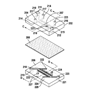
electrode, have
infrared, optical (e.g. camera, LED), or magnetic capabilities or utilize any
other appropriate
type of technology to provide the monitoring data. For instance, the sensor
may be one or
more microelectrodes which analyze electrical characteristics across the
membrane (e.g.
potential difference, resistance, and short circuit current) to confirm the
formation of an
organized barrier, as well as its fluid/ion transport function across the
membrane. It should
be noted that the sensor 120 may be external to the device 102 or be
integrated within the
device 102. It is contemplated that the CPU 110 controls operation of the
sensor 120,
although it is not necessary. The data is preferably shown on the display 116.
[0076] Figure 2A illustrates a perspective view of the tissue interface
device in
accordance with an embodiment. In particular, as shown in Figure 2A, the
device 200 (also
referred to reference numeral 102) preferably includes a body 202 having a
branched
microchannel design 203 in accordance with an embodiment. The body 202 may be
made of
a flexible material, although it is contemplated that the body be
alternatively made of a non-
flexible material. It should be noted that the microchannel design 203 is only
exemplary and
not limited to the configuration shown in Figure 2A. The body 202 is
preferably made of a
flexible biocompatible polymer, including but not limited to, polydimethyl
siloxane (PDMS),
or polyimide. It is also contemplated that the body 202 may be made of non-
flexible
materials like glass, silicon, hard plastic, and the like. Although it is
preferred that the
interface membrane be made of the same material as the body 202, it is
contemplated that the
interface membrane be made of a material that is different than the body of
the device.
[0077] The device in Figure 2A includes a plurality of ports 205 which will
be described
in more detail below. In addition, the branched configuration 203 includes a
tissue-tissue
interface simulation region (membrane 208 in Figure 2B) where cell behavior
and/or passage
of gases, chemicals, molecules, particulates and cells are monitored. Figure
2B illustrates an
exploded view of the organ mimic device in accordance with an embodiment. In
particular,
18
CA 02730928 2011-01-14
WO 2010/009307
PCT/US2009/050830
the outer body 202 of the device 200 is preferably comprised of a first outer
body portion
204, a second outer body portion 206 and an intermediary porous membrane 208
configured
to be mounted between the first and second outer body portions 204, 206 when
the portions
204, 206 are mounted to one another to form the overall body.
[0078] Figure 2B illustrates an exploded view of the device in accordance
with an
embodiment. As shown in Figure 2B, the first outer body portion 204 includes
one or more
inlet fluid ports 210 preferably in communication with one or more
corresponding inlet
apertures 211 located on an outer surface of the body 202. The device 100 is
preferably
connected to the fluid source 104 via the inlet aperture 211 in which fluid
travels from the
fluid source 104 into the device 100 through the inlet fluid port 210.
[0079] Additionally, the first outer body portion 204 includes one or more
outlet fluid
ports 212 preferably in communication with one or more corresponding outlet
apertures 215
on the outer surface of the body 202. In particular, fluid passing through the
device 100 exits
the device 100 to a fluid collector 108 or other appropriate component via the
corresponding
outlet aperture 215. It should be noted that the device 200 may be set up such
that the fluid
port 210 is an outlet and fluid port 212 is an inlet. Although the inlet and
outlet apertures
211, 215 are shown on the top surface of the body 202, one or more of the
apertures may be
located on one or more sides of the body.
[0080] In an embodiment, the inlet fluid port 210 and the outlet fluid port
212 are in
communication with the first central microchannel 250A (see Figure 3A) such
that fluid can
dynamically travel from the inlet fluid port 210 to the outlet fluid port 212
via the first central
microchannel 250A, independently of the second central microchannel 250B (see
Figure 3A).
[0081] It is also contemplated that the fluid passing between the inlet and
outlet fluid
ports may be shared between the central sections 250A and 250B. In either
embodiment,
characteristics of the fluid flow, such as flow rate and the like, passing
through the central
microchannel 250A is controllable independently of fluid flow characteristics
through the
central microchannel 250B and vice versa.
[0082] In addition, the first portion 204 includes one or more pressure
inlet ports 214 and
one or more pressure outlet ports 216 in which the inlet ports 214 are in
communication with
corresponding apertures 217 located on the outer surface of the device 100.
Although the
inlet and outlet apertures are shown on the top surface of the body 202, one
or more of the
apertures may alternatively be located on one or more sides of the body.
19
CA 02730928 2011-01-14
WO 2010/009307
PCT/US2009/050830
[0083] In operation, one or more pressure tubes (not shown) connected to
the pressure
source 118 (Figure 1) provides positive or negative pressure to the device via
the apertures
217. Additionally, pressure tubes (not shown) are connected to the device 100
to remove the
pressurized fluid from the outlet port 216 via the apertures 223. It should be
noted that the
device 200 may be set up such that the pressure port 214 is an outlet and
pressure port 216 is
an inlet. It should be noted that although the pressure apertures 217, 223 are
shown on the
top surface of the body 202, it is contemplated that one or more of the
pressure apertures 217,
223 may be located on one or more side surfaces of the body 202.
[0084] Referring to Figure 2B, the second outer body portion 206 preferably
includes one
or more inlet fluid ports 218 and one or more outlet fluid ports 220. It is
preferred that the
inlet fluid port 218 is in communication with aperture 219 and outlet fluid
port 220 is in
communication with aperture 221, whereby the apertures 219 and 221 are
preferably located
on the outer surface of the second outer body portion 206. Although the inlet
and outlet
apertures are shown on the surface of the body 202, one or more of the
apertures may be
alternatively located on one or more sides of the body.
[0085] As with the first outer body portion 204 described above, one or
more fluid tubes
connected to the fluid source 104 (Figure 1) are preferably coupled to the
aperture 219 to
provide fluid to the device 100 via port 218. Additionally, fluid exits the
device 100 via the
outlet port 220 and out aperture 221 to a fluid reservoir/collector 108 or
other component. It
should be noted that the device 200 may be set up such that the fluid port 218
is an outlet and
fluid port 220 is an inlet.
[0086] In addition, it is preferred that the second outer body portion 206
includes one or
more pressure inlet ports 222 and one or more pressure outlet ports 224. In
particular, it is
preferred that the pressure inlet ports 222 are in communication with
apertures 227 and
pressure outlet ports 224 are in communication with apertures 229, whereby
apertures 227
and 229 are preferably located on the outer surface of the second portion 206.
Although the
inlet and outlet apertures are shown on the bottom surface of the body 202,
one or more of
the apertures may be alternatively located on one or more sides of the body.
Pressure tubes
connected to the pressure source 118 (Figure 1) are preferably engaged with
ports 222 and
224 via corresponding apertures 227 and 229. It should be noted that the
device 200 may be
set up such that the pressure port 222 is an outlet and fluid port 224 is an
inlet.
[0087] In an embodiment, the membrane 208 is mounted between the first
portion 204
and the second portion 206, whereby the membrane 208 is located within the
body 202 of the
device 200 (see Figure 5E). In an embodiment, the membrane 208 is a made of a
material
CA 02730928 2011-01-14
WO 2010/009307 PCT/US2009/050830
having a plurality of pores or apertures therethrough, whereby molecules,
cells, fluid or any
media is capable of passing through the membrane 208 via one or more pores in
the
membrane 208. As discussed in more detail below, it is contemplated in an
embodiment that
the porous membrane 208 may be made of a material which allows the membrane
208 to
undergo stress and/or strain in response to pressure differentials present
between the central
microchannels 250A, 250B and the operating microchannels. Alternatively, the
porous
membrane 208 is relatively inelastic in which the membrane 208 undergoes
minimal or no
movement while media is passed through one or more of the central
microchannels 250A,
250B and cells organize and move between the central microchannels 250A, 250B
via the
porous membrane.
[0088] Referring Figure 2C illustrates a perspective view of the tissue-
tissue interface
region of the first outer portion 204 of the body taken at line C-C (from
Figure 2B). As
shown in Figure 2C, the top portion of the tissue-tissue interface region 207A
is within the
body of the first portion 204 and includes a top portion of a central
microchannel 230 and one
or more top portion side operating microchannels 232 located adjacent to the
central
microchannel 230. Microchannel walls 234 preferably separate the central
microchannel 230
from the operating microchannels 232 such that fluid traveling through the
central
microchannel 230 does not pass into operating microchannels 232. Likewise, the
channel
walls 234 prevent pressurized fluid passing along operating microchannels 232
from entering
the central microchannel 230. It should be noted that a pair of operating
microchannels 232
are shown on opposing sides of central microchannel 230 in Figures 2C and 3A,
however it is
contemplated that the device may incorporate more than two operating
microchannels 232. It
is also contemplated that the device 200 may include only one operating
microchannel 232
adjacent to the central microchannel 230.
[0089] Figure 2D illustrates a perspective view of the tissue interface
region taken at line
D-D of the second outer portion 206 of the body. As shown in Figure 2D, the
tissue
interface region includes a bottom portion of the central microchannel 240 and
at least two
bottom portions of operating microchannels 242 located adjacent to the central
microchannel
240 portion. A pair of channel walls 234 preferably separate the central
microchannel 240
from the operating microchannels 232 such that fluid traveling through the
central
microchannel 230 does not pass into operating microchannels 232. Likewise, the
channel
walls 234 prevent pressurized fluid passing along operating microchannels 232
from entering
the central microchannel 230.
21
CA 02730928 2011-01-14
WO 2010/009307 PCT/US2009/050830
[0090] As shown in Figures 2C and 2D, the top and bottom portions 230 and
240 of the
central microchannel each have a range of width dimension (shown as B) between
50 and
1000 microns, and preferably around 400 microns. It should be noted that other
width
dimensions are contemplated depending on the type of physiological system
which is being
mimicked in the device. Additionally, the top and bottom portions of the
operating
microchannels 232 and 242 each have a width dimension (shown as A) between 25
and 800
microns, and preferably around 200 microns, although other width dimensions
are
contemplated. The height dimensions of the central and/or operating
microchannels range
between 50 microns and several centimeters, and preferably around 200 microns.
The
microchannel walls 234, 244 preferably have a thickness range between 5
microns to 50
microns, although other width dimensions are contemplated depending on the
material used
for the walls, application in which the device is used and the like.
[0091] Figure 3A illustrates a perspective view of the tissue interface
region within the
body in accordance with an embodiment. In particular, Figure 3A illustrates
the first portion
207A and the second portion 207B mated to one another whereby the side walls
228 and 238
as well as channel walls 234. 244 form the overall central microchannel 250
and operating
microchannels 252. As stated above, it is preferred that central microchannel
250 and
operating microchannels 252 are separated by the walls 234, 244 such that
fluid is not able to
pass between the channels 250, 252.
[0092] The membrane 208 is preferably positioned in the center of the
central
microchannel 250 and is oriented along a plane parallel to the x-y plane shown
in Figure 3A.
It should be noted that although one membrane 208 is shown in the central
microchannel 250,
more than one membrane 208 may be configured within the central microchannel
250, as
discussed in more detail below. In addition to being positioned within the
central
microchannel 250, the membrane 208 is sandwiched in place by channel walls
234, 244
during formation of the device.
[0093] The membrane 208 preferably separates the overall central
microchannel 250 into
two or more distinct central microchannels 250A and 250B. It should be noted
that although
the membrane 208 is shown midway through the central microchannel 250, the
membrane
208 may alternatively be positioned vertically off-center within the central
microchannel 250,
thus making one of the central microchannel sections 250A, 250B larger in
volume or cross-
section than the other microchannel section.
[0094] As will be discussed in more detail below, the membrane 208 may have
at least a
portion which is porous to allow cells or molecules to pass therethrough.
Additionally or
22
CA 02730928 2011-01-14
WO 2010/009307
PCT/US2009/050830
alternatively, at least a portion of the membrane 208 may have elastic or
ductile properties
which allow the membrane 208 to be manipulated to expand/contract along one or
more
planar axe. Thus, it is contemplated that one or more portions of the membrane
208 may be
porous and elastic or porous, but inelastic.
[0095] With regard to the porous and elastic membrane, a pressure
differential may be
applied within the device to cause relative continuous expansion and
contraction of the
membrane 208 along the x-y plane. In particular. as stated above, one or more
pressure
sources preferably apply pressurized fluid (e.g. air) along the one or more
operating
microchannels 252, whereby the pressurized fluid in the microchannels 252
creates a pressure
differential on the microchannel walls 234, 244. The membrane 208 may have an
elasticity
depending on the type of material that it is made of. If the membrane 208 is
made of more
than one material, the weight ratio of the respective materials which make up
the membrane
is a factor in determining the elasticity. For example, in the embodiment that
the membrane
208 is made of PDMS, the Young's modulus values are in the ranges of 12 kPa-20
MPa,
although other elasticity values are contemplated.
[0096] In the embodiments shown in Figures 3A and 3B, the pressurized fluid
is a
vacuum or suction force that is applied only through the operating
microchannels 252. The
difference in pressure caused by the suction force against the microchannel
walls 234, 244
causes the walls 234, 244 to bend or bulge outward toward the sides of the
device 228, 238
(see Figure 3B). Considering that the membrane 208 is mounted to and
sandwiched between
the walls 234, 244, the relative movement of the walls 234, 244 thereby causes
the opposing
ends of the membrane to move along with the walls to stretch (shown as 208' in
Figure 3B)
along the membrane's plane. This stretching mimics the mechanical forces
experienced by a
tissue-tissue interface, for example, in the lung alveolus during breathing,
and thus provides
the important regulation for cellular self assembly into tissue structures and
cell behavior.
[0097] When the negative pressure is no longer applied (and/or positive
pressure is
applied to the operating channels), the pressure differential between the
operating channels
252 and the central channel 250 decreases and the channel walls 234, 244
retract elastically
toward their neutral position (as in Figure 3A). During operation, the
negative pressure is
alternately applied in timed intervals to the device 200 to cause continuous
expansion and
contraction of the membrane 208 along its plane, thereby mimicking operation
of the tissue-
tissue interface of the living organ within a controlled in vitro environment.
As will be
discussed, this mimicked organ operation within the controlled environment
allows
monitoring of cell behavior in the tissues, as well as passage of molecules,
chemicals,
23
CA 02730928 2011-01-14
WO 2010/009307
PCT/US2009/050830
particulates and cells with respect to the membrane and the associated first
and second
microchannels 250A, 250B.
[0098] It should be noted that the term pressure differential in the
present specification
relates to a difference of pressure on opposing sides of a particular wall
between the central
microchannel and the outer operating channel. It is contemplated that the
pressure
differential may be created in a number of ways to achieve the goal of
expansion and/or
contraction of the membrane 208. As stated above, a negative pressure (i.e.
suction or
vacuum) may be applied to one or more of the operating channels 252.
Alternatively, it is
contemplated that the membrane 208 is pre-loaded or pre-stressed to be in an
expanded state
by default such that the walls 234, 244 are already in the bent configuration,
as show in
Figure 3B. In this embodiment, positive pressure applied to the operating
channel 252 will
create the pressure differential which causes the walls 234, 244 to move
inward toward the
central microchannel (see in Figure 3A) to contract the membrane 208.
[0099] It is also contemplated, in another embodiment, that a combination
of positive and
negative pressure is applied to one or more operating microchannels 252 to
cause movement
of the membrane 208 along its plane in the central microchannel. In any of the
above
embodiments, it is desired that the pressure of the fluid in the one or more
operating channels
252 be such that a pressure differential is in fact created with respect to
the pressure of the
fluid(s) in one or more of the central microchannel(s) 250A, 250B to cause
relative
expansion/contraction of the membrane 208. For example, fluid may have a
certain pressure
may be applied within the top central microchannel 250A, whereby fluid in the
bottom
central microchannel 250B may have a different pressure. In this example,
pressure applied
to the one or more operating channels 252 must take into account the pressure
of the fluid in
either or both of the central microchannels 250A, 250B to ensure desired
expansion/contraction of the membrane 208.
[00100] It is possible, in an embodiment, for a pressure differential to exist
between the
top and bottom microchannels 250A, 250B to cause at least a portion of the
membrane 208 to
expand and/or contract vertically in the z-direction in addition to
expansion/contraction along
the x-y plane.
[00101] In an embodiment, the expansion and retraction of the membrane 208
preferably
applies mechanical forces to the adherent cells and ECM that mimic
physiological
mechanical cues that can influence transport of chemicals, molecules
particulates, and/or
fluids or gas across the tissue-tissue interface, and alter cell physiology.
It should be noted
that although pressure differentials created in the device preferably cause
24
CA 02730928 2011-01-14
WO 2010/009307 PCT/US2009/050830
expansion/contraction of the membrane, it is contemplated that mechanical
means, such as
micromotors or actuators, may be employed to assist or substitute for the
pressure differential
to cause expansion/contraction of the cells on the membrane to modulatecell
physiology.
[00102] Figure 3E and 4C illustrate perspectives view of the membrane 208
which
includes a plurality of apertures 302 extending therethrough in accordance
with an
embodiment. In particular, the membrane shown in Figures 3E and 4C includes
one or more
of integrated pores or apertures 302 which extend between a top surface 304
and a bottom
surface 306 of the membrane 208.
[00103] The membrane is configured to allow cells, particulates, chemicals
and/or media
to migrate between the central microchannel portions 250A, 250B via the
membrane 208
from one section of the central microchannel to the other or vice versa. The
pore apertures
are shown to have a pentagonal cross sectional shape in Figures 4A-4C,
although any other
cross sectional shape is contemplated, including but not limited to, a
circular shaped 302,
hexagonal 308, square. elliptical 310 and the like. The pores 302, 308, 310
(generally
referred to as reference numeral 302) preferably extend vertically between the
top and bottom
surfaces 304, 306, although it is contemplated that they may extend laterally
as well between
the top and bottom surfaces, as with pore 312. It should also be noted that
the porous may
additionally/alternatively incorporate slits or other shaped apertures along
at least a portion of
the membrane 208 which allow cells, particulates, chemicals and/or fluids to
pass through the
membrane 208 from one section of the central microchannel to the other.
[00104] The width dimension of the pores are preferably in the range of .5
microns and 20
microns, although it is preferred that the width dimension be approximately 10
microns. It is
contemplated, however, that the width dimension be outside of the range
provided above. In
some embodiments, the membrane 208 has pores or apertures larger than
traditional
molecular/chemical filtration devices, which allow cells as well as molecules
to migrate
across the membrane 208 from one microchannel section (e.g. 250A) to the other
microchannel section (e.g. 250B) or vice versa. This may be useful in
culturing cells which
polarize in the top and bottom central channels in response to being in the
microchannel
environment provided by the device whereby fluid(s) and cells are dynamically
passed
through pores that connect these microchannels 250A, 250B.
[00105] As shown in Figure 4B, the thickness of the membrane 208 may be
between 70
nanometers and 50 microns, although a preferred range of thickness would
between 5 and 15
microns. It is also contemplated that the membrane 208 be designed to include
regions which
have lesser or greater thicknesses than other regions in the membrane 208. As
shown in
CA 02730928 2011-01-14
WO 2010/009307 PCT/US2009/050830
Figure 3C, the membrane 208 is shown to have one or more decreased thickness
areas 209
relative to the other areas of the membrane 208. The decreased thickness
area(s) 209 may
run along the entire length or width of the membrane 208 or may alternatively
be located at
only certain locations of the membrane 208. It should be noted that although
the decreased
thickness area 209 is shown along the bottom surface of the membrane 208 (i.e.
facing
microchannel 250B), it is contemplated that the decreased thickness area(s)
209 may
additionally/alternatively be on the opposing surface of the membrane 208
(i.e. facing
microchannel 250A). It should also be noted that at least portions of the
membrane 208 may
have one or more larger thickness areas 209' relative to the rest of the
membrane, as shown in
Figure 3D and capable of having the same alternatives as the decreased
thickness areas
described above.
[00106] In an embodiment, the porous membrane 208 may be designed or surface
patterned to include micro and/or nanoscopic patterns therein such as grooves
and ridges,
whereby any parameter or characteristic of the patterns may be designed to
desired sizes,
shapes, thicknesses, filling materials, and the like.
[00107] In an embodiment, the membrane 208 is made of polydimethylsiloxane
(PDMS)
or any other polymeric compound or material, although this is not necessary.
For instance,
the membrane 208 may be made of polyimide, polyester, polycarbonate,
cyclicolefin
copolymer, polymethylmethacrylate, nylon, polyisoprene, polybutadiene,
polychlorophene,
polyisobutylene, poly(styrene-butadiene-styrene), nitriles, the polyurethanes
and the
polysilicones, GE RTV 615, a vinyl-silane crosslinked (type) silicone
elastomer (family) may
be used. Polydimethysiloxane (PDMS) membranes are available HT-6135 and HT-
6240
membranes from Bisco Silicons (Elk Grove, Ill.) and are useful in selected
applications. The
choice of materials typically depends upon the particular material properties
(e.g., solvent
resistance, stiffness, gas permeability, and/or temperature stability)
required for the
application being conducted. Additional elastomeric materials that can be used
in the
manufacture of the components of the microfluidic devices described in Unger
et al., (2000
Science 288:113-116). Some elastomers of the present devices are used as
diaphragms and in
addition to their stretch and relax properties, are also selected for their
porosity,
impermeability, chemical resistance, and their wetting and passivating
characteristics. Other
elastomers are selected for their thermal conductivity. Micronics Parker
Chomerics
Thermagap material 61-02-0404-F574 (0.020" thick) is a soft elastomer (<5Shore
A) needing
only a pressure of 5 to 10 psi to provide a thermal conductivity of 1.6 W/m-
K. Deformable
films, lacking elasticity, can also be used in the microfluidic device. One
may also use silk,
26
CA 02730928 2011-01-14
WO 2010/009307
PCT/US2009/050830
ECM gels with or without crosslinking as other such suitable materials to make
the devices
and membranes as described.
[00108] It should be noted that although the central and operating
microchannels 250, 252
are shown to have substantially square or rectangular cross sections, other
cross-sectional
shapes are contemplated such as circular, oval, hexagonal, and the like. It is
also
contemplated that the device 200 may have more or less than two operating
channels 252
and/or more or less than two central microchannels 250A, 250B in accordance
with an
embodiment.
[00109] In an embodiment, it is contemplated that the central microchannel has
a non-
uniform width dimension B along at least a portion of its length in the
device. Figure 2E
illustrates a cross sectional top-down view of the tissue interface region 400
in accordance
with an embodiment. As shown in Figure 2E, the interface 400 includes the
central
microchannel 402 along with adjacent operating channels 406 separated by
microchannel
walls 404. In the embodiment in Figure 2E, the central microchannel 402 is
shown to have a
gradually increasing width from width dimension C (at end 408) to width
dimension D (at
end 410). In the embodiment in Figure 2E, the operating channels 406 each have
a
correspondingly decreasing width dimension (from width dimension E at end 408
to width
dimension F at end 410). In another embodiment, as shown in Figure 2F, the
operating
channels 406' have a substantially uniform width dimension F at ends 408 and
410. It is
contemplated that the membrane (not shown) be placed above the central
microchannel 402
and mounted to the top surface of the walls 404, whereby the membrane has a
tapered shape
similar to the central microchannel 402. The tapered membrane would thereby
undergo non-
uniform stretching in the direction of the arrows when the pressure
differential is applied
between the operating microchannels 406 and the central microchannel 402.
[00110] In another embodiment, the central microchannel may have a portion
which has a
partially circular cross sectional shape, as shown in Figure 2G. In particular
to the
embodiment in Figure 2G, a pressure differential created between the central
microchannel
502 and the adjacent operating microchannels 504 will cause the microchannel
walls 506 to
move in the direction represented by the arrows. With regard to the circular
portion 508 of
the central microchannel 502, equiaxial outward movement of the walls (as
shown by the
arrows) at the central portion 508 produces equiaxial stretching of the
membrane (not shown)
mounted atop of the walls 506.
[00111] The device 200 described herein has potential for several
applications. For
example, in one application, the membrane 208 may be subjected to
physiological
27
CA 02730928 2011-01-14
WO 2010/009307 PCT/US2009/050830
mechanical strain generated by cyclic stretching of the membrane 208 and/or
the flow of
biological fluids (e.g. air, mucus, blood) to recapitulate the native
microenvironment of the
alveoli and underlying pulmonary capillaries. In an embodiment, the culture
conditions of
cells upon the membrane 208 may be optimized under extracellular matrix (ECM)
coating,
media perfusion, or cyclic mechanical strain to maintain morphological and
functional
characteristics of the co-cultured cells and to permit their direct cellular
interaction across the
membrane 208. The device 200 would thus permit long-term cell culture and
dynamic
mechanical stretching of a adjacent monolayers of lung epithelial or
endothelial cells grown
on the membrane at the same time.
[00112] In utilizing the membrane 208 in simulating the tissue-tissue
interface between the
alveolar epithelium and pulmonary endothelium in the lung, one method may be
to apply
type I alveolar epithelial cells to the side of the membrane 208 facing the
first section 250A
(hereinafter top side of membrane) to mimic the epithelial layer. It is
possible, however, to
mix type I-like and type II-like alveolar epithelial cells at a ratio of
approximately 7:13 to
reconstruct the in vivo cellular composition of the alveolar epithelium. In
the example
method, lung microvascular endothelial cells are cultured on the opposite side
of the
membrane 208 facing the second section 250B (hereinafter bottom side of
membrane). In the
example method, negative pressure is cyclically applied to the device 200 to
cause the
membrane 208 to continuously expand and contract along its plane.
[00113] During such operation, a physiological alveolar-capillary unit may be
formed on
the membrane 208 since typical junctional structures may form on the membrane
207 and
fluids as well as ions be transported across the membrane 208 between the
first and second
sections 250A, 250B. The formation of tight junctions on the membrane 208 may
be
evaluated using on-chip immunohistochemical detection of tight junction
proteins such as
ZO-1 and occludin. Additionally or alternately, the exclusion of fluorescently
labeled large
molecules (e.g. dextrans of different weight) may be quantitated to determine
the
permeability of the membrane and optimize epithelial membrane barrier
formation by
varying culture conditions. Additionally, histological, biochemical, and
microfluorimetric
techniques may be employed to demonstrate formation of a functional alveolar-
capillary unit
that reproduces the key structural organization of its in vivo counterpart on
the membrane
208.
[00114] In an example, the gas exchange function of the tissue-tissue
interface self
assembled on membrane 208 may be determined by injecting different fluids,
each having
their own oxygen partial pressures and blood, into the respective first and
second sections
28
CA 02730928 2011-01-14
WO 2010/009307
PCT/US2009/050830
250A, 250B, whereby the first section 250A acts as the alveolar compartment
and the second
section 250B acts as the microvascular compartment. A blood-gas measurement
device
preferably within the device 200 is used to measure the level of oxygen in the
blood in the
respective sections 250A, 250B before and after the passing of the blood
through the device.
For example, blood can flow through the channel 250B while air is being
injected into the
upper channel 250A, whereby the exiting air is collected and measured to
determine the
oxygen level using an oximeter. Oximeters can be integrated with the existing
system or as a
separate unit connected to the outlet port of one or more central
microchannels. In an
embodiment, air or another medium with aerosols containing drugs or
particulates may flow
through the device, whereby the transport of these drugs or particulates to
the blood via the
membrane is then measured. It is also contemplated that pathogens or cytokines
are added to
the air or gaseous medium side and then the sticking of immune cells to nearby
capillary
endothelium and their passage along with edema fluid from the blood side to
the airway side,
as well as pathogen entry into blood, are measured.
[00115] Since the functionality of an epithelium requires polarization of
constituent cells,
the structure of the membrane may be visualized using transmission electron
microscopy,
immunohistocytochemistry, confocal microscopy, or other appropriate means to
monitor the
polarization of the alveolar epithelial cell side of the membrane 208. In a
lung mimic
embodiment, a flourescent dye may be applied to the first and/or second
microchannels
250A, 250B to determine pulmonary surfactant production by the airway
epithelium at the
membrane 208. In particular, alveolar epithelial cells on the membrane 208 can
be monitored
by measuring the fluorescence resulting from cellular uptake of the
fluorescence dye that
specifically labels intracellular storage of pulmonary surfactant (e.g.
quinacrine) or using
specific antibodies.
[00116] One of the unique capabilities of the tissue interface device 200
allows
development of in vitro models that simulate inflammatory responses of the
lung at the organ
level to allow study of how immune cells migrate from the blood, through the
endothelium
and into the alveolar compartment. One way this is achieved is by controlled
and
programmable microfluidic delivery of pro-inflammatory factors (e.g. IL-113,
TNF-a, IL-8,
silica micro- and nanoparticles, pathogens) to the first section 250A as well
as whole human
blood flowing or medium containing circulating immune cells in the second
section 250B.
Electrical resistance and short circuit current across the membrane may be
monitored to study
changes in the vascular permeability, extravasation of fluid and cell passage
into the alveolar
29
CA 02730928 2011-01-14
WO 2010/009307 PCT/US2009/050830
space during inflammatory responses. Fluorescence microscopy can be used to
visualize
dynamic cell motile behavior during the extravasation response.
[00117] The tissue interface device 200 may also be used to examine how
nanomaterials
behave with respect to the lung tissue-tissue interface. In particular,
nanomaterials (e.g. silica
nanoparticles, superparamagnetic nanoparticles, gold nanoparticles, single-
walled carbon
nanotubes) may be applied to the airway surface of the membrane 208 to
investigate potential
toxic effects of nanomaterials on airway or endothelial cells grown on the
membrane 208, as
well as their passage from the airway channel into the blood channel. For
instance, sensors
120 can be used to monitor transmigration of nanomaterials through tissue
barriers formed on
the membrane 208 and nanomaterial-induced changes in barrier functions such as
gas
exchange and fluid/ion transport.
[00118] The tissue interface device 200 permits direct analysis of a variety
of important
areas of lung biology and physiology including but not limited to gas
exchange, fluid/ion
transport, inflammation, infection, edema/respiratory distress syndrome,
cancer and
metastasis development, fungal infection, drug delivery as well as drug
screening,
biodetection, and pulmonary mechanotransduction. In addition, the device 200
allows for
accurately modeling biological tissue-tissue interfaces found in other
physiological systems
such as the blood-brain barrier, intestine, bone marrow, glomerulus, and
cancerous tumor
microenvironment. As stated above, more than one tissue interface device 200
may be
multiplexed and automated to provide high-throughput analysis of cell and
tissue responses to
drugs, chemicals, particulates, toxins, pathogens or other environmental
stimuli for drug,
toxin and vaccine screening, as well as toxicology and biodetection
applications. The device
may be used for studying complex tissue and organ physiology in vitro, as well
as tissue and
organ engineering in vivo with biocompatible or biodegradeable devices.
[00119] In an embodiment, the tissue interface device 200 can be used to
produce artificial
tissue layers therein. In the embodiment, two or more different types of cells
are applied on
opposing surfaces of the membrane 208 and grown under conditions that mimic
the
appropriate physiological environments. Additionally or altematively, a
pressure differential
can be applied between the central microchannel and at least one of the
operating
microchannels which causes the microchannel walls to move and thus causes the
membrane
208 to undergo expansion/contraction along its plane.
[00120] In another example, the device 200 utilizes the porous membrane 208,
whereby
lung cells are grown on one side of the membrane 208 and endothelial cells are
maintained on
the other side of the membrane 208. During the operation of the device 200,
these two cells
CA 02730928 2011-01-14
WO 2010/009307
PCT/US2009/050830
layers communicate with each other through passage of chemical and molecular
cues through
the pores on the membrane 208. This communication may be monitored and
analyzed to
understand how the cells function differently as a tissue-tissue interface,
with or without
physiological mechanical simulation, and compared to when grown as single
tissue types in
isolation as in standard tissue culture systems. By monitoring changes in cell
and tissue
physiology, as well as passage of chemicals, molecules, particulates and cells
across this
tissue-tissue interface, information is obtained which may be used to produce
more effective
drugs or therapies, to identify previously unknown toxicities, and to
significantly shorten the
timescale of these development processes. In particular, the behavior of cells
in such a
controlled environment allows researchers to study a variety of physiological
phenomena
taking place in the systems mentioned above that can not be recreated using
conventional in
vitro culture techniques. In other words, the device 200 functions to create a
monitorable
artificial blood-air barrier outside a patient's body and in a controllable
environment that still
retains key physiological functions and structures of the lung. It should be
noted that
although the device above is described in terms of mimicking lung function,
the device may
easily be configured to mimic other physiological systems such as peristalsis
and absorption
in the gastrointestinal tract containing living microbial populations,
perfusion and urine
production in the kidney, function of the blood-brain barrier, effects of
mechanical
deformation on skin aging, bone marrow-microvessel interface with
hematopoietic stem cell
niche, and the like.
[00121] Details of membrane surface treatment and types of media which can be
applied
to the membrane and/or through the central microchannels 250A, 250B in
operating the
device will now be discussed. The membrane including the porous membrane can
be coated
with substances such as various cell adhesion promoting substances or ECM
proteins, such as
fibronectin, laminin or various collagen types or combinations thereof, as
shown in Figure
4D. In general, as shown in Figure 4D, one or more substances 608 is shown on
one surface
of the membrane 604 whereas another substance 610 is applied to the opposing
surface of the
membrane 604, or both surfaces can be coated with the same substance. In some
embodiments, the ECMs, which may be ECMs produced by cells, such as primary
cells or
embryonic stem cells, and other compositions of matter are produced in a serum-
free
environment.
[00122] In an embodiment, one coats the membrane with a combination of a cell
adhesion
factor and a positively-charged molecule that are bound to the membrane to
improve cell
attachment and stabilize cell growth. The positively charged molecule can be
selected from
31
CA 02730928 2011-01-14
WO 2010/009307
PCT/US2009/050830
the group consisting of polylysine, chitosan, poly(ethyleneimine) or acrylics
polymerized
from acrylamide or methacrylamide and incorporating positively-charged groups
in the form
of primary, secondary or tertiary amines, or quaternary salts. The cell
adhesion factor can be
added to the membrane and is preferably fibronectin, laminin, collagen,
vitronectin or
tenascin, or fragments or analogs having a cell binding domain thereof. The
positively-
charged molecule and the cell adhesion factor can be covalently bound to the
membrane. In
another embodiment, the positively-charged molecule and the cell adhesion
factor are
covalently bound to one another and either the positively-charged molecule or
the cell
adhesion factor is covalently bound to the membrane. Also, the positively-
charged molecule
or the cell adhesion factor or both cam be provided in the form of a stable
coating non-
covalently bound to the membrane.
[00123] In an embodiment, the cell attachment-promoting substances, matrix-
forming
formulations, and other compositions of matter are sterilized to prevent
unwanted
contamination. Sterilization may be accomplished, for example, by ultraviolet
light, filtration,
or heat. Antibiotics may also be added, particularly during incubation, to
prevent the growth
of bacteria, fungi and other undesired micro-organisms. Such antibiotics
include, by way of
non-limiting example, gentamicin, streptomycin, penicillin, amphotericin and
ciprofloxacin.
[00124] In another embodiment, the membrane is coated with cell cultures,
including
without limitation, primary cell cultures, established cell lines, or stem
cell cultures, such as
ESC, attached to ECM substances that comprise comprise or consist essentially
of
fibronectin or collagen.
[00125] In an
embodiment, the primary cells or cell lines attached to the membrane may
be alveolar cells, endothelial cells, intestinal cells, keratinocytes, which
include without
limitation, human dermal keratinocytes, or any other type of cell listed
elsewhere in this
specification or well known to one skilled in the art. In other embodiments,
the primary cells
may be fibroblast cells, which include without limitation, human fetal
fibroblast cells. In
some embodiments, the stem cells of the stem cell cultures are embryonic stem
cells. The
source of embryonic stem cells can include without limitation mammals,
including non-
human primates and humans. Non-limiting examples of human embryonic stem cells
include
lines BG01. BG02, BG03, BGOly, CHA-hES-1, CHA-hES-2, FCNCBS1, FCNCBS2,
FCNCBS3, H1, H7, H9, H13, H14, HSF-1, H9.1. H9.2, HES-1, HES-2, HES-3, HES-4,
HES-5, HES-6, hES-1-2, hES-3-0, hES-4-0, hES-5-1. hES-8-1, hES-8-2, hES-9-1,
hES-9-2,
hES-101, hICM8, hICM9, hICM40, hICM41, hICM42, hICM43, HSF-6, HUES-1, HUES-2,
HUES-3, HUES-4 HUES-5, HUES-6, HUES-7 HUES-8, HUES-9, HUES-10, HUES-11,
32
CA 02730928 2011-01-14
WO 2010/009307 PCT/US2009/050830
HUES-12, HUES-13, HUES-14, HUESS-15, HUES-16, HUES-17, 13, 14, 16, 13.2, 13.3,
16.2, J3, J3.2, MB01, MB02, MB03. Miz-hES1, RCM-1, RLS ES 05, RLS ES 07, RLS
ES
10, RLS ES 13, RLS ES 15, RLS ES 20, RLS ES 21, SA01, SA02, and SA03. In an
embodiment, the stem cells of the stem cell cultures are induced pluripotent
stem cells.
[00126] In an embodiment, the cell cultures may be cell cultures such as
primary cell
cultures or stem cell cultures which are serum-free. In some these
embodiments, a serum-free
primary cell ECM is used in conjunction with a primary cell serum-free medium
(SFM).
Suitable SFM include without limitation (a) EPILIFE Serum Free Culture Medium
supplemented with EPILIFE Defined Growth Supplement and (b) Defined
Keratinocyte-
SFM supplemented with Defined Keratinocyte-SFM Growth Supplement, all
commercially
available from Gibco/lnvitrogen (Carlsbad, Calif., US). In some of these
embodiments, a
serum-free stem cell ECM is used in conjunction with stem cell SFM. Suitable
SFM include
without limitation STEMPRO hESC Serum Free Media (SFM) supplemented with
basic
fibroblast growth factor and .beta.-mercaptoethanol. KNOCKOUT. D-MEM
supplemented
with KNOCKOUT'. Sertim Replacement (SR), STEMPROO. MSC SFM and
STEMPROO. NSC SFM, all commercially available from Gibco/Invitrogen (Carlsbad,
Calif., US).
[00127] In an embodiment, the compositions can also be xeno-free. A
composition of
matter is said to be "xeno-free" when it is devoid of substances from any
animal other than
the species of animal from which the cells are derived. In an embodiment, the
cell cultures
which may be cell cultures such as primary cell cultures or stem cell cultures
are xeno-free.
In these embodiments, a xeno-free ECM which may be an ECM such as a primary
cell ECM
or ECM designed specifically to support stem cell growth or differentiation.
These matrices
may be specifically designed to be xeno-free.
[00128] In an embodiment, the cell cultures are primary cells or stem cells
cultured in a
conditioned culture medium. In other embodiments, the culture medium is an
unconditioned
culture medium.
[00129] In an embodiment, the cell culture conditions are completely defined.
In these
embodiments, one uses a completely defined cell culture medium in the fluid
chambers.
Suitable media include without limitation, for primary cells, EPILIFE . Serum
Free Culture
Medium supplemented with EPILIFE . Defined Growth Supplement, and, for stem
cells,
STEMPROO. hESC SFM, all commercially available from Gibco/Invitrogen,
Carlsbad,
Calif., US.
33
CA 02730928 2011-01-14
WO 2010/009307 PCT/US2009/050830
[00130] To study the effects of pharmaceuticals, environmental stressors,
pathogens,
toxins and such, one can add these into the desired cell culture medium
suitable for growing
the cells attached to the porous membrane in the channel. Thus, one can
introduce pathogens,
such as bacteria, viruses, aerosols, various types of nanoparticles, toxins,
gaseous substances,
and such into the culture medium which flows in the chambers to feed the
cells.
[00131] A skilled artisan will also be able to control the pH balance of the
medium
according to the metabolic activity of the cells to maintain the pH in a
suitable level for any
cell or tissue type in question. Monitors and adjustment systems to monitor
and adjust pH
may be inserted into the device.
[00132] The membrane is preferably coated on one or both sides with cells,
molecules or
other matter, whereby the device provides a controlled environment to monitor
cell behavior
along and/or between the microchannels via the membrane. One can use any cells
from a
multicellular organisms in the device. For example, human body comprises at
least 210
known types of cells. A skilled artisan can easily construct useful
combinations of the cells
in the device. Cell types (e.g., human) which can be used in the devices
include, but are not
limited to cells of the integumentary system including but not limited to
Keratinizing
epithelial cells, Epidermal keratinocyte (differentiating epidermal cell),
Epidermal basal cell
(stem cell), Keratinocyte of fingernails and toenails, Nail bed basal cell
(stem cell),
Medullary hair shaft cell, Cortical hair shaft cell, Cuticular hair shaft
cell, Cuticular hair root
sheath cell, Hair root sheath cell of Huxley's layer, Hair root sheath cell of
Henle's layer,
External hair root sheath cell, Hair matrix cell (stem cell); Wet stratified
barrier epithelial
cells, such as Surface epithelial cell of stratified squamous epithelium of
cornea, tongue, oral
cavity, esophagus, anal canal, distal urethra and vagina, basal cell (stem
cell) of epithelia of
cornea, tongue, oral cavity, esophagus, anal canal, distal urethra and vagina,
Urinary
epithelium cell (lining urinary bladder and urinary ducts); Exocrine secretory
epithelial cells,
such as Salivary gland mucous cell (polysaccharide-rich secretion), Salivary
gland serous cell
(glycoprotein enzyme-rich secretion), Von Ebner's gland cell in tongue (washes
taste buds),
Mammary gland cell (milk secretion), Lacrimal gland cell (tear secretion).
Ceruminous gland
cell in ear (wax secretion), Eccrine sweat gland dark cell (glycoprotein
secretion), Eccrine
sweat gland clear cell (small molecule secretion), Apocrine sweat gland cell
(odoriferous
secretion, sex-hormone sensitive), Gland of Moll cell in eyelid (specialized
sweat gland),
Sebaceous gland cell (lipid-rich sebum secretion), Bowman's gland cell in nose
(washes
olfactory epithelium), Brunner's gland cell in duodenum (enzymes and alkaline
mucus),
Seminal vesicle cell (secretes seminal fluid components, including fructose
for swimming
34
CA 02730928 2011-01-14
WO 2010/009307 PCT/US2009/050830
sperm), Prostate gland cell (secretes seminal fluid components), Bulbourethral
gland cell
(mucus secretion), Bartholin's gland cell (vaginal lubricant secretion), Gland
of Littre cell
(mucus secretion), Uterus endometrium cell (carbohydrate secretion), Isolated
goblet cell of
respiratory and digestive tracts (mucus secretion), Stomach lining mucous cell
(mucus
secretion), Gastric gland zymogenic cell (pepsinogen secretion), Gastric gland
oxyntic cell
(hydrochloric acid secretion), Pancreatic acinar cell (bicarbonate and
digestive enzyme
secretion), pancreatic endocrine cells, Paneth cell of small intestine
(lysozyme secretion),
intestinal epithelial cells, Types I and II pneumocytes of lung (surfactant
secretion), and/or
Clara cell of lung.
[00133] One can also coat the membrane with Hormone secreting cells, such as
endocrine
cells of the islet of Langerhands of the pancreas, Anterior pituitary cells,
Somatotropes,
Lactotropes, Thyrotropes, Gonadotropes, Corticotropes, Intermediate pituitary
cell, secreting
melanocyte-stimulating hormone; and Magnocellular neurosecretory cells
secreting oxytocin
or vasopressin; Gut and respiratory tract cells secreting serotonin,
endorphin, somatostatin,
gastrin, secretin, cholecystokinin, insulin, glucagon, bombesin; Thyroid gland
cells such as
thyroid epithelial cell, parafollicular cell, Parathyroid gland cells,
Parathyroid chief cell,
Oxyphil cell, Adrenal gland cells, chromaffin cells secreting steroid hormones
(mineralcorticoids and gluco corticoids), Leydig cell of testes secreting
testosterone, Theca
interna cell of ovarian follicle secreting estrogen, Corpus luteum cell of
ruptured ovarian
follicle secreting progesterone, Granulosa lutein cells, Theca lutein cells,
Juxtaglomerular cell
(renin secretion), Macula densa cell of kidney, Peripolar cell of kidney,
and/or Mesangial cell
of kidney.
[00134] Additionally or alternatively, one can treat at least one side of the
membrane with
Metabolism and storage cells such as Hepatocyte (liver cell), White fat cell,
Brown fat cell,
Liver lipocyte. One can also use Barrier function cells (Lung, Gut, Exocrine
Glands and
Urogenital Tract) or Kidney cells such as Kidney glomerulus parietal cell,
Kidney glomerulus
podocyte, Kidney proximal tubule brush border cell, Loop of Henle thin segment
cell, Kidney
distal tubule cell, and/or Kidney collecting duct cell.
[00135] Other cells that can be used in the device include Type I pneumocyte
(lining air
space of lung), Pancreatic duct cell (centroacinar cell), Nonstriated duct
cell (of sweat gland,
salivary gland, mammary gland, etc.), principal cell, Intercalated cell, Duct
cell (of seminal
vesicle, prostate gland, etc.), Intestinal brush border cell (with
microvilli), Exocrine gland
striated duct cell, Gall bladder epithelial cell, Ductulus efferens
nonciliated cell, Epididymal
principal cell, and/or Epididymal basal cell.
CA 02730928 2011-01-14
WO 2010/009307
PCT/US2009/050830
[00136] One can also use Epithelial cells lining closed internal body cavities
such as Blood
vessel and lymphatic vascular endothelial fenestrated cell, Blood vessel and
lymphatic
vascular endothelial continuous cell, Blood vessel and lymphatic vascular
endothelial splenic
cell, Synovial cell (lining joint cavities, hyaluronic acid secretion),
Serosal cell (lining
peritoneal, pleural, and pericardial cavities), Squamous cell (lining
perilymphatic space of
ear), Squamous cell (lining endolymphatic space of ear), Columnar cell of
endolymphatic sac
with microvilli (lining endolymphatic space of ear), Columnar cell of
endolymphatic sac
without microvilli (lining endolymphatic space of ear), Dark cell (lining
endolymphatic space
of ear), Vestibular membrane cell (lining endolymphatic space of ear), Stria
vascularis basal
cell (lining endolymphatic space of ear), Stria vascularis marginal cell
(lining endolymphatic
space of ear), Cell of Claudius (lining endolymphatic space of ear), Cell of
Boettcher (lining
endolymphatic space of ear), Choroid plexus cell (cerebrospinal fluid
secretion), Pia-
arachnoid squamous cell, Pigmented ciliary epithelium cell of eye,
Nonpigmented ciliary
epithelium cell of eye, and/or Corneal endothelial cell.
[00137] Te following cells can be used in the device by adding them to the
surface of the
membrane in culture medium. These cells include cells such as Ciliated cells
with propulsive
function such as Respiratory tract ciliated cell, Oviduct ciliated cell (in
female), Uterine
endometrial ciliated cell (in female), Rete testis ciliated cell (in male),
Ductulus efferens
ciliated cell (in male), and/or Ciliated ependymal cell of central nervous
system (lining brain
cavities).
[00138] One can also plate cells that secrete specialized ECMs, such as
Ameloblast
epithelial cell (tooth enamel secretion), Planum semilunatum epithelial cell
of vestibular
apparatus of ear (proteoglycan secretion), Organ of Corti interdental
epithelial cell (secreting
tectorial membrane covering hair cells), Loose connective tissue fibroblasts,
Corneal
fibroblasts (corneal keratocytes), Tendon fibroblasts. Bone marrow reticular
tissue
fibroblasts, Other nonepithelial fibroblasts, Pericyte, Nucleus pulposus cell
of intervertebral
disc, Cementoblast/cementocyte (tooth root bonelike cementum secretion),
Odontoblast/odontocyte (tooth dentin secretion), Hyaline cartilage
chondrocyte,
Fibrocartilage chondrocyte, Elastic cartilage chondrocyte,
Osteoblast/osteocyte,
Osteoprogenitor cell (stem cell of osteoblasts), Hyalocyte of vitreous body of
eye. Stellate
cell of perilymphatic space of ear, Hepatic stellate cell (Ito cell), and/or
Pancreatic stellate
cell.
[00139]
Additionally or alternatively, contractile cells, such as Skeletal muscle
cells, Red
skeletal muscle cell (slow), White skeletal muscle cell (fast), Intermediate
skeletal muscle
36
CA 02730928 2011-01-14
WO 2010/009307 PCT/US2009/050830
cell, nuclear bag cell of muscle spindle, nuclear chain cell of muscle
spindle, Satellite cell
(stem cell), Heart muscle cells, Ordinary heart muscle cell, Nodal heart
muscle cell, Purkinje
fiber cell, Smooth muscle cell (various types), Myoepithelial cell of iris,
Myoepithelial cell of
exocrine glands can be used in the present device.
[00140] The following cells can also be used in the present device: Blood and
immune
system cells, such as Erythrocyte (red blood cell), Megakaryocyte (platelet
precursor),
Monocyte, Connective tissue macrophage (various types). Epidermal Langerhans
cell,
Osteoclast (in bone), Dendritic cell (in lymphoid tissues), Microglial cell
(in central nervous
system), Neutrophil granulocyte, Eosinophil granulocyte, Basophil granulocyte,
Mast cell,
Helper T cell, Suppressor T cell, Cytotoxic T cell, Natural Killer T cell, B
cell, Natural killer
cell, Reticulocyte, Stem cells and committed progenitors for the blood and
immune system
(various types). One can use these cells as single cell types or in mixtures
to determine
effects of the immune cells in the tissue culture system.
[00141] One can also treat the membranes with one or more Nervous system
cells, Sensory
transducer cells such as Auditory inner hair cell of organ of Corti, Auditory
outer hair cell of
organ of Corti, Basal cell of olfactory epithelium (stem cell for olfactory
neurons), Cold-
sensitive primary sensory neurons, Heat-sensitive primary sensory neurons,
Merkel cell of
epidermis (touch sensor), Olfactory receptor neuron, Pain-sensitive primary
sensory neurons
(various types); Photoreceptor cells of retina in eye including Photoreceptor
rod cells,
Photoreceptor blue-sensitive cone cell of eye, Photoreceptor green-sensitive
cone cell of eye,
Photoreceptor red-sensitive cone cell of eye. Proprioceptive primary sensory
neurons (various
types); Touch-sensitive primary sensory neurons (various types); Type I
carotid body cell
(blood pH sensor); Type II carotid body cell (blood pH sensor); Type I hair
cell of vestibular
apparatus of ear (acceleration and gravity); Type II hair cell of vestibular
apparatus of ear
(acceleration and gravity); and/or Type I taste bud cell.
[00142] One can further use Autonomic neuron cells such as Cholinergic neural
cell
(various types), Adrenergic neural cell (various types). Peptidergic neural
cell (various types)
in the present device. Further, sense organ and peripheral neuron supporting
cells can also be
used. These include, for example, Inner pillar cell of organ of Corti, Outer
pillar cell of organ
of Corti, Inner phalangeal cell of organ of Corti, Outer phalangeal cell of
organ of Corti,
Border cell of organ of Corti, Hensen cell of organ of Corti. Vestibular
apparatus supporting
cell.. Type I taste bud supporting cell, Olfactory epithelium supporting cell,
Schwann cell,
Satellite cell (encapsulating peripheral nerve cell bodies) and/or Enteric
glial cell. In some
embodiments, one can also use central nervous system neurons and glial cells
such as
37
CA 02730928 2011-01-14
WO 2010/009307 PCT/US2009/050830
Astrocyte (various types), Neuron cells (large variety of types, still poorly
classified),
Oligodendrocyte, and Spindle neuron.
[00143] Lens cells such as Anterior lens epithelial cell and Crystallin-
containing lens fiber
cell can also be used in the present device. Additionally, one can use pigment
cells such as
melanocytes and retinal pigmented epithelial cells; and germ cells, such as
Oogonium/Oocyte, Spermatid, Spermatocyte, Spermatogonium cell (stem cell for
spermatocyte), and Spermatozoon.
[00144] In some embodiments one can add to the membrane nurse cells Ovarian
follicle
cell, Sertoli cell (in testis), Thymus epithelial cell. One can also use
interstitial cells such as
interstitial kidney cells.
[00145] In an embodiment, one can coat at least one side of the membrane with
epithelial
cells. Epithelium is a tissue composed of cells that line the cavities and
surfaces of structures
throughout the body. Many glands are also formed from epithelial tissue. It
lies on top of
connective tissue, and the two layers are separated by a basement membrane. In
humans,
epithelium is classified as a primary body tissue, the other ones being
connective tissue,
muscle tissue and nervous tissue. Epithelium is often defined by the
expression of the
adhesion molecule e-cadherin (as opposed to n-cadherin, which is used by
neurons and cells
of the connective tissue).
[00146] Functions of epithelial cells include secretion, selective
absorption, protection,
transcellular transport and detection of sensation and they commonly as a
result present
extensive apical-basolateral polarity (e.g. different membrane proteins
expressed) and
specialization. Examples of epithelial cells include squamous cells that have
the appearance
of thin, flat plates. They fit closely together in tissues; providing a
smooth, low-friction
surface over which fluids can move easily. The shape of the nucleus usually
corresponds to
the cell form and helps to identify the type of epithelium. Squamous cells
tend to have
horizontally flattened, elliptical nuclei because of the thin flattened form
of the cell.
Classically, squamous epithelia are found lining surfaces utilizing simple
passive diffusion
such as the alveolar epithelium in the lungs. Specialized squamous epithelia
also form the
lining of cavities such as the blood vessels (endothelium) and heart
(mesothelium) and the
major cavities found within the body.
[00147] Another example of epithelial cells is cuboidal cells. Cuboidal cells
are roughly
cuboidal in shape, appearing square in cross section. Each cell has a
spherical nucleus in the
centre. Cuboidal epithelium is commonly found in secretive or absorptive
tissue: for example
the (secretive) exocrine gland the pancreas and the (absorptive) lining of the
kidney tubules
38
CA 02730928 2011-01-14
WO 2010/009307
PCT/US2009/050830
as well as in the ducts of the glands. They also constitute the germinal
epithelium which
produces the egg cells in the female ovary and the sperm cells in the male
testes.
[00148] Yet another type of epithelial cells are columnar epithelial cells
that are elongated
and column-shaped. Their nuclei are elongated and are usually located near the
base of the
cells. Columnar epithelium forms the lining of the stomach and intestines.
Some columnar
cells are specialised for sensory reception such as in the nose, ears and the
taste buds of the
tongue. Goblet cells (unicellular glands) are found between the columnar
epithelial cells of
the duodenum. They secrete mucus, which acts as a lubricant.
[00149] Still
another example of the epithelial cells are pseudostratified cells. These are
simple columnar epithelial cells whose nuclei appear at different heights,
giving the
misleading (hence "pseudo") impression that the epithelium is stratified when
the cells are
viewed in cross section. Pseudostratified epithelium can also possess fine
hair-like extensions
of their apical (lumina]) membrane called cilia. In this case, the epithelium
is described as
"ciliated" pseudostratified epithelium. Cilia are capable of energy dependent
pulsatile beating
in a certain direction through interaction of cytoskeletal microtubules and
connecting
structural proteins and enzymes. The wafting effect produced causes mucus
secreted locally
by the goblet cells (to lubricate and to trap pathogens and particles) to flow
in that direction
(typically out of the body). Ciliated epithelium is found in the airways
(nose, bronchi), but is
also found in the uterus and Fallopian tubes of females, where the cilia
propel the ovum to the
uterus.
[00150] Epithelium lines both the outside (skin) and the inside cavities and
lumen of
bodies. The outermost layer of our skin is composed of dead stratified
squamous, keratinised
epithelial cells.
[00151] Tissues that line the inside of the mouth, the oesophagus and part of
the rectum
are composed of nonkeratinized stratified squamous epithelium. Other surfaces
that separate
body cavities from the outside environment are lined by simple squamous,
columnar, or
pseudostratified epithelia] cells. Other epithelial cells line the insides of
the lungs, the
gastrointestinal tract, the reproductive and urinary tracts, and make up the
exocrine and
endocrine glands. The outer surface of the cornea is covered with fast-
growing, easily-
regenerated epithelial cells. Endothelium (the inner lining of blood vessels,
the heart, and
lymphatic vessels) is a specialized form of epithelium. Another type,
mesothelium, forms the
walls of the pericardium, pleurae, and peritoneum.
[00152] Accordingly, one can recreate any of these tissues in the cell culture
device as
described by plating applicable cell types on the porous membranes and
applying applicable
39
CA 02730928 2011-01-14
WO 2010/009307 PCT/US2009/050830
vacuum to provide physiological or artificial mechanical force on the cells to
mimic
physiological forces, such as tension on skin or mechanical strain on lung. In
an
embodiment, one side of the membrane is coated with epithelial cells and the
other side is
coated with endothelial cells.
[00153] The endothelium is the thin layer of cells that line the interior
surface of blood
vessels, forming an interface between circulating blood in the lumen and the
rest of the vessel
wall. Endothelial cells line the entire circulatory system, from the heart to
the smallest
capillary. These cells reduce turbulence of the flow of blood allowing the
fluid to be pumped
farther. Endothelial tissue is a specialized type of epithelium tissue (one of
the four types of
biological tissue in animals). More specifically, it is simple squamous
epithelium.
[00154] The foundational model of anatomy makes a distinction between
endothelial cells
and epithelial cells on the basis of which tissues they develop from and
states that the
presence of vimentin rather than keratin filaments separate these from
epithelial cells.
Endothelium of the interior surfaces of the heart chambers are called
endocardium. Both
blood and lymphatic capillaries are composed of a single layer of endothelial
cells called a
monolayer. Endothelial cells are involved in many aspects of vascular biology,
including:
vasoconstriction and vasodilation, and hence the control of blood pressure;
blood clotting
(thrombosis & fibrinolysis); atherosclerosis; formation of new blood vessels
(angiogenesis);
inflammation and barrier function - the endothelium acts as a selective
barrier between the
vessel lumen and surrounding tissue, controlling the passage of materials and
the transit of
white blood cells into and out of the bloodstream. Excessive or prolonged
increases in
permeability of the endothelial monolayer, as in cases of chronic
inflammation, may lead to
tissue oedema/ swelling. In some organs, there are highly differentiated
endothelial cells to
perform specialized 'filtering' functions. Examples of such unique endothelial
structures
include the renal glomerulus and the blood-brain barrier.
[00155] In an embodiment, the membrane side that contains culturedendothelial
cells can
be exposed to various test substances and also white blood cells or specific
immune system
cells to study effects of the test agents on the function of the immune system
cells at the
tissue level.
[00156] Details on how the tissue interface device 200 is formed will now be
discussed in
accordance with an embodiment. The fabrication of the PDMS membrane preferably
involves parallel processing of multiple parts which are assembled in stages.
Figure 4A
illustrates a perspective view of a master 600 in accordance with an
embodiment which is
ultimately used to produce the porous membrane 208. As shown in Figure 4A, the
master
CA 02730928 2011-01-14
WO 2010/009307 PCT/US2009/050830
600 is preferably formed by patterning a photoresist to the desired shape and
size on a silicon
substrate.
[00157] It should be noted that the posts 602 may be designed in any desired
array
depending on the intended design of the membrane 208. For example, the posts
602 may be
arranged in a circular pattern to correspondingly form a circular patterned
set of pores in the
membrane 208. It should be noted that the posts 602 may have any other cross
sectional
shape other than pentagonal to make the corresponding pores in the membrane,
as discussed
above. It should also be noted that the master 600 may contain different
height ridges to
create non planar membranes.
[00158] Thereafter, as shown in Figure 4B, the master 600 is preferably spin-
coated with
PDMS to form a spin coated layer 604. Thereafter, the spin-coated layer 604 is
cured for a
set time and temperature (e.g. 110 C at 15 minutes) and peeled off the master
600 to produce
a thin PDMS membrane 604 having the array of pentagonal through-holes 606, as
shown in
Figure 4C. The example shown depicts fabrication of a 10 pm-thick PDMS
membrane,
although other thickness values are contemplated.
[00159] Although other materials may be used, PDMS has useful properties in
biology in
that it is a moderately stiff elastomer (1 MPa) which is non-toxic and is
optically transparent
to 300 nm. PDMS is intrinsically very hydrophobic, but can be converted to
hydrophilic
form by treatment with plasma. The membrane 604 may be engineered for a
variety of
purposes, some discussed above. For example, the pores 606 on the membrane 604
may be
coated or filled with ECM molecules or gels, such as MATRIGEL, laminin,
collagen,
fibronectin, fibrin, elastin, etc., which are known to those skilled in the
art. The tissue-tissue
interface may be coated by culturing different types of cells on each side of
the membrane
604, as shown in Figure 4D. In particular, as shown in Figure 4D, one type of
cells 608 are
coated on one side of the membrane 604 whereas another type of cells 610 are
coated on the
opposing side of the membrane 604.
[00160] Figures 5A and 5B illustrate the process how the first outer body
portion 202, a
second outer body portion 204 are formed in accordance with an embodiment. The
first and
second outer body portions 202, 204 are preferably formed using soft
lithography techniques,
although other techniques well known in the art are contemplated. In an
embodiment, a
photoresist (not shown) is formed on a substrate in which the photoresist has
positive relief
features which mirror the desired branching configuration in the first outer
body portion.
Similarly, a second photoresist (not shown) is formed on another substrate in
which the
second photoresist has corresponding positive relief features which mirror the
branching
41
configuration in the second outer body portion 204. The microchannels along
with the
communicating ports and port apertures are preferably generated by preferably
casting PDMS
or other appropriate material onto each master. Once the first and second
outer body portions
202, 204 are formed, through-holes which serve as the port apertures are made
through the
PDMS slab preferably using an aperture forming mechanism or stamp.
[00161] As shown in Figure 5C, the already formed PDMS membrane 208 is then
sandwiched between the first outer body portion 202 and the second outer body
portion 204,
whereby the microchannel walls 234, 244 as well as the outside walls 238, 248
are aligned
using appropriate manufacturing equipment and techniques. Thereafter, the
microchannel
walls 234, 244 and outside walls are preferably bonded to the membrane 208
using an
appropriate adhesive or epoxy. Additionally, the remaining portions of the
outer body
portions 202, 204 are permanently bonded to one another using an appropriate
adhesive or
epoxy to form the overall device.
[00162] Subsequently, as shown in Figure 5D, a PDMS etching solution is
introduced into
the operating channels to etch away the PDMS membrane segments in the
operating
channels. This results in resulting in the generation of the two side
operating channels 252
being free from the membrane, although the membrane is maintained in the
central
microchannel, as shown in Figure 5E. The above is preferably formed using soft
lithography
techniques, the details of which are described in "Soft Lithography in Biology
and
Biochemistry," by Whitesides, et al., published Annual Review, Biomed
Engineering, 3.335-
3.373 (2001), as well as "An Ultra-Thin PDMS Membrane As A Bio/Micro-Nano
Interface:
Fabrication And Characterization", by Thangavvng et al., Biomed Microdevices,
vol. 9,
num. 4, 2007, p. 587-95.
[00163] Figure 6 illustrates a schematic of a system having multiple tissue
interface
devices in accordance with an embodiment. In particular, as shown in Figure 6,
the system
700 includes one or more CPUs 702 coupled to one or more fluid sources 704 and
pressure
sources (not shown), whereby the preceding are coupled to three shown tissue
interface
devices 706A, 706B, and 706C. It should be noted that although three devices
706 are shown
in this embodiment, fewer or greater than three devices 706 are contemplated.
In the system
700, two of the three devices (i.e. 706A and 706B) are connected in parallel
with respect to
the fluid source 704 and devices 706A and 706C are connected in serial fashion
with respect
to the fluid source 704. It should be noted that the shown configuration is
only one example
and any other types of connection patterns may be utilized depending on the
application.
42
CA 2730928 2018-05-22
CA 02730928 2011-01-14
WO 2010/009307 PCT/US2009/050830
[00164] In the example shown, fluid from the fluid source 704 is provided
directly to the
fluid inlets of devices 706A and 706B. As the fluid passes through device
706A, it is output
directly into the fluid inlet port of devices 706B and 706C. Additionally, the
fluid outlet from
device 706B is combined with the output from device 706A into device 706C.
With multiple
devices operating, it is possible to monitor, using sensor data, how the cells
in the fluid or
membrane behave after the fluid has been passed through another controlled
environment.
This system thus allows multiple independent "stages" to be set up, where cell
behavior in
each stage may be monitored under simulated physiological conditions and
controlled using
the devices 706. One or more devices are connected serially may provide use in
studying
chemical communication between cells. For example. one cell type may secrete
protein A in
response to being exposed to a particular fluid, whereby the fluid, containing
the secreted
protein A, exits one device and then is exposed to another cell type
specifically patterned in
another device, whereby the interaction of the fluid with protein A with the
other cells in the
other device can be monitored (e.g. paracrine signaling). For the parallel
configuration, one
or more devices connected in parallel may be advantageous in increasing the
efficiency of
analyzing cell behavior across multiple devices at once instead of analyzing
the cell behavior
through individual devices separately.
[00165] Figure 7A illustrates a perspective view of an organ mimic device in
accordance
with an embodiment that contains three parallel microchannels separated by two
porous
membranes. As shown in Figure 7A, the organ mimic device 800 includes
operating
microchannels 802 and an overall central microchannel 804 positioned between
the operating
microchannels 802. The overall central microchannel 804 includes multiple
membranes
806A, 806B positioned along respective parallel x-y planes which separate the
microchannel
804 into three distinct central microchannels 804A, 804B and 804C. The
membranes 806A
and 806B may be porous, elastic, or a combination thereof. Positive and/or
negative
pressurized media may be applied via operating channels 802 to create a
pressure differential
to thereby cause the membranes 806A, 806B to expand and contract along their
respective
planes in parallel.
[00166] Figure 7B illustrates a perspective view of an organ mimic device in
accordance
with an embodiment. As shown in Figure 7B, the tissue interface device 900
includes
operating microchannels 902A, 902B and a central microchannel 904 positioned
between the
microchannels 902. The central microchannel 904 includes multiple membranes
906A, 906B
positioned along respective parallel x-y planes. Additionally, a wall 910
separates the central
microchannel into two distinct central microchannels, having respective
sections, whereby
43
CA 02730928 2011-01-14
the wall 910 along with membranes 904A and 904B define microchannels 904A,
904B,
904C, and 904D. The membranes 906A and 906B at least partially porous, elastic
or a
combination thereof.
[00167] The device in Figure 7B differs from that in Figure 7A in that the
operating
microchannels 902A and 902B are separated by a wall 908, whereby separate
pressures
applied to the microchannels 902A and 902B cause their respective membranes
904A and
904B to expand or contract. In particular, a positive and/or negative pressure
may be applied
via operating microchannels 902A to cause the membrane 906A to expand and
contract along
its plane while a different positive and/or negative pressure is applied via
operating
microchannels 902B to cause the membrane 906B to expand and contract along its
plane at a
different frequency and/or magnitude. Of course, one set of operating
microchannels may
experience the pressure while the other set does not experience a pressure,
thereby only
causing one membrane to actuate. It should be noted that although two
membranes are
shown in the devices 800 and 900, more than two membranes are contemplated and
can be
configured in the devices.
[00168] In an example, shown in Figure 7C, the device containing three
channels
described in Figure 7A has two membranes 806A and 806B which are coated to
determine
cell behavior of a vascularized tumor. In particular, membrane 806A is coated
with a
lymphatic endothelium on its upper surface 805A and with stromal cells on its
lower surface,
and stromal cells are also coated on the upper surface of the second porous
membrane 805B
and a vascular endothelium on its bottom surface 805C. Tumor cells are placed
in the central
microchannel surrounded on top and bottom by layers of stromal cells on the
surfaces of the
upper and lower membranes in section 804B. Fluids such as cell culture medium
or blood
enters the vascular channel in section 804C. Fluid such as cell culture medium
or lymph
enters the lymphatic channel in section 804A. This configuration of the device
800 allows
researchers to mimic and study tumor growth and invasion into blood and
lymphatic vessels
during cancer metastasis. In the example, one or more of the membranes 806A,
806B may
expand/contract in response to pressure through the operating microchannels.
Additionally or
alternatively, the membranes may not actuate, but may be porous or have
grooves to allow
cells to pass through the membranes.
[00169] The unique capabilities of the present device have been monitored in
experiments
that address acute toxicity and extrapulmonary translocation of engineered
nanomaterials
induced by physiological mechanical forces. The device has been used to model
pulmonary
inflammation in which it can precisely recreate and directly visualize the
complex interplay
44
CA 02730928 2011-01-14
WO 2010/009307 PCT/US2009/050830
of pulmonary tissues with cytokines and blood-borne immune cells that
transmigrate across
the alveolar-capillary barrier. Using this model, the device reveals
significant inflammatory
responses of the mimicked lung to nanomaterials. Finally, the device is used
to simulate
pulmonary infection with bacteria and its clearance by neutrophil recruitment
and
phagocytosis.
[00170] The device has been used in experiments which have led to the
discovery that
physiological mechanical forces can induce or exacerbate toxicity of
engineered
nanomaterials in the lung and may facilitate their translocation into the
systemic circulation.
Furthermore, in vitro models that simulate lung inflammation have been
developed that
enable direct observation of the adhesion of circulating blood-borne immune
cells to inflamed
endothelia and their transmigration across the alveolar-capillary barrier.
Based on this model,
significant proinflammatory activities of engineered nanoparticles have been
revealed. Based
on this evidence, a model of pulmonary infection can be established and re-
creation may be
done of the innate immune response of the lung to bacteria mediated by
neutrophil infiltration
into the alveoli and bacterial phagocytosis.
[00171] The present device was utilized in several experiments, whereby the
device was
used to mimic the living lung. The observations and findings with the present
device are
described hereafter. During normal inspiration of a real lung, the thoracic
cavity enlarges due
to the contraction of the diaphragm and expansion of the rib-cage and, as a
result, the
intrapleural pressure outside the alveoli decreases. The increased pressure
difference across
the alveolar wall causes the alveoli to expand and forces air into the lungs,
resulting in
stretching of the alveolar epithelium and endothelium in the surrounding
capillaries.
Alveolar epithelial cells are co-cultured with pulmonary microvascular
endothelial cells on a
thin porous membrane to produce two opposing tissue layers that mimic the
interface
between the alveolar epithelium and pulmonary endothelium. The
compartmentalized
microchannel configuration makes it readily possible to manipulate fluidic
environment of
the epithelium and endothelium independently, and to apply physiological
mechanical strain.
[00172] In the experiment, co-culture of alveolar epithelial cells and primary
pulmonary
microvascular endothelial cells of human origin was developed over two weeks
without loss
of viability. The microfluidic culture resulted in the production of tight
alveolar-capillary
barriers with structural integrity as evidenced by typical junctional
complexes present in both
epithelial and endothelial layers. The microfluidic device was integrated with
computer-
controlled vacuum to enable cyclic membrane/cell stretching at varying
frequencies and
levels of strain in a programmable manner. It was observed that applied vacuum
generated
CA 02730928 2011-01-14
WO 2010/009307 PCT/US2009/050830
unidirectional tension which is uniform across the wide central microchannel.
Concurrently,
it was discovered that this tension was perceived by adherent cells and caused
them to stretch
and increase their projected surface area. Also effective application of
mechanical strain to
cells was confirmed by showing stretch-induced alignment and transient calcium
responses of
endothelial cells.
[00173] Based on the unique capabilities afforded by on-chip production of
pulmonary
tissues and faithful recapitulation of their native microenvironment, the
device was used to
assess the potential adverse effects of nanomaterials. Despite the widespread
use of
engineered nanomaterials, much remains to be learned about their risks to
health and
environment. Existing toxicology methods rely on oversimplified in vitro
models or lengthy,
expensive animal testing that often poses challenges to mechanistic studies at
the cellular
level. To bridge the gap between cell culture studies and animal models, the
device was used
to permit a more realistic, accurate evaluation of nanomaterial toxicity in a
tightly controlled
biomimetic microenvironment.
[00174] In the experiment, the alveolar epithelial tissues prepared in the
device were
exposed to various nanomaterials and oxidative stress was examined by
measuring
intracellular production of reactive oxygen species (ROS) using
microfluorimetry. Through
the testing of colloidal silica nanoparticles and quantum dots, it was
discovered that
physiological mechanical strain can dramatically increase nanoparticle-
generated oxidative
stress and induce early toxic responses in the pulmonary epithelium. For
example, when the
cells were exposed to 12 nm silica nanoparticles in combination with a cyclic
stretch of 10%
strain at 0.2 Hz which simulates normal respiration, ROS production increased
by more than
five times after two hours, whereas nanoparticles or mechanical strain alone
did not cause
any measurable responses over the duration of the experiments (see Figure 8).
The response
of cells treated with carboxylated quantum dots showed similar trends (see
Figure 9). It was
noted that similar levels of ROS increase were achieved after 24 hour-long
exposures to silica
nanoparticles alone, as shown in Figure 9.
[00175] It was also found that cyclic strain alone did not have any
significant impact
regardless of its duration, as shown in Figure 9. Taken together, these
observations suggest
that physiological forces act in synergy with nanoparticles to exert early
toxic effects or
aggravate nanoparticle toxicity in the lung. This stretch-induced ROS response
to
nanomaterials depended on the level of strain and induced apoptosis of the
epithelial cells as
detected by caspase activity. When treated with a clinically used free radical
scavenger, N-
acetylcysteine (NAC) during nanoparticle exposure, the cells were completely
rescued from
46
CA 02730928 2011-01-14
WO 2010/009307
PCT/US2009/050830
oxidative stress presumably due to the antioxidant activity of NAC leading to
increased
intracellular glutathione. It was also observed that oxidative stress
generated by the combined
effect of nanomaterials and strain varied significantly with the type of
nanomaterials. For
example, exposures to 50 nm superparamagnetic iron nanoparticles under the
same
conditions only resulted in a transient increase in oxidative stress. This
unique ROS response
was not observed in the testing of other nanomaterials including single walled
carbon
nanotubes, gold nanoparticles, polystyrene nanoparticles, and quantum dots
coated with
polyethylene glycol, as shown below in Table 1.
Table 1
ROS response ROS
response
Nanomaterials Surface coating Size
(0% strain) (10%
strain)
Carboxyl groups 500 nm No No
Carboxyl groups 200 nm No No
Polystyrene
Amine groups 200 nm No No
nanoparticles
Carboxyl groups 100 nm No No
Carboxyl groups 20 nm No No
Carboxyl groups 16 nm No Yes
Quantum dots
polyethylene glycol 13 nm No No
Silica
N/A 12 nm No Yes
nanoparticles
Magnetic iron
Carboxyl groups 50 nm No Yes
nanoparticles
Gold nanoparticles N/A 3 nm No No
[00176] To understand the influence of physiological forces on tissue-
nanomaterial
interactions, confocal microscopy was used to analyze internalization of 100
nm fluorescent
nanoparticles into the epithelial cells after 1 hour of exposure. However, the
number of
particles or their aggregates detected in intracellular compartments was much
greater in the
presence of mechanical strain, and over 80% of the cells were found to have
taken up the
47
CA 02730928 2011-01-14
WO 2010/009307
PCT/US2009/050830
nanoparticles, whereas the extent of nanoparticle uptake was considerably
smaller in the
absence of strain. These results indicate that physiological mechanical forces
may facilitate
cellular uptake of nanomaterials, allowing them to interact with subcellular
components and
thereby rendering them potentially more harmful.
[00177] Moreover, the device provides an opportunity to investigate
extrapulmonary
translocation of nanomaterials from the alveolar space to the
microvasculature. Increasing in
vivo evidence suggests that nanomaterials in the alveoli have the capacity to
cross the
alveolar-capillary barrier and enter the pulmonary circulation, potentially
impacting other
organs. To investigate this situation, 20 nm fluorescent nanoparticles were
introduced on the
epithelial side and nanoparticle translocation was monitored by counting the
number of
particles carried out of the lower vascular channel by continuous fluid flow.
This model
revealed a marked increase in the rate of nanoparticle migration into the
vascular
compartment under physiological conditions with 10% cyclic strain, as compared
to transport
across a relaxed, static tissue barrier. These findings provide in vitro
evidence that the
inherent mechanical activity of the living lung may allow nanomaterials to
translocate from
the alveolar space into the bloodstream. The data from the experiment also
supports the
systematic distribution and accumulation of inhaled nanomaterials observed in
animal studies
and may potentially contribute to delineating the mechanism of this process,
as well as
providing a surrogate model system for studying this response.
[00178] To further demonstrate the device's capabilities to reconstitute the
integrated
organ-level responses in the lung, a more sophisticated model was developed
that
incorporated circulating blood-borne immune cells and reproduced the key steps
of lung
inflammation. Generally, inflammatory responses in the lung involve a highly
coordinated
multistep cascade of epithelial production and release of early response
cytokines, activation
of vascular endothelium through upregulation of leukocyte adhesion molecules
and
subsequent leukocyte infiltration from the pulmonary microcirculation into the
alveolar
space. To simulate this process, the apical surface of the alveolar epithelium
was first
stimulated with tumor necrosis factor-a (TNF-a), which is a potent pro-
inflammatory
mediator, and endothelial activation was examined by measuring the expression
of
intercellular adhesion molecule-1 (ICAM-1). In response to TNF-a stimulation
of the
alveolar tissue for 5 hours, the endothelial cells on the opposite side of the
membrane
dramatically increased their surface expression of ICAM-1. Furthermore, the
activated
endothelium supported capture and firm adhesion of human neutrophils flowing
in the
vascular microchannel, which did not adhere in the absence of cytokine
exposure. Treatment
48
CA 02730928 2011-01-14
WO 2010/009307
PCT/US2009/050830
of the epithelial cells with low doses of TNF-a resulted in weak activation of
the
endothelium, which caused captured neutrophils to roll continuously in the
direction of flow
without being arrested. Direct microscopic visualization revealed that
adherent neutrophils
became flattened and crawled from a site of firm adhesion to distant locations
where they
extravasated through the endothelium and transmigrated across the alveolar-
capillary barrier
through the membrane pores over the period of several minutes. The
transmigrated
neutrophils then emigrated onto the apical surface of the alveolar epithelium
preferentially
through paracellular junctions and were retained on the epithelial layer in
spite of fluid flow
and cyclic stretching. These sequential events successfully replicate the
entire process of
neutrophil recruitment from the microvasculature to the alveolar compartment,
which is a
hallmark of lung inflammation.
[00179] Using the
device, proinflammatory effects of colloidal silica nanoparticles on the
lung were investigated. Upon the alveolar epithelial cells being exposed to 12
nm silica
nanoparticles for 5 hours, the microvascular endothelium became activated and
exhibited
high levels of ICAM-1 expression. It was noted that application of 10% cyclic
strain along
with nanoparticles synergistically upregulated endothelial expression of ICAM-
1. Human
neutrophils circulating in the vascular channel were seen to firmly adhere to
the inflamed
endothelium, to transmigrate across the tissue barrier, and to accumulate on
the epithelial
surface. These observations evidence significant proinflammatory activities of
these silica
nanoparticles, which may become more pronounced due to physiological forces
that provoke
acute inflammation in the lung.
[00180] In an experiment, the present device was configured to mimic the
innate immune
response to pulmonary infection of bacterial origin. To imitate the lung
afflicted with
bacterial infection, alveolar epithelial cells were apically stimulated with
Escherichia coli (E.
coli) constitutively expressing green fluorescent protein (GFP) for 5 hours.
When human
neutrophils were subsequently allowed to flow in the vascular microchannel,
they attached to
the endothelial cells and underwent diapedesis across the tissue layers,
indicating that
bacterial stimulation of the epithelium gave rise to endothelial activation.
Upon reaching the
epithelial surface, the neutrophils showed directional movement towards GFP-
labeled
bacteria and engulfed them as illustrated by detection of phagocytosed
bacteria with
fluorescently labeled moving neutrophils. It was also observed that
neutrophils are capable
of ingesting more than one bacterium over short periods of time and that their
phagocytic
activity continued until a majority of the bacteria were cleared from the
observation area.
These results clearly demonstrate the ability of this model to recreate the
complete process of
49
CA 02730928 2011-01-14
WO 2010/009307
PCT/US2009/050830
the integrated immune response to microbial infection within a 3D
physiological organ
context in vitro.
[00181] While embodiments and applications have been shown and described, it
would be
apparent to those skilled in the art having the benefit of this disclosure
that many more
modifications than mentioned above are possible without departing from the
inventive
concepts disclosed herein. The embodiment(s), therefore, are not to be
restricted except in
the spirit of the appended claims.
[00182] The present inventive subject matter can be defined in any of the
following
alphabetized paragraphs:
[A] An organomimetic device comprising:
a body having a central microchannel therein; and
an at least partially porous membrane positioned within the central
microchannel and
along a plane, the membrane configured to separate the central microchannel to
form a first
central microchannel and a second central microchannel, wherein a first fluid
is applied
through the first central microchannel and a second fluid is applied through
the second central
microchannel, the membrane coated with at least one attachment molecule that
supports
adhesion of a plurality of living cells.
[B] The device of [A] wherein the porous membrane is at least partially
flexible,
the device further comprising:
a first chamber wall of the body positioned adjacent to the first and second
central
microchannels, wherein the membrane is mounted to the first chamber wall; and
a first operating channel adjacent to the first and second central
microchannels on an
opposing side of the first chamber wall, wherein a pressure differential
applied between the
first operating channel and the central microchannels causes the first chamber
wall to flex in
a first desired direction to expand or contract along the plane within the
first and second
central microchannels.
[C] The device of [A] or [B] further comprising:
a second chamber wall of the body positioned adjacent to the first and second
central
microchannels, wherein an opposing end of the membrane is mounted to the
second chamber
wall; and
CA 02730928 2011-01-14
WO 2010/009307
PCT/US2009/050830
a second operating channel positioned adjacent to the central microchannel on
an
opposing side of the second chamber wall, wherein the pressure differential
between to the
second operating channel and the central rnicrochannels causes the second
chamber wall to
flex in a second desired direction to expand or contract along the plane
within the first and
second central microchannels.
[D] The device of any or all of the above paragraphs wherein at least one
pore
aperture in the membrane is between .5 and 20 microns along a width dimension.
[E] The device of any or all of the above paragraphs wherein the membrane
further comprises a first membrane and a second membrane positioned within the
central microchannel, wherein the second membrane is oriented parallel to the
first
membrane to form a third central microchannel therebetween.
[F] The device of any or all of the above paragraphs wherein the membrane
comprises PDMS,
[G] The device of any or all of the above paragraphs wherein the membrane is
coated
with one or more cell layers, wherein the one or more cell layers are applied
to a
surface of the membrane.
[H] The device of any or all of the above paragraphs wherein one or both sides
of the
membrane are coated with one or more cell layers, wherein the one or more cell
layers
comprise cells selected from the group consisting of metazoan, mammalian, and
human cells.
[I] The device of any or all of the above paragraphs, wherein the cells are
selected
from the group consisting of epithelial, endothelial, mesenchymal, muscle,
immune,
neural, and hemapoietic cells.
[J] The device of any or all of the above paragraphs wherein one side of the
membrane is coated with epithelial cells and the other side of the membrane is
coated with endothelial cells.
51
CA 02730928 2011-01-14
WO 2010/009307 PCT/US2009/050830
[K] The device of any or all of the above paragraphs wherein the body of the
device
and the membrane are made of a biocompatible or biodegradable material.
[L] The device of any or all of the above paragraphs wherein the device is
further
implanted to a living organism.
[M] The device of any or all of the above paragraphs wherein the living
organism
is a human.
[N] The device of any or all of the above paragraphs wherein the membrane is
coated
with the one or more cell layers in vitro.
[0] The device of any or all of the above paragraphs, wherein the at least one
membrane is coated with the one or more cell layers in vivo.
[P] The device of any or all of the above paragraphs, wherein the membrane is
coated
with a biocompatible agent which facilitates attachment of the at least one
cell
layer onto the membrane.
[Q] The device of any or all of the above paragraphs wherein the biocompatible
agent
is extracellular matrix comprising collagen, fibronectin and/or laminin.
[R] The device of any or all of the above paragraphs wherein the biocompatible
material is selected from the group consisting of collagen, laminin,
proteoglycan,
vitronectin, fibronectin, poly-D-lysine and polysaccharide.
[S] The device of any or all of the above paragraphs wherein the first fluid
contains
white blood cells.
[T] A method comprising:
selecting a organomimetic device having a body, the body including an at least
partially porous membrane positioned along a plane within a central
microchannel to partition
the central microchannel into a first central microchannel and a second
central microchannel,
52
CA 02730928 2011-01-14
WO 2010/009307 PCT/US2009/050830
the membrane coated with at least one attachment molecule that supports
adhesion of a
plurality of living cells;
applying a first fluid through the first central microchannel;
applying a second fluid through the second central microchannel; and
monitoring behavior of cells with respect to the membrane between the first
and
second central microchannels.
[U] The method of any or all of the above paragraphs wherein the membrane is
at
least partially elastic and the body includes at least one operating channel
positioned adjacent to the first and second central microchannels, the method
further comprising:
adjusting a pressure differential between the central microchannels and the at
least
one operating channels, wherein the membrane stretches along the plane in
response to the
pressure differential.
[V] The method of any or all of the above paragraphs wherein the adjusting of
the
pressure differential further comprises:
increasing the pressure differential such that one or more sides of the
membrane move
in desired directions along the plane; and
decreasing the pressure differential such that the one or more sides of the
membrane
move in an opposite direction along the plane.
[W] The method of any or all of the above paragraphs wherein at least one
pore
aperture in the membrane is between .5 and 20 microns along a width dimension.
[X] The method of any or all of the above paragraphs further comprising
treating the
membrane with one or more cell layers, wherein the one or more cell layers are
applied to a surface of the membrane.
[Y] The method of any or all of the above paragraphs further comprising
applying one
or more cell layers onto one or both sides of the membrane, wherein the one or
more cell layers comprise cells selected from the group consisting of
metazoan,
mammalian, and human cells.
53
CA 02730928 2011-01-14
WO 2010/009307 PCT/US2009/050830
[Z] The method of any or all of the above paragraphs wherein the cells are
selected
from the group consisting of epithelial, endothelial, mesenchymal, muscle,
immune, neural, and hemapoietic cells.
[AA] The method of any or all of the above paragraphs wherein one side of the
membrane is coated with epithelial cells and the other side of the membrane is
coated with endothelial cells.
[BB] The method of any or all of the above paragraphs wherein the body of the
device and the membrane are made of a biocompatible or biodegradable material.
[CC] The method of any or all of the above paragraphs wherein the device is
further
implanted to a living organism.
[DD] The method of any or all of the above paragraphs wherein the living
organism
is a human.
[EE] The method of any or all of the above paragraphs wherein the membrane is
coated with the one or more cell layers in vitro.
[FF] The method of any or all of the above paragraphs wherein the at least one
membrane is coated with the one or more cell layers in vivo.
EGG] The method of any or all of the above paragraphs wherein the membrane is
coated with a biocompatible agent which facilitates attachment of the at least
one
cell layer onto the membrane.
[HH] The method of any or all of the above paragraphs wherein the
biocompatible
agent is extracellular matrix comprising collagen, fibronectin and/or laminin.
[II] The method of any or all of the above paragraphs wherein the
biocompatible
material is selected from the group consisting of collagen, laminin,
proteoglycan,
vitronectin, fibronectin, poly-D-lysine and polysaccharide.
54
CA 02730928 2011-01-14
WO 2010/009307
PCT/US2009/050830
[JJ]The method of any or all of the above paragraphs wherein the first fluid
contains
white blood cells.
[KK] A method for determining an effect of at least one agent in a tissue
system
with physiological or pathological mechanical force, the method comprising:
selecting a device having a body, the body including an at least partially
porous
membrane positioned along a plane within a central microchannel to partition
the central
microchannel into a first central microchannel and a second central
microchannel;
contacting the membrane with at least one layer of cells on a first side of
the
membrane and at least one layer of cells on a second side of the porous
membrane thereby
forming a tissue structure comprising at least two different types of cells;
contacting the tissue structure comprising at least two different types of
cells with the
at least one agent in an applicable cell culture medium;
applying uniform or non-uniform force on the cells for a time period; and
measuring a response of the cells in the tissue structure comprising at least
two
different types of cells to determine the effect of the at least one agent on
the cells.
[LL] The method of any or all of the above paragraphs wherein the applicable
cell
culture medium is supplemented with white blood cells.
[MM] The method of any or all of the above paragraphs wherein the uniform or
non-
uniform force is applied using vacuum.
[NN] The method of any or all of the above paragraphs wherein the tissue
structure
comprising at least two different types of cells comprises alveolar epithelial
cells
on the first side of the porous membrane and pulmonary microvascular cells on
the second side of the porous membrane.
[00] The method of any or all of the above paragraphs wherein the agent is
selected
from the group consisting of nanoparticles, environmental toxins or pollutant,
cigarette smoke, chemicals or particles used in cosmetic products, drugs or
drug
candidates, aerosols, naturally occurring particles including pollen, chemical
weapons, single or double-stranded nucleic acids, viruses, bacteria and
unicellular
organisms.
CA 02730928 2011-01-14
WO 2010/009307 PCT/US2009/050830
[PP] The method of any or all of the above paragraphs wherein the measuring
the
response is performed by measuring expression of reactive oxygen species.
[QQ] The method of any or all of the above paragraphs wherein the measuring
the
response is performed using tissue staining.
[RR] The method of any or all of the above paragraphs further comprising prior
to
measuring the effect of the agent, taking a biopsy of the membrane comprising
tissue structure comprising at least two different types of cells, wherein the
biopsy
is stained.
[SS] The method of any or all of the above paragraphs wherein the measuring
the
response is performed from a sample of the cell culture medium in contact
wherein the measuring the response is performed from a sample of the cell
culture
medium in contact with the first or the second or both sides of the membrane
form
tissue structure comprising at least two different types of cells.with the
first or the
second or both sides of the membrane comprising tissue structure comprising at
least two different types of cells.
[TT] The method of any or all of the above paragraphs further comprising
comparing the effect of the agent to another agent or a control without the
agent in
a similar parallel device system.
[UU] The method of any or all of the above paragraphs further comprising a
step of
contacting the membrane with at least two agents, wherein the first agent is
contacted first to cause an effect on the tissue structure comprising at least
two
different types of cells and the at least second agent in contacted after a
time
period to test the effect of the second agent on the tissue structure
comprising at
least two different types of cells affected with the first agent.
[VV] An organomimetic device comprising:
a body having a central microchannel; and
56
CA 02730928 2011-01-14
WO 2010/009307 PCT/US2009/050830
a plurality of membranes positioned along parallel planes in the central
microchannel,
wherein at least one of the plurality of membranes is at least partially
porous, the plurality of
membranes configured to partition the central microchannel into a plurality of
central
microchannels.
57