Note: Descriptions are shown in the official language in which they were submitted.
CA 02731226 2011-01-18
WO 2010/011656 PCT/US2009/051263
EXTENDED RANGE IMAGING
TONY H. KO, YONGHUA ZHAO, AND DAVID HUANG
CROSS-REFERENCE TO RELATED APPLICATIONS
This application relates to and claims priority to U.S. Provisional Patent
Application
No. 61/135,613 filed July 21, 2008, the disclosure of which is incorporated
herein by
reference, as if fully stated here, for all purposes.
BACKGROUND
1. Field of Invention:
The present disclosure is directed to systems for biomedical imaging and
ranging, and more specifically to methods and systems associated with optical
coherence
tomography (OCT) imaging and low coherence interferometry (LCI).
2. Discussion of Related Art
Optical coherence tomography (OCT) is a two-dimensional imaging modality based
on low coherence interferometry (LCI) principles. OCT has been used for non-
invasive
human eye retinal imaging for many years. Great interest has also been shown
in the use of
OCT to image anterior chamber as well as perform axial eye length measurements
for
refractive, cataract, and glaucoma surgical planning. See D. Huang, Y. Li, and
S.
Radhakrishnan, "Optical coherence tomography of the anterior segment of the
eye,"
Ophthalmology Clin. N. Am. 17, 1-6 (2004).
However, imaging the entire anterior chamber of the eye remains challenging
due to
the limited scan depth of typical OCT techniques. The depth of the anterior
chamber is very
long compare to that of the retina. Average depth from cornea to crystalline
lens is about
3.5 mm. Typically, anterior segment OCT scan depth should be about 5- 6 mm. If
the
CA 02731226 2011-01-18
WO 2010/011656 PCT/US2009/051263
posterior capsule of the crystalline needs to be imaged, the depth of the
image should be at
least 9 to 10 mm. If the entire eye length is to be measured, the scan depth
should be more
than 30 mm. In performing axial eye length measurements, only two low
coherence
interferometry (LCI) measurements acquired from the front and back surfaces of
the eye are
typically utilized. However, the eye is likely to move in the axial direction
between the two
measurements at the two surfaces, thereby decreasing the accuracy of the eye
length
measurements.
Therefore, a need exists for a method that can simultaneously acquire multiple
OCT
images spanning multiple axial ranges in order to perform imaging and/or
measurements
over large scan ranges.
SUMMARY
In accordance with some embodiments of the present invention, an imager can
include a light source; a sample arm that receives light from the light
source, directs the
light to a sample, and captures light returning from the sample; a modulation
source that
provides different modulations corresponding to differing imaging depths in
the sample; a
detector system to receive the captured light from the sample with the
different
modulations; and a processor that receives signals from the detector system
and separates a
plurality of images corresponding with the differing image depths in the
sample.
In some embodiments, the modulation source includes a reference arm with a
plurality of reference paths. In some embodiments, each reference path
includes a mirror
and a modulator coupled to the mirror, and wherein a path length of the
reference path
correlates with the image depth of one of the plurality of images. In some
embodiments, the
imager may further include a splitter/coupler coupled to the light source, the
sample arm,
the reference arm, and the detection system, wherein the splitter/coupler
provides light to
the sample arm and the reference arm, receives light from the sample arm and
the reference
2
CA 02731226 2011-01-18
WO 2010/011656 PCT/US2009/051263
arm, and provides combined light from the sample arm and the reference arm to
the detector
system.
In some embodiments, the modulation source includes an interferometer coupled
between the light source and a light coupler, the light coupler providing
light to the sample
arm from the interferometer and to the detector system from the sample arm. In
some
embodiments, the light coupler can be a splitter/coupler. In some embodiments,
the light
coupler can be a circulator.
In some embodiments, the modulator system includes an interferometer, and
further
including a splitter/coupler that receives light from the light source,
provides the light to the
sample arm and the interferometer, combines light received from the sample arm
and the
interferometer, and provides light to the detector system.
In some embodiments, the modulation source includes a first reflector and a
second
reflector, and further includes a splitter/coupler coupled to receive light
from the light
source and provide light to the first reflector and the second reflector of
the modulation
source, the splitter/coupler also receiving light from the first reflector and
the second
reflector and provide combined light; and a light coupler coupled to receive
the combined
light from the splitter/coupler, couple light to the sample arm, and direct
light received from
the sample arm to the detector system.
In some embodiments, the processor executes instructions to acquire a combined
dataset with the OCT imager having a plurality of images; perform a transform
on the
combined dataset to form a frequency distribution; spectrally separate the
frequency
distribution into a plurality of separated data based on a modulation
frequency of each of the
plurality of separated data; and perform mathematical operations on each of
the plurality of
separated data to generate separate images.
3
CA 02731226 2011-01-18
WO 2010/011656 PCT/US2009/051263
These and other embodiments are further discussed below with respect to the
following Figures.
FIGURES
Figure 1 shows a conventional OCT apparatus.
Figure 2A shows an example of imaging results that may be achieved from the
conventional OCT apparatus shown in Figure 1.
Figures 2B and 2C show an example of imaging results that may be achieved
utilizing some embodiments of an OCT apparatus according to the present
invention.
Figure 3 shows an OCT system according to some embodiments of the present
invention.
Figures 4A and 4B show embodiments of phase-scanning mechanisms that may be
utilized in some embodiments of the present invention.
Figures 5A and 5B illustrate an embodiment of a signal processing procedure
that
may be utilized in some embodiments of the present invention.
Figures 6A and 6B show exemplary utilization of some embodiments of the
present
invention to extend the imaging range inside human tissue.
Figure 7 illustrates another OCT system according to some embodiments of the
present invention.
Figure 8 illustrates a flow chart for acquiring images according to some
embodiments of the present invention.
Figures 9A, 9B, and 9C illustrate some further embodiments of the invention.
4
CA 02731226 2011-01-18
WO 2010/011656 PCT/US2009/051263
Figure 10 illustrates an embodiment of an interferometer that may be utilized
in
some embodiments of the invention.
In the figures, elements having the same designation have the same or similar
function.
DETAILED DESCRIPTION
A new branch of OCT technology based on Fourier-domain (FD-OCT) or Spectral-
Domain OCT principles has been emerging. See M. Wojtkowski, R. Leitgeb, A.
Kowalczyk, T. Bajraszewski, and A. F. Fercher, "In vivo human retinal imaging
by Fourier
domain optical coherence tomography," J. Biomed. Opt. 7, 457-463 (2002). FD-
OCT
provides significant signal-to-noise and speed improvements over previous time-
domain
OCT systems. See R. Leitgeb, C. K. Hitzenberger, and A. F. Fercher,
"Performance of
fourier domain vs. time domain optical coherence tomography," Opt. Express 11,
889-894
(2003); J. F. de Boer, B. Cense, B. H. Park, M. C. Pierce, G. J. Tearney, and
B. E. Bouma,
"Improved signal-to-noise ratio in spectral-domain compared with time-domain
optical
coherence tomography," Opt. Lett. 28, 2067-2069 (2003); and M. A. Choma, M. V.
Sarunic, C. H. Yang, and J. A. Izatt, "Sensitivity advantage of swept source
and Fourier
domain optical coherence tomography," Opt. Express 11, 2183-2189 (2003).
However, the
signal-to-noise performance in FD-OCT decreases with increasing scan depth,
which
typically limits the scan range in FD-OCT to about 2 to 3 millimeters. To
increase the scan
depth range, phase shifting methods may be introduced to achieve full-range FD-
OCT.
Full-range complex FD-OCT uses phase shifting methods to resolve the ambiguity
between
negative and positive optical path differences with respect to the reference
mirror in order to
recover the full useful imaging range. See M. Wojtkowski, A. Kowalczyk, R.
Leitgeb, and
A. F. Fercher, "Full range complex spectral optical coherence tomography
technique in eye
imaging," Opt. Lett. 27, 1415-1417 (2002). Many other phase shifting
mechanisms and
5
CA 02731226 2011-01-18
WO 2010/011656 PCT/US2009/051263
algorithms have also been introduced to realize full range complex FD-OCT.
See, e.g., Y.
Yasuno, S. Makita, T. Endo, G. Aoki, M. Itoh, and T. Yatagai, "Simultaneous B-
M-mode
scanning method for real-time full-range Fourier domain optical coherence
tomography,"
Appl. Opt. 45, 1861-1865 (2006) ("Yasuno"); R. K. Wang, "In vivo full range
complex
Fourier domain optical coherence tomography," Appl. Phys. Lett. 90, 054103
(2007)
("Wang"); and B. Baumann, M. Pircher, E. Gotzinger, and C. K. Hitzenberger,
"Full range
complex spectral domain optical coherence tomography without additional phase
shifters,"
Opt. Express 15, 13375-13387 (2007) ("Baumann"). However, these methods can
only
increase the scan depth range of FD-OCT a limited amount, for example to about
4 to 6
millimeters.
Figure 1 illustrates a conventional OCT apparatus 100 for simultaneous
acquisition
of images. OCT apparatus 100 can either be of the time-domain or Fourier-
domain OCT
variety. See B. Grajciar, M. Pircher, C. K. Hitzenberger, O. Findl, and A. F.
Fercher, "High
sensitive measurement of the human axial eye length in vivo with Fourier
domain low
coherence interferometry," Opt. Express 16, 2405-2414 (2008). OCT Apparatus
100 can
also be applied to both sweep-source based and spectrometer-based Fourier-
domain OCT.
As shown in Figure 1, OCT apparatus 100 includes a light source 101 coupled to
provide light to a splitter/coupler 103. Splitter/coupler 103 provides light
to a sample arm
113 and a reference arm 112. Light source 101 can be any light source that is
suitable for
the purpose of OCT imaging. A suitable light source that may be used in time-
domain OCT
or Fourier-domain OCT includes, but is not limited to, a broadband light
source such as a
superluminescent diode. A suitable light source that can be utilized in a
swept-source
version of Fourier-domain OCT includes, but is not limited to, a tunable laser
source. In
some embodiments, light source 101 may generate different wavelengths or
different
bandwidths for performing imaging at different tissue penetration and/or axial
resolution.
6
CA 02731226 2011-01-18
WO 2010/011656 PCT/US2009/051263
Splitter/coupler 103 receives light from optical source 101 and sends the
energy into
both sample arm 113 and reference arm 112. As shown in Figure 1, sample arm
113 may
include various collimating lenses 109 and focusing lenses 110. Additionally,
sample arm
113 includes a beam scanning mechanism 116 to direct the beam to perform two-
or three-
dimension transverse beam scanning and imaging of a sample 111. For achieving
simultaneous imaging, reference arm 112 includes an additional
splitter/coupler 104 that
separates the beam of light received from splitter/coupler 103 into two or
more reference
arm paths, reference path 114 and reference path 115. Reference path 114
includes
collimating lenses 105 and mirror 107. Reference path 115 includes collimating
lenses 106
and mirror 108. Collimator lenses 105 and 106 in reference paths 114 and 115,
respectively, collimate the beam from an optical fiber coupled to
splitter/coupler 104 and
focuses the beams back into the optical fiber after it is reflected from
reference mirrors 117
and 118, respectively.
Reference mirrors 117 and 118 can be utilized to perform depth scans in the
time-
domain OCT, or can remain stationary in a Fourier-domain OCT process. The
position of
reference mirrors 117 and 118 can be adjusted to reflect the different axial
scanning region
of interest. In the example shown in Figure 1, reference mirror 107 is
adjusted to
correspond with the anterior segment of the eye while reference mirror 108 is
adjusted to
correspond with the posterior segment of the eye. Therefore, as shown in
Figure 1,
simultaneous images from the anterior and posterior segments of the human eye
can be
obtained.
The beams returning from the sample arm 113 and reference arm 112 are combined
in splitter/coupler 103 and transmitted to detection system 102. Detection
system 102 can
be a spectrometer in spectrometer based Fourier-Domain OCT or a photo-diode
detector
system in swept-source based Fourier-domain OCT. The detected signal can then
be sent to
7
CA 02731226 2011-01-18
WO 2010/011656 PCT/US2009/051263
a processor 117, which is typically a computer system with sufficient data
storage
capabilities to hold the received image data.
As shown in Figure 1, in order to acquire OCT images spanning different axial
ranges, two reference mirrors (reference mirrors 107 and 108) with different
reference arm
lengths are simultaneously used. Each reference arm corresponds to a different
depth
position in the sample and both OCT images are detected simultaneously by a
single
detection system 102. However, in the technique illustrated in Figure 1, any
overlap of the
OCT images will prevent the interpretation of the summed image since all the
images are
detected simultaneously and there is no information on how to separate the
contributions
from each of reference mirrors 107 and 108. Therefore, this method is limited
to two
reference mirrors and can only be used to image very simple samples with
images that do
not overlap or be used to acquire single-line OCT measurements (LCI
measurements) with
signals that also do not overlap.
Figure 2A illustrates the typical result obtained with OCT apparatus 100 shown
in
Figure 1. Since two reference arms, reference arms 114 and 115, are used in
the system
depicted in Figure 1, the detection system 102 will simultaneously detect and
acquire
signals arriving from two different axial scanning regions of interest. Figure
2A illustrates
images from anterior and posterior segments of the human eye, as illustrated
by the
positioning of mirrors 107 and 108 of Figure 1. However, as shown in Figure
2A, the
drawback with this technique is that the simultaneously detected signals can
not be
distinguished from each other and will both appear as overlapping images in
the displayed
image. The overlapping images shown in Figure 2A diminish the interpretability
of the
resultant image and prevent unambiguous measurements between the signals
arising from
two different axial scanning regions of interest.
Figures 2B and 2C illustrate separated images of the posterior and anterior
portion of
8
CA 02731226 2011-01-18
WO 2010/011656 PCT/US2009/051263
the eye, respectively. Figures 2B and 2C illustrate the results obtained by
some
embodiments of the present invention. Some embodiments of the present
invention
provide a way to distinguish the simultaneously acquired signals and are able
to separate the
signals arising from two axial scanning regions of interest into two
independent images. As
mentioned above, separating the images removes the problems of
interpretability arising
from overlapping images. Additionally, some embodiments of the present
invention allow
for simultaneous acquisition of images, which allows for precise images
arising from two
different axial scanning regions of interest.
Some OCT imaging systems, as described for example in U.S. Pat. No. 7,400,410,
include two separate OCT imagers operating at different optical wavelengths,
which can be
combined to simultaneously receive separate images from a single sample.
Although this
technique allows for the separation of two simultaneously acquired images,
each of which
may be set to measure images from differing depths, it also requires two
separate OCT
imagers. Multiple OCT imagers can significantly increase the complexity and
the cost of
the imaging system.
Separation of images as shown in Figure 2B may be accomplished by substituting
a
switch for beamsplitter/coupler 104 in Figure 1. However, even if a high-speed
optical
switching device is utilized, the two separated images will not be
simultaneous.
Simultaneous images can only be approximated if the switching speed of the
optical
switching device starts to approach zero. However, if the signals are not
acquired
simultaneously, then they are not registered one to the other and therefore
the images are
less valuable.
In accordance with some embodiments of the present invention, a method that
can
simultaneously acquire multiple OCT images spanning multiple axial ranges is
presented.
Under those circumstances, accurate registration in both the axial and
transverse dimensions
9
CA 02731226 2011-01-18
WO 2010/011656 PCT/US2009/051263
across all OCT images can be achieved and large scan-depth imaging or accurate
morphometric measurements across large distances can be performed. In some
embodiments of the present invention, the detection system only reads the
images once and
the resulting images can be precisely registered in both the axial and
transverse dimensions.
Further, some embodiments according to the present invention can be associated
with an
optical scanner that is used for non-invasive eye anatomy measurement, an
optical imaging
system for anterior chamber imaging, and/or an optical imaging system for
posterior
segment imaging.
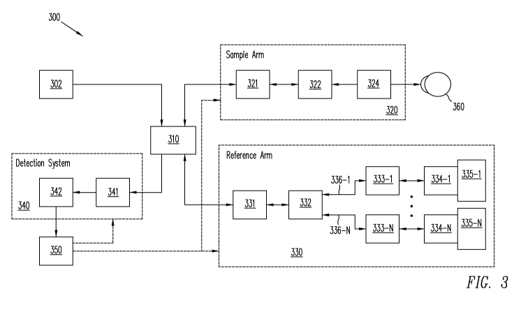
Figure 3 illustrates an imager 300 according to some embodiments of the
present
invention. Imager 300 includes a light source 302 that may be chosen
appropriately for
either a swept-source or a spectrometer based Fourier-domain OCT procedure. As
such,
light source 302 may comprise any light source suitable for the purpose of OCT
imaging. A
suitable light source for the purpose of Fourier-domain OCT may include, but
is not limited
to, a broadband light source such as a superluminescent diode. A suitable
light source for
the purpose of achieving the swept-source version of Fourier-domain OCT may
include, but
is not limited to, a tunable laser source. In various embodiments, light
source 302 may
produce radiation at different wavelengths or with different bandwidths for
performing
imaging at different tissue penetration and/or axial resolution.
As shown in Figure 3, light from light source 302 is directed to a light
coupler 310,
which sends energy from light source 302 into a sample arm 320 and a reference
arm 330.
Light coupler 310 of Figure 3 can be a splitter/coupler that receives light
from light source
302 and directs it to both sample arm 320 and reference arm 330, and receives
light from
sample arm 320 and reference arm 330 and directs the combined light beam to
detection
system 340. Sample arm 320 can include optics including collimating optics
321, beam
scanning 322, and focusing optics 324. Beam scanning mechanism 322 may direct
a light
CA 02731226 2011-01-18
WO 2010/011656 PCT/US2009/051263
beam received from light coupler 310 to perform two- or three- dimension
transverse beam
scanning and imaging of sample 360. In some embodiments, collimating optics
321 may
additionally include polarization controllers, which may be utilized in some
embodiments to
more precisely detect data resulting in an image. Sample arm 320 then provides
the
backscattered light from sample 360 to light coupler 310.
Reference arm 330 receives light from light source 302 through light coupler
310
and provides reference light to light coupler 310. Reference light from
reference arm 330 is
combined with backscattered light from sample arm 320 to produce spectral
interference
that can be detected by a detection system 340.
As shown in Figure 3, reference arm 330 may include polarization controller
331 to
assist in maximizing the interference fringe contrast detected by detection
system 340. The
reference arm may have one or more splitter/couplers 332 to further separate
the reference
beam into two or more reference paths for simultaneous detection. Reference
paths 336-1
through 336-N are specifically shown in Figure 3, where N can be any number of
reference
arms. In general, the number of separate reference paths N will be the number
of separate
image depths of interest.
Each of reference paths 336-1 through 336-N includes various optics 333-1
through
333-N as well as reference mirrors 334-1 through 334-N, respectively, for
reflecting the
energy from the light source 302 to provide the reference light. The optics
333-1 through
333-N in reference arm 330 may be used to collimate the beams from
splitter/coupler 332
and couple the beams back into splitter/coupler 332 when they are reflected
back from
reference mirrors 334-1 through 334-N, respectively. In some embodiments,
splitter/coupler 332 can be coupled to optics 333-1 through 333-N with optical
fiber. Optics
333-1 through 333-N may include, but are not limited to, various collimating
lenses suitable
for this purpose.
11
CA 02731226 2011-01-18
WO 2010/011656 PCT/US2009/051263
As has been reported, for example, in Yasuno, Wang, and Baumann, a carrier
frequency can be introduced into the spatial spectrograms by introducing a
constant phase
modulation in the reference and/or sample arm across the transverse scan. Such
a
modulation is typically utilized to double the conventional imaging range of a
single
reference arm OCT imager.
In accordance with some embodiments of the invention, reference beams
returning
from different reference paths include encoded information by utilizing
different
modulations into each of reference paths 336-1 through 336-N. Mirrors 334-1
through 334-
N may be stationary or may be modulated by modulators 335-1 through 335-N,
respectively. Modulation of reference mirrors 334-1 through 334-N during the
transverse
scanning of the sample may be equivalent to frequency modulation of the
detected signal at
detection system 340. As discussed above, it is therefore possible to encode
information on
the reference beams returning from different reference paths by using
different phase
modulations on each of reference mirrors 334-1 through 334-N.
Various methods may be enlisted in modulators 335-1 through 335-N to introduce
a
constant phase modulation into the reflected light beam from each of mirrors
334-1 through
334-N, respectively. In various embodiments, modulators 335-1 through 335-N
may be a
linear piezo-translation stage onto which mirrors 334 -1 through 334-N,
respectively, are
mounted. The piezo-translation stage may be configured to move mirrors 334-1
through
334-N at some constant velocity across a transverse scan in the x or y
direction (B-scan). In
some embodiments, the phase modulation may be achieved in the sample arm
scanning
mechanism 322 by introducing an offset from the pivot point of scanner 321, as
discussed in
Baumann. In some embodiments, a grating-based phase delay line can be placed
in
reference arm 330 such that the optical group delay can be close to zero and
only phase
modulation is achieved. Another exemplary embodiment is shown in Figures 4A
and 4B,
12
CA 02731226 2011-01-18
WO 2010/011656 PCT/US2009/051263
which can also achieve phase modulation with nearly zero group delay.
The beams returning from sample arm 320 and reference arm 330 can be combined
in coupler 310 and sent to detection system 340. Detection system 340 includes
a detector
342 and optical components 341. Detector 342 can be a spectrometer in a
spectrometer
based Fourier-Domain OCT or a photo-diode detector system in a swept-source
based
Fourier-domain OCT. Optical components 341 may include appropriate optics to
focus the
beam from light coupler 310 onto detector 342. The detected signal is sent to
a processor
350, which is typically a computer operating software to analyze the signals
received from
detector 342, store the data, and present the results in an appropriate
fashion. Since the
phase modulation in the reference arm may be synchronized to the transverse
scanning
performed in the sample arm, in some embodiments processor 350 may also send
control
and synchronization signals to sample arm 320, to reference arm 330, and to
detection
system 340 (dashed arrows).
Figures 4A and 4B illustrate exemplary embodiments of modulation apparatus 401
and 402, respectively, suitable to achieve constant phase modulation in the
reference arm.
Each of apparatus 401 and 402 can be utilized in place of a mirror 334-j and
modulator 335-
j pair, where mirror 334-j is an arbitrary one of mirrors 334-1 through 334-N
and modulator
335-j is a corresponding arbitrary one of modulators 335-1 through 335-N, and
corresponds
to the mirror and modulator in reference path 336-j.
Apparatus 401 shown in Figure 4A illustrates a double-pass configuration
utilizing a
galvanometer scanner 420 to achieve constant phase modulation in reference arm
330. In
apparatus 401, the input beam may enter into collimating optics 400 and pass
through a lens
system 410 that focuses the beam to a mirror mounted on a galvanometer scanner
420. The
beam hits the galvanometer mirror at an offset from the pivot point which will
introduce
phase modulation as the galvanometer mirror of galvanometer scanner 420 is
rotated. In
13
CA 02731226 2011-01-18
WO 2010/011656 PCT/US2009/051263
galvanometer 420, the galvanometer mirror is mounted at the focal plane of
lens 410 and
reflects the beam back through lens 410 to finally reach a retro-reflector
430, which can be a
mirror. The returning beam from reflector 430 passes through lens 410, hits
the
galvanometer mirror of galvanometer scanner 420 again, and returns to the
input through
lens 410 and collimating optics 400. Because the galvanometer mirror of
galvanometer
scanner 420 is located at the back focal plane of lens 410, the beam reflected
back from
reflector 430 will return to the input of collimating optics 400 following the
incident path,
which is a double-pass configuration.
Apparatus 402 shown in Figure 4B illustrates another exemplary embodiment of
an
apparatus suitable to achieve constant phase modulation. In apparatus 402, the
input beam
may enter into collimating optics 400 and passes through a phase modulation
system 440
that can change the optical path length of the reference beam. An exemplary
embodiment
of phase modulation system 440 is an optical window mounted on a galvanometer
scanner
inserted into the reference beam path. As the galvanometer is rotated, the
optical window
changes angle with respect to the reference beam and the optical path length
is changed.
The beam passing through the phase modulation system continues to reach a
retro-reflector
450, which may be a mirror. The returning beam from reflector 450 can go back
through
phase modulation system 440 before returning to the collimating optics 400
again to be
coupled out of apparatus 402.
Figures 5A and 5B illustrate an exemplary embodiment of signal processing
technique 550 that may be executed by processor 350 to distinguish the
simultaneously
acquired images. Figure 5A illustrates the resulting data sets while Figure 5B
illustrates a
flow chart of the data processing procedure that may be executed on processor
350. By
using different phase modulation on each of reference arm paths 336-1 through
336-N,
different carrier frequency can be induced into the spatial spectrograms
corresponding to
14
CA 02731226 2011-01-18
WO 2010/011656 PCT/US2009/051263
each of reference arm paths 336-1 through 336-N. Further, by arranging for
different path
lengths in each of reference arm paths 336-1 through 336-N, a plurality of
images
corresponding to different depths in sample 360 can be obtained.
For the illustrative purpose of Figures 5A and 5B, assume that a constant
phase
modulation is applied to modulator 335-1 such that the carrier frequency has a
spatial
frequency of ul in the transverse Fourier space. Furthermore, assume a
constant phase
modulation is applied to modulators 335-2 such that the carrier frequency has
a spatial
frequency of u2 in the transverse Fourier space. If ui is sufficiently
separated from u2 in the
transverse Fourier space, it will be possible to distinguish signals that are
simultaneously
acquired as illustrated in Figure 5A. Although only reference paths 336-1 and
336-2 are
illustrated here, one skilled in the art will readily recognize how to extend
this to any
number of reference paths 336-1 through 336-N in order to separate the images
from each
of the reference paths 336-1 through 336-N.
In step 562 of Figure 5B, a combined dataset 500, as shown in Figure 5A, is
acquired. The spatial spectrograms from different ones of reference arm paths
336-1
through 336-N are detected simultaneously by detector 342 and stored in
combined image
data set 500. The combined image data set 500 contains the image data from all
reference
arm paths 336-1 through 336-N, of which the data from reference arm paths 336-
1 and 336-
2 are illustrated here. The detected data set may be a two-dimensional data
set that has a
dimension in spatial frequency k (or may be in wavelength k before conversion
to k).
Another dimension will be in transverse position x or y depending on the
scanning pattern
and coordinate definition. In some embodiments, this second dimension can also
simply be
acquisition time when no transverse scanning is performed in the sample arm.
In
conventional FD-OCT, an inverse Fourier transform is performed along the k-
dimension for
every transverse position x or y, which yields the OCT signals for each
transverse position.
CA 02731226 2011-01-18
WO 2010/011656 PCT/US2009/051263
In processing the simultaneously acquired images stored in combined data set
500, a
Fourier transform 501 is performed along the transverse (x or y) dimension for
every value
in the k dimension. Due to the carrier frequencies ui and u2 introduced by
modulators 335-1
through 335-N, respectively, the frequency content associated with reference
mirrors 334-1
through 334-N, respectively, will be centered at different carrier frequencies
in the
transverse Fourier space, as is shown in frequency distribution 503 of Figure
5A. As shown
in frequency distribution 503, the frequency content 511 centered on carrier
frequency ui
contains information on the spatial spectrograms from reference arm mirror 334-
1. The
frequency content 512 centered on carrier frequency u2 contains information
on the spatial
spectrograms from reference arm mirror 334-2. In general, each of reference
arms 336-1
through 336-N is centered at different frequency ui through uN in frequency
distribution
503. If ui is sufficiently separated from u2 in the transverse Fourier space,
the information
from different reference mirrors can be selected in spectrum selection step
564 by using
Frequency filters. In some embodiments, in order to perform full range complex
FD-OCT,
only the spectra in the positive Fourier space is selected (i.e., applying a
Heaviside function
before spectrum selection) as illustrated by filters 505 and 507. As is
illustrated in Figure
5A, frequency content 511 can be separated from frequency content 512.
Applying an inverse Fourier transform 509 to filtered spectrum 511, complex
data
set 521 can be generated. Applying an inverse Fourier transform 513 to
filtered spectrum
512, complex data set 522 can be generated. In general, an inverse Fourier
transform can be
applied to each of the separated spectra formed in spectrum selection 564. As
discussed
above, a complex data set such as complex data sets 521 and 522 can then be
generated for
each of reference paths 336-1 through 336-N.
Complex data set 521 shown in Figure 5A corresponds to the spatial spectrogram
from reference mirror 334-1 and complex data set 522 corresponds to the
spatial
16
CA 02731226 2011-01-18
WO 2010/011656 PCT/US2009/051263
spectrogram from reference mirror 334-2. Through the appropriate selection of
phase
modulations on modulators 335-1 and 335-2, it is therefore possible to
distinguish
simultaneously acquired signals.
The final step in the process to generate OCT images shown in the embodiment
shown in Figures 5A and 5B is to perform inverse Fourier transform along the k-
dimension
for every transverse position x or y as in conventional FD-OCT. As shown in
Figures 5A
and 5B, inverse Fourier transform 515 is performed on complex data set 521 to
form full-
range image 531. Similarly, inverse Fourier transform 517 is performed on
complex data
set 522 to form full-range image 532. Because complex data sets 521 and 522
include both
real and imaginary information, the complex conjugate mirror image will not be
present and
the full imaging range (+z to -z) of the FD-OCT system can be utilized. As
shown in
Figure 5A, the full-range OCT image 531 corresponds to the image acquired from
reference
mirror 334-1 and the full-range OCT image 532 corresponds to the image
acquired from
reference mirror 334-2. By selecting appropriate optical path delays in the
reference paths
containing reference mirrors 334-1 and 334-2, it is then possible to
simultaneously acquire
images from different axial scanning regions of interest in the sample. In
general, a full-
range image can be obtained for each of reference paths 336-1 through 336-N.
Although Figures 5A and 5B show an example for two reference paths 336-1 and
336-2, as discussed above any number of reference paths 336-1 through 336-N
can be
utilized. Process 550 illustrated in Figures 5A and 5B can be applied
generally to multiple
reference mirrors such that multiple spatial spectrograms are detected
simultaneously. As
long as sufficient carrier frequencies can be selected such that there is no
overlap of the
frequency contents in the transverse Fourier space, all the simultaneously
detected signals
can be distinguished from each other.
In some embodiments, one of the carrier frequencies (u1 for example) can be
zero
17
CA 02731226 2011-01-18
WO 2010/011656 PCT/US2009/051263
such that no phase modulation is performed in that reference arm path (i.e. a
stationary
mirror). This case will be the same as conventional FD-OCT and the full
imaging range (+z
to -z) will not be available. However, for imaging thin samples such as the
retina, half of
the full imaging range (positive-z or negative-z space) is often sufficient.
As long as the
second carrier frequency (u2 for example) is sufficiently separated from ui
(zero in this
case) in the transverse Fourier space, it will be possible to distinguish
signals that are
simultaneously acquired from two different axial scanning regions of interest.
Figures 6A and 6B illustrate examples of utilizing embodiments of the present
invention to extend the imaging range inside a sample such as the human eye.
Because the
images can be acquired simultaneously, precise registration can be achieved
across both the
axial and transverse dimensions. Therefore it is possible to extend the
imaging range
through precise calibration of the path length differences in reference paths
336-1 through
336-N. Figure 6A shows an extended imaging range in the anterior segment of a
human eye
600. As shown in Figure 6A, scan range 602 can be performed. The maximum
imaging
range of full range complex FD-OCT is usually about 6 mm, which is not
sufficient to
imaging the entire anterior chamber including the posterior capsule of the
lens. The
example shown in Figure 6A shows that the optical paths of two reference
mirrors can be
adjusted such that one reference mirror, for example reference mirror 334-1,
images a front
part 604 of the anterior chamber while a second reference mirror, for example
reference
mirror 334-2, images a back part 606 of the anterior chamber. Imaging region
602
corresponds to rectangular boxes with diagonal lines.
Using conventional prior art techniques, the simultaneously acquired images
would
overlap and render the resultant image, as is shown in image 610 in Figure 6A,
uninterpretable. In some embodiments of the present invention, the images
acquired from
the two separate axial scanning regions of interest can be distinguished and
combined
18
CA 02731226 2011-01-18
WO 2010/011656 PCT/US2009/051263
together to form one image 620 that effectively doubles the imaging range of
the system to
about 12 mm, sufficient to cover the entire anterior chamber of eye 600.
Figure 6B shows an example of utilizing some embodiments of the present
invention
for performing simultaneous imaging at vastly different axial scanning regions
of interest.
As shown in Figure 6B, imaging regions 650 and 652 are of interest in eye 600.
The optical
path in two reference mirrors can be adjusted such that one reference mirror,
for example
reference mirror 334-1, images the front part of the anterior chamber while
the second
reference mirror, for example reference mirror 334-2, images the retina in the
posterior
segment of the eye. Imaging regions 650 and 652 correspond to rectangular
boxes with
diagonal lines. Using conventional prior art techniques, the simultaneously
acquired images
would overlap and render the resultant image, shown as image 660 in Figure 6B,
uninterpretable. Image 670 illustrates separated images 672 and 674 acquired
from the two
separate axial scanning regions of interest. Because the optical path
difference between the
two reference mirrors 334-1 and 334-2 can be measured precisely, the
separation distance
between the two images 672 and 674 can be determined and the images can be
placed in
their correct anatomical relationship in the context of the entire imaging
sample, such as the
human eye 600. Furthermore, since the two images are acquired simultaneously,
morphmetric measurements such as the distance from the front surface of the
eye to the
back surface of the eye can be precisely determined.
As discussed above, any number of separated images can be obtained. Figures 6A
and 6B illustrate separation of two images from two reference paths. In some
embodiments,
the examples shown in Figures 6A and 6B can be combined through the use of
three
reference mirrors for simultaneous acquisition. It is therefore possible to
perform imaging
of the entire anterior chamber with about 12 mm of imaging range as shown in
Figure 6A
while simultaneously acquiring an image of the retina in the posterior segment
for
19
CA 02731226 2011-01-18
WO 2010/011656 PCT/US2009/051263
morphometric measurements as shown in Figure 6B.
Figure 7 illustrates an OCT imager 700 according to some embodiments of the
present invention. OCT imager 700 represents a dual-beam low coherence
interferometer.
In some embodiments, OCT imager 700 is insensitive to the motion of the
sample. In some
embodiments, OCT imager 700 can be suitable for both swept-source and
spectrometer
based Fourier-domain low-coherence interferometry (LCI). In general, OCT
imager 700
includes a light source 702, an interferometer 730, a sample arm 720, a
detection system
740, and a processor 750. Light source 700 may include any light source
suitable for the
purpose of LCI or OCT imaging. A suitable light source for the purpose of
Fourier-domain
OCT may include, but is not limited to, a broadband light source such as a
superluminescent
diode. A suitable light source for the purpose of achieving the swept-source
version of
Fourier-domain OCT may include, but is not limited to, a tunable laser source.
In some
embodiments, light source 702 may contain different wavelengths or different
bandwidths
for performing imaging at different tissue penetration and/or axial
resolution.
As shown in Figure 7, interferometer 730 may include reflective surfaces 731
and
732 separated by an adjustable distance. The relative optical paths of the
reflective surfaces
731 and 732 correspond with the separation in depth of the acquired images One
or both of
the two reflective surfaces may be modulated during the data acquisition by
modulators 735
and 736 to provide a constant phase modulation to the detected signal during
acquisition.
Lens systems 733 and 734 couple light in and out of interferometer 730. Light
from
interferometer 730 is provided to light coupler 710, which directs light into
sample arm 720
and directs light received from sample arm 720 to detection system 740. In
some
embodiments, light coupler 710 can be an optical circulator. In some
embodiments, light
coupler 710 can be a splitter/coupler. Sample arm 720 can include various
collimating
optics 721, a beam scanning mechanism 722, and focusing optics 724. Beam
scanning
CA 02731226 2011-01-18
WO 2010/011656 PCT/US2009/051263
mechanism 722 can direct the beam to perform two- or three- dimension
transverse beam
scanning and imaging of a sample 760, or it can remain stationary for axial
measurements.
The distance d between the two reflective surfaces 731 and 732 can be adjusted
to
match the axial length of the eye. In such case, the low-coherence
interferometry signal
returning from both the cornea and the retina can be presented to the
detection system 740.
Detection system 740, as shown in Figure 7, can include optics 741 and a
detector 742.
Detector 742 can be a spectrometer in spectrometer based Fourier-Domain OCT or
a photo-
detector system (e.g., a photo-diode detector system) in swept-source based
Fourier-domain
OCT. Appropriate optics or optical components 741 may be employed to focus the
beam
onto detector 742. Detector 742 provides a signal to processor 750 in response
to the beam.
Processor 750, which can be a computer system, stores the signal as image data
and can
process the image data as has been previously described. Since the phase
modulation in the
reference arm needs to be synchronized to the acquisition, the computer may
also send
control and synchronization signals to the sample arm, the reference arm,
and/or the
detection system (dashed arrows).
In some embodiments, one or both of reflective surfaces 731 and 732 of
interferometer 730 may be modulated respectively by modulators 735 and 736
during data
acquisition to provide a constant phase modulation. It is therefore possible
to encode a
phase modulation to the signal returning from the longer optical path length
of the sample
arm (e.g., the retina). This will allow separation of the signals returning
from different path
lengths in the sample (e.g., the cornea and the retina). Various methods may
be enlisted in
the modulators 735 and 736 to introduce a constant phase modulation to
reflective surfaces
731 and 732. Another exemplary embodiment is shown in Figures 4A and 4B, which
can
be used to achieve constant phase modulation during data acquisition.
Figure 8 illustrates a process 800 for providing images in an OCT imager
according
21
CA 02731226 2011-01-18
WO 2010/011656 PCT/US2009/051263
to some embodiments of the present invention. As shown in Figure 8, first a
sample, such
as sample 360 shown in Figure 3 or sample 760 shown in Figure 7, is aligned
with OCT
imager in step 802 so that signal strength can be optimized. After alignment,
in step 804
data acquisition is begun. In step 806, a line of data for an A-line scan is
acquired. In step
808, phase shift modulation for the next line of data is performed. Phase
shift modulations
are detected, for example, in detection system 340 of the embodiment shown
Figure 3 or
detection system 740 of the embodiment shown in Figure 4. In step 810, the
transverse
position is changed. Changing transverse position can be accomplished, for
example, by
scan mechanism 322 in the embodiment shown in Figure 3 or scan mechanism 722
in the
embodiment shown in Figure 7. In step 812, if the full scan is not yet
completed then
process 800 returns to step 806. If the full scan has been completed, then
process 800 enters
image processing 814. Image processing 814 can, for example, execute process
550
illustrated in Figures 5A and 5B.
Figure 9A illustrates OCT imager 700 where light coupler 710 is implemented as
circulator 910. Circulator 910 receives light from interferometer 730 and
provides it to
sample arm 720 and receives light from sample arm 720 and provides it to
detector system
740. An advantage of circulator 910 over a splitter/coupler as light coupler
710 is the
higher percentage of light coupled into sample arm 720 and detection system
740.
Figure 9B illustrates imager 920, which represents another embodiment of an
imager
according to some embodiments of the present invention. Imager 920 includes
light source
702, detection system 740, processor 750, and sample arm 720 as discussed with
respect to
imager 700 of Figure 7. Light from light source 702 is coupled into sample arm
720 and
interferometer 930 through splitter/coupler 930. Light received at
splitter/coupler 930 from
sample arm 720 and interferometer 930 is combined and coupled into detection
system 740.
As shown in Figure 9B, interferometer 930 includes reflectors 931 and 932,
each of which
22
CA 02731226 2011-01-18
WO 2010/011656 PCT/US2009/051263
may be coupled to a modulator 935 and 936, respectively. As discussed with
respect to
Figure 7, the distance between reflectors 931 and 932 corresponds with the
difference in
depth between images in sample 760. Lens systems 933 and 934 couple and focus
light
through interferometer 930. As shown in Figure 9B, reflector 931 can be
partially reflecting
and reflector 932 can be fully reflecting.
Figure 9C shows an imager 950, which illustrates another embodiment according
to
the present invention. As shown in Figure 9C, light from source 702 is coupled
into
splitter/coupler 957, which transmits light to reflectors 951 and 952.
Reflectors 951 and
952 may also include coupling optics to receive light from splitter/coupler
957 and couple
light back into splitter/coupler 957. As shown in Figure 9C, reflectors 951
and 952 may be
coupled to modulators 955 and 956, respectively. Although only two reflectors,
reflectors
951 and 952, are shown in Figure 9C, additional splitters may be utilized to
add as many
reflectors, each providing a different modulated beam corresponding to a
different image
depth, as desired, which is similar to the embodiment shown in Figure 3.
Light received from reflectors 951 and 952 is combined in splitter/coupler 957
and
coupled into light coupler 959. Light coupler 959 can be a splitter/coupler or
a circulator
such as circulator 910 shown in Figure 9A. As shown in Figure 9C, Light from
light
coupler 959 is coupled into sample arm 720. Light received from sample arm 720
is
received in light coupler 959 and transmitted into detection system 740. As
before,
processor 750 can be coupled to control aspects of imager 950.
Figures 7 and 9A illustrate interferometer 730, which includes two partially
reflecting mirrors 731 and 732. Figure 9B illustrates interferometer 930,
which includes
one partially reflecting mirror 931 and a fully reflecting mirror 932. Figure
10 illustrates an
interferometer 1000 that may be utilized in place of interferometer 930 of
Figure 9B or
interferometer 730 of Figures 7 or 9A.
23
CA 02731226 2011-01-18
WO 2010/011656 PCT/US2009/051263
As shown in Figure 10, light enters interferometer 1000 in circulator 1010. In
some
embodiments, a splitter/coupler can be substituted for circulator 1010. Light
from circulator
1010 enters beam splitter 1020, where it is split and coupled into reflectors
1030 and 1050.
As discussed above, reflectors 1030 and 1050 may include coupling optics.
Further,
reflectors 1030 and 1050 are coupled to modulators 1040 and 1060,
respectively. The
difference in path length utilizing reflector 1030 and reflector 1050
corresponds with the
different depth of image acquired.
In each of the embodiments, light may be coupled from one component to another
in
any fashion, for example with optical fiber. Further, some embodiments may
include
focusing or coupling optics in various positions, as needed.
For purposes of explanation, some embodiments of the invention are discussed
above. One skilled in the art may recognize various alternatives from the
embodiments
disclosed. Such alternatives are intended to be within the scope of this
disclosure. Further,
these embodiments are not intended to be limiting on the scope of the
invention. Therefore,
the invention is limited only by the following claims.
24