Note: Descriptions are shown in the official language in which they were submitted.
CA 02731255 2013-05-08
STRUCTURAL VARIANTS OF ANTIBODIES FOR
IMPROVED THERAPEUTIC CHARACTERISTICS
BACKGROUND OF THE INVENTION
Field of the Invention
10011 The present invention relates to structural variants of anti-CD20
antibodies and/or
antigen binding fragments thereof, preferably involving the amino acid
sequences of
complementarity-determining regions (CDRs), with improved therapeutic
characteristics. In
particular embodiments, the structural variation may comprise changes to the
third CDR
sequence of the antibody heavy chain (CDRH3), for example substitution of an
aspartate
residue for an asparagine residue at Kabat position 101. In other particular
embodiments, the
structural variation may comprise an arginine residue at Kabat position 94,
which may form a
salt bridge with an aspartate at Kabat position 101. In still other particular
embodiments, the
structural variation may comprise a valine residue at Kabat position 102. Such
structural
variants may provide improved efficacy for diseases related to proliferation
of B-cells, such
as B-cell leukemias, lymphomas or autoimmune diseases, as well as other immune
diseases
implicating B-cells. In preferred embodiments, the improved efficacy may allow
administration of low dosages of anti-CD20 antibody or antigen binding
fragment thereof,
such as 80 mg or less, more preferably 50 mg or less, most preferably 30 ing
or less, which
may be administered two or more times about one to three weeks apart, or even
two or more
times weekly..
10021 The anti-CD20 antibody may be a humanized, chimeric or human anti-CD20
antibody, particularly a monoclonal antibody (MAb). Other embodiments may
concern
therapeutic and/or diagnostic conjugates of humanized, chimeric or human anti-
CD20
antibodies and methods of treating B-cell lymphomas and leukemias and various
autoimmune
diseases, for example using humanized, chimeric or human anti-CD20 antibodies.
Still other
embodiments may relate to antibody fusion proteins or antigen binding
fragments thereof
comprising at least one anti-CD20 MAb or antigen binding fragment thereof, in
some cases in
combination with a second, different antibody, especially anti-CD20 antibody,
preferably anti-
CD20 MAb, or antigen binding fragment thereof. The humanized, chimeric or
human MAbs,
1
CA 02731255 2011-01-19
WO 2010/011697 PCT/US2009/051321
antigen binding fragments thereof or antibody fusion proteins may be
administered alone, as
a therapeutic immunoconjugate or in combination with one or more therapeutic
agents, with
other naked antibodies or other immunoconjugates. Still other embodiments
relate to DNA
sequences encoding humanized, chimeric or human anti-CD20 antibodies and
antibody
fusion proteins, vectors and host cells containing the DNA sequences, and
methods of
making the humanized, chimeric or human anti-CD20 antibodies.
Background
[004] The immune system of vertebrates consists of a number of organs and cell
types
which have evolved to accurately recognize foreign antigens, specifically bind
to, and
eliminate/destroy such foreign antigens. Lymphocytes, amongst others, are
critical to the
immune system. Lymphocytes are divided into two major sub-populations, T cells
and B
cells. Although inter-dependent, T cells are largely responsible for cell-
mediated immunity
and B-cells are largely responsible for antibody production (humoral
immunity).
[005] In humans, each B-cell can produce an enormous number of antibody
molecules.
Such antibody production typically ceases (or substantially decreases) when a
foreign antigen
has been neutralized. Occasionally, however, proliferation of a particular B-
cell will
continue unabated and may result in cancers known as B-cell lymphomas or
leukemias. B-
cell lymphomas, such as the B-cell subtype of non-Hodgkin's lymphoma, are
significant
contributors to cancer mortality. The response of B-cell malignancies to
various forms of
treatment is mixed. For example, in cases in which adequate clinical staging
of non-
Hodgkin's lymphoma is possible, field radiation therapy can provide
satisfactory treatment.
Still, about one-half of the patients die from the disease. Devesa et al., J.
Nat. Cancer Inst.
79:701 (1987).
[006] The majority of chronic lymphocytic leukemias are of B-cell lineage.
Freedman,
Hematol. Oncol. Clin. North Am. 4:405 (1990). This type of B-cell malignancy
is the most
common leukemia in the Western world. Goodman et al., Leukemia and Lymphoma
22:1
(1996). The natural history of chronic lymphocytic leukemia falls into several
phases. In the
early phase, chronic lymphocytic leukemia is an indolent disease,
characterized by the
accumulation of small mature functionally-incompetent malignant B-cells having
a
lengthened life span. Eventually, the doubling time of the malignant B-cells
decreases and
patients become increasingly symptomatic. While treatment can provide
symptomatic relief,
the overall survival of the patients is only minimally affected. The late
stages of chronic
2
CA 02731255 2011-01-19
WO 2010/011697 PCT/US2009/051321
lymphocytic leukemia are characterized by significant anemia and/or
thrombocytopenia. At
this point, the median survival is less than two years. Foon et al., Annals
Int. Medicine
113:525 (1990). Due to the very low rate of cellular proliferation, chronic
lymphocytic
leukemia is resistant to cytotoxic drug treatment. Both chronic and acute
lymphocytic
leukemias of B-cell origin are suitable targets for the therapies described
herein.
[007] Traditional methods of treating B-cell malignancies, including
chemotherapy and
radiotherapy, have limited utility due to toxic side effects. The use of
monoclonal antibodies
to direct radionuclides, toxins, or other therapeutic agents offers the
possibility that such
agents can be delivered selectively to tumor sites, thus limiting toxicity to
normal tissues.
Also, the presence of B-cell antigens on these B-cell malignancies makes them
optimal
targets for therapy with unconjugated B-cell antibodies, such as against CD19.
CD20, CD21,
CD23, and CD22 markers on B-cells. HLA-DR, CD30, CD37, CD40, CD45, CD70,
CD79a,
and other antigens may serve as targets for normal and malignant B-cells,
although they are
also expressed on other cell types. Further. certain MUC1, MUC2, MUC3, and
MUC4
antigens, preferably MU Cl, as well as also insulin-like growth factors
(ILGF), insulin-like
growth factor receptor, macrophage migration-inhibitory factor (MIF), are also
expressed in
different hematopoietic malignancies, including B-cell tumors expressing CD20
and other B-
cell markers. Still other antigen targets, such as those associated with the
vascular
endothelium of tumors, including tenascin, vascular endothelium growth factor
receptor
(VEGFR), and placental growth factor (P1GF), as well as other categories of
antigens
associated with B-cell malignancies, such as oncogene products (cMET, Kras,
bc1-2, bc1-6),
are also suitable targets for therapeutic antibodies.
[008] B-cells comprise cell surface proteins which can be utilized as markers
for
differentiation and identification. One such human B-cell marker is the human
B
lymphocyte-restricted differentiation antigen, Bp35, referred to as CD20. CD20
is expressed
during early pre-B-cell development and remains until plasma cell
differentiation. CD20 is
expressed on both normal B cells and malignant B cells whose abnormal growth
can lead to
B-cell lymphomas and leukemias. Antibodies against the CD20 antigen have been
investigated for the therapy of B-cell lymphomas and leukemias. For example, a
chimeric
anti-CD20 antibody, designated as "IDEC-c2B8" (rituximab), has activity
against B-cell
lymphomas when provided as unconjugated antibodies at repeated injections of
doses
exceeding 500 mg per injection. Maloney et al., Blood 84:2457 (1994); Longo,
Curr. Opin.
Oncol. 8:353 (1996). About 50 percent of non-Hodgkin's patients, having the
low-grade
3
CA 02731255 2011-01-19
WO 2010/011697 PCT/US2009/051321
indolent form, treated with this regimen showed responses. Therapeutic
responses have also
been obtained using "1I-labeled B1 (tositumomab) anti-CD20 murine monoclonal
antibody
when provided as repeated doses with pretreatment of unlabeled antibodies
exceeding 600
mg per injection. Kaminski et al., N. Engl. J. Med. 329:459 (1993); Press et
al., N. Engl. J.
Med. 329:1219 (1993); Press etal., Lancet 346:336 (1995). However, these
antibodies,
whether provided as unconjugated forms or radiolabeled forms, have not shown
high rates of
objective and durable responses in patients with the more prevalent and lethal
form of B-cell
lymphoma, the intermediate or aggressive types. Therefore, a need exists to
develop an
immunotherapy for B-cell malignancies that achieves a therapeutic response of
significant
duration.
[009] Additional studies targeting CD20 surface antigen have been performed
using an anti-
CD20 murine monoclonal antibody, IFS.. which was administered by continuous
intravenous
infusion to B-cell lymphoma patients. Extremely high levels (>2 grams) of 1F5
were
reportedly required to deplete circulating tumor cells, and the results were
described as being
"transient." Press et al., "Monoclonal Antibody 1F5 (Anti-CD20) Serotherapy of
Human B-
Cell Lymphomas." Blood 69/2:584-591 (1987). However, a potential problem with
this
approach is that non-human monoclonal antibodies (e.g., murine monoclonal
antibodies)
typically lack human effector functionality, i.e., they are unable to mediate
complement-
dependent lysis or lyse human target cells through antibody-dependent cellular
toxicity or Fc-
receptor mediated phagocytosis. Furthermore, non-human monoclonal antibodies
can be
recognized by the human host as a foreign protein and, therefore, repeated
injections of such
foreign antibodies can lead to the induction of immune responses leading to
harmful
hypersensitivity reactions. For murine-based monoclonal antibodies, this is
often referred to
as a Human Anti-Mouse Antibody (HAMA) response.
[0010] The use of chimeric antibodies is preferred because they do not elicit
as strong a
HAMA response as murine antibodies. Chimeric antibodies are antibodies which
comprise
portions from two or more different species. For example, Liu, A. Y. et al.
"Production of a
Mouse-Human Chimeric Monoclonal Antibody to CD20 with Potent Fc-Dependent
Biologic
Activity" J. Inununol. 139/10:3521-3526 (1987), describe a mouse/human
chimeric antibody
directed against the CD20 antigen. See also, PCT Publication No. WO 88/04936.
An
exemplary chimeric antibody would comprise mouse variable region sequences
attached to
human antibody constant region sequences.
4
CA 02731255 2011-01-19
WO 2010/011697 PCT/US2009/051321
[0011] The use of humanized antibodies is even more preferred, in order to
further reduce the
possibility of inducing a HAMA reaction. As discussed below, techniques for
humanization
of murine antibodies by replacing murine framework and constant region
sequences with
corresponding human antibody framework and constant region sequences are well
known in
the art and have been applied to numerous murine anti-cancer antibodies.
Antibody
humanization may also involve the substitution of one or more human framework
amino acid
residues with the corresponding residues from the parent murine framework
region
sequences.
[0012] Another approach that has improved the ability of antibodies to be
effective in the
treatment of B-cell disorders has been to conjugate a therapeutic agent, such
as a radioactive
or chemotherapeutic agent to the antibody, such that the agent is localized at
the tumor site.
For example, the above-referenced 1F5 antibody and other B-cell antibodies
have been
labeled with 1311 and were evaluated for biodistribution in two patients. See
Eary, J. F. et al.,
"Imaging and Treatment of B-Cell Lymphoma" J. Nuc. Med. 31/8:1257-1268 (1990);
see
also, Press, 0. W. et al., "Treatment of Refractory Non-Hodgkin's Lymphoma
with
Radiolabeled MB-1 (Anti-CD37) Antibody" J. Clin. Oncol. 7/8:1027-1038 (1989)
(indication
that one patient treated with 131I-labeled IF-5 achieved a partial response);
Goldenberg, D. M.
et al., "Targeting, Dosimetry and Radioimmunotherapy of B-Cell Lymphomas with
1311-
Labeled LL2 Monoclonal Antibody" J. Clin. Oncol. 9/4:548-564 (1991) (three of
eight
patients receiving multiple injections reported to have developed a HAMA
response to this
CD22 murine antibody); Appelbaum, F. R. "Radiolabeled Monoclonal Antibodies in
the
Treatment of Non-Hodgkin's Lymphoma" Hem./Oncol. Clinics of N. Am. 5/5:1013-
1025
(1991) (review article); Press, 0. W. et al. "Radiolabeled-Antibody Therapy of
B-Cell
Lymphoma with Autologous Bone Marrow Support." New England Journal of Medicine
329/17: 1219-12223 (1993) (131I-labeled anti-CD20 antibody IF5 and B1 -
tositumomab); and
Kaminski, M. G. et al "Radioimmunotherapy of B-Cell Lymphoma with [13111 Anti-
B1 (Anti-
CD20) Antibody". NEJM 329/7:459 (1993) (131I-labeled anti-CD20 antibody B1):
PCT
published application WO 92/07466 (antibodies conjugated to chemotherapeutic
agents such
as doxorubicin or mitomycin). However, these approaches have not eliminated
the obstacles
associated with using murine antibodies, despite the fact that many patients
with lymphoma
who have received prior aggressive cytotoxic chemotherapy are immune
suppressed, thus
having lower HAMA rates than lymphoma patients who have not been heavily
pretreated.
CA 02731255 2011-01-19
WO 2010/011697 PCT/US2009/051321
[0013] Autoimmune diseases are a class of diseases associated with B-cell
disorders.
Examples comprise acute idiopathic thrombocytopenic purpura, chronic
idiopathic
thrombocytopenic purpura, dermatomyositis, Sydenham's chorea, myasthenia
gravis,
systemic lupus erythematosus, lupus nephritis, rheumatic fever, polyglandular
syndromes,
bullous pemphigoid, Type-I diabetes mellitus, Henoch-Schonlein purpura, post-
streptococcal
nephritis, erythema nodosum, Takayasu's arteritis. Addison's disease,
rheumatoid arthritis,
multiple sclerosis, sarcoidosis, ulcerative colitis, erythema multiforme. IgA
nephropathy,
polyarteritis nodosa, ankylosing spondylitis, Goodpasture's syndrome,
thromboangitis
obliterans, Sjogren's syndrome, primary biliary cirrhosis, Hashimoto's
thyroiditis,
thyrotoxicosis, scleroderma, chronic active hepatitis,
polymyositis/dermatomyositis,
polychondritis, pemphigus vulgaris, Wegener's granulomatosis, membranous
nephropathy,
amyotrophic lateral sclerosis, tabes dorsalis, giant cell
arteritis/polymyalgia, pernicious
anemia, rapidly progressive glomerulonephritis and fibrosing alveolitis. The
most common
treatments are corticosteroids and cytotoxic drugs, which can be very toxic.
These drugs also
suppress the entire immune system, can result in serious infection, and have
adverse affects
on the bone marrow, liver and kidneys. There is a need for more effective
methods of
treating autoimmune diseases, particularly Class-III autoimmune diseases. A
further need
exists for the development of more effective antibodies for the treatment of
cancer and/or
autoimmune disease.
[0014] Still other diseases with immunological dysregulation are suitably
treated by the novel
compositions and methods described herein, such as hemolytic anemias,
cryoglobulinemias,
hepatitis (particularly hepatitis C), graft-versus-host disease (GVHD)
(particularly after
allogeneic stem cell transplantation), allo sensitization (particularly with
organ
transplantation). There is now mounting evidence that B-cells are involved in
these
pathological states, so depleting B-cells by anti-B-cell therapies is gaining
in interest
(Roccatello et al., Clin Rev Allergy Immunol 2008, 34:111-117; Cutler et al.,
Blood 2006,
108:756-752; Vo et al., N Engl J Med 2008, 359:242-251; Vieira et al.,
Transplantation
2004;77:542-548; Abdallah & Prak, Clin. Transpl. 2006:427-37; Zaja et al.,
Bone Marrow
Transplant., 2007, 40:273-77; Saadoun et al., Curr. Opin. Rheurnatol. 2008,
20:23-8;
Antonelli et al., Clin. Exp. Rheumatol. 2008, 26:S39-47).
6
CA 02731255 2011-01-19
WO 2010/011697 PCT/US2009/051321
SUMMARY OF THE INVENTION
[0015] The present invention provides structural variants of anti-CD20
antibodies and/or
antigen binding fragments thereof with improved therapeutic characteristics.
In particular
embodiments, the structural variation may comprise an aspartate residue at
Kabat position
101, for example as a substitution for an asparagine residue, in VH of CDR3
(CDRH3). In
other particular embodiments, the structural variation may comprise an
arginine residue at
Kabat position 94, which may form a salt bridge with an aspartate at Kabat
position 101. In
still other particular embodiments, the structural variation may comprise a
valine residue at
Kabat position 102. The skilled artisan will realize that the possible amino
acid substitutions
that may be performed are not limited to the particular examples recited above
and may
comprise substitutions at additional and/or different Kabat positions.
[0016] The improved therapeutic characteristics may take a variety of forms,
such as a slower
dissociation rate from the target antigen, an increase in complement-dependent
cytotoxicity
(CDC) and/or a reduction in EC50 in complement-dependent cytotoxicity (CDC).
In
preferred embodiments, the characteristics may include efficacy at a lower
dosage, preferably
a dosage of 80 mg or less for a human subject, more preferably a dosage of 50
mg or less,
most preferably a dosage of 30 mg or less. The dosage may be administered two
or more
times. Preferably, where multiple administrations are provided, they are
administered about
I to 3 weeks apart. But in certain disease setting, a more frequent,
fractionated dosing is
preferred, such as, for example, twice weekly for 4 or more weeks in chronic
lymphocytic
leukemia.
[0017] In certain embodiments, the anti-CD20 antibodies or antigen binding
fragments
thereof may be humanized, chimeric or human antibodies that bind to a human B-
cell marker,
such as anti-CD20 antibodies, of use for the treatment and diagnosis of B-cell
disorders, such
as B-cell malignancies and autoimmune diseases, as well as other diseases
involving B-cells,
such as GVHD, hemolytic anemia, cryoglobulinemia, allosensitization, and in
organ
transplant rejection, as well as certain viral diseases, such as hepatitis C.
The humanized,
chimeric or human anti-CD20 antibodies may be used for methods of treatment of
mammalian subjects, such as humans or domestic animals, as naked antibodies
either alone
or in combination with one or more therapeutic agents, as immunoconjugates
labeled with
one or more therapeutic and/or diagnostic agents, as an antibody fusion
protein, in pre-
targeting methods with targetable constructs that are conjugated to one or
more therapeutic or
diagnostic agents, or as a multimodal therapy with other antibodies, other
therapeutic agents
7
CA 02731255 2011-01-19
WO 2010/011697 PCT/US2009/051321
or immunomodulators. The humanized, chimeric or human anti-CD20 antibodies can
also be
used as a diagnostic imaging agent alone, in combination with other diagnostic
imaging
agents, and/or in conjunction with therapeutic applications.
[0018] The skilled artisan will realize that the disclosed methods and
compositions for
sequence variations with improved therapeutic characteristics are not limited
to anti-CD20
antibodies and may be applied to antibodies against other tumor-associated
antigens or
autoimmune and other immune disease-associated antigens, including but not
limited to
carbonic anhydrase IX, CCCL19, CCCL21, CD1, CD la, CD2, CD3, CD4, CD5, CD8,
CDI1A, CD14, CDI5, CD16, CD18, CD19, CD20, CD21, CD22, CD23, CD25, CD29,
CD30, CD32b, CD33, CD37, CD38, CD40, CD4OL, CD45, CD46, CD52, CD54, CD55,
CD59, CD64, CD66a-d, CD67, CD70, CD74, CD79a, CD80, CD83, CD95, CD126, CD133,
CD138, CD147, CD154, CEACAM6, B7, ED-B fibronectin, Factor H. FHL-1, Flt-1,
Flt-3,
folate receptor, GROB, HMGB-1, hypoxia inducible factor (HIF), HM1.24, insulin-
like
growth factor-1 (ILGF-1). ILGF-1R, IFN-y, 1FN-a, IFN-13, IL-2, IL-4R, IL-6R,
IL-13R, IL-
15R, IL-17R, IL-18R, IL-6, IL-8, IL-12, IL-15, IL-17, IL-25, IP-10, MACE,
mCRP, MCP-1,
MIP-1A, MIP-1B, MIF, MUC1, MUC2, MUC3, MUC4, NCA-66, NCA-95, NCA-90, Ia,
HLA-DR, tenascin, Le(y), RANTES, T101, TAC, Tn antigen, Thomson-Friedenreich
antigens, tumor necrosis antigens. TNF-a, TRAIL receptors (R1 and R2). VEGFR,
EGFR,
P1GF, complement factors C3. C3a, C3b, C5a, C5, and oncogene products,
including bc1-2,
bc1-6, Kras and cMET.
[0019] Other embodiments may be directed to anti-CD20 MAbs or antigen binding
fragments thereof that contain specific murine CDRs or a combination of murine
CDRs from
more than one murine or chimeric anti-CD20 MAb. These MAbs can be humanized,
chimeric or human anti-CD20 MAbs. The CDR sequences may include, but are not
limited
to, light chain CDR sequences CDRL1 (RASSSVSYIH. SEQ ID NO:1), CDRL2
(ATSNLAS, SEQ ID NO:2) and CDRL3 (QQWTSNPPT, SEQ ID NO:3) and heavy chain
CDR sequences CDRH1 (SYNMH, SEQ ID NO:4), CDRH2 (AIYPGNGDTSYNQKFKG,
SEQ ID NO:5) and CDRH3 (STYYGGDWYFDV, SEQ ID NO:6).
[0020] Various embodiments may concern bispecific antibodies or antibody
fusion proteins
comprising at least two anti-CD20 MAbs or antigen binding fragments thereof or
a first MAb
comprising an anti-CD20 MAb or antigen binding fragment thereof and a second
MAb. The
second MAb may bind to a tumor-associated antigen, such as those listed above,
or a hapten,
for example on a targetable conjugate.
8
CA 02731255 2013-05-08
100211 Other embodiments may concern therapeutic or diagnostic conjugates of
anti-CD20
MAbs or antigen binding fragments thereof or antibody fusion proteins, bound
to at least one
therapeutic agent or at least one diagnostic agent. Antibodies and fusion
proteins with
multiple therapeutic agents of the same or different type are also
encompassed. In alternative
embodiments, the anti-CD20 antibodies, antigen binding fragments or fusion
proteins may be
used in therapeutic or diagnostic pre-targeting methods, for example using
bispecific
antibodies with one arm that binds specifically to a cell, disease, tissue or
pathogen (e.g.,
hepatitis-C-associated target antigen) and a second arm that binds to a
targetable conjugate
attached to one or more diagnostic or therapeutic agents. Methods of pre-
targeting with
bispecific antibodies are well known in the art (see, e.g., U.S. Patent Nos.
7,300,644;
7,138,103; 7,074,405; 7,052,872; 6,962,702; 6,458,933).
100221 Alternative embodiments may concern methods of using the anti-CD20 MAbs
or
antigen binding fragments thereof or antibody fusion proteins for therapy,
either alone, in
combination with one or more other therapeutic agents, for example as the
antibody
component of a therapeutic immunoconjugatc with one or more therapeutic agents
or as a
naked antibody, antigen binding fragment or fusion protein administered alone
or in
combination with one or more therapeutic agents. Use for diagnostic methods in
combination
with one or more diagnostic agents is also contemplated. In preferred
embodiments, the
disease to be diagnosed or treated is a B-cell mediated immune disease,
autoimmune disease,
B-cell lymphoma or leukemia. B-cell mediated immune disease refers to a sub-
class of
autoimmune disease, as well as the other immune diseases discussed above
(e.g., GVHD,
cryoglobulinemia, hemolytic anemia, allosensitization, transplant organ
rejection) in which the
disease state is primarily mediated by production of autoantibodies, rather
than by autoreactive T
lymphocytes, or by a combination of B- and T-cell immunity.
BRIEF DESCRIPTION OF THE FIGURES
[00231 FIG. 1. Variable light chain (cA20Vk) and variable heavy chain (cA2OVH)
sequences of cA20, a chimeric anti-CD20 antibody. The CDR region sequences are
shown in
bold and underlined. The amino acid residues and the nucleotides are numbered
sequentially
and same numbering system is used for humanized V sequences shown in FIG. 2.
The light
chain variable region is shown in FIG. lA (SEQ ID NOS 7 & 8) and the heavy
chain variable
region is shown in FIG. 1B (SFQ ID NOS 9 & 10). The Kabat numbering scheme was
used
9
CA 02731255 2011-01-19
WO 2010/011697 PCT/US2009/051321
for amino acid residues. Amino acid residues numbered by a letter represent
the insertion
residue according to Kabat, and have the same number as that of the previous
residue.
[0024] FIG. 2. Nucleotide and amino acid sequences of the hA20 light chain
hA20Vk (FIG.
2A) (SEQ ID NOS 11 & 12), and heavy chain hA2OVH1 (FIG. 2B) (SEQ ID NOS 13 &
14),
as well as the adjacent flanking sequences of the VKpBR2 (FIG. 2A) and VHpBS2
(FIG. 2B)
staging vectors, respectively. The non-translated nucleotide sequences are
shown in
lowercase. The restriction sites used for subcloning are underlined and
indicated. The
secretion signal peptide sequence is indicated by a double underline.
[0025] FIG. 3. Comparison of the variable region sequences of cA20 (SEQ ID NOS
8 &
10), rituximab (from murine C2B8, SEQ ID NOS 15 & 16) and hA20 (SEQ ID NOS 12
&
14). Dots indicate homology to cA20. CDR sequences are in boxes. The heavy
chain (FIG.
3A) and light chain (FIG. 3B) variable region sequences are shown.
[0026] FIG. 4. Scatchard analysis - the binding characteristics of veltuzumab
and rituximab
were determined by binding the 125I-labeled MAbs to Raji cells. Direct cell
surface saturation
binding and Scatchard plot analysis (inset) - closed triangles veltuzumab;
circles rituximab.
Bmax and Kd were determined by non-linear regression analysis using a one-site
binding
model with Prism software. These results are representative of one of three
repeated
experiments.
[0027] FIG. 5. Comparison of dissociation rates of veltuzumab and rituximab
from live
cells. Daudi (A), Ramos (B), and Raji (C- E) cells were stained with PE-
labeled rituximab
(closed triangle), veltuzumab (closed square), cA20 (upside down closed
triangle), D101N
(closed circle), 1F5 (open circle) or Bl (tositumomab) (open square). The
labeled MAbs
were incubated at 37 C with (A-D) or without (E) excess veltuzumab Fab'-NEM
and the
cells analyzed by flow cytometry over time. The off-rate was determined by non-
linear
regression (one phase exponential decay) and P-values were generated by F-test
using
GraphPad Prism software.
[0028] FIG. 6. Effects of veltuzumab and rituximab on proliferation of non-
Hodgkin's
lymphoma cell lines. Anti-proliferative effects were assessed by MTT
cytotoxicity assays.
Cells were cultured with the MAbs with or without a second antibody for
crosslinking.
White bars, no second antibody; gray bars with GAH second antibody; error
bars, SD.
[0029] FIG. 7. In vitro depletion of B-cells from healthy blood donors. The
effect of
veltuzumab on peripheral blood lymphocytes from healthy volunteers was
evaluated in vitro
using flow cytometry. Decrease in the percent of CD19+ cells present in the
lymphocyte gate
CA 02731255 2011-01-19
WO 2010/011697 PCT/US2009/051321
after a two-day incubation of heparinized whole blood of healthy volunteers
with veltuzumab
is shown. Each line represents a different blood donor. Error bars, SD.
[0030] FIG. 8. Survival curves for veltuzumab in a disseminated Burkitt's
lymphoma
xenograft model comparing intraperitoneal versus subcutaneous administration.
C.B. 17
SCID mice were administered 1.5x10⁷ Daudi cells i.v. on day 0. Therapy with
veltuzumab
began on day 1 with mice receiving either a single i.p. or single s.c.
injection of veltuzumab.
Doses administered were 60, 20, or 5 lig veltuzumab. Control mice received an
i.p. injection
of either saline or 60 ms hMN-14 IgG (labetuzumab, anti-CEACAM5 isotype
matched
antibody).
[0031] FIG. 9. The minimal effective dose of veltuzumab was determined in a
disseminated
Burkitt lymphoma xenograft model. C.B. 17 SCID mice were administered 1.5x10⁷
Daudi
cells i.v. on day 0. Therapy with veltuzumab began on day 1 with mice
receiving a single i.p.
injection of veltuzumab. Doses administered were 0.5, 0.25, 0.1, or 0.05 p.g
veltuzumab.
Control mice received a 200 [IL i.p. of saline.
[0032] FIG. 10. Representative survival curves of mice bearing disseminated
follicular cell
lymphoma and treated with decreasing doses of veltuzumab. C.B. 17 SCID mice
were
administered 2.5x10⁶ WSU-FSCCL cells i.v. on day 0. On day 5 mice received a
single i.p.
injection of veltuzumab at a dose of 35, 3.5, 0.35, or 0.035 1.1.g. Control
mice received only
saline.
[0033] FIG. 11. The effect of depleting NK cells and neutrophils on anti-
lymphoma activity
was assessed in SCID mice. C.B. 17 SCID mice were depleted of NK and
neutrophils as
described in the Methods section and injected with lx106Raji cells i.v.
Therapy with
veltuzumab began on day 3 with mice receiving 200 lig veltuzumab i.v. on days
3, 5, 7, and
11. Control mice received 100 1AL saline.
[0034] FIG. 12. Serum pharmacokinetics - veltuzumab (150 rig) was administered
to naive
Swiss-Webster mice either via the i.p. or s.c. route. Animals were bled over a
14-day period
and serum was assayed for veltuzumab concentrations as described in Example 11
(N=6).
[0035] FIG. 13. Veltuzumab is more effective than rituximab in controlling the
growth of
lymphoma in vivo in SCID mice. SCID mice were inoculated with Raji cells by
tail vein
injection. On days +5, +10, +15 and +20, the mice received either rituximab or
veltuzumab
(hA20) at 10 mg/kg/dose. Treatment with veltuzumab resulted in a significantly
longer
11
CA 02731255 2011-01-19
WO 2010/011697 PCT/US2009/051321
cumulative survival time compared to treatment with an identical dosage of
rituximab (P =
0.005).
[0036] FIG. 14. Kaplan-Meier estimates of duration of response (DR) and time
to
progression (TTP) for 24 follicular lymphoma responders in human studies of
veltuzamab
effects in non-Hodgkin's lymphoma.
[0037] FIG. 15. B-cell levels for NHL patients treated with veltuzumab in
different dose
groups measured at baseline, prior to infusions 2, 3 and 4, at 4 and 12 weeks
later, then at 3-
month intervals for up to 12 months after last infusion.
DETAILED DESCRIPTION OF THE INVENTION
Definitions
[0038] As used herein, an antibody refers to a full-length (i.e., naturally
occurring or formed
by normal immunoglobulin gene fragment recombinatorial processes)
immunoglobulin
molecule (e.g., an IgG antibody) or an immunologically active, antigen-binding
portion of an
immunoglobulin molecule, like an antibody fragment.
[0039] An antibody fragment is a portion of an antibody such as F(ab'),,
F(ab)2, Fab', Fab,
Fv, scFv and the like. Regardless of structure, an antibody fragment binds
with the same
antigen that is recognized by the intact antibody. For example, an anti-CD20
monoclonal
antibody fragment binds to CD20. The term "antibody fragment" also includes
isolated
fragments consisting of the variable regions, such as the "Fv" fragments
consisting of the
variable regions of the heavy and light chains, recombinant single chain
polypeptide
molecules in which light and heavy variable regions are connected by a peptide
linker ("scFv
proteins"), and minimal recognition units consisting of the amino acid
residues that mimic the
hypervariable region. As used herein, the term "antibody fragment" does not
include
portions of antibodies without antigen binding activity, such as Fc fragments.
[0040] A naked antibody refers to an antibody or antigen binding fragment
thereof which is
not conjugated to a therapeutic agent. The Fe portion of the antibody molecule
may provide
effector functions, such as complement fixation and ADCC (antibody dependent
cell
cytotoxicity), which set mechanisms into action that may result in cell lysis.
However, it is
possible that the Fe portion is not required for therapeutic function, with
other mechanisms,
such as apoptosis, coming into play. Naked antibodies may include polyclonal
and
monoclonal antibodies, as well as recombinant antibodies, such as chimeric,
humanized or
human antibodies.
12
CA 02731255 2011-01-19
WO 2010/011697 PCT/US2009/051321
[0041] A therapeutic agent is a molecule or atom which is administered
separately,
concurrently or sequentially with an antibody moiety or conjugated to an
antibody moiety,
i.e., antibody or antibody fragment, or a subfragment, and is useful in the
treatment of a
disease. Non-limiting examples of therapeutic agents include antibodies,
antibody fragments,
drugs, toxins, nucleases, hormones, immunomodulators, chelators, boron
compounds,
photoactive agents. oligonucleotides (e.g. anti-sense oligonucleotides or
RNAi) and
radioisotopes.
[0042] A diagnostic agent is a detectable molecule or atom that may be
conjugated to an
antibody, antibody fragment, targetable construct or other moiety for delivery
to a cell, tissue,
pathogen or other target associated with a disease or medical condition.
Useful diagnostic
agents include, but are not limited to, radioisotopes, ultrasound, dyes (such
as with the biotin-
streptavidin complex), contrast agents, fluorescent compounds or molecules and
enhancing
agents (e.g. paramagnetic ions for magnetic resonance imaging).
[0043] An immunoconjugate is a conjugate of an antibody component with at
least one
therapeutic or diagnostic agent. An antibody component may be conjugated with
multiple
therapeutic and/or diagnostic agents to form an immunoconjugate.
[0044] The term antibody fusion protein may refer to a recombinantly produced
antigen-
binding molecule in which one or more of the same or different single-chain
antibody or
antibody fragment segments with the same or different specificities are
linked. Valency of
the fusion protein indicates how many binding arms or sites the fusion protein
has to a single
antigen or epitope; i.e., monovalent, bivalent, trivalent or multivalent. The
multivalency of
the antibody fusion protein means that it can take advantage of multiple
interactions in
binding to an antigen, thus increasing the avidity of binding to the antigen.
Specificity
indicates how many antigens or different epitopes an antibody fusion protein
is able to bind;
i.e., monospecific, bispecific, trispecific, multispecific. Using these
definitions, a natural
antibody, e.g., an IgG, is bivalent because it has two binding arms but is
monospecific
because it binds to one epitope type. Monospecific, multivalent fusion
proteins have more
than one binding site for an epitope but only bind with one epitope. The
fusion protein may
comprise a single antibody component, a multivalent or multispecific
combination of
different antibody components or multiple copies of the same antibody
component. The
fusion protein may additionally comprise an antibody or an antibody fragment
and a
therapeutic agent. Examples of therapeutic agents suitable for such fusion
proteins include
immunomodulators ("antibody-immunomodulator fusion protein") and toxins
("antibody-
13
CA 02731255 2011-01-19
WO 2010/011697 PCT/US2009/051321
toxin fusion protein"). One preferred toxin comprises a ribonuclease (RNase),
preferably a
recombinant RNase. Another preferred immunomodulator fusion protein is an
immunocytokine, such as fusing an interferon to a specific antibody or
multivalent antibody
or multispecific antibody as described herein.
[0045] A multispecific antibody is an antibody that can bind simultaneously to
at least two
targets that are of different structure, e.g., two different antigens, two
different epitopes on
the same antigen, or a hapten and/or an antigen or epitope. One specificity
may be for a B-
cell, T-cell, myeloid-, plasma- or mast-cell antigen or epitope. Another
specificity may be to
a different antigen on the same cell type, such as CD20, CD19, CD21, CD23,
CD37, CD45,
CD70, CD79a, CD80, HLA-DR, CD74, MUC1 or CD22 on B-cells. Multispecific,
multivalent antibodies are constructs that have more than one binding site,
and the binding
sites are of different specificity.
[0046] A bispecific antibody is an antibody that can bind simultaneously to
two targets which
are of different structure. In preferred embodiments, bispecific antibodies
(bsAb) and
bispecific antibody fragments (bsFab) have at least one arm that specifically
binds to, for
example, a B-cell, T-cell, myeloid-, plasma- or mast-cell antigen or epitope
and at least one
other arm that specifically binds to a targetable conjugate that bears a
therapeutic or
diagnostic agent.
Improved Anti-CD20 Antibodies
[0047] Advances in medical therapy during the last ten years have witnessed
the introduction
of 9 antibodies for the treatment of diverse cancers (Sharkey and Goldenberg,
CA Cancer J
Clin. 2006, 56:226-243). Most of these new biological therapeutics are used in
combination
with conventional cytotoxic drugs, indicating that the antibodies require
additional measures
to improve their efficacy (Id.). This is best exemplified with rituximab, the
first-generation
chimeric anti-CD20 monoclonal antibody (MAb) that was approved initially as a
monotherapy for the treatment of non-Hodgkin lymphoma (NHL) (Castillo et al.,
Exp
Hematol. 2008, 36:755-768). Rituximab is well known in the art and is
commercially
available from Biogen/IDEC and Genentech (see, e.g., U.S. Patent Nos.
5,736,137;
5,776,456: 6,399.061; 6,455,043; 6,846,476). Based on this success, efforts
are underway to
introduce improved anti-CD20 antibodies (Stein et al., Clin Cancer Res. 2004,
10:2868-78:
Teeling et al., Blood. 2004, 104:1793-1800; Vugmeyster et al., .1 Immunother.
2005, 28:212-
219, Umana et al., Ann Oncol. 2008, 19(Suppl 7), abstract 98; Forero et al.,
Proc 99111 Ann
14
CA 02731255 2013-05-08
Meeting of the Am Assoc Cancer Res, 2008, abstract LB-70; Glennie eta!,, Mol
Immunol.
2007, 44:3823-37).
[00481 Most of these new anti-CD20 MAbs are intended to reduce the murine
components
while enhancing FcyR or complement-mediated functions (Glennie et al., Mol
Ithmunol.
2007, 44:3823-37; Maloney, Hematology Am Soc Hematol Ethic Program.2007:226-
232;
Martin etal., Semin Hematol. 2008, 45:126-132). One of the First second-
generation MAbs
developed to mitigate the infusion-related reactions experienced with
rituximab is the hA20
MAb now termed veltuzumab (e.g., Quotal., Blood 2008, 111:2211-19; Stein
etal., Clin
Cancer Res. 2004, 10:2868-78; U.S. Patent No. 7,151,164). Veltuzumab has a
shorter
infusion time than rituximab while indicating a higher complete response (CR)
rate than has
been reported for rituximab (Morschhauser et al., Proc Am Soc Clin Oncol, J
Clin Oncol.
2007, 25(18S):449s; Goldenberg et al., Proc. Amer Soc Clin Oncol, I Clin.
Oncol. 2008,
26(15S):142s). As described in the Examples below, veltuzumab was
recombinantly
engineered using the backbone framework regions of the humanized anti-CD22
MAb,
epratuzumab or hLL2 (see, e.g., Leung eral., Mol Immunol. 1995, 32:1413-1427;
U.S. Patent
Nos. 6,306,393; 6,183,774 and 7,074,403), while having identical light chain
CDRs, identical
heavy chain CDR1 and CDR2, but a different CDR3-V11(CDRH3) construct, compared
to
rituximab, as shown in Table 1 below.
[00491 As discussed in the Examples below, as a result of the differences in
sequence,
veltuzumab has unique characteristics in terms of significantly improved
complement-
dependent cytotoxicity (CDC) in the Daudi cell line, slower off-rates in all
three lymphoma
cell lines tested, compared to rituximab, and potent anti-B-cell activity in
cynomolgus
monkeys and therapeutic effects in human lymphoma-murine models, as well as
significantly
better control of Raji lymphoma xenografts in mice as compared to rituximab,
thus
corroborating the activity observed in patients at very low doses. See,
Goldenberg et al.,
2009, "Properties and structure-function relationships of veltuzumab (hA20), a
humanized
anti-CD20 monoclonal antibody," Blood 113:1062-70. Surprisingly, as described
in the
Examples below, these functional differences between veltuzumab and rituximab
are related
to a non-conservative single amino acid change in the heavy chain third
complementarity-
determining region (CDRH3) (Kabat residue 101) of rituximab. Such a non-
conservative
change would not be considered by one skilled in the art due to the lack of a
reasonable
CA 02731255 2011-01-19
WO 2010/011697 PCT/US2009/051321
chance or the reduced chance of success to improve the antibody in the
designated
characteristics.
[0050] In various embodiments, the present invention provides humanized,
chimeric or
human anti-CD20 antibodies, and antibody fusion proteins thereof, useful for
treatment of
mammalian subjects, humans and domestic animals, alone, as a conjugate or
administered in
combination with other therapeutic agents, including other naked antibodies
and antibody
therapeutic conjugates.
[0051] In preferred embodiments, the instant anti-CD20 MAbs or antigen binding
fragments
thereof comprise an aspartate residue at Kabat position 101, an arginine
residue at Kabat
position 94, and a valine residue at Kabat position 102 of CDR3's VH. More
preferably, the
anti-CD20 antibodies and antigen binding fragments thereof comprise the CDR
sequences of
veltuzumab, comprising light chain CDR sequences CDRL1 (RASSSVSYIH, SEQ ID
NO:1), CDRL2 (ATSNLAS, SEQ ID NO:2) and CDRL3 (QQWTSNPPT, SEQ ID NO:3)
and heavy chain CDR sequences CDRH1 (SYNMH, SEQ ID NO:4), CDRH2
(AIYPGNGDTSYNQKFKG, SEQ ID NO:5) and CDRH3 (STYYGGDWYFDV, SEQ ID
NO:6). In most preferred embodiments, the anti-CD20 antibody is veltuzumab.
[0052] The humanized anti-CD20 MAb or antigen binding fragment thereof may
comprise the
CDRs of a murine anti-CD20 MAb (or sequences derived from the CDRs of a murine
anti-
CD20 antibody) and the framework (FR) and constant regions of the light and
heavy chain
variable regions of one or more human antibodies, while retaining the B-cell,
B-cell lymphoma
and B-cell leukemia targeting characteristics of the parent murine anti-CD20
MAb. The
humanized anti-CD20 MAb or antigen binding fragment thereof may further
comprise at least
one amino acid from the corresponding 1-R5 of the parent murine MAb.
Specifically, the
humanized anti-CD20 MAb or antigen binding fragment thereof may contain at
least one amino
acid residue corresponding to amino acids 1. 5, 27. 30, 38, 48, 67, 68, 70,
95, 115 or 116 of the
heavy chain variable region shown in FIG. 2B (hA2OVH1, SEQ ID NO:14) and/or at
least one
amino acid residue corresponding to amino acid residues 4, 21, 35, 38, 45, 46,
59, 99, 104 or
106 of the light chain variable region shown in HG. 2A (hA20Vk, SEQ ID NO:12).
The murine
framework amino acid residues can be substituted in the human 1-R regions of
the light and
heavy variable chains if necessary to maintain proper binding or to enhance
binding to the CD20
antigen. More preferably the humanized anti-CD20 MAb or antigen binding
fragment thereof
comprises the amino acid sequences of hA20Vk (SEQ ID NO:12) and hA2VH1 (SEQ ID
NO:14).
16
CA 02731255 2011-01-19
WO 2010/011697 PCT/US2009/051321
[0053] Chimeric anti-CD20 MAbs or antigen binding fragments thereof may
comprise the
variable region sequences of a murine anti-CD20 antibody (or derived from a
murine anti-CD20
antibody), attached to human antibody constant region sequences. In preferred
embodiments,
the light and heavy chain variable regions of a chimeric anti-CD20 MAb
comprise the CDR
sequences of cA20Vk (SEQ ID NO:8) and cA2OVH (SEQ ID NO:10), shown in FIGS. lA
and
1B. Most preferably, the chimeric anti-CD20 MAb or antigen binding fragment
thereof
comprises the light and heavy chain variable region sequences of cA20Vk (SEQ
ID NO:8) and
cA2OVH (SEQ ID NO:10).
[0054] Certain embodiments may concern a human anti-CD20 MAb or antigen
binding
fragment thereof, having substantially the B-cell, and B-cell lymphoma and
leukemia cell
targeting and cell binding characteristics of a murine anti-CD20 MAb, wherein
the CDRs are as
set forth above for the chimeric and humanized anti-CD20 MAbs as shown in
FIGS. 1 and 2.
[0055] Other embodiments may encompass antibody fusion proteins or antigen
binding
fragments thereof comprising at least one anti-CD20 MAb or antigen binding
fragments thereof,
as described above. The antibody fusion protein or antigen binding fragment
thereof is also
intended to encompass an antibody fusion protein or antigen binding fragment
thereof
comprising at least one first anti-CD20 MAb or antigen binding fragment
thereof as described
above and at least one second MAb or antigen binding fragment thereof, other
than the anti-
CD20 MAb or antigen binding fragment thereof described above. More preferably
this second
MAb is a MAb reactive with B7, CD4, CD5, CD8 CD14, CD15, CD16, CD19, CD20,
CD21,
CD22, CD23, CD25, CD30, CD32b, CD33, CD37, CD38, CD40, CD4OL, CD45, CD46,
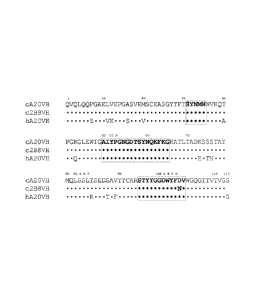
CD52, CD54, CD55, CD59, CD70, CD74, CD80, CD95, CD126, CD133, CD138, CD154,
CEACAM6, ED-B fibronectin, Factor H, FHL-1, Flt-1, Flt-3, folate receptor,
GROB,
HMGB-1, hypoxia inducible factor (HIF), HM1.24, insulin-like growth factor-1
(ILGF-1),
insulin-like growth factor-1 receptor (ILGF-1R), IFN-y, IFN-a, IFN-P, IL-2, IL-
4R, IL-6R,
IL-13R, IL-15R, IL-17R, IL-18R, IL-6, IL-8, IL-12, IL-15, IL-17, IL-25, IP-10,
MAGE,
mCRP. MCP-1. MIP-1A, MIP-1B, MIF, MUC1, MUC2, MUC3, MUC4, NCA-66, Ia,
HM1.24. HLA-DR, tenascin, T101. TAC, TRAIL-R1, TRAIL-R2, VEGFR, EGFR. P1GF,
complement factor C5, and an oncogene product (e.g., Kras, cMET, bc1-2, bc1-6)
or a
combination thereof, and even an anti-CD20 MAb that is different than the anti-
CD20 MAb
described herein.
[0056] The humanized, chimeric or human anti-CD20 antibody may possess
enhanced
affinity binding with the epitope, as well as antitumor and anti-B-cell
activity, as a result of
17
CDR mutation and manipulation of the CDR and other sequences in the variable
region to
obtain a superior therapeutic agent for the treatment of B-cell disorders,
including B-cell
lymphomas and leukemias and autoirnmune diseases and also other immune
diseases
involving B-cells (GVHD, hemolytic anemia, organ transplant rejection).
Amino Acid Substitutions
[0057] It may also be desirable to modify the amino acid sequences to improve
effector
function, e.g., to enhance antibody-dependent cell-dependent cytotoxicity
(ADCC) and/or
complement-dependent cytotoxicity (CDC). One or more amino acid substitutions
or the
introduction of cysteine in the Fc region may be made, thereby improving
internalization
capability and/or increased complement-dependent cell killing and ADCC. See
Caron et at.,
J. Exp. Med. 176:1191-1195 (1991) and Shopes, Br. J. Immutiol. 148:2918-2022
(1992).
An antibody fusion protein may be prepared that has dual Fc regions with both
enhanced complement
lysis and ADCC capabilities.
[0058] Changes to the Fc region to enhance effector function or other antibody
functional
characteristics have been reported (see, e.g., Lazar et at., Proc. Natl. Acad.
Sci. USA 2006,
103:4005-10; Stavenhagen et al., Cancer Res. 2007, 67:8882-90; Hinton etal.,
J. 'minimal.
2006, 176:346-56; Idusogie et al., J. lmmunol. 2001, 166:2571-75) and any such
known Fc
region amino acid substitutions may be utilized in the claimed methods and
compositions.
For example, replacement of a serine residue with an aspartate residue at
Kabat position 239
was reported to enhance ADCC activity (Lazar et al., ibid., 2006).
Substitution of
phenylalanine 243 with leucine, arginine 292 with proline, tyrosine 300 with
leucine, valine
305 with isoleucine and proline 396 with leucine also appeared to optimize
ADCC activity
(Stagenhagenet al., 20007). Replacement of threonine 250 with glutamine and
methionine
428 with leucine resulted in an apparent increase in serum half-life (Hinton
et al., ibid.,
2006). Substitution of lysine 326 with tryptophan and glutamate 333 with
serine appeared to
increase CDC activity (1dusogie et at., ibid., 2001). These and other known
modifications to
enhance antibody physiological function may be combined with changes to
variable region
sequence, described herein, to produce anti-CD20 antibodies of improved
therapeutic
characteri sties.
[0059] In certain embodiments, the disclosed methods and compositions may
involve
production and use of antibodies or antigen-binding fragments thereof with one
or more
substituted amino acid residues. As discussed below, methods for making
monoclonal
18
CA 2731255 2017-06-16
CA 02731255 2011-01-19
WO 2010/011697 PCT/US2009/051321
antibodies against virtually any target antigen are well known in the art.
Typically, these
result in production of murine antibodies against a target antigen. As is well
known in the
art, the antigen-binding specificity of murine monoclonal antibodies is
determined largely by
the hypervariable complementarity determining region (CDR) sequences. Murine
antibodies
generally comprise 6 CDR sequences, 3 on the antibody light chain and 3 on the
heavy chain.
As described in detail below, chimeric. humanized or human versions of murine
antibodies
may be constructed by techniques such as CDR grafting, where the murine CDR
sequences
are inserted into, for example, human antibody framework and constant region
sequences, or
by attaching the entire murine variable region sequences to human antibody
constant region
sequences. In alternative embodiments, the variable region sequences of an
antibody may be
constructed, for example, by chemical synthesis and assembly of
oligonucleotides encoding
the entire light and heavy chain variable regions of an antibody.
[0060] In various embodiments, the structural, physical and/or therapeutic
characteristics of
chimeric, humanized or human antibodies may be optimized by replacing one or
more amino
acid residues.
[0061] The skilled artisan will be aware that, in general, amino acid
substitutions typically
involve the replacement of an amino acid with another amino acid of relatively
similar
properties (i.e., conservative amino acid substitutions). The properties of
the various amino
acids and effect of amino acid substitution on protein structure and function
have been the
subject of extensive study and knowledge in the art.
[0062] For example, the hydropathic index of amino acids may be considered
(Kyte &
Doolittle, 1982, J. Mol. Biol., 157:105-132). The relative hydropathic
character of the amino
acid contributes to the secondary structure of the resultant protein, which in
turn defines the
interaction of the protein with other molecules. Each amino acid has been
assigned a
hydropathic index on the basis of its hydrophobicity and charge
characteristics (Kyte &
Doolittle, 1982), these are: isoleucine (+4.5); valine (+4.2); leucine (+3.8);
phenylalanine
(+2.8); cysteine/cystine (+2.5); methionine (+1.9); alanine (+1.8); glycine (-
0.4); threonine (-
0.7); serine (-0.8); tryptophan (-0.9); tyrosine (-1.3); proline (-1.6);
histidine (-3.2); glutamate
(-3.5); glutamine (-3.5); aspartate (-3.5); asparagine (-3.5); lysine (-3.9);
and arginine (-4.5).
In making conservative substitutions, the use of amino acids whose hydropathic
indices are
within 2 is preferred, within 1 are more preferred, and within 0.5 are
even more
preferred.
19
CA 02731255 2011-01-19
WO 2010/011697 PCT/US2009/051321
[0063] Amino acid substitution may also take into account the hydrophilicity
of the amino
acid residue (e.g., U.S. Pat. No. 4,554,101). Hydrophilicity values have been
assigned to
amino acid residues: arginine (+3.0); lysine (+3.0); aspartate (+3.0);
glutamate (+3.0); serine
(+0.3); asparagine (+0.2); glutamine (+0.2); glycine (0); threonine (-0.4);
proline (-0.5 ±1);
alanine (-0.5); histidine (-0.5); cysteine (-1.0); methionine (-1.3); valine (-
1.5); leucine (-1.8);
isoleucine (-1.8); tyrosine (-2.3); phenylalanine (-2.5); tryptophan (-3.4).
Replacement of
amino acids with others of similar hydrophilicity is preferred, but not
required.
[0064] Other considerations include the size of the amino acid side chain. For
example, it
would generally not be preferred to replace an amino acid with a compact side
chain, such as
glycine or serine, with an amino acid with a bulky side chain, e.g.,
tryptophan, tyrosine. The
effect of various amino acid residues on protein secondary structure is also a
consideration.
Through empirical study, the effect of different amino acid residues on the
tendency of
protein domains to adopt an alpha-helical, beta-sheet or reverse turn
secondary structure has
been determined and is known in the art (see, e.g., Chou & Fasman, 1974,
Biochemistry,
13:222-245; 1978, Ann. Rev. Biochem., 47: 251-276; 1979, Biophys. J., 26:367-
384).
[0065] Based on such considerations and extensive empirical study, tables of
conservative
amino acid substitutions have been constructed and are known in the art. For
example:
arginine and lysine; glutamate and aspartate; serine and threonine; glutamine
and asparagine;
and valine, leucine and isoleucine. Alternatively: Ala (A) leu, ile, val; Arg
(R) gln, asn, lys;
Asn (N) his, asp, lys, arg, gln; Asp (D) asn, glu; Cys (C) ala, ser; Gln (Q)
glu, asn; Glu (E)
gln, asp; Gly (G) ala; His (H) asn, gln, lys. arg; Ile (I) val, met, ala, phe,
leu; Leu (L) val, met,
ala, phe, ile; Lys (K) gln, asn, arg; Met (M) phe, ile. leu; Phe (F) leu, val,
ile, ala, tyr; Pro (P)
ala; Ser (S), thr; Thr (T) ser; Trp (W) phe, tyr; Tyr (Y) trp, phe, thr, ser;
Val (V) ile, leu, met,
phe, ala.
[0066] Other considerations for amino acid substitutions include whether or
not the residue is
located in the interior of a protein or is solvent exposed. For CDR residues,
the residue in the
free antibody would normally be assumed to be solvent exposed. For interior
residues,
conservative substitutions would include: Asp and Asn; Ser and Thr; Ser and
Ala; Thr and
Ala; Ala and Gly; Ile and Val; Val and Leu; Leu and Ile; Leu and Met; Phe and
Tyr; Tyr and
Trp. (See, e.g., PROWL website at rockefeller.edu) For solvent exposed
residues,
conservative substitutions would include: Asp and Asn; Asp and Glu; Glu and
Gln; Glu and
Ala; Gly and Asn; Ala and Pro; Ala and Gly; Ala and Ser; Ala and Lys; Ser and
Thr; Lys and
CA 02731255 2011-01-19
WO 2010/011697 PCT/US2009/051321
Arg; Val and Leu; Leu and Ile; Ile and Val; Phe and Tyr. (Id.) Various
matrices have been
constructed to assist in selection of amino acid substitutions, such as the
PAM250 scoring
matrix, Dayhoff matrix, Grantham matrix, McLachlan matrix, Doolittle matrix,
Henikoff
matrix, Miyata matrix, Fitch matrix, Jones matrix, Rao matrix, Levin matrix
and Risler
matrix (Idem.)
[0067] In determining amino acid substitutions, one may also consider the
existence of
intermolecular or intramolecular bonds, such as formation of ionic bonds (salt
bridges)
between positively charged residues (e.g., His, Arg, Lys) and negatively
charged residues
(e.g., Asp, Glu) or disulfide bonds between nearby cysteine residues.
Preparation of Monoclonal Antibodies including Chimeric, Humanized or Human
Antibodies
[0068] Techniques for preparing monoclonal antibodies against virtually any
target antigen
are well known in the art. See, for example, Kohler and Milstein, Nature 256:
495 (1975),
and Coligan et al. (eds.), CURRENT PROTOCOLS IN IMMUNOLOGY, VOL. 1, pages
2.5.1-2.6.7 (John Wiley & Sons 1991). Briefly, monoclonal antibodies can be
obtained by
injecting mice with a composition comprising an antigen, removing the spleen
to obtain B-
lymphocytes, fusing the B-lymphocytes with myeloma cells to produce
hybridomas, cloning
the hybridomas, selecting positive clones which produce antibodies to the
antigen, culturing
the clones that produce antibodies to the antigen, and isolating the
antibodies from the
hybridoma cultures.
[0069] MAbs can be isolated and purified from hybridoma cultures by a variety
of well-
established techniques. Such isolation techniques include affinity
chromatography with
Protein-A Sepharose, size-exclusion chromatography, and ion-exchange
chromatography.
See, for example, Coligan at pages 2.7.1-2.7.12 and pages 2.9.1-2.9.3. Also,
see Baines et
al., "Purification of Immunoglobulin G (IgG)," in METHODS IN MOLECULAR
BIOLOGY, VOL. 10, pages 79-104 (The Humana Press, Inc. 1992).
[0070] After the initial raising of antibodies to the immunogen, the
antibodies can be
sequenced and subsequently prepared by recombinant techniques. Humanization or
chimerization of murine antibodies and antibody fragments are well known to
those skilled in
the art. For example, humanized monoclonal antibodies are produced by
transferring mouse
complementary determining regions from heavy and light variable chains of the
mouse
immunoglobulin into a human variable domain, and then, substituting human
residues in the
framework regions of the murine counterparts. The use of antibody components
derived from
21
CA 02731255 2011-01-19
WO 2010/011697 PCT/US2009/051321
humanized monoclonal antibodies obviates potential problems associated with
the
immunogenicity of murine constant regions.
Chimeric Antibodies
[0071] A chimeric antibody is a recombinant protein in which the variable
regions of a
human antibody have been replaced by the variable regions of, for example, a
mouse
antibody, including the complementarity-determining regions (CDRs) of the
mouse antibody.
Chimeric antibodies exhibit decreased immunogenicity and increased stability
when
administered to a subject. General techniques for cloning murine
immunoglobulin variable
domains are disclosed, for example, in Orlandi et al., Proc. Nat. Acad. Sci.
USA 86: 3833
(1989). Techniques for constructing chimeric antibodies are well known to
those of skill in
the art. As an example. Leung et al., Hybridoma /3:469 (1994), produced an LL2
chimera
by combining DNA sequences encoding the VK and VH domains of murine LL2, an
anti-
CD22 monoclonal antibody, with respective human K and IgGi constant region
domains.
Humanized Antibodies
[0072] Techniques for producing humanized MAbs are well known in the art (see,
e.g., Jones
et al., Nature 321: 522 (1986), Riechmann et al., Nature 332: 323 (1988),
Verhoeyen et al.,
Science 239: 1534 (1988), Carter et al., Proc. Nat. Acad. Sci. USA 89: 4285
(1992), Sandhu,
Crit. Rev. Biotech. 12: 437 (1992), and Singer et al., J. Immun. 150: 2844
(1993)). A
chimeric or murine monoclonal antibody may be humanized by transferring the
mouse CDRs
from the heavy and light variable chains of the mouse immunoglobulin into the
corresponding variable domains of a human antibody. The mouse framework
regions (1-R) in
the chimeric monoclonal antibody are also replaced with human FR sequences. As
simply
transferring mouse CDRs into human FRs often results in a reduction or even
loss of antibody
affinity, additional modification might be required in order to restore the
original affinity of the
murine antibody. This can be accomplished by the replacement of one or more
some human
residues in the PR regions with their murine counterparts to obtain an
antibody that possesses
good binding affinity to its epitope. See, for example, Tempest et al.,
Biotechnology 9:266
(1991) and Verhoeyen et al., Science 239: 1534 (1988). The affinity of
humanized antibodies
for a target may also be increased by selected modification of the CDR
sequences
(W00029584A1).
Human Antibodies
[0073] Methods for producing fully human antibodies using either combinatorial
approaches
or transgenic animals transformed with human immunoglobulin loci are known in
the art
22
CA 02731255 2013-05-08
(e.g., Mancini et al., 2004, New Microbiol. 27:315-28; Conrad and Scheller,
2005, Comb.
Chem. High Throughput Screen. 8:117-26; Brekke and Loset, 2003, Curr. Opin.
Phamacol.
3:544-50). A fully human antibody also can be constructed by genetic or
chromosomal
transfection methods, as well as phage display technology, all of which are
known in the art.
See for example, McCafferty etal., Nature 348:552-553 (1990). Such fully human
antibodies are expected to exhibit even fewer side effects than chimeric,
humanized or human
antibodies and to function in vivo as essentially endogenous human antibodies.
In certain
embodiments, the claimed methods and procedures may utilize human antibodies
produced
by such techniques.
[0074] In one alternative, the phage display technique may be used to generate
human
antibodies (e.g., Dantas-Barbosa et al., 2005, Genet. Ma Res. 4:126-40). Human
antibodies
may be generated from normal humans or from humans that exhibit a particular
disease state,
such as cancer (Dantas-Barbosa et al., 2005). The advantage to constructing
human
antibodies from a diseased individual is that the circulating antibody
repertoire may be biased
towards antibodies against disease-associated antigens.
[0075] In one non-limiting example of this methodology, Dantas-Barbosa et al.
(2005)
constructed a phage display library of human Fab antibody fragments from
osteosarcoma
patients. Generally, total RNA was obtained from circulating blood lymphocytes
(Id.).
Recombinant Fab were cloned from the It, 7 and 1( chain antibody repertoires
and inserted
into a phage display library (Id.). RNAs were converted to cDNAs and used to
make Fab
cDNA libraries using specific primers against the heavy and light chain
irnmunoglobulin
sequences (Marks et al., 1991, J. Mel. Biol. 222:581-97). Library construction
was
performed according to Andris-Widhopf et al. (2000, In: Phage Display
Laboratory Manual,
Barbas et al. (eds), 1st edition, Cold Spring Harbor Laboratory Press, Cold
Spring Harbor, NY
pp. 9.1 to 9.22). The final Fab fragments were digested with restriction
endonucleases and
inserted into the bacteriophage genome to make the phage display library. Such
libraries may
be screened by standard phage display methods, as known in the art. Phage
display can be
performed in a variety of formats, for their review, see e.g., Johnson and
Chiswell, Current
Opinion in Structural Biology 3:5564-571 (1993). Human antibodies may also be
generated
by in vitro activated B-cells. See U.S. Patent Nos. 5,567,610 and 5,229,275.
The skilled
artisan will realize that these techniques are exemplary and any known method
for making
and screening human antibodies or antibody fragments may be utilized.
23
CA 02731255 2013-05-08
[0076] In another alternative, transgenic animals that have been genetically
engineered to
produce human antibodies may be used to generate antibodies against
essentially any
immunogenic target, using standard immunization protocols. Methods for
obtaining human
antibodies from transgenic mice are disclosed by Green et al., Nature Genet.
7:13 (1994),
Lonberg eta!,, Nature 368:856 (1994), and Taylor etal., Int. I/limn. 6:579
(1994). A non-
limiting example of such a system is the XenoMouse (e.g., Green et al.,
1999,1 Immunol.
Methods 231:11-23) from Abgenix (Fremont, CA). In the XenoMouse and similar
animals,
the mouse antibody genes have been inactivated and replaced by functional
human antibody
genes, while the remainder of the mouse immune system remains intact.
[0077] The XenoMouse was transformed with germline-configured YACs (yeast
artificial
chromosomes) that contained portions of the human IgH and Igkappa loci,
including the
majority of the variable region sequences, along with accessory genes and
regulatory
sequences. The human variable region repertoire may be used to generate
antibody
producing B-cells, which may be processed into hybridomas by known techniques.
A
XenoMouse immunized with a target antigen will produce human antibodies by
the normal
immune response, which may be harvested and/or produced by standard techniques
discussed
above. A variety of strains of XenoMouse are available, each of which is
capable of
producing a different class of antibody. Transgenically produced human
antibodies have
been shown to have therapeutic potential, while retaining the pharmacokinetic
properties of
normal human antibodies (Green etal., 1999). The skilled artisan will realize
that the
claimed compositions and methods are not limited to use of the XenoMouse
system but
may utilize any transgenic animal that has been genetically engineered to
produce human
antibodies.
24
CA 02731255 2013-05-08
Production of Antibody Fragments
[0078] Antibody fragments which recognize specific epitopes can be generated
by known
techniques. The antibody fragments are antigen binding portions of an
antibody, such as F(ab1)2,
Fab', F(ab),, Fab, Fv, scFv and the like. 17(ab')2 fragments can be produced
by pepsin digestion
of the antibody molecule and Fab' fragments can be generated by reducing
disulfide bridges
of the F(ab')2 fragments. Alternatively, Fab' expression libraries can be
constructed (Huse et
ol., 1989. Science, 246:1274-1281) to allow rapid and easy identification of
monoclonal Fab'
fragments with the desired specificity.
10079] A single chain Fv molecule (scFv) comprises a VL domain and a VH
domain. The
VL and VH domains associate to form a target binding site. These two domains
are further
covalently linked by a peptide linker (L). Methods for making scFv molecules
and designing
suitable peptide linkers are described in US Patent No. 4,704,692, US Patent
No. 4,946,778,
R. Raag and M. Whitlow, -Single Chain Fvs." FASEB Vol 9:73-80 (1995) and R.E.
Bird and
B.W. Walker, "Single Chain Antibody Variable Regions," TIBTECH, Vol 9: 132-137
(1991).
[0080] An antibody fragment can be prepared by proteolytic hydrolysis of the
full length
antibody or by expression in E. coli or another host of the DNA coding for the
fragment. An
antibody fragment can be obtained by pepsin or papain digestion of full length
antibodies by
conventional methods. For example, an enzymatic cleavage using papain produces
two
monovalent Fab fragments and an Fc fragment. These methods are described, for
example,
by Goldenberg, U.S. Patent Nos. 4,036,945 and 4,331,647 and references
contained therein.
Also, see Nisonoff et al., Arch Biochein. Biophys. 89: 230 (1960); Porter,
Biochein. 1 73: 119
(1959), Edelman et al., in METHODS IN ENZYMOLOGY VOL. 1, page 422 (Academic
Press 1967), and Coligan at pages 2.8.1-2.8.10 and 2.10.-2.10.4.
Bispecific and Multispecific Antibodies
[0081] Bispecific antibodies are useful in a number of biomedical
applications. For instance,
a bispecific antibody with binding sites for a tumor cell surface antigen and
for a T-cell
surface receptor can direct the lysis of specific tumor cells by T cells.
Bispecific antibodies
recognizing gliomas and the CD3 epitope on T cells have been successfully used
in treating
brain tumors in human patients (Nitta, et al., Lancet 1990; 355:368-371). Pre-
targeting
methods with bispecific antibodies comprising at least one binding site for a
tumor-associated
CA 02731255 2013-05-08
antigen (TAA) or other disease target, as well as at one binding site for a
targetable construct
conjugated to therapeutic or diagnostic agents, are also well known in the art
(see, e.g., U.S.
Patent Nos. 7,300,644; 7,138,103; 7,074,405; 7,052,872; 6,962,702; 6,458,933.
[0082] Bispecifie antibodies comprising the antigen-binding variable region
sequences of any
known anti-TAA antibody may be utilized, including but not limited to hPAM4
(U.S. Patent
No. 7,282,567), hA20 (U.S. Patent No. 7,251,164), hAl9 (U.S. Patent No.
7,109,304),
hIMMU31 (U.S. Patent No. 7,300,655), hIJ,1 (U.S. Patent No. 7,312,318), h1-1,2
(U.S.
Patent No. 7,074,403), hMu-9 (U.S. Patent No. 7,387,773), hL243 (U.S. Patent
No.
7,612,180), hMN-14 (U.S. Patent No. 6,676,924), hRS7 (U.S. Patent No.
7,238,785),
hMN-3 (U.S. Patent No. 7,541,440) and hRl.
[0083] Other antibodies of use may be commercially obtained from a wide
variety of known
sources. For example, a variety of antibody secreting hybridoma lines are
available from the
American Type Culture Collection (ATCC, Manassas, VA). A large number of
antibodies
against various disease targets, including but not limited to tumor-associated
antigens, have
been deposited at the ATCC and/or have published variable region sequences and
are
available for use in the claimed methods and compositions. See, e.g., U.S.
Patent Nos.
7,312,318; 7,282,567; 7,151,164; 7,074,403; 7,060,802; 7,056,509; 7,049,060;
7,045,132;
7,041,803; 7,041,802; 7,041,293; 7,038,018; 7,037,498; 7,012,133; 7,001,598;
6,998,468;
6,994,976; 6,994,852; 6,989,241; 6,974,863; 6,965,018; 6,964,854; 6,962,981;
6,962,813;
6,956,107; 6,951,924; 6,949,244; 6,946,129; 6,943,020; 6,939,547; 6,921,645;
6,921,645;
6,921,533; 6,919,433; 6,919,078; 6,916,475; 6,905,681; 6,899,879; 6,893,625;
6,887,468;
6,887,466; 6,884,594; 6,881,405; 6,878,812; 6,875,580; 6,872,568; 6,867,006;
6,864,062;
6,861,511; 6,861,227; 6,861,226; 6,838,282; 6,835,549; 6,835,370; 6,824,780;
6,824,778;
6,812,206; 6,793,924; 6,783,758; 6,770,450; 6,767,711; 6,764,688; 6,764,681;
6,764,679;
6,743,898; 6,733,981; 6,730,307; 6,720,15; 6,716,966; 6,709,653; 6,693,176;
6,692,908;
6,689,607; 6,689,362; 6,689,355; 6,682,737; 6,682,736; 6,682,734; 6,673,344;
6,653,104;
6,652,852; 6,635,482; 6,630,144; 6,610,833; 6,610,294; 6,605,441; 6,605,279;
6,596,852;
6,592,868; 6,576,745; 6,572;856; 6,566,076; 6,562,618; 6,545,130; 6,544,749;
6,534,058;
6,528,625; 6,528,269; 6,521,227; 6,518,404; 6,511,665; 6,491,915; 6,488,930;
6,482,598;
6,482,408; 6,479,247; 6,468,531; 6,468,529; 6,465,173; 6,461,823; 6,458,356;
6,455,044;
26
CA 02731255 2013-05-08
6,455,040, 6,451,310; 6,444,206' 6,441,143; 6,432,404; 6,432,402; 6,419,928;
6,413,726;
6,406,694; 6,403,770; 6,403,091; 6,395,276; 6,395,274; 6,387,350; 6,383,759;
6,383,484;
6,376,654; 6,372,215; 6,359,126; 6,355,481; 6,355,444; 6,355,245; 6,355,244;
6,346,246;
6,344,198; 6,340,571; 6,340,459; 6,331,175; 6,306,393; 6,254,868; 6,187,287;
6,183,744;
6,129,914; 6,120,767; 6,096,289; 6,077,499; 5,922,302; 5,874,540; 5,814,440;
5,798,229;
5,789,554; 5,776,456; 5,736,119; 5,716,595; 5,677,136; 5,587,459; 5,443,953,
5,525,338.
These are exemplary only and a wide variety of other antibodies and their
hybridomas are
known in the art. The skilled artisan will realize that antibody sequences or
antibody-
secreting hybridomas against almost any disease-associated antigen may be
obtained by a
simple search of the ATCC, NCBI and/or USPTO databases for antibodies against
a selected
disease-associated target of interest. The antigen binding domains of the
cloned antibodies
may be amplified, excised, ligated into an expression vector, transfected into
an adapted host
cell and used for protein production, using standard techniques well known in
the art.
[0084] Numerous methods to produce bispecific or multispecific antibodies are
known, as
disclosed, for example, in U.S. Patent Application Publication No.
20050002945, filed
2/11/2004. Bispecific antibodies can be produced by the quadroma method, which
involves
the fusion of two different hybridomas, each producing a monoclonal antibody
recognizing a
different antigenic site (Milstein and Cuello, Nature, 1983; 305:537-540).
[0085] Another method for producing bispecific antibodies uses
heterobifunctional cross-
linkers to chemically tether two different monoclonal antibodies (Staerz,
etal., Nature 1985;
314:628-631; Perez, at al., Nature 1985; 316:354-356). Bispecific antibodies
can also be
produced by reduction of each of two parental monoclonal antibodies to the
respective half
molecules, which are then mixed and allowed to reoxidize to obtain the hybrid
structure
(Staerz and Bevan, Proc Neal Acad Sci USA 1986; 83:1453-1457). Another
alternative
involves chemically cross-linking two or three separately purified Fab'
fragments using
appropriate linkers. (See, e.g., European Patent Application 0453082).
100861 Other methods include improving the efficiency of generating hybrid
hybridomas by
gene transfer of distinct selectable markers via retrovirus-derived shuttle
vectors into
respective parental hybridomas, which are fused subsequently (DeMonte, et al.,
Proc Nat!
Acad Sci USA 1990, 87:2941-2945); or transfection of a hybridoma cell line
with expression
plasmids containing the heavy and light chain genes of a different antibody.
27
CA 02731255 2013-05-08
[0087] Cognate VH and VL domains can be joined with a peptide linker of
appropriate
composition and length (usually consisting of more than 12 amino acid
residues) to form a
single-chain Fv (scFv) with binding activity. Methods of manufacturing scFvs
are disclosed
in U.S. Pat. No. 4,946,778 and U.S. Pat. No. 5,132,405. Reduction of the
peptide linker
length to less than 12 amino acid residues prevents pairing of VH and VL
domains on the
same chain and forces pairing of VH and VL domains with complementary domains
on other
chains, resulting in the formation of functional multimers. Polypeptide chains
of VI/ and Vi.
domains that are joined with linkers between 3 and 12 amino acid residues form
predominantly dimers (termed diabodies). With linkers between 0 and 2 amino
acid residues,
trimers (termed triabody) and tetramers (termed tetrabody) are favored, but
the exact patterns
of oligomerization appear to depend on the composition as well as the
orientation of V-
domains (VH-linker-VL or VL-linker-VH), in addition to the linker length.
[0088] These techniques for producing multispecific or bispecific antibodies
exhibit various
difficulties in terms of low yield, necessity for purification, low stability
or the labor-
intensiveness of the technique. More recently, a technique known as "dock and
lock" (DNL)
has been utilized to produce combinations of virtually any desired antibodies,
antibody
fragments and other effector molecules (see, e.g., U.S. Patent Application
Publ. Nos.
20060228357 (now issued U.S. Patent 7,550,143); 20060228300 (now issued U.S.
Patent
7,521,056); 20070086942 (now issued U.S. Patent 7,534,866); 20070140966 (now
issued
U.S. Patent 7,527,787); 20070264265; 20090060862 and 20090202487). The
technique
utilizes complementary protein binding domains, referred to as anchoring
domains and
dimerization and docking domains, which bind to each other and allow the
assembly of
complex structures, ranging from dimers, trimers, tetrainers, quintamers and
hexamers.
These form stable complexes in high yield without requirement for extensive
purification.
The DNL technique allows the assembly of monospecific, bispecific or
multispecific
antibodies, either as naked antibody moieties or in combination with a wide
range of other
effector molecules such as immunomodulators, enzymes, chemotherapeutic agents,
chemokines, cytokines, diagnostic agents, therapeutic agents, radionuclides,
imaging agents,
anti-angiogenic agents, growth factors, oligonucleotides, hormones, peptides,
toxins, pro-
apoptotic agents, or a combination thereof. Any of the techniques known in the
art for
28
CA 02731255 2013-05-08
making bispecific or multispecific antibodies may be utilized in the practice
of the presently
claimed methods.
Pre-Targeting
[00891 Bispecific or multispecific antibodies may be utilized in pre-targeting
techniques.
Pre-targeting is a multistep process originally developed to resolve the slow
blood clearance
of directly targeting antibodies, which contributes to undesirable toxicity to
normal tissues
such as bone marrow. With pre-targeting, a radionuclide or other therapeutic
agent is
attached to a small delivery molecule (targetable construct or targetable
conjugate) that is
cleared within minutes from the blood. A pre-targeting bispecific or
multispecific antibody,
which has binding sites for the targetable construct as well as a target
antigen, is administered
first, free antibody is allowed to clear from circulation and then the
targetable construct is
administered.
100901 Pre-targeting methods are well known in the art, for example, as
disclosed in
Goodwin et al., U.S. Pat. No. 4,863,713; Goodwin etal., I Nucl. Med. 29:226,
1988;
Hnatowich et al., J. Nucl. Med. 28:1294, 1987; Oehr et al., J Nucl. Med.
29:728, 1988;
Klibanov etal., J. Nucl. Med. 29:1951, 1988; Sinitsyn et al., J. Nod. Med.
30:66, 1989;
Kalofonos et al., J. Nucl. Med. 31:1791, 1990; Schechter et al., Int. J.
Cancer 48:167, 1991;
Paganelli etal., Cancer Res. 51:5960, 1991; Paganelli et al., Nucl. Med.
Common. 12:211,
1991; U.S. Pat. No. 5,256,395; Sticicney etal., Calleel^ Res. 51:6650, 1991;
Yuan et al.,
Cancer Res. 51:3119, 1991; U.S. Pat. No, 6,077,499; U.S. Pat. No. 7,011,812;
U.S. Pat. No.
7,300,644; U.S. Pat. No. 7,074,405; U.S. Pat. No. 6,962,702; U.S. Pat. No.
7,387,772; U.S.
Pat. No. 7,052,872; U.S. Pat. No. 7,138,103; U.S. Pat. No. 6,090,381; U.S.
Pat. No.
6,472,511; U.S. publication. No. 20020176856; U.S. publication No.
20040044076; U.S.
publication No. 20030113333; U.S. publication No. 20030148409; U.S. Pat. No.
7,833,528;
U.S. publication No. 20030162709; and U.S. Patent No. 6,962,702.
10091] A pre-targeting method of treating or diagnosing a disease or disorder
in a subject
may be provided by: (1) administering to the subject a bispecific antibody or
antigen binding
antibody fragment; (2) optionally administering to the subject a clearing
composition, and
allowing the composition to clear the antibody from circulation; and (3)
administering to the
subject the targetable construct, containing one or more chelated or
chemically bound
therapeutic or diagnostic agents. The technique may also be utilized for
antibody dependent
29
CA 02731255 2011-01-19
WO 2010/011697 PCT/US2009/051321
enzyme prodrug therapy (ADEPT) by administering an enzyme conjugated to a
targetable
construct, followed by a prodrug that is converted into active form by the
enzyme.
Therapeutic and Diagnostic Agents
[0092] In certain embodiments, the antibodies, antigen binding antibody
fragments or fusion
proteins described herein may be administered alone, as a "naked" antibody,
antigen binding
fragment thereof or fusion protein. In alternative embodiments, the antibody,
antigen binding
fragment thereof or fusion protein may be administered before, concurrently
with, or after at
least one other therapeutic agent. In other alternatives, an antibody, antigen
binding fragment
thereof or fusion protein may be covalently or non-covalently attached to at
least one therapeutic
and/or diagnostic agent to form an immunoconjugate.
[0093] Diagnostic agents are preferably selected from the group consisting of
a radionuclide, a
radiological contrast agent, a paramagnetic ion, a metal, a fluorescent label,
a
chemiluminescent label, an ultrasound contrast agent and a photoactive agent.
Such
diagnostic agents are well known and any such known diagnostic agent may be
used. Non-
limiting examples of diagnostic agents may include a radionuclide such as
110In, 111In, 177Lu,
18 52 62 64 67 67 68 86 90 89 94m 94 99m
'20T, 1231 124 125
F, Fe, Cu, Cu, Cu, Ga, Ga, Y, Y, Zr, Tc, Tc, Tc, I, L I.
131 - Pm
I 154158 32 11 13 15
, Gd, P, C. N, 0. 186 188 51 Re, Re, Mn, 52mMn, 55 72
75 76 Co, As. Br, Br, Rb 83, Sr,
or other gamma-, beta-, or positron-emitters. Paramagnetic ions of use may
include
chromium (III), manganese (II), iron (III), iron (II), cobalt (II), nickel
(II), copper (II),
neodymium (III), samarium (III), ytterbium (III), gadolinium (III), vanadium
(II), terbium
(III), dysprosium (III), holmium (III) or erbium (III). Metal contrast agents
may include
lanthanum (III). gold (III), lead (II) or bismuth (III). Ultrasound contrast
agents may
comprise liposomes, such as gas filled liposomes. Radiopaque diagnostic agents
may be
selected from barium compounds, gallium compounds and thallium compounds. A
wide
variety of fluorescent labels are known in the art, including but not limited
to fluorescein
isothiocyanate, rhodamine, phycoerytherin, phycocyanin, allophycocyanin, o-
phthaldehyde
and fluorescamine. Chemiluminescent labels of use may include luminol,
isoluminol, an
aromatic acridinium ester, an imidazole, an acridinium salt or an oxalate
ester.
[0094] Therapeutic agents are preferably selected from the group consisting of
a radionuclide,
an immunomodulator, an anti-angiogenic agent, a cytokine, a chemokine, a
growth factor, a
hormone, a drug, a prodrug, an enzyme, an oligonucleotide, an interference
RNA, a pro-
CA 02731255 2011-01-19
WO 2010/011697 PCT/US2009/051321
apoptotic agent, a photoactive therapeutic agent, a cytotoxic agent, which may
be a
chemotherapeutic agent or a toxin, a second antibody or fragment thereof and a
combination
thereof. The drugs of use may possess a pharmaceutical property selected from
the group
consisting of antimitotic, antikinase, alkylating, antimetabolite, antibiotic,
alkaloid, anti-
angiogenic, pro-apoptotic agents and combinations thereof.
[0095] Exemplary drugs of use include, but are not limited to, 5-fluorouracil,
aplidin,
azaribine, anastrozole, anthracyclines, bendamustine, bleomycin, bortezomib,
bryostatin-1,
busulfan, calicheamycin, camptothecin, carboplatin, 10-hydroxycamptothecin.
carmustine,
celebrex, chlorambucil, cisplatin (CDDP), Cox-2 inhibitors, irinotecan (CPT-
11), SN-38,
carboplatin, cladribine, camptothecans, cyclophosphamide, cytarabine,
dacarbazine,
docetaxel, dactinomycin, daunorubicin, doxorubicin, 2-pyrrolinodoxorubicine
(2P-DOX),
cyano-morpholino doxorubicin, doxorubicin glucuronide, epirubicin glucuronide,
estramustine, epidophyllotoxin, estrogen receptor binding agents. etoposide
(VP16), etoposide
glucuronide, etoposide phosphate, floxuridine (FUdR), 3',5'-0-dioleoyl-FudR
(FUdR-d0),
fludarabine, flutamide, famesyl-protein transferase inhibitors, gemcitabine,
hydroxyurea,
idarubicin, ifosfamide, L-asparaginase, lenolidamide, leucovorin, lomustine,
mechlorethamine, melphalan, mercaptopurine, 6-mercaptopurine, methotrexate,
mitoxantrone, mithramycin, mitomycin, mitotane, navelbine, nitrosurea,
plicomycin,
procarbazine, paclitaxel, pentostatin, PSI-341, raloxifene, semustine,
streptozocin,
tamoxifen, taxol, temazolomide (an aqueous form of DTIC), transplatinum,
thalidomide,
thioguanine, thiotepa, teniposide, topotecan, uracil mustard, vinorelbine,
vinblastine,
vincristine and vinca alkaloids.
[00%] Toxins of use may include ricin, abrin, alpha toxin, saporin,
ribonuclease (RNase),
e.g., onconase, DNase T, Staphylococcal enterotoxin-A, pokeweed antiviral
protein, gelonin,
diphtheria toxin, Pseudomonas exotoxin, and Pseudomozzas endotoxin.
[0097] Immunomodulators of use may be selected from a cytokine, a stem cell
growth factor, a
lymphotoxin, a hematopoietic factor, a colony stimulating factor (CSF), an
interferon (IFN),
erythropoietin, thrombopoietin and a combination thereof. Specifically useful
are
lymphotoxins such as tumor necrosis factor (TNF), hematopoietic factors, such
as interleukin
(IL), colony stimulating factor, such as granulocyte-colony stimulating factor
(G-CSF) or
granulocyte macrophage-colony stimulating factor (GM-CSF), interferon, such as
interferons-a, -f3 or -7, and stem cell growth factor, such as that designated
"Si factor".
31
CA 02731255 2011-01-19
WO 2010/011697 PCT/US2009/051321
Included among the cytokines are growth hormones such as human growth hormone,
N-
methionyl human growth hormone, and bovine growth hormone; parathyroid
hormone;
thyroxine; insulin; proinsulin; relaxin; prorelaxin; glycoprotein hormones
such as follicle
stimulating hormone (FSH), thyroid stimulating hormone (TSH), and luteinizing
hormone
(LH); hepatic growth factor; prostaglandin, fibroblast growth factor;
prolactin; placental
lactogen, OB protein; tumor necrosis factor-a and - B; mullerian-inhibiting
substance; mouse
gonadotropin-associated peptide; inhibin; activin; vascular endothelial growth
factor;
integrin; thrombopoietin (TP0); nerve growth factors such as NGF-B; platelet-
growth factor;
transforming growth factors (TGFs) such as TGF- a and TGF- B; insulin-like
growth factor-I
and -II; erythropoietin (EPO); osteoinductive factors; interferons such as
interferon-a, -13, and
-7; colony stimulating factors (CSFs) such as macrophage-CSF (M-CSF);
interleukins (ILs)
such as IL-1, IL-la, IL-2, IL-3, IL-4, IL-5, IL-6, IL-7, IL-8, IL-9, IL-10, IL-
11, IL-12; IL-13,
IL-14, IL-15, IL-16. IL-17, IL-18, IL-21, IL-25, LIF, kit-ligand or FLT-3, Flt-
1, angiostatin,
thrombospondin, endostatin, tumor necrosis factor (TNF, such as TNF-a) and LT.
As used
herein, the term cytokine includes proteins from natural sources or from
recombinant cell
culture and biologically active equivalents of the native sequence cytokines.
[0098] Chemokines of use include RANTES, MCAF, MIP1-alpha, MIP1-Beta and IP-
10.
[0100] Radioactive isotopes useful for treating diseased tissue include, but
are not limited to-
1"1n, 177Lu, 212Bi. 213Bi, 211At,62Cu, 67Cu,
90y, 1251, 1311, 32p, 33p, 47sc, 111Ag, 67Ga,
142Pr, 153SM, 161Tb, 166Dy, 166Ho, 186Re, 188Re. 189Re, 212Pb, 223Ra, 225Ac,
59Fe, 75Se,
77As, 89Sr, "Mo, 105Rh, 109pd, 143 -r,
P 149Pm, 169Er, 1941r, 198Au, 199Au, and 2"Pb. The
therapeutic radionuclide preferably has a decay energy in the range of 20 to
6,000 keV,
preferably in the ranges 60 to 200 keV for an Auger emitter, 100-2,500 keV for
a beta
emitter, and 4,000-6,000 keV for an alpha emitter. Maximum decay energies of
useful beta-
particle-emitting nuclides are preferably 20-5,000 keV, more preferably 100-
4,000 keV, and
most preferably 500-2,500 keV. Also preferred are radionuclides that
substantially decay
with Auger-emitting particles. For example, Co-58, Ga-67, Br-80m, Tc-99m, Rh-
103m, Pt-
109, In-111, Sb-119, 1-125, Ho-161, Os-189m and Ir-192. Decay energies of
useful beta-
particle-emitting nuclides are preferably <1,000 keV, more preferably <100
keV, and most
preferably <70 keV. Also preferred are radionuclides that substantially decay
with
generation of alpha-particles. Such radionuclides include, but are not limited
to: Dy-152, At-
211, Bi-212, Ra-223, Rn-219, Po-215, Bi-211, Ac-225, Fr-221, At-217, Bi-213
and Fm-255.
32
CA 02731255 2011-01-19
WO 2010/011697 PCT/US2009/051321
Decay energies of useful alpha-particle-emitting radionuclides are preferably
2.000-10.000
keV, more preferably 3,000-8,000 keV, and most preferably 4,000-7,000 keV.
Additional
, 126/, 133-,
I
potential radioisotopes of use include nc, EN, 150 75Br, 'Au, 224Ac, 77Br,
113m-n,
I 95RU, 97RU, 1 3RU, 105Ru, io7Hg, 203H0., 121mTe, 122mTe, 125Te, m 165Tm,
167
Tm,
168 197 109 105 142 143 161 166 199 57 58 51
59 75
Tm, Pt, Pd, Rh, Pr, Pr, Tb, Ho, Au, Co. Co, Cr, Fe, Se,
201 225 76 169 124
Tl, Ac, Br, Yb, and the
like. Some useful diagnostic nuclides may include I,
123 131 18 52 62 64 67 67 68 86 89 94 94m 99m 111
1, I, F, Fe, Cu, Cu, Cu, Ga, Ga, Y, Zr, Tc, Tc, Tc, or In.
[0101] Therapeutic agents may include a photoactive agent or dye. Fluorescent
compositions, such as fluorochrome, and other chromogens, or dyes, such as
porphyrins
sensitive to visible light, have been used to detect and to treat lesions by
directing the suitable
light to the lesion. In therapy, this has been termed photoradiation,
phototherapy, or
photodynamic therapy. See Jon i et al. (eds.), PHOTODYNAMIC THERAPY OF TUMORS
AND OTHER DISEASES (Libreria Progetto 1985); van den Bergh. Chem. Britain
(1986),
22:430. Moreover, monoclonal antibodies have been coupled with photoactivated
dyes for
achieving phototherapy. See Mew etal., J. Immunol. (1983),130:1473; idem.,
Cancer Res.
(1985). 45:4380; Oseroff et al.. Proc. Natl. Acad. Sci. USA (1986), 83:8744;
idem.,
Photochem. Photobiol. (1987), 46:83; Hasan et al., Prog. Clin. Biol. Res.
(1989), 288:471;
Tatsuta etal., Lasers Surg. Med. (1989), 9:422; Pelegrin etal., Cancer (1991),
67:2529.
[0102] Corticosteroid hormones can increase the effectiveness of other
chemotherapy agents,
and consequently, they are frequently used in combination treatments.
Prednisone and
dexamethasone are examples of corticosteroid hormones. .
[0103] In certain embodiments, anti-angiogenic agents, such as angiostatin,
baculostatin,
canstatin, maspin, anti-VEGF antibodies, anti-P1GF peptides and antibodies,
anti-vascular
growth factor antibodies, anti-Flk-1 antibodies, anti-Flt-1 antibodies and
peptides, anti-Kras
antibodies, anti-cMET antibodies, anti-MIF (macrophage migration-inhibitory
factor)
antibodies, laminin peptides, fibronectin peptides, plasminogen activator
inhibitors, tissue
metalloproteinase inhibitors, interferons, interleukin-12, IP-10, Gro-B,
thrombospondin, 2-
methoxyoestradiol, proliferin-related protein, carboxiamidotriazole, CM101,
Marimastat,
pentosan polysulphate, angiopoietin-2, interferon-alpha, herbimycin A,
PNU145156E, 16K
prolactin fragment, Linomide, thalidomide, pentoxifylline, genistein, TNP-470,
endostatin,
paclitaxel, accutin, angiostatin, cidofovir, vincristine, bleomycin, AGM-1470,
platelet factor
4 or minocycline may be of use.
33
CA 02731255 2011-01-19
WO 2010/011697 PCT/US2009/051321
[0104] Other useful therapeutic agents comprise oligonucleotides, especially
antisense
oligonucleotides that preferably are directed against oncogenes and oncogene
products of B-
cell malignancies, such as bc1-2. Preferred antisense oligonucleotides include
those known as
siRNA or RNAi.
Immunoconjugates
[0105] Any of the antibodies, antigen binding antibody fragments or antibody
fusion proteins
described herein may be conjugated to one or more therapeutic or diagnostic
agents. The
therapeutic agents do not need to be the same but can be different, e.g., a
drug and a
radioisotope. For example, 1311 can be incorporated into a tyrosine of an
antibody or fusion
protein and a drug attached to an epsilon amino group of a lysine residue.
Therapeutic and
diagnostic agents also can be attached, for example to reduced SH groups
and/or to
carbohydrate side chains. Many methods for making covalent or non-covalent
conjugates of
therapeutic or diagnostic agents with antibodies or fusion proteins are known
in the art and
any such known method may be utilized.
[0106] A therapeutic or diagnostic agent can be attached at the hinge region
of a reduced
antibody component via disulfide bond formation. Alternatively, such agents
can be attached
using a heterobifunctional cross-linker, such as N-succinyl 3-(2-
pyridyldithio)propionate
(SPDP). Yu et al., Int. J. Cancer 56: 244 (1994). General techniques for such
conjugation
are well-known in the art. See, for example, Wong, CHEMISTRY OF PROTEIN
CONJUGATION AND CROSS-LINKING (CRC Press 1991); Upeslacis et at.,
"Modification of Antibodies by Chemical Methods," in MONOCLONAL ANTIBODIES:
PRINCIPLES AND APPLICATIONS. Birch et al. (eds.), pages 187-230 (Wiley-Liss,
Inc.
1995); Price, "Production and Characterization of Synthetic Peptide-Derived
Antibodies," in
MONOCLONAL ANTIBODIES: PRODUCTION, ENGINEERING AND CLINICAL
APPLICATION, Ritter et at. (eds.), pages 60-84 (Cambridge University Press
1995).
Alternatively, the therapeutic or diagnostic agent can be conjugated via a
carbohydrate moiety
in the Fc region of the antibody. The carbohydrate group can be used to
increase the loading
of the same agent that is bound to a thiol group, or the carbohydrate moiety
can be used to
bind a different therapeutic or diagnostic agent.
[0107] Methods for conjugating peptides to antibody components via an antibody
carbohydrate moiety are well-known to those of skill in the art. See, for
example, Shih ei at.,
Int. J. Cancer 41: 832 (1988); Shih et al., Int. J. Cancer 46: 1101 (1990);
and Shih ei at., U.S.
34
Patent No. 5,057,313. The general method involves reacting an antibody
component having
an oxidized carbohydrate portion with a carrier polymer that has at least one
free amine
function. This reaction results in an initial Schiff base (imine) linkage,
which can be
stabilized by reduction to a secondary amine to form the final conjugate.
101081 The Fe region may be absent if the antibody used as the antibody
component of the
immunoconjugate is an antigen binding antibody fragment. However, it is
possible to
introduce a carbohydrate moiety into the light chain variable region of a full
length antibody
or antigen binding antibody fragment. See, for example, Leung etal., J.
liniminol. 154: 5919
(1995); Hansen et al., U.S. Patent No. 5,443,953 (1995), Leung etal., U.S.
Patent No.
6,254,868. The engineered carbohydrate moiety is used to attach the
therapeutic or diagnostic
agent.
101091 In some embodiments, a chelating agent may be attached to an antibody,
antigen
binding antibody fragment or fusion protein or to a targetable construct and
used to chelate a
therapeutic or diagnostic agent, such as a radionuclide. Exemplary chelators
include but are
not limited to DTPA (such as Mx-DTPA), DOTA, TETA, NETA or NOTA. Methods of
conjugation and use of chelating agents to attach metals or other ligands to
proteins are well
known in the art (see, e.g., U.S. Patent No. 7,563,433).
[0110] In certain embodiments, radioactive metals or paramagnetic ions may be
attached to
proteins or peptides by reaction with a reagent having a long tail, to which
may be attached a
multiplicity of chelating groups for binding ions. Such a tail can be a
polymer such as a
polylysine, polysaccharide, or other derivatized or derivatizable chains
having pendant
groups to which can be bound chelating groups such as, e.g.,
ethylenediaminetetraacetic acid
(EDTA), diethylenetriaminepentaacetic acid (DTPA), porphyrins, polyamines,
crown ethers,
bis-thioseinicarbazones, polyoximes, and like groups known to be useful for
this purpose.
101111 Chelates may be directly linked to antibodies or peptides, for example
as disclosed in
U.S. Patent 4,824,659. Particularly useful metal-chelate
combinations include 2-benzyl-DTPA and its monomethyl and cyclohexyl analogs,
used with
diagnostic isotopes in the general energy range of 60 to 4,000 keV, such as
125i, 131 f7 1231, 12,1j,
62cu,
64Cu, IsF, I "In, "Ga, 65Ga,"'Te,94mTc, "C, I3N, 150, "Br , for radio-imaging.
The
same chelates, when complexed with non-radioactive metals, such as manganese,
iron and
gadolinium are useful for MRI. Macrocyclic chelates such as NOTA, DOTA, and
TETA are
CA 2731255 2017-06-16
CA 02731255 2013-05-08
of use with a variety of metals and radiometals, most particularly with
radionuclides of
gallium, yttrium and copper, respectively. Such metal-chelate complexes can be
made very
stable by tailoring the ring size to the metal of interest. Other ring-type
chclates such as
macrocyclic polyethers, which are of interest for stably binding nuclides,
such as 223Ra for
RAIT are encompassed.
[01121 More recently, methods of I8F-labeling of use in PET scanning
techniques have been
disclosed, for example by reaction of F-18 with a metal or other atom, such as
aluminum.
The 18F-Al conjugate may be complexed with chelating groups, such as DOTA,
NOTA or
NETA that are attached directly to antibodies or used to label targetable
constructs in pre-
targeting methods. Such F-18 labeling techniques are disclosed in U.S. Patent
No. 7,563,433.
Methods of Therapeutic Treatment
[01131 Various embodiments concern methods of treating a B-cell lymphoma or
leukemia cell
disease or an autoimmune disease in a subject, such as a mammal, including
humans, domestic
or companion pets, such as dogs and cats, comprising administering to the
subject a
therapeutically effective amount of an antibody, antigen binding fragment
thereof or fusion
protein. In preferred embodiments, the antibody or antigen binding fragment
thereof is an anti-
CD20 MAb. In certain embodiments, the therapy may utilize a "naked antibody"
that does not
have a therapeutic agent bound to it.
101141 The administration of a "naked" anti-CD20 antibody can be supplemented
by
administering concurrently or sequentially a therapeutically effective amount
of another "naked
antibody" that binds to or is reactive with another antigen on the surface of
the target cell.
Preferred additional MAbs comprise at least one humanized, chimeric or human
MAb selected
from the group consisting of a MAb reactive with CD4, CD5, CD8, CD14, CD15,
CD16,
CD19, CD20, CD21, CD22, CD23, CD25, CD30, CD32b, CD33, CD37, CD38, CD40,
CD4OL, CD45, CD46, CD52, CD54, CD70, CD74, CD79a, CD80, CD95, CD126, CD133,
CD138, CD154, CEACAM6, B7, MUC1, MUC2, MUC3, MUC4, Ia, HM1.24, HLA-DR,
tenascin, Flt-1, Flt-3, VEGFR, PIGF, ILGF, ILC1F-1R, IL-6, IL-25, tenascin,
MIF,
complement factor C5, an oncogene, oncogene product, bc1-2, bc1-6, Kras, cMET,
or a
combination thereof.
[0115] Both the naked anti-CD20 therapy alone or in combination with other
naked MAbs can
be further supplemented with the administration, either concurrently or
sequentially, of at least
36
CA 02731255 2013-05-08
one therapeutic agent, as discussed above. Multimodal therapies may include
therapy with
naked anti-CD20 antibodies supplemented with administration of anti-CD22, anti-
CD19,
anti-CD21, anti-CD74, anti-CD80, anti-CD23, anti-CD45, or HLA-DR (including
the
invariant chain) antibodies in the form of naked antibodies, fusion proteins,
or as
immunoconjugates. Immunoconjugates may comprise an antibody or antigen binding
fragment thereof conjugated to one or more of any therapeutic and/or
diagnostic agent as
discussed above. The naked anti-CD20 antibodies or antigen binding fragments
thereof may
also be supplemented with naked antibodies against a MUC1 antigen that is
expressed on
certain B-cells. Various antibodies of use, such as anti-CD19 and anti-CD22
antibodies, are
known to those of skill in the art. See, for example, Ghetie et al., Cancer
Res. 48:2610
(1988); Hekman et al., Cancer Immunol. Immanother. 32:364 (1991); Longo, Carr.
Opin.
Oncol. 8:353 (1996), U.S. Patent Nos. 5,798,554; 6,187,287; 6,306,393;
6,676,924;
7,109,304; 7,151,164; 7,230,084; 7,230,085; 7,238,785; 7,238,786; 7,282,567;
7,300,655;
7,312,318; and U.S. Patent Application Publ. Nos. 20080131363: 20080089838;
20070172920; 20060193865; 20060210475; 20080138333; and 20080146784.
10116] In another form of multimodal therapy, subjects receive naked anti-CD20
antibodies,
and/or immunoconjugates, in conjunction with standard cancer chemotherapy. For
example,
''CVB" (1.5 g/m2 cyclophosphamide, 200-400 mg/m2 etoposide, and 150-200 mg/m2
carmustine) is a regimen used to treat non-Hodgkin's lymphoma. Patti et al.,
Ear. .1
Haematol. 5/: 18 (1993). Other suitable combination chemotherapeutic regimens
are well-
known to those of skill in the art. See, for example, Freedman et al., "Non-
Hodgkin's
Lymphomas," in CANCER MEDICINE, VOLUME 2, 3rd Edition, Holland et al. (eds.),
pages 2028-2068 (Lea & Febiger 1993). As an illustration, first generation
chemotherapeutic
regimens for treatment of intermediate-grade non-Hodgkin's lymphoma (NHL)
include C-
MOPP (cyclophosphamide, vincristine, procarbazine and prednisone) and CHOP
(cyclophosphamide, doxorubicin, vincristine, and prednisone). A useful second
generation
chemotherapeutic regimen is m-BACOD (methotrexate, bleomyein, doxorubicin,
cyclophosphamide, vincristine, dexamethasone and leucovorin), while a suitable
third
generation regimen is MACOP-B (methotrexate, doxorubicin, cyclophosphamide,
vincristine,
prednisone, blcomycin and lcucovorin). Additional useful drugs include phenyl
butyrate,
bendamustine, and bryostatin-1. In a preferred multimodal therapy, both
chemotherapeutic
drugs and cytokines are co-administered with an antibody, immunoconjugate or
fusion
37
CA 02731255 2011-01-19
WO 2010/011697 PCT/US2009/051321
protein, preferably comprising an anti-CD20 antibody or antigen binding
fragment thereof.
Generally, co-administration would refer to the simultaneous administration of
two or more
agents, such as an antibody and a therapeutic agent. The cytokines,
chemotherapeutic drugs
and antibody or immunoconjugate can be administered in any order, or together.
[0117] Immunoconjugates or naked antibodies can be formulated according to
known
methods to prepare pharmaceutically useful compositions, whereby the
immunoconjugate or
naked antibody is combined in a mixture with a pharmaceutically suitable
excipient. Sterile
phosphate-buffered saline is one example of a pharmaceutically suitable
excipient. Other
suitable excipients are well-known to those in the art. See, for example,
Ansel et al.,
PHARMACEUTICAL DOSAGE FORMS AND DRUG DELIVERY SYSTEMS, 5th
Edition (Lea & Febiger 1990). and Gennaro (ed.), REMINGTON'S PHARMACEUTICAL
SCIENCES, 18th Edition (Mack Publishing Company 1990), and revised editions
thereof.
[0118] The immunoconjugate or naked antibody can be formulated for intravenous
administration via, for example, bolus injection or continuous infusion.
Preferably, the
antibody is infused over a period of less than about 4 hours, and more
preferably, over a
period of less than about 3 hours. For example, the first 25-50 mg could be
infused within 30
minutes, preferably even 15 min, and the remainder infused over the next 2-3
hrs.
Formulations for injection can be presented in unit dosage form, e.g., in
ampoules or in multi-
dose containers, with an added preservative. The compositions can take such
forms as
suspensions, solutions or emulsions in oily or aqueous vehicles, and can
contain formulatory
agents such as suspending, stabilizing and/or dispersing agents.
Alternatively, the active
ingredient can be in powder form for constitution with a suitable vehicle,
e.g., sterile
pyrogen-free water, before use.
[0119] Additional pharmaceutical methods may be employed to control the
duration of action
of the therapeutic or diagnostic conjugate or naked antibody. Control release
preparations
can be prepared through the use of polymers to complex or adsorb the
immunoconjugate or
naked antibody. For example, biocompatible polymers include matrices of
poly(ethylene-co-
vinyl acetate) and matrices of a polyanhydride copolymer of a stearic acid
dimer and sebacic
acid. Sherwood etal., Bio/Technology 10: 1446 (1992). The rate of release of
an
immunoconjugate or antibody from such a matrix depends upon the molecular
weight of the
immunoconjugate or antibody, the amount of immunoconjugate, antibody within
the matrix,
and the size of dispersed particles. Saltzman ei al.. Biophys. J. 55: 163
(1989); Sherwood el
al., supra. Other solid dosage forms are described in Ansel ei al.,
PHARMACEUTICAL
38
CA 02731255 2011-01-19
WO 2010/011697 PCT/US2009/051321
DOSAGE FORMS AND DRUG DELIVERY SYSTEMS, 5th Edition (Lea & Febiger 1990),
and Gennaro (ed.), REMINGTON'S PHARMACEUTICAL SCIENCES, 18th Edition (Mack
Publishing Company 1990), and revised editions thereof.
[0120] The immunoconjugate, antibody fusion proteins, or naked antibody may
also be
administered to a mammal subcutaneously or even by other parenteral routes.
Moreover, the
administration may be by continuous infusion or by single or multiple boluses.
Preferably,
the antibody is infused over a period of less than about 4 hours, and more
preferably, over a
period of less than about 3 hours. This is preferably performed by infusing
slowly at first.
For example, a dose of 25 to 50 mg is infused within 15-30 minutes and the
remainder of the
dose is infused over a period of up to 2-3 hrs.
[0121] Generally, the dosage of an administered immunoconjugate, fusion
protein or naked
antibody for humans will vary depending upon such factors as the patient's
age, weight,
height, sex, general medical condition and previous medical history. With
therapeutic
antibodies of lower efficacy than veltuzumab, it may be desirable to provide
the recipient
with a dosage of immunoconjugate, antibody fusion protein or naked antibody
that is in the
range of from about 1 mg/kg to 20 mg/kg as a single intravenous infusion,
although a lower
or higher dosage also may be administered as circumstances dictate. Dosages
may range
from 1 to 20, 5 to 10, 2 to 10, 10 to 20, 5 to 15, 1 to 10, 1 to 5, 2 to 5
mg/kg or any range in
between I and 20 mg/kg. The dosage may be repeated as needed, for example,
once per
week for 4-10 weeks, once per week for 8 weeks, once per week for 4 weeks, or
twice or 3-
times per week for 2-8 weeks. It may also be given less frequently, such as
every other week
for several months, or monthly or quarterly for many months, as needed in a
maintenance
therapy.
[0122] Alternatively, an antibody such as a naked anti-CD20 MAb, may be
administered as
one dosage every 2 or 3 weeks, repeated for a total of at least 3 dosages. Or,
the antibodies
may be administered once per week for 4-8 weeks. If the dosage is lowered to
approximately
200-300 mg/m2 (340 mg per dosage for a 1.7-m2 patient, or 4.9 mg/kg for a 70
kg patient) or
less, it may be administered once weekly for 4 to 8 weeks. Alternatively, the
dosage
schedule may be decreased, namely every 2 or 3 weeks for 2-3 months. The
dosing schedule
can optionally be repeated at other intervals and dosage may be given through
various
parenteral routes, with appropriate adjustment of the dose and schedule
[0123] In an exemplary embodiment, NHL or an autoimmune disease may be treated
with 4
weekly infusions of a humanized anti-CD20 antibody at a dose of 100-400 mg/m2
weekly for
39
CA 02731255 2013-05-08
4 consecutive weeks (iv. (intravenously) over 2-6 hours), repeated as needed
over the next
months/yrs. Alternatively, the humanized anti-CD20 antibody may be
administered at a dose
of 100-300 mg/m2 once every other week or every third week, for 4 to 8
injections. In
another alternative, NHL may be treated with 4 weekly infusions as above, or
injections less
frequently as above, but combined with epratuzumab (anti-CD22 humanized
antibody) on the
same days, at a dose of 360 mu/m2, given as iv, (intravenously) infusion over
1 hour, either
before, during or after the anti-CD20 monoclonal antibody infusion. Or, the
antibodies used
in combination therapy may also be infused in alternative sequences, such that
they are
alternated on different weeks, resulting in each being given every other week
for a total
injection sequence for each of 4 to 8 or more doses. These dosage schedules
may then be
repeated at different intervals, such as every 3-6 months, depending on the
patient's clinical
status and response to each therapy regimen. In a further alternative, NHL may
be treated
with 4 weekly infusions, or less frequent infusions, of an anti-CD20 antibody,
combined with
one or more injections of CD22 MAb radiolabeled with a therapeutic isotope
such as yttrium-
90 (at a total dose of Y-90 between 5 and 35 ma/meter-square as one or more
injections over
a period of weeks or months). U.S. Patent No. 7,074,403 discloses
immunotherapy of
autoimmune disorders using an anti-CD22 antibody.
[0124] In preferred embodiments, a naked or conjugated anti-CD20 antibody that
has been
engineered to be particularly efficacious, such as veltuzumab, may be
administered at very
low dosages for treatment of diseases such as B-cell diseases, such as B-cell
lymphomas or
leukemias, systemic lupus erythematosus, follicular lymphoma, non-Hodgkin's
lymphoma or
immune thrombocytopenic purpura, or pemphig,us vulgaris, as well as such
immune diseases
as GVHD, hemolytic anemia, cryoglobulinemia, allosensitization, and organ
transplant
rejection. As described in the following Examples, doses as low as 80 mg or
less of
veltuzumab, more preferably 50 mg or less, most preferably 30 mg or less, may
be
efficacious when administered to a human subject. Such dosages may preferably
be
administered two or more times to the subject at an interval of about one to
three weeks, and
may even be given more than once weekly, e.g., twice or thrice weekly, such as
in a
fractionated dosing which may continue over several weeks (which may be
preferred in
certain diseases, such as, for example, chronic lymphocytic leukemia).
Surprisingly, such
low doses of highly efficacious anti-CD20 antibodies such as veltuzumab have
been found to
be effective to deplete circulating B-cells and/or to inhibit the growth of B-
cell related
CA 02731255 2011-01-19
WO 2010/011697 PCT/US2009/051321
tumors. Administration of low dosage anti-CD20 antibodies is preferably by
intravenous or
subcutaneous delivery.
[0125] As described in the Examples below, preferred protocols for low-dosage
administration of veltuzumab have been found to work well in the practice of
the claimed
methods and may be utilized. For example, in a mouse model system of Burkitt
lymphoma, a
single i.p. (intraperitoneal) or s.c. (subcutaneously) injection of as low as
5 lig veltuzumab
per 20 gm mouse produced a significant decrease in mortality compared to
controls (Example
13). Extrapolating to a 70 kg human the 5 ig dosage would be equivalent to a
single 17.5 mg
injection of veltuzumab. A higher mouse dosage of 20 lig veltuzumab
(equivalent to 70 mg
for a 70 kg individual), administered as a single i.p. or s.c. injection,
resulted in a four-fold
increase in mean survival time (Example 13). Dosages in mice as low as 0.05
jig single dose
(equivalent to 0.175 mg for a 70 kg individual) still resulted in a
significant improvement in
survival relative to controls, with a two-fold increase in mean survival time.
While such low
dosages may produce significant improvement relative to controls, the
effective treatment of
B-cell related diseases may utilize somewhat higher dosages for better
therapeutic effects.
Non-limiting examples include administration of 80 mg i.v. veltuzumab in 2
doses at an
interval of two weeks (Example 15). 4 once-weekly doses of 138 mg (80 mg/m2)
i.v.
veltuzumab (Example 15), 4 doses of 80 mg veltuzumab administered s.c. at two
week
intervals (Example 15), 4 weekly doses of 80 mg/m2 i.v. veltuzumab (Example
15), 2 doses
of 10 mg veltuzumab s.c. at two week interval (Example 17), doses of 40 mg
veltuzumab s.c.
twice weekly for 6 weeks (Example 18), 2 doses of 40 mg veltuzumab s.c. 2
weeks apart
(Example 19), 3 doses of 120 mg veltuzumab s.c. weekly(Example 20), an initial
i.v.
injection of 15 mg veltuzumab followed by 40 mg s.c. weekly for 3 weeks
(Example 21), 4
s.c. injections of 40 mg veltuzumab at zero, 8, 12 and 21 days (Example 22)
and 4 s.c.
injections of 80 mg veltuzumab two weeks apart (Example 16). It is noteworthy
that in the
latter case (Example 16), a single s.c. injection of 80 mg veltuzumab produced
a significant
regression of a tumor neck mass and rapid depletion of circulating B-cells.
Example 24
shows that 4 once-weekly doses of veltuzumab as low as 80 mg/m2 resulted in
complete
responses in at least some human patients with NHL.
[0126] The skilled artisan will realize that the disclosed dosages of
veltuzumab are
exemplary only and that other non-limiting dosages may be utilized in the
practice of the
claimed methods, such as 5, 10, 15, 20, 25, 30, 35, 40, 45, 50, 55, 60, 65,
70, 75, 80, 85, 90,
95 or 100 mg veltuzumab (total dose) or 5, 10, 15, 20, 25, 30, 35, 40, 45, 50,
55, 60, 65, 70,
41
CA 02731255 2011-01-19
WO 2010/011697 PCT/US2009/051321
75, 80, 85, 90, 95 or 100 mg/m2 veltuzumab, administered i.v. or more
preferably s.c.
Potential useful ranges of In certain embodiments, veltuzumab may be provided
in the form
of prefilled syringes or autoinjection pens, formulated for s.c., i.v. or
other parenteral
injection, at a dosage of 10 to 180, 10 to 100 mg, 20 to 80 mg, 30 to 60 mg,
40 to 50 mg or
any other range of dosages.
[0127] Exemplary ranges of low-dosage veltuzumab for s.c., i.v. or i.p.
administration may
be less than 1 mg, Ito 2 mg. 1 to 5 mg, Ito 10 mg, Ito 20 mg, Ito 50 mg, Ito
75 mg, 1 to
100 mg, 2 to 5 mg, 2 to 10 mg, 2 to 20 mg, 2 to 50 m2, 2 to 75 mg, 2 to 100
mg, 5 to 10 mg,
to 20 mg, 5 to 30 mg, 5 to 40 mg, 5 to 50 mg, 5 to 60 mg, 5 to 75 mg, 5 to 100
mg, 10 to 20
mg. 10 to 30 mg, 10 to 40 mg, 10 to 50 mg, 10 to 60 mg, 10 to 75 mg, 10 to 100
mg, 20 to 30
mg. 20 to 40 mg, 20 to 50 mg, 20 to 60 mg, 20 to 75 mg, 20 to 100 m2, 25 to 40
mg. 25 to 50
mg. 25 to 60 mg, 25 to 75 mg, 25 to 100 mg, 30 to 40 mg, 30 to 50 m2, 30 to 60
mg. 30 to 75
mg. 30 to 100 mg, 40 to 50 mg, 40 to 60 mg, 40 to 75 mg, 40 to 100 mg. 50 to
60 mg, 50 to
75 mg, 50 to 100 mg, 60 to 70 mg, 60 to 80 mg, 60 to 90 mg, 60 to 100 mg or 75
to 100 mg.
Alternatively, the same ranges in mg/m2 may be administered to a human
subject. As
discussed in the Examples below, such low dosages of veltuzumab have been
shown to be
effective at least in animal model systems and some exemplary human subjects
with B-cell
related leukemias or lymphomas or autoimmune diseases, or other immune
diseases.
[0128] The compositions described herein are particularly useful for treatment
of various
autoimmune diseases as well as indolent forms of B-cell lymphomas, aggressive
forms of B-
cell lymphomas, chronic lymphatic leukemias, acute lymphatic leukemias, and
Waldenstrom's macroglobulinemia, as well as GVHD, cryoglobulinemia, hemolytic
anemia,
allosensitization, and organ transplant rejection. For example, the humanized
anti-CD20
antibody components and immunoconjugates can be used to treat both indolent
and
aggressive forms of non-Hodgkin's lymphoma, various autoinamune diseases
(e.g.,
rheumatoid arthritis, SLE, Sjogren's syndrome, pemphigus vulgaris, immune
thrombocytopenic purpura), and various other immune diseases (organ transplant
rejection,
such as kidney and heart rejection, GVHD after allogeneic stem cell
transplantion, and
immune hemolytic anemia).
[0129] As discussed supra, the antibodies can be used for treating B-cell
lymphoma and
leukemia, and other B-cell diseases or disorders. Exemplary types of cancers
that may be
targeted include acute lymphoblastic leukemia, chronic lymphocytic leukemia,
Hodgkin's
lymphoma, non-Hodgkin's lymphoma and multiple myeloma.
42
CA 02731255 2011-01-19
WO 2010/011697 PCT/US2009/051321
[0130] Anti-CD20 antibodies can be used to treat B-cell related autoimmune
diseases.
including Class III autoimmune diseases such as immune-mediated
thrombocytopenias (acute
idiopathic thrombocytopenic purpura and chronic idiopathic thrombocytopenic
purpura0,
dermatomyositis, Sjogren's syndrome, multiple sclerosis, Sydenham's chorea,
myasthenia
gravis, systemic lupus erythematosus, lupus nephritis, rheumatic fever,
rheumatoid arthritis,
polyglandular syndromes, bullous pemphigoid, diabetes mellitus, Henoch-
Schonlein purpura,
post-streptococcal nephritis, erythema nodosum. Takayasu's arteritis,
Addison's disease,
sarcoidosis, ulcerative colitis, erythema multiforme, IgA nephropathy,
polyarteritis nodosa,
ankylosing spondylitis, Goodpasture's syndrome, thromboangitis obliterans,
primary biliary
cirrhosis, Hashimoto's thyroiditis, thyrotoxicosis, scleroderma, chronic
active hepatitis,
polymyositis/dermatomyositis, polychondritis, pemphigus vulgaris, Wegener's
granulomatosis, membranous nephropathy, amyotrophic lateral sclerosis, tabes
dorsalis, giant
cell arteritis/polymyalgia, pernicious anemia, rapidly progressive
glomerulonephritis and
fibrosing alveolitis.
[0131] Anti-CD20 antibodies may also induce apoptosis in cells expressing the
CD20
antigen. For example, it was demonstrated that apoptosis could be induced
using lymphoid
cells that have Fc-receptors reactive with the IgGl-Fc of CD20 MAbs that are
crosslinked.
See Shan et al., Cancer Immunol. Immunother. 48(12):673-683 (2000). Further,
it was
reported that aggregates of a chimeric CD20 MAb, i.e., homopolymers, induced
apoptosis.
See Ghetie et al., Blood 97(5): 1392-1398 (2000) and Ghetie etal., Proc. Natl.
Acad. Sci USA
94(14): 7509-7514 (1997).
Kits
[0132] Various embodiments may concern kits containing components suitable for
treating
or diagnosing diseased tissue in a patient. Exemplary kits may contain at
least one antibody,
antigen binding fragment or fusion protein as described herein. If the
composition containing
components for administration is not formulated for delivery via the
alimentary canal, such as
by oral delivery, a device capable of delivering the kit components through
some other route
may be included. One type of device, for applications such as parenteral
delivery, is a
syringe that is used to inject the composition into the body of a subject.
Inhalation devices
may also be used. In certain embodiments, an anti-CD20 antibody or antigen
binding
fragment thereof, such as veltuzumab, may be provided in the form of a
prefilled syringe or
43
CA 02731255 2011-01-19
WO 2010/011697 PCT/US2009/051321
autoinjection pen containing a sterile, liquid formulation or lyophilized
preparation of
antibody (e.g., Kivitz et al., Clin. Ther. 2006, 28:1619-29).
[0133] The kit components may be packaged together or separated into two or
more
containers. In some embodiments, the containers may be vials that contain
sterile,
lyophilized formulations of a composition that are suitable for
reconstitution. A kit may also
contain one or more buffers suitable for reconstitution and/or dilution of
other reagents.
Other containers that may be used include, but are not limited to, a pouch,
tray, box, tube, or
the like. Kit components may be packaged and maintained sterilely within the
containers.
Another component that can be included is instructions to a person using a kit
for its use.
Expression Vectors
[0134] Still other embodiments may concern DNA sequences comprising a nucleic
acid
encoding an antibody, antigen binding fragment thereof, fusion protein or
bispecific antibody.
Exemplary sequences that may be encoded and expressed include an anti-CD20 MAb
or antigen
binding fragment thereof, a fusion protein comprising at least one anti-CD20
antibody or antigen
binding fragments thereof, a fusion protein comprising at least one first
antibody or antigen
binding fragment thereof and at least one second antibody or antigen binding
fragment thereof.
The first and second antibodies may comprise an anti-CD20 antibody, an
antibody against a
tumor or B-cell associated antigen such as B7, CD4, CD5, CD8 CD14, CD15, CD16,
CD19,
CD20, CD21, CD22, CD23, CD25, CD30, CD32b, CD33, CD37, CD38, CD40, CD4OL,
CD45, CD46, CD52, CD54, CD55, CD59, CD70, CD74, CD79a, CD80, CD95, CD126,
CD133, CD138, CD154, CEACAM6, ED-B fibronectin, IL-2, IL-6, IL-25, MUC1, MUC2,
MUC3, MUC4, MIF, NCA-66, Ia, HM1.24, HLA-DR, tenascin, T101, TAC, TRAIL-R1,
TRAIL-R2, VEGFR, EGFR, P1GF, ILGF, ILGF-1R, Flt-1. Flt-3, tenascin, complement
factor
C5, an oncogene product, Kras, cMET, bc1-2, bc1-6, and/or a hapten on a
targetable construct.
[0135] Various embodiments relate to expression vectors comprising the coding
DNA
sequences. The vectors may contain sequences encoding the light and heavy
chain constant
regions and the hinge region of a human immunoglobulin to which may be
attached chimeric,
humanized or human variable region sequences. The vectors may additionally
contain
promoters that express MAbs in a selected host cell, immunoglobulin enhancers
and signal or
leader sequences. Vectors that are particularly useful are pdHL2 or GS. More
preferably, the
light and heavy chain constant regions and hinge region may be from a human EU
myeloma
44
CA 02731255 2013-05-08
immunoglobulin, where optionally at least one of the amino acid in the
allotype positions is
changed to that found in a different IgG I allotype, and wherein optionally
amino acid 253 of the
heavy chain of EU based on the EU number system may be replaced with alanine.
See Edelman
et al., Proc. Natl. Acad. Sci USA 63: 78-85 (1969).
[01361 Also encompassed is a method of expressing antibodies or antigen
binding fragments
thereof or fusion proteins. The skilled artisan will realize that methods of
genetically
engineering expression constructs and insertion into host cells to express
engineered proteins are
well known in the art and a matter of routine experimentation. Host cells and
methods of
expression of cloned antibodies or antigen binding fragments have been
described, for example,
in U.S. Patent No. 7,531,327, U.S. publication No. 20070092947 and U.S. Patent
No. 7,537,930.
General techniques for construction of anti-C1)20 antibodies
101371 The VK (variable light chain) and V11 (variable heavy chain) sequences
for anti-CD20
antibodies may be obtained by a variety of molecular cloning procedures, such
as RT-PCR, 5'-
RACE, and cDNA library screening. Specifically, the V genes of an anti-CD20
MAb from a
cell that expresses a murine anti-CD20 MAb can be cloned by PCR amplification
and
sequenced. To confirm their authenticity, the cloned VI, and V0 genes can be
expressed in cell
culture as a chimeric Ab as described by Orlandi etal., (Proc. Natl. Acad.
Sci., USA, 86: 3833
(1989)). Based on the V gene sequences, a humanized anti-CD20 MAb can then be
designed
and constructed as described by Leung et al. (Ma Iminutiol., 32: 1413 (1995)).
101381 cDNA can be prepared from any known hybridoma line or transfected cell
line
producing a murine anti-CD20 MAb by general molecular cloning techniques
(Sambrook et al.,
Molecular Cloning, A laboratory manual, 21'd Ed (1989)). The V1( sequence for
the MAb may be
amplified using the primers VKlBACK and VK1FOR (Orlandi etal., 1989) or the
extended
primer set described by Leung et al. (BioTechnicitr, 15: 286 (1993)). The V11
sequences can be
amplified using the primer pair VH1BACK/VH1FOR (Orlandi et al., 1989) or the
primers
annealing to the constant region of murine IgG described by Leung eral.
(Hybridoina, 13:469
(1994)).
101391 PCR reaction mixtures containing 10 ul of the first strand cDNA
product, 10 ul of 10X
PCR buffer [500 mM KC1, 100 mM Tris-HC1 (pH 8.3), 15 mM MgC12, and 0.01% (w/v)
gelatin] (Perkin Elmer Cetus, Norwalk, CT), 250 uM of each dNTP, 200 nM of the
primers, and
CA 02731255 2013-05-08
units of Taq DNA polymerasc (Perkin Elmer Cetus) can be subjected to 30 cycles
of PCR.
Each PCR cycle preferably consists of denaturation at 94 C for 1 mm, annealing
at 50 C for 1.5
min, and polymerization at 72 C for 1.5 min. Amplified Vic and VH fragments
can be purified
on 2% agarose (BioRad, Richmond, CA). The humanized V genes can be constructed
by a
combination of long oligonucleotide template syntheses and PCR amplification
as described by
Leung etal. (Mot Irnmunol., 32: 1413 (1995)).
[0140] PCR products for Vic can be subcloned into a staging vector, such as a
pBR327-based
staging vector, VKpBR, that contains an 1g promoter, a signal peptide sequence
and convenient
restriction sites to facilitate in-frame ligation of the Vx PCR products. PCR
products for V0 can
be subcloned into a similar staging vector, such as the pBluescriptml-based
VHpBS. Individual
clones containing the respective PCR products may be sequenced by, for
example, the method
of Sanger at al. (Proc. Natl. Acad. Sc!., USA, 74: 5463 (1977)).
[0141] Expression cassettes containing the VI( and V0 sequences, together with
the promoter
and signal peptide sequences, can be excised from VKpBR and VHpBS,
respectively, by double
restriction digestion as Hindlll-BamIII fragments. The Vie and VII expression
cassettes can be
ligated into appropriate expression vectors, such as pl(h and pG1g,
respectively (Leung et al.,
Hybridoma, 13:469 (1994)). The expression vectors can be co-transfected into
an appropriate
cell, e.g., myeloma Sp2/0-Agl 4 (ATCC, VA), colonies selected for hygromycin
resistance, and
supernatant fluids monitored for production of a chimeric, humanized or human
anti-CD20
MAb by, for example, an ELISA assay. Alternatively, the Vi( and VII expression
cassettes can
be assembled in the modified staging vectors, VKpBR2 and VHpBS2, excised as
XbaI/BamHI
and XhoI/BaniHI fragments, respectively, and subcloned into a single
expression vector, such as
pdfIL2, as described by Gilles et al. (J. Immunol. Methods 125:191 (1989) and
also shown in
Losman et al., Cancer, 80:2660 (1997)). Another vector that is useful is the
GS vector, as
described in Barnes ct , Cytotechnology 32:109-123 (2000). Other appropriate
mammalian
expression systems are described in Werner et al., Arzneim.-Forsch./Drug Res.
48(11), Nr. 8,
870-880 (1998).
[0142] Co-transfection and assay for antibody secreting clones by EL1SA, can
be carried out as
follows. About 10 ug of VKpKh (light chain expression vector) and 20 lig of
VHpG1g (heavy
chain expression vector) can be used for the transfection of 5 X 10⁶ SP2/0
myeloma cells by
electroporation (BioRad, Richmond, CA) according to Co etal., J. Immunol.,
148: 1149 (1992).
Following transfection, cells may be grown in 96-well microtiter plates in
complete HSFM
medium (Life Technologies, Inc., Grand Island, NY) at 37 C, 5% CO). The
selection process
46
CA 02731255 2013-05-08
can be initiated after two days by the addition of hygromycin selection medium
(Calbiochem,
San Diego, CA) at a final concentration of 500 units/m1 of hygromycin.
Colonies typically
emerge 2-3 weeks post-electroporation. The cultures can then be expanded for
further analysis.
Transfectoma clones that are positive for the secretion of chimeric, humanized
or human heavy
chain can be identified by ELISA assay.
[0143] Antibodies can be isolated from cell culture media as follows.
Transfectoma cultures are
adapted to serum-free medium. For production of chimeric antibody, cells are
grown as a 500
ml culture in roller bottles using HSFM. Cultures are centrifuged and the
supernatant filtered
through a 0.2 n. membrane. The filtered medium is passed through a protein A
column (1 x 3
cm) at a flow rate of 1 ml/min. The resin is then washed with about 10 column
volumes of PBS
and protein A-bound antibody is eluted from the column with 0.1 M glycine
buffer (pH 3.5)
containing 10 mM EDTA. Fractions of 1.0 ml are collected in tubes containing
10 ul of 3 M
Tris (pH 8.6), and protein concentrations determined from the absorbance at
280/260 nm. Peak
fractions are pooled, dialyzed against PBS, and the antibody concentrated, for
example, with the
Centricon 30TM (Amicon, Beverly, MA). The antibody concentration is determined
by ELISA
and its concentration adjusted to about 1 mg/ml using PBS. Sodium azide, 0.01%
(w/v), is
conveniently added to the sample as preservative.
EXAMPLES
Example 1. Construction of chimeric and humanized anti-CD20 antibodies
[01441 The construction of chimeric (cA20) and humanized (hA20, veltuzumab)
anti-CD20
antibodies was performed as described in U.S. Patent No. 7,151,164. (See also,
Stein at al.,
Clin Cancer Res 2004; 10: 2868-2878.) The variable region DNA and amino acid
sequences
of cA20 and hA20 are disclosed, respectively, in FIG. 1 and FIG. 2.
[01451 The CDR sequences of cA20 and hA20 are identical to those of rituximab
(parental
murine MAb C2B8), with the exception of the third CDR of the heavy chain
(CDRH3). For
convenience, the heavy chain CDR sequences are referred to as CDRH1-113 and
the light
chain CDRs as CDRL1-L3. Of the reported CDRH3 sequences for anti-CD20
antibodies,
only C2B8 and the corresponding rituximab have an asparagine residue at Kabat
position
101.
101461 The framework region sequences of veltuzumab (hA20) were constructed
using the
same human 1gG donor framework regions (FRs) as the humanized anti-CD22
antibody
47
CA 02731255 2011-01-19
WO 2010/011697 PCT/US2009/051321
epratuzumab (Leung et al., Mol Immunol 1995; 32: 1413-1427). Specifically,
FR1, FR2, and
FR3 of the human EU antibody and FR4 of the human NEWM antibody were selected
for the
heavy chain and the FRs of the human REI antibody were selected for the light
chain of the
humanized hA20 antibody. As disclosed in U.S. Patent No. 7,151,164, key murine
residues
were retained in the FRs to maintain the binding specificity and affinity of
veltuzumab for
CD20 similar to those of the parental murine antibody. The heavy chain of hA20
(hA2OVH1) contains nine changes from the human EU frameworks, while the light
chain of
hA20 (hA20Vic) contains seven amino acid changes from the REI framework (U.S.
Patent
No. 7,151,164).
[0147] A comparison of the variable region sequences of cA20, hA20 and
rituximab (from
c2B8) is shown in FIG. 3. As indicated in FIG. 3A and 3B, cA20 differs from
veltuzumab
(hA20) in the variable framework regions but has identical CDRs as veltuzumab.
The
framework region sequences of cA20 are identical to those of rituximab (c2B8),
with the
exception of three amino acid residues at the N-terminal end of the light
chain, while the
framework region sequences of ritixumab and veltuzumab differ at 18 residues
in the light
chain and 14 residues in the heavy chain. The CDR sequences of rituximab
differ from those
of veltuzumab (hA20) and cA20 only at Kabat position 101 of the VH of CDR3.
[0148] As described in the following Examples, veltuzumab and rituximab differ
significantly with respect to the rate of dissociation from CD20 positive
lymphoma cells and
in certain therapeutic characteristics in vivo and in vitro. To determine
whether or not these
changes in characteristics were attributable to the differences in framework
sequence or the
substitution of Asp for Asn at Kabat position 101 of CDR3's VH, a mutant of
veltuzumab,
designated D101N, was engineered with a non-conservative single amino acid
change of
aspartic acid to asparagine at position 101 in CDRH3. Thus, D101N has the same
CDRs as
rituximab but identical FRs to veltuzumab. Further details of the construction
of D10 1N are
provided below. Table 1 compares the CDRH3 sequences of veltuzumab, cA20,
D101N,
rituximab, and 1F5, all of which have identical CDRH1 and CDRH2 sequences.
Table 1. Comparison of CDRH31
Kabat numbering 95 100 101 102
Veltuzumab S T YY GG - DWYF D V
D101N = = = = = = - = = = = N =
48
CA 02731255 2013-05-08
cA20 I = - = = = = = =
Rituximab I = - = = = = N =
1F5 = H= GS N Y V D = = =
'Residues marked as = are identical to those of veltuzumab in the same
position.
Example 2. Construction of D101N Sequence Variant of Veltuzumab
[0149] All restriction endonucleases and other enzymes were purchased from New
England
Biolabs (Beverly, MA). Oligonucleotides were synthesized by Sigma Genosys
(Haverhill,
UK). PCR reactions were performed using Amplitaq polymerase (Applied
Biosystems,
Foster City, CA) and a Perkin Elmer (Wellesley, MA) GeneAmpTM PCR system 9600.
Two
PCR reactions using hA20-pdHL2 (see U.S. Patent No. 7,151,164) vector as a
template and
the oligonucleotide primer pairs of 5' D101N
(cggtgactggtaetteaatgtaggggecaaggcaccacg
SEQ ID NO:17) and 3' Hind3 (aaagettgeggccgcgatec SEQ ID NO:18) or 3' D101N
(cgtggtgccttggccccagacattgaag taccagtcaccg SEQ ID NO:19) and 5' XhoI
(cctcgageacacaggaectc SEQ ID NO:20) produced 210 bp or 510 bp amplimers,
respectively.
A third PCR reaction using a mixture of the 210 bp and 510 bp products as
template and the
XhoI and 3' Hind3 primers produced a 680 bp amplimer, which was gel isolated
and
cloned into the pGemT PCR cloning vector (Promega, Madison, WI). The sequence
of the
amplimer was confirmed by automated DNA sequencing. The 680 bp fragment was
excised
from the pGemT vector with XhoI and Hind III restriction enzymes and ligated
into the
hA20-pdHL2 vector, which was prepared by digestion with the same enzymes. The
sequence
of the final vector, D101N-hA20-pdilL2, was confirmed by automated DNA
sequencing.
Example 3. Scatchard analysis of binding of anti-CD20 antibodies
Cell Lines
[0150] In the following Examples, the murine hybridoma 1F5 and the human
Burkitt
lymphoma lines, Daudi, Raji and Ramos, were purchased from the American Type
Culture
Collection (Manassas, VA). The non-Burkitt lymphoma cell lines used were: SU-
DHL-6
from Dr. Alan Epstein (University of Southern California, Los Angeles, CA),
and WSU-
FSCCL from Dr. Mitchell Smith (Fox Chase Cancer Center, Philadelphia, PA). The
cells
were grown as suspension cultures in DMEM (Life Technologies, Inc.
Gaithersburg, MD),
49
CA 02731255 2013-05-08
supplemented with 10% fetal bovine serum, penicillin (100 units/ml),
streptomycin (100
1.1g/m1), and L-glutamine (2 mM).
Scatchard Analysis
101511 The maximum number of binding sites per Raji cell and the apparent
dissociation
constants of veltuzumab and rituximab were determined by nonlinear regression
analysis of
the saturation binding data obtained with the radioiodinated samples and Raji
cells, using
PrisrnTM software (GraphPad Software Inc., San Diego, CA). Raji cells (5x10⁵)
were
incubated with radioiodinated MAbs in an assay volume of 225 'al, using tissue
culture media
as diluent, for 1 h at 37 C or room temperature. Cells then were washed twice
with PBS
containing I% horse serum, and cell pellets were counted in a gamma counter.
The MAbs
were titrated at a 2:3 serial dilution starting with approximately 2x10⁵ cpm/2
tg MAb/tube,
run in triplicate. Non-specific binding was measured by including replicate
unlabeled MAb.
Immunoreactivity of the radiolabeled MAbs, measured by binding to anti-
idiotype antibodies,
was 90% or greater.
[0152] The results shown in FIG. 4 demonstrate that both the number of binding
sites per cell
and the equilibrium dissociation constants (functional Kd) were similar for
veltuzumab and
rituximab. The number of binding sites per Raji cell was calculated to range
from 2.0x10⁵ ¨
4.2x10⁵ for veltuzumab versus 1.8x10⁵¨ 3.6 x 10⁵ for rituximab. In replicate
assays, the
apparent dissociation constant values ranged from 6.23 ¨ 12.02 nM for
veltuzumab versus
6.70 - 8.63 nM for rituximab. These values are similar to those reported
previously by
ourselves and others, as well as in the prescribing information for rituximab
(Melhus et al.,
Cancer Biother. Radiopharm., 22: 469-479, 2007; Stein et al., Clin. Cancer
Res., 10: 2868-
2878, 2004).
Example 4. Competitive cell surface binding assay.
101531 Ag (Antigen)-binding specificity and affinity studies of the anti-CD20
Abs cA20,
hA20, and c1F5, purified by affinity chromatography on a Protein A column were
evaluated
by a cell surface competitive binding assay with murine 2B8 and rituximab
(IDEC
Pharmaceuticals Corp., San Diego, CA). A constant amount (100,000 cpin, ¨10
Ciii.tg) of
125I-labeled m2B8 or rituximab was incubated with Raji cells in the presence
of varying
concentrations (0.2-700 nM) of competing Abs (cA20, hA20, m2B8, c1F5, or
rituximab) at
4 C for 1-2 h. Unbound Abs were removed by washing the cells with PBS.
Radioactivity
associated with the cells was determined after washing. The results (not
shown)
CA 02731255 2011-01-19
WO 2010/011697 PCT/US2009/051321
demonstrated that cA20, hA20 and cIF5 all competed for binding to CD20 with
rituximab
(c2B8) and murine 2B8.
Example 5. Comparison of dissociation or off-rates
[0154] The dissociation rates of veltuzumab, rituximab, cA20, D101N, 15F and
tositumomab
(anti-CD20, BEXXARO, GlaxoSmithKline) were compared by flow cytometry using
Daudi,
Raji and Ramos cells (FIG. 5). Each MAb was labeled with phycoerythrin (PE)
using a
Zenon R-Phycoerythrin Human IgG labeling kit following the manufacturer's
suggested
protocol (Invitrogen, Molecular Probes, Z-25455). Cells (Daudi, Raji, or
Ramos) in 0.5 mL
of CM (phenol red-free RPMI 1640 media supplemented with 10% FBS) at 1 x 10⁶
cells/mL
were incubated with 5 jag of each PE-labeled MAb at room temperature for 30
min, pelleted
at 400 x g, washed twice with CM, resuspended in 1.5 mL of CM, and split into
two 0.75-mL
aliquots. To prevent rebinding, N-ethyl-maleimide (NEM)-blocked veltuzumab-
Fab' was
added to each replicate (1 me/mL final concentration) and the mean
fluorescence intensity
(MFI) was immediately measured to determine the maximal binding (T=0) using a
Guava
PCA and Guava Express software (Guava Technologies, Inc., Hayward, CA).
Subsequent
measurements were taken at 30-min intervals. The percent maximal binding,
which is the
quotient of the MFI at T=X divided by that at T=0, was plotted against time,
and the results
analyzed by Prism software (GraphPad Software Inc., San Diego, CA) to yield
the half-life or
off-rates.
[0155] The dissociation rates of veltuzumab, rituximab and cA20 from Daudi
(FIG. 5A),
Ramos (FIG. 5B) and Raji (FIG. 5C) cells were compared in the presence of
excess
veltuzumab-Fab'-NEM at 37 C. Additional measurements were performed to compare
the
dissociation of veltuzumab, rituximab, and Dl 01N from Raji cells (FIG. 5D)
under similar
conditions. For each cell line tested, the half-life of veltuzumab on average
was 2.7-fold
( 0.3) longer than that of rituximab (P<0.0001), but indistinguishable from
that of cA20
(P>0.2). In contrast, the D101N mutant dissociates from Raji cells with an off-
rate two- and
six-fold faster than rituximab and veltuzumab, respectively. These results
indicate that the
change of Asnioi to Aspioi is responsible for the slower dissociation of
veltuzumab. We also
compared the dissociation of veltuzumab, rituximab, D101N, 1F5, and
tositumomab from
Raji cells (FIG. 5E) in the absence of the competing veltuzumab-Fab'-NEM, and
found that
veltuzumab has the longest half-life (281 min), followed by 1F5 (195 min),
rituximab (94
min), tositumomab (50 min), and D101N (42 min).
51
CA 02731255 2011-01-19
WO 2010/011697 PCT/US2009/051321
Example 6. In-vitro cell proliferation inhibition by MTT assay
[0156] The in-vitro cytotoxicity assay was based on the method of Mosmann ( J
Immunol
Methods, 65: 55-63, 1983). Briefly, cell lines were plated at 1-2 x 10⁴
cells/well (100 ill) in
96-well plates, to which antibodies were added (100 1). After incubation for 4
days at 37 C
in a humidified CO2 (5%) incubator. 25 p.1 of 5.0 mg/ml MTT [3-(4,5-
dimethylthiazol-2-y1)-
2,5-diphenyltetrazolium bromide] was added, and the cells were incubated for
an additional 4
h at 37 C. Plates were then centrifuged and supernatants removed. Pellets were
dissolved
using 100 jtl DMSO/well and optical density was measured at 570 nm on a
microplate reader
(Molecular Devices, Sunnyvale. CA). Because unlabeled rituximab and veltuzumab
were
reported previously to require crosslinking for cytotoxic activity (Stein et
al., Blood, 104:
3705-3711, 2004), goat-anti-human IgG (GAH) was added to some of the wells.
Veltuzumab
and rituximab were used at a final concentration of 5 ig/m1 and GAH was used
at 20 g/ml.
All tests were performed in 4 replicates.
[0157] The ability of veltuzumab and rituximab to inhibit proliferation was
examined using
MTT cytotoxicity assays on four lymphoma cell lines, SU-DHL-6, Daudi, Raji,
and WSU-
FSCCL, which differ in their expression levels of CD20 ( Stein et al., Clin
Cancer Res, 10:
2868-2878, 2004). While the sensitivity to both MAbs correlated with CD20
expression
(SU-DHL-6 > Raji > Daudi > WSU-FSCCL), no significant differences in potency
were
observed between veltuzumab and rituximab within a cell line (FIG. 6).
Crosslinking with
second antibody (GAH, goat anti-human IgG) increased the efficacy of
veltuzumab and
rituximab on Raji and Daudi cells, two cell lines with intermediate levels of
CD20 expression
and sensitivity to killing by anti-CD20 MAbs (FIG. 6). Inhibition of
proliferation of cell
lines on either extreme of anti-CD20 sensitivity (e.g., SU-DHL-6, which is
very sensitive,
and WSU-FSCCL, which is very insensitive) did not benefit by the addition of
second
antibody (FIG. 6).
Example 7. B- and T-cell depletion in vitro.
[0158] The effects of veltuzumab on human peripheral blood lymphocytes of
healthy
volunteers were assessed in vitro using flow cytometry. Aliquots of whole
blood were
incubated with veltuzumab for two days followed by analysis of B-cells (CD19+)
and T-cells
(CD3+) by FACS. Controls included no antibody and a negative control humanized
MAb
(anti-CEACAM5 monoclonal antibody, hMN-14). Incubation of whole blood with
veltuzumab led to significant decreases in the number of B-cells, but not T-
cells (FIG. 7).
52
CA 02731255 2013-05-08
Decreases of B-cells ranged from 26¨ 80% using 5 tigiml, and 11 ¨61 % using 1
tg/m1 (P
values vs. untreated cells <0.05 with 1 ng/m1 [6.7 nM] for 3/3 blood donors
and 5 p.g/ml
[33.3 nM] for 5/5 blood donors). Because whole blood was used in the
incubation mixtures,
the decreases observed in the cell counts could be due to CDC or ADCC, as well
as direct
signaling.
Example 8. Complement-dependent eytotoxicity (CDC) assays.
[0159] A fluorometric method was used to evaluate and compare CDC activity of
veltuzumab vs. rituximab. Daudi, Raji or Ramos cells (1 x 10⁶/mL) were seeded
(50 tit per
well) in black 96-well plates (Nunc) and incubated for 3 h at 37 C and 5% CO-,
with each test
MAb (0.001 to 10 jug/mL) in the presence of human complement (Quidel Corp.,
San Diego,
CA) at 1/20 final dilution. The indicator dye, AlamarBluelm (BioSource,
Camarillo, CA),
was added and the incubation continued overnight. Viable cells were then
quantified by
measuring the fluorescence intensity with excitation at 530 nM and emission at
590 nM using
a BioTek Synergy' HT Multi-Detection Microplate Reader and KC4 Signature
Software
(BioTek Instruments, Inc., Winooski, VT).
[0160] The dose-response curves generated from the mean of 6 replicate
determinations were
analyzed using Prism software to obtain EC50 values. In the case of Daudi
cells, to account
for day-to-day variations in the assay, as well as to increase the precision
of the EC50
estimates, the experiments used a multi-factorial design, where the assay for
each antibody
was performed in triplicate each day, and repeated on three different days for
a total of 9
assays per antibody. The samples included 3 different lots of veltuzumab and
one of
rituximab. Statistical analysis of the EC50 data was based on a 2-way analysis
of variance
(ANOVA) model with day and antibody type as factors. Dunnett's multiple
comparison
procedure was utilized to perform the 3 comparisons of all 4 constructs at an
overall
experimental error rate of 0.05.
[0161] With Daudi as the target cells for CDC, we observed consistently a
lower value of
EC50 for veltuzumab (Table 2) when compared to rituximab. Further measurements
addressing any effect of day-to-day variation as well as any differences in
EC50 patterns
amongst the antibodies across different days indicated that the mean
difference in EC50
observed between rituximab and each of the three lots of veltuzumab was
consistently
statistically significant (P< 0.0001). However, no differences between
veltuzumab and
rituximab were observed with CDC results in the other two cell lines, Raji and
Ramos.
53
CA 02731255 2013-05-08
Table 2. Comparison of Veltuzumab versus Rituximab: Summary of CDC Results
(EC50) in the Daudi cell line.
No. of
Experim en EC so ([ ]g/ft] I.) Mean 95% Confidence
Antibody ts Mean SW. Difference Interval (1]
(N) Dev. (Vmab-Rmab)
Rituximab 9 0.1485 0.0200 -
Veltuzumah
9 0.0990+ 0.0232 -0.0495 (-0.0611, -0.0378)
(Lot 1)
Veltuzumab
9 0.0843 0.0215 -0.0642 (-0.0758, -0.0525)
(Lot 2)
Veltuzumab
9 0.0904 + 0.0239 -0.0581 (-0.0697, -0.0464)
(Lot 3)
[II Based on 2-way ANOVA model adjusted for multiple comparisons using
Dunnett's
method.
Example 9. Antibody-dependent cellular cytotoxicity (ADCC) assays.
[01621 Induction of ADCC was measured using peripheral blood mononuclear cells
(PBMCs) and Daudi as effector and target cells, respectively. Daudi cells were
incubated
with each test antibody in triplicate at 5 jig/mL for 30 min at 37 C and 5%
CO?. Freshly
isolated peripheral blood mononuclear cells (PBMCs) obtained from healthy
volunteers were
then added at a predetermined optimal effector-to-target ratio of 50:1.
Following a four hour
incubation, cell lysis was assessed by CytoTox-Onemi (Promega, Madison, WI).
ADCC was
determined for MAb + effector cells, MAb + target cells, and MAb + target
cells + effector
cells. Control wells containing MAb alone and target cells alone also were
processed.
[0163] To evaluate the results, the mean background value obtained from wells
with media
alone was subtracted from all other wells, and the % lysis was calculated
using the following
equation in which MET represents wells containing MAb, effector and target
cells; ET is
wells containing effector and target cells; T, wells containing target cells
only; and Max is
wells containing target cells and lysis buffer:
MET - ET
% Lysis ¨ ________________________ x100.
Max ¨ T.
54
CA 02731255 2011-01-19
WO 2010/011697
PCT/US2009/051321
[0164] The level of ADCC activity of five lots of veltuzumab was compared to
those of
rituximab and a negative control MAb (hMN-14) by measuring cytolysis of the
CD20-
expressing Daudi cell line. Effector cell function was provided by PBMCs
freshly isolated
from two volunteer blood donors. The results indicated that rituximab and each
of the 5 lots
of veltuzumab produced statistically similar (P=0.12) levels of ADCC (40-45%
lysis) (data
not shown). Rituximab and veltuzumab both showed significantly higher levels
of ADCC
(P<0.0001) compared to the control MAb (9.9% lysis, humanized anti-CEA
labetuzumab).
Example 10. Effects of natural killer (NK) cell and neutrophil depletion on
therapy.
[0165] Depletion of NK cells and neutrophils was performed as described
previously
(Hernandez-Ilizaliturri et. al., Clin Cancer Res 9:5810-12, 2003). Briefly,
each mouse
received 100 of anti-
mouse Gr-1 ascites i.p. (provided by Dr. F.J. Hernandez-Ilizaliturri,
Roswell Park Cancer Institute, Buffalo, NY) and 100 ps anti-mouse IL-2
receptor antibody
(TMI3-1, BD PharMingen, Inc., San Jose, CA) one day before inoculating lx 106
Raji cells,
followed by two more i.p. injections of anti-mouse Gr-1 ascites on Days 6 and
13. Depletion
was confirmed by FACS analysis of blood samples taken from 1 depleted and 1
non-depleted
mouse on Days 3 and 13. Veltuzumab (200 j.tg) or saline was administered i.v.
on Days 3, 5,
7, and 11.
[0166] The role of effector cells on veltuzumab's inhibition of Raji tumor
growth in vivo was
examined (FIG. 11). In those animals depleted of NK and neutrophils, there was
no
difference between saline control and treated mice, both having the same MST
(16.4 + 1.3
days). In contrast, in the non-depleted mice, veltuzumab-treated mice had a
significantly
improved MST over the saline controls (38.6 + 7.3 vs. 18.4 + 0.9 days:
P<0.0035).
Example 11. Analysis of serum pharmacokinetics in mice after i.p. or s.c.
administration.
[0167] Twelve 9-week-old naïve female Swiss-Webster mice (Taconic Farms;
Germantown,
NY) were weighed prior to injection and grouped six to a cage such that the
mean and
standard deviations between the two sets of mice were not significantly
different.
Veltuzumab, 1 nmole (150 g), was administered either as a 2001.1L i.p. or
s.c. injection.
Serum samples were taken by retro-orbital bleeding at 0.5, 1, 4, 6, 24, 48,
120, 168 and 336 h
and stored frozen until analysis for veltuzumab.
CA 02731255 2013-05-08
[0168] A capture ELISA was used to quantify the amount of veltuzumab in the
serum
samples. A 96-well assay plate (Nunc MaxisorPim Certified Flat-Bottom Immuno
Modular
w/frame; NalgeNunc International Corp., Rochester, NY) was coated with a rat
anti-
veltuzumab idiotype monoclonal antibody, WR2 (Immunomedics, Inc., Morris
Plains, NJ).
A standard curve was constructed by making 8 serial dilutions of veltuzumab
(100 to 1.56
ng/m1). The murine serum samples were appropriately diluted and, together with
the
standards, pipetted into triplicate wells. Bound veltuzumab was detected with
a peroxidase-
conjugated goat anti-human polyclonal antibody (Jackson humunoResearch
Laboratories,
Inc., West Grove, PA), and the plates developed using an OPD solution (o-
phenylenediamine
dihydrochloride, Sigma-Aldrich, St. Louis, MO). The plates were incubated in
the dark until
the color developed in the standard curve wells (-20 min). At this time, 4N
sulfuric acid was
added to the wells to stop the reaction, and the plates read on a plate reader
at OD490nm.
[0169] Using Prism software (GraphPad Software, Inc.), the concentration of
veltuzumab
was calculated in the mouse serum samples based on the standard curve. To
ensure accuracy,
each sample was run multiple times and the mean value from all these runs was
used in the
PK-analysis. Plasma concentrations that were determined from the above assay
were
converted to nmoVmL and analyzed using the WinNonLinrm PK software package
(v5.1,
Pharsight Corp., Mountain View, CA). Non-compartmental analysis was performed
on both
the i.p. and s.c. data (representing the best-fit model).
[0170] While serum concentrations and clearance of veltuzumab were very
similar between
those animals injected i.p. versus s.c. (FIG. 12), several of their respective
PK-parameters
were significantly different (Tables 3 and 4). In terms of maximum serum
concentrations
and the time to Cõõ, (Tniax), there were no significant differences between
the injection
routes. This was also true for comparisons between clearance (Cl) and area
under the curve
(AUC) values. However, notable differences were observed in the terminal half-
life (T1/2) and
mean residence time (MRT), with the i.p. route yielding significantly higher
values for each
(P=0.0316 and P=0.0357, respectively).
56
CA 02731255 2011-01-19
WO 2010/011697
PCT/US2009/051321
Table 3. Pharmacokinetics of hA20 with Intraperitoneal Injection
Individual Mouse Serum PK of hA20 IgG Administered as a Single Intraperitoneal
Injection.
Injected
Animal Dose C..õ T..x T1/2 AUCo¨. Cl
MRT0_,õ,
No. (pmoles) (pmole/mL) (h) (h) (h*pmole/mL) (mL/h) (h)
1 1000 143.7 24 308.5 86,170 0.0116 540.8
2 1000 173.9 24 225.9 54,204 0.0184 379.2
3 1000 229.8 24 846.1 214,007 0.0047 1219.5
4 1000 246.5 24 562.8 149,725 0.0067 840.1
1000 76.5 24 292.8 28,628 0.0349 439.6
6 1000 296.7 24 333.0 107,101 0.0093
500.8 ,
1., Mean w 195 428
106,639 0.0143 (53 '
:.:.(Stdv)..t.....................................
.................M.............1:.......................1A.2a*:y..........07-
2SP...........10.,Qt.1 Zn.......40,01......
[0171] The tumor-free Swiss-Webster mice used in our PK study weighed an
average of 30
g. Whole blood volume in mice is estimated based on their weight and uses a
range of 5.5 to
7% of body weight as mL of whole blood. If we use an average of 6.25%, our
mice had
approximately 1.9 mL whole blood of which approximately 50% is serum or 0.95
mL. We
injected 150 pg into these mice i.p., so if all of it ended up in the blood we
would have a
serum concentration of approximately 158 g/mL. We found that after injecting
150 p,g of
veltuzumab into these 30 EZ mice, we achieved a Cmax of 30 2/mL serum, or 5.3-
fold less
than what could be maximally expected.
57
CA 02731255 2011-01-19
WO 2010/011697 PCT/US2009/051321
Table 4. Pharmacokinetics of hA20 with Subcutaneous Injection
Individual Mouse Serum PK of hA20 IgG Administered as a Single Subcutaneous
Injection.
Injected
Animal Dose Cm ax T. T1/2 AUCo-, Cl MRT0-,,
No. (pmoles) (pmole/mL) (h) (h) (h*pmole/mL) (mL/h) (h)
1 1000 227.2 24
166.1 45,734 0.0219 261.3
2 1000 256.1 24
173.4 55,139 0.0181 265.4
3 1000 172.7 24 244.9
66,351 0.0151 355.9
4 1000 204.1 24
189.7 58,214 0.0172 278.4
1000 195.5 24 162.5 41,018 0.0244 247.9
6 1000 162.6 24
185.0 43,580 0.0229 280.3
in 703 187 51 673 0 0199 181
(sidv) (30)
[0172] Using the above observations, we injected 50 ng (0.05 pg) into -20-g
SCID mice for
our lowest therapy dose. Based on their weight, they should have approximately
1.14 mL
whole blood volume, of which 0.57 mL is serum. If all 50 ng resided in the
blood, there
would be a serum concentration of 87.7 ng/mL. Given the same kinetics as
observed in the
normal mouse PK studies, we estimate a C. concentration of approximately 16.5
ng/mL
veltuzumab in the serum (87.7 ng/mL divided by 5.3). This does not take into
account the
presence of Daudi cells that can be an antigen sink in these mice, and thus
lower the serum
concentration.
Example 12. Tolerability and toxicokinetics in cynomologus monkeys.
[0173] An exploratory single- and repeated-dose study of intravenous and
s.c.injections of
veltuzumab was conducted in cynomolgus monkeys (Macaca fascicularis) at SNBL
USA,
Ltd. (Everett, WA). Sixteen male and 16 female monkeys weighing 2.5 to 6.6 kg
(3-7 years
old) were given iv. or s.c. doses of 0, 6.7, 33.5, and 67 mg/kg (which
correspond to 80, 375,
and 800 mg/m2doses, respectively, in humans), either once or three times (2
weeks apart).
58
CA 02731255 2011-01-19
WO 2010/011697 PCT/US2009/051321
The monkeys were examined regularly, with blood sampling taken for MAb titers
and PK,
blood chemistry, coagulation, and hematology testing, as well as urinalysis,
and then
postmortem evaluation of lymphoid tissue status in spleen, mandibular, and
mesenteric
lymph nodes.
[0174] Veltuzumab administered i.v. or s.c. as single or multiple doses was
well tolerated,
with no clinical or persistent laboratory test abnormalities noted other than
B-cell depletion in
the circulation and lymphatic organs. Post-mortem changes in the animals
receiving all doses
included follicular lymphoid depletion of the spleen, mandibular, and
mesenteric lymph
nodes at all doses (data not shown). Transient decreases in white blood cells,
neutrophils,
lymphocytes, and basophils were noted, but only a rapid and persistent
reduction in the
number of peripheral blood B-cells was observed (not shown). These effects
occurred within
2 days of dosing by either route and were present at doses of 6.7 mg/kg or
higher. The
animals recovered at either 28 days when treated once, or at 56 days when
given 3 doses.
Pharmacokinetic analyses (data not shown) indicated that the half-life was
estimated at 5 to 8
days after i.v. injection or 6-13 days following s.c. administration, and the
T. for both
routes ranged from 2 to 5 days. C. following i.v. injection was linear and
showed no
accumulation, and the AUC0_77days was greater for i.v. administration than for
the s.c. route
(not shown). This is likely related to the longer period required for the MAb
to enter the
blood stream via the s.c. route with a similar rate of clearance. The dose-
normalized AUC
values showed accumulation of veltuzumab after i.v.-infusion or s.c.
administration at all
dose levels, but the mean volume of distribution was greater after s.c.
administration than
after i.v. infusion at all dose levels (not shown). These results indicate
that at the lowest
single dose of 6.7 mg/kg (equivalent to 80 mg/m2 in humans), rapid depletion
of peripheral
and splenic B-cell depletion occurs for veltuzumab given either by i.v. and
s.c. routes at this
low dose.
Example 13. In-vivo efficacy of veltuzumab in mouse models.
[0175] These studies were performed in C.B.17 homozygous severe combined
immune
deficient (SCID) mice of approximately 20 grams (7-week-old when received from
Taconic,
Germantown, NY). For the Daudi (Burkitt's lymphoma) model, mice were
inoculated i.v. on
Day 0 with 1.5x10⁷ cells, weighed, and randomly assigned to six treatment and
two control
groups (8 per group). On Day 1, mice in a treatment group received a single
dose of
veltuzumab (5. 20, or 60 lig) s.c. or i.p., and those in the control groups
received either saline
59
CA 02731255 2011-01-19
WO 2010/011697 PCT/US2009/051321
(2001.11_,) or 60 ig of a non-Daudi targeting isotype-matched anti-CEACAM5
monoclonal
antibody, hMN-14 IgG (labetuzumab, Immunomedics, Inc., Morris Plains, NJ).
[0176] In addition to this study, a minimal effective dose experiment was
performed in the
same Daudi model. Groups of 14 mice received a single dose of veltuzumab (0.5,
0.25, 0.1,
or 0.05 fig) i.p with saline given to controls. In the WSU-FSCCL follicular
cell lymphoma
model, each mouse in groups of 15 was inoculated with 2.5 x 10⁶ cells i.v. and
5 days later
received a single dose of veltuzumab (0.035, 0.35, 3.5, or 35 i.p.
Animals were monitored
daily and sacrificed humanely when hind-limb paralysis developed, when they
became
moribund, or if they lost more than 20% of initial body weight. Survival
curves were
analyzed using Kaplan-Meier plots (log-rank analysis), using the Prism (v4.03)
software
package (GraphPad Software, Inc.).
Intraperitoneal versus subcutaneous therapy of Burkitt lymphoma xeno grafts.
[0177] Mice bearing disseminated disease were treated with single i.p. or s.c.
injections of
veltuzumab. All three doses of veltuzumab, regardless of whether administered
i.p. or s.c.,
significantly increased survival of mice in comparison to the saline and
labetuzumab control
groups (FIG. 8, P=0.0001). Comparisons between equal doses administered i.p.
and s.c. did
not yield any significant differences (FIG. 8). While the control mice
succumbed to disease
(hind-limb paralysis) on day 28, the mean survival times (MSTs) of the two 60-
14 groups
were 101.9 26.8 and 114.6 21.8 days for i.p. and s.c., respectively, with
4/8 and 6/8 mice
still alive when the study ended on Day-126. Similar results were obtained for
the animals
given 20 jig, with the MSTs of 116.4 14.0 (i.p.) and 108.4 26.2 (s.c.)
days, and 5/8 mice
alive at the end of the study in both groups. Only at the lowest dose (514)
was a >50%
mortality rate observed (3/8 and 1/8 mice were still alive at the end of the
study in each
i.p./s.c.group), but these mice still had a >3.2-fold increase in the MSTs
(91.1 30.9 and 91.6
22.5 days for i.p. and s.c., respectively) compared to controls.
Minimum effective dose of veltuzumab in Daudi Burkitt lymphoma xeno grafts.
[0178] Since a single 5 lig dose of veltuzumab proved to be potent in the
Daudi disseminated
Burkitt lymphoma model, even lower doses (0.5, 0.25, 0.1, and 0.05 lag) were
then examined
in this same disease model. Remarkably, all four doses improved survival
significantly
(P<0.0001) when compared to saline control mice (FIG. 9). For example, mice
receiving a
single dose of 0.5 jig had a 3-fold improvement in the MST compared to
controls (69.5
CA 02731255 2011-01-19
WO 2010/011697 PCT/US2009/051321
23.9 vs. 21.4 1.1 days). Even the lowest tested dose of 0.05 p,g (50 ng)
increased the MST
(50 8 days) by more than 2-fold over the controls.
Minimum effective dose of veltuzumab in follicular cell lymphoma xenogrctfts.
[0179] In a disseminated follicular cell lymphoma model, WSU-FSCCL xenografts,
groups
of mice were administered single i.p. injections of veltuzumab (FIG. 10). Each
group
received a 10-fold dilution ranging from a high of 35 p,g to a low of 0.035
lig (35, 3.5, 0.35,
and 0.035 p,g). Therapy did not start until 5 days after the administration of
the WSU-
FSCCL cells. All four doses significantly increased survival of the mice when
compared to
the saline control (FIG. 10, control MST=28 days; P<0.0001). The MST of mice
administered the 35-p,g dose (44.3 4.9 days) was not significantly different
from that of the
3.5-pg group (39.5 4.6 days), but was significantly (P<0.021) longer than
that of the 0.35-
and 0.035- g (35 ng) groups (40.5 1.6 days and 33.3 2.1 days,
respectively). However,
all mice, except for one in the 3.514 group, succumbed to disease progression
by Day-61.
Example 14. Veltuzumab shows improved in vivo efficacy compared to rituximab
in a
mouse model system.
[0180] SCID mice were inoculated with human Burkitt lymphoma (Raji) cells by
tail vein
injection. At 5, 10, 15 and 20 days after inoculation, the mice received
either rituximab or
veltuzumab (hA20) at 10 mg/k2/dose. The results of cumulative survival are
shown in FIG.
13.
[0181] As seen in HG. 13, SCID mice injected with Raji lymphoma cells and
treated with
veltuzumab (hA20) showed a significant improvement (P = 0.005) in cumulative
survival
compared to mice treated with an identical dose of rituximab. In this study,
time to limb
paralysis was used as a surrogate end-point for survival. FIG. 13 shows that
at over 90 days
after inoculation, SCID mice with Raji lymphoma that had been treated with
veltuzumab
exhibited about an 80% survival rate, while equivalent mice treated with
rituximab exhibited
a survival rate close to zero. The lower part of FIG. 13 shows that the
estimated median
cumulative survival was 22.1 days for control mice, 47.9 days for mice treated
with rituximab
and not yet reached for mice treated with veltuzumab.
[0182] These results demonstrate that the replacement of Kabat 101 asparagine
with aspartate
in the CDRH3 sequence produces a significant improvement in in vivo efficacy
in a mouse
model system using human Burkitt lymphoma cells.
61
CA 02731255 2011-01-19
WO 2010/011697 PCT/US2009/051321
Example 15. Treatment with low-dose veltuzumab.
Idiopathic Thrombocytopenic Purpura
[0183] A 39-year-old female with a 6-month history of ITP had received
steroids as standard
of care without satisfactory improvement of platelet levels. She received 2
doses of 80
mg/m2 veltuzumab administered intravenously 2 weeks apart. In spite of the low
dose, her B-
cell levels were depleted following the first dose, and her serum antibody
levels increased to
g/mL after the first dose, reaching 19 iug/mL after the second dose, and then
clearing
slowly but still measurable (1 iug/mL) 8 weeks later. Most importantly, her
platelet levels
rapidly increased from 24,000/mm3 at study entry to normal limits
(>150,000/mm3) within 4
days after the first dose. This complete response is currently continuing and
her platelets are
still >150,000/mm3 at the last evaluation 18 weeks after treatment.
Interestingly, the patient
also suffered from ulcerative colitis, and went off therapy for this condition
during the trial
with veltuzumab. During veltuzumab therapy, all signs and symptoms of
ulcerative colitis
abated over the current 18-week followup, which appears to be a collateral
effect of this B-
cell therapy.
Marginal Zone Lymphoma
[0184] A 79-year-old female with marginal zone lymphoma was initially
diagnosed 12 years
earlier and previously treated with CHOP and then CVP, but no rituximab. She
presented
with stage-IV disease including 2 enlarged iliac lymph nodes and bone marrow
involvement.
She received 4 once-weekly doses of 138 mg (80 mg/m2) veltuzumab administered
intravenously. B-cell levels were depleted following the first dose, and serum
antibody levels
increased with successive doses, reaching 120 g/ml after the last dose and
then clearing
slowly with values still measurable (5 g/m1) at last evaluation, 12 weeks
later. Most
importantly, the patient had an excellent response to treatment, with
regression of the iliac
lymph nodes to normal size on a CT scan and a negative bone marrow biopsy. The
complete
response is currently continuing at the last evaluation, 15 months after
treatment.
Follicular Lymphoma
[0185] A 42-year-old female with a 15-year history of follicular lymphoma had
received
multiple prior treatment regimens, including intravenous veltuzumab to which
she had
achieved a 9-month response. She received 80 mg veltuzumab injected
subcutaneously every
two weeks for a total of 4 doses. In spite of the low dose administered by
this route, B-cell
levels were depleted following the first dose. Serum antibody levels measured
over several
62
CA 02731255 2011-01-19
WO 2010/011697 PCT/US2009/051321
days following the initial dose showed levels increasing slowly to 10 g/ml,
which is
comparable to levels obtained with intravenous administration. At the 4-week
followup after
the last treatment, examination of the patient revealed evidence of most
enlarged lymph
nodes with disease regressing, and at 4 weeks later, the repeat examination,
including
computed tomography scans, showed that the sum of all dimensions of involved
lymph nodes
were reduced by more than 50% compared to the cumulative dimensions of these
nodes at
baseline, indicating that the patient had at this point a partial response.
Further evaluations
are pending for this recent patient, but the demonstration of good
bioavailability and the
evidence of activity with B-cell depletion as well as >50% reduction of
disease proves that
the s.c. route was effective.
Follicular Lymphoma
[0186] A 41-year-old male with stage-IV, grade-3, follicular lymphoma was
initially
diagnosed 8 years earlier and had previously been treated with CHOP
chemotherapy but no
rituximab. He received 4 once-weekly doses of 80 mg/m2 veltuzumab administered
intravenously. B-cell levels were depleted following the first dose, and serum
antibody levels
increased with successive doses, reaching 86 p.g/mL after the last dose and
then clearing
slowly with values still measurable (4 p g/mg) at last evaluation, 12 weeks
later. Most
importantly, there was complete disappearance of all disease including a 3.6
cm scalp lesion
that had been present at study entry. The complete response continued until 9
months after
treatment, at which time a new lesion was seen by FDG-PET imaging. This
patient also
shows that the low dose of veltuzumab was potent, inducing a complete response
in this
patient, including ablation of circulating B-cells after only a single
administration of 80
mg/nrt2 of veltuzumab.
Example 16. Therapy of Follicular Large Cell Lymphoma
[0187] AF is a 63-year-old white male with Grade-3 follicular large-cell non-
Hodgkiin's
lymphoma, proven to be CD20+ by lymph node biopsy in January of 2004. His
stage at
diagnosis was stage 3, but presents now in stage 2. His prior therapy in 1992
included
doxorubicin, vincristine, high-dose cyclophosphamide, which resulted in a
remission of 24
months. In 1996, he received a regimen of ICE (ifosfamide, carboplatin and
etoposide), and
also stem-cell therapy, followed by consolidation radiation to a
retroperitoneal mass. He
responded for 88 months. He recently presented with no B-symptoms, no
significant CBC,
serum chemistry or serum immunoglobulin abnormalities, 687 CD3-T cells/pL, 32
CD19-B-
63
CA 02731255 2011-01-19
WO 2010/011697 PCT/US2009/051321
ce1ls/0_, and enlarged supraclavicular node of 10 x 8 cm, left and right
axillary masses of 2.0
and 2.8 cm diameters, and a lateral neck mass of 2-3 cm. The patient was given
four s.c.
injections of 80 mg veltuzumab, two weeks apart. There were no significant
adverse
reactions or safety issues, only minor transient erythematous reactions and
tenderness at the
injection sites. At four days after the first injection, CD19+ B-cells in the
blood were
measured as 0, so were completely ablated. The patient's large neck mass
shrank, as
measured by palpation, by 50% 4 days after the first injection, and he
indicated that he was
feeling better. CT scans are pending in another 4 weeks. The patient
apparently had a rapid
depletion of circulating B-cells and a significant regression of his neck mass
after a single s.c.
injection of 80 mg veltuzumab.
[0188] This surprising result shows that a single 80 mg s.c. injection of the
CDRH3 (Kabat
101) substituted anti-CD20 humanized antibody veltuzumab is capable of
ablating peripheral
B-cells and significantly shrinking an apparent B-cell tumor mass. This is the
first report of
such a low dose s.c. injection of veltuzumab producing such a profound effect
on circulating
and sessile B-cells, as well as a NHL masses.
Example 17. Evans Syndrome
[0189] MT is a 3-year old Asian female followed since age 11 months for
multiple
hematological autoantibodies and pancytopenia. At age 11 months she presented
with
diarrhea, red spots on her legs, blood in stools, diffuse petechiae and
purpura on head, neck,
trunk, and extremities, cervical adeonopathy, including several 1-cm axillary
nodes, slightly
enlarged spleen, increased fatigue, decreased appetite, bruising, and blasts
on peripheral
blood smear, increased sed rate (116 mm/h), D-dimer elevated, and bone marrow
aspirate
showing a hypercellular marrow (>90% cellularity), erythroid, myeloid and
megakaryocyte
precursors being abundant (maturation arrest myelocyte/metamyelocyte level but
mature
neutrophils seen). Red cell precursors also showed some maturation arrest, but
no
cytogenetic abnormalities. Lab studies also indicated elevations in
immunoglobulins IgA,
IgG, and IgM, and an increase in circulating CD19+ B-cells (48%). There was
also evidence
of a strong positive neutrophil antibody and a strong positive platelet
antibody, with IIb/IIIa
specificity. The patient was first given steroids, but had a poor or no
initial response.
Therefore, she was given two courses of rituximab. In the first course, she
received 4 weekly
doses and the platelet count stabilized, but she continued to be anemic and
neutropenic. Her
CD20 and CD19 lymphocytes decreased, but after only 5-6 weeks, these increased
64
CA 02731255 2011-01-19
WO 2010/011697 PCT/US2009/051321
dramatically. A second course of rituximab involved 3 weekly doses, and over
several
weeks, her platelet count, hemoblobin and absolute neutrophil normalized, and
after 2-3
months, red cell and platelet antibodies resolved. This remission to rituximab
lasted for 9
months, but then thrombocytopenia was noted on a routine CBC, and again red
cell,
neutrophil and platelet antibody was detected. The patient was then given
prednisone and
intravenous immunoglobulin, and showed a partial response.
[0190] Then a third course of rituximab, 2 weekly doses, was given, and two
episodes of
anaphylaxis resulted, requiring aggressive medical management. The patient
showed a
transient response, 2-3 months, but pancytopenia and hematological antibodies
recurred. The
patient has since had transfusions for her anemia, with red cell antibodies
and anemia being
her most significant problem, with intermittent episodes of thrombocytopenia;
she was not
responsive to steroids or intravenous immunoglobulin.
[0191] Since the next option is splenectomy or a course of cytotoxic
chemotherapy, she is
given an experimental regimen of veltuzumab, consisting of 10 mg
subcutaneously, repeated
at a dose of 10 mg veltuzumab given subcutaneously 2 weeks later. Prior to the
second dose,
depletion of circulating CD19-positive B-cells is found, and improvement in
her red blood
cell and platelet counts is observed. Two weeks after the second s.c.
injection of veltuzumab,
the patient's general condition shows improvement, her red blood cell,
neutrophil, and
platelet antibodies are barely measureable, and all hematological
abnormalities show
significant improvement, with platelet, neutrophil, and RBC counts being at
the lower end of
the normal ranges. At 2 months post therapy, the patient continues to be in
remission. No
evidence of any anaphylaxis is noted at anytime during the veltuzumab therapy,
indicating
that there is no immune crossreactivity ro rituximab, and that very low
subcutaneous doses of
veltuzumab are therapeutic in this patient with Evans syndrome.
Example 18. Chronic Lymphocytic Leukemia
[0192] RT is a 47-year-old female with a 3-year history of chronic lymphocytic
leukemia,
now relapsed after a course of alemtuzumab (CAMPATWD), but prior history shows
that she
had responses of various durations to chlorambucil and other cytotoxic drugs.
She presents
now with evidence of CLL in her bone marrow and also an elevated peripheral
lymphocyte
count with prominent blasts of 58,000/cmm. She is given subcutaneous
injections of 40 mg
veltuzumab twice weekly for 6 weeks, and after the second injection, the
peripheral
lymphocyte count drops by 55%. Two weeks after the fourth s.c. injection, her
peripheral
CA 02731255 2011-01-19
WO 2010/011697 PCT/US2009/051321
blood counts are slightly above the normal range, but more importantly, bone
marrow
aspiration shows a definite good partial response, and the clinical picture of
fatigue,
petechiae, and diffuse enlarged cervical and other lymph nodes improve
dramatically.
Example 19. Immune Thrombocytopenic Purpura (ITP)
[0193] RH is a 66-year-old male with a history of ITP for 1.5 years, 4 prior
therapies, no
splenectomy, and presenting with 26.000 platelets/cmm at baseline. He is given
2 doses of
40 mg veltuzumab subcutaneously, 2 weeks apart. Prior to the second injection,
measurement of his blood CD19+ lymphocytes shows >90% reduction. Two weeks
after the
second injection of veltuzumab, his platelet count shows a doubling, which
increases to
70,000 by the 5th week post therapy, which is maintained until week 11, when
the platelet
count again falls to counts of <5,000/cmm. The patient is then retreated twice
with 30 mg
veltuzumab s.c., again 2 weeks apart. Four days after the second injection the
platelet count
rises to 28,000/cmm, and then to 42,000/cmm by week 3 post therapy. At 6 weeks
post
therapy, the platelet count is significantly improved to 67,000/cmm, and the
patient is
considered to have a good partial response, with no evidence of bleeding or
petechiae during
veltuzumab therapy, and returns to full-time activity and work.
Example 20. Rheumatoid arthritis
[0194] FH is a 37-year-old female who gradually develops painful joints,
particularly her
wrists, over 3 months, with pain and early morning stiffness of about 40 min.
On
examination, both her wrists and the metacarpophalangeal joints of both her
hands are
swollen and tender, but are not deformed; there are no nodules or vasulitic
lesions. She has
an elevated C-reactive protein (CRP) level of 30 mg/1 but otherwise normal
laboratory
findings, but with a positive rheumatoid factor and antinuclear antibodies.
She clearly is at
the beginning of RA and is first treated with ibuprofen. However, despite some
initial
symptomatic improvement, the pain, stiffness and swelling of the hands
persist, and 2 months
later, her knees are similarly affected. Six months after the initial
presentation, she develops
3 subcutaneous nodules on the left elbow, small, painless and immobile, but
not tender. X-
rays of the hands show bony erosions in the metacarpal heads, again an
elevated CRP (53
mg/1). She is now given 120 mg veltuzumab subcutaneously for 3 weekly
injections. Her
first evidence of improvement occurs after the second injection, when she
notices that her
morning joint pain and stiffness is reduced to about 15 minutes, and within 2
weeks after the
66
CA 02731255 2011-01-19
WO 2010/011697 PCT/US2009/051321
third injection, she experiences improved mobility, her joints appear to have
reduced
swelling, and her CRP levels falls to 15 mg/1, slightly above the normal
range. Her
symptoms and signs of RA are markedly improved for the next 8 weeks of
followup. No
methotrexate or steroid therapy is required during the veltuzumab treatment or
for the 2
months thereafter.
Example 21. Sjiigren's syndrome with arthritis
[0195] K.S. is a 41-year-old woman who is referred to an oral surgeon for
evaluation of a dry
mouth. Except for an elevated sedimentation rate (ESR, 60 mrn/h), she has no
remarkable
laboratory abnormalities. She develops a mild conjunctivitis and sore eyes 2
months later.
Her rheumatoid factor becomes positive, her total serum proteins are raised
(100g/1), as also
is her IgG level (30 g/1). The Schirmer test is markedly abnormal (only 3 mm
of the filter
strip in the right eye and 1 mm of that in the left eye became wet). She is
treated with
methylcellulose eye drops to prevent corneal ulceration, and over the next
year shows a
steady elevation of rheumatoid factor titer and anti-nuclear antibodies, and
also develops
evidence of polyarthritic changes in her hands and wrists. She is considered
to have a mild
Sjogren's syndrome and receives non-steroidal anti-inflammatory drugs (NSAIDs)
for the
arthritis, but these have no effect on the sicca complex. She is then given a
course of
veltuzumab, starting with an intravenous infusion of 15 mg, followed after a
week by three
more subcutaneous injections of 40 mg veltuzumab weekly. Two weeks after the
completion
of this therapy course, her polyarthritic symptoms and signs show considerable
improvement,
but the most dramatic effect is the improvement of her salivation, confirmed
by an improved
Schirmer test. The patient now has only minimal dry mouth and no
conjunctivitis or sore
eyes, and this remission is maintained for 4 months.
Example 22. Desensitization During Renal Transplantation
[0196] SW is a 56-year-old, 55 kg, woman with 5 previous living births and
with end-stage
renal disease and is waiting for a transplant to replace her left kidney. .She
is highly HLA
sensitized, showing high titers of anti-HLA antibodies. She is undergoing
regular
hemodialysis. In order to undergo renal transplantation, she requires
desensitization to the
anti-HLA antibodies, and therefore is administered one intravenous dose of 120
g of human
polyclonal immune globulin (10% formulation), followed 4 days later by an s.c.
injection of
40 mg veltuzumab. The veltuzumab s.c. injection is repeated on day 8, day 12,
and again on
67
CA 02731255 2011-01-19
WO 2010/011697 PCT/US2009/051321
day 21. After the first injection, the blood CD19+ B-cells totally disappear,
and 8 weeks after
the 4th s.c. injection of veltuzumab, they remain depleted.
[0197] Prior to receiving the intravenous immune globulin, she is given 40 mg
intravenous
methylprednisolone, 650 mg oral acetaminophen, and 50 mg diphenhydramine. No
premedication is given prior to the subsequent veltuzumab injections.
Following this course
of pre-transplant therapy, the panel-reactive antibody level is reduced
significantly, and T-
cell flow-cytometric cross-matching shows a 50% drop prior to transplantation.
Three
months later, the patient receives a kidney transplant from a deceased donor,
and a typical
induction therapy immediately thereafter (30 mg alemtuzumab s.c.) and then
immunosuppressive therapy consisting of prednisone, mycophenolate mofetil, and
tacrolimus, tapered over the next year, as well as prophylactic antibiotics,
are given. She has
no rejection episode as monitored for the year post transplantation, and her
renal functions
normalize. This appears to be a successful case of desensitizing a patient in
need of a renal
transplant quite effectively, resulting in a successful transplant.
Example 23. Structure-function relationships and therapeutic characteristics
in anti-
CD20 antibodies
[0198] Approaches to improve on rituximab, the first chimeric anti-CD20 MAb
introduced
into lymphoma and autoimmune disease therapy, include reducing the murine
component by
CDR-grafting (humanization) and making fully human MAbs from transgenic mice,
targeting
a different CD20 epitope, and enhancing CDC or ADCC activity by altering Fc
structures
(Forero et al., Proc 99th Ann Meeting of the Am Assoc Cancer Res, 2008,
abstract LB-70;
Glennie et al., Mol Immunol. 2007, 44:3823-37). However, it is not yet known
which of
these modifications will result in a more potent anti-CD20 MAb, as measured
either by B-cell
depletion, control of autoimmunity, or lymphoma responses in patients,
especially when they
are refractive or resistant to rituximab.
[0199] At present, rituximab induces objective responses, mostly partial
responses, in about
half of patients with follicular lymphoma (Castillo et al., Exp Hematol. 2008,
36:755-768),
yet both the optimal therapeutic dose and schedule still remain undefined.
Despite the use of
rituximab in virtually all NHL patients for the past decade, its mechanisms of
action, as well
as those of other anti-CD20 MAbs, either in model systems or patients, remain
debated
among numerous studies demonstrating cell killing mediated by CDC, ADCC, and
direct
68
CA 02731255 2011-01-19
WO 2010/011697 PCT/US2009/051321
signaling with apoptotic effects. (Glennie etal., Mol Immunol. 2007, 44:3823-
37; Maloney,
Hematology Am Soc Hematol Echtc Program.2007:226-232; Martin etal., Semin
Hematol.
2008;45:126-132.)
[0200] Because of our observations that rituximab combined with epratuzumab
showed
evidence of improved responses in NHL patients, with no increased side-effects
over those
resulting from monotherapy with rituximab (Goldenberg, Expert Rev Anticancer
Ther. 2006,
6:1341-1353; Leonard et al., J Clin Oncol. 2005, 23:5044-5051; Strauss et al.,
J Clin Oncol.
2006, 24:3880-3886) our original purpose was to construct a humanized anti-
CD20 MAb that
could be combined with epratuzumab, but would be more tolerable for rapid
infusions due to
having the FRs of epratuzumab. The first characterization study of veltuzumab
reported
similarities to rituximab in terms of epitope binding, affinity, ADCC. CDC,
and cell growth
inhibition in vitro (Stein etal., Clin Cancer Res. 2004, 10:2868-78). After
treating patients
with indolent NHL, the anticipated improved tolerability and infusion profile
was confirmed,
but also a high rate of complete responses (CR/CRu) was found. The Phase I/II
trial in 82
indolent NHL patients demonstrated complete response rates for all doses
tested (27% for all
follicular lymphoma patients for doses between 80 and 750 mg/m2 once-weekly x
4 weeks)
(Morschhauser et al., Proc Am Soc Clin Oncol, J Clin Oncol. 2007,
25(18S):449s, Abstract
8032; Goldenberg etal., Proc Amer Soc Clin Oncol, J. Clin. Oncol. 2008,
26(15S):142s,
Abstract. 3043; Morschhauser etal., .1 Clin Oncol, 2009 Jul 10;27(20):3346-53.
Epub 2009
May 18) exceed those reported for repeated use of rituximab at its
conventional dose in
comparable patients (Davis et al., J Clin Oncol. 2000, 18:3135-3143). These
findings
prompted us to re-evaluate the functional properties of veltuzumab in
comparison to
rituximab.
[0201] Studies in cynomolgus monkeys (Example 12) have confirmed the effects
of various
iv. and s.c. doses, and we found that a single dose as low as the equivalent
of 80 mg/n12 in
humans, given by either route, is sufficiently potent to induce peripheral
blood and lymphatic
organ B-cell depletion. In addition, enhanced survival and even cures were
demonstrated in
mice bearing CD20+ lymphoma xenografts after a single, i.p. or s.c. dose as
low as 0.05 [Lg.
In these mouse studies, a dose-response was observed, but no significant
difference between
the i.v. or s.c. routes was noted.
[0202] Although different anti-CD20 MAbs have shown some variations in
functional
properties and epitope specificities, mediating different CDC and cell-killing
effects (Nishida
etal., Int J Oncol. 2007, 31:29-40), virtually all recognize the large,
extracellular loop and
69
CA 02731255 2011-01-19
WO 2010/011697 PCT/US2009/051321
partially or completely crossblock each other (Polyak et al., .1 Immuno1.1998,
161:3242-3248;
Polyak and Deans, 2002, 99:3256-3262; Perosa et al., Blood 2006, 107:1070-
1077) except
ofatumumab, which is reported to bind to a novel epitope of CD20 (Teeling et
al., .1 Immunol.
2006, 177:362-371). Veltuzumab crossblocks binding by rituximab (Stein etal.,
Clin Cancer
Res. 2004, 10:2868-78), suggesting either the same epitope is recognized by
both MAbs or
binding to an adjacent epitope could result in steric hindrance.
[0203] In the Examples above, the binding and dissociation parameters of
veltuzumab and
rituximab were compared both by Scatchard analyses (Example 3) and off-rate
measurements
(Example 5). The Scatchard analyses confirmed that veltuzumab and rituximab
have similar
affinity for cell-surface CD20 and number of binding sites per cell (Example
3).
Surprisingly, statistically significant differences between veltuzumab or cA20
vs. rituximab
or D101N were found in a slower off-rate (i.e., longer cell-surface retention)
in all 3 human
lymphoma cell lines tested (Example 5), and a higher CDC-mediated cell killing
in Daudi
lymphoma cells by veltuzumab (Example 8), compared to rituximab or D101N.
Whether
measured in the presence or absence of a competitive binder, veltuzumab and
cA20, both
containing Asp all instead of Asnioi in CDR3-VH, yielded significantly
(P<0.0001) slower off
rates (-2.5-fold) than rituximab or D101N (Example 5).
[0204] The demonstration of CDC activity for veltuzumab in Daudi being
significantly more
than rituximab or D101N is also intriguing, since the Fc portion of veltuzumab
is derived
from that of epratuzumab, which fails to show CDC functions (Carnahan et al.,
Mol
Immunol. 2007, 44:1331-41). This suggests that rapid internalization of
epratuzumab may
prevent it from residing on the cell surface long enough to form the membrane
attack
complexes. The results above also suggest that the off-rate difference between
veltuzumab
and rituximab is not related to the enhanced CDC observed in Daudi cells, as
first postulated
for ofatumumab (Teeling etal., Blood 2004, 104:1793-1800), since this
difference was not
observed for CDC in two other cell lines that also showed a significantly
slower off-rate with
veltuzumab compared to rituximab (Examples 5 and 8). Since these results with
veltuzumab
involve evidently a different targeted epitope of CD20 than ofatumumab, it
does not appear
that such off-rate changes are due to the position of the epitope, as
postulated by Teeling et
al. (2004). Nevertheless, it appears that such off-rate changes, as suggested
by Teeling et al.
(2004) for ofatumumab and reported herein for veltuzumab, may explain why a
given anti-
CD20 antibody functions at lower concentrations than other MAbs (e.g.,
rituximab), such as
CA 02731255 2011-01-19
WO 2010/011697
PCT/US2009/051321
we have found with veltuzumab (Examples 12 and 13). Whether CDC plays a role
remains
unknown.
[0205] It is unlikely that these differences are related to veltuzumab having
the FRs of
epratuzumab. The VH and VK chains of cA20 differ from those of rituximab in
six positions,
but except for the 101-residue in CDRH3, the remaining five residues (two in
FR4-VH and
three in FR1-VK) are unlikely to be responsible for the differential off-
rates. Du et al. (Mol
Immunol. 2008, 45:2861-2868) reported a weaker interaction of the CDRs of
another anti-
CD20 MAb, c2H7, compared to rituximab, suggesting that the amino acid residues
of 2H7 at
the equivalent positions in CDRH3 have more bulky side chains, resulting in a
wider pocket
to accomodate CD20 peptide. The fact that cA20 and veltuzumab have virtually
the same
affinity and off-rate, whereas cA20 has more similar FRs to rituximab than to
veltuzumab,
emphasizes the more critical role of CDRs than the FRs in interacting with
CD20. Thus, the
significant difference observed in the off-rate between veltuzumab/cA20 and
rituximab/D101N is apparently due to the single amino acid difference in
CDRH3, and not to
the more extensive differences in the FRs between veltuzumab and rituximab.
Accordingly,
we believe this is the first single amino-acid change in a CDR that is shown
to cause a
functional effect, resulting in a more potent antibody.
[0206] The in-vitro off-rate and CDC differences described herein are
comparable to the
findings with another anti-CD20 MAb, ofatumumab, which was reported to bind to
a
different epitope than rituximab (Teeling et al., J Immunol. 2006, 177:362-
371) and claimed
to be therapeutically more active than rituximab in vitro (Teeling et al.,
Blood 2004,
104:1793-1800). However, this is not consistent with the relatively high doses
of
ofatumumab chosen for clinical studies, also requiring long infusion times
like rituximab
(Coiffier et al., Blood 2008, 111:1094-1100; Hagenbeek et al., Blood 2008,
111:548609), or
the lowest dose of 0.5 mg/kg (10 ug/mouse) shown to elicit growth inhibition
in lymphoma
xenografts (Bleeker et al., Brit. J. Haematol. 2007, 140:303-312).
[0207] Still another recently developed human anti-CD20 MAb, G101, which has
properties
of a Type-II anti-CD20 MAb (Umana et al., Ann Oncol. 2008, 19 (Suppl 7),
abstract 98), has
been shown to be more potent in vitro and in animal models than rituximab,
when mice were
given repeated doses of 10-30 mg/kg (Id.) This translates to each dose of
repeated
applications being between 200 and 600 [ig in a 20-g mouse, which are at least
4,000- to
12,000-fold higher than the single doses of 0.05 to 0.35 [ig of veltuzumab
showing high anti-
growth activity in the lymphoma xenografts tested (Examples 11 and 13).
Depletion of
71
CA 02731255 2011-01-19
WO 2010/011697 PCT/US2009/051321
murine NK cells and neutrophils prevented these effects of veltuzumab (Example
10),
emphasizing the role of ADCC in vivo, as shown previously for rituximab
(Hernandez-
Ilizaliturri etal., Clin Cancer Res. 2003;9:5866-73).
[0208] These in-vitro, mouse, monkey, and other human studies indicate (i)
that veltuzumab
is active at a fraction of the conventional clinical dose of rituximab or of
the minimal
therapeutic doses of two other second-generation anti-CD20 MAbs in preclinical
models, (ii)
that the two distinguishing differences in activity vs. rituximab involve CDC
and off-rate
functions, and (iii) that the lower off-rate appears to be related to a single
amino acid
mutation at the Kabat-101 residue in the CDRH3.
Example 24. Low-dose veltuzumab in recurrent Non-Hodgkin's lymphoma (NHL)
Summary
[0209] A total of 82 patients (34 male, 48 female; age 33-85) received 4 once-
weekly doses
of 80-750 mg/m2 veltuzumab, with study evaluations continued for 12 weeks and
then until
disease progression. They had follicular (FL, N=55) or other (N=27) B-cell
NHL, were
predominantly stage III/IV (79%) at study entry, and had received 1-7 prior
treatment
regimens (median, 1.5), most (89%) including at least one rituximab-containing
regimen.
[0210] Veltuzumab was generally well-tolerated, with drug-related adverse
events being
transient (Grade 1-2), and with shorter infusion times (typically 2 h
initially and 1 h
subsequently) at lower doses. In follicular lymphoma. 24/55 patients had
objective responses
(OR, 44%). with 15 (27%) complete responses (CR/CRu) occurring even after 2-5
prior
rituximab-regimens, with less favorable prognosis (elevated LDH, tumors >5 cm,
FLIPI >2),
and at all dose levels. In other B-cell NHL, 5/6 patients with marginal zone
lymphoma had
ORs (83%), including 2 CR/CRu's (33%), one at 80 mg/m2, while 3/7 patients
with diffuse
large B-cell lymphoma had partial responses (43%), including one at 80 mg/m2.
Even at 80
mg/m2. B-cells were depleted after 1st infusion, and mean antibody serum
levels exceeded
values (25 p g/mL) considered important for anti-CD20 therapy (with apparent
higher levels
in responders at this dose), with post-treatment serum clearance similar to or
slower than
rituximab.
72
CA 02731255 2011-01-19
WO 2010/011697 PCT/US2009/051321
Background
[0211] The chimeric anti-CD20 monoclonal antibody (MAb), rituximab (RituxanCi;
Genentech, South San Francisco, CA; Biogen Idec Pharmaceuticals, San Diego,
CA), was
approved more than a decade ago for the treatment of relapsed or refractory
low-grade or
follicular CD20 positive, B-cell lymphoma. In the pivotal trial in this
population, a 375-
mg/m2 dose administered once weekly for 4 consecutive weeks resulted in a 48%
overall
response rate (6% complete responses). (McLaughlin et al., J Clin Oncol
16:2825-2833,
1998.) Expanding on this initial success, rituximab has been broadly adopted
for use in B-
cell malignancies (Traulle & Coiffier, Future Oncol 1:297-306, 2005; Marcus &
Ha2enbeek,
Eur J Haematol Suppl 67:5-14, 2007; Cheun2 et al., Cancer Treat Rev 33:161-
176, 2007),
primarily in combination with chemotherapy (e.g., Czuczman etal., J Clin Oncol
22:4711-
4716, 2004; Coiffier etal., N Engl J Med 346:235-242, 2002), as well as in
autoimmune
disorders (Chambers & Isenberg, Lupus 14:210-214, 2005; Silverman, Front
Biosci 12:2194-
2206, 2007; Cohen et at., Arthritis Rheum 54:2793-2806, 2006; Hauser et al., N
Engl J Med
358:676-688, 2008). Second-generation anti-CD20 antibodies have been
constructed in order
to increase efficacy, decrease toxicity (primarily infusion reactions) or
immunogenicity, and
allow more rapid administrations (Dorner & Burmester, Curr Opin Rheumatol
20:263-268,
2008; Martin et al.õSemin Hematol 45:126-132, 2008; Genovese et al., Arthritis
&
Rheumatism 54:S66, 2006; Morschhauser etal., Blood 110:199a, 2007; Hagenbeek
et al.,
Blood 111:5486-5495, 2008; Coiffier et al., Blood 111:1094-1100, 2008).
[0212] Veltuzumab (hA20) is a CDR (complementarity-determining region)-
grafted,
humanized. anti-CD20 IgG-kappa MAb that was constructed with light chain CDRs
and
heavy chain CDR1 and CDR2 identical to rituximab, but a different CDR3-
variable-region-
heavy-chain construct, and with the remaining framework regions taken from
epratuzumab, a
humanized anti-CD22 MAb (Goldenberg et al., Proc Am Soc Clin Oncol 22:595,
2003;
Goldenberg et al., Proc Am Soc Clin Oncol 26:142s, 2008; Example 1). These
changes
resulted in important differences compared to rituximab, such as significantly
slower off-rates
in all 3 human lymphoma cell lines tested (Example 5) and a significantly
increased
complement-dependent cytotoxicity in one of these cell lines (Example 8),
while no
significant differences in direct proliferation inhibition, apoptosis, or
antibody-mediated
cytotoxicity were observed in vitro (Examples 6 and 9; Goldenberg et al., Proc
Am Soc Clin
Oncol 22:595, 2003; Stein etal., Clin Cancer Res 10:2868-2878, 2004;
Goldenberg et al.,
73
CA 02731255 2011-01-19
WO 2010/011697
PCT/US2009/051321
Proc Am Soc Clin Oncol 26:142s, 2008). In addition, studies in normal
cynomolgus
monkeys (Example 12) and in mice bearing human lymphoma xenografts (Example
13)
found that very low doses of veltuzumab not only given intravenously, but also
subcutaneously, were very effective in depleting B-cells and controlling tumor
growth,
respectively, even curing a significant number of mice (Examples 13 and 14;
Goldenberg et
al., Proc Am Soc Clin Oncol 26:142s, 2008).
[0213] The first clinical test of veltuzumab was in a young patient with
systemic lupus
erythematosus (SLE) with life threatening cytopenias who had become refractory
to standard
salvage medications and no longer responded to rituximab (Tahir et al.,
Rheumatology
44:561-562, 2005). Veltuzumab was able to deplete peripheral B-cell levels
even in the face
of extremely high serum levels of anti-rituximab antibodies (HACA 43,000
ng/mL; normal
<5 ng/mL), and the patient responded rapidly.
[0214] Since most experience with rituximab is in non-Hodgkin's lymphoma
(NHL), the
present Example was performed to characterize the basic safety, tolerability,
pharmacokinetics, pharmacodynamics, immunogenicity and preliminary efficacy of
veltuzumab in this population, particularly follicular or low-grade lymphoma
(Morschhauser
et al., Blood 106:683a. 2005; Morschhauser et al., Blood 108:769a, 2006;
Morschhauser et
al., Proc Am Soc Clin Oncol 25, 449, 2007). Complete trial results, including
follow-up with
the last patients entered now at least 6 months beyond treatment, and with
several of the
earliest patients continuing in remission now for more than 3 years, are
provided below.
Methods
[0215] An open-label, single-arm, multicenter phase I/II study of veltuzumab
administered
by intravenous infusion to patients with refractory or recurrent NHL was
conducted to
evaluate the safety and effectiveness of veltuzumab in patients with
refractory or recurrent
NHL. The study end-points were safety. efficacy (objective and complete
response rates,
duration of response, time to progression), pharmacokinetics,
pharmacodynamics, and
immunogenicity.
[0216] All patients received once-weekly doses for 4 consecutive weeks. The
initial portion
of the study enrolled cohorts treated at increasing dose levels from 120 mg/m2
up to 750
mg/m2, i.e., up to twice the dose usually given with rituximab. In the second
portion of the
study, additional patients received veltuzumab at 375 mg/m2 and below
(Morschhauser ci al.,
74
CA 02731255 2011-01-19
WO 2010/011697 PCT/US2009/051321
Blood 106:683a, 2005). It became apparent that objective responses (increasing
complete
responses) occurred at all dose levels without obvious dose response
(Morschhauser et al.,
Blood 108:769a, 2006). consistent with observations in animal studies that low
doses of this
antibody may be effective (Example 13). Since even lower doses of veltuzumab
(including
60 mg/m2) had shown promise in several other patients with moderate SLE
activity (Example
15), the protocol was amended and the final patients enrolled in the study
were treated at the
lower dose level of 80 mg/m2 (Morschhauser et al., Proc Am Soc Clin Oncol 25,
449, 2007).
[0217] Patient population - To be eligible, adults with documented CD20+ B-
cell NHL by
WHO criteria must have failed at least one prior standard chemotherapy regimen
or rituximab
treatment for NHL and have measurable disease with at least one lesion >1.5 cm
by CT, but
no mass >10 cm. Patients must be at least 12 months beyond any rituximab,
without
progression during or within 6 months of rituximab treatment, NCI CTC Grade 3
or 4 toxicity
to rituximab, or known HACA positivity. Eligibility also required hemoglobin
10 g/dL,
ANC 1.5 x 10⁹/L, platelets 100 x 109/L (all without transfusional support),
creatinine and
bilirubin 1.5 x institutional upper limit of normal (IULN), AST and ALT 2.5 x
IULN, 0-1
ECOG or KPS 70 performance status, life expectancy 6 months, 12 weeks beyond
any
autologous stem cell transplant, and 4 weeks any beyond chemotherapy, other
experimental
treatments, or any radiation therapy to the index lesion(s). Patients with
primary CNS
lymphoma, HIV lymphoma, transformed lymphoma, symptomatic CNS metastases or
carcinomatous meningitis, pleural effusion with positive cytology for
lymphoma, prior
radioimmunotherapy, or prior therapy with other human or humanized monoclonal
antibodies
(unless HAHA tested negative) were ineligible. Other exclusion criteria were
known HIV,
hepatitis B or C positivity, known autoimmune disease or presence of
autoimmune
phenomena, infection or antibiotic use within 5 days, corticosteroids within 2
weeks, other
cancer within 5 years (except non-melanoma skin cancer or cervical carcinoma
in situ), and
other conditions likely to interfere with study interpretation or procedures.
Women of
childbearing potential must have a negative pregnancy test, and patients of
childbearing
potential must practice birth control for at least 12 weeks after treatment.
[0218] Treatment - Veltuzumab was given on a weekly basis for 4 consecutive
weeks. All
patients were premedicated each week with an anti-histamine and an anti-
pyretic, but no
steroids were given routinely. During dose escalation, cohorts of 3-6 patients
received
CA 02731255 2011-01-19
WO 2010/011697 PCT/US2009/051321
veltuzumab at increasing dose levels of 120, 200. 375, and 750 mg/m2, while
subsequent
patients received veltuzumab at 80, 120, 200 or 375 mg/m2. For patients
remaining stable in
the absence of infusion reactions, guidelines for veltuzumab infusions allowed
the infusion
rate to be advanced every 15-30 minutes in increments of 50 mg/h for the 1st
infusion and
100-200 mg/h for subsequent infusions. Otherwise, recommended actions included
slowing
the infusion rate for mild toxicity; interrupting the infusion for moderate
toxicity for at least
15 minutes or until symptoms resolve and then resuming at the slowed infusion
rate, if the
patient was stable; and permanently discontinuing the infusion for more
serious toxicity.
[0219] Data collection - CT scans (neck, chest, abdomen, pelvis, other sites
of known
disease) and physical examinations were obtained at baseline and 4 weeks after
last infusion.
Patients without disease progression continued CT scans and physical
examination at 12
weeks and then every 3 months until the occurrence of disease progression.
Bone marrow
biopsy was required at baseline and in those patients with bone marrow
infiltration only if
needed to confirm a complete response. During infusions, patients were
monitored for
adverse reactions, with vital signs obtained every 15 min until completion,
and then 30 and
60 min later. They continued to be monitored for adverse events at evaluations
4 and 12
weeks after the last infusion and then every 3 months until resolution of any
treatment-related
abnormalities or other changes warranting additional follow-up. Blood samples
for routine
safety laboratories (serum chemistry, hematology) and physical examinations
were obtained
prior to each infusion, 4 and 12 weeks after last infusion, and then every 3
months at follow-
up evaluations until progression of disease.
[0220] Blood samples for B-cell counts (CD19+) were obtained prior to each
infusion, at 1,
4, 8 and 12 weeks after last infusion, and during follow-up every 3 months
until decreased
levels returned towards baseline. Urinalysis, blood samples for T-cell levels
(CD3+) and
serum immunoglobulins were determined at baseline, prior to last infusion, 4
and 12 weeks
after last infusion, and then during follow-up every 3 months until resolution
of any
treatment-related abnormalities. Blood samples for veltuzumab serum levels
were obtained
prior to and 30 minutes after each infusion, at 24. 48, 72 and 96 hours after
the first and last
infusions, and then at 1, 2, 3, 4, 8 and 12 weeks after last infusion. Blood
samples for
immunogenicity (HAHA; human anti-veltuzumab antibodies) were obtained at
baseline, 4
and 12 weeks after last infusion, and then during follow-up if positive at 12
weeks.
76
CA 02731255 2011-01-19
WO 2010/011697 PCT/US2009/051321
[0221] Study Evaluations - Treatment responses were determined using
international
workshop criteria (Cheson et al., J Clin Oncol. 1999, 17:1244-1253), with each
patient's best
response classified as either complete response (CR), complete response
unconfirmed (CRu),
partial response (PR), stable disease, or progressive disease. Adverse events
(AEs) were
classified according to MedDRA system organ class and preferred term. Toxicity
grading of
AEs and laboratories utilized National Cancer Institute (NCI) Common Toxicity
Criteria
(CTC), version 3Ø Dose-limiting toxicity (DLT) was defined as any treatment-
related
Grade 3 or 4 toxicity or the following Grade 2 events: autoimmune reaction,
asymptomatic
bronchospasm, or generalized urticaria. All laboratory values were determined
locally,
except for veltuzumab serum levels and HAHA determinations, which were
performed by the
sponsor using an ELISA test. Pharmacokinetics following last infusion was
evaluated with a
single compartment model using WinNonLin 2.1 (Pharsight Corporation. Mountain
View,
CA).
[0222] Statistical Analysis - Descriptive statistics were used to summarize
demographics,
safety and laboratory data, and treatment response rates, including exact 95%
confidence
intervals where indicated. Progression-free survival (PFS), defined as the
duration from the
first day of study drug administration to the day of disease progression
(based on physical or
radiological (CT) evidence), death, or last contact, whichever occurred
earliest, was
summarized using descriptive statistics as well as statistics based on the
Kaplan-Meier
product-limit method. Patients were considered as censored if they never
experienced
disease progression or death. Duration of response, defined as the duration
from the first day
of onset of an objective response (OR), ie, CR. CRu, or PR, to the day of
disease progression,
death, or last contact, whichever occurred earliest, was summarized using
similar methods.
Results
[0223] Patient Characteristics - A total of 82 patients (34 men, 48 women,
median age 64 years)
were enrolled. They were a median of 5 years from initial diagnosis and 1.8
years from last
treatment. Most patients were in good performance status (83% ECOG 0) at study
entry, but
with widespread disease (79% Stage 111/IV), and with 17 patients (21%) having
elevated LDH,
and 30 (40%) having at least one tumor mass > 5 cm. All patients received at
least one prior
treatment regimen (range, 1-7), and most patients (89%) had received at least
one prior
rituximab-containing regimen. Based on WHO classification (Harris & Ferry,
2001), 55 patients
77
CA 02731255 2011-01-19
WO 2010/011697 PCT/US2009/051321
had follicular lymphoma (FCL), while 27 patients had non-follicular lymphomas:
diffuse large
B-cell lymphoma (DLBCL, N=7), mantle cell lymphoma (MCL, N=7), small
lymphocytic
lymphoma (SLL, N=5), marginal zone lymphoma (MZL, N=6) [including nodal MZL
(N=2)
and extranodal MZL of mucosa-associated lymphoid tissue (MALT, N=4)], and
lymphoplasmacytoid lymphoma (N=2). Published criteria were used to assign
FLIPI and IPI
scores for risk of poor outcome for patients with follicular and non-
follicular lymphomas,
respectively (Solal-Celigny et al., Blood 104:1258-1265, 2004). Demographics
and patient
characteristics are summarized in Table 5.
Table 5. Demographics and Baseline Information.
All Patients FCL1 Other2
(N=82) (N=55) (N=27)
Sex (M/F) 34/48 21/34 13/14
Age, median yrs (range) 64 (33-85) 61(33-80) 66 (43-85)
ECOG: 0, 1, 2 68, 18, 2 45,10, 0 17, 8, 2
Disease stage at study entry
9 5 4
7 6 2
II 26 20 6
39 24 15
IV
Yrs from diagnosis
5.1 (1.2-31.5) 5.1 (1.6-31.5) 5.1 (1.2-15.3)
median (range)
Prior treatment regimens
Number, median (range) 1.5(1-7) 2(1-7) 1(1-5)
Rituximab containing: 0, 1, >2 9, 49, 24 7, 31, 17 2, 18, 7
Last treatment
Yrs from. median (range) 1.8 (0.1-11) 1.9 (0.1-8) 1.6 (0.1-11)
Response (yes/no) 76/6 53/2 23/4
Duration of (mo), median (range) 18.0 (2-143) 18.0 (2-79)
19.0(2-143)
Elevated LDH 17 13 4
Bulky Disease > 5 cm 30 24 6
FLIPI
Low (0-1) 20
Intermediate (2-3) 31
High (4-5) 4
IPI
Low (0-1) 9
Low/intermediate (2) 12
High/intermediate (3) 5
High (4-5) 1
Veltuzumab Dose Level
80 mg/m2 14 9 5
120 mg/m2 21 17 4
78
CA 02731255 2011-01-19
WO 2010/011697 PCT/US2009/051321
200 mg/m2 18 13 5
375 mg/m2 26 14 12
750 mg/m2 3 2 1
'Includes follicular grades 1 (N=31), 2(N=18), and 3(N=4). Grades were not
assigned for 2
patients.
2Includes diffuse large B-cell lymphoma (N=7), mantle cell lymphoma (N=7),
marginal zone
lymphoma (N=6), small lymphocytic lymphoma (N=5), and lymphoplasmacytoid
lymphoma
(N=2).
[0224] Drug Administration - Of the 82 patients, 78 received 4 infusions,
while 3 patients
with early progression of disease withdrew after completing 2-3 veltuzumab
infusions, and
one SLL patient with hives and chills at prior rituximab withdrew after
similar Grade 1-2
reactions at the first veltuzumab infusion. Doses were otherwise administered
as prescribed,
based on the patient's body surface area and dose level, except for 3 patients
given 1 or 2 of
the 4 infusions at reduced doses due to allergic reactions, and one patient
who was
inadvertently administered one infusion at the next higher dose level.
Infusion times are
summarized in Table 6 for all doses given.
Table 6. Median Infusion Times (hours).*
Dose N Infusion 1 Infusion 2 Infusion 3 Infusion 4
(mg/m2)
80 14 1.8 (1.1-3.6) 1.4 (0.6-2.3) 1.2 (0.8-1.6)
1.2 (0.8-1.7)
120 21 2.1 (1.1-4.6) 1.4 (0.7-3.5) 1.4 (0.7-3.5)
1.3 (0.6-3.7)
200 18 2.4 (1.3-8.1) 1.3 (0.8-7.9) 1.5 (0.8-8.8)
1.3 (0.8-8.3)
375 26 3.1 (2.1-7.6) 2.1 (1.6-4.8) 2.1 (1.7-3.8)
2.1 (1.3-6.9)
750 3 4.7 (3.8-6.2) 2.6 (2.2-3.2) 2.4 (2.0-2.8)
2.5 (2.5-2.7)
* Median (range) times in hours from start of infusion to termination of
infusion.
[0225] Treatment Response - The single patient who withdrew at first infusion
was unable to
be assessed for efficacy. Of the 81 patients evaluated, 23 had disease
progression at or prior
to the first scheduled evaluation 4 weeks after treatment and underwent no
further response
evaluations. Otherwise, the best response in each patient prior to disease
progression
included 10 with CR, 7 with CRu, 16 with PR, and 25 with stable disease.
[0226] Calculating objective responses (OR = CR + CRu + PR), the overall OR
and CR/CRu
rates were 40.7% (33/81) and 21.0% (17/81), respectively. In follicular
lymphoma, the OR
and CR/CRu rates were 44% (24/55) and 27% (15/55), respectively, with both ORs
and
CR/CRu's occurring even after 2-5 prior rituximab-regimens, among follicular
patients with
less favorable prognosis (FLIPI >2, elevated LDH, bulky disease >5 cm), and at
all dose
79
CA 02731255 2011-01-19
WO 2010/011697 PCT/US2009/051321
levels including 80 mg/m2. Among the other non-follicular lymphomas, the
overall OR and
CR/CRu rates were 35% (9/26) and 27% (7/26), respectively. This included 5/6
MZL
patients with ORs (83%), including 2 CR/CRu's (33%), one at 80 mg/m2, and
partial
responses in 3/7 DLBCL patients (43%). and 1/7 MCL patients (14%). Table 7
summarizes
these results.
Table 7. Objective Treatment Responses.*
OR CR/CRu
All Evaluable Patients (N=81) 40.7% (33/81) 21.0% (17/81)
Follicular Lymphoma (N=55) 43.6% (24/55) 27.3% (15/55)
Dose (mg/m2): 80 22.2% (2/9) 11.1% (1/9)
120 41.2 % (7/17) 29.4% (5/17)
200 46.2% (6/13) 30.8% (4/13)
375 50.0% (7/14) 21.4% (3/14)
750 100% (2/2) 100% (2/2)
FLIPI: 0-1 55.0% (11/20) 40.0% (8/20)
>2 37.1% (13/35) 20.0% (7/35)
Prior Rituximab: 0 57.1% (4/7) 42.9% (3/7)
1 45.2% (14/31) 22.6% (7/31)
2-5 35.3% (6/17) 29.4% (5/17)
Elevated LDH: Yes 23.1% (3/13) 15.4% (2/13)
No 50.0% (21/42) 31.0% (13/42)
Bulky Disease(> 5 cm): Yes 25.0% (6/24) 8.3% (2/24)
No 58.1%(18/31) 41.9%(13/31)
Non-Follicular (N=26) 34.6% (9/26) 26.9% (7/26)
Marginal zone lymphoma' 83.3% (5/6) 33.3% (2/6)
Diffuse large B-cell lymphoma2 42.9% (3/7) 0.0% (0/7)
Mantle cell lymphoma3 14.3% (1/7) 0.0% (0/7)
Small lymphocytic lymphoma4 0.0% (0/4) 0.0% (0/4)
Lymphoplasmacytoid lymphoma5 0.0% (0/2) 0.0% (0/2)
*Best response prior to disease progression based on Cheson criteria (OR =
CR+CRu+PR, CR = complete response, CRu = unconfirmed CR, PR = partial
response,
SD = stable disease. POD = progression of disease)
1MZL: 1 CR (80 mg/m2). 1 CRu ( 375 mg/m2), 3 PR (80 mg/m2. 2 x 200 me/m2), 1
SD
(120 mg/m2)
2DLBCL: 3 PR (80 mg/m2, 2 x 375 mg/m2), 4 POD (80 mg/m2, 2 x 120 mg/m2. 200
mg/m2)
3MCL: 1 PR (375 mg/m2), 3 SD (200 mg/m2, 375 mg/m2. 750 mg/m2), 3 POD (120
mg/m2. 2 x 375 mg/m2)
4sLL: 2 SD (375 mg/m2), 2 POD (80 mg/m2, 375 mg/m2); 5LPL: 2 SD (200 mg/m2,
375
mg/m2)
CA 02731255 2011-01-19
WO 2010/011697 PCT/US2009/051321
[0227] For all 55 follicular patients, based on Kaplan-Meier estimates, the
median TTP was
6.7 months (95% CI: 3.7 ¨ 9.3 mo) and the median time from first infusion to
the onset of an
OR (TTR) was 3.3 months (95% CI: 1.7 ¨ 3.7 mo). The 24 patients with an OR had
a median
DR of 10.2 months (95% CI: 6.0 ¨ 22.6 mo) and median TTP of 15.2 mo (95% CI:
9.5 ¨ 28.2
mo). Kaplan-Meier DR and TTP curves for the 24 responders are presented in
FIG. 15. For
the 15 follicular patients who achieved CR/CRu, the Kaplan-Meier estimated
median DR and
TTP were 19.7 months (95% CI: 8.7 ¨ 32.5 mos.) and 24.2 months (95% CI: 13.8 ¨
34.4
mos.), respectively, including 5 patients still continuing long-lived
responses 15.9 to 37.6
months from the date of 1st infusion. Although the sample sizes are small,
there was no
decrease in the durability of complete responses at lower doses, and the
single follicular
patient with a complete response at 80 mg/m2 is still in remission 15.9 months
after the 1st
infusion. Among the non-follicular histologies. the 2 complete responses that
occurred (one
at 80 mg/m2), as well as one long-lived stable response (all in marginal zone
lymphoma),
were still continuing 15-24 months after treatment.
[0228] Adverse Events - Seventy-eight patients had one or more adverse events
during the
study. Of 48 patients with events considered at least possibly treatment-
related, only one
patient had a Grade 3 event, hypogobulinemia which developed during long-term
remission >
1 year after treatment. Otherwise, all events considered at least possibly
treatment-related
were mild-moderate (Grades 1-2), most of which were infusion reactions that
occurred
predominantly at first infusion, with < 11 patients having such events at each
of the
subsequent infusions. Table 8 summaries the most frequent events.
[0229] Ten patients had serious adverse events, none of which were considered
even possibly
treatment-related, including pre-existing atrial fibrillation and events
occurring during the
treatment period (trauma, cellulitis, sepsis), within the 12-week post-
treatment evaluation
period (decreased performance status, back pain, incidental finding of A-V
malformation) or
during long-term follow-up (bladder tumor, pulmonary embolism, urinary fungal
infection).
Table 8. Adverse Events Occurring in > 5% of Patients.
Patients with Events Patients with Events Considered at Least
(Grade > 3) Possibly Treatment Related (Grade > 3)
Fatigue 23% (0%) 13% (0%)
Pruritis 13% (0%) 7% (0%)
Fever 13% (0%) 9% (0%)
Headache 11% (0%) 4% (0%)
Asthenia 11% (0%) 9% (0%)
81
CA 02731255 2011-01-19
WO 2010/011697
PCT/US2009/051321
Dyspnea 10% (0%) 4% (0%)
Cough 10% (0%) 0% (0%)
Abdominal pain 9% (0%) 4% (0%)
Chills 9% (0%) 7% (0%)
Arhralgia 9% (0%) 2% (0%)
Diarrhea 7% (0%) 5% (0%)
Nausea 7% (0%) 5% (0%)
Nasal 7% (0%) 4% (0%)
Congestion
Peripheral 6% (0%) 2% (0%)
Edema
Back Pain 6%(1%) 1%(0%)
Myalgia 6% (0%) 2% (0%)
Dizziness 6% (0%) 4% (0%)
Pharyngeal pain 6% (0%) 4% (0%)
Hypertension 5% (0%) 2% (0%)
Urticaria 5% (0%) 1% (0%)
Constipation 5% (0%) 1% (0%)
Anemia 5% (2%) 0% (0%)
[0230] Eighteen patients (22%) had one or more infections. None of the 4 Grade
3-4
infections were considered treatment-related. including 3 infections (sepsis,
cellulitis, fungal
urine infection) requiring IV antibiotics and one infection (not otherwise
specified) treated
with multiple oral antibiotics. Otherwise, all infections were Grade 1-2
events treated with
oral medications, predominantly involving the respiratory tract, urinary tract
or sinuses.
[0231] Safety Laboratories - Blood samples for hematology and serum
chemistries were
obtained prior to each infusion. 4 and 12 weeks after the last infusion, and
then every 3
months as needed at follow-up evaluations. No abnormal pattern of changes in
standard
safety laboratories occurred and few patients had increases in toxicity grades
after treatment
(Table 9).
Table 9. Changes of Safety Laboratories: Patients with Increases in CTC v 3.0
Toxicity
Grades From Baseline (N=82).
Maximum Post-Treatment Grade
1 2 3 4
Hematology
Hemoglobin 10 5 1 0
WBC 11 5 1 0
ANC 2 4 2 0
Platelets 4 2 0 0
Serum Chemistry
Creatinine 1 1 1 0
Total bilirubin 5 0 0 0
82
CA 02731255 2011-01-19
WO 2010/011697 PCT/US2009/051321
Alkaline phosphatase 6 0 0 0
SGPT(ALT) 4 0 0 0
SGOT (AST) 4 1 0 0
* Grade 3 events: 3 patients had abnormal laboratories (grade 2 creatinine,
grade 2 ANC,
grade 1 hemoglobin) at study entry which became Grade 3 at week 12, while the
other patient
maintained normal WBC and ANC levels until week 12.
[0232] Pharmacokinetics - A total of 72 patients who completed all 4 infusions
received all veltuzumab doses at the intended dose, had serum samples
collected
after both first and last infusions, and had negative EL1SA assay results
prior to
receiving veltuzumab (one patient was excluded due to an apparent interfering
serum factor of unknown cause) were included in the analysis of
pharmacokinetics.
Table 10 summarizes the mean serum levels prior to and 30 min following each
infusion. At all doses, the mean peak levels at the first infusion exceeded
the 25-
g/mL value considered important for maintaining efficacy with rituximab
(Berinstein et al., Ann Oncol 9: 995-1001, 1998; Gordon et al., J Clin Oncol
23:1096-1102, 2005; Cartron et al., Crit Rev in Oncol Hematol 62:43-52, 2007).
The antibody accumulated with infusion number at all doses, and even at 80
mghirt2, the mean trough serum levels exceeded the 25-p g/mL value by the last
infusion. Post-treatment serum samples were scheduled to be collected 30 min
after last infusion, 1, 2, 3 and 4 days later, and at 1, 2, 3, 4, 8 and 12
weeks.
Table 10. Veltuzumab Serum Levels (Mean SD): Pre- and Post-Infusion Results
(110111-).
80 mg/m2 120 mg/m2 200 mg/m2 375 mg/m2 750 mg/m2
(N =12) (N=19) (N=14) (N=24) (N=3)
Infusion 1
Pre 1.3 1.5 0.3 0.5 0.3 0.6 0.1 0.3 0.7
0.5
Post 39 9 59 13 94 22 194 35 474 33
Infusion 2
Pre 11 5 18 11 28 18 75 45 222 158
Post 48 15 86 31 125 44 276 90 632. 99
Infusion 3
Pre 20 7 30 19 57 29 118 51 385 77
Post 55 15 92 44.4 140 49 335 91 801 29
Infusion 4
Pre 27 11 43 26 72 41 173 69 495 64
Post 68 25 100 37 170 67 404 105 996 66
83
CA 02731255 2011-01-19
WO 2010/011697 PCT/US2009/051321
[0233] A single-compartment (mono-exponential) model was fit to all available
post-treatment serum-level data in each of the 72 patients. Table 11
summarizes
the resulting fit-determined pharmacokinetic parameters showing that the mean
Cmax and AUC increased with dose level, while the clearance (CL) values showed
no consistent pattern of dose dependence. Most importantly, the mean T112
values
appeared comparable at all doses, and even at 80 mg/m2, the antibody remained
in
circulation with a mean half-life of 20 days. Although a formal comparison was
not performed, there were no major differences in mean peak and trough or post-
treatment pharmacokinetics for the 50 follicular patients compared to the 22
other
non-follicular patients (data not shown).
Table 11. Veltuzumab Serum Levels (Mean SD): Pharmacokinetics After 4th
Infusion.
80 mg/m2 120 mg/m2 200 mg/m2 375 mg/m2 750 mg/m2
(N =12) (N=19) (N=14) (N=24) (N=3)
Cmax
56 19 91 34 155 60 358 90 884 90
(ng/mL)
T1/2
(d) 19.7 9.4 14.7 7.7 18.4 9.8 13.3 4.1 16.1
1.4
AUC0-00
(d xpz/mL) 1487 646 1952 1204 4188 2286 7191 3262 20647 3313
CL
(mL/d/m2) 67 33 108 103 230 604 72 52 37 7
[0234] Similar to what has been reported with rituximab (Berinstein et al.,
Ann Oncol 9: 995-
1001, 1998; Gordon et al., J Clin Oncol 23:1096-1102, 2005; Cartron et al.,
Crit Rev in
Oncol Hematol 62:43-52, 2007), both mean and median peak and trough serum
levels at each
infusion were generally higher among patients with objective responses than in
nonresponders, and in spite of small number of patients, even for those
patients treated at the
lowest dose of 80 mg/m2. Median pre- and post-infusion results are summarized
in Table 12
for all follicular patients with available data.
Table 12. Veltuzumab Serum Levels During Treatment for Follicular Lymphoma
Responders and Nonresponders.
Infusion Responder Preinfusion Postinfusion
84
CA 02731255 2011-01-19
WO 2010/011697
PCT/US2009/051321
Median (iu g/mL) N Median ( g/mL)
1 Yes 20 0.0 16 90.7
No 21 0.0 15 59.0
2 Yes 20 36.5 16 152.9
No 20 16.1 14 83.5
3 Yes 20 69.2 16 201.0
No 21 33.0 16 75.2
4 Yes 20 93.0 16 201.0
No 21 43.0 16 85.4
[0235] B-cell and Other Immunological Changes - Peripheral blood B-cell levels
(CD19+)
were to be determined at baseline, prior to each infusion, at 4 weeks
following the last
infusion, and then at 3-month intervals until recovery to baseline. At study
entry, 68 patients
had low or low-normal peripheral blood B-cell levels (1-256 cells/ 1, median
60), 9 patients
(including all 5 SLL patients) had elevated levels (572-14,712 cells/ 1,
median 1,782), and 5
patients did not have baseline levels determined.
[0236] FIG. 15 graphs the course of B-cell levels for all patients who had non-
elevated B-
cell levels at baseline and at least one sample obtained during treatment. At
laboratories
where B-cell levels were reported as percentage of total lymphocytes,
decreases below the
lower limit of quantitation (typically 1%) could not be determined and for
analysis were
conservatively set at the limit prior to converting results to absolute cell
counts. As such, the
actual extent of B-cell depletion is likely more complete, but nonetheless,
the results appear
comparable for all the dose levels, including the lowest dose level of 80
mg/m2.
[0237] The B-cells generally remained depleted until onset of recovery 6
months after last
infusion, with decreased values then returning towards baseline by 9-12
months, and there is
no evidence that B-cell depletion is less durable at lower dose levels,
although long-term data
are still limited for patients treated at the most recent dose level of 80
mg/m2.
[0238] The few patients with elevated B-cell levels at study entry were not
included in FIG.
14 for clarity. In spite of greatly increased baseline values, particularly
for the SLL patients,
their B-cell levels were also substantially decreased with treatment (median
96% decrease),
including a 94% decrease in one patient treated at 80 mg/m2. However, these
were primarily
short-lived responses, most beginning to return towards elevated baseline
values by 12 weeks
after the last infusion.
CA 02731255 2011-01-19
WO 2010/011697 PCT/US2009/051321
[0239] Quantitative serum immunoglobulin and T-cell levels were to be obtained
from blood
samples evaluated at baseline, prior to last infusion, 4 and 12 weeks later,
and then every 3
months for patients remaining in follow-up. There was no consistent pattern of
clinically
significant decreases during the 12-week study period or in smaller subsets of
patients
followed up to one year after last infusion, with median changes from baseline
being small at
most time points (typically <5% for IgA and IgG, <15% for T cells, <20% for
IgM).
[0240] Immunogenicity (HAHA) - Seventy patients had at least one post-
treatment serum
sample at 4 weeks (N=58), 12 weeks (N=53), 6 months (N=8), or one year (N=2)
after last
infusion analyzed for HAHA by ELISA assay. One SLL patient with prior
rituximab
exposure was negative at study entry, but developed elevated titers (3,380
ng/mL) 4 weeks
after treatment without any apparent clinical consequence. All other samples
were negative
(<50 ng/mL).
Discussion
[0241] Despite evidence of cell killing mediated by antibody dependent
cellular cytotoxicity
(ADCC), complement dependent cytotoxicity (CDC), and direct signaling with
apoptosis
effects, the clinically relevant mechanisms of action of anti-CD20
immunotherapy in B-cell
malignancies or various autoimmune disorders remains uncertain (Glennie et
al., Mol
Immunol 44:3823-3837, 2007). As such, clinical trials of second generation
anti-CD20
antibodies remain necessary and most of these have focused on antibody doses
close to or
higher than that typically given with rituximab (Morschhauser et al., Blood
110:199a, 2007;
Hagenbeek et al., Blood 111:5486-5495, 2008; Coiffier et al., Blood 111:1094-
1100, 2008).
However, several lines of inquiry, including animal studies (Example 15), as
well as
independent studies of shaving effects in CLL (Kennedy et al., J Immunol
172:3280-3288,
2004; Williams et al., J Immunol 177:7435-7443, 2006), have suggested that
lower doses
than the typical 375 mg/rn2 dose used with rituximab should be explored. The
present results
demonstrate that at least with veltuzumab, much lower doses than 375 mg/m2 are
efficacious,
both with regard to clinical responses and ability to deplete peripheral blood
B-cells.
[0242] Most patients in this study had follicular lymphoma for which
veltuzumab had an
overall 44% objective response, between the 48% rate reported with rituximab
in the initial
pivotal trial of rituximab-naive patients (McLaughlin et al, J Clin Oncol
16:2825-2833, 1998)
or 40% in patients who previously responded to rituximab (since many patients
here had prior
exposure to one or more rituximab-containing regimens) (Davis et al., J Clin
Oncol 18:3135-
86
CA 02731255 2011-01-19
WO 2010/011697 PCT/US2009/051321
3143, 2000). Similarly, the rate of complete responses. either 27% CR/CRu or
16% for CRs
alone, exceeded the complete response rates of 6% and 11% reported in those
groups,
although different criteria were used in the earlier studies.
[0243] It is important that veltuzumab responses, including complete
responses, occurred in
patients who had received multiple prior rituximab-regimens as well in
patients generally
considered at risk for less-favorable outcome (higher FLIPI scores, elevated
LDH, tumor
masses >5 cm). The highest response rates occurred in the small number of
rituximab-naive
patients, achieving 57% ORs and 43% CR/CRu's.
[0244] The 10.2 months median DR found for all FL responders in this study is
comparable
with the 11.2 months DR reported in relapsed/refractory, indolent NHL patients
after a first
application of rituximab (McLaughlin et al, J Clin Oncol 16:2825-2833, 1998).
Although a
longer 15.0 months median DR was reported in patients retreated with
rituximab, they all had
achieved an objective response to prior rituximab lasting at least 6 months
(Davis et al., J
Clin Oncol 18:3135-3143, 2000). In contrast, patients in this study could not
have progressed
within 6 months of rituxmab, but an objective response was not required. As
such, they may
likely be more refractory and require more time for veltuzumab to be
effective, consistent
with the median time to onset of response of 3.3 months in this study compared
to 1.6 months
in both of the other rituximab studies (McLaughlin et al.. 1998; Davis et al.,
2000), and also
consistent with the relatively long median TTP for responders of 15.2 months
seen in this
study.
[0245] As discussed above, there also appeared to be a greater number of
complete responses
with veltuzumab compared to these other studies. This is particularly
important because
patients with CR/CRu's in this study generally had durable responses, with the
median DR
and PFS currently being 19.7 and 24.2 months, respectively, both of which may
increase
further since 5 patients are still continuing with long-lived responses (15.9
¨ 37.6 months).
Thus, despite the lower dose levels studied here, efficacy results with
veltuzumab appeared
favorable compared to rituximab when given once-weekly for 4 weeks.
[0246] Among the non-follicular histologies, veltuzumab achieved an overall
objective
response rate of 35% with 27% CR/CRu's. Veltuzumab did particularly well in
marginal
zone lymphoma, with ORs in 5/6 patients (83%) having either extranodal MALT or
nodal
type disease, including 3 long-term responses (15-24 months), one at a dose
level of only 80
mg/m2. Thus, consistent with favorable responses previously reported with
rituximab
87
CA 02731255 2011-01-19
WO 2010/011697 PCT/US2009/051321
(Tsimberidou etal., Cancer 107:125-135, 2006; Conconi etal., Blood 102:2741-
2745, 2003),
veltuzumab shows promising activity for use in marginal zone lymphoma.
[0247] In DLBCL, no complete responses occurred, but 3/7 patients achieved a
partial
response, including one patient treated with 80 mg/m2. The resulting OR rate
of 43%
obtained here with 4 weekly doses of veltuzumab is comparable to the 37% rate
reported in
DLBCL with 8 weekly doses of rituximab (Coiffier et al., Blood 92:1927-1932,
1998). These
findings of promising activity suggest that veltuzumab may be useful in
combination with
chemotherapy in this population, similar to what has been found with rituximab
(Czuczman
et al., JClin Oncol 22:4711-4716, 2004; Coiffier et al., N Engl J Med 346:235-
242, 2002;
Habermann et al., J Clin Oncol 24:3121-3127, 2006), which is approved for use
in DLBCL
in combination with CHOP.
[0248] Only one of seven patients with mantle cell lymphoma had an objective
response, a
short-lived partial response in a patient treated with veltuzumab at 120
mg/m2. This is
consistent with modest response rates, and predominantly partial responses,
reported with
single-agent rituximab in this disease (Igarashi et at., Ann Oncol 13:928-943,
2002; Foran et
al., J Clin Oncol 18:317-324, 2000). Patients with relapsed small lymphocytic
lymphoma are
known to be relatively refractive to single-agent rituximab (McLaughlin et
al., 1998; Foran et
al., J Clin Oncol 18:317-324, 2000), and none of the few patients in this
study had an
objective response.
[0249] However, veltuzumab did have other evidence of activity in SLL, since
all the
patients had elevated B-cell levels at study entry (3 above 10,000/mm3) which
decreased after
the first veltuzumab infusion and remained decreased for at least 4 weeks
after the last
infusion, and this included one patient treated at 80 mg/m2 with initial
levels >10,000/mm3
subsequently decreased by 94%. Two studies of rituximab in patients with
Waldenstrom's
macroglobulenemia and immunocytoma reported only modest response rates with no
complete responses (Foran et at., J Gun Oncol 18:317-324, 2000; Gertz etal.,
Leuk
Lymphoma 45:2047-2055, 2004), and thus it is not surprising that neither of
the 2 patients in
this study with lymphoplasmacytic lymphoma had an objective response.
[0250] Concerning veltuzumab pharmacokinetics, at 375 mg/m2 the mean peak and
trough
serum antibody levels achieved with veltuzumab were comparable to values
reported for
rituximab (Berinstein etal., Ann Oncol 9: 995-1001, 1998). Even at 80 mg/m2. B-
cell
depletion occurred after the first infusion, antibody serum levels after the
first infusion
exceeded the 25-p g/mL value associated with maintained efficacy (Berinstein
etal., 1998;
88
CA 02731255 2011-01-19
WO 2010/011697 PCT/US2009/051321
Gordon etal., 2005; Cartron et al. 2007), and continued to increase with each
successive
infusion, and veltuzumab remained in circulation after last infusion with a
half-life
comparable to or at least as long as reported in rituximab studies (Berinstein
etal., 1998;
Cartron etal. 2007).
[0251] We found the same relationship of higher serum levels in responders
reported with
rituximab (Berinstein etal., 1998; Gordon et al., 2005; Cartron et al., 2007),
and although the
numbers were small, this trend was still seen even for patients only treated
at the lowest dose
of 80 mg/m2. These pharmacokinetic and pharmacodynamic findings suggest the
lower
veltuzumab doses were adequate to overcome the antigenic sink, thus supporting
the
demonstration of clinical activity that occurred at all dose levels in this
study, including 80
mg/m2.
[0252] An added benefit of using lower doses is the decreased infusion time.
With
veltuzumab, the median first infusion times were 4.7 hours at 750 mg/m2, 3.1
hours at 375
mg/m2. and 1.8-2.4 hours at lower doses, while median times for subsequent
infusions were
2.1-2.6 hours at 375 or 750 mg/m2, and 1.2-1.5 hours at lower doses. At these
shorter
infusion times, there were no serious infusion reactions or increases in the
frequency of more
common infusion reactions. This is important, because the protocol limited the
rate of
infusions in this first study, so that even more rapid administrations are
likely possible with
this agent. Veltuzumab also had no significant clinical impact on standard
safety
laboratories, T-cell levels, or serum immunoglobulins. Only one case of HAHA
response
occurred, of uncertain clinical significance; otherwise, there was no evidence
of veltuzumab
immunogenicity.
[0253] The first clinical study of rituximab evaluated single doses of 10 ¨
500 mg/m2, with
several partial responses achieved at doses of 100 mg/m2 and above (Maloney
etal., Blood
84: 2457-2466, 1994). In the next rituximab study, only doses of 125 mg/m2 and
higher were
given once weekly for 4 weeks, and the now standard 375-mg/m2 dose was
selected at that
point for further development, apparently on the basis of logistical rather
than scientific
considerations, since the actual response rates (all partial responses) were
identical at each
dose level tested (Maloney et al., J Clin Oncol 15:3266-3274, 1997). A better
understanding
of pharmacokinetics and factors influencing patient response is clearly needed
to optimize
dosing (Cartron et al., Cut Rev in Oncol Hematol 62:43-52, 2007), but after
the initial
approval of rituximab, 375-mg/m2 and even higher doses were accepted for
clinical use with
anti-CD20 antibodies without critical evaluation, or consideration of lower
levels. However,
89
CA 02731255 2011-01-19
WO 2010/011697 PCT/US2009/051321
the recent evidence of "shaving" in chronic lymphocytic leukemia has led
others to suggest
that low doses may be effective in that disease (Kennedy et al., J Immunol
172:3280-3288,
2004; Williams et al., J Immunol 177:7435-7443,2006). Low doses also would be
expected
to be effective against lower CD20 tumor burdens, such as lymphoma patients
with small
volume disease or undergoing maintenance therapy, or in B-cell mediated
autoimmune
diseases, where there may be much less of a CD20 sink than in malignant
diseases, and where
overly aggressive B-cell suppression may not be needed nor desirable.
[0254] In summary. this Example demonstrated that veltuzumab is not only well-
tolerated,
but also that low doses are active, with B-cell depletion and complete
responses occurring at
all doses evaluated, including 80 mg/m2. Lower doses are also important
because of the
opportunity to deliver veltuzumab by subcutaneous injection using a more
concentrated
antibody formulation, as has been shown in animal models (Example 15).
Example 25. hA20 Variants
Nomenclature Alternate H- Fc Note
Name CDR3
101 102 239 332
v-mab Vcltuzumab; D V S I Slower off-rate
hA20
V102Y = Y = = CDR variant of v- Ex. 25A
mab with similar or
equivalent in vitro
and in vivo
properties
YDE = Y D E Fc variant of V102Y Ex. 25B
with enhanced
ADCC
v-mab-DE = = D E Fc variant of v-mab Ex. 25C
with enhanced
ADCC
D101N = = = CDR variant of v-
mab with faster off-
rate
The amino acid residue identical to that of v-mab at the corresponding
position is indicated
with =
Materials and methods
[0255] PCR were performed according to the standard PCR protocol. DNA
sequencing was
performed by SeqWright (Houston, TX). The restriction enzymes were purchased
from New
England Biolabs. The primers were made through Fisher Scientific.
CA 02731255 2011-01-19
WO 2010/011697
PCT/US2009/051321
A. Variant V102Y
[0256] V102Y is the variant of hA20 with one amino acid change of Val to Tyr
at the 102
position (Kabat's numbering) in the CDR3 of VH.
102 102
V102Y mutant: STYYGGDWYFDV -> STYYGGDWYFDY
(Val ->Tyr) VH-CDR3 VH-CDR3
[0257] Thus, CDRH3 in V102Y is STYYGGDWYFDY (SEQ ID N0:21).
Vector construction and cloning scheme
[0258] Four primers were used:
5' V102Y primer (40 mers)
CGGTGACTGGTACTTCGATTACTGGGGCCAAGGCACCACG (SEQ ID N0:22)
3' V102Y primer (40 mers)
CGTGGTGCCTTGGCCCCAGTAATCGAAGTACCAGTCACCG (SEQ ID NO:23
5' Xho I primer (20 mers)
CCTCGAGCACACAGGACCTC (SEQ ID N0:24)
3' Hind III primer (20 mers)
AAAGCTTGCGGCCGCGATCC (SEQ ID N0:25)
[0259] The cloning scheme was as follows:
DNA template: hA20-IgG-pdHL2
5' Xho I primer & 3' V102Y primer
PCR product #1: 510 bp
DNA template: hA20-IgG-pdHL2
5' V102Y primer & 3' Hind III primer
PCR product #2: 210 bp
[0260] PCR
product #1& #2 were purified from gel, mixed in equal molar amounts as
template and PCR with 5' Xho I & 3' Hind III as primers to generate PCR
product #3 (680
bp), which was cloned into pGEMT and confirmed with XhoI and HindIII
digestion,
followed by sequencing. Ligating PCR product #3 with the Xho VHind III
restricted fragment
91
CA 02731255 2011-01-19
WO 2010/011697 PCT/US2009/051321
of hA20-IgG-pdHL2 completed the expression vector for V102Y, designated as
V102Y-
hA20-IgG-pdHL2).
Transfection and protein purification
[0261] V102Y-hA20-pdHL2 (30 rig) was linearized by digestion with Sal I,
followed by
phenol, chloroform extraction, and precipitate with 100% ethanol and ammonium
acetate.
The DNA pellet was resuspended into electroporation buffer (20 mM HEPES, pH
7.0; 137
mM NaCl. 5mM KC1, 0.7 mM Na2HPO4, and 6 mM Dextrose), then mixed with 2.8x 10⁶
SpESF cells and electroporated with electroporator GenePulser Xcell BioRad @
capacitance
25 pf, 450 V, resistance infinite ohms, 4 mm cuvette. The medium was added and
plated on
96-wells plates.
[0262] After 48 hours, equal volume of the selection medium containing 0.21,LM
MTX was
added. About 10 days, the clones were screen by ELISA using mouse anti-hA20
antibody
coated on the plates and anti-human Fc-HRP conjugate. The color was developed
with OPD
and H202.
[0263] The clone with the highest productivity (42 mg/L) was selected and
designated as
3D10. V102Y was made in roller bottles, purified with protein A, and its
purity shown by
SDS-PAGE and size-exclusion HPLC. Other characterizations included off-rate
determination and ADCC assay.
B. Variant YDE
[0264] YDE is a mutant of V102Y with two 2 amino acid mutations on the CH2
domain of
Fc: S 239 D (TCA GAT) & I 332 E (ATC -> GAA)
[0265] Like V102Y, CDRH3 in YDE is STYYGGDWYFDY (SEQ ID NO:21).
Vector construction and cloning scheme
[0266] Six primers were designed for the two amino acid mutations:
5' PstI-Fc (20 mers)
5' CTCTGCAGAGCCCAAATCTT (SEQ ID NO:26)
3' S239D (23 mers)
5' AGAGGAAGACATCCGGTCCCCCC (SEQ ID NO:27)
92
CA 02731255 2011-01-19
WO 2010/011697 PCT/US2009/051321
5' S239D (23 mers)
5' GGGGGGACCGGATGTCTTCCTCT (SEQ ID NO:28)
3' 1332E (23 mers)
5' ATGGTTTTCTCTTCGGGGGCTGG (SEQ ID NO:29)
5' 1332E (23 mers)
5' CCAGCCCCCGAAGAGAAAACCAT (SEQ ID NO:30)
3' PstI-Fc (20 mers)
5' ACCTGCAGGCGGCCGTCGCA (SEQ ID NO:31)
[0267] The cloning scheme was as follows:
DNA template: 1826-SV3 (an in-house staging vector)
PCR#1: 5' PstI-Fc & 3' S239D primers-> 207 bp
PCR#2: 5' S239D & 3' 1332E primers-> 303 bp
PCR#3: 5' 1332E & 3' PstI-Fc primers-> 477 bp
[0268] Each PCR product was gel-purified, mixed in equal molar concentration
to obtain
PCR#4 (941 bp) using 5' PstI-Fc and 3' PstI-Fc as primers. PCR#4 was cloned
into pGEMT
and confirmed by sequencing. The fragment was then digested from pGEMT with
PstI and
cloned into an intermediate vector. 1826-SV3, which was also digested with
PstI. Ligating
PCR#4 cut from the staging vector with EagI with the EagI-restricted V102Y-
hA20-IgG-
pdHL2 completed the construction of the final YDE-hA20-pdHL2.
Transfection and protein purification
[0269] The same procedures as described above for V102Y were used to obtain
the
production clone for YDE, which was purified from cell culture by protein A,
and its purity
shown by SDS-PAGE and SE-HPLC. YDE was shown to have enhanced ADCC compared to
either v-mab or V102Y.
C. Variant v-mab-DE
[0270] v-mab-DE is a Fc variant of v-mab, with the same amino acid mutations
on the CH2
domain of Fc as YDE. The same primers and cloning scheme as described for YDE
in
Example 2 were used in the construction of the expression vector v-mab-DE-hA20-
pdHL2,
which has not been transfected.
93
CA 02731255 2013-05-08
ADCC Assays of hA20 Variants
[0271] ADCC assay was performed as follows. Briefly, Daudi cells were plated
at 104
cells/well in 50 pit of assay media (RPMI 1640 no phenol red, 1% Glutamax, 1%
PenStrepTm). One set of wells received only the media for background control
and another set
of wells received cells only and served as the control for maximum cell lysis.
[0272] For each assay, the PBMC collected from one donor was used. Blood was
drawn into
heparinized tubes (approximately 50mL of blood/donor) at which time UNI-SEPINI
maxi
tubes (NOVAmed, LTD.) were used for density gradient separation of the
lymphocytes.
Approximately 3 mL of PBMC at 1.6x10⁷ cells AnL was obtained with each donor.
102731 Each assay used an effector to target ratio of 40 to 1, 5 ug/mL of
antibody. After 4-h
incubation in a humidified incubator at 37 C, 5% CO2, the plates were removed
from the
incubator, allowed to reach room temperature, and analyzed with CytoTox-OneIm
Homogenous Membrane Integrity Assay Kit according to manufacturer's protocol.
The
percent of lysis for each sample in 6 replicates was determined using the
following formula:
A) Lysis = [Experimental ¨ (Effector + Tar2et Control)] x 100
(Maximum Lysis ¨ Target Control)
[0274] The table below summarizes the ADCC results (% lysis) obtained with
Daudi as the
target cell from three donors. For each donor, the difference between YDE and
either v-mab
or V102Y was statistically significant (p<0.0001 for donor 009; p<0.0002 for
donor 006; p<
0.02 for donor 001). The difference between v-mab and V102Y was not
statistically
significant. The isotype control (h734 IgG) showed minimal ADCC.
Donor 009 Donor 006 Donor 001
v-mab 36.7 3.7 21.1 4.2 52.5 4.3
V102Y 33.4 7.7 24.2 2.7 47.5 0.5
YDE 54.5 2.7 31.5 3.6 58.3 3.3
h734 IgG 4.2 2.6 0.06 3.5 4.2 + 2.8
[0275] All of the COMPOSITIONS and METHODS disclosed and claimed herein can be
made and used without undue experimentation in light of the present
disclosure. The scope
of the claims should not be limited by the preferred embodiments and examples,
but should
94
CA 02731255 2013-05-08
be given the broadest interpretation consistent with the description as a
whole.