Note: Descriptions are shown in the official language in which they were submitted.
CA 02731409 2011-01-19
WO 2010/009747 PCT/EP2008/006142
Quantitative Multi-spectral opto-acoustic tomography (MSOT)
of tissue biomarkers
Field of the invention
The present invention relates to a method and a device for
quantitative three-dimensional sensing and imaging of target
tissue biomarkers, in particular in clinical, small animal
and small organism imaging applications using multiple-
wavelength illumination.
Background of the invention
Non-invasive imaging of functional and molecular biomarkers
in vivo is an emerging and important capacity in biological
discovery, drug discovery and several clinical applications,
which goes beyond anatomical imaging and retarded disease
identification. Another important prospect of visualizing
tissue biomarkers is the ability to examine and quantify
treatment responses in vivo by monitoring specific primary
molecules or downstream targets. Therapeutic efficacy could
then be probed dynamically on timescales of hours to days.
This ability is in contrast to the mainstay of today's
healthcare with traditionally late end points of drug effi-
cacy, a practice that often impairs prompt revision and ex-
clusion of ineffective treatment strategies with potentially
lethal results.
Similarly, while microscopy gives unprecedented insights into
biology, it can only penetrate for a few hundred microns in
tissues. Therefore the biological in vivo observation is lim-
ited by the microscopy penetration limit. Clearly methodolo-
gies that can penetrate deeper in tissue and visualize the
microscopic contrast or utilize new contrast mechanisms are
CA 02731409 2011-01-19
2
WO 2010/009747 PCT/EP2008/006142
of immense importance in dynamic observations of biological
phenomena, in developmental studies and in the drug discovery
process.
Optical functional and molecular mesoscopic and macroscopic
imaging of tissues has opened new pathways for study of many
pathological processes in vivo. Indeed, optical wavelengths
offer great variety of probing mechanisms that can be used
for variety of interrogations, from intrinsic functional in-
formation on blood oxygenation to molecular sensing. The use
of extrinsically-administered fluorescent optical agents has
further advanced the noninvasive photonic imaging by allowing
visualization of otherwise invisible cellular and sub-
cellular processes. For instance, the use of contrast agents
and fluorescent reporters with specificity to proteins and
enzymes has shown a high potential to differentiate several
diverse disease biomarkers, such as inflammation and tumor
progression.
US patent 6,641,798 discloses tumor-targeted optical contrast
agents useful for diagnostic imaging and therapy. The biocon-
jugates described include cyanine dyes with a variety of bis-
and tetrakis (carboxylic acid) homologes. The compounds may
be conjugated to bioactive peptides, carbohydrates, hormones,
drugs, or other bioactive agents. The small size of the com-
pounds allows more favorable delivery to tumor cells as com-
pared to larger molecular weight imaging agents. These con-
trast agents are useful for diagnostic imaging and therapy,
in endoscopic applications for the detection of tumors and
other abnormalities, for localized therapy, for opto-acoustic
tumor imaging, detection and therapy, and for sonofluores-
cence tumor imaging, detection and therapy. Fluorescence mo-
lecular tomography (FMT) is also capable of sensing picomole
to femtomole quantities of fluorochromes in deep tissues at
macroscopic scale, i.e. in whole animals with millimeter res-
olution. The technique shares tomographic principles with
CA 02731409 2011-01-19
3
WO 2010/009747 PCT/EP2008/006142
diffuse optical tomography and utilizes multi-projection il-
lumination, combined with mathematical models that describe
photon propagation in tissues, in order to reconstruct three-
dimensional tomographic images of fluorochrome concentration.
US patent 6,615,063 describes a fluorescence-mediated molecu-
lar tomographic imaging system, designed to detect near-
infrared fluorescence activation in deep tissues. The system
can use targeted fluorescent molecular probes or highly sen-
sitive activable fluorescence molecular probes. Such probes
add molecular specificity and yield high fluorescence con-
trast, to allow early detection and molecular target assess-
ment of diseased tissue, such as cancers, in vivo.
Recently, tomographic imaging of tissues using opto-acoustics
(photo-acoustics) has also demonstrated the ability to
achieve penetration depths from several millimeters up to
centimeters range with ultrasonic resolution. Opto-acoustic
imaging relies on ultrasonic detection of opto-acoustically
induced signals following absorption of pulsed light. The am-
plitude of the generated broadband ultrasound waves reflects
local optical absorption properties of tissue. Since scatter-
ing of ultrasonic waves in biological tissues is extremely
weak, as compared to that of light, biomedical opto-acoustic
imaging combines high optical absorption contrast with good
spatial resolution limited only by ultrasonic diffraction.
Photo-acoustic imaging was proven efficient in imaging vascu-
lar trees, tumor angiogenesis, blood oxygenation monitoring,
as well as sensitive to tissue chromophores, light-absorbing
nanoparticles and dyes, and chromogenic assays.
For instance, US patent 5,840,023 teaches a laser opto-
acoustic imaging system, which utilizes time-resolved meas-
urement of profiles of laser-induced transient pressure
(acoustic) waves. The pressure waves are emitted by acoustic
sources preferentially generated in absorbing tissues of di-
CA 02731409 2011-01-19
4
WO 2010/009747 PCT/EP2008/006142
agnostic interest. This technique allows visualization of ab-
sorbed light distribution in turbid, layered and heterogene-
ous tissues irradiated by laser pulses in vivo. The laser op-
to-acoustic tomography can be used for the characterization
of structure and properties of normal tissue, and for the de-
tection of tissue pathological changes. The optical heteroge-
neities that can be imaged with the laser opto-acoustic imag-
ing system include abnormal tissues such as tumors, injured
tissues, blood vessels and other layered tissues. Further,
three dimensional images of organs and portions of organs can
be obtained.
Therefore, multi-spectral detection is often applied, as a
means to better discriminate spectral signatures of various
objects of interest. For example, US patent 6,208,749 dis-
closes a system for multi-spectral imaging of skin tissue
that enables automatic characterization of the condition of a
region of interest of the skin, based on direct digital imag-
ing of that region or the digitization of its color photo-
graphic slides, when illuminating by appropriately filtered
light. Parameters related to the texture, asymmetry, blotchi-
ness and border irregularities are automatically estimated.
The region of interest is automatically characterized by the
digital processor, based on those parameters. The region of
interest may include a skin lesion, in which case the charac-
terization of the lesion as malignant or benign is enabled.
In US 6,760,609, a method for determining an arterial blood
oxygen saturation level by measuring the light transmittance
through tissue of light of a first wavelength and a second
wavelength, is suggested. A steady-state component of the
measured light transmission is used to select an appropriate
calibration curve. A pulsatile component of the measured
light transmission is used to determine the arterial blood
oxygen saturation level using the selected calibration curves
of bxy- and deoxy-hemoglobin spectral signatures. An oximetry
CA 02731409 2011-01-19
WO 2010/009747 PCT/EP2008/006142
system is further provided wherein a plurality of light
transmission measurements are used to determine a blood oxy-
gen saturation level.
5 In opto-acoustic spectroscopy, multi-wavelength methods were
previously applied for differentiating blood chromophores (J.
Laufer et al., "Phys. Med. Biol." vol. 52, p. 141-168, 2007,
US 7 298 869).
US patent 6,498,942 also discloses an opto-acoustic apparatus
which includes a radiation source of pulsed radiation and a
probe having a front face to be placed in close proximity to
or in contact with a tissue site of an animal body. The probe
further includes a plurality of optical fibers terminating at
the surface of the front face of the probe and connected at
their other end to a pulsed laser. The front face of the
probe also has mounted therein or thereon a transducer for
detecting an acoustic response from blood in the tissue site
to the radiation pulses connected to a processing unit which
converts the transducer signal into a measure of venous blood
oxygenation. Another method, disclosed in US patent applica-
tion 2004/0127783, was suggested for imaging of dye markers
by generating images with and without dye stimulation using
two wavelengths (inside and outside the frequency band of
fluorescence of the dye) and combining those for image en-
hancement.
A limitation of the above illumination techniques is that
when operating with optically complex structures, such as
tissue, the resulting images are a combined effect of the
targeted chromophore and other native tissue chromophores.
This complexity is particularly important in molecular imag-
ing applications where molecular marker has to be resolved in
the presence of many other non-specific tissue absorbers. In
addition, opto-acoustic (or: photo-acoustic) observations so
far have been limited to utilizing mono-directional homoge-
CA 02731409 2011-01-19
6
WO 2010/009747 PCT/EP2008/006142
nous illuminations, operating on the assumption that a simi-
larly homogeneous illumination will occur as light propagates
in tissue.
For example, WO 2007/084771 describes a method that delivers
illumination which establishes "a homogeneous distribution of
an energy fluence within any given plane or slice inside the
body...". Such illumination field is very difficult to
achieve in practice, since tissue heterogeneity is not known
and can impose significant variations of light intensity at
any given plane inside tissue. When cylindrical objects are
considered, such as the mouse torso, the conversion of mono-
directional illumination in polar co-ordinates results in the
utilization of multiple illumination points, arranges so that
light is directed towards the center of the object, in the
longitudinal sense. In this case, in order to simplify the
illumination and detection arrangements, it is required that
the tissue of investigation is surrounded by water or a simi-
lar fluid.
Objective of the invention
The objective of the invention is to provide an improved im-
aging method, in particular for clinical and preclinical im-
aging or laboratory search purposes, which is capable of
avoiding disadvantages of conventional techniques. In par-
ticular, the objective is to provide an imaging method which
enables three-dimensional localization in tissues and quanti-
fication of molecular probes with increased precision. Fur-
thermore, the objective of the invention is to provide an im-
proved imaging device in particular being adapted for con-
ducting the inventive imaging method. The method and device
are to be provided yielding in particular practical implemen-
tations and highly accurate discrimination of tissue bio-
markers in vivo.
CA 02731409 2011-01-19
7
WO 2010/009747 PCT/EP2008/006142
Summary of the invention
The above objective is solved by an imaging method and/or an
imaging device comprising the features of the independent
claims. Advantageous embodiments of the invention are defined
in the dependent claims.
The present invention is based on the general technical
teaching of quantitative three-dimensional sensing and imag-
ing of tissue biomarkers, in particular in clinical, small
animal and small organism imaging applications using multi-
ple-wavelength illumination while accounting for photon
propagation in tissue to achieve accurate knowledge of the
multi-spectral photon excitation field, which in turn gener-
ates acoustic pressure waves. The method combines pressure
wave measurements together with a photon propagation model
and multi-spectral information, in order to achieve three-
dimensional biomarker images of unprecedented image quality,
fidelity and overall accuracy.
Accordingly, with a first aspect of the invention, the above
objective is solved by a method of multi-spectral opto-
acoustic tomography (MSOT) imaging a target tissue including
a target tissue biomarker, comprising the steps of illuminat-
ing the target tissue with at least one pulsed illumination
pattern at several illumination wavelengths that are absorbed
by the target tissue biomarker, detecting pressure signals
(in particular acoustic signals) from the target tissue bio-
marker, wherein the pressure signals being produced by the
target tissue biomarker in the target tissue in response to
the said illumination, and reconstructing a quantitative to-
mographic image of a distribution of the target tissue bio-
marker in the target tissue, wherein the pressure signals are
analyzed using a photon propagation model, which depends on a
light pattern illuminating the target tissue and on the illu-
CA 02731409 2011-01-19
8
WO 2010/009747 PCT/EP2008/006142
mination wavelengths, a spectral processing scheme, and an
inversion scheme providing the tomographic image.
Accordingly, with a second aspect of the invention, the above
objective solved by an imaging device, which is adapted for
multi-spectral opto-acoustic tomography (MSOT) imaging of the
target tissue including the target tissue biomarker. The im-
aging device comprises an illumination device being config-
ured for illuminating the target tissue with at least one
pulsed illumination pattern including several illumination
wavelengths absorbed by the target tissue biomarker, a detec-
tor device being configured for detecting pressure signals
being produced from the target tissue biomarker in the target
tissue in response to the said illumination, and a recon-
struction device reconstructing a quantitative tomographic
image of a distribution of the target tissue biomarker in the
target tissue. The reconstruction device includes a processor
calculating a photon propagation model, a processor imple-
menting a spectral processing scheme, and a processor imple-
menting an inversion scheme providing the tomographic image.
The image constructed according to the invention represents a
spatial distribution of at least one biomarker in the target
tissue.
Preferably, the reconstruction device is adapted for applying
inverse methods and spectral processing in order to build the
image of a vessel, in particular blood vessel, like a coro-
nary or a carotid artery, wherein the image represents a spa-
tial distribution of the biomarker at a wall of the vessel.
Advantageously, the invention combines wavelength-tuned ma-
thematical photon modeling in tissue together with a multi-
spectral processing technique to improve functional and mo-
lecular imaging across different imaging scales. With the in-
vention, three-dimensional biomarker images of unprecedented
image quality, fidelity and overall accuracy are achieved.
CA 02731409 2011-01-19
9
WO 2010/009747 PCT/EP2008/006142
Furthermore, the invention provides the multi-spectral illu-
mination biomarker reporter imaging device that can be built
with a small form factor to detect tissue biomarkers. Advan-
tageously, this device can be applied to imaging molecular
markers in biological samples and in clinical applications.
Particular advantageous applications comprise resolving fluo-
rescent proteins and/or extrinsically-administered chro-
mogenic or fluorescent dyes in clinical inflammatory and car-
diovascular applications and in other living biological sam-
ples.
The invention is based on the following considerations of the
inventors. To detect a biomarker in the target tissue with
optical methods, light is delivered locally at the area of
the biomarker (or biomarker reporter). However, as light
propagates in tissue, intrinsic tissue absorption and overall
light propagation characteristics alter the propagation pat-
tern, by creating a heterogeneous deposition of energy in the
various tissue elements which is also wavelength-dependent.
Thus it becomes challenging to isolate the contribution of
the biomarker on the detected signal.
As outlined in the above background section, multi-spectral
methods, including opto-acoustic methods, have been utilized
in functional measurements to resolve tissue attenuation in
selected wavelengths, and derive the concentrations of oxy-
and deoxy-hemoglobin, cytochrome oxidase and possibly other
tissue chromophores and externally administered dyes. However
the conventional implementations assume simple photon propa-
gation patterns. A common conventional assumption is that
plane wave illumination will result in a plane-wise uniform
photon distribution in tissue, which is a very crude assump-
tion that has so far resulted in only superficial blood ves-
sel images.
CA 02731409 2011-01-19
WO 2010/009747 PCT/EP2008/006142
Contrary to the conventional techniques, the inventors have
developed a method to perform opto-acoustic imaging of tissue
biomarker reporter, offering high-fidelity, true three-
dimensional and quantitative imaging not only of superficial
5 but also of deeper seated contrast. Compared to techniques
that have been applied to resolve common chromophores, a par-
ticularly advantageous features of the invention is the inte-
gration of multi-spectral measurements together with a wave-
length-depended model of photon propagation in tissue, in or-
10 der to provide an accurate estimate of photon propagation in
tissue. This approach is essential for providing accurate
opto-acoustic images and is particularly important in clini-
cal imaging, whereas the generic assumptions of conventional
photo-acoustic imaging (uniform illumination, immersion in
matching fluids) are not practical. It is thus one feature of
the invention to provide quantitative information of photon
distribution in the target tissue.
According to the invention, the correction for light distri-
bution can be applied to the reconstructed images of bio-
marker distribution. Alternatively, the correction for light
distribution is directly applied to the detected raw opto-
acoustic signals. In this case, the final quantified optical
absorption image will be reconstructed (by e.g. back-
projection) using already normalized raw opto-acoustic re-
cordings.
Imaging molecular marker distribution in real tissues by
means of opto-acoustics may further present an additional
challenge. First, in-vivo optical absorption contrast can
reach up to two orders of magnitude at some wavelengths. In
particular, some areas with high blood content are very ab-
sorptive, making the marker hard to distinguish from the
highly absorbing background. Images obtained from real tis-
sues will usually represent an added contribution of absorp-
tion not only by molecular markers of interest but also by
CA 02731409 2011-01-19
11
WO 2010/009747 PCT/EP2008/006142
numerous tissue chromophores, like melanin, red blood cells
etc. that may also considerably change their optical absorp-
tion with the wavelength, especially in the visible. Some of
these chromophores may have a significant cross-talk with the
extinction / absorption spectra of the biomarker of interest,
which might further complicate its detection over background.
Therefore, another important feature of the invention is the
application of a multi-wavelength spectral matching proce-
dure, incorporating an a-priori known or measured spectra of
the marker as well as the mostly important intrinsic tissue
constituents. This is crucial for reaching the ability of
quantification of molecular marker accumulation in highly he-
terogeneous tissues. Advantageously, the spectral matching
procedure can be applied during various phases of the image
formation, e. g. during the calculation of the photon propa-
gation model, and/or during the image reconstruction from the
opto-acoustic data by back-projection.
Multi-wavelength excitation is considered particularly advan-
tageous for molecular imaging applications since it does not
require "baseline" measurements, i.e. measurements before the
administration of the molecular marker. Therefore molecular
marker with long accumulation or activation times, or the
modulation of intrinsic tissue molecular markers, such as
fluorescent proteins can be accurately detected with high
sensitivity. Conversely, since illumination at multiple wave-
lengths is provided, the method is even applicable in imaging
dynamic phenomena, such as hemo-dynamics or the circulation
of non-specific dyes of varying concentration over small pe-
riod of times (such as ICG), whereas preferably correction
steps are applied based on prior knowledge on kinetics.
The invention enables molecular imaging with powerful poten-
tial applications due to its superior spatial resolution in
the opto-acoustic mode, the use of non-ionizing radiation and
CA 02731409 2011-01-19
12
WO 2010/009747 PCT/EP2008/006142
the increased availability of molecular markers that can im-
pact detection sensitivity, such as numerous targeted or ac-
tivatable fluorochromes, fluorescence proteins or chromo-
phoric substances.
As compared to most pure chromophores, having relatively
broadband optical absorption characteristics, many fluoro-
chromes, e. g. Alexa or Cy-based dyes, ICG, fluorescent pro-
teins (GFP, RFP), exhibit sharp resonances in the vicinity of
their peak excitation spectra, making them convenient candi-
dates for highly sensitive multi-wavelength imaging. Also,
some fluorochromes, especially in the near-infrared, possess
relatively high molar extinction coefficients in excess of
105 M-lcm-1 in conjunction with low quantum yield (acting in
favor of opto-acoustic signal generation). Thus, even though
more specific pure chromogenic molecular markers may be de-
veloped, imaging of readily available fluorochromes can be
achieved at physiologically useful concentrations even in the
presence of highly absorbing tissue chromophores. Acquisition
at an even larger number of wavelengths could lead to inde-
pendently resolving multiple absorbers, markers and fluoro-
chromes at the expense of longer acquisition times.
Preferably, the pressure signals are detected with an acous-
tic detector device. Alternatively, the pressure signals can
be obtained with optical measurements sensing variations of
the target tissue surface. Operating with optical detection,
the inventive method can be utilized in free-space mode and
complete projection mode for complete-body small animal imag-
ing (G. Zacharakis et al., PNAS 102 (51): pp. 18252-18257,
2005) or mesoscopic imaging C. Vinegoni, C. Pitsouli, D.
Razansky, et al., NATURE METHODS 5(1), 2008) with varying
resolution depending on the dimensions of the object imaged.
Implementations in tomographic reflection or transillumina-
tion can be further utilized for clinical imaging in detect-
CA 02731409 2011-01-19
13
WO 2010/009747 PCT/EP2008/006142
ing through several centimeters of breast tissue in e.g.
transillumination mode or at a depth of 4 cm to 5 cm in re-
flectance mode, for example in detecting cardiovascular or
neurological disease. Operating with acoustic detection this
method can be applied with increased (ultrasound like) reso-
lution in similar applications and geometrical implantations,
typically however through matching media for acoustic detec-
tion, for example matching fluids or gels.
According to a preferred embodiment of the invention, the il-
lumination pattern includes at least two spectrally distinct
wavelength ranges in a time-shared fashion. Preferably, the
illuminating step comprises illuminating the target tissue
with at least two pulse-shaped illumination patterns, which
are subsequently directed onto the target tissue. Particu-
larly preferred, the illumination patterns are provided with
a time interval below 1 s, preferably below 1 ms, down to 10
ps depending on size of the imaged object and its distance
from the point where pressure measurement are recorded. The
minimal possible interval has to be selected such that the
pressure signals originating from all the points in the im-
aged area have to be measured before launching the next illu-
mination pulse. In this way, distortions of the pressure sig-
nals collected with the distinct wavelength ranges can be
avoided.
According to a further preferred embodiment of the invention,
the at least two spectrally distinct wavelength ranges of the
illumination pattern include at least two wavelengths with
different absorptions of the target biomarker, resp.. The
distinct wavelength ranges cover at least two spectral ab-
sorption areas, in which the target tissue biomarker has dif-
ferent absorption values. Preferably, the biomarker molecules
have a variation in the absorbing spectrum within a range be-
low 100 nm, particularly preferred below 70 nm, e.g. in the
range of 20 nm to 50 nm.
CA 02731409 2011-01-19
14
WO 2010/009747 PCT/EP2008/006142
The photon propagation model considered can account not only
for absorption heterogeneity in the target tissue but also
for scattering heterogeneity if necessary. According to fur-
ther preferred features of the invention, the photon propaga-
tion model is preferably calculated on the basis of at least
one of the following approaches.
Firstly, the photon propagation model can be calculated (con-
structed) using a solution of a photon transport equation,
adapted to a geometry of illumination in the target tissue
and detection of the pressure signals. Secondly, the photon
propagation model can be calculated using an empiric model of
photon transport in the target tissue.
With the first and second approach, distribution of the illu-
minating light fluence can be calculated according to the
concrete geometric conditions of the target tissue. The pho-
ton propagation model depends on changes of the illumination
light wave front due to structures in the target tissue pro-
viding an improved analysis of the pressure signals. For ex-
ample, as previously showcased by the present inventors (D.
Razansky and V. Ntziachristos, MEDICAL PHYSICS 34 (11): pp.
4293-4301, 2007), the light fluence throughout the sample can
be calculated by solving the diffusion equation based on the
absorption map and boundary conditions derived by opto-
acoustic image at the previous step. As another example, for
most clinical applications, where photons will penetrate for
several millimeters to centimeters in tissue, a diffusion
model may be appropriate. Correspondingly for small animal
and in particular for mesoscopic imaging, whereas mesoscopic
implies the 0 cm to 1 cm sized tissues, a solution of a more
accurate model of photon propagation, including numerical or
analytical solutions of the transport equations, will gener-
ally be preferred.
CA 02731409 2011-01-19
WO 2010/009747 PCT/EP2008/006142
Thirdly, a model incorporating incident photon distribution
and/or the illumination pattern can be used for calculating
the photon propagation model. This variant is preferred if
the illumination pattern is provided with a predetermined
5 geometric distribution of photon density. As an example, if
the illumination pattern is provided at the output of an op-
tical fibre, the photon propagation model is calculated on
the basis of a point-shaped incident photon distribution and
a spherical propagation of the illumination light. With an-
10 other example of a rectangular illumination array, the inci-
dent photon distribution and/or illumination pattern intro-
duced into the photon propagation model is adapted accord-
ingly.
15 With a fourth approach, the detected pressure signals and/or
opto-acoustic images produced at any reconstruction stage can
be used for calculating the photon propagation model. In this
advantageous case, no assumptions on the illumination field
are needed, so that this method can operate in any illumina-
tion set-up, from operating a handheld scanner with multiple
illumination areas, to intravascular imaging. This is one of
the particularly preferred features of this invention. While
most conventional systems follow guidelines that are directed
towards utilizing matching fluids and certain optical ar-
rangements that allow for homogenous illumination of tissue,
this embodiment is independent of the particulars of the geo-
metrical setup of the source and the detector. In addition,
the use of multi-spectral imaging approach allows to resolve
important tissue biomarkers in a functional and molecular im-
aging sense over nonspecific absorption background.
With other words, in the preferred embodiment, instead of in-
direct photon propagation modeling, the photon fluence in
tissue can be directly extracted from the opto-acoustic data.
As outlined with further details below, the opto-acoustic
signals represent a product between the local light fluence
CA 02731409 2011-01-19
16
WO 2010/009747 PCT/EP2008/006142
and the local absorption coefficient. In most practical cas-
es, it can be assumed that the fluence exhibits much slower
spatial dependence as compared to more rapid absorption coef-
ficient variations. This fact can be utilized in order to ef-
fectively decompose these two contributions using blind
source separation methods, e.g. by fitting the combined opto-
acoustic response Vk(A)=uk(A)iik() into sparse representation
dictionary that contains two or more bases with distinct spa-
tial characteristics. The particular advantage of this meth-
odology is its independence from the particular experimental
geometry and measurement conditions.
According to the invention, the above approaches for calcu-
lating the photon propagation model can be combined for fur-
ther improving the image reconstruction of the invention.
The inversion step of the inventive method is provided for
reconstructing the e. g. three-dimensional distribution of
the biomarker from a set of measured pressure signals. The
specific inversion scheme will differ in each case depending
on particular geometrical and physical characteristics and
spatial distribution of the detection elements used. Typi-
cally, the inversion can be done by backprojecting the raw or
spectrally processed signals recorded by each point detector
into the virtual imaged volume and summarizing over all the
detector positions (projections).
The inversion may also include normalization of the raw opto-
acoustic signals or image by the photon propagation model
(light distribution model). Accordingly, with a preferred em-
bodiment of the invention, the inversion scheme combines the
photon propagation model and an acoustic propagation model in
a tomographic reconstruction to yield the quantitative tomo-
graphic image.
CA 02731409 2011-01-19
17
WO 2010/009747 PCT/EP2008/006142
According to the above embodiments of the invention, the in-
version scheme preferably combines the photon propagation
model and/or the acoustic propagation model in an iterative
fashion. In many practical implementations, especially in
small animal and clinical applications, optical absorption
maps reconstructed opto-acoustically can be fed into the pho-
ton propagation model in an iterative fashion, to further im-
prove the prediction of photon propagation and the resulting
opto-acoustic reconstructions.
As a further advantage of the invention, the spectral proc-
essing scheme can be conducted during various phases of the
image reconstruction. In particular, according to preferred
embodiments of the invention, the spectral processing scheme
includes an integration into the inversion scheme, a process-
ing step on the collected pressure signal data, and/or a
processing step on the reconstructed image data.
Due to the improved processing of the pressure signal data,
in particular in dependence on the photon propagation model
and the spectral model, the invention offers new options of
designing the imaging device, which is adapted for implement-
ing the inventive imaging method. According to a first advan-
tageous variant of the invention, both the illumination de-
vice and the detector device, in particular illumination
light output elements and sensor elements thereof, can be in-
tegrated into an integral component (so called: measuring
head unit). Using the measuring head unit provides essential
advantages in terms of conducting the imaging and detecting
steps. Positioning the illumination and detector devices is
essentially facilitated as the measuring head unit simply can
be positioned in contact with a target tissue component to be
investigated. In particular, the measuring head unit can be
positioned on an inner surface of the target tissue, e.g. in
a hollow organ or a vessel, like a blood vessel, or on an
outer surface of the target tissue, e.g. on the outer skin.
CA 02731409 2011-01-19
18
WO 2010/009747 PCT/EP2008/006142
Accordingly, with a particular advantageous variant of the
invention, at least one of the illumination device and the
detector device of the imaging device is included in an endo-
scopic, laparoscopic or interstitial device.
As a particular advantage, the measuring head unit can be
provided as a hand-held device for non-invasive or endoscopic
and intravascular applications. Furthermore, the measuring
head unit, according to the invention, can be used without a
matching fluid between the measuring head unit and the target
tissue. Advantageously, the contact of the measuring head
unit with the target tissue is sufficient for introducing the
illumination pattern and for collecting the pressure signals.
Advantageously, the measuring head unit can be designed in
dependence on particular requirements of application. Accord-
ing to a preferred embodiment of the invention, the measuring
head unit comprises an array of illumination elements and
sensor elements. The array of illumination and sensor ele-
ments comprises an arrangement of the illumination and sensor
elements with distances relative to each other on a contact
surface of the measuring head unit, which depending on the
application of the invention is a plane contact surface or a
curved contact surface.
The array of illumination and sensor elements provides the
illumination pattern (geometric pattern of illumination light
to be introduced into the target tissue) and a geometric pat-
tern of sensor elements collecting the pressure signals for
tomographic image reconstruction. With a particular preferred
embodiment of the invention, the array of illumination and
sensor elements comprises at least one line-shaped arrange-
ment of the illumination elements and at least one line-
shaped arrangement of the sensor elements, and/or a matrix-
shaped arrangement of the illumination and sensor elements
with an alternating distribution thereof.
CA 02731409 2011-01-19
19
WO 2010/009747 PCT/EP2008/006142
According to a second variant of the imaging device, the il-
lumination device and the detector device, in particular, the
illumination elements and sensor elements thereof, can be
provided as separate components. In this case, advantages in
terms of adapting the geometry and position of the illumina-
tion and detector devices relative to the target tissue can
be obtained. As a first example, both the illumination and
detector devices are commonly arranged on an outer surface or
an inner surface of the target tissue as noted above. Pref-
erably, one of the illumination and detector devices is ar-
ranged in the target tissue, in particular, in contact with
an inner surface thereof, while the other of the illumination
and detector devices is arranged outside the target tissue,
in particular in contact with the outer surface thereof. If
the illumination device is arranged in the target tissue,
e.g. in a vessel or in a subcutaneous condition directly in
the tissue, the illumination of the target tissue can be im-
proved, while with the detector device arranged on the outer
surface of the target tissue, the collection of the pressure
signals can be facilitated.
In the opposite case, the illumination device can be arranged
on the outer surface of the target tissue, so that the posi-
tioning of the illumination elements relative to the tissue
to be investigated can be improved. In this case, the detec-
tor device, e.g. as a part of an endoscopic device can be ar-
ranged in the target tissue, like e.g. in a hollow organ or a
vessel of the target tissue or if necessary even in a subcu-
taneous condition.
Another advantage of the array of illumination elements is
obtained if the illumination elements are configured for pro-
viding illumination light with different projection direc-
tions relative to the target tissue. Preferably, the illumi-
nation elements are arranged such that at least two different
CA 02731409 2011-01-19
WO 2010/009747 PCT/EP2008/006142
diffusive projection directions are obtained. Illuminating
the target tissue with at least two different projection di-
rections has the particular advantage of providing a complex
illumination light field which facilitates the inversion of
5 the collected pressure signals to the reconstructed target
tissue image.
According to the invention, the detection of tissue bio-
markers can be accomplished by resolving intrinsic tissue
10 chromophores and fluorochromes or utilize biomarker reporters
i.e. at least one endogenous reporter such as a fluorescent
protein or an extrinsically administered probe with specific-
ity to certain tissue biomarkers. Reporters that absorb light
such as fluorochromes and fluorescent dyes or fluorescent
15 conjugates, chromophoric agents and substrates or nano-
particle agents based on noble (gold, silver etc) or other
metals are preferred. Advantageously, existing molecular
markers can be resolved with the inventive method, including
fluorescent probes, absorbing targeted or encapsulated nano-
20 particles and fluorescent proteins. Accordingly, with a fur-
ther preferred embodiment of the invention, the target tissue
includes a light-absorbing reporter to target the biomarker.
This allows applications in basic biological imaging as well
as in pre-clinical imaging and clinical applications.
As preferred examples, the light-absorbing reporter includes
at least one of fluorescent or chromophoric molecules, e. g.
AlexaFluor, fluorescent proteins, e. g. GFP, noble-metal-
containing particles, e. g. gold nanoparticles, super-
paramagnetic particles, e. g. iron-oxide nanoparticles
(SPIO), carbon particles, and activatable substrates, e. g.
X-gal.
Accordingly, the inventive method operates with a plurality
of substances that absorb light. Preferably, imaging perform-
ance is increased by selecting predetermined biomarker re-
CA 02731409 2011-01-19
21
WO 2010/009747 PCT/EP2008/006142
porters with a characteristic pattern in their absorption
spectrum, for example a steep absorption change. The term
"steep change in the absorption spectrum" refers to an ab-
sorption property according to which at least 80% of the peak
extinction (or absorption) of the reporter is lost within
spectral window of less than 100 nm, particularly preferred
less than 50 nm, like e.g. 20 nm (as it is the case with the
fluorescent molecule AlexaFluor750) in the window 750 nm to
770 nm.
Of particular general interest is imaging near-infrared fluo-
rescent markers since their extinction / absorption spectrum
exhibits a steep drop in the spectral window above 630 nm
compared to the smooth absorption variation of the spectra of
common tissue chromophores in this region. In this way, in-
trinsic tissue contrast can be readily suppressed with a mul-
ti-wavelength approach, yielding highly sensitive cancerve
imaging of fluorochrome distribution in tissue obtained by
spectral matching of opto-acoustic images acquired at several
different adjacent wavelengths. In addition, multi-spectral
imaging can be employed to resolve multiple absorb-
ers/fluorochromes in tissues and, as mentioned above, the
overall method can be further improved by more accurately
considering the relative background absorption attenuation of
tissue at each of the wavelengths used.
Preferably, the invention is used for imaging tissue of small
animals, tissue of mesoscopic size i.e. tissue having a typi-
cal dimension in the range of 100 pm to 5 cm in particular
from 0.5 mm to 1 cm, or tissue or a tissue component of a hu-
man body (or an animal body having a size comparable with a
human body). Preferably, the imaging allows to obtain infor-
mation on the basis of which subsequently a diagnosis can be
prepared. The inventive imaging of target tissue biomarkers
in particular provides information for diagnosing a cancer
disease, a cardiovascular disease, in particular including
CA 02731409 2011-01-19
22
WO 2010/009747 PCT/EP2008/006142
arteriosclerotic plaque, an inflammatory disease. Alterna-
tively, the imaging allows to obtain an information on a dis-
ease state and/or the development of a disease treatment.
A particular preferred implementation is described herein
that can image fluorescent proteins in biological specimen
such as insects, worms, fish and mice, rabbits, pigs, non-
invasively. In another embodiment, a particular implementa-
tion is described to detect atherosclerotic biomarkers in
cardiovascular disease. However different approaches in can-
cer, immunology, neurodegenerative disease etc can be fore-
seen.
The quantitative tomographic image is provided as the result
of the inventive method. Additionally, the image can be at
least one of being displayed by a display device, stored in
a computer storage device, recorded with a recording device,
like e.g. a printer or other image output device, and pro-
vided as input data for an image processing method.
Brief description of the drawings
Further details and advantages of the invention are described
in the following with reference to the attached drawings,
which show in:
Figure 1: a schematic representation of embodiments of
target tissue biomarker imaging according to
the invention;
Figure 2: a schematic flowchart illustrating features of
preferred embodiments of the imaging method;
Figures 3 to 5 schematic illustrations of a measuring head
unit of an imaging device according to the in-
vention;
CA 02731409 2011-01-19
23
WO 2010/009747 PCT/EP2008/006142
Figure 6: an illustration of an array of illumination
and sensor elements of an imaging device ac-
cording to the invention;
Figures 7 to 9: schematic illustrations of alternative em-
bodiments of the inventive imaging method and
device; and
Figure 10: an experimental set-up for imaging small ani-
mals in a laboratory experiment.
Description of the preferred embodiments
With specific reference now to the drawings in detail, it is
stressed that the particulars shown are by way of example and
for purposes of illustrative discussion of the preferred em-
bodiments of the present invention only, and are presented in
the cause of providing what is believed to be the most useful
and readily understood description of the principles and con-
ceptual aspects of the invention. In this regard, no attempt
is made to show structural details of the invention in more
detail than is necessary for a fundamental understanding of
the invention, the description taker with the drawings making
apparent to those skilled in the art how the several forms of
the invention may be embodied in practice. As used herein, an
element or step recited in the singular and proceeded with
the word "a" or "an" should be understood as not excluding
plural elements or steps, unless such exclusion is explicitly
recited. In the description of the figures, like numbers re-
fer to like parts. The drawings are generally not to scale.
For clarity, non-essential elements were omitted from some of
the drawings. Some optional elements may be drawn in dashed
lines.
CA 02731409 2011-01-19
24
WO 2010/009747 PCT/EP2008/006142
1. Features of preferred embodiments
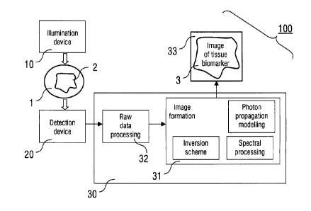
The essential components of the imaging method and imaging
device of the invention are illustrated in Figure 1. The im-
aging device 100 comprises the illumination device 10, the
detector device 20 and the reconstruction device 30. The il-
lumination device 10 is arranged for introducing illumination
light with a predetermined illumination pattern into the tar-
get tissue 1 including a distribution of biomarker 2 to be
imaged.
The illumination device 10 can be embodied by various light
sources as outlined below. The particular light source used
is selected in dependence on the requirements of the applica-
tion of the invention. Typically, the illumination device 10
comprises a light source, like a laser source or a light-
emitting diode (LD), and a light guiding device, like an op-
tical fibre transmitting the illumination light from the
light source to an output or a contact surface of the illumi-
nation device 10. Furthermore, the illumination device 10 is
preferably adapted for emitting at least one pulsed illumina-
tion pattern at several illumination wavelengths in the far
red or near-infrared wavelength range, i.e. preferably with
wavelengths above 630 nm.
The detector device 20 is adapted for sensing pressure sig-
nals from the target tissue 1, which are produced by the bio-
marker 2 in the target tissue 1 in response to the illumina-
tion. Typically, the detector device 20 is an acoustic detec-
tor device including at least one movable detector element
and/or a plurality (array) of detector elements. The latter
is known e.g. from ultrasonic imaging techniques. Alterna-
tively, the pressure signals can be collected with an optical
detector device immersed in a matching liquid or noncontactly
by sensing surface variations of the target tissue with opti-
cal means, e.g. by an optical interferometric set-up.
CA 02731409 2011-01-19
WO 2010/009747 PCT/EP2008/006142
The reconstruction device 30 generally is adapted for recon-
structing a quantitative tomographic image of the biomarker 2
in the target tissue 1. The reconstruction device 30 includes
5 at least one processor 31, which is adapted for calculating
the photon propagation model, implementing the spectral proc-
essing scheme and implementing the inversion scheme for pro-
viding the tomographic image. Additionally, a processor 32
adapted for raw data processing can be provided. Processors
10 31 and 32 can be implemented in a common circuitry. Alterna-
tively, the above functions of the processor 31 can be ful-
filled by a plurality of separate processor elements included
in the reconstruction device 30. Each processor can be imple-
mented with a microprocessor programmed for fulfilling the
15 particular function thereof.
The reconstruction device 30 is connected with an output de-
vice 33, which is adapted for providing the reconstructed to-
mographic image for further processing or application. In
20 particular, the output device 33 includes at least one of a
display device, like e.g. a display of a computer, a storage
device, like e.g. a storage medium in a computer, and a re-
cording device, like e.g. a printer.
25 The inventive imaging method is conducted with the imaging
device 100 of Figure 1 as outlined in the following. Illumi-
nation light is beamed upon the imaged region of interest in
tissue 1 using the illumination device 10. In the preferred
embodiment, a pulsed illumination at multiple wavelengths is
emitted at one or more positions, or angles, into the tissue
1 in the visible and/or near-infrared spectral range. This
ability to utilize light forming multiple projections (posi-
tions or angles) facilitates the provision of the imaging de-
vice as a handheld scanner, or intravascular scanner (see be-
low). Preferably, the duration of individual pulses lie in
the nanosecond range (i.e. below 100 ns, particularly pre-
CA 02731409 2011-01-19
26
WO 2010/009747 PCT/EP2008/006142
ferred below 10 ns) with an interval of at least 10 to
100 ps.
A broadband acoustic radiation is induced in tissue 1 follow-
ing the instantaneous temperature elevation caused by absorp-
tion of the above pulses in tissue 1. The magnitude of the
induced acoustic waves is proportional to the local light
fluence, optical absorption coefficient and thermoelastic
properties of the object.
The pressure signals (acoustic waves, in particular sound)
generated in response to the illumination is subsequently de-
tected by the detector device 20. The induced response is
collected by translating acoustic detector elements around
the tissue 1 or, alternatively, by placing an array of sta-
tionary detector elements in the vicinity of the tissue 1.
The optical absorption can be then reconstructed by back-
projecting the detected pressure signals into the virtual im-
aged volume or by various Radon transformations. When assum-
ing constant thermoelastic properties, selected tissue bio-
markers 2 can be quantitatively reconstructed based on a dis-
tinct absorption spectrum, by solving the composite problem
of photon propagation in the tissue 1, which is either wave-
length dependent or operates under a simplification that all
wavelengths considered propagate in a medium with the same or
similar optical properties.
Preprocessing of raw data with the processor 32 may include
basic filtering and denoising. The image formation processor
31 applies the inversion scheme appropriate for the particu-
lar illumination and detection configuration. It also applies
the spectral processing step responsible for differentiation
of biomarker from the background absorption in tissue 1 and
photon propagation modeling step intended for biomarker image
quantification. In the image formation phase, the order of
CA 02731409 2011-01-19
27
WO 2010/009747 PCT/EP2008/006142
the inversion, photon propagation modeling and spectral proc-
essing steps can be changed based on the particular implemen-
tation and application needs. As a result, an image 3 of the
tissue biomarker 2 of interest is produced.
The specific inversion scheme will differ in each case de-
pending on particular geometrical and physical characteris-
tics and spatial distribution of the detection elements used.
For example, in case a phased-array of acoustic detector ele-
ments is used, the images can be formed in the real-time by
incorporating into the inversion process simple ultrasound
beam forming algorithms.
The basic result of the inversion can be presented in a form
of image/s 3 representing local optical absorption coeffi-
cient of tissue 1.
2. Theoretical considerations
In practice, the detected opto-acoustic response does not di-
rectly provide the local absorption coefficient P(A) but the
reconstructed image of absorbed energy density 'k V)rather
represents a combination of the absorption coefficient P(a)
and optical fluence U k \
A) in the sample, i.e.
k
yl (A) = u k (a) k(\
(A) Due to strong optical attenuation and het-
erogeneity of biological tissues, the fluence cannot usually
be assumed constant throughout the region of interest. Yet,
only the absorption coefficient itself can provide the rele-
vant quantitative information on biomarker distribution.
Therefore, the ability to quantify the actual distribution of
the marker within the sample heavily relies on the initial
accuracy of reconstruction of the optical absorption map at
each wavelength that is to be deconvolved from the light flu-
ence distribution.
CA 02731409 2011-01-19
28
WO 2010/009747 PCT/EP2008/006142
Opto-acoustic inversion
A broadband acoustic radiation is induced in tissue following
the instantaneous temperature elevation caused by absorption
of short pulses of light energy in matter. The magnitude of
the induced acoustic waves is proportional to the local en-
ergy density, optical absorption coefficient, and thermoelas-
tic properties of the object. Their spectrum, in turn, is
mainly dependent upon the spatial frequency of energy deposi-
tion variations and duration of the emitted pulses. For pulse
durations in the ns range, a biologically relevant opto-
acoustic spectrum will be of ultrawideband nature with useful
information contained between several tens of kHz and several
tens of MHz, depending on size and spatial distribution of
optical absorption variations within the imaged object.
Preserving the correct shape of the detected response is im-
portant for the correct quantification of the resulting im-
ages. Since it may be difficult to effectively implement such
a broadband detection, a preferred way to restore the initial
tissue response is to deconvolve the recorded signal from the
frequency response of the detector. Alternatively, ultrawide-
band detection approaches may be used, such as optical inter-
ferometric approaches based on detection of surface movements
or mechanical oscillations in optically resonant elements,
e.g. Fabry-Perot films, ring resonators, or etalons.
The inversion is provided for reconstructing the e. g. three-
dimensional distribution of the biomarker from the collected
ultrasonic pressures P(F,t) by backprojecting the raw or
spectrally processed signals. The specific inversion scheme
will differ in each case depending on particular geometrical
and physical characteristics and spatial distribution of the
detection elements used. For example, in case a phased-array
of detector is used, the images can be formed in the real-
CA 02731409 2011-01-19
29
WO 2010/009747
PCT/EP2008/006142
time by using the simple ultrasound beam forming algorithms.
Generally, under conditions of heat confinement, i.e. when
the light energy pulse is short enough so that the thermal
diffusion is insignificant during the pulse, the spatio-
temporal dependence between opto-acoustically induced pres-
sure P07,0, absorbed energy density V(FJ) (in J/m13) and local
temperature elevation T(FJ) can be expressed as
2 1 32 p(F ,t ) a2T ,t ) /3av(F,t)
V p(F,t) _________________ = PmP (1)
V2 at2 at 2 u at
where vs, Pm, fl and C are the corresponding speed of sound,
mass density, isobaric volume expansion, and specific heat of
the medium, all are in general spatially and frequency de-
pendent.
In practice, the thermal confinement conditions are fulfilled
for excitation pulse durations less then 1 ps. When for in-
stance a point-shaped detector element of small diameter
(e.g. below 1 mm) is placed in the position at the
first
approximation it will sense an integrated pressure wave,
which is the solution of (1), namely,
, f3 ) d3F
,t)=--
4 at (2)
2-cC V ' F -
ti=tv,
The basic result of the inversion step can be presented in a
form of image/s representing local deposition of tissue bio-
marker.
CA 02731409 2011-01-19
WO 2010/009747 PCT/EP2008/006142
Photon propagation modelling
Tissue biomarker imaging is based on reconstruction of the
local optical absorption. However, as already mentioned, raw
opto-acoustic data do not represent the absorption coeffi-
cient directly but rather a combination of the absorption co-
efficient and optical fluence in the sample. In one of the
preferred embodiments of the current invention a quantitative
description of photon propagation (fluence rate) in tissue
10 based on known models of light propagation in tissues is
utilized in order to decompose optical absorption from flu-
ence.
The fluence throughout the region of interest can be calcu-
15 lated using light transport equation in diffuse media. One of
the preferred approximations, the diffusion equation, takes
the form
-DV2U(F)+ paU(F)= go (F) (3)
where D=1/[302:+/-lan is the diffusion coefficients of the me-
20 dium (Pa and P: are the absorption and reduced scattering
coefficient, respectively) and tic#) is the source distribu-
tion. For solving this differential equation, spatially-
varying optical properties of the medium P. and P; as well
as the spatial distribution and strength of the source ele-
25 ment on the right-hand-side have to be known. In complex ge-
ometries, the light diffusion can be calculated with Eq. (3)
by using finite-element method approaches.
It must be noted that the light diffusion approximation is
30 only valid in macroscopic objects with size many times larger
than the mean-free path (MFP) in tissue, normally correspond-
ing to objects larger than 10 mm. For smaller object,
mesoscopic approximations to light transport equation are ap-
plied. One of the most accurate yet computationally extensive
CA 02731409 2011-01-19
31
WO 2010/009747 PCT/EP2008/006142
approached in this case will be applying Monte-Carlo simula-
tions of light transport. Yet, some simple analytical ap-
proximations, like fermi function, can be effectively ap-
plied, as we have demonstrated in C. Vinegoni, C. Pitsouli,
D. Razansky, et al., NATURE METHODS 5(1), 2008.
Spectral processing
The current invention provides an efficient method for imag-
ing of molecular marker of interest by suppressing intrinsic
tissue contrast with the multi-wavelength approach. This
yields highly sensitive imaging of molecular marker distribu-
tion in tissue obtained by spectral matching of images ac-
quired at several different wavelengths. While the simplest
qualitative version of this operation can be achieved by im-
age subtraction at two wavelengths, three- and overall multi-
wavelength imaging will further suppress the background sig-
nals. This processing can occur in several stages, an effi-
cient one being the simultaneous inversion of spectral data
so that all information is accurately accounted for.
One preferred embodiment, which simplifies computation how-
ever will utilize the following general quantification for-
mula for the reconstructed amount (concentration) of the ma-
lecular marker of interest Ck on a per pixel basis:
4
ck = min v rwk (A)_ ck e (A)12
(4)
et t71,12"
where Ck is the reconstructed amount (concentration) of the
molecular marker of interest on a per pixel/voxel basis, N is
the total number of illuminating wavelengths, Vk(A) is the
reconstructed absorption in pixel/voxel k, Ck and e(A) are
the concentration and wavelength-dependent molar absorptivity
of the marker, respectively. We note that the wavelength-
CA 02731409 2011-01-19
32
WO 2010/009747
PCT/EP2008/006142
dependent absorption coefficient P(A) in each pixel/voxel
will be written in a conventional form as
(5)
m=1
where M is the total number of wavelength-dependent markers
and tissue chromophores considered in the reconstruction pro-
cedure. The procedure in Eq. (4) will then include minimiza-
tion over a set of concentrations cm (m=1, ..., M.
Alternatively, it can be assumed that every pixel k in the
opto-acoustic image may represent a combined contribution of
the molecular and other background tissue chromophores. For
every imaged wavelength A, this can be written in the form
of linear equation:
pak (A) = a A,fm (.1)cmõ,, + a,(2)c,k a2(.1,)c2 +
where All;(2) is the reconstructed wavelength-dependent absorp-
tion in pixel k, amw(2) and a1(2),a2(2),=== are the molar ex-
tinction spectra of the molecular marker and the background
chromophores, and ckmm and are
the corresponding con-
centrations. Using the measured absorption values and the
known spectra for the seven wavelength, the concentration
CAM of the molecular marker/s and the background chromopho-
res can be subsequently reconstructed from the above linear
equations on a per-pixel basis using linear regression
method.
The preferred methodology for achieving molecular marker dif-
ferentiation resides in including spectral information into
the inversion mode using a single-step or a two step method.
CA 02731409 2011-01-19
33
WO 2010/009747 PCT/EP2008/006142
The single-step method comprises inverting a tomographic equ-
ation simultaneously for the different wavelengths employed,
therefore simultaneously accounting for 1) the photon at-
tenuation as a function of depth (distance from the source),
2) the detection process and 3) the wavelength dependence of
the measurements.
The dual step method pre-processes the raw data using a spec-
tral matching or decomposition algorithm and then utilizes
one processed measurement as the input to an inversion code
that accounts only for 1) the photon attenuation as a func-
tion of depth (distance from the source) and 2) the detection
process. An alternative two-step method can be implemented by
reconstructed images at different wavelengths and then proc-
essing the resulting images on a pixel by pixel basis.
Image formation
An example of image formation process is shown in Figure 2.
The raw opto-acoustic recordings (step Si) are initially fil-
tered (step S2) and sent into the inversion scheme (step S3).
The resulting initial reconstructed image is then processed
in order to extract the geometry (boundary, inner or outer
surface) of the imaged target tissue (step S4). This is pro-
vided for the subsequent light distribution modelling (step
S5) in the tissue that is calculated using (step S6) a-priori
known pattern of the light incident upon the tissue. The
process is repeated in an iterative manner, where, at each
step, the inversion scheme normalizes the reconstructed image
by the calculated light distribution, which is also itera-
tively improving. For biomarker visualization (step S8), the
images are spectrally processed (step S7) for background ab-
sorption elimination.
CA 02731409 2011-01-19
34
WO 2010/009747 PCT/EP2008/006142
3. Further applications
There is a wealth of applications for the invented method.
While not limited only to the biomedical field, the applica-
tion of the technique to medical and biological imaging is an
important direction.
3.1 Biological imaging
Figure 3 schematically illustrates an embodiment whereas the
invention is used for imaging a part of a human proband 4 (e.
g. patient), e. g. the target tissue 1 comprising an organ 5.
The imaging device 100 comprises the illumination device 10,
the detector device 20 and the reconstruction device 30,
which are integrated into a common casing 34 and a measuring
head 40 being connected with the illumination and detector
devices 10, 20 via optical fibres and electrical cables.
The measuring head 40 can include separate components of ii-
lumination and sensor elements as illustrated below in Figure
4. Alternatively, the measuring head comprises an integral
measuring head unit including the illumination elements and
sensor elements in a common casing as outlined with further
details below (Figures 5, 6).
In a preferred embodiment an agent is injected intravenously
or locally to the proband 4, and targets areas or processes
of interest. The measuring head unit 40 is brought in contact
with the tissue so that illumination light is coupled into it
and pressure signals can be sensed. The collected pressure
data are processed and presented in the form of two- or
three-dimensional image on a monitor.
An application example includes the administration of fluo-
rescence emitting agents that are preferentially uptaken by
macrophages. Image of their absorption yields areas of in-
CA 02731409 2011-01-19
WO 2010/009747 PCT/EP2008/006142
creased inflammation as in the case of image atherosclerotic
plaque, in the carotids or other vessels. Similarly targeted
absorbing particles can show information on targeted mole-
cules such as peptides, receptors etc..
5
Figure 4 schematically illustrates the adjustment of the im-
aging device 100 relative to the target tissue 1 to be inves-
tigated. The illumination device comprises at least two illu-
mination elements 11, 12, which are arranged with a distance
10 relative to each other, e.g. 15 mm. The distance of the illu-
mination elements 11, 12 from the outer surface 6 (e.g. skin)
of the target tissue 1 comprises e.g. 20 mm. Alternatively,
the illumination elements 11, 12 can be arranged in contact
with the outer surface 6. The illumination elements 11, 12
15 comprise e.g. LED's with a predetermined emission character-
istic defining the projection direction towards the target
tissue 1. Alternatively, the illumination elements 11, 12
comprise the output ends of optical fibres being connected
with a laser source of the imaging device 100, e.g. in the
20 casing 34.
The detector device 20 comprises an array of detector ele-
ments 21 being embedded in a surface (contact surface) of the
detector device 20. The contact surface is adapted to be
25 brought into contact to the outer surface 6 of the target
tissue 1. The detector device 20 comprises e.g. a sound sen-
sor as it is known from conventional ultrasound imaging de-
vices.
30 Alternative embodiments, wherein the illumination and sensor
elements 11, 12, 21 are integrated within a common measuring
head unit 40 are illustrated in Figures 5 and 6. The measur-
ing head unit 40 comprises a casing body 41, into which the
illumination elements 11, 12 and the sensor elements 21, 22
35 are embedded. The illumination and sensor elements 11, 12,
21, 22 are integrated into the contact surface 42 of the
CA 02731409 2011-01-19
36
WO 2010/009747 PCT/EP2008/006142
measuring head unit 40. Elements 11, 12, 21, 22 are respec-
tively connected via optical fibres 13, 14 and electrical
wires 23, 24 with the associated parts of the illumination
and detector devices 10, 20 integrated in the casing 34 (see
e.g. Figure 3).
Figures 6A, 6B and 6C illustrate embodiments of the invention
being characterized by different distributions of the illumi-
nation and sensor elements 11, 12, 21, 22 in the contact sur-
face 42 of the measuring head unit 40. According to Figure
6A, line-shaped arrangements are provided with two outer rows
of illumination elements 11, 12 (e.g. LED's or output ends of
optical fibres) and a central row of sensor elements 21
(acoustical sound sensors). Figure 6B illustrates the oppo-
site geometry with a central row of illumination elements 11
and outer rows of sensor elements 21, 22. Figure 6C shows a
matrix arrangement of the elements 11, 12, 21, 22.
The illumination elements 11, 12 are configured for illumi-
nating the target tissue with at least one pulsed illumina-
tion pattern at several illumination wavelengths. As an exam-
ple, for providing two distinct wavelength ranges, a first
group of illumination elements 11 (e.g. indicated with "a")
is adapted for emitting illumination light with wavelengths
in the range of 610 nm to 650 nm, while a second group (indi-
cated with "b") is adapted for emitting wavelengths in the
range of 670 nm to 730 nm. For emitting a larger number of
wavelength ranges, a third or more groups are provided.
It is emphasized that the number of illumination and detector
elements shown in Figure 6 is selected for illustrative pro-
poses only. In practice, the number of elements can be se-
lected in dependence on the illumination and sound detection
requirements.
CA 02731409 2011-01-19
37
WO 2010/009747 PCT/EP2008/006142
Figures 7 to 9 illustrate further embodiments of the inven-
tion, wherein illumination and detector elements are used
that are separated from each other. As an example, imaging a
target tissue 1 including a blood vessel 7 is illustrated.
According to Figure 7, the illumination device 10 comprises a
light source 15 and an optical fibre 16, that is introduced
into the blood vessel 7 to the position of the target tissue
1 to be imaged. The detector device 20 comprises an array of
detector elements, which is adapted to be brought into con-
tact with the outer surface 6 of the target tissue 1, e.g.
skin of a human body. In operation, illumination light pat-
terns with distinct wavelength ranges are emitted via the op-
tical fibre 16 onto the inner surface of the blood vessel 1.
Pressure signals created by absorbing biomarkers within tis-
sue 1 are sensed with the detector device 20.
For example, if Cy5.5 dye is used for bio-marker targeting
with peak absorption at 670 nm, the multi-spectral illumina-
tion device might include diode-laser-based illumination de-
vice emitting light at 7 distinct wavelengths, namely, 610,
630, 650, 670, 690, 710, and 730 nm, that cover areas of both
high and low absorption of the dye to ease on the subsequent
multi-spectral processing and suppression of background ab-
sorption signals.
According to Figure 8, both the optical fibre 16 of the illu-
mination device 10 and the sensor element 25 of the detector
device 20 are arranged in the blood vessel in the target tis-
sue 1. Both components can be integrated in an endoscopic de-
vice (not shown).
According to Figure 9, the illumination elements 11, 12 of
the illumination device 10 are arranged outside the target
tissue, while the detector element 25 of the detector device
20 is provided in the vessel within the target tissue 1.
CA 02731409 2011-01-19
38
WO 2010/009747 PCT/EP2008/006142
Figure 10 illustrates a preferred application of the inven-
tive technique in biomedical imaging of mesoscopic-size ob-
jects and small animals, like mice or other rodents, flies,
fishes, worms, animal embryos. A container device 50 is pro-
vided, which comprises a tank 51 and holding elements 52, 54
which are adapted for positioning components of the imaging
device 100. The tank 51 contains a matching fluid 53, e.g.
water or oil. The object to be investigated (living mouse 8)
is positioned on the lower part 54 of the rod or plate shaped
holding element.
The illumination device 10 and the detector device 20 are
partially integrated in a casing 34 (see above, Figure 3),
that is are arranged outside the container device 50. The il-
lumination device 10 comprises a pulsed laser source whose
light is directed to the mouse 8 from two opposite directions
17,18 by e.g. using optical mirrors or fibers. The detector
device 20 comprises an array 26 of acoustic detector ele-
ments. The detector device 20 is arranged in the neighbour-
hood of the holding element 52 with the mouse 8. Advanta-
geously, there are no particular restrictions with regard to
the position of the detector device 20. The preferred loca-
tion however will be as close as possible to the object to
obtain measurements with high signal-to-noise ratios. For im-
plementing the above image reconstruction, it is only neces-
sary to have an information on the location of the array of
detector elements relative to the object (mouse 8).
The embodiment schematically illustrated in Figure 10 is not
restricted to the investigation of small animals. Alterna-
tively, other biological objects can be imaged, e.g. human
beings or larger animals or parts thereof. As an example,
tank 51 can be adapted for accommodating a part of a human
patient instead of the mouse 8.
CA 02731409 2015-07-14
39
3.2 Clinical imaging
Areas of preferred clinical applications include imaging of
cardiovascular disease, cancer, inflammation and neuro-
degenerative disease, to name a few examples. Imaging of
natural states such as growth and aging are also contem-
plated. As a particular advantage, the inventive near-field
imaging can be conducted without using a matching fluid be-
tween the near-field source device and the object to be in-
vestigated, thus essentially facilitating the clinical appli-
cations.
Another application example includes imaging of the effect of
treatment, via drugs, radiation or chemotherapy, by similarly
administrating absorbing particles in the body and monitoring
their relative update or targeting over time.
In other embodiments, the same detection can be achieved by
portable devices, or endoscopic devices inserted into body
cavities or through invasive procedures by operatively in-
serting the device into the tissue.
It is appreciated that certain features of the invention,
which are, for clarity, described in the context of separate
embodiments, may also be provided in combination in a single
embodiment. Conversely, various features of the invention,
which are, for brevity, described in the context of a single
embodiment, may also be provided separately or in any suit-
able sub-combination. Although the invention has been de-
scribed in conjunction with specific embodiments thereof, it
is evident that many alternatives, modifications and varia-
tions will be apparent to those skilled in the art. The scope
of the claims should not be limited by the preferred embodiments
set forth in the examples, but should be given the broadest
CA 02731409 2015-07-14
purposive construction consistent with the description as a
whole.
In addition, citation or identification of any reference in
5 this application shall not be construed as an admission that
such reference is available as prior art to the present
invention.