Note: Descriptions are shown in the official language in which they were submitted.
}' CA 02731657 2011-01-21
TECHNIQUE FOR DETECTING NEURODEGENERATIVE DISORDERS
FIELD OF THE INVENTION
[0001] The present invention relates to a technique for detecting
neurodegenerative
disorders such as Alzheimer's disease and Lewy body dementia, and preferred
embodiments include a detection program for images including a
neurodegenerative
disorder, a method for detecting images including said neurodegenerative
disorder
using a computer, and an apparatus for detecting images including a
neurodegenerative disorder.
BACKGROUND OF THE INVENTION
[0002] As a result of increases in the elderly population, it is expected that
the
number of patients with neurodegenerative disorders involving forms of
dementia
such as Alzheimer's disease will increase. Because these diseases progress
with
age and affect both the patient and their living environment, it is important
to
diagnose such cases at an early stage.
[0003] Such neurodegenerative disorders involving dementia are diagnosed by
applying the results of, for example, neuropsychological tests, including the
well-known Mini Mental Status Examination (hereinafter referred to as "MMSE"),
as
well as interviews and clinical findings, etc. to diagnostic criteria such as
DSM-III-R or
ICD-10. These diagnoses do not necessarily have high specificity. So these
diagnoses are combined with diagnostic imaging such as CT, MRI, or SPECT in
order to improve the diagnostic accuracy rate. However, even when diagnostic
imaging such as CT, MRI, or SPECT is involved, because the diagnostic accuracy
of
diagnostic imaging depends on the level of proficiency and the subjectivity of
the
radiography interpreter, there is a problem in that the results vary between
facilities
and examiners. Accordingly, there has been a desire for techniques allowing
for
neurodegenerative disorders to be detected in a more objective manner.
[0004] Recent studies have shown that in cases of neurodegenerative disorders
involving dementia, brain functions such as cerebral blood flow and glucose
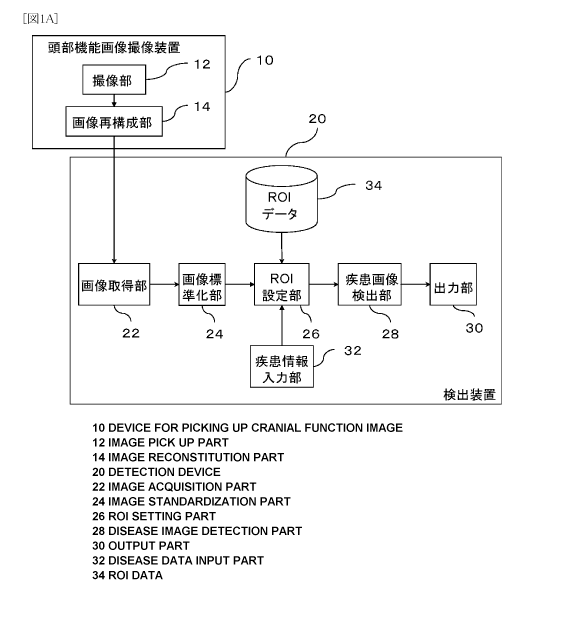
metabolic rate become partially deteriorated (See below Non-patent Document
1).
Using this knowledge, The below Non-patent Document 2 discloses a method of
using positron-emission tomography (hereinafter referred to as "PET") images
obtained by administering the glucose metabolism tracer
CA 02731657 2011-01-21
2
2-[18F]fluoro-2-deoxy-D-glucose (hereinafter referred to as "FDG") to conduct
comparisons with a healthy group, calculate the t-values of the pixel values
for each
pixel, and discriminate between Alzheimer's disease patients and healthy
subjects.
[0005] Further, International Publication No. 2007/063656 discloses methods
for
objectively detecting images based on neurodegenerative disorders at an early
stage
by calculating t-values or z-scores based on comparisons with a healthy
subject
database for pixels within a preset region of interest in a subject image, and
defining
a fixed threshold value for the obtained t-values or z-scores (Patent Document
1).
[Known Prior Art Documents]
[Non-patent Document 1] Kazunari Ishii, "Clinical application of positron
emission tomography for diagnosis of dementia", Annals. of Nuclear Medicine,
2002,
16(8), p.515-525
[Non-patent Document 2] K. Herholz et al., "Discrimination between
Alzheimer dementia and controls by automated analysis of multicenter FDG PET",
Neurolmage, 2002, 17, p.302-316
[Patent Document 1] International Publication No. 2007/063656
DISCLOSURE OF THE INVENTION
PROBLEMS TO BE SOLVED BY THE INVENTION
[0006] As described above, for neurodegenerative disorders, there is a need
for
techniques that are capable of objectively detecting early-stage lesions. As
disclosed
by Herholz, etc., by measuring local deteriorations in brain functions such as
glucose
metabolism, etc., it is possible to detect Alzheimer's disease and other
neurodegenerative disorders. However, in order to detect Alzheimer's disease
and
other neurodegenerative disorders using diagnostic imaging, it is necessary to
define
conditions for discriminating the neurodegenerative disorders from other
conditions
to perform detection. Moreover, because this method is a method based on
comparisons with healthy subject data, for implementation, it is necessary to
prepare
a database for healthy subjects.
[0007] According to the technique disclosed in International Publication No.
2007/063656, it is possible to discriminate images of Alzheimer's disease and
other
neurodegenerative disorders from other conditions to perform an objective
determination. However, as with the above method disclosed by Herholz, etc.,
CA 02731657 2011-01-21
3
because this method is also a method based on comparisons with healthy subject
data, it is necessary to prepare a database for healthy subjects to practice
the
technique.
[0008] However, most subjects undergoing imaging tests in hospitals, etc.,
generally
present with some kind of lesions, and therefore, it is not easy to create an
image
database of healthy subjects. Consequently, it is preferable if there is no
need to use
a database for healthy subjects for distinguishing and determining images of
neurodegenerative disorders patients from images of healthy subjects
objectively
and accurately, but such technique has not been found yet.
[0009] The present invention has been devised based on the above
considerations,
and the objective is to provide a technique for detecting neurodegenerative
disorders
such as Alzheimer's disease and Lewy body dementia, etc. based only on brain
functional images of the subject and without using a database for healthy
subjects.
SOLUTIONS TO THE PROBLEMS
[0010] As a result of numerous studies, the present inventors discovered that
in a
brain functional image, there is a disease-specific region where the
probability of
functional deterioration is high (hereinafter sometimes referred to as
"functionally
deteriorated site") as well as a disease-specific region where the probability
of
functional preservation is high even in cases of diseases (hereinafter
sometimes
referred to as "functionally preserved site"), and that it is possible to
objectively
determine images in which a neurodegenerative disorder is believed to exist
without
using a healthy subject database by comparing the respective pixel values of
the
functionally deteriorated site and the functionally preserved site.
[0011] Based on this discovery, it becomes possible to determine a
neurodegenerative disorder using the same brain functional images of the same
subject. In other words, it becomes possible to determine images containing a
neurodegenerative disorder using only images derived from the subject, without
the
need to prepare a database for healthy subjects.
[Demonstration 1: Example of determining images of Alzheimer's disease
patients]
[0012] As one example for demonstrating that neurodegenerative disorders can
be
detected using the above technique, images derived from Alzheimer's disease
patients and images derived from healthy subjects were used to obtain the
sensitivity,
CA 02731657 2011-01-21
4
specificity, and diagnostic accuracy rate of the technique according to the
present
invention.
(Setting of regions of interest)
[0013] To define the regions of interest, 1231-IMP-administered brain SPECT
images
of 20 Alzheimer's disease patients (mean age: 73.6 4.6 years old) and 15
healthy
subjects (mean age: 60.5 7.1 years old) were used (hereinafter, the groups are
respectively referred to as the "disease group" and the "healthy group").
[0014] For each image, software called as iNEUROSTAT (produced by Nihon
Medi-Physics Co., Ltd.) was used to perform anatomic standardization. Then the
pixel values of each image were subtracted by the respective mean values of
all pixel
values in each image to normalize the pixel values (hereinafter collectively
referred to
as "normalized images").
[0015] Using these images, an inter-group comparison was conducted between the
disease group and the healthy group, and z-scores representing decreases in
pixel
value were obtained for each pixel. The obtained z-scores were put on
corresponding pixels. And clusters representing regions with decreased pixel
values
were extracted by using the threshold value of 3. From the obtained clusters,
the
largest cluster was selected and defined as functionally deteriorated region
1.
Similarly, z-scores representing increases were obtained, and a threshold
value of 3
was employed to extract clusters representing regions with increased pixel
values.
From the obtained clusters, the largest cluster was selected and defined as
functionally preserved region 1.
[0016] Separately, for brain template stored in the iNEUROSTAT program (Fig.
5),
segments indicating functionally deteriorated regions and segments indicating
functionally preserved regions were selected by comparing the template with
the
normalized images. Each selected segment was compared with both said
functionally deteriorated region 1 and said functionally preserved region 1.
And
regions with substantive commonality were extracted and used as region-of-
interest
data corresponding to the functionally deteriorated site and the functionally
preserved site, respectively (Fig. 9).
(Evaluation of sensitivity, specificity, and diagnostic accuracy rate of image
detection
for Alzheimer's disease patients)
CA 02731657 2011-01-21
[0017] To evaluate the sensitivity, specificity, and diagnostic accuracy rate,
the
1231-IMP-administered brain SPECT images of 17 Alzheimer's disease patients
(mean age: 60.1 8.2 years old) and 17 healthy subjects (mean age: 61.1 7.3
years
old) were used. For each image, the software iNEUROSTAT was applied for
anatomic standardization, and the region-of-interest data obtained above were
applied to define regions of interest for the functionally deteriorated site
and the
functionally preserved site respectively. For each image, a t-test was
performed
between the functionally deteriorated site and the functionally preserved site
for pixel
values with a risk rate of 5%. Images in which the mean pixel value of the
functionally
preserved site was determined to be significantly greater than the mean pixel
value
of the functionally deteriorated site by the t-test were determined as images
of
Alzheimer's disease patients, and the other images were determined as healthy
subject images. Based on these results, the sensitivity, specificity, and
diagnostic
accuracy rate were obtained using heretofore known techniques.
[0018] The sensitivity, specificity, and diagnostic accuracy rate were 82.4%,
88.2%,
and 85.3%, respectively. Each shows high value. Based on the above results, it
was
confirmed that the technique according to the present invention can detect
patient
images derived from Alzheimer's disease objectively and with high accuracy.
[Demonstration 2: Example of detection of Lewy body dementia]
[0019] As yet another example for demonstrating that neurodegenerative
disorders
can be detected using techniques according to the present invention, images
derived
from Lewy body dementia patients and images derived from healthy subjects were
used to seek the sensitivity, specificity, and diagnostic accuracy rate of
detection of
the disease.
(Settings of regions of interest)
[0020] To define the regions of interest, 1231-IMP-administered brain SPECT
images
of 15 Lewy body dementia patients (mean age: 79.0 6.6 years old) were used.
[0021] For each image, the program iNEUROSTAT (produced by Nihon
Medi-Physics Co., Ltd.) was used to perform anatomic standardization. These
standardized images were compared with the brain template (Fig. 5) stored in
the
iNEUROSTAT software. Based on the comparison, the occipital lobe was selected
as
a segment indicating a functionally deteriorated site and used as region-of-
interest
data. Similarly, the sensorimotor area was selected as a segment indicating a
CA 02731657 2011-01-21
6
functionally preserved site and used as region-of-interest data.
(Evaluation of sensitivity, specificity, and diagnostic accuracy rate of image
detection
for Lewy body dementia patients)
[0022] To evaluate the sensitivity, specificity, and diagnostic accuracy rate,
the
1231-IMP-administered brain SPECT images of 15 Lewy body dementia patients
(mean age: 79.0 6.6 years old) and 15 healthy subjects (mean age: 60.5 7.1
years
old) were used (hereinafter, the groups are respectively referred to as the
"DLB
disease group" and the "healthy group"). For each image, the iNEUROSTAT
software
was used for anatomic standardization, and the region-of-interest data
obtained
above were applied to define regions of interest in the functionally
deteriorated site
and the functionally preserved site respectively. For each image, a t-test was
performed between the functionally deteriorated site and the functionally
preserved
site for pixel values with a risk rate of 5%. Images in which the mean pixel
value of
the functionally preserved site was determined to be significantly greater
than the
mean pixel value of the functionally deteriorated site by the t-test were
determined as
images of Lewy body dementia, and the other images were determined as images
of
healthy subject. Based on these results, the sensitivity, specificity, and
diagnostic
accuracy rate were obtained using heretofore known techniques.
[0023] The sensitivity, specificity, and diagnostic accuracy rate were 73.3%,
86.7%,
and 80.0%, respectively. Each value is high. Based on the above results, it
was
confirmed that the technique according to the present invention enables to
detect
Lewy body dementia objectively and with high accuracy.
[0024] As can be understood from the above two examples,
some neurodegenerative disorders show a region where the possibility of
functional
deterioration is high and a region where the possibility of functional
preservation is
high, in brain functional images. And in cases of such diseases, it is
possible to
detect the disease by comparing brain functional images between these regions.
The
Demonstrations introduced in the present specification are limited to
Alzheimer's
disease and Lewy body dementia, but it is clear that the present invention can
be
applied to various neurodegenerative disorders presenting with a functionally
deteriorated site and a functionally preserved site in brain functional
images.
Examples of diseases with a very high potential for applicability include
Alzheimer's
disease, Lewy body dementia, frontotemporal dementia, and progressive
supranuclear palsy, etc.
CA 02731657 2011-01-21
7
[0025] One important aspect is that, in order to identify a disease-specific
functionally
deteriorated site and a functionally preserved site, although there are cases
in which
it is preferable to have healthy subject data, once those sites are
identified, such
healthy subject data become unnecessary, and it becomes possible to detect a
disease using only images derived from a subject. Operations to identify these
sites
do not necessarily have to be performed at a general hospital and may be
performed
at a specialized research institute. Once a functionally deteriorated site and
a
functionally preserved site specific to a certain disease are identified, and
if that data
becomes available for use, owners of a device according to the present
invention
should immediately be able to begin operations to determine related diseases
by
using the data, without having to construct a healthy subject database as
before.
[0026] In this way, the present invention makes the operations required for
determining an existence of neurodegenerative disorders extremely easy
compared
to before, and can contribute great advantages in the fields of medical
service and
image analysis programs.
[0027] In the present specification, data on disease-specific functionally
deteriorated
sites and functionally preserved sites may be referred to as region-of-
interest data.
As can be seen in the above explanations, the region-of-interest data are used
for
extracting regions for performing inter-region comparisons. In cases of
Alzheimer's
disease, the functionally deteriorated site and the functionally preserved
site can be
defined as the parietal lobe and the sensorimotor area, respectively. In cases
of Lewy
body dementia, the functionally deteriorated site and the functionally
preserved site
can be defined as the occipital lobe and the sensorimotor area, respectively.
As
described above, once region-of-interest data are obtained in one facility,
the need
for other facilities to perform the same experiments is greatly reduced and
that data
can be also be used at other facilities.
[0028] Region-of-interest data can be obtained through various techniques. For
example, such data can be obtained based on the results of an inter-group
comparison between brain functional images derived from multiple subjects
affected
by a neurodegenerative disorder (hereinafter referred to as the "disease
group") and
brain functional images derived from multiple healthy subjects (hereinafter
referred to
as the "healthy group"). By using this technique, it is possible to define
regions of
interest in sites that statistically show functional deterioration and sites
that
statistically show functional preservation in cases of the subject disease.
For the
inter-group comparison, any heretofore known technique, such as techniques
described in the literature (International Publication No. 2007/063656), for
example,
CA 02731657 2011-01-21
8
can be used. Here, It is preferable to normalize each of the images included
in the
disease group and the healthy subject group using the mean of all pixel values
in
each image, and the use them. By performing normalization operations, the
pixel
values of the functionally preserved site in the disease group become
relatively
higher, making extraction based on the inter-group comparison easier.
[0029] It is possible to obtain region-of-interest data from a different way,
which uses
only brain functional images derived from patients affected by a
neurodegenerative
disorder. Specifically, it is possible to use a technique of defining a
threshold value for
the pixel values in a brain functional image to extract a functionally
deteriorated site
and a functionally preserved site for use as region-of-interest data.
[0030] It is also possible to obtain region-of-interest data from further
different way,
which uses a predetermined template for a standard brain. For example, it is
possible
to select segments corresponding to a disease-specific functionally
deteriorated site
and functionally preserved site from the various region data that have been
defined
in the Talairach brain atlas or other brain atlases, etc., for use as region-
of-interest
data.
[0031] Although the techniques for defining region-of-interest data
exemplified above
may each be used independently, it is also possible to use two or more
techniques in
combination. For example, it is possible to use region-of-interest data
defined by two
different techniques to perform region extraction for each, and define regions
commonly extracted through both techniques as region-of-interest data. By
using
region-of-interest data obtained by combining two or more techniques, it can
be
expected that the accuracy of disease detection will be further improved.
[0032] In order for the region-of-interest data to have versatility, it is
preferable if the
region-of-interest data presents a functionally deteriorated site and a
functionally
preserved site in an anatomically standardized brain image (standard brain).
Consequently, in a preferred embodiment, analysis is conducted after the brain
functional images of a subject undergoing disease detection also undergo
anatomic
standardization. Alternatively, the region-of-interest data may be modified to
match
the brain functional images of the subject for use in analysis. For anatomic
standardization, a heretofore known technique described in the literature
(Minoshima
S. et al., J. Nucl. Med., 1994, 35, p.1528-37, or Friston K. J. et al., Human
Brain
Mapping, 1995, 2, p.189-210), for example, may be used.
[0033] In the present specification, regions for actually comparing image data
to
CA 02731657 2011-01-21
9
perform disease determination may be referred to as regions of interest. The
regions
of interest may be regions that are automatically extracted using the above
region-of-interest data, but further adjustments may be made either
automatically or
manually.
[0034] According to one embodiment, comparisons between regions of interest
may
be performed by comparing the pixel values of image data contained in each
region
of interest. Generally, blood flow and glucose metabolism of a subject
presenting with
a disease decrease depending on regions. Such regions appear darker than other
regions in brain functional images obtained through SPECT or PET, etc.
Consequently, if at least a certain number of pixel values of the regions of
interest of
a functionally deteriorated site are smaller than the pixel values of the
regions of
interest of a functionally preserved site, it is possible to infer the
presence of a
disease. However, because there may be errors in determination due to noise,
etc.
when performing a simple comparison of mean values, etc., it is preferable to
perform a comparison using a significance test such as a t-test, etc. In such
configuration, it is preferable to use a configuration for determining whether
the mean
pixel value of the regions of interest of the functionally preserved site is
significantly
greater than the mean pixel value of the regions of interest of the
functionally
deteriorated site, rather than using a configuration for simply determining
the
presence or absence of a significant difference. By using such a
configuration, the
rate of errors in judgment can be roughly halved.
[0035] Embodiments of the present invention include neurodegenerative
disorders
image detecting apparatuss such as the following. This device comprises a
region-of-interest defining section that defines regions of interest in a
functionally
deteriorated site where functions could be specifically deteriorated in a
neurodegenerative disorder to be detected, and a functionally preserved site
where
functions could be preserved even in said neurodegenerative disorder to be
detected,
respectively, in said brain functional image; and a disease-image
determination
section that is configured to conduct a significance test using pixel values
within said
regions of interest defined for each of said functionally deteriorated site
and said
functionally preserved site, and determine that said neurodegenerative
disorder to be
detected is present if the mean pixel value of said regions of interest in
said
functionally preserved site is significantly greater than the mean pixel value
of said
regions of interest in said functionally deteriorated site.
[0036] Other embodiments of the present invention include computer programs
such
as the following. This program is capable of handling image data composing
brain
CA 02731657 2011-01-21
functional images and is a computer program for operating a computer equipped
with
a storage means and a CPU, and when the program is executed by said CPU, the
program operates the computer as: a first memory means for storing image data
corresponding to a first region of a brain functional image; a second memory
means
for storing image data corresponding to a second region different from said
first
region in said brain functional image; and a neurodegenerative disorders
detection
means that determines neurodegenerative disorders based on a comparison of the
image data stored in said first memory means and the image data stored in said
second memory means.
[0037] In a preferred embodiment, the above first and second memory means may
be memory that is logically formed on a physical medium by the program. The
program may be configured so that in either one of the first and second memory
means, image data of a site where functions may specifically deteriorate in
cases of
the neurodegenerative disorders in a disease to be detected are saved, and in
the
other means, image data of a site where functions may be preserved even in
cases
of the neurodegenerative disorders are saved. In other words, in a preferred
embodiment, the above first and second regions are regions of interest that
have
been respectively defined for the above functionally deteriorated site and the
functionally preserved site.
[0038] Other embodiments of the present invention include methods for
detecting
neurodegenerative disorders images such as the following. This method
comprises:
a step for defining regions of interest in a functionally deteriorated site
where
functions could be specifically deteriorated in a neurodegenerative disorder
to be
detected, and a functionally preserved site where functions could be preserved
even
in said neurodegenerative disorder to be detected, respectively, in said brain
functional image; and a disease-image determining step for performing a
significance
test using pixel values of regions of interest defined in each of said
functionally
deteriorated site and said functionally preserved site, and determining that
said
neurodegenerative disorder to be detected exists if the mean pixel value of
said
regions of interest in said functionally preserved site is significantly
greater than the
mean pixel value of said regions of interest in said functionally deteriorated
site.
[0039] The various embodiments of the present invention include those that
perform
anatomic standardization of the brain functional images. In this way, regions
of
interest can be defined for standardized brain functional images. Conversely,
it is
also possible to use a technique of using inverse transformation to transform
region-of-interest data defined with a standard brain into the form of the
brain
CA 02731657 2011-01-21
11
functional images of the subject, and superimposing the transformed
region-of-interest data on the brain functional images of the subject to
define the
regions of interest on the brain functional images of the individual subject.
[0040] As described above, it is preferable to configure the data to be
automatically
called in response to information such as disease name, etc, because the
region-of-interest data are disease-specific.
[0041] Several preferred embodiments of the present invention are specified in
the
attached Claims. However, embodiments of the present invention are not limited
to
those explicitly described in the Claims or the Description and Drawings, and
the
present invention may take on various configurations without deviating from
the sprits
of the present invention. The present invention includes in its scope any new
and
beneficial configurations that may be suggested in these documents, regardless
of
whether such configurations are explicitly disclosed in the Claims or the
Description
and Drawings of the present application.
BEST MODE FOR CARRYING OUT THE INVENTION
[0042] Embodiments of techniques according to the present invention for
detecting
images containing a neurodegenerative disorder will be described in detail
with
reference to the drawings. It should be noted that the example described below
only
provides a description of an example believed to be the most optimal, and the
embodiments of the present invention is not limited in any way by these
descriptions.
[0043] Fig. 1A is a diagram showing the configuration of an optimal mode for a
neurodegenerative disorders image detecting apparatus 20 according to the
present
invention. Fig. 2 is a diagram showing the operations of the optimal mode of
the
neurodegenerative disorders image detecting apparatus 20 according to the
present
invention. The neurodegenerative disorders image detecting apparatus 20
according
to the present invention may be configured as a computer into which a
neurodegenerative disorder image detection program 100 (described below) has
been read. As shown in Fig. 1A, the neurodegenerative disorders image
detecting
apparatus 20 according to the present invention functionally comprises: an
image
acquirer 22 that acquires a brain functional image from a brain function
imaging
apparatus 10 such as a SPECT apparatus, etc.; an image standardization section
24
that is configured to perform anatomic standardization of the acquired brain
functional image; a region-of-interest defining section (described as "ROI
defining
CA 02731657 2011-01-21
12
section" in Fig. 1A) 26 that defines regions of interest in the standardized
brain
functional image; a disease-image determination section 28 that determines
whether
said brain functional image corresponds to an image of the disease to be
detected;
and an output section 30 that outputs the detection results.
[0044] Fig. 1 B is an explanatory diagram of the hardware configuration of the
image
detecting apparatus 20. As shown in Fig. 1 B, the image detecting apparatus 20
comprises a CPU 40, a main memory 42, an auxiliary storage unit 44, and,
preferably,
a communication unit 46, etc. In other words, in terms of hardware, the image
detecting apparatus 20 may have the same configuration as a general-purpose
computer. In the auxiliary storage unit 44, which may be a hard disk, etc., a
program
for operating apparatus 20 as a neurodegenerative disorders image detecting
apparatus is stored, and when this program is executed by the CPU 40, the
functions
required for detecting neurodegenerative disorders are provided. In other
words, part
or all of the functions of the image acquirer 22, the image standardization
section 24,
the ROI defining section 26, and the disease-image determination section 28,
etc.,
are realized using software processing.
[0045] In a preferred mode, an auxiliary storage unit 50, a display 52, and
user
interfaces 54-58, etc. are connected to the image detecting apparatus 20 via
external
interfaces 48a-48e. The user interfaces 54-58 may be, for example, a touch
panel 54,
a keyboard 56, and a mouse 58, etc. The auxiliary storage unit 50 may be, for
example, an optical disk drive such as a DVD-ROM drive, etc. According to one
embodiment, the touch panel 54 is configured by being integrated in the
display 52.
In a preferred embodiment, the brain function imaging apparatus 10 is
connected to
the image detecting apparatus 20 via the communication unit 46, and it is
possible to
download images captured with the brain function imaging apparatus 10 into the
auxiliary storage unit 44 via a network.
[0046] The embodiment will now be described mainly with reference to Fig. 1A.
Various devices that are capable of acquiring brain functional images may be
used
as the cranial functional imaging apparatus 10. Specific examples include a
SPECT
apparatus, a PET device, an MRI device, or a CT device. The brain function
imaging
apparatus 10 includes an imager 12 and an image reconstructor 14. The imager
12
acquires the brain functional image data of a subject. The image reconstructor
14
performs image reconstruction processes for the acquired brain functional
image
data to generate a brain functional image. Using the example of a SPECT
apparatus,
the imager 12 acquires projection data from a subject who has been
administered
radiopharmaceutical agents such as 99mTc HMPAO and 1231 IMP. These projection
CA 02731657 2011-01-21
13
data correspond to the brain functional image data of the present embodiment.
The
image reconstructor 14 performs the necessary reconstruction processes for the
acquired projection data and generates a series of tomographic images. This
series
of tomographic images corresponds to the brain functional images of the
present
embodiment. The image reconstruction can be performed using a heretofore known
technique.
[0047] The image acquirer 22 acquires the brain functional image generated in
the
image reconstructor 14 (step S1). The brain functional image is saved in a
computer-readable data format such as, for example, DICOM. In order to
transfer the
saved brain functional image data to the image detecting apparatus 20, the
data may
be stored in a storage medium such as a DVD, etc. in the brain function
imaging
apparatus 10, and such disk may be inserted into a reading device (the
auxiliary
storage unit 50). Preferably, the data may be directly transferred to the
auxiliary
storage unit 44 via the communication unit 46 as computer data signals
conveyed on
carrier waves. As described above, the auxiliary storage unit 44 may be a hard
disk
or a unit of flash memory, etc. In a preferred embodiment, the image acquirer
22
reads brain functional image data stored in the auxiliary storage unit 44 or
the
auxiliary storage unit 50 and stores the data in a logical memory region
formed on
the main memory 42 or the auxiliary storage unit 44 using software. The stored
data
are provided for processing at the next processing block (the image
standardization
section 24).
[0048] The image standardization section 24 performs an anatomic
standardization
process on the brain functional image acquired by the image acquirer 22, and
transforms the brain functional image into a standard brain (step S2). This
anatomic
standardization process may be performed using a heretofore known technique
described in the technique (Minoshima S. et al., J. Nucl. Med., 1994, 35,
p.1528-37,
or Friston K. J. et al., Human Brain Mapping, 1995, 2, p.189-210), for
example. The
transformed brain image data are stored in a logical memory region formed on
the
main memory 42 or the auxiliary storage unit 44 using software. In some
embodiments, the transformed brain image data may be displayed on the display
52.
[0049] The region-of-interest defining section 26 defines regions of interest
in a site
where functional deterioration may occur in cases of the disease to be
detected
(functionally deteriorated site), and a site with a high possibility of
functional
preservation (functionally preserved site), respectively, in the brain
functional image
transformed to the standard brain (step S5). In a preferred mode, the
region-of-interest defining section 26 is linked to both a disease-information
input
CA 02731657 2011-01-21
14
section 32 and a region-of-interest database (described as "ROI data" in Fig.
1A) 34
stored in the auxiliary storage unit 44 or 55, etc.
[0050] The disease-information input section 32 is capable of receiving inputs
from at
least one of the user interfaces 54-58, and receives inputs of information on
the
disease to be detected, most typically the disease name (step S3). As long as
the
disease information is information that can be used for selecting region-of-
interest
data from said database, there are no particular limitations to the disease
information.
Typically, the information may be the common name of the neurodegenerative
disorders, but abbreviations or typical symptoms, etc. may be used. For the
input of
disease information, it is also possible to combine heretofore known
techniques
related to menu selection, such as displaying disease information in a pull-
down
menu and making the subject disease selectable, etc.
[0051] In a region-of-interest database 34, data on sites that should be
subject to
examination for each neurodegenerative disorders-in other words, data on sites
where functions may deteriorate in cases of the disease (functionally
deteriorated
site) and sites with a high possibility of functional preservation even in
cases of the
disease (functionally preserved site) (i.e., region-of-interest data)-are
stored and
associated with disease information. Based on the input disease information,
the
region-of-interest defining section 26 reads the region-of-interest data
corresponding
to the disease to be detected (step S4), and defines regions corresponding to
the
region-of-interest data in said brain functional image that has been
transformed to
the standard brain as regions of interest (step S5). For the region-of-
interest data, It
is possible to employ data formed using various techniques may be used.
Techniques for forming region-of-interest data will be described later.
[0052] In some embodiments, the defining of regions of interest may be
configured to
be performed manually instead of through automatic defining using region-of-
interest
data. It may also be possible for the operator to automatically or manually
adjust
automatically define regions of interest. For example, a configuration may be
provided in which the operator uses a touch panel 54 or a mouse 56, etc. to
select
desired regions on a brain image displayed on the display 52 to determine
regions of
interest. Further, a configuration may be provided in which a brain atlas is
overlapped
and displayed over a brain functional image of a subject transformed to a
standard
brain, and desired regions can be selected using the user interfaces 54-58.
[0053] The region-of-interest defining section 26 stores region-of-interest
data
defined respectively for the functionally deteriorated site and the
functionally
CA 02731657 2011-01-21
preserved site in different logical memories (logical memory regions formed on
the
main memory 42 and the auxiliary storage unit 44 using software). The stored
image
data are provided for the following processes.
[0054] The disease-image determination section 28 performs a process for
detecting
images containing a neurodegenerative disorder (step S6). Fig. 3 is a flow
chart
showing processes in the disease-image detection process. The disease-image
determination section 28 reads the data that was stored in each of the logical
memories in step S5 and conducts a significance test of the pixel values
between the
pixels in the functionally deteriorated site and the pixels in the
functionally preserved
site (step S11). The significance test may be performed using a heretofore
known
technique. In an optimal mode, a t-test may be used for the significance test.
As a
result of this significance test, if it is determined that the mean pixel
value of the
functionally preserved site is significantly greater than the mean pixel value
of the
functionally deteriorated site ("Yes" in step S12), the image is determined to
be an
image in which the neurodegenerative disorders to be detected may exist (step
S13).
On the other hand, if the mean pixel value of the functionally preserved site
is not
significantly greater than the mean pixel value of the functionally
deteriorated site
("No" in step S12), the image is not determined to be an image containing the
neurodegenerative disorders to be detected (step S14). The disease-image
determination section 28 stores the necessary data, such as the results of
determinations, in a logical memory, and the disease-image detection process
(step
S6) is completed.
[0055] The output section 30 outputs the results of the detection process
performed
by the disease-image determination section 28 to the display 52 via a display
interface 48b (step S7). Outputs may also be made to other output devices,
such as
a printer or a sound generator, etc. The format of the output does not
necessarily
have to be limited, and may be a format in which the t-value or the detection
result (or
both) is displayed on the image, or a format in which a color is allocated to
distinguish
the image from others if the mean pixel value of the functionally preserved
site is
determined to be significantly greater than the mean pixel value of the
functionally
deteriorated site.
[0056] As described above, it is possible to employ various types of data
obtained by
different techniques for the region-of-interest data to be stored in the
region-of-interest database. Some examples of techniques for defining
region-of-interest data will be explained below, which include a technique
based on
disease images, a technique using a template defined on a standard brain, and
a
CA 02731657 2011-01-21
16
technique based on an inter-group comparison between a disease group and a
healthy subject group.
[0057] First, a case in which region-of-interest data are defined based on
disease
images will be described. In this example, first, an image derived from a
patient
affected by the neurodegenerative disorders to be detected (e.g., Alzheimer's
disease) is acquired. For the disease image, it is preferable to use an image
that has
been preliminarily transformed to a standard brain. The disease image may also
be
obtained by averaging the pixel value of each pixel in images derived from
multiple
patients that have been transformed to a standard brain. Or it is possible to
use
representative examples exhibiting typical image patterns for each disease.
Then, for
the acquired disease image, the functionally deteriorated site and the
functionally
preserved site are both extracted using the threshold value technique. These
site-data will be used as region-of-interest data. Examples of region-of-
interest data
extracted according to the present technique are shown in Fig. 4(a) and Fig.
4(b). Fig.
4(a) and Fig. 4(b) respectively show the functionally preserved site and the
functionally deteriorated site in a case in which the disease to be detected
is
Alzheimer's disease. Each region of interest corresponding to the functionally
deteriorated site and the functionally preserved site may be defined using the
same
disease image, but they may each be defined using different images.
[0058] Next, the technique of using a brain template defined on a standard
brain will
be described. In this technique, a brain template that has been anatomically
defined
on a standard brain is compared with a disease image, and regions (segments)
corresponding to the functionally deteriorated site and the functionally
preserved site
are selected. Fig. 5(a)-(g) shows one example of a brain template. It is
possible to
compare this brain template to the image undergoing detection, select segments
corresponding to the functionally deteriorated site and the functionally
preserved site
in the disease to be detected, and use these as region-of-interest data
corresponding
to the disease.
[0059] Next, the technique based on an inter-group comparison between a
disease
group and a healthy subject group will be described. In this technique, first,
a plurality
of disease images and a plurality of healthy subject images are acquired.
Then, an
anatomic standardization is performed for each acquired image, and then an
inter-group comparison is performed for each pixel to obtain values that will
act as
indices of the amount of change in the pixel values, such as t-values or z-
scores
(hereinafter referred to as "index values"). The corresponding index values
are
displayed on each pixel on the standard brain, and using the threshold value
CA 02731657 2011-01-21
17
technique, the functionally deteriorated site and the functionally preserved
site are
both extracted and used as region-of-interest data.
[0060] The above-mentioned techniques may be used independently, or may be
used with two or more of them in combination. For example, regions of interest
extracted through each of the above techniques may be displayed in overlapped
manner, and the common areas may be extracted and may be used as
region-of-interest data. By combining two or more techniques in such manner,
it
becomes possible to further improve the accuracy of detection of disease
images.
[0061] Next, a neurodegenerative disorder detection program according to the
present invention will be described. Fig. 6 is a diagram showing a
configuration
according to an optimal mode of the neurodegenerative disorders image
detection
program 100 according to the present invention, along with a storage medium
200.
[0062] In a preferred mode, the neurodegenerative disorders image detection
program 100 according to the present invention comprises a main module 110
that
controls the processes, an image-data acquisition module 120, an image
standardization module 130, a disease-information input module 140, a
region-of-interest defining module 150 (described as "ROI defining module" in
Fig. 6),
a disease-image detection module 160, and an output module 170.
[0063] In a preferred embodiment, the neurodegenerative disorders image
detection
program 100 is provided by being stored in the storage medium 200. Examples of
the
storage medium 200 include a flexible disk, a hard disk, a CD-ROM, a DVD, or a
semiconductor memory, etc. By inserting the storage medium 200 that stores the
neurodegenerative disorders image detection program 100 into a reading device
(e.g., the auxiliary storage unit 50 of Fig. 1B) built into a computer, the
neurodegenerative disorders image detection program 100 becomes available for
access by the computer, and using this program 100, it becomes possible for
the
computer to operate as the neurodegenerative disorders detecting apparatus 20
described above. Of course, the program 100 may be installed and used in a
high-speed memory unit (e.g., the auxiliary storage unit 44 of Fig. 1B), such
as a
hard disk, etc. The neurodegenerative disorders image detection program 100
according to the present invention may be provided via a network as computer
data
signals conveyed on carrier waves.
[0064] The image-data acquisition module 120 causes the computer to function
as
the image acquirer 22. The image standardization module 130 causes the
computer
CA 02731657 2011-01-21
18
to function as the image standardization section 24. The disease-information
input
module 140 causes the computer to function as the disease-information input
section
32. The region-of-interest defining module 150 causes the computer to function
as
the region-of-interest defining section 26. The disease-image detection module
160
causes the computer to function as the disease-image determination section 28.
The
output module 170 causes the computer to function as the output section 30.
[0065] These module configurations are only simplified representations of one
technique for programming the program 100, and it should be noted that
programming techniques having similar functions as the program 100 are not
limited
to these module configurations.
[0066] Next, a neurodegenerative disorders image detection method according to
the
present invention will be described. Fig. 7 and Fig. 8 are flow charts showing
processes in preferred modes of the neurodegenerative disorders detection
method
according to the present invention. As can be seen from these diagrams, the
neurodegenerative disorders image detection method according to the present
invention may be implemented by executing the neurodegenerative disorders
image
detection program described above. However, it is not always necessary to
program
this method, and the method may be implemented by providing instructions
related to
each step directly into the computer.
[0067] Several examples of preferred embodiments of the present invention have
been described based on the drawings, but embodiments of the present invention
are not limited to these examples and may take on various configurations
without
deviating from the sprit of the present invention. For example, in the above
examples,
the regions of interest used in significance tests were defined on brain
functional
images to which the anatomic standardization has not been applied. But as long
as
these regions can be defined on brain functional images derived from a
subject, any
techniques can be employed without any limitations. For example, it is
possible to
use a technique in which the operator defines the functionally deteriorated
site and
the functionally preserved site based on visual observation for each acquired
brain
functional image. Further, a technique may be used in which region-of-interest
data
defined on a standard brain are transformed to the form of a brain functional
image of
a subject using inverse transformation, and the transformed region-of-interest
data
are overlapped on the brain functional image of the subject to define regions
of
interest on the brain functional image of the subject.
[0068] Because the technique according to the present invention is a technique
CA 02731657 2011-01-21
19
based on a significance test between a disease-specific functionally
deteriorated site
and functionally preserved site, it may also be applied for other
neurodegenerative
disorders by applying the functionally deteriorated sites and functionally
preserved
sites unique to various diseases to the brain functional images of a subject.
BRIEF DESCRIPTION OF THE DRAWINGS
[0069]
Fig. 1A is a diagram showing one example of the functional configuration of a
neurodegenerative disorders image detecting apparatus according to the present
invention.
Fig. 1113 is a diagram showing one example of the hardware configuration of a
neurodegenerative disorders image detecting apparatus according to the present
invention.
Fig. 2 is a diagram showing one example of the operations of a
neurodegenerative
disorders image detecting apparatus according to the present invention.
Fig. 3 is a diagram showing one example of the process flow of a disease-image
detection process of a neurodegenerative disorders image detecting apparatus
according to the present invention.
Fig. 4 is a diagram showing an example extraction of region-of-interest data
based
on the present technique, where (a) shows a functionally preserved part and
(b)
shows a functionally deteriorated part.
Fig. 5 is a diagram showing one example of a template, where (a) shows the
parietal
lobe, (b) shows the temporal lobe, (c) shows the sensorimotor area, (d) shows
the
frontal lobe, (e) shows the occipital lobe, (f) shows the posterior cingulate
gyrus, and
(g) shows the anterior cingulate gyrus.
Fig. 6 is a diagram showing one example of the configuration of a
neurodegenerative
disorder image detection program according to the present invention.
Fig. 7 is a flow chart showing the processes of one example of a
neurodegenerative
disorders image detection method according to the present invention.
Fig. 8 is a flow chart showing the disease-image detection process of one
example of
a neurodegenerative disorders image detection method according to the present
invention.
Fig. 9 shows the regions of interest defined in Demonstration 1, where (a)
shows the
functionally deteriorated part and (b) shows the functionally preserved part.
EXPLANATION OF THE SYMBOLS
[0070]
10: Brain function imaging apparatus
CA 02731657 2011-01-21
12: Imager
14: Image reconstructor
20: Neurodegenerative disorders image detecting apparatus
22: Image acquirer
24: Image standardization section
26: Region-of-interest defining section
28: Disease-image determination section
30: Output section
32: Disease-information input section
34: Region-of-interest data
100: Neurodegenerative disorders image detection program
110: Main module
120: Image-data acquisition module
130: Image standardization module
140: Disease-information input module
150: Region-of-interest defining module
160: Disease-image detection module
170: Output module
200: Storage medium