Note: Descriptions are shown in the official language in which they were submitted.
CA 02732539 2011-02-25
WIRELESS LAPAROSCOPIC CAMERA
BACKGROUND
Technical Field
[00021 The present disclosure relates to a laparoscopic camera, and more
particularly,
to a wireless video camera and system for use in laparoscopic surgeries.
Background of Related Art
[00031 Due to recent advancements in minimally invasive, or laparoscopic
surgical
technology, the number of surgeries capable of being performed laparoscopicly
has
greatly increased. Laparoscopic surgical procedures are minimally invasive
procedures
in which operations are carried out within the body by means of elongated
instruments
inserted through small incisions in the body. The incisions are typically
created by a
tissue piercing instrument such as a trocar. Laparoscopic instruments are
inserted into
the patient through a cannula or port which maintains the incision opening in
the body
during the procedure.
-1-
CA 02732539 2011-02-25
[0004] Laparoscopic procedures are desirable in that they allow for quicker
recovery
time and shorter hospital stays as compared to open surgical procedures.
Laparoscopic procedures also leave minimal scarring (both internally and
externally)
and reduce patient discomfort during the recovery period.
[0005] However, because the interior dimensions of the cannulas and/or access
ports
used in laparoscopic procedures are necessarily small, only elongated, small
diametered instrumentation may be used to access the internal body cavities
and
organs. Visibility into the surgical site is also limited, if not completely
occluded.
[0006] Accordingly, it would be desirable to provide a wireless laparoscopic
camera
configured for insertion through relatively small cannulas and/or access ports
and into
an internal body cavity which is capable of providing the surgeon with a real-
time video
image of the surgical site.
SUMMARY
[0007] In accordance with the present disclosure, a wireless laparoscopic
camera
system is provided. The wireless laparoscopic camera system includes a housing
having a proximal end and a distal end. The housing is configured for
insertion into an
internal body cavity. A lens is disposed at the distal end of the housing. A
chip
package is disposed within the housing and is positioned proximally of the
lens. The
chip package includes an image sensor, a processing component, and a wireless
transmitter. The image sensor, the processing component, and the wireless
transmitter
are configured as bare die, or integrated circuits, and are stacked and
coupled in
sequence with respect to one another to form the chip package. The chip
package is
-2-
CA 02732539 2011-02-25
configured to convert an optical image produced by the lens into an electrical
signal.
The electrical signal is then transmitted wirelessly to a wireless receiver
positioned
remote of the housing.
[0008] In one embodiment, an antenna is disposed at the proximal end of the
housing.
The antenna is configured to facilitate the wireless transmission of the
signal to the
wireless receiver.
[0009] In another embodiment, the wireless receiver is coupled to a video
display for
displaying the transmitted signal as a video image.
[0010] In still yet another embodiment, the laparoscopic camera includes one
or more
batteries disposed within the housing for powering the chip package. The
housing may
also include a battery charging circuit disposed therein. The battery charging
circuit is
configured for charging the at least one battery. A power transmitter
positioned remote
of the housing may be provided for wirelessly transmitting power, e.g., by
radio
frequency (RF) power transfer, to the battery charging circuit.
[0011] In another embodiment, the housing includes a clip disposed on an outer
surface
thereof. The clip is configured to releasably engage the housing to a shaft or
other
portion of a surgical instrument.
BRIEF DESCRIPTION OF THE DRAWINGS
[0012] Various embodiments of the present disclosure are described herein with
reference to the drawings wherein:
-3-
CA 02732539 2011-02-25
[0013] Fig. 1 is a schematic of a wireless laparoscopic camera system in
accordance
with the present disclosure;
[0014] Fig. 2 is a perspective view of a surgical instrument having a wireless
laparoscopic camera mounted thereto;
[0015] Fig. 3 is a enlarged, perspective view of a distal end of the surgical
instrument of
Fig. 2 shown having the wireless laparoscopic camera mounted thereto; and
[0016] Fig. 4 is a block diagram of the laparoscopic camera system of Fig. 1.
DETAILED DESCRIPTION
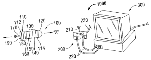
[0017] Turning now to Fig. 1, a wireless laparoscopic camera system in
accordance with
the present disclosure is shown indicated by reference numeral 1000. Wireless
laparoscopic camera system 1000 generally includes a wireless camera 100, a
remote
receiver, or transceiver 200 and a video display 300. As will be described in
greater
detail hereinbelow, wireless camera 100 includes a housing 110 configured to
house
the various components of wireless camera 100. An antenna 170 extends from a
proximal end 112 of housing 110. Alternatively, antenna 170 may be internally
disposed within housing 110. Antenna 170 is configured to facilitate wireless
communication between the wireless camera 100 and the remote receiver, or
transceiver 200. Remote receiver 200 likewise includes an antenna 210 to
facilitate
wireless communication therebetween. Cables 220 couple remote receiver 200 to
video display 300. Further, remote receiver 200 is adapted to connect to an
energy
source (not shown) via cable 230.
-4-
CA 02732539 2011-02-25
[00181 Housing 110 of wireless camera 100 is generally cylindrical in shape
and defines
a longitudinal axis "X." It is envisioned that housing 110 define a relatively
low profile
configuration such that wireless camera 100, when disposed on a laparoscopic
instrument, e.g., surgical instrument 400 (Fig. 2), does not inhibit surgical
instrument
400 (Fig. 2) from being inserted through a relatively small incision in
tissue, or access
port (not shown), as is often required during laparoscopic procedures. It is
also
envisioned that housing 110 be formed from, or coated with a biocompatible
material
such that wireless camera 100 may be inserted into an internal body cavity
without the
risk of an allergic reaction or rejection by surrounding tissue. Additionally,
housing 110
may define a relatively smooth surface geometry so as to prevent wireless
camera 100
from catching on tissue or tearing tissue during insertion, use and/or removal
of the
wireless laparoscopic camera 100 from an internal body cavity. Further,
wireless
camera 100 may be configured to clip-on or otherwise engage a surgical
instrument,
e.g., surgical forceps 400 (Fig. 2), or, alternatively, may simply be
positioned within the
internal body cavity. In either embodiment, wireless camera 100 is configured
to
wirelessly communicate with a remote wireless receiver 200 positioned
externally of the
body to provide a real-time video image of the surgical site.
[00191 With continued reference to Fig. 1, the components of wireless
laparoscopic
camera 100 will now be described in detail. Disposed at a distal end 114 of
housing
110 is an optical lens 120 (or series of lenses). The lens 120 is configured
to project an
optical image onto an image sensor 140 that is disposed within the housing 110
and
positioned proximal of the lens 120. The image sensor 140 is manufactured as a
bare
die, or integrated circuit and is packaged together with a bare die processing
-5-
CA 02732539 2011-02-25
component 150 and a bare die wireless transmitter, or transceiver 160. The
image
sensor 140, processing component 150, and wireless transmitter 160 are
disposed
within housing 110 and are stacked and coupled in sequence distally to
proximally from
the lens 120 in a single "chip-stack" package 130. Stacking the relatively
thin bare die
image sensor 140, bare die processing component 150, and bare die wireless
transmitter 160 in a single package 130 allows the package 130 to operate as a
single
"chip" having a reduced area. As can be appreciated, such a configuration
reduces the
overall size, and, more particularly, the diameter of the housing 110, as is
desired for
laparoscopic instruments.
[0020] Positioned proximally of the chip package 130 and coupled thereto is
one or
more batteries 180 configured to power the chip package 130. The batteries 180
are
generally disc-shaped and are stacked within the housing 110. As can be
appreciated,
the lens 120, chip package 130 (which includes the image sensor 140, the
processing
component 150, and the wireless transmitter 160) and the batteries 180 are all
stacked
in columnar fashion about longitudinal axis "X" and are disposed within the
relatively
small diametered cylindrical housing 110 of wireless laparoscopic camera 100.
[0021] With continued reference to Fig. 1, an antenna 170 is disposed at the
proximal
end 112 of housing 110 and extends proximally and axially from the housing 110
along
longitudinal axis "X." The antenna 170 is configured to facilitate wireless
communication between the wireless camera 100 and the wireless receiver 200. A
battery charging circuit 190 may also be disposed within the housing 110. The
battery
charging circuit 190 is positioned in column with and proximally of the
batteries 180 and
is configured to charge the batteries 180, as will be described in greater
detail below.
-6-
CA 02732539 2011-02-25
[0022) As mentioned above, a wireless receiver, or transceiver 200 is
positioned remove
of the wireless camera 100 and is configured to wirelessly communicate with
the
wireless camera 100. More particularly, the receiver 200 receives an
electrical signal
from the wireless camera 100, decouples the signal and feeds the signal, e.g.,
via
cables 220, to a video monitor 300 to display the signal as a video image. The
receiver
200 may include an antenna 210 to facilitate wireless communication between
the
wireless camera 100 and the receiver 200. Further, the receiver 200 may be
configured
as a transceiver 200, functioning to both receive the signal from the wireless
camera
100 and to transfer energy to the battery charging circuit 190 to charge the
batteries
180, as will be described in greater detail below.
[00231 Referring now to Figs. 2-3, a surgical instrument 400, and more
particularly, a
surgical forceps 400, is shown generally including a handle 410, an elongated
shaft 420
and an end effector 430 disposed at a distal end of the elongated shaft 420.
Wireless
laparoscopic camera 100 is disposed, e.g., clipped or mounted, onto elongated
shaft
420 toward the distal end 422 thereof. Although surgical instrument 400 is
shown as a
surgical forceps 400, it is envisioned that wireless camera 100 may be
clipped, or
mounted onto various other surgical instruments. Further, clip 500 may be
configured
to engage camera 100 to surgical instrument 400 at various positions along
shaft 420 or
on end effector 430, depending, for example, on the dimensions of the surgical
instrument, the particular procedure to be performed, and/or the desired field
of view.
However, as mentioned above, camera 100 need not be engaged to a surgical
instrument, but may simply be positioned within the internal body for
providing a video
image of the surgical site.
-7-
CA 02732539 2011-02-25
100241 The operation of wireless laparoscopic camera 100 and corresponding
wireless
camera system 1000 will now be described with reference to Fig. 4. As
mentioned
above, optical lens 120 projects (1) an optical image of the field of view,
e.g., the
surgical site, onto the image sensor 140. The image sensor 140, which is
coupled to
the processing component 150 and wireless transmitter 160 within chip package
130,
converts the optical image into an electrical signal and communicates (2) the
electrical
signal to the processing component 150. The processing component 150 may be
configured to convert the signal from an analog signal to a digital signal,
from a digital
signal to an analog signal, or to modulate the signal. The processed signal is
then
communicated (3) to the wireless transmitter 160. The wireless transmitter
160, along
with the antenna 170, (5) transmits the signal wirelessly (6) to the receiver
200, which is
positioned remote of the wireless camera 100. The receiver 200 then feeds the
signal
(7) to the video monitor 300 to display the signal as a video image.
[00251 With continued reference to Fig. 4, the wireless receiver 200 may be
configured
as a transceiver including a transmitting component 240, for wirelessly
transferring
power to the battery charging circuit 190, and a receiving component 250, for
receiving
the signal from the wireless transmitter 160. More particularly, the
transceiver 200,
which is coupled (8) to an energy source, transmits (9) energy, e.g., radio
frequency
(RF) energy, to the battery charging circuit 190 (10). The battery charging
circuit 190
converts the RF energy into power to charge (11) the batteries 180, which, in
turn power
(12) the chip package 130.
100261 From the foregoing and with reference to the various figure drawings,
those
skilled in the art will appreciate that certain modifications can also be made
to the
-8-
CA 02732539 2011-02-25
present disclosure without departing from the scope of the same. While several
embodiments of the disclosure have been shown in the drawings, it is not
intended that
the disclosure be limited thereto, as it is intended that the disclosure be as
broad in
scope as the art will allow and that the specification be read likewise.
Therefore, the
above description should not be construed as limiting, but merely as
exemplifications of
particular embodiments- Those skilled in the art will envision other
modifications within
the scope and spirit of the claims appended hereto.
-9-