Note: Descriptions are shown in the official language in which they were submitted.
CA 02732662 2011-02-23
DELIVERY SYSTEM AND METHOD FOR BIFURCATED GRAFT
This application is divided from Canadian Patent Application Serial No.
2,443,104 filed on April 11, 2002.
TECHNICAL FIELD
The present invention relates generally to a system and method for the
treatment
of disorders of the vasculature. More specifically, a system and method for
treatment of
thoracic or abdominal aortic aneurysm and the like, which is a condition
manifested by
expansion and weakening of the aorta.
BACKGROUND ART
Prior methods of treating aneurysms have consisted of invasive surgical
methods
with graft placement within the affected vessel as a reinforcing member of the
artery.
However, such a procedure requires a surgical cut down to access the vessel,
which in
turn can result in a catastrophic rupture of the aneurysm due to the decreased
external
pressure from the surrounding organs and tissues, which are moved during the
procedure to gain access to the vessel. Accordingly, surgical procedures can
have a high
mortality rate due to the possibility of the rupture discussed above in
addition to other
factors. Other risk factors for surgical treatment of aortic aneurysms can
include poor
physical condition of the patient due to blood loss, anuria, and low blood
pressure
associated with the aortic abdominal aneurysm. An example of a surgical
procedure is
described in a book entitled Surgical Treatment of Aortic Aneuysms by Cooley
published in 1986 by W. B. Saunders Company.
Due to the inherent risks and complexities of surgical intervention, various
attempts have been made to develop alternative methods for deployment of
grafts within
aortic aneurysms. One such method is the non-invasive technique of
percutaneous
delivery by a catheter-based system. Such a method is described in Lawrence,
Jr. et al.
in "Percutaneous endovascular graft: experimental evaluation", Radiology (May
1987).
Lawrence described therein the use of a Gianturco stent as disclosed in U.S.
Patent No.
4,580,568. The stent is used to position a Dacron fabric graft within the
vessel. The
Dacron graft is compressed within the catheter and then deployed within the
vessel to be
treated. A similar procedure has also been described by Mirich et al. in
CA 02732662 2011-02-23
-2-
"Percutaneously placed endovascular grafts for aortic aneurysms: feasibility
study,"
Radiology (March 1989). Mirich describes therein a self-expanding metallic
structure
covered by a nylon fabric, with said structure being anchored by barbs at the
proximal
and distal ends.
One of the primary deficiencies of the existing percutaneous devices and
methods
has been that the grafts and the delivery systems used to deliver the grafts
are relatively
large in profile, often up to 24 French, and stiff in longitudinal bending.
The large
profile and relatively high bending stiffness of existing delivery systems
makes delivery
through the vessels of a patient difficult and can pose the risk of dissection
or other
trauma to the patient's vessels. In particular, the iliac arteries of a
patient are often too
narrow or irregular for the passage of existing percutaneous devices. Because
of this,
non-invasive percutaneous graft delivery for treatment of aortic aneurysm is
contraindicated for many patients who would otherwise benefit from it.
What is needed is an endovascular graft and delivery system having a small
outer
diameter relative to existing systems and high flexibility to facilitate
percutaneous
delivery in patients who require such treatment. What is also needed is a
delivery
system for an endovascular graft that is simple, reliable and that can
accurately and
safely deploy an endovascular graft within a patient's body, lumen or vessel.
DISCLOSURE OF INVENTION
The invention is directed generally to a delivery system for delivery of an
expandable intracorporeal device, specifically, an endovascular graft.
Embodiments of
the invention are directed to percutaneous non-invasive delivery of
endovascular grafts
which eliminate the need for a surgical cut-down in order to access the
afflicted artery
or other intracorporeal conduit of the patient being treated. Such a non-
invasive delivery
system and method result in shorter procedure duration, expedited recovery
times and
lower risk of complication. The flexible low profile properties of some
embodiments
of the invention also make percutaneous non-invasive procedures for delivery
of
endovascular grafts available to patient populations that may not otherwise
have such
treatment available. For example, patients with small anatomies or
particularly tortuous
vasculature may be contraindicated for procedures that involve the use of
delivery
CA 02732662 2011-02-23
-3-
systems that do not have the flexible or low profile characteristics of
embodiments of
the present invention.
In accordance with an illustrative embodiment, there is provided an
endovascular graft delivery system configured to deliver an endovascular graft
to a
target location and deploy the endovascular graft at the target location
within a
patient's vasculature, comprising: an endovascular graft including a tubular
graft body
section and a distal self-expanding member which is disposed distally of a
second
self-expanding member and the graft body section; an elongate shaft having a
proximal section and a distal section; a first handle disposed on a proximal
portion of
the delivery system and configured to deploy the distal self-expanding member;
and a
second handle disposed on the proximal portion of the delivery system and
configured
to deploy the second self-expanding member.
In accordance with another illustrative embodiment, there is provided an
endovascular graft delivery system, comprising: an endovascular graft
including a
tubular graft body section and a distal self-expanding member which is
disposed
distally of a second self-expanding member and the graft body section; and a
delivery
catheter configured to deliver and deploy the endovascular graft in a
constrained state
to a treatment site within a patient's body, including an elongate shaft
having a
proximal section and a distal section, an outer tubular member disposed over
the
constrained graft configured to cover the endovascular graft in a constrained
state
during delivery to a treatment site within the patient's body and configured
to be
retracted proximally to expose the endovascular graft, and a first handle
disposed on a
proximal portion of the delivery catheter and configured to deploy the distal
self-
expanding member.
In accordance with another illustrative embodiment, there is provided an
endovascular graft delivery system configured to deliver an endovascular graft
to a
target location within a patient's vasculature and deploy the endovascular
graft at the
target location, comprising: an endovascular graft including a graft body
section and a
distal self-expanding member which is disposed distally of the graft body
section; an
elongate shaft having a proximal section and a distal section; a handle
disposed on a
CA 02732662 2011-02-23
-4-
proximal portion of the delivery system configured to partially deploy the
endovascular graft upon actuation of the handle; and a handle disposed on the
proximal portion of the delivery system configured to deploy the self-
expanding
member of the endovascular graft.
The delivery system is used for deploying an expandable intracorporeal device
within a patient's body. The method includes providing a delivery system for
delivery
of an expandable intracorporeal device including an elongate shaft having a
proximal
section and a distal section. The distal section of the elongate shaft has an
elongate
belt support member disposed adjacent a portion of the expandable
intracorporeal
device and a belt which is secured to the belt support member. The belt is
circumferentially disposed about the expandable intracorporeal device and has
a
configuration that constrains the expandable intracorporeal device. A release
member
releasably secures the belt in the constraining configuration.
Next, the distal end of the delivery system is introduced into the patient's
body
and advanced to a desired site within the patient's body. The release member
is then
activated, releasing the belt from the constraining configuration. Optionally,
the
delivery system may also have an outer protective sheath disposed about the
endovascular graft in a constrained state, the belt in its constraining
configuration and
at least a portion of the release wire disposed at the belt. In such an
arrangement, the
method of deployment of an expandable intracorporeal device also includes
retraction
of the outer protective sheath from the endovascular graft prior to activation
of the
release member.
In an embodiment of the invention directed to delivery of a bifurcated
intracorporeal device, an elongate shaft has a proximal section and a distal
section.
The distal section of the shaft has an elongate primary belt support member
and at
least one primary belt disposed on the primary belt support member. The
primary belt
support member is configured to be circumferentially disposed about a
bifurcated
intracorporeal device and at least partially constrain the device. A primary
release
member is configured to engage and releasably secure the primary belt in a
constraining configuration. At least one elongate secondary belt support
member is
CA 02732662 2011-02-23
-5-
disposed adjacent the elongate primary belt support member. At least one
secondary
belt is disposed on the secondary belt support member. This at least one
secondary
belt is configured to be circumferentially disposed about a bifurcated
intracorporeal
device and at least partially constrain the device. A secondary release member
is
configured to engage and releasably secure the secondary belt in a
constraining
configuration.
In a method for deploying a bifurcated intracorporeal device within a
patient's
body, a delivery system for delivery and deployment of a bifurcated
intracorporeal
device is provided. The delivery system includes an elongate shaft having a
proximal
section and a distal section. The bifurcated intracorporeal device is disposed
on the
distal section of the elongate shaft. The distal section of the elongate shaft
also
includes an elongate primary belt support member and at least one primary belt
secured to the primary belt support member. The primary belt is configured to
be
circumferentially disposed about a bifurcated intracorporeal device and at
least
partially constrain the device. A primary release member engages and
releasably
secures the primary belt in the constraining configuration. The distal section
of the
elongate shaft also includes at least one elongate secondary belt support
member
disposed adjacent the elongate primary belt support member. At least one
secondary
belt is secured to the secondary belt
CA 02732662 2011-02-23
-6-
support member and is configured to be circumferentially disposed about a
bifurcated
intracorporeal device to at least partially constrain the device. A secondary
release
member engages and releasably secures the secondary belt in a constraining
configuration.
The distal end of the delivery system is introduced into the patient's body
and
advanced to a desired site within the patient's body. The release members are
then
activated to release the belts from the constraining configuration and the
device is
deployed. Thereafter, the delivery system can be removed from the patient's
body. In
some embodiments of the invention, the secondary belt support member is
detached and
removed from the delivery system prior to withdrawal of the delivery system
from the
patient. In another embodiment, the secondary belt support member is displaced
laterally towards the primary belt support member so as to be substantially
parallel to
the primary belt support member and enable withdrawal of the delivery system
through
an ipsilateral side of the bifurcated intracorporeal device.
BRIEF DESCRIPTION OF DRAWING
The objects, advantages and features of this invention will be more readily
appreciated from the following detailed description, when read in conjunction
with the
accompanying drawing, in which:
FIG. 1 is an elevational view in partial longitudinal section illustrating an
embodiment of a delivery system for an expandable intracorporeal device having
features
of the invention.
FIG. 2 is a transverse cross sectional view of the delivery system of FIG. 1
taken
along lines 2-2 of FIG. 1.
FIG. 3 is a transverse cross sectional view of the delivery system of FIG. 1
taken
along lines 3-3 of FIG. 1.
FIG. 4 is a transverse cross sectional view of the delivery system of FIG. 1
taken
along lines 4-4 of FIG. 1.
FIG. 5 is a transverse cross sectional view of the delivery system of FIG. 1
taken
along lines 5-5 of FIG. 1.
CA 02732662 2011-02-23
-7-
FIG. 6A is an enlarged elevational view in partial section of the delivery
system
in FIG. 1.
FIG. 6B is an enlarged elevational view in partial section of the delivery
system
of FIG. 1 with portions of the graft and self-expanding members cut away for
clarity of
view of the belt bushings.
FIG. 7A is a perspective view showing release belt configurations having
features
of the invention.
FIG. 7B is a perspective view showing an alternative embodiment of release
belts,
FIG. 7C is an end view showing an alternative embodiment of release belts.
FIG. 7D is a perspective view of the embodiment of FIG. 7C.
FIG. 7E is an enlarged view of a particular coupling configuration between end
loops of release belts.
FIG. 7F is a perspective view, partially cut away, of a particular embodiment
of
an end loop of a release belt.
FIG. 7G is a perspective view of an alternative embodiment of a release belt.
FIG. 7H is a perspective view of an alternative embodiment of a release belt.
FIG. 71 is a perspective view of an alternative embodiment of a branched
release
wire.
FIG. 7J is an end view showing an alternative embodiment of a release belt.
FIG. 7K is a transverse cross sectional view showing the alternative
embodiment
of the release belt configuration of FIG. 71 constraining a self-expanding
member.
FIG. 7L is a detail of the connection formed where a release wire is used with
the alternative release belt embodiment of FIGS. 7J-7K.
FIG. 8 is an elevational view in partial section of the proximal adapter shown
in
FIG. 1.
FIG. 9 is a diagrammatic view of a patient's body illustrating the patient's
heart,
aorta, iliac arteries, femoral arteries, and a delivery system having features
of the
invention disposed within the femoral artery and aorta.
CA 02732662 2011-02-23
-8-
FIG. 10 is a diagrammatic view of a delivery system having features of the
invention disposed within an artery of a patient with an expandable
intracorporeal device
being deployed within the artery.
FIG. 11 is a diagrammatic view of a delivery system having features of the
invention disposed within an artery of a patient with an expandable
intracorporeal device
being deployed within the artery.
FIG. 12 is an enlarged diagrammatic view of a delivery system having features
of the invention disposed within an artery of a patient with an expandable
intracorporeal
device being deployed within the artery.
FIG. 13 is an elevational view in partial section of a connection between an
inflation tube and an inflation port of an endovascular graft.
FIG. 14 is an elevational view in partial longitudinal section illustrating an
embodiment of a delivery system for an expandable intracorporeal device having
features
of the invention.
FIG. 15 is a transverse cross sectional view of the delivery system of FIG. 14
taken along lines 15-15 in FIG. 14.
FIG. 16 is an enlarged elevational view in partial section of the delivery
system
shown in FIG. 14.
FIG. 17 is an elevational view in partial section of the proximal adapter of
the
delivery system shown in FIG. 14.
FIG. 18 is an elevational view in partial section of an alternative embodiment
of
the proximal adapter of the delivery system shown in FIG. 14 with a nested
handle
configuration.
FIG. 19 is an elevational view of a bifurcated stent graft suitable for
delivery and
deployment by embodiments of the invention.
FIG. 20 is a transverse cross sectional view of the scent graft of FIG. 19
taken
along lines 20-20 in FIG. 19.
FIG. 21 is a transverse cross sectional view of the stent graft of FIG. 19
taken
along lines 21-21 of FIG. 19.
FIG. 22 is a transverse cross sectional view of the stent graft of FIG. 19
taken
along lines 22-22 of FIG. 19.
CA 02732662 2011-02-23
-9-
FIG. 23 is an elevational view in partial section of an embodiment of a
delivery
system having features of the invention.
FIG. 24 is a transverse cross sectional view of the delivery system of FIG. 23
taken along lines 24-24 of FIG. 23.
FIG. 25 is a transverse cross sectional view of the delivery system of FIG. 23
taken along lines 25-25 of FIG. 23.
FIG. 26 is an elevational view in partial section showing an enlarged view of
a
distal portion of the delivery system of FIG. 23.
FIG. 27 is a transverse cross sectional view of the delivery system of FIG. 26
taken along lines 27-27 of FIG. 26.
FIG. 28 is a transverse cross sectional view of the delivery system of FIG. 26
taken along lines 28-28 of FIG. 26.
FIG. 28A is a transverse cross sectional view of an alternative embodiment of
a secondary belt support member of a delivery system similar in function to
that shown
in FIG. 28.
FIG. 28B is an elevational view of the alternative embodiment of the secondary
belt support member of FIG. 28A.
FIG. 29 is a transverse cross sectional view of the delivery system of FIG. 26
taken along lines 29-29 of FIG. 26.
FIG. 30 is a transverse cross sectional view of the delivery system of FIG. 26
taken along lines 30-30 in FIG. 26.
FIG. 31 is an elevational view in partial section of the proximal adapter of
the
delivery system of FIG. 23.
FIG. 31A is an elevational view in partial section of the proximal adapter of
the
delivery system of FIG. 23, showing an optional ripcord and flexible fill
catheter.
FIG. 31B is a simpler cross sectional schematic view of a bent or angled
contralateral leg inflatable channel having a bead or lumen patency member
disposed in
a channel lumen taken.along line 31B-31B in FIG. 19.
FIG. 32 is a perspective view of the belt support member assembly at a distal
portion of the delivery system of FIG. 23.
CA 02732662 2011-02-23
-10-
FIG. 33 illustrates a portion of the internal vasculature of a patient,
including
the aorta, iliac and femoral arteries branching therefrom.
FIG. 34 is a magnified view of the abdominal aorta area of the patient shown
in FIG. 33 and shows a guidewire positioned in the aorta from the right iliac
artery.
FIGS. 35-37 illustrate the magnified view of the abdominal aorta of the
patient
shown in FIG. 33 and depict a deployment sequence of the bifurcated
endovascular
stent graft of FIG. 19 with the delivery system of FIG. 23.
FIG. 37A is a perspective view of a marker disposed on the delivery system
distal section in the vicinity of the nosepiece.
FIG. 37B is a perspective view of an alternative embodiment of a marker for
use in the delivery system of the present invention.
FIGS. 38-52 continue to illustrate a deployment sequence of the bifurcated
endovascular stent graft of FIG. 19.
FIGS. 53-57 illustrate a number of alternative catheter distal shaft
arrangements in which a well is provided to facilitate the orderly and tangle-
free
withdrawal of the release strand from the delivery catheter.
FIGS. 58-60 illustrate a further alternative belt support member and
contralateral leg delivery system configurations and operation.
BEST MODE FOR CARRYING OUT THE INVENTION
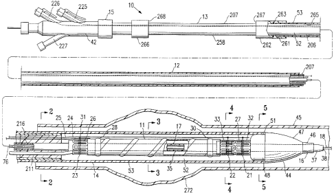
FIGS. 1-8 and 10 illustrate an embodiment of delivery system 10 for
delivering a variety of expandable intracorporeal devices; specifically, an
expandable
endovascular graft 11. One such expandable endovascular graft 11 useful for
delivery
and deployment at a desired site within a patient is disclosed in U.S. Patent
No.
6,395,019 issued May 28, 2002 to M. Chobotov.
Delivery system 10 in FIG. 1 has an elongate shaft 12 with a proximal section
13, a distal section 14, a proximal end 15 and a distal end 16. The distal
section 14 has
an elongate belt support member in the form of a guidewire tube 17 disposed
adjacent
a portion of the expandable endovascular graft 11. A guidewire 18 is disposed
within
guidewire tube 17. A plurality of belts 21, 22, and 23 are secured to the
guidewire
tube 17 and are circumferentially disposed about portions of the endovascular
graft
11. FIG. I shows the belts in a configuration that constrains the endovascular
graft 11.
CA 02732662 2011-02-23
-11-
First and second release members 24 and 25 releasably secure belts 21, 22, and
23 in a
constraining configuration as shown.
The endovascular graft I I has a proximal end 26, a distal end 27, a proximal
inflatable cuff 28, a distal inflatable cuff 30, a proximal self-expanding
member 31, a
first distal self-expanding member 32 and a second distal self-expanding
member 33.
As defined herein, the proximal end of the elongate shaft is the end 15
proximal to an
operator of the delivery system 10 during use. The distal end of the elongate
shaft is
the end 16 that enters and extends into the patient's body. The proximal and
distal
directions for the delivery system 10 and endovascular graft 1 l loaded within
the
delivery system 10 as used herein are the same. This convention is used
throughout
the specification for the purposes of clarity, although other conventions are
commonly
used. For example, another useful convention defines the proximal end of an
endovascular graft as that end of the graft that is proximal to the source of
blood flow
going into the graft. Such a convention is used in the previously discussed
U.S. Patent
No. 6,395,019 although that convention is not adopted herein.
The guidewire tube 17 has an inner lumen 34, as shown in FIG. 2, a distal
section 35, a proximal end 36, as shown in FIG. 8, and a distal end 37. The
inner
lumen 34 of the guidewire tube 17 terminates at the distal end 37 with a
distal
guidewire tube port 38, as shown in FIG. 10. As seen in FIG. 8, the proximal
end 36
of guidewire tube 17 terminates in a port 41 disposed in the proximal adapter
42. The
port 41 is typically a tapered fitting such as a Luer lock fitting which
facilitates the
attachment of a hemostasis valve (not shown). The guidewire tube 17 is a
hollow
tubular member that normally has an annular cross section, although oval cross-
sectional profiles and others are also suitable.
A portion of the distal section 35 of the guidewire tube 17, shown in FIG. 1,
is
disposed within an inner lumen 43 of a distal nose piece 44, as shown in FIG.
5.
Distal nose piece 44 is configured in a streamlined bullet shape for easy
passage
within a patient lumen or vessel such as aorta 45. Guidewire tube 17 may be
bonded
to the inner lumen 43 of the nose piece 44, or it may be molded into the nose
piece 44
during
CA 02732662 2011-02-23
-12-
manufacture. Referring to FIG. 1, the nose piece 44 has a distal portion 46,
an
intermediate portion 47 and a proximal shoulder portion 48 configured to
slidingly
engage the distal portion 51 of an inner lumen 52 of an outer tubular member
53.
Referring to FIGS. 1, 6A, 6B and 7A, on the distal section 35 of guidewire
tube 17, proximal to the proximal shoulder portion 48 of nose piece 44, a
first distal
belt 21 is secured to the guidewire tube 17. The first distal belt may be
secured to the
guidewire tube 17 with any suitable adhesive such as cyanoacrylate, epoxy or
the like.
Both free ends 55 and 56 of the first distal belt 21 are secured to the
guidewire tube 17.
The guidewire tube 17 may be made from a variety of suitable materials
including
polyethylene, teflon, polyimide and the like.
Referring to FIGS. 2-5, the inner lumen 34 of the guidewire tube 17 has an
inside diameter that can accommodate a guidewire suitable for guiding a device
such as
delivery system 10. The inner lumen 34 of the guidewire tube 17 may have an
inside
diameter of about 0.015 inch to about 0.045 inch; specifically, about 0.020
inch to about
0.040 inch. The outer diameter of the guidewire tube 17 may range from about
0.020
inch to about 0.060 inch; specifically, about 0.025 inch to about 0.045 inch.
Referring again to FIGS. 6A, 6B and 7A, an optional first distal belt bushing
57
is disposed about the guidewire tube 17 so as to cover the portions of the
free ends 55
and 56 of the first distal belt 21 that are secured to the distal section 35
of the guidewire
tube 17. This bushing 57 may also serve to control the constrained
configuration of the
belted self-expanding members, and may include geometric features to engage or
support
the belted members. A similar configuration is present at a second distal belt
22 which
has free ends secured to the guidewire tube 17 proximal to the first distal
belt 21. A
second distal belt bushing 63 is disposed about the guidewire tube 17 so as to
cover the
portions of the free ends of the second distal belt 22 that are secured to the
guidewire
tube 17. A proximal belt 23 has free ends secured to the guidewire tube 17
proximal
to the second distal belt 22 and has an optional proximal belt bushing 67, as
shown in
FIG. 6, configured similarly to the first and second distal belt bushings 57
and 63.
The belts 21, 22 and 23 can be made from any high strength, resilient material
that can accommodate the tensile requirements of the belt members and remain
flexible
after being set in a constraining configuration, Typically, belts 21, 22 and
23 are made
CA 02732662 2011-02-23
-13-
from solid ribbon or wire of a shape memory alloy such as nickel titanium or
the like,
although other metallic or polymeric materials are possible. Belts 21, 22 and
23 may
also be made of braided metal filaments or braided or solid filaments of high
strength
synthetic fibers such as Dacron , Spectra or the like. An outside transverse
cross
section of the belts 21, 22 and 23 may range from about 0.002 to about 0.012
inch,
specifically, about 0.004 to about 0.007 inch. The cross sections of belts 21,
22 and 23
may generally take on any shape, including rectangular (in the case of a
ribbon),
circular, elliptical, square, etc.
In general, we have found that a ratio of a cross sectional area of the belts
to a
cross sectional area of the release members, 24 and 25, of about 1:2 is useful
to balance
the relative strength and stiffness requirements. Other ratios, however, may
also be used
depending on the desired performance characteristics.
The inner diameters of belt bushings 57, 63 and 67 are sized to have a close
fit
over the guidewire tube 17 and secured portion 71, as shown in FIG. 7A, of the
free
ends of the belts 21, 22 and 23 that are secured to the guidewire tube 17.
Typically, the
inner diameter of the belt bushings 57, 63 and 67 range from about 0.025 inch
to about
0.065 inch; specifically, about 0.030 inch to about 0.050 inch. In addition,
the outer
diameter of belt bushing 57 may be sized to approximate an inner diameter 70,
as shown
in FIG. 4, of the respective first distal self-expanding member 32 of the
endovascular
graft 11 when the member 32 is in a fully constrained state. The other belt
bushings 63
and 67 may be similarly configured with respect to the second distal self-
expanding
member 33 and the proximal self-expanding member 31.
Such an arrangement keeps the self-expanding members 31, 32 and 33 properly
situated when in a constrained state and prevents the various portions of the
self-
expanding members 31, 32 and 33 from overlapping or otherwise entangling
portions
thereof while in a constrained state. The outer diameter of the belt bushings
57, 63 and
67 may range from about 0.040 inch to about 0.200 inch; specifically, about
0.060 inch
to about 0.090 inch. The material of the belt bushings 57, 63 and 67 may be
any
suitable polymer, metal, alloy or the like that is bondable. Generally, the
belt bushings
57, 63 and 67 are made from a polymer such as polyurethane, silicone rubber or
PVC
plastic.
CA 02732662 2011-02-23
-14-
As shown in FIG. 7A, belts 21, 22 and 23 extend radially from the guidewire
tube 17 through optional standoff tubes 72, 73 and 74. Standoff tubes 72, 73
and 74 are
disposed about belts 21-23 adjacent the guidewire tube 17 and act to prevent
separation
of belts 21-23 in a circumferential direction as tension is applied to the
belts. Standoff
tubes 72 - 74 also prevent belts 21-23 from applying other undesirable forces
on portions
of the endovascular graft 11 that are constrained by the belts. Specifically,
the standoff
tubes 72 -74 prevent the belts 21-23 from spreading the self-expanding members
31 -
33, or portions thereof, at those locations where the belts 21-23 extend
radially through
the self-expanding members.
The standoff tubes 72 - 74 typically have a length substantially equal to a
single
wall thickness of the self-expanding members 31, 32 and 33. The length of the
standoff
tubes 72 - 74 may range from about 0.010 inch to about 0.030 inch. An inner
diameter
of an inner lumen 75 of the standoff tubes, as shown in FIG. 4, may range from
about
0.004 to about 0.024 inch, with a wall thickness of the standoff tubes being
about 0.002
inch to about 0.006 inch. Typically, the standoff tubes 72 - 74 are made from
a high
strength metal or alloy such as stainless steel, although they may be
polymeric as well.
Belts 21-23 exit the outer apertures of standoff tubes 72 - 74 and extend
circumferentially about the respective portions of the expandable
intracorporeal device
11. The term "circumferential extension" as used with regard to extension of
the belts
21-23 is meant to encompass any extension of a belt in a circumferential
direction. The
belts may extend circumferentially a full 360 degrees, or any portion thereof.
For
example, belts or belt segments may extend partially about an endovascular
device, and
may be combined with other belts or belt segments that also partially extend
circumferentially about an endovascular device. Typically, a plane formed by
each of
the belts 21-23 when in a constraining configuration is generally
perpendicular to a
longitudinal axis 76, shown in FIG. 1, of the distal section 14 of shaft 12.
As shown
in FIGS. 6A and 6B, loop ends 81, 82 and 83 of the belts 21, 22 and 23,
respectively,
are releasably locked together by one or more release members. For example, in
the
embodiment shown in FIG. 1, a release member in the form of a first release
wire 24
is shown disposed within end loops 81 of the first distal belt 21 and end
loops 82 of the
second distal belt 22 so as to secure the first and second distal belts 21 and
22 in a
CA 02732662 2011-02-23
-15-
constraining configuration about the endovascular graft 11. Another release
member in
the form of a second release wire 25 is shown disposed within end loops 83 of
the
proximal belt 23 so as to secure the proximal belt 23 in a constraining
configuration
about the endovascular graft 11.
A single release wire may also be used to perform the function of each of the
first and second release wires, 24 and 25, so that first distal belt 21,
second distal belt
22, and proximal belt 23 may be releasably secured by a single release wire. A
highly
controlled, sequential belt deployment scheme may be realized with the use of
a single
release wire.
Any number of release wires and belts as may be needed to effectively secure
and deploy graft 11, in combination, are within the scope of the present
invention.
In some embodiments of the invention, when constrained, the end loops of any
single belt touch each other or are spaced closely together such that the belt
as a whole
forms a substantially circular constraint lying substantially in a plane.
Release wire 24
and 25 may be made from suitable high strength materials such as a metal or
alloy (e.g.,
stainless steel) which can accommodate the torque force applied to the release
wire by
the belt end loops 83 when the belts 23 are under tension from the outward
radial force
of the constrained portions of the endovascular graft 11, i.e., the self-
expanding members
32 and 33.
The release wires 24 and 25 may generally have an outer diameter ranging from
about 0.006 to about 0.014 inch. Distal end portions 84 and 85 of release
wires 24 and
25, respectively, may terminate at any appropriate site distal of the end
loops 81-83 of
belts 21-23. As shown in FIG. 8, the proximal ends 86 and 87 of the release
wires 24
and 25 extend through the elongate shaft 12 of the delivery system 10 through
proximal
ports 91 and 92 on the proximal adapter 42, respectively, and terminate at
respective
release wire handles 93 and 94 which are releasably secured to the proximal
adapter 42.
FIG. 7B illustrates an alternative embodiment of the belts 21-23 of FIG. 7A.
In
FIG. 7A, belts 21-23 are shown as each consisting of a single strand of wire
formed into
the end loops 81-83, respectively, with the end loops in an overlapping
configuration.
Free ends 55 and 56 of belt 81 are shown secured to the distal section 35 of
the
guidewire tube 17. In contrast, FIG. 7B, wherein like elements with regard to
FIG. 7A
CA 02732662 2011-02-23
-16-
are shown with like reference numerals, shows belts 21B, 22B and 23B formed of
two
strands of wire, with each strand formed into a single loop which overlaps a
loop of the
other strand to form end loops 81B, 82B and 83B. The free ends of the belts
21B-23B
may be secured in a similar manner to those of free ends 55 and 56 of FIG. 7A.
Turning now to FIGS. 7C and 7D, alternative embodiments for portions of the
delivery system of the present invention are shown. FIGS. 7C and 7D illustrate
alternative belts 21C, 22C and 23C disposed on guidewire tube 17. Single or
multiple
belts 21C - 23C may be deployed at various locations along guidewire tube 17
as
desired. In addition, the members comprising belts 21C-23C are shown as a
single line.
However, belts 21C-23C may be of a single- or multiple strand or filament
design with
various cross-sectional shapes as previously described. A single solid ribbon
or wire is
particularly useful.
Belts 21C-23C shown in FIGS. 7C and 7D are a single strand filament wrapped
around guidewire tube 17 and fixed thereon via any number of suitable
techniques, such
as gluing with adhesive, mechanical fixation, etc. Especially useful is fixing
the belt
with an ultraviolet-curable adhesive.
Alternatively, belts 21C-23C may comprise two strand filaments each wrapped
around guidewire tube 17 so that, for instance, belt 21C is a two-filament
component.
Belt 21C includes belt arms 112 and 114, each of which, in the embodiments
shown, is a loop of filament twisted upon itself to form a helix. Any number
of twists
may be imparted to arms 112 and 114 to provide a relatively loose or
relatively tight
helix as desired. Typically the number of twists (with a single twist being
defined as
a single overlap of wire segment) in each belt arm 112 and 114 numbers from
zero to
about 50 or more; specifically, about two to about 10. The choice of material
used for
belt 21C is an important factor in determining the optimum number of twists
for each
belt arm. Belt arms 112 and 114 may be formed into other configurations (e.g.,
braid,
double helix, etc.) as well.
Disposed within the end loops of the belt arms 112 and 114 are distal
apertures
or openings 120, 122, respectively. During assembly of the delivery system, a
release
wire (such as wire 24) is passed through each aperture 120, 122 after the belt
arms are
wrapped around the graft self-expanding member, preferably in a
circumferential groove
CA 02732662 2011-02-23
-17-
as further described below. The release wire may also be disposed through any
aperture
created along the length of belt arms 112, 114 by each helix twist, although
the distal-
most apertures 120, 122 are preferred.
The wire optionally may be welded, glued, or otherwise fixed to itself at
discrete
points or along all or any portion of belt arms 112, 114, save their
corresponding
apertures 120 and 122. For instance, the belt arm wire may be glued or welded
to itself
at the overlap or twist points, such as points 124.
FIG. 7D shows an optional belt arm sleeve 126 that may be used to enclose a
portion of one or both belt arms 112, 114, or any of the other belt
embodiments
contemplated herein. Belt 112 is shown in FIG. 7D being constrained or covered
over
a length thereof by a flexible sleeve or coating 126 (or alternatively, a coil
wrapping or
by fixing the loop to itself by adhesives, welding, soldering, brazing, etc.).
Sleeve or
coating 126 may optionally be shrink-wrapped, crimped, or otherwise configured
to
constrain or cover belt arm 112 therein. These fixation and sleeve features
help to
minimize the potential of belt arm untwisting and tend to close or block some
or all of
the helix apertures along the length except those through which the release
wire are
intended to pass. They can also provide greater structural and operational
stability to
the catheter system as a whole.
Belt arm sleeve 126 can be configured to have a transverse dimension that is
sized to fit a twisted belt arm with fixed nodal points such as the belt arm
112 shown
in FIG. 7D. In order to accommodate such a twisted belt arm 112, the inner
diameter
and outer diameter would be large relative to a transverse dimension of the
wire material
that forms the belt arm 112. However, the belt arm sleeve 126 can also be only
slightly
larger in transverse dimension that the wire that forms the belt arm. For
example,
embodiments of belt arms that do not have twisted wires may have a sleeve 126
that fits
closely or tightly over two strands of wire forming a belt arm. The sleeve 126
can
cover substantially the entire length of such an untwisted belt arm from at
least the
guidewire tube to just proximal of the distal loop, such as distal loop 120.
The distal
loop should remain exposed for engagement by a release wire. In such an
embodiment,
the sleeve covered portion of the belt arm may also be wrapped around and
secured to
the guidewire tube just as the unsleeved belt portion of the belt arm 112
shown in FIG.
CA 02732662 2011-02-23
-18-
7D is shown at 71 C. This type of low profile belt arm sleeve may also be used
to cover
twisted belt arm embodiments, although a slightly larger diameter sleeve would
be
required.
It may be desirable to impart a particular free resting angle to the belt arms
112,
114 to improve the reliability of the system and further reduce the
possibility of the arms
112 and 114 interfering with other components of the prosthesis or delivery
system. The
FIG. 7C view shows belt arms 112, 114 symmetrically disposed at an angle a as
measured from a horizontal plane 125. This angle a may range from zero to 180
degrees. For example, one or both belt arm 112, 114 may lie along plane 125 or
they
may rest in the configuration shown (a = 45 degrees). Any known techniques may
be
used to impart a desired resting configuration to the system, such as, for
example, cold
working or shape-setting by way of an athermal phase transformation (in the
case of
shape memory alloys).
FIG. 7J shows a single belt example of the version shown in FIGS. 7C and 7D.
Here, a single belt arm 113 is shown disposed about the distal end 35 of
guidewire tube
17. Belt arm 113 is significantly longer than either belt arm 112 or 114 of
the FIGS.
7C-7D embodiment so that it may extend at least around the circumference of
any one
of self-expanding members 31, 32, or 33. The distal portion 115 of belt arm
113 meets
a more proximal portion 117 where one or both strands (when the belt arm 113
is a
twisted variety) extends through an end loop 119 in the belt arm 115 distal
portion. As
discussed with other embodiments, a release member such as release wire 24 may
be
inserted through end loop 119 and the intersecting portion of the belt arm
proximal
portion 117 to releasably secure belt arm 113 in a constraining configuration
about the
endovascular graft 11. FIG. 7K depicts a simplified schematic cross-sectional
view of
belt arm 113 (shown here untwisted) held in place by a release wire 24 about
an
exemplary self-expanding member 32. FIG. 7L is a detail of the connection
formed
where release wire 24 intersects the distal and proximal portions, 115 and
117,
respectively, of belt arm 113.
All of the features discussed herein with respect to the FIGS. 7C-7D
embodiment
may be employed in the embodiment of FIGS. 7J-7K as well.
CA 02732662 2011-02-23
-19-
This helix configuration shown in the embodiments of FIGS. 7C-7D and 7J-7L
is a particularly reliable configuration. It reduces the possibility that a
portion of belt
21C becomes entangled with a self-expanding member (such as members 31, 32 and
33)
or otherwise interferes with the safe and effective deployment of the
prosthesis.
FIG. 7E depicts a particularly useful arrangement for configuring the belt end
loops 81-83 with release wires 24-25 during assembly of delivery system 10. In
this
example, first and second end loops 81' and 81" of belt 21 are shown connected
via
release wire 24. To achieve the configuration of FIG. 7E, first end loop 81'
is passed
through aperture 88 disposed in second end loop 81". A portion of aperture 89
disposed in first end loop 81' should extend through the plane created by
second end
loop 81" as shown in FIG 7E.
Next, release wire 24 is passed through the portion of aperture 89 that
extends
beyond this plane so that wire 24 "locks" the two looped ends 81' and 81"
together as
shown. We have found that this is a stable configuration that lends itself
well to a
reliable and safe deployment protocol.
Other techniques for assembling wire 24 and first and second end loops 81' and
81" may be used; the method described above is merely exemplary. Wire 24 may
simply pass through loop ends as configured and as shown at reference numerals
81, 82
and 83 in FIG. 7A, and 81B, 82B and 83B of FIG. 7B as well.
In the embodiment of FIG. 7F, belt 110 is a member in the shape of a wire
formed into an end loop 116B having an aperture 120 for receiving a release
wire. This
arrangement may be used on one or both ends of belt 110 or, alone if belt 110
is in the
form of a single belt arm as discussed above. Connection 123 is shown in FIG.
7F as
a simple wrapping of the distal end 11 6A of the wire comprising belt 110.
Connection
123 need not be limited to such a tapered or cylindrical sleeve or coating,
however.
Other methods to form end loop 116B are contemplated, including, for example,
the use
of adhesives, welding, brazing, soldering, crimping, etc. An optional
protective sleeve
or coating 127 (shown in sectional view in FIG. 7F) covers or is part of
connection 123
and serves to protect the patient as well as components of the delivery system
and
prosthesis from damage.
CA 02732662 2011-02-23
-20-
Turning now to FIGS. 7G and 7H, two alternative embodiments of a ribbon-like
belt 81G and 81H are shown. In FIG. 7G, a section 128 of material has been
partially
displaced from belt 81G distal end 116C and worked into a loop-like member 129
such
that two generally orthogonal apertures 130, 132 are formed in belt distal end
116C. A
set of hinges or other protective mechanism or material may be used on each
end of this
member 128 so that further tearing or peeling of this member may be prevented.
Section 128 may be formed integrally from the belt distal end 116C as shown in
FIG.
7G or may be a separate component that is attached to the belt distal end by
any suitable
means.
Second belt distal end 118C in FIG. 7G is shown as having an aperture 133
disposed therein. In use, a half-twist is imparted to the ribbon-like belt 81G
as the
second distal end 118C is brought through aperture 130 such that apertures 132
and 133
are at least partially aligned. A release wire (such as wire 24) is then
brought through
apertures 132 and 133 to releasably join ends 116C and 118C.
FIG. 7H shows yet another embodiment of a belt 81H where a simple rectangular
aperture 133A is disposed in the distal end 117 of belt 81H through which
another belt
end and release wire may be disposed as taught herein. As with the embodiment
of
FIG. 7G, a half-twist is imparted to the belt 81H in use so that the second
distal end
118D is brought through aperture 133. A release wire may then be threaded
through
apertures 132 and 133 to releasably join ends 117 and 118D. In this
embodiment,
aperture 132 should be large enough to accommodate both second distal end 11
8D and
a release wire.
FIG. 71 shows a perspective view of a belt assembly similar to that shown in
FIG. 7A, wherein like elements are shown with like reference numerals. An
alternative
embodiment of a release wire consisting of a branched release wire 150 is
illustrated in
FIG. 71. The branched release wire 150 engages belts 21-23 and is configured
to release
belts 21-23 at different times with a proximal withdrawal movement of the
branched
release wire 150, the direction of which is indicated by arrow 151. Branched
release
wire 150 has a main portion 152 and a branch portion 153. Branch portion 153
is
secured to main portion 152 by a solder joint 154. The joint 154 could also be
made
by any other suitable means, such as welding, bonding with an epoxy,
mechanically
CA 02732662 2011-02-23
-21-
binding the joint, or the like. The embodiment of the branched release wire
shown in
FIG. 71 consists of wire which is generally round in cross section. The wire
of the
branched release wire can have the same or similar material and mechanical
properties
to the wire of the release wires 24 and 25 discussed above. Branch portion 153
engages first distal belt 21 and second distal belt 22. A distal segment 155
has a length
L indicated by arrow 156 which extends distally' from first distal belt 21 to
the distal end
157 of branch portion 153.
Main portion 152 of the branched release wire 150 engages the proximal belt 23
and has a distal segment 158 that extends distally from the proximal belt 23
to a distal
end 161 of the main portion. The length L' of the distal segment 158 of the
main
portion 152 is indicated by arrow 162. Length L of distal segment 155 is
greater than
length L' of distal segment 158. In this way, as the branched release wire is
withdrawn
proximally, proximal belt 23 is released first, first distal belt 21 is
released second and
second distal belt is released last. Such a branched release wire allows a
wide variety
of belt release timing with a single continuous withdrawal or movement of a
proximal
end (not shown) of the branched release wire 150. The proximal end of the
branched
release wire may be terminated and secured to a release wire handle or the
like, as
discussed herein with regard to other embodiments of release wires. The
ability to
deploy multiple release wires in a desired timing sequence with a single
branched release
wire 150 gives the designer of the delivery system great flexibility and
control over the
deployment sequence while making the deployment of the belts simple and
reliable for
the operator of the delivery system. Although the branched release wire 150
has been
shown with only a single branch, any number of branches or desired
configuration could
be used to achieve the deployment sequence required for a given embodiment of
a
delivery system. For example, a separate branch could be used for each belt in
a
multiple belt system, with varying distal segment length used to control the
sequence of
deployment. Also, multiple branched release wires, or the like, could be used
in a single
delivery system to achieve the desired results.
A number of embodiments for the belt and belt arm components of the present
invention are described herein. In general, however, we contemplate any belt
or belt
arm configuration in which the belt may be used to releasably hold or restrain
an
CA 02732662 2011-02-23
-22-
implant member in conjunction with a release member. The particular
embodiments
disclosed herein are not meant to be limiting, and other variations not
explicitly
disclosed herein, such as those in which multiple apertures (which may have
varying
shapes and sizes) are disposed along the belt length, those in which the belt
or belt arm
distal ends comprises a separate material or element that is affixed to the
belt or belt
arm, etc. are within the scope of the invention. Furthermore, various
embodiments of
the ends of the belts or belt arms taught herein may exist in any combination
in a single
delivery system.
Turning now to FIG. 6A, belts 21-23 lie within circumferential grooves or
channels 95, 96 and 97, respectively, formed into the respective self-
expanding members
31, 32 and 33. Grooves 95-97 prevent axial displacement of the belts 21-23
prior to
activation or release of the releasable members 24 and 25, i.e., proximal
retraction of the
first and second release wires. Although grooves 95-97 are illustrated in the
embodiment shown, other alternatives are possible to achieve the same or
similar
function of the grooves. For example, abutments extending slightly from the
self-
expanding members 31-33 on either side of the belts 21-23 in their
constraining
configuration could prevent axial movement of the belts. A detachable adhesive
or the
like could also be used.
As shown in FIG. 10, the release of end loops 81-83 occurs when the distal end
portions 84 and 85 of the release wires 24 and 25, respectively, pass from
within the
overlapped end loops 81-83. If the end loops 81-83 move axially in response to
movement of the release wires 24 and 25 due to frictional forces imposed on
the end
loops 81-83 by the release wires, the point at which the distal ends of the
release wires
84 and 85 pass from within the end loops 81-83 would vary depending on the
amount
of movement of the end loops 81-83.
If the end loops 81-83 were to be axially displaced from their normal position
relative to the distal ends of the release wires prior to deployment, the
timing of the
release of the belts 21-23 could be adversely affected. Thus, the prevention
of axial
displacement of the belts 21-23 during proximal retraction of the release
wires 24 and
25 facilitates accurate release of the belts by keeping the overlap joint of
the belt looped
end portions in a constant axial position during such retraction.
CA 02732662 2011-02-23
-23-
In addition, it may be desirable to keep belts 21-23 positioned at or near the
general center of a given constrained self-expanding members 31-33 so that the
self-
expanding member 31-33 is substantially uniformly and evenly constrained over
its axial
length. If belts 21-23 constrain the self-expanding members 31-33 at a non-
centered
axial position on the member, an end of the member opposite that of the non-
centered
position may be less constrained and may interfere with axial movement of the
outer
tubular member 53 (and consequently deployment of the endovascular graft 11).
Tubular body member 205 of the endovascular graft 11 is disposed between and
secured to the second distal self-expanding member 33 and the proximal self-
expanding
member 31. The tubular body member comprised of flexible material 204, is
shown
constrained in an idealized view in FIGS. 1, 3 and 6, for clarity. In
practice, tubular
body member 205 while constrained is tightly compressed with minimal air space
between layers of flexible material 204 so as to form a tightly packed
configuration as
shown in FIG. 3. Tubular body member 205 is optionally radially constrained by
an
inside surface 206 of the inner lumen 52 of outer tubular member 53.
An inner tubular member 207 is slidably disposed within the inner lumen 52 of
outer tubular member 53. Release wires 24 and 25, guidewire tube 17 and an
inflation
tube 211 are disposed within an inner lumen 212 of the inner tubular member
207.
Inner lumen 212 is optionally sealed with a sealing compound, depicted in
FIGS. 1, 2
and 6 by reference numeral 213 at distal end 214. The sealing compound 213
prevents
leakage of fluids such as blood, etc., from a proximal end 215, shown in FIG.
8, of the
inner tubular member 207. Sealing compound 213 fills the space within the
inner lumen
212 of the inner tubular member 207 between an outer surface 216 of the
guidewire tube
17, the outer surface 217 of the inflation tube 211 and outer surfaces 221 and
222 of a
tubular guide 223 for the first release wire 24 and a tubular guide 224 for
the second
release wire 25. The sealing compound 213 can be any suitable material,
including
epoxies, silicone sealer, ultraviolet cured polymers, or the like.
In FIG. 2, the tubular guides 223 and 224 for the first release wire 24 and
the
second release wire 25 allow axial movement of the release wires with respect
to the
sealing compound 213 and inner tubular member 207. The inside diameter of the
inner
lumens of the tubular guides 223 and 224 are sized to fit closely with an
outer diameter
CA 02732662 2011-02-23
-24-
or transverse dimension of the release wires 24 and 25. Alternatively, tubular
guides
223 and 224 may be replaced by a single tubular guide that houses one or more
release
wires, such as wires 24 and 25.
Turning to FIG. 8, the inner tubular member 207 terminates proximally with the
proximal adapter 42 having a plurality of side arms 225, 226 and 227 and a
proximal
exit port 231 for the inner lumen 34 of the guidewire tube 17. First release
wire side
arm 225 branches from a proximal adapter body portion 233 and has an inner
lumen 234
and proximal end 86 of the first release wire 24. A proximal extremity 236 of
the first
release wire 24 is anchored to the first release wire proximal handle 93 which
is
threaded onto the proximal end 238 of the first release wire side ann 225. The
proximal
extremity 236 of first release wire 24 is configured as an expanded bushing or
other
abutment that captures the handle 93 and translates proximal axial movement of
the
handle 93 to the first release wire 24 but allows relative rotational movement
between
the handle 93 and the proximal end 86 of the first release wire 24.
A similar configuration exists for the proximal end 87 of the second release
wire
25. There, a second release wire side arm 226 branches from the proximal
adapter body
portion 233 and has an inner lumen 244 that houses the proximal end 87 of the
second
release wire 25 which is free to slide in an axial orientation within the
lumen 244. A
proximal extremity 246 of the second release wire 25 is configured as an
expanded
bushing or other abutment that captures the second release wire handle and
translates
axial proximal movement of the second release wire handle 94 to the second
release wire
25, but allows relative rotational movement between the proximal end 87 of the
second
release wire 25 and the second release wire handle 94.
The first release wire handle 93 and second release wire handle 94 may
optionally be color coded by making each, or at least two, release wire
handles a color
that is distinctly different from the other. For example, the first release
wire handle 93
could be made green in color with the second release wire handle 94 being red
in color.
This configuration allows the operator to quickly distinguish between the two
release
wire handles and facilitates deployment of the belts in the desired order.
In another embodiment, instead of color coding of the release wire handles 93
and 94, the spatial location of the handles can be configured to convey the
proper order
CA 02732662 2011-02-23
-25-
of deployment of the release wires to the operator of the delivery system. For
example,
if three release wire handles are required for a particular embodiment, the
corresponding
three side arms can be positioned along one side of the proximal adapter. In
this
configuration, the release wire handle that needs to be deployed first can
extend from
the distal-most side arm. The release wire handle that needs to be deployed
second can
extend from the middle side arm. The release wire handle that is to be
deployed last can
extend from the proximal-most side arm. For such a configuration, the operator
is
merely instructed to start deployment of the release wires at the distal-most
release wire
handle and work backward in a proximal direction to each adjacent release wire
handle
until all are deployed. Of course, an opposite or any other suitable
configuration could
be adopted. The configuration should adopt some type of spatially linear
deployment
order, either from distal to proximal or proximal to distal, in order to make
reliable
deployment of the release wires in the proper order easy to understand and
repeat for
the operator of the delivery system. Other types of release order indicators
such as those
discussed above could also be used, such as numbering each release wire handle
or side
arm with a number that indicates the order in which that handle is to be
deployed.
The proximal end 36 of the guidewire tube 17 terminates and is secured to an
inner lumen 251 of the proximal end 259 of the proximal adapter 42. Inner
lumen 251
typically has a longitudinal axis 253 that is aligned with a longitudinal axis
254 of the
proximal section 13 elongate shaft 12 so as to allow a guidewire to exit the
proximal
end 15 of the elongate shaft 12 without undergoing bending which could create
frictional
resistance to axial movement of the guidewire. A proximal port 255 of the
proximal
adapter 42 may be directly fitted with a hemostasis valve, or it may be fitted
with a Luer
lock fitting which can accept a hemostasis valve or the like (not shown).
The proximal adapter 42 may be secured to the proximal end 215 of the inner
tubular member 207 by adhesive bonding or other suitable method. A strain
relief
member 256 is secured to the distal end 257 of the proximal adapter 42 and the
inner
tubular member 207 to prevent kinking or distortion of the inner tubular
member 207
at the joint.
As seen in FIG. 1, the proximal end 261 of the outer tubular member 53 is
secured to a proximal fitting 262 that slides over an outer surface 258 of the
inner
CA 02732662 2011-02-23
-26-
tubular member 207. A seal 263 located in proximal fitting 262 provides a
fluid seal
for the lumen 265 formed between the outer surface 258 of the inner tubular
member
207 and the inner surface 206 of the inner lumen 52 of the outer tubular
member 53.
The fit between the outer surface 258 of the inner tubular member 207 and the
inner
surface 206 of the outer tubular member 53 is typically close, but still
allows for easy
relative axial movement between outer tubular member 53 and inner tubular
member
207. A stop 266 is disposed and secured to the outer surface 258 of the inner
tubular
member 207 distal of the proximal adapter 42 to limit the amount of proximal
axial
movement of the outer tubular member 53 relative to the inner tubular member
207.
When the outer tubular member 53 is positioned on the proximal shoulder 48 of
the distal nose piece 44 prior to deployment of endovascular graft 11, the
distance
between a proximal extremity 267 of proximal fitting 262 and a distal
extremity 268 of
stop 266 is approximately equal to or slightly greater than an axial length of
the
endovascular graft 11 in a constrained state. This configuration allows the
outer tubular
member 53 to be proximally retracted to fully expose the endovascular graft 11
in a
constrained state prior to deployment of the graft. This distance may be
greater, but
should not be less than the length of the endovascular graft 11 in a
constrained state in
order to completely free the constrained graft 11 for radial expansion and
deployment.
Retraction limiters may alternatively be used to prevent excessive axial
movement
of the release wires 24 and 25 in a proximal direction during deployment.
Particularly
in embodiments of the invention where single release wires are used to
constrain and
deploy multiple belts such as with first release wire 24, retraction limiters
may be used
to allow enough axial movement of the release wire 24 to deploy a first belt
21, but
prevent deployment of a second more proximally located belt 22. For example,
as
shown in FIG. 8, a retraction limiter in the form of a filament 268 could be
disposed
between the proximal adapter 42 and the handle 93 of the first release wire 24
such that
proximal retraction of the first release wire 24 sufficient for deployment of
the first
distal belt 21 could be achieved, but not so much as to allow deployment of
the second
distal belt 22. In order to deploy the second distal belt 22, the filament 268
would have
to be severed or otherwise released. This type of configuration can allow more
control
over deployment of the endovascular graft 11 and allow deployment in stages
which are
CA 02732662 2011-02-23
-27-
sequentially controlled to prevent inadvertent deployment of a portion of the
graft 11 in
an undesirable location within the patient's vessels.
In use, the delivery system 10 is advanced into a patient's arterial system
271
percutaneously as shown in FIG. 9 and positioned so that the endovascular
graft 11
spans an aneurysm 272 in the patient's aorta 45 as illustrated in FIGS. 1 and
9-12. It
is generally desirable to have the tubular body portion 205 of the graft 11
positioned
below the renal arteries 273 in order to prevent significant occlusion of the
renal arteries.
The procedure typically begins with the placement of guidewire 18 into the
patient's
target vessel 45 across the target location, e.g., the aneurysm 272. Common
percutaneous techniques known in the art may be used for the initial placement
of the
guidewire 18. For example, as shown in FIG. 9, percutaneous access to the
aorta may
be had through the femoral or iliac artery, although other access sites may be
used. The
delivery system 10 may then be advanced over the guidewire 18 to a desired
position
within the patient's vessel 45. Alternatively, delivery system 10 and
guidewire 18 could
be advanced together into the patient's vasculature 272 with the guidewire 18
extending
distally from the distal port 38 of the guidewire tube 17. In addition, it may
be
desirable in some cases to advance the delivery system 10 to a desired
location within
the patient without the use of a guidewire 18.
Generally, the position of the delivery system 10 is determined using
fluoroscopic
imaging or the like. As such, it may be desirable to have one or more
radiopaque
markers (not shown) secured to the delivery system at various locations. For
example,
markers may be placed longitudinally coextensive with the respective distal
and proximal
extremities 274 and 275, as shown in FIG. 11. In this way, it can be readily
determined
whether the graft 11 is spanning the aneurysm 272 of the patient's artery.
Imaging
markers, such as radiopaque markers, may also be secured to desirable
positions on the
endovascular graft 11 itself. Other types of imaging and marking systems may
be used
such as computed tomography (CT), magnetic resonance imaging (MRI) and nuclear
magnetic resonance (NMR) imaging systems and markers.
Once the distal section 14 of the delivery system 10 is properly positioned
within
the patient's artery 45, the operator moves the proximal end 261 of outer
tubular
member 53 in a proximal direction relative to inner tubular member 207. The
relative
CA 02732662 2011-02-23
-28-
axial movement is carried out by grasping the proximal end 215 of the inner
tubular
member 207 or proximal adapter 42, and grasping the proximal end 261 of the
outer
tubular member 53, and moving the respective proximal ends towards each other.
This
retracts the distal section 276 of the outer tubular member 53 from the
constrained
endovascular graft 11 and frees the graft for outward radial expansion and
deployment.
However, in this deployment scheme, note that the operator is free to reinsert
graft 11
back into the outer tubular member 53 if necessary, as the release bands have
not yet
released the graft.
Once the distal section 276 of the outer tubular member 53 has been retracted,
handle 93 of the first release wire 24 may then be unscrewed or otherwise
freed from
the proximal adapter 42 and retracted in a proximal direction indicated by
arrow 279 in
FIG. 10 until the distal end 84 of the first release wire 24 passes from
within the end
loops 81 of the first- distal belt 21. When this occurs, the looped ends 81 of
the first
distal belt 21 are released and the first distal belt 21 ceases to radially
constrain the first
distal self-expanding member 32 which thereafter self-expands in a radial
direction into
an inner surface 278 of the patient's aorta 45 as shown in FIG. 10.
If the operator of the delivery system 10 is not satisfied with the position,
particularly the axial position, of the endovascular graft 11 after deployment
of the first
distal self-expanding member 32, it may then be possible to re-position the
endovascular
graft 11 by manipulating the proximal end 15 of the elongate shaft 15.
Movement of
the elongate shaft 12 can move the endovascular graft 11, even though physical
contact
between the expanded member 32 and the vessel inner surface 278 generates some
static
frictional forces that resist such movement. It has been found that the
endovascular graft
11 can be safely moved within a blood vessel 45 even in the state of partial
deployment
discussed above, if necessary.
Once the operator is satisfied with the position of the graft 11, the first
release
wire 24 may then be further proximally retracted so as to deploy the second
distal belt
22 in a manner similar to the deployment of the first distal belt 21. The
deployment of
the second distal belt 22 occurs when the distal end 84 of the first release
wire 24 passes
from within end loops 82 of the second distal belt 22 which are held in a
radially
constraining configuration by the first release wire 24. Upon release of the
second distal
CA 02732662 2011-02-23
-29-
belt 22, the second distal self-expanding member 33 expands in a radial
direction such
that it may engage inner surface 278 of the patient's aorta 45. The amount of
outward
radial force exerted by the self-expanding members 32 and 33 on the inside
surface 278
of the patient's aorta 45, which may vary between members 32 and 33, is
dependent
upon a number of parameters such as the thickness of the material which
comprises the
self-expanding members 32 and 33, the nominal diameter which the self-
expanding
members 32 and 33 would assume in a free unconstrained state with no inward
radial
force applied, material properties of the members and other factors as well.
Once the distal members 32 and 33 are deployed, the handle 94 for the second
release wire 25 can be disengaged and axially retracted in a proximal
direction from the
proximal adapter 42 until the distal end 85 of the second release wire 25
passes from
within the end loops 83 of the proximal belt 23. Once the proximal belt 23 is
released,
the proximal self-expanding member 31 is deployed and expands in an outward
radial
direction, such that it may engage or be in apposition with the inner surface
278 of the
patient's aorta 45 as shown in FIG. 11. Thereafter, the endovascular graft 11
may be
inflated with an inflation material (not shown) introduced into the proximal
injection
port 282 in the proximal adapter 42, through the inflation tube 211, and into
the inflation
port 283 of the endovascular graft 11. Inflation material may be injected or
introduced
into the inflation port 283 until the proximal and distal inflatable cuffs 28
and 30 and
inflatable channels 284 of the graft 11 have been filled to a sufficient level
to meet
sealing and other structural requirements necessary for the tubular body to
meet clinical
performance criteria.
Before or during the deployment process, and preferably prior to or
simultaneous
with the step of inflating the endovascular graft 11, it may be beneficial to
optionally
treat vessel 45 in which the graft 11 is deployed so to obtain a better seal
between the
graft 11 and the vessel inner surface 278, thus improving the clinical result
and helping
to ensure a long term cure.
One approach to this treatment is to administer a vasodilator, or spasmolytic,
to
the patient prior to deploying graft 11. This has the effect of reducing the
tone of the
smooth muscle tissue in the patient's arteries; specifically, the smooth
muscle tissue in
the wall of vessel 45 into which graft 11 is to be deployed. Such tone
reduction in turn
CA 02732662 2011-02-23
-30-
induces the dilation of vessel 45, reducing the patient's blood pressure. Any
number of
appropriate vasoactive antagonists, including the direct acting organic
nitrates (e.g.,
nitroglycerin, isosorbide dinitrate, nitroprusside), calcium channel blocking
agents (e.g.,
nifedipine), angiotensin-converting enzyme inhibitors (e.g., captopril), alpha-
adrenergic
blockers (e.g., phenoxybenzamine, phentolamine, prasozin), beta-adrenergic
blockers
(e.g., esmolol) and other drugs may be used as appropriate. Particularly
useful are those
vasodilators that can be administered intravenously and that do not have
unacceptable
contraindications such as aoritic aneurysm dissection, tachycardia,
arrhythmia, etc.
The degree of vasodilatation and hypotensive effect will depend in part on the
particular vessel in which graft 11 is to be placed and the amount of smooth
muscle cell
content. Generally, the smaller the vessel, the larger percentage of smooth
muscle cell
present and thus the larger effect the vasodilator will have in dilating the
vessel. Other
factors that will effect the degree of vasodilatation is the health of the
patient; in
particular, the condition of the vessel 11 into which graft 11 is to be
placed.
In practice, once the vasodilator has been administered to the patient, graft
11
may be deployed and filled with inflation material so that graft 11 reaches a
larger
diameter than would otherwise be possible if such a vasodilator was not used.
This
allows the inflation material to expand the diameter of graft 11, for a given
inflation
pressure, beyond that which would be achievable if the vessel 45 were in a non-
dilated
state (and nominal diameter). Alternatively, a larger diameter graft 11 may be
chosen
for deployment. We anticipate that an increased vessel diameter of between two
and
twenty percent during vasodilatation may be optimal for achieving an improved
seal.
The vessel 45 in which graft 11 is to be placed may optionally be monitored
pre-
and/or post-dilation but before deployment of graft 11 (via computed
tomography,
magnetic resonance, intravenous ultrasound, angiography, blood pressure, etc.)
so to
measure the degree of vasodilatation or simply to confirm that the vasodilator
has acted
on the vessel 45 prior to deploying graft 11.
Once the vasodilator wears off, preferably after between about five and thirty
minutes from the time the drug is administered, the vessel 45 surrounding
graft 11
returns to its normal diameter. The resultant graft-vessel configuration now
contains an
enhanced seal between graft 11 and vessel inner surface 278 and provides for
reduced
CA 02732662 2011-02-23
-31-
luminal intrusion by graft 11, presenting an improved barrier against leakage
and
perigraft blood flow compared to that obtainable without the sue of
vasodilators or the
like.
Such vasodilating techniques may be used with all of the embodiments of the
present invention, including the tubular graft I 1 as well as a bifurcated
graft version of
the expandable intracorporeal device of the present invention as is discussed
in detail
below.
Once graft 11 is fully deployed, a restraining or retention device, such as
retention wire 285 that binds the distal end 286 of the inflation tube 111 to
the inflation
port 283, as shown in FIGS. 12 and 13, is activated. The retention wire 185 is
activated
by pulling the proximal end of the wire in a proximal direction so as to
disengage the
distal ends 293 and 294 from the holes 295 and 296. This eliminates the shear
pin
function of the distal ends 293 and 294 and allows the distal end 286 of the
inflation
tube 211 to be disengaged from the inflation port 283. The release wires 24
and 25 may
then be fully retracted from the elongate shaft 12 in a proximal direction and
the
delivery system 10 retracted in a proximal direction from the deployed
endovascular
graft 11. The unconstrained distal belts 21-23 slip through the openings in
the expanded
members 31, 32 and 33 as the delivery system 10 is retracted and are withdrawn
through
the inner passageway 287 of the deployed graft 11. The distal nosepiece 44 is
also
withdrawn through the inner passageway 287 of the deployed graft 11 as the
delivery
system 10 is withdrawn as shown in FIG. 10-12.
FIG. 13 illustrates the junction between the distal end 286 of inflation tube
211
and inflation port 283. Typically, retention wire 285 extends from the
inflation port 283
proximally to the proximal end 15 of delivery system 10. In this way, an
operator can
disengage the distal end 286 of the inflation tube 211 from the inflation port
283 by
pulling on the proximal end 283 of retention wire 285 from a proximal end 15
of
delivery system 10. The retention wire 285 can be a small diameter wire made
from a
material such as a polymer, stainless steel, nickel titanium, or other alloy
or metal; in
a particular embodiment of the invention, retention wire 285 may be a spring
formed of
a variety of suitable spring materials. Alternatively retention wire 285 may
have a
braided or stranded configuration.
CA 02732662 2011-02-23
-32-
FIG. 13 shows a single retention filament or wire 285 disposed within the
lumen
291 of the inflation tube 211. The distal end 292 of retention wire 285 may
have one
or more loops 293 and 294, respectively, disposed within one or more side
holes
disposed in the inflation port 283 of the distal end 286 of the inflation tube
211. A
number of side hole configurations may be utilized. The embodiment of FIG. 13
has
two sets of opposed side hole locations 295 and 296. The distal loops 293 and
294 of
the retention wire 285 act to interlock the side holes 295 and 296 by creating
a
removable shear pin element which prevents relative axial movement between the
distal
end 286 of the inflation tube 211 and the inflation port 283. Alternate
embodiments
may include multiple retention filaments or wires disposed within the lumen
291 of the
inflation tube 211. An external sleeve (not shown) may be added over this
assembly to
further secure the interface and prevent leakage of inflation material through
side holes
295 and 296. This sleeve is attached to inflation tube 211 and is received
with it.
FIGS. 14-17 illustrate an alternative embodiment of the delivery system shown
in FIG. 1. In FIGS. 14-17, like elements with respect to the embodiment of
FIG. 1 will
be shown with like reference numerals where appropriate. The delivery system
300 has
an outer tubular member 53 and inner tubular member 207 at a distal section
303 of the
delivery system 300. An endovascular graft 11 is disposed within the outer
tubular
member in the distal section 303. An inflation tube 305, similar to that of
the
embodiment shown in FIG. 1 is coupled to an inflation port 283 of the
endovascular
graft 11. However, the inflation tube 305, having a proximal end 307 and a
distal end
308, does not extend the majority of the length of the delivery system 300.
Instead, the
proximal end 307 of the inflation tube 305 terminates at a proximal end 311 of
the
potted section 213 as shown in FIGS. 14 -16.
Referring to FIG. 14 and 16, first release wire 312 having distal end 313
engages
end loops 82 of second distal belt 22. The second distal belt 22 is disposed
about and
constrains the second distal self-expanding member 33. A second release wire
316
having a distal end 317 engages the end loops 81 of the first distal belt 21
and the end
loops 83 of the proximal belt 23. The first distal belt 21 is disposed about
and
constrains the first distal self-expanding member 32. The proximal belt 23 is
disposed
about and constrains the proximal self-expanding member 31. A release wire
tube 318,
CA 02732662 2011-02-23
-33-
having a proximal end 321, as shown in FIG. 17, and a distal end 322, shown in
FIG.
16, extends from the potted section 213 of the distal section 303 of the
delivery system
300 to the proximal adapter 323 shown in FIG. 17. The release wire tube 318
has a
lumen 324, as shown in FIG. 15, which contains the first release wire 312 and
the
second release wire 316,
The proximal adapter 323 has a first side arm 324 with an inner lumen 325 that
secures the proximal end 321 of the release wire tube 318. A threaded end cap
326 is
secured to a proximal end 327 of the first side arm 324 and has a threaded
portion 328.
A second release wire handle 331, having a distal threaded portion 332 and a
proximal
threaded portion 333, is threaded onto the threaded end cap 326. A proximal
end 334
of the second release wire 316 is secured to the second release wire handle
331. A first
release wire handle 335 has a threaded portion 336 that is releasably threaded
onto the
proximal threaded portion 333 of the second release wire handle 331. A
proximal end
337 of the first release wire 312 is secured to the first release wire handle
335.
Once the outer tubular member 53 has been proximally retracted, belts 21-23
can
be released. This configuration allows the operator of the delivery system 300
to first
disengage and proximally retract the first release wire handle 335 so as to
first release
the second distal self-expanding member 33 without releasing or otherwise
disturbing
the constrained state of the first distal self-expanding member 32 or the
proximal self-
expanding member 31. Once the second distal self-expanding member 33 has been
deployed or released, the endovascular graft 11 may be axially moved or
repositioned
to allow the operator to adjust the position of the graft 11 for final
deployment.
This is advantageous, particularly in the treatment of abdominal aortic
aneurysms,
because it allows the physician to accurately place graft 11 into position. In
many cases,
it is desirable for the physician to place the graft 11 such that the distal
end of the
tubular body portion 205 of the graft is just below the renal arteries 273,
shown in FIG.
9, to prevent occlusion of the renal arteries by the tubular body portion 205.
If a self-
expanding member, such as self-expanding member 32 is radiopaque and the
delivery
procedure is performed using fluoroscopic imaging, adjustment of the position
of the
graft after release of self-expanding member is readily achievable. Because
self-
expanding member 32 is immediately adjacent the distal end of the tubular body
portion
CA 02732662 2011-02-23
-34-
205 of the graft 11, the ability to visualize and reposition the self-
expanding member 32
is particularly useful in order to position the distal end of the tubular body
portion 205
just below the renal arteries without occluding the renal arteries, if such
positioning is
indicated for the patient being treated.
Thereafter, the second release wire handle 331 may be unscrewed or otherwise
released from the end cap 326 and proximally retracted so as to first release
the first
distal belt end loops 81 and then the proximal belt end loops 83. Of course,
the position
of the graft 11 may still be adjustable even with both distal self-expanding
members 32
and 33 deployed, depending on the particular configuration of the graft 11 and
the self-
expanding members 32 and 33. The release of the belts 21, 22 and 23 is the
same or
similar to that of the belts of the embodiment of FIG. 1 and occurs when the
distal end
of the release wires 313 and 317 which lock the end loops 81-83 together is
proximally
retracted past the end loops 81-83 of the belts 21-23 which are constrained.
Once the self-expanding members 31-33 of the endovascular graft 11 have been
deployed or released, and the graft 11 is in a desired location, the graft 11
can then be
inflated by injection of an inflation material (not shown) into the injection
port 338 on
a second side arm 341 of the proximal adapter 323. The inflation material is
introduced
or injected directly into an inner lumen 212 of the inner tubular member 207,
as shown
in FIG. 17, and travels distally between an inside surface 342 of the inner
tubular
member 207, outside surface 343 of the release wire tube 318 and outside
surface 216
of the guidewire tube 17. This allows the inflation material, which can be
highly
viscous, to flow through the cross sectional area between the inside surface
342 of the
inner tubular member 207 and the outside surfaces 216 and 343 of the release
wire tube
318 and guidewire tube 17. This cross sectional area is large relative to the
cross
sectional area of the inner lumen of the inflation tube 211 of the embodiment
of FIG.
1. This results in more rapid flow of inflation material to the inflatable
cuffs 28 and 30
and channels 284 of the endovascular graft 11 and decreases inflation time.
Once the inflation material, which is travelling distally in the delivery
system 300
during inflation, reaches the potted portion 213 of the distal section 303 of
the delivery
system, it then enters and flows through -a lumen 344, as shown in FIG. 16, at
the
proximal end 307 of the inflation tube 305 and into the inflation port 283 of
the graft
CA 02732662 2011-02-23
-35-
11. Upon inflation of the graft 11 with an inflation material, a release
device, such as
retention wire 285 can be retracted or otherwise activated so as to de-couple
the inflation
tube 305 from the inflation port 283 of the endovascular graft 11.
A proximal end 36 of the guidewire tube 17 is secured within a central arm 345
of the proximal adapter 323 that has a potted section 346. A seal 349 is
disposed on a
proximal end 347 of the central arm 345 for sealing around the guidewire 18
and
preventing a backflow of blood around the guidewire. A hemostasis adapter (not
shown)
can be coupled to the proximal end 347 of the central arm 345 in order to
introduce
fluids through the guidewire tube lumen 348, as shown in FIG. 15, around an
outside
surface of the guidewire 18. The potted section 346 of the central arm 345
prevents any
fluids injected through the hemostatis adapter from passing into the inflation
material
lumen 351 within the proximal adapter 323 or the inner tubular member 207.
FIG. 18 illustrates an alternative embodiment to the proximal adapters 42 and
323 used in the embodiments of the invention of FIG. 1 and FIG. 14. In this
embodiment, the proximal adapter 360 has a first release wire handle 361 and a
second
release wire handle 362 which are in a nested configuration. The proximal end
334 of
the second release wire 316 is secured to the second release wire handle 362.
The
proximal end 337 of the first release wire 312 is secured to the first release
wire handle
361. This configuration prevents the operator from inadvertently deploying or
activating
the second release wire 316 prior to deployment or activation of the first
release wire
312 which could result in an undesirable endovascular graft deployment
sequence.
In use, the operator first unscrews or otherwise detaches a threaded portion
363
of the first release wire handle 361 from an outer threaded portion 364 of a
first side
arm end cap 365 of a first side arm 366. The first release wire handle 361 is
then
proximally retracted which releases the end loops 82 of the second distal belt
22 as
discussed above with regard to the embodiment of the invention shown in FIG.
14.
Once the first release wire handle 361 is removed from the first side arm end
cap
365, the second release wire handle 362 is exposed and accessible to the
operator of the
delivery system. A threaded portion 367 of the second release wire handle 362
can then
be unscrewed or otherwise detached from an inner threaded portion 368 of the
first side
arm end cap 365. The second release wire handle 362 can then be retracted
proximally
CA 02732662 2011-02-23
-36-
so as to sequentially deploy the first distal belt 21 and self-expanding
member 32 and
proximal belt 23 and proximal self-expanding member 31, respectively. The
other
functions and features of the proximal adapter 360 can be the same or similar
to those
of the proximal adapters 42 and 323 shown in FIG. 1 and FIG. 17 and discussed
above.
Optionally, this embodiment may comprise reverse or oppositely threaded
portions, 363 and 367 respectively, of the first and second release wire
handles 361 and
362. Thus, for instance, a counter-clockwise motion may be required to
unthread
threaded portion 363 of the first release wire handle 361 from the outer
threaded portion
364, while a clockwise motion is in contrast required to unthread threaded
portion 367
of the second release wire handle 367 from the inner threaded portion 368.
This feature
serves as a check on the overzealous operator who might otherwise prematurely
unscrew
or detach the threaded portion 367 of the second release wire handle 362 by
unscrewing
in the same direction as required to release the threaded portion 363 of the
first release
wire handle 361.
In another aspect of the invention, a delivery system 400 for delivery and
deployment of a bifurcated intracorporeal device, specifically, an embodiment
of the
invention directed to delivery and deployment of a bifurcated endovascular
graft or stent
is contemplated. As with all the delivery systems disclosed herein, the
delivery system
400 for a bifurcated device is configured for delivery and deployment a wide
variety of
intracorporeal devices. Although the focus of the specific embodiments are
directed to
systems for delivery of endovascular grafts or stent grafts, embodiments of
the delivery
systems disclosed herein can are also suitable for delivery of intravascular
filters, stents,
including coronary stents, other types of shunts for intracorporeal channels,
aneurysm
or vessel occluding devices and the like.
The structure, materials and dimensions of the delivery system 400 for
bifurcated
devices can be the same or similar to the structure, materials and dimensions
of the
delivery systems discussed above. In addition, the structure, materials and
dimensions
of bifurcated grafts contemplated herein can have structure, materials and
dimensions
similar to those of grafts having a primarily tubular shape discussed above.
FIGS. 19-22 illustrate an embodiment of an expandable intracorporeal device in
the form of a bifurcated stent-graft 401. This embodiment includes a main body
portion
CA 02732662 2011-02-23
-37-
402 at a distal end 403 of the graft 401 that has a generally tubular cross-
sectional
profile when the graft takes on an expanded or deployed configuration. An
ipsilateral
leg 404 and contralateral leg 405 (short leg), both having a substantially
tubular
configuration when expanded or deployed, branch from the main body portion 402
at
bifurcation 406 and extend in a proximal direction from the bifurcation 406.
The
ipsilateral leg 404 terminates proximally with a proximal self-expanding
member 407
and the contralateral leg 405 terminates proximally with a proximal self-
expanding
member 408.
The main body portion 402 of the graft may have a transverse dimension when
in an expanded or deployed state ranging from about 10 mm to about 40 mm,
specifically from about 15 mm to about 30 mm. The legs 404 and 405 of the
graft 401
may have a transverse dimension when in an expanded or deployed state ranging
from
about 5 mm to about 16 mm, specifically from about 8 mm to about 14 mm. The
main
body portion 402 of the graft 401 may have a length ranging from about 2 cm to
about
12 cm, specifically from about 4 cm to about 8 cm.
A second distal self-expanding member 411 is disposed at a distal end 412 of
the
main body portion 402 of the graft 401 as with the graft embodiments
previously
discussed. Also, as with other endovascular graft embodiments discussed
herein, the
graft 401 may have inflatable channels and inflatable cuffs that serve, among
other
functions, to provide support for the graft 401 and the inflatable channels
and cuffs can
have configurations which are the same or similar to those inflatable channels
and cuffs
of other graft embodiments discussed herein, as well as other configurations.
A distal
inflatable cuff 413 is disposed at the distal end 412 of the main body portion
402.
Proximal inflatable cuffs 414 and 415 are disposed on a proximal end 416 of
the
ipsilateral leg 404 and a proximal end 417 of the contralateral leg 405
respectively.
Inflatable channels 418 are fluid tight conduits which connect the inflatable
cuffs 413,
414 and 415. The inflatable channels 418 and inflatable cuffs 413 and 414 are
inflatable
through an inflation port 421 that may be disposed at or near the proximal end
416 of
the ipsilateral leg 404. The inflation port 421 may also be disposed at or
near the
proximal end 417 of the contralateral leg 405, or it may be disposed on other
portions
of the device as necessary. Generally, the structure and the materials used in
the graft
CA 02732662 2011-02-23
-38-
401 (both the graft portion and the self-expanding members) can be similar to
the
structure and materials of the other graft embodiments discussed above. In one
particular embodiment, the main body portion and legs of the graft are made of
expanded polytetrafluoroethylene (ePTFE) and the self-expanding members are
made of
nickel titanium, stainless steel or the like.
A first distal self-expanding member 422 is secured to the second distal self-
expanding member 411 as shown in FIG. 19. This configuration is similar to
that of
endovascular graft 11 illustrated in FIGS. 1-6B, 10-12 and 14-16 above. Graft
11 has
first and second distal self-expanding members 32 and 33 that may be deployed
in any
desired sequence. In a particular embodiment having first and second distal
self-
expanding members, it may be desirable to first deploy the second distal self-
expanding
member 33 prior to deploying the first distal self-expanding member 32. As
discussed
above, deploying the second distal self-expanding member 33 first may allow
the
operator to accurately adjust the axial position of the graft in the body
lumen or vessel
to within one to several millimeters before deploying the first distal self-
expanding
member 32. Using this technique, deployment of the second distal self-
expanding
member 33 alone provides sufficient resistance to axial displacement of the
graft 11 for
the graft position to be maintained in normal blood flow, but still allows
deliberate axial
displacement by the operator to achieve a desired axial position. This may be
particularly important if tissue-penetrating members are included on the
distal-most or
first distal self-expanding member 32. If such tissue penetrating members are
used on
the first distal self-expanding member 32, axial movement may be difficult or
even
impossible once this member 32 is deployed without risking damage to the body
lumen
or vessel. As such, accurate axial placement of the graft 11 prior to
deployment of the
first distal self-expanding member 32 can be critical.
In addition, although not shown in the figures, this graft embodiment 401 may
include two or more proximal self-expanding members disposed on one or both of
the
ipsilateral leg 404 and/or contralateral leg 405. These self-expanding members
may have
a configuration similar to that of the first and second distal self-expanding
members 411
and 422
CA 02732662 2011-02-23
-39-
FIGS. 23-32 illustrate an embodiment of a delivery system 400 having features
of the invention. FIG. 23 shows delivery system 400 in partial section having
an
elongate shaft 423 with a proximal end 424, a distal end 425 and a distal
section 426.
A proximal adapter 427 is disposed at the proximal end 424 of the elongate
shaft 423
and houses the controls that enable the operator to manipulate elements at the
distal
section 426 of delivery system 400 to release and deploy the graft 401,
including
inflating the graft channels 418 and cuffs 413, 414 and 415. The elongate
shaft 423 has
an inner tubular member 430 and an outer tubular member 431 disposed about the
inner
tubular member 430. The outer tubular member 431 is generally configured to
slide in
an axial direction over the inner tubular member 430. A proximal end 432 of
the inner
tubular member 430 is secured to or disposed on the proximal adapter 427. The
inner
and outer tubular members 430 and 431 may be made of polymeric materials,
e.g.,
polyimides, polyester elastomers (HYTREL(D), or polyether block amides (PEBAX
),
and other thermoplastics and polymers. The outside diameter of the outer
tubular
member 431 may range from about 0.1 inch to about 0.4 inch; specifically from
about
0.15 inch to about 0.20 inch. The wall thickness of the outer tubular member
431 may
range from about 0.002 inch to about 0.015 inch, specifically from about 0.004
inch to
about 0.008 inch. The proximal adapter 427 is generally fabricated from a
polymeric
material such as polyethylene, acetal resins (DELRIN ), etc., but can also be
made from
any other suitable material.
Bifurcated stent graft 401 is shown in FIGS. 23-28 disposed within the distal
section 426 of the elongate shaft 423 in a constrained configuration. The
outer tubular
member 431 is disposed about the graft 401 in the constrained state but can be
retracted
proximally so as to expose the constrained graft 401 by proximally retracting
a proximal
end 433 of the outer tubular member 431. As illustrated more fully in FIG. 37,
a distal
nosepiece 434 may be disposed on a distal end 435 of the outer tubular member
431 and
forms a smooth tapered transition from a guidewire tube 436 to the outer
tubular
member 431. This transition helps to facilitate the tracking of the outer
tubular member
431 over a guidewire 437. In order to form this smooth transition, the
nosepiece 434
may have a length to major diameter ratio ranging from about 3:1 to about 10:1
(the
"major diameter" being defined as the largest diameter of the nosepiece). The
outer
CA 02732662 2011-02-23
-40-
tubular member ;431 is not typically permanently secured to the nosepiece 434
and may
be retractable from the nosepiece 434 during the deployment sequence. A
secondary
release cable 438 extends from an opening in the distal section of the
elongate shaft.
Nosepiece 434 may be grooved to receive secondary release cable 438 if
desired.
FIG. 24 shows the inner tubular member 430 disposed within the outer tubular
member 431 and the guidewire tube 436 disposed within the inner tubular member
430.
The guidewire tube 436 may be made from polymeric materials such as polyimide,
polyethylene, polyetheretherketones (PEEKTM), or other suitable polymers, and
may have
an outside diameter ranging from about 0.02 inch to about 0.08 inch,
specifically about
0.035 inch to about 0.055 inch. The guidewire tube 436 wall thickness may
range from
about 0.002 inch to about 0.025 inch, specifically from about 0.004 inch to
about 0.010
inch.
A release member tube in the form of a release wire tube 441 is disposed about
a distal primary release member in the form of a distal primary release wire
442. The
release wire tube 441 is also disposed about a proximal primary release member
in the
form of a proximal primary release wire 443. Both the release member tube 441
and
an inflation tube 444 are disposed within an inner lumen 445 of the inner
tubular
member 430. The outside diameter of the release wire tube 441 may range from
about
0.01 inch to about 0.05 inch, specifically about 0.015 inch to about 0.025
inch. The
wall thickness of the release wire tube 441 may range from about 0.001 inch to
about
0.006 inch, specifically from about 0.002 inch to about 0.004 inch.
The outside diameter of the inflation tube 444 may range from about 0.02 inch
to about 0.10 inch; specifically from about 0.04 inch to about 0.08 inch. The
inflation
tube 444 wall thickness may range from about 0.002 inch to about 0.025 inch;
specifically from about 0.003 inch to about 0.010 inch.
In FIG. 25, a potted portion 446 is disposed between an inner surface 447 of a
distal end 448 of the inner tubular member 430, the release wire tube 441, the
guidewire
tube 436 and the inflation tube 444. The potted portion 446 seals the inner
lumen 445
of the inner tubular member 430 from bodily fluids that are exposed to the
constrained
graft 401 and potted portion 446 once the outer tubular member 431 is
proximally
retracted. The potted portion 446 may be made from adhesives, thermoforming
plastics,
CA 02732662 2011-02-23
-41-
epoxy, metals, or any other suitable potting material. Alternatively, a molded
or
machined plug may be bonded or affixed to the distal end of the inner tubular
member,
with lumens to accommodate the passage of tubes 441, 436 and 444.
A more detailed view of the distal section 426 of the elongate shaft 423 is
shown
in partial section in FIGS. 26-30. A distal section 451 of the guidewire tube
436 serves
as a primary belt support member 452 and is disposed within the main body
portion 402
and ipsilateral leg 404 of the graft 401. Alternatively, the primary belt
support member
452 may be disposed adjacent the graft main body portion 402 and ipsilateral
leg 404.
A secondary belt support member housing 453 is secured to the primary belt
support
member 452. An additional length of guidewire tube or other elongate member
serving
as a secondary belt support member 454 is slidably disposed within an
appropriately
configured lumen 455 of the housing 453. The secondary belt support member 454
is
shown in FIG. 26 disposed within the graft main body portion 402 and
contralateral leg
405; however, the secondary belt support member 454 may also be disposed
adjacent
the contralateral leg 405, regardless of whether the primary belt support
member 452 is
disposed adjacent or within the main body portion 402 and ipsilateral leg 404.
The secondary belt support member housing lumen 455 and secondary support
member 454 cross sections may be keyed, singly or in combination, to allow
relative
sliding motion without relative rotation motion and therefore limit any
twisting of the
secondary support member 454 and the contralateral leg 405. The secondary belt
support member 454 may be made from alloys such as nickel titanium, stainless
steel,
or polymeric materials such as polyimide and can have an outside transverse
dimension
ranging from about 0.01 inch to about 0.06 inch.
A proximal primary belt 456 is shown in FIG 26 disposed about and radially
constraining the proximal self-expanding member 407 of the ipsilateral leg
404. This
proximal self-expanding member 407 in turn is disposed about a bushing 457
that is
shown as cylindrical in form, but which may have other configurations as well.
The
bushing 457 is secured to the primary belt support member 452 adjacent the
proximal
self-expanding member 407 of the ipsilateral leg 404.
A first distal primary belt 458 is disposed about and radially constraining
the first
distal self-expanding member 422, which itself is disposed about a cylindrical
bushing
CA 02732662 2011-02-23
-42-
461. A second distal primary belt 462 is disposed about and radially
constraining the
second distal self-expanding member 411 and the second distal self-expanding
member
411 is disposed about a cylindrical bushing 463.
A secondary belt 464 is shown disposed about and radially constraining the
proximal self-expanding member 408 of the contralateral leg 405. This proximal
self-
expanding member 408 is disposed about a bushing 465 that is cylindrical in
shape.
As with the other embodiments of the present invention, the belts 456, 458,
462
and 464 are typically made from nickel titanium, an alloy that is capable of
exhibiting
a unique combination of high strain without elastic deformation, high strength
and
biocompatability. However, any other suitable materials may be used including
other
metallic alloys such as stainless steel, high strength fibers such as carbon,
KEVLAR ,
polytetrafluoroethylene (PTFE), polyimide, or the like. The outer transverse
dimension
or diameter of the belts 456, 458, 462 and 464 can be from about 0.002 inch to
about
0.012 inch; specifically about 0.004 inch to about 0.007 inch.
A distal portion 466 of the proximal primary release wire 443 is disposed
within
end loops 468 of the proximal primary belt 456 so as to releasably secure the
proximal
self-expanding member 407 of the ipsilateral leg 404 in a constrained state.
The
proximal primary belt 456 may be disposed about the self-expanding member 407
in a
hoop-like configuration. The proximal self-expanding member 407 exerts outward
radial
pressure on the releasably secured belt 456. The primary proximal release wire
443 is
axially moveable within the end loops 468 of the proximal primary belt 456 to
allow for
release of the belt by proximal retraction of the primary proximal release
wire 443 in
the same manner as described above with respect to other embodiments of the
present
invention.
Likewise, a distal portion 471 of the distal primary release wire 442 is
disposed
within end loops 472 of the second distal primary belt 462 that radially
constrains the
second distal self-expanding member 411. The second distal primary belt 462 is
formed
in a hoop configuration about the second distal self-expanding member 411 and
the
second distal self-expanding member 411 exerts outward radial force on the
second distal
primary belt 462. The distal primary release wire 442 is axially moveable
within the
end loops 472 of the second distal primary belt 462 to allow for release of
the radial
CA 02732662 2011-02-23
-43-
constraint as discussed above with respect to the proximal primary release
wire 443 and
as discussed above for other embodiments of the present invention. The distal
portion
471 of the distal primary release wire 442 is also disposed within end loops
473 of the
first distal primary belt 458 and radially constrains the first distal self-
expanding member
422 in a similar fashion.
Although the distal primary release wire 442 and proximal primary release wire
443 are shown as two separate components, the release wires 442 and 443 could
be
combined into a single release member, such as the branched release wire 150
shown
in FIG. 71 above. A branched release wire is capable of releasing multiple
belts in a
desired sequence by proper configuration of the lengths of the various
branches of the
wire. The relative amount of the release wire extending beyond the looped ends
of the
belt as indicated by reference numeral 156 in FIG. 71 controls the timing of
the release
of the belts. Alternatively, a single release wire may engage both distal and
proximal
primary belts 456, 458 and 462. As this single release wire 150 is moved
proximally,
the first distal primary belt 458 is first released, followed by the release
of the second
distal primary belt 462 and then release of the proximal primary belt 456.
A distal portion 474 of a secondary release member in the form of a secondary
release wire 475 is disposed within end loops 476 of a secondary belt 464 that
radially
constrains the proximal self-expanding member 408 of the contralateral leg
405. The
proximal self-expanding member 408 of the contralateral leg 405 exerts outward
radial
force on the secondary belt 464 when the self-expanding member 408 is in a
constrained
configuration. The secondary release wire 475 is axially moveable within the
end loops
476 of the secondary belt 464.
A proximal end 477 of the secondary release wire 475 is secured to an actuator
hub 478. A release strand 481 is secured to the actuator hub 478 and-is
attached to the
secondary belt support member 454, and is shown by way of example in the
embodiment of FIG. 26 as being looped through a hole 482 in the proximal end
483 of
the secondary belt support member 454. Both portions of the release strand 481
that are
looped through the proximal end 483 of the secondary belt support member 454
pass
into an inner lumen 484 of a release strand tube 485 as seen in FIG. 27. The
release
strand tube 485 passes through an aperture 486 in the distal end 435 of the
outer tubular
CA 02732662 2011-02-23
-44-
member 431. Release strand 481 may comprise any filamentary thread or wire,
metallic,
polymeric, or otherwise, suitable for manipulation as will be herein
described. It also
may be braided or twisted if desired. The release strand 481 may be made of a
filamentary thread of ePTFE.
As discussed above with respect to other embodiments, the release wires 442,
443
and 475 are generally made from a biocompatible high strength alloy such as
stainless
steel, but can also be made from any other suitable materials. Examples
include other
metallic alloys such as nickel titanium, non-metallic fibers such as carbon,
polymeric
materials, composites thereof, and the like. As discussed above, the diameter
and
stiffness of the release wires 442, 443 and 475 can be important with respect
to the
diameter and stiffness of the belts 456, 458, 462 and 464.
The configuration of the end loops 468, 472, 473 and 476 of the belts 456,
458,
462 and 464 may vary to suit the particular embodiment of the delivery system
400 and
device to be delivered. For example, FIGS. 7C-7H illustrate a variety of belt
and end
loop configurations that may be suitable for delivery systems for bifurcated
devices.
Referring to FIG. 7C, belts 112 and 114 are shown having a twisted
configuration that
has a tendency to reduce snagging or entanglement of the belts 112 and 114
after
deployment and release of the belts from a constrained configuration. In
addition, FIG..
7C illustrates an angle a that belts 112 and 114 make with respect to line
125. In one
embodiment, belts 112 and 114 would be substantially parallel to each other
when in an
unconstrained state such that this angle is approximately ninety degrees. It
may also be
desirable to use belts that have end loops that have different cross sectional
areas (or
transverse dimensions). For example, FIG. 7E shows end loops 81' and 81"
constrained
by release wire 24. We have found that, depending on the transverse dimension
and
material of loop 81' disposed within loop 81", elastic deformation of loop 81'
can hinder
the release process when release wire 24 is proximally retracted. Therefore,
it may be
desirable to make loop 81' from a material that is substantially smaller in
cross sectional
area or transverse dimension than that of loop 81". In a particular example,
loop 81'
is made from nickel titanium wire having a diameter of about 0.003 to about
0.005 inch,
and loop 81" is made from the same material having a diameter ranging from
about
0.005 to about 0.007 inch.
CA 02732662 2011-02-23
-45-
Inflation port 421 extends proximally from the proximal end 416 of the
ipsilateral
leg 404 of the graft 401. The inflation port 421 is coupled to a distal end
487 of the
inflation tube 444 by a retention mechanism, such as a retention wire 488, the
operation
of which can be the same or similar to like embodiments of retention wire 285
discussed
above. Typically, the retention wire 488 extends from the inflation port 421
proximally
to the proximal adapter 427 of delivery system 400. The distal end 487 of the
inflation
tube 444 can be disengaged from the inflation port 421 by pulling on a
proximal end
491 of retention wire 488, as shown in FIGS. 23, 26 and 31. The retention wire
488
may be a small diameter wire made from a material such as a polymer, stainless
steel,
nickel titanium, other alloy or metal, or composite; in a particular
embodiment of the
invention, retention wire 488 may be a spring formed of a variety of suitable
spring
materials. Alternatively, the retention wire 488 may have a braided or
stranded
configuration.
FIG. 31 illustrates proximal adapter 427 which is suitable for use with
embodiments of the present invention. The proximal adapter 427 houses the
proximal
termination of the primary release wires 442 and 443, guidewire tube 436,
retention wire
488 and release wire tube 441. The proximal adapter 427 has a first side arm
492 with
an inner lumen 493 that secures the proximal end 494 of the release wire tube
441 and
second side arm 499 having an inner lumen in fluid communication with
inflation
material lumen 506 that houses proximal end 491 of retention wire 488. The
proximal
adapter 427 has a distal primary release wire handle 495 and a proximal
primary release
wire handle 496 that are disposed in a nested configuration on the first side
arm 492.
A proximal end 497 of the proximal primary release wire 443 is secured to the
proximal
primary release-wire handle 496. A proximal end 498 of the distal primary
release wire
442 is secured to the distal primary release wire handle 495. This
configuration prevents
the operator from inadvertently deploying or activating the proximal primary
release
wire 443 prior to deployment or activation of the distal primary release wire
442 which
could result in an undesirable graft 401 deployment sequence.
A proximal end 501 of the guidewire tube 436 is secured within a central arm
502 of the proximal adapter 427 that has a potted section 503. A seal 504 may
be
disposed on a proximal end 505 of the central arm 502 for sealing around the
guidewire
CA 02732662 2011-02-23
-46-
lumen and preventing a backflow of fluid. The potted section 503 of the
central arm
502 prevents any injected fluids from passing into the inflation material
lumen 506
within the proximal adapter 427 or the inner tubular member 430. The other
functions
and features of the proximal adapter 427 may be the same or similar to those
of the
proximal adapters 42 and 323 shown in FIG. 1 and FIG. 17 and discussed above.
FIG. 32 illustrates a belt support member assembly 507 of the delivery system
400. The distal end 508 of the secondary belt support member 454 is slidingly
disposed
within the secondary belt support member housing 453 that is secured to the
primary
belt support member 452. The second distal primary belt 462 is secured to the
primary
belt support member 452 (which in this embodiment is the guidewire tube 436)
and
extends radially therefrom through an optional second distal primary standoff
tube 511.
Similar optional first distal primary standoff tube 512, proximal primary
standoff tube
513 and optional secondary standoff tube 514 are disposed on the first distal
primary
belt 458, proximal primary belt 456 and secondary belt 464, respectively.
In general, the various features and components (including, e.g., details of
various
embodiments of the release wires, the self-expanding members, belts, inflation
port and
tube, guidewire tube, standoff tubes, proximal adapter and its associated
components, the
materials and dimensions for each of the various components, etc.) as
discussed herein
with respect to those embodiments of FIGS. 1-18 may be used in the bifurcated
embodiments of the present invention as discussed herein and as illustrated in
FIGS. 19-
32.
In use, the delivery system 400 for delivery of a bifurcated intracorporeal
device,
specifically, a bifurcated graft 401, can be operated in a similar fashion to
the delivery
systems discussed above. FIG. 33 illustrates generally the anatomy of a
patient's heart
515, aorta 516 and iliac arteries 517. The aorta extends from the heart 515
and descends
into the abdomen of the patient's body. An aneurysm 518 is disposed in the
aorta 516
just below the renal arteries 519. The aorta 516 branches into the right and
left iliac
arteries 517 below the aneurysm, which then become the femoral arteries 520.
One delivery procedure of the present invention begins with delivery of a
first
guidewire 530 into an access hole 531 in a femoral artery, the right femoral
artery 532
for the procedure depicted in FIG. 34, and advanced distally through the iliac
artery 517
CA 02732662 2011-02-23
-47-
and into the patient's aorta 516. Access into the femoral artery 532 is
generally
accomplished with a standard sheath and trocar kit, although sheathless access
may also
be employed. It should be noted that although the procedure described herein
and
illustrated in FIGS. 34-52 is initiated in the right femoral artery 532, the
same procedure
could be carried out beginning in the left femoral artery 533 with the
orientation
reversed. A vasodilator may optionally be administered to the patient at this
point as
previously discussed. If desired, a vasodilator may also be administered later
in the
procedure, but preferably prior to or simultaneous with the step of
introducing inflation
material into the graft 401.
With the first guidewire 530 positioned across the aneurysm 518, a second
guidewire 534 is then introduced into the ipsilateral or right femoral artery
532 and
guided into the iliacs 517 and then back down into the contralateral or left
femoral artery
533 as shown in FIG. 35. A distal end 535 of the second guidewire 534 may then
be
captured with a snare 536 or similar device inserted through an access hole
537 in the
left femoral artery 533. The distal end 535 of the second guidewire 534 may
then be
pulled out of the left femoral artery 533 through the same left femoral artery
access hole
537, providing a continuous length of wire passing through each iliac artery
517 via the
left and right femoral artery access holes 537 and 531 as shown in FIG. 35.
Once the second guidewire 534 exits the access hole 537 in the left femoral
artery 533, a tubular catheter 538 may be advanced over the second guidewire
534
through the left femoral artery access hole 537 so as to extend out of the
body from the
access hole 531 in the right femoral artery 532 as shown in FIG. 36. This
provides a
continuous conduit between the right and left iliac arteries 517. With a
distal end 541
of the tubular catheter 538 extending from the access hole 531 in the right
femoral artery
532, a distal end 542 of the secondary release cable 438 may then be affixed
to a
proximal end 543 of the second guidewire 534 as shown in FIG. 37. For purposes
of
simplicity, the secondary release cable 438 is shown in, e.g., FIGS. 37-40 in
schematic
form as a single strand. However, it is understood that the term "secondary
release
cable" encompasses a single or multiple-component feature of the present
invention that
may be used to assist in the deployment of the graft. For instance, in the
embodiment
depicted herein, the secondary release cable 438 represents the combination of
the
CA 02732662 2011-02-23
-48-
release strand 481 and release strand tube 441 discussed above in conjunction
with, e.g.,
FIG. 26. Other variations of this combination are within the scope of the
present
invention.
The second guidewire 534 is then pulled out of the tubular catheter 538 from
the
left femoral artery access hole 537, in the direction indicated by the arrow
544 in FIG.
37, so that the secondary release cable 438 then extends through the tubular
catheter 538
from the right iliac artery to the left iliac artery. The tubular catheter 538
may then be
withdrawn, leaving the secondary release cable 438 extending through the left
and right
iliac arteries 517 from the access hole 531 in the right femoral artery 532 to
the access
hole 537 in the left femoral artery 533 as shown in FIG. 38. The first
guidewire 530
remains in position across the aneurysm 518.
The delivery system 400 is then advanced into the patient's right femoral
artery
532 through the access hole 531 over the first guidewire 530 as shown in FIG.
39. It
may be desirable to apply tension to the secondary release cable 438 as the
delivery
system 400 is advanced to the vicinity of the aneurysm 518 so as to remove
slack in the
cable 438 and prevent tangling of the cable 438 or the like. Tension on the
secondary
release cable 438 may also help to prevent twisting of the delivery system 400
during
insertion.
FIGS. 37A-B show an optional marker band that may disposed adjacent
nosepiece 434 or generally in the vicinity of the distal end of the delivery
system 425.
Such a marker band 551 may also be integral with the delivery system 400; for
example,
it may be incorporated as part of the distal nosepiece 434. A useful marker
551 can be
one that does not add to the profile of the delivery system 400 as shown in
FIG. 37A
(i.e., one that does not give the delivery system 400 a higher diameter). The
embodiments of FIGS. 37A-B are useful in the present embodiment, although they
may
be used in the embodiments discussed above. Such a marker may be used to aid
the
operator in introducing the delivery system 400 without twisting.
For example, the marker embodiment 551 of FIG. 37A comprises a marker body
552 in the form of a simple discontinuous ring made of an appropriate
radiopaque
material (e.g., platinum, gold, etc.) visible under fluoroscopy, etc. The
cross section of
the ring may be asymmetric so that under fluoroscopy the cross section may be
seen in
CA 02732662 2011-02-23
-49-
the vicinity of the discontinuity 553. The operator will be able to tell if
the delivery
system 400 is twisted by how the ring 552 is presented under fluoroscopy.
Alternatively, ring 552 may be continuous but have a notch or similar cutout
to serve
the same purpose.
The embodiment 554 of FIG. 37B is an example of such a marker. Here, both
a notch 555 and two circular holes 556 have been cut out of the marker body
557 for
easier determination of its orientation when disposed on the notch or other
part of the
delivery system 400. For instance, in an orientation where the two circular
holes 556
are aligned with respect to the fluoroscope field of view, the user will see a
single
circular hole to the left of a triangular or vee-shape cutout 555 on the side
of the marker
554. As the angular orientation of the device 400 (and thus the marker 554)
about the
longitudinal axis changes, the appearance of the two circular holes 556 and
side notch
555 will change. If the device is twisted clockwise ninety degrees from this
orientation
along its central longitudinal axis 554A, for instance, the circles 556 will
largely
disappear from view and the side notch 555 will generally appear in the front
of the
field of view as a symmetric diamond. Comparing these views will allow the
user to
know that the entire delivery system 400 has twisted about ninety degrees.
Keeping the
same orientation, then, will be made easier with such a marker 554.
For each of the embodiments of FIGS. 37A-B, variations in the shape, number,
orientation, pattern and location of the notch 553 and 555, holes 556 or other
discontinuity, as well as various marker body dimensions cross sectional
shape, etc., may
be realized, as long as the marker 551 and 554 is configured so that the
angular
orientation of the delivery system 400 may readily be determined by the user
under
fluoroscopy or similar imaging technique.
The delivery system 400 is positioned in a location suitable for initiating
the
deployment process, such as one in which the distal end 425 of the delivery
system 400
is disposed beyond, or distal to the position in which the graft 401 will be
placed, as
shown in FIG. 40. This position allows the proximal end 483 of the secondary
belt
support member 454 to be laterally displaced without mechanical interference
from the
patient's vasculature. Such clearance for lateral displacement is shown in
FIG. 44.
CA 02732662 2011-02-23
-50-
Once the distal section 426 of the elongate shaft 423 and the endovascular
graft
401 are positioned, the deployment process is initiated. First, the outer
tubular member
431 is proximally retracted by pulling on the proximal end 433 of the outer
tubular
member 431 relative to the inner tubular member 430. The inner tubular member
430
should be maintained in a stable axial position, as the position of the inner
tubular
member 430 determines the position of the constrained bifurcated graft 401
prior to
deployment. Upon retraction of the outer tubular member 431, the constrained
bifurcated graft 401 is exposed and additional slack is created in the
secondary release
cable 438 as shown in more detail in FIG. 41.
Alternatively, a variety of different components may be substituted for the
outer
tubular member 431 in some of the embodiments of the invention. For instance,
a
shroud, corset, mummy-wrap, or other cover may be released or actuated to
expose the
constrained graft 401 after the delivering system 400 is introduced into the
vasculature.
The slack in the secondary release cable 438 is taken up by applying tension
to
both lengths 561 and 562 of the release strand 481 as shown by the arrows 563
in FIG.
41. In alternative embodiments, release strand is not continuous such that
lengths 561
and 562 each has a free end, each of which may be manipulated by the operator.
As
tension continues to be applied to both lengths 561 and 562 of the release
strand 481,
the secondary belt support member 454 begins to slide within the secondary
belt support
member housing 453 in a proximal direction as shown by the arrow 564 in FIG.
42.
The secondary belt support member 454 continues to slide proximally until all
the slack
is removed from an axially compressed or folded portion 565 of the
contralateral leg 405
of the graft 401 shown in FIG. 41 and the primary and secondary belt support
members
452 and 454 are oriented relative to the secondary belt support member housing
453 as
generally shown in FIG. 43. Rotational movement of the secondary belt support
member 454 relative to the secondary belt support member housing 453 is
prevented by
the non-circular or asymmetric cross section of the member 454 as shown in
FIGS. 28-
28B. This prevents the contralateral leg 405 from twisting or becoming
entangled with
other components of the graft 401 or delivery system 400 during deployment.
Axial compression of all or a portion of the contralateral leg 405 while the
graft
401 is in a constrained state within the delivery system 400 prior to
deployment allows
CA 02732662 2011-02-23
-51-
the axial position of the two proximal self-expanding members 407 and 408 to
be axially
offset from each other. Alternatively, graft legs 404 and 405 having different
lengths
may be used to prevent overlap of the self-expanding members 407 and 408
within the
delivery system 400. The cross sectional profile or area of the overlap self-
expanding
members 407 and 408 is generally greater than that of the adjacent polymer
material
portion of the legs 404 and 405 of the graft 401, so eliminating the overlap
can be
desirable. The self-expanding members 407 and 408 are typically made of a
metal or
metallic alloy and maintain a cylindrical configuration, even when in a
constrained state.
The polymer material of the legs 404 and 405 or main body portion 402 of the
graft
401, by contrast, is relatively soft and malleable and can conform to the
shape of
whatever lumen in which it may be constrained. Placing both proximal self-
expanding
members 407 and 408 adjacent each other in a compressed state at a single
axial position
within the delivery system 400 would require a configuration in which two
objects
having an approximately circular cross section are being placed within another
circular
lumen. Such a configuration generates a significant amount of wasted or unused
cross
sectional area within that axial position of the delivery system 400 and would
likely
result in less flexibility and greater cross section than a delivery system
400 in which
the proximal self-expanding members 407 and 408 are axially offset.
A gap 566 indicated by the arrows 567 in FIG. 44 allows the proximal end 483
of the secondary belt support member 454 and secondary release wire actuator
hub 478
to move in a lateral direction without mechanical interference from the canna
568 of the
iliac artery bifurcation 569. Gap 566 may vary depending on the patient's
particular
anatomy and the specific circumstances of the procedure.
The lateral movement of the contralateral leg 405 and secondary belt support
member 454 is accomplished by application of tension on both lengths 561 and
562 of
the release strand 481 as shown by the arrows 571 in FIG. 44. This movement
away
from the primary belt support member 452 allows the secondary belt support
member
454 to transition from'alignment with the right iliac artery 572 to alignment
with the left
iliac artery 573 as shown in FIG. 44.
Once the ipsilateral leg 404 of the graft 401 and contralateral leg 405 of the
graft
401 are aligned with the right and left iliac arteries 572 and 573,
respectively, the
CA 02732662 2011-02-23
-52-
delivery system 400 may then be retracted proximally, as shown by the arrow
574 in
FIG. 45, so as to reposition the distal section 426 of the elongate shaft 423
and the
bifurcated graft 401 into the desired position for deployment as shown in FIG.
45.
As discussed above with respect to placement of a tubular graft 11 embodiment
of the present invention, when deploying the graft 401 in the abdominal aorta
516 it is
generally desirable to ensure that the distal end 403 of the graft main body
portion 402
is installed proximal to, or below, the renal arteries 519 in order to prevent
their
significant occlusion. However, the distal self-expanding members 411 and 422
of the
graft 401 may, depending upon the anatomy of the patient and the location of
the
aneurysm 518, partially or completely span the ostia 575 of one or both renal
arteries
519. It can be desirable, however, to ensure that ostia 575 of the renal
arteries 519 are
not blocked by the distal end 403 of the graft main body portion 402. As
discussed
previously, a variety of imaging markers 551 and 554 may be used on either or
both the
delivery system 400 and the graft 401 itself to help guide the operator during
the graft
positioning process.
After proper positioning, the first and second distal self-expanding members
411
and 422 may then be deployed. The operator first unscrews or otherwise
detaches a
threaded portion 576 of the distal primary release wire handle 495 from an
outer
threaded portion 577 of a first side arm end cap 578 shown in FIG. 31. Next,
the distal
primary release wire handle 495 is proximally retracted, which in turn
retracts the distal
primary release wire 442 in a proximal direction, as shown by the arrow 581 in
FIG. 46.
As the distal end 582 of the distal primary release wire 442 passes through
the end loops
472 and 473 of the first distal primary belt 458 and second distal primary
belt 462, the
end loops 472 and 473 are released, freeing the first distal self-expanding
member 422
and second distal self-expanding member 411 to self-expand in an outward
radial
direction so to contact an inner surface 583 of the patient's aorta 516. The
first and
second distal primary belts 458 and 462 remain secured to the primary belt
support
member 452 and will eventually be retracted from the patient with the delivery
system
400 after deployment is complete.
As the first and second distal self-expanding members 411 and 422 expand and
contact the aorta 516, a distal end 403 of the graft main body portion 402
opens with
CA 02732662 2011-02-23
-53-
the self-expanding members 411 and 422 and promotes opening of the graft
polymer
material portion from the flow of blood into the distal end 403 of the graft
main body
portion 402 with a "windsock" effect. As a result, once the first and second
distal self-
expanding members 411 and 422 are expanded to contact the aorta inner surface
583,
the graft main body portion 402 and legs 404 and 405 balloon out or expand
while the
proximal ends 416 and 417 of the legs 404 and 405 of the graft 401 remain
constricted
due to the constrained configuration of the proximal self-expanding members
407 and
408 of the ipsilateral and contralateral legs 404 and 405, as shown in FIG.
46. At this
point, there typically will be partial or restricted blood flow through and
around the graft
401.
Bifurcated graft 401 may then be optionally be inflated with an inflation
material
via inflation tube 444 and inflation port 421 until the inflatable channels
418 and
inflatable cuffs 413, 414 and 415 have been filled to a sufficient level to
meet sealing
and other structural requirements necessary for the bifurcated graft main body
portion
402 and the ipsilateral and contralateral legs 404 and 405 to meet clinical
performance
criteria. As described in later conjunction with an alternative embodiment of
the present
invention, inflating the graft 401 prior to deploying the proximal and distal
self-
expanding members 407 and 408, respectively, is useful in anatomies where the
vasculature is tortuous or angled.
Next, the proximal self-expanding member 407 of the ipsilateral leg 404 is
deployed. Deployment of the first and second distal self-expanding member 411
and
422 has exposed the proximal primary release wire handle 496, making it
accessible to
the operator. A threaded portion 584 of the proximal primary release wire
handle 496
is unscrewed or otherwise detached from an inner threaded portion 585 of the
first side
arm end cap 578. The proximal primary release wire handle 496 may then be
retracted
proximally so as to deploy the proximal primary belt 456 and proximal self-
expanding
member 407 of the ipsilateral leg 404 as shown in FIG. 47.
FIG. 48 depicts an enlarged view of the proximal end 483 of the secondary belt
support member 454. The proximal self-expanding member 408 of the
contralateral leg
405 is secured to the proximal end 417 of the contralateral leg 405. The
proximal self-
expanding member 408 is constrained in a radial direction by the secondary
belt 464,
CA 02732662 2011-02-23
-54-
which has end loops 476 releasably constrained by the distal end 587 of the
secondary
release wire 475. The proximal end 477 of the secondary release wire 475
terminates
with and is secured to the actuator hub 478. The release strand is secured to
the actuator
hub 478 and loops through an aperture or hole 482 in the proximal end 483 of
the
secondary belt support member 454. As discussed above, a portion of the
release strand
481 is disposed within the release strand tube 485 to form the secondary
release cable
438.
When both a first length 561 and second length 562 of the release strand 481
are
pulled together in a proximal direction from a proximal end 588 of the
secondary release
cable 438, the entire pulling force is exerted on the proximal end 483 of the
secondary
belt support member 454 because the looped distal end 542 of the release
strand 481
pulls on the proximal end 483 of the secondary belt support member 454 without
displacing the actuator hub 478.
When deployment of the proximal self-expanding member 408 of the
contralateral leg 405 is desired, the operator applies tension in a proximal
direction only
to the first length 561 of the release strand 481, which extends proximally
from the
actuator hub 478. The direction of such tension is indicated in FIG. 48 by the
arrows
591. Upon the application of this proximal tension, the actuator hub 478 is
moved
proximally, as is the secondary release wire 475 that is secured to the
actuator hub 478.
The proximal self-expanding member 408 of the contralateral leg 405 deploys
when the
distal end 587 of the secondary release wire 475 passes through the end loops
468 of
the secondary belt 464 so as to release the radial constraint on the proximal
self-
expanding member 408 imposed by the secondary belt 464. Upon release of the
radial
constraint, the proximal self-expanding member 408 expands so as to contact an
inside
surface 592 of the left iliac artery 573 as shown in FIG. 49. Once the
proximal self-
expanding member 408 of the contralateral leg 405 is expanded, the operator
may then
apply tension to both lengths 561 and 562 of the release strand 481 to
withdraw the
secondary belt support member 454 from the housing 453 (as shown in FIG. 50)
and
remove it from the patient's vasculature through the left femoral artery
access hole 537.
CA 02732662 2011-02-23
-55-
FIG. 51 depicts an alternative embodiment of a belt support member assembly
600 in which the secondary belt support member 601 is detached from the
primary belt
support member 602 by withdrawal of a latch wire 603. Generally, all other
features
of the delivery system 604 of the embodiment of FIG. 51 can be the same as the
delivery systems discussed above. It should be noted, however, that the
embodiment
shown in FIG. 51 does not allow the secondary belt support member 601 to slide
in an
axial direction relative to the primary belt support member 602. As such, it
may be
desirable to use this embodiment to deliver and deploy a graft having legs
that are not
substantially equal in length. Otherwise, if proximal self-expanding members
are to be
axially offset, the secondary belt support member 601 would have to be
detached from
the primary belt support member 602 prior to deploying and releasing the
secondary belt
(not shown).
In another configuration (not shown), a similar retention or latch wire 603
passes
through aligned apertures in the secondary belt support member 454 and a
housing, such
as secondary belt support member housing 453 of FIG. 43. Linear and rotational
motion
of secondary belt support member 454 relative to primary belt support member
452 is
prevented until wire 603 is withdrawn, freeing member 454 to be removed from
housing
453. Typically the apertures are disposed at an angle (such as about 45
degrees) relative
to the surface of the members through which they reside so to minimize the
angles
through which retention wire 603 turn as is passes through the apertures.
Retention wire
may double as the primary proximal release wire for one or both of proximal
self-
expanding members 411 and 422.
FIG. 52 shows an alternative belt support member assembly 606 wherein the
secondary belt support member 607 is laterally displaced and locked into a
position
parallel with the primary belt support member 608 prior to removal of the
delivery
system 609 from the patient's vasculature. All other features of the delivery
system 609
of the embodiment of FIG. 52 can be the same as the delivery systems discussed
above.
In use, after all self-expanding members have been deployed, the delivery
system 609
is advanced distally into the patient's vasculature, as shown by the arrow 610
in FIG.
52, in order to achieve a gap between a proximal end 611 of the secondary belt
support
member 607 and the patient's vasculature as shown by the arrows 612 in FIG.
52. A
CA 02732662 2011-02-23
-56-
constraining ring 613 is then retracted proximally, as indicated by the arrow
614, so as
to force the secondary belt support member 607 to be laterally displaced as
shown by
the arrow 615, also in FIG. 52. Once the secondary belt support member 607 has
been
fully retracted in a lateral direction so as to be substantially parallel to
the primary belt
support member 608, the delivery system 609 can then be retracted from the
patient's
vasculature.
If not previously filled, the bifurcated graft 401 may thereafter be inflated
with
an inflation material described with respect to the tubular graft embodiment
11.
For all the embodiments described, both tubular and bifurcated, inflation is
generally accomplished by inserting or injecting, via one or more device such
as a
syringe or other suitable mechanism, the inflation material under a pressure-
or volume-
control environment.
For instance, in one embodiment of a pressure-control technique, a volume of
inflation material is first injected into the delivery system 400 (which at
this point may
include the graft, but may also include the inflation tube 444). The
particular desired
volume of inflation material will depend on several factors, including, e.g.,
the
composition and nature of the inflation and polymer graft material, the size
of the graft
401 to be deployed, the vessel or lumen diameter into which the graft 401 is
deployed,
the configuration of the graft 401 (tubular, bifurcated, etc.), the features
of the graft
main body 402 and (if present) legs 404 and 405, and the conditions during the
procedure (such as temperature).
Thereafter, the operator may affix a pressure control device, such as an
inflation
syringe, to the injection port 621 of the proximal adapter 427 of the
inflation tube and
apply a pressure to the delivery system 400 and a graft 401 for a period of
time. This
serves to ensure that the fill material previously introduced enters the graft
401 and fills
it to the desired pressure level.
We have found that a useful pressure-control approach involves a series of
such
constant pressure applications, each for a period of time. For instance, the
graft 401
may first be pressurized at a level from about 5 psi to about 12 psi or
higher, preferably
about 9 psi, for between about 5 seconds and 5 minutes, preferably about 3
minutes or
more. Optional monitoring of the fluid and the device during the fill
procedure may be
CA 02732662 2011-02-23
-57-
used to help ensure a proper fill. Such monitoring may be accomplished under
fluoroscopy or other technique, for instance, if the fill material is
radiopaque.
Thereafter, the fill protocol may be completed, or the pressure may be
increased
to between about 10 psi and about 15 psi or higher, preferably about 12 psi,
for an
additional period of time ranging from between about 5 seconds and 5 minutes
or more,
preferably about 1 minute. If the graft 401 so requires, the pressure may be
increased
one or more additional times in the same fashion to effect the proper fill.
For instance,
subsequent pressure may be applied between about 12 and 20 psi or more,
preferably
about 16 psi to 18 psi, for the time required to satisfy the operator that the
graft 401 is
sufficiently filled.
The details of particular pressure-time profiles, as well as whether a single
pressure-time application or a series of such applications is used to fill
embodiments of
the graft 401 will depend on the factors described above with respect to the
volume of
fill material used; the properties and composition of the fill material tend
to be of
significance in optimizing the fill protocol. For example, a stepped series of
pressure-
time profiles as described above is useful when the fill material comprises a
hardenable
or curable material whose physical properties may be time-dependent and which
change
after being introduced into the graft 401 and its delivery system 400.
Alternatively, a volume-control method may be utilized to fill embodiments of
the grafts 11 and 401, including both tubular and bifurcated. Here, a volume
of fill
material is again introduced into the delivery system 400 as described above.
In this
method, however, the volume of fill material used is precisely enough material
to fill the
graft 401, the inflation tube 444, and any other component in the delivery
system 400
through which the fill fluid may travel on its way to the graft 401. The
operator
introduces the predetermined quantity of fill material, preferably with a
syringe or
similar mechanism, into the inflation tube 444 and graft 401. A precise amount
of fill
material may be measured into a syringe, for example, so that when the syringe
is
emptied into the delivery system 400 and graft 401, the exact desired amount
of fill
material has reached the graft 401. After a period of time (which period will
depend
on the factors previously discussed), the syringe or equivalent may be removed
from the
CA 02732662 2011-02-23
-58-
inflation tube 444 or injection port 621 of proximal adapter 427 and the
procedure
completed.
A pressurized cartridge of gas or other fluid may be used in lieu of a syringe
to
introduce the fill material into the delivery system and graft under this
volume-control
regime so to provide a consistent and reliable force for moving the fill
material into the
graft 401. This minimizes the chance that variations in the force and rate of
fill material
introduction via a syringe-based technique affect the fill protocol and
possibly the
clinical efficacy of the graft 401 itself.
For each of the pressure- and volume-control configurations, an optional
pressure
relief system may be included so to bleed any air or other fluid existing in
the delivery
system 400 prior to the introduction of the fill material (such as the
inflation tube 444
or graft 401) so to avoid introducing such fluid into the patient. Such an
optional
system may, for example, comprise a pressure relief valve at the graft
401/inflation tube
444 interface and a pressure relief tube disposed through the delivery system
400 (e.g.,
adjacent the inflation tube 444) terminating at the proximal adapter 427 and
vented to
the atmosphere.
When graft 401 is deployed in certain anatomies, such as those where the iliac
arteries are tortuous or otherwise angled, the lumen of one or more of graft
inflatable
cuffs 413, 414 and 415 and channels 418 of may become pinched or restricted in
those
portions of the graft 401 experiencing a moderate or high-angle bend due to
the
tortuosity of the vessel into which that portion of graft 401 is deployed.
This reduction
or even elimination of cuff/channel patency can hinder and sometimes prevent
adequate
cuff and channel inflation.
In addition, graft 401 main body 402 and/or legs 404, 405 may, upon initial
retraction of outer tubular member 431 and deployment into the vasculature,
resist the
"windsock" effect that tends to open up the graft to its nominal diameter.
Then in turn
may lead to inadequate cuff 413, 414, and 415 and channel 418 patency prior to
their
injection with inflation material. The windsock effect has a higher likelihood
of being
hindered when graft 401 is deployed in relatively tortuous or angled
anatomies; however,
it may also be made more difficult when graft 401 (and even tubular graft
embodiments
such as graft 11) is deployed in relatively non-tortuous anatomies.
CA 02732662 2011-02-23
-59-
To address this issue, we have found it useful to incorporate an optional
ripcord
or monofilament into the inflatable channel 418. Pre-loading such a ripcord
510 into
all or a portion of the channel 418 that runs along graft ipsilateral leg 404
and main
body portion 402 promotes effective inflation of the graft cuffs and channels
as will be
described below in detail.
Ripcord 510 extends in one embodiment from distal cuff 413 through channel
418, proximal cuff 414 and inflation port 421, and continues through inflation
tube 444
and through second side arm 499 of proximal adapter 427 as shown in FIG. 31A.
A
flexible fill catheter 523 may be affixed to end of second side arm 499 at
injection port
509. Ripcord 510 extends through injection port 509 and catheter 523 where it
is
affixed to a removable Luer-type fitting or cap 521 at catheter 523 terminus
525 (which
can serve as an injection port). Alternatively, in lieu of catheter 523,
fitting 521 may
be removably connected directly to injection port 509. Fill catheter may
compromise
an optional pressure relief valve (not shown).
In use, after graft 401 has been deployed into the vasculature but prior to
injecting the inflation material through second side arm 499, the operator
removes fitting
521 from catheter 523 and pulls ripcord 510 proximally out of the ipsilateral
graft
channel 418, second side arm 499 and out through the end of catheter 523. This
leaves
behind an unobstructed lumen in channel 418 through which inflation material
may pass
as it is injected into the device, despite any folds, wrinkles, or angles that
may exist in
graft 401 due to vessel tortuosity or angulation, lack of windsocking, or
other
phenomena. Inflation material may then be injected into channel 418 and cuffs
413, 414
and 415 through second side arm 499 as described elsewhere herein. Inflation
material
passes through the lumen in channel 418 left behind after ripcord 510 is
removed and
reaches distal cuff 413. As cuff 413 fills, a hemostatic seal is created at
distal end of
graft 401 which promotes the desired windsocking of the graft. This in turn
promotes
the effective filling of the rest of the cuffs 414, 415 and channels 418 and
any other
lumens in which the inflation material may be directed.
Suitable materials for ripcord 510 include polymeric monofilaments, such as
PTFE, Polypropylene, nVion, etc. Metallic filaments such as stainless steel,
nickel
titanium, etc. may be used as well. The diameter of ripcord 510 should be
small
CA 02732662 2011-02-23
-60-
compared to the diameter of channel 418 lumen to minimize impact on delivery
system
profile, yet large enough to permit reasonable flow of inflation material into
channel 418
lumen following its removal. We have found that a ripcord 510 diameter of
between
about 0.005 inch and 0.025 inch to be appropriate; in particular, a ripcord
diameter of
about 0.015 inch is suitable.
Alternatively, or in conjunction with ripcord 510, one or more permanent
monofilament lumen patency members or beads may be incorporated into one or
more
of the cuffs and channels to facilitate the inflation process. We have found
it useful to
incorporate a single bead into graft contralateral leg 405 channel 418 along
with ripcord
510 in the graft ipsilateral leg 404 channel 418.
FIG. 31B is a simplified cross sectional schematic view of contralateral leg
405
inflatable channel 418 having a bead 520 disposed in a lumen 522 of channel
418, taken
along line 31B-31B in FIG. 19. Typically bead 520 extends from proximal cuff
414 to
distal cuff 413, although it may be disposed in only a portion of channel 418
or in other
cuffs or channels of graft 401.
Channel 418 is shown in FIG. 31B as bent or angled out of the plane of the
page
to simulate contralateral limb 405 placement in a highly angled iliac artery.
Under such
bending forces, the walls 524 of channel 418 tend to close on lumen patency
member
520, reducing the size of lumen 522 to be confined to the areas indicated in
FIG. 31B.
As can be seen, bead 520 prevents the lumen 522 from collapsing to the point
where
lumen 522 loses patency sufficient for satisfactory passage of inflation
material.
Bead 520 may have the same dimensions and comprise materials the same as or
similar to ripcord 510. In particular, we have found a PTFE bead having a
diameter of
about 0.020 inch to be useful in the channel 418 embodiments of the present
invention.
We have found that incorporating a ripcord 510 and/or one or more lumen
patency members 520 in the system of the present invention enhances the
likelihood that
graft cuffs and channels will reliably and sufficiently fill with inflation
material. In one
extreme experiment designed to test the feasibility of this concept, a
bifurcated graft
contralateral leg 405 having a bead 520 disposed in the contralateral limb
channel 418
was tied into a knot at the leg proximal end 417. Inflation material was
injected through
ipsilateral leg inflation port 421 under a pressure-control protocol. All
cuffs and
CA 02732662 2011-02-23
-61-
channels of graft 401, including contralateral leg channel 418 and proximal
cuff 415,
filled completely without having to increase the fill pressure beyond normal
levels.
Although the benefits of ripcord 510 and one or more beads 520 (together
or in combination) may be most readily gained when graft 401 is deployed in
tortuous
or highly angled anatomies, these components are also useful in grafts
deployed in
relatively straight and non-tortuous anatomies. They may also be used in
tubular stent-
grafts of the present invention.
Turning now to FIG. 53, an embodiment of a bifurcated graft delivery system
625 and method is illustrated. This embodiment is tailored to provide for a
controlled
withdrawal of a secondary release cable from a lumen of an inner tubular
member 628
so to help eliminate the possibility that the release cable 626 becomes
entangled or
otherwise twisted during deployment.
Shown in FIG. 53 is a well 633 is disposed in the inner tubular member 628.
Well 633 contains a release strand 629 that is looped at its proximal end 634
outside the
well 633 through an aperture 635 in the secondary belt support member 636 and
that is
affixed or attached at its distal end 637 to a second guidewire 638. The
second
guidewire 638 is shown in the embodiment of FIG. 53 as disposed in its own
optional
lumen 639 within the inner tubular member 628.
Within the well 633, the release strand 629 is arranged to form a "u-turn" in
which it changes direction to double back on itself at juncture 641 as shown
in FIG. 53.
At juncture 641, a friction line 642 is looped around all or a portion of the
release strand
629. This friction line 642 is fixed to the bottom of the well 633 on one end
642A and
is free on another end 642B. The friction line 642 is preferably a polymeric
monofilament such as polyimide, etc., but may be metallic and may be braided
as
necessary to achieve the desired friction characteristic needed to interact
with release
strand 629. Friction line 642 has a length sufficient to interact with the
release strand
629 during the deployment process until the release strand 629 has been
completely
removed from the well 633 as will now be described in detail.
In use, the configuration of FIG. 53 works as follows. Once the left and right
femoral access holes 531 and 537, discussed above, have been created, the
delivery
system 625 is introduced into and through the patient's vasculature. A snare
catheter
CA 02732662 2011-02-23
-62-
643 is introduced into the left femoral artery access hole, such as the left
femoral artery
access hole 537 discussed above. The operator then captures the tip 644 of the
second
guidewire 638 with the snare 643. In the embodiment of FIG. 53, the second
guidewire
638 is shown as pre-attached to the release strand 629 at the distal end 637.
A ball capture tip 638A or similar member may optionally be disposed on the
tip 644 of second guidewire 638 to facilitate its capture by snare catheter
643 and
prevent possible injury to the vessel intima. In addition, tip 638A may be
made
radiopaque so that it may be readily located by the operator during the
procedure. When
in the form of a ball, tip 638A may have a diameter ranging from between about
0.020
inch to about 0.120 inch, specifically, between about 0.040 inch to about
0.060 inch.
Although not shown in the figures, second guidewire 638 may also have one or
more
additional sections branching therefrom, each having a tip or member similar
to tip 644,
including tip 638A, so to provide the operator with one or more alternative
sites for
capture with snare 643 in case tip 638A is inaccessible.
An angled extension 639A may optionally be provided on one or both of the top
of optional lumen 639 and/or the top of well 633. Angled extension 639A may be
made
of any suitable polymeric or metallic material such as stainless steel. As
seen in FIGS.
53-54, extension 639A disposed on the top of lumen 639 is generally biased
towards the
artery in which snare 643 is disposed at an angle of between about 20 degrees
and about
120 degrees, specifically, between about 40 degrees and about 95 degrees, so
to guide
the release strand 629 and 653 in the proper direction and thus facilitate
ease of capture
by snare 643.
As the second guidewire 638 is pulled out of the inner tubular member 628 from
the left femoral artery access hole 537 in the direction shown by the arrow
544 in FIG.
37, the release strand 629 feeds out of the well 633 in an orderly and linear
fashion in
a direction from the release strand distal end 637 to its proximal end 634.
This is made
possible by the forces created at the "u-turn" or juncture 641 by the physical
interface
with the friction line 642. The friction force (which can be tailored by the
proper
combination of release strand 629 and friction line 642 diameters and their
materials and
by properly dimensioning of the well 633, for example) provides enough
resistance to
counter the force applied by the operator so that the "u-turd" or juncture 641
moves in
CA 02732662 2011-02-23
-63-
an orderly fashion in a direction from the well bottom 633 to the distal end
646 of the
inner tubular member 628 until it exits out of the outer tubular member 628.
At this
point, any remaining friction line 642 at the juncture 641 is superfluous as
it has served
its purpose of facilitating an orderly withdrawal of the release strand 629.
The operator
continues to pull on the second guidewire 638 as previously described so that
the release
strand 629 extends through the left femoral artery access port 537. We have
found the
embodiment of FIG. 53 to be useful in achieving an orderly and tangle-free
deployment.
' Alternatively, any number of other arrangements in which the release strand
629
may be fed out of the outer tubular member 628 in an orderly manner is within
the
scope of the present invention. For instance, the well 651 shown in FIGS. 54-
56 is, for
instance, an extruded polymeric part having a unique cross-sectional
configuration that
eliminates the need for the friction line 642 in the embodiment shown in FIG.
53. Here,
a narrowing constraint or gap 652 runs the length of the well interior 651,
forming a
physical barrier between first and second opposing portions 654 and 655 of the
release
strand 653, shown in FIGS. 54-56. The constraint or gap 652 is sized to allow
the
passage therethrough of the release strand juncture or "u-turn" 656. As the
operator
pulls the release strand 653 out of the well 651, the constraint or gap 652
prevents the
opposing portions 654 and 655 of the release strand 653 from crossing into the
other
side of the well 651. Said another way, the constraint or gap 652 keeps the
juncture or
"u-turn" 656 within its vicinity to facilitate an orderly withdrawal of the
release strand
653 from the well 651. In this embodiment, the release strand 653 can have a
diameter
of between about 0.004 and 0.010 inch; specifically between about 0.006 and
0.007 inch.
The gap or constraint 652 should be between about 0.003 and about 0.009 inch;
preferably between about 0.005 and about 0.006 inch.
Yet another variation of this embodiment, shown in FIG. 57, includes a post
661
disposed in a well 652 around which the release strand 663 is wound such that
as the
operator pulls the distal portion 664 of the release strand 663 out of the
distal end 665
of the well 652, the release strand 663 unwinds in an orderly fashion from the
post 661.
The post 661 may be optionally configured to spin on its longitudinal axis,
similar to
that of a fishing reel spinner, to facilitate the exit of the release strand
663.
CA 02732662 2011-02-23
-64-
Other variations, such as a block and tackle arrangement (not shown), are
envisioned in which the release strand 663 is looped through a grommet or
similar
feature. The grommet provides the necessary friction to prevent the entire
release strand
663 from pulling out of the well 652 in one mass as soon as the operator
applies a force
on a distal end thereof. Any arrangement in which a frictional or similar
force is
utilized to allow for the orderly dispensation of the release strand 663 from
the shaft or
post 661 is within the scope of the embodiment contemplated.
FIG. 58 depicts an optional hinged design for the belt support members that is
particularly useful for deploying the bifurcated stent-graft in tortuous
and/or angled
anatomies, although it may be used in all anatomies. Bifurcated graft 401 is
depicted
in phantom for reference. A hinge body 700 is affixed to guide wire tube 436
or
primary belt support member 452. Aperture 702 disposed on one side of primary
belt
support member 452 is configured to receive hinge attachment member 704, which
in
this embodiment is a wire that is looped through aperture 702 and fixed to
secondary
belt support member 454. The hinge created at aperture 702 allows support
member 454
to swing away from and towards primary belt support member 452 in the
direction
indicated by arrows 708 in FIG. 58.
As shown in FIG. 58, aperture 702 is disposed on the side of primary belt
support member 452 opposite that on which secondary belt support member 454
resides
to facilitate extraction of the belt support members from the graft and the
patient's body
after graft deployment. However, aperture 702 may also be disposed on the same
side
of primary belt support member 452 as that of secondary belt support member
454 or
in any suitable orientation around member 452.
Release strand 710 is affixed to release strand attachment member 706 at
secondary belt support member proximal end 714 and is preferably a stainless
steel wire
having a diameter of between about 0.004 inch and 0.010 inch, although other
materials
and diameters may be used. Secondary belt 716 is shown disposed on support
member
454 along with optional silicone tubing 711.
Chiefly in tortuous or angled anatomies, but also in straighter vessels, it is
useful
to allow for a degree of slack in the contralateral limb 405 to be loaded into
the elongate
shaft 423. Such slack helps the contralateral leg 405 negotiate various bends
in the iliac
CA 02732662 2011-02-23
-65-
and/or femoral arteries. The total amount of slack Al ideally necessary for a
graft limb
such as limb 405 to negotiate an angle d.A6 is represented by the equation:
Al = dAO
where "AO" is the cumulative angle change (the sum of the absolute value of
the
angles through which the limb must negotiate) along its length, measured in
radians, and
where "d" is the diameter of the graft limb.
The hinge design of FIG. 58 allows the necessary amount of slack Al to be
maintained in the contralateral leg 405 both during the step of loading graft
401 in shaft
423 and during graft deployment and placement. Note that in an embodiment of
the
present invention, a predetermined amount of slack may also be built into the
ipsilateral
leg 404 as it is assembled for delivery. By building a predetermined amount of
slack
in each of the legs of graft 401, the most prevalent patient anatomies may,
for instance,
be targeted so that the average graft delivery procedure will require the
smallest amount
of leg adjustment or manipulation by the operator.
After graft 401 has been deployed, the apparatus of FIG. 58 is next withdrawn
from the graft and. the patient's vasculature in the direction of arrows 712
as shown in
FIG. 59 over guide wire 530. During this withdrawal, secondary belt support
member
454 rotates about aperture 702 and pivots towards primary belt support member
452 in
the direction of arrow 713. An optional buttress may be employed as described
later to
facilitate the withdrawal process.
Both primary and secondary belt support members are ideally radiopaque to
facilitate withdrawal from the vasculature. Secondary belt support member 454
and
hinge attachment member 704 should be flexible enough to turn the corner
around graft
bifurcation 406 with little or no permanent deformation as the operator
withdraws the
primary belt support member 452 in the direction of arrows 712.
Withdrawal of member 452 causes secondary belt support member 454 to first
retreat from contralateral limb 405 until the proximal end 714 of secondary
belt support
member 454 clears the graft walls in the vicinity of bifurcation 406, allowing
the hinge
to further act to align secondary belt support member in a generally parallel
relationship
with primary belt support member 452 as both are then withdrawn through the
ipsilateral
CA 02732662 2011-02-23
-66-
leg 404 and eventually out of the patient's body through right femoral access
hole 531.
Release strand 710 follows secondary belt support member 454 out of the body.
FIGS. 59A-B depict a variation of this hinge design that limits rotation of
the
secondary belt support member 454 to a single plane. Here, hinge body 732 is
fixedly
disposed on a distal portion 451 of primary belt support member 452 and
comprises an
offset flanged pin 734 or like element. Pin 734 is disposed in an aperture 736
that runs
through the distal end 508 of secondary belt support member 454 and hinge body
732.
In this configuration, secondary belt support member 454 is rotatably secured
to pin 734
by optional flange 738 and is free to rotate about pin 734 in the direction
indicated by
arrows 740 to facilitate withdrawal of the delivery apparatus from the
patient. The
optional offset feature of pin 734 assists in the extraction of the belt
support members
from the graft and the Patient's body after graft deployment.
FIG. 60 shows a close up partial cross-sectional view of the proximal end 417
of graft contralateral leg 405 disposed on the FIGS. 58-59 (or alternatively
FIG. 59A-B)
secondary belt support member 454. Release strand tube 718, part of secondary
release
cable 721, houses release strand 710, a secondary release wire 719 (which
holds
secondary belt 716 around contralateral proximal self expanding member 408),
and a
shield line 720 that is fixedly attached at its distal end 722 to optional
contralateral self-
expanding member shield 724.
Optional expanding member shield 724 comprises PET or similar polymeric
material. Shield 724 acts as a shroud to cover proximal self-expanding member
408,
protecting ipsilateral leg 404 from being damaged by self-expanding member 408
during
delivery system assembly and graft deployment. Further, shield 724 prevents
direct
contact between contralateral self-expanding member 408 and ipsilateral self-
expanding
member 407, keeping the various self expanding member components from snaring
one
another or otherwise getting entangled. The exact position of graft
contralateral
proximal self-expanding member 408 relative to graft ipsilateral leg 404 and
self-
expanding member 407 will depend on several factors, one of which is the
degree of
slack built into the graft legs 404, 405 on members 452 and 454.
Shield 724 may be removed prior to retraction of secondary release wire 719 by
retracting shield line 720 in the direction indicated by arrow 729, typically
after release
CA 02732662 2011-02-23
-67-
strand tube 718 has been removed, and ultimately out of the patient's body
through left
femoral artery access hole 537. As shield 724 is retracted, release strand 710
and
secondary release wire 719 pass through wire apertures 728 and 730,
respectively.
Alternatively, a single wire aperture may be disposed on shield 724 through
which both
release strand 710 and secondary release wire 719 pass.
A variation in the deployment sequence that may be used with any of the
sequences and equipment described above may be appropriate in certain clinical
settings
when the patient's vasculature exhibits a degree of tortuosity and/or
angulation.
Related to the cuff and channel lumen patency matter discussed above are at
least
two additional considerations when deploying a device such as bifurcated graft
401 in
tortuous or angled anatomies. First, it can be more challenging to maintain
the patency
of either or both the blood flow passageways formed by the walls of graft
contralateral
leg 405 and/or ipsilateral leg 404. Such challenges may also be presented in
the blood
flow passageways defined by graft main body 402 of the bifurcated graft 401
and
tubular graft 11 embodiments. This may in turn negatively affect the patency
of the cuff
and channel lumens such that the cuffs and channels cannot adequately be
filled with
inflation material. Second, the outer tubular member 431 can be more difficult
to retract
proximally relative to inner tubular member 430 when the delivery system 400
is
disposed in such angled and/or tortuous anatomies.
The delivery method discussed with respect to FIGS. 34-50 teaches that the
steps
of deploying the distal and proximal self-expanding members are accomplished
prior to
the step of inflating the graft cuffs and channels. A variation in this
deployment
sequence that is useful for tortuous or angled patient anatomies is discussed
below in
conjunction with the delivery system components of FIGS. 31A, 31B and 58-60,
although any of the delivery systems or their components described herein may
employ
this sequence variation.
During the delivery procedure, after the first and second distal self
expanding
members 411 and 412 have been released, the operator removes release strand
tube 718
from the body through the left femoral access hole 537. This exposes release
strand
710, secondary release wire 719, and shield line 720.
CA 02732662 2011-02-23
-68-
Next, the shield line 720 is pulled in a proximal direction 729 by the
operator
to remove shield 724 from the contralateral leg proximal end 417, exposing
self-
expanding member 408. A buttress, which can be a tubular member such as a
catheter
or the like, is threaded on the remaining secondary release wire 719 and
release strand
710 and advanced distally until it physically abuts the proximal end 483 of
the secondary
belt support member 454. This provides a relatively stiff column that the
operator may
use to move the graft contralateral leg 405 in a distal direction as well as
react the force
necessary to deploy self-expanding member 408 by retracting release wire 719.
The operator next detaches Luer-type fitting or cap 521 from flexible fill
catheter
523 and removes ripcord 510 from channel 418. Graft 401 cuffs and channels may
then
be filled with inflation material as previously described. When the inflation
material is
radiopaque or otherwise observable in vivo, the operator may interrogate the
shape of
the graft 401 and the various cuffs and channels under fluoroscopy or other
suitable
imaging technique to determine graft limb patency, the sufficiency of graft
cuff and
channel inflation, and whether any folds or other irregularities in the graft
exist so that
they may be corrected. When observed under fluoroscopy, the operator may
adjust the
C-arm of the fluoroscope to interrogate graft 401 from a number of angles.
If necessary, and after cuff and channel inflation but before proximal self-
expanding member deployment, the operator may manipulate both the buttress
catheter
and/or release strand 710 to push or pull, respectively, the graft
contralateral leg into.
the proper position. By making fine adjustments in either direction, the
operator may
remove or add slack in the graft contralateral leg 405 and ensure optimal
graft placement
and patency. To minimize operator confusion, the release strand 710 and stent
release
wire 719 may be different lengths, color coded, flagged or otherwise labeled,
etc. We
have found that making the stent release wire 719 shorter than release strand
710 helps
in maintaining optimal operator orientation with respect to the various
components of
the graft delivery system.
When the operator is satisfied with the position, pateney, and appearance of
graft
401, contralateral self-expanding member 408 may be deployed by applying
tension in
the proximal direction 729 on secondary release wire 719 so that secondary
belt 716
releases proximal self-expanding member 408 in the manner previously
described.
CA 02732662 2011-02-23
-69-
Similarly, the operator next may adjust the position of the ipsilateral leg
404 of
graft 401 by adjusting the position of primary belt support member 452 and
then release
proximal self-expanding member 407 of the ipsilateral leg 404 as described
herein.
To withdraw the delivery apparatus, guide wire 530 is partially withdrawn in
the
proximal direction through nosepiece 434 into guide wire tube 436 to a point
proximal
of cuff 413. This prevents the guide wire 530 from possible interference with
proper
inflation of cuff 413. Next, the distal end 487 of the inflation tube 444 may
be
disengaged from the inflation port 421 by pulling on a proximal end 491 of
retention
wire 488 as previously discussed. Using the buttress to push on belt support
member
proximal portion 483 if necessary, the operator may then proximally withdraw
the
primary belt support member 452 over guide wire 530 with the secondary belt
support
member 454 following. Finally, guide wire 530 is removed through left and
right
femoral access holes 537, 531, which may then be repaired using conventional
techniques.
It is clear to those of skill in the art that although particular techniques
and steps
are described herein that we have found to be useful, variations in the order
and
techniques in which the various deployment steps described herein are within
the scope
of the present invention.
While particular forms of the invention have been illustrated and described,
it
will be apparent that various modifications can be made without departing from
the spirit
and scope of the invention. Accordingly, it is not intended that the invention
be so
limited.