Note: Descriptions are shown in the official language in which they were submitted.
CA 02732769 2011-01-28
WO 2010/014799 PCT/US2009/052236
Docket No. 83529.0029.PCT
PARANASAL OSTIUM FINDER DEVICES AND METHODS
CROSS-REFERENCES TO RELATED APPLICATIONS
[0001] This application claims the benefit of Provisional Application Serial
No. 61/084,965,
filed July 30, 2008, the contents of which are incorporated by reference.
FIELD OF THE INVENTION
[0002] The present invention relates generally to medical devices, systems and
methods and
more particularly to methods and devices for locating and dilating paranasal
sinus ostia.
BACKGROUND OF THE INVENTION
[0003] The skull contains a series of cavities known as paranasal sinuses that
are connected
by passageways. The paranasal sinuses include frontal sinuses, ethmoid
sinuses, sphenoid
sinuses and maxillary sinuses. The paranasal sinuses are lined with mucous-
producing mucosal
tissue and ultimately open into the nasal cavity. Normally, mucous produced by
the mucosal
tissue slowly drains out of each sinus through an opening known as an ostium.
If the mucosal
tissue of one of these passageways becomes inflamed for any reason, the
cavities which drain
through that passageway can become blocked. This blockage can be periodic
(resulting in
episodes of pain) or chronic. This interference with drainage of mucous (e.g.,
occlusion of a
sinus ostium) can result in mucosal congestion within the paranasal sinuses.
Chronic mucosal
congestion of the sinuses can cause damage to the epithelium that lines the
sinus with subsequent
decreased oxygen tension and microbial growth (e.g., a sinus infection).
[0004] The term "sinusitis" refers generally to any inflammation or infection
of the
paranasal sinuses caused by bacteria, viruses, fungi (molds), allergies or
combinations thereof. It
has been estimated that chronic sinusitis (e.g., lasting more than 3 months or
so) results in 18
million to 22 million physician office visits per year in the United States.
Patients who suffer
from sinusitis typically experience at least some of the following symptoms:
headaches or facial
pain; nasal congestion or post-nasal drainage; difficulty breathing through
one or both nostrils;
bad breath; and/or pain in the upper teeth.
1 Doc. # CC-210134 v.1
CA 02732769 2011-01-28
WO 2010/014799 PCT/US2009/052236
Docket No. 83529.0029.PCT
[0005] One of the ways to treat sinusitis is by restoring the lost mucous
flow. The initial
therapy is typically drug therapy using anti-inflammatory agents to reduce the
inflammation and
antibiotics to treat the infection. A large number of patients do not respond
to drug therapy.
Currently, the gold standard for patients with chronic sinusitis that do not
respond to drug
therapy is a corrective surgery called Functional Endoscopic Sinus Surgery
(FESS).
[0006] During FESS, an endoscope is inserted into the nose and, under
visualization through
the endoscope, the surgeon may remove diseased or hypertrophic tissue or bone
and may enlarge
the ostia of the sinuses to restore normal drainage of the sinuses. FESS
procedures are typically
performed with the patient under general anesthesia.
[0007] Although FESS continues to be the gold standard therapy for surgical
treatment of
severe sinus disease, FESS does have several shortcomings. For example, FESS
can cause
significant post-operative pain. Also, some FESS procedures are associated
with significant post-
operative bleeding and, as a result, nasal packing is frequently placed in the
patient's nose for
some period of time following the surgery. Such nasal packing can be
uncomfortable and can
interfere with normal breathing, eating, drinking etc. Also, some patients
remain symptomatic
even after multiple FESS surgeries. Additionally, some FESS procedures are
associated with
risks of iatrogenic orbital, intracranial and sino-nasal injury. Many
otolaryngologists consider
FESS an option only for patients who suffer from severe sinus disease (e.g.,
those showing
significant abnormalities under CT scan). Thus, patients with less severe
disease may not be
considered candidates for FESS. One of the reasons why FESS procedures can be
bloody and
painful relates to the fact that instruments having straight, rigid shafts are
used. In order to target
deep areas of the anatomy with such straight rigid instrumentation, the
physician needs to resect
and remove or otherwise manipulate any anatomical structures that may lie in
the direct path of
the instruments, regardless of whether those anatomical structures are part of
the pathology.
[0008] New devices, systems and techniques are being developed for the
treatment of
sinusitis and other disorders of the ear, nose, throat and paranasal sinuses.
For example, various
catheters, guide wires and other devices useable to perform minimally
invasive, minimally
traumatic ear, nose and throat surgery have been described in U.S. patent
applications Ser. No.
10/829,917 entitled "Devices, Systems and Methods for Diagnosing and Treating
Sinusitis and
Other Disorders of the Ears, Nose and/or Throat," Ser. No. 10/912,578 entitled
"Implantable
2 Doc. # CC-210134 v.1
CA 02732769 2011-01-28
WO 2010/014799 PCT/US2009/052236
Docket No. 83529.0029.PCT
Device and Methods for Delivering Drugs and Other Substances to Treat
Sinusitis and Other
Disorders," Ser. No. 10/944,270 entitled "Apparatus and Methods for Dilating
and Modifying
Ostia of Paranasal Sinuses and Other Intranasal or Paranasal Structures" Ser.
No. 11/037,548
entitled "Devices, Systems and Methods For Treating Disorders of the Ear, Nose
and Throat",
and Ser. No. 11/116,118 entitled "Methods and Devices For Performing
Procedures Within the
Ear, Nose, Throat and Paranasal Sinuses". Each of these applications is hereby
incorporated
herein, in its entirety, by reference thereto. Many of these new devices,
systems and techniques
are useable in conjunction with endoscopic, radiographic and/or
electronic/electromagnetic
visualization assistance to facilitate precise positioning and movement of
catheters, guide wires
and other devices within the ear, nose, throat and paranasal sinuses and to
avoid undesirable
trauma or damage to critical anatomical structures such as the eyes, facial
nerves and brain.
[0009] In one new procedure (referred to herein as a "Flexible Transnasal
Sinus
Intervention" or FTSI, or the Balloon SinuplastyTM procedure), a dilatation
catheter (e.g., a
balloon catheter or other type of dilator) is advanced through the nose or
some other entry path
into the patient's head to a position within the ostium of a paranasal sinus
or other location,
without requiring removal or surgical alteration of other intranasal
anatomical structures. The
dilatation catheter is then used to dilate the ostium or other anatomical
structures (such as man-
made openings into a paranasal sinus and/or spaces within the nasal cavity) to
facilitate natural
drainage from the sinus cavity. In some cases, a tubular guide may be
initially inserted through
the nose and advanced to a position near the sinus ostium, and a guide wire
may then be
advanced through the tubular guide and into the affected paranasal sinus. The
dilatation catheter
may then be advanced over the guide wire and through the tubular guide to a
position where its
dilator (e.g., balloon) is positioned within the sinus ostium. The dilator
(e.g., balloon) is then
expanded, causing the ostium to dilate. In some cases, such dilatation of the
ostium may fracture,
move or remodel bony structures that surround or are adjacent to the ostium.
Optionally, in some
procedures, irrigation solution and/or therapeutic agents may be infused
through a lumen of the
dilatation catheter and/or other working devices (e.g., guide wires,
catheters, cannula, tubes,
dilators, balloons, substance injectors, needles, penetrators, cutters,
debriders, microdebriders,
hemostatic devices, cautery devices, cryosurgical devices, heaters, coolers,
scopes, endoscopes,
light guides, phototherapy devices, drills, rasps, saws, etc.) may be advanced
through the tubular
guide and/or over the guide wire to deliver other therapy to the sinus or
adjacent tissues during
3 Doc. # CC-210134 v.1
CA 02732769 2011-01-28
WO 2010/014799 PCT/US2009/052236
Docket No. 83529.0029.PCT
the same procedure in which the FTSI is carried out. In FTSI procedures,
structures and
passageways other than sinus ostia may be dilated using the tools described
above, tissue may be
resected or ablated, bone may be restructured, drugs or drug delivery systems
may be deployed,
etc., as described in the documents incorporated herein by reference.
[0010] In FTSI procedures that include positioning of a guide wire into a
paranasal sinus, the
placement of the guide wire through a sinus ostium is typically preceded by
the user finding the
target ostium with a sinus seeker. The user or surgeon places a sinus seeker
into the nasal
passageway, and then by tactile feedback (i.e., by "feel") finds the target
ostium by contacting
the distal end of the sinus seeker with the target sinus ostium. Use of more
than one sinus seeker
device may be required to locate the target ostium. The surgeon then removes
the sinus seeker
from the patient and introduces a guide catheter into the nasal passage. The
guide wire is
introduced into the nasal passageway through the guide catheter and, by
tactile memory, the
surgeon directs or positions the guide wire to the target ostium. When
fluoroscopy or other x-ray
visualization techniques are available, the physician may still utilize a
sinus seeker prior to
inserting the guide wire into a patient due to the physician's familiarity
with using a sinus seeker
to find the target ostium.
[0011] The insertion and removal of the ostium locating device, followed by
introduction of
a guide catheter and guide wire, results in repeated intrusion of devices into
the patient's
paranasal cavity and may correspondingly result in increased tissue trauma,
increased post-
operative recovery time, and/or increased surgery time (and thus cost)
involved in the procedure.
Presently, no single device is capable of both finding a target ostium and
introducing a guide
wire into the paranasal cavity to the target ostia, thus allowing the
completion of two tasks in one
step. There is a need for such methods and devices that can accurately
determine the position of
a target paranasal sinus ostium and also feed or position a guide wire into
the target ostium
during sinus procedures.
[0012] A need also exists for simplified devices and methods for accessing and
dilating a
maxillary sinus ostium. The maxillary sinus ostium can often be difficult to
locate and treat, and
in many cases it may be advantageous to dilate the maxillary ostium and also
dilate an area or
move an anatomical structure outside of the sinus (in the paranasal cavity) to
help treat sinusitis.
For example, it may be desirable in some case to dilate the middle meatus or
infundibulum or
4 Doc. # CC-210134 v.1
CA 02732769 2011-01-28
WO 2010/014799 PCT/US2009/052236
Docket No. 83529.0029.PCT
move the middle meatus, anterior ethmoid air cell or uncinate process. It
would be ideal if a
physician could do so without removing tissue and with a relatively convenient
tool or set of
tools. The present invention will address at least some of these needs.
[0013] The present disclosure addresses these and other needs.
Doc. # CC-210134 v.1
CA 02732769 2011-01-28
WO 2010/014799 PCT/US2009/052236
Docket No. 83529.0029.PCT
SUMMARY
[0014] The invention provides sinus seeker or sinus ostium finder or seeker
devices and
methods for introducing a guide wire into a target sinus ostium using the
sinus seeker device
itself. The present disclosure also provides a probe with a dilator for
locating and dilating the
maxillary sinus and for dilating a space outside the maxillary sinus.
[0015] The sinus ostium finder of the invention comprises, in general terms: a
shaft having a
distal end, a proximal end, a curved region located between the distal and
proximal ends, and an
interior channel; an extensible and retractable guide wire movably mounted
within the interior
channel; and a probe tip joined to the guide wire. The guide wire is
reversibly movable between
a retracted position wherein the probe tip is adjacent to the distal end, and
an extended position
wherein the probe tip is separated from the distal end.
[0016] The probe with dilator device includes a shaft with a rigid proximal
end and a less
rigid, curved distal end with an atraumatic, probe-like distal tip. The device
can further include
one or more expandable dilators attached along the shaft such as to the curved
distal portion or
which is advanceable along the shaft.
[0017] In certain embodiments the probe tip is detachable and interchangeable.
[0018] In certain embodiments the sinus ostium finder further comprises a
handle joined to
the proximal end.
[0019] In certain embodiments the shaft further comprises an exterior sheath
and an interior
element, the interior channel extending through the interior element.
[0020] In certain embodiments the interior element comprises a rigid material
and the
exterior sheath comprises a resilient material.
[0021] In certain embodiments the interior element is removable and
interchangeable.
[0022] In certain embodiments the sinus ostium finder further comprises an
actuator element
mechanically coupled to the guide wire. The actuator element may be located on
the handle and
mechanically coupled to the guide wire. The actuator element may be slidably
mounted within a
slot on the handle.
6 Doc. # CC-210134 v.1
CA 02732769 2011-01-28
WO 2010/014799 PCT/US2009/052236
Docket No. 83529.0029.PCT
[0023] In certain embodiments the shaft further comprises a tubular inner
sheath and a
tubular outer sheath, the inner sheath positioned within the outer sheath, the
interior channel
extending through the inner sheath.
[0024] In certain embodiments the interior sheath may be extensible and
retractable with
respect to the outer sheath.
[0025] In certain embodiments the shaft further comprises a slot communicating
with the
interior channel, the slot structured and configured to allow the guide wire
to be inserted into and
removed from the interior channel through the slot.
[0026] In certain embodiments the outer sheath includes a first slot and the
inner sheath
includes a second slot, the first and second slots structured and configured
to allow the guide
wire to be inserted into and removed from the interior channel through the
first and second slots
when the first and second slots are aligned with each other.
[0027] In certain embodiments the shaft further comprises a front portion and
a back portion
joined to the front portion, the front and back portions defining a tubular
shape, the interior
channel located between the front and back portions.
[0028] In certain embodiments the front portion further comprises a slot, the
slot
communicating with the interior channel, the slot structured and configured to
allow the guide
wire to be inserted into and removed from the interior channel through the
slot.
[0029] In many embodiments the sinus ostium finder of the invention may
comprise:
an elongated shaft having a distal end and a proximal end, and a curved region
between the proximal and distal ends; a handle joined to the proximal end; a
longitudinal interior
channel extending through the shaft and the handle; an extensible and
retractable guide wire
movably mounted within the interior channel; a probe tip joined to an end of
the guide wire; and
an actuator element associated with the handle and mechanically coupled to the
guide wire, the
guide wire extensible and retractable according to adjustment of the actuator
element.
[0030] The invention also provides methods for locating a target ostium. The
subject
methods comprise, in general terms: providing a sinus ostium finder having a
shaft with a distal
end, a proximal end, a curved region located between the distal and proximal
ends, and an
interior channel, with an extensible and retractable guide wire movably
mounted within the
7 Doc. # CC-210134 v.1
CA 02732769 2011-01-28
WO 2010/014799 PCT/US2009/052236
Docket No. 83529.0029.PCT
interior channel, and a probe tip joined to the guide wire; inserting the
shaft of the sinus ostium
finder into a patient's paranasal cavity; adjusting the position of the distal
end of the shaft; and
adjusting the position of the guide wire and the probe tip until the target
ostium is located.
[0031] In certain embodiments the methods further comprise withdrawing the
shaft from the
paranasal cavity while leaving the guide wire and the probe tip in the
adjusted position.
[0032] In certain embodiments the methods further comprise introducing a
surgical device
along the guide wire to the target ostium.
[0033] In certain embodiments, the probe can embody a device for locating and
dilating a
natural ostium of a maxillary sinus, the device comprising an elongate shaft,
comprising a
substantially rigid proximal portion, a curved distal portion, an atraumatic
distal tip at the end of
the curved distal portion, wherein the curved distal portion has a size and
shape to allow passage
of the distal portion into a nasal cavity to position the atraumatic distal
tip within or near a
maxillary sinus ostium and an inflation lumen passing through at least part of
the shaft, at least
one expandable dilator coupled with the distal portion of the shaft in fluid
communication with
the inflation lumen.
[0034] In other embodiments, the device for locating and dilating a natural
ostium of a
maxillary sinus can embody an elongate inner shaft, comprising a substantially
rigid proximal
portion, a curved distal portion, and an atraumatic distal tip at the end of
the curved distal
portion, wherein the curved distal portion has a size and shape to allow
passage of the distal
portion into a nasal cavity to position the atraumatic distal tip within or
near a maxillary sinus
ostium and an outer shaft slidably disposed over the inner shaft and including
an inflation lumen,
and at least one expandable dilator coupled with the distal portion of the
shaft in fluid
communication with the inflation lumen.
[0035] In a related method, locating and dilating a maxillary sinus ostium can
involve a
maxillary sinus, the method comprising advancing a curved distal portion of a
maxillary sinus
device into a nasal cavity, wherein a proximal portion of the maxillary sinus
device is
substantially rigid, passing an atraumatic distal end of the distal portion
through the natural
ostium of the maxillary sinus, using tactile feedback to confirm passage of
the distal end through
the ostium and dilating at least one expandable dilator coupled with the
curved distal portion of
the maxillary sinus device to dilate the natural maxillary sinus ostium.
8 Doc. # CC-210134 v.1
CA 02732769 2011-01-28
WO 2010/014799 PCT/US2009/052236
Docket No. 83529.0029.PCT
[0036] Additionally, in certain embodiments, the distal tip of the device can
light up to
provide transillumination.
[0037] In certain embodiments, the device can be coupled or used with a
variable degree of
view endoscope for viewing the maxillary ostium.
[0038] These and other advantages and features of the invention will become
apparent to
those persons skilled in the art upon reading the details of the devices,
methods and systems as
more fully described below.
9 Doc. # CC-210134 v.1
CA 02732769 2011-01-28
WO 2010/014799 PCT/US2009/052236
Docket No. 83529.0029.PCT
BRIEF DESCRIPTION OF THE DRAWING
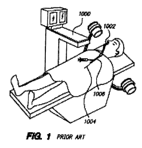
[0039] Fig. 1 is an illustration of a patient being treated by a prior art
system for catheter-
based sinus surgery according to prior art techniques.
[0040] Fig. 2a is a side elevation view of a sinus ostium finder in accordance
with the
invention shown with a guide wire in a retracted position.
[0041] Fig. 2b is a side elevation view of the sinus ostium finder of Fig. 3a
shown with a
guide wire in an extended position.
[0042] Fig. 3a and 3b are fluoroscopic images (A-P orientation) showing the
sinus ostium
finder of Figs. 2a and 2b locating the left maxillary sinus ostium and
deploying a guide wire.
[0043] Fig. 4a through Fig. c are fluoroscopic images (A-P orientation)
showing another
embodiment of a sinus ostium finder locating the left frontal sinus ostium and
deploying a guide
wire.
[0044] Fig. 5a is a perspective view of another embodiment of a sinus ostium
finder in
accordance with the invention shown with a guide wire in a retracted position.
[0045] Fig. 5b is a perspective view of the sinus ostium finder of Fig. 4a
shown with the
guide wire in an extended position.
[0046] Fig. 5c is a cross-section of the sinus ostium finder of Fig. 4a and 4b
taken through
line A-A.
[0047] Fig. 6 is a perspective view of another embodiment of a sinus ostium
finder in
accordance with the invention.
[0048] Fig. 7 is a perspective view of the distal end portion of another
embodiment of a sinus
ostium finder in accordance with the invention.
[0049] Figs. 8a is a perspective view of another embodiment of a sinus ostium
finder in
accordance with the invention shown with a guide wire in a partially detached
position.
[0050] Fig. 8b is a perspective view of a distal end portion of another
embodiment of a sinus
ostium finder in accordance with the invention.
Doc. # CC-210134 v.1
CA 02732769 2011-01-28
WO 2010/014799 PCT/US2009/052236
Docket No. 83529.0029.PCT
[0051] Fig. 9 is a partial perspective view of another embodiment of the sinus
ostium finder
of the invention.
[0052] Fig. I Oa is a front elevation view of a distal end portion of another
embodiment of the
invention, shown without the guide wire.
[0053] Fig, I Ob is a longitudinal sectional view taken through line B-B of
the distal end
portion of Fig. I Oa shown with a guide wire positioned within the internal
longitudinal channel.
[0054] Fig, I Oc shows the distal end portion of Fig. I Ob with the guide wire
partially
removed from the internal longitudinal channel.
[0055] Fig. 11 a is a perspective view of the distal end portion of another
embodiment of a
sinus ostium seeker in accordance with the invention shown without a guide
wire.
[0056] Fig. 1 lb is a cross-sectional view of the distal end portion of Fig. 1
la taken through
line C-C.
[0057] Fig. 12a is a cross-sectional view of a distal end portion of another
embodiment of a
sinus seeker apparatus in accordance with the invention shown without a guide
wire.
[0058] Fig. 12b shows the distal end portion of Fig. 12b with a guide wire.
[0059] Fig. 12c shows the distal end portion of Fig. 12b including a
lubricant.
[0060] Fig. 13 is a flow chart illustrating one embodiment of the methods of
the invention.
[0061] Fig. 14 is a cross-sectional view of anatomy proximate a maxillary
sinus.
[0062] Fig. 15 is a cross-sectional view depicting use of a guide, guide wire
and balloon
catheter for treating a maxillary sinus.
[0063] Fig. 16 is a side view of one embodiment of a probe device with a
dilator.
[0064] Figs. 17a-d are cross-sectional views depicting treating a maxillary
sinus with the
device of Fig. 16.
[0065] Figs. 18a-c are partial cross-sectional views depicting use of a probe
over a shaped
mandrel.
[0066] Figs. 19a-b are perspective views of another approach to a probe device
with a
dilator.
11 Doc. # CC-210134 v.1
CA 02732769 2011-01-28
WO 2010/014799 PCT/US2009/052236
Docket No. 83529.0029.PCT
[0067] Fig. 20 is a side view of yet another embodiment of a probe device.
[0068] Fig. 21 is a perspective view of another approach to a probe device.
[0069] Fig. 22 is a perspective view of yet another approach to a probe
device.
[0070] Fig. 23 is a partial cross-sectional view depicting another embodiment
of a probe
device.
[0071] Fig. 24 is a partial cross-sectional view depicting another alternate
embodiment of a
probe device.
[0072] Fig. 25 is a perspective view depicting a probe device including
details of a handle
assembly.
[0073] Fig. 26 is a partial cross-sectional view depicting a probe with a
finder tip.
[0074] Fig. 27 is a perspective view of a handle for a probe device.
[0075] Figs. 28a-c depict steps involved in a method of use of the device of
Fig. 26.
12 Doc. # CC-210134 v.1
CA 02732769 2011-01-28
WO 2010/014799 PCT/US2009/052236
Docket No. 83529.0029.PCT
DETAILED DESCRIPTION
[0076] This invention is not limited to particular embodiments described, as
such may, of
course, vary. It is also to be understood that the terminology used herein is
for the purpose of
describing particular embodiments only, and is not intended to be limiting,
since the scope of the
present invention will be limited only by the appended claims.
[0077] Where a range of values is provided, it should be understood that each
intervening
value, to the tenth of the unit of the lower limit unless the context clearly
dictates otherwise,
between the upper and lower limits of that range is also specifically
disclosed. Each smaller
range between any stated value or intervening value in a stated range and any
other stated or
intervening value in that stated range is encompassed within the invention.
The upper and lower
limits of these smaller ranges may independently be included or excluded in
the range, and each
range where either, neither or both limits are included in the smaller ranges
is also encompassed
within the invention, subject to any specifically excluded limit in the stated
range. Where the
stated range includes one or both of the limits, ranges excluding either or
both of those included
limits are also included in the invention.
[0078] Unless defined otherwise, all technical and scientific terms used
herein have the same
meaning as commonly understood by one of ordinary skill in the art to which
this invention
belongs. Although any methods and materials similar or equivalent to those
described herein can
be used in the practice or testing of the present invention, the preferred
methods and materials
are now described. All publications mentioned herein are incorporated herein
by reference to
disclose and describe the methods and/or materials in connection with which
the publications are
cited.
[0079] It must be noted that as used herein and in the appended claims, the
singular forms
"a", "an", and "the" include plural referents unless the context clearly
dictates otherwise. Thus,
for example, reference to "a tube" includes a plurality of such tubes and
reference to "the shaft"
includes reference to one or more shafts and equivalents thereof known to
those skilled in the art,
and so forth.
[0080] The publications discussed herein are provided solely for their
disclosure prior to the
filing date of the present application. Nothing herein is to be construed as
an admission that the
13 Doc. # CC-210134 v.1
CA 02732769 2011-01-28
WO 2010/014799 PCT/US2009/052236
Docket No. 83529.0029.PCT
present invention is not entitled to antedate such publication by virtue of
prior invention.
Further, the dates of publication provided may be different from the actual
publication dates
which may need to be independently confirmed.
[0081] Turning now to Fig. 1, an illustration of a patient being treated by a
system for
catheter-based minimally invasive sinus surgery according to prior art
techniques is shown. A
C-arm fluoroscope 1000 that is useable to visualize a guide catheter 1002, a
guide wire 1002, and
a working device 1006 (e.g., a balloon catheter, other dilatation catheter,
debrider, cutter, etc.).
The guide tube 1002 may be introduced under direct visualization,
visualization provided by
fluoroscope 1000 and/or from endoscopic visualization, to place the distal end
of tube 1002 at a
location associated with an ostium of a sinus to be treated. Guide wire 1004
is then inserted
through tube 1002 and advanced to extend the distal end of guide wire 1004 to
the ostium to be
treated. Proper placement often involves advancement and retraction of the
distal end of guide
wire 1004 until it has been visually confirmed that the guide wire is properly
positioned.
Working device 1006 is next passed over the guide wire 1004 to the target
location where a
surgical procedure is to be performed. After performance of the surgical
procedure, the working
device 1006 is deactivated and withdrawn from the patient, after which the
guide wire 1004 and
guide catheter 1002 are withdrawn to complete the procedure.
[0082] Referring now to Figs. 2a and 2b, a sinus ostium seeker, finder or
locator device 10 in
accordance with the invention is shown. The sinus finder 10 of the invention
comprises a shaft
or body 11 having a proximal end 12, a distal end 14, and a curved portion or
region 16. Shaft
11 and curved portion 16 define an elongated tubular shape and support a guide
wire 18 (Fig. 2b)
within an internal channel or cavity (not shown) that extends through shaft 11
and curved region
16 to distal end 14. Curved region 16 may be adjacent to distal end 14 or
separated from distal
end 14 by a straight or uncurved portion of shaft or body 11. Guide wire 18 is
extensible and
retractable from shaft 11 through the distal end 14. A probe tip 20 on guide
wire 18 is
configured for locating a target ostium. Guide wire 18 is shown in a retracted
position in Fig. 2a
wherein probe tip 20 is adjacent or proximate to distal end 14, and in an
extended position in Fig.
2b wherein probe tip 20 is positioned away from distal end 14. Proximal end 12
may be joined
to a handle (not shown). The extension and retraction of guide wire 18 may be
controlled by
application of suitable force to guide wire 18.
14 Doc. # CC-210134 v.1
CA 02732769 2011-01-28
WO 2010/014799 PCT/US2009/052236
Docket No. 83529.0029.PCT
[0083] Shaft 11 and curved portion 16 are shown as integral portions of a
single piece or unit
in the embodiment of Fig. 2a and 2b. In certain embodiments the shaft 11 and
curved portion 16
may comprise separate components that are joined together. The curved portion
16 as shown
defines an angle of approximately 90 degrees or slightly greater than ninety
degrees. This angle
may vary as required for different uses of the invention. Preferably, curved
portion defines an
angle of between about 0 degrees and about 180 degrees, and more preferably
between about 0
degrees and about 120 degrees, and providing several iterations at 0 degrees,
30 degrees, 70
degrees, 90 degrees and 110 degrees, or as required to accommodate a
particular sinus surgery
operation.
[0084] Curved portion 16 may be detachable from the remainder of shaft 11 and
interchangeable to allow variation of curvature. Curved portion 16 and/or body
11 in many
embodiments are resilient to facilitate positioning within a patient's
paranasal cavity. In certain
embodiments curved portion 16 and shaft 11 are malleable or bendable. In still
other
embodiments curved portion 16 is resilient or malleable, and shaft 11 is
substantially rigid in
nature.
[0085] Probe tip 20 is structured and configured to facilitate location of a
target ostium. In
many embodiments probe tip 20 is spherical or oblong in shape, but may be
varied in shape as
required for different uses of the invention. Probe tip 20 and distal end 14
are structured and
configured to provide atraumatic surfaces to minimize trauma or damage to the
patient's
paranasal cavity. Probe tip 20 may be detachable from guide wire 18 and
interchangeable, so
that different sized and/or shaped probe tips 20 may be utilized for location
of different sinus
ostia.
[0086] Guide wire 18 may be extended or retracted manually by hand actuated,
electric, or
air-driven mechanism (not shown), such as a slide, rotatable crank, winch
device, reel assembly,
or the like. In certain embodiments the extension and retraction of guide wire
18 may be
achieved by an electric or air-driven motor (not shown) that is mechanically
interfaced with
guide wire 18. Guide wire 18 is resilient or flexible in many embodiments to
allow the user to
easily locate probe tip 20 to a desired location. In certain embodiments guide
wire 18 may be
bendable or malleable rather than resilient.
15 Doc. # CC-210134 v.1
CA 02732769 2011-01-28
WO 2010/014799 PCT/US2009/052236
Docket No. 83529.0029.PCT
[0087] Shaft 11, curved portion 16, probe tip 20, guide wire 18, as well as
components of the
invention in the several embodiments described herein, may be made of various
metals or metal
alloys, or polymeric materials such as engineering resins, or composite
materials thereof, or
various combinations of such materials. Preferably biocompatible materials, or
coatings of
biocompatitble materials, are utilized for shaft 11, curved portion 16, probe
tip 20 and guide wire
18 to minimize trauma to paranasal cavity tissue that comes in contact with
sinus seeker 10.
Shaft 11 and curved portion 16 in many embodiments are integral portions of a
single
component. Shaft 11 and/or curved portion 16 may be malleable, as noted above,
such that the
angle of curved portion 16 is adjustable by bending to accommodate a
particular use. Shaft 11,
curved portion 16, probe tip 20 and guide wire 18 and other components of the
invention in
many embodiments are made of materials that are autoclavable or otherwise
sterilizable so that
the apparatus 10 or individual components may be re-used. In certain
embodiments the shaft 11,
curved portion 16, probe tip 20 and guide wire 18 may be made of inexpensive,
disposable
materials.
[0088] In Figs 3a and 3b, the sinus ostium finder 10 is shown fluoroscopically
in use to
locate the left maxillary sinus ostium O1. The body or shaft 11 of sinus
ostium finder is
positioned within the patient's paranasal cavity P, and guide wire 18 (Fig.
3b) is extended from
body 11 to locate ostium O1. Guide wire 18 is resilient or malleable as noted
above, and may
undergo flexing or bending over a substantial range of angle. As shown in Fig.
3b, the portion of
guide wire 18 adjacent probe tip 20 is approximately parallel with the portion
of guide wire 18
adjacent to distal end 14, indicating a flexion angle of approximately 180
degrees in guide wire
18 between distal end 14 and probe tip 20. Once the guide wire 18 has been
positioned into the
maxillary sinus cavity O1, shaft 11 and curved portion 16 may be removed or
withdrawn from
the patient's paranasal cavity P while leaving behind the guide wire 18, thus
allowing other
working devices (not shown) to be introduced over the guide wire 18 and then
into the target
sinus. In certain embodiments, the sinus ostium finder 10 may include radio-
opaque markings
(not shown) on shaft 11, curved portion 16, probe tip 20 and/or guide wire 18
to facilitate
fluoroscopic visualization of the finder 10 and help in the navigation of
finder 11 within the
patient's nasal passageway P. The radio-opaque markings may comprise, for
example, gradation
markings to show dimensions or distances and numerical indicia identifying the
gradation
markings.
16 Doc. # CC-210134 v.1
CA 02732769 2011-01-28
WO 2010/014799 PCT/US2009/052236
Docket No. 83529.0029.PCT
[0089] Referring now to Figs. 4a through 4c, there is shown another embodiment
sinus
ostium finder 21 in accordance with the invention, with like reference numbers
used to denote
like parts. The sinus ostium finder 21 is shown fluoroscopically in use to
locate the left frontal
sinus ostium 02 of a patient. In the embodiment of Figs. 4a through 4c, the
curved region 16 of
sinus ostium finder 21 defines an angle of approximately 150 degrees of
curvature. The sinus
ostium finder 10 of Figs. 3a and 3b, in comparison, has a curved region 16
that defines an angle
of approximately 110 degrees. In other respects the sinus ostium finder 21 is
identical to sinus
ostium finder 10.
[0090] In Fig. 4a, sinus ostium finder 21 is shown inserted into paranasal
cavity P, with
guide wire (not shown) in a fully retracted position such that probe tip 20 is
positioned adjacent
distal end 14. In Fig. 4b, guide wire 18 is shown partly extended from distal
end 14. In Fig. 4c,
guide wire is more fully extended such that probe tip 20 at the end of guide
wire 18 is able to
locate ostium 02.
[0091] Figs. 5a, 5b and 5c show yet another embodiment of sinus seeker or
finder 22 in
accordance with the invention, wherein like reference numbers are used to
represent like parts.
The sinus seeker 22 includes a handle portion 24 having distal and proximal
end portions 26, 28
respectively (with only part of the proximal end portion 28 being shown). In
the embodiment of
Figs. 5a-5c, handle 24 is of elongated cylindrical configuration and is
structured to allow a user
to manually adjust the position of shaft 11 and curved portion 16 within a
paranasal cavity.
Handle 24 is joined to the proximal end 12 of body or shaft 11 at joint 30 by
means of internal
threading (not shown), snap fitting or other suitable attachment means, and
may be detachable
from shaft 11 and interchangeable. Alternatively, handle 24 may be made
integral with shaft 11.
Curved portion 16 is joined to body or shaft 11 at joint 32 by internal
threading (not shown),
snap fitting or other suitable attachment means, and may be detachable from
shaft 11 as noted
above. Alternatively, curved portion 16 may be integral with shaft 11.
[0092] An interior opening or channel 34 extends longitudinally through handle
24, shaft 11
and curved portion 16, with channel 34 being configured to slidably or movably
accommodate
guide wire 18. In general, the inner diameter of the longitudinal channel 34
ranges from about
0.5 mm to about 5 mm, and more preferably from about lmm to about 3mm,
depending on the
size of guide wire 18 utilized with the invention.
17 Doc. # CC-210134 v.1
CA 02732769 2011-01-28
WO 2010/014799 PCT/US2009/052236
Docket No. 83529.0029.PCT
[0093] Referring more particularly to Fig. 5c, the curved portion 16 of sinus
seeker 22 may
further comprise a flexible or resilient outer sheath 36 and an internal
element 38 within sheath
36. In the embodiment of Fig. 5c, outer sheath 36 is of circular cross-
sectional shape, while
internal element 38 is of a "U" or "C" cross-sectional shape such that
longitudinal channel 34 and
guide wire 18 are located between portions or ends 40, 42. Internal element 38
may be
removable from sheath 36 and interchangeable. Internal element 38 may be of
higher modulus
material than sheath 36, such that the curvilinear shape and flexural
properties of internal
element 38 are imparted to curved portion 16. The material of internal portion
38 may be
selected for desired flexural or malleable properties. In many embodiments
internal element may
also extend through shaft 11, or through shaft 11 and handle. In certain
embodiments the outer
sheath 36 may comprise a higher modulus material than internal element 38 such
that the shape
and flexural properties of the curved region are derived from sheath 36 rather
than internal
element 38. In many embodiments internal element 38 may also extend through
shaft 11, or
through shaft 11 and handle 24.
[0094] Referring now to Fig. 6, another embodiment of a sinus ostium finder or
seeker 44 in
accordance with the invention is shown, with like reference numbers used to
denote like parts.
The handle 24 of sinus seeker 44 is of elongated cylindrical shape and
includes a longitudinal
slot 46 that communicates with interior channel 34. A knob or actuator element
48 is slidably
mounted within slot 46. Knob 48 is mechanically coupled to guide wire 18 such
that movement
of knob towards distal end 26 of handle 24 advances guide wire 18 and probe
tip 20 from distal
end 14, as shown in Fig. 6, when actuator 48 is positioned adjacent to distal
end of slot 46.
Sliding of actuator 48 to the proximal end of slot 46 results in a
corresponding retraction of guide
wire 18 and probe tip 20, as illustrated in phantom lines.
[0095] Referring next to Fig. 7, there is shown a portion of another
embodiment of a sinus
ostium finder 50 in accordance with the invention. Sinus seeker 50 includes an
outer sheath 52
of substantially tubular shape, and an inner sheath 54 positioned within outer
sheath 52. Inner
sheath 54 is also of substantially tubular shape. A longitudinal channel 34
extends through inner
sheath 54 and is structured and configured to slidably accommodate guide wire
18. In the
embodiment of Fig. 7, longitudinal channel 34 is of substantially circular
cross-sectional shape.
18 Doc. # CC-210134 v.1
CA 02732769 2011-01-28
WO 2010/014799 PCT/US2009/052236
Docket No. 83529.0029.PCT
[0096] Outer sheath 52 may extend along a portion of, or the entire length of
shaft 11,
including the curved region (not shown). Inner sheath 54 may likewise extend
along a portion
of, or the entire length of, the curved portion and shaft. Inner sheath 54 may
be slidably
extensible and retractable with respect to outer sheath 52, such that during
extension the distal
end 56 of inner sheath 54 moves away from distal end 58 of outer sheath, and
during retraction
the distal end 56 of inner sheath 54 approaches distal end 58 of outer sheath
52. The extension
and retraction of inner sheath 54 with respect to outer sheath 52 may be
controlled by an actuator
knob such as knob 48 in Fig. 6
[0097] Inner sheath 54 may be of higher modulus material than outer sheath 52,
such that the
shape and mechanical properties of inner sheath are imparted to curved portion
and/or shaft (not
shown) of the sinus seeker 50. In other embodiments the outer sheath 52 may
comprise higher
modulus material than that of the inner sheath. The material of inner sheath
52 and/or outer
sheath may be selected for specific flexural or malleable properties in
accordance with the
desired use of the invention.
[0098] Figs. 8a shows yet another embodiment of a sinus seeker 60, wherein
like reference
numbers denote like parts. The distal end 14 of sinus seeker 60 includes a
longitudinal slot 62
that communicates with the internal longitudinal channel 34. Slot 62 may
extend along the
length of all or portion of curved region 16 and shaft 11. Actuation of knob
48 in the manner
described above allows guide wire 18 to be advanced or retracted with respect
to distal end 14.
Fig. 8a shows knob 48 positioned adjacent to proximal end 28 of handle 24,
corresponding to a
retracted position for guide wire 18.
[0099] Slot 62 allows guide wire 18 to be removed from channel 34 through slot
62, as well
as by extension from distal end 14. Curvilinear portion 16 in this regard may
be made of
resilient material such that guide wire 18 is retained within channel 34 under
normal conditions,
but can "snap" out of slot 62 to disengage from channel 34 upon application of
a lateral force to
guide wire 18. As shown in Fig. 8a, guide wire 18 is partially disengaged from
channel 34 and
slot 62. Disengagement of guide wire 18 through slot 62 as provided by sinus
seeker 60
facilitates removal and interchanging of guide wire 18 and probe tip 20, and
facilitates removal
of shaft 11, curved portion 16 and distal end 14 from a target sinus or
adjacent regions of the
paranasal cavity while leaving guide wire 18 and probe tip 20 in place. A
working device (not
19 Doc. # CC-210134 v.1
CA 02732769 2011-01-28
WO 2010/014799 PCT/US2009/052236
Docket No. 83529.0029.PCT
shown) may then be directed along guide wire 18 to the target sinus to carry
out surgical
procedures.
[00100] In the embodiment of Fig. 8a, shaft 11 is extensible and retractable
with respect to
handle 24. A sleeve or collar 64 supports shaft 11 and may be tensioned by
means of a screw or
threaded parts (not shown) to secure shaft in place. Loosening of collar 64
allows shaft 11 to be
extended or retracted from collar 64 and handle 24 to provide a different
length and
configuration to sinus seeker 60. Once shaft 11 has been adjusted to a desired
length by
extension or retraction from handle 24, collar 64 may be tensioned to retain
the adjusted position
of shaft 11.
[00101] Fig. 8b shows a distal portion of another embodiment of a sinus ostium
finder 68 in
accordance with the invention. The apparatus 68 includes an outer sheath 70 of
substantially
cylindrical or tubular shape, and an inner sheath 72 substantially cylindrical
or tubular shape
positioned within outer sheath 70. A longitudinal channel 34 extends through
inner sheath 72
and is structured and configured to slidably accommodate guide wire 18. In the
embodiment of
Fig. 8b, longitudinal channel 34 is of substantially circular cross-sectional
shape.
[00102] A longitudinal slot 74 extends through inner sheath 72 and
communicates with
longitudinal channel 34. Slot 74 permits guide wire 18 to be removed from
channel 34 in a
lateral direction upon exertion of a lateral force on guide wire 18. Outer
sheath 70 may also
include a longitudinal slot 76, which extends through outer sheath 72 to
communicate with
longitudinal slot 74 and hence longitudinal channel 34. Thus, guide wire 18
may be removed
laterally from inner and outer sheaths 72, 70 via slots 74, 72 upon
application of a suitable lateral
force on guide wire 18.
[00103] In the embodiment of Fig. 8b longitudinal slots 74, 76 are aligned so
that both slots
74, 76 communicate with internal channel 34 to allow insertion and release of
guide wire 18
from channel. In certain embodiments one or both of the inner sheath 72 and
outer sheath 70
may be rotatable with respect to each other along the longitudinal axis A of
the shaft and curved
region (not shown) of the apparatus 68. Inner sheath 72, outer sheath 70, or
both may be
mechanically coupled to a rotational adjustment mechanism on the handle (not
shown of the
apparatus 68, so that the rotational position of sheath 70 and/or sheath 72
may be rotatably
adjusted to control alignment of slots 74, 76. Thus, when one of sheaths 70,
72 is rotated with
20 Doc. # CC-210134 v.1
CA 02732769 2011-01-28
WO 2010/014799 PCT/US2009/052236
Docket No. 83529.0029.PCT
respect to the other, slots 74, 76 may be moved out of alignment so that guide
wire 18 cannot be
removed through slots 74, 76, or aligned as shown in Fig. 8b so that guide
wire 18 can be
removed from channel 34 through slots 74, 76.
[00104] Outer sheath 70 may extend along a portion of, or the entire length
of, the curved
portion and shaft (not shown in Fig. 8b) of the sinus seeker apparatus 68.
Inner sheath 72 may
similarly extend along a portion of, or the entire length of, the curved
portion and shaft. Inner
sheath 72 may be slidably extensible and retractable with respect to outer
sheath 70, with distal
end 78 of inner sheath 72 moving away from distal end 80 of outer sheath 70
during extension,
and with distal end 78 moving towards distal end 80 during extension.
[00105] Fig. 9 illustrates still another embodiment of a sinus ostium seeker
82 in accordance
with the invention, wherein like reference numbers denote like parts. The
apparatus 82 includes
a longitudinal slot 84 that extends from distal end 14 to the distal end 26 of
handle 24.
Longitudinal slot 84 communicates with longitudinal channel 34, which extends
through handle
24, shaft 11 and curved region 16 to distal end 14. Slot 84 includes a
laterally curving region 86
such that the end 88 of slot 84 adjacent handle distal end 26 has a different
angular orientation
(relative to a central axis passing through the shaft 11) than the end 90 of
slot 84 adjacent distal
end 14 with respect to shaft 11 and curved region 16, such that slot 84
"twists" relative to shaft
11 as it traverses from its distal end to its proximal end. Thus, in Fig. 9,
the portion of slot 84
adjacent slot end 88 is rotated from the portion of slot 84 adjacent end 90
with respect to the
longitudinal axis (not shown) defined by shaft 11 and curved portion 16. The
laterally curving
region 86 of slot 84 facilitates the insertion and removal of guide wire 18
into or out of interior
longitudinal channel 34.
[00106] Referring now to Fig. I Oa through l Oc, a distal portion of another
embodiment of a
sinus ostium finder 92 in accordance with the invention is shown, with like
numbers used to
denote like parts. The apparatus 92 includes an opening 94 that extends
longitudinally from
distal end 14 across curved portion 16 to shaft 11. Opening 94 communicates
with internal
longitudinal channel 34. Opening 94 is structured and configured to allow a
user to grasp guide
wire 18 through opening 94, using fingers, forceps or other grasping tool (not
shown). Opening
94 facilitates the removal of guide wire 18 from the channel 34 and the
apparatus 92. In this
way, the proximal end (not shown) of the guide wire can be loaded into the
distal end 14 of the
21 Doc. # CC-210134 v.1
CA 02732769 2011-01-28
WO 2010/014799 PCT/US2009/052236
Docket No. 83529.0029.PCT
shaft 11 where the cutout 94 in the bend helps pass a stiff proximal end of
the guidewire into the
body of the guide catheter despite the sharp bend angle of the curved tip.
Thus, the guidewire
can take a less severe bend during loading. Once loaded, the system is
configured as shown in
Fig. I OB. After the system is used to place the tip of the guide wire into
the sinus, the guide
catheter can be pulled back off the proximal end of the guide wire.
[00107] Figs. 11 a and 1 lb show a distal portion of another embodiment of a
sinus ostium
finder 96 in accordance with the invention, with like reference numbers used
to denote like parts.
The apparatus 96 includes an elongated back portion 98 and an elongated front
portion 100 that
are joined together along seams 102, 104 by adhesive, heat welding or other
bonding means.
Back and front portions 98, 100 together define a tubular shape, with an
interior channel 34 of
circular cross-sectional shape between the front and back portions 98, 100.
Front and back
portions 98, 100 extend from distal end 14 along curved region 16 and shaft
(not shown), and
together define the curved region 16 and shaft (not shown) of the apparatus
96. A longitudinal
slot 106 in front portion 100 communicates with interior channel 34. In the
embodiment of Figs.
1 la and 1 lb, front and back portions 100, 98 each are semicircular in cross-
sectional shape and
impart a circular cross-sectional shape to interior channel 34.
[00108] In many embodiments back portion 98 is made of a rigid or
substantially rigid higher
modulus material, while front portion 100 comprises a resilient lower modulus
material. The
resilient nature of front portion 100 allows a guide wire (not shown) to "snap
fit" through slot
106 between ends 108, 110 (Fig. I lb). The guide wire thus can be easily
inserted into and
removed from channel 34 by application of a suitable force against front
portion 100 to force or
move the guide wire through slot 106. In certain embodiments both front and
back portions may
comprise flexible materials.
[00109] Figs. 12a through 12c provide cross-sectional views of a portion of
yet another sinus
ostium finder 112 in accordance with the invention, with like numbers used to
denote like parts.
The apparatus 112 includes an outer sheath 114 and an inner section or portion
116 positioned
within sheath 114. Sheath 114 is of elongated tubular configuration and
defines an interior
channel 34 that extends longitudinally through the curved region and shaft
(not shown) of the
apparatus 112. Inner section 116 fits within channel 34 and extends along all
or a portion of the
shaft and curved region. A slot 118 extends longitudinally along sheath 114
and communicates
22 Doc. # CC-210134 v.1
CA 02732769 2011-01-28
WO 2010/014799 PCT/US2009/052236
Docket No. 83529.0029.PCT
with interior channel 34. In the embodiment shown in Figs. 12a through 12c,
sheath 114 is of
circular cross-sectional shape and inner section 116 is of arcuate or
semicircular cross-sectional
shape such that the cross-sectional shape of inner section 116 conforms to the
cross-sectional
shape of sheath 114. A guide wire 18 (Figs. 12b and 12c) fits within channel
34.
[00110] Sheath 114 is made of resilient material such that guide wire 18 can
be forced
between ends or portions 120, 122 through slot 188 and into channel 34. Guide
wire 18 then is
retained within channel 34 until a suitable force is applied to wire to bring
wire through slot 118
between ends 120, 122 and out of channel 34. Inner section 116 in many
embodiments is made
of a rigid or substantially rigid material, or a material of higher modulus
than that of sheath 114.
[00111] A coating 124 (Fig. 12c) of biocompatible low friction coefficient
material such as
TEFLONTM may be included on guide wire 18 to facilitate sliding motion of
guide wire 18
within channel 34 and to reduce or minimize possible trauma to a patient's
paranasal cavity. A
lubricating oil or gel 126 (Fig. 12c) may be included within channel 34 to
facilitate movement of
guide wire 34 within channel 34.
[00112] The methods of the invention will be more fully understood by
reference to the flow
chart of Fig. 13, as well as Figs. 2-12. The sequence of the events described
below may vary and
should not be considered limiting. Not all events described may occur in a
particular use of the
invention, and in certain embodiments additional events not shown in Fig. 13
may be carried out.
[00113] In event 200, the shaft 11 of the sinus ostium finder of the invention
is inserted into a
patient's paranasal cavity. As shown in Figs. 3 and 4, this event is carried
out by inserting distal
end 14, followed by curved region 16 and shaft 11 into paranasal cavity P. The
insertion may be
carried out by a surgeon or other medical personnel, and may be monitored
fluoroscopically
and/or endoscopically, or may be carried out without visualization tools.
[00114] In event 210, the position of distal end 14 is adjusted. In many
embodiments the
adjustment is carried out manually, positioning distal end 14 by suitable
manual positioning of
handle 24. The positioning of distal end 14 in many embodiments is monitored
fluoroscopically,
so that the distal end 14, as well as shaft 11 and curved region 14 may be
visualized. It is to be
recognized that alternatively, such positioning can be visualized solely by
endoscopic
visualization. The adjustment of the position of distal end 14 is carried out
with the goal of
locating a target sinus ostium in the event(s) below. In certain embodiments
radio-opaque
23 Doc. # CC-210134 v.1
CA 02732769 2011-01-28
WO 2010/014799 PCT/US2009/052236
Docket No. 83529.0029.PCT
markings or markings provided by visually contrasting colors may be included
on distal end 14,
shaft 11 and/or curved region 16 to assist in locating the target ostium.
[00115] In event 220, the position of guide wire 18 and probe tip 11 is
adjusted by extension
of guide wire 18 from distal end 14 until probe tip 20 approaches or reaches
the target ostium.
The positioning of guide wire 18 and probe tip 20 is generally monitored
fluoroscopically, so
that the position of probe tip 20 with respect to the target ostium may be
visualized. In certain
embodiments radio-opaque and radio-transparent markings may be included on
probe tip 20
and/or guide wire 20 to assist in locating the target ostium.
[00116] In event 230, a determination is made whether or not the target ostium
has been
located. The determination is made by visually such as endoscopically,
fluoroscopically or using
light-emitting transillumination to observe the location or position of probe
tip 20 with respect to
the target ostium. In many embodiments the probe tip 20 is selected to have a
diameter that
matches that of the target ostium, and location of the target ostium is
determined by exactly
fitting the probe tip 20 into the target ostium. If the target ostium has been
located, event 240 is
carried out.
[00117] If it is determined in event 230 that the target ostium has not been
located, event 220
may be repeated by again adjusting the position of guide wire 18 and probe tip
20. This may be
carried out by retracting guide wire 18 towards distal end 14, and then re-
extending guide wire
18 from distal end 14 to adjust the position of guide wire 18 and probe tip 20
and direct probe tip
20 towards the target ostium.
[00118] In certain instances where it is determined in event 230 that the
target ostium has not
been located, both events 210 and 220 are repeated. Thus, guide wire 18 is
retracted, the
position or orientation of distal end 14 is adjusted by manually positioning
the sinus ostium
finder apparatus, and then guide wire 18 is again advanced to adjust the
position of guide wire 18
and probe tip 20.
[00119] In still further instances where it is determined in event 230 that
the target ostium has
not been located, events 200 through 220 may be repeated. In such instances
guide wire 18
would be retracted, and the sinus ostium finder withdrawn from the paranasal
cavity. Then, a
different, more suitably configured sinus ostium finder would be re-inserted
into the paranasal
cavity and events 210 through 230 are repeated. Alternatively, the probe tip
20 maybe removed
24 Doc. # CC-210134 v.1
CA 02732769 2011-01-28
WO 2010/014799 PCT/US2009/052236
Docket No. 83529.0029.PCT
from guide wire and a differently sized or shaped probe tip 20 may be
introduced to guide wire,
after which events 210 through 230 are repeated.
[00120] At event 240, shaft 11 (including curved region 16 and distal end 14)
is removed from
the paranasal cavity while leaving guide wire 18 and probe tip 20 in place in
their adjusted
position. In embodiments of the invention wherein the shaft 11 includes a
slot, guide wire 18
may be disengaged from the slot prior to removal of the shaft 11.
[00121] In event 250, a surgical or working device or devices are introduced
along the guide
wire 18 and directed along the guide wire 18 to the target ostium. Such
devices may comprise,
for example, catheters, cannula, tubes, dilators, balloons, substance
injectors, needles,
penetrators, cutters, debriders, microdebriders, hemostatic devices, cautery
devices, cryosurgical
devices, heaters, coolers, scopes, endoscopes, light guides, phototherapy
devices, drills, rasps,
saws, and the like.
[00122] In event 260 a surgical or other procedure is carried out using the
working device
introduced in event 250.
[00123] In event 270, the working device and guide wire are withdrawn from the
paranasal
cavity.
[00124] Referring now to Figs. 14 and 15, in a related approach, various
embodiments may
provide for dilating and/or remodeling a sinus ostium and/or a transitional
space leading to an
ostia. In particular, the devices, systems and methods described below are
directed to
remodeling a maxillary sinus ostium and/or a transitional space leading to a
maxillary ostium. In
alternative embodiments, ostia and/or transitional spaces of other paranasal
sinuses may be
dilated. Dilating or remodelling a transitional space may mean dilating a
general anatomical area
in the vicinity of an ostium and/or moving one or more anatomical structures
in that general
anatomical area. Such dilation or remodelling may in some cases facilitate or
enhance flow of
air, mucus and/or other substances into and/or out of a maxillary sinus.
[00125] With reference to Fig. 14, the nasal/paranasal cavity outside the
maxillary sinus has a
transitional space formed by and including the anatomical structures and
spaces called the
infundibulum I, the uncinate process U, the ethmoid bulla B, the middle
turbinate MT and the
25 Doc. # CC-210134 v.1
CA 02732769 2011-01-28
WO 2010/014799 PCT/US2009/052236
Docket No. 83529.0029.PCT
middle meatus. In various embodiments, any of these structures may be moved
and/or any of
these areas may be dilated.
[00126] Referring now to Fig. 15, the anatomy of the maxillary sinus
transitional space, like
that of the paranasal cavity and the sinuses themselves, consists of bone and
mucosa. Flexible
and rigid instruments may be conceived to remove obstruction in the
transitional space. As
shown in Fig. 15, one flexible embodiment may involve a balloon catheter 300.
The area may be
accessed using a guide 302 and guidewire 304 and the balloon catheter 300 may
be positioned in
the transitional space and inflated. However, this approach requires the
coordinated use of
several devices. The approach may further require multiple guides 302 to
position the balloon
300 appropriately.
[00127] Referring now to Fig. 16, a simplified probe device 310 can include a
malleable or
semi-rigid region 312 extending from a handle 314. The probe tip 316 can be
curved and is
contemplated to embody a dilator 318 such as a balloon. A proximal end of the
handle 314 is
equipped with a luer 320 for accepting an inflation device operable to expand
the dilator 318. In
this configuration, the curve of the probe tip 316 may be adjusted to optimize
access behind the
uncinate and in the transitional space leading to the maxillary sinus. When an
inflation device
(not shown) is attached the balloon can be inflated, creating space in the
transitional area as well
as dilating the maxillary sinus ostium. This device may be used as a single
hand instrument
under direct vision, fluoroscopy, and/or image guidance. Guides and guidewires
may be adapted
for use therewith but may not be necessary.
[00128] The balloon dilator 318 of the probe device 310 may have various
attributes and
configurations. For example, the balloon 318 may be non-compliant, semi-
compliant, or
compliant. Further there may be one or several balloons, and the balloons may
be concentric or
non-concentric. Moreover, the contemplated balloon 318 may have multiple
diameters and
lengths, multiple taper geometries, and it may end at or before the distal tip
of the probe, or
extend beyond the probe. The balloon 318 may also have modified frictional
properties to
release or gain traction on anatomy, such as a non-slip surface. In various
embodiments, the
balloon 318 may have round or non-round cross-sectional geometries to assist
re-wrap and
profile.
26 Doc. # CC-210134 v.1
CA 02732769 2011-01-28
WO 2010/014799 PCT/US2009/052236
Docket No. 83529.0029.PCT
[00129] In one contemplated approach, as shown in FIGS. 17a-d, the balloon 318
may be non-
concentric and maybe oriented to inflate on the outside of the curve 316 of
the probe 310. The
balloon 318 may relatively long, extending from the medial shaft 320 to beyond
the probe tip
316. When inflated, the balloon 318 does not push the uncinate U. However, the
balloon is
configurable to push medially on the middle turbinate MT and posteriorly on
the bulla B, thereby
opening the transitional space (See Figs. 17c and d). The balloon 318 may also
extend beyond
the probe tip 316 to ensure that the infundibulum has been remodelled and to
exert medial force
on the middle turbinate MT.
[00130] In another example, a concentric and relatively short balloon (not
shown) may push
the uncinate U anteriorly and may have some posterior impact on the bulla B.
There would not
necessarily be an impact on the middle turbinate MT. In alternative
embodiments, the same or
similar devices may be used to remove or reduce obstruction in the frontal and
sphenoid
transitional spaces. When used in conjunction with a viewing device, the
physician may be able
to open the transitional space for the maxillary sinus and visually confirm if
the ostium is open or
closed. If the ostium is closed, the surgeon may opt to use traditional
sinuplasty devices or other
methods. If the ostium is open, then removal of obstructions in the
transitional space may be a
sufficient treatment.
[00131] As shown in Figs. 18a-c, another probe device 330 for dilating the
infundibulum,
bulla, and/or middle turbinate, as well as the maxillary ostium, is shown. In
particular, probe
device 330 is configured to first access the maxillary ostium by tactile feel.
Next, a member is
advanced through the maxillary ostium and then employed to dilate the
anatomical structures in
the area. Here, the probe device 300 includes a balloon 332 configured with a
ball tip 334. The
device 330 is further configured to receive a shaped mandrel 336 within an
interior lumen 338.
[00132] As shown in Fig. 18b, the balloon portion 332 is advanced over the
mandrel 336, the
mandrel 336 directing the balloon 332 transversely. By using this structure,
the balloon 332
passes through the maxillary ostium O. Next, the balloon 332 is dilated, which
consequently
pushes the middle turbinate MT medially and the bulla B posteriorly. The
dilation also opens the
ostium 0 and infundibulum, whereas the uncinate is pushed anteriorly. The
mandrel 336 enables
the probe device 330 to tolerate the dilation pressure used to expand the
balloon 332 without
using a guidewire.
27 Doc. # CC-210134 v.1
CA 02732769 2011-01-28
WO 2010/014799 PCT/US2009/052236
Docket No. 83529.0029.PCT
[00133] The interior lumen 338 or the probe device 330 is constructed to allow
retraction over
the mandrel 336 without kinking. The mandrel 336 itself could be spring
tempered or malleable.
The mandrel 336 may also have a short coil or soft tip to reduce kinking of
the inner member
during balloon retraction. The mandrel 336 may further be constructed of a
shape memory alloy
which would conform to the balloon 336 geometry when inflated. This may also
help in
reducing stress on the interior lumen 338 and kinking during balloon
retraction.
[00134] In an alternative embodiment, the probe device 320 may be modified to
address the
sphenoid or frontal paranasal sinuses by using a substantially straight or
less severely curved
mandrel, respectively. In some embodiments, an optional sheath (not shown) may
be integrated
onto a shaft of the probe to help re-wrap the balloon and thus reduce the
overall profile of the
balloon after dilation and deflation.
[00135] In some embodiments, the probe device 320 facilitates the use of
tactile feel and
balloon advancement to confirm ostial access. This is generally desirable when
using the device
in the maxillary and sphenoid sinuses and/or their transitional areas, but it
may not work as well
in the frontal sinus. Several additional means of confirmation may also be
adapted. For
example, a fluid may be flushed through a lumen of the probe device 336. If
the fluid is seen
endoscopically in the nasal cavity, it can be assumed that the device has not
entered the sinus.
Alternatively, light fibers may be added to the tip of the device to
transilluminate a sinus. This
addition of light fibers and transillumination may be used in the maxillary,
sphenoid or frontal
sinus.
[00136] In another embodiment, an image guidance sensor may be fixed to the
tip of the probe
device 320 and tracked with an electro-magnetic system. This would provide
confirmation for
each of the sinuses. Fluoroscopy could also be used to confirm access.
Likewise, a flexible fiber
scope could be passed down the center of the probe device 320 to visualize the
area if the tip of
the device 320 has entered the target sinus.
[00137] Turning now to Figs. 19a and b, an alternative approach to a probe
device 350 is
shown. The device 350 includes a rigid or malleable shaft member 352 that
terminates with an
uncinate hook 354. A balloon 356 is configured about the hook 354 to provide a
supported
shape intended to maintain an access turn about an uncinate. Upon dilation,
the balloon 356
forms a C-like shape, and anatomy at the interventional site is moved. For
example, when
28 Doc. # CC-210134 v.1
CA 02732769 2011-01-28
WO 2010/014799 PCT/US2009/052236
Docket No. 83529.0029.PCT
placed into the sinuses, a terminal end 358 of the device 350 opens an
infundibulum, an area 360
proximal the end 358 opens a supra-balloon space, a middle section 362 opens
the middle meatus
and a most proximal portion 364 of the balloon moves the middle turbinate.
Thus, the device
350 can greatly and uniformly open the meatus and infundibulum to make easier
the subsequent
access the to the frontal, maxillary or ethmoid sinuses.
[00138] In a related device 370, and with reference now to Fig. 20, a balloon
portion 372 is
eccentrically located on a hook portion 374. The hook portion 374 can be
either rigid or
malleable. A shaft 376 is configured proximal to the balloon portion 372, and
the device 370 can
further include a tube 376 extending to a terminal end of the device, the tube
376 including an
exit for a guidewire (not shown).
[00139] Yet further approaches to probe devices are depicted in Figs. 21 and
22. In one
embodiment, as shown in Fig. 21, a probe device 380 can embody a shaft 382
with a curved
middle meatus/maxillary balloon 384 attached thereto. The balloon 384 includes
a distal portion
forming a maxillary region 386 and a proximal portion defining a middle meatus
region 388.
The balloon 384 is configured to have a built-in curve to turn about a
patient's uncinate process.
[00140] In another embodiment, as shown in Fig. 22, a probe device 390 may
include a shaft
396 and a balloon 391 having a maxillary region 392 embodying an increased
diameter
configured distally and about a curve from a meatus region 394. In either this
or the previous
embodiment, the maxillary region 392 and meatus region 394 of the ballon 391
may be formed
from different balloon materials, with for example, the maxillary region 392
being more
compliant. The two regions 392, 394 can further embody different shapes such
as the maxillary
side defining a dog-bone configuration. In this way, use of the probe devices
380, 390
accomplishes simultaneous dilation of both the maxillary and middle meatus
regions, while
protecting the uncinate from trauma. Also, the meatus balloon region 388, 394
of these devices
can function to anchor the maxillary balloon region 386, 392 against sudden
movement.
[00141] Referring now to Fig. 23, in another embodiment, a transition space
dilation tool 400
may include a balloon 402 attached about a distal portion of a semi rigid,
rigid or malleable shaft
404. A distal end of the shaft 404 can be equipped with a ball-like atraumatic
tip 406. The
balloon 402 can be mechanically captured by a ball-shaft interface 408 to
thereby minimize a
neck region of the balloon 402. Moreover, the shaft 404 may include an
inflatable lumen exit
29 Doc. # CC-210134 v.1
CA 02732769 2011-01-28
WO 2010/014799 PCT/US2009/052236
Docket No. 83529.0029.PCT
410 for expanding the balloon 402, which may be pre-shaped into a hook. An
inflation hub 411
may further be included to provide a dilation means.
[00142] The balloon 402 of this probe device 400 generally includes three
regions: an
infundibulum balloon region 412; a bullar balloon region 414; and a middle
meatal balloon
region 416. The infundibulum region 412 is configured to dilate infundibulum
transitional
space, the bullar region 414 compresses bulla to make room for scopes or other
devices and the
middle meatal region 416 opens meatal space to reduce potential trauma from
subsequent device
insertion. Each region may have different diameters or thicknesses and can
define a myriad of
shapes, angles and curves. Further, the shaft 404 can be single or multi-
lumened, and the balloon
can be compliant, semi-compliant or non-compliant.
[00143] With reference now to Fig. 24, an infundibular meatal or recess-
transition space
dilator 420 may include a shaft 422 having a ball tip 424 and a curved distal
portion surrounded
by a balloon 426. An inflation opening 428 is provided in the shaft 422 region
surrounded by the
balloon 426 and notably, the ball tip 424 is encapsulated by the balloon 426.
The balloon 426
further includes a middle turbinate region 430, a bullar region 432 and an
infundibular region
434. The balloon can be molded with a closed end or can be traditionally
molded and then the
tip molded over and closed. As before, the balloon material can be non-
elastic, partially elastic,
compliant, non-compliant or partially compliant.
[00144] In yet another approach, and with reference now to Fig. 25, a probe
device 440
includes a separate internal element 442 that can be advanced and retracted to
give tactile
feedback to find a maxillary ostium. Once the maxillary ostium has been found,
the user can
independently advance a balloon portion 444 over a hypotube portion 446. The
internal element
442 can be a wire or some other flexible element which can be extended out of
the shaped
hypotube 446 to probe for the sinus. In certain approaches, the internal
element 442 can be
coiled wire over a nitinol core, a solid flexible wire or plastic member, a
light wire, or other
flexible element. The shaped hypotube 446 can be formed from a steel or
plastic tube that has a
curve to direct the internal flexible member 442 and external balloon 444. In
various
embodiments, the tube can be pre-shaped for a specific sinus or malleable to
allow the user to
shape the device for a target trajectory.
30 Doc. # CC-210134 v.1
CA 02732769 2011-01-28
WO 2010/014799 PCT/US2009/052236
Docket No. 83529.0029.PCT
[00145] The balloon 444 is configured about an outside of the shaped hypotube
446 and can
be advanced independently of the internal element 442. A balloon shaft 448 is
provided with
one or two lumens to allow advancement of the balloon 444 and for inflation.
The shaft 448 may
extend over around the hypotube curve 446 or can start proximal the curve. A
balloon pusher
450 is further provided to translate force from a balloon driver 452 to the
balloon 444. This
structure also may act as a manifold for fluid to fill/pressurize the balloon.
[00146] A probe handle 454 can be shaped to form an elongate structure and can
be slender
for easy holding and control. The handle 454 can further include a finger grip
for assisting with
traction for holding or advancing elements. The handle 454 also includes an
internal element
control 455 which slides within the handle 454 and allows the user to control
advancing/
retracting the internal element 442 as well as receiving tactile feedback from
the internal element
442. The handle also includes a balloon driver 456 which slides within the
handle 454 and
allows user to advance/retract balloon. Rails 457 are further provided for
guiding the movement
of the internal control element 455 and push rods (not shown) connecting the
balloon driver to
the balloon pusher. In various alternative embodiments, balloon inflation
fluid may be passed
through one or both of the rails 457, if they are tubes, or alternatively, the
probe device 440 may
include one or more separate inflation lumens for delivering fluid/pressure to
the balloon.
[00147] In certain circumstances, when attempting to access a maxillary sinus
opening with a
probe device including a shaping mandrel, it can be useful to have a mandrel
with a very tight
radius to send the probing end of the tip into the right place. It may also be
useful to have the
starting tip length itself be relatively short. The tip may need to have a
finer selection end on the
front. If the balloon catheter lumen itself is too large, bulky and/or stiff,
it can pass by an ostium
opening when advanced, without entering the opening, because the opening can
be more like a
hole in the side of the wall, and not necessarily at the end of an
infundibular pocket. Further,
even if the tip does momentarily engage the ostium, the stiffness of the
balloon catheter can
overpower it, and it will not enter the opening once the balloon is attempted
to be advanced.
Thus, a more flexible lead-in section that supports the balloon stiffness
transition into the
opening may be employed in some embodiments.
[00148] Turning now to Fig. 26, a probe device 460 having a small, short
angled tip to select
the side-hole ostium when that anatomy is present is described. This device
includes a floppy
31 Doc. # CC-210134 v.1
CA 02732769 2011-01-28
WO 2010/014799 PCT/US2009/052236
Docket No. 83529.0029.PCT
lead-in catheter section that also has good column strength that permits
advancement forward
into an ostium to support the transition to the stiffer balloon. A nitinol
super-elastic mandrel can
be provided to form a very tight initial radius that maintains its shape as
the floppy lead-in
catheter is advanced but opens up as the stiffer balloon catheter is advanced.
[00149] As shown, the probe device 460 includes a finder tip 462, which could
be shapeable
or fixed and oriented outwardly to find an opening in a "wall" or
infundibulum. The tip 462
could be made of a polymer or could be a wire tip. The probe device 460 can
further embody a
mandrel 464 formed from shape memory, super elastic, spring steel or other
semi-rigid materials.
This helps keep angles very tight when solely selecting with a flexible
catheter. Moreover, the
probe device 460 may include a flexible finder catheter body 464, which acts
like a guidewire to
track behind the tip 462 and acts to confirm access to the sinus by advancing
without resistance.
[00150] Referring now to Fig. 27, in one embodiment, a handle 470 for the
probe device 460
may include a thumb pusher 472 operatively connected to the balloon portion
474 so that
manipulation of the pusher 472 advances and/or retracts the balloon 474. The
handle 470 may
further include a stabilization substructure 473 sized and shaped to receive
figures of an operator.
[00151] With reference now to Figs. 28a-28c, in one embodiment of a method for
using a
probe device 460, the probe device 460 may be placed in a pre-deployment
configuration (Fig.
28a), with the mandrel 464 loaded within the flexible catheter body 466.
During mid-
deployment (Fig. 28b), the mandrel 464 is withdrawn proximally, but is left to
extend beyond the
balloon portion 474. Next, the mandrel 464 is further withdrawn proximal the
balloon 474 (Fig.
28c) to achieve complete deployment. The balloon 474 can then be expanded to
create space or
accomplish desired tissue manipulation.
[00152] The above description has often focused on embodiments of devices,
systems and
methods for use in maxillary paranasal sinuses. In some cases, however, the
above-described
embodiments may be used in procedures involving frontal, sphenoid and/or
ethmoid sinuses. In
some cases, these embodiments may be used as described in these other sinuses,
while in other
cases minor modifications may be made to the devices, systems or methods to
make them more
amenable to use in the frontal, sphenoid or ethmoid sinuses. In any event, the
description above
related to usage in the maxillary sinus should not be interpreted to limit the
present invention to
applications in only that sinus.
32 Doc. # CC-210134 v.1
CA 02732769 2011-01-28
WO 2010/014799 PCT/US2009/052236
Docket No. 83529.0029.PCT
[00153] While the present invention has been described with reference to the
specific
embodiments thereof, various changes may be made and equivalents may be
substituted without
departing from the true spirit and scope of the invention. In addition, many
modifications may
be made to adapt a particular situation, material, composition of matter,
process, process step or
steps, to the objective, spirit and scope of the present invention. All such
modifications are
intended to be within the scope of the claims appended hereto.
33 Doc. # CC-210134 v.1