Note: Descriptions are shown in the official language in which they were submitted.
CA 02732980 2016-07-14
55232-15
DESCRIPTION
CAPRIN-1 AS A TARGET FOR DIAGNOSING CANCER
TECHNICAL FIELD
The present invention relates to a method for detecting cancer using
CAPRIN-1 as a tumor marker.
BACKGROUND ART
Cancer is the leading cause of death.
Treatment currently
performed for cancer is mainly symptomatic therapy that mostly consists of
surgical therapy with a combination of radiation therapy and chemotherapy.
Owing to advancements in medical technology, cancer is now almost a
curable disease if it can be detected early. Hence, a method for detecting
cancer, by which detection can be conveniently performed using serum,
urine, or the like without imposing physical or economic burdens on cancer
patients, is now required.
As a cancer diagnostic method using blood or urine, a method for
measuring a tumor product such as a tumor marker has recently become
popular. The term "tumor product" refers to a tumor-associated antigen, an
enzyme, a specific protein, a metabolite, a tumor gene, a tumor gene product,
a tumor suppressor gene, and the like. Carcinoembryonic antigen CEA,
glycoprotein CA19-9, CA125, prostate-specific antigen PSA, calcitonin,
which are peptide hormones produced in the thyroid and the like are used as
tumor markers for diagnosis of some cancer types. However, tumor markers
useful for cancer diagnosis are absent for many cancer types. Also, most
currently known tumor markers are present in only trace amounts (on roughly
a pginaL order) in body fluids. Therefore, highly sensitive measurement
methods or special techniques are required for detecting such tumor markers.
-1-
CA 02732980 2011-02-02
Under the current circumstances, it is expected that provision of a new
cancer testing means capable of detecting various types of cancer with high
sensitivity involving a convenient procedure creates diagnostic applications
for various types of cancer.
Also, such cancer testing means is very useful if it is capable of not
only detecting cancer but also diagnosing cancer having developed in a
location invisible to the naked eye, the extent of cancer, the malignancy or
postoperative course of cancer, recurrence, metastasis, and the like.
Specifically, if diagnosis of cancer that has developed in a location
invisible to the naked eye becomes possible, such cancer testing means
would be useful for early detection of cancer within a location such as an
intraperitoneal part that is difficult to recognize. Also, a tumor that does
not have a grossly visible size such as cancer that is undetectable even by
ultrasonography, CT (computer tomography), or MRI (nuclear magnetic
resonance imaging) can be detected.
Additionally, the extent of cancer is classified based on the degree
to which a tumor spreads at the primary site and the presence or the absence
of metastasis to regional lymph nodes or distant organs. In general, there
are 5 disease stages (each referred to as "stage"), and higher stage numbers
indicate more advanced stages of the disease. Strictly, the definition of
stage differs depends on organs. However, for example, cancer at stage 0 is
cancer that remains intraepithelial and cancer at stage IV is cancer that has
metastasized to a distant location. If
such extent of cancer is found,
decisions about appropriate treatment courses as well as diagnosis of the
therapeutic effects of an anticancer agent become possible. As specific
examples of decisions about treatment courses, in the case of prostate cancer
and the like, there is a type requiring no treatment because it has very low
malignancy and will almost never progress. In contrast, there is a type
requiring treatment because it is progressive and metastasizes to bone or the
- 2 -
CA 02732980 2011-02-02
like and causes patients to die painfully. Therapies such as hormone
therapy and extirpative surgery are each associated with an adverse reaction.
Thus, therapies should be appropriately determined and decided upon.
Also, if evaluation concerning the selection of an anticancer agent can be
appropriately made or if timing or the like for the termination of
administration of an anticancer agent can be appropriately determined,
physical and economical burdens on patients can also be reduced.
Therefore, it is important to be able to diagnose the extent of cancer.
One of the characteristics of cancer cells is that they undergo
blastogenesis; that is, dedifferentiation.
Except for some cancer types,
poorly differentiated or undifferentiated cancer cells with a low degree of
differentiation rapidly grow after metastasis and result in poor prognosis
after therapy. Such cancer is said to have high malignancy. Conversely,
highly differentiated cancer cells with a high degree of differentiation
retain
the structural and functional characteristics of affected organs. Such cancer
can be said to have relatively low malignancy. If the malignancy of cancer
can be determined, the following measures can be taken. Even if the tumor
is small, a wide surgical margin can be secured upon tumor removal, when
the malignancy is high. Moreover, follow-up is possible while paying
attention to a wide range of peripheral tissue.
If diagnosis of postoperative courses including recurrence and
metastasis is possible, diagnosis of whether or not a tumor can be completely
removed by surgery becomes possible. Incomplete tumor removal likely
results in recurrence.
Hence, such diagnosis can provide criteria for
determining to more carefully perform follow-up at short intervals or to
perform early reoperation if necessary. Also, if recurrence takes place,
there is a high possibility of early detection. Detection is often delayed
when distant metastasis takes place. However, if diagnosis of metastasis
becomes possible, it becomes possible to provide criteria by which the range
- 3 -
CA 02732980 2011-02-02
of testing can be broadened to include areas other than the site of removal
and the periphery thereof.
It is known that dogs grow old 7 times faster than humans.
Recently, companion animals are being raised as family members and often
have lifestyle habits similar to those of their owners.
Therefore, it is
predictable that an owner's risk of developing cancer would be high when his
or her companion animal develops cancer. If convenient and precise cancer
diagnosis becomes possible for companion animals, it would be expected to
provide clues for preventing cancer of owners.
Currently, the number of domestic dogs in Japan is said to be about
6,700,000, and the same figure for the U.S. is said to be about 17,640,000.
Quintuple, septuple, and octuple combined vaccines and the like have
become prevalent, in addition to rabies shots,and thereby highly lethal
infectious diseases have decreased, such as canine parvovirus infection,
canine distemper virus infection, canine parainfluenza (kennel cough), canine
adenovirus-2 infection (kennel cough), infectious canine hepatitis, canine
coronavirus infection, and leptospirosis. Therefore, the average life span of
dogs has increased. Elderly dogs, which are seven years old or older,
account for 35.5% of all domestic dogs. Causes of death of domestic dogs
are also similar to those of humans, such as cancer, hypertension, and cardiac
disease, which are on the rise. In
the U.S., about 4,000,000 dogs are
diagnosed with cancer annually. Also in Japan, it is said that about
1,600,000 dogs are potentially affected with tumors.
However, convenient cancer diagnostic agents for animals have been
absent. Furtheremore, in animal medical care, testing methods that involve
photographing or filming using X-rays, CT scans, MRI scans, or the like
have not been prevalent. After palpation, a simple blood test, and testing
using X-ray photography are performed, diagnosis currently depends
significantly on the experience of veterinarians. Testing methods using
- 4 -
CA 02732980 2011-02-02
,
,
serum have been partially begun, but the methods use human tumor markers
since no canine tumor marker has been discovered.
Precise cancer diagnosis requires abdominal surgery that imposes
significant physical burdens on dogs and cost burdens on owners. If cancer
diagnosis can be conveniently made for companion animals such as dogs and
cats, it would lead to early detection or precise diagnosis of cancer and
would be expected to be useful for cancer therapy for companion animals.
Also, if such convenient cancer diagnosis using serum becomes possible, it
would be expected not only to enable cancer diagnosis but also to
significantly contribute to periodic health examinations, preoperative
diagnosis, and decisions about therapeutic strategy.
Health examination for companion animals, unlike the case of
humans, is not prevalent. Hence, detection of cancer often occurs too late,
such that an owner finds out the disease and then comes to a hospital only
after the tumor has become large in many cases. If such tumor that has
increased in size is malignant, it often results in treatment that is too
late,
even when surgical therapy such as surgery or medication using an anticancer
agent or the like is performed. Hence, when a veterinarian determines that
the tumor is malignant, anticancer agent treatment is generally performed
without surgery. If surgery is performed, measures during surgery, such as
determination of the size of margin to be secured, determination of the
amount of blood required during surgery, and measures against cell
scattering should also be strictly taken. It is desired that anticancer agent
treatment is initiated immediately after surgery and that follow-up is
performed at short intervals. Incorporation of the above cancer diagnosis
into dog health checkups that are recently increasingly prevalent and are
referred to as complete medical checkups for dogs is expected to lead to
early cancer detection.
On the other hand, in the case of a benign tumor, surgery can be
- 5 -
CA 02732980 2011-02-02
advised even if a tumor is large. After surgery, only resected areas need
care without requiring any expensive anticancer agent treatment and without
any need for apprehensions concerning follow-ups.
Under the current situation, provision of a convenient means for
detecting cancer with high sensitivity, which is applicable to cancer
diagnosis for animals, enables precise and efficient treatment and results in
a
number of advantages for both owners and veterinarians.
Cytoplasmic-and proliferation-associated protein 1 (CAPRIN-1) is
an intracellular protein that is expressed when normal cells in resting phase
are activated or undergo cell division. CAPRIN-1 is also known to be
involved in mRNA transport through intracellular formation of intracellular
stress grains with RNA and translation control, for example. Meanwhile,
CAPRIN-1 has many different names. Examples of such names include
GPI-anchored membrane protein 1 and membrane component surface marker
1 protein (M11S1), as if the protein has been known to be a membrane
protein. These different names are derived from a report (J Biol Chem. 270:
20717-20723 (1995)) that the gene sequence of CAPRIN-1 originally has a
GPI-binding region and CAPRIN-1 is a membrane protein expressed in large
bowel-derived cell lines. It has been later reported that: the CAPRIN-1
gene sequence in this report is an error; frame shift takes place by deletion
of 1 nucleotide from the CAPRIN-1 gene sequence currently registered with
GenBank or the like, so that 80 amino acids are deleted from the C terminus
and the resulting artifact (74 amino acids) corresponds to the GPI binding
portion of the previous report; and an error is also present on the 5' side of
the gene sequence and deletion of 53 amino acids from the N terminus has
been proven (J Immunol. 172: 2389-2400 (2004)). Also, it has been
reported that a protein encoded by the CAPRIN-1 gene sequence currently
registered with GenBank or the like is not a cell membrane protein (J
Immunol. 172: 2389-2400 (2004)).
-6-
CA 02732980 2011-02-02
In addition, based on the report of J Biol Chem. 270: 20717-20723
(1995) that CAPRIN-1 is a cell membrane protein, US2008/0075722 and
W02005/100998 disclose that CAPRIN-1 under the name of M11S1 can be a
target for cancer therapy as a cell membrane protein (not mentioned in the
Examples). However, as reported in J Immunol. 172: 2389-2400 (2004), it
has been accepted from the time of filing of US2008/0075722 and
W02005/100998 up to now that CAPRIN-1 is not expressed on cell surfaces.
It is obvious that the content of US2008/0075722 and W02005/100998 based
only on misinformation to the effect that CAPRIN-1 is a cell membrane
protein should not be understood as technical commonsense of persons
skilled in the art. Moreover, it has never been reported that CAPRIN-1 is
expressed at higher levels in breast cancer cells or the like than in normal
cells.
SUMMARY OF THE INVENTION
PROBLEM TO BE RESOLVED BY THE INVENTION
An object of the present invention is to provide a means for
detecting cancer that is useful for cancer diagnosis.
MEANS FOR RESOLVING THE PROBLEM
As a result of intensive studies, the present inventors have obtained
cDNA encoding a protein that binds to an antibody existing in cancer-bearing
living organism-derived serum by a SEREX method using a canine
testis-derived cDNA library and the serum of a cancer-bearing dog, and thus
they have prepared canine CAPRIN-1 proteins having the amino acid
sequences shown in SEQ ID NOS: 6, 8, 10, 12, and 14 based on the cDNA.
Also, the present inventors have prepared human CAPRIN-1 proteins having
the amino acid sequences shown in SEQ ID NOS: 2 and 4 based on human
genes homologous to the obtained genes. The present inventors have further
- 7 -
CA 02732980 2011-02-02
discovered that: genes encoding these proteins are specifically expressed in
canine and human testes and malignant cancer cells (see Example 1 described
later); recombinant polypeptides prepared based on the amino acid sequences
of these proteins specifically react only with sera from cancer-bearing living
organisms; and CAPRIN-1 can be specifically detected from a cancer-bearing
living organism using antibodies prepared using the recombinant
polypeptides.
Thus, the present inventors have completed that present
invention.
Specifically, the present invention provides a method for detecting
cancer comprising measuring CAPRIN-1 expression, which is performed for
samples separated from living organisms. Also, the present invention
provides a reagent for detecting cancer comprising an antibody that is
induced in vivo against CAPRIN-1 and a polypeptide that undergoes an
antigen-antibody reaction. Furthermore, the present invention provides a
reagent for detecting cancer comprising an antibody that undergoes an
antigen-antibody reaction with CAPRIN-1 or an antigen-binding fragment
thereof. Furthermore, the present invention provides a reagent for detecting
cancer comprising a polynucleotide that specifically hybridizes to a partial
sequence of 15 or more nucleotides, preferably 20 to 25 or more nucleotides,
and more preferably 30 or more nucleotides in the nucleotide sequence
shown in SEQ ID NO: 1, 3, 5, 7, 9, 11, 13, or the like in the Sequence
Listing.
Specifically, the present invention has the following characteristics.
(1) A method for detecting a cancer, comprising measuring the expression of
a polypeptide having a reactivity of binding via an antigen-antibody reaction
to an antibody against a CAPRIN-1 protein having any one of the amino acid
sequences shown in the even-numbered SEQ ID NOS: 2-30 in the Sequence
Listing, in a sample separated from a living organism.
(2) The method according to (1) above, wherein the polypeptide to be
- 8 -
CA 02732980 2011-02-02
measured is a CAPRIN-1 protein having any one of the amino acid sequences
shown in the even-numbered SEQ ID NOS: 2-30 (i.e., SEQ ID NOS: 2, 4, 6,
8,...30) or a polypeptide having 85% or more sequence identity with the
CAPRIN-1 protein.
(3) The method according to (1) or (2) above, wherein the living organism is
a human, a dog, or a cat.
(4) The method according to (3) above, wherein the living organism is a dog
and the polypeptide to be measured has an amino acid sequence shown in any
one of the even-numbered SEQ ID NOS: 2-30.
(5) The method according to (4) above, wherein the living organism is a dog
and the polypeptide to be measured has the amino acid sequence shown in
SEQ ID NO: 6, 8, 10, 12, or 14.
(6) The method according to (3) above, wherein the living organism is a
human and the polypeptide to be measured has the amino acid sequence
shown in SEQ ID NO: 2 or 4.
(7) The method according to any one of (1) to (6) above, wherein the
expression of the polypeptide is measured by immunoassay of an antibody
that can be contained in the sample and is induced in vivo against the
polypeptide to be measured.
(8) The method according to any one of (1) to (7) above, wherein the sample
is serum, blood plasma, ascite, or pleural effusion.
(9) The method according to any one of (1) to (6) above, wherein the
expression of the polypeptide is measured by measuring mRNA encoding the
polypeptide, which is contained in the sample.
(10) The method according to (9) above, comprising examining the existing
amount of the mRNA in the sample using a polynucleotide that specifically
hybridizes to a partial sequence of 15 or more nucleotides, preferably 20 to
25 or more nucleotides, and more preferably 30 or more nucleotides in the
nucleotide sequence of the above mRNA.
- 9 -
CA 02732980 2011-02-02
(11) The method according to (10) above, wherein the above living organism
is a dog and the above polynucleotide is a polynucleotide specifically
hybridizing to a partial sequence of 15 or more nucleotides, preferably 20 to
25 or more nucleotides, and more preferably 30 or more nucleotides in the
nucleotide sequence shown in SEQ ID NO: 5, 7, 9, 11, or 13.
(12) The method according to (10) above, wherein the above living organism
is a human and the above polynucleotide is a polynucleotide specifically
hybridizing to a partial sequence of 15 or more nucleotides, preferably 20 to
25 or more nucleotides, and more preferably 30 or more nucleotides in the
nucleotide sequence shown in SEQ ID NO: 1 or 3.
(13) The method according to any one of (9) to (12) above, wherein the
above sample is a tissue or a cell.
(14) The method according to any one of (1) to (13) above, wherein the
cancer is at least one type of cancer selected from the group consisting of
brain tumor, squamous cell carcinoma of the head, neck, lung, uterus, or
esophagus, melanoma, adenocarcinoma of the lung or uterus, renal cancer,
malignant mixed tumor, hepatocellular carcinoma, basal cell carcinoma,
acanthoma-like gingival tumor, tumor of the oral cavity, perianal
adenocarcinoma, anal sac tumor, anal sac apocrine adenocarcinoma, sertoli
cell carcinoma, cancer of vaginal vestibule, sebaceous adenocarcinoma,
sebaceous epithelioma, sebaceous adenoma, sweat gland carcinoma,
intranasal adenocarcinoma, nasal adenocarcinoma, thyroid cancer,
large-bowel cancer, bronchial adenocarcinoma, adenocarcinoma, ductal
carcinoma, breast adenocarcinoma, composite type breast adenocarcinoma,
malignant mammary mixed tumor, intraductal papillary adenocarcinoma,
fibrosarcoma, hemangiopericytoma, osteosarcoma, chondrosarcoma, soft
tissue sarcoma, histiocytic sarcoma, myxosarcoma, undifferentiated sarcoma,
lung cancer, mastocytoma, cutaneous leiomyoma, intraperitoneal leiomyoma,
leiomyoma, chronic lymphocytic leukemia, lymphoma, gastrointestinal
- 10 -
CA 02732980 2011-02-02
lymphoma, digestive lymphoma, small-cell-to-medium-cell lymphoma,
adrenomedullary tumor, granulosa cell tumor, and pheochromocytoma.
(15) The method according to any one of (1) to (14) above, comprising
further detecting the malignancy of cancer based on the fact that the
malignancy of a cancer is high when the expression level of the above
polypeptide is higher than that of a control.
(16) The method according to any one of (1) to (15) above, comprising
further detecting the progression of cancer on the basis of the indicator that
the extent of cancer is advanced when the expression level of the above
polypeptide is higher than that of a control.
(17) A reagent for detecting a cancer, comprising a polypeptide that has a
reactivity of binding via an antigen-antibody reaction to an antibody that is
induced in vivo against a CAPRIN-1 protein having any one of the amino
acid sequences shown in the even-numbered SEQ ID NOS: 2-30 in the
Sequence Listing.
(18) A reagent for detecting a cancer, comprising an antibody or an
antigen-binding fragment thereof that undergoes an antigen-antibody reaction
with a polypeptide, wherein the polypeptide has a reactivity of binding via an
antigen-antibody reaction to an antibody against a CAPRIN-1 protein having
any one of the amino acid sequences shown in the even-numbered SEQ ID
NOS: 2-30 in the Sequence Listing and is produced in vivo(or in a living
body).
(19) The reagent for detecting cancer according to (18), wherein the antibody
or antigen-binding fragment thereof that undergoes an antigen-antibody
reaction with the polypeptide is an antibody or antigen-binding fragment
thereof that binds to the surface of a cancer cell.
(20) The reagent for detecting cancer according to (18) or (19), wherein the
antibody or antigen-binding fragment thereof that undergoes an
antigen-antibody reaction with the polypeptide has an immunological
-11-
CA 02732980 2011-02-02
reactivity with:
a polypeptide comprising an amino acid sequence of 7 or more continuous
amino acid residues within the region of amino acid residue Nos. 50-98 or
amino acid residue Nos. 233-305 in any one of the amino acid sequences
shown in the even-numbered SEQ IDS NO: 2-30 excluding SEQ ID NO: 6
and SEQ ID NO: 18 or
a polypeptide comprising the polypeptide as a partial sequence.
(21) The reagent for detecting a cancer according to any one of (18) to (20),
wherein the antibody or antigen-binding fragment thereof that undergoes an
antigen-antibody reaction with the polypeptide is an antibody or
antigen-binding fragment thereof which binds to SEQ ID NO: 43 , a
monoclonal antibody or antigen-binding fragment thereof having the amino
acid sequences of SEQ ID NOS: 44 and 45, a monoclonal antibody or
antigen-binding fragment thereof having the amino acid sequences of SEQ ID
NOS: 44 and 46, a monoclonal antibody or antigen-binding fragment thereof
having the amino acid sequences of SEQ ID NOS: 44 and 47, a monoclonal
antibody or antigen-binding fragment thereof having the amino acid
sequences of SEQ ID NOS: 44 and 48, a monoclonal antibody an
antigen-binding fragment thereof having the amino acid sequences of SEQ ID
NOS: 49 and 50, a monoclonal antibody or antigen-binding fragment thereof
having the amino acid sequences of SEQ ID NOS: 51 and 52, a monoclonal
antibody or antigen-binding fragment thereof having the amino acid
sequences of SEQ ID NOS: 53 and 54, a monoclonal antibody or
antigen-binding fragment thereof having the amino acid sequences of SEQ ID
NOS: 55 and 56, a monoclonal antibody or antigen-binding fragment thereof
having the amino acid sequences of SEQ ID NOS: 57 and 58, or a monoclonal
antibody or antigen-binding fragment thereof having the amino acid
sequences of SEQ ID NOS: 59 and 60.
(22) A reagent for detecting a cancer, comprising a polynucleotide that
- 12 -
CA 02732980 2016-07-14
55232-15
specifically hybridizes to a partial sequence of 15 or more nucleotides,
preferably 20 to 25 or
more nucleotides, and more preferably 30 or more nucleotides in any one of the
nucleotide
sequences shown in the odd-numbered SEQ ID NOS: 1-29 (i.e., SEQ ID NOS: 1, 3,
5, 7,..29)
in the Sequence Listing.
(23) A method for detecting a cancer, comprising measuring the expression of a
polypeptide
having a reactivity of binding via an antigen-antibody reaction to an antibody
against a
CAPRIN-1 protein having any one of the amino acid sequences shown in the even-
numbered
SEQ ID NOS: 2-30 in the Sequence Listing, on a cancer cell surface in a sample
separated
from a living organism.
ADVANTAGE OF THE INVENTION
According to the present invention, a new method for detecting a cancer is
provided. As specifically described in Examples given later, a recombinant
polypeptide
prepared based on the amino acid sequence of CAPRIN-1 (or also referred to as
Caprin-1)
reacts with an antibody that specifically exists in the serum of a patient
with cancer.
Therefore, the cancer existing in a living body can be detected by measuring
the antibody in a
sample by the method of the present invention. Also, the cancer existing in a
living body can
be detected by measuring CAPRIN-1 itself. According to the method of the
present invention,
small-size cancer invisible to the naked eye or cancer in a deep part in vivo
can be detected.
Hence, the method of the present invention is useful for early detection of
cancer at the time
of health examination or the like. Furthermore, recurrent cancer can be
detected early by the
use of the method of the present invention for the follow-up of a patient
after cancer
treatment. Moreover, according to the method of the present invention, the
extent of cancer
can also be diagnosed, such as tumor increase, infiltration to the peripheral
tissue, and cancer
metastasis to a lymph node and a distant organ. Also, the serum antibody level
is higher in a
patient with highly malignant cancer than in a patient with low-malignant
cancer. According
to the method of the present invention, the malignancy of cancer can also be
diagnosed. Also,
as described in Examples below, mRNA encoding CAPRIN-1 is specifically
expressed at high
levels in testes and cancer cells. Therefore, cancers can
- 13 -
CA 02732980 2011-02-02
also be detected by measuring the mRNA.
BRIEF DESCRIPTION OF THE DRAWINGS
Fig. 1 shows the expression patterns of the gene encoding a
CAPRIN-1 protein in normal tissues and tumor cell lines. Reference No. 1
indicates the expression patterns of the gene encoding the CAPRIN-1 protein.
Reference No. 2 indicates the expression patterns of the GAPDH gene.
Fig. 2 shows the results of detecting by Coomassie staining the
canine CAPRIN-1-derived polypeptide that is an example of polypeptides to
be used in the present invention, which were produced and purified using
Escherichia coli in the Examples. Reference No. 3 indicates the band of a
canine CAPRIN-1-derived polypeptide.
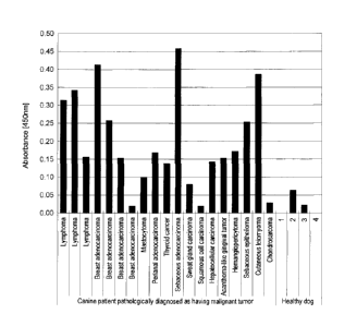
Fig. 3 shows some of the results of cancer diagnosis for
cancer-bearing dogs using the canine CAPRIN-1-derived polypeptides
prepared in the Examples.
Fig. 4 shows some of the results of detailed cancer diagnosis for
cancer-bearing dogs using the canine CAPRIN-1-derived polypeptides
prepared in the Examples.
BEST MODE OF CARRYING OUT THE INVENTION
According to the method of the present invention, CAPRIN-1
expression is measured using a sample separated from a living organism.
Examples of a method for measuring CAPRIN-1 expression include a method
(1st method) that involves immunoassay for an antibody against CAPRIN-1
contained in a sample, a method (2" method) that involves immunoassay for
CAPRIN-1 itself contained in a sample, and a method (3"1 method) that
involves measurement of mRNA encoding CAPRIN-1 contained in a sample.
In the method of the present invention, CAPRIN-1 expression may be
measured by any of these methods. In the present invention, the term
- 14 -
CA 02732980 2011-02-02
"measurement" refers to any of detection, qualitative determination,
quantitative determination, and semi-quantitative determination.
The amino acid sequence shown in SEQ ID NO: 6, 8, 10, 12, or 14 is
the amino acid sequence of canine CAPRIN-1. Canine CAPRIN-1 having
the amino acid sequence was identified as a polypeptide binding to an
antibody specifically existing in the cancer-bearing dog-derived serum by the
SEREX method using a canine testis-derived cDNA library and the serum of
a cancer-bearing dog (see Example 1). Specifically, an antibody against
CAPRIN-1 having the amino acid sequence shown in SEQ ID NO: 6, 8, 10,
12, or 14 is specifically induced in vivo in a cancer-bearing dog. Therefore,
canine cancer can be detected by measuring the above antibody against
CAPRIN-1 having the amino acid sequence shown in SEQ ID NO: 6, 8, 10,
12, or 14 using the above 1" method (see Examples 3 and 4). Canine cancer
can also be detected by measuring CAPRIN-1 itself as an antigen shown in
SEQ ID NO: 6, 8, 10, 12, or 14 using the above 2nd method (see Examples 5
and 6). Also, canine cancer can be detected, as described in the following
Examples, by measuring mRNA encoding CAPRIN-1 since the mRNA is
expressed at significantly high levels in testes and cancer cells (see Example
1).
The term "having an amino acid sequence" as used herein refers to
amino acid residues being aligned in the relevant order. Therefore, for
example, the expression "polypeptide having the amino acid sequence shown
in SEQ ID NO: 2" refers to a polypeptide having 709 amino acid residues,
which consists of the amino acid sequence of Met Pro Ser
Ala===(abbreviated)==Gln Gln Val Asn shown in SEQ ID NO: 2. Also, for
example, the expression "polypeptide having the amino acid sequence shown
in SEQ ID NO: 2" may also be abbreviated as "the polypeptide of SEQ ID
NO: 2." The same applies to the expression "having a/the nucleotide
sequence." In this case, the term "having" may be substituted with the
- 15 -
CA 02732980 2011-02-02
expressions "consisting of."
Also, the term "polypeptide" as used herein refers to a molecule that
is formed via peptide bonding of a plurality of amino acids. Examples of
such molecule include not only polypeptide molecules with a large number of
constituent amino acids, but also low-molecular-weight molecules
(oligopeptides) with small number of amino acids and full-length proteins.
The present invention further encompasses full-length CAPRIN-1 proteins
each having an amino acid sequence shown in an even-numbered sequence ID
from among SEQ ID NOS: 2-30.
In the method of the present invention, not only canine CAPRIN-1 of
SEQ ID NO: 6, 8, 10, 12, or 14, but also CAPRIN-1 of other mammals
(hereinafter, may also be referred to as "homolog" for canine CAPRIN-1.
When simply referred to as "CAPRIN-1," CAPRIN-1 from not only a dog but
also from another mammal is also encompassed herein) are also subjected to
measurement. As specifically described in the following Examples, mRNA
encoding human CAPRIN-1 is significantly expressed at a high level in
human testis and cancer cells, as in the case of canine CAPRIN-1 of SEQ ID
NO: 6, 8, 10, 12 or 14. However, no antibody against the human CAPRIN-1
is detected in a healthy human body. Also, an antibody against feline
CAPRIN-1 is not detected in a healthy cat body, but is detected in a
cancer-bearing cat alone. Therefore, cancer of a mammal other than a dog
can be detected by measuring CAPRIN-1 expression in the mammal.
Examples of CAPRIN-1 of mammals other than dogs, which are measurement
subjects in the method of the present invention, include, but are not limited
to, human CAPRIN-1 and feline CAPRIN-1. A nucleotide sequence
encoding human CAPRIN-1 and the amino acid sequence thereof are as
separately shown in SEQ ID NO: 1 and 3, and 2 and 4, respectively, in the
Sequence Listing. Sequence identity with canine CAPRIN-1 is 94% in
terms of nucleotide sequence and is 98% in terms of amino acid sequence.
- 16 -
CA 02732980 2011-02-02
Even dogs and humans which are genetically distant mammals share as very
high as 98% sequence identity in terms of the amino acid sequence of
CAPRIN-1. Therefore, it is thought that a dog and a mammal other than a
human; that is, canine CAPRIN-1 and homolog thereof share sequence
identity as high as about 85% or more.
Therefore, CAPRIN-1, the
expression of which is measured in the method of the present invention, has
preferably 85% or more and more preferably 95% or more sequence identity
with the amino acid sequence of canine CAPRIN-1 shown in SEQ ID NO: 6,
8, 10, 12, or 14.
However, such examples are not particularly limited
thereto.
In the 1" method above, the above antibody that can be present in a
sample can be easily measured by immunoassay using an antigenic substance
that undergoes an antigen-antibody reaction with the antibody.
Immunoassay itself is a known conventional method as specifically described
below. As an antigenic substance for immunoassay, the canine CAPRIN-1
of SEQ ID NO: 6, 8, 10, 12, or 14 that causes the induction of the antibody
within a cancer-bearing dog body can be used. Furthermore, an antibody
has cross-reactivity.
Thus, even a molecule other than an antigenic
substance actually having served as an immunogen can bind to an antibody
induced against the immunogen via an antigen-antibody reaction, as long as a
structure analogous to the epitope of the immunogen is present on the
molecule. In particular, a protein from a mammal and homologthereof from
another mammal share high amino acid sequence identity and often have
epitope structures analogous to each other. As specifically described in the
following Examples, the canine CAPRIN-1 of SEQ ID NO: 6, 8, 10, 12, or
14 undergoes an antigen-antibody reaction not only with an antibody induced
against the canine CAPRIN-1 within a cancer-bearing dog body, but also
with an antibody induced against feline CAPRIN-1 within a cancer-bearing
cat body. Moreover, human CAPRIN-1 undergoes an antigen-antibody
- 17 -
CA 02732980 2011-02-02
=
reaction with the above antibody induced within cancer-bearing dog or
cancer-bearing cat bodies. Accordingly, in the 1st method of the present
invention, CAPRIN-1 from any mammal can be used as an antigen for
immunoassay.
In general, when an antigenic substance is a protein or the like
having a complicated structure and high molecular weight, a plurality of sites
having different structures are present on the molecule.
Therefore, a
plurality of types of antibody capable of recognizing and binding to different
sites of such antigenic substances are produced in vivo. Specifically, an
antibody that is produced in vivo against an antigenic substance such as
protein is a polyclonal antibody that is a mixture of a plurality of types of
antibody. An antibody discovered by the present inventors is also a
polyclonal antibody. It is specifically present in cancer-bearing living
organism-derived serum and specifically binds to a recombinant CAPRIN-1
protein via an antigen-antibody reaction. The term "polyclonal antibody"
used in the present invention refers to an antibody that exists in serum from
a
living organism containing an antigenic substance therein and is induced in
vivo against the antigenic substance.
In Examples described later, polypeptides of SEQ ID NO: 6 and
SEQ ID NO: 8 (canine CAPRIN-1) and the polypeptide of SEQ ID NO: 2
(human CAPRIN-1) were prepared as antigens for immunoassay of specific
antibodies in the cancer-bearing living animals. Then reactivity between
these polypeptides and the above antibody in serum from a cancer-bearing
living organism was confirmed.
However, the above antibody is a
polyclonal antibody, so that it naturally binds to a polypeptide consisting of
the homolog of SEQ ID NO: 6, 8, or 2. Even in the case of a fragment of
said polypeptides, it can bind to the above antibody contained in serum from
a cancer-bearing living organism, since the polyclonal antibody can contain
an antibody capable of recognizing the structure of the relevant fragment.
- 18 -
CA 02732980 2011-02-02
That is, either a polypeptide (that is, full-length CAPRIN-1 protein) of the
homolog of SEQ ID NO: 6, 8, or 2 or a fragment thereof can be similarly
used for measurement of the above polyclonal antibody contained
specifically in serum of a cancer-bearing living organism and is useful for
cancer detection. Therefore, examples of a polypeptide to be used as an
antigen for immunoassay in the 1" method of the present invention include,
not only a polypeptide that consists of the full-length region of CAPRIN-1
(e.g., SEQ ID NO: 6, 8, or 2), but also a polypeptide fragment that consists
of continuous 7 or more, preferably continuous 8 or more, 9 or more, or 10 or
more amino acids in the amino acid sequence of CAPRIN-1 and undergoes an
antigen-antibody reaction with a polyclonal antibody against CAPRIN-1
(hereinafter, may be conveniently referred to as "a specifically reactive
partial polypeptide"). It is known in the art that a polypeptide of about 7 or
more amino acid residues exerts antigenicity. However, if the number of
amino acid residues constituting a polypeptide is too low, such polypeptide
highly likely cross-reacts with antibodies, which existes in the sample,
against proteins other than CAPRIN-1. Accordingly, in view of increasing
the accuracy of immunoassay, the desirable number of amino acid residues of
a polypeptide fragment may be preferably 30 or more or 50 or more, further
preferably 100 or more or 150 or more, further preferably 300 or more, even
more preferably 600 or more, and further preferably 1000 or more and 1500
or more.
Specific preferable examples of the polypeptides to be used as
antigens are the polypeptides of the even-numbered SEQ ID NOS: 2-30 or
fragments thereof.
Nucleotide sequences of polynucleotides encoding proteins
consisting of the amino acid sequences of the even-numbered SEQ ID NOS:
2-30 (that is, SEQ ID NOS: 2, 4, 6-28, 30) are shown in the odd-numbered
SEQ ID NOS: 1-29 (that is, SEQ ID NOS: 1, 3, 5-27, 29).
- 19 -
CA 02732980 2011-02-02
=
In general, it is broadly known by persons skilled in the art
concerning protein antigens such that even when few amino acid residues
have been substituted, deleted, added, or inserted in the amino acid sequence
of the protein, the resultant may retain antigenicity almost equivalent to
that
of the original protein. Therefore, a polypeptide: having a sequence that
has a substitution, a deletion, and/or an insertion of a few (preferably one
or
several) amino acid residues with respect to the amino acid sequence of
CAPRIN-1 and has 80% or more, 85% or more, preferably 90% or more,
more preferably 95% or more, and further preferably 98% or more sequence
identity with the original sequence; and specifically binding to a polyclonal
antibody against CAPRIN-1 via an antigen-antibody reaction (hereinafter,
may be conveniently referred to as "specifically reactive modified
polypeptide") can be used for cancer detection in a manner similar to that for
the above polypeptides.
Preferably, the specifically reactive modified
polypeptide has an amino acid sequence that has a substitution, a deletion, an
addition, and/or an insertion of one or several amino acid residues with
respect to the amino acid sequence of CAPRIN-1. The term "several" as
used herein refers to an integer of 2-10, preferably an integer of 2-6, and
further preferably an integer of 2-4.
The term "sequence identity (of amino acid sequences)" as used
herein is obtained by aligning two amino acid sequences to be compared so
that amino acid residues match as many as possible, subtracting the number
of amino acid residues that have matched from the total number of amino
acid residues, and then expressing the result in percentage form. Upon the
above alignment, if necessary, gaps are appropriately inserted into one of or
both sequences to be compared. Such sequence alignment can be performed
using a known program such as BLAST, FASTA, or CLUSTAL W (Karlin and
Altschul, Proc. Natl. Acad. Sci. U.S.A., 87: 2264-2268, 1993; Altschul et al.,
Nucleic Acids Res., 25: 3389-3402, 1997).
- 20 -
CA 02732980 2011-02-02
Twenty types of amino acid constituting natural proteins can be
grouped into neutral amino acids having side chains with low polarity (Gly,
Ile, Val, Leu, Ala, Met, and Pro), neutral amino acids having hydrophilic
side chains (Asn, Gln, Thr, Ser, Tyr, and Cys), acidic amino acids (Asp and
Glu), basic amino acids (Arg, Lys, and His), and aromatic amino acids (Phe,
Tyr, Trp, and His) in which the members of each group have properties
analogous to each other. It is known that substitution among these amino
acids (that is, conservative substitution) rarely alters the properties of the
resulting polypeptide. Therefore, when amino acid residues of CAPRIN-1
are substituted, substitution is performed between members of the same
group so that a possibility of maintaining binding with the corresponding
antibody becomes higher. However, in the present invention, the above
variant may involve non-conservative substitution, as long as
immune-inducing activity equivalent to or almost equivalent to that of a
non-variant is imparted.
A polypeptide (hereinafter, may conveniently be referred to as
"specifically reactive addition polypeptide") that contains as a partial
sequence the above polypeptide to be used in the present invention (that is,
prepared by addition of another (poly)peptide to one end or both ends of a
polypeptide to be used in the present invention) and specifically binds to a
polyclonal antibody against CAPRIN-1 via an antigen-antibody reaction can
also be used for cancer detection in a manner similar to that for the above
polypeptides.
The above polypeptides to be used in the present invention can be
synthesized according to a chemical synthesis method such as an Fmoc
method (fluorenylmethyloxycarbonyl method) and a tBoc method
(t-butyloxy-carbonyl method) (Ed., The Japanese Biochemical Society,
Seikagaku Jikken Koza (Biochemical Experimental Lecture Series) 1, Protein
Chemistry IV, Chemical Modification and Peptide Synthesis, TOKYO
-21-
CA 02732980 2011-02-02
KAGAKU DOZIN CO., LTD (Japan), 1981). Also, the polypeptides can
also be synthesized by a conventional method using various commercially
available peptide synthesizers. Alternatively, the polypeptides can be easily
prepared using known genetic engineering techniques (Sambrook et al.,
Molecular Cloning, 21d Edition, Current Protocols in Molecular Biology
(1989), Cold Spring Harbor Laboratory Press, Ausubel et al., Short Protocols
in Molecular Biology, 3rd Edition, A Compendium of Methods from Current
Protocols in Molecular Biology (1995), John Wiley & Sons, and the like).
For example, from RNA extracted from a tissue expressing a gene encoding
the human CAPRIN-1 of SEQ ID NO: 2 or a homolog thereof, cDNA of the
gene is prepared by RT-PCR. The full-length sequence or a desired partial
sequence of the cDNA is incorporated into an expression vector and then the
vector is introduced into host cells, so that a polypeptide of interest can be
obtained. The nucleotide sequences of cDNAs encoding canine CAPRIN-1
of SEQ ID NOS: 6, 8, 10, 12, and 14 are shown in SEQ ID NOS: 5, 7, 9, 11,
and 13, respectively. The
human factors homolog thereof; that is, the
nucleotide sequences of cDNAs encoding human CAPRIN-1 of SEQ ID NOS:
2 and 4 are shown in SEQ ID NOS: 1 and 3, respectively. Hence, primers to
be used for RT-PCR can be easily designed in reference to these nucleotide
sequences.
Also, as described later, a gene encoding CAPRIN-1 of a
non-human mammal can be amplified using primers designed in reference to
the nucleotide sequences of the odd-numbered SEQ ID NOS: 1-29. For
example, cDNA encoding feline CAPRIN-1 can be easily prepared by
techniques similar to the above techniques. RNA extraction, RT-PCR,
cDNA incorporation into a vector, and introduction of a vector into host cells
can be performed by known methods as described below, for example. Also,
vectors and host cells to be used herein are also known and various vectors
and host cells are commercially available.
The above host cells may be any cells, as long as they can express
- 22 -
CA 02732980 2011-02-02
the above polypeptides.
Examples of prokaryotic host cells include
Escherichia coli and the like. Examples of eukaryotic host cells include
mammalian cultured cells such as monkey kidney cells (COS 1), Chinese
hamster ovary cells (CHO), the human embryonic kidney cell line (HEK293),
and the mouse embryonic skin cell line (NIH3T3), budding yeast, fission
yeast, silkworm cells, and Xenopusoocytes.
When prokaryotic cells are used as host cells, an expression vector
having a replication origin in prokaryotic cells, a promoter, a
ribosome-binding site, a multi-cloning site, a terminator, a drug-resistance
gene, an auxotrophic complementary gene, and the like are used. As
expression vectors for Escherichia coli, pUC vectors, pBluescriptII, pET
expression systems, pGEX expression systems, and the like can be
exemplified. A DNA encoding the above polypeptide is incorporated into
such an expression vector, prokaryotic host cells are transformed with the
vector, and then the thus obtained transformant is cultured, so that the
polypeptide encoded by the DNA can be expressed in the prokaryotic host
cells. At
this time, the polypeptide can also be expressed as a fusion
protein with another protein. A DNA encoding the above polypeptide can
be obtained by preparing a cDNA by RT-PCR as described above, for
example. Moreover, such DNA encoding the above polypeptide can be also
synthesized by a conventional method using a commercially available nucleic
acid synthesizer as described below. The nucleotide sequences of cDNAs of
the genes encoding CAPRIN-1 of SEQ ID NOS: 2 and 4 are shown in SEQ ID
NOS: 1 and 3, respectively, in the Sequence Listing.
When eukaryotic cells are used as host cells, expression vectors for
eukaryotic cells having a promoter, a splicing region, a poly(A) additional
site, and the like are used. Examples of such expression vectors include
pKA1, pCDM8, pSVK3, pMSG, pSVL, pBK-CMV, pBK-RSV, EBV vector,
pRS, pcDNA3, and pYES2. Similarly to the above, a DNA encoding a
- 23 -
CA 02732980 2011-02-02
polypeptide to be used in the present invention is incorporated into such an
expression vector, eukaryotic host cells are transformed with the vector, and
then the thus obtained transformant is cultured, so that the polypeptide
encoded by the above DNA can be expressed in eukaryotic host cells. When
pIND/V5-His, pFLAG-CMV-2, pEGFP-N1, pEGFP-C1, or the like is used as
an expression vector, the above polypeptide can be expressed as a fusion
protein with various tags, such as a His tag (e.g., (His)6 to (His)10), a FLAG
tag, a myc tag, a HA tag, and GFP.
For introduction of an expression vector into a host cell, known
methods can be employed such as electroporation, a calcium phosphate
method, a liposome method, a DEAE dextran method, microinjection, viral
infection, lipofection, and binding with a cell-membrane-permeable peptide.
Isolation and purification of a polypeptide of interest from host cells
can be performed using known isolation techniques in combination.
Examples of such known techniques include treatment using a denaturing
agent such as urea or a surfactant, ultrasonication, enzymatic digestion,
salting-out, solvent fractionation and precipitation, dialysis,
centrifugation,
ultrafiltration, gel filtration, SDS-PAGE, isoelectric focusing, ion exchange
chromatography, hydrophobic chromatography, affinity chromatography, and
reverse phase chromatography.
Polypeptides obtained by the above methods include polypeptides in
the form of fusion proteins with any other proteins. An example of such a
fusion protein include a fusion protein with glutathione-S-transferase (GST),
a His tag, or the like. Polypeptides in the form of such fusion proteins are
also examples of the above-described specifically reactive addition
polypeptides and can be used for the 1st detection method of the present
invention. Furthermore, a polypeptide expressed in transformed cells may
be subjected to various types of modification within cells after translation.
Such polypeptide that is modified after translation can be used in the 1st
- 24 -
CA 02732980 2011-02-02
detection method of the present invention, as long as it is capable of binding
to a polyclonal antibody against CAPRIN-1.
Examples of such
post-translation modification include the removal of N-terminal methionine,
N-terminal acetylation, glycosylation, limited proteolysis by intracellular
protease, myristoylation, isoprenylation, and phosphorylation.
An antibody in a sample can be easily measured by immunoassay
using the above polypeptide as an antigen. Immunoassay itself is known in
the art. Immunoassay is classified into a sandwich method, a competition
method, an agglutination method, Western blot method, and the like based on
types of reaction. Also, immunoassay is classified based on labels into
radioimmunoassay, fluorescence immunoassay, enzyme immunoassay, and
biotin immunoassay, for example. Immunoassay of the above antibody can
be performed using any of these methods.
Sandwich ELISA or the
agglutination method are preferably applicable as an immunoassay technique
for the above antibody in the method of the present invention, since the
procedures of these methods are convenient and require no extensive
apparatus and the like. But the techniques are not limited to them. When
an enzyme is used as a label for an antibody, such enzyme is not particularly
limited, as long as it satisfies conditions such that: the turn over number is
high; it remains stable even if it is bound to an antibody, it specifically
causes the color development of the substrate, and the like. Examples of
enzymes that can be used for general enzyme immunoassay include
peroxidase, 13-galactosidase, alkaline phosphatase, glucose oxidase,
acetylcholine esterase, glucose-6-phosphorylation dehydrogenase, and malic
acid dehydrogenase. Also, enzyme-inhibiting substances, coenzymes, and
the like can be used. Binding of these enzymes with antibodies can be
performed by known methods using a cross-linking agent such as a
maleimide compound. As a substrate, a known substance can be used
depending on the type of an enzyme to be used. For example, when
- 25 -
CA 02732980 2011-02-02
peroxidase is used as an enzyme, 3,3 ',5,5'-tetramethylbenzidine can be used.
Also when alkaline phosphatase is used as an enzyme, para-nitrophenol or
the like can be used. As a radio isotope, a radio isotope that is generally
used for radioimmunoassay, such as 1251 and 3H can be used. As a
fluorescent dye, a fluorescent dye that is used for general fluorescent
antibody techniques, such as fluorescence isothiocyanate (FITC) and
tetramethylrhodamine isothiocyanate (TRITC) can be used.
There is no need to explain the above immunoassay techniques in
the Description, since they are well-known.
However, when these
immunoassay techniques are briefly described, the sandwich method involves
immobilizing the above polypeptide to be used as an antigen to a solid phase,
reacting it with a sample such as serum, washing, reacting with an
appropriate secondary antibody, washing, and then measuring the secondary
antibody bound to the solid phase, for example. An unbound secondary
antibody can be easily removed by immobilization of an antigen polypeptide
to a solid phase. Hence, this is preferable as an embodiment of the method
for detecting cancer of the present invention. As a secondary antibody, an
anti-canine IgG antibody can be used if a sample is derived from a dog. A
secondary antibody is labeled in advance with a labeling substance
exemplified above, so that the secondary antibody binding to a solid phase
can be measured. The thus measured amount of the secondary antibody
corresponds to the amount of the above antibody in the serum sample.
When an enzyme is used as a labeling substance, the amount of the antibody
can be measured by adding a substrate that is digested to develop color by
enzymatic action and then optically measuring the amount of the substrate
degraded. When a radio isotope is used as a labeling substance, the amount
of radiation from the radio isotope can be measured using a scintillation
counter or the like.
In the 2nd method of the present invention, CAPRIN-1 that can be
- 26 -
CA 02732980 2011-02-02
contained in a sample from a living organism is measured. As described
above, among cancer patients, the amount of an antibody that undergoes an
antigen-antibody reaction with CAPRIN-1 of a dog, a human, or the like is
significantly high.
This indicates that the amount of CAPRIN-1
accumulated as an antigen is significantly high in cancer cells. Cancer can
also be detected by directly measuring CAPRIN-1, as specifically described
in Examples below. Therefore, cancer can be detected in vivo by measuring
CAPRIN-1 itself similarly to the 1st method above.
A polypeptide in a sample can be easily measured by well-known
immunoassay techniques.
Specifically, for example, an antibody or an
antigen-binding fragment thereof, which undergoes an antigen-antibody
reaction with CAPRIN-1, is prepared, immunoassay is performed using the
antibody or its antigen-binding fragment thereof, and then CAPRIN-1 that
may be present in the sample can be measured. As described above, an
antibody has cross-reactivity. Hence, for example, through the use of an
antibody or the antigen-binding fragment thereof, which undergoes an
antigen-antibody reaction with the canine CAPRIN-1 of SEQ ID NO: 6, not
only the canine CAPRIN-1 of SEQ ID NO: 6, but also its homolog in other
mammals (e.g., the human CAPRIN-1 of SEQ ID NO: 2 or 4 and feline
CAPRIN-1) can be measured. An immunoassay technique itself is a known
conventional technique as described above.
This examination revealed that CAPRIN-1 is a cell membrane
protein that is expressed on the surfaces of cancer cells. A living organism
with cancer contains many kinds of proteases. Specifically, in a living
organism with cancer, an extracellularly expressed portion of the CAPRIN-1
sequence is separated from the cancer cells by degradation, so that such
portion exists at a level higher than an intracellularly expressed portion of
the CAPRIN-1 sequence. Therefore, when an antibody against CAPRIN-1
or an antigen-binding fragment thereof to be used in this measurement, which
- 27 -
CA 02732980 2011-02-02
binds to the surface of the cancer cell, is used, CAPRIN-1 can be detected at
higher levels and cancer can be diagnosed with higher sensitivity.
Therefore, in the present invention, antibodies binding to a portion of the
CAPRIN-1 protein existing on the surfaces of cancer cells, are preferably
used. An example of a partial peptide of the CAPRIN-1 protein existing on
the surfaces of cancer cells, is a polypeptide comprising a sequence of
continuous 7 or more amino acid residues within the region of amino acid
residue Nos. (aa) 50-98 or amino acid residue Nos. (aa) 233-305 in the amino
acid sequences shown in the even-numbered SEQ ID NOS: 2-30 in the
Sequence Listing excluding SEQ ID NO: 6 and SEQ ID NO: 18. A specific
example thereof is the amino acid sequence shown in SEQ ID NO: 43 or SEQ
ID NO: 61 (in the amino acid sequence shown in SEQ ID NO: 61, a region of
the amino acid sequence shown in SEQ ID NO: 62 or SEQ ID NO: 63 is
preferred) or an amino acid sequence having 80% or more, preferably 85% or
more, more preferably 90% or more, further preferably 95% or more
sequence identity with the relevant amino acid sequence. Examples of an
antibody to be used in the present invention include all antibodies binding to
these peptides. Specific examples of the antibody include an antibody or
antigen-binding fragment thereof which binds to SEQ ID NO: 43, a
monoclonal antibody or antigen-binding fragment thereof having the amino
acid sequences of SEQ ID NOS: 44 and 45, a monoclonal antibody or
antigen-binding fragment thereof having the amino acid sequences of SEQ ID
NOS: 44 and 46, a monoclonal antibody or antigen-binding fragment thereof
having the amino acid sequences of SEQ ID NOS: 44 and 47, a monoclonal
antibody or antigen-binding fragment thereof having the amino acid
sequences of SEQ ID NOS: 44 and 48, a monoclonal antibody an
antigen-binding fragment thereof having the amino acid sequences of SEQ ID
NOS: 49 and 50, a monoclonal antibody or antigen-binding fragment thereof
having the amino acid sequences of SEQ ID NOS: 51 and 52, a monoclonal
- 28 -
CA 02732980 2011-02-02
antibody or antigen-binding fragment thereof having the amino acid
sequences of SEQ ID NOS: 53 and 54, a monoclonal antibody or
antigen-binding fragment thereof having the amino acid sequences of SEQ ID
NOS: 55 and 56, a monoclonal antibody or antigen-binding fragment thereof
having the amino acid sequences of SEQ ID NOS: 57 and 58, or a monoclonal
antibody or antigen-binding fragment thereof having the amino acid
sequences of SEQ ID NOS: 59 and 60.
The term "antigen-binding fragment" as used herein refers to an
antibody fragment capable of binding to an antigen such as a Fab fragment
and a F(ab')2 fragment contained in an antibody molecule. An antibody to
be used herein may be a polyclonal antibody or a monoclonal antibody. For
immunoassay and the like, a monoclonal antibody with high reproducibility
is preferable. A method for preparing a polyclonal antibody and a
monoclonal antibody using a polypeptide as an immunogen is known and can
be easily performed by a conventional method. For example, CAPRIN-1 is
bound to a carrier protein such as keyhole limpet hemocyanin (KLH), casein,
or serum albumin and then an animal is immunized with the resultant as an
immunogen together with an adjuvant, and thereby an antibody against
CAPRIN-1 can be induced. Antibody-producing cells such as splenocytes or
lymphocytes collected from the immunized animal are fused to myeloma
cells to prepare hybridomas, and then hybridomas producing an antibody that
binds to CAPRIN-1 are selected and then grown, so that a monoclonal
antibody, whose the corresponding antigen is CAPRIN-1, can be obtained
from the cultured supernatant. The above method is a known conventional
method.
In the 3'd method of the present invention, mRNA encoding
CAPRIN-1 that can be contained in a sample obtained from a living organism
is measured. As specifically described in Examples below, mRNA encoding
the canine CAPRIN-1 of SEQ ID NO: 6, 8, 10, 12, or 14 or human CAPRIN-1
- 29 -
CA 02732980 2011-02-02
of SEQ ID NO: 2 or 4 is expressed at a significantly high level in cancer
cells. Therefore, cancer can be detected in vivo by measuring such mRNA
in a sample.
mRNA in a sample can be quantitatively determined by a
conventional method such as real-time detection RT-PCR using the mRNA as
a template, for example.
Such mRNA can generally be quantitatively
determined based on staining intensity or the like in Northern blot that is a
conventional method. The cDNA sequences encoding CAPRIN-1
polypeptides of the even-numbered SEQ ID NOS: 2-30 are shown in the
odd-numbered SEQ ID NOS: 1-29, respectively. Hence, based on these
sequences, a polynucleotide specifically hybridizing to a partial region in
the
nucleotide sequence shown in any of the odd-numbered SEQ ID NOS: 1-29
(hereinafter, referred to as "polynucleotide for cancer detection") is
prepared
and then the amount of the mRNA in a sample can be measured using the
polynucleotide as a probe or a primer for a nucleic acid amplification
method. As
described later, if it is a polynucleotide specifically
hybridizing to a partial region in the nucleotide sequence shown in any of the
odd-numbered SEQ ID NOS: 1-29, mRNA encoding CAPRIN-1 in mammals
other than dogs and humans can also be determined. In addition, in the
present invention, a polynucleotide may be RNA or DNA.
The term "specifically hybridizing to" as used herein refers to that
under general hybridization conditions, a subject hybridizes to only a target
partial region, but does not substantially hybridize to the other regions.
The term "(under) general hybridization conditions" as used herein
refers to conditions employed for annealing in general PCR or detection
using a probe. For example, in the case of PCR using Taq polymerase, the
term refers to conditions under which a reaction is performed at an
appropriate annealing temperature ranging from about 54 C to 60 C using a
general buffer such as 50 mM KC1, 10 mM Tris-HC1 (pH8.3-9.0), and 1.5 mM
- 30 -
CA 02732980 2011-02-02
MgC12. Also, in the case of Northern hybridization, for example, the term
refers to conditions under which a reaction is performed using a general
hybridization solution such as 5 x SSPE, 50% formamide, 5 x Denhardt's
solution, and 0.1%SDS-0.5%SDS, or 0.1-5 x SSC and 0.1-0.5% SDS at an
appropriate hybridization temperature ranging from about 42 C to 65 C.
Furthermore, after hybridization, washing is performed with 0.1-0.2 x SSC
and 0.1% SDS, for example. However, appropriate annealing temperatures
or hybridization temperatures are not limited to the above examples, and are
determined based on Tm value for a polynucleotide for cancer detection,
which is used as a primer or a probe, and the empirical rule of experimenters.
Persons skilled in the art can easily determine such temperature range.
The expression "does not substantially hybridize to" as used herein
refers to that a subject does not really hybridize to a target partial region
or
a subject hybridizes to a target partial region in a significantly low amount;
that is, in a relatively negligibly-small amount, even when it hybridizes to
the target partial region. An
example of a polynucleotide specifically
hybridizing under such conditions is a polynucleotide having sequence
identity at a level or more with the nucleotide sequence of a target partial
region. A
specific example of such polynucleotide has 70% or more,
preferably 80% or more, 85% or more, more preferably 90% or more, further
preferably 93% or more, further preferably 95% or more, and further more
preferably 98% or more sequence identity.
Most preferably, the
polynucleotide has a nucleotide sequence identical to the nucleotide
sequence of a target partial region. Sequence identity is defined in the same
manner as that for the sequence identity of the above amino acid sequence.
Even if a terminus of a polynucleotide for cancer detection contains a region
not hybridizing to a subject, in the case of a probe, it can be used for
detection as long as a hybridizing region occupies as much as about a half or
more of the entire probe. Also, in the case of a primer, it can be used for
-31-
CA 02732980 2011-02-02
detection as long as a hybridizing region occupies as much as about a half or
more of the entire primer and is located on the 3' terminal side, since normal
annealing and extension reaction can take place. As described above, when
a terminus of a polynucleotide for cancer detection contains a
non-hybridizing region, sequence identity with a target nucleotide sequence
is calculated focusing on only a hybridizing region without taking
non-hybridizing region into consideration.
The term "partial sequence" in the present invention refers to a
partial sequence in the nucleotide sequences shown in the odd-numbered SEQ
ID NOS: 1-29, specifically the partial sequence having a sequence of
continuous 15 or more nucleotides, preferably continuous 18 or more
nucleotides, more preferably continuous 20 or more nucleotides or 25 or
more nucleotides, and further preferably continuous 30, 40, or 50 or more
nucleotides. The expression "the nucleotide sequence shown in SEQ ID
NO: 5" as used herein refers to, in addition to the nucleotide sequence
actually shown in SEQ ID NO: 5, a sequence complementary to the sequence.
Therefore, for example, the expression "a polynucleotide having the
nucleotide sequence shown in SEQ ID NO: 5" refers to a single-stranded
polynucleotide having the nucleotide sequence actually shown in SEQ ID
NO: 5, a single-stranded polynucleotide having a nucleotide sequence
complementary to that shown in SEQ ID NO: 5, and a double-stranded
polynucleotide comprising them. When a polynucleotide to be used in the
present invention is prepared or a polynucleotide encoding a polypeptide to
be used in the present invention is prepared, any one nucleotide sequence is
appropriately selected and this selection can be easily performed by persons
skilled in the art.
The number of nucleotides in a polynucleotide for cancer detection
is preferably 18 or more nucleotides in view of ensuring specificity. When
used as a probe, the size of the polynucleotide is preferably 18 or more
- 32 -
CA 02732980 2011-02-02
nucleotides, is further preferably 20 or more nucleotides and the full-length
or less of the coding region. When used as a primer, the size of the
polynucleotide is preferably 18 or more nucleotides and 50 or less
nucleotides. A preferred example of the polynucleotide for cancer detection
is a polynucleotide comprising continuous 18 or more nucleotides in a
nucleotide sequence shown in any of the odd-numbered SEQ ID NOS: 1-29.
It is obvious for persons skilled in the art who refer this Description
that: a polynucleotide specifically hybridizing to a partial region in SEQ ID
NO: 5, 7, 9, 11, or 13 is used for measurement of the amount of mRNA
encoding the canine CAPRIN-1 of SEQ ID NO: 6, 8, 10, 12, or 14,
respectively; and a polynucleotide specifically hybridizing to a partial
region
in SEQ ID NO: 1 or 3 is used for measurement of the amount of mRNA
encoding the human CAPRIN-1 of SEQ ID NO: 2 or 4, respectively.
However, a protein from a mammal and a homolog thereof from another
mammal generally share high sequence identity even at the nucleotide
sequence level. Thus, the sequence identity among the sequences of the
odd-numbered SEQ ID NOS: 1-13 also is as very high as 94% to 100%.
Accordingly, a polynucleotide specifically hybridizing to a partial region of
the sequence of SEQ ID NO: 5 can also specifically hybridize to a partial
region corresponding to the relevant partial region of any of the
odd-numbered SEQ ID NOs: 1-29.
Actually as described in Examples below, a pair of primers having
the nucleotide sequences shown in SEQ ID NO: 33 and 34, respectively,
specifically hybridizes to both a partial region of any of the sequences of
the
odd-numbered SEQ ID NOS: 1-29 and a partial region of the sequence of
SEQ ID NO: 5, so that both mRNA encoding the canine CAPRIN-1 of SEQ
ID NO: 6 and mRNA encoding a homolog thereof can be measured, for
example.
Accordingly, for example, with the use of a polynucleotide
specifically hybridizing to a partial region of the sequence of SEQ ID NO: 5,
=
-33 -
CA 02732980 2011-02-02
not only mRNA encoding the canine CAPRIN-1 of SEQ ID NO: 6, but also
mRNA encoding the human CAPRIN-1 of SEQ ID NO: 2 or 4 can be
measured. Similarly, a mRNA encoding CAPRIN-1 of another mammal such
as a cat can also be measured. When a polynucleotide for cancer detection
is designed, it is desirable to select partial regions having a specifically
high
sequence identity between the SEQ ID numbers (odd-numbered SEQ ID NOS:
1-29) (preferably, the nucleotide sequences are the same). If
a partial
region having particularly high sequence identity between a dog and a human
is present, a region having very high sequence identity with the region is
expected to be present in a homologous gene of another animal species.
Through selection of such partial regions, accuracy for measuring mRNA
encoding CAPRIN-1 of an animal species other than dogs and humans can be
increased.
A method itself for measuring a test nucleic acid using a
polynucleotide specifically hybridizing to a partial region of the test
nucleic
acid as a primer or a probe for a nucleic acid amplification method such as
PCR is well-known. Examples of such method include, in addition to
RT-PCR that is specifically described in Examples below, Northern blot and
In situ hybridization. When the amount of mRNA is measured in the
present invention, all of these known measuring methods can be employed.
A nucleic acid amplification method itself such as PCR is
well-known in the art and thus reagent kits and apparatuses therefor are
commercially available, so that the method can be easily performed.
Specifically, for example, denaturation, annealing, and extension steps are
each performed using a test nucleic acid (e.g., the cDNA of a gene encoding
a protein having an amino acid sequence shown in any of the even-numbered
SEQ ID NOS: 2-30) as a template and a pair of polynucleotides (primers) for
cancer detection in a known buffer in the presence of thermostable DNA
polymerase such as Taq polymerase or Pfu polymerase and dNTP (here, N =
- 34 -
CA 02732980 2011-02-02
A, T, C, or G) by varying the temperature of the reaction solution. In
general, the denaturation step is performed at 90 C-95 C, the annealing step
is performed at or near the Tm of the template and the primers (preferably
within 4 C), and the extension step is performed at 72 C which is an
optimum temperature for thermostable DNA polymerase such as Taq
polymerase or Pfu polymerase or a temperature near the optimum
temperature. Each step is performed for about 30 seconds to 2 minutes, as
appropriately selected. This heating cycle is repeated about 25 to 40 times,
for example, so that the template nucleic acid region flanked by a primer pair
is amplified. A nucleic acid amplification method is not limited to PCR and
any other nucleic acid amplification methods known in the art can be
employed herein. As described above, when a nucleic acid amplification
method is performed using a pair of polynucleotides for cancer detection as
primers and a test nucleic acid as a template, the test nucleic acid is
amplified.
However, if no test nucleic acid is contained in a sample,
amplification does not take place. Hence, through detection of
amplification products, the presence or the absence of the test nucleic acid
in
a sample can be confirmed. An amplification product can be detected by a
method that involves subjecting a reaction solution after amplification to
electrophoresis, and then staining the band with ethidium bromide or the like
or a method that involves immobilizing an amplification product after said
electrophoresis onto a solid phase such as a nylon membrane, performing
hybridization with a labeling probe that specifically hybridizes to a test
nucleic acid, washing, and then detecting the label. Also, namely real-time
detection PCR is performed using a quencher fluorescent dye and a reporter
fluorescent dye, and thereby the amount of a test nucleic acid in a specimen
can be quantitatively determined. Since kits for real-time detection PCR are
commercially available, real-time detection PCR can be easily performed.
Furthermore, semi-quantitative determination of a test nucleic acid is also
- 35 -
CA 02732980 2011-02-02
possible based on electrophoresis band intensity. A test nucleic acid may
be either mRNA or cDNA resulting from mRNA via reverse transcription.
When mRNA is amplified as a test nucleic acid, a NASBA method (3SR
method or TMA method) using the above primer pair can also be employed.
The NASBA method itself is well-known and kits for the method are also
commercially available, so that the method can be easily performed using the
above primer pair.
As a probe, a labeled probe that is prepared by labeling a
polynucleotide for cancer detection with a fluorescent label, a radiolabel, a
biotin label, or the like can be used. A method for labeling a
polynucleotide itself is well-known. The presence or the absence of a test
nucleic acid in a sample can be examined by immobilizing a test nucleic acid
or an amplification product thereof, performing hybridization with a labeled
probe, washing, and then measuring the label bound to the solid phase.
Alternatively, a polynucleotide for cancer detection is immobilized, a test
nucleic acid is hybridized thereto, and then the test nucleic acid bound to
the
solid phase can be detected using the labeled probe or the like. In such a
case, a polynucleotide for cancer detection bound to a solid phase is also
referred to as a probe. A method for measuring a test nucleic acid using a
polynucleotide probe is also known in the art. The method can be
performed by causing, in a buffer, a polynucleotide probe to come into
contact with a test nucleic acid at Tm or near Tm (preferably, within 4 C)
for hybridization, washing, and then measuring the labeled probe that has
hybridized or the template nucleic acid bound to the solid-phase probe.
Examples of such method include well-known methods such as Northern blot,
in situ hybridization, and Southern blot methods. In the present invention,
any well-known method is applicable.
It is determined by the detection method of the present invention
whether or not a subject animal has cancer based on the expression level of
- 36 -
CA 02732980 2011-02-02
CAPRIN-1 measured as described above. Cancer can be detected only by
measuring CAPRIN-1 expression in a subject animal. However, it is
preferable in view of enhancing detection accuracy to examine the expression
levels (antibody level, polypeptide level, or mRNA level) of CAPRIN-1 in
one or a plurality of samples of healthy subjects so as to obtain a standard
value of healthy subjects and then to compare the measured value of a
subject animal with the standard value obtained from healthy subjects. To
further enhance detection accuracy, CAPRIN-1 expression levels are
examined for samples obtained from many patients found to have cancer so
as to obtain a standard value of cancer patients and then the measured value
of a subject animal may be compared with both the standard value of healthy
subjects and the standard value of cancer patients. The above standard
values can be determined by quantifying the CAPRIN-1 expression level in
each sample and then calculating the mean value thereof, for example. A
standard value of healthy subjects and the same of cancer patients can be
determined in advance by examining CAPRIN-1 expression levels in many
healthy subjects and cancer patients. Therefore, when comparison with a
standard value is performed in the method of the present invention, a
standard value determined in advance may be used.
In the detection method of the present invention, diagnosis based on
other cancer antigens or cancer markers may be used in combination.
Accordingly, cancer detection accuracy can be further increased. For
example, when an antibody specifically existing in cancer patients is
measured by the method of the present invention, another polypeptide that is
often expressed in a cancer tissue can be used in combination as an antigen
in a manner similar to that for polypeptides above. Also, the method of the
present invention and diagnosis using a previously known cancer marker may
be performed in combination.
Cancer can be detected in vivo according to the detection method of
- 37 -
CA 02732980 2011-02-02
the present invention. Particularly, as described in Examples below, even a
small-size tumor, which is invisible to the naked eye, or a tumor in a deep
part in vivo can be detected according to the method of the present invention.
Thus, the method of the present invention is useful for early cancer
detection. Also, through application of the detection method of the present
invention for a patient during follow-up after treatment of cancer, cancer can
be detected early if a cancer recurrence has taken place.
Also, in a cancer-bearing living organism, as the number of cancer
cells expressing CAPRIN-1 measured in the present invention increases, the
amounts of the protein and its mRNA accumulated in the living organism
increase and the production amount of the antibody against CAPRIN-1 in
serum increases. Meanwhile, as the number of cancer cells decreases, the
amounts of the protein and its mRNA accumulated in vivo decrease and the
amount of the antibody against CAPRIN-1 in serum decreases. Therefore,
when the expression level of CAPRIN-1 is higher than that of a control, it
can be determined that a tumor increase or a cancer metastasis is occurring;
that is, the extent of cancer is advanced. Actually, as specifically described
in the Examples below, an increase in the above serum antibody level in a
cancer-bearing living organism was observed in association with cancer
progression (malignant) such as tumor increase and metastasis. As
described above, the extent of cancer can also be detected by the method of
the present invention.
Also, as described in Examples below, among tumors of the same
type, the above antibody levels in malignant type tumors were significantly
higher than those in benign type tumors. Accordingly, when the expression
level of CAPRIN-1 is high, it can be determined that cancer malignancy is
higher. Specifically, cancer malignancy can also be detected by the method
of the present invention.
Cancer to be subjected to the method for detecting cancer of the
- 38 -
CA 02732980 2011-02-02
present invention is cancer expressing CAPRIN-1. Examples of such cancer
include, but are not limited to, brain tumor, squamous cell carcinoma of the
head, neck, lung, uterus or esophagus, melanoma, adenocarcinoma of the
lung or uterus, renal cancer, malignant mixed tumor, hepatocellular
carcinoma, basal cell carcinoma, acanthoma-like gingival tumor, tumor of the
oral cavity, perianal adenocarcinoma, anal sac tumor, anal sac apocrine
adenocarcinoma, sertoli cell carcinoma, cancer of the vaginal vestibule,
sebaceous adenocarcinoma, sebaceous epithelioma, sebaceous adenoma,
sweat gland carcinoma, intranasal adenocarcinoma, nasal adenocarcinoma,
thyroid cancer, large-bowel cancer, bronchial
adenocarcinoma,
adenocarcinoma, ductal carcinoma, breast adenocarcinoma, composite type
breast adenocarcinoma, malignant mammary mixed tumor, intraductal
papillary adenocarcinoma, fibrosarcoma, hemangiopericytoma, osteosarcoma,
chondrosarcoma, soft tissue sarcoma, histiocytic sarcoma, myxosarcoma,
undifferentiated sarcoma, lung cancer, mastocytoma, cutaneous leiomyoma,
intraperitoneal leiomyoma, leiomyoma, chronic lymphocytic leukemia,
lymphoma, gastrointestinal lymphoma, digestive
lymphoma,
small-cell-to-medium-cell lymphoma, adrenomedullary tumor, granulosa cell
tumor, and pheochromocytoma. Also, a living organism to be subjected to
the method of the present invention is a mammal and is preferably a human, a
dog, or a cat.
Examples of a sample to be subjected to the method of the present
invention include body fluids such as blood, serum, blood plasma, ascites,
and pleural effusion, tissues, and cells. In particular, in the 1st method and
the 2nd method above, serum, blood plasma, ascites, and pleural effusion can
be preferably used and in the 3rd method above for measurement of mRNA,
tissue samples and cell samples are preferable.
The above polypeptides to be used as antigens for immunoassay in
the 1st method (that is, the canine CAPRIN-1 of SEQ ID NO: 2 and a
- 39 -
CA 02732980 2011-02-02
homolog thereof, a specifically reactive partial polypeptide, a specifically
reactive modified polypeptide, and a specifically reactive addition
polypeptide) can be provided as reagents for cancer detection. The reagent
may consist of only the above polypeptide or may contain various additives
or the like, for example, useful for stabilization of the polypeptide. Also,
the reagent can be provided in a form immobilized onto a solid phase such a
plate or a membrane.
Preferable examples of the polypeptide are as
described above.
An antibody that undergoes an antigen-antibody reaction with
CAPRIN-1 or an antigen-binding fragment thereof, which is used for
immunoassay of CAPRIN-1 itself in the 2' method, can also be provided as
a reagent for cancer detection. The reagent for cancer detection in this case
may also consist of only the above antibody or an antigen-binding fragment
thereof or may contain various additives or the like useful for stabilization
and the like of the antibody or an antigen-binding fragment thereof. Also,
the antibody or an antigen-binding fragment thereof may be in a form
binding to a metal such as manganese or iron. When such metal-bound
antibody or antigen-binding fragment thereof is administered to the body of a
living organism, the metal-bound antibody or antigen-binding fragment
thereof is accumulated at an increased level at a site where the antigen
protein is present at a higher level. Therefore, the metal is measured by
MRI or the like, and thereby the presence of cancer cells producing the
antigen protein can be detected.
Furthermore, the above polynucleotide for cancer detection to be
used for mRNA measurement in the 31-d method can also be provided as a
reagent for cancer detection. The reagent for cancer detection in this case
may also consist of only the polynucleotide or may contain various additives
and the like useful for stabilization and the like of the polynucleotide. The
polynucleotide for cancer detection contained in the reagent is preferably a
- 40 -
CA 02732980 2011-02-02
primer or a probe.
Conditions and preferable examples of the
polynucleotide for cancer detection are as described above.
EXAMPLES
The present invention will be described in more detail with
reference to the examples set forth below; however, the technical scope of
the present invention is not limited to the examples.
Example 1: Obtainment of new cancer antigen protein by SEREX method
(1) Construction of cDNA library
Total RNA was extracted from a testis tissue of a healthy dog by an
Acid guanidium-Phenol-Chloroform method and then a polyA RNA was
purified using Oligotex-dT30 mRNA purification Kit (Takara Shuzo Co.,
Ltd.) according to protocols included with the kit.
A canine testis cDNA phage library was synthesized using the thus
obtained mRNA (5 [ig). The cDNA phage library was constructed using a
cDNA Synthesis Kit, a ZAP-cDNA Synthesis Kit, and a ZAP-cDNA
GigapackIII Gold Cloning Kit (STRATAGENE) according to protocols
included with the kits. The size of the thus constructed cDNA phage library
was 7.73x 10⁵ pfu/ml.
(2) Screening of cDNA library using serum
Immunoscreening was performed using the above constructed canine
testis cDNA phage library. Specifically, host Escherichia coli (XL1-Blue
MRF') was infected with the phage on an NZY agarose plate (090x15mm) so
as to obtain 2210 clones. E. coli cells were cultured at 42 C for 3 to 4
hours to form plaques. The plate was covered with a nitrocellulose
membrane (Hybond C Extra: GE Healthcare Bio-Science) impregnated with
IPTG (isopropyl-P-D-thiogalactoside) at 37 C for 4 hours, so that the protein
was induced, expressed, and then transferred to the membrane.
-41-
CA 02732980 2011-02-02
Subsequently, the membrane was collected and then immersed in TBS (10
mM Tris-HC1, 150 mM NaCI, and pH 7.5) containing 0.5% powdered skim
milk, followed by overnight shaking at 4 C, thereby suppressing nonspecific
reaction. The filter was reacted with a 500-fold diluted serum of a canine
patient at room temperature for 2 to 3 hours.
As the above serum of a canine patient, a serum collected from a
canine patient with breast cancer was used. These sera were stored at -80 C
and then subjected to pre-treatment immediately before use. A method for
pretreatment of serum is as follows. Specifically, host Escherichia coil
(XL1-Blue MRF') was infected with a 21/4, ZAP Express phage in which no
foreign gene had been inserted and then cultured overnight on a NZY plate
medium at 37 C.
Subsequently, buffer (0.2 M NaHCO3 and pH 8.3)
containing 0.5 M NaC1 was added to the plate, the plate was left to stand at
4 C for 15 hours, and then a supernatant was collected as an Escherichia
co/i/phage extract. Next, the thus collected Escherichia co/i/phage extract
was applied to an NHS-column (GE Healthcare Bio-Science), so that an
Escherichia co/i=phage-derived protein was immobilized. The serum of a
canine patient was applied to the protein-immobilized column for reaction
and then Escherichia coil and an antibody adsorbed to the phage were
removed from the serum. The serum fraction that had passed through the
column was diluted 500-fold with TBS containing 0.5% powdered skim milk.
The resultant was used as an immunoscreening material.
A membrane onto which the treated serum and the above fusion
protein had been blotted was washed 4 times with TBS-T (0.05%
Tween20/TBS) and then caused to react with goat anti-canine IgG (Goat
anti-Dog IgG-h+I HRP conjugated (BETHYL Laboratories)) diluted
5000-fold with TBS containing 0.5% powdered skim milk as a secondary
antibody for 1 hour at room temperature. Detection was performed via an
enzyme coloring reaction using an NBT/BCIP reaction solution (Roche).
- 42 -
CA 02732980 2011-02-02
Colonies that matched sites positive for a coloring reaction were collected
from the NZY agarose plate (090 x 15 mm) and then suspended in 500 1 of
an SM buffer (100 mM NaC1, 10 mM MgC1S0 , 50 mM Tris-HC1, 0.01%
4
gelatin, and pH 7.5). Until colonies positive for coloring reaction were
unified, secondary screening and tertiary screening were repeated by a
method similar to the above, so that 30,940 phage clones reacting with serum
IgG were screened. Thus, 5 positive clones were isolated.
(3) Homology search for isolated antigen gene
For nucleotide sequence analysis of the 5 positive clones isolated by
the above method, a procedure for conversion from phage vectors to plasmid
vectors was performed. Specifically, 200 I of a solution was prepared to
contain host Escherichia coli (XL1-Blue MRF') so that absorbance OD
600
was 1Ø The solution was mixed with 250 1 of a purified phage solution
and then with 1 1 of an ExAssist helper phage (STRATAGENE), followed
by 15 minutes of reaction at 37 C. Three mililiters of LB medium was
added and then culture was performed at 37 C for 2.5 to 3 hours.
Immediately after culture, the temperature of the solution was kept at 70 C
by water bath for 20 minutes, centrifugation was performed at 4 C and 1000
x g for 15 minutes, and then the supernatant was collected as a phagemid
solution.
Subsequently, 200 Ill of a solution was prepared to contain
phagemid host Escherichia coli (SOLR) so that absorbance 0D600 was 1Ø
The solution was mixed with 10 IA of a purified phage solution, followed by
15 minutes of reaction at 37 C. The solution (50 1) was seeded on LB agar
medium containing ampicillin (to a final concentration of 50 g/m1) and then
cultured overnight at 37 C. Transformed SOLR single colonies were
collected and then cultured in LB medium containing ampicillin (final
concentration: 50 g/ml) at 37 C. A plasmid DNA containing an insert of
interest was purified using a QIAGEN plasmid Miniprep Kit (QIAGEN).
The purified plasmid was subjected to analysis of the full-length
-43-
CA 02732980 2011-02-02
sequence by a primer walking method using the T3 primer according to SEQ
ID NO: 31 and the T7 primer according to SEQ ID NO: 32. As a result of
sequence analysis, the gene sequences according to SEQ ID NOS: 5, 7, 9, 11,
and 13 were obtained. A homology search program, BLAST search
(http://www. ncbi. Nlm. Nih. gov/BLAST/), was performed using the
nucleotide sequences of the genes and amino acid sequences (SEQ ID NOS:
6, 8, 10, 12, and 14) of the proteins encorded by the genes. As a result of
this homology search with known genes, it was revealed that all of the 5
obtained genes encoded CAPRIN-1. Regarding regions to be translated to
proteins, the sequence identity among the 5 genes was 100% in terms of
nucleotide sequence and 99% in terms of amino acid sequence. Also,
regarding regions to be translated to proteins, the sequence identity between
the genes and genes encoding human homolog thereof was 94% in terms of
nucleotide sequence and 98% in terms of amino acid sequence. The
nucleotide sequences of the human homolog are shown in SEQ ID NOS: 1
and 3 and the amino acid sequences of the same are shown in SEQ ID NOS: 2
and 4. Also, regarding regions to be translated to proteins, the sequence
identity between the obtained canine genes and a gene encoding a cattle
homolog was 94% in terms of nucleotide sequence and 97% in terms of
amino acid sequence. The nucleotide sequence of the cattle homolog is
shown in SEQ ID NO: 15 and the amino acid sequence of the same is shown
in SEQ ID NO: 16. Regarding regions to be translated to proteins, the
sequence identity between the genes encoding the human homolog and the
gene encoding the cattle homolog was 94% in terms of nucleotide sequence
and ranged from 93% to 97% in terms of amino acid sequence. Also,
regarding regions to be translated to proteins, the sequence identity between
the obtained canine genes and a gene encoding an equine homolog was 93%
in terms of nucleotide sequence and 97% in terms of amino acid sequence.
The nucleotide sequence of the equine homolog is shown in SEQ ID NO: 17
- 44 -
CA 02732980 2011-02-02
and the amino acid sequence of the same is shown in SEQ ID NO: 18.
Regarding regions to be translated to proteins, the sequence identity between
the genes encoding the human homolog and the gene encoding the equine
homolog was 93% in terms of nucleotide sequence and 96% in terms of
amino acid sequence. Also, regarding regions to be translated to proteins,
the sequence identity between the obtained canine genes and genes encoding
mouse homolog ranged from 87% to 89% in terms of nucleotide sequence and
ranged from 95% to 97% in terms of amino acid sequence. The nucleotide
sequences of the mouse homolog are shown in SEQ ID NOS: 19, 21, 23, 25,
and 27 and the amino acid sequences of the same are shown in SEQ ID NOS:
20, 22, 24, 26, and 28. Regarding regions to be translated to proteins, the
sequence identity between the genes encoding the human homolog and the
genes encoding the mouse homolog ranged from 89% to 91% in terms of
nucleotide sequence and ranged from 95% to 96% in terms of amino acid
sequence. Also, regarding regions to be translated to proteins, the sequence
identity between the obtained canine genes and a gene encoding a chicken
homolog was 82% in terms of nucleotide sequence and 87% in terms of
amino acid sequence. The nucleotide sequence of the chicken homolog is
shown in SEQ ID NO: 29 and the amino acid sequence of the same is shown
in SEQ ID NO: 30. Regarding regions to be translated to proteins, the
sequence identity between the genes encoding the human homolog and the
gene encoding the chicken homolog ranged from 81% to 82% in terms of
nucleotide sequence and was 86% in terms of amino acid sequence.
(4) Gene expression analysis in each tissue
Expression of the genes obtained by the above method in canine and
human normal tissues and various cell lines was examined by an RT-PCR
(Reverse Transcription-PCR) method. A reverse transcription reaction was
performed as follows. Specifically, total RNA was extracted from each
tissue (50 mg to 100 mg) and each cell line (5 to 10 x 106 cells) using a
- 45 -
CA 02732980 2011-02-02
TRIZOL reagent (Invitrogen Corporation) according to protocols included
therewith. cDNA was synthesized using the total RNA and Superscript
First-Strand Synthesis System for RT-PCR (Invitrogen Corporation)
according to protocols included therewith. PCR was performed as follows
using primers specific to the obtained genes (according to SEQ ID NOS: 33
and 34). Specifically, PCR was performed by preparing a reaction solution
adjusted to a total amount of 25 111 through addition of each reagent and an
included buffer (0.25 1.1.1 of a sample prepared by reverse transcription
reaction, the above primers (2 l_iM each), dNTP (0.2 mM each), and 0.65 U of
ExTaq polymerase (Takara-baio Co., Ltd.)) and then by reacting the solution
through repeating 30 times a cycle of 94 C/30 seconds, 60 C/30 seconds, and
72 C/30 seconds using a Thermal Cycler (BIO RAD). The gene-specific
primers mentioned above were used to amplify the region between nucleotide
206 and nucleotide 632 in the nucleotide sequence of SEQ ID NO: 5 (canine
CAPRIN-1 gene) and the region between nucleotide 698 and nucleotide 1124
in the nucleotide sequence of SEQ ID NO: 1 (human CAPRIN-1 gene). For
control, GAPDH-specific primers (according to SEQ ID NOS: 35 and 36)
were used at the same time. As a result, as shown in Fig. 1, strong
expression was observed in testis in the case of healthy canine tissues, while
expression was observed in canine breast cancer and adenocarcinoma tissues.
Furthermore, expression of the human homolog of the obtained genes was
also confirmed. As a result, similarly to the case of canine CAPRIN-1
genes, expression could be confirmed only in the testis in the case of normal
tissues. However, in the case of cancer cells, expression was detected in
many types of cancer cell line, such as cell lines of breast cancer, brain
tumor, leukemia, lung cancer, and esophageal cancer. Expression was
confirmed in a particularly large number of breast cancer cell lines. Based
on the results, it was confirmed that CAPRIN-1 expression was not observed
in normal tissues other than those of the testis, while CAPRIN-1 was
- 46 -
CA 02732980 2011-02-02
expressed in many cancer cells and particularly in breast cancer cell lines.
In addition, in Fig. 1, Reference No. 1 along the longitudinal axis
indicates the expression pattern of each of the above-identified genes and
Reference No. 2 along the same indicates the expression pattern of the
GAPDH gene for control.
(5) Immunohistochemical staining
(5)-1 CAPRIN-1 expression in normal mouse and canine tissues
Mice (Balb/c, female) and dogs (beagle dogs, female) were
exsanguinated under ether anesthesia and ketamine/isoflurane anesthesia.
After laparotomy, organs (stomach, liver, eyeball, thymus gland, muscle,
bone marrow, uterus, small intestine, esophagus, heart, kidney, salivary
gland, large intestine, mammary gland, brain, lung, skin, adrenal gland,
ovary, pancreas, spleen, and bladder) were each transferred to a 10cm dish
containing PBS. Each organ was cut open in PBS and then fixed by
perfusion overnight with 0.1 M phosphate buffer (pH 7.4) containing 4%
paraformaldehyde (PFA). The perfusate was discarded, the tissue surface of
each organ was rinsed with PBS, and then a PBS solution containing 10%
sucrose was added to a 50m1 centrifugal tube. Each tissue was then placed
in each tube and then shaken using a rotor at 4 C for 2 hours. Each solution
was substituted with a PBS solution containing 20% sucrose and then left to
stand at 4 C until tissues precipitated. Each solution was substituted with a
PBS solution containing 30% sucrose and then left to stand at 4 C until
tissues precipitated. Each tissue was removed and a necessary portion was
excised with a surgical scalpel. Next, an OCT compound (Tissue Tek) was
applied and spread over each tissue surface, and then the tissues were placed
on Cryomold. Cryomold was placed on dry ice for rapid freezing. Tissues
were sliced into pieces 10 to 20 i_tm long using a cryostat (LEICA) and then
the sliced tissue pieces were air-dried on glass slides for 30 minutes using a
hair dryer, so that glass slides onto which sliced tissue pieces had been
- 47 -
CA 02732980 2011-02-02
applied were prepared. Next, each glass slide was placed in a staining
bottle filled with PBS-T (saline containing 0.05% Tween20), so that a
procedure involving exchange with PBS-T every 5 minutes was performed 3
instances.
Excess water around each specimen was removed using
Kimwipes and then each section was encircled using DAKOPEN (DAKO).
As blocking solutions, a MOM mouse Ig blocking reagent (VECTASTAIN)
was applied onto mouse tissue and PBS-T solution containing a 10% fetal
calf serum was applied onto canine tissue. The resultants were left to stand
in a moist chamber at room temperature for 1 hour. Next, a solution
prepared with the blocking solution to a 10 [ig/m1 anti-CAPRIN-1
monoclonal antibody (monoclonal antibody #8) having the heavy chain
variable region of SEQ ID NO: 55 and the light chain variable region of SEQ
ID NO: 56, which reacts with the cancer cell surfaces prepared in Example 3,
was applied onto each slide glass and then left to stand within a moist
chamber at 4 C overnight. After 3 instances of 10 minutes of washing with
PBS-T, a MOM biotin-labeled anti-IgG antibody (VECTASTAIN) diluted
250-fold with the blocking solution was applied onto each glass slide and
then left to stand within a moist chamber at room temperature for 1 hour.
After 3 instances of 10 minutes of washing with PBS-T, an avidin-biotin
ABC reagent (VECTASTAIN) was applied and then left to stand within a
moist chamber at room temperature for 5 minutes. After 3 instances of 10
minutes of washing with PBS-T, a DAB staining solution (DAB 10 mg + 30%
HO 10 0/0.05 M Tris-HC1 (pH 7.6) 50 ml) was applied and then the glass
2 2
slides were left to stand within a moist chamber at room temperature for 30
minutes.
Glass slides were rinsed with distilled water and then a
hematoxylin reagent (DAKO) was applied. After being left to stand at room
temperature for 1 minute, the glass slides were rinsed with distilled water.
The glass slides were immersed in 70%, 80%, 90%, 95%, and 100% ethanol
solutions in such order for 1 minute each and then left to stand in xylene
- 48 -
CA 02732980 2011-02-02
overnight. The glass slides were removed, coverslipped with Glycergel
Mounting Medium (DAKO), and then observed. As a result, CAPRIN-1
expression was observed to a slight degree within cells in all salivary gland,
kidney, colon, and stomach tissues, but CAPRIN-1 expression was never
observed on cell surfaces. Also, absolutely no CAPRIN-1 expression was
observed in tissues from other organs.
(5)-2 CAPRIN-1 expression in canine breast cancer tissue
With the use of 108 frozen canine breast cancer tissue specimens
from dogs diagnosed by pathological diagnosis as having malignant breast
cancer, frozen section slides were prepared by a method similar to the above
and immunohistochemical staining was performed using the monoclonal
antibody #8 prepared in Example 3. As a result, CAPRIN-1 expression was
confirmed in 100 out of the 108 specimens (92.5%).
CAPRIN-1 was
particularly strongly expressed on the surfaces of highly atypical cancer
cells.
(5)-3 CAPRIN-1 expression in human breast cancer tissue
Immunohistochemical staining was performed using 188 breast
cancer tissue specimens of a paraffin-embedded human breast cancer tissue
array (BIOMAX). After 3 hours of treatment at 60 C, the human breast
cancer tissue array was immersed into a staining bottle filled with xylene and
then xylene replacement every 5 minutes was performed 3 instances. Next,
a similar procedure was performed using ethanol and PBS-T instead of
xylene. The human breast cancer tissue array was immersed into a staining
bottle filled with 10 mM citrate buffer (pH6.0) containing 0.05% Tween20,
treated for 5 minutes at 125 C, and then left to stand at room temperature for
40 minutes or more. Excess water around each specimen was removed from
the array using Kimwipes, each section was encircled using DAKOPEN
(DAKO), and then an appropriate amount of Peroxidase Block (DAKO) was
added dropwise onto the array. The the array was left to stand at room
- 49 -
CA 02732980 2011-02-02
temperature for 5 minutes and then immersed into a staining bottle filled
with PBS-T. PBS-T replacement every 5 minutes was performed 3
instances. As a blocking solution, a PBS-T solution containing 10% FBS
was applied onto the array and then the array was left to stand within a moist
chamber at room temperature for 1 hour. Next, the monoclonal antibody #8
prepared in Example 3 adjusted to 10 lg/m1 using a PBS-T solution
containing 5% FBS was applied and then the array was left to stand overnight
within a moist chamber at 4 C. After 3 instances of 10 minutes of washing
with PBS-T, an appropriate amount of Peroxidase Labeled Polymer
Conjugated (DAKO) was added dropwise onto the array, and then the array
was left to stand at room temperature for 30 minutes within a moist chamber.
After 3 instances of 10 minutes of washing with PBS-T, a DAB staining
solution (DAKO) was applied onto the array and then the array was left to
stand at room temperature for 10 minutes. The DAB staining solution was
discarded from the array and then 10 minutes of washing was performed with
PBS-T for 3 instances. The array was rinsed with distilled water and then
immersed in 70%, 80%, 90%, 95%, and 100% ethanol solutions in order for 1
minute each and then left to stand in xylene overnight. The array was
removed, coverslipped with Glycergel Mounting Medium (DAKO), and then
observed. As a result, strong CAPRIN-1 expression was observed for 138
(73%) out of the total 188 breast cancer tissue specimens.
(5)-4 CAPRIN-1 expression in human malignant brain tumor
With the use of 247 malignant brain tumor tissue specimens of
paraffin-embedded human malignant brain tumor tissue arrays (BIOMAX),
immunohistochemical staining was performed by a method similar to that in
(5)-3 above using the monoclonal antibody #8 prepared in Example 3. As a
result, strong CAPRIN-1 expression was observed in 227 (92%) out of the
total 247 malignant brain tumor tissue specimens.
(5)-5 CAPRIN-1 expression in human breast cancer metastatic lymph node
- 50 -
CA 02732980 2011-02-02
With the use of 150 tissue specimens of human breast cancer
metastatic lymph nodes of paraffin-embedded human breast cancer metastatic
lymph node tissue arrays (BIOMAX), immunohistochemical staining was
performed by a method similar to that in (5)-3 above using the monoclonal
antibody #8 prepared in Example 3. As a result, strong CAPRIN-1
expression was observed in 136 (90%) out of the total 150 tissue specimens
of human breast cancer metastatic lymph nodes.
Specifically, it was
revealed that CAPRIN-1 is also strongly expressed in a cancer tissue that has
metastasized from breast cancer.
Example 2: Preparation of new canine and human cancer antigen proteins
(1) Preparation of recombinant protein
A recombinant protein was prepared by the following method based
on the gene of SEQ ID NO: 5 obtained in Example 1. PCR was performed
by preparing a reaction solution adjusted to a total amount of 50 ill through
addition of each reagent and an included buffer (1 ill of a vector prepared
from the phagemid solution obtained in Example 1 and then subjected to
sequence analysis, 2 types of primer (0.4 !AM each; according to SEQ ID
NOS: 37 and 38) containing Nde I and Kpn I restriction enzyme cleavage
sequences, 0.2 mM dNTP, 1.25 U PrimeSTAR HS polymerase (Takara-baio
Co., Ltd.)) and then by reacting the solution through repeating 30 times a
cycle of 98 C/10 seconds and 68 C/1.5 minutes using a Thermal Cycler (BIO
RAD) . The above 2 types of primer were used to amplify the region
encoding the full-length amino acid sequence of SEQ ID NO: 6 (P47). After
PCR, the thus amplified DNA was subjected to 1% agarose gel
electrophoresis and then a DNA fragment of about 1.4 kbp was purified from
the gel using a QIAquick Gel Extraction Kit (QIAGEN).
The purified DNA fragment was ligated to a pCR-Blunt cloning
vector (Invitrogen Corporation). The vector was transformed into
Escherichia coli and then the plasmid was collected. It was confirmed
-51-
CA 02732980 2011-02-02
based on the sequence that the amplified gene fragment matched the target
sequence. The plasmid that matched the sequence of interest was treated
with Nde I and Kpn I restriction enzymes and then the resultant was purified
using a QIAquick Gel Extraction Kit. Then the gene sequence of interest
was inserted into a pET30b expression vector (Novagen) for Escherichia coli
treated with Nde I and Kpn I restriction enzymes. A
His tag-fused
recombinant protein can be produced using the vector. The plasmid was
transformed into Escherichia coli BL21 (DE3) for expression and then
expression induction was performed using 1 mM IPTG, so that the target
protein was expressed within Escherichia coli.
Also, the recombinant protein of a canine homologous gene was
prepared by the following method based on the gene of SEQ ID NO: 7. PCR
was performed by preparing a reaction solution adjusted to a total amount of
50 ill through addition of each reagent and an included buffer (1 1 of cDNA
from among cDNAs of various tissues and/or cells constructed in Example 1,
for which the expression could be confirmed by an RT-PCR method, 2 types
of primer (0.4 [I,M each; according to SEQ ID NOS: 39 and 40) containing
Nde I and Kpn I restriction enzyme cleavage sequences, 0.2 mM dNTP, 1.25
U PrimeSTAR HS polymerase (Takara-baio Co., Ltd.)) and then by reacting
the solution through repeating 30 times a cycle of 98 C/10 seconds and
68 C/2.5 minutes using a Thermal Cycler (BIO RAD). The above 2 types of
primer were used to amplify the region encoding the full-length amino acid
sequence of SEQ ID NO: 8. After PCR, the thus amplified DNA was
fractionated with 1% agarose gel electrophoresis and then a DNA fragment of
about 2.2 kbp was purified using a QIAquick Gel Extraction Kit (QIAGEN).
The purified DNA fragment was ligated to pCR-Blunt cloning vector
(Invitrogen Corporation). The vector was transformed into Escherichia
coli, and then the plasmid was collected. It was then confirmed based on
the sequence that the amplified gene fragment matched the sequence of
- 52 -
CA 02732980 2011-02-02
interest. The plasmid that matched the sequence of interest was treated with
Nde I and Kpn I restriction enzymes and then the resultant was purified using
a QIAquick Gel Extraction Kit. Then the gene sequence of interest was
inserted into a pET30b expression vector (Novagen) for Escherichia coil
treated with Nde I and Kpn I restriction enzymes. A His tag-fused
recombinant protein can be produced using the vector. The plasmid was
transformed into Escherichia coil BL21 (DE3) for expression and then
expression induction was performed using 1 mM IPTG, so that the protein of
interest was expressed within Escherichia coli.
Also, the recombinant protein of a human homologous gene was
prepared by the following method based on the gene of SEQ ID NO: 1. PCR
was performed by preparing a reaction solution adjusted to a total amount of
50 jtl through addition of each reagent and an included buffer (cDNA (1 ul)
from among cDNAs of various tissues and/or cells constructed in Example 1,
for which the expression could be confirmed by an RT-PCR method, 2 types
of primer (0.4 i_tM each; according to SEQ ID NOS: 41 and 42) containing
Sac I and Xho I restriction enzyme cleavage sequences, 0.2 mM dNTP, 1.25
U PrimeSTAR HS polymerase (Takara-baio Co., Ltd.)) and then by reacting
the solution through repeating 30 times a cycle of 98 C/10 seconds and
68 C/2.5 minutes using a Thermal Cycler (BIO RAD). The above 2 types of
primer were used to amplify the region encoding the full-length amino acid
sequence of SEQ ID NO: 2. After PCR, the thus amplified DNA was
subjected to I% agarose gel electrophoresis and then a DNA fragment of
about 2.1 kbp was purified using a QIAquick Gel Extraction Kit (QIAGEN).
The purified DNA fragment was ligated to a cloning vector
pCR-Blunt (Invitrogen Corporation). The vector was transformed into
Escherichia coil, and then the plasmid was collected. It was then confirmed
based on the sequence that the amplified gene fragment matched the
sequence of interest. The plasmid that matched the sequence of interest was
- 53 -
CA 02732980 2011-02-02
treated with Sac I and Xho I restriction enzymes and then the resultant was
purified using a QIAquick Gel Extraction Kit. Then the gene sequence of
interest was inserted into a pET30a expression vector (Novagen) for
Escherichia coil treated with Sac I and Xho I restriction enzymes. A His
tag-fused recombinant protein can be produced using the vector. The
plasmid was transformed into Escherichia coli BL21 (DE3) for expression
and then expression induction was performed using 1 mM IPTG, so that the
protein of interest was expressed within Escherichia coil.
(2) Purification of recombinant protein
The above-obtained recombinant Escherichia coli expressing SEQ
ID NO: 1, 5, or 7 was cultured at 37 C in LB medium containing 30 ig/m1
kanamycin until the absorbance at 600 nm reached around 0.7. Then
isopropy1-13-D-1-thioga1actopyranoside was added to a final concentration of
1 mM, followed by 4 hours of culture at 37 C. Subsequently, cells were
collected by 10 minutes of centrifugation at 4800 rpm. The cell pellet was
suspended in phosphate buffered saline and then centrifuged at 4800 rpm for
minutes for washing cells.
The cells were suspended in phosphate buffered saline and then
subjected to ultrasonication on ice. The thus ultrasonicated Escherichia coil
solution was centrifuged at 6000 rpm for 20 minutes. The thus obtained
supernatant was used as a soluble fraction and the thus obtained precipitate
was used as an insoluble fraction.
The soluble fraction was added to a nickel chelate column (carrier:
Chelating Sepharose (TradeMark) Fast Flow (GE Healthcare), column
capacity: 5 mL, 50 mM hydrochloric acid buffer (pH 8.0) as equilibrated
buffer)) prepared according to a conventional method. The unbinded
fraction was washed with 50 mM hydrochloric acid buffer (pH 8.0) in an
amount 10 times the capacity of the column and 20 mM phosphate buffer
(pH8.0) containing 20 mM imidazole. Immediately after washing, 6 beds
- 54 -
CA 02732980 2011-02-02
were eluted with 20 mM phosphate buffer (pH8.0) containing 100 mM
imidazole. After the elution of the protein of interest had been confirmed
by Coomassie staining, an elution fraction of 20 mM phosphate buffer
(pH8.0) containing 100 mM imidazole was added to a strong anion exchange
column (carrier: Q Sepharose (TradeMark) Fast Flow (GE Healthcare),
column capacity: 5 mL, and 20 mM phosphate buffer (pH8.0) as equilibrated
buffer). The unbinded fraction was washed with 20 mM phosphate buffer
(pH7.0) in an amount 10 times the column capacity and 20 mM phosphate
buffer (pH7.0) containing 200 mM sodium chloride. Immediately after
washing, 5 beds were eluted using 20 mM phosphate buffer (pH7.0)
containing 400 mM sodium chloride. Thus, purified fractions of proteins
each having the amino acid sequence shown in SEQ ID NO: 2, 6, or 8 were
obtained. These purified fractions were then used as materials for an
administration test. Fig. 2 shows the result of the protein of SEQ ID NO: 2
fractionated by electrophoresis and detected by Coomassie staining.
200 .1 of each purified preparation obtained by the above method
was dispensed into 1 ml of reaction buffer (20 mM Tris-HC1, 50 mM NaCl, 2
mM CaC12 pH7.4) and then 2 1 of enterokinase (Novagen) was added. The
preparation was left to stand at room temperature overnight for reaction, His
tag was cleaved, and then purification was performed according to included
protocols using an Enterokinase Cleavage Capture Kit (Novagen). Next, 1.2
ml of each purified preparation obtained by the above method was substituted
with physiological phosphate buffer (Nissui Pharmaceutical Co., Ltd.) using
ultrafiltration NANOSEP 10K OMEGA (PALL). Sterilized filtration was
performed using 0.22 1..tm HT Tuffryn Acrodisc (PALL) and then the
resultants were used for the following experiments.
Example 3: Preparation of antibody against CAPRIN-1
(1) Preparation of polyclonal antibody against CAPRIN-1-derived peptide
To obtain an antibody binding to CAPRIN-1, CAPRIN-1-derived
- 55 -
CA 02732980 2011-02-02
peptide
(Arg-Asn-Leu-Glu-Lys-Lys-Lys-Gly-Lys-Leu-Asp-Asp-Tyr-Gln
(SEQ ID NO: 43)) was synthesized. One miligram of the peptide as an
antigen was mixed with an incomplete Freund's adjuvant (IFA) solution in an
amount equivalent to the peptide. The mixture was subcutaneously
administered to a rabbit 4 times every 2 weeks. Subsequently, blood was
collected, so that an antiserum containing a polyclonal antibody was
obtained. Furthermore, the antiserum was purified using a protein G carrier
(GE Healthcare Bio-Sciences) and then a polyclonal antibody against the
CAPRIN-1-derived peptide was obtained.
Next, the reactivity of the
obtained polyclonal antibody to the breast cancer cell surface was examined.
Specifically, 106 cells of the MDA-MB-231V human breast cancer cell line
were subjected to centrifugation in a 1.5 ml microcentrifugal tube. A PBS
solution supplemented with 0.1% fetal calf serum (FBS) containing the
polyclonal antibody was added to the tube. The solution was left to stand
on ice for 1 hour. After washing with PBS, an FITC-labeled goat
anti-mouse IgG antibody (Invitrogen Corporation) diluted 500-fold with PBS
containing 0.1% FBS was added to the solution, and then the solution was
left to stand on ice for 1 hour. After washing with PBS, fluorescence
intensity was measured using a FACS Calibur (Becton, Dickinson and
Company). Meanwhile, a procedure similar to the above was performed so
that a control was prepared by adding PBS containing 0.1% FBS instead of
the polyclonal antibody. As a result, it was revealed that fluorescence
intensity was found to be stronger in cells treated with the polyclonal
antibody than that in control cells. Therefore, it was demonstrated that the
obtained polyclonal antibody binds to the breast cancer cell surface.
(2)Preparation of monoclonal antibody against CAPRIN-1 protein
The antigen protein (human CAPRIN-1) (100 lig) shown in SEQ ID
NO: 2 prepared in Example 2 was mixed with a MPL+TDM adjuvant (Sigma)
in an amount equivalent to that of the antigen protein. The mixture was
- 56 -
CA 02732980 2011-02-02
used as an antigen solution per mouse. The
antigen solution was
administered intraperitoneally to a 6-week-old Balb/c mouse (Japan SLC
Inc.) and then further administered 3 instances every week. Spleen was
removed on day 3 after the final immunization and then ground in between
two sterilized glass slides. The resultant was washed with PBS (-) (Nissui)
and then centrifuged at 1500 rpm for 10 minutes, so that a procedure to
remove supernatants was repeated 3 instances. Thus, spleen cells were
obtained. The thus obtained spleen cells were mixed with mouse myeloma
cells SP2/0 (purchased from ATCC) at a ratio of 10 : 1. The PEG solution
prepared by mixing 200 .1 of RPMI1640 medium containing 10% PBS heated
at 37 C and 800 I of PEG1500 (Boehringer) was added to the cells. The
solution was left to stand for 5 minutes for cell fusion. Centrifugation was
performed at 1700 rpm for 5 minutes to remove supernatants. Cells were
suspended in 150 ml of RPMI1640 medium (HAT selective medium)
containing 15% FBS, to which 2% equivalent of HAT solution (Gibco) had
been added and then seeded onto fifteen 96-well plates (Nunc) at 100 I per
well. Cells were cultured for 7 days under conditions of 37 C and 5% CO2,
so that hybridomas resulting from fusion of spleen cells to myeloma cells
were obtained.
Hybridomas were selected using as an index the binding affinity of
the antibody produced by the thus prepared hybridomas for the CAPRIN-1
protein. The CAPRIN-1 protein solution (I .g/m1) prepared in Example 2
was added at 100 I per well of 96-well plates and then left to stand at 4 C
for 18 hours. Each well was washed 3 instances with PBS-T, and then 0.5%
Bovine Serum Albumin (BSA) solution (Sigma) was added at 400 I per well,
and then the plates were left to stand at room temperature for 3 hours. The
solution was removed and then each well was washed 3 instances with 400 I
of PBS-T. Each culture supernatant of the hybridomas obtained above was
added at 100 1,11 per well and then left to stand at room temperature for 2
- 57 -
CA 02732980 2011-02-02
hours. Each well was washed 3 instances with PBS-T, an HRP-labeled
anti-mouse IgG (H+L) antibody (Invitrogen Corporation) diluted 5000-fold
with PBS was added at 100 j.ii per well and then left to stand at room
temperature for 1 hour. Each well was washed 3 instances with PBS-T A
TMB substrate solution (Thermo) was added at 100 IA per well and then left
to stand for 15-30 minutes, so that a color reaction was performed. After
color development, 1N sulfuric acid was added at 100 111 per well to stop the
reaction. Absorbance was measured at 450 nm and 595 nm using an
absorption spectrometer. As a result, a plurality of hybridomas producing
antibodies with high absorbances were selected.
The thus selected hybridomas were added at 0.5 hybridomas per well
of 96-well plates and then cultured. After 1 week, hybridomas forming
single colonies in wells were observed. Cells in these wells were further
cultured. Hybridomas were selected using as an index the binding affinity
of the antibody produced by cloned hybridomas for the CAPRIN-1 protein.
The CAPRIN-1 protein solution (1 lg/m1) prepared in Example 2 was added
at 100 IA per well of 96-well plates and then left to stand at 4 C for 18
hours. Each well was washed 3 instances with PBS-T. A 0.5% BSA solution
was added at 400 ,1 per well, and then left to stand at room temperature for
3
hours. The solution was removed and then each well was washed 3
instances with 400 ill of PBS-T.
Each culture supernatant of the
hybridomas obtained above was added at 100 vtl per well and then left to
stand at room temperature for 2 hours. Each well was washed 3 instances
with PBS-T. An HRP-labeled anti-mouse IgG (H+L) antibody (Invitrogen
Corporation) diluted 5000-fold with PBS was added at 100 .1 per well and
then left to stand at room temperature for 1 hour. Each well was washed 3
instances with PBS-T, a TMB substrate solution (Thermo) was added at 100
per well and then left to stand for 15-30 minutes, so that a color reaction
was performed. After color development, 1N sulfuric acid was added at 100
- 58 -
CA 02732980 2011-02-02
ill per well to stop the reaction. Absorbance was measured at 450 nm and
595 nm using an absorption spectrometer. As
a result, a plurality of
hybridoma cell lines producing monoclonal antibodies exerting reactivity to
the CAPRIN-1 protein were obtained. Culture supernatants of hybridomas
were purified using a protein G carrier, so that 150 monoclonal antibodies
binding to the CAPRIN-1 protein were obtained.
Next, from among these monoclonal antibodies, monoclonal
antibodies exerting reactivity to the surfaces of breast cancer cells
expressing CAPRIN-1 were selected.
Specifically, 106 cells of the
MDA-MB-231V human breast cancer cell line were subjected to
centrifugation with a 1.5 ml microcentrifugal tube. The supernatant (100
vtl) of each hybridoma above was added and then left to stand on ice for 1
hour. After washing with PBS, an FITC-labeled goat anti-mouse IgG
antibody (Invitrogen Corporation) diluted 500-fold with PBS containing
0.1% fetal calf serum was added and then left to stand on ice for 1 hour.
After washing with PBS, fluorescence intensity was measured using FACS
Calibur (Becton, Dickinson and Company). Meanwhile, a procedure similar
to the above was performed so that a control supplemented with a medium
instead of the antibody was prepared. As a result, 10 monoclonal antibodies
(#1-#10) having fluorescence intensity stronger than that of the control; that
is, reacting with the surfaces of breast cancer cells were selected. The
heavy chain variable regions and the light chain variable regions of these
monoclonal antibodies were shown in SEQ ID NOS: 44-60. The above
monoclonal antibody #1 comprises the heavy chain variable region of SEQ
ID NO: 44 and the light chain variable region of SEQ ID NO: 45, the
monoclonal antibody #2 comprises the heavy chain variable region of SEQ
ID NO: 44 and the light chain variable region of SEQ ID NO: 46, the
monoclonal antibody #3 comprises the heavy chain variable region of SEQ
ID NO: 44 and the light chain variable region of SEQ ID NO: 47, the
- 59 -
CA 02732980 2011-02-02
,
,
monoclonal antibody #4 comprises the heavy chain variable region of SEQ
ID NO: 44 and the light chain variable region of SEQ ID NO: 48, the
monoclonal antibody #5 comprises the heavy chain variable region of SEQ
ID NO: 49 and the light chain variable region of SEQ ID NO: 50, the
monoclonal antibody #6 comprises the heavy chain variable region of SEQ
ID NO: 51 and the light chain variable region of SEQ ID NO: 52, the
monoclonal antibody #7 comprises the heavy chain variable region of SEQ
ID NO: 53 and the light chain variable region of SEQ ID NO: 54, the
monoclonal antibody #8 comprises the heavy chain variable region of SEQ
ID NO: 55 and the light chain variable region of SEQ ID NO: 56, the
monoclonal antibody #9 comprises the heavy chain variable region of SEQ
ID NO: 57 and the light chain variable region of SEQ ID NO: 58, and the
monoclonal antibody #10 comprises the heavy chain variable region of SEQ
ID NO: 59 and the light chain variable region of SEQ ID NO: 60.
(3) Identification of a peptide in CAPRIN-1 protein, to which an antibody
against CAPRIN-1 reacting to cancer cell surface binds
With the use of monoclonal antibodies #1-#10 against CAPRIN-1,
reacting with the surfaces of cancer cells obtained above, partial sequences
in the CAPRIN-1 protein to be recognized by these monoclonal antibodies
were identified.
First, DTT (Fluka) was added to 100 IA of a recombinant CAPRIN-1
protein solution adjusted to contain the protein at a concentration of 1 g/ 1
with PBS to a final concentration of 10 mM, followed by 5 minutes of
reaction at 95 C, so that reduction of disulfide bonds within the CAPRIN-1
protein was performed. Next, iodoacetamide (Wako Pure Chemical
Industries, Ltd.) with a final concentration of 20 mM was added and then an
alkylation reaction was performed for thiol groups at 37 C for 30 minutes
under shading conditions. Fifty microgram each of monoclonal antibodies
#1-#10 against CAPRIN-1 was added to 40 g of the thus obtained
- 60 -
CA 02732980 2011-02-02
reduced-alkylated CAPRIN-1 protein. The volume of the mixture was
adjusted to 1 mL of 20 mM phosphate buffer (pH7.0), and then the mixture
was left to react overnight at 4 C while stirring and mixing each mixture.
Next, trypsin (Promega) was added to a final concentration of 0.2
1.1g. After 1 hour, 2 hours, 4 hours, and then 12 hours of reaction at 37 C,
the resultants were mixed with protein A-glass beads (GE) subjected in
advance to blocking with PBS containing 1% BSA (Sigma) and washing with
PBS in 1 mM calcium carbonate and NP-40 buffer (20 mM phosphate buffer
(pH7.4), 5 mM EDTA, 150 mM NaCl, and 1% NP-40), followed by 30
minutes of reaction.
The reaction solutions were each washed with 25 mM ammonium
carbonate buffer (pH8.0) and then antigen-antibody complexes were eluted
using 100 ill of 0.1% formic acid. LC-MS analysis was conducted for
eluates using Q-TOF Premier (Waters-MicroMass) according to protocols
included with the instrument.
As a result, the polypeptide of SEQ ID NO: 61 was identified as a
partial sequence of CAPRIN-1, which was recognized by all of the
monoclonal antibodies #1-#10 against CAPRIN-1. Furthermore, the peptide
of SEQ ID NO: 62 was identified as a partial sequence in the polypeptide of
SEQ ID NO: 61 above, which was recognized by the monoclonal antibodies
#1-#4, #5-#7, and #9. It
was further revealed that the monoclonal
antibodies #1-#4 recognized the peptide of SEQ ID NO: 63 that was a partial
sequence peptide thereof.
Example 4: Cancer diagnosis using CAPRIN-1 polypeptide
(1) Canine cancer diagnosis
Blood was collected from 342 canine patients confirmed to have
malignant or benign tumors and 6 healthy dogs, and serum was separated.
With the use of the canine CAPRIN-1 polypeptide (SEQ ID NO: 8) and the
anti-canine IgG antibody prepared in Example 2, the titer of the serum IgG
-61-
CA 02732980 2011-02-02
antibody specifically reacting with the polypeptide was measured by an
ELISA method.
Immobilization of the thus prepared polypeptide was performed by
adding a recombinant protein solution diluted to 5 fig/mL with phosphate
buffered saline to 96-well immobilizer amino plates (Nunc) at 100 i_tl/well
and then leaving the plates to stand at 4 C overnight. Blocking was
performed by adding a 50 mM sodium bicarbonate buffer solution (pH 8.4)
(hereinafter, blocking solution) containing 0.5% BSA (bovine serum
albumin) (Sigma Aldrich Japan) at 100 [tl/well and then shaking the solution
at room temperature for 1 hour. Serum diluted 1000-fold with the blocking
solution was added at 100 Al/well and then the mixture was shaken at room
temperature for 3 hours for reaction. The reaction solutions were washed 3
instances with phosphate buffered saline (hereinafter, PBS-T) containing
0.05% Tween20 (Wako Pure Chemical Industries, Ltd.). An HRP modified
canine IgG antibody (Goat anti-Dog IgG-h+I HRP conjugated: BETHYL
Laboratories) diluted 3000-fold with the blocking solution was added at 100
l/well, followed by 1 hour of reaction at room temperature while shaking
the solution. After 3 instances of washing with PBS-T, HRP substrate TMB
(1-Step Turbo TMB (tetramethylbenzidine), PIERCE) was added at 100
i_tl/well and then an enzyme-substrate reaction was conducted at room
temperature for 30 minutes. Subsequently, a 0.5 M sulfuric acid solution
(Sigma Aldrich Japan) was added at 100 p1/well to stop the reaction.
Absorbance at 450 nm was measured using a microplate reader. As
controls, a specimen in connection with which no recombinant protein
prepared had been immobilized and a specimen in connection with which a
reaction with the serum of a cancer-bearing dog had not been conducted were
similarly subjected to the above treatment and comparison.
As a result of pathologic diagnosis using excised tumor tissue,
definitive diagnosis was made indicating that 215 out of the total 342
- 62 -
CA 02732980 2011-02-02
specimens used for the cancer diagnosis were malignant.
Specifically, specimens were diagnosed as having cancer such as
malignant melanoma, malignant mixed tumor, hepatocellular carcinoma,
basal cell carcinoma, acanthoma-like gingival tumor, tumor of oral cavity,
perianal adenocarcinoma, anal sac tumor, anal sac apocrine adenocarcinoma,
Sertoli cell carcinoma, cancer of vaginal vestibule, sebaceous
adenocarcinoma, sebaceous epithelioma, sebaceous adenoma, sweat gland
carcinoma, intranasal adenocarcinoma, nasal adenocarcinoma, thyroid cancer,
large-bowel cancer, bronchial adenocarcinoma, adenocarcinoma, ductal
carcinoma, breast adenocarcinoma, composite type breast adenocarcinoma,
malignant mammary mixed tumor, intraductal papillary adenocarcinoma,
fibrosarcoma, hemangiopericytoma, osteosarcoma, chondrosarcoma, soft
tissue sarcoma, histiocytic sarcoma, myxosarcoma, undifferentiated sarcoma,
lung cancer, mastocytoma, cutaneous leiomyoma, intraperitoneal leiomyoma,
leiomyoma, squamous cell carcinoma, chronic lymphocytic leukemia,
lymphoma, gastrointestinal lymphoma, digestive
lymphoma,
small-cell-to-medium-cell lymphoma, adrenomedullary tumor, granulosa cell
tumor, and pheochromocytoma.
The sera from the living bodies of these cancer-bearing dogs were
found to have significantly high antibody titers against the recombinant
protein as shown in Fig. 3. When the reference value as malignant cancer
regarding the diagnostic method was determined to be twice or more the
average value for healthy dogs, it was demonstrated that malignancy could be
diagnosed for 108 specimens, which accounted for accounting for 50.2% of
all the specimens. The cancer types of these 108 specimens are as follows.
Although development of a plurality of types of cancer had indicated for
some specimens, the following numerical values are cumulative total values
for each cancer type:
6 cases of malignant melanoma, 11 cases of lymphoma, 1 case of
- 63 -
CA 02732980 2011-02-02
suppurative inflammation, 1 case of granulosa cell tumor, 4 cases of
hepatocellular carcinoma, 3 cases of malignant testicular tumor, 3 cases of
tumor of oral cavity, 7 cases of perianal adenocarcinoma, 12 cases of
sarcoma, 35 cases of breast adenocarcinoma, 1 case of lung cancer, 6 cases
of ductal carcinoma, 2 cases of sebaceous adenocarcinoma, 5 cases of
mastocytoma, 1 case of smooth muscle sarcoma, 3 cases of squamous cell
carcinoma, 2 cases of malignant mixed tumor, 1 case of hemangiopericytoma,
1 case of transitional epithelial cancer, 1 case of hemangiopericytoma, 1 case
of hemangiopericytoma, and 1 case of sebaceous epithelioma.
As a result of similar diagnosis using pleural effusions and ascites
collected from canine patients with terminal cancer, values similar to the
results obtained by the diagnostic method using serum could be detected and
cancer diagnosis could be made.
Also, it was demonstrated that the use of the diagnostic method
enables diagnosis of cancer in a location invisible to the naked eye, the
extent of cancer, malignancy or postoperative course of cancer, recurrence,
metastasis, and the like. Several specific examples of detailed diagnosis
shown in Fig. 4 are as described below.
(2)-1 Cancer diagnosis for tumor invisible to the naked eye
On June 7, 2007, no tumor mass was confirmed for canine patient 1
(flat coated retriever). However, about 20 days later, on June 24, 2007, a
peduncular tumor mass with a diameter of 2 mm was found in the gum at the
root of the maxillary left cuspid tooth of canine patient 1. On the day when
the mass was found, the peduncular portion was ligated and excised.
Absorbance at 450 nm was found to be 0.06 before the tumor mass could be
visually confirmed, and this figure was almost the same as 0.04, which was
determined when the tumor was found. It was also demonstrated by the
result that diagnosis of cancer in a location invisible to the naked eye, such
as intraperitoneal cancer, is possible with the use of this technique.
- 64 -
CA 02732980 2011-02-02
,
In addition, it can be said that a warning sign of tumor development
was successfully detected, since a rise in the aforementioned level could be
confirmed before the tumor could be confirmed with the naked eye. Hence,
it was confirmed that the technique is also useful for health examinations
such as routine health checkups.
(2)-2 Diagnosis of the extent of cancer
The extent of cancer is determined based on tumor size, tumor depth,
how the tumor affects the peripheral tissue, and the presence or the absence
of metastasis. It
was revealed that a higher value was detected when
metastasis had occurred or cancer had progressed.
(2)-3 Diagnosis of cancer malignancy
Basal cell tumors include malignant basal cell tumors and benign
basal cell tumors. In recent year, malignant basal cell tumors have tended
to be classified as examples of basal cell carcinoma and benign basal cell
tumors tend to be classified as examples of trichoblastoma according to the
new WHO.
Canine patient 2 (Beagle) diagnosed as having basal cell carcinoma
(malignant) was subjected to serodiagnosis upon surgery, so that the
absorbance at 450 nm was found to be 0.04. Meanwhile, canine patient 3
(mongrel) diagnosed as having trichoblastoma (benign) was subjected to
serodiagnosis upon surgery, so that the absorbance at 450 nm was found to
be 0, indicating no detection. Therefore, it was demonstrated that different
types of basal cell tumor, i.e., malignant basal cell carcinoma and benign
trichoblastoma, can be diagnosed, even if they are classified as basal cell
tumors.
Next, examples of mammary gland tumors are as follows.
Mammary gland tumors are classified as malignant tumors such as breast
adenocarcinoma and malignant mammary mixed tumor and benign mammary
gland tumors exhibiting no malignant findings.
- 65 -
CA 02732980 2011-02-02
Canine patient 4 (Shetland Sheepdog) underwent extirpative surgery
on July 17, 2007, for breast adenocarcinoma. Canine patient 4 had 3
tumors. Pathologic diagnosis using isolated tissue resulted in the same
diagnosis.
Strongly atypical and invasive mammary gland tissue
experienced somewhat widespread papillary-adenoid growth, and vascular
invasion was also confirmed for the specimens. Thus, canine patient 4 was
diagnosed as having highly malignant breast cancer. As a result of
serodiagnosis using blood collected upon surgery, absorbance at 450 nm was
found to be 0.41.
Meanwhile, canine patient 5 (toy poodle) had extirpative surgery on
October 9, 2007, for a mammary gland tumor. Pathologic diagnosis using
isolated tissues at this time revealed that: whereas tumors were formed in
which mammary gland epithelial cells and myoepithelial cells grew,
myoepithelial cell components were uniform spindle cells and no malignancy
was detected; and the mammary gland epithelial cell component exhibited a
slight difference in size and dyskaryosis as observed. Hence, canine patient
was diagnosed as having a benign mammary gland tumor for which no
malignancy was detected. As a result of blood collection and serodiagnosis
upon surgery thereof, absorbance at 450 nm was found to be 0.
The above results for the 2 specimens revealed that the malignancy
of a highly malignant tumor is greater than that of a benign low-malignant
tumor.
Also, distribution of the diagnoses for 54 malignant tumor (breast
cancer) specimens, such as breast adenocarcinoma or malignant mammary
mixed tumor specimens and 21 benign mammary gland tumor specimens
exhibiting no malignancy, were examined. Whereas benign mammary gland
tumor specimens showed a distribution similar to that in the case of healthy
dogs, breast cancer specimens showed a distribution of high values.
(2)-4 Diagnosis of postoperative course
- 66 -
CA 02732980 2011-02-02
Canine patient 6 (mongrel) visited the hospital because of
mastocytoma and had extirpative surgery on May 23, 2005. As a result of
serodiagnosis made at this time, absorbance at 450 nm was found to be 0.10.
Mastocytoma is a tumor that repeatedly undergoes recurrence or metastasis
when resected incompletely. Hence, whether or not complete tumor
resection can be achieved by surgery is important. At the follow-up on
December 19, 2006, absorbance at 450 nm was found to be 0.05, so that a
decreased antibody titer was confirmed. At this time, no recurrence was
confirmed. Hence, in the case of canine patient 6, it can be said that since
the tumor could be completely resected, the serodiagnosis results were lower
than those upon surgery.
Canine patient 7 (Beagle) had extirpative surgery on February 14,
2008, for mastocytoma. As a result of serodiagnosis performed at this time,
absorbance at 450 nm was found to be 0.17. As a result of histopathological
diagnosis using excised tissues, invasive hyperplasia was observed and
Canine patient 7 was diagnosed as having mastocytoma corresponding to the
moderately differentiated type (Patnaik II type). Canine patient 7 visited
again for follow-up on March 10, 2008 and was subjected to serodiagnosis
again. As a result, absorbance at 450 nm was found to be 0.07. At this
time, neither metastasis nor recurrence was confirmed. Thus, in the case of
canine patient 7, it can be said that the serodiagnosis results were lower
than
those upon surgery since the tumor could be completely resected.
(2)-5 Recurrence diagnosis
Canine patient 8 (Husky) had extirpative surgery on May 8, 2007,
for breast adenocarcinoma. As a result of serodiagnosis performed at this
time, absorbance at 450 nm was found to be 0.05. As a result of pathologic
diagnosis using excised tissue, strongly atypical epithelial cells grew mainly
forming a tubular structure. Thus, canine patient 8 was diagnosed as having
adenocarcinoma from the primary mammary gland. At
this time, the
- 67 -
CA 02732980 2011-02-02
presence of many cancer cells in lymph ducts had already been confirmed,
indicating a high risk of metastasis to or recurrence at the lymph nodes or
distant sites. On June 28, 2007, (about 1 and a half months after surgery),
recurrence was confirmed at the same site. The result of serodiagnosis at
this time was 0.09, and thus an increased value was confirmed. In the case
of canine patient 8, it was revealed that because of incomplete tumor
resection or recurrence thereof, the diagnostic results were higher in late
June than in early May.
(2)-6 Diagnosis of metastasis
Canine patient 9 (Scottish terrier) experienced multiple metastases
and recurrences, including a mammary gland tumor in February 2003,
intraoral malignant melanoma in August 2003, labial malignant melanoma in
January 2005, and intraoral melanoma on April 13, 2005. All of these
tumors had been resected by surgery. Canine patient 9 revisited the hospital
on December 17, 2006, for follow-up after the recurrence of intraoral
melanoma on April 2005 and was subjected to serodiagnosis. As a result,
absorbance at 450 nm was found to be 0.09. Half a year later, canine
patient 9 revisited the hospital on June 20, 2007 because of cervical
lymphoid and popliteal lymphoid hyperplasia. In
the case of lymphoma,
the lymph nodes swell up systemically. Canine patient 9 had swelling
lymph nodes at only two sites. Hence, canine patient 9 was clinically
diagnosed as likely to have lymphoma due to metastasis. Diagnosis made
by this technique also revealed that absorbance at 450 nm was increased to
0.10, indicating that the lymphoma was caused by metastasis from the
previous tumor.
Canine patient 10 (Shiba mu) underwent tumorectomy on March 11,
2006, because of intraoral malignant melanoma of the right lip. Canine
patient 10 had a history of treatment with an anticancer agent
(cyclophosphamide) from June 10, 2006, to September 26, 2006, and had
- 68 -
CA 02732980 2011-02-02
been under medication with BIREMO S having organic germanium as a major
ingredient since May 23, 2006. Serodiagnosis was made on March 20, 2007,
upon the removal of a tumor thought to have resulted from metastasis of the
previous tumor, so that the absorbance at 450 nm was found to be almost
0.03, indicating almost no detection. Pathologic diagnosis was made for the
tissue excised at this time so that the disease was diagnosed as metastatic
malignant melanoma.
However, metastasis occurred again on June 27,
2007, 3 months after surgery for metastatic melanoma. A tumor developed
at the right portion of the cervix on March 20, 2007, and further tumor
development occurred on the side opposite to such portion on June 27, 2007.
The tumors formed black masses analogous to those of the previous findings.
Tumor size was 3.1 x 3.2 x 0.8 cm, and the tumors were clinically diagnosed
as metastatic tumors. As a result of serodiagnosis at this time, absorbance
at 450 nm was confirmed to have increased to 0.23, suggesting that the
tumors resulted from metastasis of previous tumors.
(2)-7 Cancer diagnosis using human CAPRIN-1-derived polypeptide
With the use of the polypeptide (SEQ ID NO: 2) of human
CAPRIN-1 prepared in Example 2, the titer of canine serum IgG antibody
reacting with the polypeptide was measured in a manner similar to that used
above. As a result of examination using serum of a healthy dog, almost no
absorbance was detected at 450 nm, similarly to the case above.
Meanwhile, canine patient 11 (Shih tzu) had extirpative surgery for
breast adenocarcinoma on June 21, 2007. As
a result of pathologic
diagnosis using excised tissues, canine patient 11 was diagnosed as having
breast adenocarcinoma of moderate malignancy, wherein strongly atypical
and invasive mammary gland tissues underwent adenoid-tubular-papillary
growth so as to form large and small masses, in addition to the presence of
somewhat diffuse hyperplasia of fibrillar connective tissues. The
absorbance at 450 nm for canine patient 11 was found to be 0.26.
- 69 -
CA 02732980 2011-02-02
(3) Feline cancer diagnosis
Next, cancer-bearing cats and healthy cats were diagnosed. With
the use of the polypeptide of canine CAPRIN-1 (used above) and an
anti-feline IgG antibody, the titer of feline serum IgG antibody specifically
reacting with the polypeptide was measured, in a manner similar to the
above. As a secondary antibody, an HRP modified anti-feline IgG antibody
(PEROXIDASE-CONJUGATED GOAT IgG FRACTION TO CAT IgG
(WHOLE MOLECULE): CAPPEL RESERCH REAGENTS) was diluted
8000-fold with a blocking solution and then used.
Feline patient 1 (mongrel) had tumor extirpative surgery for breast
adenocarcinoma on May 8, 2007. The absorbance at 450 nm for feline
patient 1 was found to be 0.21.
Also, in the case of feline patient 2
(Himalayans) that had extirpative surgery for ductal carcinoma on October
17, 2006, the absorbance at 450 nm was found to be 0.18. On the other
hand, no absorbance was detected in the case of healthy cats.
Also, with the use of the polypeptide (SEQ ID NO: 2) of human
CAPRIN-1 prepared in Example 2, the titer of feline serum IgG antibody
reacting with the polypeptide was measured in a manner similar to the above.
As a result, in the case of healthy cats, almost no absorbance was detected at
450 nm when the polypeptide had been immobilized. Meanwhile, feline
patient 3 (American Shorthair) had extirpative surgery for breast
adenocarcinoma on April 15, 2008. As a result of pathologic diagnosis
using excised tissues, feline patient 3 was diagnosed as having highly
malignant breast adenocarcinoma associated with large and small dead
tissues, wherein strongly atypical and invasive mammary gland tissues
underwent sheet-like growth into large and small masses. Also in the case
of feline patient 3, the absorbance at 450 nm was found to be 0.12.
Therefore, it was demonstrated that cancer diagnosis is also possible
for cats by this technique, similarly to dogs, since values were detected for
- 70 -
CA 02732980 2011-02-02
specimens from cats with cancer, but none was detected for specimens from
healthy cats.
(4) Human cancer diagnosis
With the use of the polypeptide (SEQ ID NO: 2) of human
CAPRIN-1 prepared in Example 2 and an anti-human IgG antibody, the titer
of a healthy human serum IgG antibody specifically reacting with the
polypeptide was measured. Immobilization of the prepared polypeptide was
performed by adding a recombinant protein solution diluted to 100 1.1g/mL
with phosphate buffered saline to 96-well immobilizer amino plates (Nunc)
at 100 p1/well and then leaving the plates to stand overnight at 4 C.
Blocking was performed as follows. Four gram of Block Ace powder (DS
PHARMA BIOMEDICAL Co., Ltd.) was dissolved in 100 ml of purified
water and then the solution was diluted 4-fold with purified water. Then the
solution (hereinafter, blocking solution) was added at 100 [il/well and then
shaken at room temperature for 1 hour. Serum diluted 1000-fold with the
blocking solution was added at 100 1/well and then shaken at room
temperature for 3 hours for reaction. After washing 3 instances with
phosphate buffered saline (hereinafter, PBS-T) containing 0.05% Tween20
(Wako Pure Chemical Industries, Ltd.), an HRP-modified anti-human IgG
antibody (HRP-Goat Anti-Human IgG (H+L) Conjugate: Zymed Laboratories)
diluted 10000-fold with the blocking solution was added at 100 ill/well and
then shaken at room temperature for 1 hour for reaction. After 3 instances
of washing with PBS-T, HRP substrate TMB (1-Step Turbo TMB
(tetramethylbenzidine), PIERCE) was added at 100 [11/well and then an
enzyme-substrate reaction was performed at room temperature for 30
minutes.
Subsequently, a 0.5 M sulfuric acid solution (Sigma Aldrich
Japan) was added at 100 p,l/well to stop the reaction and then absorbance at
450 nm was measured using a microplate reader. An ovalbumin antigen
adjusted to 50 jig/m1 with phosphate buffered saline was immobilized and
-71 -
CA 02732980 2011-02-02
then used as a positive control. As a result, absorbance at 450 nm was
found to be as high as 0.45 on average as the results for 7 healthy subjects
in
the case of the ovalbumin antigen, but no absorbance (0) was detected in the
case of the above polypeptide.
In a manner similar to the above, 17 serum specimens (purchased
from ProMedDx) from patients with malignant breast cancer were further
subjected to measurement of the titer of serum IgG antibody specifically
reacting with the human-derived cancer antigen protein (the amino acid
sequence of SEQ ID NO: 3). As a result, absorbance at 450 nm was found
to be as high as 0.48 in the case of the above polypeptide, on average as the
results for 17 breast cancer patients.
Also, with the use of the polypeptide (SEQ ID NO: 8) of canine
CAPRIN-1 prepared in Example 2 and an anti-human IgG antibody, the titer
of human serum IgG antibody specifically reacting with the polypeptide was
measured in a manner similar to that above. As a result, whereas the
average of the results for 7 healthy subjects was 0.04, the average of the
results for 17 breast cancer patients was as high as 0.55.
Based on the above results, it was demonstrated that cancer in
humans can also be detected by this technique.
Example 5: Cancer diagnosis through measurement of antigen polypeptide
With the use of the polyclonal antibody against CAPRIN-1-derived
peptide (SEQ ID NO: 43) obtained in Example 3 (1) and each monoclonal
antibody against the CAPRIN-1 protein obtained in Example 3 (2) in
combination, the antigen polypeptide itself contained in specimens
(cancer-bearing living organism-derived serum) reacted positive upon cancer
diagnosis using the polypeptide of CAPRIN-1 in Example 4 (1)-(3) was
detected by Sandwich ELISA. The polyclonal antibody was used as a
primary antibody and each monoclonal antibody was used as a secondary
antibody. The serum protein level of the protein specifically reacting with
- 72 -
CA 02732980 2011-02-02
each of the above antibodies was measured.
The primary antibody was immobilized by adding the polyclonal
antibody diluted to a concentration of 5 g/m1 with phosphate buffered saline
to 96-well immobilizer amino plates (Nunc) at 100 l/well and then shaking
the plates at room temperature for 2 hours. Blocking was performed by
adding a 50 mM sodium bicarbonate buffer solution (pH 8.4) (hereinafter,
blocking solution) containing 0.5% BSA (bovine serum albumin, Sigma
Aldrich Japan) at 100 l/well and then shaking at room temperature for 1
hour. Subsequently, a cancer-bearing living organism-derived serum diluted
using the blocking solution was added at 100 l/well and then the resultants
were shaken at room temperature for 3 hours for reaction. The dilution rate
at this time was adjusted with 10-fold (10-1000-fold) dilution series. After
3 instances of washing with phosphate buffered saline (hereinafter, PBS-T)
containing 0.05% Tween20 (Wako Pure Chemical Industries, Ltd.), each
monoclonal antibody as a secondary antibody diluted to a concentration of 1
g/m1 with the blocking solution was added at 100 l/well and then the
resultants were shaken at room temperature for 1 hour for reaction. After 3
instances of washing with PBS-T, an HRP-labeled anti-mouse IgG (H+L)
antibody (Invitrogen Corporation) as a tertiary antibody diluted 5000-fold
with the blocking solution was added at 100 I per well and then left to stand
at room temperature for 1 hour. After 3 instances of washing of wells with
PBS-T, a TMB substrate solution (Thermo) was added at 100 1 per well and
then left to stand for 15-30 minutes for color reaction. After color
development, 1 N sulfuric acid was added at 100 I per well to stop the
reaction and then absorbance at 450 nm was measured using an absorption
spectrometer.
As a result, when the #1-#10 monoclonal antibodies reacting with
the surfaces of cancer cells were used as secondary antibodies, absorbance
values (polypeptide levels) of 0.3 or higher were detected for all specimens
-73-
CA 02732980 2016-07-14
55232-15
from cancer-bearing dogs and cancer-bearing cats with breast cancer,
malignant melanoma, and the like, but no absorbance was detected for
healthy dogs and healthy cats. On
the other hand, when monoclonal
antibodies reacting with the CAPRIN-1 protein itself but not reacting with
the surfaces of cancer cells were used as secondary antibodies, polypeptide
levels were detected for all specimens, but absorbance values were all 0.05
or less, which were lower than the results for combinations of antibodies
reacting with the surfaces of cancer cells.
Therefore, cancer can also be diagnosed by this technique that
involves detection of antigen polypeptides using antibodies against
CAPRIN- 1.
INDUSTRIAL APPLICABILITY
The present invention is industrially useful for diagnosis or
detection of cancer.
This description includes part or all of the contents as disclosed in
the description and/or drawings of Japanese Patent Application No.
2008-202320, which is a priority document of the present application.
SEQUENCE LISTING FREE TEXT
SEQ ID NOS: 31-42: primers
- 74 -
= ' CA 02732980 2011-02-17
SEQUENCE LISTING IN ELECTRONIC FORM
In accordance with Section 111(1) of the Patent Rules, this description
contains a sequence listing in electronic form in ASCII text format
(file: 72813-338 Seq 09-FEB-11 vl.txt).
A copy of the sequence listing in electronic form is available from the
Canadian Intellectual Property Office.
74a