Note: Descriptions are shown in the official language in which they were submitted.
CA 02732996 2011-02-03
WO 2010/053617 PCT/US2009/053183
SPECTROSCOPIC SENSORS
CROSS-REFERENCE TO RELATED APPLICATIONS
This application claims priority to U.S. Provisional Patent Application Serial
No.
61/087,084, filed on August 7, 2008, the entire contents of which are
incorporated herein by
reference.
STATEMENT AS TO FEDERALLY SPONSORED RESEARCH
This invention was made with Government support under National Space
Biomedical
Research Institute Grant No. SMS00004, and under U.S. Army Medical Research
and Materiel
Command Contract No. W81XWH-06-1-0545. The Government has certain rights in
this
invention.
TECHNICAL FIELD
This disclosure relates to sensors, and in particular, to spectroscopic
sensors for
measuring sample properties.
BACKGROUND
Near-infrared radiation can generally pass through layers of skin and fat to
illuminate
blood vessels in muscle tissues. The radiation can be absorbed by hemoglobin
in red blood cells,
myoglobin in muscle fibers, water, and other proteins in blood plasma.
Radiation is scattered by
both muscle fibers and blood cells, and the scattered radiation can be
detected and analyzed to
determine the wavelength dependence of the scattered radiation. The absorbance
spectrum of the
various absorbing components in muscle tissues can be determined by comparing
the spectra of
incident radiation delivered to the tissues and the scattered radiation from
the tissues. For certain
samples, particular spectral features in the absorbance spectrum can be
assigned to particular
components in the muscle tissues (e.g., certain spectral signatures can be
assigned to absorption
by hemoglobin and/or myoglobin).
SUMMARY
1
CA 02732996 2011-02-03
WO 2010/053617 PCT/US2009/053183
Disclosed herein are devices, e.g., sensors, and methods for measuring near-
infrared
spectra of samples, including tissues of humans and animals, and for
determining one or more
properties of the samples based on the spectra. In particular, the apparatus
disclosed herein
includes circuit board-based sensors that include multiple radiation sources,
a spectral detector,
and an electronic processor that controls the sources and detector, processes
spectral information
from the detector to calculate absorbance spectra of samples, and determines
properties of the
samples based on the absorbance spectra.
The sensors can include radiation sources at different source-detector
distances. In
particular, the sensors can include multiple long-distance sources, each of
which can illuminate a
sample, and following which illumination scattered radiation from the sample
can be measured.
Scattered radiation spectra derived from long-distance source illumination of
the sample
typically include spectral contributions from both muscle tissues within the
sample, and from
layers of skin and/or fat positioned between the sensor and the muscle
tissues. Absorbance
spectra can be generated from the scattered radiation spectra by comparing the
scattered
radiation spectra to incident radiation spectra from the long-distance
sources.
In the following discussion, reference is made to absorbance spectra of
samples.
However, the apparatus and methods disclosed herein can also be used to derive
reflectance
spectra from measured scattered radiation spectra. In general, reflectance and
absorbance are
related by a simple mathematical transformation, and the apparatus and methods
disclosed herein
can be used interchangeably with reflectance and/or absorbance information
derived from
samples. Methods for converting spectral scattered radiation information into
reflectance and/or
absorbance spectra for a sample are disclosed, for example, in U.S. Patent
Application
Publication No. US 2008/0097173, the entire contents of which are incorporated
herein by
reference.
The sensors also typically include one or more short-distance sources, that
can illuminate
the sample, and following which illumination scattered radiation from the
sample can be
measured. Typically, scattered radiation spectra derived from short-distance
source illumination
of the sample include spectral contributions substantially only from the
layers of skin and/or fat
positioned between the sensor and the muscle tissues. As above, absorbance
spectra can be
generated from the scattered radiation spectra by comparing the scattered
radiation spectra to
incident radiation spectra from the short-distance sources. Furthermore, by
combining the
2
CA 02732996 2011-02-03
WO 2010/053617 PCT/US2009/053183
absorbance spectra derived from both long-distance and short-distance
illumination sources, the
absorbance spectra can be corrected to reduce spectral contributions due to
the intervening skin
and/or fat layers.
In sensors that include multiple long-distance sources, the electronic
processor can be
configured to choose a particular long-distance source for illumination of the
sample. Typically,
the electronic processor is configured to measure multiple absorbance spectra
(either corrected or
uncorrected for overlying skin and/or fat layers) of the sample, where each
one of the absorbance
spectra is measured following illumination of the sample by one of the long-
distance sources.
The processor fits each of the absorbance spectra to a Taylor series model for
the primary
chromophores in the sample (e.g., oxygenated and de-oxygenated hemoglobin and
water). The
processor then determines a root mean-square error for each fit, and selects
the long-distance
source that yields sample spectra with the smallest measured error, provided
the sample spectra
satisfy at least a minimum suitability criterion for further sample
measurements. One or more
absorbance spectra of the sample can then be obtained by illuminating the
sample with radiation
from the selected long-distance source and determining absorbance spectra
based on scattered
radiation from the sample.
Alternatively or in addition, to select an appropriate long-distance source,
the processor
can, in some embodiments, identify (e.g., measure or retrieve from a storage
or memory unit) an
expected spectrum of the sample and/or an expected spectral shape of
particular features in the
spectrum of the sample, and analyze each of the measured absorbance spectra to
determine a
correspondence between expected and measured spectra (or between certain
portions of the
expected and measured spectra). Typically, the processor then selects as the
illumination source
the long-distance source that produces a measured absorbance spectrum or
spectral feature shape
that corresponds most closely with the expected spectrum or spectral feature
shape of the sample.
As above, one or more absorbance spectra of the sample can then be obtained by
illuminating the
sample with radiation from the selected long-distance source and determining
absorbance spectra
based on scattered radiation from the sample.
In general, in a first aspect, the invention features sensors that include:
(a) a circuit board
that includes an electronic processor; (b) a plurality of radiation sources,
each source being
attached to the circuit board; and (c) a spectral detector attached to the
circuit board, the spectral
detector being configured to analyze radiation derived from one or more of the
plurality of
3
CA 02732996 2011-02-03
WO 2010/053617 PCT/US2009/053183
radiation sources. During use the sensor is configured to be worn on a portion
of a body of a
subject. Further, the electronic processor is configured to cause two or more
of the plurality of
radiation sources to direct incident radiation to the subject, to cause the
spectral detector to
analyze radiation from the subject, and to determine one or more properties of
the subject based
on the radiation from the subject.
In a further aspect, the invention features sensors that include: (a) a
flexible mounting
member that includes an adhesive surface configured to attach directly to a
sample and to assume
a shape corresponding to at least a portion of the sample when it attaches to
the sample; and (b) a
plurality of radiation sources, a spectral detector, and an electronic
processor attached to the
mounting member. The electronic processor can be configured to cause at least
two of the
radiation sources to direct incident radiation to a sample, to cause the
spectral detector to analyze
radiation from the sample, and to determine one or more properties of the
sample based on the
radiation from the sample.
In another aspect, the invention features sensors that include: (a) a
plurality of radiation
sources, each of the radiation sources being positioned to illuminate a sample
with incident
radiation; (b) a spectral detector configured to analyze radiation scattered
from the sample in
response to incident radiation; and (c) at least one electronic processor
configured to select one
of the plurality of radiation sources and to measure an absorbance spectrum of
the sample based
on incident radiation from the selected radiation source. Selecting one of the
plurality of
radiation sources can include measuring a plurality of sample absorbance
spectra, each
absorbance spectrum corresponding to illumination of the sample by one of the
plurality of
radiation sources, and determining a correlation between an expected shape and
a measured
shape of a spectral feature in each of the plurality of absorbance spectra.
In a further aspect, the invention features sensors that include: (a) a
circuit board
including at least one electronic processor; (b) a radiation source attached
to the circuit board; (c)
and a plurality of spectral detectors attached to the circuit board, each
spectral detector being
configured to analyze radiation derived from the radiation source. The
electronic processor(s)
can be configured to cause the radiation source to direct incident radiation
to a sample, to cause
two or more of the plurality of spectral detectors to analyze radiation
scattered from the sample,
and to determine one or more properties of the sample based on the scattered
radiation.
4
CA 02732996 2011-02-03
WO 2010/053617 PCT/US2009/053183
In another aspect, the invention features sensors that include a disposable
mounting
member configured to attach directly to a sample and to assume a shape
corresponding to at least
a portion of the sample, and a plurality of radiation sources, a spectral
detector, and at least one
electronic processor attached to the mounting member. The electronic
processor(s) can be
configured to cause two or more of the plurality of radiation sources to
direct incident radiation
to a sample, to cause the spectral detector to analyze radiation scattered
from the sample, and to
determine one or more properties of the sample based on the scattered
radiation.
In a further aspect, the invention features apparatus that include a wearable
assembly
including an integrated circuit board and, attached to the circuit board, a
plurality of radiation
sources, a spectral detector, and at least one electronic processor. During
operation, the
assembly is worn on a portion of a body of a human being. The electronic
processor is
configured to cause at least some of the plurality of radiation sources to
direct radiation to be
incident on the portion of the body, to direct the detector to analyze
scattered radiation from the
portion of the body, and to determine one or more properties of the portion of
the body based on
the scattered radiation.
Embodiments of the sensors and/or apparatus can include one or more of the
following
features.
The electronic processor can be configured to selectively adjust at least one
of the
radiation sources to produce the incident radiation. The electronic processor
can be configured
to selectively adjust at least one of (i) a duty cycle of, and (ii) an
electrical drive current supplied
to, each of the radiation sources to produce incident radiation having a
selected spectral shape.
The electronic processor can be configured to adjust the radiation sources to
compensate for
absorption of incident radiation by the subject, where the compensation
includes adjusting the
radiation sources based on an absorbance spectrum of the subject. The
electronic processor can
be configured to adjust the radiation sources to (i) correct for different
emission intensities
among the radiation sources, or (ii) to correct for variations in spectral
detection efficiency by the
detector. The electronic processor can be configured to adjust each of the
radiation sources so
that each of the radiation sources has a selected spectral profile.
The radiation sources can include a short-distance source positioned at a
distance of 9
mm or less from the detector, and at least two long-distance sources each
positioned at a distance
5
CA 02732996 2011-02-03
WO 2010/053617 PCT/US2009/053183
of 10 mm or more from the detector. The radiation sources can include at least
two short-
distance sources and at least three long-distance sources.
The electronic processor can be configured to select one of the long-distance
sources to
produce at least a portion of the incident radiation by illuminating the
subject with incident
radiation produced by each of the long-distance sources, measuring an
absorbance spectrum of
the subject corresponding to illumination by each of the long-distance
sources, and comparing
the measured absorbance spectra to select one of the long-distance sources.
The comparing can
include: (a) for each of the long-distance sources, fitting the absorbance
spectrum corresponding
to the long-distance source to a Taylor series model for the subject's
absorbance spectrum, and
determining an average error between the absorbance spectrum and the model;
and (b) selecting
the long-distance source corresponding to a smallest average error between the
absorbance
spectrum and the model. The comparing can include, prior to fitting the
absorbance spectra
corresponding to the long-distance sources, normalizing the absorbance
spectra. The comparing
can include, prior to fitting the absorbance spectra corresponding to the long-
distance sources,
correcting each of the absorbance spectra corresponding to the long-distance
sources to reduce
spectral effects due to layers of skin and fat in the subject using
information derived from an
absorbance spectrum obtained by exposing the subject to radiation from the
short-distance
source.
Selecting the long-distance source can include determining whether the
selected long-
distance source satisfies a minimum suitability criterion. Determining whether
the selected long-
distance source satisfies a minimum suitability criterion can include
determining an average
value (.t) and a standard deviation (6) of model fitting errors, where the
electronic processor can
be configured to select the long-distance source if an average error between
the model and an
absorbance spectrum corresponding to the selected long-distance source is
within an interval (.t-
36, .i+36).
The sensors can include radiation sources that include two or more short-
distance
sources, and the electronic processor can be configured to select a
combination of a short-
distance source and a long-distance source to produce at least a portion of
the incident radiation
by: (a) illuminating the subject with incident radiation produced by each of
the short-distance
sources; (b) measuring absorbance spectra corresponding to each of the short-
distance sources;
(c) correcting each of the spectra corresponding to the long-distance sources
with each of the
6
CA 02732996 2011-02-03
WO 2010/053617 PCT/US2009/053183
spectra corresponding to the short-distance sources; (d) fitting the corrected
spectra to a Taylor
series model for the subject's absorbance spectrum and determining a fitting
error between each
of the corrected spectra and the model; and (e) identifying a combination that
includes a short-
distance source and a long-distance source that corresponds to a smallest
fitting error among the
corrected spectra.
The electronic processor can be configured to measure a corrected absorbance
spectrum
of the subject by measuring a first absorbance spectrum of the subject based
on radiation from
the sample derived from illumination of the subject by one of the long-
distance sources,
measuring a second absorbance spectrum of the subject based on radiation from
the sample
derived from illumination of the subject by one or more of the short-distance
sources, and
correcting the first absorbance spectrum based on the second absorbance
spectrum.
The sensors can include a non-disposable portion and a disposable portion,
wherein the
disposable portion contacts the non-disposable portion and comprises a
flexible layer having an
adhesive surface configured to attach directly to the sample. The sensors can
include a short-
distance radiation source positioned on the non-disposable portion of the
sensor, and two or more
long-distance radiation sources positioned on the disposable portion of the
sensor.
The sensors can include a display unit, where the display unit is positioned
on a surface
of the sensor opposite to a surface through which the incident radiation is
emitted by the plurality
of radiation sources. The display unit can be configured to display values of
at least some of the
one or more properties of the subject. The display can be further configured
to display
previously measured values of the one or more properties of the subject.
The sensors can include a communication interface that includes a wireless
transmitter
and receiver configured to transmit data to and from the sensor, where the
sensor is configured to
transmit the data over a network.
The one or more properties can include at least one of oxygen saturation,
oxygen tension,
pH, hematocrit, hemoglobin concentration, anaerobic threshold, water content,
and oxygen
consumption of the subject.
The electronic processor can be configured to maintain a non-zero measured
detector
signal intensity within a predetermined range of signal intensities during
analysis of the radiation
from the subject. Maintaining the detector signal intensity within a
predetermined range can
include adjusting at least one of an electronic gain of the detector and a
signal acquisition time to
7
CA 02732996 2011-02-03
WO 2010/053617 PCT/US2009/053183
control the signal intensity. Maintaining the detector signal intensity within
a predetermined
range can include selecting a different one of the plurality of radiation
sources to direct incident
radiation to the subject. Selecting a different one of the plurality of
radiation sources can include
selecting a different radiation source from among the radiation sources
positioned at a distance of
10 mm or more from the detector. Selecting a different one of the plurality of
radiation sources
can include selecting a different radiation source from among the radiation
sources positioned at
a distance of 9 mm or less from the detector.
The electronic processor can be configured to provide information about the
one or more
properties of the subject to a therapeutic device to control the therapeutic
device.
The mounting member can include a first disposable portion that contacts the
sample, and
a second non-disposable portion to which the plurality of radiation sources,
the detector, and the
electronic processor are attached, where the disposable portion is at least
partially transmissive to
near-infrared radiation and forms a window through which incident radiation
produced by the
radiation sources passes to reach the sample.
In some embodiments, the plurality of radiation sources can be directly
attached to the
circuit board. In certain embodiments, the plurality of radiation sources can
be fixedly attached
to the circuit board. In some embodiments, the plurality of radiation sources
can be attached to
the circuit board so that during use, the plurality of radiation sources
directly contact the subject,
or directly contact a layer of material (e.g., an adhesive layer) positioned
between the sensor and
the subject. The radiation sources can be directly electrically contacted to
the circuit board.
In certain embodiments, the sensors can include a plurality of spectral
detectors and one
or more radiation sources.
The sensors can include a power source attached to the circuit board. The
power source
can include a battery. The battery can be one of a rechargeable battery and a
disposable battery.
For example, the battery can be a rechargeable battery, and the sensor can
include an apparatus
configured to support the sensor during charging of the battery.
The sensors can be configured to be attached directly to the sample. At least
a portion of
the sensor can be flexible, and the sensor can be configured to adapt to a
shape of the sample.
The detector can include a charge coupled device. Alternatively, or in
addition, the
detector can include a complementary metal oxide semiconductor-based device.
The detector
can include a linear variable filter.
8
CA 02732996 2011-02-03
WO 2010/053617 PCT/US2009/053183
A maximum dimension of the sensor can be less than 15 cm (e.g., less than 8
cm). A full
width at half maximum (FWHM) spectral resolution of the detector can be 10.0
nm or less (e.g.,
2.0 nm or less, 0.5 nm or less).
At least some of the plurality of radiation sources can include light emitting
diodes. For
example, each one of the plurality of radiation sources can include one or
more light emitting
diodes. At least some of the plurality of radiation sources can include
multiple light emitting
diodes. Alternatively, or in addition, at least some of the plurality of
radiation sources can
include incandescent sources.
Radiation emitted by the light emitting diodes can include near-infrared
radiation. The
near-infrared radiation can include radiation that includes wavelengths
between 600 nm and
1100 nm. The multiple light emitting diodes can be configured to produce
incident radiation
having a full width at half maximum (FWHM) spectral bandwidth of 25 nm or more
(e.g., 100
nm or more, 500 nm or more).
The electronic processor(s) can be configured to selectively adjust at least
some of the
light emitting diodes to produce the incident radiation. Selectively adjusting
at least some of the
light emitting diodes can include adjusting an intensity of radiation emitted
by the light emitting
diodes. The light emitting diodes can be adjusted by adjusting a duty cycle of
the light emitting
diodes. The light emitting diodes can be adjusted by adjusting a drive current
supplied to the
light emitting diodes. The light emitting diodes can be adjusted to increase
or decrease a total
output radiation intensity from the plurality of radiation sources.
The light emitting diodes can be adjusted to compensate for absorbance of
incident
radiation by the sample. The compensation for absorbance can include adjusting
at least some of
the light emitting diodes based on selected absorbance bands within a
radiation absorbance
spectrum of the sample. The electronic processor(s) can be configured to
adjust an output
intensity of at least some of the multiple light emitting diodes to produce
incident radiation
having a selected spectral shape. The spectral shape of the incident radiation
can be selected to
at least partially correct for absorption of the incident radiation by the
sample. The spectral
shape of the incident radiation can be selected to at least partially correct
for differing emission
intensities among the multiple light emitting diodes. The spectral shape of
the incident radiation
can be selected to at least partially correct for variations in spectral
detection efficiency by the
detector.
9
CA 02732996 2011-02-03
WO 2010/053617 PCT/US2009/053183
At least some of the radiation sources can include short-distance sources
positioned at a
distance of 9 mm or less from the detector (e.g., 8 mm or less, 7 mm or less,
6 mm or less, 5 mm
or less, 4 mm or less, 3 mm or less, 2.5 mm or less from the detector). The
sensor can include
one or more short-distance sources (e.g., two or more short-distance sources,
three or more short-
distance sources, five or more short-distance sources, seven or more short-
distance sources, more
than seven short-distance sources).
At least some of the radiation sources can include long-distance sources
positioned at a
distance of 10 mm or more from the detector (e.g., 20 mm or more from the
detector, 50 mm or
more from the detector). Each of the long-distance sources can be positioned
at a different
distance from the detector relative to the other long-distance sources.
At least some of the plurality of radiation sources can include packages that
include
multiple radiation emitting elements. The at least some of the plurality of
radiation sources each
can include two or more packages. At least some of the packages can include
two or more
radiation emitting elements.
The electronic processor(s) can be configured to select one of two or more
long-distance
sources to produce incident radiation. The electronic processor(s) can be
configured to select the
long-distance source based on a spectral feature in an absorbance spectrum of
the sample, or to
select the long-distance source based on a correlation between an expected
shape and a measured
shape of an absorption band in a spectrum of the sample. The measured shape of
the absorption
band can be determined by directing incident radiation from the long-distance
source to the
sample and measuring radiation scattered from the sample.
In additional embodiments, the electronic processor(s) can be configured to
select the
long-distance source by illuminating the sample with incident radiation
produced by each of the
long-distance sources, measuring an absorbance spectrum of the sample based on
the incident
radiation from each of the long-distance sources, and comparing the absorbance
spectra to select
one of the long-distance sources. The comparing can include: (i) for each of
the long-distance
sources, fitting the absorbance spectrum corresponding to the long-distance
source to a model
(e.g., a Taylor series model, or another type of model) for the absorbance
spectrum, and
determining errors between the absorbance spectrum and the model; and (ii)
selecting the long-
distance source corresponding to the smallest average error between the
absorbance spectrum
and model. The comparing can also include, prior to the fitting, correcting
each of the spectra
CA 02732996 2011-02-03
WO 2010/053617 PCT/US2009/053183
corresponding to the long-distance sources based on absorbance information
measured by
illuminating the sample with incident radiation produced by one or more of the
short-distance
sources. In other embodiments, the comparing can also include selecting a long-
distance source
for which an error between the corresponding spectrum and the model satisfies
a minimum
suitability criterion. The minimum suitability criterion can include the
spectrum having an error
relative to the model that is within 36 of a mean value of the errors.
In yet other embodiments, the electronic processor(s) can be configured to
measure a
corrected absorbance spectrum of the sample by measuring a first absorbance
spectrum of the
sample based on scattered illumination radiation derived from one of the long-
distance sources,
measuring a second absorbance spectrum of the sample based on scattered
illumination radiation
derived from one or more of the short-distance sources, and correcting the
first absorbance
spectrum based on the second absorbance spectrum. The first absorbance
spectrum can be
corrected to reduce the spectral effects of skin pigmentation in the sample.
Alternatively, or in
addition, the first absorbance spectrum can be corrected to reduce the
spectral effects of fat in the
sample.
The electronic processor(s) can also be configured to measure at least three
corrected
absorbance spectra of the sample based on scattered illumination radiation
from at least three of
the long-distance sources.
In certain embodiments, the sensors can include an adhesive element positioned
to attach
the sensor to the sample. The adhesive element can be disposable. In other
embodiments, the
sensors can be disposable or non-disposable. Alternatively, the sensors can
include a non-
disposable portion and a disposable portion connected to the non-disposable
portion.
The plurality of radiation sources can include one or more short-distance
radiation
sources and one or more long-distance radiation sources relative to the
position of the detector,
and each of the short-distance sources can be positioned on the non-disposable
portion and each
of the long-distance sources can be positioned on the disposable portion. The
sensors can
include a power source including a disposable battery, where the disposable
battery is positioned
on the disposable portion. Alternatively, the sensors can include a power
source including a
disposable battery, where the disposable battery is positioned on the non-
disposable portion.
11
CA 02732996 2011-02-03
WO 2010/053617 PCT/US2009/053183
In various embodiments, the sensors can include a sleeve configured to attach
to the
sample, the sleeve including a pocket configured to accommodate the sensor.
The sleeve can be
at least partially transmissive of near-infrared radiation.
The sensors can include a display unit. The display unit can be positioned on
a surface of
the sensor opposite to a surface through which the incident radiation is
emitted by the plurality of
radiation sources.
In certain embodiments, the sensors can include, or also include, a
communication
interface. The communication interface can include a wireless transmitter and
receiver
configured to transmit data from, and receive data sent to, the sensor. The
communication
interface can include a port configured to transmit data from, and receive
data sent to, the sensor.
The sensors can be configured to transmit data to an external device through
the communication
interface. The sensors can be configured to transmit data to a network through
the
communication interface. The network can be the internet. The network can be a
mobile
telephone network. The support apparatus can include a communication
interface, and the
sensors can be configured to transmit data to the support apparatus during
charging of the
battery.
The one or more properties can include at least one of oxygen saturation,
oxygen tension,
pH, hematocrit, hemoglobin concentration, anaerobic threshold, water content,
and oxygen
consumption of the sample. The sample can include muscle tissue. The sample
can include a
portion of a human or an animal. The sample can include skin and fat layers
positioned between
the sensor and the muscle tissue.
The sensors can include a housing that encloses the circuit board, the
plurality of
radiation sources, and the detector, where the housing is configured to attach
to a subject that
includes the sample.
The sensors can be configured to transmit to an external system values of at
least one of
oxygen saturation, oxygen tension, pH, water content, and hematocrit, and the
external system
can be configured to control the at least one of oxygen saturation, oxygen
tension, pH, water
content, and hematocrit in a subject that includes the sample.
In various embodiments, selecting one of the plurality of radiation sources
can include
illuminating the sample with incident radiation produced by each of the
plurality of sources,
12
CA 02732996 2011-02-03
WO 2010/053617 PCT/US2009/053183
measuring an absorbance spectrum of the sample based on the incident radiation
from each of
the sources, and comparing the absorbance spectra to select one of the
sources.
Selecting one of the plurality of radiation sources can include selecting a
radiation source
that corresponds to a closest correlation between the expected shape and the
measured shape of
the spectral feature. The spectral feature can be an absorption band.
Embodiments of the sensors and/or apparatus can also include any of the other
features
disclosed herein, as appropriate.
In another aspect, the invention features methods for measuring one or more
sample
properties, the methods including selecting one of a plurality of radiation
sources and directing
radiation from the selected source to be incident on the sample, detecting
radiation from the
sample, and determining the one or more sample properties based on the
detected radiation. The
selecting includes: (a) for each one of the plurality of radiation sources,
measuring an
absorbance spectrum of the sample by exposing the sample to radiation from the
radiation
source; (b) fitting the absorbance spectra to a model for absorbance of the
sample, and
determining an average fitting error for each spectrum relative to the model;
and (c) selecting the
source that corresponds to the spectrum with the smallest average fit error.
Embodiments of the methods can include one or more of the following features.
The model can be a Taylor series model. The selecting can include normalizing
each of
the absorbance spectra prior to determining the average fitting errors. The
selecting can include
correcting each of the absorbance spectra to reduce spectral effects due to
skin and fat layers in
the sample prior to determining the average fitting errors. The selecting can
include determining
an average value and a standard deviation value 6 related to the fitting
errors, and selecting a
source for which the average fitting error determined from the absorbance
spectrum
corresponding to the source is within an interval (.t-36, .t+36).
The methods can include, during the detection of radiation from the sample,
maintaining
an intensity of a detected radiation signal greater than zero and within a
predetermined range of
signal intensities. Maintaining the signal intensity within a predetermined
range can include
adjusting at least one of an electronic gain of the detector and a signal
acquisition time during
which the radiation is detected to control the signal intensity. Maintaining
the signal intensity
within a predetermined range can include selecting a different one of the
plurality of radiation
sources to direct radiation to the sample.
13
CA 02732996 2011-02-03
WO 2010/053617 PCT/US2009/053183
The methods can include transmitting to an external system values of at least
one of
oxygen saturation, oxygen tension, pH, water content, and hematocrit, where
the external system
is configured to control the at least one of oxygen saturation, oxygen
tension, pH, water content,
and hematocrit in a subject that includes the sample.
Embodiments of the methods can also include any of the other steps and/or
features
disclosed herein, as appropriate.
The various embodiments of the disclosure can include one or more of the
following
advantages.
In some embodiments, the sensors disclosed herein do not use optical fibers to
couple
incident radiation from illumination sources to the sample and/or to couple
scattered radiation
from the sample to the detector. Typically, optical fibers can be fragile and
are subject to
breakage during use. Manufacturing optical fibers to exacting tolerances can
be difficult, time-
consuming, and expensive. Further, sensors that include optical fiber coupling
of radiation
between sources, the sample, and the detector, may benefit from periodic
recalibration to account
for degradation of the optical fibers over time. The sensors disclosed herein
couple radiation
from sources to the sample and from the sample to the detector through the
sample, through air,
and through various bulk optical elements. These radiation propagating media
are not subject to
the same manufacturing limitations, costs, and degradation that can be typical
of optical fibers.
In certain embodiments, the sensors disclosed herein include all solid-state
components,
including both electronic and optical components. As a result, the components
can typically be
manufactured reliably and/or cheaply, in large production runs if necessary.
Mass production of
the components can yield sensors which are inexpensive enough to be partially
or completely
disposable following use. In some embodiments, for example, the sensors are
attached to a body
part using an adhesive pad that is disposable. In certain embodiments, the
entire sensor is
formed as a sealed one-piece unit, and is disposable after use. In some
embodiments, a portion
of the sensor (e.g., a portion that includes the long-distance illumination
sources only) is
disposable, while the remainder of the sensor is reusable.
In some embodiments, some or all of the sensor's radiation sources include
multiple light
emitting diodes (LEDs), and the sensor's electronic processor can adjust the
integrated output
intensity of some or all of the LEDs to generate incident radiation having
selected spectral
properties. For example, the intensities of some or all of the LEDs can be
adjusted to
14
CA 02732996 2011-02-03
WO 2010/053617 PCT/US2009/053183
compensate for: stronger absorption of incident radiation at certain
wavelengths than others by
the sample; variable wavelength-dependent detection efficiency in the
detector; and variable
wavelength- and diode-dependent emission intensities. As a result, the
spectral properties of the
incident radiation can be adjusted to provide enhanced sensitivity in portions
of the
electromagnetic spectrum in which the sample strongly absorbs incident
radiation.
Typically, the sensors include multiple LEDs that are configured to
collectively emit
incident radiation having a relatively broad bandwidth. Accordingly, the
spectral detector can be
configured to sample scattered radiation at a relatively large number of
wavelengths, and can
therefore provide relatively high spectral resolution. In addition, because
absorbance spectra of
the sample can be determined at a relatively large number of wavelengths, the
absorbance
spectra can be corrected to reduce and/or remove spectral contributions that
arise from skin and
fat layers in the sample.
In certain embodiments, the sensors include a spectral detector that includes
a linear
variable filter (LVF) or a variable Fabry Perot etalon (FPE), which have
relatively high
temperature stability. For example, due to the construction of the LVF, the
temperature stability
of the LVF is typically higher than the temperature stability of certain other
types of spectral
detectors such as grating-based systems. As a result, the sensors disclosed
herein can typically
be used over a wide range of temperatures without having to re-calibrate the
detector.
The sensors disclosed herein can be portable and even wearable, and can
include a circuit
board upon which are mounted sensor components including multiple radiation
sources, a
spectral detector, an electronic processor, a communication interface, and a
power source. As a
result, the sensors can be worn under clothing or as part of clothing, and can
be used in
environments such as during athletic training, in patient monitoring, in
rehabilitation and field
medicine, and during patient transport, with relatively little disruption or
burden imposed upon
the wearer. The sensors can also be worn by animals, with comparatively little
discomfort
relative to more conventional monitoring devices.
Unless otherwise defined, all technical and scientific terms used herein have
the same
meaning as commonly understood by one of ordinary skill in the art to which
this disclosure
belongs. Although methods and materials similar or equivalent to those
described herein can be
used in the practice or testing of the present disclosure, suitable methods
and materials are
described below. All publications, patent applications, patents, and other
references mentioned
CA 02732996 2011-02-03
WO 2010/053617 PCT/US2009/053183
herein are incorporated by reference in their entirety. In case of conflict,
the present
specification, including definitions, will control. In addition, the
materials, methods, and
examples are illustrative only and not intended to be limiting.
The details of one or more embodiments are set forth in the accompanying
drawings and
the description below. Other features and advantages will be apparent from the
description,
drawings, and claims.
DESCRIPTION OF DRAWINGS
FIGS. IA and lB are bottom and top schematic diagrams, respectively, of an
embodiment of a sensor.
FIG. 1 C is a bottom schematic diagram of another embodiment of a sensor.
FIG. 2 is a schematic diagram showing a sensor attached to a surface of a
sample.
FIGS. 3A and 3B are views of a sensor showing the sensor housing.
FIG. 4 is a schematic diagram showing an embodiment of a detector.
FIG. 5 is a schematic diagram showing a side view of a detector that includes
a
collimating element.
FIG. 6A is a schematic diagram showing attachment of a sensor to a sample with
an
adhesive pad.
FIG. 6B is a schematic diagram showing attachment of a sensor to a sample with
a
disposable member on which radiation sources are mounted.
FIG. 7 is a schematic diagram showing a sensor that is secured to a sample
with an
adhesive patch.
FIG. 8 is a schematic diagram showing a sleeve that is used to attach a sensor
to a
sample.
FIG. 9 is a schematic diagram showing an embodiment of a charging cradle for a
sensor.
FIG. 10 is a flow chart that shows steps in a calibration check and source
selection
procedure for a sensor.
FIG. 11 is a flow chart that shows steps in a measurement procedure that uses
a sensor.
FIGS. 12A-D are plots of reflectance spectra for a human test subject measured
at
different positions on the subject's body.
16
CA 02732996 2011-02-03
WO 2010/053617 PCT/US2009/053183
FIG. 13A is a bar chart comparing calculated values of oxygen saturation for a
test
subject based on spectral reflectance measurements performed at different
locations on the
subject's body, and at different source-detector spacings.
FIG. 13B is a bar chart showing Taylor series model fitting errors associated
with the
values of oxygen saturation shown in FIG. 13A.
FIG. 14A is a plot showing a temporal sequence of reflectance spectra measured
for a test
subject.
FIG. 14B is a plot showing Taylor series model fitting errors associated with
the temporal
sequence of spectra shown in FIG. 14A.
FIG. 15 is a measured spectrum of emitted radiation from a plurality of LEDs
where each
LED receives the same percentage driving current from a power source.
FIG. 16 is a measured spectrum of emitted radiation from a plurality of LEDs
where
some of the LEDs receive different percentage driving currents from a power
source.
FIG. 17 is a plot showing sample temperature as a function of time during
measurement
of reflectance spectra from the sample.
FIG. 18 is a plot showing average gain levels determined for a sensor.
FIGS. 19A-B are plots showing measured light intensity as a function of
nominal
reflectance standard for a fiber optic probe and a sensor using a long source-
detector distance.
FIGS. 20A-B are plots showing measured light intensity as a function of
nominal
reflectance standard for a fiber optic probe and a sensor using a short source-
detector distance.
FIG. 21 is a plot showing wavelength calibration curves measured using
different sensor
calibration methods.
FIG. 22 is a plot showing a series of reflectance spectra obtained over time
during an
arterial occlusion test protocol.
FIG. 23 is a plot of oxygen saturation as a function of time derived from the
reflectance
spectra of FIG. 22 during the blood occlusion test protocol.
FIG. 24 is a graph showing predicted reflected radiation intensity as a
function of fat
thickness for a series of tissue phantoms.
FIGS. 25A-B are plots showing sensor-measured reflected radiation intensity as
a
function of fat thickness for medium- and dark-toned tissue phantoms,
respectively, using a short
source-detector spacing.
17
CA 02732996 2011-02-03
WO 2010/053617 PCT/US2009/053183
FIGS. 26A-B are plots showing sensor-measured reflected radiation intensity as
a
function of fat thickness for medium- and dark-toned tissue phantoms,
respectively, using a long
source-detector spacing.
FIG. 27 is a plot showing fiber optic probe-measured reflected radiation
intensity as a
function of fat thickness for medium- and dark-toned tissue phantoms using a
short source-
detector spacing.
FIG. 28 is a plot showing fiber optic probe-measured reflected radiation
intensity as a
function of fat thickness for medium- and dark-toned tissue phantoms using a
long source-
detector spacing.
FIG. 29 is a bar chart showing calculated values of muscle oxygen saturation
at different
points during a test protocol for a fiber optic probe and a sensor.
FIG. 30 is a plot showing a correspondence between known values of muscle pH
in a test
subject and values of muscle pH derived from reflectance spectra measured with
a sensor.
FIG. 31 is a schematic diagram of an embodiment of a sensor that includes
short-distance
and long-distance radiation sources on opposite sides of a detector.
FIG. 32 is a schematic diagram of an embodiment of a sensor that includes
short-distance
and long-distance radiation sources spaced from a detector along different
directions.
FIG. 33 is a schematic diagram of an embodiment of a sensor that includes
annular
radiation sources.
FIG. 34 is a schematic diagram of an embodiment of a sensor that includes one
radiation
source and multiple detectors.
FIG. 35 is a schematic diagram of an embodiment of a sensor that includes
multiple
short-distance sources.
Like reference symbols in the various drawings indicate like elements.
DETAILED DESCRIPTION
Disclosed herein are sensors and associated methods for determining properties
of
samples including, in particular, human subjects. The sensors are typically,
but not exclusively,
configured to measure near-infrared absorbance or reflectance spectra from the
samples, and to
calculate one or more sample parameters based on the absorbance or reflectance
spectra. The
sensors are relatively small, and can include a circuit board upon which are
mounted all sensor
18
CA 02732996 2011-02-03
WO 2010/053617 PCT/US2009/053183
components. As a result, the sensors are particularly amenable to prolonged
wear by a human
subject, even during periods of relatively high physical stress.
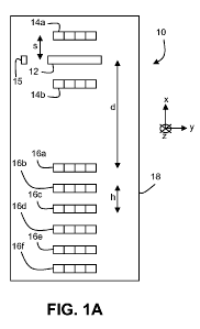
FIGS. IA and lB are schematic diagrams showing bottom and top surfaces,
respectively,
of a sensor 10. Sensor 10 includes a spectral detector 12, two short-distance
radiation sources
14a and 14b, and six long-distance radiation sources 16a, 16b, 16c, 16d, 16e,
and 16f. Detector
12 and radiation sources 14a-b and 16a-f are mounted to circuit board 18. Each
of short-distance
radiation sources 14a and 14b can include one or more packages, and each
package can include
one or more elements that produce illumination radiation. Similarly, each of
long-distance
radiation sources 16a-f can include one or more packages, and each package can
include one or
more elements that produce illumination radiation.
While FIGS. IA and lB show an embodiment of sensor 10 that includes two short-
distance sources 14a and 14b and six long-distance sources 16a-f, more
generally, sensor 10 can
include any number of short-distance radiation sources and any number of long-
distance
radiation sources. For example, in some embodiments, sensor 10 can include one
or more short-
distance radiation sources (e.g., two or more short-distance radiation
sources, three or more
short-distance radiation sources, four or more short-distance radiation
sources, five or more
short-distance radiation sources, six or more short-distance radiation
sources, eight or more
short-distance radiation sources, or even more short-distance radiation
sources). In certain
embodiments, sensor 10 can include one or more long-distance radiation sources
(e.g., two or
more long-distance radiation sources, three or more long-distance radiation
sources, four or more
long-distance radiation sources, five or more long-distance radiation sources,
six or more long-
distance radiation sources, eight or more long-distance radiation sources, or
even more long-
distance radiation sources).
The short- and long-distance sources in sensor 10 can be directly attached to
circuit board
18. That is, the sources can be mounted directly to circuit board 18, rather
than being connected
to circuit board 18 via electrical wires or cables, or optical fibers. In some
embodiments, the
short- and long-distance sources can be soldered directly to circuit board 18
(e.g., with no spacer
or other element separating the sources and circuit board 18). In certain
embodiments, the short-
and long-distance sources can also be fixedly attached to circuit board (e.g.,
mounted on circuit
board 18 such that a fixed spatial relationship exists between the sources and
circuit board 18).
By virtue of the fixed attachment, the sources do not move independently of
circuit board 18, as
19
CA 02732996 2011-02-03
WO 2010/053617 PCT/US2009/053183
would occur if the sources were attached with a cable or fiber. Instead, the
sources are rigidly
attached to circuit board 18 so that the position of the sources with respect
to circuit board 18
does not change.
In general, each of the short-distance and long-distance radiation sources can
include one
or more packages (e.g., two or more packages, three or more packages, four or
more packages,
five or more packages, six or more packages, or even more packages). Each of
the packages can
include one or more elements that produce illumination radiation (e.g., two or
more elements,
three or more elements, four or more elements, or even more elements).
Further, elements that
emit radiation at different wavelengths can be positioned at different spatial
locations, depending
upon the sample the detector. For example, if detector 12 is configured to
resolve different
wavelengths at different spatial positions, the elements and/or packages in
some or all of the
short- and long-distance sources can be positioned to correspond directly or
opposingly to the
configuration of detector 12.
In some embodiments, the number of packages in some of the short- and/or long-
distance
radiation sources can vary. For example, sources that are positioned further
from detector 12 can
include larger numbers of packages, to ensure that sufficient scattered
radiation intensity is
measured by detector 12. In general, any of the short- and/or long-distance
sources can include
any number of packages, the number of packages being selected to ensure that
the sample is
sufficiently illuminated with a desired distribution of incident radiation,
and to ensure that
detector 12 obtains suitable measurements of scattered radiation from the
sample. As an
example, in some embodiments, a long-distance source that is positioned
furthest from detector
12 can include 1.5 times as many packages (e.g., 2.0 times as many packages,
2.5 times as many
packages, 3.0 times as many packages, 3.5 times as many packages, 4.0 times as
many packages
as a long-distance source that is positioned nearest to detector 12.
The elements within the packages of each short- and long-distance radiation
source are
typically selected so that, when the elements are activated (e.g., emitting
light), the spectrum of
the light produced collectively by the elements corresponds to a desired
spectral distribution of
illumination radiation. The spectral distribution can be altered by
positioning particular elements
within the short- and/or long-distance sources, so that the sample can be
illuminated according to
specific spectral distributions. In some embodiments, for example, the
illumination spectrum for
CA 02732996 2011-02-03
WO 2010/053617 PCT/US2009/053183
one or more short- and/or long-distance sources can be selected so that
measurement sensitivity
of sensor 10 in particular regions of the spectrum is enhanced, as discussed
previously.
As shown in FIG. IA, the emission windows of radiation sources 14a-b and 16a-
f, and
the radiation entry surface of detector 12, are exposed on the bottom surface
of sensor 10.
Sensor 10 also includes an electronic processor 20, an optional applications
processor 22,
an optional display unit 24, a power source 26, and a communication interface
28. Processors 20
and 22, display 24, power source 26, and interface 28 are mounted to the upper
surface of circuit
board 18, as shown in FIG. 113. In some embodiments, processor 22 is not
included in sensor 10;
instead, processor 22 is part of an external computing device (e.g., a
personal computer) that
communicates with sensor 10 via communication interface 28, and performs some
or all of the
functions of processor 22 (or processor 20) disclosed herein.
In some embodiments, some (or all) of the long-distance radiation sources can
be
mounted on a separate circuit board that interfaces to circuit board 18 via a
suitable connector.
FIG. 1 C shows a schematic diagram of the bottom of a sensor 10 that includes
a first circuit
board 18 and a second circuit board 19. First circuit board 18 includes
detector 12 and two
short-distance sources 14a-b. Second circuit board 19 includes five long-
distance sources 16a-e.
A connector 21 connects the first and second circuit boards, and permits
communication (e.g.,
exchange of data and control signals) between the circuit boards. Typically,
for example,
processor 20 (and, optionally, processor 22) are located on first circuit
board 18, and
communicate with long-distance sources 16a-e via connector 21.
In certain embodiments, power source 26 is mounted on first circuit board 18,
and can
also communicate with sources 16a-e via connector 21. Power source 26 can
include, for
example, a rechargeable battery. In some embodiments, power source 26 can
include a
disposable battery. In the embodiment shown in FIG. 1C for example, the
disposable battery can
be positioned on or connected to first circuit board 18. Alternatively, the
disposable battery can
be positioned on or connected to second circuit board 19. If second circuit
board 19 is a
disposable circuit board, the battery can be disposed of at the same time as
second circuit board
19.
FIG. 2 shows a schematic diagram of sensor 10 mounted on a sample 30. Sample
30
includes one or more layers of skin 32, a subcutaneous layer of fat 34, and
underlying muscle
tissue 36. Sensor 10 is configured to interrogate muscle tissue 36 by
directing radiation 38,
21
CA 02732996 2011-02-03
WO 2010/053617 PCT/US2009/053183
generated by at least one (e.g., all) of radiation sources 14a-b and at least
one of the radiation
sources 16a-f, to be incident on muscle tissue 36. Scattered radiation 40 is
received and analyzed
by detector 12 (not shown) to determine a spectrum of the scattered radiation.
The scattered
radiation spectrum is then processed by electronic processor 20 and/or
processor 22 (not shown)
to determine an absorbance spectrum of muscle tissue 36. Based on the
absorbance spectrum,
electronic processor 20 and/or 22 can determine one or more properties of
sample 30 (and in
particular, of muscle tissue 36 within sample 30).
In general, the scattered radiation spectrum measured by detector 12, which
typically
includes wavelength-dependent information about scattered radiation from
sample 30, can be
converted by an electronic processor to an absorbance spectrum of muscle
tissue 36 using well-
known methods. As noted previously, in the following discussion, reference is
made to
absorbance spectra of samples such as sample 30. However, the apparatus and
methods
disclosed herein can also be used to derive reflectance spectra from measured
scattered radiation;
reflectance and absorbance are related by a simple mathematical
transformation. Methods for
converting spectral scattered radiation information into reflectance and
absorbance spectra for a
sample are disclosed, for example, in U.S. Patent Application Publication No.
US 2008/0097173.
In addition to converting scattered radiation information into absorbance
and/or
reflectance spectra, processor 20 and/or 22 can be configured (e.g., using
calibration equations
and/or data stored in memory units, magnetic storage units, and/or optical
storage units) to
analyze absorbance spectra to obtain measurements of physiologically important
parameters for
sample 30. In general, processor 20 and/or 22 can be configured to perform any
of the analysis
steps that are discussed herein.
In some embodiments, one or more absorbance spectra for sample 30 can be
analyzed to
determine pH (e.g., muscle tissue pH) in the sample. Systems and methods for
determining
tissue pH are disclosed, for example, in U.S. Patent No. 5,813,403 entitled
"Optical
Measurement of Tissue pH," the entire contents of which are incorporated
herein by reference.
In certain embodiments, one or more absorbance spectra for sample 30 can be
analyzed
to determine blood hematocrit in the sample. Systems and methods for
determining blood
hematocrit are disclosed, for example, in U.S. Patent No. 6,006,119 entitled
"Noninvasive
Optical Measurement of Blood Hematocrit," the entire contents of which are
incorporated herein
by reference.
22
CA 02732996 2011-02-03
WO 2010/053617 PCT/US2009/053183
In some embodiments, one or more absorbance spectra for sample 30 can be
analyzed to
determine quantities such as hemoglobin concentration, and/or water content,
and/or oxygen
tension and/or tissue oxygen saturation. Systems and methods for determining
these quantities
are disclosed, for example, in U.S. Patent Application Publication No. US
2008/0097173, and in
U.S. Patent No. 6,766,188, the entire contents of each of which are
incorporated herein by
reference.
In certain embodiments, one or more absorbance spectra for sample 30 can be
analyzed
to determine quantities such as anaerobic threshold and/or metabolic rate
(e.g., oxygen
consumption rate) in the sample. Systems and methods for determining these
quantities are
disclosed, for example, in U.S. Patent Application Serial No. 12/172,942,
entitled "Physical
Performance Monitoring and Monitors," filed on July 14, 2008, the entire
contents of which are
incorporated herein by reference.
In some embodiments, one or more absorbance spectra for sample 30 can be
analyzed to
determine additional quantities such as a temperature of a tissue of interest
within sample 30. In
addition, processor 20 and/or 22 can include a hardware-based temperature
monitor that
effectively monitors a temperature of the sample surface to which sensor 10 is
attached, for
example.
Typically, sensor 10 includes a housing that encloses components such as
circuit board
18, and which also includes apertures that permit radiation generated by the
short- and long-
distance sources to emerge from the housing, and permit scattered radiation
from the sample to
be incident on detector 12. FIGS. 3A and 3B show bottom and top views,
respectively, of a
sensor 10 that includes a housing 11. Apertures formed in the bottom surface
of housing 11
expose long-distance sources 16a-e, short-distance sources 14a-b, and detector
12, as shown in
FIG. 3A. Apertures 17a and 17b, formed in a side surface of housing 11, permit
connection to
communication interface 28 and power source 26, respectively. Loops 15 admit a
fastener such
as a strap (e.g., a VelcroTM strap or another type of strap) to secure housing
11 to a sample (e.g.,
an arm or leg of a subject).
Typically, the dimensions of sensor 10 are smaller than corresponding
dimensions of
conventional spectral devices. With reference to FIG. 3B, the housing of
sensor 10 includes a
bottom surface that has a maximum dimension L, a maximum width W measured in a
direction
23
CA 02732996 2011-02-03
WO 2010/053617 PCT/US2009/053183
perpendicular to the maximum dimension L, and a thickness T measured in a
direction
perpendicular to both the maximum dimension L and the maximum width W.
The dimensions L, W, and T of sensor 10 can vary according to the various
components
included in sensor 10 (e.g., numbers and spatial positions of radiation
sources, processors,
display unit, power source). In the embodiment shown in FIGS. 3A and 3B, the
dimensions L,
W, and T are approximately 110 mm, 55 mm, and 20 mm, respectively.
In general, however, the dimensions L, W, and T of sensor 10 can differ in
various
embodiments. In some embodiments, the maximum dimension L can be 15 mm or more
(e.g.,
20 mm or more, 30 mm or more, 40 mm or more, 50 mm or more, 60 mm or more, 70
mm or
more, 80 mm or more) and/or 150 mm or less (e.g., 140 mm or less, 130 mm or
less, 120 mm or
less, 110 mm or less, 100 mm or less, 90 mm or less). In certain embodiments,
the maximum
width W can be 10 mm or more (e.g., 15 mm or more, 20 mm or more, 25 mm or
more, 30 mm
or more, 35 mm or more, 40 mm or more) and/or 75 mm or less (e.g., 70 mm or
less, 65 mm or
less, 60 mm or less, 55 mm or less, 50 mm or less, 45 mm or less).
In some embodiments, the thickness T can be 5 mm or more (e.g., 10 mm or more,
15
mm or more, 20 mm or more) and/or 30 mm or less (25 mm or less). Typically,
sensor 10 is
sufficiently thin (e.g., thickness T is sufficiently small) so that sensor 10
can be comfortably
worn by a human or animal subject without causing undue discomfort. For human
subjects, such
sensors can comfortably be worn underneath clothing, for example.
Detector 12 is a spectral detector configured to analyze input radiation as a
function of
wavelength. In certain embodiments, for example, detector 12 can include a
linear variable filter
or a variable Fabry Perot etalon (FPE) coupled to a radiation detector such as
a linear photodiode
array, a charge coupled device (CCD) or a complementary metal oxide
semiconductor (CMOS)
device. FIG. 4 is a schematic diagram of a detector 12 that includes a linear
variable filter (LVF)
54 coupled to a linear array CCD detector 50. LVF 54 is essentially a wedged
bandpass filter,
and includes mirror layers 42 and 44, a spacer layer 46, and a substrate 48,
which collectively
function as an etalon or interference bandpass filter. Radiation 52 (e.g.,
collimated radiation) is
incident on detector 12 along the z-direction shown in FIG. 4. The design,
operation and
function of bandpass interference filters and variable bandpass filters, such
as LVFs, are
disclosed, for example, in the "Interference Filter Handbook," published by
JDS Uniphase
(Second Edition), the entire contents of which are incorporated herein by
reference.
24
CA 02732996 2011-02-03
WO 2010/053617 PCT/US2009/053183
In some embodiments, detector 12 has a length, measured in the direction of
the width W
of sensor 10, of 2 mm or more (e.g., 4 mm or more, 6 mm or more, 8 mm or more,
10 mm or
more, 12 mm or more) and/or 20 mm or less (e.g., 18 mm or less, 16 mm or less,
14 mm or less).
In certain embodiments, detector 12 has a thickness, measured in the direction
of the thickness T
of sensor 10, of 0.1 mm or more (e.g., 0.2 mm or more, 0.3 mm or more, 0.5 mm
or more, 1.0
mm or more, 2.0 mm or more) and/or 5.0 mm or less (e.g., 4.0 mm or less, 3.0
mm or less, 2.5
mm or less).
In some embodiments, detector 12 has a width, measured in the direction of the
length L
of sensor 10, of 1.0 mm or more (e.g., 1.5 mm or more, 2.0 mm or more, 2.5 mm
or more) and/or
4.0 mm or less (e.g., 3.5 mm or less, 3.0 mm or less).
Devices such as LVFs, FPEs, and CCD detectors are generally robust and do not
appreciably degrade over time. As a result, the spectral properties of these
devices typically
remain relatively constant, obviating the need to perform re-calibration of
detector 12 over time.
In addition, LVFs, FPEs, and CCD detectors are relatively stable under the
influence of
temperature fluctuations. Typically, the layers of LVF 54 are formed of
various amorphous or
crystalline materials, which do not appreciably expand or contract with modest
changes in
temperature. As a result, the spectral filtering properties of LVF 54 remain
relatively unchanged
for modest temperature changes, and detector 12 does not typically have to be
calibrated for
variable temperature operation.
In general, detector 12 can include various types of spectral detectors. For
example,
detector 12 can include detectors that include a radiation sensitive element
(e.g., photodiode
array and/or CCD and/or CMOS device) coupled to a wavelength-dispersive
element such as one
or more diffraction gratings and/or prisms. In addition, detector 12 can
include other types of
dispersive and/or filtering elements (e.g., diffractive optical elements,
liquid crystal-based filters,
bandpass filters, tunable etalons) that are used to provide wavelength-
sensitive detection and/or
analysis of incoming radiation.
In certain embodiments, a full width at half maximum (FWHM) spectral
resolution of
detector 12 is 10.0 nm or less (e.g., 8.0 nm or less, 6.0 nm or less, 5.0 nm
or less, 4.0 nm or less,
3.0 nm or less, 2.0 nm or less, 1.0 nm or less, 0.5 nm or less, 0.25 nm or
less). In general, the
FWHM spectral resolution depends upon the number of active detector elements
(e.g., pixels on
a CCD detector) and the wavelength-dispersing ability of the optical elements
in the detector.
CA 02732996 2011-02-03
WO 2010/053617 PCT/US2009/053183
In some embodiments, sensor 10 can include one or more optical elements that
are
configured to effectively control the range of angles at which scattered
radiation is incident on
detector 12 from sample 30. For example, FIG. 5 shows a sensor 10 that
includes a collimating
element 56 attached to a surface of detector 12 (e.g., the surface of detector
12 that receives
scattered radiation from sample 30). Detector 12 can include, for example, a
LVF, and
collimating element 56 can be attached directly to the LVF. Detector 12 can
also include, for
example, a CCD detector coupled to the opposite surface of the LVF. The entire
assembly -
collimating element 56, the LVF, and the CCD detector - can be mounted on
circuit board 18, as
shown in FIG. 5. Collimating element 56 functions to collimate scattered
radiation 40 from
sample 30 to control the range of angles at which the scattered radiation is
incident on detector
12. The spectral bandpass properties of LVF 54, such as a FWHM spectral width
and/or shape
of a spectral passband of LVF 54, depend upon the angle of incidence of
incoming radiation. In
particular, variations in the angle of incidence of the scattered radiation on
LVF 54 can result in
blue-shifting of the passband wavelength at one or more positions along CCD
detector 50, and/or
loss of spectral resolution (e.g., increase of passband width) in LVF 54. By
controlling the range
of angles of incidence via collimating element 56, the spectral properties of
detector 12 can be
reproducible over relatively long periods of use.
In general, a variety of different collimating elements 56 can be used in
sensor 10.
Exemplary collimating elements include fiber faceplates (e.g., fiber optic
windows), collimating
hole devices, gradient index (GRIN) lenses, fiber bundles, lens arrays,
optical windows
(including shaped optical windows), and other similar devices.
Sensor 10 typically includes a plurality of radiation sources. In some
embodiments, some
or all of the radiation sources include light emitting diodes (LEDs). Some (or
all) of the
radiation sources of sensor 10 can provide relatively broad bandwidth incident
radiation for
illuminating sample 30. To provide such radiation, the radiation sources can
include one or more
LEDs. For example, in certain embodiments, some radiation sources can include
a single
broadband LED. In some embodiments, certain radiation sources can include
multiple LEDs.
The multiple LEDs can each emit radiation having different central wavelengths
and/or spectral
bandwidths. In some embodiments, some of the multiple LEDs can emit radiation
having the
same central wavelength and/or bandwidth.
26
CA 02732996 2011-02-03
WO 2010/053617 PCT/US2009/053183
In the embodiment shown in FIG. IA, for example, each of sources 14a-b and 16a-
f
includes six LEDs having central emission wavelengths of 735 nm, 780 nm, 810
nm, 850 nm,
890 nm, and 940, respectively. The six LEDs together can be powered to deliver
up to
approximately 500 mW of total radiation power, depending upon the method used
to drive/power
the LEDs. In the embodiment shown in FIG. IA, the LEDs are custom packaged as
surface-
mount technology devices with a width of about 2 mm. Each package can be
configured to hold
up to three LED dies (the radiation-emitting elements). The six LEDs are
distributed among two
LED packages; one package includes three LED dies, and the other includes two
LED dies. The
LEDs are typically powered by a regulated supply of between 3.5V and 5V from
power source
26. In some embodiments, power source 26 can be a transformer block, for
example, that
delivers 6 V or more.
In some embodiments, any one or more of radiation sources 14a-b and 16a-f can
include
other types of radiation emitting elements. For example, the radiation sources
can include
incandescent (e.g., tungsten filament) lamps. Suitable lamps include, for
example, Gilway
models T-1 and T-1'/4, available from International Light Technologies
(Peabody, MA). These
lamps have relatively low operating voltage (5 V), operating current (0.06 A),
and can provide
up to 200,000 hours of operation. In addition, the lamps can be operated at
3.5 V with relatively
minor reductions in near-infrared radiation output, and with relatively large
increases in stability
and lifetime. Similar lamp models are also available, for example, from
companies such as
Welch Allyn (Skaneateles Falls, NY).
In general, some or all of the radiation sources of sensor 10 can include any
number of
radiation emitting elements (e.g., LEDs, tungsten lamps). In some embodiments,
for example,
radiation sources can include one or more radiation emitting elements (e.g.,
two or more
radiation emitting elements, three or more radiation emitting elements, five
or more radiation
emitting elements, seven or more radiation emitting elements, nine or more
radiation emitting
elements).
In some embodiments, the number of radiation emitting elements in some of the
short-
and/or long-distance radiation sources (and/or some of the packages within the
short- and/or
long-distance radiation sources) can vary. For example, sources that are
positioned further from
detector 12 can include packages with larger numbers of radiation emitting
elements, to ensure
that sufficient scattered radiation intensity is measured by detector 12. In
general, any of the
27
CA 02732996 2011-02-03
WO 2010/053617 PCT/US2009/053183
packages of the short- and/or long-distance sources can include any number of
radiation emitting
elements, the number of elements being selected to ensure that the sample is
sufficiently
illuminated with a desired distribution of incident radiation, and to ensure
that detector 12
obtains suitable measurements of scattered radiation from the sample.
When some or all of the radiation sources of sensor 10 include multiple
radiation emitting
elements, electronic processor 20 can be configured to adjust the radiation
emitting elements to
control the properties of the output radiation produced by the multiple
elements. In particular,
for example, when certain radiation sources of sensor 10 include multiple
LEDs, processor 20
can control the emitted radiation intensity from each of the individual LEDs
to control the
overall distribution of radiation produced by the sources. Control of the
emitted radiation
intensity can be achieved, for example, through a digital-to-analog converter
(DAC) which
converts a digital control signal from processor 20 and/or 22 into an analog
control voltage
applied to a given radiation source element. With a DAC of suitable resolution
(e.g., 14-bit),
relatively fine control over emission intensities of individual LEDs can be
achieved, and
continuous-wave emission can be implemented.
Alternatively, in some embodiments, direct digital control of individual LEDs
can be
achieved via pulse width modulation (PWM) of the LEDs by processor 20 and/or
22. Pulse
width modulation provides a modulated (e.g., pulsed) LED output. Under PWM
control, the
integrated intensity of LED output over a selected temporal window is
controlled, from a
maximum value (always on) to zero (always off), as defined by the duty cycle
of the modulated
signal. Emission intensities between these limits are realized by high
frequency pulsing of the
LEDs by processor 20 and/or 22. By controlling the rate at which pulses are
emitted by the
LEDs, the duty cycle of the LEDs can be adjusted. For example, to reduce the
emission intensity
of a particular LED from its maximum value to a half-maximum value, the duty
cycle of the
LED is reduced to 50%.
In certain embodiments, the emission intensities of individual LEDs are fixed.
That is,
suitable emission intensities for each of the LEDs are determined based on a
particular
measurement application, and the current supplied to each of the LEDs to
achieve the desired
intensity output for each is determined (as discussed further in Example 3).
After suitable
driving currents for each LED have been determined, resistors can be
introduced into the driving
circuit for each of the LEDs to maintain an appropriate drive current for each
LED. The
28
CA 02732996 2011-02-03
WO 2010/053617 PCT/US2009/053183
determination of suitable drive currents (and resistors) for a particular
selection of LEDs can be
made once using a calibration sensor, and subsequent sensors built using the
same combination
of LEDs can include the set of pre-determined resistors; separate calibration
of each sensor is not
required.
Processor 20 and/or 22 can be configured to control the emitted radiation
intensity from
multiple radiation emitting elements for a variety of reasons. For example, in
certain
embodiments, the intensities of some of the radiation emitting elements may be
higher than the
intensities of others. By controlling (e.g., reducing) the intensities of
certain radiation emitting
elements, the spectral profile of illumination radiation directed to a sample
can be made more
uniform, for example, or can be modified so that the spectral profile more
generally assumes a
desired (and known) shape. By using illumination radiation with a known shape,
it can be easier
to identify spectral features of interest, for example, in measured scattered
radiation from the
sample.
As another example, in some embodiments, the sensitivity of detector 12 to
incident
radiation can vary as a function of the wavelength of the radiation.
Accordingly, the spectral
profile of the illumination radiation can be selected to reduce or remove
spectral features in
measured scattered radiation spectra that arise from such variations in
detector sensitivity. As
above, selecting the spectral profile of the illumination radiation can
include increasing and/or
decreasing emitted radiation intensity from certain radiation emitters
relative to other radiation
emitters under the control of processor 20 and/or 22.
As a further example, in some embodiments, the sample (e.g., sample 30) can
include
moieties that absorb incident radiation at one or more well-known wavelengths.
To enhance a
signal-to-noise ratio in measured scattered radiation (and even, to enable
measurement of
scattered radiation at the wavelengths of absorption), the emission
intensities of certain radiation
emitters can be increased and/or decreased relative to other radiation
emitters. In particular,
emission intensities of radiation emitters that emit radiation that falls
within absorption bands
can be increased relative to emission intensities of other emitters (or the
intensities of the other
emitters can be decreased relative to the emission intensities of the emitters
that emit within the
absorption bands).
Typically, for example, each radiation source of sensor 10 emits radiation
that includes
multiple radiation wavelengths. In some embodiments, a FWHM spectral bandwidth
of the
29
CA 02732996 2011-02-03
WO 2010/053617 PCT/US2009/053183
emitted radiation from each source is 10 nm or more (e.g., 15 nm or more, 20
nm or more, 50 nm
or more, 100 nm or more, 200 nm or more, 300 nm or more, 400 nm or more, 500
nm or more,
700 nm or more). The usable range of the radiation emitted by each source can
be determined by
a transmission range of detector 12. In certain embodiments, for example,
detector 12 includes a
LVF with a transmission range of from 600 nm to 1100 nm. The usable range of
the emitted
radiation can also depend, in some embodiments, on the spectral response of a
detector coupled
to the wavelength dispersive element in detector 12. For example, photodiode
arrays, CCD
arrays, and CMOS arrays that are formed of silicon typically have a usable
spectral response that
reaches an upper limit at about 1100 nm. Detectors formed of other materials
can be used to
measure sample responses at even longer wavelengths.
In general, each radiation source of sensor 10 emits radiation having a
central
wavelength. The central wavelength of emitted radiation from each source can
be between 600
nm and 1100 nm (e.g., between 650 nm and 1050 nm, between 700 nm and 1000 nm,
between
750 nm and 1000 nm, between 800 nm and 1000 nm, between 800 nm and 1100 nm).
Each radiation source (e.g., sources 14a-b and 16a-f) includes one or more
radiation
emitters such as LEDs and/or tungsten filaments. The radiation emitters can
all emit at the same
central emission wavelength, or some of the emitters can emit at different
wavelengths.
Alternatively, or in addition, the radiation emitters can all have different
FWHM emission
bandwidths, or at least some of the emitters can have the same bandwidths.
In general, each emitter emits radiation having a central emission wavelength
between
600 nm and 1100 nm (e.g., between 650 nm and 1050 nm, between 700 nm and 1000
nm,
between 750 nm and 1000 nm, between 800 nm and 1000 nm, between 800 nm and
1100 nm).
Typically, for example, each emitter has a FWHM emission bandwidth of 3 nm or
more (e.g., 5
nm or more, 10 nm or more, 15 nm or more, 20 nm or more, 30 nm or more, 40 nm
or more, 50
nm or more, 60 nm or more, 80 nm or more, 100 nm or more, 150 nm or more, 200
nm or more).
Sensor 10 includes both short-distance sources 14a-b and long-distance sources
16a-f.
Short-distance sources are positioned at a distance s from detector 12,
measured along the x-
direction as shown in FIG. IA. Typically, for example, s is about 2.5 mm. In
general, however,
s can be 0.5 mm or more (e.g., 1.0 mm or more, 1.5 mm or more, 2.0 mm or more,
2.5 mm or
more, 3.0 mm or more, 4.0 mm or more, 5.0 mm or more) and/or 10.0 mm or less
(e.g., 9.0 mm
or less, 8.0 mm or less, 7.0 mm or less, 6.0 mm or less).
CA 02732996 2011-02-03
WO 2010/053617 PCT/US2009/053183
As shown in FIG. IA, in certain embodiments, sensor 10 includes two short-
distance
sources 14a-b. The number of short-distance sources is typically selected to
ensure that detector
12 is relatively uniformly illuminated with scattered radiation from a sample
when short-distance
sources are used to illuminate the sample. Accordingly, in general, sensor 10
can include one or
more short-distance sources. For example, in some embodiments, sensor 10 can
include from
zero to four short-distance sources positioned on one side of detector 12.
From zero to four
short-distance sources can also be positioned on the other side of detector
12. Each of the
sources can include one or more packages, as discussed previously, and each of
the packages can
include one or more radiation emitting elements.
In certain embodiments - for example, where sensor 10 has an extended length L
- the
number of short-distance sources on each side of detector 12 can be even
greater (e.g., five or
more, six or more, seven or more, eight or more, nine or more, ten or more).
Sensor 10 also includes a plurality of long-distance sources 16a-f. In some
embodiments,
as shown in FIG. IA, sensor 10 includes six long-distance sources 16a-f. The
depth to which
incident radiation from a particular radiation source penetrates a sample and
generates detected
scattered radiation from a tissue of interest therein is generally related to
a linear distance
between the radiation source and the detector. Each of the long-distance
sources of sensor 10
therefore generally corresponds to interrogation of the sample to a certain
depth below the
sample surface. Typically, an appropriate long-distance source is selected to
illuminate muscle
tissue 36 in sample 30 by selecting a long-distance source that produces
radiation that can
penetrate through overlying layers to sufficiently illuminate muscle tissue 36
below the surface
of sample 30 so that reflected light from the muscle can be adequately
measured by detector 12.
Sensor 10 can, in general, include any number of long-distance sources to
enable measurement
of tissues at a variety of depths below the surface of a sample. In certain
embodiments, for
example, sensor 10 can include one or more long-distance sources (e.g., two or
more long-
distance sources, three or more long-distance sources, four or more long-
distance sources, five or
more long-distance sources, seven or more long-distance sources, nine or more
long-distance
sources, or even more long-distance sources). All of the long-distance sources
can be positioned
at different distances measured along the x-direction from detector 12, as
shown in FIG. IA, or at
least some of the sources can be positioned at the same distance from detector
12.
31
CA 02732996 2011-02-03
WO 2010/053617 PCT/US2009/053183
Typically, a shortest distance between any of the long-distance sources and
detector 12 is
d, as shown in FIG. IA. Each of the short-distance sources is positioned at a
distance less than d
from detector 12, and each of the long-distance sources is positioned at a
distance of d or more
from detector 12, the distances being measured in the x-direction. In some
embodiments, d is 5
mm or more (e.g., 6 mm or more, 8 mm or more, 10 mm or more, 12 mm or more, 14
mm or
more, 16 mm or more, 18 mm or more, 20 mm or more, 22.5 mm or more, 25 mm or
more, 27.5
mm or more, 30.0 mm or more, 35.0 mm or more, 40 mm or more, 50 mm or more).
Referring again to FIG. IA, in some embodiments, a spacing h between each of
the long-
distances sources is the same, so that each successive long-distance source is
displaced from
detector 12 by an additional distance increment h. In the embodiment shown in
FIG. IA, for
example, the six long-distance sources 16a-f are positioned at distances of 25
mm, 30 mm, 35
mm, 40 mm, and 45 mm from detector 12, respectively, measured along the x-
direction.
In certain embodiments, the spacings between each of the long-distance sources
are not
all the same. For example, sensor 10 can include a first group of long-
distance sources and a
second group of long-distance sources, where each member of the first and
second groups is
positioned relatively closely to other members of the same group, but
relatively farther away
from sources in the other group.
In general, the spacing h between any two long-distance radiation source
elements can be
0.5 mm or more (e.g., 1.0 mm or more, 2.0 mm or more, 3.0 mm or more, 4.0 mm
or more, 5.0
mm or more, 7.5 mm or more, 10.0 mm or more, 12.5 mm or more, 15.0 mm or more,
17.5 mm
or more, 20.0 mm or more, 30.0 mm or more, 40.0 mm or more, 50.0 mm or more,
60.0 mm or
more, 70.0 mm or more, 100 mm or more, 150 mm or more, or even more).
Typically, short-distance sources are spaced from detector 12 by a distance s,
measured
along the x-direction as shown in FIG. IA. In general, the spacing s can be
0.5 mm or more
(e.g., 1.0 mm or more, 2.0 mm or more, 3.0 mm or more, 4.0 mm or more, 6.0 mm
or more, 8.0
mm or more, 10.0 mm or more, 15.0 mm or more, 20.0 mm or more, or even more).
When
multiple short-distance sources are implemented in sensor 10, the multiple
short-distance sources
can be evenly spaced along the x-direction, or the spacings between some or
all short-distance
sources can differ. In general, the spacing between any two short-distance
sources can be 0.5
mm or more (e.g., 1.0 mm or more, 2.0 mm or more, 3.0 mm or more, 4.0 mm or
more, 6.0 mm
or more, 8.0 mm or more, 10.0 mm or more, 15.0 mm or more, 20.0 mm or more, or
even more).
32
CA 02732996 2011-02-03
WO 2010/053617 PCT/US2009/053183
In some embodiments, circuit board 18 can be flexible and can deform when
attached to
the surface of a sample, assuming a shape that is at least partially
complementary to a shape of
the sample. In certain embodiments, for example, circuit board 18 can be a
flex circuit board. In
some embodiments, circuit board 18 can be formed of one or more deformable
materials such as
one or more flexible plastic materials.
In certain embodiments, circuit board 18 can be relatively rigid and resistant
to
deformation. Circuit board 18 can be formed of certain types of rigid plastic
materials, for
example, which remain relatively rigid to ensure that distances between
various sensor radiation
sources and detector 12 are not significantly distorted by deformation of
circuit board 18.
In some embodiments, circuit board 18 can be formed so that deformation of the
circuit
board along one direction - the x-direction in FIG. IA - does not readily
occur during use. As a
result, the relative distances between detector 12 and the short- and long-
distance sources can be
maintained, ensuring that accurate and reproducible correction of the measured
spectra to reduce
the effects of overlying skin and fat layers can be performed. In addition,
however, circuit board
18 can deformed at its edges to conform to the shape of a sample (e.g., an arm
or leg of a
subject), so that sensor 10 can be comfortable and unobtrusively worn by the
subject.
In some embodiments, circuit board 18 is formed of two different circuit board
components. A first, relatively rigid component corresponds to a mounting
member to which the
various components of sensor 10, including processors, radiation sources,
detectors, power
sources, interfaces, and displays are attached. A second, relatively flexible
component is
attached to the first component and also contacts the sample. By using a two-
part construction,
sensor 10 ensures that distances between the various radiation sources and
detector 12 remain
relatively constant during use, but also assumes at least partially a shape
complementary to a
surface of the sample to which the sensor is attached.
In certain embodiments, sensor 10 can be attached to sample 30 via an adhesive
element
such as adhesive pad or layer. FIG. 6A shows a schematic diagram of a sensor
10 that is
attached to sample 30 with an adhesive layer 58. Adhesive layer 58 is
positioned between sensor
10 (e.g., a bottom surface of housing 11) and a surface of sample 30. In some
embodiments,
layer 58 can be formed by a paste or another similar substance that can be
applied to the surface
of a sample and/or to the bottom surface of sensor 10 to affix sensor 10 to
the sample.
33
CA 02732996 2011-02-03
WO 2010/053617 PCT/US2009/053183
In some embodiments, adhesive layer 58 can be disposable, and can form a
portion of a
two-layer sensor 10. For example, as shown in FIG. 6B, sensor 10 can include a
first non-
disposable portion that includes circuit board 18 and components mounted
thereon enclosed in
housing 11, and a second disposable portion that includes adhesive layer 58
(and, possibly, other
layers). Following use, the disposable portion of sensor 10 can be discarded
and the non-
disposable portion retained for future use. Adhesive layer 58 can be
implemented as a flexible
material on which some of the radiation sources (e.g., the long-distance
sources) are mounted.
Short-distance sources can be mounted on circuit board 18 and enclosed within
housing 11.
When layer 58 is disposed of following use, the short-distance sources are
retained within
housing 11. A new layer 58 can be attached to housing 11 prior to using sensor
10 to make
additional measurements.
Adhesive layer 58, positioned between housing 11 and sample 30, is at least
partially
transmissive to near-infrared radiation. For example, when the sensor of FIG.
6 is in use,
radiation generated by one or more radiation sources of sensor 10 passes
through adhesive layer
58 and is incident on sample 30. Scattered radiation from sample 30 also
passes through
adhesive layer 58 before being incident on detector 12.
In certain embodiments, layer 58 can be implemented as a multilayer structure.
For
example, layer 58 can include two layers: a first layer that is relatively
inflexible and that
supports some or all of the components of sensor 10 (e.g., radiation sources,
processors,
detectors, and other circuitry), and a second layer that contacts the first
layer and is also
configured to contact the sample. The second layer can be an adhesive layer,
and can be flexible
so that the second layer deforms to match the surface of the sample when
applied to the sample.
Many different materials can be used to form the first and second layers. For
example, the first
layer can include one or more metals, plastics (e.g., high-density plastics),
polymer materials,
and paper- and/or wood-based materials (e.g., fiberboard). The second layer
can include one or
more plastic materials, polymer materials, rubber, latex, gels, and other
types of flexible
materials.
A variety of different disposable and non-disposable configurations are
possible. In some
embodiments, for example, both the first and second layers are disposable
(e.g., all of sensor 10
is disposable). In certain embodiments, neither layer is disposable. Further
still, in some
embodiments, one of the layers (e.g., the second layer) is disposable, while
the other layer (e.g.,
34
CA 02732996 2011-02-03
WO 2010/053617 PCT/US2009/053183
the first layer) is not. Typically, in a two-layer structure, at least
portions of both the first and the
second layers are at least partially transmissive to near-infrared radiation,
as discussed above, or
include a window positioned in the layers to allow near-infrared radiation to
pass through the
layers.
In some embodiments, as shown in FIG. 7, sensor 10 can be attached to sample
30 with
an adhesive patch 60. Adhesive patch 60 includes adhesive portions 62a and 62b
which adhere
to a surface of sample 30, maintaining contact between sensor 10 and the
surface of sample 30 as
a result. Adhesive patch 60 can be at least partially transmissive to wireless
communication
signals transmitted by communication interface 28.
In some embodiments, sensor 10 can be completely disposable. Following
attachment of
sensor 10 to sample 30, measurement of one or more absorbance spectra, and
calculation of one
or more properties of sample 30, sensor 10 can be detached from the sample and
discarded.
In some embodiments, a portion of sensor 10 can be disposable. For example,
referring
to FIG. 1 C, sensor 10 can include a first circuit board 18 that includes
detector 12 and the short-
distance sources, and a second circuit board 19 that includes the long-
distance sources. Second
circuit board 19 can be a disposable circuit board. Following use of sensor
10, second circuit
board 19 (including the long-distance sources) can be detached from first
circuit board 18 and
discarded, while first circuit board 18 is retained for subsequent use. In
certain embodiments,
most or all of the electronic components can be positioned on the disposable
portion of sensor
10. For example, sensor 10 can include a disposable circuit board, to which
both short- and
long-distance sources are attached, along with a processor (e.g., processor 20
and/or 22),
electronic memory, a power source (e.g., a disposable battery), and/or other
electronic
components disclosed previously. Following use of sensor 10, the disposable
circuit board and
all of the attached electronic components can be discarded, and the remaining
portion of sensor
10 can be retained for subsequent use.
In certain embodiments, sensor 10 can be attached to a sample with a
complementary
sleeve. FIG. 8 shows a schematic view of a sleeve 64 that is attached to a
surface of sample 30.
For example, sleeve 64 can be attached to an arm or leg of a patient who is
exercising or
performing aerobic activity, or receiving medical treatment.
Sleeve 64 includes an internal pocket 68 that is dimensioned to accommodate a
sensor.
Sensor 10 can be attached to sample 30 by inserting sensor 10 into pocket 68
along the direction
CA 02732996 2011-02-03
WO 2010/053617 PCT/US2009/053183
shown by arrow 66. Typically, sleeve 64 is formed from a flexible material
such as a plastic
material. At least a portion of sleeve 64 (e.g., lower portion 70) can be at
least partially
transmissive to radiation generated by one or more radiation sources of sensor
10 and/or to
wireless communication signals generated by communication interface 28. During
operation,
incident radiation from the one or more sources passes through lower portion
70 of sleeve 64 and
into sample 30. Scattered radiation from sample 30 passes through portion 70
before being
incident on detector 12.
Sensor 10 includes power source 26. In some embodiments, for example, power
source
26 can be a connector (e.g., a plug) that receives power from an external
source, such as a
hospital or treatment center power source and/or a conventional wall socket,
which can include a
transformer block. In certain embodiments, power source 26 can be a connector
such as a
conventional power connector or a USB connector that connects to an external
processing device
such as a computer. Sensor 10 can be configured to receive electrical power
from the external
processing device through the connector. Power source 26 can also generally
include various
types of electronic power conditioning devices such as transformers,
resistors, capacitors,
inductors, transistors, and other circuit elements.
In certain embodiments, power source 26 can be a self-contained power source
such as a
battery, a photo-voltaic cell, a fuel cell, or another type of stand-alone
source. Suitable battery
types for power source 26 include, for example, nickel metal hydride
batteries, lithium ion
batteries, and solid electrolyte (primary) batteries. In some embodiments,
power source batteries
can be rechargeable, and can be recharged when sensor 10 is not in use. In
certain embodiments,
power source batteries can be disposable batteries of various types.
In certain embodiments, power source 26 can include a connector that connects
to a
portable power source such as a battery that is worn by a patient (e.g., worn
on an arm or leg of a
patient, or attached via one or more straps to a patient). This arrangement
may allow for the use
of sensor 10 with a larger, higher-capacity battery than would otherwise be
available if the
battery was attached directly to circuit board 18.
In some embodiments, power source 26 can include a replaceable battery similar
to, for
example, a mobile phone battery. Sensor 10 can include a connector which mates
with a portion
of the replaceable battery to enable the battery to supply electrical current
to components
attached to circuit board 18. The connector can form a portion of a cradle
which supports the
36
CA 02732996 2011-02-03
WO 2010/053617 PCT/US2009/053183
replaceable battery. One replaceable battery can then be easily exchanged for
another, for
example, by removing the old battery and sliding the new battery into the
cradle.
In certain embodiments, when power source 26 includes a rechargeable component
such
as a battery, a charging cradle can be configured to support sensor 10 while
the rechargeable
component is charged. FIG. 9 shows a schematic diagram of a charging cradle 72
that includes a
support member 76 and a power connector 74. Support member 76 includes
vertical grooves
into which the edges of sensor 10 are accommodated, maintaining sensor 10 in a
relatively fixed
position with respect to charging cradle 72. Power connector 74 engages with a
mating
connector on power source 26 of sensor 10; power source 26 is typically a
rechargeable battery,
for example. Power is supplied through power connector 74 to power source 26
to recharge
power source 26. Cradle 72 can include, for example, power-limiting circuits
that sense when
power source 26 is nearing a full-charge condition, and which then restrict
the flow of power to
power source 26 to prevent over-charging.
Sensor 10 includes electronic processor 20, and optionally includes one or
more
additional applications processors (e.g., applications processor 22). The
processors generally
coordinate all sensor functions, including directing radiation sources to
produce incident
radiation, directing detector 12 to receive and analyze scattered radiation,
and performing a
variety of mathematical operations on data received from detector 12. The
processors are also
generally responsible for delivering control signals to various sensor
components, receiving
status signals from sensor components, monitoring the delivery of operating
power to sensor
components and the supply of power from power source 26, sending data to
display 24 to be
displayed, and transmitting and receiving communications signals to external
devices via
communication interface 28. If sensor 10 includes one or more applications
processors 22, some
of these functions can be provided by the applications processors. In
particular, the applications
processors can be configured to perform mathematical operations on data
received from detector
12 to derive one or more sample properties from the data, as discussed
previously. In general,
processor functions can be distributed among various processors as desired;
the main criteria
which generally govern the distribution of processor functions include
maintaining relatively
efficient sensor operation where possible without significant processor-
related lags, and keeping
power consumption relatively low (by keeping processor clock rates relatively
low and avoiding
the use of cooling devices, for example).
37
CA 02732996 2011-02-03
WO 2010/053617 PCT/US2009/053183
In general, the methods disclosed herein are performed by processor 20 and/or
one or
more applications processors 22. In particular, any of the configuration,
control, or analysis
steps can be performed automatically by one or more processors of sensor 10.
Processor 20
and/or one or more applications processors 22 can be configured to measure
absorbance spectra
of sample 30, and to derive from the absorbance spectra one or more properties
of sample 30,
including oxygen tension, oxygen saturation, pH, hematocrit, hemoglobin
concentration,
anaerobic threshold, and oxygen consumption of the sample.
In certain embodiments, sensor 10 does not include a processor. For example,
sensor 10
can include a connector through which control signals, configuration signals,
data, and analysis
results can be delivered to a processor in another device (e.g., another
calculating device such as
a computer, a personal digital assistant, a control unit, a mobile telephone,
a remote control, or
another such device).
In some embodiments, sensor 10 can include a display 24. Display 24 can
generally be
any type of display, such as a low-power liquid crystal or organic LED
display, for example.
Display 24 can receive data from processor 20 or any of applications
processors 22, and can
display the data to a subject wearing the sensor and/or to an operator
monitoring the subject. The
data received and displayed can include sample information, calibration
information, values of
various parameters calculated from absorbance spectra of the sample, and other
types of data.
The display can be integrated into housing 11 and/or can be located remote
from housing 11 and
configured to communicate with one or more processors of sensor 10 via
communication
interface 28 (e.g., which can include a signal cable and/or a wireless
transmitter-receiver
combination).
In certain embodiments, sensor 10 can be configured to display trend
information using
display 24. Previously-measured values of one or more parameters measured over
a period of
time (which can be user-selectable) can be displayed, for example, in
graphical format, to show
the evolution of the one or more parameters over time. Trend information for
individual
parameters can be displayed on different axes. Alternatively, or in addition,
trend information
for certain parameters can be displayed on a common axis (e.g., in different
colors, and/or using
different symbols) to show relationships between the parameters, for example.
Sensor 10 can be
configured to fit trend lines to measured data points for any of the
parameters. Further, sensor 10
can present a warning to a system operator (e.g., an audible warning, a visual
warning, or both)
38
CA 02732996 2011-02-03
WO 2010/053617 PCT/US2009/053183
when trend lines for one or more parameters satisfy certain criteria (e.g.,
approach closer than a
certain distance, intersect, diverge by more than a certain amount, change
slope in certain
directions, change curvature by more than a certain amount). Sensor 10 can
present a warning to
a system operator when values of one or more parameters satisfy certain
criteria (e.g., reach pre-
determined and/or user-selectable thresholds).
Sensor 10 also includes a communication interface 28. In general, sensor 10
can include
a wide variety of different types of communication interfaces, and can include
more than one
type of communication interface. For example, in certain embodiments,
communication
interface 28 includes a serial communication interface and/or port such as a
USB interface. In
some embodiments, communication interface 28 includes a parallel communication
interface, or
a mixed serial/parallel communication interface.
In some embodiments, communication interface 28 can include a wireless
communication interface, including either a wireless transmitter alone, or
both a wireless
transmitter and receiver. Wireless communication interfaces on sensor 10 can
be configured to
transmit and/or receive data at radio frequencies, infrared frequencies,
microwave frequencies,
and other frequencies.
Sensor 10 can be configured to transmit and receive data over both wireless
and wired
communication interfaces to a variety of external devices. For example, data
can be transmitted
to external processing devices such as computers, personal digital assistants,
cellular telephones
and/or smartphones, and other dedicated processing devices. Data can also be
transmitted to
storage units, including flash drives, and magnetic and/or optical storage
devices. Storage
devices can also be portable storage devices that can be worn by a subject,
for example (e.g.,
around the waist of a subject), or embedded in a subject's clothing (e.g., a
chip-based storage
device embedded in a shoe of the subject). Further, data can be transmitted to
devices over one
or more networks, including private networks, public networks, local and/or
wide area networks,
mobile telephone and/or data networks, and the internet.
Data can also be transmitted to one or more display devices that can be used
by medical
personnel, athletic trainers, a subject wearing sensor 10, and other personnel
to observe the
analyzed data. Typically, data which is transmitted to display devices
includes one or more
parameters derived from absorbance spectra of a sample. Data which is
transmitted to networks
39
CA 02732996 2011-02-03
WO 2010/053617 PCT/US2009/053183
and/or storage devices can include one or more calculated parameters and can
also include, for
example, measured absorbance spectra and sensor calibration and/or
configuration information.
Where a charging cradle for sensor 10 is provided, as shown in FIG. 9, the
charging
cradle can also include a communication interface for receiving data from
sensor 10 (e.g., during
charging of power source 26). The charging cradle's communication interface
can be configured
to transmit the received data to storage devices, display devices, and various
networks. Use of a
relatively low-power communication interface on sensor 10 for transmitting
data to charging
cradle 72 that includes a higher-power communication interface for
transmitting the data to other
devices can reduce the overall power consumption of sensor 10.
As discussed previously, sensor 10 typically includes a plurality of long-
distance
radiation sources, each of which corresponds to interrogation of tissues
(e.g., muscle tissue 36)
at a different depth below a surface of sample 30. Prior to use, sensor 10 is
typically calibrated
(e.g., by performing a standardization check routine) relative to a standard,
and then attached to a
sample (such as a portion of a subject's body) and activated for use. Sensor
10 is typically
configured, in an initial measurement step, to select an appropriate long-
distance radiation source
for sample illumination.
FIG. 10 is a flow chart 100 that shows various steps in a standardization or
reference
check and source selection procedure for sensor 10. In a first step 102, each
of the short-distance
and long-distance radiation sources on sensor 10 are calibrated to correct for
changes in radiation
emission properties over time. Calibration typically includes the steps of
placing a reference
standard against the bottom surface of sensor 10 (e.g., the surface that
contacts sample 30 during
use). Each short-distance and long-distance radiation source is then activated
in turn for a
selected duration, and radiation from each source is incident on the reference
standard. The
intensity of reflected radiation from the reference standard for each
radiation source is measured
by detector 12 and the measured intensity values are stored. The dark current
signal of detector
12 (e.g., with no radiation incident on detector 12) is also measured and
stored.
The measured reflected radiation intensity values are then compared to
reference
intensity values for each radiation source that are stored, for example, in
applications processor
22. The reference intensity values can correspond to values that were measured
at the time of
manufacture of sensor 10. If the integrated radiation intensity and/or the
wavelength-dependent
intensity of any of the radiation sources have changed, correction factors can
be calculated and
CA 02732996 2011-02-03
WO 2010/053617 PCT/US2009/053183
stored for use during later processing of measured data derived from
illumination of a sample
with radiation from the sources with changed emission properties.
After standardizing the radiation sources, in step 104 the sensor is attached
to a sample
(e.g., an arm or leg of a human subject) and a system optimization routine is
performed by
electronic processor 20. Sensor 10 can generally be attached to a subject's
body using any of the
attachment devices discussed previously. System optimization ensures that at
least a specified
number of radiation intensity counts are measured by detector 12, but not
higher than a
maximum specified intensity. Typically, for example, system optimization is
performed so that a
substantial portion of the detector's dynamic range is used to measure
scattered radiation signals.
System optimization can include adjustment of electronic amplification of
measured
signals by detector 12 (e.g., detector gain), adjustment of signal acquisition
times (e.g.,
measurement integration times) on detector 12, adjustment of emission
intensities of selected
short- and/or long-distance radiation sources and/or of radiation emitters
therein, and
combinations of these various techniques. Scattered radiation spectra from
sample 30 can be
normalized based on detector electronic gain, signal acquisition times, and
illumination times
(e.g., duty cycles), as appropriate. If adequate scattered radiation intensity
can be measured
within a desired spectral band for a subject, electronic signal amplification
by detector 12 may be
particularly straightforward to implement.
Typically, some or all of the above adjustments can be performed by electronic
processor
20 to place the sensor in a suitable operating configuration prior to
collecting measurement data
from a sample. Adjustments can be performed in alternating fashion, where one
parameter is
adjusted (e.g., detector gain), followed by adjustment of another parameter
(e.g., one or more of
the intensities of the radiation sources). Each of the parameters can be
adjusted more than once
by electronic processor 20 to achieve a suitable operating configuration for
the sensor. As an
example, to adjust the signal acquisition times for one or more of the
radiation sources,
electronic processor 20 can selectively illuminate the sample with light from
one of the short- or
long-distance sources for a predetermined time interval, and then measure
scattering light using
detector 12. By measuring the intensity of scattered radiation corresponding
to the
predetermined time interval, an appropriate signal acquisition time for the
selected short- or
long-distance source can be determined. In general, for any particular source,
it is desirable to
measure scattered light that nearly, but not completely, fills the dynamic
range of detector 12.
41
CA 02732996 2011-02-03
WO 2010/053617 PCT/US2009/053183
For example, if detector 12 has a dynamic range of up to 4000 intensity
counts, electronic
processor 20 can be configured to adjust the signal acquisition times for each
of the sources so
that measured scattered light corresponding to illumination of the sample with
radiation from any
one of the sources has a measured intensity or approximately 3500 counts.
Processor 20 determines a suitable signal acquisition time for each source by
applying an
appropriate scaling factor to the predetermined time interval, where the
scaling factor is based on
the intensity of scattered radiation during the predetermined time interval.
As an example, for a
selected radiation source, exposure of a sample for a predetermined time
interval of 50 ms and
measurement of scattered radiation from the sample during the time interval
might yield a total
radiation intensity of 700 counts. To reach an intensity of approximately 3500
counts for the
selected radiation source, processor 20 calculates that a scaling factor of
3500/700 = 5 should be
applied to the predetermined time interval. Accordingly, processor 20
determines that an
appropriate signal acquisition time for the selected radiation source is 5 x
50 ms = 250 ms.
Processor 20 can repeat the determination of an appropriate signal acquisition
time for some or
all of the other short- and/or long-distance sources on the sensor. The
predetermined time
interval and/or the target scattered radiation intensity (e.g., 3500 counts)
can be selected
automatically by processor 20, or this information can be entered manually by
a system operator.
For some samples, heating can occur, particularly when signal acquisition
times become
relatively long. In some embodiments, selection of the signal acquisition time
for particular
sources can be coupled with adjustment of the electronic gain of detector 12
to ensure that
suitable scattered radiation intensities are measured without undue sample
heating. In some
embodiments, the sensor can include a manually- or automatically-determined
maximum signal
acquisition time (e.g., 1000 ms or 500 ms). If the signal acquisition time for
a particular
radiation source exceeds the maximum signal acquisition time, the electronic
gain of detector 12
can be increased when detecting scattered radiation corresponding to
illumination with radiation
from the selected source. In particular, the electronic gain of detector 12
can be incrementally
increased, and the procedure discussed above can be repeated for the selected
radiation source to
determine a new (e.g., lower) signal acquisition time at the higher gain
setting. For any of the
short- and/or long-distance sources, the process of incrementing the gain of
detector 12 and re-
determining the signal acquisition time can be repeated until a suitable
acquisition time (e.g.,
42
CA 02732996 2011-02-03
WO 2010/053617 PCT/US2009/053183
lower than the maximum signal acquisition time) at a particular gain setting
of detector 12 is
determined.
In general, the maximum signal acquisition time can vary for different
radiation sources,
as radiation from particular sources might heat the sample to a greater extent
than radiation from
other sources. The maximum signal acquisition times, the electronic gain
settings, and the
determined signal acquisition times for each of the radiation sources can be
stored in the sensor's
on-board data storage unit, for example, or in an external storage device or
medium.
In some embodiments, the sensor can include a temperature monitor that can be
used to
measure the temperature of a sample to prevent undue sample heating during
measurements. As
discussed above, processor 20 and/or processor 22 can include an internal
temperature sensor
that can be used to monitor the sample's temperature. The internal temperature
sensor can
include, for example, a circuit element with a resistance that changes in a
reproducible way as
the temperature of the circuit element changes. As the resistance of the
circuit element changes,
electrical signals that propagate through the circuit element also change.
Processor 20 and/or
processor 22 can detect such changes in the electrical signals, and can
include software
instructions that convert the changes in the electrical signals to a
temperature measurement of the
circuit element (and, e.g., of the sample when the sensor is attached to the
sample).
Alternatively, as shown in FIG. IA, a temperature sensor 15 can be mounted on
the
bottom surface of sensor 10. Temperature sensor 15 can be electronically
coupled to processor
20, and can provide information about the temperature of a sample to processor
20. Processor 20
can use this temperature information to adjust the signal acquisition time,
detector gain, and light
source selection to ensure that the sample does not suffer undue heating
during exposure to
incident radiation.
In certain embodiments, the system optimization step can also include
adjustment of the
intensities of radiation produced by the packages and/or LEDs in each of the
sources. For
example, the output radiation intensities of individual LEDs and/or packages
in a particular
source can be adjusted to ensure that the incident radiation that the
particular source provides to
illuminate the sample has a particular spectral distribution. In some
embodiments, the packages
and/or LEDs can be adjusted to produce a spectral distribution of incident
radiation that has
nearly constant intensity across a particular range of spectral wavelengths.
In certain
embodiments, the packages and/or LEDs can be adjusted to produce a spectral
distribution of
43
CA 02732996 2011-02-03
WO 2010/053617 PCT/US2009/053183
incident radiation that is more intense in certain spectral regions than in
others; for example, the
intensity of the incident radiation in spectral regions corresponding to
strong sample absorption
can be adjusted to be larger than in non-absorbed spectral regions, so that
scattered radiation in
the strongly-absorbed regions is sufficiently strong to measure with detector
12. In some
embodiments, the detection efficiency of detector 12 can vary as a function of
wavelength. The
intensity of the incident radiation can be adjusted to compensate for such
variations in detection
efficiency; for example, in spectral regions where the detection efficiency is
low, the incident
radiation intensity can be increased accordingly to increase measured
scattered radiation signals
in these regions.
In some embodiments, adjustment of the intensities of radiation produced by
the
packages and/or LEDs can also include activating or de-activating certain
packages that emit
incident radiation in certain spectral regions. For example, short-distance
sources can be
adjusted to provide additional incident radiation in certain wavelength
regions (e.g., in
wavelength regions that yield scattered radiation that is used to correct for
intervening skin and
fat layers) by activating packages and/or LEDs that emit in these wavelength
regions.
Alternatively, or in addition, the short-distance sources can be configured to
de-activate packages
and/or LEDs that emit radiation in wavelength regions that do not yield
scattered radiation that is
used to correct for intervening skin and fat layers.
In general, the emitted radiation intensity from packages and/or LEDs can be
adjusted by
varying control voltages applied to the packages and/or LEDs, and/or by
varying the duty cycle
of the packages and/or LEDs, as disclosed previously. In some embodiments, the
various short-
and long-distance sources can be adjusted so that both short- and long-
distance sources produce
incident radiation that has the same, or nearly the same, relative spectral
intensity distribution. In
certain embodiments, some or all of the short-distance sources, and/or some or
all of the long-
distance sources, can be adjusted to produce incident radiation having
different relative spectral
intensity distributions. Control parameters and desired spectral intensity
values for each of the
short- and long-distance sources can be stored in the sensor's on-board data
storage unit, for
example, or in an external storage device or medium.
The various adjustments that are part of the system optimization routine can
generally be
performed either before or after the selection of a suitable long-distance
source. In FIG. 10, the
system optimization routine occurs prior to selecting the long-distance
source. However, in
44
CA 02732996 2011-02-03
WO 2010/053617 PCT/US2009/053183
some embodiments, a suitable long-distance source can be selected first, and
then various
operating parameters of the system - including signal acquisition time,
electronic detector gain,
and the relative spectral profile of the emitted radiation - for both the
short-distance source and
any one or more of the long-distance sources (e.g., the selected long-distance
source) can then be
determined.
In optional step 106, the sample is then illuminated with emitted radiation
from some or
all of the short-distance sources, and scattered radiation from the sample is
measured by detector
12. The wavelength-dependent scattered radiation intensity data is received by
processor 20
(and/or processor 22), and the processor determines an absorbance (or
reflectance) spectrum for
the sample corresponding to short-distance illumination (absorbance and
reflectance, as
discussed above, are related by a simple mathematical transformation, and
provide essentially the
same information about the sample).
In the next step 108, the sample is then illuminated with emitted radiation
from a selected
one of the long-distance radiation sources, scattered radiation from the
sample is measured by
detector 12, and processor 20 determines an absorbance spectrum for the sample
corresponding
to the selected long-distance illumination. The procedure is repeated in turn
for each of the long-
distance radiation sources, so that a series of absorbance spectra are
obtained, each
corresponding to a different long-distance illumination of the sample.
In optional step 110, each of the long-distance illumination spectra is
corrected to reduce
the spectral effects of overlying skin and fat layers. As shown in FIG. 2 for
example, sample 30
typically includes tissues of interest such as muscle tissue 36 and overlying
layers of skin 32 and
subcutaneous fat 34. The layers of skin and fat can produce spectral effects
that are not related
to the muscle tissue of interest, and which can reduce the accuracy of sample
parameters
calculated from the spectra. Accordingly, data from the spectra corresponding
to short-distance
illumination are combined with data from one of the long-distance illumination
spectra to
provide a corrected long-distance illumination spectrum in which spectral
effects due to the
overlying layers are reduced. The process is repeated for each of the long-
distance illumination
spectra to generate a set of corrected long-distance illumination spectra.
Correcting long-distance illumination spectral data typically includes
orthogonalizing the
long-distance data against spectral components derived from the short-distance
illumination
spectrum. Systems and methods for implementing such corrections are disclosed,
for example,
CA 02732996 2011-02-03
WO 2010/053617 PCT/US2009/053183
in U.S. Patent Application Publication No. US 2007/0038041, the entire
contents of which are
incorporated herein by reference.
In step 112, the corrected (or uncorrected, if steps 106 and 110 are omitted)
long-distance
illumination spectra are analyzed to select a particular long-distance source
for subsequent
spectral measurements of the sample. As discussed previously, each of the long-
distance
radiation sources effectively probes to a selected depth beneath the surface
of the sample.
Accordingly, the selection of a particular long-distance source can
essentially correspond to
selecting the long-distance source which most effectively illuminates the
tissue of interest (e.g.,
muscle tissue 36).
Several methods can be implemented by processor 20 to select a suitable long-
distance
source. In some embodiments, for example, corrected and/or uncorrected long-
distance
illumination spectra are presented to a system operator, who manually selects
a particular long-
distance source based on the spectra. The operator's selection of the long-
distance source can be
based on various criteria including the shape of the different illumination
spectra, for example.
In some embodiments, the selection of a suitable long-distance source can be
highly or
even completely automated. Processor 20 can be configured to select a
particular long-distance
source based on an analysis of the corrected and/or uncorrected illumination
spectra
corresponding to the various long-distance sources. In certain embodiments,
for example,
processor 20 can select a particular long-distance source by fitting the
corrected and/or
uncorrected illumination spectra to a Taylor series-based model for the
primary chromophores in
the sample, and then determining the error between the model and each of the
illumination
spectra. Processor 20 then selects the long-distance source that produces the
smallest error. The
Taylor series model can take a number of functional forms, depending in part
upon the nature of
the various chromophores in the sample. Suitable models that can be
implemented are disclosed,
for example, in U.S. Patent No. 7,532,919, the entire contents of which are
incorporated herein
by reference. As an example, a Taylor series expansion model for a light
attenuation spectrum
Amodei(k) as a function of the wavelength 2 of radiation scattered or emitted
from a sample is:
('1)
Amodel (I) = In to
I (~ )
= (co + c12) + ln(10). (L) = IcHb+Mb EHb (2) + CHbo 2+Mbo2 EHbo2 (11) + Cwat
Ewat (2)]
46
CA 02732996 2011-02-03
WO 2010/053617 PCT/US2009/053183
where Io(k) is an intensity of incident radiation on the sample, I(k) is an
intensity of reflected or
scattered radiation from the sample, co and ci are constants, <L> is a mean
path length of the
reflected or scattered light through the sample, cHb(2) is a wavelength-
dependent extinction
coefficient for deoxygenated hemoglobin in the sample, CHbo2(2) is a
wavelength-dependent
extinction coefficient for oxygenated hemoglobin in the sample, cwat is a
concentration of water
in the sample, and Cwat(2) is a wavelength-dependent extinction coefficient
for water in the
sample.
In general, Taylor series model fitting errors increase with increasing source-
detector
distance. Accordingly, if a long-distance source was selected based entirely
on a minimum
fitting error criterion, the long-distance source nearest detector 12 might
have the highest a priori
probability of being selected. To eliminate path-length related effects from
the long-distance
source selection algorithm, the illumination spectra can be normalized prior
to fitting to fitting to
the Taylor series-based model. A variety of different normalization methods
can be
implemented by processor 20. In some embodiments, for example, processor 20
normalizes the
illumination spectra by dividing each absorbance value in a particular
spectrum by the maximum
absorbance value in the spectrum. Other normalization methods can also be
implemented by
processor 20, including normalization by signal acquisition time and
normalization by the mean
value in each particular spectrum.
Following normalization, the Taylor series model fitting errors calculated
from the
normalized illumination spectra are generally free from effects due to varying
optical path
lengths of the different sources and different magnitudes of the measured
signals corresponding
to the different long-distance sources. Instead, the Taylor series model
fitting errors are accurate
metrics for the suitability of the various illumination spectra for
determining oxygen saturation in
the sample. Selecting the long-distance source that produces the smallest
Taylor series model
fitting error based on the normalized illumination spectra is therefore
analogous to selecting the
long-distance source that most accurately produces a spectrum of a target
(e.g., muscle tissue) in
the sample.
In conjunction with selecting one of the long-distance sources based on the
Taylor series
model fitting error derived from the spectrum to which the long-distance
source corresponds, the
quality of the data measured by illuminating the sample with incident
radiation from the source
is checked against a minimum suitability criterion using a "3o" method. To
implement the 3o
47
CA 02732996 2011-02-03
WO 2010/053617 PCT/US2009/053183
method, processor 20 determines a value of the quantity 6, which corresponds
to the standard
deviation of Taylor series model fitting errors for all of the long-distance
sources. Processor 20
also determines an average value of the Taylor series model fitting errors
for all of the long-
distance sources. The determination of and 6 can be based on previously-
measured spectra
and their associated fitting errors, for example.
Processor 20 determines a root mean square (RMS) value of the Taylor series
model
fitting error for each spectrum (e.g., each spectrum corresponding to a
particular long-distance
source) by calculating a sum of squared differences between the Taylor series
model fitting
errors and the average value of the Taylor series model fitting errors over
all measurement
wavelengths, dividing the sum of squared differences by the number of
measurement
wavelengths, and taking the square root of the quotient. Processor 20 compares
the RMS value
of the Taylor series model fitting error for a particular spectrum to the
average value of the
Taylor series model fitting errors. If the RMS value lies within an error
interval centered at the
average value and having a width of 36 on either side of the average value
(e.g., within an
interval (.t-36, .t+36), processor 20 concludes that, for at least a 99% level
of confidence, sample
spectra measured by illuminating the sample with radiation from the long-
distance source
corresponding to the particular spectrum being analyzed are of suitable
quality to make accurate
determinations of one or more quantities for the sample. The long-distance
source corresponding
to the particular spectrum being analyzed can then be used to collect data
from the sample by
measuring scattered radiation from the sample in response to illumination with
radiation from the
long-distance source.
If the RMS value of the Taylor series model fitting error for a particular
spectrum does
not fall within the above interval, however, processor 20 determines that the
corresponding long-
distance source cannot be used to collect data of sufficient quality from the
sample. In this
manner, processor 20 implements the 36 method to establish a minimum
suitability criterion for
any particular long-distance source: the RMS value of the Taylor series model
fitting error for
the spectrum derived from illumination of the sample by the corresponding long-
distance source
must fall within the interval (.t-36, .t+36).
The 36 method can be implemented at various points in the long-distance source
selection process. In some embodiments, after the Taylor series model fitting
errors are
determined for each spectrum corresponding to a particular long-distance
source, the spectrum
48
CA 02732996 2011-02-03
WO 2010/053617 PCT/US2009/053183
can be checked to make sure it also satisfies the minimum suitability
criterion; long-distance
sources that do not satisfy the criterion can be removed from further
consideration. After all
spectra have been checked and only the spectra that satisfy the criterion have
been retained, the
long-distance source that corresponds to the smallest RMS Taylor series model
fitting error can
be selected for use.
In certain embodiments, the Taylor series model fitting errors can be
determined for the
spectra corresponding to each of the long-distance sources first. Processor 20
selects the
spectrum with the smallest RMS Taylor series model fitting error, and checks
the spectrum using
the 36 method to make sure the spectrum satisfies the minimum suitability
criterion. If the
criterion is satisfied, the corresponding long-distance source is selected for
further use. If the
criterion is not satisfied, processor 20 evaluates the spectrum with the next-
smallest RMS Taylor
series model fitting error, and repeats the minimum criterion check to
determine whether the
long-distance source corresponding to this spectrum is a suitable illumination
source for further
sample measurements. The entire process is further repeated until a long-
distance source
corresponding to the smallest RMS Taylor series model fitting error and that
also satisfies the
minimum suitability criterion is identified. This long-distance source is then
used to provide
incident radiation to the sample for measurement of sample information.
Other criteria can also be used, in addition or as alternatives, to select a
suitable long-
distance source. For example, in some embodiments, signal acquisition times
for each of the
long-distance sources can influence the selection of one of the long-distance
sources. Generally,
signal acquisition times have been observed to increase with increasing source-
detector spacing.
Accordingly, where several long-distance sources produce illumination spectra
with comparable
Taylor series model fitting errors, and where the sources each produce
sufficiently high quality
data to accurately obtain target (e.g., muscle tissue) spectra according to
the 36 method,
processor 20 can be configured to select, for example, the long-distance
source that is closest to
the detector to reduce signal acquisition time.
In certain embodiments, other methods are used to select a suitable long-
distance source.
For example, the long-distance spectra can be analyzed to determine which
spectrum
corresponds most closely to an expected spectrum of the tissue of interest.
The comparison can
be based on the entire measured and expected spectra, or based on selected
spectral features
(such as absorption peaks at particular wavelengths, for example) within the
spectra. Long-
49
CA 02732996 2011-02-03
WO 2010/053617 PCT/US2009/053183
distance sources which correspond to probing depths that are too short will
generally produce
poorly resolved spectral features.
In general, any of the methods for selecting a suitable long-distance source
can be used
with or without correction of the measured long-distance spectra to account
for spectral effects
due to intervening layers of skin and fat. That is, in some embodiments, prior
to comparing the
long-distance spectra, the long-distance spectra can be corrected using
information derived from
one or more sample absorbance spectra measured based on sample illumination by
short-distance
sources. Such corrections can be used to reduce or eliminate the effects of
skin and fat layers
positioned between the sensor and a target of interest (e.g., muscle tissue)
in the sample.
However, in certain embodiments, the long-distance spectra can be compared
without
performing a correction for intervening skin an d fat layers. The decision as
to whether to
perform the correction can be made by a system operator (e.g., as a user-
selectable option, and/or
in response to a prompt from the sensor), or the decision can be made
automatically by the
sensor based on properties of the long-distance spectra, for example.
Mathematical algorithms can be applied to calculate and/or estimate
correlations between
the measured and expected spectra, or between certain features of the measured
and expected
spectra. In some embodiments, the long-distance source that corresponds to the
shortest probe
depth that produces measured spectra having an acceptable correlation with
expected spectra is
selected for subsequent interrogation of the sample. If no long-distance
source is found to be
suitable, sensor 10 provides an alert to a system operator in the form of a
visual and/or auditory
signal, and a prompt to check and adjust the position of the sensor. Either
result leads to the
termination of the procedure at step 114.
In some embodiments, sensor 10 can be used for spatially-resolved spectroscopy
(SRS),
in which spectra based on at least three different long-distance radiation
sources are analyzed to
determine various sample properties. When sensor 10 operates in SRS mode, at
least three
different long-distance sources are selected for subsequent illumination based
on
correspondences between expected and measured spectra and/or spectral features
of the sample
for the long-distance sources. If at least three suitable long-distance
sources cannot be found,
sensor 10 provides a visual and/or auditory alert, and a prompt to check
placement of the sensor.
In certain embodiments, where the sensor includes multiple short-distance
sources, the
sensor can be configured to select an appropriate short-distance source for
sample illumination
CA 02732996 2011-02-03
WO 2010/053617 PCT/US2009/053183
(e.g., to correct long-distance spectra for intervening layers of skin and
fat). A variety of
methods can be used to select a suitable short-distance source. In some
embodiments, the
selection of a short-distance source can be performed in conjunction with
selection of a long-
distance source. As discussed above, a plurality of long-distance spectra
corresponding to
different long-distance sources are acquired. Short-distance spectra
corresponding to each of the
short-distance sources are also acquired. Each long-distance spectrum is then
corrected (e.g.,
orthogonalized) using each one of the short-distance spectra in turn. For each
pair of long-
distance and short-distance spectra, the corrected long-distance spectrum is
fitted to a Taylor
series-based model, and the model fitting error is determined. After corrected
long-distance
spectra from each of the pairs of long-distance and short-distance spectra
have been fitted and
the model fitting error determined, the corrected long-distance spectra are
checked using the 36
method discussed above, and combinations of short- and long-distance sources
that do not yield
spectra that satisfy the minimum suitability criterion corresponding to the 36
method are
eliminated from further consideration. Sensor 10 then selects the combination
of short- and
long-distance sources that yield spectra with the smallest fitting error,
provided the spectra
measured using this combination also satisfy the minimum criterion of the 36
method.
In some embodiments, selection of a suitable short-distance source can be
performed
manually by a system operator. Sensor 10 can display a prompt requesting that
the system
operator select a suitable short-distance source, for example. Short-distance
source selection can
also be achieved through one or more configuration settings that the operator
enters into sensor
10 (e.g., with or without prompting). Sensor 10 can display to the operator
one or more sample
absorbance spectra measured with the short-distance sensors to assist the
system operator in
selecting a suitable short-distance source.
The standardization routine discussed above in connection with FIG. 10 (e.g.,
step 102) is
optional, and is not required prior to using sensor 10 to perform the
measurements disclosed
herein. In some embodiments, for example, sensor 10 is not standardized prior
to use. Instead,
sensor 10 can be used in an un-standardized configuration, or standardization
information can be
retrieved (e.g., from an external storage unit or an on-board storage unit
such as a system
memory) and used to configure sensor 10 prior to use. Sensor 10 can then,
optionally, be
configured to select a suitable long-distance source, as discussed above in
connection with FIG.
10.
51
CA 02732996 2011-02-03
WO 2010/053617 PCT/US2009/053183
Following selection of a suitable long-distance radiation source (or sources),
measurement of spectral data from the sample and calculation of one or more
parameters from
the spectral data can begin. FIG. 11 shows a flow chart 200 that includes a
series of
measurement steps. In first step 202, the short-distance radiation sources are
activated and a
short-distance absorbance spectrum of the sample is determined, as discussed
in connection with
step 106 of FIG. 10. Then, in step 204, the selected long-distance radiation
source is activated
and a long-distance absorbance spectrum of the sample is determined in a
manner similar to step
108 of FIG. 10. In step 206, the long-distance spectrum is corrected to reduce
spectral effects
due to skin and fat layers by orthogonalizing the long-distance spectral data
against spectral
components derived from the short-distance spectrum, in accordance with step
110 of FIG. 10.
In step 208, one or more sample parameters are calculated by processor 20
based on the
corrected long-distance spectral data. As discussed previously, calculated
parameters can
include one or more of oxygen saturation, oxygen tension, pH, hematocrit,
hemoglobin
concentration, anaerobic threshold, water content, and oxygen consumption of
the sample. The
measured data and/or calculated parameters can be stored in one or more
storage units and/or
transmitted to one or more external devices or networks in step 210. If
display 24 is present (or
if another display is linked to sensor 10), the display can be updated with
newly measured and/or
calculated values. As discussed previously, trend information - including
previously-measured
values of the one or more parameters - can be displayed on display 24 for a
user-selectable time
window. Trend information can be updated on display 24 as additional values of
the one or more
parameters are measured by the sensor.
In decision step 212, processor 20 and/or 22 decides whether to continue
monitoring the
sample or to terminate data acquisition (e.g., in response to an interrupt
signal initiated by a
user). If measurement of sample data is to continue, flow control returns to
step 202 and the
measurement procedure repeats. If measurement is to terminate, the procedure
ends at step 214.
Processor 20 can perform a series of additional automated functions during
data
acquisition. In some embodiments, for example, processor 20 is configured to
determine
whether measured spectral data for the sample either exceeds a maximum
intensity threshold, or
falls below a minimum intensity threshold. The thresholds can be entered
manually by a system
operator, determined automatically by processor 20, or retrieved by processor
20 from a sensor
memory or storage unit, or from an external storage device or medium. To test
whether the
52
CA 02732996 2011-02-03
WO 2010/053617 PCT/US2009/053183
spectral data exceeds the maximum intensity threshold, the spectral data is
analyzed before it is
converted to an absorbance spectrum. Processor 20 compares the highest
intensity value in the
measured spectral data to the maximum intensity threshold. If the highest
intensity value does
not exceed the maximum intensity threshold, the spectral data is converted to
an absorbance
spectrum, which is then further analyzed by processor 20. However, if the
highest intensity
value exceeds the maximum intensity threshold, the spectral data is not
converted into an
absorbance spectrum and additional spectral data (e.g., another illumination
spectrum) is
acquired by directing incident radiation from the selected long-distance
source to the sample and
measuring scattered light from the sample.
Processor 20 then compares the highest intensity value in the additional
spectral data to
the maximum intensity threshold. If the highest intensity value in the
additional spectral data
does not exceed the maximum intensity threshold, the additional spectral data
is converted to an
absorbance spectrum, which is then further analyzed by processor 20. However,
if the highest
intensity value in the additional spectral data exceeds the maximum intensity
threshold, the
spectral data is not converted into an absorbance spectrum. Processor 20 then
re-determines the
signal acquisition time for the long-distance source, and can, in certain
embodiments, re-
determine the signal acquisition time for some or all of the short-distance
sources. Typically, the
signal acquisition time for the long-distance source will be reduced to
further limit the
accumulated intensity of the scattered radiation measured by detector 12.
To determine whether the scattered radiation signals measured by detector 12
are too
small, processor 20 compares each of the measured spectral intensity values to
the minimum
intensity threshold. If none of the measured spectral intensity values is less
than the minimum
intensity threshold, processor 20 converts the spectral data to an absorbance
spectrum for the
sample, and further analyzes the absorbance spectrum. However, if one or more
of the measured
spectral intensity values is less than the minimum intensity threshold, the
spectral data is not
converted to an absorbance spectrum. Instead, processor 20 acquires additional
spectral data
(e.g., another illumination spectrum) by directing incident radiation from the
selected long-
distance source to the sample, and measuring scattered light from the sample
via detector 12.
Processor 20 then compares the additional spectral data to the minimum
intensity
threshold. If none of the measured spectral intensity values in the additional
spectral data is less
than the minimum intensity threshold, processor 20 converts the additional
spectral data to an
53
CA 02732996 2011-02-03
WO 2010/053617 PCT/US2009/053183
absorbance spectrum for the sample, and further analyzes the absorbance
spectrum. However, if
one or more of the measured spectral intensity values in the additional
spectral data is less than
the minimum intensity threshold, the spectral data is not converted to an
absorbance spectrum.
Instead, processor 20 then re-determines the signal acquisition time for the
long-distance source,
and can, in certain embodiments, re-determine the signal acquisition time for
some or all of the
short-distance sources. Typically, the signal acquisition time for the long-
distance source will be
increased to increase the accumulated intensity of the scattered radiation
measured by detector
12.
In some embodiments, adjustment of the detector's electronic gain can be used
in place
of, or in combination with, re-determination of the signal acquisition time
for the short-distance
sources and/or the selected long-distance source. For example, to reduce the
intensity of
radiation measured by detector 12, processor 20 can be configured to reduce
the signal
acquisition time of the selected long-distance radiation source, to reduce the
electronic gain of
detector 12 when scattered radiation from the sample is measured following
illumination of the
sample with incident radiation from the selected long-distance source, or
both. Conversely, to
increase the intensity of radiation measured by detector 12, processor 20 can
be configured to
increase the signal acquisition time of the selected long-distance radiation
source, to increase the
electronic gain of detector 12 when scattered radiation from the sample is
measured following
illumination of the sample with incident radiation from the selected long-
distance source, or
both.
In certain embodiments, selection of a different long-distance source can be
used in place
of, or in combination with, re-determination of the signal acquisition time
for the short-distance
sources and/or the selected long-distance source, and/or adjustment of the
detector's electronic
gain. For example, in addition to increasing the signal acquisition time
and/or increasing
electronic gain to increase the intensity of measured spectral data, processor
20 can be
configured to select a new long-distance source to illuminate the sample. The
selected long-
distance source can be closer to detector 12 than the initial long-distance
source. Alternatively,
in addition to decreasing the signal acquisition time and/or decreasing
electronic gain to reduce
the intensity of measured spectral data, processor 20 can be configured to
select a new long-
distance source to illuminate the sample. The selected long-distance source
can be further from
detector 12 than the initial long-distance source.
54
CA 02732996 2011-02-03
WO 2010/053617 PCT/US2009/053183
Electronic gain adjustments and selection of different long-distance sources
are
particularly useful when the signal acquisition time for a selected long-
distance source is already
relatively long. Long signal acquisition times can lead to heating of the
sample, yielding
erroneous spectral data that leads to incorrect determination of values of
various sample
properties based on the spectral data. To avoid such errors, the signal
acquisition time can be
increased by relatively small amounts (or not at all), while other system
parameters such as
electronic gain and the selected long-distance source can be adjusted instead.
Typically, gain
adjustments are made first by processor 20, and if such adjustments are
insufficient to yield
spectral data within a desired measurement intensity range and/or without
significantly heating
the sample, processor 20 can select a different long-distance radiation source
to illuminate the
sample.
Adjustments of one or more of signal acquisition time, electronic gain, and
the selected
long-distance source can also be used to compensate for changes in the sample
during
measurement of spectral data. For example, where the sample is tissue in a
human subject,
significant changes in blood flow and other physiological parameters can occur
when the subject
is exercising. Such changes can affect spectral measurements by either
increasing or reducing
the amount of scattered light measured by detector 12. The sensors disclosed
herein can
compensate for such changes by adjusting parameters such as signal acquisition
time, electronic
gain, and selected long-distance source to compensate for the effects of the
changes.
Any of the adjustments and analysis steps can be performed with the
intervention of a
system operator, or completely automatically by processor 20 with no operator
intervention. In
some embodiments, for example, processor 20 is configured to examine spectral
data as it is
acquired in real time or near-real time, and to adjust the various operating
parameters of the
sensor appropriately to yield measurement signals that fall within a desired
range of signal
intensities.
In some embodiments, sensor 10 can include radiation sources (e.g., short-
distance and/or
long-distance radiation sources) in arrangements that differ from the
arrangement shown in FIG.
IA. For example, FIG. 31 shows a bottom view of a sensor 10 that includes a
plurality of long-
distance radiation sources 16 arranged on an opposite side of detector 12 from
a plurality of
short-distance radiation sources 14. FIG. 32 shows a bottom view of a sensor
10 that includes a
plurality of short-distance radiation sources 14 spaced from detector 12 along
an x-direction of
CA 02732996 2011-02-03
WO 2010/053617 PCT/US2009/053183
the sensor, and a plurality of long-distance radiation sources 16 spaced from
detector 12 along a
y-direction of the sensor. FIG. 33 shows a bottom view of a sensor 10 having
an approximately
circular shape, and including an annular short-distance radiation source 14
and a plurality of
annular long-distance radiation sources 16.
In general, embodiments of sensor 10 can include any number of short-distance
sources
and any number of long-distance sources. The various sources can have
different shapes,
including circular or arc-shapes, square, rectangular, and/or other regular or
irregular shapes.
The radiation sources can generally be distributed in any manner relative to
one another,
provided the distribution of radiation sources is consistent with the
functionality of sensor 10
disclosed herein.
In certain embodiments, sensor 10 can include multiple detectors. For example,
embodiments of sensor 10 can include a single radiation source and multiple
detectors, or
multiple radiation sources and multiple detectors. FIG. 34 shows a bottom view
of a sensor 10
that includes a single radiation source 25 and a plurality of detectors 23a-f.
Radiation source 25
can generally have any of the properties disclosed herein in connection with
sources 14a-c and
16a-e, for example. Each of detectors 23a-f can typically have any of the
properties disclosed
herein in connection with detector 12, for example.
Detector 23a corresponds to a short source-detector spacing, while each of
detectors 23b-
f corresponds to a long source-detector spacing. Scattered radiation detected
by detectors 23b-f
corresponds to different probe depths beneath a sample surface, as discussed
previously.
Accordingly, operation of sensor 10 in FIG. 34 is generally similar to
operation of sensor 10 in
FIG. IA, for example. One of the long-distance detectors 23b-f is selected to
detect scattered
radiation from a tissue of interest within the sample according to an
procedure similar to the
procedure of FIG. 10. Spectra corresponding to long-distance illumination of
the sample -
measured by detectors 23b-f - are corrected to reduce spectral effects of skin
and fat layers by
orthogonalization against spectral components derived from short-distance
spectral data
measured by detector 23a. The other properties and features of sensor 10 in
FIG. 34 are
generally similar to properties and features of the other sensors disclosed
herein.
The number and positions of detectors in FIG. 34 can generally be selected as
desired.
Any number of detectors can be positioned on the bottom surface of sensor 10,
in analogous
fashion to the placement of radiation sources in the embodiments shown in
FIGS. 31-33.
56
CA 02732996 2011-02-03
WO 2010/053617 PCT/US2009/053183
Detectors 23a-f can generally have a wide variety of shapes, including
circular, annular,
rectangular, square, and other regular and/or irregular shapes. The numbers,
positions, and
shapes of detectors 23a-f are chosen to be consistent with the functionality
of sensor 10 disclosed
herein.
In some embodiments, sensor 10 can include multiple short-distance sources.
Some of
the short-distance sources can be spaced differently from the detector
relative to other short-
distance sources. FIG. 35 shows an exemplary embodiment of a sensor 10 that
includes a
plurality of short-distance sources 34a-c, and a plurality of long-distance
sources 36a-f. Short-
distance sources 34a-c are each spaced differently relative to detector 32.
For example, in some
embodiments, the spacings s of sources 34a-c with respect to detector 32 are 6
mm, 3 mm, and 9
mm, respectively. In general, sensor 10 can include any number of short-
distance sensors,
spaced from detector 32 in any manner to provide suitable short-distance
incident radiation for
measuring sample spectra and/or correcting measured long-distance spectra.
Applications
The sensors disclosed herein can be used in a variety of different
applications for
monitoring both human and animal subjects. Due to their relatively low
profile, small size and
weight, and self-contained nature, the sensors can be comfortably worn without
imposing
burdensome movement restrictions on subjects.
In some embodiments, the sensors disclosed herein can be used to monitor a
subject
performing exercise, such as an athlete undergoing a training regimen. By
measuring relevant
muscle tissue properties such as anaerobic threshold, oxygen consumption, and
muscle
temperature, the progress of the regimen can be followed and evaluated. During
training, the
sensor can be worn by the athlete and near real-time measurement results can
be transmitted
wirelessly to a monitoring station, where a coach, trainer, doctor, or other
person can monitor the
athlete's progress and condition.
In similar fashion, the sensors can be used to monitor subjects undergoing
physical
rehabilitation following an injury, or stress testing in an evaluation center.
The sensors can
provide data relating to the effectiveness of a rehabilitation program, and
can also provide for
monitoring of the subject's condition during periods of physical exertion. The
sensors can
transmit data wirelessly or via one or more wires that are connected to
display and/or data
57
CA 02732996 2011-02-03
WO 2010/053617 PCT/US2009/053183
storage units, for example. Display units can be built into various pieces of
exercise equipment
such as treadmills and exercise bicycles, and the sensors can transmit data to
some or all of these
devices as monitoring of the subject occurs.
In certain embodiments, the sensors can be used to monitor the condition of
individuals
engaged in dangerous and/or stressful activities. For example, the sensors can
be used to
monitor soldiers and/or astronauts. Hemorrhage is a major cause of soldier
death. To help
soldiers survive major injury, seriously injured soldiers should be quickly
identified and
appropriate resuscitation techniques applied. Significant blood loss leads to
shock, which in turn
produces inadequate organ perfusion and tissue oxygenation. Resuscitation from
shock corrects
the mismatch between available oxygen and oxygen demands of critical organs.
Accordingly,
rapid response to hemorrhage - including resuscitation from shock - within the
first hour can
prevent cardiovascular collapse and death. Traditional methods for assessing
shock - including
the measurement of parameters such as blood pressure, heart rate, urine
output, and systemic
measures of oxygen transport such as oxygen delivery and consumption, blood
lactate, arterial
pH, and base excess - may provide uncertain markers as to the onset and/or
endpoint of shock
and response to resuscitation.
Measurements of the partial pressure of oxygen and/or oxygen saturation in
peripheral
muscle tissue are related to changes in central blood volume, as markers of
the hemodynamic
compensatory responses that shunt blood away from the skeletal muscles and
internal organs
(e.g., liver, stomach, intestines, kidney) to the heart and brain, preserving
blood pressure. As
such, these quantities can provide an indication of internal bleeding prior to
the onset of shock
(drop in blood pressure) and provide a more accurate and early indication of
adequate
resuscitation during hemorrhage. The sensors disclosed herein can be used to
provide real-time
or near real-time measures of the partial pressure of oxygen and/or oxygen
saturation in muscle
tissue, and can therefore be used for early identification of soldiers most at
risk of developing
hemorrhagic shock. If a reduction in muscle oxygenation is not rapidly
reversed, the patient's
muscle pH decreases. Resuscitation to restore normal muscle oxygen without
restoring normal
levels of muscle pH can lead to poor patient outcomes. As such, the sensors
disclosed herein
permit continuous monitoring of muscle oxygen saturation/partial pressure,
and/or pH, and data
transmitted from sensors to control centers can be used, e.g., to alert
support personnel to the
need for medical attention when injuries are sustained. The sensors disclosed
herein can, in
58
CA 02732996 2011-02-03
WO 2010/053617 PCT/US2009/053183
some embodiments, be incorporated into clothing worn by soldiers and/or
astronauts, making
monitoring of these individuals even more unobtrusive. When resuscitation
occurs, the sensors
can be used to monitor muscle oxygen saturation/partial pressure and/or pH to
direct the
resuscitation therapy, in some cases by providing input into electronic
controllers on therapeutic
devices, thereby improving patient outcomes. Therapeutic devices that can
receive input
information and/or control signals from the sensors disclosed herein include,
for example,
infusion pumps (e.g., to deliver one or more drugs and/or fluids such as blood
or saline solution),
ventilators, and other devices configured to deliver fluids to a patient
and/or monitor a patient's
condition.
In some embodiments, the sensors disclosed herein can be used to monitor
critically
and/or chronically ill patients in treatment facilities such as hospitals
(e.g., in operating rooms,
emergency rooms, and intensive care units), during patient transport (e.g., in
air and ground
ambulances), and in the field. The sensors can also be used for patient
monitoring in doctors'
offices, clinics, and in patients' homes. Data can be transmitted from the
sensors to monitoring
stations so that doctors, nurses, and other patient care personnel can monitor
the condition of
patients and take appropriate actions in the event of emergency conditions or
other critical
events. Data can be transmitted directly from an ambulance to a receiving
hospital in advance of
a patient's arrival, so that hospital staff can be prepared to treat the
patient immediately upon
arrival. In some applications, patients with chronic diseases such as
congestive heart failure can
use the sensors disclosed herein at home, continuously or intermittently, to
alert a physician
when their physical condition worsens to the point where medical intervention
is suggested
and/or necessary.
The sensors disclosed herein are particularly well suited to applications
involving
monitoring and treatment of patients with traumatic injuries, sepsis, and
patients undergoing
surgery. One factor common to each of these conditions is that deaths and
complications are
frequently a result of poor blood flow to key organs such as the intestines,
liver, stomach and
kidney, a situation which is typically referred to as poor tissue perfusion.
If poor tissue perfusion
is recognized early, it can be treated by delivering appropriate volumes of
fluid and, if necessary,
medications to improve blood flow. If poor tissue perfusion continues
untreated, however, it can
result in tissue acidosis (low tissue pH), which leads to cell injury and
tissue death. This is one
59
CA 02732996 2011-02-03
WO 2010/053617 PCT/US2009/053183
of the causes of sepsis and multiple organ dysfunction, and can result in long
hospital stays,
expensive medical treatment, and high mortality rates.
The sensors disclosed herein can determine, based on measurements of scattered
radiation from a subject's tissue, muscle oxygen levels, which provide a
surrogate measurement
of internal organ oxygenation. This can lead to early identification of poor
tissue perfusion. The
sensors can also determine muscle pH and muscle oxygen saturation, which must
typically be
maintained above threshold levels during patient care. The output from these
sensors can be
connected to other equipment which delivers fluids, drugs or other therapies
aimed at improving
tissue perfusion and restoring appropriate levels of tissue oxygen and pH. The
output from these
sensors can be used to control such treatment equipment so that muscle oxygen,
pH and
hematocrit remain at pre-defined levels. Other properties determined by the
sensors disclosed
herein can also be evaluated and used to assess managed care programs for both
acutely and
chronically afflicted patients.
Hardware and Software Implementation
The method steps and procedures described herein can be implemented in
hardware or in
software, or in a combination of both. In particular, processor 20 (and/or
other processors of
sensor 10 such as processor 22) can include software and/or hardware
instructions to perform
any of the methods discussed above. The methods can be implemented in computer
programs
using standard programming techniques following the method steps and figures
disclosed herein.
Program code is applied to input data to perform the functions described
herein. The output
information is applied to one or more output devices such as a printer, or a
display device, or a
web page on a computer monitor with access to a website, e.g., for remote
monitoring.
Each program is preferably implemented in a high level procedural or object
oriented
programming language to communicate with a processor. However, the programs
can be
implemented in assembly or machine language, if desired. In any case, the
language can be a
compiled or interpreted language. Each computer program can be stored on a
storage medium or
device (e.g., an electronic memory) readable by the processor, for configuring
and operating the
processor to perform the procedures described herein.
EXAMPLES
CA 02732996 2011-02-03
WO 2010/053617 PCT/US2009/053183
The invention is further described in the following examples, which are not
intended to
limit the scope of the invention described in the claims.
Example 1
To assess the magnitude and distribution of Taylor series model fitting
errors, near
infrared spectral reflectance measurements were obtained from six adult
subjects on different
parts of the body, while the adults were in the supine position. Multiple
measurement locations
on each subject were selected so that the intervening fat layer between the
subject's skin and the
muscle tissue of interest and the locations spanned a range of thicknesses
from 5 mm to 20 mm.
Fat thickness measurements were determined quantitatively using an ultrasound
scanner
(SonoSite, Bothell, WA). At each location on each subject, five or six
duplicate measurements
were performed. The duplicate spectra were normalized and then fitted to a
Taylor series model,
and Taylor series model fitting errors were determined as the root mean-square
error (e.g., the
square root of the quotient of sum of squared errors divided by the number of
individual
wavelength points in each spectrum) between the measured spectra and the
Taylor series model.
After all measurements and errors were determined, an average fitting error
and a standard
deviation of the fitting errors were determined for each location from the
duplicate
measurements.
Exemplary spectra for one of the subjects are shown in FIGS. 12A-D. The
spectra
correspond to measurements performed on the subject's calf (FIG. 12A, fat
thickness 9.4 mm),
shoulder (FIG. 12B, fat thickness 9.4 mm), high thigh (FIG. 12C, fat thickness
13.1 mm), and
normal thigh (FIG. 12D, fat thickness 9.6 mm) at different source-detector
separations (Ll = 30
mm, L2 = 35 mm, L3 = 40 mm, L4 = 45 mm, L5 = 50 mm). Despite the differences
in fat
thickness and measurement location, all of the spectra have similar shapes,
with maximum
absorption near 760 nm; there are minimal differences as a function of the
various source-
detector spacings. The spectra corresponding to the largest fat thickness
(FIG. 12C) appear to
show more variance than the spectra corresponding to smaller fat thicknesses.
Measurements of oxygen saturation derived from the spectra shown in FIGS. 12A-
D, and
fitting errors associated with the oxygen saturation measurements, are shown
in FIGS. 13A and
13B, respectively. In general, the oxygen saturation measurements determined
for various
locations and fat thicknesses on the subject are similar in magnitude in FIG.
13A. This suggests
61
CA 02732996 2011-02-03
WO 2010/053617 PCT/US2009/053183
that Taylor series model fitting is a reliable way to determine which long-
distance source is
selected for sample measurements. In general, Taylor series model fitting
errors (as well as
oxygen saturation measurements) are independent of fat thickness, provided
that incident
radiation penetrates through the skin/fat layer into the muscle and short-
distance correction (e.g.,
orthogonalization) adequately reduces or removes spectral contributions from
the skin and fat.
In this example, the observation that the different fat thicknesses do not
significantly change the
calculated oxygen saturation values suggests that light penetration is
adequate, and skin and fat
correction is sufficient.
In addition, spectra that would yield erroneous measurements of oxygen
saturation can be
identified and removed from further consideration relatively easily by an
electronic processor
configured to determine the Taylor series model fitting errors. To examine the
feasibility of
removing abnormal spectra from a measured data set, near infrared spectral
reflectance spectra
from 34 different adult human subjects were obtained during periods of
exercise using a fiber
optic probe coupled to a compact spectrometer (Ocean Optics USB2000, available
from Ocean
Optics, Dunedin, FL) at a source-detector spacing of 30 mm. FIG. 14A shows a
plurality of
spectra measured for one of the 34 subjects. Each of the measured spectra was
normalized, fitted
to a Taylor series model, and the Taylor series fitting error was calculated.
The Taylor series fitting errors for each of the spectra are shown in FIG.
14B. From the
error plot, it is evident that among the spectra collected for the subject,
two (the first and the last
spectra recorded) were abnormal; if these spectra were used to determine
oxygen saturation, the
results would likely be erroneous. The unusually large values of the computed
Taylor series
fitting error for these two spectra therefore serve as an accurate predictor
of abnormal spectra.
An electronic processor can implement the Taylor series fitting procedure
disclosed above and
then, by a process such as thresholding or comparing to a predetermined or
previous signal level,
can readily identify and remove spectra from the measured data set that are
likely to yield
calculated parameter values that are erroneous.
Example 2
To examine the synthesis of an incident radiation spectrum by activating
multiple
individual LEDs, a study was conducted with two different sets of LEDs. The
first set of LEDs
included diodes configured to emit at the following peak wavelengths: 735,
810, 850, 850, 890,
62
CA 02732996 2011-02-03
WO 2010/053617 PCT/US2009/053183
and 940 nm. The second set of LEDs included diodes configured to emit at the
following
wavelengths: 735, 780, 810, 850, 890, and 940 nm. For each of the sets of
LEDs, the driving
current applied to each of the LEDs in the set was an adjustable fraction of
the maximum driving
current for each of the LEDs. In this study, the fraction was adjusted to be
equal for each of the
individual LEDs in a set. The spectrum for each set, which included emission
from each of the
LEDs, was measured using a 30 mm source-detector spacing to detect reflected
light from a 99%
SPECTRALON reflectance standard. FIG. 15 shows the measured spectrum of the
second set
of LEDs. The spectrum shown in FIG. 15 suggests that a relatively smooth
incident radiation
spectrum can be obtained by simultaneously activating multiple LEDs in a
particular wavelength
region of interest. For near infrared spectral measurements of blood, the most
important
wavelength is approximately 760 nm, the wavelength of maximum absorption of
deoxyhemoglobin. The spectral intensity in the vicinity of 760 nm in FIG. 15
shows a moderate
decrease, however. Further, the LED that emits at 940 nm does not appear to
contribute
significant intensity to the measured incident radiation. Accordingly, a
different distribution of
individual LED sources might yield a composite spectrum better suited for
measuring near
infrared reflectance spectra of blood, particularly one in which the 940 nm
LED is replaced with
a LED having a peak emission wavelength closer to 760 nm. Depending upon the
particular
applications of the sensor, different distributions of LEDs can be selected
for use to improve the
quality of measured signals in the near-infrared region of the spectrum.
Example 3
The spectral emission from a set of LEDs is a convolution of the individual
emission
spectra of the LEDs and the spectral response of the detector. Even when each
of the LEDs
contributes approximately equally to the overall emission spectrum, as shown
in FIG. 15,
measured emitted radiation in certain portions of the spectrum can be weaker
in intensity than in
other parts of the spectrum due to the detector's spectral response function,
for example. By way
of illustration, in FIG. 15, the radiation intensity between about 700 nm and
780 nm is weaker
than the radiation intensity between 780 nm and 860 nm.
To adjust the spectral intensity profile of the combined output of several
LEDs, however,
different driving voltages can be applied to different LEDs to adjust the
relative amount of
radiation emitted in different parts of the spectrum. Alternatively, or in
addition, the duty cycle
63
CA 02732996 2011-02-03
WO 2010/053617 PCT/US2009/053183
of each of the individual LEDs can be adjusted, as discussed previously. In
Example 2, the
LEDs of the first and second sets were adjusted so that each LED received the
same percentage
of its maximum drive current as the other LEDs in the same set. In this
example, however, the
driving currents supplied to individual LEDs in the second set (or,
alternatively or in addition,
the duty cycles of the LEDs in the second set) were adjusted to increase
spectral intensity
between 700 nm and 780 nm in the measured emission spectrum for the diode set.
Individual
diodes in the set were adjusted with the following driving currents (expressed
as a percentage of
maximum driving current for each LED): 735 nm, 35%; 780 nm, 15%; 810 nm, 15%;
850 nm,
15%; 890 nm, 15%; and 940 nm, 15%. The adjusted LEDs yielded a composite
emitted
radiation spectrum as shown in FIG. 16. In FIG. 16, the amount of spectral
intensity from 700-
780 nm relative to the spectral intensity at 760 nm has increased relative to
the composite
spectrum in FIG. 15. Accordingly, by adjusting the driving currents applied to
the LEDs, the
duty cycle of the LEDs, or both, the output from each LED can be controlled,
affording control
over the complete spectral profile of the incident radiation, and permitting
compensation for
certain hardware and/or intrinsic measurement limitations such as a spectrally-
varying detector
response.
Example 4
In general, by providing increasing amounts of incident radiation, the one or
more
sources on the sensors disclosed herein measure stronger reflectance signals,
thereby leading to
more accurate determinations of parameters based on the detected signals.
However, LEDs
generate heat during operation, and providing increasing amounts of incident
radiation using
long-distance sources can lead to heating of a patient's skin, for example,
which can make the
determination of parameters based on measured radiation signals prone to
error. In establishing
operating conditions that lead to the provision of sufficient quantities of
incident radiation to
yield measured reflectance spectra of sufficient quality to accurately
determine values of
parameters such as oxygen saturation, care should be taken to avoid excessive
heating of the
patient's skin.
In a series of experiments, the effect of sample heating was investigated by
exposing a
sample to incident radiation from a set of LEDs while adjusting the overall
intensity of the
radiation produced by the set. Intensity adjustments were performed by
controlling the
64
CA 02732996 2011-02-03
WO 2010/053617 PCT/US2009/053183
percentage of maximum operating current (e.g., from 0 to 100%) applied to the
LEDs, and by
controlling the exposure time (e.g., the amount of time during which the LEDs
produce incident
radiation and the detector measures reflectance spectra from the sample). The
amount of
incident radiation produced by the LEDs could also have been controlled by
adjusting the duty
cycle of the LEDs, or by changing fixed resistors in the driving circuits for
each of the LEDs.
Initially, operating conditions were identified that resulted in uniform
intensity
illumination from all LEDs with exposure times less than 5000 ms. The driving
currents
supplied to each of the LEDs in this configuration, as a percentage of the
maximum current for
each LED, were as follows: 735 nm, 35%; 780 nm, 15%; 810 nm, 15%; 850 nm, 15%;
890 nm,
15%; 940 nm, 15%.
Then, a temperature study was performed to determine a maximum allowable
exposure
time for the sample, and an appropriate duty cycle to ensure that the sample
temperature did not
exceed 40 C. Temperature studies were initially performed on silicone phantom
samples. The
determined operating conditions for the LEDs were then confirmed by further
experiments on a
human test subject to evaluate the effects of temperature on blood flow in
skin and muscle. In all
experiments, two microthermocouples were used. The first thermocouple was
attached to a glass
window covering the plurality of long-distance sources on the sensor; this
thermocouple yielded
a measurement of LED temperature. The second thermocouple was attached to the
phantom or
test subject in a position near, but not directly under, the sensor. The
second thermocouple
yielded a measurement of the sample temperature.
During the study, the goal was to adjust the operating conditions of the LEDs
while
maintaining the temperature readings from both thermocouples below 40 C.
Temperature
measurements were collected for 30 minutes. FIG. 17 shows a plot of
temperature measurement
data from each of the thermocouples as well as the calculated difference
between the two, as a
function of time, for the human test subject. From the silicone phantom
studies, it was
determined that the exposure time should be limited to a maximum of
approximately 4000 ms,
and a 30 s delay should be implemented between successive measurements. This
protocol was
evaluated on the human test subject and produced a skin temperature increase
of 4.5 C, but the
subject's skin temperature remained below 40 C. The oscillations that were
observed in the
temperature measurements resulted from successive cycles of 4000 ms exposure
of the sample
followed by a 26 s cooling period. The temperature relaxation during the
cooling period is
CA 02732996 2011-02-03
WO 2010/053617 PCT/US2009/053183
relatively small in comparison to the overall temperature increase during the
sequence of
measurements.
Example 5
In some cases, maximum exposure times of 4000 ms may be too small to acquire
spectral
data of sufficient quality to extract measurements of parameters such as
oxygen saturation. To
overcome this difficulty, it may be possible to increase the detector's
electronic gain to enhance
detection of relatively weak reflectance signals from samples. This increased
sensitivity can be
achieved without increasing sample exposure time, thereby preventing
additional sample heating.
Increasing the detector gain can be particularly useful, for example, for
subjects with relatively
dark skin and/or thin layers of fat between the skin surface and the tissue of
interest.
A study was performed to investigate the effect of different gain settings on
spectral
reflectance measurements. Three different detector gain settings were used
(nominally, 1.35X,
1.68X, and 2.OX). Measurements were performed on a silicone phantom sample,
and on a 99%
SPECTRALON reflectance standard. Measurements were repeated six times,
collecting
reflectance spectra corresponding to each of six different long-distance
sources each time. To
ensure that the detector did not saturate, the electronic gain was adjusted to
the nominal 2.OX
setting and the incident radiation intensity was adjusted to yield a detector
reflectance signal of
about 3500 counts. Measurements at each of the three gain levels were
normalized to
measurements without electronic gain.
FIG. 18 shows measurement results for the 99% SPECTRALON reflectance standard
illuminated at a source-detector spacing of 30 mm. Results were similar at the
other source-
detector spacings for the SPECTRALON standard, and for the phantom sample.
FIG. 18 shows
the reflectance spectrum with no gain, and at each of the three different gain
levels (normalized
to remove most of the spectral envelope shape). The resulting gain spectra
exhibit very little
wavelength dependence (e.g., in FIG. 18, pixel position on the ordinate axis
correlates with
wavelength). Some variation is visible among the results for the different
gain levels in the first
eight pixels; accordingly, these pixels are not included in the calculated
average gains. Pixels
15-120 were used to determine the gain averages shown in FIG. 18.
Based on the calculated gain averages, it appears that for the particular
sensor used in
these measurements, a nominal gain setting of 1.35X actually yields a gain of
1.44X. Similarly,
66
CA 02732996 2011-02-03
WO 2010/053617 PCT/US2009/053183
a nominal gain setting of 1.68X yields an actual gain of 1.80X, and a nominal
gain setting of
2.OX yields an actual gain of 2.34X. The foregoing method can be used to
investigate the
wavelength dependence of detector electronic gain and calibrate the various
gain settings for
other sensors.
Example 6
Prior to use, the sensors disclosed herein are typically calibrated to provide
accurate
wavelength-specific measurements. The sensors determine reflectance and/or
absorbance
spectra by calculating a ratio of reflected light intensity from a sample to
measured light intensity
reflected from a reflectance standard. To assess calibration stability of the
sensors disclosed
herein, a study was performed to standardize an exemplary sensor against a 99%
SPECTRALON reflectance standard (available from Labsphere, North Sutton, NH).
Radiation
reflected from the 99% reflectance standard provided an approximate measure of
radiation
emitted from the sources of the sensor. The sensor's calibration was compared
to the calibration
of a fiber optic-based probe that was performed in the same study. For both
the sensor and the
fiber optic probe, experiments were also performed to measure radiation
reflected from 50% and
2% SPECTRALON reflectance standards as well (also available from Labsphere).
To calibrate the fiber optic probe, the probe was positioned a fixed distance
above each
reflectance standard, a radiation source was activated to produce incident
light, and the probe
was used to measure incident light reflected from the reflectance standard.
The height of the
probe above the reflectance standard was selected to yield spectral
reflectance measurements that
were as insensitive as possible to small variations in probe height. In
general, different heights
achieve this condition for long- and short-distance sources. In previous
experiments, suitable
heights for the fiber optic probe were determined to be 11 mm for short-
distance illumination,
and 75 mm for long-distance illumination. These heights were used in this
study without further
investigation.
The exemplary sensor was calibrated in a similar manner against the 99%, 50%,
and 2%
reflectance standards. For the sensor, a suitable height above the reflectance
standards for short-
distance illumination was determined to be 16 mm, and for long-distance
illumination, a suitable
height was determined to be 65 mm. The study was repeated for all pairs of
short and long
distance sources on the sensor, under the additional constraint that the
selected heights were
67
CA 02732996 2011-02-03
WO 2010/053617 PCT/US2009/053183
those that yielded measured data that were the most insensitive to differences
in the long-
distance source-detector spacing.
By standardizing the fiber optic probe against known reflectance standards,
calibration
equations developed for similar probes could be transferred to the probe in
this study as
described, for example, in Soyemi et al., "Standardization method for
correcting spectral
differences across multiple units of a portable near infrared-based medical
monitor," Proc. SPIE
5702: 135-142 (2005), and in U.S. Patent Application Publication No. US
2007/0112258, the
entire contents of each of which are incorporated herein by reference. The
calibration process
permits compensation for variations among different probes resulting from
manufacturing-
induced variability in optical components, for example.
To examine whether calibration equations developed for other systems (e.g.,
fiber optic
probes and/or other sensors of the type disclosed herein) could be transferred
to the sensor in this
study, the linearity of the measured response of the sensor, for both short-
and long-distance
illumination of the sample, was investigated for the different reflectance
standards at five
different wavelengths (725 nm, 760 nm, 800 nm, 840 nm, and 880 nm). FIGS. 19A
and 19B
show intensity measurements from the different reflectance standards for the
fiber optic probe
and the sensor, respectively, at each of the five different wavelengths. FIGS.
19A and 19B
correspond to a source-detector spacing of 30 mm. As shown in these figures,
the measured
intensity response of both the fiber optic probe and the sensor scales
approximately linearly with
the nominal reflectance of the standards. Accordingly, calibration equations
can be successfully
transferred to both the fiber optic probe and the sensor without significant
errors due to nonlinear
detector response.
FIGS. 20A and 20B show intensity measurements from the different reflectance
standards at a source-detector spacing of 3 mm for the fiber optic probe and
the sensor,
respectively. The measured intensity response of both the fiber optic probe
and the sensor scales
approximately linearly with the nominal reflectance of the standards, so that
calibration
equations can be successfully transferred to both the probe and the sensor for
short-distance
illumination as well.
Sensors are typically wavelength-calibrated to map specific detector pixels to
particular
wavelengths of radiation. Various methods can be used to calibrate the sensors
disclosed herein
for wavelength-dependent measurements. A study was performed to evaluate
different
68
CA 02732996 2011-02-03
WO 2010/053617 PCT/US2009/053183
wavelength calibration methods. The calibration methods were each referenced
to a single
spectral peak in the reflectance spectrum of a test subject. The test subject
was subjected to
vascular occlusion for 10 minutes, followed by a one minute period of
exercise. Most
hemoglobin in the subject's blood was converted to deoxyhemoglobin via this
procedure;
deoxyhemoglobin has a characteristic absorption peak at 760 nm.
Two different radiation sources were used to provide incident radiation for
wavelength
calibration. The first source included six near infrared LEDs. The second
source included three
near infrared laser diodes. The laser diodes typically had narrower spectral
emission peaks than
the LEDs. The actual wavelengths of the emission peaks of the individual LEDs
and laser
diodes were measured using a calibrated spectrometer (Ocean Optics USB2000,
available from
Ocean Optics, Dunedin, FL). The selected radiation source in a particular
experiment was used
to either illuminate the sensor directly, or to illuminate a 99% reflectance
standard spaced from
the sensor by a distance of 65 mm. For both types of illumination, the
radiation source was
positioned in the same horizontal plane as the sensor's detector, and spaced
from the detector by
30 mm. The sensor was used to measure either the direct illumination or the
reflected radiation
from the standard.
FIG. 21 shows wavelength calibration results for different combinations of
radiation
sources and illumination geometries for two different sensors ("V2" and "V5").
For each sensor,
direct illumination leads to detection of the 760 nm peak at a slightly
different pixel position than
illumination with light reflected from the reflectance standard. When these
spectral results were
compared with wavelength calibration measurements recorded from a human tissue
sample, it
was observed that detection of radiation generated by LEDs and reflected from
the reflectance
standard produced a calibration that most reliably reproduced the position of
the
dexoyhemoglobin peak at 760 nm in the human tissue sample. Without wishing to
be bound by
theory, it is believed that this calibration method produced the most accurate
results, because it
most closely approximates experimental conditions when real tissue samples are
illuminated and
their spectra measured. Accordingly, LED-based illumination and detection of
reflected light
from a 99% reflectance standard was the method selected for use to calibrate
the sensors
disclosed herein for wavelength.
During the course of the study, the main peak - attributed to deoxyhemoglobin
above -
shifted to slightly different wavelengths. For example, the peak appeared
closest to 760 nm near
69
CA 02732996 2011-02-03
WO 2010/053617 PCT/US2009/053183
the end of occlusion and during exercise, and shifted to wavelengths further
away from 760 nm
during recovery. Exemplary reflectance spectra demonstrating this effect are
shown in FIG. 22.
Each of the spectra shown in FIG. 22 includes an admixture of the pure spectra
of both
deoxyhemoglobin and oxyhemoglobin in a proportion defined by the oxygen
saturation
parameter. The deoxyhemoglobin peak at 760 nm has a molar extinction
coefficient of 1.67
MM-1 cm 1. Between 800 nm and 900 nm, deoxyhemoglobin has an extinction
coefficient of
approximately 0.8 mM-1 cm1, while oxyhemoglobin absorbance increases from
about 0.8 mM_1
cm-1 at 800 nm to about 1.34 mM-1 cm -1 at 900 nm. As the concentration of
oxyhemoglobin
increases during recovery, absorption at wavelengths larger than 760 nm begins
to increase
relative to absorption at wavelengths less than 760 nm, and so the overall
spectral peak shifts to
longer wavelengths. FIG. 23 shows oxygen saturation calculated from the
reflectance spectra
shown in FIG. 22 and plotted as a function of time. During occlusion, as
oxygen saturation falls
and the proportion of deoxyhemoglobin in the subject's blood increases, the
deoxyhemoglobin
peak shifts closer to 760 nm. During recovery, as oxygen saturation increases
and the proportion
of oxyhemoglobin in the subject's blood increases, the deoxyhemoglobin peak
shifts further
away from 760 nm.
Example 7
The various source-detector spacings provided by the sensors disclosed herein
permit
non-invasive interrogation of tissues that include overlying fat layers of
various thicknesses. A
study was performed to determine the penetration depth of radiation from
various sources on the
sensors. Two-layer phantoms were prepared using methods described in, for
example, Yang et
al., "Simultaneous correction of skin color and fat thickness for tissue
spectroscopy using a two-
distance fiber optic probe and orthogonalization techniques," Optics Letters,
30: 2269-2271
(2005), the entire contents of which are incorporated by reference. In the
phantoms, fat was
simulated with agar containing a known amount of intralipid so that the
reduced scattering
coefficient (.t ') is similar to that of fat. The fat layers were poured into
molds of known
thickness to produce fat layers 2, 4, 6, 8, 10, and 20 mm thick, so that fat
thicknesses between 2
mm and 20 mm in increments of 2 mm could be obtained by combining no more than
2
phantoms. Skin layers 1 mm thick were molded to produce medium- and dark-toned
skin
phantoms with reduced scattering coefficients adjusted to match the reduced
scattering
CA 02732996 2011-02-03
WO 2010/053617 PCT/US2009/053183
coefficient of real skin by adding melanin to the phantoms (0.15 mg/mL and
0.25 mg/mL for
medium- and dark-toned skin, respectively). A skin layer was placed on a fat
layer (consisting of
one or two of the fat phantoms), and the layers were placed on a black, highly
absorbing support
material. Reflectance spectra were measured for each phantom using both a
fiber optic probe
and one of the sensors disclosed herein. Spectra recorded using the sensor
were measured using
a variety of different signal acquisition times. To determine depth
penetration of the incident
radiation, the intensity of the reflected radiation was measured as a function
of fat thickness. In
theory, an S-shaped curve, as shown in FIG. 24, is expected to describe the
relationship between
reflected radiation intensity and fat thickness. In particular, if radiation
penetrates completely
through the skin and fat layers, it is absorbed by the black support material;
the measured
reflectance signal is therefore relatively small. However, when radiation only
partially
penetrates into the skin and fat layers, a larger fraction of the incident
radiation is reflected and
reaches the detector; thus, the measured reflectance signal increases. The
measured reflectance
signal reaches a maximum when nearly all of the incident radiation fails to
penetrate the skin and
fat layers and is reflected (e.g., the plateau in intensity at large thickness
values in FIG. 24).
FIGS. 25A and 25B show reflected radiation intensity measured as a function of
fat
thickness for phantoms with medium-toned (FIG. 25A) and dark-toned (FIG. 25B)
skin,
respectively. In these figures, the lower plateau region at small fat
thicknesses is missing,
indicating that none of the incident radiation penetrates to the absorbing
support material. This
situation is desirable; if the short-distance spectra included contributions
from radiation
absorption by the tissue of interest, these contributions would be removed
from the corrected
data that represents only the target of interest, yielding erroneous data. The
spectra in FIGS.
25A-B appear to level off between fat thicknesses of 6 mm and 8 mm, indicating
that short-
distance illumination (at the particular selected short distance) can be used
to reduce the effects
of overlying skin and fat layers of thicknesses up to 6-8 mm.
FIG. 27 shows reflected radiation intensity as a function of fat thickness for
the same
phantoms as in FIGS. 25A and 25B, but measured with a fiber optic probe. The
fiber optic
results also indicate that for the same short-distance illumination as in
FIGS. 25A-B, the incident
radiation penetrates to a depth of 6-8 mm.
Long-distance illumination experiments were also performed on the tissue
phantoms.
Both the fiber optic probe and the sensor were used to obtain reflectance
spectra at a source-
71
CA 02732996 2011-02-03
WO 2010/053617 PCT/US2009/053183
detector spacing of 30 mm. FIGS. 26A and 26B show reflected radiation
intensity as a function
of fat thickness for long-distance illumination, measured with the sensor, for
medium-toned and
dark-toned skin phantoms. FIG. 28 shows measurement results under similar
conditions
obtained using the fiber optic probe. The data in FIGS. 26A-B and 28 show that
the long-
distance incident radiation penetrates fat thicknesses of at least 8-10 mm,
and still provides a
significant amount of signal from the underlying target. Even for thicker fat
layers, the measured
intensities of the reflected radiation have not reached an upper plateau,
indicating the further
information about tissues underlying the fat layer may be obtained.
The target concentration dependence was also investigated in a series of
measurements.
Three-layer phantoms including layers of muscle, fat, and skin were fabricated
according to the
methods described in, for example, Yang et al., "Removal of analyte-irrelevant
variation in near
infrared tissue spectra," Applied Spectroscopy, 60: 1070-1077 (2006), the
entire contents of
which are incorporated herein by reference. The absorber in the muscle layer
was India ink, and
the scattering coefficient of the muscle layer was adjusting by adding 20%
intralipid. Three
different concentrations of melanin were used in the skin layers to produce
light, medium, and
dark skin tones. New fat and skin layers were fabricated each time new muscle
layers were
fabricated. All phantoms were refrigerated overnight and sealed to avoid
moisture loss. The
composition of the various muscle, fat, and skin layers are shown in Tables 1,
2, and 3,
respectively.
72
CA 02732996 2011-02-03
WO 2010/053617 PCT/US2009/053183
TABLE 1
Reduced Ink Used Intralipid
Phantom Scattering Ink Cone. (mL of 20% Water Agar (g)
Coefficient
10% sol'n) (M,) (mL)
1 7.0 0.00550% 0.330 21 578.67 6
2 7.0 0.00600% 0.360 21 578.64 6
3 5.0 0.00550% 0.330 15 584.67 6
4 5.0 0.00600% 0.360 15 584.64 6
7.0 0.00650% 0.390 21 578.61 6
6 7.0 0.00700% 0.420 21 578.58 6
7 5.0 0.00650% 0.390 15 584.61 6
8 5.0 0.00700% 0.420 15 584.58 6
9 7.0 0.00750% 0.450 21 578.55 6
5.0 0.00750% 0.450 15 584.55 6
TABLE 2
Phantom Reduced Intralipid 20% Water (mL) Agar (g)
Scattering (mL)
Coefficient
(cm-1)
Fat(3,5,7mm) 12 48 752.0 8
5
TABLE 3
Phantom Melanin Final Melanin Intralipid Water (mL) Agar (g)
Concentration (mL of 1 20%
(mg/mL) mg/mL stock (mL)
solutionn)
1 0.05 5 7.5 87.5 2
2 0.15 15 7.5 77.5 2
3 0.25 25 7.5 67.5 2
73
CA 02732996 2011-02-03
WO 2010/053617 PCT/US2009/053183
Phantoms consisting of materials representing muscle only, and of materials
representing
muscle, fat, and skin, were measured in parallel using both a fiber optic
probe and an exemplary
one of the sensors disclosed herein. A neutral density filter was attached to
the short-distance
source of the sensor to reduce the intensity of incident radiation produced by
the short-distance
source. The output radiation intensities of each of the selected short- and
long-distance sources
for the sensor were adjusted by controlling the driving currents applied to
each of the LEDs in
each source. Driving currents applied to the short-distance source LEDs,
expressed as a
percentage of maximum driving current for each LED, were as follows: 735 nm,
17%; 780 nm,
5%; 810 nm, 2%; 850 nm, 2%; 890 nm, 2%; and 940 nm, 2%. Driving currents
applied to the
long-distance source LEDs, expressed as a percentage of maximum driving
current for each
LED, were as follows: 735 nm, 35%; 780 nm, 15%; 810 nm, 15%; 850 nm, 15%; 890
nm, 15%;
and 940 nm, 15%.
Before undertaking measurements with the sensor and the fiber optic probe,
both the
short- and long-distance sources of the sensor and the probe were calibrated
against 99%, 50%,
and 2% SPECTRALON reflectance standards as discussed above. The measurement
acquisition times were selected to achieve as many intensity counts as
possible on the sensor's
detector as possible. All reference and sample spectra were normalized
according to the
acquisition times prior to performing further calculations based on the
spectra.
Following measurement and normalization of spectra from the three-layer
phantoms
using both the fiber optic probe and the sensor, the spectra were analyzed
using partial least
squares (PLS) methods to predict the ink concentration in each measured
phantom. The total
number of spectra in each set was 90. The long-distance spectra were
orthogonalized using the
short-distance spectra according to the method of Andersson (see, for example,
Yang et al.,
"Simultaneous correction of skin color and fat thickness for tissue
spectroscopy using a two-
distance fiber optic probe and orthogonalization technique," Optics Letters
30: 2269-2271
(2005), and U.S. Patent No. 7,532,919, the entire contents of each of which
are incorporated by
reference). Next, the orthogonalized spectra were processed by PCALC as
necessary, and the
PLS model with cross validation was calculated. All spectra and concentrations
were mean
centered. The cross validation method included leaving random groups of 20% of
all sample
spectra out at each pass until all spectra had been predicted. Cross
validation of all spectra was
repeated for 20 iterations and averaged results were reported. The model
calculations were
74
CA 02732996 2011-02-03
WO 2010/053617 PCT/US2009/053183
repeated four times to obtain averaged correlation coefficient (R) and root
mean-square error of
cross-validation (RMSECV) values.
Residual plots of the sample spectra were examined for spectral outlier
candidates using
Q residuals, Hotelling T2 residuals, Y Studentized residuals, and Leverage
plots. Q residuals
were used to identify lack of fit between spectra and the PLS model. Y
Studentized residuals
were used to identify spectra with large differences between measured and
predicted
concentrations. Hotelling T2 residuals and Leverage plots were used to
identify differences
between an individual spectrum and the other spectra. Samples with residuals
much larger than
the residuals of the rest of the samples were taken to be indicative of
measurement error.
The foregoing analysis was repeated for spectra with different pre-processing,
and spectra
measured with two different instruments (e.g., two different sensors). To
determine whether
RMSECV differences between instruments was statistically significant, a formal
statistical
analysis of the concentration residuals was performed for the orthogonalized
set of spectra
(Andersson orthogonalization) after PCALC. The method used was a two-way fixed
effects
ANOVA test of the concentration residuals for each sample spectrum organized
in two groups
from each spectrometer. The residuals were calculated as the square of
differences between the
mean value for a particular sample and instrument and measured values for the
sample and
instrument. Outlier samples from the two sets were removed from both sets
before analysis to
ensure equal sample numbers (83) in each group.
Results from the PLS analysis for spectra measured with the fiber optic probe
are shown
in Table 4. Applying PCALC preprocessing generally appears to improve
concentration
prediction, as measured by the R2 and RMSECV values. Orthogonalization
appeared to
contribute more to the improvement of results than PCALC preprocessing. For
this set of
phantoms, orthogonalization removed or reduced spectral interference from skin
and fat layers,
while PCALC reduces spectral variations arising from the muscle layer that
arise from variations
in the reduced scattering coefficient. The phantoms investigated included only
two different
reduced scattering coefficients, and the values of the different coefficients
were still relatively
close.
CA 02732996 2011-02-03
WO 2010/053617 PCT/US2009/053183
TABLE 4
Model PLS Factors R2 10 x RMSECV (%)
Spectral orthogonalization 7 0.639 4.33
Spectral orthogonalization with 6 0.652 4.24
preprocessing
Long distance absorbance spectra 9 0.590 4.63
Long distance absorbance spectra with 8 0.596 4.62
preprocessing
In the investigated phantoms, strong absorption of incident radiation by the
India ink
limits the concentration range that can be investigated. As a result, the
concentration differences
between different phantoms in the study were relatively small (e.g.,
concentrations of 0.0055,
0.0060, 0.0065, 0.0070, and 0.0075% were used). Spectral variations due to
such small
concentration differences can be comparable in magnitude to variations due to
other factors such
as scattering of incident radiation in the muscle and fat layers. In addition,
India ink does not
have well defined spectral absorption peaks in the near infrared region;
accordingly, the spectral
contribution of the India ink is convolved with the scattering contribution
from Intralipid.
Similar problems do not arise for human test subjects, which have well-defined
absorption peaks,
and in which concentration-induced changes in hemoglobin spectra are readily
identified.
TABLE 5
Model PLS Factors R2 10 x RMSECV (%)
Spectral orthogonalization 9 0.5797 0.4775
Spectral orthogonalization with 9 0.6214 0.4425
preprocessing 8 0.6208 0.4427
9 0.6227 0.4423
Long distance absorbance spectra 6 0.4841 0.525
Long distance absorbance spectra with 6 0.5071 0.502
preprocessing 6 0.5006 0.505
Table 5 shows the results from the PLS analysis for spectra measured with the
sensor. As
in Table 4, the data in Table 5 show that PCALC preprocessing of spectra
provides improved
prediction results, as does spectral orthogonalization, for spectra measured
with the sensor.
76
CA 02732996 2011-02-03
WO 2010/053617 PCT/US2009/053183
ANOVA results for significance level a = 0.05 showed a p-value of 0.769,
indicating that
differences between RMSECV for similar (but different) sensors is not
statistically significant.
To examine differences between the performance of the fiber optic probe and
the sensor
on human test subjects, a group of test subjects was subjected to an identical
test protocol on two
separate occasions. Upon an initial visit to the research laboratory, each
subject underwent a test
protocol that included cuff occlusion at 90 mm over systolic blood pressure,
followed by 1
minute of handgrip exercise with occlusion. Reflectance spectra were measured
using the fiber
optic probe. During the second visit to the laboratory (at least 48 hours
later), each subject
underwent the same protocol, and reflectance spectra were measured with the
sensor.
The sensor was calibrated each day against three different reflectance
standards, as
discussed above. The sensor was attached to each subject's flexor digitorum
profundus with
medical grade adhesive. Spectra were measured every 30 seconds and muscle
oxygen saturation
was calculated throughout the entire protocol using methods described in, for
example, Yang et
al., "Quantitative measurement of muscle oxygen saturation without influence
from skin and fat
using continuous-wave near infrared spectroscopy," Optics Express 15: 13715-
13730 (2007), the
entire contents of which are incorporated herein by reference). To compare the
results from
different subjects, four time points in the study were identified and muscle
oxygen saturation
values for all of the subjects at each of the time points were averaged. The
identified time points
were as follows: baseline (final 3 minutes before occlusion); occlusion (final
3 minutes before
hand grip exercise); exercise (1 minute hand grip exercise and occlusion); and
recovery (first 3
minutes after occlusion released). Values of muscle oxygen saturation
determined by the fiber
optic probe and by the sensor at each time point were compared using a paired
t-test with p <
0.05 considered significant.
The muscle oxygen saturation results for the fiber optic probe and the sensor
are shown
in Table 6, and in FIG. 29. At all stages of the test protocol, the
measurement results obtained
from the fiber optic probe and the sensor are similar, indicating that the
sensor is a suitable
replacement for the fiber optic probe in various applications.
77
CA 02732996 2011-02-03
WO 2010/053617 PCT/US2009/053183
TABLE 6
Measurement Protocol Stage
Device
Baseline Occlusion Exercise Recovery
fiber optic probe 66.63 8.9 4.78 10.69 3.20 7.15 70.90 5.50
sensor 69.52 7.8 9.36 16.88 0.14 0.31 69.50 11.58
The measured spectra were also used to investigate the feasibility of using
the sensor to
determine muscle pH. Typically, the method for calculating muscle pH depends
more strongly
on the optical system used to obtain reflectance spectra than the method used
to calculate muscle
oxygen saturation. Muscle pH is calculated from the measured spectra using a
partial least
squares model which was developed for the fiber optic probe (see, for example,
Soller et al.,
"Noninvasive determination of exercise-induced hydrogen ion threshold through
direct optical
measurement," Journal of Applied Physiology 104: 837-844 (2008), the entire
contents of which
are incorporated herein by reference). Small differences in spectra measured
with the fiber optic
probe and the sensor were significant enough that the PLS model equations
developed for the
probe could not be directly applied to the spectra collected with the sensor.
To investigate the feasibility of developing muscle pH models directly for
spectra
measured with the sensor, pH values determined using the fiber optic probe
were used as
"known" pH values for PLS model development, relative to spectra measured
using the sensor at
corresponding time points in the test protocol. Spectra measured using the
sensor were
orthogonalized and outliers were removed. The model accuracy was evaluated
with "random
subsets" cross-validation with 5 data splits and 20 iterations. A model was
developed for each
separate test subject.
78
CA 02732996 2011-02-03
WO 2010/053617 PCT/US2009/053183
TABLE 7
Subject Number of R2 RMSECV Number of
Factors Analysis Points
1 1 0.913 0.026 41
3 2 0.964 0.060 41
4 1 0.989 0.035 41
3 0.982 0.046 42
6 3 0.983 0.029 41
7 2 0.952 0.080 41
Accuracy metrics for each of six test subjects are shown in Table 7. The R2
values for
each model provide an indication of the model's trending capability, and the
RMSECV values
5 provide estimates of each model's accuracy. FIG. 30 shows the correspondence
between known
and calculated muscle pH values for one of the test subjects. In general,
among the test subjects,
R2 values are high and RMSECV values are low compared to the range of measured
pH (6.9-
7.5). These results are comparable to data obtained using pH measurement
electrodes in rabbit
muscle during vascular occlusion (see, for example, Zhang et al., "Partial
least-squares modeling
of near-infrared reflectance data for noninvasive in vivo determination of
deep-tissue pH,"
Applied Spectroscopy 52: 400-406 (1998), the entire contents of which are
incorporated by
reference herein). The rabbit spectra were obtained directly from muscle
tissue, while in this
study, the muscle spectra were obtained by illuminating the muscle tissue
through layers of skin
and fat, and correcting the measured spectra to reduce or eliminate
contributions from the skin
and fat layers prior to model development. This further suggests that the
sensors disclosed
herein can be used to obtain accurate estimates of a wide variety of
physiological parameters,
including parameters that are relatively sensitive to the optical arrangement
used to measure the
reflectance spectra.
OTHER EMBODIMENTS
A number of embodiments have been described. Nevertheless, it will be
understood that
various modifications may be made without departing from the spirit and scope
of the disclosure.
Accordingly, other embodiments are within the scope of the following claims.
79