Note: Descriptions are shown in the official language in which they were submitted.
CA 02733272 2011-03-01
SURGICAL GRASPER WITH INTEGRATED PROBE
BACKGROUND
Technical Field
The present disclosure relates generally to a laparoscopic surgical instrument
and more
particularly to a surgical grasping instrument with an integrated probe.
Background Of Related Art
In laparoscopic surgery, surgery is performed through access ports extending
into the
abdominal cavity. The advantages of laparoscopic and other minimally invasive
surgical
procedures are well established and include reduced infection, reduced costs
and reduced patient
recovery time. In many of these procedures, several access ports are required,
each dimensioned
to receive a surgical instrument, providing a guide for accessing the surgical
site. One of the
access ports is configured to receive an endoscopic camera for viewing the
abdominal cavity and
enabling display of the cavity and the manipulation of the instrumentation and
tissue within the
body cavity on a video monitor.
It would be advantageous to reduce the number of access ports in the abdominal
cavity
while maintaining the same instrumentation and maneuverability of the
instruments within the
body cavity. It would also be advantageous to alternatively provide the same
number of access
ports but enable use of additional instrumentation within the body cavity.
1
CA 02733272 2011-03-01
Further, during laparoscopic procedures, it would be advantageous to
investigate the
tissue grasped by a grasping instrument to determine desired characteristics
of the tissue or to
treat the tissue confined within the grasping instrument without requiring
access through another
port. Additionally, it would be advantageous in certain instances to provide a
darkened
background area within the body cavity for diagnosis or imaging of the target
tissue.
SUMMARY
The present disclosure provides in one aspect a surgical instrument for
minimally
invasive surgical procedures comprising a handle portion, an elongated body
portion extending
distally from the handle portion, an end effector extending distally of the
elongated body portion
and movable between a first position and a second position, and an elongated
tissue probe
movably positioned within the elongated portion. The probe is movable between
a retracted
position and an advanced position, wherein movement of the probe moves the end
effector from
the first position to the second position.
In some embodiments, the end effector comprises first and second jaws, wherein
at least
one of the jaws has a cavity to receive tissue therein. In a preferred
embodiment, movement of
the probe to the advanced position effects movement of the end effector to the
second position
which is a closed position of the first and second jaws.
In some embodiments, a sheath for receiving at least a portion of the probe
therein is
provided. The sheath in some embodiments can include a caroming member(s)
engageable with
a caroming slot of the end effector to move the end effector between the first
and second
positions.
In some embodiments, the probe is a light emitting illumination probe. In
other
embodiments, the probe is a visualization probe for imaging the tissue
captured within the cavity
2
CA 02733272 2011-03-01
of the jaws. In other embodiments, the probe is a detection probe for
determining characteristics
of tissue. The probe can also include both illumination, visualization and/or
detection functions.
The end effector in some embodiments can include first and second jaws forming
a cavity
therebetween when in the closed position to retain tissue therein and block
out external light.
In another aspect of the present disclosure a surgical instrument is provided
for minimally
invasive surgical procedures comprising an actuator, an elongated portion
extending distally
from the actuator, and first and second jaws. At least one of the jaws has a
tissue receiving
cavity formed therein. A probe is movably positioned within the elongated
portion for one or
more of imaging, diagnosis, treatment of tissue positioned within the cavity
when the first and
second jaws are in a closed position.
In some embodiments, movement of the jaws to the closed position forms a
substantially
enclosed cavity and moves the probe from a retracted position to an advanced
position. The
probe in some embodiments can be contained within a sheath operatively
connected to the
actuator, wherein movement of the sheath from a proximal position to a distal
position moves the
probe from a retracted position to a distal position.
The probe is preferably operably connected to the actuator wherein actuation
of the
actuator moves the probe between the retracted and advanced positions.
In some embodiments, the first and second jaws are pivotally attached and both
jaws are
movable between an open and closed position.
In another aspect, the present disclosure provides a method for performing
minimally
invasive surgery comprising:
providing a grasping instrument having at least one movable jaw;
positioning the at least one movable jaw adjacent target tissue;
3
CA 02733272 2011-03-01
closing the at least one jaw to form a cavity and to automatically deliver a
tissue probe to
target tissue; and
applying energy from the probe to the tissue.
In some embodiments, light energy is applied from the probe and the probe is a
fiber
optic probe. In other embodiments, the probe is an imaging probe to visualize
tissue within the
cavity. In other embodiments, the probe is a diagnostic probe to diagnose
tissue within the
cavity.
BRIEF DESCRIPTION OF THE DRAWINGS
Various embodiments of the presently disclosed device are described herein
with
reference to the drawings, wherein:
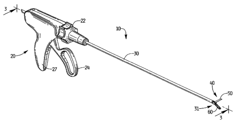
Figure 1 is a perspective view of one embodiment of the grasping instrument of
the
present disclosure showing the jaws in the open position;
Figure 1A is a perspective view of the jaws of the instrument of Figure 1 in
the open
position;
Figure 2 is an exploded view of the jaw assembly of Figure 1;
Figure 3 is a longitudinal cross-sectional view taken along line 3-3 of Figure
1;
Figure 4 is a cross-sectional view taken along line 4-4 of Figure 3;
Figure 5 is a side view in partial cross-section illustrating the jaws in the
closed position
with the probe in the advanced position;
Figure 6 is a cross-sectional view taken along line 6-6 of Figure 5;
Figure 7 is a perspective view of an alternate embodiment of the jaw assembly
of the
present disclosure illustrating the jaws in the open position with the probe
in the retracted
position;
4
CA 02733272 2011-03-01
Figure 8 is an exploded view of the jaw assembly of Figure 7;
Figure 9 is a side view in partial cross-section of the jaws of Figure 7 in
the open position
and the probe in the retracted position; and
Figure 10 is a cross-sectional view taken along line 10-10 of Figure 9;
DETAILED DESCRIPTION OF EMBODIMENTS
The surgical instrument of the present disclosure will now be described in
detail with
reference to the drawings in which like reference numerals designate identical
or corresponding
elements in each of the several views. Throughout this description, the term
"proximal" will
refer to the portion of the instrument closer to the operator and the term
"distal" will refer to the
portion of the instrument further from the operator. The presently disclosed
surgical instrument
is particularly suited for laparoscopic surgery but the system can be utilized
for other minimally
invasive surgical procedures.
The surgical instrument of the present disclosure is designated generally by
reference
numeral 10 and includes, with reference to Figure 1, a handle portion 20, an
endoscopic or
elongated tubular portion 30 extending distally from the handle portion and an
end effector 40.
End effector 40 extends from a distal portion 31 of elongated portion 30. A
rotation knob 22 can
be provided to rotate the endoscopic portion 30 and attached end effector 40
about the
longitudinal axis of the endoscopic portion 30 to reorient the end effector
40.
The end effector 40 includes a pair of jaws 50, 60, which function as tissue
graspers and
are pivotally mounted for movement between an open spaced apart position and a
closed
approximated position to capture tissue in the manner described below.
Although in the
illustrated embodiment both jaws 50, 60 move between open and closed
positions, it is also
CA 02733272 2011-03-01
contemplated that one of the jaws could be stationary and the other jaw
movable between open
and closed positions. The jaws 50, 60 can have teeth about their periphery to
enhance their
grasping function.
With reference to Figures 1, IA and 2, jaw 50 includes a cam slot 52 and a
pivot hole 54.
Similarly, jaw 60 includes a cam slot 62 and a pivot hole 64. An elongated
tissue probe 70 is
slidably mounted within the endoscopic portion 30. More specifically, the
probe 70 is fixed
within a sheath 80 which is slidably mounted within a lumen in the endoscopic
portion 30 and
exits through distal opening 33 of the lumen of the endoscopic portion 30.
Sheath 80 includes
transverse posts or pegs 82, 84 which engage cam slots 52, 62, of jaws 50, 60,
respectively.
Consequently, movement of sheath 80 by the handle mechanism as described below
moves the
sheath 80 and encased probe 70 distally as well. Endoscopic portion 30
terminates in yoke 36
with parallel arms 37, 39 having inwardly extending posts 38a, 38b,
respectively. Posts 38a, 38b
engage pivot holes 54, 64 of jaws 50, 60 respectively. Longitudinal slots 35a,
35b of arms 37, 39
receive posts 82 and 84 of sheath 80 to accommodate sliding movement of sheath
80. Probe 70
preferably terminates at the distal end of sheath 80 but alternatively could
terminate distal of the
distal end of the sheath 80 so it protrudes from the sheath 80.
Jaws 50 and 60 each have a cavity 55, 65, respectively, illustratively
substantially
elliptical in shape, dimensioned and configured to capture target tissue. In
the closed position,
the cavities 55, 65 form a closed cavity 59 (FIG. 5) which can block external
light to enhance
illumination of the tissue specimen within the cavity 59 if an illumination
probe e.g. fiber optic,
is utilized. The cavity 59 as shown has a substantially oval configuration,
although other
configurations for the tissue capturing retaining cavity, e.g. spherical, cup
shape, etc. are also
contemplated.
6
CA 02733272 2011-03-01
Movement of sheath 80 functions to open and close the jaws 50, 60. More
specifically,
movement of the sheath 80 moves the encased and attached probe 70 from a
retracted position of
Figure IA to an advanced position of Figures 5 and 6 and moves the jaws 50, 60
to a closed
position as transverse posts 82, 84 of sheath 80 engaged within cam slots 52,
62 cam jaws 50, 60
toward one another about posts or pins 38a, 38b of arms 37, 39. Posts 82, 84
also move distally
within longitudinal slots 35a, 35b of arms 37, 39. In this manner, when the
jaws 50, 60 are
closed to capture tissue within the cavity 59, the probe 70 is in its advanced
position to
illuminate, image, diagnose and/or treat the tissue specimen captured and
retained therein. Thus,
advancement of the probe 70 to its operative position occurs automatically
with jaw closure.
Stated another way, the closing of the jaws 50, 60 automatically advances the
tissue probe 70 to
an advanced position adjacent the target tissue contained within the jaw
cavity. The probe 70 in
one embodiment is in the form of a fiber optic bundle. Alternatively, it could
be a digital sensor.
The probe can also be a multi-functional probe to perform more than one
function. For example,
it could perform both illumination and visualization or both illumination and
detection. For
performing both illumination and visualization it could for example have an
LED for
illumination and some form of a confocal or camera. The probe may be wired or
wireless and
information can be accessed from the device itself or by using integrated
operating room systems
(computer/TV monitors, surgical navigation systems, etc.).
When the probe 70 is retracted by retraction of sheath 80, the jaws 50, 60 are
moved back
to their open position due to the engagement of transverse posts 82, 84 within
cam slots 52, 62 of
jaws 50, 60 causing the jaws 50, 60 to move in the reverse direction. Note
posts 82, 84 move
proximally within longitudinal slots 35a, 35b of endoscopic portion 30.
7
CA 02733272 2011-03-01
The sliding movement of the probe 70 between advanced and retracted positions
is
achieved by the operable connection of the sheath 80 with pivotable handle 24
of handle portion
20 as shown in Figure 3. That is, yoke 25 of pivotable handle or trigger 24 is
fixedly secured to
a proximal end 81 of sheath 80 via connecting block 29. Pivotal movement of
handle 24 in a
proximal direction, i.e. towards stationary handle 27, advances sheath 80 and
attached probe 70
distally. As sheath 80 is advanced, transverse posts 82, 84 advance distally
within longitudinal
slots 35a, 35b of arms 37, 39 of endoscopic portion 30 and cam jaws 50, 60 to
a closed position
due to their engagement with respective cam slots 52, 62. Note the probe 70
can be flexible and
formed into a loop portion 72 as shown in Figure 3, which would somewhat
straighten as
advanced. As noted above, in the advanced position of the probe 70, the jaws
50, 60 form a
closed cavity for the tissue specimen, and the probe 70 can then be utilized
to illuminate, image,
diagnose and/or treat tissue.
After use of the probe 70 in the surgical procedure, the instrument 10 can be
withdrawn
through the access port with the jaws 50, 60 maintained in the closed position
and withdrawing
the tissue specimen encapsulated in cavity 59 formed by cavities 55, 65 of
jaws 50, 60
respectively.
If it is desired to release the specimen from the jaws within the surgical
site, e.g. after an
in situ diagnostic function of probe 70, the jaws 50, 60 can be opened by
return of handle 24 to
its original more distal position. That is, return of handle 24 to its distal
position causes sheath
80 and attached probe 70 to retract (move proximally).
In the alternate embodiment of Figures 7-10, a grasping mechanism is provided
similar to
the embodiment of Figures 1-6, except instead of transverse posts on the arras
of the endoscopic
portion, an eyehole hinge 170 is provided. More specifically, jaws 150 and 160
are identical to
8
CA 02733272 2011-03-01
jaws 50 and 60 and have cam slots 152, 162 and pivot holes 154, 164. Jaws 150
and 160 also
have cavities 155, 165 like cavities 55 and 65 of the instrument of Figure 1
which together when
closed form a tissue capturing or retaining cavity. Endoscopic portion 130 is
also identical to
endoscopic portion 30 of Figure 1 except that instead of transverse posts on
its arms to engage
pivot holes of the jaws, it has openings 131a, 131b on arms 137, 139 of yoke
136 to receive the
transverse posts 171, 173 of eyehole hinge 170. Hinge 170 has an opening 172
through which
the probe 170 and sheath 180 travel through. It also forms the pivot point for
the jaws 150, 160
and holds the two sides of the jaws 150, 160 in place.
The grasping jaws of Figures 7-10 operate similar to the instrument 10 in that
actuation
of the instrument handle will advance the sheath 180 distally to cam the jaws
150, 160 to the
closed position via the engagement of transverse posts 182, 184 and cam slots
152, 162, of jaws
150, 160, respectively. Transverse posts 182, 184 slide distally within
longitudinal slots 135a,
135b of arms 137, 139. Advancement of the sheath 180 to close the jaws 50, 60
carries the
attached probe 170 distally for use within the cavity formed by cavities 155,
165 of the closed
jaws 150, 160. Thus, as in the embodiment of Figure 1, movement of the jaws
150, 160 to the
closed position and movement of the probe 170 (and sheath 180) to the advanced
position occur
substantially simultaneously. Note probe 170 and sheath 180 can be in the same
form as probe
70 and sheath 80 described herein.
To open the jaws 150, 160, as in the embodiment of Figure 1, the handle is
returned to its
distal position, thereby moving sheath 80, and attached probe 70, proximally
such that transverse
posts 182, 184 travel proximally within longitudinal slots 135a, 135b and
force the jaws 150, 160
to the open position due to their engagement with cam slots 152, 162.
9
CA 02733272 2011-03-01
As can be appreciated, the delivery of the probe in the foregoing embodiments
is
achieved automatically as the instrument jaws are moved to a closed position.
In a preferred embodiment, the probe 70 (or 170) has a diameter of about 3mm
or less,
although other dimensions are also contemplated. The probe can be, for
example, a confocal
fluorescence microscope probe, a near infrared Raman spectroscopy probe, an
auto-fluorescence
probe, a dye assisted fluorescence probe, etc. Tissue could thereby be
diagnosed, illuminated,
imaged and/or treated during a surgical procedure. Advancement of the probe
can place it
adjacent, and if desired, in contact with, the target tissue contained within
the cavity formed by
jaw cavities 55, 65 (or jaw cavities 155, 165).
The probe and sheath in some embodiments can be in the form or a fiber bundle
surrounded by a sheath. In such embodiments, or other embodiments wherein the
probe is
encased or positioned within a sheath (and attached to the sheath), the sheath
contains the
transverse posts or other structure for moving, e.g., pivoting, the jaws
between the open and
closed positions. In other embodiments, where the probe is not contained
within a sheath, the
probe can have transverse posts or other structure to engage and move, e.g.
pivot, the jaws
between open and closed positions.
It should be appreciated that although pin/cam slot arrangements are shown to
close the
jaws, other structure to achieve opening and closing the jaws is also
contemplated. Also,
structure can be provided to move the jaws between open and closed positions
in substantially
parallel movement.
The probe can be used with biopsy jaws, grasping jaws with or without teeth as
well as
other jaw configurations. As noted above, the jaws can have different shaped
cavities. Also,
although shown as forming a closed cavity, partially open cavities are also
contemplated.
CA 02733272 2011-03-01
The actuator for movement of the jaws and probe are shown in the form of a
trigger,
however, other actuators are also contemplated.
In use, the instrument enables grasping of tissue and diagnosis, illumination,
imaging
and/or treatment of target tissue positioned with the jaw cavity. That is, the
device can provide
surgical graspers integrated with a visualization probe such as fiber optic,
confocal, optical
tomography, etc. The jaws can enclose the tissue specimen within a confined
space for
interrogation with some form of light/ imaging technology while protecting it
from external light.
In the case of fluorescence, the working space and volume are reduced by
confining the
tissue within the jaw cavity which provides the ability to excite and
visualize tissue in smaller
volumes. It may also allow simultaneous visualization of white light (normal
laparoscope) and
fluorescence (locally in the cavity formed by jaw cups) in a single instrument
and through a
single port.
As can be appreciated, the probe in some embodiments can be utilized for
illumination,
imaging, diagnosis and/or treatment within the body cavity when the jaws are
in the open
position as well as within the confined space within the jaw cavity when the
jaws are in the
closed position.
Although shown within the abdominal cavity, the instrument can be used in
other regions
of the body.
It will be understood that various modifications may be made to the
embodiments
disclosed herein. Therefore, the above description should not be construed as
limiting, but
merely as exemplifications of preferred embodiments. Those skilled in the art
will envision
other modifications within the scope and spirit of the claims appended hereto.
11