Note: Descriptions are shown in the official language in which they were submitted.
CA 02734131 2011-02-14
WO 2010/019898 PCT/US2009/053911
TEMPERATURE CONTROLLED NUCLEIC-ACID DETECTION METHOD
SUITABLE FOR PRACTICE IN A CLOSED-SYSTEM
CROSS-REFERENCE TO RELATED APPLICATIONS
[0001] This application claims the benefit of U.S. Provisional Application
61/089,001 filed
on August 14, 2008, the entire contents of which is hereby incorporated by
reference. This
patent application is related to US 61/019,809; US 61/038,389; US 10/477,422;
US 11/640,495;
WO 05/127,709 and WO 08/013,462.
FIELD
[0002] This application relates generally to a method that can be practiced
within a device
for the rapid detection of target nucleic acid in a sample. More specifically,
it relates to a nucleic
acid treatment method that is compatible with temperature-controlled detection
methods which
can be used in a closed-system device for the detection of target nucleic
acids present in a
sample.
BACKGROUND
[0003] Portable devices that accurately and robustly detect nucleic acids in a
sample are
desirable for a variety of medical, industrial, environmental, security,
research and quality
control purposes. Preferably, such devices are also rapid, yield accurate
results and operate
using a closed-system, i.e. a system that does not need to be opened during
the course of the
analysis in order to prevent yield reduction or accidental contamination with
unwanted nucleic
acids or nucleases. Nucleic acid detection strategies can be split into three
stages: nucleic acid
treatment, signal amplification and signal detection/analysis. A primary
difficulty with these
stages is that they generally require different devices to perform them.
Therefore any automated
beginning-to-end device requires different instrumentation for each stage in
addition to a method
for transferring the material from one internal instrument to the next. The
need to combine the
technology for each stage complicates the design for miniaturisation.
[0004] In regards to the nucleic acid treatment step, nucleic acid-based
diagnostic
procedures often require nucleic acid preparation from natural substances.
Applications range
from forensic DNA-fingerprinting to medical, agricultural and environmental
monitoring. It is
important that the nucleic acid treatment be free from contamination,
particularly where the
-1-
CA 02734131 2011-02-14
WO 2010/019898 PCT/US2009/053911
concentration of nucleic acid in the initial sample is very low or where
contamination can lead to
incorrect outcomes. This is particularly the case in forensic and evidence
analyses where
quantities of starting material may be on the order of picograms or less.
Standard nucleic acid
treatment techniques are problematic as the sample tube may require opening
and shutting at
stages throughout the preparation procedure, where contamination may occur
simply as a result
of the sample tube being opened to the atmosphere or being touched by a
technician. Because of
the ease with which a sample can be contaminated, it is preferred that
reproducible nucleic acid
treatment techniques utilize protocols directed to minimising such
contamination.
[0005] After a nucleic acid has been treated to minimize contaminants, it can
be subjected to
nucleic acid detection methods to determine if a target nucleic acid is
present in a sample. Many
situations arise where it is desirable to detect low levels of specific
nucleic acid sequences within
the context of a complex mixture. A method intended for this purpose must be
highly specific
and sensitive. No simple method currently exists that can directly detect a
single nucleic acid
molecule of a specific sequence, and so all currently employed methods include
a step or steps
which amplify the signal. The most widespread method used to achieve this goal
is the
polymerase chain reaction (PCR). This method provides exponential
amplification of target
molecules by using thermal cycling and a thermostable DNA polymerase.
[0006] Current PCR technology used for the amplification of signal often
necessitates
lengthy purification procedures involving long incubations with proteinases,
phenol/chloroform
extractions and a finally an ethanol salt precipitation step before PCR can be
conducted.
Additionally, DNA purification protocols involving cells often involve
incubating samples with
Proteinase K and detergents, causing lysis of cells at temperatures where
deleterious enzymes are
released from cells that may degrade sample DNA and interfere with detection
of target nucleic
acid sequences. PreTagTM was commercially available as a thermostable
alternative to
Proteinase K to clean up DNA without degradation, however the temperature-
activity profile of
PreTagTm is not ideal as it remains active and is not readily removed at high
temperatures and
thus itself becomes a contaminant. It would therefore be advantageous to
develop a protocol
enabling simple, closed-tube reactions minimising the likelihood of
contamination and removing
the use of Proteinase K and other substances that may interfere with the PCR.
[0007] An inherent complication in this method is the requirement for the
repeated cycling
of the reaction between high and low temperatures. Thus, the method requires
equipment that is
more difficult to miniaturize. In response to this limitation, much effort has
been expended to
develop single- temperature, or isothermal, equivalents of PCR. One approach
has been to use a
-2-
CA 02734131 2011-02-14
WO 2010/019898 PCT/US2009/053911
polymerase that simultaneously achieves strand-displacement and strand-
synthesis, thereby
removing the need for the high-temperature step to produce single stranded DNA
in traditional
PCR methods.
[0008] There is clearly a need for an accurate, rapid and accessible method to
identify target
nucleic acid, particularly those corresponding to microorganisms, encountered
in a wide range of
situations or in mixed populations. The terms "target region", "target
sequence", "target nucleic
acid", "target nucleic acid sequence", "target polynucleotide", and "target
polynucleotide
sequence" and grammatical equivalents thereof refer to a region of a nucleic
acid which is to be
detected. The term "target nucleic acid" or "target nucleic acid sequence" as
used herein
therefore includes the target nucleic acid to be detected, for example that
present in a sample.
[0009] Essentially, the three stages in the nucleic acid detection process are
preparation or
treatment of the nucleic acid, amplification of a single indicating the
presence of the target
nucleic acid in a sample, and detecting the single produce by the presence of
the target nucleic
acid in the sample. Current strategies for each of these stages require
different instrumentation
and so multiple units must be incorporated into a miniaturised device and the
materials must be
transferred between these units by either hydrostatic or electromotive forces.
Preferably each
step would be simplified and be suitable for practice in a closed-system where
all the stages can
be processed in the same unit under compatible buffer conditions thus limiting
complexity, cost,
contamination and streamlining nucleic acid detection.
SUMMARY OF PREFERRED EMBODIMENTS
[0010] A method for detecting target nucleic acid in a sample that is suitable
for use in a
temperature controlled device is described herein. The method includes i)
treatment of nucleic
acid in a sample, ii) production of a single indicating the presence of the
target nucleic acid in a
sample, and iii) detecting the single produce by the presence of the target
nucleic acid in the
sample.
[0011] In a first aspect, an embodiment provides a method for the detection of
a target
nucleic acid in a sample, the method including:
a. treating a sample with a thermophilic proteinase to prepare a target
nucleic acid
for detecting,
-3-
CA 02734131 2011-02-14
WO 2010/019898 PCT/US2009/053911
b. providing detection reagents that produce a signal indicating the presence
of the
target nucleic acid in the sample, and
c. detecting the signal to determine the presence of the target nucleic acid,
the steps i), ii) and iii) are advantageously performed in a single vessel or
tube.
[0012] In one embodiment, the vessel or tube is a device. In a further
embodiment, the
device is a hand-held device.
[0013] In one embodiment, one or more steps i), ii) or iii) are temperature
controlled.
[0014] In one embodiment, the thermophilic proteinase is EA1.
[0015] In one embodiment, step a) is performed at a temperature of about 65-80
C for a
time sufficient to digest protein.
[0016] In a further embodiment, step a) further includes incubating the
thermophilic
proteinase at a temperature at or above about 90 C for a time that is
sufficient to inactivate the
proteinase.
[0017] In one embodiment, the method further includes the steps of:
a. treating the sample with a mesophilic enzyme, and
b. incubating the sample at a temperature below about 40 C for a period of
time that
is sufficient to effect removal of cell walls from cells.
[0018] In a further embodiment, the mesophilic enzyme is a cellulase.
[0019] In one embodiment, the signal is fluorescence.
[0020] In one embodiment, the detecting is by PCR detection methods. In a
further
embodiment, the PCR detection method is real-time PCR.
[0021] In one embodiment, the detecting is by isothermal detection methods. In
a further
embodiment, the isothermal detection methods is by strand displacement
amplification, rolling
circle amplification, loop-mediated isothermal amplification, isothermal
chimeric primer-
initiated amplification of nucleic acids, Q-beta amplification systems or
OneCutEventAmplificatioN. In yet a further embodiment, the isothermal
detection methods
-4-
CA 02734131 2011-02-14
WO 2010/019898 PCT/US2009/053911
utilizes Nuclease Chain Reaction (NCR), RNAse-mediated Nucleases Chain
Reaction (RNCR),
Polymerase Nuclease Chain Reaction (PNCR), RNAse-Mediated Detection (RMD),
Tandem
Repeat Restriction Enzyme Facilitated (TR-REF) Chain Reaction or Inverted
reverse
Complement Restriction Enzyme Facilitated (IRC-REF) Chain Reaction.
[0022] In one embodiment, the detection reagents are provided by microfluidics
or a solid
dispenser.
[0023] In one embodiment, the detection reagents are provided by
microcapsules. In a
further embodiment, the microcapsules are pre-disposed in the vessel or tube.
In yet a further
embodiment, the microcapsules are heat-labile capsules. In a further
embodiment, the heat-labile
capsules are agarose or wax beads. In yet another embodiment, the heat-labile
capsules released
the detection reagents at temperatures above the preferred incubation
temperature used in the
extraction step.
[0024] In one embodiment the detection reagents are resistant to the treatment
process, in
particular any enzymes required for the detection steps are resistant to
proteolytic cleavage by
the proteinase present for the purpose of preparing the nucleic acid from the
biological material.
[0025] In one embodiment, the detection of the target nucleic acid is
automated.
[0026] In one embodiment, the sample is blood, urine, saliva, semen, stool,
tissue, swabs,
tears or mucus.
[0027] In another embodiment, the sample is bacteria, fungi, archaea, eukarya,
protozoa or
virus.
[0028] In a further embodiment, the device or components of the device are
disposable.
In a further embodiment, the device comprises an inlet port, an outlet port, a
chamber, a detector
for emitted fluorescence and an excitation light source.
[0029] In a further embodiment, the device further comprises microfluidics,
microchips,
nanopore technologies and miniature devices.
BRIEF DESCRIPTION OF THE FIGURES
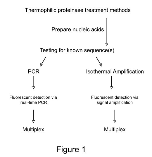
[0030] Figure 1. Overview of nucleic acid detection strategies within a
temperature
controlled device.
-5-
CA 02734131 2011-02-14
WO 2010/019898 PCT/US2009/053911
[0031] Figure 2. Single chamber nucleic acid treatment and detection using
sequential
liquid delivery of reagents.
[0032] Figure 3. Single chamber nucleic acid treatment and detection using
encapsulated
reagents.
[0033] Figure 4. Tube-based nucleic acid treatment and detection using
encapsulated
reagents.
[0034] Figure 5. Real-time PCR traces where the treatment and detection steps
were
performed in the same closed tube.
[0035] Figure 6. CT values obtained in a qPCR reaction for different cell
counts when DNA
extraction and qPCR are performed in a single vessel.
DETAILED DESCRIPTION
[0036] Nucleic acid detection strategies can be split into three stages:
nucleic acid treatment,
signal amplification and signal detection/analysis. Therefore, any fully
automated nucleic acid
detection device requires different instrumentation for each stage, and a
method of transferring
the material from one internal instrument to the next, complicating the design
for
miniaturization. The thermophilic proteinase nucleic acid treatment method
disclosed herein is
temperature modulated as are all amplification methods, whether isothermal or
cycling. In
addition, the conditions required for the thermophilic treatment are
compatible with those for
most amplification processes. Because of these factors, a device can be
simplified to no more
than a vessel with a heating/cooling mechanism to process raw sample material
and take it all the
way to a detectable signal. The inclusion of a detector is also facile. Hence
the currently
disclosed method enables devices with no pumps or need for microfluidics,
however these can be
used for more complex downstream applications if required.
[0037] Herein, a method for nucleic acid treatment, signal amplification and
detection is
described which can be practiced in closed-system devices that utilize heat-
controlled reaction
chains. The devices may be portable. Thermostable proteinases are used to
prepare nucleic acid
in a sample in tandem with nucleic acid identification techniques including
PCR or isothermal
detection methods. Heat control using either temperature dependent enzyme
mixtures or
temperature controlled release of encapsulated reagents simplifies the design
of current nucleic
acid diagnostic devices. Reducing complexity can reduce associated failure
rate and cost. These
-6-
CA 02734131 2011-02-14
WO 2010/019898 PCT/US2009/053911
techniques have the added benefit of being amendable to multiplexing for the
simultaneous
identification of multiple target nucleic acids in a mixed sample.
[0038] The term "treatment" used throughout the application refers to the
process of
increasing the availability of nucleic acid within a sample for processing by
other manipulations.
Implicit in the concept of "treatment" is that the nucleic acid is
sufficiently free of interfering
substances such as inhibitors, nucleases, other enzymes and nucloproteins that
it is effective in
other manipulation methods. It is understood that the nucleic acid is not
necessarily purified
away from non-interfering compounds as to do so serves no purpose in the
present device. The
nucleic acid treatment minimizes the negative effects of interfering
compounds.
[0039] The terms "nucleic acid", "nucleic acid sequence", "polynucleotide(s),"
"polynucleotide sequence" and equivalents thereof as used herein mean a single
or double-
stranded deoxyribonucleotide or ribonucleotide polymer of any length, and
include as non-
limiting examples, coding and non-coding sequences of a gene, sense and
antisense sequences,
exons, introns, genomic DNA, cDNA, pre-mRNA, mRNA, rRNA, siRNA, miRNA, tRNA,
ribozymes, recombinant polynucleotides, isolated and purified naturally
occurring DNA or RNA
sequences, synthetic RNA and DNA sequences, nucleic acid probes, primers,
fragments, genetic
constructs, vectors and modified polynucleotides. There is no intended
distinction in length
between the terms "nucleic acid", "oligonucleotide" and "polynucleotide", and
these terms will
be used interchangeably.
[0040] The method detailed in this disclosure is outlined in Figure 1. In a
first step, a
thermostable proteinase such as EA1 is added to a sample to digest
contaminating proteins at a
temperature optimal for thermostable proteinase activity.
[0041] Samples can be obtained from a wide range of substrates including
clinical, food and
beverage or environmental samples. Typically, microbial samples are obtained
from
environmental sources and for food testing by either taking a sample of a
liquid or solid or by
swabbing a solid surface. Conveniently, clinical samples may be taken from
tissues, blood,
serum, plasma, cerebrospinal fluid, urine, stool, semen, swabs or saliva.
Tissue samples may be
obtained using standard techniques such as cell scrapings or biopsy techniques
to collect animal
tissue. Similarly, blood sampling is routinely performed, for example for
pathogen testing, and
methods for taking blood samples are well known in the art. Likewise, methods
for storing and
processing biological samples are well known in the art. For example, tissue
samples may be
frozen until tested. In addition, one of skill in the art would realize that
some test samples would
-7-
CA 02734131 2011-02-14
WO 2010/019898 PCT/US2009/053911
be more readily analyzed following a fractionation or purification procedure,
for example,
separation of whole blood into serum or plasma components.
[0042] Initially, mesophilic enzymes may also be utilized to degrade cell wall
proteins or
other contaminants. The temperature can then be adjusted to inactivate the
thermophilic
proteinase while at the same time, in certain embodiments, release detection
reagents contained
in heat-labile materials. After the nucleic acid has been prepared from the
sample and the
proteinase has been inactivated, the nucleic acid is combined with detection
reagents customized
for the detection of known nucleic acid sequences.
[0043] Known nucleic acid sequences can be detected by fluorescence using
traditional PCR
or isothermal signal amplification methods. Unlike PCR, isothermal signal
amplification does
not require temperature cycling. Both PCR and isothermal detection methods can
be
multiplexed for the simultaneous detection of multiple target sequences of
interest.
[0044] In a preferred embodiment, the method is occurring in a device. Figures
2-4
illustrate various examples of how the methods can be practiced in the context
of a device. The
preferred device would be portable and would allow for closed-system
reactions, thus requiring
little more than simple physical modulation of a reaction between sample
insertion and result
generation. Preferably, temperature is used to initiate and stop sequential
chemical reactions
allowing multi-step procedures to be performed without complex pumps, valves
or microfluidics.
Heat can be controlled by many simple devices including microelectronics,
LEDs, Peltier plates
or an incandescent light bulb.
[0045] A preferred embodiment for such a device would have compatible reaction
conditions for all stages of the process, from nucleic acid treatment to
signal amplification to
signal detection. This detection system can be integrated with existing
technologies that are
specifically designed for buffer compatibility.
[0046] In a preferred embodiment, the device includes a single chamber. In
another
embodiment the chamber holds an externally supplied tube for example a PCR
tube, which is
placed within the device. In a further embodiment, the device comprises an
inlet port, an outlet
port, a chamber, a detector for emitted fluorescence and an excitation light
source.
[0047] In other embodiments, the device further includes microfluidics,
microchips,
nanopore technologies and miniature devices. The device or components of the
device may be
disposable.
-8-
CA 02734131 2011-02-14
WO 2010/019898 PCT/US2009/053911
[0048] It should be appreciated that the present described devices are methods
may have
applications for a range of nucleic acid diagnostic techniques where clean-up
of nucleic acids to
remove contaminants is particularly beneficial, or for diagnostic techniques
where the present
devices and methods may be adapted to achieve a similar beneficial outcome.
Thermostable Proteases for Nucleic Acid Treatment
[0049] As will be apparent to persons skilled in the art, samples suitable for
use in the
methods described herein may be obtained from the environment (such as soil,
rock, water and
plant material samples, for example) or from subjects, including tissues or
fluids from a subject,
so that the sample contains the nucleic acid to be tested.
[0050] Thermostable proteinases are added to the sample. Thermostable
proteinases include
proteinases that have protein degradation activity at high temperatures.
Exemplary but not
limiting, EA1 proteinase has been identified by the applicants as a preferred
thermostable
proteinase that is easier to remove at high temperatures. The sample is then
incubated and
subjected to a temperature shift. Following the temperature shift, protein
degradation occurs.
The procedure operates at 65-80 C as these enzymes are highly active between
these
temperatures. At this temperature, the cells are lysed and the proteinases
degrade contaminating
protein. By way of example, they rapidly remove DNA-degrading nucleases at
temperatures
where these nucleases are inactive, thereby minimising degradation of the
target nucleic acid
sample.
[0051] While in preferred embodiments of the current disclosure a thermophilic
proteinase
is used, it is anticipated that thermophilic enzymes other than proteinases
could also be used.
For ease of reference throughout the specification, the thermophilic enzyme
will herein be
referred to as a proteinase. However, this should not been seen as a
limitation for other enzymes
that could also conceivably be used.
[0052] Mixtures of mesophilic enzymes active at lower temperatures and one of
the above
mentioned proteinases can be used initially to weaken and/or remove cell walls
from plant,
fungal tissue, bacteria, spores and biofilms before continuing with the closed-
system procedure.
[0053] The practice of the disclosed method within a device relies on the
proteinase and/or a
proteinase/cell-wall degrading enzyme having differential activities at
different temperatures.
By cycling through the variable temperatures, the activities of different
enzymes can be brought
into play without the need for opening the system to add new reagents.
-9-
CA 02734131 2011-02-14
WO 2010/019898 PCT/US2009/053911
[0054] For applications that require low temperature digestion of nucleic
acids (for example,
restriction enzyme digestion of DNA), a proteinase that has very low activity
at 37 C need not be
removed or inactivated. Where multi-step or multi-enzyme reactions are
required, the
proteinases can be used in an enzyme mixture. As there is such low activity
below 40 C, other
enzyme reactions are able to occur in the presence of the proteinases.
[0055] According to one aspect of the current disclosure, there is provided a
method for the
treatment of nucleic acid samples in a closed-system, including the steps of:
1) adding at least one thermophilic proteinase to a sample containing nucleic
acid for
testing, and
2) incubating the sample for a preferred period of time at 65-80 C as required
to effect one
or more of the lysis of cells, digestion of proteins and digestion of cell-
wall enzymes,
where the thermophilic proteinase is stable and active at 65-80 C but is
inactivated and/or
denatured when the sample is incubated at or above 90 C without requiring the
addition
of further denaturing agents.
[0056] In preferred embodiments, the proteinase source includes Bacillus sp.
strain EA1
being a neutral proteinase. The preferred characteristics for a thermophilic
proteinase to be used
within the proposed methods are that:
1) it is substantially stable and active within the range 65-80 C, and
2) it is able to be readily inactivated and/or denatured at or above 90 C, and
3) optionally it has a temperature-activity profile such that it has low
activity below 40 C
such that accompanying mesophilic enzymes, for example, are not degraded.
[0057] The preferred incubation temperature required to affect one or more of
the lysis of
cells, digestion of proteins, digestion of cell-wall enzymes, via activity of
the proteinase is 75 C.
The preferred incubation temperature required to effect inactivation and/or
denaturation of the
proteinase is 94 C. However, it should be appreciated that these temperatures
are given by way
of example only and are not meant to be limiting in any way. It is anticipated
that the
proteinases will have differing profiles for both enzyme activity and
stability over a range of
temperatures and that such enzyme dynamics would be known to a skilled
artisan. It is also
anticipated such enzyme profiles for the proteinases could be determined with
minimal
experimentation. According to another aspect of the disclosure there is
provided a method for
-10-
CA 02734131 2011-02-14
WO 2010/019898 PCT/US2009/053911
the treatment of nucleic acid samples as described above, the method including
the initial steps
of:
1) adding at least one mesophilic enzyme and at least one non-specific
thermophilic
enzyme to a sample containing nucleic acid for testing, and
2) incubating the sample for a preferred period of time below 40 C as required
to
effect removal of any cell walls via activity of the mesophilic enzyme.
[0058] In preferred embodiments the mesophilic enzyme is a cell wall degrading
enzyme.
The preferred initial incubation temperature required to effect removal of any
cell walls via
activity of the mesophilic enzyme is 37 C. Once again, this should not be seen
as a limitation in
any way.
[0059] After the nucleic acid has been prepared and the proteinase has been
inactivated, the
sample can then be tested for target nucleic acids. Known nucleic acid
sequences of interest can
be detected by PCR-based detection methods or isothermal-based detection
methods described
below.
Signal Production & Detection of Target Nucleic Acid
[0060] In one aspect of the current method practiced, PCR-based detection
methods can be
used to detect nucleic acid sequences of interest prepared by the treatment
methods detailed
above.
[0061] A "PCR reagent" refers to any of the reagents used for PCR, usually a
set of primers
for each target nucleic acid, a DNA polymerase (preferably a thermostable DNA
polymerase), a
DNA polymerase cofactor and one or more deoxyribonucleoside-5'-triphosphates
(dDTP's) or
similar nucleosides. Other optional reagents and materials used in PCR are
described below.
[0062] A DNA polymerase is an enzyme that will add deoxynucleoside
monophosphate
molecules to (usually the 3'-hydroxy) end of the primer in a complex of primer
and template, but
this addition is in a template dependent manner. Generally, synthesis of
extension products
proceeds in the 5' to 3' direction of the newly synthesized strand until
synthesis is terminated.
Useful DNA polymerases include, for example, Taq polymerase, E. coli DNA
polymerase I, T4
DNA polymerase, Klenow polymerase, reverse transcriptase and others known in
the art.
Preferably, the DNA polymerase is thermostable meaning that it is stable to
heat and
preferentially active at higher temperatures, especially the high temperatures
used for priming
-11-
CA 02734131 2011-02-14
WO 2010/019898 PCT/US2009/053911
and extension of DNA strands. More particularly, thermostable DNA polymerases
are not
substantially inactive at the high temperatures used in polymerase chain
reactions as described
herein. Such temperatures will vary depending on a number of reaction
conditions, including
pH, nucleotide composition, length of primers, salt concentration and other
conditions known in
the art.
[0063] Particularly useful polymerases are those obtained from various Thermus
bacterial
species, such as Thermus aquaticus, Thermus thermophilus, Thermus filiformis,
and Thermus
flavus. Other useful thermostable polymerases are obtained from various
microbial sources
including Thermococcus literalis, Pyrococcusfuriosus, Thermotoga sp. And those
described in
WO-A-89/06691 (published Jul. 27, 1989). Some useful thermostable polymerases
are
commercially available, such as, AmpliTaqTM, Tth, and UITmaTM from Perkin
Elmer, Pfu from
Stratagene, and Vent and Deep-Vent from New England Biolabs. A number of
techniques are
also known for isolating naturally-occurring polymerases from organisms, and
for producing
genetically engineered enzymes using recombinant techniques.
[0064] A DNA polymerase cofactor refers to a non-protein compound on which the
enzyme
depends for activity. Thus, the enzyme is catalytically inactive without the
presence of cofactor.
A number of materials are known cofactors including, but not limited to,
manganese and
magnesium salts, such as chlorides, sulfates, acetates and fatty acids salts.
Magnesium chlorides
and sulfates are preferred.
[0065] Also needed for PCR are two or more deoxyribonucleoside-5'-
triphosphates, such as
two or more of dATP, dCTP, dGTP and dTTP. Analogues such as dITP and 7-deaza-
dGTP are
also useful. Preferably, the four common triphosphates (dATP, dCTP, dGTP and
dTTP) are
used together.
[0066] The PCR reagents described herein are provided and used in PCR in
suitable
concentrations to provide amplification of the target nucleic acid. The
minimal amounts of
primers, DNA polymerase, cofactors and deoxyribonucleoside-5'-triphosphates
needed for
amplification and suitable ranges of each are well known in the art. The
minimal amount of
DNA polymerase is generally at least about 0.5 units/100 l of solution, with
from about 2 to
about 25 units/100 l of solution being preferred, and from about 7 to about
20 units/100 l of
solution being more preferred. Other amounts may be useful for given
amplification systems. A
"unit" is defined herein as the amount of enzyme activity required to
incorporate 10 nmoles of
total nucleotides (dNTP's) into an extending nucleic acid chain in 30 minutes
at 74 C. The
-12-
CA 02734131 2011-02-14
WO 2010/019898 PCT/US2009/053911
minimal amount of primer is at least about 0.075 Mol with from about 0.1 to
about 2 Mol
being preferred, but other amounts are well known in the art. The cofactor is
generally present in
an amount of from about 2 to about 15 mMol. The amount of each dNTP is
generally from
about 0.25 to about 3.5 mMol.
[0067] The PCR reagents can be supplied individually, or in various
combinations, or all in
a buffered solution having a pH in the range of from about 7 to about 9, using
any suitable
buffer, many of which are known in the art.
[0068] Other reagents that can be used in PCR include, for example, antibodies
specific for
the thermostable DNA polymerase. Antibodies can be used to inhibit the
polymerase prior to
amplification. Preferably, the antibodies are specific for the thermostable
DNA polymerase,
inhibit the enzymatic activity of the DNA polymerase at temperatures below
about 50 C and are
deactivated at higher temperatures. Useful antibodies include monoclonal
antibodies, polyclonal
antibodies and antibody fragments. Preferably, the antibody is monoclonal.
Antibodies can be
prepared using known methods such as those described in Harlow et al.,
Antibodies: A
Laboratory Manual, Cold Spring Harbor, N.Y. (1988).
[0069] Light emitting labels can be used in PCR and isothermal detection
methods.
Mechanisms by which the light emission of a compound can be quenched by a
second compound
are described in Morrison, 1992, in Nonisotopic DNA Probe Techniques (Kricka
ed., Academic
Press, Inc. San Diego, Calif.), Chapter 13. One well known mechanism is
fluorescence energy
transfer (FET), non-radiative energy transfer, long-range energy transfer,
dipole-coupled energy
transfer, and Forster energy transfer. The primary requirement for FRET is
that the emission
spectrum of one of the compounds, the energy donor, must overlap with the
absorption spectrum
of the other compound, the energy acceptor. Styer and Haugland, 1967, Proc.
Natl. Acad. Sci.
U.S.A. 98:719, incorporated herein by reference, show that the energy transfer
efficiency of
some common emitter-quencher pairs can approach 100% when the separation
distances are less
than 10 angstroms. The energy transfer rate decreases proportionally to the
sixth power of the
distance between the energy donor and energy acceptor molecules. Consequently,
small
increases in the separation distance greatly diminish the energy transfer
rate, resulting in an
increased fluorescence of the energy donor and, if the quencher chromophore is
also a
fluorophore, a decreased fluorescence of the energy acceptor. In the methods,
the signal
emission of label, preferably a fluorescent label, bound to the probe is
detected.
-13-
CA 02734131 2011-02-14
WO 2010/019898 PCT/US2009/053911
[0070] Exposure of a detection sequence means the detection sequence is
rendered
accessible for detection, for example accessible for binding to a detection
probe. Conversely, the
terms "hidden" or "masked" and their grammatical equivalents mean that the
element(s) in
respect of which these terms are used is/are not accessible. For example, a
detection sequence
may be hidden or masked when bound to nucleic acid molecule other than a
detection probe.
The term "hybridisation" and grammatical equivalents refers the formation of a
multimeric
structure, usually a duplex structure, by the binding of two or more single-
stranded nucleic acids
due to complementary base pairing.
[0071] Because the treatment system uses only temperature control, a PCR can
be
performed in the same vessel as the treatment, and use the same
instrumentation within the
device. The PCR buffer and the treatment buffer are compatible in the
preferred embodiment.
Deoxyribonucleotides, divalent ions and oligonucleotide primers can be
supplied alongside the
treatment reagents because these are unaffected by the enzymes and the process
used to treat the
nucleic acids. Some DNA polymerases, for examples Taq DNA polymerase, are
degraded by
the thermophilic proteinase in the treatment reagents. Hence, post-treatment
delivery strategies
for the polymerase must be considered. Possible strategies are: (1) delivery
of the polymerase
and any other sensitive reagents after the treatment process is complete. This
can be a delivery
via an inlet port by microfluidics or a solid dispenser. (2) The polymerase
and other sensitive
reagents can be added into the treatment reagents in a protected form. This
can be in the form of
a bead or film with the sensitive reagents microencapsulate within. (3) The
polymerase can be
modified to protect it from the proteinase for example by the attachment of
antibodies. (4) Novel
polymerases can be used that are resistant to proteolytic cleavage.
[0072] Once the PCR reagents have been supplied, thermal cycling can be
achieved using
the same heating device and controller used in the treatment process. PCR
reactions can be
multiplexed to assay for several target nucleic acids simultaneously.
[0073] Another aspect of the disclosure is directed to isothermal detection
methods to detect
target nucleic acid, wherein the method relies on the target nucleic acid-
dependent amplification
of signal from a detectable label bound to a nucleic acid probe. Isothermal
amplification can be
by strand displacement amplification, rolling circle amplification, loop-
mediated isothermal
amplification, isothermal chimeric primer-initiated amplification of nucleic
acids, Q-beta
amplification systems or OneCutEventAmplificatioN.
-14-
CA 02734131 2011-02-14
WO 2010/019898 PCT/US2009/053911
[0074] Techniques that may be exploited during in isothermal amplification are
Nuclease
Chain Reaction (NCR), RNAse-mediated Nucleases Chain Reaction (RNCR). Both of
these
methods replace strand displacement with the selective degradation of one of
the strands of
DNA. The process can be initiated by using restriction endonucleases or RNAse
H when one of
the strands contains ribonucelotides. The Polymerase Nuclease Chain Reaction
(PNCR) relies on
nuclease cleavage in the presence of target DNA followed by an extension
process using a DNA
polymerase, RNAse-Mediated Detection (RMD) which is a method of strand
degradation by
RNAse H on DNA:RNA hybrids. RMD is an effective linear amplification system
that is
sometimes used in combination with other methods. Tandem Repeat Restriction
Enzyme
Facilitated (TR-REF) Chain Reaction or Inverted reverse Complement Restriction
Enzyme
Facilitated (IRC-REF) Chain Reaction are two variants of a method that relies
on the cyclical
production of a detector probe that contains tandem repeats. These repeats are
copied by a DNA
polymerase when a specific oligonucleotide trigger can act as a primer. Next,
restriction
endonucleases attack the newly formed double-stranded DNA and this releases
the original
primer and a second primer so that two new cycles can be initiated. Isothermal
amplification
reactions can be multiplexed to assay for several target nucleic acid
sequences of interest
simultaneously.
[0075] It will also be appreciated that some nucleic acids exist that possess
"strand invasion"
properties, whether such strand invasion results in the displacement of the
complementary strand
of the target nucleic acid and the formation of a target probe duplex, or the
formation of a target
probe triplex, without the target sequence first being single-stranded.
Peptide Nucleic Acids
(PNAs) and derivatives thereof may be capable of strand invasion, whereby
probes currently
disclosed containing target nucleic acid binding regions comprising PNAs can
be used to detect
target nucleic acid that has not been rendered fully single-stranded. The use
of target-binding
regions comprising PNAs is particularly contemplated in circular probes,
where, prior to the
formation of the target probe hybrid, the target-binding region of the probe
may be substantially
double-stranded.
[0076] As used herein, "target-binding domain" and its equivalent "target
binding domain"
refers to nucleic acid sequence present in a nucleic acid molecule that is
sufficiently
complementary to nucleic acid sequence present in the target nucleic acid to
allow the
hybridisation of the target-binding region and the target nucleic acid, and so
to form a target
probe hybrid.
-15-
CA 02734131 2011-02-14
WO 2010/019898 PCT/US2009/053911
[0077] In certain embodiments of the current disclosure, the methods for
detecting target
nucleic acids are reliant on detecting or measuring the signal from a label,
preferably the light
emission of a probe labelled with a light-emitting label. The term "label", as
used herein, refers
to any atom, molecule, compound or moiety which can be attached to a nucleic
acid, and which
can be used either to provide a detectable signal or to interact with a second
label to modify the
detectable signal provided by the second label. Preferred labels are light-
emitting compounds
which generate a detectable signal by fluorescence, chemiluminescence, or
bioluminescence.
Still more preferred labels are light-emitting compounds the signal of which
is diminished or
rendered undetectable when in sufficiently close proximity to a masking group,
for example, a
quenching chromophore.
[0078] Alternative labelling systems can be also be used that demonstrate the
cleavage of a
label from moiety that can be bound to a solid matrix. An example would be a
biotin label that
could be bound to immobilised avidin and thus non-cleavage of the probe would
bind a
secondary label present on the other end of the probe. Such a method would
have applications
for dipstick-based detection. Yet more detection system may use labels that
can be distinguished
by nanopore technology. The methods described herein are applicable to the
detection of probes
labelled with a single label, although multiple labels may be employed.
Detection of the cleaved
probe occurs when the label, for example a fluorophore, is sufficiently
removed from the
masking group, for example a quencher, by the cleavage event, or the probe-
denaturing process
the cleavage event allows. This diminishes the interaction of the masking
group and the label
and so allows emission of the signal. As used herein, the term "masking group"
means any
atom, molecule, compound or moiety that can interact with the label to
decrease the signal
emission of the label. The separation of label and masking group resulting
from the cleavage
event or the probe-denaturing process the cleavage event allows in turn
results in a detectable
increase in the signal emission of the attached label. Depending on the label,
signal emission
may include light emission, particle emission, the appearance or disappearance
of a colored
compound, and the like.
[0079] Preferred light-emitting labels and masking groups that can interact to
modify the
light emission of the label are described below. The term "chromophore" refers
to a non-
radioactive compound that absorbs energy in the form of light. Some
chromophores can be
excited to emit light either by a chemical reaction, producing
chemiluminescence, or by the
absorption of light, producing fluorescence. The term "fluorophore" refers to
a compound
-16-
CA 02734131 2011-02-14
WO 2010/019898 PCT/US2009/053911
which is capable of fluorescing, i.e. absorbing light at one frequency and
emitting light at
another, generally lower, frequency.
[0080] The term "bioluminescence" refers to a form of chemiluminescence in
which the
light-emitting compound is one that is found in living organisms. Examples of
bioluminescent
compounds include bacterial luciferase and firefly luciferase. The term
"quenching" refers to a
decrease in fluorescence of a first compound caused by a second compound,
regardless of the
mechanism. Quenching typically requires that the compounds be in close
proximity. As used
herein, either the compound or the fluorescence of the compound is said to be
quenched, and it is
understood that both usages refer to the same phenomenon.
[0081] Many fluorophores and chromophores described in the art are suitable
for use in the
methods presently disclosed. Suitable fluorophore and quenching chromophore
pairs are chosen
such that the emission spectrum of the fluorophore overlaps with the
absorption spectrum of the
chromophore. Preferably, the fluorophore would have a high Stokes shift (a
large difference
between the wavelength for maximum absorption and the wavelength for maximum
emission) to
minimize interference by scattered excitation light.
[0082] Suitable labels which are well known in the art include, but are not
limited to,
fluoroscein and derivatives such as FAM, HEX, TET, and JOE; rhodamine and
derivatives such
as Texas Red, ROX, and TAMRA; Lucifer Yellow, and coumarin derivatives such as
7-Me2N-
coumarin-4-acetate, 7 -OH-4-CH. 3 -coumarin- 3 -acetate, and 7-NH2-4-CH3-
coumarin-3 -acetate
(AMCA). FAM, HEX, TET, JOE, ROX, and TAMRA are marketed by Perkin Elmer,
Applied
Biosystems Division (Foster City, Calif.). Texas Red and many other suitable
compounds are
marketed by Molecular Probes (Eugene, Oreg.). Examples of chemiluminescent and
bioluminescent compounds that may be suitable for use as the energy donor
include
luminol(aminophthalhydrazide) and derivatives, and Luciferases.
[0083] While in most embodiments it will be preferred that the detectable
label be a light-
emitting label and the masking group be a quencher, such as a quenching
chromophore, other
detectable labels and masking groups are possible. For example, the label may
be an enzyme
and the masking group an inhibitor of said enzyme. When the enzyme and
inhibitor are in
sufficiently close proximity to interact, the inhibitor is able to inhibit the
activity of the enzyme.
On cleavage or denaturation of the probe, the enzyme and inhibitor are
separated and no longer
able to interact, such that the enzyme is rendered active. A wide variety of
enzymes capable of
catalysing a reaction resulting in the production of a detectable product and
inhibitors of the
-17-
CA 02734131 2011-02-14
WO 2010/019898 PCT/US2009/053911
activity of such enzyme are well known to the skilled artisan, such as 13-
galactosidase and
horseradish peroxidise.
Computer Related Embodiments
[0084] Device control can be achieved by standard electronic methods using
hardware,
software and firmware typical of thermal cycling devices. Likewise, any
integrated detection
system could use similar programmable devices.
[0085] Data produced by the detection device may range from a simple yes/no
detection
when the device is used for detecting a specific agent to real-time data where
the time is
measured for the signal to reach a pre-defined threshold thereby giving
quantitative data.
Similarly, electrophoretic data could be produced in the form the taken for
peaks of fluorescence
to reach a detector placed at a point along a capillary electrophoresis
device.
[0086] Data analysis can be achieved using a computer program supplied to the
device
either via and external electronic port, wireless technology, an internal
storage device or internal
firmware. For simple purposes for example a device with a specific role of
determining the
presence or absence of a single target nucleic acid, reporting may be in the
form of any visible
indicator such as a light or and LCD or LED display.
[0087] Where data requires more complex analysis or a greater level of user
input, the raw
data, processed data or partially processed data can be transferred to an
external computer via
any form of removable storage device or a communications cable.
[0088] In certain embodiments, the results can utilize wireless technology to
obtain data
base information or use database information stored on the device that may aid
in the
identification of target nucleic acid present in the sample. Results can be
binary, i.e. present or
not present, or they can be quantitative or multivariate.
EXAMPLES
[0089] Aspects of the present disclosure have been described by way of example
only and it
should be appreciated that modifications and additions may be made thereto
without departing
from the scope thereof as defined in the appended claims.
Example 1: A single chamber device with liquid delivery of reagents
-18-
CA 02734131 2011-02-14
WO 2010/019898 PCT/US2009/053911
[0090] Figure 2 illustrates one embodiment of the present device that
comprises single
chamber treatment and detection using sequential liquid delivery of reagents.
The shape of the
container can be circular, square, triangular or any other useful shape with
numerous ports if
needed. The chamber can also be optimized for microfluidic samples or larger
volumes
depending on the application. In step 2A, treatment reagents and substrate are
added to the
reaction chamber. In step 2B, the reaction temperature is adjusted to suit the
treatment reagents.
Nucleic acids are released into solution. In step 2C, the temperature is
raised further so that the
treatment reagents are inactivated. Step 2D, where an isothermal amplification
/ detection
system is detailed, a single temperature is used. Detection reagents are added
to the chamber.
The reaction temperature is adjusted to suit the detection reagents. Detection
of a specific agent
is performed at this stage, in this example by fluorescence. In another
embodiment, such as
illustrated in step 2E, detection reagents are added to the chamber. In this
example, the reaction
temperature is cycled as for a quantitative PCR. Detection of a specific agent
is performed at
this stage, in this example by fluorescence.
Example 2: A single chamber device with encapsulated reagents
[0091] Figure 3 shows a single chamber treatment and detection device that
uses
encapsulated reagents. In step 3A, treatment reagents and substrate are added
to the reaction
chamber along with the detection reagents but these are encapsulated to
protect them from the
proteinase used for treatment. In step 3B, the reaction temperature is
adjusted to suit the
treatment reagents. Nucleic acids are released. In step 3C, on completion, the
temperature is
adjusted so that the treatment reagents are inactivated while simultaneously,
the detection
reagents are released as the encapsulation bead melts. In step 3D, the
reaction temperature is
adjusted to suit the detection reagents. For an isothermal amplification /
detection system, a
single temperature is used. Detection of a specific agent is performed at this
stage, in this
example by fluorescence. In step 3E, in this example, the reaction temperature
is cycled as for a
quantitative PCR. Detection of a specific agent is performed at this stage, in
this example by
fluorescence.
Example 3: A tube-accommodating device with encapsulated reagents
[0092] Figure 4 illustrates a tube-based treatment and detection using
encapsulated reagents.
In step 4A, a tube containing treatment reagents, substrate and encapsulated
detection reagents
are is place in the device and covered. In step 4B, the reaction temperature
is adjusted to suit the
treatment reagents. Nucleic acids are released. In step 4C, on completion, the
temperature is
-19-
CA 02734131 2011-02-14
WO 2010/019898 PCT/US2009/053911
raised further so that the treatment reagents are inactivated while
simultaneously, the detection
reagents are released as the encapsulation bead melts. In step 4D, the
reaction temperature is
adjusted to suit the detection reagents. For an isothermal amplification /
detection system, a
single temperature is used. Detection of a specific agent is performed at this
stage, in this
example by fluorescence. In 4E, the reaction temperature is cycled as for a
quantitative PCR.
Detection of a specific agent is performed at this stage, in this example by
fluorescence.
Example 4: Closed tube detection of nucleic acid from buccal cells
[0093] Figure 5 details an experiment that demonstrates how a thermophilic
proteinase can
be used in combination with amplification and detection reagents in a single,
closed vessel. In
this example, all reagents, including untreated buccal cells, treatment
reagents, amplification
reagents and detection reagents were sealed in a 200 l PCR tube and all
processing was
performed using only temperature to achieve nucleic acid detection from whole
cells.
[0094] A buccal swab was taken from an individual following standard
procedure. A
standard cotton swab was used and the participant was instructed to rub the
inside of the mouth
and gums for 1 minute. Debris on the swab was suspended in 1 ml of 5 mM Tris
(pH 8.3 at
room temperature). The following cocktail of PCR and detection reagents was
made. The
primers were, Primerl: 5'-TCTCCTCCGATTTCAACAGTGA; Primer2, 5'-
GGTCGTTGAGGGCAATGC. Platinum Taq DNA Polymerase Invitrogen, San Diego, USA.
Reagent 1 reaction 50 reactions
Water 8.9 445
Buffer 2.5 125
MgC12 (supplied) 0.75 37.5
Primerl (10 M) 0.5 25
Primer2 (10 M) 0.5 25
ROX 1 M 0.4 20
SybrGreen (1/2500) 0.5 25
dNTP's (10 mM) 0.5 25
Platinum Taq (5U / l) 0.2 10
15 750
-20-
CA 02734131 2011-02-14
WO 2010/019898 PCT/US2009/053911
[0095] Using this master mix, the following cocktails were made. These are
various
combinations of reagents containing treatment reagents, whole cells or control
DNA.
MM Buccal Human EA 1 BSA
as cell DNA Proteinas 2.5 water
above suspension 025 n,, e 0.2 U / m,, /
/ ul p] ml
1 75 5 5 15
2 75 5 5 5 10
3 75 5 20
4 75 5 5 5 10
75 5 5 15
5 [0096] Twenty five microliters of the five mixtures were dispensed into
optically clear PCR
tubes and sealed. All subsequent reactions were controlled by heat and no
further tube openings.
The tubes were heat cycled in an ABI 5700 Sequence detection system (Applied
Biosystems,
Forster City, USA) for 75 C for 10 minutes (treatment step); 95 C for 10
minutes (proteinase
heat kill step and polymerase activation step); and 35 cycles of 95 C for 30
sec, 60 C for 30 sec,
72 C for 30 sec with fluorescence measured in the last step (amplification /
detection step).
[0097] The results in Figure 5 demonstrate that cell treatment and detection
can be
performed under heat control when a thermophilic proteinase is used. The
traces on 5A
demonstrate that Platinum Taq DNA polymerase is resistant to hydrolysis by EA1
proteinase.
The traces in 5B show the effect of the presence or absence of EA1 proteinase
when whole cells
are added to the mixture. When no proteinase is added (trace 5) the CT value
is approximately
two cycles lower than when EA1 proteinase is present (trace 4). This equates
to a quarter of the
yield. Such a loss of yield is critical in trace samples.
[0098] Figure 5 illustrates qPCR traces where the treatment reagents and the
amplification
and detection reagents are combined in a sealed tube. Figure 5A shows traces
produced where
control DNA was added. Figure 5B shows traces produced where whole cells were
added (one
control trace is included for reference). Sample 1 is the positive control
(1.25 ng of purified
human DNA). Sample 2 is a positive control that demonstrates that Platinum Taq
is resistant to
the proteolytic activity of EA1 proteinase. Sample 3 is the negative control
(no trace). Sample 4
demonstrates that DNA can be prepared from human buccal cells in a closed tube
with treatment,
-21-
CA 02734131 2011-02-14
WO 2010/019898 PCT/US2009/053911
amplification and detection reagents present. Sample 5 shows the level of heat-
mediated lysis of
the buccal cells in the absence of the proteinase.
Example 5: Closed tube detection of nucleic acid from bacterial cells
[0099] The following experiment was performed on a dilution series of
Escherichia coli
cells and their presence was detected with universal 16S rRNA oligonucleotide
primers. These
primers are typical of the type used in microbial analysis. The following
reagents and materials
were used at the listed concentration were applicable: EA1 proteinase at 1
Unit per l (ZyGEM
Corporation Ltd); GIBCO UltraPureTM Distilled water (Invitrogen); Quanta
Bioscience qPCR
reagents; optically clear 96-well PCR plates (Axygen); maximum recovery filter
tips (Axygen);
and pPCR Primers at 10 M:
Forward: GTCGTCAGCTCGTGTTGTGA
Reverse: GCCCGGGAACGTATTCAC
[0100] All work was performed in a PCR hood situated in an air-locked
laboratory with
positive air pressure generated through a HEPA filter and only previously
unopened reagents,
tubes, PCR plates, and filter-tips were used. Additionally, all surfaces were
swabbed with 1%
sodium hypochlorite prior to the experiment.
[0101] Escherichia coli MG1655 cells were grown overnight in LB broth. The
cells were
then centrifuged at 12,000 r.c.f for 5 minutes and resuspended in water to a
cell density of 2x10⁷
per ml. This density is the equivalent of 105 cells per 5 l. A 1:10 serial
dilution was made in
ultrapure water wherein the lowest cell concentration was approximately 10
cells per 5 l.
Following the serial dilution, 5 l of each dilution was placed into eight
wells of an optically
transparent 96-well microtitre plate. The following solution was added to four
replicates:
Water 13
Quanta PCR mix 20
Primer 1 10 M 0.8
Primer 2 10 M 0.8
EA1 proteinase @ 1 U/ l 0.4
Total volume 35
-22-
CA 02734131 2011-02-14
WO 2010/019898 PCT/US2009/053911
[0102] In addition, four water controls were included for each reagent
cocktail. The plate
was then sealed with a transparent adhesive lid and held at 4 C for 5 minutes
in the dark after
which time the plate was then exposed through the seal to a 600 W halogen lamp
at 200 mm
distant for 5 minutes with the tubes maintained at 4 C. This step is not
necessary however can
be useful for additional pre-treatments such as those described in US
Provisional Application
Serial No. 61/222,912. The samples were then placed in an Applied Biosystems
7300 Real-time
PCR System and cycled as follows:
DNA Extraction step: 75 C 15 min
Taq Activation step: 95 C 5 min
195 C 30s
PCR: 160 C 30s x 45
172 C 30s (Fluorescence measured)
[0103] Figure 6 shows a graph of the CT values obtained for the closed vessel
reaction.
The CT value is the number of PCR cycles that elapse before the threshold is
reached. The
higher the CT value, the smaller the initial amount of DNA. The results
clearly demonstrate that
extraction and detection can be performed in a single reaction vessel without
opening the tube.
-23-