Note: Descriptions are shown in the official language in which they were submitted.
CA 02734521 2011-02-17
WO 2010/025341 PCT/US2009/055327
DETERMINING AGE RANGES OF SKIN SAMPLES
BACKGROUND OF THE INVENTION
FIELD OF THE INVENTION
[0001] The present invention relates generally to skin sampling and more
specifically to
methods of characterizing skin based on an age index.
BACKGROUND INFORMATION
[0002] Aging is the accumulation of changes in an organism or object over
time. Aging in
humans refers to a multidimensional process of physical, psychological, and
social change.
Some dimensions of aging grow and expand over time, while others decline.
Reaction time,
for example, may slow with age, while knowledge of world events and wisdom may
expand.
Research shows that even late in life potential exists for physical, mental,
and social growth
and development. Aging is an important part of all human societies reflecting
the biological
changes that occur, but also reflecting cultural and societal conventions.
[0003] As far as mammals go, humans are essentially hairless; that is, most of
the skin of
the human body can be seen without interference from hair. The skin is thus
exposed to
whatever insults (natural and man-made) the environment harbors. Since it was
first
understood that the sun caused erythema, people have taken measures to avoid
its "harmful
rays." A century ago, in Elizabethan England, it was the fashion to avoid the
sun at all costs.
Yet the skin of those Elizabethans still wrinkled and displayed other signs of
chronological
aging.
[0004] Human skin is a complex organ which extends over the entire body. There
are
different types of skin at different portions of the body; for example, facial
skin is different
from that of the scalp, and even the skin on the front (palm) of the hand is
different than that
on the back of the hand. Although the type of skin can vary over a person's
body, skin is
generally composed of two main layers of tissue. The epidermis or cuticle, the
outermost
layer, is composed of superficial layers (from the outside in: stratum
corneum, stratum
lucidem, and stratum granulosum) and deep layers (stratum spinosum and stratum
basale).
The dermis, cutis vera, or the true skin, is composed of a papillary layer
above and a reticular
layer below.
CA 02734521 2011-02-17
WO 2010/025341 PCT/US2009/055327
2
[0005] Since ancient times, a variety of substances have been applied to the
skin to
improve its appearance, generally by affecting the outermost layer of the
skin, or to treat a
skin ailment, generally by affecting the true skin. More recently, efforts
have been made to
rejuvenate the skin and reclaim the elasticity and suppleness lost from
exposure to sunlight
(UV radiation) and weather.
[0006] There is a difference between the physiology of chronologically-aged or
intrinsically-aged skin (old skin) in comparison with that of photoaged skin
(i.e., skin that
appears old due to damage from solar UV irradiation). Old skin typically
maintains a smooth
and unblemished appearance, in comparison with the leathery, blotchy, and
often deep
wrinkling of photoaged skin. The epidermis of old skin is typically thinner
than normal,
whereas that of photoaged aged skin is typically thicker than normal
(acanthotic) and
atrophies over time. Photoaged skin typically has a large Grenz zone (a wide
band of
eosinophilic material just beneath the epidermis, and collagen formation and
structures
indicative of wound healing) which is absent from chronologically-aged skin.
See also N. A.
Fenske and C. W. Lober, "Structural and functional changes of normal aging
skin," J. Am.
Acad. Dermatol., 15:571-585 (1986).
[0007] In biology, "senescence" is the state or process of aging. "Cellular
senescence" is
a phenomenon where isolated cells demonstrate a limited ability to divide in
culture (i. e., the
Hayflick Limit, discovered by Leonard Hayflick in 1965), while "Organismal
senescence" is
the aging of organisms. After a period of near perfect renewal (in humans,
between about 20
and 35 years of age), organismal senescence is characterized by the declining
ability to
respond to stress, increasing homeostatic imbalance and increased risk of
disease. This
irreversible series of changes inevitably ends in death. Some researchers
(specifically
biogerontologists) are treating aging as a disease. As genes that have an
effect on aging are
discovered, aging is increasingly being regarded in a similar fashion to other
genetic
conditions, i.e., potentially "treatable."
[0008] Clearly there is a need for further development of technology that will
enable
physicians to develop a skin age index to predict aging of individuals' skin.
CA 02734521 2011-02-17
WO 2010/025341 PCT/US2009/055327
3
SUMMARY OF THE INVENTION
[0009] The present invention is based, in part, on the discovery that analysis
of nucleic
acids or protein products from specific genes can be used to distinguish or
categorize skin
samples of individuals. The methods provide valuable genetic information based
on DNA,
RNA, or protein products obtained therefrom, for example.
[0010] In one embodiment, the method involves use of a non-invasive approach
for
recovering nucleic acids such as DNA or RNA or proteins from the surface of
skin via a tape
stripping procedure that permits a direct quantitative and qualitative
assessment of
biomarkers. Although tape-harvested nucleic acid and protein products are
shown to be
comparable in quality and utility to recovering such molecules by biopsy, the
non-invasive
method provides information regarding cells of the outermost layers of the
skin that may not
be obtained using biopsy samples. Finally, the non-invasive method is far less
traumatic than
a biopsy, although the invention does not exclude the use of such invasive
methods for skin
sampling.
[0011] Thus, the method is used to capture cells on the skin of individuals
for
determination of the appropriate age range. Nucleic acid molecules obtained
from skin are
analyzed in order to characterize the skin sample as being young or old. In
one embodiment,
a nucleic acid molecule is amplified prior to analysis. Secondary outcomes
could include
tests for diagnosis and prognosis of a variety of symptoms of photoaging
and/or chronoaging
of skin, and to predict a therapeutic or cosmetic regimen for treating the
skin. In another
embodiment, the skin cells are lysed to extract one or more proteins, which
are then
quantitated to compare with gene products of the genes listed in Table 1,
Table 2, Table 3, or
any combination thereof, for example, however, the combination must include at
least one
"young" gene and at least one "old" gene. It should be understood that the
methods of the
invention are not limited to non-invasive techniques as described herein for
obtaining skin
samples. For example, but not by limitation, one of skill in the art would
know other
techniques for obtaining a skin sample such as scraping of the skin, biopsy,
suction, blowing
and other techniques. As described herein, non-invasive tape stripping, as
described in U.S.
Pat. No. 6,949,338, incorporated herein by reference, is an illustrative
example for obtaining
a skin sample.
CA 02734521 2011-02-17
WO 2010/025341 PCT/US2009/055327
4
[0012] In another embodiment, the methods involve detection of one or more
mutations in
the nucleic acid sequence of the nucleic acid molecule obtained from the skin.
Such
mutations may be a substitution, a deletion, and/or an insertion of the
nucleic acid sequence
that results in symptoms associated with chronoaging and/or photoaging of skin
from the
subject from which the skin sample is obtained.
[0013] In one embodiment, the nucleic acid analyzed is any one or more of
those listed in
Table 1, Table 2, Table 3, or any combination thereof. Accordingly, provided
herein is a
method for determining the age range of a skin sample of a subject, including
obtaining a
nucleic acid or protein by tape stripping or biopsy of the skin or a skin
lesion, for example,
from the subject, and analyzing the nucleic acid as compared to the nucleic
acids or protein
products thereof listed in Table 1, Table 2, Table 3, or any combination
thereof. In this
method, at least one nucleic acid molecule whose expression is informative of
an appropriate
age range of the skin is detected in the sample.
[0014] The non-invasive methods of the invention involve applying an adhesive
tape to a
target area of skin in a manner sufficient to isolate a sample adhering to the
adhesive tape,
wherein the sample includes nucleic acid molecules and/or proteins. Typically,
at least one
nucleic acid molecule or protein whose expression is informative of the age or
age range of
the skin in the sample. The method of characterizing skin using tape stripping
has a number
of applications, such as the following: (i) age classification of the skin;
(ii) monitoring the
severity and progression of photoaging and/or chronoaging; (iii) monitoring
treatment
efficacy; and (iv) prediction of a particular treatment or cosmetic regimen.
All of these
applications, which themselves represent embodiments disclosed herein,
preferably use non-
invasive sampling to recover information that is otherwise difficult or
impractical to recover
(e.g., through the use of biopsies). The information may be contained in the
DNA, protein, or
RNA of skin cells close to the surface of the skin. In one embodiment,
expression of one or
more of the genes listed in Table 1, Table 2, Table 3, or any combination
thereof is detected
in the sample to characterize the sample. Tables 1-3 are presented by way of
example to
show the genes of three exemplary skin "age indexes". It should be understood
that an age
index can be developed by using less than the number of genes presented in any
of Tables 1-
3, as long as young and old stratifications are clear from the genes selected.
CA 02734521 2011-02-17
WO 2010/025341 PCT/US2009/055327
[0015] Other embodiments are based in part on the discovery that for tape
stripping of the
skin, non-polar, pliable, adhesive tapes, especially pliable tapes with rubber
adhesive, are
more effective than other types of adhesive tapes. Using pliable tapes with
rubber adhesives,
as few as 10 or less tape strippings and in certain examples as few as 4 or
even 1 tape
stripping can be used to isolate and/or detect nucleic acid molecules from the
epidermal layer
of the skin.
[0016] In another embodiment, the methods of the invention provide for
determining the
age range of a skin sample or of skin in situ, including application of a
detectably labeled
probe directly to the skin of a subject for visual analysis. At least one
nucleic acid molecule
whose expression is informative of the age range of the skin is detected on
the skin using a
specific probe. In one example, expression of one or more of the genes listed
in Table 1,
Table 2, Table 3, or any combination thereof is detected on the skin to
determine the age
range of the skin. In one embodiment, expression of one or more of the genes
listed in Table
1, Table 2, Table 3, or any combination thereof is detected in the sample to
characterize the
skin sample.
[0017] In another aspect, the invention provides kits for characterizing a
skin sample in a
subject. In one embodiment, the kit includes a skin sample collection device,
such as a
biopsy needle or an adhesive tape for non-invasive tape stripping, and one or
more probes or
primers that selectively bind to one or more nucleic acid molecules in Table
1, Table 2, Table
3, or any combination thereof, or to a nucleic acid or protein expression
product of a nucleic
acid molecule in Table 1, Table 2, Table 3, or any combination thereof. The
kit may include
one or more pairs of forward primers that selectively bind upstream of a gene
on one strand
and reverse primers that selectively bind upstream of a gene on a
complementary strand. In
another embodiment, the kit includes a microarray containing at least a
fragment of a gene or
a nucleic acid or protein product of a gene identified in Table 1, Table 2,
Table 3, or any
combination thereof, or any combination thereof.
[0018] In another embodiment, the kit for characterizing a skin sample from a
subject
includes an applicator and one or more probes or primers that selectively bind
to one or more
nucleic acid molecules in Table 1, Table 2, Table 3, or any combination
thereof, or to a
nucleic acid or protein expression product of a nucleic acid molecule in Table
1, Table 2,
CA 02734521 2011-02-17
WO 2010/025341 PCT/US2009/055327
6
Table 3, or any combination thereof. In one embodiment, the probes are
detectably labeled
for visual identification of expression of RNA.
[0019] In another aspect, the invention provides a cosmetic formulation
containing agents
for reducing or increasing expression of genes. In one embodiment, the agent
reduce or
increase expression of the genes listed in Table 1, Table 2, Table 3, or any
combination
thereof. In another embodiment, the cosmetic formulation is an emulsion, a
cream, a lotion, a
solution, an anhydrous base, a paste, a powder, a gel, or an ointment. The
emulsion may be
an oil-in-water emulsion or a water-in-oil emulsion. Alternatively, the
formulation may be a
solution, such as an aqueous solution or a hydro-alcoholic solution. In
another embodiment,
the cosmetic formulation is an anhydrous base, such as a lipstick or a powder.
In yet another
embodiment, the formulation is comprised within an anti-aging product or a
moisturizing
product. The cosmetic formulation may further contain one or more of
estradiol;
progesterone; pregnanalone; coenzyme Q 10; methylsolanomethane (MSM); copper
peptide
(copper extract); plankton extract (phytosome); glycolic acid; kojic acid;
ascorbyl palmitate;
all trans retinol; azaleic acid; salicylic acid; broparoestrol; estrone;
adrostenedione;
androstanediols; or sunblocks.
BRIEF DESCRIPTION OF THE DRAWINGS
[0020] Figure 1 is a hierarchial cluster analysis of the identified 100-gene
classifier
distinguishing skin samples of young individuals from skin samples of old
individuals. The
tree configuration shown at the left of the cluster analysis is representative
of the genes in the
order as shown in Table 1.
[0021] Figure 2 is a graphical diagram showing a skin age index generated from
the 100-
gene classifier that distinguishes skin samples of young individuals from skin
samples of old
individuals.
[0022] Figure 3 is a hierarchial cluster analysis of the identified 61-gene
classifier
distinguishing skin samples of young individuals from skin samples of old
individuals. Skin
age index = Sum of "Group A" - Sum of "group B" + a (constant). The tree
configuration
shown at the left of the cluster analysis is representative of the genes in
the order as shown in
Table 2.
CA 02734521 2011-02-17
WO 2010/025341 PCT/US2009/055327
7
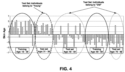
[0023] Figure 4 is a graphical diagram showing a skin age index generated from
the 61-
gene classifier that distinguishes skin samples of young individuals from skin
samples of old
individuals.
[0024] Figure 5 is a hierarchial cluster analysis of an 83-gene classifier
distinguishing skin
samples of young individuals from skin samples of old individuals. Skin age
index = Sum of
"Group A" - Sum of "group B" + a (constant). The tree configuration shown at
the left of
the cluster analysis is representative of the genes in the order as shown in
Table 3.
[0025] Figure 6 is a graphical diagram showing a skin age index generated from
the 83-
gene classifier that distinguishes skin samples of young individuals from skin
samples of old
individuals.
DETAILED DESCRIPTION OF THE INVENTION
[0026] The present invention is based, in part, on the discovery that analysis
of nucleic
acid molecules or protein products from specific genes can be used to
characterize skin
samples from individuals based on an age index. Accordingly, the present
invention provides
methods and kits useful for characterizing a skin sample based on determining
an expression
profile of the sample based on identification of one or more genes or
proteins.
[0027] In humans and other animals, cellular senescence has been attributed to
the
shortening of telomeres with each cell cycle; when telomeres become too short,
the cells die.
The length of telomeres is therefore the "molecular clock," predicted by
Hayflick. Telomere
length is maintained in immortal cells (e.g., germ cells and keratinocyte stem
cells, but not
other skin cell types) by the enzyme telomerase. In the laboratory, mortal
cell lines can be
immortalized by the activation of their telomerase gene, present in all cells
but active in few
cell types.
[0028] As used in this specification and the appended claims, the singular
forms "a", "an",
and "the" include plural references unless the context clearly dictates
otherwise. Thus, for
example, references to "the method" includes one or more methods, and/or steps
of the type
described herein which will become apparent to those persons skilled in the
art upon reading
this disclosure and so forth.
CA 02734521 2011-02-17
WO 2010/025341 PCT/US2009/055327
8
[0029] Unless defined otherwise, all technical and scientific terms used
herein have the
same meaning as commonly understood by one of ordinary skill in the art to
which this
invention belongs. Although any methods and materials similar or equivalent to
those
described herein can be used in the practice or testing of the invention, the
preferred methods
and materials are now described.
[0030] A number of genetic components of aging have been identified using
model
organisms, ranging from the simple budding yeast Saccharomyces cerevisiae to
worms such
as Caenorhabditis elegans and fruit flies (Drosophila melanogaster). Study of
these
organisms has revealed the presence of at least two conserved aging pathways.
[0031] One of these pathways involves the gene Sir2, a NAD+-dependent histone
deacetylase. In yeast, Sir2 is required for genomic silencing at three loci:
the yeast mating
loci, the telomeres and the ribosomal DNA (rDNA). In some species of yeast
replicative
aging may be partially caused by homologous recombination between rDNA
repeats;
excision of rDNA repeats results in the formation of extrachromosomal rDNA
circles
(ERCs). These ERCs replicate and preferentially segregate to the mother cell
during cell
division, and are believed to result in cellular senescence by titrating away
(competing for)
essential nuclear factors. ERCs have not been observed in other species of
yeast (which also
display replicative senescence), and ERCs are not believed to contribute to
aging in higher
organisms such as humans. Extrachromosomal circular DNA (eccDNA) has been
found in
worms, flies and humans. The role of eccDNA in aging, if any, is unknown.
[0032] Despite the lack of a connection between circular DNA and aging in
higher
organisms, extra copies of Sir2 are capable of extending the lifespan of both
worms and flies.
The mechanisms by which Sir2 homologues in higher organisms regulate lifespan
is unclear,
but the human SIRT1 protein has been demonstrated to deacetylate p53, Ku70,
and the
forkhead family of transcription factors. SIRT1 can also regulate acetylates
such as
CBP/p300, and has been shown to deacetylate specific histone residues.
[0033] RAS1 and RAS2 also affect aging in yeast and have a human homologue.
RAS2
overexpression has been shown to extend lifespan in yeast.
CA 02734521 2011-02-17
WO 2010/025341 PCT/US2009/055327
9
[0034] Other genes regulate aging in yeast by increasing the resistance to
oxidative stress.
Superoxide dismutase, a protein that protects against the effects of
mitochondrial free
radicals, can extend yeast lifespan in stationary phase when overexpressed.
[0035] In higher organisms, aging is likely to be regulated in part through
the insulin/IGF-
1 pathway. Mutations that affect insulin-like signaling in worms, flies and
mice are
associated with extended lifespan. In yeast, Sir2 activity is regulated by the
nicotinamidase
PNC 1. PNC I is transcriptionally upregulated under stressful conditions such
as caloric
restriction, heat shock, and osmotic shock. By converting nicotinamide to
niacin, it removes
nicotinamide, which inhibits the activity of Sir2. A nicotinamidase found in
humans, known
as PBEF, may serve a similar function, and a secreted form of PBEF known as
visfatin may
help to regulate serum insulin levels. It is not known, however, whether these
mechanisms
also exist in humans since there are obvious differences in biology between
humans and
model organisms.
[0036] Sir2 activity has been shown to increase under calorie restriction. Due
to the lack
of available glucose in the cells more NAD+ is available and can activate
Sir2. Resveratrol, a
polyphenol found in the skin of red grapes, was reported to extend the
lifespan of yeast,
worms, and flies. It has been shown to activate Sir2 and therefore mimics the
effects of
calorie restriction.
[0037] The particularly important causes of chronological aging of human skin
likely vary
among a population of elderly humans, including such factors as diet,
genetics, and
environment. In general, though, it is believed that chronological skin aging
is due to
activation of the stress-activated pathways (SAPs) and a repression of the
mitogen-activated
pathways (ERK). However, contrary to conventional wisdom, it has been found
that
chronoaging and photoaging of human skin have a similar molecular
pathophysiology. ERK
mediates the actions of growth factors necessary for healthy skin.
Interference with ERK can
lead to thinning of chronologically-aged skin because of reduced number of
cells in the
epidermis and dermis. Almost conversely, SAPs activate factors (e.g., c-Jun)
that promote
both inhibition of procollagen synthesis and degradation of mature collagen,
and thereby lead
to reduced form, strength, and function of skin. Chronological aging of skin
might be
expected to include some interference with ERK and/or some activation of the
SAPs.
CA 02734521 2011-02-17
WO 2010/025341 PCT/US2009/055327
[0038] Gene expression is imperfectly controlled, and it is possible that
random
fluctuations in the expression levels of many genes contribute to the aging
process as
suggested by a study of such genes in yeast. Individual cells, which are
genetically identical,
none-the-less can have substantially different responses to outside stimuli,
and markedly
different lifespans, indicating the epigenetic factors play an important role
in gene expression
and aging as well as genetic factors.
[0039] Accordingly, in one embodiment, the present invention employs a non-
invasive
tape stripping technology to obtain samples of skin from individuals. As such,
DNA
microarray assays are used to create a skin age index to predict the aging of
an individual.
Tape-stripping removes superficial cells from the surface of the skin as well
as adnexal cells.
Small amounts of nucleic acid molecules isolated from tape-stripped cells can
be amplified
and used for microarray analyses and quantitative PCR. In addition, proteins
obtained from
the lysed cells may be quantitated for characterization and determination of
age.
Consequently, tape-stripping is a non-invasive diagnostic method, which does
not interfere
with subsequent histological analyses. While tape stripping will primarily
sample superficial
cells from the epidermis, this method holds great promise in the determination
of age and
age-related disorders. Consequently, this feature may help characterize an
individual as
having skin characterized as being younger or older than the actual age of the
individual.
Further, there are changes in the dermis and epidermis resulting from
environmental factors,
such as exposure to UV radiation. Accordingly, the present invention
demonstrates that
stratum corneum RNA, harvested by tape stripping with Epidermal Genetic
Information
Retrieval (EGIR) (see U.S. Pat. No. 6,949,338, incorporated herein by
reference), can be used
to distinguish skin samples of young individuals from skin samples of old
individuals.
[0040] The term "subject" or "individual" as used herein refers to any
individual or patient
to which the subject methods are performed. Generally the subject is human,
although as will
be appreciated by those in the art, the subject may be an animal. Thus other
animals,
including mammals such as rodents (including mice, rats, hamsters and guinea
pigs), cats,
dogs, rabbits, farm animals including cows, horses, goats, sheep, pigs, etc.,
and primates
(including monkeys, chimpanzees, orangutans and gorillas) are included within
the definition
of subject.
CA 02734521 2011-02-17
WO 2010/025341 PCT/US2009/055327
11
[0041] As used herein, the terms "sample" and "biological sample" refer to any
sample
suitable for the methods provided by the present invention. A sample of cells
can be any
sample, including, for example, a skin sample obtained by non-invasive tape
stripping or
biopsy of a subject, or a sample of the subject's bodily fluid. Thus, in one
embodiment, the
biological sample of the present invention is a tissue sample, e.g., a biopsy
specimen such as
samples from needle biopsy. In one embodiment, the term "sample" refers to any
preparation
derived from skin of a subject. For example, a sample of cells obtained using
the non-
invasive method described herein can be used to isolate nucleic acid molecules
or proteins for
the methods of the present invention.
[0042] As used herein "corresponding cells" or "corresponding sample" refers
to cells or a
sample from a subject that is from the same organ and of the same type as the
cells being
examined. In one aspect, the corresponding cells comprise a sample of cells
obtained from a
healthy individual that is age-matched or within an acceptable age range such
that the sample
is representative of a sample typically obtained from individuals within the
range. Such
corresponding cells can, but need not be, from an individual that is of the
same sex as the
individual providing the cells being examined. Thus, the term "normal sample"
or "control
sample" refers to any sample taken from a subject of similar species that is
considered
healthy and of a known age. As such, a normal/standard level of RNA denotes
the level of
RNA present in a sample from a subject of known age. A normal level of RNA can
be
established by combining skin samples or cell extracts taken from normal
healthy age-
matched subjects and determining the level of one or more RNAs present. In
addition, a
normal level of RNA also can be determined as an average value taken from a
population of
subjects that fall within a known age range. Accordingly, levels of RNA in
subject and
control samples can be compared with the standard values. Deviation between
standard and
subject values establishes the parameters for characterizing age and/or
distinguishing samples
based on age.
[0043] The term "skin" refers to the outer protective covering of the body,
consisting of
the epidermis (including the stratum corneum) and the underlying dermis, and
is understood
to include sweat and sebaceous glands, as well as hair follicle structures.
Throughout the
present application, the adjective "cutaneous" can be used, and should be
understood to refer
generally to attributes of the skin, as appropriate to the context in which
they are used. The
CA 02734521 2011-02-17
WO 2010/025341 PCT/US2009/055327
12
epidermis of the human skin comprises several distinct layers of skin tissue.
The deepest
layer is the stratum basalis layer, which consists of columnar cells. The
overlying layer is the
stratum spinosum, which is composed of polyhedral cells. Cells pushed up from
the stratum
spinosum are flattened and synthesize keratohyalin granules to form the
stratum granulosum
layer. As these cells move outward, they lose their nuclei, and the
keratohyalin granules fuse
and mingle with tonofibrils. This forms a clear layer called the stratum
lucidum. The cells of
the stratum lucidum are closely packed. As the cells move up from the stratum
lucidum, they
become compressed into many layers of opaque squamae. These cells are all
flattened
remnants of cells that have become completely filled with keratin and have
lost all other
internal structure, including nuclei. These squamae constitute the outer layer
of the
epidermis, the stratum corneum. At the bottom of the stratum corneum, the
cells are closely
compacted and adhere to each other strongly, but higher in the stratum they
become loosely
packed, and eventually flake away at the surface.
[0044] As used herein, "chronoaging" refers to inevitable changes that occur
over time
that affect the skin of a subject. In contrast, "photoaging" refers to changes
to the skin of a
subject over time resulting from external environmental aggressors. Exemplary
external
aggressors include, but are not limited to UV rays, free radicals, chemicals,
and toxins.
Exemplary symptoms of chronoaging and/or photoaging of skin include, but are
not limited
to, loss of firmness and elasticity, dryness, loss of sheen, and lines and
wrinkles. As such, the
terms "sun damage" and "environmental damage," when used in reference to skin
are used
broadly to encompass any external environmental aggressors that may
prematurely age the
skin of a subject.
[0045] As used herein, the term "gene" refers to a linear sequence of
nucleotides along a
segment of DNA that provides the coded instructions for synthesis of RNA,
which, when
translated into protein, leads to the expression of hereditary character. As
such, the term
"skin marker" or "biomarker" refers to a gene whose expression level is
different between
skin surface samples from individuals of distinct ages or age ranges, and skin
surface samples
of individuals of known age or age range. Therefore, expression of a skin
marker of the
invention is related to, or indicative of, the age of the subject being
tested. Many statistical
techniques are known in the art, which can be used to determine whether a
statistically
significant difference in expression is observed at a high (e.g., 90% or 95%)
confidence level.
CA 02734521 2011-02-17
WO 2010/025341 PCT/US2009/055327
13
As such, an increase or decrease in expression of these genes is related to
and can
characterize age of the subject.
[0046] As used herein, the term "nucleic acid molecule" means DNA, RNA (e.g.,
messenger RNA, miRNA, etc.), single-stranded, double-stranded or triple
stranded and any
chemical modifications thereof. Virtually any modification of the nucleic acid
is
contemplated. A "nucleic acid molecle" can be of almost any length, from 10,
20, 30, 40, 50,
60, 75, 100, 125, 150, 175, 200, 225, 250, 275, 300, 400, 500, 600, 700, 800,
900, 1000,
1500, 2000, 2500, 3000, 3500, 4000, 4500, 5000, 6000, 7000, 8000, 9000,
10,000, 15,000,
20,000, 30,000, 40,000, 50,000, 75,000, 100,000, 150,000, 200,000, 500,000,
1,000,000,
1,500,000, 2,000,000, 5,000,000 or even more bases in length, up to a full-
length
chromosomal DNA molecule. For methods that analyze expression of a gene, the
nucleic
acid isolated from a sample is typically RNA.
[0047] Micro-RNAs (miRNA) are small single stranded RNA molecules an average
of 22
nucleotides long that are involved in regulating mRNA expression in diverse
species
including humans. Hundreds of miRNAs have been discovered in flies, plants and
mammals.
miRNAs regulate gene expression by binding to the 3'-untranslated regions of
mRNA and
catalyze either i) cleavage of the mRNA; or 2) repression of translation. The
regulation of
gene expression by miRNAs is central to many biological processes such as cell
development, differentiation, communication, and apoptosis. Recently it has
been shown that
miRNA are active during embryogenesis of the mouse epithelium and play a
significant role
in skin morphogenesis.
[0048] Given the role of miRNA in gene expression it is clear that miRNAs will
influence,
if not completely specify the relative amounts of mRNA in particular cell
types and thus
determine a particular gene expression profile (i.e., a population of specific
mRNAs) in
different cell types. In addition, it is likely that the particular
distribution of specific miRNAs
in a cell will also be distinctive in different cell types. Thus,
determination of the miRNA
profile of a tissue may be used as a tool for expression profiling of the
actual mRNA
population in that tissue. Accordingly, miRNA levels are useful for the
purposes of
characterization of a subject within an age range.
CA 02734521 2011-02-17
WO 2010/025341 PCT/US2009/055327
14
[0049] As used herein, the term "protein" refers to at least two covalently
attached amino
acids, which includes proteins, polypeptides, oligopeptides and peptides. A
protein may be
made up of naturally occurring amino acids and peptide bonds, or synthetic
peptidomimetic
structures. Thus "amino acid", or "peptide residue", as used herein means both
naturally
occurring and synthetic amino acids. For example, homo-phenylalanine,
citrulline and
noreleucine are considered amino acids for the purposes of the invention.
"Amino acid" also
includes imino acid residues such as proline and hydroxyproline. The side
chains may be in
either the (R) or the (S) configuration.
[0050] A "probe" or "probe nucleic acid molecule" is a nucleic acid molecule
that is at
least partially single-stranded, and that is at least partially complementary,
or at least partially
substantially complementary, to a sequence of interest. A probe can be RNA,
DNA, or a
combination of both RNA and DNA. It is also within the scope of the present
invention to
have probe nucleic acid molecules comprising nucleic acids in which the
backbone sugar is
other that ribose or deoxyribose. Probe nucleic acids can also be peptide
nucleic acids. A
probe can comprise nucleolytic-activity resistant linkages or detectable
labels, and can be
operably linked to other moieties, for example a peptide.
[0051] A single-stranded nucleic acid molecule is "complementary" to another
single-
stranded nucleic acid molecule when it can base-pair (hybridize) with all or a
portion of the
other nucleic acid molecule to form a double helix (double-stranded nucleic
acid molecule),
based on the ability of guanine (G) to base pair with cytosine (C) and adenine
(A) to base pair
with thymine (T) or uridine (U). For example, the nucleotide sequence 5'-ATAC-
3' is
complementary to the nucleotide sequence 5'-GTAT-3'.
[0052] The term "antibody" as used in this invention is meant to include
intact molecules
of polyclonal or monoclonal antibodies, as well as fragments thereof, such as
Fab and F(ab')2,
Fv and SCA fragments which are capable of binding an epitopic determinant. The
term
"specifically binds" or "specifically interacts," when used in reference to an
antibody means
that an interaction of the antibody and a particular epitope has a
dissociation constant of at
least about 1 x 10"6, generally at least about I x 10-7, usually at least
about 1 x 10⁻⁸, and
particularly at least about 1 x 10⁻⁹ or 1 x 10⁻¹⁰ or less.
CA 02734521 2011-02-17
WO 2010/025341 PCT/US2009/055327
[0053] As used herein "hybridization" refers to the process by which a nucleic
acid strand
joins with a complementary strand through base pairing. Hybridization
reactions can be
sensitive and selective so that a particular sequence of interest can be
identified even in
samples in which it is present at low concentrations. In an in vitro
situation, suitably
stringent conditions can be defined by, for example, the concentrations of
salt or formamide
in the prehybridization and hybridization solutions, or by the hybridization
temperature, and
are well known in the art. In particular, stringency can be increased by
reducing the
concentration of salt, increasing the concentration of formamide, or raising
the hybridization
temperature. For example, hybridization under high stringency conditions could
occur in
about 50% formamide at about 37 C to 42 C. Hybridization could occur under
reduced
stringency conditions in about 35% to 25% formamide at about 30 C to 35 C. In
particular,
hybridization could occur under high stringency conditions at 42 C in 50%
formamide, 5X
SSPE, 0.3% SDS, and 200 mg/ml sheared and denatured salmon sperm DNA.
Hybridization
could occur under reduced stringency conditions as described above, but in 35%
formamide
at a reduced temperature of 35 C. The temperature range corresponding to a
particular level
of stringency can be further narrowed by calculating the purine to pyrimidine
ratio of the
nucleic acid of interest and adjusting the temperature accordingly. Variations
on the above
ranges and conditions are well known in the art.
[0054] As used herein, the term "mutation" refers to a change in the genome
with respect
to the standard wild-type sequence. Mutations can be deletions, insertions, or
rearrangements
of nucleic acid sequences at a position in the genome, or they can be single
base changes at a
position in the genome, referred to as "point mutations." Mutations can be
inherited, or they
can occur in one or more cells during the lifespan of an individual.
[0055] As used herein, the term "kit" or "research kit" refers to a collection
of products
that are used to perform a biological research reaction, procedure, or
synthesis, such as, for
example, a detection, assay, separation, purification, etc., which are
typically shipped
together, usually within a common packaging, to an end user.
[0056] Samples from a tissue can be isolated by any number of means well known
in the
art. Invasive methods for isolating a sample include, but are not limited to
the use of needles
or scalpels, for example during biopsies of various tissues. Non-invasive
methods for
isolating a sample include, but are not limited to tape-stripping and skin
scraping.
CA 02734521 2011-02-17
WO 2010/025341 PCT/US2009/055327
16
[0057] As such, the tape stripping methods provided herein typically involve
applying an
adhesive tape to the skin of a subject and removing the adhesive tape from the
skin of the
subject one or more times. In certain examples, the adhesive tape is applied
to the skin and
removed from the skin about one to ten times. Alternatively, about ten
adhesive tapes can be
sequentially applied to the skin and removed from the skin. These adhesive
tapes are then
combined for further analysis. Accordingly, an adhesive tape can be applied to
and removed
from a target site 10, 9, 8, 7, 6, 5, 4, 3, 2, or 1 time, and/or 10, 9, 8, 7,
6, 5, 4, 3, 2, or 1
adhesive tape can be applied to and removed from the target site. In one
illustrative example,
the adhesive tape is applied to the skin between about one and eight times, in
another
example, between one and five times, and in another illustrative example the
tape is applied
and removed from the skin four times.
[0058] The rubber based adhesive can be, for example, a synthetic rubber-based
adhesive.
The rubber based adhesive in illustrative examples, has high peel, high shear,
and high tack.
For example, the rubber based adhesive can have a peak force tack that is at
least 25%, 50%,
or 100% o greater than the peak force tack of an acrylic-based tape such as D-
SQUAMETM. D-
SQUAME TM has been found to have a peak force of 2 Newtons, wherein peak force
of the
rubber based adhesive used for methods provided herein, can be 4 Newtons or
greater.
Furthermore, the rubber based adhesive can have adhesion that is greater than
2 times, 5
times, or 10 times that of acrylic based tape. For example, D-SQUAMETM has
been found to
have adhesion of 0.0006 Newton meters, whereas the rubber based tape provided
herein can
have an adhesion of about 0.01 Newton meters using a texture analyzer.
Furthermore, in
certain illustrative examples, the adhesive used in the methods provided
herein has higher
peel, shear and tack than other rubber adhesives, especially those used for
medical
application and Duct tape.
[0059] Virtually any size and/or shape of adhesive tape and target skin site
size and shape
can be used and analyzed, respectively, by the methods of the present
invention. For
example, adhesive tape can be fabricated into circular discs of diameter
between 10
millimeters and 100 millimeters, for example between 15 and 25 millimeters in
diameter.
The adhesive tape can have a surface area of between about 50 mm2 and 1000
mm2, between
about 100 mm2 to 500 mm2 or about 250 mm2.
CA 02734521 2011-02-17
WO 2010/025341 PCT/US2009/055327
17
[0060] In another embodiment, the sample may be obtained by means of an
invasive
procedure, such as biopsy. Biopsies may be taken instead of or after tape
stripping and are
subjected to standard histopathologic analysis. Analysis of biopsy samples
taken
simultaneously with tape stripping samples may then be correlated with the
data generated
from one or more of analysis of selected lesion RNA samples by DNA microarray,
correlation of gene expression data with histopathology, and creation of a
candidate
expression classifier for the skin age index.
[0061] As used herein, "biopsy" refers to the removal of cells or tissues for
analysis.
There are many different types of biopsy procedures known in the art. The most
common
types include: (1) incisional biopsy, in which only a sample of tissue is
removed; (2)
excisional biopsy, in which an entire lump or suspicious area is removed; and
(3) needle
biopsy, in which a sample of tissue or fluid is removed with a needle. When a
wide needle is
used, the procedure is called a core biopsy. When a thin needle is used, the
procedure is
called a fine-needle aspiration biopsy. Other types of biopsy procedures
include, but are not
limited to, shave biopsy, punch biopsy, curettage biopsy, and in situ biopsy.
In another
embodiment, the skin sample is obtained by scraping the skin with an
instrument to remove
one or more nucleic acid molecules from the skin.
[0062] The skin sample obtained using the tape stripping method includes,
epidermal cells
including cells comprising adnexal structures. In certain illustrative
examples, the sample
includes predominantly epidermal cells, or even exclusively epidermal cells.
The epidermis
consists predominantly of keratinocytes (> 90%), which differentiate from the
basal layer,
moving outward through various layers having decreasing levels of cellular
organization, to
become the cornified cells of the stratum corneum layer. Renewal of the
epidermis occurs
every 20-30 days in uninvolved skin. Other cell types present in the epidermis
include
melanocytes, Langerhans cells, and Merkel cells. As illustrated in the
Examples herein, the
tape stripping method of the present invention is particularly effective at
isolating epidermal
samples.
[0063] Nucleic acid molecules can also be isolated by lysing the cells and
cellular material
collected from the skin sample by any number of means well known to those
skilled in the
art. For example, a number of commercial products available for isolating
polynucleotides,
including but not limited to, RNeasyTM (Qiagen, Valencia, CA) and TriReagentTM
(Molecular
CA 02734521 2011-02-17
WO 2010/025341 PCT/US2009/055327
18
Research Center, Inc, Cincinnati, OH) can be used. The isolated
polynucleotides can then be
tested or assayed for particular nucleic acid sequences, including a
polynucleotide encoding a
cytokine. Methods of recovering a target nucleic acid molecule within a
nucleic acid sample
are well known in the art, and can include microarray analysis.
[0064] Nucleic acid molecules may be analyzed in any number of ways known in
the art.
For example, the presence of nucleic acid molecules can be detected by DNA-DNA
or DNA
RNA hybridization or amplification using probes or fragments of the specific
nucleic acid
molecule. Nucleic acid amplification based assays involve the use of
oligonucleotides or
oligomers based on the nucleic acid sequences to detect transformants
containing the specific
DNA or RNA.
[00651 In one embodiment, analysis of the nucleic acid molecules includes
genetic
analysis to determine the nucleotide sequence of a gene. Since a difference in
length or
sequence between DNA fragments isolated from a sample and those of known
sequences are
due to an insertion, deletion, or substitution of one or more nucleotides, the
determination of
nucleic acid sequences provides information concerning mutations resulting
from
environmental affects on the skin of individuals. These mutations may also
include
transposition or inversion and are difficult to detect by techniques other
than direct
sequencing. Accordingly, the methods of the present invention may be used to
detect genetic
mutations in one or more genes listed in Table 1, Table 2, Table 3, or any
combination
thereof for determination and/or characterization of the age of the subject.
[0066] A variety of protocols for detecting and measuring the expression of
nucleic acid
molecules, using either polyclonal or monoclonal antibodies specific for the
protein
expression product are known in the art. Examples include enzyme-linked
immunosorbent
assay (ELISA), radioimmunoassay (RIA), and fluorescence activated cell sorting
(FACS).
These and other assays are described, among other places, in Hampton, R. et
al. (1990;
Serological Methods, a Laboratory Manual, APS Press, St Paul, Minn.) and
Maddox, D. E. et
al. (1983; J. Exp. Med. 158:1211-1216).
[0067] In another embodiment, antibodies that specifically bind to the
expression products
of the nucleic acid molecules of the invention may be used to characterize the
skin sample of
CA 02734521 2011-02-17
WO 2010/025341 PCT/US2009/055327
19
the subject. The antibodies may be used with or without modification, and may
be labeled by
joining them, either covalently or non-covalently, with a reporter molecule.
[0068] A wide variety of labels and conjugation techniques are known by those
skilled in
the art and may be used in various nucleic acid and amino acid assays. Means
for producing
labeled hybridization or PCR probes for detecting sequences related to the
nucleic acid
molecules of Table 1, Table 2, Table 3, or any combination thereof include
oligolabeling,
nick translation, end-labeling or PCR amplification using a labeled
nucleotide. Alternatively,
the nucleic acid molecules, or any fragments thereof, may be cloned into a
vector for the
production of an mRNA probe. Such vectors are known in the art, are
commercially
available, and may be used to synthesize RNA probes in vitro by addition of an
appropriate
RNA polymerase such as T7, T3, or SP6 and labeled nucleotides. These
procedures may be
conducted using a variety of commercially available kits (Pharmacia & Upjohn,
(Kalamazoo,
Mich.); Promega (Madison Wis.); and U.S. Biochemical Corp., Cleveland, Ohio).
Suitable
reporter molecules or labels, which may be used for ease of detection, include
radionuclides,
enzymes, fluorescent, chemiluminescent, or chromogenic agents as well as
substrates,
cofactors, inhibitors, magnetic particles, and the like.
[0069] PCR systems usually use two amplification primers and an additional
amplicon-
specific, fluorogenic hybridization probe that specifically binds to a site
within the amplicon.
The probe can include one or more fluorescence label moieties. For example,
the probe can
be labeled with two fluorescent dyes: 1) a 6-carboxy-fluorescein (FAM),
located at the 5'-
end, which serves as reporter, and 2) a 6-carboxy-tetramethyl-rhodamine
(TAMRA), located
at the 3'-end, which serves as a quencher. When amplification occurs, the 5'-
3' exonuclease
activity of the Taq DNA polymerase cleaves the reporter from the probe during
the extension
phase, thus releasing it from the quencher. The resulting increase in
fluorescence emission of
the reporter dye is monitored during the PCR process and represents the number
of DNA
fragments generated. In situ PCR may be utilized for the direct localization
and visualization
of target nucleic acid molecules and may be further useful in correlating
expression with
chronoaging or photoaging of skin.
[0070] Means for producing specific hybridization probes for nucleic acid
molecules of
the invention include the cloning of the nucleic acid sequences into vectors
for the production
of mRNA probes. Such vectors are known in the art, commercially available, and
may be
CA 02734521 2011-02-17
WO 2010/025341 PCT/US2009/055327
used to synthesize RNA probes in vitro by means of the addition of the
appropriate RNA
polymerases and the appropriate labeled nucleotides. Hybridization probes may
be labeled by
a variety of reporter groups, for example, radionuclides such as 32P or 35S,
or enzymatic
labels, such as alkaline phosphatase coupled to the probe via avidin/biotin
coupling systems,
and the like.
[0071] In order to provide a basis for the determination or characterization
of chronoaging
and/or photoaging of skin associated with expression of the nucleic acid
molecules of the
invention, a normal or standard profile for expression is established. Such a
standard profile
may be used to develop a skin age index for comparison to test samples of
individuals of
unknown age. Standard hybridization may be quantified by comparing the values
obtained
from subjects of known skin characterization or age range (e.g., from subjects
falling with the
range of "young" (i.e., ages of about 18-30, about 19-30, about 23-29 or about
23-42) or
subjects falling within the range of "old" (i.e., ages of about 60-69 or about
46-90)).
Standard values obtained from such samples may be compared with values
obtained from
samples from subjects of known age and/or known age range. Deviation between
standard
and subject values is used to characterize the skin of a subject.
[0072] Accordingly, in one aspect of the invention, a non-invasive sampling
method is
provided for the characterization of the skin of a subject. In one embodiment,
a sample set of
skin samples of individuals of known age is created. Each sample consists of
nucleic acid
molecules recovered by tape stripping or biopsy sample of the superficial
epidermis of the
individuals of known age range. In addition to tape striping, a standard
biopsy of the same
lesion may also be performed, along with accompanying analysis and
characterization.
Nucleic acid molecules recovered by tape stripping the superficial epidermis
of normal skin
will serve as a negative control.
[0073] In another aspect, the invention provides a method of distinguishing
young
individuals from old individuals. In one embodiment, the method includes
analyzing a
nucleic acid molecule from one or more genes listed in Table 1, Table 2, Table
3, or any
combination thereof. The skin sample of a subject of unknown age range is
assayed for
expression of a large number of genes. Analyzing expression includes any
qualitative or
quantitative method for detecting expression of a gene, many of which are
known in the art.
The method can include analyzing expression of specific markers by measuring
expression of
CA 02734521 2011-02-17
WO 2010/025341 PCT/US2009/055327
21
the markers using a quantitative method, or by using a qualitative method. Non-
limiting
methods for analyzing polynucleotides and polypeptides are discussed below.
[0074] Methods of analyzing expression of a gene of the present invention can
utilize a
microarray, or other miniature high-throughput technology, for detecting
expression of one or
more gene products. Quantitative measurement of expression levels using such
microarrays
is also known in the art, and typically involves a modified version of a
traditional method for
measuring expression as described herein. For example, such quantitation can
be performed
by measuring a phosphor image of a radioactive-labeled probe binding to a spot
of a
microarray, using a phospohor imager and imaging software.
[0075] By identifying gene sets that are unique to a given age range,
differences in the
genetic expression can be utilized for characterization of individuals of
unknown age. In one
embodiment, the nucleic acid molecule is RNA, including messenger RNA (mRNA)
that is
isolated from a sample from the subject. Up-regulated and down-regulated gene
sets for a
given disease state may be subsequently combined. The combination enables
those of skill in
the art to identify gene sets or panels that are unique to a given age range.
Such gene sets are
of immense determinative and characteristic value as they can be routinely
used in assays that
are simpler than microarray analysis (for example "real-time" quantitative
PCR). Such gene
sets also provide insights into pathogenesis and targets for the design of new
drugs.
[0076] A reference database containing a number of reference projected
profiles is also
created from skin samples of subjects of known age and/or age range, such as,
for example,
"young" (i.e., ages of about 18-30, about 19-30, about 23-29 or about 23-42)
or "old" (i.e.,
ages of about 60-69 or about 46-90). The projected profile is then compared
with the
reference database containing the reference projected profiles. If the
projected profile of the
subject matches best with the profile of a particular age range in the
database, the subject is
determined to have skin characteristic of an individual within the identified
age range.
Various computer systems and software can be utilized for implementing the
analytical
methods of this invention and are apparent to one of skill in the art.
Exemplary software
programs include, but are not limited to, Cluster & TreeView (Stanford, URLs:
rana.lbl.gov
or microarray.org), GeneCluster (MIT/Whitehead Institute, URL:
MPRIGeneCluster/GeneCluster.html), Array Explorer (SpotFire Inc, URL:
spotfire.com/products/scicomp.asp#SAE) and GeneSpring (Silicon Genetics Inc,
URL:
CA 02734521 2011-02-17
WO 2010/025341 PCT/US2009/055327
22
sigenetics.com/Products/GeneSpring/index.html) (for computer systems and
software, see
also U.S. Pat. No. 6,203,987, incorporated herein by reference).
[0077] In another aspect, the methods of the present invention involve in situ
analysis of
the skin for characterization thereof. For in situ methods, nucleic acid
molecules do not need
to be isolated from the subject prior to analysis. In one embodiment,
detectably labeled
probes are contacted with a cell or tissue of a subject for visual detection
of expressed RNA
to characterize the skin as discussed above.
[0078] In another aspect, the methods of the present invention can also be
useful for
monitoring the progression of chronoaging and/or photoaging, and for
monitoring the
effectiveness of one or more treatments for the symptoms of chronoaging and/or
photoaging.
For example, by comparing the projected profile prior to treatment with the
profile after
treatment.
[0079] In a related aspect, the methods of the present invention can also be
useful for
determining an appropriate treatment regimen for a subject having a specific
symptom of
chronoaging and/or photoaging. Thus, the methods of the invention are useful
for providing
a means for practicing personalized medicine, wherein treatment is tailored to
a subject based
on the particular characteristics of the skin of the subject. The method can
be practiced, for
example, by first characterizing the skin of the subject, as described above.
[0080] Once photoaging and/or chronoaging of the skin of a subject is
established and a
treatment protocol is initiated, the methods of the invention may be repeated
on a regular
basis to monitor the expression profiles of the genes of interest in the
subject. The results
obtained from successive assays may be used to show the efficacy of treatment
over a period
ranging from several days to months. Accordingly, another aspect of the
invention is directed
to methods for monitoring a therapeutic regimen for treating a subject having
symptoms of
photoaging and/or chronoaging. A comparison of the expression profile or
mutations in the
nucleic acid sequence of the nucleic acid molecule prior to and during therapy
will be
indicative of the efficacy of the therapy. Therefore, one skilled in the art
will be able to
recognize and adjust the therapeutic approach as needed.
[0081] The efficacy of a therapeutic regimen for treating symptoms of
photoaging and/or
chronoaging can be identified by an absence of symptoms or clinical signs
characteristic of
CA 02734521 2011-02-17
WO 2010/025341 PCT/US2009/055327
23
the age range of the subject at the time of onset of therapy. For example,
restoration of skin
elasticity, reduction of wrinkles, and/or restoration of skin density may all
be used to identify
efficacy of a therapeutic regimen.
[0082] When performed in a high throughput (or ultra-high throughput) format,
the
methods of the invention can be performed on a solid support (e.g., a
microtiter plate, a
silicon wafer, or a glass slide), wherein cell samples and/or genes of
interest are positioned
such that each is delineated from each other (e.g., in wells). Any number of
samples or genes
(e.g., 96, 1024, 10,000, 100,000, or more) can be examined in parallel using
such a method,
depending on the particular support used. Where samples are positioned in an
array (i.e., a
defined pattern), each sample in the array can be defined by its position
(e.g., using an x-y
axis), thus providing an "address" for each sample. An advantage of using an
addressable
array format is that the method can be automated, in whole or in part, such
that cell samples,
reagents, genes of interest, and the like, can be dispensed to (or removed
from) specified
positions at desired times, and samples (or aliquots) can be monitored, for
example, for
expression products and/or mutations in the nucleic acid sequence of the
nucleic acid
molecules from any one or more of the genes listed in Table 1, Table 2, Table
3, or any
combination thereof.
[0083] Thus, the microarray can be used to monitor the expression level of
large numbers
of genes simultaneously (to produce a transcript image), and to identify
genetic variants,
mutations and polymorphisms. Polynucleotides used in the microarray may be
oligonucleotides that are specific to a gene or genes of interest in which at
least a fragment of
the sequence is known or that are specific to one or more unidentified cDNAs
which are
common to a particular cell type or age range. In order to produce
oligonucleotides to a
known sequence for a microarray, the gene of interest is examined using a
computer
algorithm which starts at the 5' or more preferably at the 3' end of the
nucleotide sequence.
The algorithm identifies oligomers of defined length that are unique to the
gene, have a GC
content within a range suitable for hybridization, and lack predicted
secondary structure that
may interfere with hybridization. In certain situations it may be appropriate
to use pairs of
oligonucleotides on a microarray. The "pairs" will be identical, except for
one nucleotide
which preferably is located in the center of the sequence. The second
oligonucleotide in the
pair (mismatched by one) serves as a control. The number of oligonucleotide
pairs may range
CA 02734521 2011-02-17
WO 2010/025341 PCT/US2009/055327
24
from two to one million. The oligomers are synthesized at designated areas on
a substrate
using a light-directed chemical process. The substrate may be paper, nylon or
other type of
membrane, filter, chip, glass slide or any other suitable solid support.
[0084] According to another aspect of the present invention, a kit is provided
that is useful
for characterizing the skin of an individual, e.g., using the methods provided
by the present
invention to determine the age range characteristic of the skin of a subject.
In one
embodiment, a kit of the invention includes a skin sample collection device
and one or more
probes or primers that selectively bind to one or more of the nucleic acid
molecules of Table
1, Table 2, Table 3, or any combination thereof. In another embodiment, the
kit includes one
or more applicators in addition to or instead of the skin sample collection
device. Such
applicators are useful for in situ analysis of gene expression on the skin of
a subject. For
example, an applicator may be used to apply detectably labeled probes for
visual detection of
expressed RNA to characterize the skin lesion.
[0085] In another embodiment, a kit of the invention includes a probe that
binds to a
portion of a nucleic acid molecule in Table 1, Table 2, Table 3, or any
combination thereof.
In another embodiment, the kit further includes a microarray that contains at
least a fragment
of a gene or a nucleic acid molecule or a protein product of any one of the
genes listed in
Table 1, Table 2, Table 3, or any combination thereof. In some embodiments,
many reagents
may be provided in a kit of the invention, only some of which should be used
together in a
particular reaction or procedure. For example, multiple primers may be
provided, only two
of which are needed for a particular application.
[0086] In another embodiment, the kit of the invention provides a
compartmentalized
carrier including a first container containing a pair of primers. The primers
are typically a
forward primer that selectively binds upstream of a gene on one strand, and a
reverse primer
that selectively binds upstream of a gene on a complementary strand.
Optionally the kits of
the present invention can further include an instruction insert, e.g.,
disclosing methods for
sample collection using the sample collection device and/or exemplary gene
expression
profiles for comparison with the expression profile of the sample taken from
the subject.
[0087] The following examples are provided to further illustrate the
advantages and
features of the present invention, but are not intended to limit the scope of
the invention.
CA 02734521 2011-02-17
WO 2010/025341 PCT/US2009/055327
While they are typical of those that might be used, other procedures,
methodologies, or
techniques known to those skilled in the art may alternatively be used.
EXAMPLE 1
RNA Quantitation and Profiling
[0088] This study is divided into two separate phases, a sample collection and
characterization phase (phase 1) and an RNA profiling phase (phase 2). In
phase 1 the tape
stripped specimens and biopsied sample collections were performed by the
principal
investigator or trained individuals delegated by the principal investigator to
obtain the biopsy
sample at various sites. The RNA profiling phase (Phase 2), includes, but is
not limited to
RNA purification and hybridization to DNA microarrays for gene expression
profiling.
[0089] Materials and reagents. Adhesive tape was purchased from Adhesives
Research
(Glen Rock, PA) in bulk rolls. These rolls were custom fabricated into small
circular discs, 17
millimeters in diameter, by Diagnostic Laminations Engineering (Oceanside,
CA). Human
spleen total RNA was purchased from Ambion (catalogue # 7970; Austin, TX).
RNeasy RNA
extraction kit was purchased from Qiagen (Valencia, CA). Reverse
transcriptase, PCR
primers and probes, and TaqMan Universal Master Mix, which included all
buffers and
enzymes necessary for the amplification and fluorescent detection of specific
cDNAs, were
purchased from Applied Biosystems (Foster City, CA). MELT total nucleic acid
isolation
system was purchased from Ambion (Austin, TX).
[0090] RNA isolation. RNA was extracted from tapes using either pressure
cycling
technology (PCT; Garrett, Tao et al. 2002; Schumacher, Manak et al. 2002) or
MELT total
nucleic acid system. Tapes were extracted in pairs by insertion into a PULSETM
tube
(Pressure Biosciences, Gaithersburg, MD) with 1.2 mls of buffer RLT (supplied
in the
Qiagen RNeasy kit). PULSETM tubes were inserted into the PCT-NEP2017 pressure
cycler
and the sample was extracted using the following parameters: room temperature;
5 pressure
cycles of 35 Kpsi with pressure held for 20 seconds at the top and bottom of
each cycle. After
pressure extraction the buffer was removed and used to process the remaining
tapes used to
strip that site; the buffer was then processed according to the standard
Qiagen RNeasy
protocol for the collection of larger RNAs (>200 nucleotides) by application
to a purification
column to which large RNA molecules (i.e. mRNAs) bind, while the column flow-
through is
CA 02734521 2011-02-17
WO 2010/025341 PCT/US2009/055327
26
saved for microRNA purification. The column flow-through, which contains miRNA
separated from mRNA, is processed according to the Qiagen miRNA purification
procedure
(on the world wide web at qiagen.com/literature/protocols/pdf/RY20.pdf) to
purify the
microRNA. RNA from the 2 sites stripped on each subject was pooled to create a
single
sample from each subject.
[0091] RNA isolation using MELT total nucleic acid protocol. Tapes were
extracted in
a 2 ml eppendorf tube with 192 gl MELT buffer plus 8 l of MELT cocktail and
vortexed for
minutes at room temperature. The MELT lysates were transferred to the
dispensed
binding bead master mix after spinning down for 3 minutes at >10,000 xg and
washed with
300 l of Wash Solution 1 and 2. RNAs were eluted in 100 l of elution
solution.
[0092] Quantitation of mRNA. Experimental data is reported as the number of
PCR
cycles required to achieve a threshold fluorescence for a specific cDNA and is
described as
the "Ct" value (Gibson, Heid et al. 1996; Heid, Stevens et al. 1996;
AppliedBiosystems
2001). Quantitation of total RNA mass was performed as previously described
(Wong, Tran
et al. 2004). Briefly, RNA mass recovered from tapes is determined by using
quantitative RT-
PCR with reference to a standard curve (Cr, actin vs. log[RNA];
AppliedBiosystems 2001)
created from commercially purchased human spleen total RNA. The average of 6
replicate
Ct, actin values was used to calculate the concentration of RNA in a sample
with reference to
the standard curve.
[0093] RNA amplification and array hybridization. RNA was isolated by the
Multi-
Enzymatic Liquefaction of Tissue method (Ambion, Austin, TX) and amplified
using the
WT-Ovation pico amplification system (NuGen, San Carlos, CA). The amplified
RNA was
hybridized to Affymetrix U133 plus 2.0 microarray and data were processed and
analyzed
using R from Bioconductor.
[0094] Preprocessing GeneChip Data. The image files from scanning the
Affymetrix
GeneChips with the Affymetrix series 3000 scanner will be converted using GCOS
software
(Affymetrix) to "CEL" format files. Normalization of CEL files will be carried
out using
software from the Bioconductor suite (on the world wide web at
bioconductor.org). In
particular, a robust multiarray analysis with adjustments for optical noise
and binding
affinities of oligonucleotide probes (Wu et al., 2006; and Wu et al., 2004) as
implemented by
CA 02734521 2011-02-17
WO 2010/025341 PCT/US2009/055327
27
the function "just.gcrma" in the "gcrma" package will be used to normalize the
GeneChip
Data.
[0095] Statistical Approach for Microarray Data Analysis. Two generic
statistical
problems are addressed in this proposal: (i) identifying genes that are
differentially expressed
in different age ranges (i.e. young versus old) and (ii) forming (and
evaluating) rules for
classification of young and old skin samples into groups based on gene
expression data.
[0096] The methods that will be used to address the problems identified above
are now
standard in the statistical evaluation of microarray data. These methods have
been applied by
others to data from Affymetrix arrays to study gene expression in prostate
cancer, to
characterize changes in gene expression subsequent to HIV infection, and to
develop a high
throughput genotyping platform. For identifying differentially expressed
genes, permutation
based estimates of false discovery rates are preferred. Scripts for the R
quantitative
programming environment were developed to implement these methods in our
previous
work, but will likely use or adapt the "siggenes" package from the
Bioconductor suite in this
project. The development of classification rules will rely on resampling
methods (k-fold
cross-validation, the 632 plus bootstrap, and/or bagging applied to the naive
Bayes classifier
and the nearest shrunken centroid classifier and the support vector machine
(SVM) which
both performed well in classifying prostate tissues as malignant or benign,
used in our
previous work. The implementation likely to be used is to perform k-fold cross-
validation.
Within each of the k train/test cycles an initial screen of the training data
for differentially
expressed genes is performed and genes are ordered according to their
posterior probability
of differential expression. Naive Bayes and nearest shrunken centroid
classifiers based on the
r genes with the highest posterior probability of differential expression are
formed choosing
enough values of r between 1 and 1024 to allow accurate interpolation of the
classification
error rate. The "one se rule" is applied to the error rates for the test sets
to choose the
classifier that minimizes the error rate. For SVM, an internal 632+ bootstrap
is applied to
each training sample to select the number of genes to be used in forming the
classifier. The "1
se rule" error rates from the k test sets are used to characterize the
classification accuracy.
[0097] In addition to the use of univariate and multivariate statistical
analysis tools,
sophisticated bioinformatic analysis approaches will help make sense of
possible biological
links between the genes found to be differentially expressed between, e.g.,
normal aging and
CA 02734521 2011-02-17
WO 2010/025341 PCT/US2009/055327
28
advanced aging samples. These approaches will focus on the analysis of genetic
networks
and pathways and have been implemented in software packages such as Ingenuity
(on the
world wide web at ingenuity.com) and MetaCore (on the world wide web at
genego.com).
The identification of the biological links between genes that emerge from a
gene expression
microarray analysis can help put into context the biological meaningfulness of
their
expression patterns as well as help reduce the set of differentially expressed
genes to be
represented on a diagnostic panel based on their biology. The end result of
this analysis will
be to define a candidate expression classifier that will be validated in
future, larger clinical
trials.
[0098] QC metrics for RNA, amplified cDNA and microarray data. Following
informed consent, the skin of subjects was tape stripped using EGIR. The
resulting RNA
isolated from the EGIR tape was amplified and profiled on the Affymetrix U133
plus 2.0
GeneChip. Microarray data were normalized by the GCRMA algorithm. To assure
high
quality of microarray data are generated, QC metrics were established for RNA,
amplified
cDNA and microarray data. The quality of RNA was assessed by capillary
electrophoresis
using the Experion system (Biorad, Hercule, CA) and RNA with at least one
visible 18S
rRNA was further processed for RNA amplification. The amplified cDNA was
quantified by
the Nanodrop system and quality of the amplified cDNA was also assessed by the
Experion
system. The yield of the amplified cDNAs greater than 5 g and the average
size distribution
of the cDNAs greater than 750 nt were carried forward for microarray
hybridization. Quality
of the array data was further assessed using simpleaffy program in R and the
array data with
scaling factor less than 5.0 and % present call greater than 30% were used for
further data
analysis.
[0099] Class Modeling - PAM. After passing the array data QC, skin specimens
from
"young" and "old" individuals were further analyzed and three separate gene
classifiers
identified.
[0100] First, gene expression values less than 100 across all samples were
filtered out and
14000 probesets were tested. These 14000 probesets were subjected to a
statistical analysis
for differentially expressed genes among "young" and "old" skin using ANOVA
(p<0.05),
multiple testing correction algorithm (Westafall and Young permutation) and
false discover
rate (FDR) of 0.05. With a false discovery rate of q < 0.05, the results
showed a two-fold
CA 02734521 2011-02-17
WO 2010/025341 PCT/US2009/055327
29
difference between "young" and "old" samples, and identified 483
differentially expressed
genes. A 100-gene panel (Table 1) was found to be a potential classifer that
discerned skin
from "young" and "old" individuals. The genes and respective classifier panels
were
analyzed using the Prediction Analysis of Microarrays (PAM) software freely
available from
Stanford University (Stanford, CA).
[0101] Second, approximately 14,000 genes were subjected to statistical
analysis for
differentially expressed genes after eliminating very low expressed genes
(expression level <
100 across all samples). t-test was performed to compare "young" (18 - 30) and
"old" (60 -
69), (p<0.05). Multiple testing correction using Benjamini and Hochberg
methods were
performed. With a false discovery rate of q < 0.05, the results showed a two-
fold difference
between "young" and "old" samples, and identified 313 differentially expressed
genes. PAM
was used to rank these genes: between "young" and "old" and a 61-gene
classifier was
selected (Table 2).
[0102] Third, approximately 24,200 genes were subjected to statistical
analysis for
differentially expressed genes after eliminating very low expressed genes
(expression level <
100 across all samples). t-test was performed to compare "young" and "old"
samples
(p<0.01), which were divided into four groups: age 31 - 39, 40 - 50, 51 - 59
and 70 - 96.
Age ranges, 31 - 39 and 40 - 50, belong to the "Young" group, while age
ranges, 51 - 59 and
70 - 96, belong to the "Old" group. Multiple testing correction using
Benjamini and
Hochberg methods were performed. With a false discovery rate of q < 0.05, the
results
identified 2354 differentially expressed genes. PAM was used to rank these
genes: between
"young" and "old" and an 83 gene(106 probesets) classifier was selected (Table
3).
[0103] The PAM software uses a modification of the nearest centroid method,
which
computes a standardized centroid for each class in a training set. This refers
to the average
gene expression for each gene in each class divided by the within-class
standard deviation for
that gene. Nearest centroid classification takes the gene expression profile
of a new sample,
and compares it to each of these class centroids. The class, whose centroid it
is closest to, in
squared distance, is the predicted class for that new sample.
[0104] These genes were all subjected to a hierarchical clustering analysis
(Figure 1), with
the "young" specimens grouped together and clearly distinguished from "old"
specimens.
CA 02734521 2011-02-17
WO 2010/025341 PCT/US2009/055327
These data suggest stratum corneum RNA, harvested by tape stripping with EGIR,
can be
used to distinguish and/or characterize skin as being "young" or "old." Thus,
RNA
harvested by EGIR technology is more than adequate for microarray-based gene
expression
profiling and appropriately reflects the pathologic state of skin.
EXAMPLE 2
Tape Stripping to Recover Nucleic Acids from Skin
[0105] The following procedure was used to recover nucleic acids from normal
skin (e.g.,
the mastoid or upper back areas) of a subject.
[0106] Tapes were handled with gloved hands at all times. A particular site
that is
relatively blemish-free and healthy was located, unless otherwise specified by
the protocol.
Preferred normal skin sites are the mastoid process (the bony process behind
the ear at the
base of the skull) and the upper back, immediately superior to the scapular
spine. Shave the
site if necessary to remove non-vellus hairs. The site was cleansed with an
alcohol wipe (70%
isopropyl alcohol) and let air dry completely before application of the tape.
The tape was
then applied to the skin site. If more than one tape was used, application was
in sequential
order starting from the left side. A surgical skin marker and/or a water
soluble marker was
used to mark the location of the tape on the skin in order to align subsequent
tapes.
[0107] The tape harvesting procedure was started by applying pressure to the
tape and
ensuring that the skin was held taut to ensure that the tape does not move
while applying
pressure. The tape was then removed slowly in one direction. An edge of the
tape was then
placed onto the strip at the top of a packet with the adhesive surface of the
tape facing down
to protect the sample. Sequential tapes were then put on the same site, if
applicable, and
removed slowly in an opposite direction to that used in the immediately
previous application.
[0108] The sites of application were stripped with a total of at least four
tapes, unless
otherwise specified in the protocol. Tapes were then put into a storage bag
and immediately
placed on dry ice or into storage at -20 C or below until analysis.
[0109] Recent work by Benson et al (2006) demonstrates that RNA can be
recovered from
psoriatic lesions and that the general RNA expression profile of tape strip
recovered RNA is
consistent with biopsy RNA derived from lesions on the same patient. Further
work (see
CA 02734521 2011-02-17
WO 2010/025341 PCT/US2009/055327
31
U.S. Pat. No. 7,183,057, incorporated herein by reference) has shown that
psoriatic lesions
can be sampled with tape during treatment with Enbrel and that strong
correlations could be
made between gene expression in week one of treatment and clinical response at
weeks 4 and
8. This work further establishes the credentials of tape stripping for the
recovery of
physiologically relevant RNA from the surface of the skin.
Table 1
No. Gene Description
1 235859 at Myeloid/lymphoid or mixed-lineage leukemia 3
2 200799 at heat shock 70kDa protein IA
3 1561754 at
4 210734 x at MYC associated factor X
208403 x at MYC associated factor X
6 212195 at Interleukin 6 signal transducer (g 130, oncostatin M receptor)
7 218250 s at CCR4-NOT transcription complex, subunit 7
8 201305 x at acidic (leucine-rich) nuclear hos p ho rotein 32 family, member
B
9 211858 x at GNAS complex locus
1558733 at zinc finer and BTB domain containing 38
11 225778_at fucosyltransferase 1 (galactoside 2-alpha-L-fucosyltransferase)
12 214656 x at myosin IC
13 227797 x at Hypothetical protein 012208.2
14 222404 x at butyrate-induced transcript 1
1559950 at hypothetical LOC401449
16 207332 sat transferrin receptor (p90, CD7 1)
17 225345 s at F-box protein 32
18 210904 s at interleukin 13 receptor, alpha 1
19 213002 at Myristoylated alanine-rich protein kinase C substrate
201670 s at myristoylated alanine-rich protein kinase C substrate
21 211945_s_at integrin, beta 1 (fibronectin receptor, beta polypeptide,
antigen CD29 includes
MDF2, MSK12)
22 201294 s at WD repeat and SOCS box-containing 1
23 204083_s at tropomyosin 2 (beta)
24 208658 at protein disulfide isomerase family A, member 4
201635 s at fragile X mental retardation, autosomal homolog 1
26 204688 at sarcoglycan, epsilon
27 218045 x at parathymosin
28 209715 at chromobox homolog 5 (HP 1 alpha homolog, Drosophila)
29 220154 at dystonin
226400 at Cell division cycle 42 (GTP binding protein, 25kDa)
31 200748 s at ferritin, heavy of a tide 1
32 211628_x_at ferritin, heavy polypeptide pseudogene 1 ferritin, heavy
polypeptide
seudogene 1
33 224559 at metastasis associated lung adenocarcinoma transcript I (non-
coding RNA)
34 211452 x at leucine rich repeat (in FLII) interacting protein 1
214316 x at Calreticulin
36 238427 at GrpE-like 2, mitochondrial (E. coli)
37 208637 x at actinin, alpha 1
38 200800 s at heat shock 70kDa protein IA heat shock 70kDa protein 1B
39 225767 at hypothetical protein LOC284801
1565525 a at t-complex 11 (mouse) like 2
41 224625 x at small EDRK-rich factor 2
42 217756 x at small EDRK-rich factor 2
CA 02734521 2011-02-17
WO 2010/025341 PCT/US2009/055327
32
43 215904_at myeloid/lymphoid or mixed-lineage leukemia (trithorax homolog,
Drosophila);
translocated to, 4
44 1558048 x at
45 201631 s at immediate early response 3
46 217137 x at Ifni repeat mma (cdna clone pcd-kpni-8), 3' end
47 224153 s at
48 217202 s at glutamate-ammonia ligase (glutamine synthetase)
49 214279 s at NDRG family member 2
50 214278 s at NDRG family member 2
51 202289 s at transforming, acidic coiled-coil containing protein 2
52 236009 at Transcribed locus
53 207320 x at staufen, RNA binding protein (Drosophila)
54 208640_at ras-related C3 botulinum toxin substrate 1 (rho family, small GTP
binding
protein RacI)
55 211383 s at WD repeat domain 37
56 200840 at 1 s l-tRNA synthetase
57 200042 at hypothetical protein HSPC117 ; hypothetical protein HSPC117
58 221532 s at WD repeat domain 61
59 203983 at translin-associated factor X
60 225176 at MSTP146 (MST146)
61 214131 at chromosome Y open reading frame 15B
62 204409 s at eukaryotic translation initiation factor 1A, Y-linked
63 201909 at ribosomal protein S4, Y-linked 1
64 225540 at Microtubule-associated protein 2
65 223279 s at uveal autoantigen with coiled-coil domains and ankyrin repeats
66 225846 at RNA binding motif protein 35A
67 204571 x at protein (peptidyl-prolyl cis/trans isomerase) NIMA-interacting,
4 (parvulin)
68 204060 s at protein kinase, X-linked ; protein kinase, Y-linked
69 218109 s at major facilitator superfamily domain containing 1
70 202088 at solute carrier family 39 (zinc transporter), member 6
71 228259 s at Erythrocyte membrane protein band 4.1 like 4A
72 202020 s at LanC lantibiotic synthetase component C-like 1 (bacterial)
73 212929_s_at family with sequence similarity 21, member B ; family with
sequence
similarity 21, member C ; similar to KIAA0592 protein; similar to KIAA0592
protein
74 217846 at glutaminyl-tRNA synthetase
75 213728 at lysosomal-associated membrane protein 1
76 211755_s_at ATP synthase, H+ transporting, mitochondrial FO complex,
subunit b, isoform
1 ; ATP synthase, H+ transporting, mitochondrial FO complex, subunit b,
isoform 1
77 217724 at SERPINEI mRNA binding protein 1
78 217900 at isoleucine-tRNA synthetase 2, mitochondrial
79 222980 at RAB10, member RAS oncogene family
80 228520 s at Amyloid beta (A4) precursor-like protein 2
81 217773 s at NADH dehydrogenase (ubi uinone) 1 alpha subcomplex, 4, 9kDa
82 217940 s at hypothetical protein FLJ10769
83 214431 at guanine monphosphate synthetase
84 218403 at p53-inducible cell-survival factor
85 209212 s at Kru el-like factor 5 (intestinal)
86 209211 at Kruppel-like factor 5 (intestinal)
87 223302 s at zinc fmger protein 655
88 209135 at aspartate beta-hydroxylase
89 233080 s at formin binding protein 3
90 208704 x at amyloid beta (A4) precursor-like protein 2
91 208248 x at amyloid beta (A4) precursor-like protein 2
92 213194 at roundabout, axon guidance receptor, homolog 1 (Drosophila)
CA 02734521 2011-02-17
WO 2010/025341 PCT/US2009/055327
33
93 231896 s at density-regulated protein
94 228538 at zinc finger protein 662
95 224650 at mal, T-cell differentiation protein 2
96 204256_at ELOVL family member 6, elongation of long chain fatty acids
(FEN1/Elo2,
SUR4/Elo3-like, yeast)
97 201553 s at lysosomal-associated membrane protein 1
98 202594 at le p tin receptor overlapping transcript-like 1
99 229017 s at receptor interacting protein kinase 5
100 227717 at FLJ41603 protein
Table 2
Class No. Gene Description
1 202227 s at bromodomain containing 8
2 201024 x at eukaryotic translation initiation factor 5B
3 228360 at hypothetical protein LOC130576
4 214314 s at eukaryotic translation initiation factor 5B
200842 s at glutamyl-prolyl-tRNA synthetase
6 213136 at protein tyrosine phosphatase, non-receptor type 2
7 36129 at RUN and TBC1 domain containing I
8 237626 at RB1-inducible coiled-coil 1
9 233303 at Threonine synthase, chloro last
"Old" 10 213387 at KIAA1240 protein
11 238408 at Oxidation resistance 1
12 241245 at Splicing factor, arginine/serine-rich 4
13 1557012 a at CDNA clone IMAGE:4816709
14 232406 at Jagged 1 (Alagille syndrome)
228103 s at Neuropilin 2
16 201685 s at chromosome 14 open reading frame 92
17 210319 x at msh homed box homolog 2 (Drosophila)
18 222513 s at sorbin and SH3 domain containing 1
19 225988 at hect domain and RLD 4
239203 at hypothetical protein FLJ39575
"Young" 21 211467 s at nuclear factor I/B
22 213029 at Nuclear factor I/B
23 226614 sat chromosome 8 open reading frame 13
24 201365 at ornithine decarboxylase antizyme 2
218062 x at CDC42 effector protein (Rho GTPase binding) 4
26 222404 x at butyrate-induced transcript 1
prion protein (p27-30) (Creutzfeld-Jakob disease, Gerstmann-Strausler-
27 215707 s at Scheinker syndrome, fatal familial insomnia)
28 226835 sat transaldolase 1 ; similar to RPE-s p ondin
29 209109 s at tetras anin 6
39249 at a ua orin 3
31 210734 x at MYC associated factor X
32 210125 sat barrier to autointe g r ation factor 1
33 218143 s at secretory carrier membrane protein 2
34 207332 sat transferrin receptor (p90, CD71)
202731 at programmed cell death 4 (neoplastic transformation inhibitor)
36 202730 s at programmed cell death 4 (neoplastic transformation inhibitor)
F37 203126 at inositol my o)-1(or 4)-mono hos hatase 2
38 200862 at 24-dehydrocholesterol reductase
CA 02734521 2011-02-17
WO 2010/025341 PCT/US2009/055327
34
39 214091 s at glutathione peroxidase 3 (plasma)
40 201348 at glutathione peroxidase 3 (plasma)
41 214687 x at aldolase A, fructose-bis hos ..hate
42 221764 at chromosome 19 open reading frame 22
43 215243 s at gap junction protein, beta 3, 31kDa (connexin 31
44 205490 x at gap junction rotein, beta 3, 3 lkDa connexin 31
45 225177 at RAB11 family interacting g p protein 1 (class I)
46 41858, at FGF receptor activating protein 1
47 209173 at anterior gradient 2 homolog (Xenopus us laevis)
48 229013 at LOC440282
49 201888 s at interleukin 13 receptor, alpha 1
50 227475 at forkhead box QI
51 214279 s at NDRG family member 2
52 229004 at ADAM metallo e p tidase with thrombos p ondin type 1 motif, 15
clusterin (complement lysis inhibitor, SP-40,40, sulfated glycoprotein 2,
53 208792 s at testosterone-repressed prostate message 2, a oli p o rotein J)
54 205470 s at kallikrein 11
55 205783 at kallikrein 13
56 209792 sat kallikrein 10
57 204733 at kallikrein 6 (neurosin, me)
58 1552620 at small proline rich protein 4
59 214549 x at small proline-rich protein 1A
60 213796 at small proline-rich protein 1A
61 232729 at F-box protein 32
Table 3
Class No. Gene description
"Young" 1 202054 s at aldehyde deh dro -enase 3 family, member A2
2 202053 s at aldehyde dehydro g enase 3 family, member A2
3 210544 s at aldehyde dehydrogenase 3 family, member A2
4 211467 s at nuclear factor I/B
213029 at Nuclear factor I/B
6 209290 s at nuclear factor I/B
7 209289 at Nuclear factor I/B
caspase 1, apoptosis-related cysteine peptidase (interleukin 1, beta,
8 1552703,s at convertase); cas ase-I dominant-negative inhibitor pseudo-ICE
9 225911 at nephronectin
217722 s at neu in, neurite outgrowth associated
11 218062 x at CDC42 effector protein (Rho GTPase binding) 4
12 224570 s -at interferon regulatory factor 2 binding protein 2
13 224571 at interferon regulatory factor 2 binding protein 2
14 233814 at CDNA: FLJ22256 fis, clone HRC02860
213032 at Nuclear factor UB
16 230291 s at Nuclear factor I/B
17 213033 s at Nuclear factor IB
18 226465 s at SON DNA binding protein
19 200906 s at palladin
200897 s at palladin
CA 02734521 2011-02-17
WO 2010/025341 PCT/US2009/055327
21 201286 at syndecan 1
22 202712 s at creatine kinase, mitochondrial lB ; creatine kinase,
mitochondrial 1A
23 223601 at olfactomedin 2
24 21 0734 x at MYC associated factor X
25 208403 x at MYC associated factor X
26 218045 x at parathymosin
27 204306 s at CD151 antigen
28 200621 at cysteine and glycine-rich protein 1
29 202592 at biogenesis of lysosome-related organelles complex-1, subunit 1
30 206140 at LIM homeobox 2
31 203921 at carbohydrate (N-acetyl. lucosamine-6-O) sulfotransferase 2
32 217897 at FXYD domain containing ion transport regulator 6
33 227317 at LIM and cysteine-rich domains 1
34 212082 s at myosin, light of e tide 6, alkali, smooth muscle and non-muscle
35 213214 x at actin, gamma 1
36 224585x at actin, gamma 1
37 212988 x at actin, gamma 1
38 211970 x at actin, gamma 1
39 211985 s at calmodulin 1 (phosphorylase horylase kinase, delta)
203752 s at jun D roto-onco ene
41 206453 s at NDRG family member 2
SRY (sex determining region Y)-box 9 (campomelic dysplasia, autosomal
42 202935_s_at sex-reversal)
SRY (sex determining region Y)-box 9 (campomelic dysplasia, autosomal
43 202936 s -at sex-reversal)
44 200762 at dihydro p y idinase-liken
209118 s at tubulin, alpha 3
46 201556 s at vesicle-associated membrane protein 2 (synaptobrevin 2)
47 202575 at cellular retinoic acid binding protein 2
48 229004 at ADAM metallo a ptidase with thrombospondin type 1 motif, 15
49 228993 s at hypothetical protein LOC92482
212593 s at programmed cell death 4 (neoplastic transformation inhibitor)
51 202730 s at programmed cell death 4 (neoplastic transformation inhibitor)
52 202731 at programmed cell death 4 (neoplastic transformation inhibitor)
53 212594 at programmed cell death 4 (neoplastic transformation inhibitor)
54 39248 at a g ua orin 3
201631 s -at immediate early response 3
56 205249 at early growth response 2 (Krox-20 homolog, Drosophila)
57 201464 x at v-jun sarcoma virus 17 onco g ene homolog (avian)
58 200965 s at actin binding LIM protein 1
59 225615 at hypothetical protein LOC126917
212377 sat Notch homolog 2 (Drosophila)
61 202443 x -at Notch homolog 2 (Drosophila)
62 226614 s at chromosome 8 open reading frame 13
CA 02734521 2011-02-17
WO 2010/025341 PCT/US2009/055327
36
63 205157 s -at keratin 17
64 212236 x at keratin 17
sema domain, transmembrane domain (TM), and cytoplasmic domain,
65 223449 at (semaphorin) 6A
66 204254 s at vitamin D (1,25- dihydroxyvitamin D3) receptor
67 229013 at LOC440282
68 225345---S-at F-box protein 32
69 228922 at Src homology 2 domain containing F
70 226899 at unc-5 homolog B (C. elegans)
71 1556839 s at S p ectrin, beta, non-erythrocytic 5
72 1553602 at small breast epithelial mucin
73 209792 s at kallikrein 10
74 213796 at small proline-rich protein 1A
75 1552620 at small proline rich protein 4
76 206595 at cystatin E/M
77 213680 at keratin 6B
78 203315 at NCK adaptor protein 2
79 233641 s at Chromosome 8 open reading frame 13
80 202341 s at tripartite motif-containin 2
81 228575 at fibronectin type III domain containing 6
82 201161 s at cold shock domain protein A
83 200696 s at gelsolin (amyloidosis, Finnish type)
84 209126 x at keratin 6B
CXYorfl -related protein ; CXYorfl -related protein CXYorfl -related
85 225035 x at protein
86 208864 s -at thioredoxin
"Old" 87 231925 at CDNA: FLJ23006 fis, clone LNG00414
88 219756 s at premature ovarian failure, lB
decay accelerating factor for complement (CD55, Cromer blood group
89 201926 s at system)
decay accelerating factor for complement (CD55, Cromer blood group
90 201925 s at system)
peptidase inhibitor 3, skin-derived (SKALP) peptidase inhibitor 3, skin-
91 203691 at derived (SKALP)
92 41469 at peptidase inhibitor 3, skin-derived (SKALP)
93 218963 s -at keratin 23 (histone deacetylase inducible)
94 236119 s at small proline-rich protein 2G
95 242204 at WAP four-disulfide core domain 5
96 206177 s -at arginase, liver
97 207381 at arachidonate 12-lipoxygenase, 12R t p e
98 203575 at casein kinase 2, alpha prime of a tide
99 215380 s at chromosome 7 open reading frame 24
100 207908 at keratin 2A (epidermal ichthyosis bullosa of Siemens)
101 237563 s at LOC440731
102 225239 at CDNA FLJ26120 fis, clone SYN00419
103 238320 at trophoblast-derived noncoding RNA
CA 02734521 2011-02-17
WO 2010/025341 PCT/US2009/055327
37
104 220983s at sprouty homolog 4 (Drosophila) ; sprouty homolog 4 (Drosophila)
105 236266 at CDNA FLJ31407 fis, clone NT2NE2000137
106 202179 at bleomycin hydrolase
[0110] Although the invention has been described with reference to the above
examples, it
will be understood that modifications and variations are encompassed within
the spirit and
scope of the invention. Accordingly, the invention is limited only by the
following claims.