Note: Descriptions are shown in the official language in which they were submitted.
CA 02735456 2016-04-13
Method and Compositions for Enhanced
Anti-Tumor Effector Functioning of T cells
BACKGROUND OF THE INVENTION
1. Technical Field
[0002] The invention relates to the field of biomedicine and specifically
methods
useful for cancer therapy. In particular, embodiments of the invention relate
to methods
for specific CTL immunotherapeutic strategies for cancer including the use of
genetically-modified T lymphocytes expressing chimeric immunoreceptors in the
treatment of human brain tumors and other cancers.
2. Description of the Background Art
[0003] Tumor-specific T cell based immunotherapies have been investigated
for
anti-tumor treatment, however the T cells suffer from the problem of not
surviving and
remaining active in vivo for a long enough period. Often, adoptively
transferred T cells
do not have the desired potency and duration of tumor cell killing. Therefore,
there is a
1
CA 02735456 2011-02-25
WO 2010/025177 PCT/US2009/055029
need in the art for tumor-specific cancer therapies with longer term anti-
tumor
functioning.
[0004] Cancer-directed immunotherapies traditionally focus on eliciting
CD8+ CTL
responses. However, stimulation of CD4+ T cell (helper) responses also is
important to
successful immunotherapy against cancer. CD4+ T cells can influence natural
tumor-
specific CTL responses directly or indirectly, through conditioning of
professional
antigen presenting cells via CD4O-CD4OL, and through the production of
cytokines such
as IL2 and IFN-y. The cytocidal effector mechanisms used by CD4+ T cells are
mediated either through release of cytokines that activate death receptors on
the tumor
cell surface, or through direct cell contact where Fas/FasL, TNF-related
apoptosis-
inducing ligand (TRAIL), or granzyme-perforin dependent pathways mediate tumor
cell
apoptosis. These helper cells can augment the early clonal expansion and
generation
of primary CD8+ CTL effectors, and also may affect both the generation and the
expansion of functional memory CD8+ T cells.
[0005] Full activation of natural CD4+ T cells requires both an antigen-
specific
signal through engagement of the T cell receptor/CD3 complex with appropriate
peptide/MHC class II complexes and costimulatory signals. These costimulatory
signals usually are delivered by ligands that are selectively expressed on
specialized
antigen presenting cells. T cell costimulation is thought to help maintain
tolerance to
normal self-antigens expressed by tissues that do not deliver this secondary
signal.
Because most tumor cells, similar to normal tissues, do not express MHC class
II or
costimulatory molecules, it stands to reason that they also do not normally
promote
CD4+ T cell stimulation directly. This theory is supported by several studies
that have
demonstrated enhanced T cell mediated anti-tumor immunity by vaccination with
tumor
cells that were transfected with the costimulatory ligand B7-1.
[0006] While altering tumor cell expression of costimulatory molecules is
one way
to help drive T cell activation, alternative strategies would be very
desirable, particularly
strategies which involve allowing the T cell to receive and act on
costimulatory signals
without the need for actual costimulatory ligand(s).
2
CA 02735456 2011-02-25
WO 2010/025177 PCT/US2009/055029
SUMMARY OF THE INVENTION
[0007] Accordingly, embodiments of the present invention provide methods
and
compositions for enhanced anti-tumor effector functioning of CD4 and CD8+ T
cells for
cancer immunotherapy; and specifically to chimeric transmembrane
immunoreceptors
(termed chimeric antigen receptors or "CARs") which comprise an extracellular
domain,
a transmembrane region and an intracellular signaling domain. The
extracellular
domain is made up of a soluble receptor ligand (that is specific for a target
tumor
antigen or other tumor cell-surface molecule) linked to an optional support
region
capable of tethering the extracellular domain to a cell surface. The
intracellular
signaling domain contains the signaling domain from the zeta chain of the
human CD3
complex (CD3) and one or more costimulatory signaling domains, such as those
from
CD28, 4-1BB and OX-40. The extracellular domain contains a recognition element
that
enables the CAR, when expressed on the surface of a T cell, to direct T cell
activity to
those cells expressing a receptor or ligand for which this recognition element
is specific.
For example, a CAR which contains an extracellular domain that contains a
recognition
element specific for a tumor antigen can direct T cell activity to tumor cells
that bear this
antigen. The intracellular region enables the T cell to receive costimulatory
signals.
The costimulatory signaling domains preferably are selected from CD28, 4-1BB,
OX-40
or any combination of these. Preferred chimeric receptors comprise a human CD4
transmembrane region, a human IgG4 Fc and a receptor or ligand that is tumor-
specific,
such as an IL13 or IL3 molecule. The IL13 molecule may contain the E13Y
mutation.
[0008] Embodiments of the invention also provide a method of cancer
immunotherapy which comprises administering to a patient in need thereof a
receptor
such as those described above. Preferred methods targeting 113Ra2 are useful
in
treatment of those cancers, including, for example, glioblastoma, breast
cancer, head
and neck cancer, kidney cancer, ovarian cancer and Kaposi's sarcoma. The
methods
are useful in treating any accessible tumor that bears an element that
specifically binds
to the recognition element on the CAR.
3
CA 02735456 2011-02-25
WO 2010/025177 PCT/US2009/055029
[0009] Further embodiments of the invention provide a method of enhancing
activity of a chimeric antigen receptor (CAR) against a tumor, which comprises
adding
CD28, and/or 4-1BB OX-40 signaling domains to the receptor.
[00010] Particular embodiments encompassed by the invention include a tumor-
specific chimeric antigen receptor (CAR) which comprises a specific
recognition
element, an optional support or linker region, a transmembrane region, the
signaling
domain for CD3 zeta chain and at least one additional costimulatory signaling
receptor.
Such CARs may include those with two costimulatory signaling receptors, for
example
those selected from the group consisting of CD28, 4-1BB and OX-40, for example
CD28 and 4-1BB.
[00011] The inventive CARs include those wherein the transmembrane region
is a
human CD4 transmembrane region, a human CD28 transmembrane region, or a
human IgG4 Fc region. Specific recognition elements of the CARs can be an IL13
molecule, an IL3 molecule or the extracellular binding domain of a single
chain
immunoglobulin that recognizes an antigen selected from the group consisting
of
Her/2Neu, a3 integrin, CD20, CD19 and EGFRVIII and preferably is an IL13
molecule,
most preferably an 11_13 molecule that contains the E13Y mutation, such as
IL13-CD28-
41BK.
[00012] Embodiments of the invention also encompass isolated polynucleic
acids
that encode any of the CARs discussed herein and isolated T lymphocytes that
express
any of the CARs discussed herein. In addition, embodiments of the invention
include
methods of cancer immunotherapy which comprises administering to a patient in
need
thereof such polynucleic acids or T lymphocytes, including as treatments for
any of the
following cancers: glioblastoma, medulloblastoma, breast cancer, head and neck
cancer, kidney cancer, ovarian cancer, Kaposi's sarcoma, acute myelogenous
leukemia, and B-lineage malignancies.
[00013] Further embodiments include methods of enhancing activity of a
chimeric
antigen receptor against a tumor, which comprises adding CD28 and 4-1BB
signaling
domains to the receptor.
4
CA 02735456 2011-02-25
WO 2010/025177 PCT/US2009/055029
BRIEF DESCRIPTION OF THE DRAWINGS
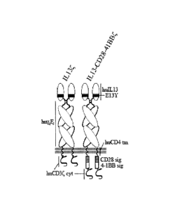
[00014] Figure 1 is a schematic representation of the IL13 and IL13-CD28-
41BK
chimeric antigen receptor (CAR) protein molecules.
[00015] Figure 2 shows the locations of exemplary primers for IL13 CAR
construction on the native IL13 sequence as indicated. The arrows indicate the
position
of the primers on the !Li 3 sequence.
[00016] Figure 3 (given as Figures 3A-3C) provides an exemplary IL13
zetakine-
encoding nucleotide sequence (SEQ ID NO:5, upper strand; SEQ ID NO:6, lower
strand). The segments of DNA in the sequence include GM-CSFR alpha signal
peptide
(SEQ ID NO:7), IL13(E13Y) (SEQ ID NO:8), IgG4(SmP)(SEQ ID NO:9), CD4tm(SEQ ID
NO:10) and CD3zeta (SEQ ID NO:11). The complete amino acid sequence is
provided
as SEQ ID NO:4.
[00017] Figure 4 is a map of the vector IL13zetakine/HyTK-pMG. An exemplary
sequence of such a vector is provided in Figure 5.
[00018] Figure 5 (given as Figures 5A-5L) provides the sequence of an
exemplary
plasmid DNA vector (SEQ ID NO:13, upper strand; SEQ ID NO:14, lower strand).
An
IL13zetakine amino acid sequence (SEQ ID NO:15) and an HyTk amino acid
sequence
(SEQ ID NO:16) also are indicated. The segments of DNA which make up the
complete sequence include hEF1p (nucleotides 6-549; SEQ ID NO:41), IL13
zetakine
(nucleotides 690-2183; SEQ ID NO:42), late sv40pAn (nucleotides 2230-2498; SEQ
ID
NO:43), On ColE1 (nucleotides 2499-3245; SEQ ID NO:44), SpAn (nucleotides 3246-
3432; SEQ ID NO:45), hCMV-1Aprom (nucleotides 3433-4075; SEQ ID NO:46), HyTK
(nucleotides 4244-6319; SEQ ID NO:47) and BGh pAna (nucleotides 6320-6618; SEQ
ID NO:48).
[00019] Figure 6 contains two schematic representations of exemplary CAR
linear
plasmid constructs. Figure 6A shows a IL13 construct and Figure 6B shows a
IL13-
CD28-41BEK construct.
[00020] Figure 7 shows western blot analysis of cell lysates derived from
mock-,
IL13- and IL13-CD28-41BBOransfected CD4+ T cells for CAR expression using a
mouse anti-human CD3 specific mAb.
[00021] Figure 8 is a panel of eight flow cytometry analyses that compare
the
cell surface phenotype of IL13- and IL13-CD28-41BK-expressing bulk CD4+ cells.
[00022] Figure 9 is a panel of six graphs that show flow cytometry results
of
surface staining of HLA-A2 and HLA-DR (MHC molecules), IL13Ra2 and the
costimulatory molecules CD80, CD86, and CD137-L (4-1 BBL) (filled histograms)
as
indicated, compared to isotype controls (open histograms) on U87 glioma target
cells.
[00023] Figure 10 is a series of immunoblots showing the results of a
kinase
assay to determine the kinetics of JNK and p38 (A) and AKT (B) activation,
which is
measured via phosphorylation of their respective substrates (i.e., P-cJun
(phosphorylated c-Jun proto-oncogene), p-GSK3 (phosphorylated glycogen
synthase
kinase 3) and P-ATF2 (phosphorylated activating transcription factor 2)).
[00024] Figure 11 shows the enhanced Thi polarization of IL13-CD28-41BK+
CD4+ T cells in terms of T cell Thi cytokine mRNA (Figure 11A) and Thi and Thz
cytokine protein production (Figure 11B).
[00025] Figure 12A provides data showing enhanced cytotoxic activity of
IL13-
CD28-41BK+ CD4+ T cells (M) against U87 targets compared to that of IL13CE
CD4+ T
cells (0) at the indicated E:T ratio in a 4 hour luciferase cytotoxicity assay
(LCA).
Figure 12B shows similar data for ILI 3-CD28-4iBK+ CD4+ T cells (black bars)
and
IL13C CD4+ T cells (white bars) co-cultured for 48 hours at an E:T ratio of
2:1, and
then again co-cultured for an additional 48 hours after addition of fresh
targets at the
same E:T ratio. Figure 12C provides data obtained with video imaging of T
cells
expressing the indicated CAR co-cultured with adherent U87 cells, which
indicates the
number of viable cells per image.
[00026] Figure 13 provides flux data showing sustained anti-tumor effect
against
established glioblastoma xenografts in vivo by I L13-CD28-41BK+ CD4+ T cells.
Results observed with IL13- and sham-transfected T cells also are shown.
6
Date Recue/Date Received 2020-07-15
CA 02735456 2011-02-25
WO 2010/025177 PCT/US2009/055029
[00027] Figure 14 provides the sequence of 1L13-IgG4-cd28tm-CD28gg-Zeta
(C0)(SEQ ID NO:36).
[00028] Figure 15 provides the sequence of IL13-IgG4-cd4tm-CD28-4-1BB-Zeta,
also referred to herein as IL13-CD28-41BK used/discussed above with respect to
the
examples below (SEQ ID NO:37). This sequence was used to genetically alter T
cells to
express the IL13-CD28-41BK CAR as described and used in Figures 1, 6, 7, 8,
10, 11,
12 and 13.
[00029] Figure 16 provides the sequence of 1L13-IgG4-cd28tm-CD28-0x40-Zeta
(SEQ ID NO:38).
[00030] Figure 17 provides the sequence of IL13-IgG4-cd28tm-CD28gg-4-1BB-
Zeta
(SEQ ID NO:39).
[00031] Figure 18 provides the sequence of IL13-IgG4-cd28tm-CD28gg^199-4-
1BB-Zeta (SEQ ID NO:40).
DETAILED DESCRIPTION OF THE PREFERRED EMBODIMENTS
[00032] Adoptive immunotherapy using T lymphocytes that express tumor-
specific
chimeric antigen receptors (CARs) can be a powerful therapeutic strategy for
the
treatment of cancer. CARs are made up of an extracellular specific recognition
element
(such as a receptor that binds a tumor antigen) linked via a transmembrane
domain to
the CD3 cytoplasmic signaling domain. These receptors therefore are able both
to bind
antigen and to transduce T cell activation, independent of MHC restriction.
Thus, CARs
are "universal" immunoreceptors which can treat a population of patients with
antigen-
positive tumors irrespective of their HLA genotype.
[00033] According to embodiments of this invention, CARs contain the
signaling
domain for CD3 and the signaling domains of one or more costimulatory
receptors that
further promote the recycling, survival and/or expansion of adoptively
transferred cells
expressing the CARs, in addition to specific receptors which allow the cells
to engage
7
CA 02735456 2011-02-25
WO 2010/025177 PCT/US2009/055029
targets such as tumors. The signaling domains of the costimulatory receptors
are the
intracellular portions of each receptor protein that generate the activating
signal in the
cell. Examples are amino acids 180-220 of the native CD28 molecule and amino
acids
214-255 of the native 4-1BB molecule. An especially preferred CAR comprises an
extracellular recognition element that is specific for a unique cancer cell
surface
receptor, is stable in vivo and has low immunogenicity. Derivation from a
naturally-
occurring soluble cell signal molecule helps to achieve these objectives.
[00034] The term "CAR" refers to a chimeric antigen receptor which is a
recombinant biomolecule that contains an extracellular recognition domain, a
transmembrane region, and an intracellular signaling domain. The term
"antigen,"
therefore, is not limited to molecules that bind antibodies, but to any
molecule that can
bind specifically to any receptor. "Antigen" thus refers to the recognition
domain of the
CAR. The extracellular recognition domain (also referred to as the
extracellular domain
or simply by the recognition element which it contains) comprises a
recognition element
that specifically binds to a molecule present on the cell surface of a target
cell. The
transmembrane region anchors the CAR in the membrane. The intracellular
signaling
domain comprises the signaling domain from the zeta chain of the human CD3
complex
and optionally comprises one or more co-stimulatory signaling domains.
[00035] A CAR that contains the IL13 domain with the E13Y mutation
(IL13(E13Y))
and the CD3 zeta chain signalling domain is referred to herein as "IL13(."
This term
includes any chimeric antigen receptor (CAR) that contains an IL13
extracellular
recognition domain (a domain that specifically recognizes IL13Ra2 on tumor
cells) a
transmembrane region, and a CD3 zeta chain intracellular signaling domain. Non-
limiting examples of such CARs are provided in Examples 8-12. A CAR that
contains
IL13(E13Y) and also contains the optional co-stimulatory intracellular domains
CD28
and 4-1BB is termed "IL13-CD28-41BK" herein.
[00036] Persons of skill will recognize that any nucleotide sequence that
encodes
IL13(E13Y) would also be suitable for this same purpose. The unnnutated
sequence of
the IL13 signaling domain also is suitable. Any IL13 or IL13(E13Y) encoding
sequence
including variants with 90%, 95%, 98% or 99% homology to the native sequence
may be
8
CA 02735456 2016-04-13
used here. Such sequences which are useful for specifically recognizing an
I1.13
receptor tumor antigen such as IL13Rcr2, therefore include those encoded by
the native
nucleic acid (see Smemov et al., Gene 155:277-281, 1995),
the same nucleic acid sequence lacking the E13Y
mutation, sequences that are 95%, 98% or 99% homologous to these sequences,
longer sequences that comprise those sequences but also include additional
nucleotides
at the 3' or 5' end, for example any number of additional nucleotides or
codons, such as
3, 6, 9,12 or more nucleotides, or up to about 12, 20, 50 or 100 additional
nucleotides,
and any sequence that encodes the same amino acid sequence as these nucleic
acids
due to the degeneracy of the genetic code. In particular, sequences that are
codon
optimized (CO) for expression by the desired host are contemplated as part of
the
invention.
[00037] Soluble
recognition elements as used in this invention are derived from de
novo synthesized polypeptides, as described for the I1.13 (El 3Y) coding
sequence In
Example 1 or from polypeptides of combinatorial libraries such as phage-
display libraries
or chemically synthesized libraries. Preferred soluble recognition elements
are of
human origin and are therefore non-immunogenic, but can be tailored in
affinity or
specificity through mutagenesis. Upon their expression on T cells, soluble
recognition
elements are able to bind a target element on the target cell (for example a
tumor cell,
but not to any appreciable extent on non-target cells), in such a way that
results in T cell
activation. Thus, the soluble recognition elements that are suitable for this
invention
have certain advantages over antibody fragments or cell adhesion molecules for
target
specificity In the inventive CARs, since they are more likely to be stable in
the
extracellular environment, non-antigenic, and more selective, and therefore
are
preferred. Examples of suitable soluble receptor elements include autocrine
and
paracrine growth factors, chemokines, cytokines, hormones, and engineered
artificial
small molecule ligands that exhibit the required specificity. Natural ligand
sequences
can be engineered to increase their specificity for a particular target cell.
Selection of a
recognition element for use in a particular CAR is governed by the nature of
the target
cell, and the qualities discussed above. In one preferred embodiment of the
invention,
9
CA 02735456 2011-02-25
WO 2010/025177 PCT/US2009/055029
the CAR exploits the tumor-restricted expression of IL13Ra2 by malignant
glioma, renal
cell carcinoma and other tumors by using as the recognition element a mutant
of
IL13(E13Y) to direct T cells specifically to IL13Ra2-expressing tumor cells.
Analogous
recognition elements can be created that are specific to any of a variety of
cancer cell
types that selectively express receptors antigens or any specific molecule on
their cell
surfaces, for which selective recognition elements are known or can be
engineered.
[00038] Examples of suitable support (transmembrane) regions for use with
the
invention include the constant (Fc) regions of immunoglobins, human CD8a, and
artificial linkers that serve to move the targeting moiety away from the cell
surface for
improved access to and binding on target cells. A preferred support region is
the Fc
region of an IgG (such as IgG4). Examples of suitable transmembrane domains
include
the transmembrane domains of the leukocyte CD markers, preferably that of CD4
or
CD28. Examples of intracellular receptor signaling domains include the T cell
antigen
receptor complex, preferably the zeta chain of CD3, however any transmembrane
region
sufficient to anchor the CAR in the membrane can be used. Persons of skill are
aware
of numerous transmembrane regions and the structural elements (such as
lipophilic
amino acid regions) that produce transmembrane domains in numerous membrane
proteins and therefore can substitute any convenient sequence. T cell
costimulatory
signaling receptors suitable for improving the function and activity of CAR-
expressing
cells include, but are not limited to, 0D28 and 4-1BB also known as (0D137),
and OX-
40.
[00039] Signaling via CD28 is required for 11_2 production and
proliferation, but
does not play a primary role in sustaining T cell function and activity. 4-1BB
(a tumor
necrosis factor-receptor family member expressed following CD28 activation)
and OX-40
are involved in driving long-term survival of T cells, and accumulation of T
cells. The
ligands for these receptors typically are expressed on professional antigen
presenting
cells such as dendritic cells and activated macrophages, but not on tumor
cells.
Expressing a CAR that incorporates CD28 and/or 4-1BB signaling domains in CD4+
T
cells enhances the activity and anti-tumor potency of those cells compared to
those
CA 02735456 2011-02-25
WO 2010/025177 PCT/US2009/055029
expressing a CAR that contains only the CD3 signaling domain. Preferably, the
inventive CARs contain both CD28 and 4-1BB signaling domains.
[00040] In order for the CAR to target tumor cells, they contain an
extracellular
binding molecule that binds a tumor surface marker and preferably specifically
binds to a
unique tumor surface molecule. Some cancers express or overexpress molecules
of the
immune system. Glionnas, for example, express IL13 receptors, and in
particular, high-
affinity IL13 receptors. However, unlike the ILI 3 receptor trimolecular
complex used by
the immune system, (which consists of the IL13Ra1, the IL4R8, and yc), glioma
cells
overexpress a unique 11_13Ra2 chain capable of binding IL13 independently of
the
requirement for IL4R8 or yc44. Like its homolog IL4, IL13 has pleotropic
immunoregulatory activity outside the CNS. Both IL13 and IL4 stimulate IgE
production
by B lymphocytes and suppress pro-inflammatory cytokine production by
macrophages.
[00041] Detailed studies using autoradiography with radiolabeled IL13 have
demonstrated abundant IL13 binding on nearly all malignant glioma tissues
studied.
This binding is highly homogeneous within tumor sections and in single cell
analysis.
However, molecular probe analysis specific for IL13Ra2 mRNA did not detect
expression of the glioma-specific receptor by normal brain elements and
autoradiography with radiolabeled IL13 also could not detect specific IL13
binding in the
normal CNS. These studies suggest that the shared IL13Ra1/1L48/yc receptor is
not
expressed detectably in the normal CNS. Therefore, IL13Ra2 is a very specific
cell-
surface target for glioma and is a highly suitable target for this invention.
Persons of skill
are aware of other suitable targets for CARs, which are expressed or
overexpressed on
the cells to be targeted and preferably are not expressed, or are expressed to
a much
lesser degree, on other cells. Another example of a tumor-specific target
suitable for
targeting with CARs of this invention is IL3 receptor (IL3R; e.g., expressed
on acute
myeloid leukemia (AML) cells.
[00042] Binding of IL13-based cytotoxins to the broadly expressed
IL13Ra1/1L48/yc receptor complex, however, has the potential of mediating
undesired
toxicities to normal tissues outside the CNS, and thus limits the systemic
administration
of these agents. An amino acid substitution in the IL13 alpha helix A at amino
acid 13 of
11
CA 02735456 2016-04-13
tyrosine for the native glutamic acid selectively reduces the affinity of 113
to the
IL13Ra1/1413/yc receptor. Binding of this mutant (termed IL13(E13Y) to
IL13Ra2,
however, was increased relative to wild-type 113 by 50-fold. Thus, this
minimally
altered 113 analog simultaneously increases IL13's specificity and affinity
for glioma
cells. Therefore, a preferred embodiment of the invention employs an 1L13
containing a
mutation at amino acid 13. 113 having the natural sequence also may be used
with the
invention, however, and can be useful, particularly in situations where the
modified T
cells are to be locally administered, such as by injection directly into a
tumor mass,
[00043] A preferred type of CAR for specifically targeting tumors that
express
113Ra2 is made up of an extracellular 113-mutant cytokine in which the 1L13
protein
contains a substitution of tyrosine for the naturally-occurring glutamic acid
at amino acid
= 13 of the protein (termed 11-13(E13Y) here), connected to a human IgG4
hinge-Fc
domain support region which is fused to a CD4 transmembrane domain and a
cytoplasmic CD3 signaling sequence. See Figure 1, left side. This CAR is
referred to
herein as an nIL134 CAR". When this CAR also contains the CD28 and 4-1BB
signaling
domains, it is referred to as113-CD28-41Big . See Figure 1, right side.
[00044] An immunoreceptor according to the present invention can be
produced by
any means known in the art, though preferably it is produced using recombinant
DNA
techniques. Nucleic acids encoding the several regions of the chimeric
receptor can be
prepared and assembled into a complete coding sequence by standard techniques
of
molecular cloning known in the art (genomic library screening, PCR, primer-
assisted
ligation, site-directed mutagenesis, etc.) as is convenient. The resulting
coding region is
preferably inserted into an expression vector and used to transform a suitable
expression host cell line, preferably a T lymphocyte cell line, and most
preferably an
autologous T lymphocyte cell line.
[00045] Briefly, an IL1g CAR may be constructed using known methods as
follows. The 113 mutant DNAIL13(E13Y) can be synthesized by PCR with primers
based on the known 113 mRNA sequence. The complete 103 gene sequence is
reported in Smemov et al., "Tandem arrangement of human genes for interleukin-
4 and
interleukin-13: resemblance in their organization." Gene 155:277-281, 1995.
12
CA 02735456 2016-04-13
De novo synthesis of the
1L13(E13Y) was performed using forward primer1L13P1 and four reverse primers,
IL13P2,1L13P3, 1L13P4, and 1L13P5, shown in Table), below, and Figure 2. This
1L13
mutant sequence then can be modified to contain a 5' leader sequence, If
desired. A
transmembrane anchor such as the human IgG4-004 transmembrane (1gG4-CD4tm) and
CD3 zetachain (CD34) cytoplasmic sequences also can be added to the 3' end by
PCR
fusion techniques or any convenient method. The complete 1L13 sequence is
given in
Figure 3 as an example of the invention. The same methods can be used to
construct
equivalent molecules using different recognition elements. The final construct
then can
be ligated into any suitable plasmid expression vector. A preferred plasmid
expression
vector is pMG (available from InvivogenTm).
[000461 The ILI 3(E13Y)-containing CAR specifically directs T cells to
target 1L13
receptor a2 (termed 1L13Ra2 here)-expressing glioma cells, renal carcinoma
cells and
cells of any cancer expressing 1L13Ra2 in an MHC-Independent manner. Anti-
tumor
0D44 T cell effectors were generated to be re-directed to recognize tumor
cells using a
CAR containing the signaling domains derived from CD3-4, CD28 and 4-1BB.
Either the
ILA g or IL13-CD28-418134 CAR was transfected into human primary T cells using
a
non-viral plasmid vector (pEK) and electroporation methods (Nucleofector
Technology",
of Amaxa Blosystemsim, Gaithersburg, MD). CD4+ T cells expressing either CAR
(11_13
or 11.13-CD28-41BK) were compared for their potential to activate effector-
associated
signaling pathways, produce cytokines, lyse target cells and control in vivo
tumor growth.
The results showed that addition of the CO28 and 4-1BB signaling domains to
IL1
enhances the anti-tumor effector functions of CD4, T cells expressing the CAR.
Effector
T cells expressing the 1L13-CD28-41BK immunoreceptor were able to mediate
costImulatory signals through JNK, p38 and AKT kinases in the tumor
environment
where costimulation would be expected to be limiting. The enforced
costimulation in the
human primary CD4* T cells supports the polarization of these cells to a Thi
phenotype
In a manner that is associated with sustained anti-tumor efficacy both In
vitro and in vivo.
Effector signals downstream of the CAR in CD4+ T cells were demonstrated.
These
13
CA 02735456 2011-02-25
WO 2010/025177 PCT/US2009/055029
effector signals correlated with the observed Thl bias and the prolonged anti-
tumor
effector activity of these cells both in vitro and in vivo.
[00047] CD3 signaling alone drives ERK activation. This correlates well
with the
finding here that ERK activity is not enhanced in 113-CD28-41BK-expressing
cells
compared to IL13-expressing controls (both CARs contain the CD3 signaling
domain).
Costimulation of CD3 with CD28 drives activation of JNK and p38; 4-1BB-
mediated co-
stimulation of CD3 also involves JNK activation. Both JNK and p38 play primary
roles in
driving Thi-polarized immune responses by CD44. T cells, including their
production of
12, IFN-y and TNF-a. The activation of AKT kinase, another downstream
signaling
component of both CD28 and 4-1BB, also is involved in up-regulation of 12 and
INF-y,
but not Th2 cytokines. The association of a pronounced Thi phenotype (see
examples,
below) with enhanced JNK and p38 MAP kinase induction and sustained ATK
activation
(see examples, below) in 113-CD28-41BK -expressing T cells strongly indicates
that
the CD28 and 4-1BB signaling moieties work with the CD3 signaling domain in
this
chimeric receptor to retain the capacity to transduce the downstream signaling
pathways
normally associated with these costimulatory receptors. Regardless of how
strong the
activated Thi phenotype driven by costimulatory domain signals may be,
retention and
recycling of functional anti-tumor effector CD4+ T cells within the tumor
microenvironment greatly assists in achieving anti-tumor potency.
[00048] Compared to CD3-mediated activation alone, CD4+ effector T cells
expressing the 113-CD28-41BK CAR exhibited augmented/sustained MAPK and AKT
activity, upregulated Thi cytokine production, and enhanced cytolytic potency
against
tumor targets. Moreover, upon recursive stimulation with tumor, the 113-CD28-
41BK+
CD4 cells retained/recycled their lytic function whereas 113(' CD4+ cells
were effective,
but sooner became anergic/exhausted. These in vitro observations correlated
with
enhanced in vivo control of established orthotopic CNS glioma xenografts in
immunodeficient mice mediated by adoptively transferred ex vivo expanded CD4+
T cells
expressing the costimulatory CAR. These studies therefore demonstrate the
effect of
integrating costimulation with CD3 signaling events to fully activate CD4+
anti-tumor
effector cells for sustained function in the tumor microenvironment.
14
CA 02735456 2011-02-25
WO 2010/025177 PCT/US2009/055029
[00049] CO28 and 4-1BB costimulatory signals mediated via AKT can inhibit
activation-induced cell death through up-regulation of anti-apoptotic
proteins. The
enhanced AKT activation seen in the 113-CD28-41BK-expressing T cells was
associated with enhanced recycling of tumor specific activity in vitro as well
as prolonged
tumor growth control in vivo. Thus, the costimulatory CAR can enhance the
duration
and/or retention of anti-tumor activity in a manner that can significantly
improve the
clinical efficacy of adoptive therapy protocols.
[00050] Tumor-specific CARs that contain their own costimulatory signaling
domains provide a new approach for activating T lymphocytes against a wider
variety of
solid tumors that do not express these costimulatory ligands. 113Ra2, for
example, has
been identified as an over-expressed cell-surface target on various human
tumors,
including breast cancer, head and neck cancer, kidney cancer, ovarian cancer
and
Kaposi's sarcoma as well as gliomas. Thus, T cells expressing a CAR that
contains an
113 zetakine and CD28 and 4-1BB can be used to treat glioblastomas (glioma)
and any
cancer, such as those listed above, that have the 113 target on their surface.
[00051] The invention specifically contemplates CARs that contain CD3, CD28
and 4-1BB (and/or other costimulatory signaling domains) which can be directed
to any
tumor by incorporating a moiety that binds to a cell-surface-expressed tumor
target, for
example an antigen. Examples of other tumor-specific target binders include
Her2/Neu
(ErbB-2), a3 integrin, CD20, CD19, EGFRVIII, IL3Ra (CD123), LEA, CD44v6 or any
target specific to a tumor, preferably a solid tumor that does not express the
costimulatory signaling domain which is contained on the CAR. Therefore,
constructs
for targeting human tumors in this manner can include those with specificities
for
Her2/Neu (ErbB-2), a3 integrin, CD20, CD19, EGFRVIII, IL3Ra (CD123), LEA,
CD44v6
or any specific tumor antigen or other cell-surface component accessible to
binding by a
chimeric T cell receptor. Persons of skill are aware of these specific tumor
antigens and
receptors which can be exploited to target a specific tumor, and are aware of
the tumors
that can be targeted in this manner.
[00052] Both CD4+ and CD8+ T cell effector functions can be triggered via
these
receptors, therefore both of these T cell types are contemplated for use with
the
CA 02735456 2016-04-13
invention. CD8+ T cells expressing the 10 3 CARs of this invention may be used
to lyse
target cells and to produce IL2 in the presence of target cells, among the
other functions
of these cells. Expression of the appropriate costimulatory CAR in either or
both CD4+
and CD8+ T cells would be used to provide the most effective population of
cells for
adoptive immunotherapy, consisting therefore of either or both professional
helper and
killer T cells that exhibit enhanced and/or long term viability and anti-tumor
activity.
[000531 The following examples are solely for the purpose of
illustrating one embodiment of the invention.
EXAMPLES
Example 1. Transfection and Expression of 103%2-specific Chimeric Receptors in
Primary Human T Lymphocytes.
[00054] To engage both T cell receptor (TCR)- and costimulatory-like
signaling
cascades upon interaction with glloma tumor antigen 11_13Ra2, signaling
elements
derived from CD28 and 4-1BB were integrated into an 1L13-zetakine (11.134)
chimeric
antigen receptor (CAR). The preferred ILlg CAR is composed of the
extracellular
103(E13Y) mutein, human IgG4 hinge-Fc finked to the human cytoplasmic CDg via
the
transmembrane domain of human CD4. See Figure 1. De novo synthesis of the
IL13(E13Y) coding sequence was performed using primers IL13P1, IL13P2, IL13P3,
It..13P4, and IL13P5. See Table 1, below, and Figure 2. The final sequence
(417bp)
was end-digested with EcoRI-BamHI, and Ilgated into the plasmid pSK
(Stratagena) as
ligation 312#3. Ligation 31243 was mutagenized (StratageneTM kit, per
manufacturer's
Instructions) to repair a deleted nucleotide using the primers 103 312#3 mut5-
3 and
1L13 312#3 mut3-5 and ligation 312#3 as a template, to form ligation 348#1
(103Z/pSK).
[00055] The human GM-CSFR alpha chain signal peptide (hsp) coding sequence
was fused to the 5' end of IL13(E13Y) by standard PCR splice overlap
extension. The
16
CA 02735456 2011-02-25
WO 2010/025177 PCT/US2009/055029
hsp sequence was obtained from the template ligation 301#10 (hsp/pSK) using
primers
5':19hsp5' and 3': hsp-IL13FR. See Table I. The IL13 sequence was obtained
using the
primers 5': hsp-IL13FF and 3': 113-IgG4FR, and ligation 312#3 as template. See
Table
[00056] A sequence encoding the IgG4 Fc, CD4 transmembrane and CD3C
cytoplasmic regions (IgG4m:zeta; nucleotides 421-1512 of the complete IL13
sequence
of Figure 3 (SEQ ID NO:12)) was fused to the 3' end of the human signal
peptide-1L13
fusion sequence using the same methods. The IgG4m:zeta sequence was obtained
using the primers 5': IL13-IgG4FF and 3': ZetaN3' (see Table 1), using the
sequence
R9.10 (IgG4mZeta/pSK) as template. The 1119 bp IgG4m:zeta sequence was fused
to
the hsp-1L13 fusion sequence using the respective sequences as templates, and
the
primers 5': 19hsp5' and 3': ZetaN3' (see Table 1), to yield a 1522 bp hsp-IL13-
IgG4nn:zeta fusion sequence. The ends were digested with Xbal-Notl, and
ligated into
pSK as ligation 351#7, to create the plasmid IL13(,/pSK (4464 bp) (i.e. the
IL13
sequence of Figure 3 , within pSK cloning vector.
[00057] An expression vector containing the IL13 coding sequence was
created
by digesting IL13(,/pSK with Xbal-Notl, and creating blunt ends with Klenow,
and ligating
the resulting fragment into the plasmid pMGAPac (lnvitrogenTM) (first prepared
by
opening with SgrAl, blunting with Klenow, and dephosphorylation with SAP), to
yield the
plasmid IL13ZipMG. The hygromycin resistance region of IL13qpMG was removed by
digestion with Notl-Nhel, and replaced by the selection/suicide fusion HyTK,
obtained
from plasmid CE7R/HyTK-pMG by digestion with Notl-Nhel, to create the
expression
vector IL13Z,/HyTK-pMG (6785 bp). This plasmid comprises the human elongation
factor-1a promoter (hEF1p) at bases 6-549, the IL13 coding sequence at bases
690-
2183, the Simian Virus 40 Late polyadenylation signal (Late SV40pAN) at bases
2230-
2498, a minimal E. coli origin of replication (On i ColE1) at bases 2499-3245,
a synthetic
poly A and Pause site (SpAN) at bases 3246-3432, the Immediate-early CMV
enhancer/promoter (h CMV-1Aprom) at bases 3453-4075, the Hygromycin resistance-
Thymidine kinase coding region fusion (HyTK) at bases 4244-6319, and the
bovine
growth hormone polyadenylation signal and a transcription pause (BGh pAn) at
bases
17
CA 02735456 2011-02-25
WO 2010/025177 PCT/US2009/055029
6320-6618. The plasmid has a Pad l linearization site at bases 3233-3240. The
hEF1p,
late SV40pAN, on ColE1, SpAn, and hCMV-1Aprom elements all were derived from
the
parent plasmid pMGAPac. In sum, IL13(./HyTK-pMG is a modified pMG backbone,
expressing the IL13 gene from the hEF1 promoter, and the HyTK fusion from the
hCMV-1A promoter. A map of the plasmid IL13(/HyTK-pMG appears in Figure 4. The
full nucleic acid sequence of the plasmid is shown in Figures 5A-5L (SEQ ID
NOs:13
and 14. The sequence of the IL13 insert also is given in Figure 3 (SEQ ID
NOs:5 and
6).
[00058] Assessment of the integrity of the expressed construct was
confirmed by
western blot using the anti-human CD3 monoclonal antibody clone 8D3 (BD
PharMingenTm, San Diego, CA) to probe whole cell lysates derived from Jurkat T
cell
stable transfectants cocultured in the presence or absence of tunicamycin, an
inhibitor of
glycosylation. Jurkat T cell stable transfectants (Jurkat-IL13-pMG bulk line)
were
obtained by electroporating Jurkat T cells with the IL13Z,/HyTK-pMG expression
vector,
followed by selection and expansion of positive transfectants. 2x10⁶ cells
from the
Jurkat-IL13-pMG bulk line were plated per well in a 24-well plate with or
without 5 pg/mL,
pg/mL, or 20 pg/mL Tunicamycin. The plate was incubated at 37 C for 22 hours.
Cells were harvested from each well, and each sample was washed with PBS and
resuspended in 50 pL RIPA buffer (PBS, 1% NP40, 0.5% sodium deoxycholate, 0.1%
SDS) containing protease inhibitor (1 tablet/10 mL Complete Protease Inhibitor
Cocktail). Samples were incubated on ice for one hour, before being
centrifuged at 4 C
for 20 minutes at 14,000 rpm. Samples of centrifuged lysate supernatant were
harvested and boiled in a 1:3 volume of sample buffer under reducing
conditions, then
subjected to SDS-PAGE electrophoresis on a 12% acrylamide gel. Following
transfer to
nitrocellulose, the membrane then was blocked in a BlottoTM solution
containing 4% non-
fat dried milk in T-TBS (0.1% Tween 2OTM in Tris buffered saline pH 8.0) for 1
hour.
Membrane was then incubated with the primary mouse anti-human CD3 monoclonal
antibody at a concentration of 0.5 pg/mL for one hour, washed, and then
incubated with
a 1:3000 dilution (in BlottoTM solution) of goat anti-mouse IgG alkaline
phosphatase
conjugated secondary antibody (Bio-RadTM lmmunoStarTM Kit) for 1 hour. Prior
to
18
CA 02735456 2011-02-25
WO 2010/025177 PCT/US2009/055029
developing, the membrane was washed 4 additional times in T-TBS, and then
incubated
with 3 mL phosphatase substrate solution (Bio-RadTM lmmunoStarTM Kit) for 5
minutes at
room temperature. The membrane was then covered with a plastic development
folder
(Tropix') and exposed to X-ray film. Consistent with the known glycosylation
pattern of
wild-type human IL13, the electrophoretic mobility of the expressed IL13(E13Y)
zetakine
indicates a heavily glycosylated protein which, when expressed in the presence
of
tunicamycin, is reduced to an amino acid backbone of approximately 54 kDa.
[00059] Construction of the co-stimulatory CAR was initiated with an HyTK-
2A-
IL13(-pcDNA3.1(+) construct, which encodes the selection/suicide fusion gene
HyTK,
the de novo synthesized self-cleavable foot-and-mouth disease 2A peptide
(TCTAGAGGAGCATGCCAGCTGTTGAATTTTGACCTTCTTAAGCTTGCGGGAGACGT
CGAGTCCAACCCTGGGCCC; SEQ ID NO: 49), and the IL13, molecule (Figure 3),
cloned into pcDNA3.1(+) (Invitrogen'). To confer resistance to methotrexate
(MTX), the
HyTK gene was replaced by PCR with an dihydrofolate reductase (DHFR) gene
(amplified from a cDNA library derived from peripheral blood mononuclear cells
(PBMC)
that had been stimulated for three days with the 0KT3 antibody which
recognizes the
CD3 chain of the T cell receptor which contained L22F and F33S mutations
generated
using a QuikChangeTM Site-Directed Mutagenesis Kit (Stratagene"). The
resulting
DHFRdm-2A-IL13 construct was then excised with Nhel and Notl, eluted and
ligated
into the similarly digested mammalian plasmid expression vector pEK. The pEK
vector
had been modified originally from pcDNA3.1(+) by removing the CMV promoter and
the
ampicillin gene and replacing them with the human Elongation Factor la
promoter
(EF1p) gene derived from pMG (lnvivogenTM) to create the plasmid DHFRdm-2A-
IL13pEK (pJ01275-9). CD28 cDNA was purchased from lnvitrogenTM and 4-1BB
coding region was amplified by PCR from a cDNA library derived from peripheral
blood
mononuclear cells (PBMC) that had been stimulated for three days with the OKT3
antibody (using primers 41BB5'and 41663', see Table 1).
[00060] The intracellular signaling regions of CD28 and 4-1 BB (amino acids
180-
220 and 214-255, respectively, of the native CD28 and 4-1BB sequences) were
fused by
PCR (using the primers CD4-CD28F, CD28-4-1-BBR, 0D28-4-1bbF, and 41bb93
19
CA 02735456 2011-02-25
WO 2010/025177 PCT/US2009/055029
provided in Table I) into the junction between the CD4 transmembrane and
cytoplasmic
CD3 (amino acids 52-164 of native CD3) regions. See Figure 6, which provides
schematic representations of examples of IL13 (Figure 6A) and 113-CD28-41BK
(Figure 6B) linear plasmid constructs. The placement of human 113 nnutein
(E13Y),
human IgG4 hinge-Fc (IgG4), human CD4 transmembrane (tnn), human CD3
cytoplasmic (Zeta), CD28 cytoplasmic (28c) and 4-1BB cytoplasmic (BBc)
segments are
indicated in Figure 6. Restriction enzyme sites that were used to insert the
different
PCR fragments also are indicated in Figure 6 (Nhel, Kpnl, Nsil, Notl), with
their predicted
base pair locations provided in parentheses. As shown in Figure 6A, the CAR,
113-
CD28-41BK, comprises the cytoplasmic domain of 0D28 and 4-1BB fused to that of
CD3( Each construct shown in Figure 6A has a hulL13 domain containing the E13Y
mutation which makes it 113Ra2-specific, a human IgG4 hinge-Fc domain
(huy4Fc), a
human CD4 transmembrane (huCD4tm) domain, and a human CD3 cytoplasmic
(huCD3 cyt) domain; the 113-CD28-41BK CAR has the signaling (sig) domains of
CD28 and 4-1BB inserted between the CD4 transmembrane and CD3 cytoplasmic
domains. The PCR primers used in construction of the plasmids and used in
expression
analysis are provided in Table I.
[00061] Bulk cultures of CD4 + T cells obtained by MACSTM separation using
the
manufacturer's protocol (Miltenyi BiotecTM Inc.) were maintained in RPMI media
with
10% FCS, 1% L-glutamine, 2.5% HEPES buffer, 50 U/mL rhIL2, 1Ong/mL rhIL15 and
0.1 pM MTX. Isolation, activation, and electroporation of human T cells was
performed
as follows. PBMC were isolated by density gradient centrifugation over Ficoll-
Paque
(Pharmacia BiotechTM) of heparinized peripheral blood obtained from consenting
healthy
donors. The cells were resuspended in nucleofection solution using the AmaxaTM
Human T cell Nucleofector kit (AmaxaTM Inc.). Plasmid (1 pg/5x106 cells) was
added,
and cells were electroporated using the AmaxaTM Nucleofector I (AmaxaTM Inc.),
program
U-14. Cells then were harvested in phenol red-free medium with 10% FCS,
allowed to
rest overnight, and then stimulated with 30 ng/mL OKT3 and 5ng/mL rhIL15 in
RPMI
with 10% FCS for three days. Successful transfectants were selected using
media
containing 0.1 pM MTX and 5ng/mL rhIL15.
CA 02735456 2011-02-25
WO 2010/025177 PCT/US2009/055029
[00062] The expression of CARs was assessed by immunoblotting analysis with
an
antibody specific to CDX Whole cell lysates of bulk MTX-selected CD4+ T cells
(mock-,
IL13- and IL13-CD28-41BK-transfected) were tested for the CAR expression
(chimeric
CD3) using known methods and a commercially available mouse anti-human CD3-
specific monoclonal antibody, 1D3. As expected with such highly glycosylated
proteins,
multiple bands within the expected molecular weights were observed. See Figure
7.
[00063] The levels of IL13 or ILI 3-CD28-41BK CAR expressed on the surface
of
CD4+ T cells were examined by detection of membrane-bound IL13 using flow
cytometry. See Figure 8. PBMC transfected with cDNA encoding IL13 or IL13-0028-
41BK CAR were propagated for an average of 10 weeks under selective
concentrations
of MTX (0.1 pM), magnetically sorted for CD44- cells by MACSTM separation, and
examined for surface expression of 1L13-containing CAR (Y-axes), and CD4, CD8,
TCRa/13, or CD28 (X-axes) as indicated. lsotype-matched fluorescent mAbs were
used
to establish the quadrants. These genetically modified T cell populations were
not only
predominantly CD4+ and CD8", as expected after magnetic bead based MACSTM
purification of CD4+ cells, but also expressed high and equivalent levels of
endogenous
TCR and low to undetectable levels of costimulatory CD28. See Figure 8.
[00064] The IL13Ra2 human glioblastoma tumor cell target line used in
these
studies, U87, also was phenotyped to confirm that those cells express MHC
class I and
class II on their surface and do not express the costimulatory ligands CD80/86
or 4-
I BBL. See Figure 9, which shows the surface staining of MHC molecules HLA-A2
and
HLA-DR, 11.13R and costimulatory molecules CD80, CD86, and CD137-L (4-1BBL)
(filled
histograms) as indicated, compared to isotype controls (open histograms) on
U87 glioma
target cells, as analyzed by flow cytometry.
[00065] Flow cytometric analysis involved evaluating the cell-surface
expression of
the ILI 3-CAR constructs by staining with PE-conjugated or FITC-conjugated
anti-human
IL13 monoclonal antibodies (BD PharMingen"). The cell-surface phenotype of
primary
human T cell transfectants was assayed with FITC-conjugated anti-CD4, anti-
CD8, and
anti-TCR a/13 antibodies or with PE-conjugated anti-CD28 antibodies (BD
PharMingen'). The cell-surface phenotype of human U87 glioma cells was assayed
21
CA 02735456 2011-02-25
WO 2010/025177 PCT/US2009/055029
with FITC-conjugated anti-HLA-A2, anti-HLA-DR, and anti-CD80 antibodies, or
with PE-
conjugated anti-CD86 and anti-CD137-L (4-1BBL) antibodies, compared to FITC-
and
PE-conjugated isotype controls (BD PharMingen'). IL13Ra2 expression was
assayed
using goat anti-human 11_13Ra2 (R&D SystemsTM) followed by FITC-conjugated
mouse
anti-goat IgG (Jackson ImmunoResearch').
22
CA 02735456 2011-02-25
WO 2010/025177 PCT/US2009/055029
Table I. PCR primers for CAR Construction.
SEQ
Primer Name Primer Sequence (5'-3') ID
NO:
I L3P1
TATGAATTCATGGCGCTTTTGTTGACCACGGTCATTGCTCTCACTTGC 17
CTTGGCGGCTTTGCCTCCCCAGGCCCTGTGCCTCCCTCTACAGCCC
TCAGGTAC
I L3P2
GTTGATGCTCCATACCATGCTGCCATTGCAGAGCGGAGCCTTCTGGT 18
TCTGGGTGATGTTGACCAGCTCCTCAATGAGGTACCTGAGGGCTGTA
GAG G GAG
I L3P3 CTCTGGGTCTTCTCGATGGCACTGCAGCCTGACACGTTGATCAGGG 19
ATTCCAGGGCTGCACAGTACATGCCAGCTGTCAGGTTGATGCTCCAT
ACCATGC
I L3P4
CCTCGATTTTGGTGTCTCGGACATGCAAGCTGGAAAACTGCCCAGCT 20
GAGACCTTGTGCGGGCAGAATCCGCTCAGCATCCTCTGGGTCTTCT
CGATGGC
I L3P5
TCGGATCCTCAGTTGAACCGTCCCTCGCGAAAAAGTTICTTTAAATGT 21
AAGAGCAGGTCCTTTACAAACTGGGCCACCTCGATTTTGGTGTCTCG
IL13 312#3 mut5-3 CAACCTGACAGCTGGCATGTACTGTGCAGCCCTGGAATC 22
IL13 312#3 mut3-5 GTTGGACTGTCGACCGTACATGACACGTCGGGACCTTAG 23
5':19hsp5 ATCTCTAGAGCCGCCACCATGCTTCTCCTGGTGACAAGCCTTC 24
3': hsp-IL13FR GAGGGAGGCACAGGGCCTGGGATCAGGAGGAATG 25
5': hsp-IL13FF CATTCCTCCTGATCCCAGGCCCTGTGCCTCCCTC 26
3': IL13-IgG4FR GGGACCATATTTGGACTCGTTGAACCGTCCCTCGC 27
5': IL13-IgG4FF GCGAGGGACGGTTCAACGAGTCCAAATATGGTCCC 28
3': ZetaN3' ATGCGGCCGCTCAGCGAGGGGGCAGG 29
41BB5' ATCGAATTCGCCGCCACCATGGGAAACAGCTGTTACAAC 30
41BB3' GATAAGCTTATCGATTCACCACATCCTCCTTCAGTT 31
CD4-CD28F CATTGGGCTAGGCATCTTCTTCAGGAGTAAGAGGAGCAGGCTC 32
CD28-4-1BBR GTTTCTTTCTGCCCCGTTTGCCACCTCCGGAGCGATAGGCTGCGAA 33
CD28-4-1BBF CTTCGCAGCCTATCGCTCCGGAGGTGGCAAACGGGGCAGAAAGAAA 34
4-1BB93' GTTGCGGCCGCTCACAGTTCACATCCTCCTTCTTCTTC 35
23
CA 02735456 2011-02-25
WO 2010/025177 PCT/US2009/055029
Example 2. Potentiation of JNK and 1)38 MAPK Signaling with Sustained AKT
Signaling by IL13-CD28-41BK.
[00066] T cells stimulated by the engagement of the TCR-CD3 complex along
with
the auxiliary receptors 0D28 or 4-1BB are known to drive signals through AKT
as well as
the mitogen-activated protein kinases (MAPKs). To investigate the ability of
costimulatory CARs to influence these downstream effector pathways, in vitro
kinase
assays were used to evaluate and compare the activity of AKT and MAPK family
members ERK, JNK and p38 in IL13- and IL13-CD28-416K-expressing CD4+ T cells
following engagement of U87 target cells. Human glioma line, U87, was obtained
from
ATCC (Rockville, MD). All tumor lines are adherent, and were grown in DMEM
(Irvine
ScientificTM) supplemented with 10% heat-inactivated FCS, 25 mM HEPES, and 2
mM L-
glutamine. CD4+ T cells expressing IL13 or IL13-CD28-41BK CAR were incubated
with U87 glioma cells for the times indicated in Figure 10 prior to assay.
[00067] After IL13- or IL13-CD28-41BK-expressing CD4+ T cells were
stimulated
with tumor target cells for up to 48 hours (Figure 10A) or 72 hours (Figure
106), levels of
the JNK, p38 and AKT total protein substrates (i.e., cJun, ATF2, and GSK3,
respectively) and the phosphorylated substrates (P-cJun, P-ATF2, and P-GSK3,
respectively) were measured by Western immunoblot. The fold increase in the
phosphorylation of each substrate, as a measure of kinase activity, is
indicated at the
bottom of each group in Figure 10.
[00068] A non-radioactive solid-state kinase assay was performed using a
method
modified from Hibi et al., "Identification of an oncoprotein- and UV-
responsive protein
kinase that binds and potentiates the c-Jun activation domain." Genes Dev.
7:2135-
2148, 1993. Using T cell lysates that had been separated from target cells by
gentle
centrifugation (1000 rpm, <3 minutes), the selected kinase was
immunoprecipitated
overnight at 4 C using antibodies specific to ERK1/2, JNK, p38, and AKT (Cell
Signaling
Technology Inc."). The immunoprecipitated complexes were washed in lysis
buffer
(PBS with 1% NP40, 0.1% SDS, and 0.5% sodium deoxycholate) and kinase buffer
(25
mM Tris, pH 7.5, containing 10 mM MgCl2 and 2 mM EGTA), and the assay was
24
CA 02735456 2011-02-25
WO 2010/025177 PCT/US2009/055029
performed at 30 C for 30 minutes, using 1 pg of substrate in the presence of
10 pf\A
ATP.
[00069] Glutathione S transferase (GST) fusion proteins: GST-ELK, GST-ATF2
and GST-GSK30 (Cell Signaling Technology' Inc.), and GST-cJun(1-79) (as
described
in Chang et al., Cell 124:601-613, 2006) were used as the substrates for the
ERK, p38,
AKT, and JNK kinase assays, respectively. The resulting products were resolved
in
12% NuPAGETM (lnvitrogenTM) according to standard methods and transferred to
nitrocellulose membrane using the Xcell II Blot ModuleTM (Invitrogen'). The
blots were
probed with antibodies to phospho-ELK, ATF2, cJun and GSK313 (Cell Signaling
Technology' Inc.) to detect phosphorylated GST fusion proteins and antibodies
to GST
(BD PharMingen") to detect the total amount of substrate. The immunoblots then
were
incubated with IRDye 680-conjugated rabbit or IRDye800-conjugated mouse
immunoglobulin-specific antibodies (LI-COR'). Blocking buffer (purchased from
LI-
CORTM) was used to pretreat blots and for antibody dilution. The blots were
viewed and
recorded using an Odyssey" Infrared Imaging System (LlCORTM) and band
intensities
were quantitated using Odyssey" v2.0 software (LI-COR'). Phosphorylation of
substrate, a measure of kinase activity, was quantitated and normalized to
corresponding detected amounts of immunoprecipitated kinase and total kinase
substrate. Relative kinase activity of ILI 3 CD4+ T cells at t = 0 was given
an arbitrary
value of 1.0; dashes (-) indicate fold differences < 1.0 (see Figure 10).
[00070] The kinase assay was able to detect enhanced JNK and p38 MAPK
activity and prolonged AKT kinase activity in IL13-CD28-41BK+ CD4+ T cells
after co-
culture with U87 glioma cells. As shown in Figure 10, JNK and p38 activation
was
stronger in CD4+ T cells expressing IL13-CD28-41BK than in those expressing
IL13.
See Figure 10. In contrast, activation of another MAPK, ERK, was comparable
between
the two cell types. Activation of AKT was observed in both T cell populations,
but was
elevated only up to 24 hours in IL13C cells while IL13-CD28-41BBC cells
displayed
elevated AKT activity for up to 72 hours or more. See Figure 10B. Thus, both
CARs
were effective, but the costimulatory domains within the IL13-CD28-41BK CAR
produced more sustained AKT activity compared to that observed with the IL13
CAR.
CA 02735456 2011-02-25
WO 2010/025177 PCT/US2009/055029
Example 3. Costimulation Signals Enforce Thi Polarization of Tumor Re-
directed CD4+
Effectors.
[00071] Because p38 activity has been detected in Thi but not Th2 cells,
and
JNK/p38 activation is known to induce Thi production of associated TNF-a and
IFN-y
cytokines, the effect of CD28 and 4-1BB costimulatory function on CAR-mediated
induction of Thi-associated cytokines was investigated. Genetically modified
CD4+ T
cells (106 cells) expressing Lig or IL13-CD28-41BK were co-cultured in 24-well
tissue
culture plates with different stimulator cells (5 x 10⁵ cells) in 2 mL of
culture medium.
The stimulator cells were U87 glioma cells (U87), parental NSO mouse myeloma
cells
(NSO), NSO cells stably expressing surface IL13Ra2 (NSO-1L13Ra2) or NSO cells
stably
expressing membrane bound OKT3 (NSO-0KT3) as indicated in Figure 11A.
[00072] Real-time quantitative RT-PCR (qPCR) was used to measure relative
mRNA levels after culture. For gene expression analysis, total cellular RNA of
the CD4+
T cell transfectants was isolated using an RNeasyTM kit (Qiagen'). Reverse
transcription of 5 pg total RNA in a volume of 30 mL (containing lx reverse
transcriptase
buffer, 2.5 mM oligo dT, 0.25 mM dNTP, 0.01 M dithiothreitol, 20 U of Rnasin
and 200 U
of SuperScriptTM II RNase reverse transcriptase (Invitrogen")) was used to
synthesize cDNA. Samples were incubated at 42 C for 1 hour and the reverse
transcriptase then was inactivated by heating 5 minutes at 70 C. Resulting
cDNA,
equivalent to 0.2 pg of total RNA, was subjected to qPCR analysis using SYBR
Green'
PCR master mix (Applied BiosystemsTM) and designed primers by DNA Engine
Opticon
2TM real time PCR detection system (MJ Research Inc."). Primer sequences of
the
tested genes IL2 and IFN-y are as follows: IL2 forward:
CAAGAATCCCAAACTCACCAG, SEQ ID NO: 50; IL2 reverse:
CGTTGATATTGCTGATTAAGTCC, SEQ ID NO: 51; IFN-y forward:
ATCCCAGTAATGGTTGTCCTGCCT, SEQ ID NO: 52; IFN-y reverse:
TCTTGCTTAGGTTGGCTGCCTAGT, SEQ ID NO: 53. The average cycle threshold
value (CT) of cyclophilin mRNA (as described in Chang et al., "The E3
ubiquitin ligase
itch couples JAK activation to TNFalpha-induced cell death by inducing c-
FLIP(L)
turnover." Cell 124:601-613, 2006) was used to normalize the tested genes. The
26
CA 02735456 2011-02-25
WO 2010/025177 PCT/US2009/055029
average CT values were determined by triplicate qPCR measurements for each
gene in
each experimental condition.
[00073] T cell total mRNA was collected at 0 hours (Figure 11A, white
bars), 7
hours (Figure 11A, black bars) and 24 hours (Figure 11A, shaded bars) for qPCR
analysis of the indicated human mRNAs. * indicates a p < 0.05 when compared to
7
hour values of 113(-expressing CD4+ T cells using an unpaired Student's t-
test. The
mouse myeloma line NSO was electroporated with either IL13Ra2-IMPDH2_pMG
(pJ00659), which confers expression of the IL13Ra2 target antigen and
resistance to
mycophenolic acid (MPA) or 0KT3-IMPDH2_pcDNA3.1(+) (pJ01056), which confers
expression of the CD3-crosslinking (and thus T cell stimulatory) OKT3 molecule
along
with resistance to MPA, and then cloned in the presence of 6 pM mycophenolic
acid
(MPA) and screened for human IL13Ra2 transgene expression. For the experiments
using U87 and NSO-1L13Ra2 tumor cells, n = 3; for the experiment using NSO-
0KT3
and NSO tumor cells, n = 1.
[00074] The levels of IL2 and INF-y mRNA were higher in IL13-CD28-41BK+ T
cells than in IL.13+ T cells after culture with U87 glioblastoma cells. See
Figure 11A.
No IL2 or INF-y mRNA induction was observed with either T cell population when
co-
cultured with NSO cells. Stimulation by ILI 3Ra2 transgene-expressing NSO
cells
restored IL2 and 1NF-y mRNA induction in IL13-CD28-41BK- but not in IL13-
expressing T cells, indicating that cytokine induction genes were IL13Ra2-
dependent.
The relative amounts of induced IL2 and INF-y mRNA directly correlate with
IL13Ra2
surface expression levels on U87 and transgene expressing-NSO cells; the U87
level is
higher than that of NSO-11_13Ra2 cells. In contrast, induction of the 1L2 and
INF-y genes
in IL13+ T cells was similar to that seen in IL13-CD28-41BI:g+ T cells when
each
population was co-cultured with NSO cells that stably expressed membrane bound
0KT3, an agonist immunoglobulin molecule that activates T cells via engagement
of
CD36. These results indicate that the lower induction of IL2 and 1NF-y mRNA
mediated
by the engagement of IL13 with IL13Ra2 is not due to an intrinsic defect in
these T
cells, but to the lack of 0D28 and 4-1BB costimulatory domains within the CAR.
27
CA 02735456 2011-02-25
WO 2010/025177 PCT/US2009/055029
[00075] To quantitate the amounts of Thi versus Th2 cytokine proteins
released
from these CAR-expressing T cells, supernatants from these co-cultures were
assayed
for cytokine content. After a 24-hour incubation, culture supernatants of 1L13
(white
bars) or IL13-CD28-41BBC (black bars) were harvested and assayed for Thi and
Th2
cytokines by multiplex cytometric bead array using the human 17-Plex PanelTM
kit per
the manufacturer's instructions (Bio-RadTM Laboratories). See Figure 11B.
[00076] U87 glioma or1L13Ra2+ NSO cells stimulated more Thi cytokine
release
(IL2, IFN-y, TNF-a and GM-CSF) and less Th2 cytokine release (IL5, RAO and
1L13)
from IL13-CD28-41BBC T cells than from IL13C T cells. Equivalent levels of Thi
and
Th2 cytokines were produced bylL13- and 1L13-CD28-41BK-expressing CD4+T cells
cultured with OKT3 expressing NSO cells, indicating that these cells remain
unpolarized
upon polyclonal activation via endogenous CD3. Levels of cytokines were all
low to
undetectable when the T cells were cultured with parental NSO cells. Levels of
the Th2
cytokine 11_4 also were low to undetectable when the T cells were cultured
with any of
the tumor cell lines. Overall, these data show that the presence of CD28 and 4-
1BB
costimulatory domains within the CAR help drive CD4+ T cell transcription and
secretion
of Thi-like cytokines.
Example 4. Increase in Recycling Anti-tumor Lytic Activity in1L13-CD28-41BBC
CD4+
T cells.
[00077] To determine if costimulatory CAR affected the tumor specific
cytotoxic
activity of CD4+ T cells, luminescent cytolytic assays (LCA) were performed to
detect the
firefly luciferase (ffLuc) transgene luminescence activity of tumor cells in
vitro. This
assay was performed as described by Brown et al., "Biophotonic cytotoxicity
assay for
high-throughput screening of cytolytic killing." J. lmmunol. Meth. 297:39-52,
2005, with
0.14 mg/mL D-luciferin and using a Victor2TM Luminometer. Briefly, ffLuc
transgene
luminescence activity of tumor cells in vitro was analyzed by LCA with 0.14
mg/mL D-
luciferin (XeonogenTM) using a Victor2TM luminometer. See Figure 12A, which
shows
28
CA 02735456 2011-02-25
WO 2010/025177 PCT/US2009/055029
enhanced cytotoxic activity of ILA 3-CD28-41BlEg+ CD4+ T cells (=) against U87
targets
compared to ILA 3 CD4+ T cells (0) at the indicated E:T ratio after 4 hours.
The mean +
SE of triplicate values are indicated; * indicates a p < 0.05 using an
unpaired Student's t-
test.
[00078] After 4 hours of co-culture with ffLuc-transfected U87 target
cells, IL13-
CD28-4113K- cells displayed a statistically significant enhancement in lytic
activity
compared to IL1 3 cells. If co-culture was extended to 48 hours, no difference
in
cytotoxic activity was observed between the IL13- and IL13-CD28-41BK-
expressing
cells (100% specific lysis was reached with both cells). The data in Figure
12B indicate
specific lysis by LCA assay after 48 hours of co-culture at an E:T ratio of
2:1, and then
again after addition of fresh targets for another 48 hours of co-culture at an
E:T ratio of
2:1. The mean + SE of triplicate values are indicated; * indicates a p < 0.05
(paired
Student's t-test) comparing IL13-CD28-41BK+ CD4+ T cells (black bars) to Lir
CD4+
T cells (white bars) in the indicated co-culture.
[00079] Perforin and granzyme B mRNA levels were equally upregulated in
IL13(`
and IL13-CD28-41BB(' cells, suggesting that these CAR-expressing T cells can
use
similar mechanisms of killing. However, if fresh ffLue targets were added for
a second
round of 48 hour co-culture with the same CAR-expressing CD4+ T cells, the
IL13-CD28-
41BK cells displayed significantly higher lytic activity than IL1X cells
(Figure 12B).
This suggests that the costimulatory CAR beneficially affects the duration
and/or
recycling of CD4+ T cell killing activity.
[00080] To further examine this phenomenon, viability of U87 tumor cells
was
analyzed during co-culture with IL13c` or IL13-CD28-41BK+ T cells using video
time-
lapse microscopy (VTLM) of co-cultures of 6x10⁵adherent U87 glioma cells with
1.2x10⁶
IL13- or IL13-CD28-41BK-expressing CD4+ T cells. The cultures were washed 45
hours later and then re-cultured with fresh U87 glioma cells (6x10⁵). Numbers
of viable
tumor cells were plotted over 42 hours (the first killing) and from 45 hours
to 139 hours
(the second killing). See Figure 12C.
[00081] Imaging was simultaneously undertaken in a 37 C warm room on four
Eclipse TS100' microscopes (Nikon' Inc.), each equipped with a tungsten-
halogen
29
CA 02735456 2011-02-25
WO 2010/025177 PCT/US2009/055029
lamp, GIF (green) filter, ELWD 0.3 NA condenser, Plan FluorTM 4x/0.13 PhL DL
infinity
corrected objective lens, DlONLC lx lensless C-mount adapter (Diagnostic
InstrumentsTM) and VCB-3524 B/W RS-170 video 1/2" CCD camera (SanyoTM North
America Corp.). To collect the data, 1.2x10⁶ T cells (in 200 pL Hank's
balanced salt
solution supplemented with 0.1% human serum albumin) were added to T-25 flasks
containing 6x10⁶ adherent U87 cells (plated 1 day prior at 3 x 10⁵
cells/flask). The flasks
were allowed to equilibrate on the microscope stage for 30 minutes prior to
imaging.
Time-lapse acquisition rate was at 2-minute intervals. Several frames of tumor
cells
alone were acquired in each video, followed by addition of T cells, The
combined cells
then were recorded continuously for 80 hours. After adding the T cells, each
flask was
gassed with 5% CO2 for 10 seconds and sealed with parafilm to insure good pH
control
(bicarbonate in HBSS) and stable focus, respectively. Images were acquired
using the
COH VTLF Camera Organizer and digitized at 640x480 pixels using a Matrox" 4..
channel frame grabber board. Viable tumor cell counts were performed at <10
hour
intervals using the "Manually Count Objects" command in MetaMorphTm 6.33
(Universal
Imaging/Molecular DevicesTM Corp.). All datasets were imported into MetaMorph"
and
saved as MetaMorph" stacks and AVI movies.
[00082] The capacity of either of the genetically modified CD4+ T cells to
kill tumor
cells during the first 42 hours of co-culture was substantially the same
(almost 100% of
the U87 cells were killed by 30 hours). However, in the second encounter with
U87
tumor cells, the recovered 113-CD28-41BK+ T cells retained greater cytolytic
activity
than the IL13+ T cells. Importantly, enumeration of T cells prior to addition
of U87 cells
for a second time revealed that there were no significant differences in cell
number.
Furthermore, CFSE-based assays performed over 72 hours of co-culture with U87
cells
revealed no differences in proliferation of IL13(. orIL13-CD28-41BEW T cells
in vitro.
This demonstrates that the greater cytolytic activity upon addition of fresh
targets was
not due to the presence of more killers, but to an enhanced ability of
individual killers to
function. Together, these data show that the costimulatory CAR supports the
recycling
and retention of CD4+ T cell function.
CA 02735456 2011-02-25
WO 2010/025177 PCT/US2009/055029
Example 5. Enhanced In Vivo Tumor Clearance by IL13-CD28-41BK+ CD4+ T Cells.
[00083] The ability of CARs with CD28 and 4-1BB signaling domains to
enhance
the anti-tumor efficacy of CD4 T cells was assessed using established U87
tumors in an
orthotopic murine xenograft model. For in vivo studies, the U87 cells were
transfected
with ffluc-zeocin_pcDNA3.1(+) (pJ00778, a plasmid expressing a protein fusion
of the
firefly luciferase enzyme and the zeocin drug resistance gene) and IL2(2)_HyTk-
pMG
(pJ00976, a plasmid expressing the IL2 cytokine and the selection/suicide
fusion gene
HyTK) using oligofectimine (lnvitrogenTM) according to the manufacturer's
instructions
and then cloned in the presence of 0.2 mg/mL Zeocin and 0.1 mg/mL Hygromycin.
[00084] To produce the orthotopic glioma xenograft model, mice were treated
as
follows. One day after irradiation with 250 rads, male 6- to 8-week-old NOD-
scid mice
were anesthetized, shaved and immobilized in a Cunningham' Mouse/Neonatal Rat
Adaptor stereotactic apparatus restraint (Stoelting'). Mice then received a
stereotactically guided injection of tumor (U87 glioma) 2 mm lateral and 0.5
mm anterior
to Bregma over 3-5 mm. U87-ffLucZeo/IL2+ tumor cells (2 x 10⁵ cells/mouse),
suspended in 2 pL of phenol-free RPM! (Irvine Scientific, Irvine, CA), were
injected at a
depth of 2.5 mm from the dura. Seven days after tumor inoculation, 106 T cells
expressing either IL13 or IL13-CD28-41BK were delivered (adoptively
transferred) in 2
pL to the tumor coordinates in the cerebrum. Control animals received PBS only
("sham
control"). Burr holes were sealed with bone-wax and the incision closed with
NexabandTM glue. Animals received a subcutaneous injection of 0.1 mg/kg
Buprenex TM
for post-surgical recovery. In this model, tumors start to spontaneously
regress at 13-14
days after injection due to recovery of the endogenous immune system, so
experiments
were completed by day 12.
[00085] Orthotopic tumor growth can be quantitated noninvasively by
monitoring
ffLuc flux signals derived from tumors in established U87 glioblastoma cells
that stably
express firefly luciferase (ffLuc) and human IL2. The in vivo luciferase
activity was
detected using in vivo biophotonic tumor imaging in mice with the XenogenTM In
Vivo
Imaging System (IVIS) as previously described by Kahlon et al., "Specific
recognition
31
CA 02735456 2011-02-25
WO 2010/025177 PCT/US2009/055029
and killing of glioblastoma multiforme by interleukin 13-zetakine redirected
cytolytic T
cells." Cancer Res. 64:9160-9166, 2004. Briefly, to monitor ffLuc flux, mice
were
injected intraperitoneally with 4.29 mg D-luciferin, anesthetized (1.5 L/min
Oxygen + 4%
lsoflurane), and light emission was measured over an integration time of 1
minute at 14
minutes post injection of luciferin. The flux (photons/second) was quantitated
as total
counts measured over time in the region of interest. See results in Figure 13.
The
values on the Y-axis represent the mean I SD of total flux levels of ffLuc+
tumors from
sham and treated groups (n = 6 for each group) at the indicated days after
tumor
engraftment. "Tx" indicates treatment with adoptively transferred T cells.
[00086] Prior to adoptive transfer of CAR-expressing CD4+ T cells, all the
mice
exhibited increasing levels of tumor-derived ffLuc flux signals as expected
(see Figure
13; compare days 2 and 6 after tumor engraftment). Two days following adoptive
transfer (Tx), tumor ffLuc flux levels were reduced in the mice treated with
either ILI
or IL13-CD28-41BK-expressing T cells, when compared to the sham treated mice.
However, 5 days post T cell treatment (day 12 after engraftment), tumor flux
signals in
the mice treated with IL13-CD28-41BBC" T cells remained low, while flux
signals from
mice treated with IL13C- T cells had increased to a level similar to that of
the sham
treated (control) group. The costimulatory signaling domains of CD28 and 4-1BB
thus
enhanced and/or prolonged tumor growth control by the genetically re-directed
T cells.
Example 6. Preparation of T cells suitable for therapy.
[00087] T lymphocytes were obtained from a patient by leukopheresis, and
the
autologous T cells were genetically altered to express the CAR, then
administered back
to the patient to achieve anti-cancer therapy.
[00088] To prepare IL13+ T cells suitable for therapeutic use, mononuclear
cells
were separated from leukopheresed blood by centrifugation over clinical grade
FiC011TM.
PBMC were washed twice in sterile phosphate-buffered saline containing 0.526
mM
EDTA and then once in sterile PBS, and suspended in culture media consisting
of RPMI
32
CA 02735456 2011-02-25
WO 2010/025177 PCT/US2009/055029
1640 HEPES, 10% heat inactivated FCS, and 4 mM L-glutamine. T cells present in
patient PBMC were polyclonally activated by addition of OrthocloneTM OKT3 (30
ng/mL)
to the culture. Cell cultures then were incubated in vented T-75 tissue
culture flasks in
the study subject's designated incubator. Twenty-four hours after initiation
of culture,
rhIL2 was added at 25 U/mL. Three days after the initiation of culture, PBMC
were
harvested, centrifuged, and resuspended in hypotonic electroporation buffer at
20x106
cells/mL. Twenty-five micrograms of the plasmid IL13qHyTK-pMG, together with
400 pL
of cell suspension, were added to a sterile 0.2 cm electroporation Guyette.
Each cuvette
was subjected to a single electrical pulse of 250V/40ps and again incubated
for ten
minutes at room temperature. Surviving cells were harvested from cuvettes,
pooled, and
resuspended in culture media containing 25 U/mL rhIL2. Flasks were placed in
the
patient's designated tissue culture incubator. Three days following
electroporation,
hygromycin was added to cells at a final concentration of 0.2 mg/mL.
Electroporated
PBMC were cultured for a total of 14 days with media and IL2 supplementation
every 48
hours.
[00089] The cloning of hygromycin-resistant CD8+ CTL from electroporated
OKT3-
activated patient PBMC was initiated on day 14 of culture. Briefly, viable
patient PBMC
were added to a mixture of 100x106 cryopreserved irradiated feeder PBMC and
20x106
irradiated TM-LCL (EBV-transformed lymphoblastoid cells that act as feeder
cells) in a
volume of 200 mL of culture media containing 30 ng/mL OKT3 and 50 U/mL rhIL2.
This
mix was plated 0.2 mL into each well of ten 96-well cloning plates. Plates
were wrapped
in aluminum foil to decrease evaporative loss and placed in the patient's
designated
tissue culture incubator. On day 19 of culture, each well received hygromycin
to a final
concentration of 0.2 mg/mL. Wells were visually inspected for cellular
outgrowth on an
inverted microscope at Day 30 and positive wells were marked for
restinnulation.
[00090] The contents of each cloning well with cell growth were
individually
transferred to T-25 flasks containing 50x106 irradiated PBMC, 10x106
irradiated LCL,
and 30ng/nnL 0KT3 in 25 mL tissue culture media. On days 1, 3, 5, 7, 9, 11,
and/or 13
after restinnulation, flasks received 50 U/mL rhIL2 and 15 mL fresh media when
needed.
On day 5 of the stimulation cycle, flasks also were supplemented with
hygromycin 0.2
33
CA 02735456 2011-02-25
WO 2010/025177 PCT/US2009/055029
mg/mL. Fourteen days after seeding, cells were harvested, counted, and
restimulated in
T-75 flasks containing 100 x 106 irradiated PBMC, 20 x 106 irradiated TM-LCL
and 30
ng/mL OKT3 in 50 mL tissue culture media. Flasks received additions to culture
of rhIL2
and hygromycin as outlined above.
[00091] CTL selected for expansion for possible use in therapy were
analyzed by
immunofluorescence on a fluorescence-activated cell sorter, using FITC-
conjugated
monoclonal antibodies WT/31 (a13TCR), Leu 2a (CD8), and OKT4 (CD4) to confirm
the
clone phenotype (a13TCR+, CD4-, CD8+, and ILI 3+). Criteria for selection of
clones for
clinical use included uniform TCR afr, CD4-, CD8+ and IL13+ as compared to
isotype
control FITC/PE-conjugated antibody. A single site of plasmid vector
chromosomal
integration was confirmed by Southern blot analysis. DNA from genetically
modified T
cell clones were screened with a DNA probe specific for the plasmid vector.
[00092] Expression of ILI 3-CD28-41BK was determined by western blot to
detect
chimeric receptor protein using the anti-CD3 zeta chain antibody described
above
according to standard methods. Briefly, whole cell lysates of transfected T
cell clones
were generated by lysis of 2 x 10 washed cells in 1 mL RIPA buffer (PBS, 1%
NP40,
0.5% sodium deoxycholate, 0.1% SDS) containing 1 tablet/10 mL Complete
Protease
Inhibitor Cocktail. After an 80-minute incubation on ice, aliquots of
centrifuged whole
cell lysate supernatant were harvested and boiled in an equal volume of
loading buffer
under reducing conditions then subjected to SDS-PAGE electrophoresis on a
precast
12% acrylamide gel. Following transfer to nitrocellulose, the membrane then
was
blocked in BlottoTM solution containing 4% non-fat dried milk in T-TBS (0.1%
Tween 20TM
in Tris buffered saline, pH 8.0) for one hour. Membranes were washed in T-TBS,
then
incubated with primary mouse anti-human CD3 monoclonal antibody 8D3
(PharmingenTM) at a concentration of 0.5 pg/mL for one hour. Following an
additional
four washes in T-TBS, membranes were incubated with a 1:3000 dilution (in
BlottoTM
solution) of goat anti-mouse IgG alkaline phosphatase-conjugated secondary
antibody
for 1 hour. Prior to adding substrate, membranes were rinsed in T-TBS, then
incubated
with 3 mL phosphatase substrate solution (Bio-RadTM lmmunoStarTM kit)
according to the
manufacturer's instructions.
34
CA 02735456 2011-02-25
WO 2010/025177 PCT/US2009/055029
[00093] Suitable doses for a therapeutic effect are between about 108 and
about
109 cells per dose, preferably in a series of dosing cycles. A preferred
dosing regimen
consists of four one-week dosing cycles of escalating doses, starting at about
10 cells
on Day 0, increasing incrementally up to a target dose of about 108 cells by
Day 5.
Suitable modes of administration include intravenous, subcutaneous,
intracavitary (for
example by reservoir-access device), intraperitoneal, and direct injection
into a tumor
mass.
Example 7. Treatment of Intracranial Recurrent Glioma in Human Patients.
[00094] Treatment of glioma or any other cancer as described herein using
113-
CD28-41BK-expressing T cells according to this invention was performed as
follows. T
cell clones, preferably as described in Example 6, were selected for:
a. TCRa/l3+, CD4-, CD8 , 113+ cell surface phenotype;
b. the presence of a single copy of chromosomally integrated plasmid vector
DNA;
c. expression of the IL13-CD28-41BK protein;
d. specific lysis of human 113Ra2+ targets;
e. dependence on exogenous 12 for in vitro growth;
f. mycoplasma, fungal and bacterial sterility and endotoxin levels less than 5
EU/mL; and
g. in vitro sensitivity of clones to ganciclovir.
[00095] Peripheral blood mononuclear cells were obtained from the patient
by
leukopheresis, preferably following recovery from initial resection surgery
and at a time
at least three weeks from tapering off steroids and/or their most recent
systemic
chemotherapy. The target leukopheresis mononuclear cell yield generally was
5x10⁸
and the target number of hygromycin-resistant cytolytic T cell clones was 25.
In general,
at least five clones were identified that met all quality control parameters
for in vitro
expansion. Clones were cryopreserved and patients monitored by serial
radiographic
CA 02735456 2011-02-25
WO 2010/025177 PCT/US2009/055029
and clinical examinations. When recurrence of progression of disease was
documented, patients underwent a re-resection and/or placement of a reservoir-
access
device for delivering T cells to the tumor resection cavity.
[00096] Following recovery from surgery and tapering of steroids, if
applicable, the
patient commenced T cell therapy as follows. The patient received a target of
at least
four one-week cycles of therapy. During the first cycle, cell dose escalation
proceeded
from an initial dose on Day 0 of about 10 cells, followed by about 5x10' cells
on Day 3
to a target dose of about 108 cells on Day 5. Cycle 2 commenced as early as
one week
from commencement of cycle 1. On the days of T cell administration, expanded
clones
were aseptically processed by washing twice in 50cc of PBS then resuspended in
pharmaceutical preservative-free normal saline in a volume that resulted in
the cell dose
for patient delivery in 2 mL. Preferably, T cells were instilled over 5-10
minutes, followed
by a 2 mL PFNS flush administered over 5 minutes. Response to therapy was
assessed
by MRI +/- gandolinium, with spectroscopy.
[00097] In general, cell doses were at least a log less than doses given in
studies
employing intracavitary LAK cells (individual cell doses up to 109 and
cumulative cell
numbers as high as 2.75x10¹⁰), ex vivo expanded TILs (up to 109 cells/dose)
and allo-
reactive lymphocyte (starting cell dose 108 with cumulative cell doses up to
51.5x10⁸).
Low-dose repetitive dosing is favored to avoid potentially dangerous
inflammatory
responses that might occur with single large cell number instillations. Each
infusion
preferably consisted of a single T cell clone, and the same clone preferably
was
administered throughout a patient's treatment course.
[00098] Those patients demonstrating tumor regression with residual disease
on
MRI may have additional courses of therapy beginning no earlier than Week 7,
consisting of repetition of Cycles 3 and 4 followed by one week of
rest/restaging
provided these treatments are well tolerated until such time that disease
progression is
documented, or a complete response (CR) is achieved based on radiographic
evaluation. Maximum toxicities generally accepted are less than grade 3,
however this
is at the discretion of the treating physician.
36
CA 02735456 2011-02-25
WO 2010/025177 PCT/US2009/055029
[00099] Treatment with ganciclovir leads to the ablation of CAR + HyTK+
CD8+ CTL
clones. Therefore, any side effects associated with therapy (headache, fever,
chills,
nausea, etc.) which may occur can be managed using established treatments
appropriate for the condition. For example, the patient may receive
ganciclovir if any
new grade 3 toxicity that progresses to grade 4, or any grade 4 treatment-
related toxicity
is observed that, in the opinion of the treating physician, puts the patient
in significant
medical danger. Parentally administered ganciclovir is dosed at 10 mg/kg/day
divided
every 12 hours. Patients should be hospitalized for the first 72 hours of
ganciclovir
therapy for monitoring purposes. If symptoms do not respond to ganciclovir
within 48
hours, additional immunosuppressive agents, including but not limited to
corticosteroids
and cyclosporin, may be added at the discretion of the treating physician. If
toxicities
are severe, decadron and/or other immunosuppressive drugs along with
ganciclovir also
may be used at the discretion of the treating physician.
[000100] Preliminary safety studies using the protocol outlined above,
where 1L13-
CAR-expressing CTL clones were administered to human patients with
intracranial
recurrent glioma, indicated that of the adverse events that had possible
correlation with
the intracavitary administration of T cells, the only Grade 3 events have been
headaches
that occurred with administration of 108 cells in each of the two patients
treated to date.
At no time were Grade 4 or 5 adverse events found to be associated with
administration
of the genetically altered T cells. Thus, the overall safety profile of this
adoptive transfer
therapy here was acceptable.
Examples 8-12. Exemplary CAR Molecules.
[000101] Figures 14-18 provide the sequences of additional CARs according
to the
invention. These serve as non-limiting examples of embodiments of the
invention.
[000102] Figure 14 provides the sequence of an 1L13-IgG4-cd28tm-CD28gg-Zeta
(CO) CAR (SEQ ID NO:36). This sequence encodes (1) the IL13 molecule with the
E13Y mutation (which is the ligand for the tumor surface receptor IL13Ra2 on
the tumor
37
CA 02735456 2011-02-25
WO 2010/025177 PCT/US2009/055029
surface (IL13)), (2) the Fc portion of the immunoglobulin isotype G4
extracellular domain
(IgG4), (3) the transmembrane portion of the costimulatory molecule CD28
(cd28tm), (4)
the signaling domain of CD28 with two leucines changed to glycines for the
purpose of
increased expression (CD28gg), and (5) the signaling domain of the CD3 chain
of the T
cell receptor (Zeta). All of the segments were codon optimized (CO) for
increased
mammalian expression. The underlined portion of the sequence is the coding
sequence
for CD28gg.
[000103] Figure 15 provides the sequence of an 1L13-IgG4-cd4tm-CD28-4-1BB-
Zeta
CAR (also referred to herein as IL13-CD28-41BK SEQ ID NO:37). This sequence
encodes (1) the IL13 molecule with the E13Y mutation (which is the ligand for
the tumor
surface receptorIL13Ra2 on the tumor surface (IL13)), (2) the Fc portion of
the
immunoglobulin isotype G4 extracellular domain (IgG4), (3) the transmembrane
portion of
CD4 (cd4tm); the signaling domain of the costimulatory molecule 0D28 (0D28)(4)
the
signaling domain of the costimulatory molecule 4-1BB (4-1BB), and (5) the
signaling
domain of the CD3 chain of the T cell receptor (Zeta). The underlined portion
of the
sequence encodes CD28 and the Bold portion of the sequence encodes 4-1BB.
[000104] Figure 16 provides the sequence of an IL13-IgG4-cd28tm-CD28-0x40-
Zeta
CAR (SEQ ID NO:38). This sequence encodes (1) the IL13 molecule with the E13Y
mutation (which is the ligand for the tumor surface receptor ILI 3Ra2 on the
tumor
surface (IL13)), (2) the Fc portion of the immunoglobulin isotype G4
extracellular domain
(IgG4), (3) the transmembrane portion of the costimulatory molecule CD28
(cd28tm), (4)
the signaling domain of CD28 (0D28), (5) the signaling domain of the
costimulatory
molecule OX-40 (0x40), and (6) the signaling domain of the CD3z chain of the T
cell
receptor (Zeta). The sequence encoding cd28tm is underlined (amino acids 364-
390);
the sequence encoding 0D28 is in italics (amino acids 391-431); the sequence
encoding
0x40 is in bold (amino acids 432-467); and the sequence encoding Zeta is both
underlined and in italics (amino acids 468-580).
[000105] Figure 17 provides the sequence of an 11_13-IgG4-cd28tm-CD28gg-4-
1BB-
Zeta CAR (SEQ ID NO:39). This sequence encodes (1) the IL13 molecule with the
E13Y mutation (which is the ligand for the tumor surface receptor IL13Ra2 on
the tumor
38
CA 02735456 2011-02-25
WO 2010/025177 PCT/US2009/055029
surface (IL13)), (2) the Fc portion of the immunoglobulin isotype G4
extracellular domain
(IgG4), (3) the transmembrane portion of the costimulatory molecule CD28
(cd28tm), (4)
the signaling domain of 0D28 with two leucines changed to glycines for the
purpose of
increased expression (CD28gg), (5) the signaling domain of the costimulatory
molecule
4-1BB (4-1BB), and (6) the signaling domain of the CD3 chain of the T cell
receptor
(Zeta). The underlined portion of the sequence encodes CD28gg and the bold
portion of
the sequence encodes 4-1BB.
[000106] Figure 18 provides the sequence of an IL13-IgG4-cd28tnn-CD28ggA199-
4-
1BB-Zeta CAR (SEQ ID NO:40). This sequence encodes (1) the IL13 molecule with
the
El 3Y mutation (which is the ligand for the tumor surface receptor IL13Ra2 on
the tumor
surface (IL13)), (2) the Fc portion of the immunoglobulin isotype G4
extracellular domain
(IgG4), (3) the transmembrane portion of the costimulatory molecule 0D28
(cd28tm), (4)
the signaling domain of 0D28 with two leucines changed to glycines for the
purpose of
increased expression, and its kinase domain deleted for the purpose of
removing its
signaling activity (i.e., as a negative control for SEQ ID NO:39)
(CD28gg^199), (5) the
signaling domain of the costimulatory molecule 4-1BB (4-1BB), and (6) the
signaling
domain of the CD3 chain of the T cell receptor (Zeta). The underlined portion
of the
sequence encodes CD28gg^199 and the bold portion of the sequence encodes 4-
1BB.
39