Note: Descriptions are shown in the official language in which they were submitted.
CA 02735575 2011-02-10
WO 2009/023517 PCT/US2008/072465
1
COMPOSITIONS AND METHODS FOR DETECTING AND MODULATING CELL
DEATH BY A TRANSLATION REGULATED GENE EXPRESSION SYSTEM
BACKGROUND
The present technology relates to Translational Regulatory (e.g. TR) nucleic
acid
molecules encoding mRNA molecules that are selectively translated and detected
early in
stressed and/or dying cells. In various embodiments, the technology relates to
expression
cassettes comprising such TR elements, mammalian cells and transgenic animals
comprising
such expression cassettes, and methods of use and treatment.
Normal biological activity in a living organism combines endogenous expression
of
genes that constitute an individual's genome with responses to the outside
world. In higher
eukaryotes, gene expression begins in the nucleus with transcription of
genomic DNA into a
pre-mRNA or "primary" RNA transcript. While still in the nucleus, the pre-mRNA
is
modified to include a 5' cap structure, forms heteronuclear ribonucleoprotein
(hnRNP)
complexes, acquires a 3' polyadenylate tail and undergoes splicing to remove
intervening
DNA sequences (e.g. introns). The mature mRNA is then exported to the
cytoplasm where
protein complexes direct (1) association with ribosomes via the 5' cap
structure, termed Cap-
dependent translation, or (2) interaction with cytosolic RNA binding proteins
that facilitate
mRNA storage, processing or degradation. Following ribosome-driven
translation, sequential
shortening of the 3'-polyadenylate tail results in transport of the mRNA body
to a complex of
ribonucleases (RNAses), termed the exosome, which degrades the aged mRNA and
effectively terminates protein synthesis.
As expected, gene expression is a highly regulated process that must produce a
desired gene product (typically a polypeptide) at a particular time, rate and
quantity. In
addition to transcriptional regulation, post-transcriptional processes such as
mRNA decay and
translation are key checkpoints in gene expression. It is not surprising that
changes in a
cellular expression profile, produced by genetic mutations or aberrant
responses to external
stimuli can cause severe abnormalities that often result in cell death and the
manifestation of
a disease phenotype.
Extensive or prolonged cellular stimulation by environmental factors, such as
altered
nutrient levels, cytokines, hormones and temperature shifts, as well as
environmental stresses
like hypoxia, hypocalcemia, viral infection and tissue injury, results in the
rapid attenuation
CA 02735575 2011-02-10
WO 2009/023517 PCT/US2008/072465
2
of cap-dependent translation. This process is adaptive as it curtails the
global synthesis of
proteins which is not needed for an immediate stress response and recovery.
However, this
translational abatement does not completely eliminate ribosome activity, since
many products
of stress response and recovery genes continue to be synthesized by an
alternative process,
termed cap-independent translation (reviewed in Guhaniyogi & Brewer, 2001,
Gene 265(1-
2):11-23).
Cap-independent translation occurs by direct recruitment of ribosomes to
specific
RNA structures termed Internal Ribosome Entry Sites (IRESs). Bypassing the
requirement
for a 5' mRNA cap structure was initially described as a mechanism for
translating viral
RNAs irrespective of a near complete inhibition of cellular cap-dependent
translation in
infected cells (Jang et al., 1988, J.Virol., 62:2636-43). Generally, IRES
sequences cannot be
identified by sequence homology and well characterized IRES elements have been
verified
using functional assays (Mountford and Smith, 1995, TIG, 11(5): 179-184; Baird
et al., 2006,
NAR, 12(10):1755-85). Current evidence shows that the conformation of the IRES
RNA and
the binding of accessory proteins to specific mRNA sequences enable ribosome
binding. In
eukaryotic cells, IRES-directed translation has often been associated with 5'
untranslated
regions (5'UTRs) of mRNAs that contain unusually long and thermodynamically
stable RNA
secondary structures with multiple short open reading frames (ORFs) that
dramatically inhibit
the initiation of ribosome-dependent translation. However, functional
verification of IRES
activity for many of these 5'UTR IRES elements has been complicated by the
presence of
transcriptional effector sequences cloned from the overlapping 5' gene
promoter. Attempts to
employ these 5'UTR elements in IRES reporter vectors have been complicated by
this
residual background transcriptional activity which masks any translational
regulation
produced by these sequences.
IRES elements have been identified in a number of eukaryotic mRNAs (Bonnal S
et
al., (2003) Nucleic Acids Res. 31:427-428 ) and ensure the efficient
expression of proteins or
fragments thereof during nuclear inactivity or acute cellular stress when "cap-
dependent"
translation initiation is inhibited (i.e., apoptosis, starvation, gamma-
irradiation, hypoxia,
mitosis, or terminal differentiation). U.S. Published Patent Application No.
2006/0173168
discloses two low molecular peptides from the C-terminus of the PLP/DM20 gene,
PIRP-M
and PIRP-L, which are produced by internal translation initiation at an IRES.
The impact of chemical or biopharmaceutical intervention on the overall health
of a
specific individual is often uncertain. While a pharmaceutical molecule may
remedy a
CA 02735575 2011-02-10
WO 2009/023517 PCT/US2008/072465
3
targeted symptom, the treatment may be accompanied by serious side effects or
unexpected
toxicity that can, in some cases be worse than the initial malady. Although
the side effects
and toxicity of a pharmaceutical preparation are often known and may be
limited to a small
subset of individuals, these effects may be so severe in this small subset of
individuals that a
drug may not achieve FDA approval which results in huge pharmaceutical losses.
A large number of chemicals are manufactured in the United States annually.
Over
2,000 new chemicals are introduced into the market each year, although very
few are
comprehensively tested for acute or chronic toxicity. In order to define the
potential toxicity
of a novel drug or chemical, the Food and Drug Administration (FDA) requires a
New Drug
Application (NDA) to include a large battery of toxicity, carcinogenicity,
mutagenicity and
reproductive/fertility tests in at least two animal species. The frequent,
invasive testing and
postmortem endpoint has raised considerable criticism from animal rights
groups and the
general public about animal suffering.
This situation underscores the need in the art for alternative, high-
throughput
molecular and biological screening technologies capable of detecting cell
stress and toxicity
in a broad spectrum of cell types following acute or chronic exposure to a
chemical.
Accordingly, novel methods for efficient and less expensive toxicity testing
that provide a
reliable alternative to animal testing are needed.
SUMMARY
Among the various aspects of the present technology are nucleic acid
expression
cassettes, which are expressible in mammalian cells and have the following
elements in a 5'
to 3' direction:
at least one transcriptional effector sequence,
a TR element encoding an mRNA molecule which is selectively translated in
stressed and/or dying cells,
a nucleotide sequence operably linked to the TR element, which is a first open
reading frame (ORF) sequence and encodes a polypeptide or a fragment
thereof and is co-translated with the TR element, and
a polyadenylation sequence, herein referred to as the TR expression cassettes.
The first ORF sequence can be selected from a reporter gene, cytotoxic tumor
suppressor gene, toxin gene, prodrug activating gene and proapoptotic gene.
In one embodiment, the transcriptional effector sequence is a promoter.
CA 02735575 2011-02-10
WO 2009/023517 PCT/US2008/072465
4
In another aspect, the TR expression cassettes can contain a second ORF
sequence,
which is situated 5' to the TR sequence and is independently translated. The
second ORF
sequence can be selected from the same sequences as the first ORF.
It is another aspect of the present technology to provide mammalian cells
transformed
with a TR expression cassette. Preferably the mammalian cells are embryonic
stem (ES)
cells.
In still another aspect, the present technology provides methods for
determining
toxicity of a substance. One such method comprises (a) contacting the
mammalian cells
transformed with a TR expression cassette, wherein the first ORF sequence
encodes a
reporter polypeptide; and (b) detecting presence or measuring levels of the
reporter
polypeptide, wherein the presence or an increase in the level of said reporter
polypeptide,
compared to control cells that are (i) not exposed to said substance or (ii)
not transfected is
indicative of the toxicity of the substance. Another method for determining
toxicity of a
substance comprises (a) transfecting or transducing a mammalian cell or stably
transforming
a mammalian cell line with a TR expression cassette wherein the first ORF
sequence encodes
a reporter polypeptide; (b) contacting transfected cells from (a) with the
substance; and (c)
detecting presence or measuring levels of the reporter polypeptide, wherein
the presence or
an increase in the level of said reporter polypeptide, compared to control
cells that are (i) not
exposed to said substance or (ii) not transfected/transduced is indicative of
the toxicity of the
substance.
It is yet another aspect of the present technology to provide a kit, which
includes
either a TR expression cassette or mammalian cells transformed with such
expression
cassette, and instructions for use.
In another aspect, the present technology provides a transgenic non-human
animal,
which has a TR expression cassette stably integrated into its genome. The
preferred
transgenic animal is a mouse.
In still another aspect, the present technology provides a method for killing
a target
cell by transforming said cell with the TR expression cassette, wherein the
first ORF
sequence is selected from a cytotoxic tumor suppressor, toxin gene, prodrug
activating gene
or proapoptotic gene.
CA 02735575 2011-02-10
WO 2009/023517 PCT/US2008/072465
DRAWINGS
FIG.1 is a schematic drawing showing the parental plasmids used to produce the
pTR-
ORF vectors. FIG. IA shows a restriction map of the pEYFP-N1 vector. Fig. lB
shows the
pPLPeyfp expression vector used to express the PLP isoform of the proteolipid
protein (PLP)
5 as a fusion protein with the Enhanced Yellow Fluorescence Protein (EYFP).
FIG 1 C shows
the pDM20eyfp expression vector that expresses the DM20 proteolipid protein
isoform as a
fusion protein with the EYFP protein. Functional plasmid elements (restriction
enzyme sites,
origins of replication, open reading frames, etc.) are represented with
vertical lines, boxes and
arrows as needed.
FIG. 2 shows schematic examples of the monocistronic pTR-EYFP expression
vectors. FIG. 2A shows a map of the pTRpip-EYFP vector. The TR-ORF cassette in
this
vector is composed of the CMV IE transcriptional promoter, the TR element
derived from the
PLP isoform cDNA sequence, the EYFP open reading frame and the SV40
polyadenylation
signal. FIG.2B shows a map of the pTRdm EYFP vector. This TR-ORF cassette is
similar to
FIG. 2A except the TR element is derived from the DM20 isoform cDNA sequence.
FIG. 3 shows schematic representations of the bicistronic pORF-TR-ORF vectors.
FIG. 3A shows a map of the pfLuc-TRpip EYFP vector. The bicistronic cassette
in this vector
is composed of the CMV IE transcriptional promoter, the firefly Luciferase
(fLuc) gene in the
"sense" orientation relative to the direction of TR transcription, the plp-
specific TR element
functionally linked to the EYFP ORF and the the SV40 polyadenylation signal.
Relevant
restriction sites and plasmid functional elements are shown. FIG. 3B shows a
map of the
pfLuc-TRdm-EYFP plasmid. FIG. 3C displays a map of the pcuLf-TRpip-EYFP
vector, in
which the fLuc ORF is in an "antisense" orientation in the TR expression
cassette. FIG. 3D
shows a map for the pcuLf-TRdm-EYFP plasmid with an antisense fLuc ORF.
FIG. 4 displays the quantitation of a Western blot analysis showing that cell
pools
expressing the bicistronic TR cassettes induce EYFP translation after
treatment with toxic
doses of the calcium ionophore A23187 or the proteasome inhibitor MG132. EYFP
protein
levels are determined by densitometry and expressed as % of the protein level
detected in
control HEK293 cells (adjusted to 100%). Cap-independent translation is
independent of the
orientation of the upstream fLuc ORF (shown as fLuc in the "sense" and cuff in
the
"antisense" orientation relative to the TR cassette).
FIG. 5 shows schematic representations of plasmid shuttle vectors that can be
used to
produce recombinant virus capable of transducing mammalian cells. FIG. 5A
shows a map
CA 02735575 2011-02-10
WO 2009/023517 PCT/US2008/072465
6
of the pAAV-MCS shuttle vector that can be used to produce recombinant Adeno-
associated
virus (rAAV) which can transduce mammalian cells in vitro and in vivo. FIG. 5B
shows the
map of the pBAC-1 shuttle vector that can be used to produce the recombinant
Baculovirus
(rBAC) virions that transduces mammalian cells to selectively translate the TR-
ORF cassettes
in stressed and dying cells.
FIG. 6 displays the quantitation of a Western blot analysis showing that cells
expressing the monocistronic TRplp/dm fLuc cassettes induce fLuc translation
after treatment
with toxic doses of the calcium ionophore A23187. fLuc protein levels are
determined by
densitometry and expressed as % of control HEK293 cells (adjusted to 100%).
FIG. 6A
shows the fLuc protein levels that are produced by four subclones (#3, 17, 13
and 16)
expressing the TRpip-fLuc cassette compared to CMV-fLuc and HEK293 cells, as
well as a
HEK293 TRpip-fLuc pool. FIG.6B shows fLuc protein quantitation for five
subclones (#12,
43, 45, 2 and 8) expressing the TRdm-fLuc cassette correlated with protein
levels in CMV-
fLuc, HEK293 and HEK293 TRdm-fLuc cells.
FIG. 7 shows a TR-specific increase in fluorescent HEK293 TRplp/dm EYFP cells
at 6
hours and 10 hours post-treatment with a toxic dose of the calcium ionophore
A23187. The
histogram represents the direct microscopic counts of fluorescent cells. Cell
numbers are
expressed as the percent of fluorescent cells relative to control HEK293 cells
(adjusted to
100%).
FIG.8 displays a TR-specific increase in HEK293 TRplp/dm fLuc cells stained by
immunofluorescence labeling with an anti-fLuc antibody following treatment
with a toxic
dose of the calcium ionophore A23187. The histogram represents the direct
microscopic
count of stained fluorescent cells. Cell numbers are expressed as the percent
of fluorescent
cells relative to control HEK293 cells (adjusted to 100%).
FIG. 9 shows the TR-dependent translation that can be produced by a series of
cell
lines expressing the TRplp/dm EYFP and TRpip/dm fLuc cassettes following
exposure to a toxic
dose of the calcium ionophore A23187. The histograms show arbitrary
fluorescence or
luciferase units that can be obtained by a microplate reader expressed as the
ratio of treated to
untreated cultures. Ratios in excess of 1.0 are indicative of cells exhibiting
TR-dependent
translation. FIG. 9A shows the results for eight cell lines expressing the
TRpip EYFP cassette
compared to HEK293, CMV-EYFP and a TRpip EYFP pool. FIG. 9B shows the results
for
five cell lines expressing the TRpip fLuc cassette, three cell lines
expressing the TRdm fLuc
cassette compared to a CMV-fLuc and HEK293 controls.
CA 02735575 2011-02-10
WO 2009/023517 PCT/US2008/072465
7
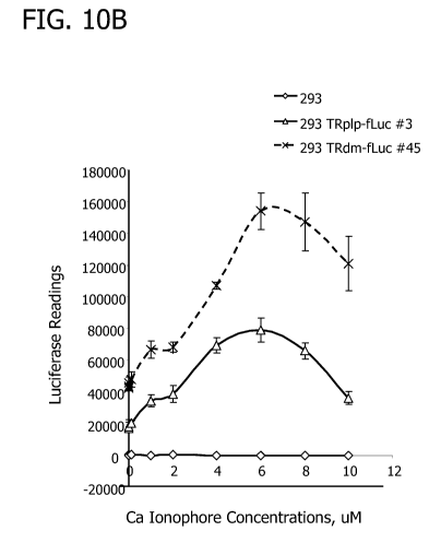
FIG. 10 shows a TR-dependent dose response that can be produced by cell lines
expressing the TRplp/dm fLuc cassettes following exposure to a toxic dose of
the calcium
ionophore A23187. FIG. 1OA shows a chart of arbitrary luminescence units that
can be
obtained by a microplate reader for HEK293, HEK293 CMV-fLuc, HEK TRpip fLuc
(subclone #3) and HEK293 TRdm fLuc (subclone #45) cells after culture in
increasing toxin
concentrations. FIG. I OB shows only the HEK293 and TRplp/dm-fLuc results to
emphasize the
dose response curve which peaks at 6 M. FIG.1OC shows a chart that expresses
arbritary
luciferase readings as the % of luciferase activity in untreated cells. This
shows the change in
cap-dependent translation that can be produced by the CMV-fLuc cells to the
increase in cap-
independent luciferase activity exhibited by the TR-ORF cells; however, the
shape of the
dose response curve is unchanged. FIG. 1OD shows the arbitrary luciferase
readings as the
ratio of the reading to CMV-fLuc cells. This comparison emphasizes the sharp
decline in
CMV-fLuc activity at high doses and reduces the apparent decline in TR-
dependent
translation at higher toxin doses.
FIG. 11 shows a TR-dependent temporal response that can be produced by cell
lines
expressing the TRplp/dm fLuc cassettes following exposure to a toxic dose of
the calcium
ionophore A23187. FIG. 11A shows a chart of arbritary luciferase readings that
can be
obtained by a microplate reader for HEK293, HEK293 CMV-fLuc, HEK TRpip fLuc
(subclone #3) and HEK293 TRdm fLuc (subclone #45) cells after culture with a
toxic dose of
the calcium ionophore A23187 as a function of increasing time. FIG. 1lB is a
chart of
HEK293 and TRplp/dm fLuc results that show the increase in luciferase activity
observed by
1.5hr post-treatment. FIG. 11C shows a chart that expresses arbitrary
luciferase readings as
the % of luciferase activity at Ohr post-treatment. FIG. l1D shows the
arbitrary luciferase
readings as the ratio to the CMV-fLuc cells.
FIG. 12 is a histogram showing the ability of rBAC TRdm EYFP virions to
transduce
HT1080 cells and exhibit TR-dependent translation in stressed and dying cells.
Cells
transduced with l Opfu/cell or 25pfu/cell rBAC virions are cultured in a toxic
concentration of
the calcium ionophore A23187 for 13.5 hours or 23 hours. Fluorescent cells are
counted
microscopically and expressed as the % of control HEK293 cells (infected but
not treated
with toxin).
FIG. 13 is a chart showing the ability of the TRpip TKsr39 cell pool to
respond to the
pro-drug ganciclovir and induce cell death. HEK293, HEK CMV-EYFP and HEK293
TRpip
TKsr39 cells are cultured in various concentrations of ganciclovir for 3 or 4
days. Cell
CA 02735575 2011-02-10
WO 2009/023517 PCT/US2008/072465
8
viability is determined by the Trypan blue exclusion assay and cell numbers
expressed as %
viable cells. In contrast to the HEK293 and HEK CMV-EYFP cultures that show no
decrease
in cell viability at any pro-drug concentration or timepoint, the TRpip-TKsr39
cells display
reduced viability after 3 days in ganciclovir supplemented medium. By 4 days,
the TRpip
TKsr39 cells show significant cell death and a dose dependent reduction in
cell viability.
FIG. 14 is a sequence comparison table of C-terminal sequences of myelin
proteolipid
proteins from a variety of vertebrates, taken from the following NCBI Genbank
numbers: (1)
P60201 Homo sapiens, (2) Q5R6E6 Pongo pygmaeus (orangutan), (3) XP_001140782
Pan
troglodytes (chimpanzee), (4) XP001088537 Macaca mulatta (rhesus monkey), (5)
Q8HXW7 Macacafascicularis (crab-eating macaque), (6) NP999139 Sus scrofa
(pig), (7)
NP035253 Mus musculus (mouse), (8) NP_112252 Rattus norvegicus (rat), (9)
XP001374483 Monodelphis domestica (opossum), (10) P47789 Oryctolagus cuniculus
(rabbit), (11) CAA08909 Bos taurus (cattle), (12) 39025 Canis familiaris
(dog), (13)
CAA43839 Gallus gallus (chicken), (14) P47790 Taeniopygia guttata (zebra
finch), (15)
AAW79015 Gekko japonicus (gecko lizard), (16) CAA79582 Xenopus laevis (frog),
and (17)
BAA84207 Latimeria chalumnae (coelacanth). Insertion mutations present in some
species
are shown double-underlined.
FIG. 15, i.e. 15A-15C, is a sequence alignment chart of murine and human
PLP/DM20 coding sequences and TR elements hereof. Key: mDM = murine DM20 cDNA;
mP = murine PLP cDNA; TRd = TRdm [SEQ ID NO:1]; TRp = TRplp [SEQ ID NO:2];
hDM = human DM20; and hP = human PLP. Because DM20 sequences omit part of the
sequence present in full-length PLP coding sequences, the numbering of DM20
seqeunces in
Figure 15 is discontinuous and, after the omitted segment, DM20 numbering is
shown
continuing below the aligned sequences. In describing sequences herein with
reference to
Figure 15, in some cases dual numbering for PLP/DM20 nucleotide positions is
utilized, e.g.,
residue 560/455; this usage refers to PLP and DM20 numbering in the
alternative, with PLP
numbering as shown above the aligned sequences, and DM20 numbering as shown
below the
aligned sequences. The last expressed codon shown is `ttc' 829/724 to 831/726.
It should be noted that the figures set forth herein are intended to exemplify
the
general characteristics of materials and methods among those of the present
technology, for
the purpose of the description of such embodiments herein. These figures may
not precisely
reflect the characteristics of any given embodiment, and are not necessarily
intended to define
or limit specific embodiments within the scope of this technology.
CA 02735575 2011-02-10
WO 2009/023517 PCT/US2008/072465
9
DETAILED DESCRIPTION
The following description of technology is merely exemplary in nature of the
subject
matter, manufacture, and use of one or more inventions, and is not intended to
limit the
scope, application, or uses of any specific invention claimed in this
application or in such
other applications as may be filed claiming priority to this application, or
patents issuing
therefrom.The following definitions and non-limiting guidelines must be
considered in
reviewing the description of the technology set forth herein.
The headings (such as "Introduction" and "Summary,") and sub-headings (such as
"Expression Cassettes") used herein are intended only for general organization
of topics
within the disclosure of the present technology, and are not intended to limit
the disclosure of
the technology or any aspect thereof. In particular, subject matter disclosed
in the
"Introduction" may include aspects of technology within the scope of one or
more inventions,
and may not constitute a recitation of prior art. Subject matter disclosed in
the "Summary" is
not an exhaustive or complete disclosure of the entire scope of the technology
or any
embodiments thereof.
The citation of references herein does not constitute an admission that those
references are prior art or have any relevance to the patentability of the
technology disclosed
herein. Any discussion of the content of references cited in the Introduction
is intended
merely to provide a general summary of assertions made by the authors of the
references, and
does not constitute an admission as to the accuracy of the content of such
references. All
references cited in the Description section of this specification are hereby
incorporated by
reference in their entirety.
The description and specific examples, while indicating embodiments of the
present
technology, are intended for purposes of illustration only and are not
intended to limit the
scope of the technology. Moreover, recitation of multiple embodiments having
stated
features is not intended to exclude other embodiments having additional
features, or other
embodiments incorporating different combinations of the stated features.
Specific Examples
are provided for illustrative purposes of how to make, use and practice the
materials and
methods of this technology and, unless explicitly stated otherwise, are not
intended to be a
representation that given embodiments of this technology have, or have not,
been made or
tested.
As used herein, the words "preferred" and "preferably" refer to embodiments of
the
technology that afford certain benefits, under certain circumstances. However,
other
CA 02735575 2011-02-10
WO 2009/023517 PCT/US2008/072465
embodiments may also be preferred, under the same or other circumstances.
Furthermore,
the recitation of one or more preferred embodiments does not imply that other
embodiments
are not useful, and is not intended to exclude other embodiments from the
scope of the
technology.
5 As used herein, the terms "comprising," "including," and "having," and their
variants,
are intended to be non-limiting, such that recitation of items in a list is
not to the exclusion of
other like items that may also be useful in the materials, compositions, and
methods of this
technology.
As used herein, the term "about," when applied to the value for a parameter of
a
10 composition or method of this technology, indicates that the calculation or
the measurement
of the value allows some slight imprecision without having a substantial
effect on the
chemical or physical attributes of the composition or method.
When introducing elements of the present technology or the preferred
embodiment(s)
thereof, the articles "a," "an," "the," and "said" are intended to mean that
there are one or
more of the elements.
The term "cytotoxic gene" refers to a nucleotide sequence which when expressed
in a
target cell induces death of the cell by lysis, apoptosis, necrosis or any
other mechanism of
cell killing.
The term "gene" refers to a nucleic acid (e.g., DNA) sequence that comprises
coding
sequences necessary for the production of a polypeptide or precursor or RNA
(e.g., tRNA,
siRNA, rRNA, etc.). The polypeptide can be encoded by a full length coding
sequence or by
any portion of the coding sequence so long as the desired activity or
functional properties
(e.g., enzymatic activity, ligand binding, signal transduction, etc.) of the
full-length or
fragment are retained. The term also encompasses the coding region of a
structural gene and
the sequences located adjacent to the coding region on both the 5' and 3'
ends, such that the
gene corresponds to the length of the full-length mRNA. The sequences that are
located 5' of
the coding region and which are present on the mRNA are referred to as 5'
untranslated
sequences. The sequences that are located 3' or downstream of the coding
region and that are
present on the mRNA are referred to as 3' untranslated sequences. The term
"gene"
encompasses both cDNA and genomic forms of a gene. A genomic form or clone of
a gene
contains the coding region, which may be interrupted with non-coding sequences
termed
"introns" or "intervening regions" or "intervening sequences." Introns are
removed or
"spliced out" from the nuclear or primary transcript, and are therefore absent
in the messenger
CA 02735575 2011-02-10
WO 2009/023517 PCT/US2008/072465
11
RNA (mRNA) transcript. The mRNA functions during translation to specify the
sequence or
order of amino acids in a nascent polypeptide.
The term "expression vector" refers to both viral and non-viral vectors
comprising a
nucleic acid expression cassette.
The term "expression cassette" is used to define a nucleotide sequence
containing
regulatory elements operably linked to a coding sequence that result in the
transcription and
translation of the coding sequence in a cell.
A "mammalian promoter" refers to a transcriptional promoter that functions in
a
mammalian cell that is derived from a mammalian cell, or both.
A "mammalian minimal promoter" refers to a 'core' DNA sequence required to
properly initiate transcription via RNA polymerase binding, but which exhibits
only token
transcriptional activity in the absence of any operably linked transcriptional
effector
sequences.
The phrase "open reading frame" or "coding sequence" refers to a nucleotide
sequence
that encodes a polypeptide or protein. The coding region is bounded in
eukaryotes, on the 5'
side by the nucleotide triplet "ATG" that encodes the initiator methionine and
on the 3' side
by one of the three triplets which specify stop codons (i.e., TAA, TAG, and
TGA).
"Operably linked" is defined to mean that the nucleic acids are placed in a
functional
relationship with another nucleic acid sequence. For example, a promoter or
enhancer is
operably linked to a coding sequence if it affects the transcription of the
sequence; or a
ribosome binding site is operably linked to a coding sequence if it is
positioned so as to
facilitate translation. Generally, "operably linked" means that the DNA
sequences being
linked are contiguous. However, enhancers do not have to be contiguous.
Linking is
accomplished by ligation at convenient restriction sites. If such sites do not
exist, the
synthetic oligonucleotide adaptors or linkers are used in accord with
conventional practice.
"Recombinant" refers to the results of methods, reagents, and laboratory
manipulations in which nucleic acids or other biological molecules are
enzymatically,
chemically or biologically cleaved, synthesized, combined, or otherwise
manipulated ex vivo
to produce desired products in cells or other biological systems. The term
"recombinant
DNA" refers to a DNA molecule that is comprised of segments of DNA joined
together by
means of molecular biology techniques.
"Transfection" is the term used to describe the introduction of foreign
material such as
foreign DNA into eukaryotic cells. It is used interchangeably with
"transformation" and
CA 02735575 2011-02-10
WO 2009/023517 PCT/US2008/072465
12
"transduction" although the latter term, in its narrower scope refers to the
process of
introducing DNA into cells by viruses, which act as carriers. Thus, the cells
that undergo
transfection are referred to as "transfected," "transformed" or "transduced"
cells.
The term "plasmid" as used herein, refers to an independently replicating
piece of
DNA. It is typically circular and double-stranded.
A "reporter gene" refers to any gene the expression of which can be detected
or
measured using conventional techniques known to those skilled in the art.
The term "regulatory element" or "effector element" refer to a transcriptional
promoter, enhancer, silencer or terminator, as well as to any translational
regulatory elements,
polyadenylation sites, and the like. Regulatory and effector elements may be
arranged so that
they allow, enhance or facilitate selective production of a mature coding
sequence that is
subject to their regulation.
The term "vector" refers to a DNA molecule into which foreign fragments of DNA
may be inserted. Generally, they contain regulatory and coding sequences of
interest. The
term vector includes but is not limited to plasmids, cosmids, phagemids, viral
vectors and
shuttle vectors.
A "shuttle" vector is a plasmid vector that is capable of prokaryotic
replication but
contains no eukaryotic replication sequences. Viral DNA sequences contained
within this
replication-deficient shuttle vector direct recombination within a eukaryotic
host cell to
produce infective viral particles.
The term "substance" as used herein refers to a matter of defined chemical
composition. It is used herein interchangeably with the term "compound."
The term "viral vector" refers to a virus which contains foreign genetic
material for
delivery into cells it infects.
A "replication-deficient" viral vector is incapable of replication in a "wild-
type" or
otherwise unmanipulated mammalian cell. Production of significant quantities
of such
viruses requires that a producer cell line be co-transfected with a helper
virus or otherwise
modified to supply or complement the missing function(s).
A "replication-competent" viral vectors is one that is capable of infecting
cells and
undergoing DNA replication, viral packaging and release from the infected
cell.
"Conditionally replicating" viral vectors as used herein are replication-
competent
vectors that are designed to be selectively expressed in particular cell types
so that undesired
broad spectrum infection is avoided. Conditional replication may be achieved
by including in
CA 02735575 2011-02-10
WO 2009/023517 PCT/US2008/072465
13
the vector tissue-specific, tumor-specific or cell type-specific or other
selectively induced
regulatory control sequences that are operably linked to early viral genes.
The terms "stress" and "toxicity" are used to refer to the disturbance of the
natural
biochemical and biophysical homeostasis of the cell. Whereas stress generally
leads to
recovery of cellular homeostasis, a toxic response eventually results in cell
death.
The translation regulated (TR) sequence (also referred to as the "TR element")
employed in the present technology is the IRES element, which can be
distinguished from the
5' UTR IRESs by (a) its nucleic acid sequence context and (b) the cellular
activity which
regulates translation (US Published Patent Application No. 2006/0173168). The
combination
of these two features forms a basis for selective translation of downstream
coding sequences
in stressed and/or dying mammalian cells that are operably linked to this IRES
sequence.
Thus, the present technology contemplates the use of any mammalian IRES as the
TR
element, which is selectively expressed in stressed and/or dying cells.
In some embodiments, the IRES element of this technology has cap-independent
translational activity which localizes within the ORF of the mammalian
Proteolipid Protein
(pip) gene. In its native context, pip IRES activity resides within a
multicistronic RNA
containing several upstream ORFs ("uORFs") which effectively block ribosome
scanning to
internal AUG codons in normal cells. However, exposure of cells to toxic
agents results in
ribosome binding and translation from specific internal RNA sequences so that
an internal
amino acid sequence is translated from the 3' end of the pip ORF. Thus, the
expression of an
appropriate coding sequence, which is regulated by the TR element, permits the
visualization,
monitoring and modulation of cell death, which finds use in numerous
applications.
Recombinant DNA molecules provided herein allow for the selective expression
of an RNA
transcript containing one or more nucleic acid sequences encoding one or more
polypeptides
in stressed or dying cells.
In some embodiments, the TR element of the present technology is derived from
exons 1-7 of the pip gene. While not being bound to a particular theory, it is
believed that the
exons 1 through 4 are sufficient to encode a functional IRES activity based on
mutational
analysis data. Furthermore, it is believed that the TR regulatory system,
which plays a role in
stress/death-specific translation is located within exons 6 and/or 7.
In contrast to the IRES element disclosed in US 2006/0173168, which is
expressed in
dying cells, a TR element of the present technology derived from PLP/DM20
differs in all of
the following features:
CA 02735575 2011-02-10
WO 2009/023517 PCT/US2008/072465
14
1) nucleotide 1 (in SEQ ID Nos. 1 and 2) was mutated from A to T to remove the
wild
type AUG start codon in the myelin proteolipid protein PLP and DM20 cDNAs that
directs
the synthesis of the full length PLP and DM20 in order to prevent such
synthesis from
occurring;
2) nucleotide 4 was mutated from G to A in order to create a stop codon in the
second
possible reading frame of the PLP and DM20 cDNAs to prevent full length
synthesis thereof;
3) nucleotides 6, 7 and 8 were mutated from C to T, T to G and T to A
respectively to
create a stop codon in the third possible reading frame of the PLP and DM20
cDNAs to
prevent synthesis of the full length PLP and DM20;
4) nucleotides 17 and 18 were mutated from G to A and T to G, respectively to
create
the first stop codon in the main (first) open reading frame of the PLP and
DM20 cDNAs to
prevent their full length synthesis;
5) nucleotide 21 was mutated from T to A in order to create the second stop
codon in
the main (first) open reading frame of the PLP and DM20 cDNAs to prevent full
length
synthesis thereof;
6) nucleotide 27 was mutated from A to T in order to remove the AUG codon from
the third possible reading frame of the PLP and DM20 cDNAs to prevent out-of
frame
translation initiation in the absence of the wild type AUG codon; and
7) the stop codon was deleted from the PLP and DM20 cDNAs to reduce
interference
with translation of the downstream open reading frame.
As a result, the TR elements of the present technology derived from PLP/DM20
do
not direct cap-dependent translation of either PIRP-M or PIRP-L. In addition
to the above
changes, the following mutations were introduced into the TR elements from the
DM 20
variant of the cDNA:
1) nucleotide 511 was mutated from A to T in order to remove the first in-
frame
internal AUG start codon in the DM20 variant that directs the synthesis of
PIRP-M protein to
prevent such synthesis from occurring; and
2) nucleotide 598 was mutated from A to T to remove the second in-frame
internal
AUG start codon in the DM20 variant that directs the synthesis of PIRP-L
protein in order to
prevent such synthesis from occurring.
Similarly, the following mutations were introduced into the TR elements from
the
PLP variant of the cDNA:
CA 02735575 2011-02-10
WO 2009/023517 PCT/US2008/072465
1) nucleotide 616 was mutated from A to T in order to remove the first in-
frame
internal AUG start codon in the PLP variant that directs the synthesis of PIRP-
M protein to
prevent such synthesis from occurring; and
2) nucleotide 703 was mutated from A to T to remove the second in-frame
internal
5 AUG start codon in the PLP variant that directs the synthesis of PIRP-L
protein in order to
prevent such synthesis from occurring.
The TR cassette of the present technology finds many uses in methods such as
detecting cell death ex vivo or in vivo; determining the cytotoxicity of a
compound in vivo or
ex vivo; in vivo diagnostics; inducing apoptosis in a cell in vivo or ex vivo;
preventing
10 apoptosis in a cell in vivo or ex vivo; and combining the imaging of cell
stress and/or death
with subsequent treatment. In addition, the present technology details the
methods for
screening for additional TR cassettes, i.e., the IRES elements which are
selectively expressed
in stressed and/or dying cells.
Expression Cassettes
15 One aspect of the present technology is directed to a nucleic acid
expression cassette
expressible in mammalian cells. The expression cassette contains the following
elements in a
5' to 3' direction: at least one transcriptional effector sequence, a TR
element encoding an
mRNA molecule that is selectively translated in stressed and/or dying cells, a
nucleotide
sequence operably linked to the TR element, and a polyadenylation sequence.
The nucleotide
sequence is a first open reading frame (ORF) sequence and encodes a
polypeptide or a
fragment thereof and is co-translated with the TR element.
In various embodiments, the TR elements of the present technology exhibit
selective
translation in stressed and/or dying cells. The term "selectively translated"
or "selective
translation" in stressed and/or dying cells means that the mRNA translation
activity is
observed in more than 95% of any cell line transformed with the TR expression
cassette at
the peak of the translation activity, e.g., within about 9 to about 18 hours
following treatment
with an acute toxic agent that induces cell stress and/or death, and that the
translational levels
of the first ORF of the inventive expression cassette rise to at least 50% of
the expression
levels of the same ORF when transcribed and translated from the same
expression cassette
lacking an operably linked TR element following treatment with the acute toxic
agent. For
example, a TR element within an expression cassette of the technology exhibits
selective
translation in stressed and/or dying cells within about 9 hours following
treatment with
calcium ionophore A23187 at a concentration of 5 M, with mRNA translation
being
CA 02735575 2011-02-10
WO 2009/023517 PCT/US2008/072465
16
observed in more than 95% of a HEK293 cell line transformed with the
expression cassette,
and translation levels of the first ORF of the expression cassette being at
least 50% of the
translation levels of the same ORF when transcribed and translated from the
same expression
cassette lacking an operably linked TR element following the treatment. In
some instances, a
TR element within an expression cassette of the technology exhibits selective
translation in
stressed and/or dying cells within about 6 to about 9 hours following
treatment with calcium
ionophore A23187 at a concentration of 5 M , with mRNA translation being
observed in
about 96, 97, 98, 99, 99.5 or 99.9% of a HEK293 cell line transformed with the
expression
cassette, and translation levels of the first ORF of the expression cassette
being about 55, 60,
65, 70, 75, 80, 85, 90, or 95% of the translation levels of the same ORF when
transcribed and
translated from the same expression cassette lacking an operably linked TR
element
following the treatment.
In some embodiments of the present invention, the TR element is a pip IRES
element,
which does not direct translation of PIRP-M or PIRP-L. In other embodiments,
the TR
element is not derived from the pip IRES.
Thus, in one embodiment, the present technology relates to a nucleic acid
expression
cassette expressible in mammalian cells, wherein the expression cassette has
the following
elements in a 5' to 3' direction: at least one transcriptional effector
sequence; a TR element
encoding a mRNA molecule which is translated in stressed and/or dying cells; a
3' sequence
flanking the TR element that contains restriction enzyme sites common in the
art; a
nucleotide sequence operably linked to the TR element, which is a first open
reading frame
(ORF) sequence and encodes a polypeptide or a fragment thereof and is co-
translated with the
TR element; and a polyadenylation sequence.
In a preferred embodiment, a TR element is selected from a human or a mouse TR
element. More preferably, the TR element is selected from murine sequences
TRdm (SEQ ID
NO: 1) and TRpip (SEQ ID NO: 2).
TRdm nucleic sequence (SEQ ID NO: 1) was derived from the DM20 splice variant
cDNA of the mouse proteolipid protein gene 1, but has been modified at
nucleotide positions
1, 4, 6, 7, 8, 17, 18, 21, 27, 511, and 598. In addition, the last 3
nucleotides encoding the stop
codon were removed.
TRpip nucleic sequence (SEQ ID NO: 2) was derived from the PLP splice variant
cDNA of the mouse proteolipid protein gene 1, and it contains modifications at
nucleotide
CA 02735575 2011-02-10
WO 2009/023517 PCT/US2008/072465
17
positions 1, 4, 6, 7, 8, 17, 18, 21, 27,616, and 703. TRpip differs from TRdm
by the presence of
nucleotides 349-453. The last 3 nucleotides encoding the stop codon were
removed.
In addition to the TR element, the expression cassettes of the present
technology
comprise an upstream transcriptional effector sequence which regulates gene
expression. In
one embodiment, the transcriptional effector sequence is a mammalian promoter.
In addition,
the transcriptional effector can also include additional promoter sequences
and/or
transcriptional regulators, such as enhancer and silencers or combinations
thereof. These
transcriptional effector sequences can include portions known to bind to
cellular components
which regulate the transcription of any operably linked coding sequence. For
example, an
enhancer or silencer sequence can include sequences that bind known cellular
components,
such as transcriptional regulatory proteins. The transcriptional effector
sequence can be
selected from any suitable nucleic acid, such as genomic DNA, plasmid DNA,
viral DNA,
mRNA or cDNA, or any suitable organism (e.g., a virus, bacterium, yeast,
fungus, plant,
insect or mammal). It is within the skill of the art to select appropriate
transcriptional
effector sequences based upon the transcription and/or translation system
being utilized. Any
individual regulatory sequence can be arranged within the transcriptional
effector element in
a wild-type arrangement (as present in the native genomic order), or in an
artificial
arrangement. For example, a modified enhancer or promoter sequence may include
repeating
units of a regulatory sequence so that transcriptional activity from the
vector is modified by
these changes.
In one embodiment, the promoters are selected from constitutive, inducible,
tissue
specific, tumor specific and response gene promoters. Constitutive promoters
can be
selected, e.g., from Rous sarcoma virus (RSV) long terminal repeat (LTR)
promoter,
cytomegalovirus immediate early gene (CMV) promoter, simian virus 40 early
(SV40E)
promoter, cytoplasmic beta-actin promoter, adenovirus major late promoter, and
the
phosphoglycerol kinase (PGK) promoter. In a preferred embodiment, a
constitutive promoter
is a CMV promoter. In another preferred embodiment, a constitutive promoter is
an SV40E
promoter.
Selection of a promoter that is regulated in response to specific physiologic
or
synthetic signals can permit inducible transcription of the gene product. For
example, in the
case where expression of a transgene is toxic to the cells in which the vector
is produced, it
may be desirable to prevent or reduce transcription of the transgene. By way
of example, a
proapoptotic transgene can be toxic to the cell in which it is produced. Thus,
several
CA 02735575 2011-02-10
WO 2009/023517 PCT/US2008/072465
18
inducible promoter systems are available for production of vectors, which
contain a transgene
encoding a toxic protein.
Non-limiting examples of inducible promoters include the metal-regulated human
metallothionine (hMT-IIA) promoter, the zinc-inducible human Zinc Transporter
1 (hZnT-1)
promoter, dexamethasone (Dex)-inducible promoter, mouse mammary tumor virus
(MMTV)
promoter, ecdysone-responsive insect promoter, tetracycline responsive Tet-On
and Tet-
Off systems, RU486-inducible promoter and rapamycin-responsive promoter. In
one
embodiment, the inducible promoter is a metallothionine hMT-IIA promoter. In
another
embodiment, the inducible promoter is a zinc-inducible hZnT-1 promoter.
Methods and compositions are provided for the controlled induction of gene
expression in a mammalian host cell. For example, DNA sequences which comprise
the
human metallothionein II (hMT-IIA) and zinc-inducible Zinc Transporter 1 (hZnT-
1)
transcriptional regulatory systems are induced by elevated concentrations of
heavy metal ions
and glucocorticoids. These inducible promoters are composed of multiple metal-
regulatory
elements (e.g., MRE) adjacent to a basal level transcriptional regulator.
In other indications, it may be desirable to activate transcription using
promoters
responsive to hormones or antibiotics. The ecdysone system (Invitrogen,
Carlsbad, Calif.)
consists of a tightly regulated expression mechanism that prevents basal level
transgene
expression, but allows for an over 200-fold induction of transcription. The
system is based
on the heterodimeric ecdysone receptor of Drosophila, and when ecdysone or an
analog
thereof (such as muristerone A) binds to the receptor, the receptor activates
a promoter to turn
on expression of the downstream transgene. In this system, both monomers of
the
heterodimeric receptor are constitutively expressed from one vector, whereas
the ecdysone-
responsive promoter which drives the expression of transgene is on a second
plasmid. Thus,
cotransfection of the two plasmids containing the regulated transgene and the
receptor
monomers into a reporter cell allows for the inducible expression of even
toxic transgenes.
The Tet-Off or Tet-On system (Clontech, Palo Alto, Calif.) originally
developed by
Gossen and Bujard (Gossen and Bujard, 1992; Gossen et al., 1995) utilize
tetracycline or
tetracycline derivatives, such as doxycycline, to regulate transgene
expression. In the Tet-
Ong system, gene expression is induced by tetracycline or doxycycline, whereas
in the Tet-
Off system, antibiotic exposure eliminates gene expression. These systems are
based on
two regulatory elements derived from the tetracycline resistance operon of E.
coli, namely the
tetracycline operator DNA sequence and the tetracycline repressor protein. A
Tet-regulated
CA 02735575 2011-02-10
WO 2009/023517 PCT/US2008/072465
19
plasmid contains a minimal promoter with tetracycline-responsive operator
elements. A
second plasmid contains the tetracycline-controlled transactivator protein,
which is a fusion
protein comprised of the VP 16 transcriptional activator domain and the wild-
type tetracycline
repressor protein of the Tet-Off system. In the Tet-On system, the
tetracycline repressor
protein has been altered so that transcription is activated by the presence of
tetracycline or
doxycycline.
Tissue specific promoters can be selected, e.g., from the transferrin (TF),
tyrosinase
(TYR), albumin (ALB), muscle creatine kinase (CKM), myelin basic protein
(MBP), glial
fibrillary acidic protein (GFAP), neuron-specific enolase (NSE), and synapsin
I (SYN1)
promoters. In one embodiment, the tissue specific promoter is a synapsin I
(SYN1)
promoter. In another embodiment, the tissue specific promoter is the ALB
promoter.
Tumor specific promoters include but are not limited to promoters for vascular
endothelial growth factor (VEGF), a VEGF receptor (i.e. KDR, E-selectin, or
endoglin),
alpha-fetoprotein (AFP), carcinoembryonic antigen (CEA), erbB2 (v-erb-b2
erythroblastic
leukemia viral oncogene homolog 2), osteocalcin (bone gamma-carboxyglutamate
protein,
BGLAP), SLP1 (secretory leukoproteinase inhibitor or antileukoproteinase 1),
hypoxia-
response element (HRE), L-plastin (lymphocyte cytosolic protein 1) and
hexokinase II
(HK2). In one embodiment, the tumor specific promoter is an alpha fetoprotein
(AFP)
promoter. In another embodiment, the tumor specific promoter is a SLP1
promoter.
Response gene promoters, which stimulate transcription preferentially or
uniquely
under certain cellular states and/or in response to external chemical or
environmental stimuli
(i.e., heat or cold shock), can be selected, e.g., from promoters for early
growth response gene
1 (EGR1/ZIF268), tissue-type plasminogen activator (t-PA), multidrug-
resistance protein 1
(mdr-1), HSPA5/Grp78/BIP (heat shock 70kDa protein 5), c-fos (v-fos FBJ murine
osteosarcoma viral oncogene homolog), c-jun (v-jun sarcoma virus 17 oncogene
homolog) or
from cell cycle-regulated genes such as, but not limited to, E2F-1 (E2F
transcription factor
1), cyclin Al (CCNA1) and CDC25C (cell division cycle 25C). In a preferred
embodiment,
the response gene promoter is a promoter for HSPA5. In another preferred
embodiment, the
response gene promoter is an EGR1 promoter.
In some embodiments, a specific transcriptional effector element is isolated
and then
operatively linked to a minimal promoter to produce an expression cassette
whose
transcriptional activity is dependent upon a single or limited type of
cellular response (e.g., a
heat shock response or metal-regulated element).
CA 02735575 2011-02-10
WO 2009/023517 PCT/US2008/072465
The expression cassette can include species-specific transcriptional
regulatory
sequences. Such DNA regulatory sequences can be selected on the basis of the
cell type into
which the expression cassette will be inserted and can be isolated from
prokaryotic or
eukaryotic cells, including but not limited to bacteria, yeast, plant, insect,
mammalian cells or
5 from viruses. In such example, a mammalian promoter would be selected to
express a nucleic
acid of choice in a mammalian cell.
The TR expression cassettes of the present technology enable selective gene
expression in stressed and dying cells, allowing for a heterologous ORF to be
inserted 3' to
the TR sequence and 5' of a polyadenylation signal. The heterologous gene can
be either a
10 full genomic sequence (e.g., including introns), synthetic nucleic acid or
a cDNA copy of a
gene of interest, which encodes a protein or a polypeptide of interest,
wherein the polypeptide
includes biologically active ("bioactive") protein fragments. In a preferred
embodiment,
cDNA sequences are used for the purposes of the present technology due to the
reduction in
genomic complexity provided by removal of mRNA splice sites.
15 Thus, in one embodiment, a first ORF sequence is selected from the group of
reporter
genes, cytotoxic tumor suppressor genes, toxin genes, prodrug activating genes
and
proapoptotic genes.
In various embodiments, the first ORF sequence is a reporter gene. As the name
implies, a reporter gene does not confer any selective advantage on the cell
into which it is
20 introduced. Rather, a reporter gene encodes a product that confers on the
cell a detectable
biochemical or visually observable (e.g., fluorescent) phenotype. The reporter
polypeptide
can also include a fused or hybrid polypeptide in which another polypeptide is
fused at the N-
terminus or the C-terminus of the polypeptide or fragment thereof. A fused
polypeptide is
produced by cloning a nucleic acid sequence (or a portion thereof) encoding
one polypeptide
in-frame with a nucleic acid sequence (or a portion thereof) encoding another
polypeptide.
Techniques for producing fusion polypeptides are known in the art, and
include, ligating the
coding sequences encoding the polypeptides so that they are in-frame and
translation of the
fused polypeptide is under the control of the TR cassette. For example,
cloning the pip ORF
in-frame with the enhanced green fluorescent protein (EGFP) ORF produced a
fusion protein
that was used to monitor the expression, subcellular localization and
biological effect of the
fusion protein in cultured cells (Ghandour S et al. Glia (2002) 40(3):300-1 1;
Boucher S et al.
J Neurosci (2002) 22(5): 1772-83).
CA 02735575 2011-02-10
WO 2009/023517 PCT/US2008/072465
21
One commonly used class of reporter genes encodes an enzyme or other
biochemical
marker, which, when expressed in a mammalian cell, cause a visible change in
the cell or the
cell environment. Such a change can be observed directly, can involve the
addition of an
appropriate substrate that is converted into a detectable product or the
addition and binding of
a metabolic tracer. Examples of these reporter genes are the bacterial lacZ
gene which
encodes the (3-galactosidase ((3-gal) enzyme, the Chloramphenicol
acetyltransferase (CAT)
enzyme, Firefly luciferase (Coleoptera beetle), Renilla luciferase (sea
pansy), Herpes
Simplex 1 thymidine kinase (HSV1-TK) and the mutant Herpes Simplex 1 thymidine
kinase
(HSV1-sr39tk) genes. In the case of (3-gal, incubation of expressing cells
with halogen-
derivatized galactose results in a colored or fluorescent product that can be
detected and
quantitated histochemically or fluorimetrically. In the case of CAT, a cell
lysate is incubated
with radiolabeled chloramphenicol or another acetyl donor molecule such as
acetyl-CoA, and
the acetylated chloramphenicol product is assayed chromatographically. Other
useful reporter
genes encode proteins that are naturally fluorescent, including the (green
fluorescent protein
(GFP), enhanced yellow fluorescent protein (EYFP), or monomeric red
fluorescent protein
(mRFP1).
As can be seen from above, exemplary reporter genes can be selected from GFP,
EYFP, mRFP1, (3-Gal, and CAT, but any other reporter gene known in the art can
be used.
See, e.g., the http World Wide Web
olympusconfocal.com/applications/fpcolorpalette.html
site. In a preferred embodiment, the reporter gene is Firefly Luciferase. In
another preferred
embodiment, the reporter gene is Renilla Luciferase.
The first ORF sequence can also encode a cytotoxic tumor suppressor gene that
encodes a polypeptide capable of suppressing the neoplastic phenotype and/or
inducing
apoptosis. Examples of tumor suppressor genes useful in the practice of the
present
technology include the p53, adenomatous polyposis coli (APC), Breast Cancer-1
(BRCA-1),
BRCA-2, Wilm's Tumor (WT-1), retinoblastoma gene (Rb), Neurofibromatosis-1 (NF-
1),
NF-2 and von Hippel-Lindau (VHL) genes. In a preferred embodiment, the
cytotoxic tumor
suppressor gene is the p53 gene.
In another embodiment, the first ORF sequence encodes a "toxin gene" that
binds to
cellular receptor proteins and after uptake interferes with protein synthesis
by blocking
ribosome assembly or function. Examples of toxin genes include proteins such
as
Pseudomonas exotoxin (e.g., Exotoxin A or "ETA"), ricin toxin, diphtheria
toxin, and the
like. In a preferred embodiment, the toxin gene is the diphtheria toxin gene.
CA 02735575 2011-02-10
WO 2009/023517 PCT/US2008/072465
22
In another embodiment, the first ORF sequence is a prodrug activating gene
(e.g.,
drug-susceptibility or suicide gene), which codes for a protein that converts
a prodrug, which
lacks a therapeutic effect into a drug which renders a cell expressing said
gene susceptible to
death following exposure to said prodrug. Examples of pro-drug genes include
the thymidine
kinase of Herpes Simplex Virus (HSV-tk), cytochrome P450, human deoxycytidine
kinase
and the bacterial enzymes cytosine deaminase and guanine phosphoribosyl
transferase (gpt)
genes. Cells which express these genes are rendered sensitive to the prodrugs
ganciclovir
(HSV-tk), cyclophosphamide (cytochrome P450), cytosine arabinoside
(deoxycytidine
kinase), 5-fluorocytosine (bacterial cytosonine deaminase) or thioxanthine
(gpt). In a
preferred embodiment, the prodrug activating gene is the HSV-tk gene which can
also
provide an important therapeutic advantage. During TK catalysis of the
antiviral guanosine
analogue ganciclovir, apoptotic molecules are released that kill surrounding
cells by a process
termed "bystander" killing. Although a limited number of target cells may
initially express
the HSV-tk gene, this localized cytocidal effect provides a therapeutic effect
to adjacent non-
expressing, undesired bystander cells.
In embodiments in which the first ORF sequence is a proapoptotic gene, such a
sequence causes programmed cell death or apoptosis of an expressing cell.
Examples of pro-
apoptotic genes include p53, the Apoptosis Stimulating Proteins of p53 (e.g.
ASPP 1, ASPP2,
and ASPP3), the Bcl-2 homologs Bax and Bc12-L-10 (Diva) , the Apoptosis-
Inducing Factor
(AIF), Fas, initiator caspases such as caspase-8 and caspase-9 or an effector
caspase such as
caspase-3. In a preferred embodiment, the proapoptotic gene is the caspase-3
gene.
In another embodiment, the first ORF sequence encodes a recombinant
intracellular
antibody ("intrabody") comprising an Fab or single chain Fv (scFv) molecule
but does not
encode an operable secretory sequence and hence are restricted to
intracellular compartments
where they bind, neutralize or modify the activity of a target antigen. This
interaction may
result in the direct inhibition of target antigen function, restoration of a
mutant deficient
activity, interference with the intracellular trafficking of the antigen or
restriction of the
folding of a pathological mutant protein. To exert their function, recombinant
intrabodies are
directed to a subcellular compartment where the antigen is located. This can
be achieved by
incorporating signal sequences routinely fused to the N- or C-terminus. For
example, the
KDEL peptide sequence allows the retention of recombinant antibodies within
the
endoplasmic reticulum and hence, can be used to block the processing of cell
surface targeted
proteins. Other signal sequences can be incorporated into the intrabody ORF
and produce
CA 02735575 2011-02-10
WO 2009/023517 PCT/US2008/072465
23
nuclear localization, ER or Golgi routing, nucleolar localization, as well as
transport to the
endosomal or liposomal compartments.
Methods for the production of single chain antibodies are well known to those
with
skill in the art. By way of example, the skilled artisan is referred to U.S.
Patent No. 5,359,046
for such methods. A single chain antibody ("scFv" or "SCA") is composed of an
antibody
variable light-chain amino acid sequence (VL) tethered to a variable heavy-
chain sequence
(VI-1) by a designed peptide that links the carboxyl term:i_in us of the VE.
sequence to the amino
Eermninus of the VH sequence, thereby reconstituting an antigen binding site
on a single
molecule. SCAs have the binding specificity and affinity of monoclonal
antibodies and, in
their native form, are about one-fifth to one-sixth of the size of a
monoclonal antibody. In
addition to these benefits, fully-human intrabodies can be isolated directly
from human
libraries (such as the human single-fold, single-chain variable fragment
(scFv) libraries)
without the need for costly and time consuming "humanization" procedures.
Almost any kind of biologic molecule can serve as an intrabody target antigen,
for
example, intermediate metabolites, sugars, lipids, and hormones as well as
macromolecules
such as complex carbohydrates, phospholipids, nucleic acids and proteins. The
preferred
target molecule is an endogenous protein. Intrabodies have been developed for
a number of
target proteins involved in cancer, infectious disease, transplantation,
neurodegenerative
disease and other diseases associated with protein overexpression or
mutagenesis. Specific
examples of intrabody target proteins include erbB-2 (androgen receptor), IL-2
receptor,
epidermal growth factor receptor, vascular endothelial growth factor receptor
2, the folate
receptor, HIV gp120 protein, CCR5, CXCR4, alphaV integrin, metalloproteinase
MMP-2 and
MMP-9, the Re1A subunit of NF-kappaB, the prion-like protein PrP, the
huntingtin protein
and the beta-amyloid precursor protein. In one embodiment, the intrabody is
directed to the
Re1A subunit of NF-kappaB.
In embodiments in which the first ORF encodes a secreted antibody fusion
protein,
such proteins can induce apoptosis or an enhanced immune response in targeted
cells.
Examples include any antibody fusion protein that delivers a therapeutic
response such as the
human interleukin-2-truncated diphtheria toxin, anti-CD22 dsFv-truncated
Pseudomonas
exotoxin, anti-CD25 scFv-truncated Pseudomonas exotoxin and the anti-B4-
blocked ricin
(anti-CD19) immunotoxin proteins. In one preferred embodiment, the antibody
fusion
protein is the anti-B4-blocked ricin immunotoxin protein.
CA 02735575 2011-02-10
WO 2009/023517 PCT/US2008/072465
24
Sequence Variants
In certain instances, sequence elements operably linked to the TR sequences
might
disrupt the selective translational activity displayed by the TR expression
cassette or exhibit
sub-optimal translational activity. To alleviate any effect on TR activity by
the linked ORF,
the present technology provides for codon-usage variants of the disclosed
nucleotide
sequences, that employ alternate codons which do not alter the polypeptide
sequence (and
thereby do not affect the biological activity) of the ORF products. These
variants are based
on the degeneracy of the genetic code, whereby several amino acids are encoded
by more
than one codon triplet. An example would be the codons CGT, CGG, CGC, and CGA,
which
all encode the amino acid, arginine (R). Thus, a protein can be encoded by a
variant nucleic
acid sequence that differs in its precise sequence, but still encodes a
polypeptide with an
identical amino acid sequence. Based on codon utilization/preference, codons
can be selected
to optimize the translation efficiency of an ORF without affecting regulated
translation from
the TR expression cassette.
Site directed mutagenesis is one particularly useful method for producing
sequence
variants by altering a nucleotide sequence at one of more desired positions.
Site directed (or
site specific) mutagenesis uses oligonucleotide sequences comprising a DNA
sequence with
the desired mutation, as well as a sufficient number of adjacent nucleotides
to provide a
sequence of sufficient size and complexity to form a stable duplex on both
sides of the
proposed mutation. Typically, a synthetic primer of about 20 to 25 nucleotides
in length is
preferred, with about 5 to 10 residues on both sides of the proposed mutation
of the sequence
being altered. Typical vectors useful in site directed mutagenesis include the
disclosed
vectors, as well as any commercially or academically available plasmid vector.
In general,
nucleotide substitutions are introduced by annealing the appropriate DNA
oligonucleotide
sequence with the target DNA and amplifying the target sequence by PCR
procedures known
in the art. The present technology contemplates the use of every possible
codon in a coding
sequence for producing the desired ORF sequence for use in accordance with
this invention.
Directed evolution techniques can be used to prepare sequence variants having
improved TR function. In a directed evolution technique, at least one round of
nucleic acid
mutation or nucleic acid splicing or homologous recombination can be
performed, starting
from a TR-containing polynucleotide. Mutation, splicing, and homologous
recombination
can be performed in a directed or random manner. For example, one or more
oligonucleotides can be designed for site-directed mutagenesis of the TR
element, as
CA 02735575 2011-02-10
WO 2009/023517 PCT/US2008/072465
described above, or one or more randomly generated oligonucleotides can be
contacted with
the initial TR-containing polynucleotide template. Alternatively, or in
addition, PCR
amplification of the initial template can be performed under error-permissive
conditions
and/or an error-prone polymerase to permit introduction of mutations, a
technique referred to
5 a "sloppy" PCR.
Similarly, a set of homologous, TR-element-containing polynucleotides can be
spliced or recombined in a directed or random manner. For example, one or more
restriction
endonucleases can be used to digest the homologous polynucleotide templates,
randomly or
in a predetermined manner, and the resulting fragments can then be ligated
together.
10 Alternatively or in addition, the set of TR-element-containing
polynucleotides can be pooled
and treated under conditions favoring homologous recombination among them,
either in vitro
or in cyto. A combination of mutation and splicing or recombination techniques
can be
employed. One or more than one rounds of any of these can be performed.
After one or more rounds of mutation, splicing, and/or recombination, the
resulting
15 polynucleotides are then tested to screen them for TR activity. Typically,
this can be done by
placing a reporter molecule coding sequence under the operative control of one
or more of
the TR variants that have been produced. The resulting construct(s) are then
expressed in a
cell that is placed under conditions, such as a condition of stress, for which
TR translation can
take place. The testing can be used to detect a desired improvement in TR
element function.
20 For example, any one of improvement in specificity of TR element
translation to a stress
condition, sensitivity of TR element activation to a cellular stress response
(e.g., a
biochemical change antecent to cell stress and/or death), or efficiency (i.e.
magnitude) of
translation initiation upon TR element activation can be the focus of the
assay.
Based on the assay result, one or more improved TR elements can be selected
for use,
25 or for further development; in some embodiments, the selected improved TR
element nucleic
acids can be used as a starting polynucleotide or as a starting set of
polynucleotides for
another round, or course of rounds, of directed evolution.
In various embodiments herein, a TR element can comprise, or can be made by
mutation of a PLP/DM20 polynucleotide comprising bases of, or corresponding
to, bases
from about 27 to about 615/5 10 of a murine or human PLP/DM20 DNA sequences of
Figure
15; and this can comprise further bases of, or corresponding to, bases from
about 616/511 to
about 702/597, bases from about 703/598 to about 772/666, and/or bases from
about 773/667
to about 810/705. For example, a TR element can comprise, or can be made by
mutation of a
CA 02735575 2011-02-10
WO 2009/023517 PCT/US2008/072465
26
PLP/DM20 polynucleotide comprising bases of, or corresponding to, bases from
about 27 to
about 810/705, with or without omission of bases from about 616/511 to about
702/597,
numbered with reference to Figure 15.
In PLP/DM20 coding sequences, and TR elements thereof or constructed
therefrom,
mutations can be made, without adverse effect on TR-element function, at one
or more
positions corresponding to the following PLP/DM20 positions stated with
reference to Figure
15, i.e. positions: 01, 02, 03, 04 to 21 (including deletion of all of part of
this segment), 25,
26, 314, 332, 560/455, 614/509, 622/518 to 696/591 (including deletion of all
or part of this
segment, which removes exon 5), 616/511, 703/598, 806/701, 811/706, 817/712,
818/713,
and 827/722. In various embodiments, other nucleobases than the foregoing can
be
conserved in PLP/DM20 coding sequences.
For example, in various embodiments, a nucleobase sequence of a PLP/DM20
coding
sequence hereof can comprise polypyrimidine motifs at nucleotide positions
corresponding to
PLP nucleotide positions 41-48, 50-56, 75-81, 150-156, 200-205, 227-244, 251-
257, and 563-
570. In some embodiments, such a sequence can further comprise polypyrimidine
motifs at
one or more of PLP positions 270-274, 299-303, 490-494, 578-582, 597-601; and
in some
embodiments, also at one or more of PLP positions 626-632, 642-648, 669-674,
707-712,
755-761, 767-771, and 800-804.
Similarly, in various embodiments, a nucleobase sequence of a PLP/DM20 coding
sequence hereof can comprise GNRA motifs at nucleotide positions corresponding
to PLP
nucleotide positions: 130-133, 142-145, 190-193, 220-223, 305-308; and in some
embodiments further at 635-638; and in other embodiments further at one or
more of
positions 329-332, 343-346, and 572-575; and in some, still further at one or
more of
positions 650-653 and 683-686.
However, as mentioned, mutation of the following positions can be undertaken
with
no adverse effect, and in some cases with an enhancing effect: 01, 04, 06, 07,
08, 17, 18, 21,
27. In some embodiments, these mutations can be one or more of. Olt, 04a, 06t,
07g, 08a,
17a, 18g, 21a, and 27t. Other positions that can be mutated with no adverse
effect on
function include mutations at one or more of PLP positions: 25, 26, 314, 332,
560/455,
616/511, 703/598, 806/701, 811/706, 817/712, 818/713, and 827/722. In some
embodiments,
these can be one or more of. 25g, 26c, 314g, 332g, 560/455c, 616/511t,
703/598t, 806/701g,
811/706t, 817/712a, 818/713a, and 827/722g. In addition, insertions, e.g.,
insertions of up to
or about 5 nucleotides, can be made at PLP position 614/509, with no adverse
effect on
CA 02735575 2011-02-10
WO 2009/023517 PCT/US2008/072465
27
function. In addition, fusions to position 831/726, e.g., in-frame fusions
thereto of reporter or
other target gene coding sequences, do not exhibit any adverse effect on TR
element function.
In various embodiments, a PLP or DM20 sequences useful herein can be a
vertebrate
sequence; in some embodiments, this can be a human, primate, rodent, equine,
bovine, ovine,
porcine, canine, feline, lapine, marsupial, avian, piscine, amphibian, or
reptilian sequence. In
various embodiments, a vertebrate sequence can be a native sequence, whether
wild-type or
variant; in some embodiments, a vertebrate sequence can be a wild-type
sequence.
As used herein in regard to PLP/DM20 sequences, "vertebrate consensus
sequence"
refers to the DNA sequence:
1 atgggyykgy wdgakkgytg yrynmgmtgy mtbrtwgggg ymccmttygc ytchbtsrtb
61 gccacwgkvy tvtgyttyky tggrgtsgcv ctvttctgyg gmtgyggrca ygargchytv
121 asygghacmg armagytvat ygagacmtay ttytccaara aytaccaaga mtaygartay
181 ctcatyvayg tsatymaygc yttycagtay gtcatctatg gaaywgccwy yttcttctty
241 cthtwyggrr ycctvctkyt ggcygarggm ttctacacca cmrsygchrt cargcavatc
301 ythggsgast wcmrrmccmc mryywkmrrs rrkggsctga kykcwacrgt racwggrggm
361 cmkaarggga grrghdcsmg rggmmvvcak cvagyycayw cywtrsagck srtstgtcrb
421 tgyttgggaa artggctmgg acayccygay aagtttgtsg gyrtyacyta tryyhtsacy
481 rtyktvtggm tmctrrystt ygcctgctcd gcygtdccyg tvtacatyta yttyaayacc
541 tggrycacyt gycagtctat ygcckyccch rssaagacyw cwrccagyrt mrgyasbcts
601 tgykcdgayg symgvatgta yggtgtycts ccmtggaayg cbttycchgg saargtktgy
661 ggswccarcc tkctbkccat ctgcaaracm rsygagttcc aratgacntt ycayctbttt
721 atygckgcvt tygtgggkgc wgcngchacw ctdgtbkcmc tgctcacytw yatgrthgsy
781 gcmwcwtwca actwygcygt sctbmrastb aykggccgrr gcwcmaagtt ytga
and to DNA complements thereof, to RNA sequences corresponding to any of the
foregoing,
to nucleic acid analogs having a nucleobase sequence corresponding to any of
these, and to
amino acid sequences encoded thereby.
As used herein in regard to PLP/DM20 amino acid and nucleotide sequences,
"vertebrate specific sequence" refers to the PLP or DM20 sequences of the
species listed in
Figure 14, i.e. Homo sapiens, Pongo pygmaeus (orangutan), Pan troglodytes
(chimpanzee),
Macaca mulatta (rhesus monkey), Macaca fascicularis (crab-eating macaque), Sus
scrofa
(pig), Mus musculus (mouse), Rattus norvegicus (rat), Monodelphis domestica
(opossum),
Oryctolagus cuniculus (rabbit), Bos taurus (cattle), Canis familiaris (dog),
Gallus gallus
(chicken), Taeniopygia guttata (zebra finch), Gekkojaponicus (gecko lizard),
Xenopus laevis
(frog), and Latimeria chalumnae (coelacanth). In some embodiments, the
vertebrate specific
sequence can comprise, or encode, any one of the amino acid sequences having
Genbank
numbers: P60201 (human), Q5R6E6 (orangutan), XP001140782 (chimpanzee),
XP001088537 (rhesus monkey), Q8HXW7 (crab-eating macaque), NP_999139 (pig),
NP_035253 (mouse), NP_112252 (rat), XP_001374483 (opossum), P47789 (rabbit),
CA 02735575 2011-02-10
WO 2009/023517 PCT/US2008/072465
28
CAA08909 (cattle), 39025 (dog), CAA43839 (chicken), P47790 (zebra finch),
AAW79015
(gecko lizard), CAA79582 (frog), or BAA84207 (coelacanth).
DNA sequences encoding these are readily available to one of ordinary skill in
the art
by searching NCBI Genbank in the Nucleotide menu at the http World Wide Web
ncbi.nlm.nih.gov/sites/entrez website. For example, useful DNA sequences
include those
listed under Genbank accession numbers: AJO06976 (human), CR860432
(orangutan),
XM001140782 (chimpanzee), XM_001088537 (rhesus monkey), ABO83324 (crab-eating
macaque), NM_213974 (pig), NM_011123 (mouse), NM_030990 (rat), XM_001374446
(opossum), NM_001082328 (rabbit), AJO09913 (cattle), X55317 (dog), X61661
(chicken),
NM001076703 (residues 113-946, zebra finch), AY880400 (gecko lizard), Z19522
(frog),
and AB025938(coelacanth).
In various embodiments, a PLP or DM20 sequence useful herein can be a
mammalian
sequence; in some embodiments, this can be a human, primate, rodent, equine,
bovine, ovine,
porcine, canine, feline, lapine, or marsupial sequence.
As used herein in regard to PLP/DM20 sequences, "mammalian consensus sequence"
refers to the DNA sequence:
1 atgggcytgt tagagtgytg ygcnagatgy ctsgtagggg ccccctttgc ttccytggtg
61 gccactggat trtgtttctt tggrgtggca ctsttctgtg gmtgtggaca tgaagchytm
121 actggyacag aaaagytaat tgagacmtat ttctccaaaa aytaccaaga ctaygagtat
181 ctcatyaatg tgatycatgc yttccagtat gtcatctatg gaactgcctc tttcttcttc
241 ctttatgggg ccctcctgct ggcygagggc ttctacacca ccggygcwgt caggcagatc
301 tttggcgact acaagaccac catctgcggs aagggcctga gygcaacggt aacagggggc
361 cagaagggga ggggttccag aggccaacat caagctcatt ctttggagcg ggtgtgtcat
421 tgtttgggaa aatggctagg acatcccgac aagtttgtgg gcatcaccta tgccytgacy
481 gttgtrtggc tcctrgtgtt tgcctgctck gctgtrcctg tgtacattta yttcaayacc
541 tggaccacyt gycagtctat tgcckycccy agcaagacyt ctgccagyat aggcastctc
601 tgygctgatg ccagaatgta tggtgttctc ccatggaatg ctttyccwgg caargtktgt
661 ggctccaacc ttctgtccat ctgcaaaaca gctgagttcc aaatgacstt ccayctgttt
721 attgctgcvt tygtgggkgc tgcrgcyaca ctrgtktccc tgctcacctt catgattgct
781 gccacttaca acttygccgt cctkaaactc atgggccgag gcaccaagtt ctga
and to DNA complements thereof, to RNA sequences corresponding to any of the
foregoing,
to nucleic acid analogs having a nucleobase sequence of any of these, and to
amino acid
sequences encoded thereby.
As used herein in regard to PLP/DM20 amino acid and nucleotide sequences,
"mammalian specific sequence" refers to the PLP or DM20 sequences of the
mammalian
species listed in Figure 14, i.e. Homo sapiens, Pongo pygmaeus (orangutan),
Pan troglodytes
(chimpanzee), Macaca mulatta (rhesus monkey), Macacafascicularis (crab-eating
macaque),
Sus scrofa (pig), Mus musculus (mouse), Rattus norvegicus (rat), Monodelphis
domestica
CA 02735575 2011-02-10
WO 2009/023517 PCT/US2008/072465
29
(opossum), Oryctolagus cuniculus (rabbit), Bos taurus (cattle), or
Canisfamiliaris (dog). In
some embodiments, the mammalian specific sequence can comprise, or encode, any
one of
the amino acid sequences having Genbank numbers: P60201 (human), Q5R6E6
(orangutan),
XP001140782 (chimpanzee), XP001088537 (rhesus monkey), Q8HXW7 (crab-eating
macaque), NP_999139 (pig), NP_035253 (mouse), NP_112252 (rat), XP_001374483
(opossum), P47789 (rabbit), CAA08909 (cattle), or 39025 (dog). TR elements
comprising
such TR polynucleotides are useful herein, as are expression cassettes
comprising those TR
elements.
In various embodiments, a TR element can have a PLP or DM20 nucleotide
sequence
that is at least 62% identical to a Fig. 15 PLP sequence or a Fig. 15 DM20
sequence,
respectively. The sequence identicality can be at least or about 65, 70, 75,
80, 85, 90, or 95%
thereto. In some embodiments, the sequence can be 97, 98, 99% or more
identical thereto.
Such a non-identical sequence will retain operative features a PLP or DM20 TR
element, i.e.
the defined polypyrimidine tracts, GNRA motifs, and 19S rRNA binding site
thereof.
In various embodiments, TR elements hereof can be used to identify agents that
induce, enhance, or inhibit a cellular stress response, e.g., a reponse to
heat stress, cold stress,
oxidation stress, tonic stress, toxication, or a combination thereof. The
cellular stress
response can comprises apoptosis and/or necrosis. In some embodiments, a
process for
identifying such agents can involve identifying the degree to which an agent
induces,
enhances, inhibits, or reverses a cellular stress response; this can be
accomplished, e.g., by
identifying a degree of cellular stress response that is proportional to the
magnitude of a
signal detected from a reporter molecule expressed under the control of the TR
element.
In various embodiments, TR elements hereof can be used in processes for
prophylactic, curative, or palliative treatment of a human subject who has
need for protection
against a cellular stress response. In such an embodiment, the TR element
being part of a
construct in which it is operatively attached to a polynucleotide comprising a
coding
sequence encoding an expression product that provides protection against a
cellular stress
response, and the treatment comprises administering a composition comprising
such a
construct. The protection provided thereby can be, e.g., an activity that
sequesters or
degrades a toxifying agent, that stabilizes biomolecules in the cell, that
catalyzes the
formation of a protective agent, or that causes expression of a protective
agent from a
different coding sequence.
CA 02735575 2011-02-10
WO 2009/023517 PCT/US2008/072465
Bicistronic TR Expression Cassettes
The expression cassette as described in the above sections is referred to as a
monocistronic cassette due to the presence of only a single ORF sequence is
the TR
transcript. In addition to the monocistronic cassette, the present technology
also
5 contemplates the use of bicistronic TR expression cassettes, which include
two ORF
sequences.
Accordingly, in one embodiment of the present technology, the bicistronic TR
expression cassette includes the second ORF sequence located 5' to the TR
sequence in
addition to the same elements described above for the monocistronic TR
expression cassette,
10 wherein the second ORF sequence is not operatively linked to the TR
element. Thus, one
skilled in the art will readily recognize that while the second ORF sequence
is transcribed
with the TR sequence and the first ORF in a single mRNA species, it is
translated
independently of the TR element and the first ORF sequence via a cap-dependent
mechanism.
The second ORF sequence finds utility when one would like to observe the
differential effect
15 of any agent or molecule on cap-dependent or cap-independent translation.
For example,
toxic agents will induce a transition from cap-dependent to cap-independent
translation.
Where the second ORF sequence is a reporter gene, the loss of reporter gene
activity provides
a temporal and quantitative measure of the transition from cap-dependent to
cap-independent
translation. Hence, a number of embodiments include a bicistronic expression
cassette in
20 which the first ORF sequence is a reporter gene, cytotoxic tumor
suppressor, toxin gene,
prodrug activating gene, antibody, derivative antibody or a proapoptotic gene,
and the second
ORF sequence is a reporter gene. Alternatively, a bicistronic TR expression
cassette can
include a first ORF sequence which is a reporter gene, and a second ORF
sequence which is
selected from a cytotoxic tumor suppressor, toxin gene, prodrug activating
gene, single chain
25 antibody or a proapoptotic gene. In this embodiment, the second ORF
provides for the
translation of a putative toxic gene product which stimulates the transition
from cap-
dependent to cap-independent translation which can subsequently be measured by
reporter
gene activity produced by TR-regulated translation of the first ORF. One
skilled in the art
can readily prepare any of these combinations. Exemplary monocistronic and
bicistronic
30 cassettes are shown in Tables 1-4.
In addition, SEQ ID NOs: 3-4 and SEQ ID NOs: 5-6 describe nucleic acid
sequences
for specific examples of monocistronic and bicistronic TR expression
cassettes, respectively.
CA 02735575 2011-02-10
WO 2009/023517 PCT/US2008/072465
31
TRdm monocistronic cassette (pCMV- TRdm -Luc) (SEQ ID NO: 3) contains the TRdm
nucleic sequence operatively linked to firefly luciferase coding sequence.
Nucleotides 1 to
589 of the cassette correspond to human cytomegalovirus immediate early
promoter from
pEYFP-Nl plasmid (Clontech). Nucleotides 590 to 630 correspond to an
artificial linker
sequence, which was extensively modified from the original pEYFP-N1 version.
Nucleotides
631 to 1356 correspond to TRdm. The next eight nucleotides are an artificial
linker.
Nucleotides 1369 to 1371 correspond to a Kozak consensus translation
initiation site derived
from the pEYFP-N1 plasmid. Nucleotides 1372 to 3024 correspond to the firefly
luciferase
open reading frame derived from phCMV-Luc-FSR plasmid (Genlantis). Nucleotides
3025 to
3171, 3179 to 3200, and 3207 to 3223 correspond to linker DNA derived from
pEYFP-N1
plasmid. Nucleotides 3172 to 3178 and 3201 to 3207 correspond to the simian
virus 40
(SV40) early gene polyadenylation signals derived from pEYFP-N1 plasmid. The
mRNAs
transcribed from this cassette start at nucleotide 583 and end at nucleotides
3211 or 3223.
TRpip monocistronic cassette (pCMV- TRpip -Luc) (SEQ ID NO: 4) contains the
TRpip
nucleic sequence operatively linked to firefly luciferase coding sequence.
Nucleotides 1 to
589 of the cassette correspond to human cytomegalovirus immediate early
promoter from
pEYFP-N1 plasmid (Clontech). Nucleotides 590 to 630 correspond to an
artificial linker
sequence (extensively modified from the original pEYFP-N1 version).
Nucleotides 631 to
1461 correspond to TRpip. The next eight nucleotides are an artificial linker.
Nucleotides
1470 to 1476 correspond to a Kozak consensus translation initiation site
derived from
pEYFP-N1 plasmid. Nucleotides 1477 to 3129 correspond to the firefly
luciferase open
reading frame derived from phCMV-Luc-FSR plasmid (Genlantis). Nucleotides 3230
to
3276, 3284 to 3305, and 3313 to 3328 correspond to a linker DNA derived from
pEYFP-N1
plasmid. Nucleotides 3277 to 3283 and 3306 to 3312 correspond to the SV40early
gene
polyadenylation signals derived from pEYFP-N1 plasmid. The mRNAs transcribed
from this
cassette start at nucleotide 583 and end at nucleotides 3316 or 3328.
The TRdm bicistronic cassette (pCMV-Luc-TRdm-EYFP) (SEQ ID NO: 5) contains the
TRdm nucleic acid sequence with the second ORF encoding the firefly luciferase
coding
sequence and the operably linked first ORF encoding the EYFP coding sequence
(the
enhanced yellow-green variant of the Aequorea victoria green fluorescent
protein).
Nucleotides 1 to 589 of the cassette correspond to human cytomegalovirus
immediate early
promoter from pEYFP-N1 plasmid (Clontech). Nucleotides 590 to 630 correspond
to an
artificial linker sequence (extensively modified from the original pEYFP-N1
version).
CA 02735575 2011-02-10
WO 2009/023517 PCT/US2008/072465
32
Nucleotides 631 to 2283 correspond to the firefly luciferase open reading
frame derived from
phCMV-Luc-FSR plasmid (Genlantis). The next six nucleotides are an artificial
linker.
Nucleotides 2290 to 3015 correspond to TRdm. Next 23 nucleotides (3016 to
3038)
correspond to an artificial linker DNA from the pEYFP-N1 plasmid. Nucleotides
3039 to
3045 correspond to a Kozak consensus translation initiation site derived from
pEYFP-N1
plasmid. Nucleotides 3046 to 3760 correspond to EYFP open reading frame
derived from
pEYFP-N1 plasmid. Nucleotides 3761 to 3912, 3920 to 3941, and 3949 to 3964
correspond
to linker DNA derived from pEYFP-N1 plasmid. Nucleotides 3913 to 3919 and 3942
to
3948 correspond to the SV40early gene polyadenylation signals derived from
pEYFP-N1
plasmid. The mRNAs transcribed from this cassette start at nucleotide 583 and
end at
nucleotides 3952 or 3964.
The TRpip bicistronic cassette (pCMV-Luc- TRpip -EYFP) (SEQ ID NO: 6) contains
the TRpip nucleic acid sequence with the second ORF encoding the firefly
luciferase coding
sequence and the operably linked first ORF encoding the EYFP coding sequence
(the
enhanced yellow-green variant of the Aequorea victoria green fluorescent
protein).
Nucleotides 1 to 589 of the cassette correspond to human cytomegalovirus
immediate early
promoter from pEYFP-N1 plasmid (Clontech). Nucleotides 590 to 630 correspond
to an
artificial linker sequence (extensively modified from the original pEYFP-N1
version).
Nucleotides 631 to 2283 correspond to the firefly luciferase open reading
frame derived from
phCMV-Luc-FSR plasmid (Genlantis). Next 6 nucleotides are an aritificial
linker.
Nucleotides 2290 to 3120 correspond to TRpip. The next 23 nucleotides (3121 to
3143)
correspond to an artificial linker DNA from the pEYFP-N1 plasmid. Nucleotides
3144 to
3150 correspond to a Kozak consensus translation initiation site derived from
pEYFP-N1
plasmid. Nucleotides 3151 to 3865 correspond to EYFP open reading frame
derived from
pEYFP-N1 plasmid. Nucleotides 3866 to 4017, 4025 to 4046, and 4054 to 4069
correspond
to linker DNA derived from pEYFP-N1 plasmid. Nucleotides 4018 to 4024 and 4047
to 4053
correspond to the SV40early gene polyadenylation signals derived from pEYFP-N1
plasmid.
The mRNAs transcribed from this cassette would start at nucleotide 583 and end
at
nucleotides 4057 or 4069.
Polyadenylation Sequence
One skilled in the art will readily recognize that any polyadenylation (polyA)
signal
can be incorporated into the 3' untranslated (3'UTR) of the monocistronic or
bicistronic TR
expression cassettes described herein. Examples of polyA sequences useful for
the present
CA 02735575 2011-02-10
WO 2009/023517 PCT/US2008/072465
33
technology include the SV40 early and late gene, the HSV-TK, and human growth
hormone
(hGH) sequences. In a preferred embodiment, the polyA sequence is the SV40
early gene
sequence.
Optional Elements
In various embodiments, the 3'UTR of the TR cassette sequence can include one
or
more elements which regulate TR gene expression by altering mRNA stability.
Typically,
mRNA decay is exemplified by the loss of the mRNA polyA tail, recruitment of
the
deadenylated RNA to the exosome, and ribonuclease (RNAse) degradation. In
select
mRNAs, this process is accelerated by specific RNA instability elements that
promote the
selective recognition of a mRNA by cellular degradation systems. In this
invention, an
unstable TR cassette mRNA contains 3'UTR AU-rich element ("ARE") sequences
derived
from mRNA species encoding cellular response/recovery genes.
Examples of ARE sequences available to this technology include 3'UTR sequences
from the c-fos, the granulocyte-macrophage colony stimulating factor (GM-CSF),
c jun,
tumor necrosis factor alpha (TNF-a), and IL-8 mRNAs. In a preferred
embodiment, the ARE
sequences from the c-fos gene are used.
The monocistronic and bicistronic expression cassettes of the present
technology can
also include a 5' untranslated region (5'UTR), which is located 3' to the
promoter and 5' to the
TR element. In some embodiments, such a region comprises a mRNA transcription
initiation
site. In other embodiments, the 5' untranslated region comprises an intron
sequence, which
directs mRNA splicing and is required for the efficient processing of some
mRNA species in
vivo. A general mechanism for mRNA splicing in eukaryotic cells is defined and
summarized in Sharp (Science 235: 736-771 (1987)). There are four nucleic acid
sequences
which are necessary for mRNA splicing: a 5' splice donor, a branch point, a
polypyrimidine
tract and a 3' splice acceptor. Consensus 5' and 3' splice junctions (Mount,
Nucl.Acids.Res.
10:459-472 (1992)) and branch site sequences (Zhuang et al., PNAS 86:2752-2756
(1989))
are known in the art.
In some embodiments, the 5' UTR sequences comprise natural introns which exist
in a
native gene sequence or an artificial intron, such as the human beta-globin-
immunoglobulin
sequence present in the pAAV-MCS vector (Stratagene).
Additionally, the expression cassettes of the present technology can include
one or
more of the following:
CA 02735575 2011-02-10
WO 2009/023517 PCT/US2008/072465
34
a sequence of between about 15-50 nucleotides located 5' to the promoter, that
includes one or more restriction sites for insertion of the TR cassette into a
plasmid, shuttle vector or viral vector;
a sequence of between about 15-50 nucleotides located 3' to the TR element
and 5' to the ORF sequence, that includes one or more restriction sites for
insertion and operative linkage of the TR element and the ORF sequence;
a sequence of between about 15-50 nucleotides located 3' to the ORF sequence
and 5' to the polyadenylation signal, that includes one or more restriction
sites
for insertion and operative linkage of the ORF sequence and the
polyadenylation sequence; and
a sequence of between about 15-50 nucleotides located 3' to the
polyadenylation sequence, that includes one or more restriction sites for
insertion of the TR cassette into a plasmid, shuttle vector or viral vector.
Vectors
The TR expression cassettes described herein can be inserted into plasmid or
viral
("shuttle") vectors depending upon the host cell which is used to replicate
the TR cassette. In
general, the TR DNA expression cassette is inserted into the appropriate
restriction
endonuclease site(s) in the disclosed vectors using techniques known in the
art. Numerous
vectors useful for this purpose are generally known (Miller, Human Gene
Therapy 15-14,
1990; Friedman, Science 244:1275-1281, 1989; Eglitis and Anderson,
BioTechniques 6:608-
614, 1988; Tolstoshev and Anderson, Current Opinion in Biotechnology 1:55-61,
1990;
Sharp, The Lancet 337:1277-1278, 1991; Cometta et al., Nucleic Acid Research
and
Molecular Biology 36:311-322, 1987; Anderson, Science 226:401-409, 1984; Moen,
Blood
Cells 17:407-416, 1991; Miller et al., Biotechniques 7:980-990, 1989; Le Gal
La Salle et al.,
Science 259:988-990, 1993; and Johnson, Chest 107:77S-83S, 1995).
A plasmid vector is selected in part based upon the host cell that is to be
transformed
with the plasmid. For example, the presence of bacterial or mammalian
selectable markers
present in the plasmid, the origin of replication, plasmid copy number, an
ability to direct
random or site specific recombination with chromosomal DNA, etc. can influence
the choice
of an appropriate vector. In some embodiments, bacterial plasmids such as
pBluescript II,
pET14, pUC19, pCMV-MCS and pCMVneo are employed for propagating a TR cassette
of
the present technology in bacterial cells. In a preferred embodiment, a
plasmid is the
pCMVneo vector. In another preferred embodiment, the plasmid is the
pBluescript II vector.
CA 02735575 2011-02-10
WO 2009/023517 PCT/US2008/072465
In another embodiment, a TR expression cassette is inserted into a mammalian
or
viral shuttle vector. Whereas mammalian shuttle vectors contain mammalian
selectable
markers and provide for the isolation of cells containing stable genomic
integrants, viral
shuttle vectors provide for the reconstitution of a viral genome using
recombination or
5 genetic complementation. In some embodiments, a mammalian shuttle vector is
selected from
the pCMV, pEYFP-N1, pEGFP-N1, or pEGFP-C1 plasmids. In a preferred embodiment,
the
mammalian shuttle vector is pEYFP-N1. In some embodiments, a viral shuttle
vector is
selected from the pAAV-MCS (Aden-associated Virus serotype 2 or AAV2 genome)
or
pBac-1, pBacPAK8/9 (Autographa californica baculovirus genome) plasmids. In
one
10 preferred embodiment, the viral shuttle vector is pAAV-MCS. In another
preferred
embodiment, the viral shuttle vector is the pBac-1 plasmid.
To insure efficient delivery of the expression cassette to a particular cell,
tissue or
organ, it can be incorporated into a non-viral delivery system, which
facilitates cellular
targeting. For example, a mammalian shuttle plasmid that includes a TR
cassette may be
15 encapsulated into liposomes. Liposomes include emulsions, foams, micelles,
insoluble
monolayers, liquid crystals, phospholipid dispersions, lamellar layers and the
like. The
delivery of DNA sequences to target cells using liposome carriers is well
known in the art as
are methods for preparing such liposomes.
The viruses useful in the practice of the present technology include
recombinantly
20 modified enveloped or non-enveloped DNA and RNA viruses, preferably
selected from the
baculoviridiae, parvoviridiae, picornoviridiae, herpesviridiae (e.g., HSV),
poxviridiae, and
adenoviridiae viruses. In some embodiments, the recombinant virus is a
baculoviridiae virus.
In a preferred embodiment, the baculovirus is an Autographa californica
derivative virus. In
other embodiments, the virus is a parvoviridiae virus, e.g., an adeno-
associated virus
25 ("AAV"). In a preferred embodiment, the AAV is an AAV serotype 2. In
another
embodiment, the AAV is an AAV serotype 1. This list is non-exclusive and can
include, e.g.,
retroviridae such as lentivirus. A selected virus can be avirulent, or can be
made avirulent as
part of preparation as a viral vector hereof.
The viral genomes are preferably modified by recombinant DNA techniques to
30 include the TR expression cassette and may be engineered to be replication
deficient,
conditionally replicating or replication competent. For example, it may prove
useful to use a
conditionally replicating virus to limit viral replication to specific,
regulated cell culture
conditions.
CA 02735575 2011-02-10
WO 2009/023517 PCT/US2008/072465
36
Chimeric viral vectors which exploit advantageous elements of more than one
"parent" virus properties are included herein. Minimal vector systems in which
the viral
backbone contains only the sequences needed for packaging of the viral vector
and optionally
includes the expression cassette may also be produced and used in the present
invention. It is
generally preferred to employ a virus from the species to be treated, such as
a human herpes
virus when a human cell or a human cell line is transduced with it. In some
instances, viruses
which originated from species other than the one which is to be transduced
therewith can be
used. For example, adeno-associated viruses (AAV) of serotypes derived from
non-human
sources may be useful for treating humans because the non-human serotypes
should not be
immediately recognized by natural or pre-existing human antibodies. By
minimizing
immune responses to the vectors, rapid systemic clearance of the vector is
avoided and the
duration of the vector's effectiveness in vivo is increased.
Mammalian Cells
Mammalian cells of this technology containing a TR cassette can be used to
screen for
molecules (such as chemicals, drugs, peptides and nucleic acids) or
environmental conditions
(such as culture conditions or manipulations) that affect metabolism or
cellular stasis and
induces cell stress or death. These cells also enable the study of drug
absorption, metabolism
and safety, the identification of factors that affect drug metabolism and the
evaluation and
validation of pharmacological effects. A monocistronic or a bicistronic TR
expression
cassette in any of the mammalian shuttle vectors described above can be
transformed into a
mammalian cell. A shuttle vector can be introduced into the host cell by any
technique
available to those of skill in the art. These include, but are not limited to,
chemical
transfection (e.g., calcium chloride method, calcium phosphate method),
lipofection,
electroporation, cell fusion, microinjection, and infection with virus
(Ridgway, A.
"Mammalian Expression Vectors" Ch24, pg470-472, Rodriguez and Denhardt, Eds.,
Butterworhs, Boston MA 1988).
A mammalian cell can be a mammalian cell that is isolated from an animal
(i.e., a
primary cell) or a mammalian cell line. Methods for cell isolation from
animals are well
known in the art. In some embodiments, a primary cell is isolated from a
mouse. In other
embodiments, a primary cell is isolated from a human. In still other
embodiments, a
mammalian cell line can be used. Exemplary cell lines include HEK293 (human
embryonic
kidney), HT1080 (human fibrosarcoma), NTera2D (human embryonic teratoma), HeLa
(human cervical adenocarcinoma), Caco2 (human colon adenocarcinoma), HepG2
(human
CA 02735575 2011-02-10
WO 2009/023517 PCT/US2008/072465
37
liver hepatocellular carcinoma), Cos-7 (monkey kidney), ES-D3 (mouse embryonic
stem
cell), BALBC/3T3 (mouse fibroblast), and hES Hl (human embryonic stem cell).
Host cell
lines are typically available from, for example, the American Tissue Culture
Collection
(ATCC), any approved Budapest treaty site or other biological depository.
In still other embodiments, a mammalian embryonic stem (ES) cell can be used,
such
as a mouse ES cell mES-D3 or a human ES cell hES HI.
Cell Selection
The foregoing method requires the preparation of mammalian cell cultures. The
cells
used in the assays may be recombinant cells tailored to express the TR
cassette. Once a
mammalian cell or cell line is transformed with the TR expression cassette
described herein,
it is desirable to select cells with high expression of the first and/or
second ORF. Several
methods for doing so are known in the art, and are briefly described below.
A drug resistance gene, referred to as a "dominant selectable marker" that is
present
on the mammalian shuttle vector is utilized for such selection. The selective
marker allows
for the isolation of cells that have stably integrated the exogenous
expression vector into the
genomic DNA so that the cells that functionally incorporate the exogenous DNA
develop
constitutive resistance to the corresponding drug. This is most typically
followed by the
selective growth of cells in restrictive mediums and the establishment of a
continuous supply
of recombinant expressing cells. Examples of selectable markers include
neomycin
phosphotransferase("NeoR or G418R"), hygromycin phosphotransferase ("HygR"),
and
puromycin N-acetyltransferase("PurR").
Two methods are typically employed in the art to establish, characterize and
store an
expressing cell. The first involves the establishment, collection and storage
of the entire
population of transformed and drug resistant cells, in which each cell
comprises at least one
integration event of the transgene conferring drug resistance, termed a "cell
pool." The
second involves the isolation of individual cell colonies/clones derived from
a single drug
resistant cell, screening for a desired trait and storage as a cellular stock
termed a "cell line."
In contrast to the first approach, which provides a mixed population of
resistant cells
with a wide array of gene expression levels, the second approach requires the
selection of
distinct clones from hundreds of isolated cell colonies to identify a select
group of colonies
which express the desired gene product at a desired level. Once these cells
are identified,
they are amplified, and either maintained in cell culture or frozen for future
use.
CA 02735575 2011-02-10
WO 2009/023517 PCT/US2008/072465
38
Embryonic Stem Cells
Stem cells in particular find application in the methods of the present
invention.
Pluripotent, adult, blastocyst-derived, gonadal, teratoma-derived, totipotent,
multipotent,
embryonic (ES), embryonic germ (EG), and embryonic carcinoma (EC) cells are
all examples
of stem cells for use in these methods.
Pluripotent stem cells can be produced from the fetal material of any animal,
such as
any mammal. However, in one embodiment, the mammal is a rodent, such as a
mouse,
guinea pig or rat. In a preferred embodiment, the mouse ES cell is mES-D3. The
fetal
material can be from livestock, such as cattle, horses, pigs, sheep, goats,
etc. The fetal
material can also be from primates, including humans. Pluripotent stem cell
lines have been
reported, for example but not limited to, in chicken (Pain, B. et al., (1996)
Development
(Cambridge, U.K.) 122, 2339-2348), mink (Sukoyan, M. A. et al., (1993) Mol.
Reprod. Dev.
36, 148-158), hamster (Doetschman, T. et al., (1988) Dev. Biol. 127, 224-227),
pig (Wheeler,
M. B. (1994) Reprod. Fertil. Dev. 6, 563-568; Shim, H. et al., (1997) Biol.
Reprod. 57, 1089-
1095), rhesus monkey (Thomson, J. A. et al., (1995) Proc. Natl. Acad. Sci. USA
92, 7844-
7848), and common marmoset (Thomson, J. A. et al., (1996) Biol. Reprod. 55,
254-259).
The derivation of stem cell lines is described in the references cited in the
above paragraph.
Stem cells exhibit a variety of distinct properties and categories of
properties. For
example, in some forms, stem cell lines are capable of prolonged proliferation
ex vivo (>1
year) in an undifferentiated state. Stem cells can also maintain a normal
karyotype while
proliferating and/or differentiating. Stem cells can also exhibit the ability
to form every cell
type in an organism (i.e. totipotent trait). Other stem cells retain the
ability to differentiate
into mesoderm, endoderm and ectoderm tissues, including germ cells, eggs and
sperm. Some
stem cells can form embryoid bodies (EBs) under certain growth conditions,
such as culture
conditions which do not maintain an undifferentiated state. Moreover, stem
cells can often
form chimeras through fusion with blastocysts, which is required for producing
transgenic
animals.
In addition to being kept in an undifferentiated state, the ES cells can be
manipulated
through changing growth conditions to induce differentiation into a particular
cell type
(referred to as "directed differentiation"). For example, pluripotent stem
cells can be directed
towards a specific lineage by molecules such as drugs, prodrugs, peptides and
nucleic acids
that 1) activate endogenous transcription programs which regulate
differentiation; 2)
introduce exogenous nucleic acids that ubiquitously express differentiation-
specific
CA 02735575 2011-02-10
WO 2009/023517 PCT/US2008/072465
39
transcription factors; 3) provide cell cultures with medium containing growth
factors/regulatory molecules that induce differentiation; or 4) allow
cocultures of stem cells
and cell types capable of lineage induction. A number of ectodermal
derivatives (ED)
directed differentiation methods are described below, and can be used in the
methods of the
present invention.
In various embodiments, the present technology provides cell based systems for
the
identification of toxic agents. More specifically, transgenic ES cells,
transformed with the TR
cassette, may be programmed by directed differentiation to differentiate into
specific cell
lineages, to provide ex vivo cell-specific screens of toxic agents. In one
embodiment the TR-
regulated ORF is a reporter gene, more specifically, the firefly luciferase
gene. In another
embodiment, the TR-regulated ORF is the EYFP protein.
Genetic manipulation can be used to alter the properties of the stem cells. A
modified
stem cell is a stem cell that has a genetic background different than the
original genotype of
the cell. For example, a modified stem cell can be a stem cell that expresses
protein
sequences from an extra-chromosomal or integrated DNA sequence. Stem cell
properties can
be modified using selection for dominant selectable markers. For example,
transformation/transduction with a vector encoding an antibiotic resistance
gene can be used
to select for a cell population that can survive antibiotic application. Cells
that express the
marker gene can also integrate cis-linked transgenes such as the TR cassette
so that these
transgenes are stably incorporated into the genome. Various methods exist in
the art to
prepare cell lines of genetically modified stem cells. One application of this
technology is a
method to employ genetically modified stem cell lines, capable of expressing
the TR cassette,
for cell based cytotoxicology assays. In one embodiment, the TR cassette
encodes a reporter
gene such as firefly luciferase, from the CMV promoter, providing a method for
constitutive
imaging of cell death. In another embodiment, the TR cassette encodes a
reporter gene such
as firefly luciferase, from the EGR-1 promoter, providing a method for imaging
cell death
during early stress responses. It is anticipated that a skilled artisan could
design similar
methods of imaging cell death in transgenic stem cells based upon a particular
need or
process of measuring and/or inducing cell death. By way of example, a number
of methods
are described in the art for producing the directed differentiation of stem
cells ex vivo. Some
of these are summarized in the subsequent sections. The formation of
ectodermal derivative
cells is common in spontaneously differentiating stem cells and is generally
considered a
default developmental pathway. The neuroectoderm cell fate can be selectively
promoted to
CA 02735575 2011-02-10
WO 2009/023517 PCT/US2008/072465
generate neural progenitors and differentiated neural cell types (e.g.
neurons, astroglia and
oligodendroglia) (Carpenter M K, et al. 2001). Oligodendrocytes can be
produced from stem
cell lines using FGF (e.g. FGF2) and epidermal growth factor (EGF), followed
by
supplementation with retinoic acid (RA). These oligodendrocyte precursors are
able to
5 mature and remyelinate neurons (Nistor G I, et al. 2005). Alternative
multistep methods can
produce dopaminergic neurons (Park S, et al. 2004; Perrier A L, et al. 2004)
and motor
neurons by culturing stem cells in RA and FGF-2, then RA and sonic hedgehog
(SHH), and
finally brain-derived neurotrophic factor (BDNF), glial-derived neurotrophic
factor (GDNF),
insulin-like growth factor-1 (IGF1) and low levels of SHH.
10 In contrast, treating stem cell cultures with bone morphogenetic protein
(BMP), an
antagonist of Noggin, generates stem cells with a flattened epithelial
morphology and a gene
expression pattern characteristic of extra-embryonic endoderm, a cell type
commonly
associated with the yolk sac and placenta in developing embryos. Thus,
prolonged culture of
stem cells in serum-free medium with BMP4 will produce flat epithelial cells
that express
15 genes (e.g., MSX2), and proteins (e.g., human chorionic gonadotrophin)
associated with
trophoblast or placental development.
Similarly, coculture of stem cells with the mouse bone marrow mesenchymal PA6
cell line, which expresses stromal cell derived inducing activity (SDIA), will
produce a
mixture of midbrain neurons that are tyrosine hydrolase positive (TH+) and
express the nurrl
20 and LMXlb genes (Kawasaki H, et al. 2002; Mizuseki K, et al. 2003), as well
as pigmented
retinal epithelium cells. Further manipulation of culture conditions with BMP4
induces the
formation of neural crest cells and dorsal-most central nervous system cells.
Suppression of
SHH promotes the formation of motor neurons (Trounson A. 2004). Stem cells can
also be
directed into midbrain dopamine neurons when grown with mouse bone marrow
25 mesenchyme cell lines (e.g. MS5 and S2 cells), where there is sequential
expression of the
Pax2, PaxS and engrailed-1 transcription factors in response to FGF-8, SHH,
ascorbic
acid/vitamin C and BDNF (Perrier A L, et al. 2004).
Exposure of partially differentiated neuroepithelial derivatives to FGF-8 and
SHH
promotes the production of dopaminergic neurons with a forebrain phenotype;
however, early
30 exposure to FGF-8 during neuroepithelial specification promotes a midbrain
phenotype and a
differentiation pathway leading to midbrain dopaminergic neurons. Hence, the
order of
administering FGF-8 and SHH can determine neuronal fate.
CA 02735575 2011-02-10
WO 2009/023517 PCT/US2008/072465
41
Coculture methodologies have also been used to produce differentiated
cardiomyocytes from stem cells. 15-20% of cultures of stem cells grown with
the mouse
visceral endoderm cell type END-2, form beating heart muscle colonies (e.g.
cardiomyocytes) (Mummery C, et al. 2002; Mummery C, et al. 2003). Beating
heart muscle
cells derived from stem cells express cardiomyocyte markers including alpha-
myosin heavy
chain, cardiac troponins and atrial natriuretic factor as well as
transcription factors typical of
cardiomyocytes, (e.g. GATA4 and MEF3) (Kehat I, et al. 2001; Xu C, et al.
2002). These
cells respond to pharmacological drugs and exhibit cardiomyocyte action
potentials most
commonly observed in human fetal left ventricular cardiomyocytes, which can be
easily
distinguished from mouse cardiomyocytes (Mummery C, et al. 2003; He J Q, et
al. 2003).
Atrial- and pacemaker-like cells can also be formed in the differentiating
stem cell cultures.
These stem cell derived cardiomyocytes integrate normally into transplanted
rodent and
porcine heart muscle, form normal gap junction connections between stem cell
myocytes and
the recipient mouse adult cardiomyocytes (Xue T, et al. 2005; Kehat I, et al.
2004; Hassink R
J, et al. 2003).
Type II pneumocytes that express Surfactant Protein C (SPC), a respiratory
specific
marker, can be generated by coculture of stem cells with mouse embryonic
foregut
mesenchyme (Denham M, et al. 2002). Stem cells can also be induced to form
airway
epithelial tissue when differentiated as embryoid bodies or grown on type 1
collagen, and
then the resulting Clara cells grown in an air-fluid interface to form a
pseudostratified surface
epithelium (Coraux C, et al. 2005).
Keratinocytes can be derived from stem cells by replating embryoid bodies
(Green H,
et al. 2003). Cells expressing the transcription factor p63 in the periphery
of the secondary
cultures identify the keratinocyte progenitors that produce more mature cell
types in which
cytokeratin 14 and basonuclin are detected. These cells can form terminally
differentiated
stratifying epithelium but are not the same as keratinocyte epithelium
isolated from neonatal
or adult skin.
Embryoid bodies (EBs) can also be used to produce hematopoietic progenitors
using a
cocktail of hematopoietic cytokines and BMP-4 (Kaufman D S, 2001, Chadwick K,
et al.
2003). EBs are formed by withdrawal of leukemia inhibitory factor (LIF) from
the ES cell
culture and manifest as cell clusters or spherical multicellular aggregates.
These progenitors
are immunologically similar to hematopoietic progenitors of the dorsal aorta.
Growth factors
such as stem cell factor (SCF), interleukins-3 and -6 (IL-3, IL-6),
granulocyte colony-
CA 02735575 2011-02-10
WO 2009/023517 PCT/US2008/072465
42
stimulating factor (GCSF), Flt-3 ligand, as well as vascular endothelial
growth factor-A
(VEGF-A) (Cerdan C, et al. 2004).
Endodermal cells can be detected in stem cells cultures following exposure to
Activin
A (Kubo A, et al. 2004). Insulin producing cells can be formed by
differentiating
neuroectodermal cells using the method of Lumelsky et al. (Lumelsky N, et al.
2001).
Similarly, Segev et al. (Segev H, et al. 2004) produced islet-like clusters by
culturing
embryoid bodies in medium containing insulin, transferrin, selenium and
fibronectin.
Disaggregated cultures were allowed to form clusters in medium containing FGF-
2 and then
exposed to nicotinamide with low glucose in suspension culture. A high
percentage of cell
clusters expressed insulin, glucagon and somatostatin similar to immature
pancreatic cells.
Responsiveness to glucose and other antagonists suggested that these cells
were immature,
fetal-like pancreatic beta-islet cells.
Rambhatla et al. (2003) reported differentiation of stem cells into cells that
expressed
markers of hepatocytes (albumin, alpha- l-antitrypsin, cytokeratin 8 and 18)
and accumulate
glycogen. Treating embryoid bodies with sodium butyrate or adherent stem cell
cultures with
dimethyl sulfoxide followed by sodium butyrate resulted in hepatic-like
endodermal cells
(Lavon N, et al. 2004). Cellular morphology in differentiated adherent stem
cell cultures can
be used to select for endodermal populations that express markers of fetal
liver (Stamp L A,
et al. 2003).
Transunic Animals
The present technology further relates to transgenic animals, which contain a
TR
expression cassette stably integrated into its genome. In some embodiments,
the targeting
nucleic acid constructs comprising the TR cassette are introduced into a
pluripotent cell (e.g.,
ES cell, Robertson, E. J., In: Current Communications in Molecular Biology,
Capecchi, M. R.
(ed.), Cold Spring Harbor Press, Cold Spring Harbor, N.Y. (1989), pp. 39-44).
Suitable ES
cells may be derived or isolated from any species or from any strain of a
particular species.
In some embodiments, the species is a mouse, such as a 129 or C57BL/6 strain.
In other
embodiments, the species is a rat. Although not required, the pluripotent
cells are typically
derived from the same species as the intended recipient. ES cells may also be
obtained from
commercial sources (e.g., Genome Systems, Inc), from International
Depositories (e.g., the
ATCC), from University facilities (e.g., the Siteman Cancer Center Murine
Embryonic Stem
Cell Core, Washington University, St. Louis, MO) or, alternatively, may be
derived as
described in Robertson, supra. Examples of clonally-derived ES cells lines
include 129/SVJ,
CA 02735575 2011-02-10
WO 2009/023517 PCT/US2008/072465
43
RW-4 and C57BL/6 ES cells (Genome Systems, Inc.) or SCC10, B6/Blu, EDJ22, R1
and
B6/GFP ES cells (Washington University).
ES cells are cultured under suitable conditions, for example, as described in
Ausubel
et al., CURRENT PROTOCOLS IN MOLECULAR BIOLOGY. Preferably, ES cells are
cultured on non-mitotic "feeder layers" of stomal cells (such as STO cells
(especially SNC4
STO cells) and/or primary embryonic fibroblast cells) as described by E. J.
Robertson, supra,
pp 71-112. Culture media preferably includes leukocyte inhibitory factor
("lif') (Gough, N.
M. et al., Reprod. Fertil. Dev. 1:281-288 (1989); Yamamori, Y. et al., Science
246:1412-1416
(1989)), which prevents ES cell differentiation ex vivo. Stomal cells
transformed with and
constitutively expressing the lif growth factor can also be used as feeder
cells.
The targeting constructs are introduced into the ES cells by any method which
will
permit the introduced molecule to undergo recombination at its regions of
homology or by
random integration, for example, but not limited to, micro-injection, calcium
phosphate
transformation, lipofection, viral vector or electroporation (Toneguzzo, F. et
al., Nucleic
Acids Res. 16:5515-5532 (1988); Quillet, A. et al., J. Immunol. 141:17-20
(1988); Machy, P.
et al., Proc. Natl. Acad, Sci. (U.S.A.) 85:8027-8031 (1988)). In some
embodiments,
microinjection is used for inserting the constructs into ES cells with the
nucleic acid construct
being linearized prior to introduction into ES cells, e.g., by digestion with
restriction
nucleases.
In one aspect, the technology provides a method of expressing the TR cassette
in a
host cell using a promoterless DNA cassette according to the invention,
allowing it to
undergo site directed recombination into the coding sequence of a target gene
of interest.
Related aspects provide a method of expressing the TR cassette in a host cell
by engineering
a functional expression construct prior to introducing the construct into the
host genome.
One such "genomic transgene" is engineered ex vivo by inserting the TR
cassette into a large
genomic sequence (i.e. a cosmid or artificial eukaryotic chromosome
encompassing the ORF
of a target gene of interest), which replaces a target gene ORF and drives
expression of the
TR cassette from the transcriptional regulatory elements of the target gene.
Large genomic
transgenes then provide the desired TR cassette expression pattern using the
target gene
transcriptional regulatory system following random integration into the host
cell.
Screening and selection of those cells into which the targeting construct has
been
integrated can be achieved using the positive selection marker and/or the
negative selection
marker in the construct. In various embodiments, the construct contains both
positive and
CA 02735575 2011-02-10
WO 2009/023517 PCT/US2008/072465
44
negative selection markers. In one aspect, methods which rely on expression of
the selection
marker are used, for example, by adding the appropriate substrate to select
only those cells
which express the product of the positive selection marker or to eliminate
those cells
expressing the negative selection marker. For example, where the positive
selection marker
encodes neomycin resistance, G418 is added to the transformed ES cell culture
media at
increasing dosages. Similarly, where the negative selection marker is used, a
suitable
substrate (e.g., gancyclovir if the negative selection marker encodes HSV-TK)
is added to the
cell culture. Either before or after selection using the appropriate
substrate, the presence of
the positive and/or negative selection markers in a recipient cell can also be
determined by
others methods, for example, hybridization, detection of radiolabelled
nucleotides, PCR and
the like. In various embodiments, cells having integrated targeting constructs
are first
selected by adding the appropriate substrate for the positive and/or negative
selection
markers. Cells that survive the selection process are then screened by other
methods, such as
PCR or Southern blotting, for the presence of integrated sequences.
After suitable ES cells containing the construct in the proper location have
been
identified, the cells can be inserted into an embryo, preferably a blastocyst.
The blastocysts
are obtained by perfusing the uterus of pregnant females. Suitable methods for
accomplishing this are known to the skilled artisan, and are set forth by,
e.g., Bradley et al,
(1992) Biotechnology, 10:534-539. As an example, naturally cycling or
superovulated
female mice mated with males can be used to harvest embryos for the
implantation of ES
cells. Embryos of the appropriate age are recovered approximately 3.5 days
after successful
mating. Mated females are sacrificed by CO2 asphyxiation or cervical
dislocation and
embryos are flushed from excised uterine horns and placed in Dulbecco's
modified essential
medium plus 10% calf serum for injection with ES cells. Approximately 10 to 20
ES cells
are injected into blastocysts using a glass microneedle with an internal
diameter of
approximately 20 m. Insertion into the embryo may be accomplished in a
variety of ways
known to the skilled artisan; however, a preferred method is by
microinjection. For
microinjection, about 10-30 ES cells are collected into a micropipet and
injected into
embryos that are at the proper stage of development to permit integration of
the foreign ES
cell containing the construct into the developing embryo. In one embodiment,
the blastocysts
are obtained from, for example, the FVB/N strain of mice and the ES cells are
obtained from,
for example, the C57BL/6 strain of mice. With respect to recipient female
mice, randomly
cycling adult females are paired with vasectomized males. Mouse strains such
as Swiss
CA 02735575 2011-02-10
WO 2009/023517 PCT/US2008/072465
Webster, ICR or others can be used for this purpose. In one embodiment,
recipient females
are mated such that they will be at 2.5 to 3.5 days post-mating when required
for implantation
with blastocysts containing ES cells. At the time of embryo transfer, the
recipient females
are anesthetized with an intraperitoneal injection of 0.015 ml of 2.5% avertin
per gram of
5 body weight. The ovaries are exposed by making an incision in the body wall
directly over
the oviduct and the ovary and uterus are externalized. A hole is made in the
uterine horn with
a 25 gauge needle through which the blastocysts are transferred. After the
transfer, the ovary
and uterus are pushed back into the body and the incision is closed by two
sutures. This
procedure is repeated on the opposite side if additional transfers are to be
made.
10 While any embryo of the right stage of development is suitable for use, it
is preferred
that blastocysts are used. In addition, preferred blastocysts are male and,
furthermore,
preferably have genes encoding a coat color that is different from that
encoded by the genes
ES cells. In this way, the offspring can be screened easily for the presence
of the knockout
construct by looking for mosaic coat color (indicating that the ES cell was
incorporated into
15 the developing embryo). Thus, for example, if the ES cell line carries the
genes for black fur,
the blastocyst selected will carry genes for white or brown fur. Southern
blots and/or PCR
may also be used to determine the presence of the sequences of interest.
Mosaic (chimeric)
offspring are then bred to each other to generate homozygous animals.
Homozygotes and
heterozygotes may be identified by Southern blotting of equivalent amounts of
genomic DNA
20 from mice that are the product of this cross, as well as mice that are
known heterozygotes and
wild type mice. Alternatively, Northern blots can be used to probe the mRNA to
identify the
presence or absence of transcripts encoding the TR cassette, the ORF nucleic
sequence, or
both. In addition, Western blots can be used to assess the level of expression
of the ORF
coding polypeptide, if a suitable antibody against such polypeptide exists. By
way of
25 example and not of limitation, if the polypeptide is GFP, an antibody
against GFP can be
used. Finally, in situ analysis (such as fixing the cells and labeling with
antibody) and/or
FACS (fluorescence activated cell sorting) analysis of various cells from the
offspring can be
conducted using suitable antibodies to look for the presence or absence of the
targeting
construct.
30 In another embodiment, transgenic animals can be made, which express the TR
cassette and the ORF nucleic sequence only in a particular organ, tissue, cell
or cell
condition. The protocol for making such animals is the same as described
above, except that
a targeting construct comprises a promoter which is expressed only in the
desired cells, tissue
CA 02735575 2011-02-10
WO 2009/023517 PCT/US2008/072465
46
or organ, as well as cellular condition such as heavy metal application, thus
limiting the
expression of the TR cassette and the ORF coding polypeptide thereto. For
example, if it is
desirable that the targeting sequences be expressed in the liver and small
intestine, the fatty
acid binding protein (FABP) promoter can be used. In another example,
transthyretin (TTR)
(Ye et al., Mol Cell Biol., 1999 Dec., 19(12), 8570-80) promoter is also well
described and
widely used promoter to achieve liver-specific expression of transgenes. Other
promoters for
achieving tissue specific expression of genes are well known in the art and
readily available.
Methods of Detecting a Reporter Protein or Reporter Nucleic Acid
Included in this technology are methods for detecting the reporter protein
expressed in
a cell transformed with a TR expression cassette wherein the first ORF
sequence is a reporter
gene. Briefly, the method comprises exposing the cell expressing such TR
cassette to
conditions suitable for translation of the reporter polypeptide, and detecting
the presence of
the reporter polypeptide. Any cell such as a primary cell, cell line, a cell
that has been
transduced in a subject, or a donor cell implanted in a subject can be used.
A variety of ex vivo protein detection methods are known in the art. For
example, the
polypeptides may be detected using methods known in the art that are specific
for the
polypeptides. These detection methods may include use of specific antibodies,
formation of
an enzyme product or disappearance of an enzyme substrate. Thus, the detection
of reporter
proteins can be achieved using any of the standard methods known in the art,
such as
fluorescence microscopy, immunohistochemistry, or ELISA assays. For example,
fluorescence microscopy can be used to detect EYFP and mRFP1. Similarly,
antibodies
against Firefly Luciferase or (3-Galactosidase can be used to detect presence
thereof following
immunofluorescence staining.
A number of noninvasive methods are available in the art to detect protein
synthesis
in vivo. By way of example, Herpes simplex virus type 1 thymidine kinase (HSV1-
TK) ORF
placed downstream of the TR element can be used to detect in vivo cell death
using metabolic
tracers. Unlike mammalian thymidine kinase, this enzyme can efficiently
phosphorylate
nucleoside analogues (e.g., ganciclovir, penciclovir), as well as various
radioactive
derivatives such as (9-(4-[18F]-fluoro-3-hydroxymethylbutyl)guanine;
[18F]FHBG), which is
then retained and accumulates within expressing cells. Thus if a cell is
undergoing
stress/death, the HSV1-TK will be translated from the TR cassette, resulting
in
accummulation of a radioactive tracer. The radioactivity can be detected using
positron
emmission tomography (PET), which allows for monitoring of the detailed
location,
CA 02735575 2011-02-10
WO 2009/023517 PCT/US2008/072465
47
magnitude, and persistence of reporter gene expression. Methods are also
provided for
mutant HSV1-TK enzymes (e.g., TKsr39) that exhibit enhanced enzymatic activity
and/or
binding constants that improve the sensitivity of PET imaging by enhancing the
cellular
accumulation of a radioactive tracer.
In a specific embodiment, transgenic cells and animals expressing TR-regulated
light
generating proteins, for example the firefly luciferase enzyme, are described.
As
demonstrated in specific examples, TR-regulated luciferase expression serves
as a
bioluminescent reporter of cell stress/death. In this embodiment, luciferase
is particularly
useful as a reporter for low-light imaging of bioluminescence in living cells
and organisms.
Although resolution is less than with MRI or PET, bioluminescence imaging,
typically with a
sensitive charged coupled device (CCD) camera, allows rapid, high throughput
(simple data
collection from multiple animals simultaneously), progressive (repeated
analysis of the same
animal), noninvasive, nondestructive data collection in vivo. A variety of
detection devices,
image processors and image analysis systems are available in the art.
The present technology also provides methods using transgenic cells and
animals
containing a TR cassette operably linked to stress- and response-specific
promoters to restrict
TR mRNA synthesis to selective cellular stress and response conditions. In
this method, the
TR nucleic acid (mRNA) and TR-regulated protein translation provide
independent measures
of cell stress and death. It is anticipated that this method will allow the
detection of
transcriptional activity regulated by cell stress which does not result in the
stress/death-
specific translational changes detected by the TR cassette. Various methods
are available in
the art to isolate, purify, and quantitate TR mRNA levels, such as, e.g.,
quantitative
polymerase chain reaction (Q-PCR), real time PCR (RT-PCR), reverse
transcriptase PCR
(RT-PCR), in situ hybridization or nucleic acid hybridization in solution or
solid support.
Method of Detecting Cell Stress and Apoptosis
In one embodiment, methods of detecting a reporter protein as described above
are
particularly useful in detecting cell stress and death. Cellular stress and
death detection can
be done either ex vivo or vivo. Thus, the present methods find use in studying
normal
biological processes, response of cells to injury or medication, exposure to a
compound or
condition thought to induce cytotoxicity, and the like. For example, as part
of studying
biological processes, a skilled artisan may want to determine if, and to what
extent apoptosis
plays a role in, e.g., cell differentiation. It may also be useful to
determine the apoptotic
potential of cells after a physical injury such as, e.g., a spinal cord trauma
or after exposure to
CA 02735575 2011-02-10
WO 2009/023517 PCT/US2008/072465
48
a cancer treatment drug. In addition, apoptosis detection is usable in
evaluating cytotoxicity,
e.g., of novel drugs.
In embodiments where detection is done ex vivo, one skilled in the art can use
a
primary cell, a cell line, or cells isolated from an animal, as discussed in
above sections.
Briefly, the cell that is to be evaluated for stress or death is initially
transformed with a TR
expression cassette, wherein the first ORF sequence is a reporter gene, such
as EYFP. The
methods for transforming cells are discussed above. The cells are cultured for
an appropriate
amount of time under toxic conditions, following which the reporter protein
expression is
detected using any of the above-mentioned methods. By way of example,
fluorescence
microscopy can be used to evaluate the number of cells that are translating a
TR-EYFP
cassette and exhibiting a toxic phenotype. Additionally, antibodies against a
reporter protein,
such as EYFP, can be used to determine the level of reporter protein
expression using
Western blot analysis.
For in vivo use, a TR cassette can be inserted into an ES cell line by any of
the
currently used transformation methods, such as, e.g., liposomes,
electroporation,
microinjection, etc., and a transgenic animal produced as described. For
tissue specific
expression of the TR cassette, a tissue specific promoter can be used. In some
embodiments,
a stress-specific promoter may be operably linked to the TR cassette to
further restrict TR
expression to stressed cells. In one preferred embodiment, a mammalian shuttle
vector is used
to deliver the TR cassette to ES cells. The transgenic animal would be exposed
for an
appropriate amount of time to a putative toxic drug or condition, following
which reporter
gene transcription and translation can be detected using the above-mentioned
methods.
Moreover, various standard methods for assaying gene expression and protein
synthesis using
postmortem animal tissue sections are well known in the art. An example would
be the use
of antibodies against a reporter protein, such as EYFP, to evaluate cell- and
tissue-specific
protein synthesis, as well as protein expression levels in stressed and dying
cells in tissue
sections. In other embodiments, the TR cassette provides methods for
nondestructive in vivo
imaging of cell stress and death. By way of example, micro-PET scanning can be
used to
evaluate the location and number of cells that are translating a TR-TKsr39
cassette.
Embryotoxicity and Cytotoxicity Assays
The technology provides in vitro test procedures for the detection of
chemically-
induced embryotoxicity and teratogenic effects based on differentiated,
transgenic pluripotent
embryonic stem cell (ESC) lines from the mouse and rat, as well as transgenic
embryonic
CA 02735575 2011-02-10
WO 2009/023517 PCT/US2008/072465
49
germ cells (EGCs) obtained from primordial germ cells of rodent embryos that
have been
transformed with the TR expression cassette. Previous in vitro efforts
employing ESC lines
to detect embryotoxic and mutagenic compounds, termed the Embryonic Stem Cell
Test or
EST, are known in the art (Laschinski et al., Reproductive Toxicol. 5, 57-65
(1991);
Spielmann et at., Ex vivo Toxicol. 10: 119-27 (1997)). In summary, assaying
disturbances of
in vitro ES differentiation can be correlated with embryonic germ-layer
aberrations. Since
abnormal mammalian development can also lead to enhanced cell death and a
resulting
preimplantative embryo death, developmental defects, maldevelopment or
malformations,
early test procedures measured cytotoxic effects using the MTT test. In this
embodiment, the
EST procedure will be modified to examine cytotoxicity resulting in cell
stress/death using
TR-specific translation, which will serve as a measure of embryocidal
properties of
teratogenic/embryotoxic substances.
In some embodiments, the toxicity of a substance to the transgenic ES cell can
be
tested. The selection of appropriate ES cells for a particular substance and
selection of
factors such as substance concentration, duration of incubation of the
substance with the ES
cells and methods of detection can be easily performed by a skilled artisan.
In general, such
assays are performed ex vivo, utilizing transgenic ES or EG cell cultures
transformed with
the TR cassette. Briefly, a substance being tested is contacted with a
population of ESCs for
a period of time and assayed for TR-mediated translational activity, as
described above. For
example, a substance can be incubated with several different ES cell cultures
over several
different time periods (e.g., 12 hours, 24 hours, 48 hours, 72 hours), wherein
the substance is
applied to each culture at 4-20 different concentrations. The TR-based
toxicity assay can
then be determined using standard methods for reporter protein detection, as
discussed above.
An agent's toxic potential can be expressed in several ways, for example in
terms of the time
needed to achieve cell death or the amount of TR-regulated reporter protein
detectable at a
given time point. In one preferred embodiment, bioluminiscence is used to
detect a TR-
regulated reporter protein.
By way of example and not of limitation, if the substance that is tested is
toxic to the
tested cells, the cell will rapidly undergo stress and death resulting in
translation of the TR
cassette and the TR-regulated reporter protein, such as EYFP or firefly
luciferase. The
presence and/or amount of the reporter protein can then be determined by
using, for example,
fluorescence or bioluminiscence detection. In another embodiment, a substance
can be
administered to animals which have previously incorporated a TR cassette into
at least some
CA 02735575 2011-02-10
WO 2009/023517 PCT/US2008/072465
of their cells. For example, a mouse can be exposed to a potentially toxic
substance
following the injection of a virus encoding the TR cassette into an
appropriate testing tissue
or tissue site, wherein the first ORF sequence is a reporter gene. This will
result in a focal
infection and transduction of susceptible cells at the injection site. Methods
are provided in
5 this technology for measuring the toxicity of the substance to the animal by
detecting the
reporter protein in cells of said animal using any of in vivo or ex vivo
methods already
discussed.
In still another embodiment, the transgenic animals, or animal cells derived
from
transgenic animals, are used to screen compounds or substances for
cytotoxicity using
10 standard methodology and as described above. As an example, in such
screening assays, the
substance is administered to the animals, or introduced into the culture media
of cells derived
from these animals, over a period of time and in various dosages, following
which the
animals or animal cells are examined for reporter protein expression, as
indicative of the
cytotoxicity.
15 In addition to the cytotoxicity assays above, an assay to evaluate the
"temporal"
transition to a toxic phenotype in the presence of the toxic cellular stimulus
can also be
envisioned. In such an assay, one can compare stress-induced RNA synthesis
from the TR
cassette transcribed from inducible or stress-regulated promoters with death
induced TR-
dependent protein synthesis. Another example of a transitional assay employs
cell-type
20 specific or organ-specific transcriptional regulatory elements to allow the
detection of cell
type-specific or organ-specific toxins using any of the previously described
detection
methods.
Method of Identifying Additional TR Elements
The present technology also provides methods for identifying translationally
regulated
25 (TR) genes that are selectively translated during cell stress/death by
stimulating translation of
an unknown target mRNA with a toxic agent that induces stress or death in
cells containing
the target mRNA. After the treatment with the toxic agent, such as MG132 or
calcium
ionophore A23187, cellular mRNA is harvested, purified and separated into
pools of actively
translated and untranslated mRNA. While not being bound to a particular
theory, it is believed
30 that an mRNA encoding a protein required for a quick response to a toxic
environment is
rapidly translated following exposure to the toxic agent and that the encoded
protein quickly
appears. Methods of the present technology identify mRNAs actively translated
during cell
death by preparing mRNA pools containing multiple ribosomes ("polysomal") and
ribosome-
CA 02735575 2011-02-10
WO 2009/023517 PCT/US2008/072465
51
free, untranslated mRNAs. Following the induction of cell/tissue death using
any
physiological, chemical, or pathological stress, the mRNAs remaining engaged
in translation
(putative TR genes translated via cap-independent translation) can be
separated from those
which are untranslated using procedures such as fractionation on a sucrose
density gradient,
high performance gel filtration chromatography, or polyacrylamide gel matrix
separation
(Ogishima et al., 1984, Menaker et al., 1974, Hirama et al., 1986, Mechler,
1987, and
Bharucha and Murthy, 1992), since mRNAs that are being translated contain
bound ribosomes
and, therefore, migrate differently than ribosome-free untranslated mRNAs.
The mRNA-ribosome fractionation can be enhanced by treating target cell/tissue
with
drugs that specifically inhibit or modulate transcription or translation and
prevent mRNA-
ribosome dissociation. Examples of such drugs are actinomycin D and
cyclohexamide,
respectively. A further refinement of the polysomal fraction can be made to
discriminate
between total polyribosomes or membrane bound ribosomes by methods known in
the art
(Mechler, 1987).
Following polysomal isolation and division into translated and untranslated
pools, the
mRNA is isolated utilizing techniques which are well known to those skilled in
the art and are
described, for example, in "Molecular Cloning; A Laboratory Manual" (Cold
Springs Harbor
Laboratory Press, Cold Spring Harbor, N.Y., 1989). Other methods for the
isolation and
extraction of RNA from cells/tissue can be used and will be known to those of
ordinary skill
in the art (Mach et al., 1986, Jefferies et al., 1994). mRNA is further
purified to remove any
contaminating ribosomal RNA using poly A selection, which is well known in the
art.
The relative abundance of the many mRNA species found in each pool can be
compared using any of the differential analysis technique common in the art
including serial
analysis of gene expression ("SAGE"), differential display, oligonucleotide
arrays,
representational difference analysis (RDA), cDNA microarrays and suppressive
subtraction
hybridization (SSH). Labeled mRNA (in a cDNA or PCR product form) from
translated and
untranslated fractions can be used as probes, to identify cDNA, genomic
clones, or mRNA
species that are fixed onto a solid matrix like microarrays (GEM) or membranes
of any kind
where clones can be either attached by electrophoresis, direct loading or
capillary action onto
the membrane. The label can be radioactive, fluorescent, or incorporating a
modified base
such as digoxigenin or biotin. As a control, mRNA levels in the translated
fraction are
compared to the total unfractionated material to discriminate between
differentials in
expression levels produced by transcription modulation from those that result
from
CA 02735575 2011-02-10
WO 2009/023517 PCT/US2008/072465
52
modulation of translation per se. A strong association of a particular mRNA
with the
translated mRNA pool can indicate that said gene is selectively expressed
during cell
stress/death.
Following the differential expression analysis, the genes which have been
identified as
putatively regulated by cap-independent translation can be PCR amplified from
any of the
available genomic or cDNA gene collections and inserted into the described TR
cassette
replacing the existing TR sequence and producing an "investigational" TR
cassette. PCR
primers will be designed to incorporate mutations analogous to those inserted
into the
previously described TR element to prevent translation from any cap-dependent
translation
initiation site. Any investigational TR element will be subjected to
comparative analysis to
determine whether these clones exhibit selective cell stress and/or death-
specific translation.
In various embodiments, to exhibit the TR defined activity, an investigational
TR
clone displays the following translational parameters:
a) Minimal translational activity in normal mitotic cells, displaying no
expression level
greater than the normal level of cell death in these cultures determined by
any of the standard
assays present in the art,
b) A rapid increase in translational activity in cells treated with an acute
dose of a toxic
agent, such as MG132 or the calcium ionophore A23187, which is initiated prior
to 6 hours
but no more than 9 hours after treatment,
c) Translation activity observed in more than 95% of any cell line transformed
with the
investigational TR cassette and treated with an acute toxic agent,
d) Translational levels that rise to more than 50% of the expression levels of
the ORF
transcribed and translated without an operably linked TR sequence following an
acute toxic
exposure, and preferably more than 60%, 70%, 80%, 85%, 90%, 95%, or 100%,
e) Stess or death-specific translation initiation that occurs proximal to the
initiation site
of the operably linked ORF,
f) No transcriptional or translational activity in the absence of any operably
linked
transcriptional effector sequence/promoter element in normal or dying cells,
g) No evidence of TR-specific mRNA splicing in expressing cells which removes
any
TR sequence from the investigational TR cassette in normal or dying cells,
h) Stress or death-specific translation is detected in multiple cell types
composed of
not less than 3 cell lines or tissues representing 3 distinct tissue types.
CA 02735575 2011-02-10
WO 2009/023517 PCT/US2008/072465
53
In a preferred embodiment, investigational TR elements will be derived from
mammalian cells, preferably human cells. In another embodiment,
investigational TR
elements will be derived from rodent cells, preferably mouse cells.
Screening Assays for Substances Affecting Cell Stress or Toxicity
Use of the TR expression cassette of this technology in toxicity testing
involves
combining a cell line expressing the TR cassette with the agent to be screened
(typically by
adding it to the culture medium). The present technology also provides high-
throughput
screening assays of substances or compounds that induce, ameliorate or prevent
cellular
stress or toxicity.
In one embodiment, a compound's cytotoxic ability is evaluated using any of
the
cytotoxicity assays described above or by detecting the reporter protein,
which is cotranslated
with the TR element. Briefly, the compound is tested by incubating it with
cells that have
previously been transformed with a TR expression cassette, wherein the first
ORF sequence
encodes a reporter protein. Next, the cells are screened for expression of the
reporter protein,
wherein the presence of the reporter protein indicates the cytotoxic potential
of the
compound. Furthermore, the higher the concentration of the reporter protein in
the cells
and/or the larger the percentage of cells which are expressing the reporter
protein correlate
with the increase with cytotoxicity of the compound.
In another embodiment, in order to identify a substance that can alleviate or
prevent
cytotoxicity, the putative therapeutic substance is administered to any of the
mammalian cells
or transgenic animals described above after exposing the cells or animals to
conditions that
promote or induce cellular toxicity. For example, the cells can be initially
exposed to
radiation or treated with drugs which induce cytotoxicity, such as, e.g.,
methotrexate.
Following the incubation of the putative therapeutic substance with the cells
or transgenic
animals, the differential expression of the reporter protein in cells or
animals which were
treated compared to the cells or animals which were untreated indicates the
ability of the
substance to alleviate or prevent cytotoxicity. For example, if the reporter
gene is GFP, the
reduction of the GFP expression in treated cells or animals compared to the
untreated ones
indicates the ability of a substance to reduce cytotoxicity. It should be
obvious to one of
ordinary skill in the art that the greater the reduction in the reporter
protein expression in the
treated cells or animals, the greater the ability of the substance that was
used to treat such
cells or animals to prevent or ameliorate cell death.
CA 02735575 2011-02-10
WO 2009/023517 PCT/US2008/072465
54
This method may find particular use in identifying substances with the ability
to
counter the cytotoxicity of many drugs that are used in treatment of, e.g.,
cancers. Such
drugs are generally known as "chemotherapeutic" agents and include DNA
damaging agents
such as methotrexate, doxorubicin, daunorubicin, mitomycin, actinomycin D,
bleomycin,
plicomycin, taxol, vincristine, vinblastine, cisplatin, carmustine, melphalan,
cyclophosphamide, chlorambucil, ifosfamide, nitosurea, tamoxifen, raloxifene,
estrogen
receptor binding agents, gemcitabien, navelbine, farnesyl-protein transferase
inhibitors,
transplatinum, 5-fluorouracil, temazolomide and analogs and derivatives of the
same. Each
chemotherapeutic agent is generally associated with several side effects which
include stress
and death in the skin, gastrointestinal tract, bone marrow, liver, and kidney.
Other drugs that are also associated with toxicity are exemplified by the
generic
categories of, sedatives, anti-inflammatory agents, antibiotics, analgesics,
anesthetics,
antiviral drugs, etc. For example, antibiotics exhibit toxic phenotypes
ranging from minor
gastrointestinal symptoms to more severe hepatotoxicity, nephrotoxicity,
anemia, myalgias,
arthalgias and cardiotoxicity.
In addition, substances that reduce the cytotoxicity of commonly used
compounds
such as food additives, social drugs, and alternative medical therapies can be
identified using
the above method. One example of a food additive, which is also used in the
production of
drugs, cosmetics and certain medical devices (i.e., contact lenses) is a color
additive. FD&C
Yellow No.5, a compound used to color beverages, dessert powders, candy, ice
cream,
custards and other foods can cause skin trauma (hives) and general toxicity in
a significant
fraction of exposed individuals. Other food additives with potential cytotoxic
effects include
the cholesterol substitute olestera, sulfites, and monosodium glutamate (MSG).
In the case of
MSG, a small percentage of the population develops MSG symptom complex, a
condition
characterized by cell stress/toxicity with neurological symptoms including a
burning
sensation in the neck, forearm and chest numbness, tingling that radiates to
the arms and
back, warmth/weakness in the upper torso and head, chest pain, nausea,
difficulty breathing,
drowsiness and general lethargy. Substances that counter the effects of social
drugs such as
alcoholic beverages, and caffeinated liquids (e.g. coffee, colas, teas and the
like) can also be
identified. Alternative medical therapies include, e.g., herbs such as
ginseng, ginkgo biloba,
St. John's wort, ephedra and kava.
Any substances or compounds may be used to test for their ability to induce,
ameliorate or prevent cell death. For example, small molecule libraries, e.g.,
obtained from a
CA 02735575 2011-02-10
WO 2009/023517 PCT/US2008/072465
commercial source can be used. Screening of such libraries, including
combinatorial
screening libraries (e.g. peptide, chemical or oligonucleotide libraries), is
a rapid and efficient
way to examine a large number of substances. Combinatorial approaches also
lend
themselves to the rapid evolution of potential drugs by the creation of
second, third or fourth
5 generation substances modeled on active, but otherwise undesirable
substances.
The substances or compounds to be screened can also include fragments or parts
of
naturally occurring substances or may be found as active combinations of known
substances,
which may be otherwise inactive. Compounds isolated from natural sources, such
as animals,
bacteria, fungi, plant sources, including leaves and bark, and marine samples
in addition to
10 the ones derived or synthesized from chemical compositions or man-made
substances can
also be assayed. Non-limiting examples of such compounds include proteins,
peptides,
amino acids, small molecules, nucleic acids, lipids, nutritional supplements
such as vitamins
or minerals, drugs or any other substance that may be designed through
rational drug design
starting from known inhibitors or stimulators of toxicity pathways.
15 Method of Inducing Cell Death
The present technology provides agents for inducing cell death. Such agents
for
inducing cell death exert a desired pharmacological effect on various diseases
by inducing
cell death resulting from the activation of cell death or inhibition of anti-
cell death cellular
pathways. Such agents include medicaments which comprise a substance having
the
20 aforementioned action as an active ingredient.
One application of this technology is in the production of therapies that
improve the
efficacy of patient treatment and simultaneously reduce any deleterious side
effects of
therapeutic applications. In many cases, combined therapies are used to
address various
clinical problems including those associated with cellular resistance to
therapy for
25 hyperproliferative diseases. In the context of the present technology, it
is contemplated that
supplemental therapy based on TR regulated polypeptides could be used in
conjunction with
curative surgery, chemotherapy, radiotherapy, gene therapy, hormone therapy or
immunotherapy treatments, as well as alternative therapies that employ
apoptosis or cell
cycle regulating agents to treat these disorders. A hyperproliferative disease
includes
30 diseases and conditions that are associated with any sort of abnormal cell
growth or abnormal
cell growth regulation.
In methods of the present technology, the patient is a mammal, preferably a
human. A
variety of hyperproliferative and degenerative diseases can be treated
according to the
CA 02735575 2011-02-10
WO 2009/023517 PCT/US2008/072465
56
methods of the present technology. Nonlimiting examples of the
hyperproliferative diseases
contemplated for treatment by the present technology are cancer, psoriasis,
rheumatoid
arthritis, inflammatory bowel disease, osteoarthritis, and pre-neoplastic
lesions of the mouth,
prostate, breast, skin, etc.
One preferred embodiment is a method for negatively affecting cancer in a
subject by
killing cancer cells, inducing apoptosis in cancer cells, reducing the
incidence or number of
metastases, reducing tumor size, inhibiting tumor growth, reducing the blood
supply to a
tumor or cancer cell, promoting an immune response against cancer cells or a
tumor,
preventing or inhibiting the progression of cancer, or increasing the lifespan
of a subject with
cancer by combining a TR-based therapeutic with other treatments that are
effective for
killing or inhibiting cellular proliferation. For example, if tumor cells from
a patient are not
responding to treatment and not undergoing cell death as determined by the
present methods,
a TR cassette transcribed by a tumor-specific promoter encoding a toxin gene,
prodrug
activating gene, tumor suppressor gene or immunotherapeutic could be
administered to
stimulate apoptosis.
By way of example, a TR cassette encoding the thymidine kinase gene (e.g. HSV1
TKsr39 enzyme derivative) transcribed by a tumor-specific promoter could be
delivered, for
example using a recombinant viral vector, to a specific tumor type where that
tumor-specific
promoter is preferentially active. Transcription of the TR cassette in these
cells would allow
the selective translation of this enzyme in tumor cells stressed by modest
chemotherapy or
radiotherapy treatments. A particular advantage is observed by treating HSV-
TKsr39
expressing cells with specific protoxic nucleoside analogues, such as
acyclovir and
ganciclovir, since this enzyme produces monophosphate intermediates that are
then
phosphorylated by cellular kinases to provide potent DNA synthesis inhibitors.
Cells
expressing HSV-TK are rendered extremely sensitive to the toxic effect of
ganciclovir,
whereas the mammalian TK enzyme is relatively insensitive, resulting in a
large therapeutic
index. Tumor modeling experiments using gene delivery of HSV-TK have
demonstrated
complete regression of established tumors and long-term animal survival, even
though only a
portion of the tumor cells were actually transduced with the HSV-TK gene. This
so-called
"bystander" cytocidal effect provides an important therapeutic advantage, as
it avoids the
need to infect/transduce 100% of the tumor cells with the HSV-TK gene. It is
anticipated that
a skilled artisan could design similar methods of treatment based on a
particular use in which
cell death is preferred.
CA 02735575 2011-02-10
WO 2009/023517 PCT/US2008/072465
57
In the context of the present technology, it is contemplated that TR regulated
proteins
could be used simultaneously with other cell death therapies. Alternatively,
TR supplemental
therapy may precede or follow another treatment by intervals ranging from
minutes to weeks.
In situations, where the first treatment and the TR therapeutic are applied
separately, one
would ensure that a significant period of time did not expire between the
times of each
delivery, such that the first agent and the TR-regulated therapeutic would
still exert an
advantageous combinatorial effect.
Another embodiment envisions methods for combining TR-based imaging and cell
death systems with standard cell death therapies. Advantages are provided for
imaging cell
death as a side effect of medical treatment, and if so, administering
monocistronic and
bicistronic TR expression cassettes encoding reporter and pro-death ORFs
allows for
temporal imaging based upon the vector TR cassette. In one example, two TR
expression
vectors could be delivered to tumor cells such that one vector expresses a TR-
regulated
reporter ORF and the second vector a TR-regulated cell death ORF. In this
case, cap-
independent translation following cell stress by a standard therapeutic, such
as a
chemotherapy drug, would direct TR-dependent cell imaging and supplemental
therapy by
the TR pro-death activity to targeted tumor cells. Alternatively, a single
bicistronic TR
cassette could be transduced into tumor cells wherein the upstream ORF encodes
a reporter
ORF and the TR-regulated ORF is a cell death regulator protein. In this
example, the
upstream ORF would allow cap-dependent translation and visualization of cell
transduction
prior to the death, so that the efficiency of transduction could be visualized
prior to
chemotherapy application, which would then be lost when cap-independent
translation was
induced by the chemotherapy application. Therefore, efficacious imaging-
treatment
paradigms can be designed by varying TR cassette composition and the timing of
therapeutic
applications which are anticipated to allow the minimization of side effects
and off-site
activities.
Furthermore, a bicistronic TR expression cassette can be used, such that both
ORF
sequences encode proteins which are cytotoxic. For example, the first and
second ORF
sequences can encode any combination of pro-apoptotic, prodrug activating,
single chain
antibodies and toxin genes, as exemplified by the following non-limiting
combinations: p53
and diphtheria toxin, p53 and sr39TK, sr39TK and ricin toxin, diphtheria toxin
and ricin
toxin, and p53 and BRCA-1.
CA 02735575 2011-02-10
WO 2009/023517 PCT/US2008/072465
58
The TR cassette can be applied to cells as naked plasmid DNA, or can be
transduced
into cells using viruses as described above. In one embodiment, it may be
useful for the TR
cassette to be expressed selectively in specific cell or tissue types. For
such purposes, tissue-,
cell- and condition-specific viruses which contain a TR cassette selectively
transcribed by a
regulated promoter can be used. For example, if a particular cancer cell type
is to be targeted,
a tumor specific promoter can be used as described above.
Method of Preventing Cell Death
One embodiment of the present technology is to provide a therapeutic that
inhibits
cell death. The present technology provides therapeutics for inhibiting cell
death that can
exert a desired pharmacological effect on various diseases by inhibiting cell
death resulting
from the activation of cell survival or inhibition of cell death cellular
pathways. Such
medicament comprise a substance having the aforementioned action as an active
ingredient.
One application of this technology is in the production of therapies that
improve the
efficacy of patient treatment and simultaneously reduce deleterious side
effects of treatment
applications. In general, anti-cell death therapies are used to address
clinical problems
associated with chronic and acute cell death. In the context of the present
technology , it is
contemplated that TR regulated polypeptides could be used in conjunction with
other
treatments, such as curative surgery, chemotherapy, radiotherapy, gene
therapy, hormone
therapy or immunotherapy treatments, as well as alternative physical
therapies, such as
induced hypothermia, to prevent cell death. Chronic degenerative diseases
would be
exemplified by any disease or condition that produces progressive cell death
over a
significant fraction of the lifespan of the patient. An acute degenerative
disease would be
exemplified by any intense trauma or condition that produces immediate cell
death.
In methods of the present technology , preferably the patient is a mammal,
more
specifically a human. A variety of degenerative diseases can be treated
according to the
methods of the present technology. Nonlimiting examples of chronic
degenerative diseases
would include neurological diseases such as Alzheimer's disease, Parkinson's
disease, as
well as diseases of other tissues such as liver necrosis. Similarly, acute
degenerative diseases
would be exemplified by traumatic injuries such as acute spinal cord injury,
acute nerve
damage, traumatic brain injury, fractures, stroke, congestive heart failure
and severe burns.
One preferred embodiment is a method of preventing cell death in a subject by
blocking cell death as a result of in situ TR-regulated gene expression or in
combination with
other treatments that are effective for inhibiting cell death. By way of
example, TR-regulated
CA 02735575 2011-02-10
WO 2009/023517 PCT/US2008/072465
59
expression of an antiapoptotic gene can reduce cell death in apoptotic cells.
As used herein,
the term "apotosis" refers to the physiological process known as programmed
cell death.
Apoptosis is unlike other forms of cell death that occur, for example, as a
result of ischemia
or necrosis, because apoptosis is an active, ATP-dependent form of cell death
that typically
requires new RNA and protein synthesis. A TR cassette encoding an
antiapoptotic gene such
as BCL2 (B-cell CLL/Lymphoma 2), BCL2L1 (Bcl-xl; BCL2-like 1), BCL2A1 (Bfl-
l/Al;
BCL2-related protein Al), BAG1 (BCL2-associated athanogene), TRAF1 (Tumor
necrosis
factor receptor-associated factor 1), BIRC3 (C-IAP2; Baculoviral inhibitor of
apoptosis
protein repeat-containing 3), BIRC5 (survivin; Baculoviral inhibitor of
apoptosis protein
repeat-containing 5), BAK1 (BCL2-antagonist/killer 1), or API5 (Apoptosis
inhibitor 5)
transcribed by a cell-specific promoter could be delivered, for example, using
a recombinant
viral vector, to a spinal cord neuron where that promoter is preferentially
active.
Transcription of the TR cassette in these cells would allow the selective
translation of an anti-
apoptosis protein in spinal cord neurons undergoing apoptosis as a result of
traumatic spinal
cord injury. It is anticipated that a skilled artisan could design similar
methods of treatment
based on a particular use in which the prevention of cell death is preferred.
In one preferred
embodiment, the antiapoptotic gene is BCL2. In another preferred embodiment,
the preferred
antiapoptotic gene is TRAF 1.
In context of the present technology, it is contemplated that TR regulated
proteins
could be used simultaneously with other cell death therapies. TR supplemental
therapy may
precede or follow another treatment by intervals ranging from minutes to
weeks. In
situations, where the first treatment and the TR therapeutic are applied
separately, one would
ensure that a significant period of time did not expire between the times of
each delivery,
such that the first agent and the TR-regulated therapeutic would still exert
an advantageous
combinatorial effect.
Another embodiment envisions methods that combine TR-based imaging and anti-
apoptosis/cell death with standard death preventative therapies. Advantages
are provided for
imaging cell death as a side effect of medical treatment, and if so,
administering
monocistronic and bicistronic TR expression cassettes encoding reporter and
anti-death ORFs
allows for temporal imaging based upon the vector TR cassette. For example,
healthy cells
often undergo apoptosis in a cancer patient as a side effect of chemotherapy
treatment, often
targeting a specific sensitive organ. In one example, two TR expression
vectors could be
delivered to the sensitive organ, for example by injection of viral vectors,
such that one
CA 02735575 2011-02-10
WO 2009/023517 PCT/US2008/072465
vector expresses a TR-regulated reporter ORF and the second vector a TR-
regulated anti-
apoptosis ORF. In this case, cap-independent translation following cell stress
by a
chemotherapeutic would direct TR-dependent cell imaging and supplemental
therapy by the
TR anti-apoptotic activity in the targeted organ. Alternatively, a single
bicistronic TR
5 cassette could be transduced into the sensitive organ wherein the upstream
ORF encodes a
reporter ORF and the TR-regulated ORF is an anti-apoptotic protein. In this
example, the
upstream ORF would allow cap-dependent translation and visualization of cell
transduction
prior to stress, so that the efficiency of transduction could be visualized
prior to
chemotherapy application, which would be lost when cap-independent translation
was
10 induced by chemotherapy-regulated death. Therefore, efficacious imaging-
treatment
paradigms can be designed by varying TR cassette composition and the timing of
therapeutic
applications which are anticipated to minimize side effects and off-site
activities.
To utilize the present method, a TR expression cassette can be applied to
potentially
dying cells, i.e., cells undergoing stress or cell death, either as naked
plasmid DNA, or can be
15 transduced into cells using viruses as described. The routes of
administration vary depending
on the type if cellular injury, where the cell is located in the body, etc.
One skilled in the art
can readily determine a preferred route of administration for a particular
situation. For
example, for a muscle injury, liposomes or vectors containing the TR cassette
can be used. In
one embodiment, it may be useful for the TR cassette to be selectively
transcribed in specific
20 cells or tissue types. For such purposes, tissue-, cell- and condition-
specific cassettes have
been constructed that are selectively transcribed by a regulated promoter. For
example, if a
particular cell type is sensitive to treatment-induced cell death/apoptosis, a
cell-specific
promoter can be used as described above. In another example, if a particular
cell condition
induces cell death then a TR cassette regulated by a responsive promoter might
be applied as
25 a therapeutic.
It is also contemplated that the present method can be used in growing large
scale cell
cultures. Generally, when the cells are expanded, at least some of them will
undergo cell
stress and/or apoptosis as a result of mixing, etc. Thus, by transforming the
cells with the TR
cassette containing the antiapoptotic gene, fewer cells will undergo
apoptosis. For example,
30 the present method can be used in bioreactors such as Wave Bioreactor in
combination with
Cellbag disposable bags for cell culture. See, e.g., U.S. Patent No.
6,544,788.
CA 02735575 2011-02-10
WO 2009/023517 PCT/US2008/072465
61
Kits
The present technology provides methods for providing services to entities
desiring to
test the effect of compounds or agents on cell stress/death. Methods may
comprise providing
cells, combinations and reagents of this technology to entities for use in
pharmacological
screening. When applied in conjunction with in vivo therapeutic treatments,
the present
technology provides diagnostic kits and methods for determining, imaging and
quantifying
the toxicity of medical treatments in terms of the cell stress/death caused
within the cell or
tissue.
Also provided herein are kits comprising one or more of the components
described
herein in any number of separate containers, packets, tubes, vials and the
like, or the
components may be combined in any combination in such containers. The kit
contains the
TR expression cassette as described above or a mammalian cell transformed with
the TR
expression cassette. According to one embodiment, the plurality of cells in
this kit are
derived from a single cell line, wherein each cell contains a specific TR
cassette which
undergoes cap-independent translation upon the induction of cell stress/death.
Optionally, a
kit of this technology may also contain one or more reagents useful for
detecting transcription
of the TR cassette (such as cassette-specific oligonucleotides useful for PCR
amplification),
translation from the TR cassette (such as an antibody or enzyme substrate),
one or more
control compounds known to induce or inhibit promoter activity (and thereby
expression of
the TR cassette), one or more control compounds that produce a defined toxic
response (and
thereby promotes stress/death-specific translation of the TR cassette), one or
more molecules
or other compounds that inhibit, influence or activate a drug target or drug
metabolizing
enzyme expressed from the TR cassette and/or written information on the use of
the vectors,
cells or other components of the kit for drug screening or validation. The
oligonucleotides
employed in the above kits and methods of this technology are chosen based
upon their
ability to specifically hybridize under high stringency conditions to the
transcription product
synthesized from the TR cassette. Various methods of selecting the
oligonucleotide
sequences are known in the art.
In cases where the TR expression cassette is provided, it is preferred that
the
expression cassette be provided as part of a vector, such as a plasmid or
virus. Any of the TR
expression cassettes described herein can be used in the kits. In some
embodiments, a
monocistronic TR expression cassette used in a kit contains a reporter gene,
such as GFP or
EGFP as the first ORF sequence. In other embodiments, a bicistronic TR
expression cassette
CA 02735575 2011-02-10
WO 2009/023517 PCT/US2008/072465
62
includes two reporter genes, which are either the same or different. Mammalian
cells that are
provided in the kit are preferably from a mammalian cell line, such as HEK293.
Pharmaceutical Compositions
The present technology also provides pharmaceutical compositions comprising a
TR
expression cassette and a pharmaceutically acceptable carrier. The TR
expression cassette
can be provided on its own, as part of a vector or as part of a mammalian
cell.
Pharmaceutical compositions are preferably administered to a subject in a
biologically
effective or therapeutically effective amount, either alone or in combination
with one or more
other agents. A therapeutically effective amount is a dosage that, when given
for an effective
period of time, achieves the desired therapeutic or clinical effect.
A therapeutically active amount of a nucleic acid construct may vary according
to
factors such as the disease state, age, sex, and weight of the individual, and
the ability of the
composition to elicit a desired response in the individual. Dosage regimes may
be adjusted to
provide the optimum therapeutic response. For example, several divided doses
may be
administered daily or the dose may be proportionally reduced as indicated by
the exigencies
of the therapeutic situation. A therapeutically effective amount of the
nucleic acid in cell
associated form may be stated in terms of either the amount of the nucleic
acid or in cell
equivalents.
Thus an effective amount is between about 1 ng and about 1 gram per kilogram
of
body weight of the recipient, more preferably between about 1 ng and 10 mg/kg,
more
preferably, between about 1 g and about 1 mg/kg. Dosage forms suitable for
internal
administration preferably contain (for the latter dose range) from about 0.1
mg to about 100
mg of active ingredient per unit. The active ingredient may vary from about
0.5 to about
95% by weight based on the total weight of the composition. Alternatively, an
effective dose
of cells expressing the nucleic acid is between about 104 and about 109 cells,
more preferably
between about 106 and about 108 cells per subject, preferably in split doses.
Those skilled in
the art of cell therapy can readily adjust these doses without undue
experimentation.
The pharmaceutical composition comprising a TR expression cassette or a cell
transfected with the same can be administered in any convenient manner, e.g.,
by injection or
infusion. The preferred routes of administration include intravenous,
intrathecal,
intracerebroventricular, subcutaneous, intradermal, and intramuscular routes.
Other possible
routes include oral administration, inhalation, or rectal administration.
CA 02735575 2011-02-10
WO 2009/023517 PCT/US2008/072465
63
Depending on the route of administration, the pharmaceutical composition of
the
present technology may be coated in a material to protect the composition from
the action of
enzymes, acids and other natural conditions which may inactivate the
composition. For
example, carriers such as liposomes (including water-in-oil-in-water emulsions
as well as
conventional liposomes (Strejan et at., (1984) J. Neuroimmunol 7:27)) can be
used. In such
instances, the TR expression cassette either on its own or as part of a vector
can be either
dispersed or variously present in corpuscles consisting of aqueous concentric
layers adherent
to lipidic layers. The active protein is preferably present in the aqueous
layer and in the
lipidic layer, inside or outside, or, in any event, in the non-homogeneous
system generally
known as a liposomic suspension. The hydrophobic layer, or lipidic layer,
generally
comprises phospholipids such as lecithin and sphingomyelin, steroids such as
cholesterol,
more or less ionic surface active substances such as dicetylphosphate,
stearylamine or
phosphatidic acid, and/or other materials of a hydrophobic nature.
The pharmaceutically acceptable carrier (herein also referred to as
"carrier"), also
known in the art as an excipient, vehicle, auxiliary, adjuvant, or diluent, is
typically a
substance which is pharmaceutically inert, confers a suitable consistency or
form to the
composition, and does not diminish the efficacy of the composition. The
carrier is generally
considered to be pharmaceutically or pharmacologically acceptable if it does
not produce an
unacceptably adverse, allergic or other untoward reaction when administered to
a mammal,
especially a human. A "pharmaceutically acceptable carrier" includes, for
example, any and
all solvents, dispersion media, coatings, antibacterial and antifungal agents,
and isotonic and
absorption delaying agents. The use of such media and agents for
pharmaceutically active
substances is well known in the art. Formulation of drugs is discussed in, for
example,
Hoover, John E., Remington's Pharmaceutical Sciences, Mack Publishing Co.,
Easton,
Pennsylvania (1975), and Liberman, H.A. and Lachman, L., Eds., Pharmaceutical
Dosage
Forms, Marcel Decker, New York, N.Y. (1980).
Injectable preparations, for example, sterile injectable aqueous or oleaginous
suspensions, can be formulated according to the known art using suitable
dispersing or
wetting agents and suspending agents. The sterile injectable preparation may
also be a sterile
injectable solution or suspension in a nontoxic acceptable diluent or solvent.
Among the
acceptable vehicles and solvents that may be employed are water, Ringer's
solution, and
isotonic sodium chloride solution. In addition, sterile, fixed oils are
conventionally employed
as a solvent or suspending medium. In addition, fatty acids such as oleic acid
are useful in
CA 02735575 2011-02-10
WO 2009/023517 PCT/US2008/072465
64
the preparation of injectables. Dimethyl acetamide, surfactants including
ionic and non-ionic
detergents, and polyethylene glycols can be used. Mixtures of solvents and
wetting agents
such as those discussed above are also useful. Prolonged absorption of the
injectable
compositions can be brought about by including in the composition an agent
which delays
absorption, for example, aluminum monostearate and gelatin.
Suppositories for rectal administration of the compounds discussed herein can
be
prepared by mixing the active agent with a suitable non-irritating excipient
such as cocoa
butter, synthetic mono-, di-, or triglycerides, fatty acids, or polyethylene
glycols which are
solid at ordinary temperatures but liquid at the rectal temperature, and which
will therefore
melt in the rectum and release the composition.
Solid dosage forms for oral administration may include capsules, tablets,
pills,
powders, and granules. In such solid dosage forms, the compounds are
ordinarily combined
with one or more adjuvants appropriate to the indicated route of
administration. If
administered per os, the compounds can be admixed with lactose, sucrose,
starch powder,
cellulose esters of alkanoic acids, cellulose alkyl esters, talc, stearic
acid, magnesium stearate,
magnesium oxide, sodium and calcium salts of phosphoric and sulfuric acids,
gelatin, acacia
gum, sodium alginate, polyvinylpyrrolidone, and/or polyvinyl alcohol, and then
tableted or
encapsulated for convenient administration. Such capsules or tablets can
contain a
controlled-release formulation as can be provided in a dispersion of active
compound in
hydroxypropylmethyl cellulose. In the case of capsules, tablets, and pills,
the dosage forms
can also comprise buffering agents such as sodium citrate, or magnesium or
calcium
carbonate or bicarbonate. Tablets and pills can additionally be prepared with
enteric
coatings.
For therapeutic purposes, formulations for parenteral administration can be in
the
form of aqueous or non-aqueous isotonic sterile injection solutions or
suspensions. These
solutions and suspensions can be prepared from sterile powders or granules
having one or
more of the carriers or diluents mentioned for use in the formulations for
oral administration.
The compounds can be dissolved in water, polyethylene glycol, propylene
glycol, ethanol,
corn oil, cottonseed oil, peanut oil, sesame oil, benzyl alcohol, sodium
chloride, and/or
various buffers. Other adjuvants and modes of administration are well and
widely known in
the pharmaceutical art.
Liquid dosage forms for oral administration can include pharmaceutically
acceptable
emulsions, solutions, suspensions, syrups, and elixirs containing inert
diluents commonly
CA 02735575 2011-02-10
WO 2009/023517 PCT/US2008/072465
used in the art, such as water. Such compositions can also comprise adjuvants,
such as
wetting agents, emulsifying and suspending agents, and sweetening, flavoring,
and perfuming
agents.
In sprayable aerosol preparations, the pharmaceutical composition of the
present
5 technology can be provided in combination with a solid or liquid inert
carrier material. Such
preparation can be packaged in a squeeze bottle or in admixture with a
pressurized volatile,
normally gaseous propellant. The aerosol preparations can contain solvents,
buffers,
surfactants, and antioxidants in addition to the composition of the
technology.
Prevention of the action of microorganisms can be achieved by various
antibacterial
10 and antifungal agents, for example, parabens, chlorobutanol, phenol,
ascorbic acid,
thimerosal, and the like.
Exemplary Expression Cassettes
Tables 1 and 2 below describe numerous monocistronic and bicistronic TR
cassettes,
respectively. As discussed previously, monocistronic TR cassettes comprise one
of the TR
15 elements operatively linked to a 3' ORF sequence whereas the bicistronic
cassettes comprise
a TR element operatively linked to upstream and downstream ORF sequences. The
exemplary monocistronic cassettes are represented by SEQ ID NOs: 3-4 whereas
the
bicistronic sequences are exemplified by SEQ ID NOs: 5-6. For purposes of both
Tables,
specific promoters, TR elements and ORF sequences that can be used are
described in greater
20 detail above.
Table 1. Monocistronic TR Expression Cassettes
Promoter Tit ORF Sequence
Element
Constitutive TRdTõ Reporter Gene
Constitutive TRplp Reporter Gene
Inducible TRdTõ Reporter Gene
Inducible TRplp Reporter Gene
Tissue Specific TRdTõ Reporter Gene
Tissue Specific TRplp Reporter Gene
Tumor Specific TRdTõ Reporter Gene
Tumor Specific TRplp Reporter Gene
CA 02735575 2011-02-10
WO 2009/023517 PCT/US2008/072465
66
Response Gene TRdTõ Reporter Gene
Response Gene TRplp Reporter Gene
Constitutive TRTõ Cytotoxic Tumor Suppressor
Constitutive TRplp Cytotoxic Tumor Suppressor
Inducible TRdTõ Cytotoxic Tumor Suppressor
Inducible TRplp Cytotoxic Tumor Suppressor
Tissue Specific TRdTõ Cytotoxic Tumor Suppressor
Tissue Specific TRplp Cytotoxic Tumor Suppressor
Tumor Specific TRdTõ Cytotoxic Tumor Suppressor
Tumor Specific TRplp Cytotoxic Tumor Suppressor
Response Gene TRdTõ Cytotoxic Tumor Suppressor
Response Gene TRplp Cytotoxic Tumor Suppressor
Constitutive TRdTõ Toxin Gene
Constitutive TRplp Toxin Gene
Inducible TRdTõ Toxin Gene
Inducible TRplp Toxin Gene
Tissue Specific TRdTõ Toxin Gene
Tissue Specific TRplp Toxin Gene
Tumor Specific TRdTõ Toxin Gene
Tumor Specific TRplp Toxin Gene
Response Gene TRdTõ Toxin Gene
Response Gene TRplp Toxin Gene
Constitutive TRdTõ Prodrug Activating Gene
Constitutive TRpip Prodrug Activating Gene
Inducible TRdTõ Prodrug Activating Gene
Inducible TRplp Prodrug Activating Gene
Tissue Specific TRdTõ Prodrug Activating Gene
Tissue Specific TRplp Prodrug Activating Gene
Tumor Specific TRdTõ Prodrug Activating Gene
Tumor Specific TRplp Prodrug Activating Gene
Response Gene TRdTõ Prodrug Activating Gene
CA 02735575 2011-02-10
WO 2009/023517 PCT/US2008/072465
67
Response Gene TRplp Prodrug Activating Gene
Constitutive TRdTõ Proapoptotic Gene
Constitutive TRpip Proapoptotic Gene
Inducible TRdTõ Proapoptotic Gene
Inducible TRplp Proapoptotic Gene
Tissue Specific TRdTõ Proapoptotic Gene
Tissue Specific TRplp Proapoptotic Gene
Tumor Specific TRdTõ Proapoptotic Gene
Tumor Specific TRplp Proapoptotic Gene
Response Gene TRdTõ Proapoptotic Gene
Response Gene TRplp Proapoptotic Gene
Table 2. Bicistronic TR Expression Cassettes
Promoter First ORF TR Element Second ORF Sequence
sequence
Constitutive Reporter Gene TRdTõ Reporter Gene
Constitutive Reporter Gene TRplp Reporter Gene
Inducible Reporter Gene TRdTõ Reporter Gene
Inducible Reporter Gene TRplp Reporter Gene
Tissue Specific Reporter Gene TRdTõ Reporter Gene
Tissue Specific Reporter Gene TRplp Reporter Gene
Tumor Specific Reporter Gene TRdTõ Reporter Gene
Tumor Specific Reporter Gene TRplp Reporter Gene
Response Gene Reporter Gene TRdTõ Reporter Gene
Response Gene Reporter Gene TRplp Reporter Gene
Constitutive Reporter Gene TRdTõ Cytotoxic Tumor Suppressor
Constitutive Reporter Gene TRplp Cytotoxic Tumor Suppressor
Inducible Reporter Gene TRdTõ Cytotoxic Tumor Suppressor
Inducible Reporter Gene TRplp Cytotoxic Tumor Suppressor
Tissue Specific Reporter Gene TRdTõ Cytotoxic Tumor Suppressor
Tissue Specific Reporter Gene TRplp Cytotoxic Tumor Suppressor
Tumor Specific Reporter Gene TRdTõ Cytotoxic Tumor Suppressor
Tumor Specific Reporter Gene TRplp Cytotoxic Tumor Suppressor
Response Gene Reporter Gene TRdTõ Cytotoxic Tumor Suppressor
Response Gene Reporter Gene TRplp Cytotoxic Tumor Suppressor
Constitutive Reporter Gene TRdTõ Toxin Gene
CA 02735575 2011-02-10
WO 2009/023517 PCT/US2008/072465
68
Constitutive Reporter Gene TRplp Toxin Gene
Inducible Reporter Gene TRdTõ Toxin Gene
Inducible Reporter Gene TRplp Toxin Gene
Tissue Specific Reporter Gene TRdTõ Toxin Gene
Tissue Specific Reporter Gene TRplp Toxin Gene
Tumor Specific Reporter Gene TRdTõ Toxin Gene
Tumor Specific Reporter Gene TRplp Toxin Gene
Response Gene Reporter Gene TRdTõ Toxin Gene
Response Gene Reporter Gene TRplp Toxin Gene
Constitutive Reporter Gene TRdTõ Prodrug Activating Gene
Constitutive Reporter Gene TRpip Prodrug Activating Gene
Inducible Reporter Gene TRdTõ Prodrug Activating Gene
Inducible Reporter Gene TRplp Prodrug Activating Gene
Tissue Specific Reporter Gene TRdTõ Prodrug Activating Gene
Tissue Specific Reporter Gene TRplp Prodrug Activating Gene
Tumor Specific Reporter Gene TRdTõ Prodrug Activating Gene
Tumor Specific Reporter Gene TRplp Prodrug Activating Gene
Response Gene Reporter Gene TRdTõ Prodrug Activating Gene
Response Gene Reporter Gene TRplp Prodrug Activating Gene
Constitutive Reporter Gene TRdTõ Proapoptotic Gene
Constitutive Reporter Gene TRpip Proapoptotic Gene
Inducible Reporter Gene TRdTõ Proapoptotic Gene
Inducible Reporter Gene TRplp Proapoptotic Gene
Tissue Specific Reporter Gene TRdTõ Proapoptotic Gene
Tissue Specific Reporter Gene TRplp Proapoptotic Gene
Tumor Specific Reporter Gene TRdTõ Proapoptotic Gene
Tumor Specific Reporter Gene TRplp Proapoptotic Gene
Response Gene Reporter Gene TRdTõ Proapoptotic Gene
Response Gene Reporter Gene TRplp Proapoptotic Gene
Constitutive Cytotoxic Tumor Suppressor TRdTõ Reporter Gene
Constitutive Cytotoxic Tumor Suppressor TRplp Reporter Gene
Inducible Cytotoxic Tumor Suppressor TRdTõ Reporter Gene
Inducible Cytotoxic Tumor Suppressor TRplp Reporter Gene
Tissue Specific Cytotoxic Tumor Suppressor TRdTõ Reporter Gene
Tissue Specific Cytotoxic Tumor Suppressor TRplp Reporter Gene
Tumor Specific Cytotoxic Tumor Suppressor TRdTõ Reporter Gene
Tumor Specific Cytotoxic Tumor Suppressor TRplp Reporter Gene
Response Gene Cytotoxic Tumor Suppressor TRdTõ Reporter Gene
Response Gene Cytotoxic Tumor Suppressor TRplp Reporter Gene
CA 02735575 2011-02-10
WO 2009/023517 PCT/US2008/072465
69
Constitutive Toxin Gene TRd,,, Reporter Gene
Constitutive Toxin Gene TRplp Reporter Gene
Inducible Toxin Gene TRd,,, Reporter Gene
Inducible Toxin Gene TRplp Reporter Gene
Tissue Specific Toxin Gene TRd,,, Reporter Gene
Tissue Specific Toxin Gene TRplp Reporter Gene
Tumor Specific Toxin Gene TRd,,, Reporter Gene
Tumor Specific Toxin Gene TRplp Reporter Gene
Response Gene Toxin Gene TRd,,, Reporter Gene
Response Gene Toxin Gene TRplp Reporter Gene
Constitutive Prodrug Activating Gene TRd,,, Reporter Gene
Constitutive Prodrug Activating Gene TRpip Reporter Gene
Inducible Prodrug Activating Gene TRd,,, Reporter Gene
Inducible Prodrug Activating Gene TRplp Reporter Gene
Tissue Specific Prodrug Activating Gene TRd,,, Reporter Gene
Tissue Specific Prodrug Activating Gene TRplp Reporter Gene
Tumor Specific Prodrug Activating Gene TRd,,, Reporter Gene
Tumor Specific Prodrug Activating Gene TRplp Reporter Gene
Response Gene Prodrug Activating Gene TRd,,, Reporter Gene
Response Gene Prodrug Activating Gene TRplp Reporter Gene
Constitutive Proapoptotic Gene TRd,,, Reporter Gene
Constitutive Proapoptotic Gene TRpip Reporter Gene
Inducible Proapoptotic Gene TRd,,, Reporter Gene
Inducible Proapoptotic Gene TRplp Reporter Gene
Tissue Specific Proapoptotic Gene TRd,,, Reporter Gene
Tissue Specific Proapoptotic Gene TRplp Reporter Gene
Tumor Specific Proapoptotic Gene TRd,,, Reporter Gene
Tumor Specific Proapoptotic Gene TRplp Reporter Gene
Response Gene Proapoptotic Gene TRd,,, Reporter Gene
Response Gene Proapoptotic Gene TRplp Reporter Gene
Further to expression cassettes shown in Tables 1 and 2, Tables 3 and 4 below
describe specific examples of monocistronic and bicistronic TR expression
cassettes. One
skilled in the art can readily recognize that the combinations described in
Tables 3 and 4 are
shown by way of example and not of limitation.
CA 02735575 2011-02-10
WO 2009/023517 PCT/US2008/072465
Table 3. Specific Examples of Monocistronic TR Expression Cassettes
Promoter TR Element ORF Sequence
Cytomegalovirus immediate early (CMV) TRdTõ Firefly Luciferase
CMV TRplp Firefly Luciferase
Metallothionein-I TRdTõ Firefly Luciferase
Metallothionein-I TRpip Firefly Luciferase
Synapsin I TRdTõ Firefly Luciferase
Synapsin I TRplp Firefly Luciferase
Alpha Fetoprotein TRdTõ Firefly Luciferase
Alpha Fetoprotein TRpip Firefly Luciferase
Heat Shock Protein 70 TRdTõ Firefly Luciferase
Heat Shock Protein 70 TRpip Firefly Luciferase
CMV TRdTõ p53
CMV TRplp p53
Metallothionein-I TRdTõ p53
Metallothionein-I TRpip p53
Synapsin I TRdTõ p53
Synapsin I TRpip p53
Alpha Fetoprotein TRdTõ p53
Alpha Fetoprotein TRpip p53
Heat Shock Protein 70 TRdTõ p53
Heat Shock Protein 70 TRpip p53
CMV TRdTõ Diphtheria toxin
CMV TRpip Diphtheria toxin
Metallothionein-I TRdTõ Diphtheria toxin
Metallothionein-I TRpip Diphtheria toxin
Synapsin I TRdTõ Diphtheria toxin
Synapsin I TRpip Diphtheria toxin
Alpha Fetoprotein TRdTõ Diphtheria toxin
Alpha Fetoprotein TRpip Diphtheria toxin
Heat Shock Protein 70 TRdTõ Diphtheria toxin
Heat Shock Protein 70 TRpip Diphtheria toxin
CMV TRdTõ Thymidine Kinase sr39
CMV TRplp Thymidine Kinase sr39
Metallothionein-I TRdTõ Thymidine Kinase sr39
Metallothionein-I TRplp Thymidine Kinase sr39
Synapsin I TRdTõ Thymidine Kinase sr39
Synapsin I TRplp Thymidine Kinase sr39
Alpha Fetoprotein TRdTõ Thymidine Kinase sr39
CA 02735575 2011-02-10
WO 2009/023517 PCT/US2008/072465
71
Alpha Fetoprotein TRplp Thymidine Kinase sr39
Heat Shock Protein 70 TRdTõ Thymidine Kinase sr39
Heat Shock Protein 70 TRplp Thymidine Kinase sr39
CMV TRdTõ Caspase 3
CMV TRpip Caspase 3
Metallothionein-I TRdTõ Caspase 3
Metallothionein-I TRpip Caspase 3
Synapsin I TRdTõ Caspase 3
Synapsin I TRpip Caspase 3
Alpha Fetoprotein TRdTõ Caspase 3
Alpha Fetoprotein TRpip Caspase 3
Heat Shock Protein 70 TRdTõ Caspase 3
Heat Shock Protein 70 TRpip Caspase 3
Table 4. Specific Examples of Bicistronic TR Expression Cassettes
Promoter First ORF TR Element Second ORF Sequence
Sequcncc
CMV Renilla Luciferase TRdTõ Firefly Luciferase
CMV Renilla Luciferase TRplp Firefly Luciferase
Metallothionein-I Renilla Luciferase TRd,,, Firefly Luciferase
Metallothionein-I Renilla Luciferase TRpip Firefly Luciferase
Synapsin I Renilla Luciferase TRd,,, Firefly Luciferase
Synapsin I Renilla Luciferase TRpip Firefly Luciferase
Alpha Fetoprotein Renilla Luciferase TRd,,, Firefly Luciferase
Alpha Fetoprotein Renilla Luciferase TRpip Firefly Luciferase
Heat Shock Protein 70 Renilla Luciferase TRdTõ Firefly Luciferase
Heat Shock Protein 70 Renilla Luciferase TRpip Firefly Luciferase
CMV p53 TRdTõ Firefly Luciferase
CMV p53 TRpip Firefly Luciferase
Metallothionein-I p53 TRd,,, Firefly Luciferase
Metallothionein-I p53 TRpip Firefly Luciferase
Synapsin I p53 TRd,,, Firefly Luciferase
Synapsin I p53 TRplp Firefly Luciferase
Alpha Fetoprotein p53 TRdTõ Firefly Luciferase
Alpha Fetoprotein p53 TRpip Firefly Luciferase
Heat Shock Protein 70 p53 TRdTõ Firefly Luciferase
Heat Shock Protein 70 p53 TRpip Firefly Luciferase
CMV Diphtheria toxin TRd,,, Firefly Luciferase
CA 02735575 2011-02-10
WO 2009/023517 PCT/US2008/072465
72
CMV Diphtheria toxin TRpip Firefly Luciferase
Metallothionein-I Diphtheria toxin TRdTõ Firefly Luciferase
Metallothionein-I Diphtheria toxin TRpip Firefly Luciferase
Synapsin I Diphtheria toxin TRd,,, Firefly Luciferase
Synapsin I Diphtheria toxin TRpip Firefly Luciferase
Alpha Fetoprotein Diphtheria toxin TRdTõ Firefly Luciferase
Alpha Fetoprotein Diphtheria toxin TRpip Firefly Luciferase
Heat Shock Protein 70 Diphtheria toxin TRdTõ Firefly Luciferase
Heat Shock Protein 70 Diphtheria toxin TRpip Firefly Luciferase
CMV Thymidine Kinase sr39 TRdTõ Firefly Luciferase
CMV Thymidine Kinase sr39 TRplp Firefly Luciferase
Metallothionein-I Thymidine Kinase sr39 TRd,,, Firefly Luciferase
Metallothionein-I Thymidine Kinase sr39 TRplp Firefly Luciferase
Synapsin I Thymidine Kinase sr39 TRdTõ Firefly Luciferase
Synapsin I Thymidine Kinase sr39 TRplp Firefly Luciferase
Alpha Fetoprotein Thymidine Kinase sr39 TRdTõ Firefly Luciferase
Alpha Fetoprotein Thymidine Kinase sr39 TRpip Firefly Luciferase
Heat Shock Protein 70 Thymidine Kinase sr39 TRd,,, Firefly Luciferase
Heat Shock Protein 70 Thymidine Kinase sr39 TRplp Firefly Luciferase
CMV Caspase 3 TRd,,, Firefly Luciferase
CMV Caspase 3 TRpip Firefly Luciferase
Metallothionein-I Caspase 3 TRdTõ Firefly Luciferase
Metallothionein-I Caspase 3 TRpip Firefly Luciferase
Synapsin I Caspase 3 TRd,,, Firefly Luciferase
Synapsin I Caspase 3 TRpip Firefly Luciferase
Alpha Fetoprotein Caspase 3 TRdTõ Firefly Luciferase
Alpha Fetoprotein Caspase 3 TRpip Firefly Luciferase
Heat Shock Protein 70 Caspase 3 TRd,,, Firefly Luciferase
Heat Shock Protein 70 Caspase 3 TRpip Firefly Luciferase
CMV Renilla Luciferase TRdTõ p53
CMV Renilla Luciferase TRpip p53
Metallothionein-I Renilla Luciferase TRdTõ p53
Metallothionein-I Renilla Luciferase TRpip p53
Synapsin I Renilla Luciferase TRdTõ p53
Synapsin I Renilla Luciferase TRpip p53
Alpha Fetoprotein Renilla Luciferase TRd,,, p53
Alpha Fetoprotein Renilla Luciferase TRpip p53
Heat Shock Protein 70 Renilla Luciferase TRdTõ p53
Heat Shock Protein 70 Renilla Luciferase TRpip p53
CA 02735575 2011-02-10
WO 2009/023517 PCT/US2008/072465
73
CMV Renilla Luciferase TRd,,, Diphtheria toxin
CMV Renilla Luciferase TRpip Diphtheria toxin
Metallothionein-I Renilla Luciferase TRd,,, Diphtheria toxin
Metallothionein-I Renilla Luciferase TRpip Diphtheria toxin
Synapsin I Renilla Luciferase TRd,,, Diphtheria toxin
Synapsin I Renilla Luciferase TRpip Diphtheria toxin
Alpha Fetoprotein Renilla Luciferase TRd,,, Diphtheria toxin
Alpha Fetoprotein Renilla Luciferase TRpip Diphtheria toxin
Heat Shock Protein 70 Renilla Luciferase TRd,,, Diphtheria toxin
Heat Shock Protein 70 Renilla Luciferase TRpip Diphtheria toxin
CMV Renilla Luciferase TRd,,, Thymidine Kinase sr39
CMV Renilla Luciferase TRplp Thymidine Kinase sr39
Metallothionein-I Renilla Luciferase TRd,,, Thymidine Kinase sr39
Metallothionein-I Renilla Luciferase TRpip Thymidine Kinase sr39
Synapsin I Renilla Luciferase TRd,,, Thymidine Kinase sr39
Synapsin I Renilla Luciferase TRplp Thymidine Kinase sr39
Alpha Fetoprotein Renilla Luciferase TRd,,, Thymidine Kinase sr39
Alpha Fetoprotein Renilla Luciferase TRpip Thymidine Kinase sr39
Heat Shock Protein 70 Renilla Luciferase TRd,,, Thymidine Kinase sr39
Heat Shock Protein 70 Renilla Luciferase TRpip Thymidine Kinase sr39
CMV Renilla Luciferase TRd,,, Caspase 3
CMV Renilla Luciferase TRpip Caspase 3
Metallothionein-I Renilla Luciferase TRd,,, Caspase 3
Metallothionein-I Renilla Luciferase TRpip Caspase 3
Synapsin I Renilla Luciferase TRd,,, Caspase 3
Synapsin I Renilla Luciferase TRpip Caspase 3
Alpha Fetoprotein Renilla Luciferase TRd,,, Caspase 3
Alpha Fetoprotein Renilla Luciferase TRpip Caspase 3
Heat Shock Protein 70 Renilla Luciferase TRd,,, Caspase 3
Heat Shock Protein 70 Renilla Luciferase TRpip Caspase 3
General Methods
Molecular biological techniques, biochemical techniques, and microorganism
techniques as used herein are well known in the art and commonly used, and are
described in,
for example, Sambrook J. et al. (1989), Molecular Cloning: A Laboratory
Manual, Cold
Spring Harbor and its 3rd Ed. (2001); Ausubel, F. M. (1987), Current Protocols
in Molecular
Biology, Greene Pub. Associates and Wiley-interscience; Ausubel, F. M. (1989),
Short
Protocols in Molecular Biology: A Compendium of Methods from Current Protocols
in
CA 02735575 2011-02-10
WO 2009/023517 PCT/US2008/072465
74
Molecular Biology, Greene Pub. Associates and Wiley-interscience; Innis, M. A.
(1990),
PCR Protocols: A Guide to Methods and Applications, Academic Press; Ausubel,
F. M.
(1992), Short Protocols in Molecular Biology: A Compendium of Methods from
Current
Protocols in Molecular Biology, Greene Pub. Associates; Ausubel, F. M. (1995),
Short
Protocols in Molecular Biology: A Compendium of Methods from Current Protocols
in
Molecular Biology, Greene Pub. Associates; Innis, M. A. et al. (1995), PCR
Strategies,
Academic Press; Ausubel, F. M. (1999), Short Protocols in Molecular Biology: A
Compendium of Methods from Current Protocols in Molecular Biology, Wiley, and
annual
updates; Sninsky, J. J. et al. (1999), PCR Applications: Protocols for
Functional Genomics,
Academic Press; Special issue, Jikken Igaku [Experimental Medicine] "Idenshi
Donyu &
Hatsugenkaiseki Jikkenho [Experimental Method for Gene introduction &
Expression
Analysis]", Yodo-sha, 1997; and the like. Relevant portions (or possibly the
entirety) of each
of these publications are herein incorporated by reference.
Any technique may be used herein for introduction of a nucleic acid molecule
into
cells, including, for example, transformation, transduction, transfection, and
the like. Such a
nucleic acid molecule introduction technique is well known in the art and
commonly used,
and is described in, for example, Ausubel F. A. et al., editors, (1988),
Current Protocols in
Molecular Biology, Wiley, New York, N.Y.; Sambrook J. et al. (1987) Molecular
Cloning: A
Laboratory Manual, 2nd Ed. and its 3rd Ed., Cold Spring Harbor Laboratory
Press, Cold
Spring Harbor, N.Y.; Special issue, Jikken Igaku [Experimental Medicine]
Experimental
Method for Gene introduction & Expression Analysis", Yodo-sha, 1997; and the
like. Gene
introduction can be confirmed by method as described herein, such as Northern
blotting
analysis and Western blotting analysis, or other well-known, common
techniques.
Amino acid deletion, substitution or addition of the polypeptide of the
present
technology can be carried out by a site-specific mutagenesis method which is a
well known
technique. One or several amino acid deletions, substitutions or additions can
be carried out
in accordance with methods described in Molecular Cloning, A Laboratory
Manual, Second
Edition, Cold Spring Harbor Laboratory Press (1989); Current Protocols in
Molecular
Biology, Supplement 1 to 38, John Wiley & Sons (1987-1997); Nucleic Acids
Research, 10,
6487 (1982); Proc. Natl. Acad. Sci., USA, 79, 6409 (1982); Gene, 34, 315
(1985); Nucleic
Acids Research, 13, 4431 (1985); Proc. Natl. Acad. Sci. USA, 82, 488 (1985);
Proc. Natl.
Acad. Sci., USA, 81, 5662 (1984); Science, 224, 1431 (1984); PCT
W085/00817(1985);
Nature, 316, 601 (1985); and the like.
CA 02735575 2011-02-10
WO 2009/023517 PCT/US2008/072465
Examples
The following non-limiting examples are provided to further illustrate the
present
technology. It should be appreciated by those of skill in the art that the
techniques disclosed
in the examples that follow represent approaches the inventors have found
function well in
5 the practice of the technology, and thus can be considered to constitute
examples of modes
for its practice. However, those of skill in the art should, in light of the
present disclosure,
appreciate that many changes can be made in the specific embodiments that are
disclosed and
still obtain a like or similar result without departing from the spirit and
scope of the
technology .
10 Example I - Construction of the Monocistronic TR Cassette
This example describes the preparation of mammalian expression vectors
containing a
"monocistronic" cassette (Fig 2) which allows selective translation of open
reading frames
(ORFs) under the control of the Translational Regulator (TR) sequence during
cell stress and
death. The pTR-ORF plasmid construct described in this example contains a
mammalian
15 promoter, a TR element operably linked to a single protein coding sequence,
as well as a
mRNA polyadenylation signal (i.e., the TR expression cassette).
A. Preparation of the TR Regulated Expression Cassette
In order to demonstrate the feasibility and efficacy of TR regulated
translation, an
initial series of mammalian expression vectors are prepared containing a TR
expression
20 cassette. The pTR-EYFP expression vectors are constructed essentially as
follows. DNA
fragments corresponding to sequences -16 to +858 of the PLP and DM20 cDNAs are
cloned
into the pEYFP-N1 vector generating the pPLPeyfp and pDM20eyfp expression
vectors
(Figure 1). The mammalian expression sequences of the pEYFP-N1 plasmid contain
the
CMV early promoter/enhancer, the EYFP ORF and the SV40 early polyadenylation
signal.
25 Other mammalian-specific elements in the pEYFP-N1 backbone include an SV40
origin of
replication, a cassette consisting of the SV40 early (Large T) gene promoter
fused to the
neomycin phosphotransferase gene and polyadenylation signals from the herpes
simplex
thymidine kinase gene. The neomycin resistance provided by this expression
cassette can be
used as a selectable marker for preparing stably transformed mammalian cells.
For selection
30 and growth in E. coli, a bacterial promoter upstream of the SV40 promoter
provides
kanamycin resistance, whereas, a pUC 19 origin of replication allows plasmid
propagation in
E. coli.
CA 02735575 2011-02-10
WO 2009/023517 PCT/US2008/072465
76
The initial round of oligonucleotide site directed mutagenesis, of the
pPLPeyfp and
pDM20eyfp expression vectors, use internal oligonucleotide primer sets
[L205.5w (SEQ ID
NO: 7) and L205.3w (SEQ ID NO: 8 ); L235.5 (SEQ ID NO: 9) and L235.3 (SEQ ID
NO:
10)] to remove the translation initation codons at nucleotides 511 and 598 in
the TRdm
sequence and nucleotides 616 and 703 in the TRpip sequence, respectively. The
mutagenesis
procedure used in this effort is as described for the QuikChange mutagenesis
of the TK ORF,
see subsequent section. An ensuing mutation, using a 5'-specific primer set
[RI-stops (SEQ
ID NO: 11 ) and RI-stop_a (SEQ ID NO: 12)] alters both TR sequences so that
cap-
dependent translation was eliminated by the insertion of stop codons and the
removal of 5'
proximal AUG initiation codons. Subsequently, a 3'-specific primer set [H-Xh-
Xb_s (SEQ
ID NO: 13) and H-Xh-Xb_a (SEQ ID NO: 14) removes the PLP/DM20 translation
termination codon and introduces HindIII, Xhol, and Xbal sites into the
intervening
sequences between the TR sequence and EYFP AUG codon. Procedures for site-
directed
mutagenesis are well known in the art (for example, QuikChange Mutagenesis
Kit,
Stratagene) and are discussed in a subsequent section of this example. These
constructs are
designated pTRpip-EYFP and pTRdm EYFP (Fig 2). Thus the TR expression cassette
in the
pTRpip-EYFP plasmid is composed of the CMV immediate early promoter/enhancer,
the PLP
TR sequence, the EYFP ORF and the SV40 polyadenylation signal. Similarly, the
TR
expression cassette in the pTRdm EYFP plasmid contains the CMV
promoter/enhancer, the
DM20 TR sequence, the EFYP ORF and the SV40 polyadenylation signal.
The sequence identity of these constructs and subsequent derivatives are
verified
using any or all of the following sequencing primers; SK15 (SEQ ID NO: 15),
SK16 (SEQ
ID NO: 16), SK17 (SEQ ID NO: 17), EYFP(-)l (SEQ ID NO: 18), EYFP(-)2 (SEQ ID
NO:
19), BAC-1 (SEQ ID NO: 20), BAC-2 (SEQ ID NO: 21) or BAC-3 (SEQ ID NO: 22).
B. Selection and Cloning of ORFs Into the TR Cassette
To demonstrate TR regulation of a variety of ORFs, a number of different
plasmids
are created by varying the TR regulated ORF. Derivatives of the monocistronic
pTRpip/dm
EYFP plasmids are used to construct other monocistronic TR-ORF vectors by
modifying
some or all of these functional elements, including (a) exchanging the pCMV lE
promoter,
(b) removing the EYFP ORF, and/or (c) addition or subtraction of restriction
sites as needed.
To clone the non-EYFP ORFs into the same position as the EYFP ORF, two
nucleotides are
inserted upstream of the H-Xh-Xb sequence using site directed mutagenesis with
the
AUGback_s (SEQ ID NO: 23) and AUGback_a (SEQ ID NO: 24) primer set. This
allows
CA 02735575 2011-02-10
WO 2009/023517 PCT/US2008/072465
77
subsequent ORF-specific PCR primer sets to include 1) a common 5' sequence
which
replaced the native translational initiation codon with an identical Kozak
consensus sequence
and 2) 5' HindIll and 3' Xhol restriction enzyme sites which allowed
directional cloning.
Following digestion with Hindlll and Xhol, these ORF-specific PCR fragments
are cloned
into the pTRpip ORF and pTRdm-ORF vectors. The EYFP, fLuc, TK, CAT and LacZ
ORFs
are PCR amplified from the pEYFP-N1 (Clontech), phCMV-LUC-FSR (Genlantis),
pHSV106 (GenBank sequence V00470), pCAT-Enhancer (Promega), and pAAV-LacZ
(Stratagene) plasmid vectors, respectively. The following exemplary plasmids
are made:
pTRpip-fLuc, which includes the plp TR element and the firefly Luciferase
(fLUC)
ORF that was PCR amplified using the Luc-1 (SEQ ID NO: 25) and Luc-2 (SEQ ID
NO: 26)
primer set;
pTRdm fLuc, which includes the dm20 TR element and the fLUC ORF that was PCR
amplified using the Luc-1 and Luc-2 primer set;
pTRpip-TK, which includes the plp TR element and the HSV thymidine kinase (TK)
ORF that was PCR amplified using the TK-1 (SEQ ID NO: 27) and TK-2 (SEQ ID NO:
28)
primer set;
pTRdm TK, which includes the dm20 TR element and the TK ORF that was PCR
amplified using the TK-1 and TK-2 primer set;
pTRpip-CAT, which includes the plp TR element and the bacterial choramphenicol
acetyltransferase (CAT) ORF that was PCR amplified using the CAT-1 (SEQ ID NO:
29) and
the CAT-2 (SEQ ID NO: 30) primer set;
pTRdm CAT, which includes the dm20 TR sequence and the CAT ORF that was PCR
amplified using the CAT-1 and the CAT-2 primer set;
pTRpip-LacZ, which includes the plp TR sequence and the bacterial LacZ ORF
encoding the beta-galactosidase protein that was PCR amplified using the LacZ-
1 (SEQ ID
NO: 31) and LacZ-2 (SEQ ID NO: 32) primer set; and
pTRdm LacZ, which includes the dm20 TR element and the LacZ ORF that was PCR
amplified using the LacZ-1 and LacZ-2 primer set.
C. Site Directed Mutagenesis of the TR-Regulated ORFs
This example shows that the TR-regulated ORF can be altered to improve a
functional
characteristic of a protein translated from the TR cassette or eliminate any
RNA
sequence/structure that might interfere with TR regulation. In this example,
the five TK sr39
CA 02735575 2011-02-10
WO 2009/023517 PCT/US2008/072465
78
amino acid mutations (Gambhir etal; Proc Natl Acad Sci USA; 97: 2785-2790) are
inserted
into the TK ORF using the Sr39-1 (SEQ ID NO: 33) and Sr39-2 primer set (SEQ ID
NO: 34).
Compared to the wildtype TK protein, the TKsr39 protein displays beneficial
kinetic
properties, such as enhanced prodrug binding (83-fold higher than the wildtype
protein for
GCV), increased pro-drug mediated cell killing at lower pro-drug
concentrations, and
superior binding of metabolic tracers (for example 18F-labeled penciclovir).
In the latter
case, this enhanced binding efficiency improves noninvasive imaging techniques
such as
positron emission tomography.
To validate the process of site directed mutagenesis in the TR cassette and
produce an
improved TK marker protein, oligonucleotide directed mutagenesis is used to
insert the
TKsr39 DNA mutations into the wild type TR-ORF sequence. Procedures for site-
directed
mutagenesis are well known in the art (for example, QuikChange Site-Directed
Mutagenesis
Kits, Stratagene). Basically, oligonucleotide primers (e.g. Sr39-1 and Sr39-2)
were
constructed that contained the desired mutations. PCR amplification of the
pTRpip-TK and
pTRdm TK templates with the Sr39 primer set (1 cycle of 95 C for 30sec, 12
cycles of 95 C
for 30sec, 55 C for 1min, 68 C for 12min) incorporated the mutations into the
exponentially
amplified DNA strand. Following amplification, the DNA is digested with the
DpnI
restriction enzyme which specifically recognizes methylated and hemimethylated
DNA.
Since the pTRpip-TK and pTRdm-TK templates were grown in a methylation-
positive bacterial
strain, DpnI digestion removed the parental template prior to bacterial
transformation. The
transformed bacteria are plated on selective media and isolated colonies used
to prepare DNA
minipreps which are analyzed by restriction mapping and DNA sequencing.
The resultant plasmids are termed pTRpip-TKsr39 and pTRdm-TKsr39. The pTRpip-
TKsr39 vector includes the plp TR element and the HSV-1 TKsr39 mutations in
the
pHSV106 TK ORF (SEQ ID NO; 39). Similarly, the pTRdm-TKsr39 plasmid contains
the
dm20 TR element and the TKsr39 mutations in the pHSV106 TK ORF (SEQ ID NO:
40).
Example II - Construction of a Bicistronic TR Cassette
This example describes the preparation of mammalian expression vectors
containing a
bicistronic TR cassette (Fig 3) which allows cap-dependent translation of an
ORF upstream
of the TR cassette and cap-independent translation of an ORF under the control
of the
Translational Regulator (TR) element during cell stress and death. These
vectors allow the
CA 02735575 2011-02-10
WO 2009/023517 PCT/US2008/072465
79
examination of cap-dependent and cap-independent translational processes from
a single
mRNA.
A. Inserting the Firefly Luciferase ORF Upstream of the TR-EYFP Cassette
Bicistronic vectors are constructed by inserting the Firefly Luciferase (fLuc)
ORF
(termed ORF2) upstream of the TR cassette containing the EYFP gene (ORF1). The
phCMV-
LUC-FSR vector (Genlantis) are digested with EcoRl, the restriction fragment
containing the
fLuc sequence purified and cloned into the EcoRl site of the pTRpip-EYFP and
pTRdm EYFP
vectors (Fig 3). The orientation of the fLuc ORF is verified by restriction
mapping and
forward (sense) and reverse (antisense) inserts are recovered. The sense
vectors are termed
the pfLuc-TRpip EYFP and pfLuc-TRdm-EYFP plasmids and the antisense vectors
are termed
the pcuLf-TRpip EYFP and pcuLf-TRdm EYFP plasmids
The sense pORF-TR-ORF vectors encode a single mRNA species that allows
constitutive steady-state cap-dependent translation of the upstream ORF and
selective cap-
independent translation of the ORF operatively linked to the TR element during
cell stress
and death. The antisense bicistronic vectors serve as a control for TR
activity by providing a
large segment of upstream mRNA sequences which structurally block cap-
dependent
translation of the TR-regulated ORF in normal cells. Protein synthesis from
the antisense
vectors in stressed and dying cells is TR-regulated cap-independent
translation.
For example, the pfLuc-TRplp-EYFP vector encodes a single mRNA species that
constitutively exhibits cap-dependent translation of the fLuc ORF and
selective cap-
independent translation of the EYFP ORF from the plp TR element during cell
stress and
death. Similarly, a single mRNA species is transcribed from the pfLuc-TRdm-
EYFP cassette
that provides cap-dependent translation of the fLUC ORF and cap-independent
translation of
the EYFP ORF from the dm20 TR element during cell stress and death (Fig 4).
Example III - Construction and Production of Recombinant Viral Vectors for
Expression of TR Cassettes in Mammalian Cells
Recombinant adenovirus-associated virus (rAAV) and baculovirus (rBAC) vectors
are
designed to produce infectious virions that can transduce mammalian cells with
the TR
expression cassette. For illustration, a series of rAAV and rBAC viruses are
prepared that
direct constitutive expression of TR expression cassettes in mammalian cells
from the CMV-
IE promoter/enhancer.
CA 02735575 2011-02-10
WO 2009/023517 PCT/US2008/072465
A. Inserting the TR Cassettes Into Recombinant AAV (rAAV) Virions
The pAAV-TRpip and pAAV-TRdm transfer (or shuttle) vectors are derived from
the
pAAV-MCS vector (Stratagene). To allow bacterial propagation, the pAAV-MCS
backbone
provides (1) the bacterial beta-lactamase gene, (2) the pUC19 origin of
replication, and (3)
5 the fl replication origin for single-stranded DNA synthesis. For AAV viral
production and
mammalian gene expression, the pAAV-MCS plasmid contains the Left and Right
adeno-
associated virus-2 (AAV2) inverted terminal repeats (ITRs) flanking the CMV IE
promoter/enhancer, the CMV IE transcriptional start site, the beta-globin
intron, multiple
unique restriction sites for cloning (multiple cloning site or MCS), and the
human growth
10 hormone polyadenylation signal (Fig 5).
The TR-ORF cassettes are produced by digesting the appropriate TR-ORF
expression
plasmid with EcoRI/Xhol and cloning into the EcoRI/XhoI sites of the pAAV-MCS
vector.
For example, the pTRpip fLUC and pTRdm fLUC plasmids can be digested with
EcoRI/Xhol
and cloned into the EcoRI/XhoI sites of the pAAV-MCS vector to create the pAAV-
TR-LUC
15 shuttle vectors listed below.
Following restriction mapping and/or DNA sequencing, the following examples of
pAAV-TR-ORF shuttle vectors can be produced:
pAAV-TRpip-EYFP, which contains the plp TR sequence and the EYFP ORF;
pAAV-TRdm EYFP, which contains the dm20 TR element and the EYFP ORF;
20 pAAV-TRpip-fLuc, which contains the plp TR element and the firefly
Luciferase
ORF;
pAAV-TRdm fLuc, which contains the dm20 TR sequence and the fLUC ORF;
pAAV-TRpip-TK, which contains the plp TR element and the HSV-1 TK ORF;
pAAV-TRdm TK, which contains the dm20 TR element and the HSV-1 TK ORF;
25 pAAV-TRpip-TKsr39, which contains the plp TR sequence and the sr39
derivative of
the HSV-1 TK ORF;
pAAV-TRdm TKsr39, which contains the dm20 TR element and the sr39 derivative
of
the HSV-1 TK ORF;
pAAV-TRpip-CAT, which contains the plp TR element and the bacterial
30 chloramphenicol acetyltransferase (CAT) ORF;
pAAV-TRdm CAT, which contains the dm20 TR sequence and the CAT ORF;
pAAV-TRpip-LacZ, which contains the plp TR sequence and the bacterial LacZ
ORF;
and
CA 02735575 2011-02-10
WO 2009/023517 PCT/US2008/072465
81
pAAV-TRdm LacZ, which contains the dm20 TR element and the LacZ ORF.
To recover recombinant AAV2 viral particles, HEK293 cells are transfected
using the
triple plasmid transfection procedure that requires a pAAV shuttle vector, the
pAAV-RC
(replication competent helper plasmid) and the pHelper (adenovirus helper
plasmid). The
rAAV 3-plasmid procedure is well known in the art and is briefly described
below. The
pAAV shuttle vector is cotransfected into a packaging cell line along with the
7.3-kb pAAV-
RC vector and the 11.6-kb pHelper vector. The pAAV-RC vector encodes the rep
(DNA
replication protein) and cap (the AAV2 capsid protein) genes, which are
required for
infectious virions. The pHelper vector contains a deleted Adenovirus genome
that expresses
various adenovirus genes required for AAV production. Genetic complementation
between
proteins expressed from the three plasmids and the adenovirus E 1 A and E 1 B
proteins
(provided by the HEK293 cells) allows the generation of packaged virions
following
recombination at the left and right inverted terminal repeats (L-ITR and R-
ITR) in the pAAV
shuttle vector.
For this example, HEK293 cells are transfected with 15 micrograms of a pAAV-TR-
ORF shuttle vector or 15 micrograms of the pAAV-PLP/DM20eyfp plasmids, 10
micrograms
of the pAAV-RC plasmid, and 10 micrograms of the pHelper vector using a
standard calcium
phosphate transfection protocol. Following transfection, the HEK293 cells are
incubated for
72hrs total. At this time, the cells are collected, the medium removed and a
lysate produced
using three freeze-thaw cycles. Clarified lysates are prepared by
centrifugation at 2500rpm
for 10min.
Specific examples of rAAV-TR-ORF viruses that can be generated in this manner
include:
rAAV-TRpip-EYFP, which contains the plp TR element operatively linked to the
EYFP ORF;
rAAV-TRdm-EYFP, which contains the dm20 TR sequence linked to the EYFP ORF;
rAAV-TRpip-fLuc, which includes the plp TR element and the firefly Luciferase
ORF;
rAAV-TRdm-fLuc, which contains the dm20 TR sequence and the fLUC ORF;
rAAV-TRpip-TK, which contains the plp TR element and the HSV-1 TK ORF;
rAAV-TRdm-TK, which contains the dm20 TR element and the HSV-1 TK ORF;
rAAV-TRpip-TKsr39, which contains the plp TR sequence and the sr39 derivative
of
the HSV-1 TK ORF;
CA 02735575 2011-02-10
WO 2009/023517 PCT/US2008/072465
82
rAAV-TRdm TKsr39, which contains the dm20 TR element and the sr39 derivative
of
the HSV-1 TK ORF;
rAAV-TRpip-CAT, which contains the plp TR element and the CAT ORF;
rAAV-TRdm CAT, which contains the dm20 TR sequence and the CAT ORF;
rAAV-TRpip-LacZ, which contains the plp TR sequence and the bacterial LacZ
ORF;
and
rAAV-TRdm LacZ, which contains the dm20 TR element and the LacZ ORF.
B. Preparation of Recombinant Baculovirus (rBAC) Virions Transducing the TR
Expression Cassette
A set of mammalian expression vectors are prepared using the pBAC-1 shuttle
transfer backbone (Novagen). The commercial pBACTM-1 vector is a baculovirus
transfer
plasmid designed for cloning and expressing recombinant proteins in insect
cells using the
polh promoter. The commercial pBAC-1 plasmid contains (1) the bacterial beta-
lactamase
gene, as well as (2) the pUC19 origin of replication and (3) the fl origin of
replication (Fig
5). The vector also contains the polh promoter and a series of unique
restriction sites
commonly used to clone ORFs for expression in insect cells. Flanking the
insect expression
elements are baculovirus sequences needed for DNA recombination, the late
expression
factor 2 (lef-2) and the open reading frame 1629 (orfl629) genes. Transfecting
insect cells
with a pBAC shuttle vector and replication deficient baculoviral DNA allows
recombination
between the lef-2/orfl629 shuttle vector sequences with homologous sequences
in the viral
DNA and the generation of a replication competent recombinant virus.
To provide mammalian expression, the viral polh promoter and polh
transcription
initiation site are removed by ligating the pBAC-1 vector digested with BglII
and BamHI,
thus producing the pBAC polh- plasmid. For this example, the CMV IE
promoter/enhancer
is PCR amplified using the oligonucleotide primer set [CMV-1 (SEQ ID NO: 35)
and CMV-2
(SEQ ID NO: 36)] which introduces a 5' Stul site and a 3' EcoRI site into the
PCR fragment.
The amplified DNA is cut with Stul and EcoRI and directionally cloned into the
Stul/EcoRI
sites of the pBAC polh- vector, resulting in the pBAC-CMV plasmid. To
introduce a mRNA
polyadenylation signal, two complementary oligonucleotides [PolyA-1 (SEQ ID
NO: 37) and
PolyA-2 (SEQ ID NO: 38)] containing the SV40 early polyadenylation signal
flanked by
AvrII and Sphl restriction sites are annealed and cloned into the AvrII/SphI
sites of the
pBAC-CMV vector to create the pBAC-CMV-PolyA plasmid.
CA 02735575 2011-02-10
WO 2009/023517 PCT/US2008/072465
83
The TR-ORF cassettes are removed from the pTRpip-EYFP and pTRdm EYFP
plasmids with EcoRI/NotI and cloned into the EcoRI/NotI sites of the pBAC-CMV-
PolyA
vector to create the pBAC shuttle vectors listed below. In a related
procedure, the other
pBAC shuttle vectors listed below are produced by digesting the appropriate TR-
ORF
expression plasmid with EcoRI/Xhol and cloning into the EcoRI/Xhol sites of
the pBAC-
CMV-PolyA vector.
Following restriction mapping and/or DNA sequencing, the following specific
examples of pBAC-TR-ORF shuttle vectors are produced:
pBAC-TRpip-EYFP, which contains the plp TR sequence and the EYFP ORF;
pBAC-TRdm EYFP, which includes the dm20 TR element and the EYFP ORF;
pBAC-TRpip-fLuc, which includes the plp TR element and the firefly Luciferase
ORF;
pBAC-TRdm fLuc, which contains the dm20 TR sequence and the fLUC ORF;
pBAC-TRpip-TK, which contains the plp TR element and the HSV-1 TK ORF;
pBAC-TRdm TK, which contains the dm20 TR element and the HSV-1 TK ORF;
pBAC-TRpip-TKsr39, which contains the plp TR sequence and the sr39 derivative
of
the HSV-1 TK ORF;
pBAC-TRdm TKsr39, which contains the dm20 TR element and the sr39 derivative
of
the HSV-1 TK ORF;
pBAC-TRpip-CAT, which contains the plp TR element and the bacterial
chloramphenicol acetyltransferase (CAT) ORF;
pBAC-TRdm CAT, which contains the dm20 TR sequence and the CAT ORF;
pBAC-TRpip-LacZ, which includes the plp TR sequence and the bacterial LacZ
ORF;
and
pBAC-TRdm LacZ, which includes the dm20 TR element and the LacZ ORF.
The methods for producing infectious rBAC virions are well known in the art.
Briefly, the process can be described as outlined below. Recovery of
recombinant
baculovirus particles was accomplished by transfection of Spodoptera fi
ugiperda (Sf9) cells
with the pBAC-TR shuttle vector and BacVector-2000 (or BacVector-3000) Triple
Cut Virus
DNA (Novagen). Genetic recombination results in insertion of the CMV-TR
cassette into the
baculovirus genome and packaging into BAC virions. In this example, 500ng of
shuttle
vector was mixed with 100ng of BacVector Triple Cut Virus DNA and transfected
into
500,000 Sf-9 cells for 1 hour using the Insect GeneJuice Transfection Reagent
(Novagen).
Cells were washed in serum free BacVector Insect Cell Medium and overlaid with
complete
CA 02735575 2011-02-10
WO 2009/023517 PCT/US2008/072465
84
medium (containing 5% fetal bovine serum) for 4-5 days. At this time, the
cells were
collected in the media overlay and pelleted by centrifugation at 2000rpm for
10min. This
primary viral media stock was transferred to sterile tubes and stored at 4C.
High titer rBAC
viral stocks (termed secondary or tertiary stocks) were prepared by low titer
infections of Sf-9
cells (infectivity of 0.lpfu/cell) using the media overlay procedure described
above. rBAC
titers were determined by standard plaque overlay techniques. Restriction
mapping of rBAC
viral DNA preparations was used to verify viral integrity and composition.
Specific examples of rBAC-TR-ORF viruses that can be generated in this manner
include:
rBAC-TRpip-EYFP, which includes the plp TR element operatively linked to the
EYFP ORF;
rBAC-TRdm-EYFP, which includes the dm20 TR sequence linked to the EYFP ORF;
rBAC-TRpip-fLuc, which includes the plp TR element and the firefly Luciferase
ORF;
rBAC-TRdm-fLuc, which contains the dm20 TR sequence and the fLUC ORF;
rBAC-TRpip-TK, which contains the plp TR element and the HSV-1 TK ORF;
rBAC-TRdm-TK, which contains the dm20 TR element and the HSV-1 TK ORF;
rBAC-TRpip-TKsr39, which contains the plp TR sequence and the sr39 derivative
of
the HSV-1 TK ORF;
rBAC-TRdm-TKsr39, which contains the dm20 TR element and the sr39 derivative
of
the HSV-1 TK ORF;
rBAC-TRpip-CAT, which contains the plp TR element and the CAT ORF;
rBAC-TRdm-CAT, which contains the dm20 TR sequence and the CAT ORF;
rBAC-TRpip-LacZ, which includes the plp TR sequence and the bacterial LacZ
ORF;
and rBAC-TRdm-LacZ, which includes the dm20 TR element and the LacZ ORF.
Example IV - Preparation of Mammalian Cells Stably Expressing the TR-ORF
Cassettes
A. Cell Culture
All mammalian cells are maintained at 37 C, 5% CO2 in complete medium which is
Dulbecco's Modified Eagle Medium (DMEM) (Invitrogen Life Technologies),
supplemented
with 10% fetal bovine serum (Hyclone), 3.7 g/L sodium bicarbonate, and 30-50
mg/L
gentamicin sulfate (Invitrogen Life Technologies).
CA 02735575 2011-02-10
WO 2009/023517 PCT/US2008/072465
B. Transfection and Isolation of Stably Transformed Mammalian Cells
Mammalian transfections are performed using the Calcium Phosphate transfection
procedure (using reagents such as the Profection Mammalian Transfection
System,
Promega) or the nonlipidic Transfectol Transfection Reagent (Continental Lab
Products) as
5 described by the vendor. In this example, HEK293 cells are transfected with
various
monocistronic pTR-ORF vectors, which include the pTRpip EYFP, pTRdm EYFP,
pTRpip-
fLuc, pTRdm fLuc, pTRpip-TK, pTRdm TK, pTRpip-CAT, pTRdm CAT, pTRpip-LacZ, and
pTRdm LacZ vectors. In related efforts, the bicistronic vectors pfLuc-TRpip-
EYFP, pfLuc-
TRdmEYFP, pcuLf-TRpip-EYFP and pcuLf-TRdm EYFP are introduced into HEK293
cells.
10 Prior to transfection, mammalian cells are grown to 50-70% confluence and
fed 1-3
hrs prior to addition of the DNA/transfection reagent mixture. A standard
transfection assay
contains 15 g of plasmid DNA. Each DNA/transfection reagent mixture is
incubated with
cells overnight. At this time, the culture medium is replaced, incubated for
another 24hr, and
G418 selective DMEM medium (500 g/mL) applied about 48 hrs post transfection.
The
15 selection medium is changed every second day for 2-3 weeks, during which
the majority of
cells detach and G418 resistant "primary" colonies emerge.
Depending upon the number and density of colonies, surviving cells are grown
for 3-5
days in G418-free medium prior to pool isolation or colony subcloning. Once
colonies reach
an appropriate size, each plate is examined by phase contrast or fluorescence
microscopy and
20 colonies marked for subcloning. Flame sterilized cloning rings are placed
around the
colonies with a light coating of grease and the cells removed by treatment
with trypsin-EDTA
(Invitrogen). After passage into 24-well trays, subclones are fed 24-48 hrs
after plating and
grown until 80% confluent. Alternatively, all of the "primary" colonies on a
selection plate
are collected together in one sample, transferred to 100mm dish or a T-75
flask, fed 24hrs
25 after plating, and grown until 80% confluent. This collection of colonies
is termed a stable
cellular "pool".
In some situations, stable colonies are prepared from pooled samples. For this
effort,
cells are diluted and replated prior to colony subcloning. Cell pools are
diluted at ratios
ranging from 1:2500 to 1:5000, passaged onto 100 mm dishes, and allowed to
grow into
30 colonies (about 1 week). The resultant colonies are then processed as
described for the
original selection plates.
Each cellular isolate is assayed using one or more of the cytotoxicity assays
described
in Examples V - VIII to verify expression and selective translation of the TR-
ORF mRNA in
CA 02735575 2011-02-10
WO 2009/023517 PCT/US2008/072465
86
stressed or dying cells. Candidate cellular resources, whether subclones or
pools, are frozen
for storage. For freezing, cells are grown to 90% confluence, treated with
trypsin-EDTA (1
min, RT), collected in 2 mL freezing medium (90% fetal bovine serum, 10% DMSO)
per
100mm dish, and transferred to cryotubes (lml cells per tube). Cryotubes are
placed in a -
70/-80C freezer in a slow freeze container for 16-24hr, then submerged in
liquid nitrogen for
long-term storage.
For this example, stable HEK293 cell lines are prepared from colonies isolated
after
transfection of the pTRpip-EYFP, pTRdm EYFP, pTRpip fLuc, and pTRdm fLuc
plasmids.
Alternatively, HEK293 pools are isolated after transfection with the pTRpip-
TK, pTRdm TK,
pTRpip-CAT, pTRdm CAT, pTRpip LacZ, and pTRdm LacZ expression vectors, as well
as the
bicistronic pfLuc-TRpip-EYFP, pfLuc-TRdm-EYFP, pcuLf-TRpip-EYFP and pcuLf-TRdm-
EYFP plasmids. Each cellular isolate constitutively expresses a unique
monocistronic TR-
ORF or bicistronic ORF2-TR-ORF1 mRNA from the CMV IE promoter/enhancer and
selectively translated the ORF operably linked to the TR element in stressed
or dying cells as
determined by one of the cytotoxicity assays described below.
Example V - Procedure for Cytotoxicity Assay Using Western Blot Analysis
A. Western Blot Analysis of Mammalian Cells Expressing a Monocistronic TR
Expression Cassette
For this example, expression and regulation of the TRpip EYFP, TRdm-EYFP,
TRpip-
fLUC, or TRdm fLUC expression cassettes in stably transformed HEK293 cells are
validated
using Western blot analysis. Cell lines or pools are grown in six-well trays
or 60 mm dishes
to about 60% confluence and treated with complete medium supplemented with a
toxic
chemical. For this example, the toxic agents were the proteasomal inhibitor
MG132 (50
micromolar) or the Calcium lonophore A23187 (5-6.7 micromolar), which are
shown to
produce complete cell death in HEK293 cells within 24hr as determined by
Trypan blue
staining. Control samples are treated with fresh medium. Treated and control
cells were
removed by pipette, collected in media, pelleted for 5 min at 800 rpm (room
temperature),
and either stored at -70/-80 C or immediately processed for total proteins.
For protein extraction, cells are resuspended in equal volumes of Suspension
Buffer
(100 mM NaCl, 10 mM Tris-HC1 [pH 7.6], 1 mM EDTA, 1 mg/mL aprotinin, 100 g/mL
PMSF) and 2 x SDS Buffer (100 mM Tris-HC1 [pH 6.8], 4% SDS, 20% glycerol, 200
mM
DTT). Frozen cells are thawed on ice prior to resuspension. The extracts are
homogenized
CA 02735575 2011-02-10
WO 2009/023517 PCT/US2008/072465
87
by several passages through a syringe fitted with a 26 G needle. Samples are
subsequently
incubated at RT for 1-2 hr. Samples are transferred to -70/-80 C for storage
or applied to an
SDS PAGE gel and examined by Western blot analysis.
To insure equal protein loading levels on Western blots, 10 L of each protein
extract
are initially resolved by SDS PAGE, fixed overnight in Preblot gel fixer (25%
isopropanol,
10% acetic acid), and stained in 0.05% Coomassie blue (0.05% brilliant blue R,
50%
methanol, 10% acetic acid) for 20 min, RT. Gels are destained in 10% acetic
acid and dried
under vacuum. Using the dried gels, any necessary volume adjustment was made
to samples
prior to Western blotting.
Western blot analysis is a well established technique in the art and is
summarized in
the following. Following SDS PAGE gel resolution and protein transfer to a
solid membrane
support by electrophoresis, the membranes are dried overnight. Subsequent
Western analysis
requires membrane rehydration, washing with a protein solution (5% powdered
milk or 3%
BSA) to block stray protein binding sites on the filter, incubation with an
antibody that
recognizes the TR regulated ORF, and chemiluminescent detection using a
labeled secondary
antibody. For the EYFP protein, the primary antibody is an anti-GFP antibody
(Molecular
Probes; 1:500 to 1:1500 dilution). Similarly, fLUC protein is detected using
an anti-fLUC
antibody (Sigma; 1:1000 dilution). After incubation with a primary antibody
and extensive
washing (1X PBS-T), proteins binding the primary antibody are detected by
incubation with a
horseradish peroxidase (HRP) conjugated anti-rabbit or anti-mouse antibody
(Amersham;
1:5000 to 1:10000 dilution). Following incubation with the secondary antibody
and
extensive washing, reactive proteins are detected with the ECL reagent system
(Amersham)
as described by the vendor.
For this example, the results for HEK293 cell lines expressing the TRpip-fLUC,
TRdm
fLUC or fLUC mRNAs are shown in Fig 6. Following treatment with toxic levels
of the
calcium ionophore A23187, highly significant increases in fLuc protein levels
are observed in
the stressed and dying cells that were not evident in untreated cultures.
Individual protein
bands are quantitated by densitometry on a Beckman DU7400 spectrophotometer.
Cell pools
and lines treated with the calcium ionophore A23187 exhibit increases in fLUC
protein levels
that range from 160-800% of the protein levels detected in untreated TRpip-
fLUC/TRdm fLUC
cells. The average increase in fLuc protein levels observed in stressed and
dying HEK293
TRpip-fLuc expressing cells is 402.8% (n=5), which is similar to the 524.3%
(n=6) increase
observed in calcium ionophore treated HEK293 TRdm-fLuc cells. Furthermore,
cells
CA 02735575 2011-02-10
WO 2009/023517 PCT/US2008/072465
88
expressing the TR-fLUC cassette exhibit fLUC protein levels in stressed and
dying cells that
are as high as 110% of the protein levels produced by cells expressing the cap-
dependent
CMV-fLuc sequence (Fig 6).
B. Cytotoxicity Assays Using Cells that Stably Express a Bicistronic TR
Cassette
In this example, HEK293 cell pools expressing the fLUC-TRp1p-EYFP, fLUC-TRdm
EYFP, pcuLf-TRpip-EYFP or pcuLf-TRdm EYFP expression cassettes are treated
with the
calcium ionophore A23187 and MG132 as above. Briefly, the translation of both
reporter
proteins is assayed using the Western blot cytotoxicity procedure as described
above.
Translation of the upstream reporter protein (fLUC) in the sense vectors
reflect cap-
dependent translation, while the level of the downstream reporter protein
(EYFP) correlate
with cap-independent (i.e. TR regulated) translation. Although cap-dependent
translation
may not be measured in the antisense constructs, cap-independent translation
can be detected
from the ORF operatively linked to the TR element. The primary and secondary
antibodies
used in these assays and the assay procedure are as above.
Following the induction of cell stress and death, TR-mediated translation as
measured
by increases in EYFP protein levels is detected in all cell pools expressing
the TR-expression
cassette. EYFP protein levels increase to 130 - 750% of untreated cellular
levels. Although
the anti-EYFP and anti-fLuc antibodies may not be applied to a single blot,
cap-independent
translation is more efficient from the monocistronic TR cassette than the
bicistronic
orientation (Fig 4). It is also evident that cap-independent TR-mediated
translation is not
inhibited by insertion of the antisense fLuc ORF upstream of the TR expression
cassette.
Example VI - Procedure for Cytotoxicity Assay Using Fluorescent Microscopy
Fluorescence of single cells, tissues or cell suspensions can be detected and
quantitated by several means such as visual inspection under a microscope,
automated or
semiautomated fluorescence imaging, flow cytometry, fluorescence spectroscopy
in a
fluorometer or in a microplate reader, using an appropriate filter set. For
this example, visual
inspection under a microscope (Nikon TE2000-S) was used to validate TR
translational
regulation in stressed and dying HEK293 cells expressing the TRpip EYFP, TRdm
EYFP,
TRplp-fLUC and TRdmfLUC mRNAs.
A. Direct Visualization of TR-Mediated EYFP Translation During a Cytotoxic
Event
In this study, HEK293 cells transformed with the CMV-EYFP, TRpip EYFP or TRdm
EYFP expression cassettes are directly visualized following translation of the
spontaneously
CA 02735575 2011-02-10
WO 2009/023517 PCT/US2008/072465
89
fluorescent EYFP protein. One day prior to toxin addition, 200,000 cells
(HEK293, HEK293
CMV-EYFP, HEK293 TRpip-EYFP or HEK293 TRdm EYFP) are plated into a six-well
tray
containing a flame sterilized glass slide. Cells are cultured for 24hr in
complete DMEM
medium. Fresh medium supplemented with 5 micromolar calcium ionophore A23187
is
applied and slides were collected at Ohr, 2hr, 4hr, 6hr, 9hr, lOhr, l lhr and
25hr. Slides were
fixed for 10min in 4% paraformaldehyde, washed extensively with 1X PBS and
mounted for
fluorescence microscopy.
Fluorescent cell counts are performed on the 6hr and l0hr timepoints using an
EYFP-
selective filter set (Nikon) and are shown in Fig 7. In untreated cell
cultures, the frequency of
EYFP-positive cells range from 1.5 - 2.5%, which is consistent with the
frequency of
nonviable cells in these cultures, as determined by Trypan blue staining.
Following toxin
treatment, EYFP translation from the TR-EYFP cassette, defined as the number
of
fluorescent cells in at least 5 random sections containing no fewer than 500
cells total,
increased as a function of time. In contrast, no significant change in the
number of
fluorescent cells is detected in the HEK293 or HEK CMV-EYFP cells. The
frequency of
fluorescent cells in the TRpip-EYFP expressing cells rises by 1000 - 2300% of
untreated
control cultures. Similar increases are observed in TRdm EYFP expressing cells
with cell
numbers rising 600-1465% of control cultures (Fig 7).
B. Direct Visualization of TR-Mediated fLUC Translation During a Cytotoxic
Event
To examine TR-mediated translation from the TR-fLUC cassettes, the translated
fLUC protein requires immunolabeling with fluorescent antibodies. In this
study, HEK293
cells transformed with the CMV-fLUC, TRpip-fLUC or TRdm fLUC expression
cassettes are
visualized following immunodetection of the fLUC protein using a primary anti-
fLUC
antibody (Sigma) and a species-specific rhodamine-labeled secondary antibody
(Kirkegaard
and Perry Laboratories, Inc.). DAPI labeling of nuclear DNA is a well known
procedure in
the art and was used to label nuclei.
Forty hours prior to toxin exposure, 300,000 cells (HEK293, HEK293 CMV-fLUC,
HEK293 TRplp-fLUC subclone #3, HEK293 TRplp fLUC subclone #17, HEK293 TRdm
fLUC
subclone # 12, or HEK293 TRdm fLUC subclone #45) were plated on flame
sterilized glass
coverslips in a 12-well tray. Cells are cultured in complete DMEM medium.
Fresh medium
supplemented with 6.7 micromolar calcium ionophore A23187 is applied and
slides collected
at 12hr. Coverslips are fixed in 4% paraformaldehyde (10min, RT), washed
extensively with
1X PBS, permeabilized in 100% methanol (2min, RT), washed with 1X PBS, blocked
in 3%
CA 02735575 2011-02-10
WO 2009/023517 PCT/US2008/072465
BSA (5min, RT) and incubated with a primary anti-fLUC antibody (Sigma; 1:500
dilution)
for lhr at RT. Following the primary antibody staining, the coverslips are
washed extensively
with 1X PBS and incubated with a rhodamine-labeled anti-rabbit secondary
antibody
(Kirkegaard and Perry Laboratories; 1:100 to 1:200 dilution) for lhr at RT.
After this, the
5 coverslips are washed in 1X PBS, labeled with DAPI (30sec, RT), washed in 1X
PBS, and
mounted for fluorescence microscopy.
Fluorescent cell counts are performed using rhodamine-selective and DAPI
filter sets
(Nikon) and are shown in Fig 8. In untreated cell cultures, the frequency of
fLUC-positive
cells ranged from 2.4 - 7.2%, which is 1 - 3 times higher than the normal
frequency of
10 nonviable cells, as determined by Trypan blue staining. Subsequent visual
inspection of
DAPI stained nuclei establishes that the anti-fLUC antibody selectively cross-
reacts with
telophase cells, which results in elevated estimates of cell stress in
untreated cultures.
Following toxin treatment, the frequency of fLUC positive cells in the HEK293
TRplp-fLUC
subclone #3 and #17 cell lines increased by 1150% and 2245% compared to the
number of
15 positive cells in untreated samples, respectively. Similarly, the number of
fLUC positive
cells in the HEK293 TRdm fLUC subclone #12 and #45 cell lines increased by
3640% and
1440%, respectively.
Example VII - Procedure for Cytotoxicity Assay Using a Microplate Reader
20 Microplate readers are designed to scan, analyze and obtain numerical
results using
absorbance, fluorescence and luminescence on high density sample arrays. In
this example, a
microplate reader was used to measure fluorescent and luminescent marker
proteins
translated from the TR-ORF cassette.
A. Assay Procedures for the Microplate Reader
25 For this example, HEK293 cells expressing the TRpip-EYFP, TRdm EYFP, TRpip-
fLUC and TRdm fLUC expression cassettes are evaluated using a microplate
reader. To
quantitate the TR translational response, 24,000 cells are plated in a 96-well
microtiter plates
and allowed to grow for about 40 hr to achieve the proper cell density prior
to incubation
with a toxic agent. Each well is cultured with complete DMEM medium containing
either no
30 toxin or a defined concentration of toxin and incubated at 37C for specific
time periods. At
that time, fluorescence or chemiluminescence on a FLUOstar Optima (BMG
Labtech)
microplate reader is used measure TR-ORF response.
CA 02735575 2011-02-10
WO 2009/023517 PCT/US2008/072465
91
Direct detection of the spontaneously fluorescent EYFP protein fluorophore is
achieved by excitation of samples in black optical bottom 96-well trays with a
fluorescein
filter (545nm) or a YFP filter (YFPex) and emission measured at 590nm or 544nm
using
gains of 1000-2000. To reduce the background fluorescence associated with
media
components, the trays are centrifuged at 1200rpm, RT for 3min to collect any
floating cells,
and the media replaced with 200 microliters of 1X PBS prior to detection. For
the fLUC
protein, protein activity is quantified by luminescence produced during live
cell or lysed cell
luciferase assays. For a live cell assay, cells are cultured as above, but in
white optical
bottom 96-well trays. After toxin incubation, the 96-well plates are
centrifuged at 1200rpm
for 3min to pellet detached cells. Luminescence is developed by injection of
20 microliters
of D-luciferin solution dissolved in sodium citrate/DMSO assay buffer (50%
100mM sodium
citrate, pH5.2, 50% DMSO, 6.7mM ATP and 3.35mM D-luciferin) and a 45 sec
incubation to
allow for cell penetration. Luminescence was detected using lens filter at
gains of 2000-3000.
For a lysed cell assay, cells are cultured as above, but in regular flat
bottom 96-well
trays. After toxin incubation, the 96-well plates are centrifuged as above.
The media is
removed, replaced with 50 microliters of Cell Lysis Buffer (25mI Tris-
phosphate (pH7.8),
10% glycerol, 1% TritonX-100, lmg/ml BSA, 2mM EGTA and 2mM DTT) and incubated
for 10min at RT. Cell lysis is verified using a phase contrast microscope, and
the lysates
transferred to a white bottom 96-well tray. Luminescence is developed by
injection of 5
microliters of D-luciferin solution dissolved in Reaction Buffer (25mM
Glycylglycine (pH
7.8), 15mM MgS04, 4mM EDTA, 15mM Potassium phosphate, 1mM DTT, 1mM
Coenzyme A, 6.7mM ATP and 3.35mM D-luciferin). After 4 sec with shaking,
luminescence values were measured using the lens filter at gains of 2000-3000.
B. Measuring a Cytotoxic Event at Fixed Time and Toxin Concentration
Assays based upon fixed time and concentration of a cytotoxic agent are used
to
identify cellular colonies/pools that exhibit TR-mediated translation. In this
example,
HEK293 colonies transformed with the CMV-EYFP, TRpip-EYFP, TRdm EYFP, CMV-
fLUC,
TRpip-fLUC or TRdm fLUC expression cassettes are screened for TR-specific
translational
responses following incubation for 12 hr with DMEM medium supplemented with
6.7
micromolar calcium ionophore A23187 (a toxic concentration defined by Trypan
blue
staining). Colonies are initially propagated in 60-100mm dishes until 70-80%
confluent,
transferred to 96-well trays and assayed using cytotoxic medium as described
above.
CA 02735575 2011-02-10
WO 2009/023517 PCT/US2008/072465
92
Characteristic results for an independent set of HEK293 TRpip-EYFP colonies
(Fig 9),
as well as select HEK293 TRpip-fLUC and TRdm fLUC colonies (Fig 9, live cell
assay) are
shown. As examples of this screening assay, HEK293 TRpip EYFP subclone #41 and
HEK293 TRpip-fLUC subclone #3 displayed significant TR responses and are
selected for
subsequent Western blot and microscopy validation.
C. Measuring a Cytotoxic Event as a Function of Toxin Concentration (Dose
Response)
Dose response assays are necessary to define toxic concentrations of candidate
cytotoxic agents. To establish a TR-mediated cellular response and identify a
TR effective
dose, HEK293 cells transformed with the TRpip-fLUC (subclone #3) and TRdm fLUC
(subclone #45) expression cassettes are subjected to a range of subtoxic to
toxic
concentrations of the calcium ionophore A23187 (0, 1nM, l OnM, l OOnM, 1 M, 2
M, 4 M,
6 M, 8 M, 1O M) for l2hr and analyzed as described above using a lysed cell
luciferase
assay (Fig 10).
At the l2hr time point, using the raw luciferase numbers (Fig 10, panels A &
B), TR-
mediated translation is initially detected from each cassette at 1 M with a
peak in fLUC
values at 6 M. Lower subtoxic doses produce no apparent TR-specific
translational activity
and the 8-10 M toxin concentrations exhibit a decline in fLuc activity.
However, correlating
cap-dependent to cap-independent luciferase values produc different results.
Adjusting the
mean of each luciferase value to the mean of the Ohr timepoint (expressed as
100%) produces
a dose response curve similar to the raw data results (Fig 10, panel Q. In
contrast, adjusting
the cap-independent fLuc values generated by the TR expression cassette for
the significant
decline in cap-dependent ribosomal activity exhibited by the HEK293 CMV-fLUC
control
cells results in significantly higher apparent translation rates and a less
significant decline in
cap-independent translation even at the highest tested toxin dose.
D. Measuring a Cytotoxic Event as a Function of Time (Temporal Response)
Temporal response assays are used to define the timing of TR-regulated
translation
during incubation in toxic levels of a cytotoxic agent. HEK293 TRpip-fLUC
subclone #3 and
HEK293 TRdm fLUC subclone #45 are cultured in 6.7 micromolar calcium ionophore
A23187 for various times (Ohr, 1.5hr, 3hr, 4.5hr, 6hr, 7.5hr, 9hr, 10.5hr,
l2hr and 13.5hr) and
analyzed as described above using a lysed cell luciferase assay (Fig. 11).
By 1.5hr post-incubation, using the raw luciferase numbers, significant TR-
specific
translation can be detected in each TR transformed cell line (Fig 11, panels A
& B). After
1.5hr, translational activity increases linearly up to 9hr post-incubation,
where translation
CA 02735575 2011-02-10
WO 2009/023517 PCT/US2008/072465
93
exhibits an apparent plateau or slightly lowered activity. However, as before,
correlating cap-
dependent to cap-independent translation produces different graphs. Adjusting
the mean of
each luciferase value to the mean of the Ohr timepoint (expressed as 100%)
produces a dose
response curve similar to the raw data results (Fig 11, panel Q. In contrast,
adjusting the
cap-independent fLuc values generated by the TR expression cassette for the
significant
decrease in cap-dependent translation observed in HEK293 CMV-fLUC cells
results in
significantly higher apparent translational activity and a linear increase in
translational
activity to 12hr before an apparent translational decline.
Example VIII - Transduction of the TR Expression Cassette into Mammalian Cells
Using Recombinant Viruses and Procedures for Assaying TR-Mediated Translation
During a Cytotoxic Event
A. Method for Transducing Mammalian Cells with rAAV-TR-ORF Virions
This example is to show delivery of the TRpip-EYFP and TRdm EYFP cassettes to
HEK293 and HT1080 cells using rAAV transduction. HEK293 or HT1080 cells are
plated on
flame sterilized glass slides and grown until 60-70% confluent (1-2 days),
washed with L-
DMEM (DMEM containing 2% fetal bovine serum), and infected with rAAV for 2hr.
The
medium is replaced with DMEM, 10% FBS and the cells are incubated for 24
hours. Infected
cells are treated with DMEM, 10% FBS supplemented with toxic levels of MG132
(25 M)
for 24hr prior to direct microscopic or Western analysis as described above.
Cells transduced
with rAAV virions exhibit selective translation of the fluorescent EYFP
protein compared to
uninfected control cells.
B. Assaying a Cytotoxic Event Using rAAV Gene Delivery
HT1080 cells are plated in 6-well trays and allowed to grow until 70-80%
confluent.
These cells are transduced with rAAV-TRdm EYFP virions (multiplicity of
infection of 1-
1Opfu/cell) for 24hr and then treated with medium containing compounds known
to induce
apoptosis/cell death. For this example, DMEM containing 50 M MG132, 2 M
thapsigargin,
1 g/ml actinomycin D, 20 g/ml cycloheximide, 1mM dibutrylcyclic-adenosine
monophosphate (dbcAMP), 200 g/ml G418, 5 M calcium ionophore A23187, 10 g/ml
mitomycin D, 5% methanol or 10% ethanol are separately or in combination added
to
transduced HT1080 cells for 24hr. At this time, untreated and treated cells
are processed and
examined by Western blot analysis, direct microscopic analysis or plate reader
analysis as
described above.
CA 02735575 2011-02-10
WO 2009/023517 PCT/US2008/072465
94
It is expected that the TRdm EYFP cassette will be translationally active in
transduced
HT1080 cells cultured in a cytotoxic medium.
C. Transduction of Mammalian Cells With the TR-ORF Cassette Using rBAC Virions
This example is to show delivery of the TRpip-EYFP and TRdm EYFP cassettes to
HEK293 cells using rBAC transduction. HEK293 cells are plated on flame
sterilized glass
slides and grown until 60-70% confluent (1-2 days), washed with serum-free
DMEM, and
infected with rBAC for 1-2hr. The medium is replaced with DMEM, 10% FBS and
the cells
incubated for 24 hours. Infected cells were treated with DMEM, 10% FBS
supplemented
with 5 M calcium ionophore A23187 for 24hr prior to direct microscopic
examination. Cells
transduced with rBAC virions exhibit selective translation of the fluorescent
EYFP protein
compared to uninfected control cells.
D. Assaying a Cytotoxic Event Using a rBAC Gene Delivery System
HT1080 cells are plated in 6-well trays and allowed to grow until 70-80%
confluent.
Two sets of HT1080 cells are transduced with rBAC-TRdm-EYFP virions
(multiplicity of
infection of 1 Opfu/cell and 25pfu/cell) for 24hr and then treated with medium
containing 5 M
calcium ionophore A23187 for 13.5hr and 23hr. At this time, untreated and
treated cells are
processed and examined by fluorescence microscopic analysis as described
above.
As shown in Fig 12, the TRdm EYFP cassette is translationally active in
transduced
HT1080 cells cultured in cytotoxic medium. Significant increases in EYFP
positive cells are
observed at 13.5hr (3344% at lOpfu/cell and 4925% at 25pfu/cell) and 23hr
(725% at
lOpfu/cell and 875% at 25pfu/cell) compared to infected (lOpfu/ml) but
untreated cell
samples. Total cell counts show that increased cell detachment at 23hr
produces the apparent
decrease in the total number of positive cells, as the number of attached
cells at 23hr had
decreased by more than 50%.
Example IX - Procedure for Inducing a Cytotoxic Event Using a Pro-Drug ORF
Expressed From the TR Expression Cassette
Due to normal cytotoxic events (i.e. cell contact inhibition, anoikis, etc),
mammalian
cell cultures generally contain 1-10% nonviable cells. If these cells
transcribe a pro-drug
ORF operatively linked to the TR cassette, the stressed or dying cells should
selectively
translate the pro-drug ORF and sensitize these stressed cells to pro-drugs
that do not normally
affect the parental cell type.
CA 02735575 2011-02-10
WO 2009/023517 PCT/US2008/072465
A. Inducing a Cytotoxic Event by Varying Toxin Concentration and Variable Time
In this example, HEK293, HEK CMV-EYFP, and HEK TRpip TKsr39 cells are
treated with various amounts of the pro-drug ganciclovir and tested for cell
death after 4 days
incubation using the Trypan blue staining assay. Cells are plated into 6-well
trays, grown to
5 70-80% confluence, and treated with DMEM, 10% FBS supplemented with
ganciclovir (0,
lOnM, l00nM, 1 M, 5 M or 1O M) for 4 days. Viable cell counts are performed at
3 days
and 4 days post-incubation (Fig 13).
The HEK293 or HEK CMV-EYFP cells do not exhibit any significant increase in
cell
death at any concentration of ganciclovir after 3 or 4 days of culture. In
these cultures, 98-
10 100% of the cells remain Trypan blue viable during the entire treatment
period. In contrast,
the HEK293 TRpip-TKsr39 cells exhibit some microscopic cell death within 3
days as
exemplified by detached cells with deformed cellular morphology, condensed
nuclei and
Trypan blue reactivity. At this time, cell viability falls from 98.5% in
untreated cultures to a
low of 86% cell viability in 10 M ganciclovir medium. After 3 days, the lOnM
culture does
15 not exhibit any decrease in cell viability compared to untreated cells,
although a slight
decrease was detected in the l OOnM sample.
After an additional 24hr, microscopic analysis establishes that a significant
fraction of
HEK293 TRpip-TKsr39 cell cultures treated with ganciclovir doses of greater
than 1OnM are
detached and apparently dead (Fig 13). This is confirmed by the Trypan blue
cell viability
20 assay which detects a decline in cell viability at all ganciclovir doses
that ranged from 99.7%
cell viability in lOnM cultures to 33.7% viability in 10 M ganciclovir medium.
This
example establishes that translation from the TR cassette in stressed or dying
cells can be
used to selectively synthesize a pro-drug protein which can enhance cell death
following pro-
drug application. The use of the TKsr39 protein provides an example of
bystander killing
25 which is dependent upon selective translation for initiation and
underscores the use of
selective translation in supplemental gene therapy.
Having described the technology in detail, it will be apparent that
modifications and
variations are possible without departing the scope of the technology defined
in the appended
claims. Furthermore, it should be appreciated that all examples in the present
disclosure,
30 while illustrating the technology, are provided as non-limiting examples
and are, therefore,
not to be taken as limiting the various aspects of the technology so
illustrated.