Note: Descriptions are shown in the official language in which they were submitted.
CA 02735789 2013-08-06
1
IONTOPHORETIC DRUG DELIVERY PACKAGING
BACKGROUND OF THE rummoN
I. Field of the Invention
The present invention relates generally to
iontophoretic drug delivery systems for transdermal delivery
of therapeutic agents and, more particularly, to packaging
such systems for long shelf life and easy assembly for use.
The system package includes an iontophoretic skin worn patch
component that accommodates a power source, electronics,
electrodes and a drug pack component that carries a
therapeutic agent which is contained as a separate sealed
component. The packaged system further provides for ease of
assembly at the time of use.
II. Introduction
The process of iontophoresis is well known and has
found significant commercial use in the delivery of
ionically charged compounds across.the akin at the sites of
system electrodes of like charge.
Self-contained, wearable iontophoretic systems have
been developed in which the electrical circuitry and power
supply have been integrated into a single, skin-worn patch.
In many of these devices, drug ions are delivered into the
body from an aqueous Idrug' reservoir contained in the
iontophoretic device, and counter ions of opposite charge
are delivered from a 'counter' reservoir. Because drug/ion
CA 02735789 2011-03-02
WO 2010/027468
PCT/US2009/004969
2
solutions are often stored remotely in bulk quantity and
introduced to an absorbent layer of the iontophoresis
electrode of interest at the time of use, additional steps
are necessary to incorporate drug ions and counter ions into
the device. However, the electrodes can be easily over-
filled or under-filled, thus this aspect requires trained
personnel with good technique. Additionally, because the
drug solution is stored separately from the electrodes,
management of two inventories is required.
To avoid the need for users to incorporate the aqueous
drug or ion reservoir at the time of use, the drug solution
can be pre-packaged with an electrode, or an aqueous
reservoir can be stored in contact with an electrode
assembly, and a dry medicament layer introduced to the
aqueous reservoir at the time of use. Unfortunately, with
either configuration, an electrode is still stored in wet
environment, and that and other components may succumb to
corrosive deterioration.
For the above and other reasons, co-packaging
iontophoretic transdermal drug delivery patches with active
pharmaceuticals remains a challenging problem. Because
iontophoretic patches contain electrodes and electronics and
the drug solution is usually aqueous in nature, without a
barrier between the aqueous environment and the electronics,
degradation of both the electronics and the drug solution
will occur within the desired shelf life, which may be 2
years. A packaging solution that provides a barrier and
therefore meets shelf life requirements between the
electronics and the drug solution, yet still allows the drug
solution and electrodes to be combined in an assembled
device at time of use is sought. A solution that not only
CA 02735789 2011-03-02
WO 2010/027468
PCT/US2009/004969
3
addresses shelf life stability issues surrounding co-
packaging aqueous drug solutions with electrodes and
electronic circuits but which also makes it easier for the
operator or user to activate and apply the patch is even
more desirable.
SUMMARY OF THE INVENTION
The present invention presents a pre-packaged complete
iontophoretic drug delivery system that is easily assembled
from the packaged state. Pre-packaged complete
iontophoretic drug delivery systems of the invention include
both an iontophoresis patch and an agent to be administered
and enjoy a long shelf life. The system includes two main
components, namely, a drug pack component containing one or
more absorbent pads, at least one of which contains an
active agent, and an iontophoresis patch component which
contains electrodes and a source of electric power. The
drug pack and patch are packaged together, but as separated
components during storage of the system. They are readily
incorporated into an assembled state at the time of use by
the use of a built-in alignment technique that employs an
alignment structure that may take any of several forms. One
form includes a conjoined folding platform or support
structure that carries the components on separate panels and
another involves the use of a separate alignment fixture or
guide element.
In one embodiment, an iontophoresis patch component and
a sealed therapeutic ion-containing or an active ingredient-
containing drug pack (also known as a "blister pack")
component are carried in a distinct arrangement by
consecutive supporting panel structures in a configuration
that is designed to fold on itself in different manners to
CA 02735789 2011-03-02
WO 2010/027468
PCT/US2009/004969
4
accommodate both storage and use. This type of an
arrangement may be characterized as a folding configuration
or folding support structure.
Alternatively, an iontophoresis patch and,a sealed drug
pack may be stored as separate components in a package and
assembled together using an alignment fixture or guide
element prior to use. The alignment fixture or guide
element may be a separate component or may be packaged as
initially attached to either the iontophoresis patch or the
drug pack.
In addition, while most drug or therapeutic ion species
generally will be contained in gel form in the drug pack,
some may be carried in a dry state in the iontophoresis
patch. In this arrangement, the therapeutic ion species is
combined with the gel or other solution upon assembly of the
system.
The folding embodiment features a plurality of
consecutive conjoined panels in a platform or support
structure in which a transdermal, iontophoretic patch is
affixed to one panel support structure and a formed and
sealed therapeutic agent chamber or drug pack is affixed to
an adjacent panel with the corresponding drug and electrode
parts in aligned registration and an appropriate fold line
therebetween. The folding support configuration or platform
preferably is fabricated with a paper board or polymer
material with selectively applied release coatings and
'pressure sensitive tapes for affixing the transdermal patch
and drug pack to the panels. The transdermal patch includes
all necessary adhesive tapes, liners, electrodes, and
circuit elements of a typical iontophoretic patch device
except a drug imbibed absorbent pad.
=
CA 02735789 2011-03-02
WO 2010/027468
PCT/US2009/004969
The sealed drug pack is formed using low moisture vapor
transmission materials and contains at least one permeable
absorbent pad imbibed with the desired drug solution
generally in gel form. The drug imbibed pad or pads remain
5 separately housed in a sealed drug pack during its shelf
life until time of use.
The folding configuration contains cut outs and fold
lines to allow and guide various panels to fold inward or
collapse on top of one another and includes a release
coating (which may be siliconized) applied to the back
surface with a coating on the front side having a surface on
which printing can be applied. The printable coating
surface may include a conventional clay material. The
transdermal patch is affixed to a first panel on the
release-coated or back side of the platform. The drug pack
is bonded to the adjacent panel on the printable or clay
coated front side. The folding system further contains cut
outs in the shape and position of the adsorbent pads on the
transdermal patch panel which allows the patch to
communicate and register with the contents of the drug pack
when the system is folded. As indicated, the patch and drug
pack are registered to the panels so that when the system is
folded together in an assembled arrangement, the formed
blisters or drug chambers of the tray are aligned with
corresponding wells of patch electrodes.
In certain of these embodiments where a folding support
structure associated with said drug pack component and said
iontophoresis patch component is present, the folding
support structure may further include a separator component
configured to physically separate the drug pack and
iontophoresis patch components when the iontophoretic drug
CA 02735789 2013-08-06
6
delivery system is present in a folded storage stage. As
described above, in these embodiments the support structure
may include a first panel that is associated with the
iontophoresis patch component and a second panel that is
associated with the drug pack component, where these first
and second panels are joined by a fold line. The support
structure further includes a separator component that is
made up of one or more additional panels, e.g., joined to
the second panel on a side opposite the side that the second
panel is joined to the first panel, where these additional
one or more panels are configured to physically separate the
iontophoretic and drug pack components when the system is in
the folded storage state. The
separator component not only separates the drug pack from
the patch component in the folded storage state, but also
acts as a protective packaging for the system components.
Storing the aqueous drug imbibed absorbent pad or pads
in a generally inert sealed blister or drug pack prior to
use prevents the contents from interacting with the
surroundings, thereby, preventing any degradation of the
drug solution or of any electronic or other patch components
housed in proximity to the drug pack. In accordance with
preserving the integrity of the contents, the materials in
direct contact with the drug solution during storage are
preferably limited to relatively inert materials. These
include a formed tray, the absorbent pad and the lid or
barrier layer of the blister or drug pack. Materials of low
water vapor transmission include vinyls, polyesters,
polyamides, including nylon, or polyalkylines, such as
polyethylene and polypropylene. The material may further be
coated on one or both sides with a material selected from a
CA 02735789 2011-03-02
WO 2010/027468
PCT/US2009/004969
7
diverse fabric, foil, metalized film or other materials to
reduce water vapor transmission still further. The tray and
lid also should be formed of materials that are inert to or
stable in the presence of the components of the drug
solution and absorbent pads.
One embodiment includes a tray and lid of a composite
aluminum/polymer material. The lid is provided with an
easily peeled seal layer for easy removal at the time of
use. In that embodiment, the absorbent pads consist of a
lamination of suitable polymer layers and coatings that are
stable in the presence of and contact with the drug
solution. The absorbent pads are preferably of a non-woven
matrix which has a known gel absorbency or take-up rate.
Examples of materials that may be suitable for the absorbent
non-woven matrix include cotton, polypropylene,
polyethylene, and polyester. Preferably, the absorbent
material is polypropylene.
Alternate embodiments assemble the drug delivery device
system from separate components using an alignment fixture
or guide element which may be furnished as a separate
component or combined with a transdermal iontophoretic patch
or a drug pack. Separation of the assembled wearable
iontophoresis device and drug pack is similar in each case
and the construction of the iontophoresis patch and drug
pack is similar to that described in connection with the
folding embodiments.
In the case of the folding panel embodiments, at time
of use, the operator first peels off the formed drug pack
tray lid held by the seal layer material exposing the drug
imbibed pad or pads which remain affixed to a panel of the
system. The patch component is attached to an adjacent
CA 02735789 2011-03-02
WO 2010/027468
PCT/US2009/004969
8
,
panel. Next, the operator folds the panels together
bringing the drug imbibed pads in intimate contact with the
wells of the patch electrodes. The patch electrodes are
provided with a ring of adhesive that bonds to a matching
ring layer portion of the surface of the absorbent pads when
the two are brought into contact. The patch is then peeled
from a siliconized or other suitable release coating on the
support configuration leaving the drug-imbibed pads now
permanently attached to the electrodes of the patch by the
peripheral adhesive. Finally, the patch is applied to the
patient. Multiple embodiments or variations around this
basic concept and method are contemplated.
Embodiments with a separate alignment fixture component
or guide element are assembled by registering alignment
openings in drug pack and iontophoretic patch support
structures consecutively with guide members on an alignment
fixture or guide element. The drug pack on its flat
substrate is first assembled on the guide element and the
lid is removed as in other embodiments. Next, the
iontopatch is assembled on top of the open drug pack which
again places gel-imbibed pads of the drug pack in alignment
with corresponding electrodes. This again results in a
combined configuration in which the drug-imbibed pads are
permanently bonded to the electrodes by peripheral adhesive
and in which the assembly can be separated and applied to a
patient. In alternate embodiments, the guide element can be
packaged assembled and carrying the blister or drug pack
component and the iontophoretic patch component assembled to
that combination or the iontophoretic patch component can be
packaged assembled to the guide element and thereafter
combined with the drug pack component.
CA 02735789 2011-03-02
WO 2010/027468
PCT/US2009/004969
9
BRIEF DESCRIPTION OF THE DRAWINGS
In the drawings wherein like characters denote like
parts throughout the same:
Figure 1A is an exploded cross sectional view through
an embodiment of a folding iontophoretic drug delivery
system;
Figure 1B is an assembled view of the device shown in
Figure 1A;
Figure 1C is a greatly enlarged, fragmentary cross
section of a portion of the folding support structure of
Figures 1A and 1B showing release coating and printable
layers.
Figure 2 is a top view of the embodiment shown in
section in Figures 1A and 1B;
Figures 3A-3D are cross sectional views illustrating a
step-wise activation and deployment of the device of Figure
4;
Figure 4 is a top view of the embodiment in Figures 3A-
3D with the formed lid removed from the drug package;
Figures 5A-5E are cross sectional views illustrating a
step-wise method and design for packaging the drug and
saline gels on non-woven absorbent pads;
Figure 6 is a top view of the embodiment shown section
in Figures 5A-5E as assembled;
Figures 7A and 7B are top and cross sectional views,
respectively, of the absorbent pad of the embodiment of
Figure 6;
Figures 8A and 8B are top and cross sectional views,
respectively, of an alternative embodiment of an absorbent
pad;
Figure 9A is a side view of a folding iontophoretic
CA 02735789 2011-03-02
WO 2010/027468
PCT/US2009/004969
drug delivery system in accordance with the invention in a
folded packaged (stored) configuration;
Figure 93 is a top view of the packaged configuration
of Figure 9A;
5 Figure 10 is a top view of an alternative embodiment of
the device in an opened, flat configuration;
Figure 11A depicts an exploded cross-sectional view
through an alternate embodiment of the device of the present
invention with separate iontophoretic patch and drug pack
10 components and a guide element;
Figure 11B is a cross-sectional view depicting the
exploded parts of Figure 11A assembled together;
Figure 11C depicts the separation for use of the
assembled transdermal iontophoretic drug delivery system of
Figures 11A and 11B;
Figure 12 is a top view of the assembly of Figure 11B;
Figure 13 is an exploded cross-sectional view of
another embodiment alternative to that shown in Figures 11A-
11C with the drug pack carried by the guide element; and
Figure 14 is an exploded cross-sectional view of still
another embodiment alternative to that shown in Figures 11A-
11C.
DETAILED DESCRIPTION
The invention provides for a fully functional, self
contained, easy-to-use iontophoresis device in the form of a
pre-packaged drug delivery system which enjoys a relatively
long stable shelf life. The system contains a drug
reservoir pack, folding panel support structure
construction, and a transdermal patch containing a power
source, current controlling electronics, and electrodes.
The device is ready to use and requires only a few simple
CA 02735789 2011-03-02
WO 2010/027468
PCT/US2009/004969
11
operations to activate and apply the patch to a treatment
site. The operations in some embodiments consist of
removing a drug pack barrier lid, folding the panels onto
themselves, and peeling the patch from a release coating. In
others, the transdermal patch and drug pack are assembled on
an alignment fixture or guide element which is then removed.
Several preferred embodiments of the devices will be
described below to illustrate the concepts of the invention,
but they are not meant to limit the scope of the inventive
concept in any manner.
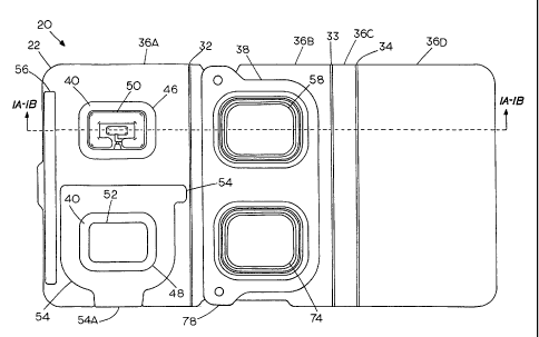
Figures 1A, 1B, and 2 respectively show exploded cross
sectional, assembled cross sectional, and top views of one
embodiment of a folding device, generally at 20, in an
opened or flat configuration. Figures 9A and 9B
respectively show the embodiment in a side cross sectional
and top view of the device folded in a packaged
configuration for long term storage. The device consists of
3 main elements: a folding support structure 22, a
transdermal iontophoretic patch 24, and a drug containing
pack or blister pack 26.
The folding support structure 22 may include a
paperboard, or similar material, substrate with a release
coating layer 28 applied to one side and a printable coating
applied to the opposite side. Figure 1C is a greatly
25 enlarged representative fragmentary cross section of a
portion of the folding support structure 22, further
illustrating the coating layer 28 and printable coating 30.
The release coating layer 28 may be a siliconized coating
and the printable coating 30 may be a clay coating. An
30 alternative folding substrate or support layer may be a
thermoformable polymer or the like. The support structure
CA 02735789 2011-03-02
WO 2010/027468
PCT/US2009/004969
12
22 may contain several fold lines as at 32, 33 and 34 that
are created by perforating, scoring, and/or creasing. In
the case of a thermoformable substrate, living hinges may be
thermoformed at 32, 33 and 34. Depending on the number of
fold lines, the support structure may be divided into a
number of panels which provide areas to attach various
components of the device, apply printing for directions,
and/or provide a release coated barrier for exposed
adhesives on one panel from permanently sticking to other
panels when folded together during storage.
As shown, transdermal iontophoretic patch 24 is
adhesively attached to a first panel 36A of the support
structure 22 on the release coated side of the substrate.
The transdermal iontophoretic patch 24 includes an adhesive
coated foam layer 38, an occlusive double sided tape layer
40, an electrode subassembly layer 42 consisting of a power
source, electronics and electrodes to operate the patch (not
shown), and an overlay tape layer 44. As shown in Figure
1B, the adhesive side of the foam and the overlay tape are
attached to the release coated side of the support
structure.
As shown in Figure 2, cut outs in the foam layer 38 and
support structure 22 layers create an empty anode well or
recess 46 and an empty cathode well 48 aligned to receive
the corresponding anode and cathode imbibed drug pads,
preferably gel pads during assembly/activation. The anode
and cathode cut outs 46, 48, respectively, expose underlying
electrodes, including anode 50 and cathode 52. A half-panel
release liner 54 is created by perforating the first panel
36A as also shown in Figure 2. The half-panel release liner
54 serves to peel the patch off of the substitute layer of
CA 02735789 2011-03-02
WO 2010/027468
PCT/US2009/004969
13
the support structure after it is activated.
The half-panel release liner serves the purpose of
stiffening the flexible patch to aid in application and
additionally allows the operator to handle the patch easily
without the patch sticking to the operator's fingers.
Preferably, the half of the patch not covered by the half-
panel release liner 54 is affixed to the patient's skin
first. Subsequently, the half-panel release liner is removed
by peeling at a tab 54A of the half-panel release liner.
Finally, the other half of the patch is affixed to the
patient's skin.
A strip of double-sided tape 56 is attached to the
printable side of the support structure 22 on the first
panel 36A. The adhesive strip 56 serves a dual function of
keeping the structure closed during its long term storage
condition by temporarily bonding to a release coated side as
shown in Figure 9A. The second function is to permanently
bond to a second panel 36B of the support structure when the
parts are folded together to transfer the anode and cathode
gel pads 60, 62 as shown in Figure 3C so that the first
panel 36A cannot be re-opened.
As shown in Figure 9A, the support structure also
includes third and fourth panels 36C and 36D, respectively.
Third and fourth panels 36C and 36D collectively make up a
separator component that is configured to physically
separate the drug pack and iontophoresis patch components
when the iontophoretic drug delivery system is present in a
folded storage stage. The third and fourth panels 36C and
36D of the support structure 22 as folded create a release
coated barrier that prevents the occlusive tape 40 in the
transdermal patch 24 from touching and permanently sticking
CA 02735789 2011-03-02
WO 2010/027468
PCT/US2009/004969
14
to the formed lid layer 64 of the drug pack 26 during long
term storage.
As shown in Figure 2, when the system is present in a
pre-folded state prior to storage, the drug pack components
are present in the center region of the support structure
and flanked on a first side by the iontophoresis patch
component and on a second side opposite the first side by
the separator component of the support structure which
affords both structured separation and external protection
for the stored system.
A second piece of double-sided tape 66 is attached to
the second panel 36B on the printable side of the support
structure 22 to permanently bond the drug containing blister
pack 26 to the support structure 22. Alternatively, for
example, instead of a double-sided adhesive 66, the drug
containing blister pack 26 could be heat sealed to the
support structure 22 as by applying a heat seal coating to
the bottom of the drug containing pack or to the printable
side of the support structure.
As indicated, the drug pack 26 is provided with a
formed barrier lid having low moisture vapor permeability, a
generally flat bottom layer, containing two spaced gel
locations, one containing an anode gel-imbibed non-woven pad
60, another containing a cathode gel-imbibed non-woven pad
62. The low moisture vapor permeable barrier formed lid
layer is shown at 64. Preferably, the generally flat bottom
layer 68 is constructed of an aluminum foil composite film
that may or may not contain a heat seal coating (not shown)
on the side that contacts the gel pads. If it is used, the
heat seal coating is preferably a readily peelable coating.
The gel-imbibed pads as at 70 are constructed of a composite
CA 02735789 2011-03-02
WO 2010/027468
PCT/US2009/004969
or laminated non-woven material. The anode and cathode gels
are dispensed onto the pads and soak into the composite non-
woven material.
The low moisture vapor permeable formed lid layer 64
5 has been successfully constructed from a cold-formable
aluminum composite material consisting of a seal layer on
the product contacting under side 64A and a nylon layer on
the opposite side 64B. Alternatively, for example, the
product contact side 64 may consist of PVC with no seal
10 layer. If a seal layer is employed, preferably it is a
peelable heat seal coating. Anode and cathode cavities 72
and 74, respectively, may be mechanically formed with
traditional cold form tooling using Teflon
(polytetrafluorethylene) plugs or in combination with vacuum
15 or pressure assist. The material may be thermoformed if
using an alternative material including other fluorine-
containing plastics in sheet or film form such as material
sold under the trademark Aclar@, PVDC, and other low
moisture vapor transmission barrier thermoformed packaging
materials.
Figures 7A and 73 show top and side cross-sectional
views, respectively, illustrating the structure of one
embodiment anode 72 or cathode 74 composite pad materials.
As indicated, the anode and cathode pad composite materials
are preferably of a non-woven structure to maintain the
continuity of the drug-containing material in the structure
and may include a plurality of layers, possibly up to three
layers, of material. These may include a thick needle-
punched polypropylene layer 76, a thin, permeable
polyethylene net layer 78, and a thin, occlusive
polypropylene layer 80. The layers may be heat fused
CA 02735789 2011-03-02
WO 2010/027468
PCT/US2009/004969
16
together without requiring adhesives. All three layers are
cut to have the same outside perimeter shape. The occlusive
layer 80 is cut to the shape of a perimeter ring that
remains intact and occlusive. Inside the ring, the
occlusive layer 80 is cut out completely or perforated so
that the inside region 84 becomes permeable. The permeable
region 84 is shaped to coincide with the shape of the anode
50 and cathode 52 electrodes, by allowing the gel to migrate
through this layer and contact the full area of the
electrodes when the device is assembled for use.
Importantly, the occlusive ring 80 provides a barrier for
gel migration so the outside surface remains relatively dry
during storage to aid in adhesive transfer of the drug-
imbibed pad 70 during activation of the device.
In one embodiment, both the anode 72 and cathode 74
composite pads are similar in shape. Of course, the
electrodes may be any convenient shape and the electrodes in
a given patch embodiment may be of like or different shapes.
Figures 8A and 83 show a plan view and cross sectional view
of an alternate shape of what may be either an anode and/or
cathode of composite non-woven material 86. This embodiment
has a shaped perimeter ring 88, with permeable inside area
90. Figure 10 is a top view of an alternative embodiment
showing a device 100 with a drug pack 102 having anode and
cathode formed cavities, 104 and 106, respectively, of
different shapes. In similar fashion, anode and cathode
formed cavities of different, but corresponding shapes, are
reflected in the anode 108 and cathode 110 in the foam,
support structure 22, and occlusive layers 114 and 116.
Only corresponding components that fit together in an
assembled device need be of like shape.
CA 02735789 2011-03-02
WO 2010/027468
PCT/US2009/004969
17
An important aspect of the invention involves shelf
life stability of the co-packaged iontophoretic devices.
This is of paramount concern based on the history of such
devices which have had limited commercial success because of
shelf life limitations. As indicated, co-packaging
techniques have included attempts to package the wet drug
gels in direct contact with the electrodes during long term
storage, and attempts to isolate the power source and
electronics in the same package through low moisture
permeable (high barrier) materials. Wet gels have been
packaged in direct contact with the electrodes only and
connected to a power source and electronics by a cable or
other connector at time of use. As indicated, each of these
is fraught with challenges for long term stability. For
example, in time, wet gels may degrade the metals in the
electrodes, power source, and electronics which, in turn,
contaminates and degrades the stability of the gel.
In the present development, stable long term co-
packaging is realized by the provision of a storage
container for the anode and cathode gels in the form of a
separate hermetically sealed drug pack or blister cavity
with product contact layers that do not leach into the gel,
react with the gel, or absorb the gel. Since the gel
material itself provides no form, a carrier substrate
material is used to give the gel form and structure, and
provide a stable support to facilitate transfer of the gel
out of the long term storage container when the system is
assembled for use. The carrier substrate should be composed
of materials that do not leach, react, or absorb the
constituents of the gel. Preferably the blister cavity and
carrier substrate should be made from stable, relatively
CA 02735789 2011-03-02
WO 2010/027468
PCT/US2009/004969
18
inert, materials such as polypropylene and polyethylene.
Any suitable material can be used and may be selected based
on the nature of the gel.
Shelf life stability will vary with the construction of
the patch component and the stability of the integrity of
the drug composition. Patch shelf life depends on retention
of adhesive quality and the maintenance of the specified
function of the electrical circuit components. The device
should have a stable shelf life of at least two (2) years.
Figures 5A-5E show in step-wise cross sectional views
of one method for forming, filling, and sealing a drug-
containing pack or drug pack in accordance with the present
development. Figure 6 shows a top view of the drug pack of
Figure 5, as assembled generally at 120. In the method of
Figures 5A-5E, the drug containing pack is assembled in an
inverted position. The assembly starts with the provision
of a lid or cover membrane 122 of a low moisture vapor
transmission material formed to create anode and cathode
gel cavity shapes 124 and 126, respectively, at a specified
spaced interval and depth. Next, an anode pad 128 is placed
into the formed anode cavity 124, and likewise, a cathode
pad 130 is placed into the formed cathode cavity 126. The
pads may be of similar construction to those shown in
Figures 7A-7B and 8A-8B. The anode and cathode pads are
oriented so that each occlusive layer 132, 134 is placed
facing into the cavity and in contact with the bottom of the
corresponding formed cavity. The anode and cathode pads are
sized to just fit in the bottom of the formed cavities. In
this manner, the formed cavities then initially provide
registration of the pads to the formed lid layer 122.
The remaining steps are performed in a timed sequence
CA 02735789 2011-03-02
WO 2010/027468
PCT/US2009/004969
19
as will be described. Allowable open time for the assembly
is determined by the rate of pad permeation which is related
to the viscosity of the gel used.
At time t=to, as shown in Figure 5C, an amount of a
viscous anode gel 136 is dispensed to uniformly cover the
central permeable region as at 84 (Figure 7A) of the anode
pad 128; similarly, an amount of a viscous cathode gel 138
is dispensed to uniformly cover a permeable region of the
cathode pad 130. As also seen in Figure 5C, both the anode
and cathode gels are dispensed in a manner such that for a
given amount of gel, once dispensed, the total height of the
pad plus the gel height somewhat exceeds the depth of the
formed anode or cathode cavity 124 or 126. The gel must be
of a relatively high viscosity range in order for it to
maintain its shape/height for a necessary duration during
assembly of the device. At time t>to<tl, a flat bottom or
carrier substrate layer 140 is applied (Figure 5D) and heat
sealed to the formed cover membrane 122. Application of the
flat carrier substrate layer 140 contacts and compresses the
gel causing the gel to wet the inner surface of the carrier
substrate layer 140 and spread out as also shown in Figure
5D.
Alternatively, in another embodiment (not shown), the
flat bottom layer can be formed similarly to the formed
cover membrane layer to create a nested configuration, in
= which case, the gel plus the pad height can be designed so
that when the bottom and lid layers are assembled, the gel
will be in contact with the carrier substrate layer in a
similar manner as in the illustrated embodiment.
In this procedure, time span t=to to t=ti is defined as
the time it takes the dispensed anode and cathode gels to
CA 02735789 2011-03-02
WO 2010/027468
PCT/US2009/004969
soak through their respective pads and start to wet to the
bottom of the formed cavities of lid layer. Time is a
factor because it has been found that if the gels soak
completely through the respective pads and wet the bottom or
5 inner surface of the formed cover membrane cavity before the
carrier substrate layer is applied, the pads, once fully
imbibed, may preferentially stick to the inside of the
formed lid. This, of course, is undesirable as the imbibed
gel pad would adhere to the lid layer 122 instead of the
10 carrier substrate layer 140 when one attempted to assemble
the system. Time span t=to to t=ti also defines the time
in which the gel will adequately maintain its height so that
the gel will wet and adhere to the inner surface of the
bottom layer 140 when that layer is applied.
15 For the above reasons, the gels are formulated in a
preferred viscosity range to provide the correct flow rate
and surface tension. For example, a 100,000 centipoise gel
may have a to-ti time window of about 2-4 minutes. This is
adequate for normal assembly to occur.
20 In this process, the gels initially contact and wet the
bottom layer member 140. This allows the gels to act as
adhesives as the surface tension of the gels between the
member 140 and the pads 128 and 130 exceeds the
gravitational forces on the imbibing pads. Therefore, as
the pads slowly imbibe with gel, they will stick to and be
pulled towards the carrier substrate layer regardless of the
orientation of the device. Thus, after the bottom and lid
layers are sealed, the compressed gels imbibe (soak-in) into
the anode and cathode pads respectfully, creating a fully
imbibed non-woven anode pad 128a and fully imbibed pad 130a
as shown in Figure 5E. As described previously, the fully
CA 02735789 2011-03-02
WO 2010/027468
PCT/US2009/004969
21
imbibed anode and cathode gel pads continue to adhere to the
bottom layer 140 thereby creating anode and cathode
headspaces 142 and 144, respectively in the package as also
shown in Figure 5E. It has been found that due to the high
surface tension and high preferred viscosity of the gels,
the fully imbibed pads will remain registered to their
respective formed lid cavities and be attached to the
carrier substrate layer as shown throughout the anticipated
shelf life of the device.
It will be appreciated that the amount of gel added to
each cavity should be matched to the absorbency of each pad
in order to minimize excess gel. The amount and viscosity
of the gels is preferably such that imbibed gel does not wet
the outer surface of the occlusive ring on the pads. In
this manner, the outer surface of the occlusive ring 146,
148 should remain relatively dry to aid adhesive transfer
and adhesive attachment of imbibed gel pads into
corresponding empty anode and cathode wells of the
transdermal patch during activation. The inside surface of
the formed lid cavities in the anode and cathode headspace
regions as at 142 and 144 should remain free of gel and
relatively dry.
In order for this packaging concept to function, the
gels must be formulated with a preferred viscosity. The
preferred range is between 8,000-120,000 centipoise but is
not limited so long as the process can be successfully
followed. The gels useful in the system may be formulated
by dissolving an appropriate amount of drug or saline in
water, and adding a gelling agent such as HPMC
(hydroxpropylmethylcellulose) such that a conductive gel of
appropriate viscosity is created. Other gelling agents,
CA 02735789 2011-03-02
WO 2010/027468
PCT/US2009/004969
22
such as PVP (polyvinylpyrrolidone), PEO (polyethyleneoxide),
or PVA (polyvinylalcohol) can also be used. Successful gels
have been formulated from a HPMC powder at 2% w/w.
The concentration of an active agent in the gel may
vary widely depending on the agent of interest and the
desired patch dosage and planned duration of application.
Generally, the concentration will range from about 0.2% to
10% (weight).
Figures 3A-3D show in step-wise fashion, in cross-
sectional views, how one preferred embodiment is activated
and deployed to a treatment site. Figure 4 shows the top
view of Figure 3A after the blister or drug pack lid 64 has
been removed. Figures 9A and 9B show a side sectional and
top view, respectfully, of a fully packaged device.
Beginning with the fully packaged device of Figures 9A
and 9B, a deployment or assembly process will be described.
First, the fully packaged device is opened and unfolded by
pulling at the tab 160 to release adhesive strip 56 from the
release liner coated side of panel 36D. Second, the formed
cover membrane layer 64 is removed or peeled away from the
bottom layer 68 exposing the gel-imbibed anode and cathode
pads 60, 62 which are adhered through surface tension of the
gels to the bottom layer 68 as shown in Figure 3A. The peel
is initiated by peeling at the tab 78 (Figure 2) on the
formed lid member 64.
Next, the first panel 36A is folded at the fold line 32
onto the second panel 36B, thereby bringing the occlusive
region as at 80 of the occlusive layer 80 of the anode and
cathode gel pads 60, 62 in permanent adhesive contact with
the occlusive tape layer 40 of the transdermal patch 24 as
shown in Figure 3C. Also, the adhesive strip is brought
CA 02735789 2011-03-02
WO 2010/027468
PCT/US2009/004969
23
into contact with the printable coating layer 30 of the
support structure 22 and is permanently adhered to the
second panel 36B, thereby preventing the panel 36A from
being re-opened. Figure 3B shows an intermediate view of
the folding action. The outer surface of the transdermal
patch is preferably pressed to ensure good permanent bonding
of the occlusive tape 40 to the occlusive region as at 80 on
the gel pads.
Finally, the half release liner 54 is peeled from the
support structure at the tab 54A bringing the fully
assembled transdermal patch 170 with it. The exposed half
of the patch adhesive can be applied to the treatment site
and the half release liner 54 thereafter can be peeled from
the transdermal patch at the tab 54B (Figure 9B) and the
remaining half of the patch adhered to the treatment site.
Figures 11A-11C illustrate an alternative embodiment
including an alignment fixture or guide element and
illustrating activation of the embodiment. Figure 12 shows
a top view of the fully assimilated embodiment of Figure 11B
from which the cross-sectional views of 11A-11C are taken.
As best seen in Figure 11A, the device, generally 200, as
packaged, includes three main components. They are a guide
element 202 having spaced raised alignment members 204, 206,
a drug pack arrangement 208 and a transdermal patch assembly
210. The main components are designed to be stored
separately in a common package and assembled when the device
is prepared for use.
The drug pack includes a flat card substrate layer 212
which is designed with spaced alignment openings 214 and 216
which register with alignment members 204 and 206 during
assembly. Anode and cathode non-woven, gel-imbibed pads 218
CA 02735789 2011-03-02
WO 2010/027468
PCT/US2009/004969
24
and 220 are respectively carried on a bottom layer 222 and
separated from drug pack lid 224 in the manner of
embodiments previously described and illustrated in Figures
5A-5E. Drug pack 208 is adhered to card substrate layer 212
as by a double-sided tape layer at 226.
The transdermal patch component 210 is mounted on a
flat card substrate layer 228 with spaced alignment openings
230 and 232 and, as with previously described embodiments,
half release liner 234. The patch assembly may be quite
similar in construction to that previously described with
foam layer 236 and double-sided tape 238, electrode
subassembly layer 240 and overlaying tape layer 242.
At the time of use, individual components are aligned
and assembled to each other using features of a component to
self-align to adjacent components. In this manner, the
guide element 202 may be positioned on a flat surface with
the spaced alignment members 204, 206 facing up as shown in
Figure 11A. Next, the drug blister pack 208 is assembled to
the alignment fixture or guide element 202 by registering
alignment member 204 to the opening 214 in the drug pack and
alignment member 206 to opening 216. The lid 224 can then
be peeled off the drug pack 208 exposing the gel-imbibed,
non-woven anode and cathode pads 218 and 220, respectively.
Next, the transdermal iontophoresis patch 210 can be
assembled to drug pack 208 by again using alignment members
204 and 206 with alignment openings 230 and 232 thereby
placing the gel-imbibed pads in alignment with corresponding
electrodes. This results in the combined configuration
depicted in the cross-sectional view of Figure 11B and top
view of Figure 12 with the drug pack arrangement 208 and the
transdermal patch assembly 210 in consecutive assembled
CA 02735789 2011-03-02
WO 2010/027468
PCT/US2009/004969
registration on the guide element 202.
In this stacked condition, the assembled patch is ready
to be separated for placement on a patient. Separation can
be accomplished by simply peeling the half release liner 234
5 from the card thereby separating the device from the card
substrate layer 228 and bringing the fully assembled
transdermal patch 250 with it as shown in Figure 11C. The
patch is then ready to be applied to the patient as
described in relation to the previous embodiments.
10 It will be appreciated that the drug pack 208 and the
transdermal patch 210 are similar in construction to
previously described embodiments except that the card
substrate layers in this embodiment are separate flat
members rather than folding connected panels. The flat card
15 substrate layers 212, 228 include alignment openings
corresponding to the members 204 and 206 on guide element
202 and they do not require a silicon or other release
coating so that both sides may contain a printable clay
coating material or the like.
20 The card layers 212, 228 may also be constructed from
any suitable polymer material. The alignment members 204
and 206 of the guide element 202 are preferably thermoformed
or injection molded out of a suitable polymer material also.
Figure 13 is an exploded cross-sectional view of an
25 alternate embodiment to that shown in Figures 11A-11C in
which a guide element 302 with alignment members 304 and 306
is substituted in the drug pack 308 for the flat card
substrate layer 212. The drug pack is otherwise similar to
previously described drug packs and a lid is shown at 324.
The transdermal patch component 310 is also similar to that
shown in Figure 11A and includes substrate 328 with spaced
CA 02735789 2011-03-02
WO 2010/027468
PCT/US2009/004969
26
alignment openings 330 and 332 and half release layer at
334. The drug pack 308 is bonded to the guide element 302
by double-sided tape 326.
Assembly and activation is similar to that of the
embodiment of Figure 11A-11C. Thus, the drug pack lid 324
is removed and the transdermal patch 310 is aligned with the
drug pack over the alignment members 304 and 306 using the
openings 330 and 332. The assembled patch thereafter being
peeled away in the manner of Figure 11C.
Figure 14 shows an exploded cross-sectional view of yet
another embodiment of the device of the present invention
which represents another alternative to that shown in Figures
11A-11C. In this embodiment, the alignment fixture 402 with
alignment members 404 and 406 is substituted for the release
card substrate layer 228 associated with the transdermal patch
component 410. The alignment fixture includes a silicone
release coating on upper surface 402A applied to the side to
which the patch adheres prior to removal. This embodiment is
assembled and applied in a similar manner to those described
just above. Thus, the lid 424 of the drug pack is removed and
the openings 414 and 416 are aligned with the members 404 and
406 and the fully aligned device is peeled away in the manner
of Figure 11C.
It will further be appreciated that the assembled device
or patch to be applied to a user may be of any convenient size
as from as small as about lcm x 2cm to about 15cm x 20cm. The
size can vary widely depending on the active agent
administered and the condition to be treated.
This invention has been described herein in considerable
detail in order to comply with the patent statutes and to
provide those skilled in the art with the information needed
CA 02735789 2011-03-02
WO 2010/027468 PCT/US2009/004969
27
to apply the novel principles and to construct and use
embodiments of the example as required. However, it is to be
understood that the invention can be carried out by
specifically different devices and that various modifications
can be accomplished without departing from the scope of the
invention itself.
What is claimed is:
=