Note: Descriptions are shown in the official language in which they were submitted.
CA 02736499 2011-03-08
WO 2010/030933 PCT/US2009/056724
TETHER-BASED ORTHOPEDIC JOINT DEVICE DELIVERY METHODS
CROSS-REFERENCE TO RELATED APPLICATIONS
[0001] This application is a) a continuation-in-part of U.S. Application Ser.
No.
12/210,099, filed September 12, 2008, and b) a continuation-in-part of U.S.
Application Ser.
No. 12/212,587 filed Sept. 17, 2008, and also claims priority under 35 U.S.C.
119(e) to a)
U.S. Provisional Ser. No. 61/171,408, filed April 21, 2009, b) U.S.
Provisional Ser. No.
61/171,409, filed April 21, 2009, c) U.S. Provisional Ser. No. 61/171,410,
filed April 21,
2009, and e) U.S. Provisional Ser. No. 61/171,412, filed April 21, 2009, all
of the above
which are hereby incorporated by reference in their entirety.
BACKGROUND OF THE INVENTION
[0002] Today, there are an increasing number of patients with osteoarthritis,
rheumatoid
arthritis, and other joint degenerative processes. Osteoarthritis is by far
the most common
type of arthritis, and the percentage of people who have it grows higher with
age. An
estimated 12.1 percent of the U.S. population (nearly 21 million Americans)
age 25 and older
have osteoarthritis of one form or another. Although more common in older
people, it
usually is the result of a joint injury, a joint malformation, or a genetic
defect in joint
cartilage. The incidence and prevalence of osteoarthritis differs among
various demographic
groups: osteoarthritis tends to start for men before the age of 45, and after
the age of 45 it is
more common in women. It is also more likely to occur in people who are obese
or
overweight and is related to those jobs that stress particular joints.
[0003] Arthritis is a degenerative process that affects the musculoskeletal
system and
specifically the joints - where two or more bones meet. It often occurs in the
joints of the
hands and wrists (particularly in the fingers and thumbs, between the
phalanges, the
metacarpals and/or the carpals), feet (in the toes, between phalanges,
metatarsals and/or
tarsals), ankles, elbows, shoulders, knees, hips, and the spine (particularly
at the neck and
lower back). Joint problems can include inflammation and damage to joint
cartilage (the
tough, smooth tissue that covers the ends of the bones, enabling them to glide
against one
another) and surrounding structures. Such damage can lead to joint stiffness,
weakness,
instability and visible deformities that, depending on the location of joint
involvement, can
interfere with the basic daily activities such as walking, climbing stairs,
using a computer
keyboard, cutting food and brushing teeth. This ultimately results in moderate
to severe pain.
1
CA 02736499 2011-03-08
WO 2010/030933 PCT/US2009/056724
Drug regimes can provide temporary relief from the pain, but do not slow down
the crippling
affects. Drugs may also subject patients to serious side effects and risks,
such as the
increased cardiovascular risks associated with osteoarthritis drugs Vioxx and
Bextra, which
were withdrawn from the market. Drugs used to treat other forms of arthritis,
such as
corticosteroids, are associated with osteoporosis and hyperglycemia and can
lead to increased
risks of bone fracture and diabetes, for example. When pharmacologic therapy
and physical
therapy no longer provide adequate relief, only surgical options remain.
[0004] The extreme result or end point in traditional treatments is an open
surgical
procedure to implant a spacer or to perform total joint replacement with a
prosthetic device.
Current joint replacement therapies (spacers or a total prosthesis) require
the joint capsule to
be surgically opened and the bone surfaces to be partially or totally removed.
Both
modalities present various drawbacks. For example, U.S. Patent No. 6,007,580
to Lehto et al.
describes an implantable spacer that must be fixed at one or both ends to the
bone of either
end of the knuckle (e.g. the metacarpal-phalangeal (MCP) joint). The spacer
must be
implanted by opening of the joint capsule and be affixed at one or both ends
to the
corresponding bone surfaces.
[0005] Various spacers in the art can cause inflammation, while total joint
replacement can
limit the range of motion and also compromise the strength and stability of
the joint. These
surgeries are highly invasive and require the joint capsule to be surgically
opened, and the
incision itself can result in inflammation and infection. Due to the
invasiveness of the
procedure, prolonged healing times are required. Furthermore, the invasive
nature of these
surgeries sometimes precludes a second joint replacement or spacer when the
first joint
device wears out or fails.
[0006] It would be desirable as well as beneficial if there were an
intermediary step or
alternative treatment before subjecting patients to drastic joint replacement
and/or long-term
drug therapy.
BRIEF SUMMARY OF THE INVENTION
[0007] Various embodiments disclosed herein relate generally to the treatment
of
osteoarthritis, rheumatoid arthritis, and other degenerative joint processes,
and include but are
not limited to minimally invasive implantable devices to reduce bone-to-bone
contact in a
joint.
2
CA 02736499 2011-03-08
WO 2010/030933 PCT/US2009/056724
[0008] Systems and methods for treating degenerative joint conditions include
an
orthopedic device comprising a resilient elongate member, which may be
implanted in a joint
space using a suture tether or other type of bendable elongate element. Using
minimally
invasive surgical techniques, a small skin incision and arthrotomy are made to
provide access
to the joint. The suture tether is passed through the incision and joint space
and used to pull
the orthopedic device into the joint space. The suture tether may also be
inserted using a
percutaneously inserted needle or other type of needle-based delivery
instrument. The
orthopedic device may be restrained to a reduced profile that permits
minimally invasive
implantation, but changes to an enlarged profile when positioned at an
implantation site. The
orthopedic device may comprise a shape-memory and/or superelastic material,
and may
comprise an open or closed shape configuration.
[0009] To facilitate insertion of the orthopedic device through a minimally
invasive or
limited access procedure, a delivery system may be used to support and orient
the orthopedic
device during implantation. In some instances, the delivery system may also
reduce the
delivery profile of the device for insertion through a minimally invasive or
limited access
procedure.
[0010] In one example, an orthopedic joint device is provided, comprising a
resilient C-
shape joint device with a shape-memory elongate curved core and an outer
polymeric
articular jacket, where the joint device has a first configuration where the C-
shape joint
device is coupled to a suture and in a deformed reduced profile, a second
configuration where
the joint device is coupled to the suture and in an expanded profile, and a
third expanded
configuration where the joint device is in the expanded profile without
coupling to the suture.
[0011] In another example, an orthopedic device system comprises an orthopedic
device
with a resilient elongate core, a flexible polymeric jacket covering at least
a portion of the
resilient elongate core, and a first suture aperture, wherein the orthopedic
device is
configured to reside between two opposing articular surfaces and within a
joint space of a
joint. In some further examples, the elongate core may have a delivery
configuration and an
implantation configuration, and the implantation configuration is optionally a
non-linear
configuration, including but not limited to a "C"-shape configuration. In
other examples, the
delivery configuration may be a linear configuration. The first suture
aperture may comprise
a suture lumen through the jacket, or a suture eyelet coupled to the jacket,
while in some
examples, the core may comprise a suture eyelet. In one specific example, the
suture eyelet
3
CA 02736499 2011-03-08
WO 2010/030933 PCT/US2009/056724
may comprise a twisted loop of the core. Some systems may further comprise a
suture,
which may be located in the first suture aperture. In some examples, the
system may also
further comprise a penetrating member, which is optionally pre- attached to
the suture. The
penetrating member, the suture and the orthopedic device may be provided in a
single sterile
package. The system may also further comprise a penetrating member holder,
which in turn
may optionally comprise an orthopedic device retaining assembly, such as a
retaining post.
In some examples, the elongate core may have an elongate length that is at
least about 50% of
the circumference of the joint space. The joint space may be a joint space of
a carpo-
metacarpal joint, such as the carpo-metacarpal joint of a thumb. In some
systems, the
orthopedic device may further comprise an inner region at least partially
surrounded by the
resilient elongate core, and at least one span member across the inner region.
Sometimes, the
span member may have a planar configuration, and may comprise a resilient or
elastic
material, for example. The jacket of the orthopedic device may comprise a
thickened jacket
region about the first suture aperture. The system may also optionally
comprise a first pull
member and a second pull member, wherein the first pull member is coupled to
the first
suture aperture. The second pull member may be coupled to a second suture
aperture, and in
some further examples, the first and second pull members may each pass through
a third
suture aperture. An optional third pull member may also be coupled to the
third suture
aperture.
[0012] In another example, a method of implanting a orthopedic device in a
patient is
provided, comprising percutaneously inserting a needle through a first joint
capsule opening
of a joint space, passing the needle with an attached suture across the joint
space and through
a second joint capsule opening, wherein the second joint capsule opening is
smaller than the
first joint capsule opening, pulling a resilient orthopedic device into the
joint space using the
suture, wherein the resilient orthopedic device comprises a first end, a
second end, and a body
therebetween having an elongate arcuate configuration, separating at least a
portion of the
suture from the resilient orthopedic device, and removing at least a portion
of the suture from
the patient. The method may optionally further comprise abutting the resilient
orthopedic
device against the second joint capsule opening, positioning the resilient
orthopedic device
symmetrically within the joint space with respect to the second joint capsule
opening, and/or
restraining the resilient orthopedic device in a reduced profile as the
resilient orthopedic
device traverses the first joint capsule opening. In some further examples,
the method may
further comprise enlarging the resilient orthopedic device from a reduced
profile to an
4
CA 02736499 2011-03-08
WO 2010/030933 PCT/US2009/056724
enlarged profile with substantially the same volume as the orthopedic device
in the reduced
profile, and/or with substantially the same mass as the orthopedic device in
the reduced
profile. The method may also further comprise restraining the resilient
orthopedic device in a
delivery configuration as the resilient orthopedic device traverses the first
joint capsule
opening. The method may also further comprise releasing the resilient
orthopedic device
from the delivery configuration in the joint space to assume an implantation
configuration
that is non-linear. In some methods, a distance between a first end and a
second end of the
resilient orthopedic device in the delivery configuration is greater than the
distance between
the first end and the second end of the resilient orthopedic device in the
implantation
configuration. The implantation configuration may comprise at least one
arcuate section,
and/or a generally a non-planar implantation configuration. In some examples,
a first portion
of the resilient orthopedic device may have a delivery position in the
delivery configuration
that is different from an implantation position in the implantation position
with respect to a
second portion of the resilient joint implant. The method may also further
comprise orienting
the resilient orthopedic device in the joint space such that the delivery
position and the
implantation position of the first portion of the resilient orthopedic device
generally lie in a
plane that is generally aligned with an axis between the first and second
joint capsule
openings. In some examples, the resilient orthopedic device may be pre-coupled
to the suture
at the point-of-manufacture. Also, when pulling the resilient orthopedic
device, the pulling
may be performed such that the body of the resilient orthopedic device enters
the joint space
before the first and second ends. The joint space may be a trapeziometacarpal
or a carpo-
metacarpal joint, for example, and the first joint capsule opening may be
located on the dorsal
surface of the joint space.
[0013] In another embodiment, the method of implanting a orthopedic device is
provided,
comprising pulling a joint implant into a joint space from a first joint
capsule opening using a
pulling force acting through a second joint capsule opening. The joint space
maybe located
in an extremity of a patient, including the upper extremities and the lower
extremities, and the
joint space may be a carpal-metacarpal joint space, for example. The second
joint capsule
opening may be formed using a penetrating member, which may be formed from the
joint
space or external to the joint space. Examples of the penetrating member may
include a
needle attached to a suture, and the method may further comprise passing the
suture through
the first joint capsule opening and through the second joint capsule opening.
The method
may also further comprise coupling the suture and the joint implant together,
such as passing
CA 02736499 2011-03-08
WO 2010/030933 PCT/US2009/056724
the suture through the joint implant, or passing the suture through a pre-
formed lumen of the
joint implant, or looping the suture around the joint implant. The joint
implant may be a
bendable joint implant having a reduced profile and an enlarged profile,
wherein the enlarged
profile has substantially the same volume and/or mass as the reduced profile.
In some
examples, the joint implant may comprise at least one articulated joint, such
as a plurality of
pivot joints. In other examples, the joint implant may be a resilient joint
implant. While
passing through the first joint capsule opening, the resilient joint implant
may be in a
restrained configuration, and in some instances, the resilient joint implant
may be placed in
the restrained configuration at the point-of-manufacture or at the point-of-
use. A delivery
cannula may be used to restrain the resilient joint implant. The method may
also optionally
comprise positioning the delivery cannula in the joint space through the first
joint capsule
opening. In some instances, the pulling force acts through a flexible line
coupled to the joint
implant, and sometimes, any tension in the flexible line may be relieved after
the joint
implant is located in the joint space. The method may also comprise separating
at least a
portion of the flexible line from the joint implant and pulling at least a
portion of the flexible
line out of the joint space.
[0014] In one example, a joint treatment system is provided, including a
delivery
instrument comprising a housing, a housing cavity, a delivery opening in
communication
with the housing cavity, a slidable actuator joined to a push member, and a
tongue member
protruding from the housing in proximity to the delivery opening, wherein the
push member
has a movement path, and an non-linear orthopedic device located in the
housing cavity, the
non-linear orthopedic device comprising a first end, a second end, a non-
linear body
therebetween, wherein the non-linear orthopedic device is oriented in the
housing cavity so
that the non-linear body is in closer proximity to the delivery opening than
both the first and
second ends. In some examples, the on-linear orthopedic device is an arcuate
orthopedic
device. Optional features of the delivery instrument include a mounting member
located in
the housing cavity between the delivery opening and at least a portion of the
slidable actuator,
onto which the non-linear orthopedic device may be mounted. The non-linear
orthopedic
device may be oriented so that a transverse dimension relative to the movement
path of the
push member is greater than a corresponding transverse dimension of the
delivery opening.
In certain examples, the push member may be configured to pass through at
least a portion of
the mounting member and/or configured with a distal end having a complementary
shape to a
surface of the non-linear orthopedic device. In some further examples, the non-
linear
6
CA 02736499 2011-03-08
WO 2010/030933 PCT/US2009/056724
orthopedic device may be configured with a first position and a second
position in the
housing cavity, wherein the second position is closer to the delivery opening.
The tongue
member may have any of a variety of optional features and configurations,
including but not
limited to a tapered configuration, and/or a blunt tip section having a
maximum transverse
dimension that is smaller than the larger transverse dimension of the
orthopedic device. In
some embodiments, the delivery instrument comprises at least two tongue
members, and in
further embodiments, at least one of the tongue members may be biased toward
another
tongue member, and may even be configured such that at least two of the tongue
members
are biased into contact each other. One or more tongue members may also
comprise a cutting
structure. The tongue member may also be configured to displace away from the
delivery
opening by the non-linear orthopedic device.
[0015] In another example, a joint treatment system is provided, including a
delivery
instrument comprising a movable actuating assembly and a movement passage
having a
cross-sectional area with minimum first dimension and a second dimension
transverse to the
first dimension, and an non-linear elongate orthopedic device releasably
coupled to the
delivery instrument, the non-linear elongate orthopedic device having a first
end, a second
end, and a non-linear body therebetween and a non-linear longitudinal axis
between the first
and second ends, wherein the non-linear elongate orthopedic device is oriented
with respect
to the delivery instrument such that a portion of the non-linear longitudinal
axis is transverse
to the movement passage of the movable actuating assembly. The delivery
instrument may
further comprise a housing with a housing cavity and a delivery opening in
communication
with the housing cavity, and/or a tapered delivery opening. Also, the portion
of the non-
linear longitudinal axis of the orthopedic device that is transverse to the
movement passage of
the movable actuating assembly may be located between the movable actuating
assembly and
the delivery opening. In use, the cross-sectional area with the minimum first
dimension and
the second dimension transverse to the first dimension may be located at the
delivery
opening. In some examples, the movable actuating assembly may comprise a
slidable
member and a push member, and the delivery instrument may further comprise a
first guide
member distal to the cross-sectional area with the minimum first dimension and
the second
dimension transverse to the first dimension. The first guide member may be a
flat and/or may
be flexible. In one particular example, flat guide member comprises a tongue
member with a
tapered configuration and a rounded distal tip. The delivery instrument may
also further
comprise a mounting member between at least a portion of the movable actuating
assembly
7
CA 02736499 2011-03-08
WO 2010/030933 PCT/US2009/056724
and the cross-sectional area with the minimum first dimension and the second
dimension
transverse to the first dimension, and the non-linear elongate orthopedic
device may be
releasably mounted on the mounting member. In some examples, at least a
portion of the
movable actuating assembly may be movably positionable through the mounting
member.
The delivery instrument may also optionally comprise a second guide member
about the
delivery opening, and in some instances, at least one of the first and second
guide members
are at least partially biased toward the other guide member, and at least one
of the first and
second guide members may even be configured to separate from the other guide
member with
passage of the orthopedic device between the first and second guide members.
[0016] In another example, a method for treating a joint is provided,
comprising
positioning an orthopedic device about a joint, wherein orthopedic device
comprises a first
end, a second end, and a non-linear body therebetween and a non-linear
longitudinal axis
between the first and second ends, and pushing against the non-linear body
such that at least a
portion of the non-linear body enters the joint before the first and seconds
ends. The
positioning of the orthopedic device about the joint may be using a delivery
system
containing or holding the orthopedic device. The delivery system may include a
guide
assembly, which may be inserted into the tissue surrounding the joint, and
even at least
partially into the joint space. The method may also include increasing access
to the joint
space along at least one dimension of tissue adjacent to the joint space while
inserting the
guide assembly. The guide assembly may be configured, such that guide assembly
may be
bent or flexed when used. In some examples, bending the guide assembly
comprises bending
a first guide member of the guide assembly, and in examples comprising at
least two guide
members, bending the first guide member may comprise bending or flexing the
first guide
member away from a second guide member of the guide assembly, and/or bending
or flexing
the second guide member from the first guide member. Bending the first guide
member may
occur while passing or forcing the non-linear body against a surface of the
first guide
member, which may occur in some examples while pushing the non-linear body.
After
implantation, the guide member may be withdrawn from the tissue surrounding
the joint.
[0017] In some embodiments, the joint device may be secured into the joint
space using,
for example, a suture and/or an anchor. For example, an orthopedic joint
device may
comprise a resilient C-shape joint device with a shape-memory elongate curved
core and an
outer polymeric articular jacket. The joint device itself may have one or more
configurations
8
CA 02736499 2011-03-08
WO 2010/030933 PCT/US2009/056724
during implantation into the joint space. For example, a first configuration
may be where the
C-shape joint device is coupled to a pull element and in a deformed reduced
profile; a second
configuration may be where the joint device is coupled to the pull element and
in an
expanded profile; and a third expanded configuration may be where the joint
device is in the
expanded profile and where the joint device is secured to tissue (such as a
joint capsule)
adjacent to the joint device.
[0018] In some instances, the joint device may be secured using a suture that
may or may
not be the same suture used to implant the joint device. Alternatively or
additionally, the
joint device may be secured using an anchor disposed along the pull element
and wherein the
joint device is coupled to a portion of the pull element comprising the anchor
in the third
expanded configuration. The joint device may or may not be coupled to the pull
element in
the third expanded configuration. The anchor itself may be transformable from
a delivery
configuration to a deployed configuration and may comprise protrusion selected
from the
group consisting of hooks, spurs, grapples, barbs, or a combination thereof.
In some
instances, the anchor may self-deploy once the joint device is in position.
[0019] In some embodiments, an orthopedic device system may comprise an
orthopedic
device and an anchor. The orthopedic device may comprise a resilient elongate
core, a
flexible polymeric jacket covering at least a portion of the resilient
elongate core, and a first
suture aperture, wherein the orthopedic device is configured to reside between
two opposing
articular surfaces and within a joint space of a joint. The anchor may be
configured to couple
to a joint capsule of the joint.
[0020] In some instances, the orthopedic device may further comprise a suture
that may or
may not be used to secure the device into the joint capsule. The anchor may or
may not be
coupled to the first suture aperture and/or a second suture aperture. In some
embodiments,
the elongate core may have a delivery configuration and a deployed
configuration and the
anchor may be configured to deploy when the elongate core transforms to the
deployed
configuration. The anchor may be disposed on a suture for positioning the
orthopedic device
within the joint space. The anchor may protrude from the polymeric jacket and
may
automatically deploy into the joint capsule. In some embodiments, the anchor
may comprise
a protrusion selected from the group consisting of hooks, spurs, grapples,
barbs, or a
combination thereof.
9
CA 02736499 2011-03-08
WO 2010/030933 PCT/US2009/056724
[0021] The orthopedic device system may be implanted in the joint space of a
carpo-
metacarpal joint or, more specifically, in the joint space of a carpo-
metacarpal joint of the
thumb.
[0022] In some embodiments, a method may comprise positioning an orthopedic
device
between two opposing articular surfaces and within a joint space of a joint by
pulling the
orthopedic device from a first side of the joint space to a second side of the
joint space using
a suture. The method may further comprise securing the orthopedic device to
tissue (e.g., a
joint capsule) on the second side of the joint space and adjacent to the
orthopedic device.
[0023] In some embodiments, the orthopedic device may be secured by suturing
the device
to the tissue or may comprise an anchor having one or more protrusions. In
embodiments
comprising an anchor, the orthopedic device may be secured by withdrawing a
tubular
member enclosing the one or more protrusions from the anchor. Alternatively,
the orthopedic
device may be secured by urging the orthopedic device in a specified direction
or by
deploying one or more anchors. The deployment may or may not be automatic once
the
orthopedic device is positioned. The anchor may comprise one or more
protrusions selected
from the group consisting of hooks, spurs, grapples, barbs, or a combination
thereof.
[0024] In one variation, an intra-articular implant system is provided,
comprising a non-
overlapping "C"-shape flexible articular jacket comprising a surface opening
in
communication with a "C"-shape internal lumen, wherein the internal lumen has
an arcuate
lumen length that is at least about 75% of an arcuate jacket length of the
jacket, an internal
core located within the "C"-shape internal lumen of the jacket and comprising
a shape-
memory material with at least two non-penetrating ends, wherein the internal
core is slidably
removable from the articular jacket from the surface opening, a needle, and a
suture coupled
to the needle and to the articular jacket. The articular jacket maybe sized
and shaped for
implantation into a carpo-metacarpal joint of a human, including but not
limited to a carpo-
metacarpal joint of a thumb.
[0025] In another variation, an implantable orthopedic joint device is
provided, comprising
a non-linear articular jacket comprising at least one surface opening in
communication with
an internal channel system, and a flexible core comprising at least one
elongate segment
configured to reside at least partially in the internal channel system of the
jacket and to
slidably separate from the internal channel system through at least one
surface opening. In
CA 02736499 2011-03-08
WO 2010/030933 PCT/US2009/056724
some further variations, at least one elongate segment may be an arcuate
elongate segment.
The flexible core may comprise at least three elongate segments arranged in a
branched
configuration with a segment junction. The internal channel system may also
comprise a
branched configuration. In some variations, at least one elongate segment may
comprise a
hairpin turn. The internal channel system may have an internal surface
configuration and the
flexible core may have an outer surface configuration that is complementary to
the internal
surface configuration of the internal channel system. The articular jacket may
have an
arcuate configuration, and/or may comprise at least two surface openings. The
internal
channel system may also comprise two separate lumens. The flexible core may
comprise an
internal section with a longitudinal length of at least about 75% of a non-
linear longitudinal
length of the jacket. The articular jacket may be sized and shaped for
implantation into a
carpo-metacarpal joint of a mammal. In some further variations, the internal
channel system
comprises at least one closed end.
[0026] In one variation, a method of implanting an intra-articular device is
provided,
comprising placing a joint implant into a joint space, wherein the joint
implant comprises an
articular jacket with a non-linear internal channel system and an internal
core located in the
channel system, and pulling on the internal core to separate the internal core
from the
articular jacket while maintaining the articular jacket in the joint space.
[0027] In another variation, a method of implanting an intra-articular device
is provided,
comprising placing an implant into a body space, wherein the joint implant
comprises a
polymeric articular jacket with an shape memory core, wherein a maximum
internal
dimension of the core is at least about 50% of the maximum dimension of the
articular jacket,
and pulling on the internal removable core to separate the internal removable
core from the
articular jacket while maintaining the articular jacket in the joint space.
[0028] In still another variation, a method of implanting an intra-articular
device is
provided, comprising placing a joint implant into a joint space, removing an
internal
component of the joint implant using a non-linear pathway in the joint
implant. The method
may further comprise deforming the internal component while removing the
internal
component. Removing the internal component of the joint implant may comprise
pulling out
the internal component of the joint implant. The joint space may border a
phalange, a
metacarpal bone or a carpal bone.
11
CA 02736499 2011-03-08
WO 2010/030933 PCT/US2009/056724
[0029] In another embodiment, a joint treatment system is provided, comprising
a tapered
enclosure comprising a tapered cavity, an arcuate orthopedic device located in
the tapered
cavity, the arcuate orthopedic device comprising a first end, a second end, a
deformable
arcuate body therebetween, wherein the arcuate orthopedic device may comprise
a delivery
configuration with a reduced profile and a deployment configuration with an
enlarged profile,
a suture attached to the arcuate orthopedic device, and a needle attached to
the suture. In
some variations, at least a portion of the needle may be located within the
tapered cavity of
the tapered enclosure. The tapered enclosure may comprise a sleeve structure
or a pouch
structure. The tapered enclosure may further may comprise a plurality of
perforations, and
the plurality of perforations may be located along an edge of the tapered
enclosure, may be
transversely located between two edges of the tapered enclosure, or may be
located along a
perimeter of the tapered enclosure. The tapered enclosure may comprise at
least one flexible
sheet, including but not limited to at least two flexible sheets bonded
together. The tapered
enclosure may also further comprise a pre-formed distal opening, wherein at
least a portion of
the needle may protrude from the pre-formed distal opening. The tapered
enclosure may
further comprise a handle structure. The joint handle structure may comprise a
finger loop.
The joint treatment system may further comprise a needle driver at least
partially located in
the cavity of the tapered enclosure, the needle driver comprising a distal
region and a
proximal region, wherein the needle may be releasably coupled to the distal
region of the
needle driver, or wherein at least portion of the proximal region of the
needle driver may be
located outside the cavity of the tapered enclosure while at least a portion
of the distal region
of the needle driver is located within the tapered enclosure. The tapered
cavity of the tapered
enclosure may comprise a tapered section and a non-tapered section, and
sometimes the non-
tapered section may be distal to the tapered section. The arcuate orthopedic
device may be
releasably coupled to the needle driver.
[0030] In another embodiment, a method for treating a joint is provided,
comprising
positioning a tapered cavity about a joint space, deforming a joint implant to
a reduced profile
using the tapered cavity, and passing the joint implant out of the tapered
cavity and into the
joint space. The method may further comprise inserting at least a portion of
the tapered cavity
into the joint space, wherein deforming the joint implant to the reduced
profile using the
tapered cavity may comprise deforming the joint implant to the reduce profile
using a tapered
portion of the tapered cavity. The method may further comprise traversing a
non-tapered
portion of the tapered cavity with the joint implant, wherein traversing a non-
tapered portion
12
CA 02736499 2011-03-08
WO 2010/030933 PCT/US2009/056724
of the tapered cavity with the joint implant may occur after deforming the
joint implant to the
reduced profile using the tapered portion of the tapered cavity. The method
may further
comprise forming an opening in the tapered cavity, wherein forming an opening
in the
tapered cavity may comprise separating at least one perforation located along
the tapered
cavity, wherein forming an opening in the tapered cavity may comprise
separating a
releasable seal located along the tapered cavity. In some variations, passing
the joint implant
out of the tapered cavity and into the joint space may comprise pulling a
tether coupled to the
joint implant. Pulling the tether coupled to the joint implant may comprise
pulling a needle
coupled to the tether, and wherein at least a portion of the needle may be
located outside of
the joint space.
[0031] In another embodiment, a delivery device for an orthopedic device, the
delivery
device comprising a housing comprising a proximal end and a distal end,
wherein the housing
defines a chamber, an orthopedic device disposed within the chamber, an
opening at the distal
end, wherein the opening comprises a first configuration for insertion into an
incision and a
second configuration having an expanded diameter, wherein the orthopedic
device is slidable
through the opening in the second configuration, a rotation member disposed
within the
housing, and a suture wound about the bobbin and having a first end coupled to
the
orthopedic device. The delivery device may be configured to lock the rotation
member. The
rotation member may comprise a lock structure. The lock is removable from the
proximal
end of the housing. The device of claim 3, wherein the one or more features of
the rotation
member are selected from the group consisting of a slot, notch, tab, rod,
teeth, gear, flap and
loop. The distal end of the housing may be releasably coupled to a proximal
end of a
penetrating member. The proximal end of the penetrating member may be coupled
to the
opening. In some variations, a second end of a suture may be coupled to a
penetrating
member. The rotation member may be configured to rotate about a shaft disposed
in the
chamber. The rotation member may comprise a tubular element, and/or may
comprise a
flange disposed transversely to the cylinder. The orthopedic device may be
supported by the
flange. The housing may be sterile. The delivery device, the suture, and the
orthopedic
device may be provided in a single sterile package. The orthopedic device may
be configured
for implantation within a joint space of a carpo-metacarpal joint. The carpo-
metacarpal joint
may be the carpo-metacarpal joint of a thumb.
13
CA 02736499 2011-03-08
WO 2010/030933 PCT/US2009/056724
[0032] In another embodiment, a method for introducing a device into a joint
space is
provided, comprising inserting a penetrating member into a joint space,
wherein the
penetrating member is coupled to an elongate flexible member supported by a
rotation
member, unspooling the elongate flexible member from the rotation member, and
pulling an
implant attached to the elongate flexible member into the joint space. The
method may
further comprise inserting a movable guide into the joint space, and
reconfiguring the
movable guide to permit passage of the implant into the joint space. In some
variations,
reconfiguring the movable guide may comprise deforming the movable guide or
displacing at
least a portion of the movable guide from the implant at an articulation of
the movable guide.
BRIEF DESCRIPTION OF THE DRAWINGS
[0033] These and other features will now be described in connection with
various
embodiments herein, in reference to the accompanying drawings. The illustrated
embodiments, however, are merely examples and are not intended to limit the
claimed
subject matter.
[0034] FIG. IA is a schematic top view of one embodiment of an orthopedic
device
comprising a substantially straightened configuration.
[0035] FIG. lB is a schematic top view of one embodiment of an orthopedic
device
comprising an open hoop arcuate configuration.
[0036] FIG. 1C is a schematic top view of one embodiment of an orthopedic
device
comprising a nautilus-style spiral arcuate configuration.
[0037] FIG. 1D is a schematic top view of one embodiment of an orthopedic
device
comprising a closed polygonal configuration.
[0038] FIG. lE is a schematic top view of the orthopedic device of FIG. 1D in
a collapsed
delivery configuration.
[0039] FIG. IF is a schematic top view of one embodiment of an orthopedic
device
comprising a closed circular configuration.
[0040] FIG. 1G is a schematic top view of the orthopedic device of FIG. IF in
a collapsed
delivery configuration.
14
CA 02736499 2011-03-08
WO 2010/030933 PCT/US2009/056724
[0041] FIG. 2 is a schematic cross-sectional view perpendicular to a
longitudinal axis of an
embodiment of an orthopedic device comprising an elongate core and an
articular layer
surrounding at least a portion of the core.
[0042] FIG. 3A is a schematic longitudinal cross-sectional view of an
embodiment of an
orthopedic device having a substantially straightened configuration and
comprising an
elongate core and an articular layer surrounding at least a portion of the
core.
[0043] FIG. 3B is a schematic longitudinal cross-sectional view of an
embodiment of an
orthopedic device having an open hoop arcuate configuration, the device
comprising an
elongate core and an articular layer surrounding at least a portion of the
core.
[0044] FIG. 3C is a schematic cross-sectional view of an embodiment of an
orthopedic
device having a nautilus-style spiral arcuate configuration, the device
comprising an elongate
core and an articular layer surrounding at least a portion of the core.
[0045] FIG. 3D is a schematic longitudinal cross-sectional view of an
embodiment of an
orthopedic device having an open hoop arcuate configuration, the device
comprising one or
more elongate cores wrapped, braided or folded along a length of the device
and an articular
layer surrounding at least a portion of the core.
[0046] FIG. 3E is a schematic longitudinal cross-sectional view of an
embodiment of an
orthopedic device having a nautilus-style spiral arcuate configuration, the
device comprising
one or more elongate cores wrapped, braided or folded along a length of the
device and an
articular layer surrounding at least a portion of the core.
[0047] FIG. 3F is a schematic planar cross-sectional view of the orthopedic
device of FIG.
IF.
[0048] FIG. 3G is a schematic planar cross-sectional view of the orthopedic
device of FIG.
1G.
[0049] FIG. 4A is a schematic side view of an embodiment of an elongate core
comprising
one or more substantially linear or straight members.
[0050] FIG. 4B is a schematic side view of an embodiment of an elongate core
comprising
one or more wave, curve or zig-zag members disposed in one or more planes.
CA 02736499 2011-03-08
WO 2010/030933 PCT/US2009/056724
[0051] FIG. 4C is a schematic side view of an embodiment of an elongate core
comprising
one or more members in a braided or weave configuration.
[0052] FIG. 5A is a schematic top view of an embodiment of an elongate core
comprising
an open hoop arcuate configuration and one or more end pieces.
[0053] FIG. 5B is a schematic top view of an embodiment of an elongate core
comprising
an open hoop arcuate configuration and one or more bends or hooks.
[0054] FIG. 5C is a schematic top view of an embodiment of an elongate core
comprising
an open hoop arcuate configuration and one or more features bent in or out of
the primary
plane of the device.
[0055] FIG. 5D is a schematic side view of an embodiment of an orthopedic
device
comprising a multi-planar spiral configuration.
[0056] FIG. 5E is a schematic side view of an embodiment of an orthopedic
device
comprising a multi-planar arcuate configuration.
[0057] FIG. 5F is a schematic side view of an embodiment of an orthopedic
device
comprising a "W"-shape configuration.
[0058] FIGS. 6A to 6K are schematic cross-sectional views of various
embodiments of
elongate cores.
[0059] FIGS. 6L to 6S are schematic superior and cross-sectional views of
various
embodiments of orthopedic devices with non-circular cross-sectional shapes.
[0060] FIGS. 6T to 6W are schematic superior and cross-sectional views of
various
embodiments of an orthopedic device with a membrane member.
[0061] FIG. 6X is a schematic superior view of additional embodiment of
orthopedic
device with membrane member.
[0062] FIG. 6Y is a schematic superior view of an embodiment of an orthopedic
device
comprising a textured surface.
[0063] FIG. 6Z is a schematic superior view of an embodiment of an orthopedic
device
comprising one or more retaining structures.
16
CA 02736499 2011-03-08
WO 2010/030933 PCT/US2009/056724
[0064] FIG. 7A is a schematic perspective view of an embodiment of an
orthopedic device
comprising a plurality of independent or inter-connectable discrete elongate
members.
[0065] FIG. 7B is a schematic perspective view of an embodiment of an
orthopedic device
comprising a plurality of independent or inter-connectable discrete elongate
members in a
"W"-shape configuration.
[0066] FIG. 8 is a schematic perspective view of an embodiment of an
orthopedic device
comprising a plurality of independent or inter-connectable discrete members.
[0067] FIG. 9A is a schematic side view of an embodiment of an elongate core
comprising
a plurality of inter-connectable discrete members in a substantially
straightened
configuration.
[0068] FIG. 9B is a schematic side view of an inter-connectable discrete
member of FIG.
9A.
[0069] FIG. 9C is a schematic side view of an embodiment of an elongate core
comprising
a plurality of inter-connectable discrete members according to FIG. 9A in an
arcuate open
loop configuration.
[0070] FIGS. 10A to 1OL are schematic cross-sectional views of one embodiment
for
implanting an orthopedic device in a joint space using a suture tether. FIGS.
IOA, IOC, IOE,
lOG, 101 and 1OK are longitudinal cross-sectional views through the joint,
whereas FIGS.
10B, 1OD, 1OF, and 10H, 1OJ and 1OL are the corresponding axial cross-
sectional views,
respectively.
[0071] FIG. 11 is a schematic representation of an embodiment of a penetrating
member.
[0072] FIGS. 12A and 12B are schematic representations of various embodiments
of
penetrating sections of penetrating members.
[0073] FIGS. 13A to 13C are schematic representations of various embodiments
of suture
coupling structures of penetrating members.
[0074] FIG. 14 is a schematic representation of a suture-based penetrating
member.
[0075] FIG. 15 is a superior elevational view of an embodiment of an
orthopedic device.
17
CA 02736499 2011-03-08
WO 2010/030933 PCT/US2009/056724
[0076] FIGS. 16A and 16B are schematic superior elevational and side cross-
sectional
views of another embodiment of an orthopedic device.
[0077] FIGS. 16C and 16D are schematic superior elevational and side cross-
sectional
views of another embodiment of an orthopedic device.
[0078] FIGS. 17A and 17B are schematic superior elevational and side cross-
sectional
views of another embodiment of an orthopedic device.
[0079] FIGS. 18A and 18B are schematic superior elevational and side cross-
sectional
views of another embodiment of an orthopedic device.
[0080] FIG. 18C is a schematic representation of a suture-based sling; FIG.
18D depicts the
sling of FIG. 18C looped around an orthopedic device.
[0081] FIGS. 19A and 19B are schematic superior elevational and side cross-
sectional
views of another embodiment of an orthopedic device.
[0082] FIGS. 19C to 19E are schematic superior elevational views of other
embodiments of
orthopedic devices.
[0083] FIGS. 20A and 20B are schematic superior elevational and side cross-
sectional
views of another embodiment of an orthopedic device.
[0084] FIGS. 21A and 21B are schematic superior elevational and side cross-
sectional
views of another embodiment of an orthopedic device.
[0085] FIGS. 22A and 22B depict various embodiments of an orthopedic device
with
multiple sutures.
[0086] FIGS. 23A and 23B depicts another embodiment of a user-adjustable
orthopedic
device with multiple sutures, before and after adjustment; FIGS. 23C and 23D
depict another
embodiment of a user-adjustable orthopedic device, before and after suture
fixation; FIGS.
23E and 23F depict another embodiment of a user-adjustable orthopedic device,
before and
after locking.
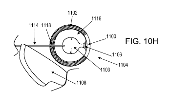
[0087] FIG. 24 depicts one embodiment of an orthopedic device coupled to a
needle using
a suture.
18
CA 02736499 2011-03-08
WO 2010/030933 PCT/US2009/056724
[0088] FIGS. 25A and 25B are superior and anterior elevational views of one
embodiment
of a needle driver.
[0089] FIG. 26 is a superior elevational view of the needle driver of FIG. 25A
loaded with
the orthopedic device of FIG. 24.
[0090] FIG. 27 is a superior elevational view of the needle driver of FIG. 25A
loaded with
an embodiment of an orthopedic device with a coiled suture tether.
[0091] FIGS. 28A and 28B are superior elevational and side cross-sectional
views of
another embodiment of a needle driver with a flanged mount and loaded with the
orthopedic
device of FIG. 24.
[0092] FIG. 29 is a side elevational view of the needle driver in FIG. 28A
loaded with the
orthopedic device of FIG. 27.
[0093] FIGS. 30A, 31A and 32A are schematic side cutaway views depicting the
use of the
system in FIG. 28A in a joint, whereas FIGS. 30B, 31B and 32B are the
corresponding
superior cutaway views, respectively.
[0094] FIGS. 33A and 33B schematically depict an orthopedic device used with a
perforated sleeve, before and after opening of the perforation zone,
respectively. FIGS. 33C
and 33D are schematic side elevational views of the perforation zone in FIGS.
33A and 33B,
respectively.
[0095] FIGS. 34A and 34B schematically depict an orthopedic device used with a
sleeve
comprising a pre-formed opening, before and during use, respectively.
[0096] FIGS. 35A and 35B schematically depict an orthopedic device used with
another
sleeve comprising a pre-formed opening, before and during use, respectively.
[0097] FIG. 36 schematically depicts an orthopedic device in a sleeve with a
non-tapering
cavity.
[0098] FIG. 37 illustrates a sleeved orthopedic device with a needle sheath in
a sealed
pouch.
19
CA 02736499 2011-03-08
WO 2010/030933 PCT/US2009/056724
[0099] FIGS. 38A to 38C illustrate various examples of an orthopedic device
within a
capped enclosure.
[0100] FIGS. 39A and 39B schematically depict a sleeved orthopedic device with
a needle
driver, before and during use, respectively.
[0101] FIG. 40A illustrates a sleeve with a folded configuration; FIG. 40B is
an axial cross-
section of the sleeve in FIG. 40A.
[0102] FIG. 41A illustrates a sleeve with a rigid section and a flexible
section; FIG. 41B is
an axial cross-section of the sleeve in FIG. 41A.
[0103] FIG. 42A illustrates another sleeve with a rigid section and a flexible
section; FIG.
42B is an axial cross-section of the sleeve in FIG. 42A.
[0104] FIG. 43A illustrates a rigid sleeve; FIG. 43B is an axial cross-section
of the sleeve
in FIG. 43A.
[0105] FIG. 44A illustrates an open sleeve; FIG. 44B is an axial cross-section
of the sleeve
in FIG. 44A.
[0106] FIG. 42A illustrates another sleeve with a rigid section and a flexible
section; FIG.
42B is an axial cross-section of the sleeve in FIG. 42A.
[0107] FIG. 43A illustrates a rigid sleeve; FIG. 43B is an axial cross-section
of the sleeve
in FIG. 43A.
[0108] FIG. 44A illustrates an open sleeve; FIG. 44B is an axial cross-section
of the sleeve
in FIG. 44A.
[0109] FIGS. 45A and 45B are a side cross-sectional view and a superior
cutaway view,
respectively, of an embodiment of a delivery device with a bobbin and loaded
with an
orthopedic device.
[0110] FIGS. 46A-46E are schematic cutaway views of one embodiment for
implanting an
orthopedic device in a joint space using a delivery device comprising a
bobbin. FIGS. 46A,
46B, 46D and 46E are side cutaway views through the delivery device, whereas
FIG. 46C is a
superior cutaway view.
CA 02736499 2011-03-08
WO 2010/030933 PCT/US2009/056724
[0111] FIG. 47 is a side cross-sectional view of an alternative embodiment of
a delivery
device with a bobbin and loaded with an orthopedic device.
[0112] FIG. 48 is a side cross-sectional view of an additional embodiment of
the delivery
device of FIG. 47 with a lockable housing.
[0113] FIG. 49 is a schematic superior elevational view of a lockable bobbin.
[0114] FIG. 50 is a schematic superior elevational view of a lock.
[0115] FIG. 51A is a side elevational view of another embodiment of a delivery
instrument
for an orthopedic device; FIGS. 51B and 51C are front elevational views of the
push delivery
instrument in FIG. 51A; FIG. 51D is a longitudinal cross-sectional view of the
delivery
instrument in a retracted position.
[0116] FIGS. 52A and 52B are schematic superior elevational views of an
actuating
member and an orthopedic device in an extended position and a post-extension
retracted
position.
[0117] FIGS. 53A and 53B are side and superior elevational views of another
embodiment
of a delivery instrument; FIG. 53C depicts the partial deployment of an
orthopedic device
from the delivery instrument in FIG. 53B.
[0118] FIGS. 54A to 54C schematically depict cross-sectional views of the
delivery
instrument and orthopedic device in FIG. 54D during an implantation procedure.
[0119] FIGS. 55A and 55B are cross-sectional views of one embodiment for
securing an
orthopedic device in a joint space using a suture. FIG. 55A is a longitudinal
cross-sectional
view through the joint, whereas FIG. 55B is the corresponding axial cross-
sectional view.
[0120] FIGS. 56A and 56B are schematic superior elevational views of an
orthopedic
device comprising anchors.
[0121] FIG. 56C is a schematic superior cutaway view depicting the use of the
orthopedic
device depicted in FIGS. 56A and 56B in a joint.
[0122] FIGS. 57A and 57B are schematic superior elevational views exemplary
anchors
that may be used to anchor an orthopedic device.
21
CA 02736499 2011-03-08
WO 2010/030933 PCT/US2009/056724
[0123] FIG. 57C is a schematic superior cutaway view depicting the use of the
system in
FIG. 57B in a joint.
[0124] FIGS. 58A and 58B are schematic side views of anchors that may be
included in the
embodiment in FIGS. 56A, 56B, and 56C.
[0125] FIGS. 59A to 59D are schematic side views of various tips that may be
used to
anchor an orthopedic device.
[0126] FIGS. 60A and 60B are schematic side views of exemplary anchors that
may be
used to anchor an orthopedic device.
[0127] FIGS. 61A and 61B are schematic superior cutaway views of one
embodiment for
anchoring an orthopedic device in a joint space using an injectable.
[0128] FIGS. 62A to 62B are schematic superior cutaway views of another
embodiment for
anchoring an orthopedic device in a joint space using a deformable fastener.
[0129] FIG. 63 is a schematic superior cutaway view of an additional
embodiment for
anchoring an orthopedic device in a joint space using a slit fastener.
[0130] FIG. 64A schematically illustrates a side cross-sectional view of an
embodiment of
an orthopedic device with a removable core; FIG. 64B is a superior elevational
view of the
orthopedic device in FIG. 64A with its core separated from its articular
layer; FIG.64C is a
superior elevational view of the orthopedic device in FIG. 64A with its core
partially pulled
from its articular layer.
[0131] FIGS. 65A to 65C are schematic superior elevational views of another
embodiment
of an orthopedic device depicting the separation of its removable core.
[0132] FIGS. 66A and 66B are schematic superior elevational views of another
embodiment of an orthopedic device with two separate cores, before and after
core removal,
respectively; FIGS 66C to 66F depict front elevational views of various
channel system
configurations of an orthopedic device.
[0133] FIG. 67 depicts one embodiment of an orthopedic device comprising an
open ended
channel system.
22
CA 02736499 2011-03-08
WO 2010/030933 PCT/US2009/056724
[0134] FIGS. 68A and 68B are schematic superior elevational views of another
embodiment of an orthopedic device, before and after core removal,
respectively.
[0135] FIGS. 69 to 73 depict various embodiments of orthopedic devices.
[0136] FIGS. 74A and 74B are superior and side cross-sectional views of an
orthopedic
device with a removable core and coupled to a suture; FIGS. 74C and 74D depict
the
orthopedic device in FIGS. 74A and 74B after suture removal but before core
removal.
[0137] FIG. 75 illustrates the orthopedic device of FIG. 68A with an optional
suture
coupling.
[0138] FIGS. 76A to 76N are schematic cross-sectional views of one embodiment
for
implanting an orthopedic device with a removable core in a joint space using a
suture. FIGS.
76A, 76C, 76E, 76G, 761, 76K and 76M are longitudinal cross-sectional views
through the
joint, whereas FIGS. 76B, 76D, 76F, 76H, 76J, 76L and 76N are the
corresponding axial
cross-sectional views, respectively.
[0139] Throughout the figures, the same reference numerals and characters,
unless
otherwise stated, are used to denote like features, elements, components or
portions of the
illustrated embodiments. In certain instances, similar reference number
schemes are used
whereby the reference numerals referred to as "AA" in reference numeral "AAxx"
correspond to a figure while the "xx" is directed to similar or
interchangeable features,
elements, components or portions of the illustrated embodiments in different
figures. In
certain instances, similar names may be used to describe similar components
with different
reference numerals which have certain common or similar features. Moreover,
while the
subject invention will now be described in detail with reference to the
figures, it is done so in
connection with the illustrative embodiments. It is intended that changes and
modifications
can be made to the described embodiments without departing from the true scope
and spirit
of the subject invention as defined by the claims.
DETAILED DESCRIPTION OF THE INVENTION
[0140] As should be understood in view of the following detailed description,
this
application is generally directed to systems and methods for minimally-
invasive treatment of
bone joints, in both medical and veterinary settings (including both small and
large animal
23
CA 02736499 2011-03-08
WO 2010/030933 PCT/US2009/056724
veterinary medicine). Bone joints contemplated for various embodiments of the
orthopedic
systems and methods include, but are not limited to, hands (fingers and
thumbs, between
phalanges, metacarpals and/or carpals), feet (in the toes, between phalanges,
metatarsals
and/or tarsals), wrists, elbows, shoulders, knees, hips, and the spine
(particularly at the neck
and lower back). In some embodiments, an orthopedic device comprises a shape
memory
body that is inserted into the joint space, which may restore proper joint
alignment and joint
mobility affected by degenerative processes. In some embodiments, the
orthopedic device
has a generally arcuate or rectilinear configuration, which may enhance self-
centering
positioning of the orthopedic device when deployed.
[0141] Referring to FIG. IA, in one embodiment, the orthopedic device 100a
comprises a
resilient or flexible elongate body 105 with a proximal end 110a and a distal
end 120a, and
adapted to undergo configurational change. For example, the elongate body 105
of
orthopedic device 100a may have a straight configuration as depicted in FIG.
IA, but may
also have, for example, an arcuate "C"-shape configuration as shown in FIG.
1B, and/or a
spiral-shape configuration in FIG. 1C. The change from one configuration to
another may,
for example, facilitate implantation of the orthopedic device in a minimally
invasive manner,
and/or facilitate force redistribution in the joint during movement or
positioning.
[0142] In one particular embodiment, the distal end 120a of the orthopedic
device 100a
may be advanced or inserted into the body of a patient first, before the
proximal end 110a of
the orthopedic device 100a is inserted. In some embodiments, the orthopedic
device 100a has
a shape or configuration that facilitates its loading into a lumen within a
needle, cannula, or
other device for delivering the orthopedic device to the implantation site.
The straightened
configuration of orthopedic device 100a may be used for delivery of the
orthopedic device
100a from a substantially straight needle. As the device 100a exits the needle
or cannula, the
configuration of the device 100a may change to assume the arcuate or spiral
configurations of
FIGS. lB and 1C. In another example, the elongate body 105 of the orthopedic
device 100a
may be bent or biased to a curve to permit delivery from curved or other non-
linear needles
or cannulae. Thus, the orthopedic device need not have a linear delivery
configuration as
depicted in FIG. IA. The orthopedic device 100a may also be configured with a
lumen or
one or more apertures to facilitate delivery over a delivery structure, such
as a rigid or
flexible guidewire. Once implanted into the joint, the orthopedic device may
be configured
to re-expand to its pre-delivery configuration, or may expand to a different
configuration.
24
CA 02736499 2011-03-08
WO 2010/030933 PCT/US2009/056724
The deployment configuration may be different, depending upon the base
configuration of
the orthopedic device, and/or whether the orthopedic device has a resilience
or bias to one or
more particular configurations. The resulting configuration may also result
from anatomical
restrictions, for example, relating to the dimensions of the joint capsule, or
the geometry of
the articular surface. The deployment configuration in the joint capsule may
vary in use,
depending upon the joint position, the body position of the patient (e.g.
standing or lying
down) and other conditions which may alter the forces acting on the joint and
the orthopedic
device. In one example, an orthopedic device has an arcuate configuration that
is less-
curved, or has a larger major diameter, than the device as fully deployed in
the joint, or has
an enlarged configuration with at least one dimension that is larger than the
corresponding
joint space dimension when deployed in the joint space.
[0143] In one embodiment, the orthopedic device may be configured and
implanted to
permit its displacement and/or deformation within the joint. In some
instances, the
movement and/or deformation facilitates the conformation of the orthopedic
device to the
natural movement of the bones through the range of motion of the joint. For
example, the
orthopedic device may be implanted into a joint without any attachment to
adjacent tissue and
constrained only by the joint capsule and/or ligaments within the joint. In
some examples,
because the device is not fixed in place (e.g. attached to either end of bones
in a joint), the
device may "float" between the ends of the bones in a joint. In some
embodiments, a floating
design and implantation procedure may provide a mechanical advantage over that
of a fixed-
type orthopedic device that is rigidly attached to bone tissue by
redistributing forces acting on
the joint.
[0144] For example, the "open ring," "hoop" or "coil" configuration, or any
"open"
embodiment, including open polygons of an orthopedic device, may permit a
greater range of
deformation than closed structures. An open design may facilitate the
distribution of the
loading, shearing and/or compressive forces seen by the articulation and/or
loading of the
joint. Thus, in certain open embodiments of orthopedic devices that are
flexible, such as
orthopedic device 100b, the open configuration may offer reduced or minimal
resistance to
shape change. Thus, the orthopedic device 100b can spring open or closed as
force is applied
to the device or to the joint, but still maintain a bearing, cushion,
slidable, or articulate
surface. However, orthopedic devices with a closed configuration may also be
used and may
also have deformation properties.
CA 02736499 2011-03-08
WO 2010/030933 PCT/US2009/056724
[0145] In some embodiments, the gap between the proximal and distal ends of an
orthopedic device with an open configuration could be extended to the entire
length of the
orthopedic device, e.g. when a device is completely straightened. However,
various
embodiments of an orthopedic device may be configured with functional
operating ranges
allow varying degrees of flexion and gap widening to support loads and
articulation in the
joint. In some embodiments, the functional operating range is based upon the
amount of
stress and strain that the orthopedic device can undergo without significant
plastic change
(e.g. less than 5%). In some embodiments comprising a shape memory material
such as
nickel-titanium, the functional operating range may lie within the range of
pseudoelastic
deformation of the shape memory component, e.g. a Nitinol core that can
undergo strain up to
about 8%. In one embodiment, the functional flexion in an open orthopedic
device allows for
a change in the gap between the open ends of the orthopedic device in situ to
flex in a range
from about 0.5 to about 6 times or more the distance between the gap when the
orthopedic
device is in its natural state, either pre-implantation or in situ. In one
embodiment, the
deformation or flex range is roughly from about 2 to about 6 times or greater
the natural gap
distance, and in another embodiment the flex range is about 3 to about 5 times
greater. In one
example, the orthopedic device has a flex range with an upper limit of about 4
times. In one
embodiment the functional gap can be as wide as a first dimension, diameter,
or width of the
over all orthopedic device. Thus, orthopedic device 100b may allow for the
redistribution of
the compressive and/or shearing forces, as well as the resulting wear along
the device. In
certain embodiments, the orthopedic devices comprise arcuate configurations,
such as an
open circle or continuous spiral configurations, rather than closed
configurations like a
complete ring or closed circular shape. The open configurations may result in
increased
dissipation or redistribution of loading and compression forces though at
least one or two
deformations in the orthopedic device. First, an open ring may allow for
dynamic loading
response as force that is applied to the joint is partially dissipated by the
force necessary to
radially-outwardly deform the open ring or spiral into a larger radius
profile. In one
embodiment, the operating range of radial deformation of an arcuate orthopedic
device is in
the range of about 0 to about 50% of the orthopedic device profile diameter
within the joint.
Second, as discussed above, the compression of the articular layer may result
in cross-
sectional deformation into a flatter shape, which may also dissipate force or
pressure in the
joint.
26
CA 02736499 2011-03-08
WO 2010/030933 PCT/US2009/056724
[0146] In one embodiment, the orthopedic device 100b is sized to snugly fit
into the joint
capsule itself. In some specific embodiments, one or more portions of the
orthopedic device
may be sized and/or configured to conform to the dimensions of the joint
capsule. This fit
may facilitate the seating or centering of the orthopedic device 100b with
respect to the axis
of the bones of the joint, such as in a proximal or distal interphalangeal
(PIP/DIP) joint of a
finger or an MCP joint of a knuckle.
[0147] As used herein, "arcuate" may refer to curved or rounded configurations
or shapes,
but can also include generally arcuate configurations and shapes that have
some straight
aspect or element with curved or rounded configurations or shapes. As used
herein, arcuate
and generally arcuate shapes can include open or closed "C", "O", "S", spiral,
nautilus, "Q"
and other generally arcuate shapes which can be planar or non-planar. Certain
embodiments
of the orthopedic device may have open or closed rectilinear configurations,
which can
include polygons such as triangles, squares, rectangles, diamonds, rhombuses,
pentagons,
hexagons, octagons and other shapes with generally straight edges, and further
including
shapes and configurations that are generally rectilinear having some curved
edge or corners
or segments among rectilinear shapes. As used herein, rectilinear and
generally rectilinear
shapes can include "N", "M", "W", "Z", "T", "Y", "V", "L", "X" and other
generally
rectilinear shapes. FIG. 5F, for example, depicts an embodiment of a
rectilinear orthopedic
device 570f, comprising a "W"-shape configuration. Various embodiments of
generally
arcuate or generally rectilinear shapes can include shapes with both
rectilinear and arcuate
portions, such as a "P", "R", "B", and "U".
[0148] Embodiments of the orthopedic device may have three major dimensions,
which can
correspond to a first major dimension, a second major dimension and a third
major
dimension. In one embodiment, the first major dimension, second major
dimension and third
major dimension correspond to a width, a height and a thickness, respectively.
Certain
embodiments have a thickness which corresponds to the smallest dimension,
which may
generally correspond to the spacing between articulating surfaces of tissue
such as bone or
cartilage in a joint. In one embodiment, the width and height can be the same,
such as with a
circular or square-shape orthopedic device. In other embodiments, the height
and width may
be different, as with an oval shape or a rectangle or other shape with non-
equal height and
width. In some embodiments, the orthopedic implant can be implanted in joints
of varying
sizes, in which the first major dimension and second major dimension may have
a range of
27
CA 02736499 2011-03-08
WO 2010/030933 PCT/US2009/056724
about 0.0394 to about 4.0 inches (or about 1.0 to about 101.6 mm) and the
third major
diameter may have a range of roughly about 0.001 to about 0.50 inches (or
about 0.025 to
about 15 mm). Orthopedic devices having other dimensions may also be used,
including but
not limited to orthopedic devices configured for larger joints such as the
spine, knee, hip,
ankle, and shoulder, for example. Although the orthopedic device may be
implanted between
the articular surfaces of two bones, the articular surfaces are not limited to
the hinge joints
and may include sliding joints. In some examples, the orthopedic device may be
inserted
into various joints and other locations of the spine, including the facet
joints, in an
intervertebral disc, or in the post-discectomy space between the endplates of
two adjacent
vertebral bodies.
[0149] As mentioned previously, certain embodiments of the orthopedic device
may have a
narrowed configuration or a reduced profile to fit in a lumen of a delivery
tube or delivery
device, or through a small opening in a joint capsule. In one embodiment, a
narrowed
configuration comprises the reduction of the first major dimension, second
major dimension
or third major dimension, or a combination thereof. In some embodiments with
narrowing
configurations, one or more dimensions are reduced while one or more other
dimensions are
increased. In one embodiment, the orthopedic device can be moved into a
narrowed
configuration by pinching, squeezing or restraining the device so that parts
of the orthopedic
device overlap, such as a "C"-shape body being collapsed into an alpha shape
(a), a gamma
shape (y), a twisted shape, a helix, and/or a multi-planar configuration, as
illustrated in the
embodiments of FIGS. 5D and 5E, for example. In one embodiment, the orthopedic
device
may be manipulated into a straightened or a substantially straightened
configuration. In one
embodiment, the orthopedic device may have a substantially straightened
configuration,
including a completely straightened, linear configuration, as well as
configurations in which
at least a part of the orthopedic device is straightened or partially
straightened, configurations
in which arcuate orthopedic devices can be made less-arcuate and
configurations in which
rectilinear orthopedic devices can be made less-rectilinear.
[0150] Referring back to FIG. IA, some embodiments of the orthopedic device
may have a
relatively uniform width or diameter along its elongate length. However, in
other
contemplated embodiments, the width of the device body can vary along its
length. For
example, some orthopedic devices may have one or more tapered sections along a
portion of
its length, or be tapered along the device's entire length. The tapered
section may have a
28
CA 02736499 2011-03-08
WO 2010/030933 PCT/US2009/056724
linear or a non-linear taper configuration, and embodiments with two or more
tapered
sections need not taper in the same direction. The width, or other dimensions
of the
orthopedic device, can vary from large to small or small to large, making the
device thicker in
some portions than in others. In one embodiment, the device may be radially
compressible
along part or over the entire length of the device. In one embodiment, the
device may be
compressed such that its cross-sectional area is reduced, so that the device
may exit a delivery
system and expand to a larger cross-sectional area. In one embodiment, the
device can be
axially compressed or axially stretched along part or over the entire length
of the device.
[0151] In one embodiment, the orthopedic device 100a comprises a shape memory
material. For example, the shape memory material can be made from a shape-
memory
material, such as Nitinol, or a shape memory plastic, polymeric, or synthetic
material, such as
polycarbonate urethane. One example of this type of a polyurethane or
polyurethane-urea
polymer shape memory material is described in United States Patent Publication
2002/0161114 Al, which is hereby incorporated by reference in its entirety and
which
describes a shape memory polyurethane or polyurethane-urea polymer including a
reaction
product of: (A) (a) silicon-based macrodiol, silicon-based macrodiamine and/or
polyether of
the formula (I): A--[(CH2)m--O-]n--(CH2)m--A', wherein A and A are endcapping
groups; m
is an integer of 6 or more; and n is an integer of 1 or greater; (b) a
diisocyanate; and (c) a
chain extender; or (B) (b) a diisocyanate: and (c) a chain extender, where the
polymer has a
glass transition temperature which enables the polymer to be formed into a
first shape at a
temperature higher than the glass transition temperature, and where the
polymer is
maintained in the first shape when the polymer is cooled to a temperature
lower than the glass
transition temperature, so that the polymer is capable of resuming its
original shape on
heating to a temperature higher than the glass transition temperature. Various
embodiments
may include a shape memory polymer alone, or a blend of two or more of the
shape memory
polyurethane or polyurethane-urea polymers or at least one shape memory
polyurethane or
polyurethane-urea polymer defined above in combination with another material.
Other
embodiments relate to processes for preparing materials having improved
mechanical
properties, clarity, processability, biostability and/or degradation
resistance and devices or
articles containing the shape memory polyurethane or polyurethane-urea polymer
and/or
composition defined above.
29
CA 02736499 2011-03-08
WO 2010/030933 PCT/US2009/056724
[0152] In other embodiments, the orthopedic device may comprise any of a
variety of rigid,
semi-rigid or flexible materials, which may be metallic or non-metallic,
polymeric or non-
polymeric, bioresorbable or non-bioresorbable, lipophilic, hydrophilic or
hydrophobic, for
example. These materials may include but are not limited to stainless steel,
cobalt-chromium,
titanium, pyrolytic carbon, any of a variety of ceramic or hydroxyapatite-
based materials,
polymers such as PTFE, silicone, nylon, polyethylene, polypropylene,
polycarbonate,
polyimide, polycarbonate, polyurethane, PEEK, PEKK and PEBAX, any of a variety
of
bioresorbable materials such as PGA, PLA, PLGA, PDS and the like, as well as
chitosan,
collagen, wax and alginate-based materials, and animal-derived materials such
as small
intestine submucosa (SIS).
[0153] In one embodiment, the orthopedic device 100a comprises an articular
layer 105,
blanket or jacket. The articular layer 105 is sized and configured to be
placed within a body,
such as in a joint, as a layer between bones of the joint to provide a
slidable articulation
surface and/or a cushion. In some embodiments, the articular layer can range
from about
0.001 to about 0.5 inches thick (or about 0.025 to about 13 mm). The
orthopedic device may
or may not include a core, backbone or other support structure, which may
support the
articular layer or contribute or impart certain features or characteristics to
the orthopedic
device. Support structures, such as the core, are described in greater detail
below.
[0154] In one embodiment, the articular layer 105 is configured to be
compressed by forces
acting on the joint. For example, in one embodiment an articular layer may be
compressed
from a substantially circular cross-sectional shape to an oval, elliptical, or
football shaped
cross-sectional shape. As the compression occurs, the amount of surface
coverage of the
articular layer with respect to bony joint contact, resulting in reduced in
relative pressure
across the joint. In one embodiment, the operating range of compression of an
orthopedic
device is in the range of about 0 to about 50% of the cross-sectional diameter
or other
dimension along the axis of compressive force.
[0155] The articular layer 105 may comprise one or more layers of material,
and any of a
variety of materials may be used for each layer. In certain embodiments of the
orthopedic
device 100a, the body of the orthopedic device 100a comprises an articular
layer with shape-
memory properties, with or without any backbone or other type of support
structure. The
shape-memory properties may include but are not limited to temperature-induced
configuration changes as well as stress-induced pseudoelastic properties. In
certain
CA 02736499 2011-03-08
WO 2010/030933 PCT/US2009/056724
embodiments, the articular layer 105 materials may include but are not limited
to silicone,
PTFE or ePTFE, ultra high molecular weight polyurethane or and any implantable
grade
material, or other materials disclosed above. The articular layer 105 can be
compliant and/or
compressible, or may have a non-compressible construction. In certain
embodiments, the
articular layer 105 can have any of a variety of durometers (material
hardness) from about 30
to about 90 Shore A, for example. In certain embodiments, the articular layer
105 may
comprise a porous material, which may have a closed or open-pore structure.
The porous
coatings, layers or structures may include but are not limited to macroporous
or nanoporous
coatings or structures. In some instances, a porous coating may facilitate
tissue ingrowth
and/or augment the inflammatory response to the orthopedic device, if any. In
another
embodiment, the coating material can form a casing (or covering) that is
spongy or harder or
less compliant. The pores of the material could be loaded with one or more
therapeutic
agents. The casing could form a scaffold for tissue ingrowth and could be used
in joints with
certain wear characteristics, but is not limited to use with these joints. In
some embodiments,
the articular layer 105 may be coated with a secondary surface layer, such as
another polymer
of a different material property, or an anti-friction high wear material such
as Parylene, or
other similar materials which are known to the art as providing for a low
friction surface.
[0156] In certain embodiments, the articular layer 105 may contain a material
or a drug to
inhibit or promote inflammation, joint deterioration etc., or a material or
drug to encourage
tissue regeneration or device encapsulation. For example, certain embodiments
of the
articular layer 105 may be coated with or contain one or more therapeutic
agents, such as a
long-acting steroid or a disease-modifying anti-rheumatic drug (DMARD). DMARDs
include but are not limited to agents such as gold, D-penicillamine,
methotrexate,
azathioprine and cyclophosphamide, leflunomide, etanercept, infliximab,
minocycline and
certain anti-malarial agents used for arthritis treatment, for example. The
therapeutic agents
need not be limited to joint-specific therapy agents, however. In other
embodiments, the
therapeutic agent may include an antibiotic (e.g. a macrolide, a
cephalosporin, a quinolone,
an aminoglycoside, a beta-lactam or beta-lactamase inhibitor, a lincosamide,
or glycopeptides
antibiotic, etc.), a sclerosing agent (e.g. bleomycin, tetracycline, talc,
alcohol, sodium
tetradecyl sulfate, etc.), or other type of inflammation-inducing agent, a
growth factor (e.g.
connective tissue growth factor, cartilage-derived retinoic acid sensitive
protein), and other
agents. In some embodiments, one or more therapeutic agents may be injected or
infused
into a joint space, separate from the orthopedic device, using any of a
variety of forms
31
CA 02736499 2011-03-08
WO 2010/030933 PCT/US2009/056724
(aqueous solution, suspension, oil, foam, a separate drug eluting disc or
other structure, etc.).
These therapeutic agents may include viscosupplements (e.g. hylan G-F 20 such
as Synvisc ,
or various formulations of sodium hyaluronate such as Hyalgan , Suppartz ,
Euflexxa and
Orthovisc ).
[0157] In other embodiments, the articular layer may comprise a plurality of
surface
projections and/or pores, which may cause a mechanical irritant response when
implanted
and may induce the growth of new tissue or cartilage, or an organization of
fluids contained
in the joint . . The projections and/or pores may be grossly visible on the
surface of the
articular layer, or may be nano- or micro-sized structures. The projections
may comprise
discrete surface structures or aggregated structures, including but not
limited to hooks, barbs,
tubes, rods, cones, spheres, cylinders, loops, pyramids, or a mix thereof.
These structures
may have a size in the range of about 5 nm to about 5 mm or more, sometimes
about 50 nm
to about 3 mm, and other times about 500 nm to about 1 mm, and in still other
times about 1
m to about 500 m.
[0158] In one embodiment the coating and or covering can be used to stimulate
a
thrombotic or coagulant response, and/or organization of tissues or fluids it
contacts. For
example, the coating or covering may comprise a hemostatic agent such as
chitosan, zeolite,
fibrinogen, anhydrous aluminum sulfate, titanium oxide, one or more clotting
factors or other
constituents of the blood clotting cascade.
[0159] In some embodiments, one or more therapeutic agents may be mixed with a
polymer material which may either biodegradable or non-biodegradable. Thus,
release of the
therapeutic agents may occur by elution from the polymer material, and/or by
degradation of
the polymer material. For example, an orthopedic device may comprise a
material or
reservoir being drug loaded and dissolvable through features provided in a
jacketing or
coating material, such as through micro holes, pores, or some other feature.
In certain
embodiments the articular layer is provided with reservoirs, depots, cavities,
wells, pockets,
porous materials, bubbles or capsules for drug delivery. In one specific
example, the
orthopedic device could be a drug-loaded element that slowly dissolves to
elute a drug of
some sort through a casing that is spongiform or porous. This would leave
behind the casing
after the ring has dissolved. In some embodiments, timed drug delivery could
be configured
for more controllable dosing. For example, about 75% to about 90% of a
therapeutic agent
may be released or dissolved over a timeframe of anywhere from about 4 hours
to about 4
32
CA 02736499 2011-03-08
WO 2010/030933 PCT/US2009/056724
months or more, sometimes from about 24 hours to about 6 weeks or more, other
times from
about 72 hours to about 4 weeks, and still other times from about 2 weeks to
about 4 weeks.
In other embodiments, the casing would maintain the space filling or
cushioning feature
desired and/or allow for tissue organization or in-growth.
[0160] The therapeutic agent may be provided on an outer surface or an inner
surface of the
articular layer, or within a volume or layer of the articular layer. As
mentioned previously,
the articular layer may comprise one or more rate control layers to alter the
rate of therapeutic
agent release. The rate control layer may comprise, for example, polymer
layers with a
reduced permeability or smaller pore structure.
[0161] In one specific embodiment, a coating may comprise a xenograft,
allograft or
autograft biological covering, from a live and/or cadaveric donor, or a
biological material
grown from a tissue culture. For example, tissue harvested directly from the
patient could be
harvested using a laparoscope or other tissue removal and collection system
and then affixed
to the core, articular layer, preshaped ring or backbone and secured to the
orthopedic device.
The tissue may include but is not limited to omental tissue, ligamentous or
tendinous tissue,
cartilage tissue, bone tissue and the like. The graft material may retain the
native tissue
structure or may have undergone additional mechanical processing (e.g.
crushing, blending,
etc.) or biological processing (treatment with glutaraldehyde or other cross-
linking agents,
sterilization with electron-beam, gamma irradiation or ethylene oxide, etc.)
The device could
then be loaded into a delivery cannula and inserted and ejected (deployed) in
the same
fashion as the delivery systems employed and described herein. In some
embodiments, the
articular layer 105 comprises a cartilage replacement material, or a natural
or synthetic
cartilage.
[0162] In another embodiment, an orthopedic device is covered with a material,
biological
agent, or other coating that expands in volume with contact to fluids. The
fluids may be the
endogenous fluid found in the joint itself, and/or externally added fluids.
Expandable
materials may permit the insertion of a device of a diameter that is smaller
than the fully
expanded finished diameter. For example, a coating on the backbone or the
articular layer
could be hydrophilic in that it could transition from one configuration or
diameter (small for
insertion) to a larger configuration or diameter when contacting either the
body fluid or some
fluid provided from an outside source, such as saline.
33
CA 02736499 2011-03-08
WO 2010/030933 PCT/US2009/056724
[0163] In one specific embodiment, the expandable or swellable covering may
comprise a
composite or matrix with a polymer and a biological material i.e. tissue,
including but not
limited to cartilage, collagen, ligaments, muscle, etc. In one embodiment, the
scaffold could
be a polymer-based material. In various embodiments, the casing or covering of
the
orthopedic device is configured to swell from the small insertion dimension or
diameter after
implantation to a larger finished dimension or diameter. In some alternate
embodiments,
such as those disclosed in U.S. Application Ser. No. 12/099,296, filed April
8, 2008, the
orthopedic device may comprise an inflatable structure. The inflatable
structure may be
inflated with a gas, liquid, gel, or slurry which may or may not be curable to
a solid state.
The inflatable structure may also be expanded by filling the structure with a
volume of solid
structures, such as microspheres or other small structures.
[0164] In certain embodiments, the articular layer 105 is radiopaque, and can
augment the
visibility of the device when implanted as viewed by X-ray and/or fluoroscopic
equipment.
In one embodiment, the radiopacity of the articular layer 105 is provided by
radiopaque
markers or structures (not shown here) on or embedded in the layer 105, or by
loading or
doping the articular layer 105 with platinum, gold or other biocompatible
metal.
[0165] In various embodiments, any of the features of the articular layer or
coatings
mentioned herein may be combined on the orthopedic device, either as different
layers of the
orthopedic device or as different sections or regions of the orthopedics
device. In one
embodiment, an articular layer or coating can provide for tissue ingrowth or
fusion with bone,
cartilage, or other tissue while another surface provides a low-friction
surface to another side
of the joint. Any combinations are possible. In some embodiments, adhesives or
transitional
polymer layers may be provided to facilitate the attachment of two or more
other layers of the
articular layer.
[0166] As described previously, the orthopedic device can have an arcuate,
rectilinear or
non-straightened configuration once it is implanted in a joint. Some non-
limiting examples
of arcuate configurations include an open ring (also called an open hoop or an
open loop)
such as is shown in the embodiment in FIG. 1B, and a nautilus-style spiral as
is shown in the
embodiment in FIG. 1C. Referring to FIG. 1B, the open hoop arcuate
configuration of the
orthopedic device 100b has a proximal end 110b and a distal end 120b in
relation to insertion
into the body of a patient, such as into a joint. In certain embodiments, the
orthopedic device
100b of FIG. lB may have similar attributes and characteristics of the
orthopedic device 100a
34
CA 02736499 2011-03-08
WO 2010/030933 PCT/US2009/056724
of FIG. IA, such as shape memory and/or an articular surface 105. In certain
embodiments,
orthopedic device 100b is an arcuate configuration of orthopedic device 100a.
In certain
embodiments, the orthopedic device 100a is biased to the configuration as
shown for
orthopedic device 100b. The bias may be a preferred configuration for a
flexible, pliable,
bendable device. In certain embodiments, the orthopedic device of 100a may
change to from
one configuration to another (e.g. from the configuration of orthopedic device
100a in FIG.
1A to the configuration of orthopedic device of 100b in FIG. 1B) by a change
in ambient or
implantation site temperature, by a release from deformation stresses, or by
the introduction
of an activating medium or material. In certain embodiments, the orthopedic
device is
reversibly configurable between various shapes or geometries.
[0167] As mentioned previously, the orthopedic device may also comprise a
closed shape
that forms a complete perimeter along at least one section or portion of the
device. In FIG.
1D, for example, the orthopedic device 130a comprises an expanded
configuration with a
closed triangular shape. Although the triangular shape in FIG. 1D comprises an
equilateral
triangular shape with uniform angles 135a, 140a and 145a, in other
embodiments, one or
more angles may be different from the other angles. Also, although the inner
angles 135a,
140a and 145a, along with outer angles 150a, 155a and 160a have sharp angles,
in other
embodiments, one or more of these angles may be rounded. In its collapsed
state, depicted in
FIG. 1E, the inner angles 135b, 140b and 145b of the orthopedic device 130b
may narrow to
collapse its triangular shape into an arrow shape. The orthopedic device 130b
may also have
a bending section 165a that collapses from a straight configuration to a bent
configuration to
facilitate the reduction in the cross-sectional profile of the orthopedic
device 165b. Although
the orthopedic device 130b is depicted as generally collapsing within the
plane of the
orthopedic device 130a in its expanded configuration, in some embodiments,
this and other
orthopedic devices disclosed herein may also fold onto themselves or otherwise
collapse out
of plane to reduce their cross-sectional profile. In other embodiments, the
orthopedic device
may comprise other polygonal shapes or curvilinear shapes, with angles and/or
sides that may
be uniform or different, with angles that narrow or widen when changing from
one
configuration to another configuration. Although several embodiments described
herein have
a base configuration that is the expanded or deployed configuration, in other
embodiments,
the base configuration may be the delivery configuration. In still other
embodiments, the
orthopedic device may comprise a malleable or plastic material or structure
with any bias
toward one or more configurations.
CA 02736499 2011-03-08
WO 2010/030933 PCT/US2009/056724
[0168] FIGS. IF and 1G illustrate another embodiment of an orthopedic device
170a/170b
comprising a closed arcuate configuration. In its deployed configuration, the
orthopedic
device 170a comprises a circular configuration, but other embodiments, may
comprise an
oval or ovoid shape (e.g. one end being larger than the other end). To
transform the device
170a to its delivery configuration, the orthopedic device 170a shortens along
a first
dimension 175a while lengthening along a second dimension 180a. In some
embodiments,
the delivery axis of the orthopedic device 170b may be transverse to the first
dimension 175b,
or parallel to the second dimension 180b.
[0169] One example of a nautilus-style spiral arcuate configuration is the
embodiment of
an orthopedic device 100c as shown in FIG. 1C. The orthopedic device 100c has
a proximal
end 110c and a distal end 120c in relation to insertion into the body of a
patient, such as into
a joint. In certain embodiments, orthopedic device 100c has many similar
attributes and
characteristics of orthopedic device 100a and/or 100b, such as shape memory
and/or an
articular surface 105. In certain embodiments, orthopedic device 100b is an
arcuate
configuration of orthopedic device 100a. In certain embodiments, the
orthopedic device of
100a may be altered in to a configuration as shown for orthopedic device of
100c. The bias
may be a preferred configuration for a flexible, pliable, bendable device. In
certain
embodiments the orthopedic device 100a, when unconstrained, can change to the
configuration as shown for orthopedic device of 100c, or by a change in
ambient or
implantation site temperature or the introduction of an activating medium or
material. In
certain embodiments, the orthopedic device is reversibly configurable between
various
shapes or geometries.
[0170] In some embodiments, the orthopedic device is configured to float
inside the joint,
which may better conform to the natural movement of the bones through the
range of motion
of the joint. The nautilus-style spiral arcuate configuration depicted in FIG.
1C, for example,
may also offer certain advantages described for the open hoop arcuate
configuration, or hoop
configuration, but also provides a larger bearing surface to the joint. With
the extended
length of the spiral configuration, the orthopedic device 100c is configured
to provide more
of an articulate surface, which may result in decreased pressure on the bones
by dissipating
forces over a larger surface area. The cross-sectional diameter multiplied by
the number of
winds in a spiral shape roughly equals the surface area coverage of the
articular surface in
conformation with the bones of the joint. For example, a small cross-sectional
diameter of a
36
CA 02736499 2011-03-08
WO 2010/030933 PCT/US2009/056724
spiral configuration allows for a plurality of windings in the spiral. This
plurality of spiral
windings can then adjust to the general surface area of either bone as the
joint articulates.
[0171] As noted previously, some embodiments of the devices can have
additional
structures within it. For example, in FIG. 2 an orthopedic device 200
comprises an elongate
core 240 and an articular layer 230 surrounding at least a portion of the core
240. Referring
back to FIGS. IA-1C, various embodiments of orthopedic devices 100a, 100b
and/or 100c
can either have an elongate core or lack an elongate core. Other embodiments
of orthopedic
devices 100a, 100b and/or 100c may also either have an articular layer or lack
an articular
layer. Thus, the orthopedic device may consist of an elongate core, an
articular layer, or
both. In various embodiments directed to use in PIP, DIP and MCP joints, for
example, the
cross-sectional diameter or thickness of a core can range from roughly about
0.001 to about
0.60 inches (or about 0.025 to about 15 mm) with some embodiments in a range
of roughly
about 0.005 to about 0.015 inches (or about 0.13 to about 0.38mm), and some
embodiments
in a range of roughly about 0.01 to about 0.0125 inches (or about 0.26 to
about 0.32 mm). In
various embodiments, the cross-sectional outer diameter or overall thickness
of an articular
layer can range from roughly about 0.003 to about 0.50 inches (or about 0.076
to about 12.7
mm) with some embodiments in a range of roughly about 0.039 to about 0.118
inches (or
about 1 to about 3 mm), and some embodiments in a range of roughly about 0.078
to about
0.098 inches (or about 2 to about 2.5 mm). In some embodiments a ratio of core
cross-
sectional diameter (or thickness) to articular layer cross-sectional outer
diameter (or
thickness) can range from about 0.000 to about 0.500 inches, and in other
embodiments may
have ranges of ratios from about 2 to about 30. Other dimensions with the
same, similar or
different ratios can be used in other parts of the patient's body. Orthopedic
devices with
cores having other dimensions may also be used, including but not limited to
orthopedic
devices configured for larger joints such as the knee, hip, ankle, and
shoulder, for example.
[0172] As illustrated in the embodiment of FIG. 2, the orthopedic device 200
may include
the elongate core 240 in addition to the articular layer 230. In some
embodiments, the
articular layer 230 surrounds, encapsulates, encloses or covers at least a
portion of the core
240. In some other embodiments, the articular layer 230 can surround or
encapsulate the
entire elongate core 240. As used herein, "surround," "encapsulate" and
"enclose" include
configurations in which a core is not completely surrounded, completely
encapsulated or
completely enclosed. For example, certain embodiments of an orthopedic device
37
CA 02736499 2011-03-08
WO 2010/030933 PCT/US2009/056724
contemplate an articular layer which "surrounds" an elongate core with a
continuous or non-
continuous helical band, discontinuous tabs, or other intermittent articular
layer structure.
[0173] In some embodiments, the articular layer 230 may have some or all of
the features
of other articular layer embodiments described herein. In one embodiment, the
ratio of the
cross-sectional size of the elongate core 240 to the articular layer 230 is in
the range of about
10:1 to 1:10, sometimes in the range of about 5:1 to about 1:5 and other times
with a ratio of
about 2:1.
[0174] In one embodiment, the elongate core 240 comprises a shape memory
material. The
shape memory material may be made from a heat set/shaped shape-memory
material, such as
Nitinol, or a shape memory plastic, polymeric, synthetic material. For
example, one
embodiment of the elongate core 240 comprises a shape memory material
including a shape
memory polyurethane or polyurethane-urea polymer, as described above. In one
embodiment
the elongate core 240 comprises a metal "open" ring such as Nitinol
encapsulated by an
articular layer 230, or outer blanket, comprising silicone. In one embodiment
the elongate
core 240 comprises a hardened polymer. In one embodiment, the elongate core
240 is
configured such that a heat set Nitinol with an arcuate configuration, such as
an open ring
configuration, a horseshoe configuration, or a spiral configuration, can be
straightened for
delivery through cooling or plastic deformation, then recovered to its
original heat-set shape
once released from a delivery system, such as one embodiment using a properly
sized
hypodermic needle. In one embodiment the elongate core 240 comprises a non-
shape
memory material which can be bent or deformed.
[0175] In certain embodiments, the elongate core 240 is coated or impregnated
with a drug
or other therapeutic agents as described previously with respect to the
articular layer. The
therapeutic agents of the elongate core 240 may be the same or different from
the therapeutic
agents of the articular layer or other layers or coatings of the orthopedic
devices.
[0176] FIGS. 3A to 3E are longitudinal cross-sectional views of the orthopedic
devices
100a to 100c in FIGS. 1A to 1C with various configurations of optional support
structures or
cores. FIG. 3A is a schematic cross-sectional view of an orthopedic device
300a comprising
a substantially straightened configuration. In this embodiment, the device
comprises an
elongate core 340a and an articular layer 330a surrounding at least a portion
of the core 340a.
The articular layer 330a has a proximal end 331a and a distal end 332a. The
elongate core
38
CA 02736499 2011-03-08
WO 2010/030933 PCT/US2009/056724
340a has a proximal end 341a and a distal end 342a. In one embodiment, the
orthopedic
device 300a may be a cross-sectional view of the orthopedic device 100a
described above,
with a core 340a. FIG. 3B shows a device an elongate core 340b and an
articular layer 330b
surrounding at least a portion of the core 340b in an open hoop arcuate
configuration. The
articular layer 330b has a proximal end 33 lb and a distal end 332b, while the
elongate core
340b has a proximal end 341b and a distal end 342b. In one embodiment, the
orthopedic
device 300b may be a cross-sectional view of the orthopedic device 100b
described above.
Certain embodiments of a spiral shaped device, such as is shown in FIG. 3C can
have a single
elongate core. For example, orthopedic device 300c comprises a nautilus-style
spiral arcuate
configuration, the device comprising an elongate core 340c and an articular
layer 330c
surrounding at least a portion of the core 340c, the articular layer 330c
comprises a proximal
end 331c and a distal end 332c, and the elongate core 340c has a proximal end
341c and a
distal end 342c. In one embodiment, the orthopedic device 300c may be a cross-
sectional
view of the orthopedic device 100c described above.
[0177] In some embodiments, the elongate core may be wrapped around itself or
comprise
of a number of distinct or separate sections or segments, as shown in FIGS. 3D
and 3E. FIG.
3D shows an orthopedic device 300d with an open hoop arcuate configuration. In
one
embodiment, the orthopedic devices 300d may be a cross-sectional view of the
orthopedic
device 100b described above, with an optional folded or overlapping core 340d.
The device
300d comprises one or more elongate cores 340d wrapped, braided or folded back
along a
length of the device, and an articular layer 330d surrounding at least a
portion of the core(s)
340d. The articular layer 330d has a proximal end 331d and a distal end 332d.
The elongate
core 340d in FIG. 3D comprises a unitary body with a proximal end 341d, a
distal end 342d,
an inner segment 350d, a middle segment 352d and an outer segment 254d. The
segments
350d to 354d may be interconnected as depicted in FIG. 3D, but in other
embodiments may
one or more segments may be separated. The segments of the embodiments
described herein
may themselves have subsegments, e.g. the inner segment 350d may comprise a
proximal
segment and a distal segment. Also, the segments of a core may generally have
a similar
length, such as segments 350d to 354d in FIG. 3D, but one or more segments may
also have a
different length In some embodiments for example, two or more elongate cores
340d are
situated in a roughly parallel or co-linear orientation, which can be twisted
or braided or
interlocked. Other embodiments of the orthopedic device need not be limited to
a single
elongate core or backbone, but may have a plurality of cores or backbones
including a
39
CA 02736499 2011-03-08
WO 2010/030933 PCT/US2009/056724
braided configuration, continuous overlaps, etc. FIG. 3E shows an orthopedic
device 300e
with a nautilus-style spiral arcuate configuration. In some embodiments, the
orthopedic
device 300e may be a cross-sectional view of the orthopedic device 100c
described
previously, but with an optional folded or overlapping core 340e. The device
300e comprises
one or more elongate cores 340e wrapped or folded along a length of the device
and an
articular layer 330e surrounding at least a portion of the core(s) 340e.
Although the core
340e generally extends from one end 33le of the orthopedic device 300e to the
other end
332e, in other embodiments, the core 340e may extend out from the articular
layer 330e at
either or both ends 331e, 332e of the orthopedic device 300e, or anywhere
between the two
ends 33 le and 332e. In other embodiments, the core 330e may have a length
that is
substantially less than the length or the orthopedic device 300e. For example,
the core may
be provided only along the outer spiral portion of the orthopedic device,
leaving the inner
overlapping portion of the orthopedic device with a portion of the core. In
other
embodiments, only the inner portion of the orthopedic device may comprise a
core, while the
outer overlapping portion lacks a core.
[0178] FIGS. 3F and 3G depict one embodiment of the orthopedic device 170a and
170b
depicted in FIGS. IF and 1G configured with one or more optional cores. As
shown in FIG.
3F, in the orthopedic device 170c in the expanded configuration comprises two
separate cores
180c and 182c. In other embodiments, the orthopedic device may have a single
core, or three
or more cores, including but not limited to four cores, five cores, or six
cores, for example.
The cores may have substantially similar lengths or their lengths may be
substantially
different. The cores may also have substantially similar or different cross-
sectional or
elongate shapes. In some embodiments, the cores 180c and 182c may be separate
but
arranged in contact with each, or they may be separated by non-core sections
184c and 186c
at one or both ends 188c, 190c, 192c and 194c of the cores 180c and 182c. In
some
embodiments, the non-core portions of an orthopedic device may facilitate a
particular
collapsed configuration. For example, the narrow oval configuration of the
orthopedic device
170d in FIG. 3G illustrates how the non-core sections 184d and 186d may permit
substantial
bending in the collapsed state compared to portions of the orthopedic device
170d along the
cores 180d and 182d. In some embodiments, the perimeter or length of the
orthopedic
device, whether having an open or closed configuration, may comprise a ratio
of core to non-
core portions in the range of about 0 to about 1, sometimes about 0.3 to about
1, and other
times in the range of about 0.7 to about 0.95.
CA 02736499 2011-03-08
WO 2010/030933 PCT/US2009/056724
[0179] The shape of the elongate core can vary, as is shown in embodiments in
FIGS. 4A
to 4C. FIG. 4A shows an elongate core 440a with one or more substantially
linear or straight
members. FIG. 4B shows an elongate core 440b with one or more wave, curve or
zig-zag
members that may be in one or more planes at any angle with respect to one
another. FIG.
4C shows an elongate core 440c with one or more members in a braided or weave
configuration. Any of these patterns can be used with any of the elongate
cores disclosed
herein.
[0180] Various embodiments of elongate cores can have different features along
the length
or ends of the core, as is shown in FIGS. 5A-5C. An elongate core 540a with an
open hoop
arcuate configuration can have one or more end segments, as is shown in FIG.
5A. Such end
segments can include proximal end segment 561a and/or distal end segment 562a.
In some
embodiments, the optional end segments 561a and/or 562a may be configured with
an
enlarged axial cross-sectional area compared to the portions of the core 540a
between the end
segments 561a, 562a. The end segments may have any of a variety of
configurations,
including but not limited to the ring or loop configurations depicted in FIG.
5A. In other
embodiments, the end segments may have a T-tag configuration, a spherical or
ovoid
configuration, a helical or spiral configuration, or any other configuration.
The orientation of
the end segments may lie within the plane of the rest of the orthopedic
device, or may be
perpendicular, transverse or some non-planar orientation with respect to the
orthopedic
device. The configuration of each end segment, if any, may be the same or
different. Each
end segment may be embedded within the articular layer of the orthopedic
device, but in
some embodiments, some or all of the end segments may at least partially
project from the
articular layer or otherwise be exposed with respect to the articular layer.
In one particular
example, the end segments 561a and 562a of FIG. 5A may be exposed so that the
ring
configurations may be used to attach a suture or other structure to the
orthopedic device. In
other embodiments, the end segments 561a and 562a may help to resist relative
separation or
displacement of the articular layer and the core 540a, and/or to reduce the
risk that ends of the
core 540a may penetrate through the articular layer of the orthopedic device.
In various
embodiments, the elongate core or cores 540a can have zero, one, two or more
end segments.
In one embodiment the end segment 561a or 562a is radiopaque or can be used as
a marker
for visualization of the ends of the orthopedic device. The end segments 561a
and 562a may
comprise the same or different material as the length of the elongate core
540a. In one
embodiment, the end segments 561a and 562a are separate elements made of the
same or
41
CA 02736499 2011-03-08
WO 2010/030933 PCT/US2009/056724
different material as the length of the elongate core 540a and which are
bonded, fused,
welded, glued, or otherwise attached to the proximal end 541a and a distal end
542a,
respectively.
[0181] Although not illustrated, it is contemplated that an elongate core 540a
may have one
or more medial segments anywhere along the length of the elongate core 540a.
In various
embodiments, elongate core 540a has end segments or medial segments to help
improve
stability of an articular layer or outer blanket, and need not be flat or
planar, but can be biased
out of the primary plane of the device at one end or both ends.
[0182] In another embodiment, an elongate core 540b may include one or more
bends, such
as proximal bend 541b and/or distal bend 542b as shown in FIG. 5B. In some
embodiments,
the bends may also include hooks. In various embodiments, the bends or hooks
can be closed
off to form a loop, as with certain embodiments of elongate core 540a. The
bends 541b
and/or 542b may be generally oriented radially inward, as shown in FIG. 5B, or
radially
outward. The bends may or may not have the same orientation. For example, the
elongate
core 540c shown in FIG. 5C comprises a proximal segment 541c that is bent
radially inward
from the curvature of the elongate core 540c and a distal segment 542c that is
bent radially
outward with respect to the overall configuration of the elongate core 540c.
In other
embodiments, proximal segment 541c and/or distal segment 542c are bent
radially inward,
radially outward, and/or up or down from the primary plane of the elongate
core 540c. FIG.
5E, for example, depicts an embodiment of an orthopedic device 570e with its
ends 572e and
574e oriented out-of-plane in a relative upward direction. The orthopedic
device 570e may
optionally comprise an arcuate core (not shown) with ends that are bent out-of-
plane. Thus,
in embodiments where the core of the orthopedic device comprises ends which
are biased or
bent slightly towards or away from its center, the optional core or support
structure of the
orthopedic device may be similarly configured. In other embodiments, however,
the general
configuration of the core and the general configuration of the articular layer
or the orthopedic
device may be the same or may be different.
[0183] FIG. 5D schematically illustrates another embodiment of a non-planar
orthopedic
device. In this particular embodiment, the portions of the orthopedic device
570d at each end
572d and 574d may have a generally planar configuration, with by a
compressible axial
member 576d therebetween. The axial member 576d may have a multi-angle
configuration,
as shown in FIG. 5D, but may also comprise a multi-curved or helical
configuration, for
42
CA 02736499 2011-03-08
WO 2010/030933 PCT/US2009/056724
example. In some embodiments, the planar ends 572d and 574d of the orthopedic
device
may facilitate the alignment of the orthopedic device 570d with the
articulating surfaces of
bones of a joint. In some embodiments, the orthopedic device with a multi-
planar
configuration may augment the shock absorbing characteristics of the
orthopedic device,
including orthopedic devices that undergo frequent or substantial axial
loading, such as a
knee joint. Here, the configuration of the axial member 576d may modify the
axial loading
characteristics of the joint relative to an orthopedic device with a generally
planar
configuration.
[0184] In embodiments of the orthopedic devices comprising elongate cores, the
cores may
have any of a variety of cross-sectional structures or profiles. For example,
some cross-
sectional profiles of various embodiments of elongate cores are shown in FIGS.
6A to 6K.
The illustrated embodiments are not limiting, but merely examples of various
possible cross-
sectional profiles of any of the embodiments of elongate cores or orthopedic
devices
described herein. The illustrated embodiments shows a variety of possible
cross-sectional
shapes for embodiments of the device or the core of the device, including a
square, ellipse,
triangle, etc., and wherein the elongate core can be modified by twisting, and
zig-zagging,
and/or undergo one or more surface treatments such as abrading or pitting, for
example.
[0185] FIG. 6A illustrates a cross-sectional view of an embodiment of a
circular profile
elongate core 640a, which can be rotated along a longitudinal axis of the core
640a. In
various embodiments, the elongate core 640a is at least partially surrounded
by an articular
layer, wherein the elongate core 640a and/or the articular layer transition
between a straight
or slightly curved configuration to a more curved or arcuate configuration.
During this
change in configuration, elongate core 640a and the articular layer may rotate
with respect to
each other. In one embodiment, the elongate core 640a and the articular layer
has some
frictional engagement, which may interfere with rotation between the elements,
resulting in
some level of deformation. Furthermore, in one embodiment, both the elongate
core 640a
and the articular layer will have different material properties which are
dependent on
stiffness, durometer and other aspects of the respective materials. Depending
on the desired
orientation of an orthopedic device during delivery to a joint, the
orientation of the elongate
core 640a and/or the articular layer may be controlled by the configuration of
the delivery
device being used.
43
CA 02736499 2011-03-08
WO 2010/030933 PCT/US2009/056724
[0186] In certain embodiments, an elongate core may be configured with a non-
circular
cross-sectional shape. For example, FIGS. 6B to 6K illustrate cross-sectional
views of a
triangular profile elongate core 640b, a rectangular profile elongate core
640c, a trapezoidal
profile elongate core 640d, an oval or elliptical profile elongate core 640e,
a ridged profile
elongate core 640f, a non-symmetric profile elongate core 640g, a cross or X-
profile
elongate core 640h, a lumen profile elongate core 640i, a pentagon profile
elongate core 640j,
and a hexagon profile elongate core 640k, respectively. In some embodiments, a
non-circular
profile may be used to resist or limit relative rotation or torsion of an
articular layer and the
core. Although several of the embodiments disclosed herein comprise one or
more cores
with an elongate configuration, in other embodiments, the cores may comprise a
branching or
interlinking structure that may have a generally planar or a generally non-
planar structure.
For example, some orthopedic devices may have a core with a "Y"-shape or "X"-
shape
branched configuration, with the arms or segments of the core arranged in a
generally the
same plane. Other orthopedic devices may also have a "Y"-shape or "X"-shape
branched
configuration but in a non-planar arranged, such as a three-leg or four-leg
tripod arrangement,
for example, where the intersection point of the "Y"-shape or "X"-shape is
located in a
different plane as one or more of the ends of the arms or segments.
Embodiments of
orthopedic devices having branched cores may or may not have articular layers
are also
branched, and embodiments of orthopedic devices with branched articular layers
may or may
not have branched cores.
[0187] In some embodiments, the articular layer of the orthopedic device may
also
comprise a non-circular cross-sectional shape. The cross-sectional shape of
elongate core of
such orthopedic devices, if any, need not have the same or similar the cross-
sectional shape
of the articular layer. In FIGS. 6L and 6M, for example, the orthopedic device
642L
comprises an articular layer 644L with a rectangular axial cross-sectional
shape and an
elongate core 640L with a circular axial cross-sectional shape. The larger
dimension 646L, if
any, of the rectangular articular layer 644L may be generally oriented within
the plane 648L
of the orthopedic device 642L, while the shorter dimension 650L, if any, (or a
dimension
transverse to the larger dimension 646L) may be generally oriented transverse
to the plane
648L of the orthopedic device 642L. In other embodiments, the orientation may
be opposite,
or may be at any other angle or orientation with respect to the plane of the
orthopedic device,
if any, or other geometric reference of the device, including but not limited
to the longitudinal
axis or a center axis of the orthopedic device, if any. The core 640L of the
orthopedic device
44
CA 02736499 2011-03-08
WO 2010/030933 PCT/US2009/056724
640L may be generally centered along the larger dimension 646L and the shorter
dimension
648L, e.g. at a position about 50% along the larger dimension 646L and the
shorter
dimension 648L. In other embodiments, the relative position of the core 640L
may be
located anywhere from about 0% to about 100% along a particular dimension,
including
about 10%, about 20%, about 30%, about 40%, about 60%, about 70%, about 80%
and about
90%, for example. The relative position of the core may be generally uniform
throughout the
orthopedic device, or may vary depending upon the particular section of the
orthopedic
device. In further embodiments, where a portion of the core extends beyond an
inner or
bottom surface, or an outer or upper surface of the articular layer with
respect to a particular
dimension, the relative position may be expressed as a negative percentage or
a percentage
greater than 100%. In some embodiments, for example, the position of the core
may be
located at about -10%, about -20%, about -30%, about -40% or about -50% or
lower, or about
110%, about 120%, about 130%, about 140% or about 150% or greater. The
orthopedic
device in FIG. 6L also illustrates that the C-shape or arcuate configurations
described herein
are not limited to generally circular devices, and may include generally oval
devices.
[0188] FIG. 6N and 60 depict another embodiment of an orthopedic device 642N,
comprising an articular layer 644N with a cross-shape cross-sectional shape
along with a
circular core 640N. FIG. 6P depicts another embodiment of an orthopedic device
642P,
comprising a triangular articular layer 644P and a circular core 640P. In
contrast to
orthopedic devices 642L and 642N in FIGS. 6L and 6N, respectively, which
depict open
configurations, FIG. 6P illustrates an orthopedic device 642P with a closed
configuration, as
well as a cross-sectional shape that varies from one section 652P to another
section 654P.
Both features, however, need not be found in the same orthopedic device. In
this particular
example, one section 652P comprises an isosceles triangular shape while the
other section
654P comprises an equilateral triangular shape. The different shapes of two or
more sections
of an orthopedic device, if any, may share one or more shape features (e.g.,
both may be
triangular or polygonal), but in other embodiments, may be completely
different (e.g. one
section may have a small circular shape, while another section may have a
large irregular
octagonal shape).
[0189] FIG. 6R depicts still another embodiment of an orthopedic device 642R,
comprising
an articular layer 644R that has a non-polygonal cross-sectional shape that is
non-uniform,
along with a non-circular core 640R. As shown in FIG. 6S, one section 652R of
the articular
CA 02736499 2011-03-08
WO 2010/030933 PCT/US2009/056724
layer 644R comprises a superior protruding edge 656R and an inferior
protruding edge 658R,
both of which have a reduced profile in other section 654R of the device 642R.
Furthermore,
the inner protrusion 660R of one section 652R may also have a different
profile compared to
another section 654R. Still another feature of the device 642R is the presence
of a second
core 662R within the articular layer 644R. In this particular embodiment,
unlike the primary
core 640R, the second core 662R may be located in only a portion of the device
642R, such
as the inferior protruding edge 656R, and may not extend along the entire
circumference or
perimeter of the device 644R.
[0190] FIGS. 6T and 6U depict another embodiment of an orthopedic device 642T,
comprising a core 640T, an articular layer 644T and a span member 674T that
crosses at least
a portion of the inner region 676T of the orthopedic device 642T. In this
particular
embodiment, the span member 674T comprises a membrane having a generally
uniform
thickness and a planar configuration located generally midway between the
superior surface
678T and the inferior surface 680T of the orthopedic device 640T. In other
embodiments, the
span member have a variable thickness, including one or more openings,
depressions or
grooves along one or more surfaces of the span member. In addition to planar
configurations,
the span member may have one or more regions with a non-planar configuration,
including
corrugated, concave, or convex regions, for example. The span member 674T may
comprise
the same or different material as the articular layer 644T, and may or may not
be attached or
embedded with reinforcement structures, e.g. wires, struts or meshes.
[0191] FIGS. 6V and 6W depict one example of an orthopedic device 642V with a
span
member 674V comprising a membrane structure with a convex configuration with
respect to
the superior surface 678V of the orthopedic device 642V. The span member 674V
further
comprises one or more through openings 682V arranged in a grid-like order, and
with a
generally cylindrical shape on cross-section, as shown in FIG. 6W. In other
embodiments,
one or more openings may have a non-circular shape (e.g. elliptical, ovoid,
squared,
rectangular, trapezoidal, or polygonal), have a non-uniform shape or diameter
(e.g. tapered,
toroidal), have a non-linear elongate configuration (e.g. angled or
undulating), or any
combination thereof.
[0192] FIG. 6X depicts another embodiment wherein a plurality of span members
674X are
provided across the inner region 676X of the orthopedic device 642X. As shown
in FIG. 6X,
the span members 674X has an elongate configuration with a generally parallel
orientation
46
CA 02736499 2011-03-08
WO 2010/030933 PCT/US2009/056724
with respect to one another. In other embodiments, however, one or more span
members may
have a non-parallel or overlapping configuration with respect to another span
member. Each
of the span members 674X may be symmetrically oriented with respect to a
midline through
the orthopedic device, but may also be asymmetrically oriented. The span
members 674X in
FIG. 6X have any of a variety of cross-sectional shapes (e.g. circular,
elliptical, ovoid,
squared, rectangular, trapezoidal, or polygonal), and may have uniform or non-
uniform cross-
sectional areas or shapes along their elongate length.
[0193] As mentioned previously, the articular layer of the orthopedic device
may comprise
a smooth outer surface, or a porous or textured surface. In FIG. 6Y, for
example, the articular
layer 644Y of the orthopedic device 642Y comprises a textured surface with
series of ridges
having a repeating angular or oscillating pattern. As mentioned previously, in
other
embodiments, the textured surface may comprise other types of surface
structures, including
but not limited to discrete or aggregated microstructures or nanostructures,
such as grooves,
pores, indentations, hooks, barbs, tubes, rods, cones, spheres, cylinders,
loops, pyramids, or a
combination thereof. The surface of the orthopedic device or its articular
layer may be
completely or partially covered with the surface textures, and the density,
spacing or size of
the ridges or other surface structures may be uniform or non-uniform. In the
embodiment
depicted in FIG. 6Y, the ridges generally have the same orientation regardless
of the
particular section of the articular layer 644Y, but in other embodiments, the
ridges, structures
or textures may be aligned or oriented in any of a variety of other ways,
including but not
limited to with respect to the longitudinal axis of the orthopedic device
644P, or
circumferentially around the device 644Y, for example.
[0194] In some embodiments, larger structures may be provided on the surface
of the
orthopedic device, in addition or in lieu of surface texturing. In FIG. 6Z,
for example, an
orthopedic device 644Z comprises a C-shape configuration with one or more
ridges or
flanges 664Z having a size that alters a gross dimension of the orthopedic
device 644Z by
about 5% or more. In this specific example, the flanges 664Z have a
circumferential
configuration around the body of the orthopedic device 644Z and are angled
such that the
narrow end 666Z of the flange 664Z is closer to the middle portion 668Z of the
orthopedic
device 644Z while the wider end 670Z of a flange 664Z is closer to the ends
672Z of the
orthopedic device 644Z. In other embodiments, the larger structures may
comprise large
grooves or indentations, or other types of projecting surface structures.
These larger
47
CA 02736499 2011-03-08
WO 2010/030933 PCT/US2009/056724
structures may be rigid, semi-rigid or flexible, and each structure can have
the same or a
different configuration, size or material composition. In some embodiments,
the flanges
664Z may resist migration or displacement of the orthopedic device 644Z within
the joint
space and/or out of the joint capsule.
[0195] In one embodiment, the articular layer can be at least partially
attached to the outer
surface of a portion of a backbone or core, either during or after
implantation. In one non-
limiting example, a core or backbone or wire of fixed length is implanted in a
joint, then an
articular layer or jacket is advanced over the core. In alternative
embodiments, the articular
layer is positioned in the joint first, followed by the insertion of the core
through the articular
layer. The core or backbone or wire is cut to size for a joint and is
implanted in a joint, then
an articular layer or jacket is advanced over the core. The articular layer or
jacket may also
be shaped or sized before being advanced over the core. In various
embodiments, the core
could have a feature such as a ball or hook at one or both ends (proximal and
distal) so that
when the articular layer is advanced over the proximal end of the core, the
articular layer can
abut against a distal feature or stop. In still other embodiments, the core
may comprise a
roughened outer surface, barbs, or other interference structures that resist
separation from the
articular layer. In an embodiment with a proximal feature such as a ball or
cap, the articular
layer may be trapped or held in position between the features to resist
separation from the
core. In other embodiments, heat bonding or adhesives may be used to attach
the articular
layer to the core. In one embodiment the articular layer can be implanted
without a backbone
or core.
[0196] Some embodiments of an elongate core include a plurality of inter-
connectable
discrete elongate members, as shown in FIGS. 7 to 9C. In various embodiments,
two or more
discrete articular structures or members may be connected along a single core
wire or a
plurality of core wires or elements. In embodiments comprising a plurality of
core elements,
a separate core element may be used to connect each adjacent pair of articular
members, or
multiple core elements may be used. In other embodiments, one or more discrete
articular
members are configured to facilitate or permit rotation or spinning about the
connector or
core wire. In another embodiment one or more discrete elongate members are
affixed to the
connectors or core wire in a manner to reduce or prevent rotation of the
elongate members
with respect to connector or core wire. For example, multiple core wires may
be beneficial in
resisting rotation of an articular structure around a single core element. As
illustrated in FIG.
48
CA 02736499 2011-03-08
WO 2010/030933 PCT/US2009/056724
7A, one embodiment of an orthopedic device 740a comprising a plurality of
inter-
connectable discrete elongate members 742, 744 and 746 which are linked by
connector 760.
In some embodiments, the connector 760 can be a single core member extending
between all
the discrete elongate members 742, 744 and 746, or it can be any number of
discrete
connecting members between the elongate members. In one embodiment, the
connector is
flexible or malleable such that orthopedic device can be arranged in a variety
of non-linear
configurations. In FIG. 7B, for example the orthopedic device may be
manipulated to
orthopedic device 740b with a plurality of independent or inter-connectable
discrete elongate
members 742, 744, 746 and 748 can having a "W"-shape generally rectilinear
configuration.
The connectors 760 can be configured to orient the elongate members such as
742, 744, 746
and 748 in any number of orientations or angles, in or out of plane. In some
embodiments,
the connectors 760 can have shape memory configurations or biases for
particular
orientations, depending on the doctor's preference or the device selected. The
overall shape
of an orthopedic device may comprise a "C", "0" and "W"-shape, but the device
and/or
articular layer and/or elongate core can specific any shape or configuration
or general class of
shape or configuration as mentioned elsewhere herein. FIG. 8 illustrates an
alternate
embodiment where the orthopedic device 840 comprises non-elongate
interconnected
articular members 841, 842, and 843 which are linked by a connector 860
passing through
each member 841, 842 and 843, for example. The articular members, may have any
of a
variety of other shapes and configurations, and need not have a uniform size
and shape, or
comprise the same material.
[0197] In some embodiments, the orthopedic device may be marked to indicate
orientation
of the device. For example, the orthopedic device can be marked with any of a
variety of
graphical or other detectable indicia, including but not limited to a symbol,
text, colors,
magnetic radiographic markers or inks, or other types of markings that can be
sensed visually
or otherwise with or without the assistance of sensors or other devices, to
indicate a side or
feature that should be directed to a specific location. In some embodiments,
identifying the
orientation of an orthopedic device when it is deformed to a substantially
straightened
configuration may be addressed by markings or other indicia on the device to
provide an
indication of the orientation of the device. The indicia can be helpful for
checking proper
function or delivery of the orthopedic device. In some embodiments, the device
or a
component thereof may comprise a material that has electroresistive property
which may
49
CA 02736499 2011-03-08
WO 2010/030933 PCT/US2009/056724
change when the device or component is stressed or deformed. Changes in these
or other
electrical properties may be used as assess the forces acting on the device.
[0198] In some embodiments, the orthopedic device may comprise one or more
articulations to facilitate configuration changes, in addition or in lieu of
flexible
interconnecting structures and/or materials. In one embodiment, for example,
an elongate
core 940a may comprise a plurality of inter-connectable discrete members, or
links 950a, in a
substantially straightened configuration, as shown in FIG. 9A. The elongate
core 940a may
be described as a multi-link elongate core, multi-link core, multi-link
orthopedic device, or
multi-link orthopedic implant. The multi-link orthopedic device may comprise a
series of
rigid or flexible links configured to translate the multi-link core from a
straight or slightly
curved configuration into a curved orientation or configuration. The diameter
of curvature of
the device could be adjustable by the ratcheting features provided on each
link 950a. In one
embodiment the links 950a are made of a material that can undergo some level
of elastic
deformation. In another embodiment, the links 950a are made of a more rigid
material. With
embodiments of the device, core, or link that are made from a superelastic
material such as
Nitinol, the implant can be straightened from its curved, deployed or
implanted configuration
and placed in a needle or cannula. Using an angled or curved delivery system,
such as one
shown in FIG. I OC below, would allow a more-rigid arcuate implant to be
slightly
straightened enough for insertion, but not enough to cause yielding.
[0199] FIG. 9B shows a side view of one link 950b. In one embodiment, link
950b is a
link 950a of FIG. 9A. In one embodiment link 950b comprises a first end 951
and a second
end 952. Various links 950b are inter-connectable between the second end 952
of a first link
950b and the first end 951 of a second link 950b', and in one embodiment the
interconnection
is a hinged connection between a first link interface 990 and a second link
interface 980. In
other embodiments, other connections or joints may be used, such as a ball-and-
socket joint,
a pivot joint, or a saddle joint, for example. Each link connection need not
be the same type
of connection. In one embodiment, the first link interface 990 is a post and
the second link
interface 980 is a channel in which the post is captured to allow rotation. In
another
embodiment, the second link interface 980 is a post and the first link
interface 990 is a
channel in which the post is captured to allow rotation. In various other
embodiments, other
link interfaces allowing some rotation including snap fits, connectors, or
other similar
interfaces may be used. In the illustrated embodiment, the link 950b comprises
a ratchet
CA 02736499 2011-03-08
WO 2010/030933 PCT/US2009/056724
prong 960 and ratchet teeth 970. The ratchet teeth 970 of one link 950b
interact with the
ratchet prong 960 of a second link 950b' to allow rotation with respect to
links 950b and
950b' while restricting or limiting rotation in the opposite direction.
[0200] Various link embodiments may be configured to an arcuate configuration,
as in FIG.
9C, which shows an elongate core 940c with links in an arcuate open loop
configuration. In
one embodiment, the elongate core 940c is actuated and locked into an arcuate
configuration
by the ratcheting mechanism as described above. In one embodiment the ratchet
locking is
configured to be disengageable such that the prong is releasable from the
teeth to allow the
elongate core 940c to rotate in a straight or less-curved configuration.
[0201] The orthopedic devices described herein may be implanted using any of a
variety of
implantation procedures. Although certain embodiments are configured for
minimally
invasive implantation, surgical implantation using an open procedure is also
contemplated.
The orthopedic device described herein may be implanted or be adapted for
implantation into
a variety of joints, including but not limited to the DIP and PIP joints of
the hands and feet,
the metatarsal-phalangeal joints, the tarsal-metatarsal joints, the metacarpal-
phalangeal joints,
the carpal-metacarpal joints, the ankle joints, the knee joints, the hip
joints, the joints of the
spine, including the facet joints, the glenohumeral joint, the elbow joint,
the
temporomandibular joint and others.
[0202] For example, in one embodiment, an arcuate orthopedic device is removed
from its
sterile packaging and optionally soaked in sterile saline. The joint is
palpated or otherwise
identified, with or without traction or other joint manipulation (e.g.
flexion, extension). The
skin region about the patient's affected joint is prepped and draped in the
usual sterile
fashion, and local, regional or general anesthesia is achieved. An anesthetic
such as
Marcaine, or other type of fluid such as sterilized water or a contrast agent,
may be injected
into the joint to cause joint distraction. As depicted in FIGS. IOA and IOB,
an arthrotomy
incision 1100 is made through the joint capsule 1102 of the joint 1104 to
access the joint
space 1106. In some embodiments, the arthrotomy incision may be performed
using a stab or
cut incision from a trocar or a scalpel 1108, for example. The joint space
1106 may be
optionally irrigated, and any osteophytes and/or loose cartilaginous material
may be removed.
[0203] In some embodiments, the incision 1100 or opening may be large enough
to insert
the orthopedic device without requiring its deformation, but in other
embodiments the
51
CA 02736499 2011-03-08
WO 2010/030933 PCT/US2009/056724
incision 1100 may be smaller than the insertion profile of the orthopedic
device. A Freer
elevator, or other type of tissue retracting tool, may optionally be placed
into the incision
1100 or opening to facilitate insertion of other components into the joint
space. Referring
next to FIGS. IOC and IOD, a needle 1110 is then inserted through the incision
1100 or
opening and passed through joint space 1106 until it reaches a portion 1112 of
the joint
capsule 1102 opposite the incision 1100 and penetrates through the opposite
skin surface. As
the needle 1110 passes through the capsular and skin tissue, a suture tether
1114 or other
tether structure coupled to the needle 1110 and to an orthopedic device 1116
is pulled
through the joint space 1106 along with the orthopedic device 1116. As
illustrated in FIGS.
10E and IOF, as the orthopedic device 1116 traverses the incision 1100 or
opening, the
orthopedic device 1116 may deform or collapse to better fit through a smaller
incision 1100
or opening. In FIGS. lOG and IOH, as the orthopedic device 1116 passes through
the
incision 1100 or opening and into the joint space 1106, the orthopedic device
1116 may
revert or expand back to its native configuration. In some embodiments, the
suture tether
1114 is pulled until no portion of the orthopedic device 1116 remains in the
incision 1100 or
opening. In other embodiments, the sutures 1114 may be pulled until the
orthopedic device
1116 abuts against the portion 1112 of the joint capsule 1102 opposite the
incision 1100. The
pathway 1118 through which the needle and suture exit the joint 1104
typically, but not
always, has a smaller cross-sectional area than the incision 1100 or opening
through which
joint access is provided. In some embodiments, a trailing suture (not shown)
may be coupled
to the orthopedic device to permit withdrawal or repositioning of the
orthopedic device
through the initial incision. The trailing suture, if any, may be coupled to
the orthopedic
device using the same or different suture lumen or coupling mechanism.
[0204] As depicted in FIGS. lOG to IOJ, once the position of the orthopedic
device 1116 is
confirmed, the suture 1114 may be cut or manipulated to permit removal of at
least a portion
of the suture 114 from the patient. In the depicted embodiment, the suture
1114 may be cut
using the scalpel 1108 (or other instrument) to permit the loop 1120 of the
suture 1114 to be
pulled away from the orthopedic device 1116. Once positioning, deployment and
functioning
of the orthopedic device 1116 is confirmed, the incision 1100 and/or the
needle pathway
1118 may be closed. In some embodiments, closure may be performed using
sutures, staples
and/or adhesives. In some instances where the incision or the needle pathway
may be self-
sealing and no specific closure procedure is required. FIGS. 1OK and 1OL
depict the
orthopedic device 1116 in its final implanted state and the joint capsule
closed. The incision
52
CA 02736499 2011-03-08
WO 2010/030933 PCT/US2009/056724
may then be dressed, or an optional splint, cast, or other immobilizing or
restraining device
may be applied to the joint or body region. In some embodiments, the joint
space may be
filled or infiltrated with one or more therapeutic agents before, during or
after the device
implantation. As mentioned previously, the therapeutic agents may include but
are not
limited to an antibiotic, an anti-inflammatory agent, or a viscosupplement
(e.g. hylan G-F 20
such as Synvisc , or various formulations of sodium hyaluronate such as
Hyalgan ,
Suppartz , Euflexxa and Orthovisc ).
[0205] In other embodiments, a portion of the suture may be left in the body
along with the
orthopedic device. For example, the loop of suture may be permanently affixed
to the
orthopedic device, such that the suture may be cut close are at the skin
surface, leaving a
portion of the suture attached to the implanted orthopedic device. In some
embodiments,
tensioning the suture results is transient displacement of the orthopedic
device from its base
location, and when the exposed portion of the tensioned suture is severed, the
unexposed
portion is pulled into the body as the orthopedic device retreats back toward
its base location.
[0206] Although the access procedure described generally above may be applied
to any of
a variety of joints, in certain embodiments described herein, the orthopedic
devices may be
sized and configured for implantation in the joints of the hands and wrists.
As mentioned
elsewhere herein, these joints include the DIP and PIP joints, the MCP joints
and the carpo-
metacarpal (CMC) joints, as well as the variety of joints between the proximal
and distal
carpal bones (e.g. scaphoid, lunate, triquetrum, trapezium, trapezoid,
capitate, hamate,
pisiform), as well as the joints formed between the carpal bones and the
radius and ulna. In
some embodiments, accessing the joints of the hand and/or wrist may involve
making an
entry incision on the dorsal side of a joint, such as the CMC joint at the
base of a patient's
thumb (CMC-1 joint), and using the needle to deliver an orthopedic implant by
having the
needle exit the CMC-1 joint on the palmar side of the joint. In other
embodiments, the entry
incision may be made on the palmar side of the joint with the needle exiting
the dorsal side.
One of skill in the art will understand that one or more needles and other
combinations of the
entry and exit of the needle are also contemplated, including but not limited
to access
procedures where the entry and exit of the needle may occur through separate
pathways on
the same side of a joint (e.g. dorsal/dorsal, or palmar/palmar) or through the
medial or lateral
side of a joint (e.g. palmar/lateral, palmar/medial, dorsal/lateral,
dorsal/medial, lateral/medial,
medial/lateral, etc.).
53
CA 02736499 2011-03-08
WO 2010/030933 PCT/US2009/056724
[0207] The needle or other penetrating member used to pull the orthopedic
device into the
joint space may have any of a variety of sizes and configurations. The
particular size and
configuration may vary and may be based upon the particular joint, the
particular access
method (e.g. percutaneous vs. cut-down) and other related anatomy (e.g. intra-
joint
ligaments, extra-capsular ligaments), and/or the type of needle driver (if
any), and the size
and configuration of the orthopedic device, for example. Other penetrating
members may
include trocars or rigid wires (e.g. Kirschner wires). In some embodiments, a
through lumen
may be provided along part or the entire penetrating member.
[0208] In some embodiments, other access procedures to the joint may be
provided. For
example, rather than a stab incision or limited access incision, the skin may
be dissected until
the joint capsule is exposed, and then a cut is made to form a flap to achieve
a larger access
opening to the joint. In other embodiments, the exit pathway for the needle
and suture may
also be created or at least enlarged using a stab incision from a scalpel, or
by forming a flap.
In another embodiment, a cannula or delivery instrument is inserted through
the joint capsule
and into the joint space. Various embodiments of delivery instruments that may
be used are
described in U.S. Application Ser. No. 12/099,296, filed April 8, 2008. As
depicted in U.S.
Application Ser. No. 12/099,296, some embodiments of the delivery instrument
may
comprise a penetrating member that may be used to access a joint without a
guidewire or
introducer. A small opening in the joint capsule may be formed by the
penetration the
cannula or delivery instrument, or by the use of a scalpel or trocar, for
example.
[0209] In some embodiments, instead of using a needle and suture to pull the
orthopedic
implant into the joint, the orthopedic device (or other type of resilient or
shape-memory
orthopedic device) may be grasped with fingers or with forceps and inserted
into the joint. In
some embodiments, the arcuate orthopedic device may be squeezed or restrained
to reduce its
profile while being inserted into the joint. Once inserted, the restraining
force acting on the
orthopedic device is relieved to permit reversion to its larger profile. The
surgeon can
reposition the orthopedic device in the joint to achieve the desired position.
The capsule and
incision are then closed with a suture and or a dressing (e.g. bandage). In
other embodiments,
other suture sizes, suture techniques and/or resorbable suture material may be
used.
[0210] Verification of the position of the various delivery components or the
orthopedic
device during one or more phases of the implantation procedure may include
ultrasound, x-
ray imaging, fluoroscopy and MRI. In some instances, verification of the
integrity of the
54
CA 02736499 2011-03-08
WO 2010/030933 PCT/US2009/056724
joint capsule may be performed to assess the potential for the orthopedic
device to migrate or
dislodge from the joint.
[0211] In another embodiment, the orthopedic device may be inserted in a
minimally
invasive manner under direct visualization using fluoroscopy, fiberscope or
arthroscope. In
other embodiments, a limited access procedure using a surgical microscope may
also be
performed. The insertion of the fiberscope or arthroscope into the joint may
be performed
percutaneously or by a cut-down procedure as exemplified above, or by other
access
methods. In some embodiments, the arthroscope may comprise a multi-lumen
arthroscope
with one or more working channels. The working channels may be used to provide
joint
irrigation and/or to insert various instruments to smooth the joint surfaces
or to cauterize any
bleeding that may have occurred, for example.
[0212] FIG. 10 depicts one embodiment of a penetrating member 1000, comprising
a
penetrating section 1002, a suture coupling section 1004 and a body 1006 there
between. The
length of the penetrating member 1000 may be in the range from about 1 to
about 14 cm,
sometimes about 2 to about 5 cm, and other times about 2 to about 4 cm. The
diameter or
transverse dimension of the penetrating member 1000 may be in the range of
about 0.5 to
about 5 mm or more, sometimes about 1 to about 3 mm, and other times about 1.5
to about
2.5 mm. The penetrating member 1000 in FIG. 10 has a linear-shape penetrating
section
1002 and body 1006, but in other embodiments, one or more of the tip, body, or
suture
coupling section may be may curved or non-linear. In some embodiments, the
penetrating
member 1000 may comprise a 1/4 curve, a 3/8 curve, a 1/2 curve, a 5/8 curve or
a compound
curve, for example.
[0213] The penetrating section 1002 of the penetrating member 1000 may
comprise a
sharpened tip 1008 or one or more sharpened edges. The penetrating section
1002 may
comprise a tapered tip, a spatula or spade tip, or a triangular cutting tip,
for example. In other
embodiments, the penetrating member 1000 may have a blunt tip. The sharpened
tip 1010,
1012 may be located centrally with respect to the body 1014, as shown in FIG.
12A, or
eccentrically with respect to the body 1016, as shown in FIG. 12B. The taper
distance 1018,
1020 and/or taper angle 1022, 1024 may vary, and may depend upon the
particular
penetration characteristics of the joint and/or access procedures, for
example.
CA 02736499 2011-03-08
WO 2010/030933 PCT/US2009/056724
[0214] As illustrated in FIGS. 13A to 13C, any of variety of structures or
methods may be
used to couple a suture to the penetrating member. In FIG. 13A, for example,
the suture
coupling section 1026 comprises an eyelet 1028 or other aperture structure
through which a
suture 1030 may be threaded. Although the suture 1030 in FIG. 13A is depicted
as being
slidably coupled to the eyelet 1026, in other embodiments, one or more suture
knots may be
used to further secure the suture to the penetrating member. The suture knots
used may
include but are not limited to square knots, half hitch knots, bowline knots,
granny knots or
surgical knots. Heat bonding, crimping, soldering and/or an adhesive may also
be optionally
used to secure the suture to the needle. FIG. 13B depicts another example of a
suture
coupling section 1032, wherein the suture 1034 is crimped or bonded to a
sleeve 1036 of the
suture coupling section 1032. The suture 1034 within the sleeve 1036 may
comprise a suture
loop, or two ends of the same suture, or two or more ends of two or more
sutures. FIG. 13C
depicts another embodiment wherein a single suture or line 1038 is attached to
the sleeve
1036 of the suture coupling section 1032. Although the sleeves 1036 in FIGS.
13B and 13C
are closed ended with a single opening into which the sutures 1032, 1038 are
inserted, in
other embodiments, the sleeves may have one or more other openings. In some
embodiments, the sutures may pass and/or be knotted through the additional
openings.
[0215] In some embodiments, the suture or elongate member may be integrally
formed
with the needle or penetrating member. In one example, the penetrating member
may
comprise a stainless steel needle section which transitions, bifurcates or
splits into one or
more stainless steel wire sections have a greater flexibility or reduced
rigidity than the needle
section. In another embodiment, depicted in FIG. 14, the distal section 1040
may comprise
the ends 1042 and 1044 of a polymeric or flexible suture 1046, but has been
heat treated
and/or adhesive bonded together and to stiffen the suture 1046. The distal
section 1040 may
be shaped to a tapered or beveled tip to function as a needle, or to
facilitate insertion of the
distal section 1040 into a pre-formed opening or passageway (e.g. by trocar or
scalpel). In
some embodiments, the polymeric or flexible material may be interwoven or
wound about
one or more metallic wires or cores to provide stiffening and penetration
characteristics. The
distal section may also be covered or encapsulated with a sleeve or other type
of structure.
[0216] The sutures used with various embodiments may have any of a variety of
sizes,
configurations and materials. The sutures may have a monofilament, a multi-
filament or
braided configuration. With multi-filament or braided sutures, the individual
filaments may
56
CA 02736499 2011-03-08
WO 2010/030933 PCT/US2009/056724
have the same or different sizes, configuration and materials. The suture
material may
comprise one or more absorbable and/or non-absorbable materials, including but
not limited
plain or chromic catgut, poliglecaprone 25, polyglactin 910, polyglycolic
acid,
polydioxanone, silk, polyester, stainless steel, polypropylene and
polyethylene, for example.
The suture diameter may range from about 0.0005 to about 0.04 inches or more
(or about size
10-0 to about size 7 per USP suture size standards), but in some embodiments,
may be in the
range of about 0.04 to about 0.01 inches (or about size 5-0 to about size 2-
0), and other times
about 0.06 to about 0.08 inches (or about size 4-0 to about size 3-0).
Although the suture or
pull member may have a generally circular cross-sectional shape, other suture
shapes are also
contemplated, including but not limited to flat or ribbon-type sutures. In
still other
embodiments, a suture may be attached to a separately formed sling section. In
other
embodiments, other flexible elongate structures may be used, including but not
limited to
chain structures. The suture or other flexible elongate structure may be
coated with one or
more substances, including but not limited to anti-infective agents (e.g.
triclosan) and
frictional or anti-frictional agents (e.g. collagen or PTFE, respectively).
[0217] In some embodiments, one or more portions of the suture 1400 may be
debraided or
loosened to form a sling 1402 or other increased surface area section, as
depicted in FIG.
14A. As depicted in FIG. 14B, the sling 1402 of the suture 1400 may facilitate
the pulling of
an orthopedic device 1404 by providing a more stable coupling through an
increased surface
area and wider force distribution. The sling 1402 may also reduce the
potential for damaging
the surface of the orthopedic device 1404 from being sliced or cut by a
narrower suture line.
The coupling of the suture 1046 and the orthopedic device 1404 by wrapping or
looping the
suture 1400 around a portion of the orthopedic device 1404, and other
mechanisms for
coupling the suture and orthopedic device, are described in greater detail
below.
[0218] FIG. 15 depicts one embodiment of an orthopedic device 1500, comprising
an
arcuate, "C"-shape body 1502 located between two ends 1504 and 1506. A suture
coupling
structure, comprising an aperture or lumen 1508 through which a suture may be
inserted, is
provided through the body 1502. In this particular embodiment, the lumen 1508
comprises a
lumen axis that is transverse to the plane of the "C"-shaped body 1502. In
other
embodiments, the suture lumen may have any of a variety of orientations, and
may lie within
the plane of the "C"-shape body 1502, either aligned with the pull axis 1510,
transverse to the
pull axis 1510, or any angle there between. The suture lumen may also be
oriented in a
57
CA 02736499 2011-03-08
WO 2010/030933 PCT/US2009/056724
skewed configuration with respect to the plane of the "C"-shape body 1502. The
suture
lumen may also have any of a variety of configurations, including but not
limited to a linear
configuration, a curved configuration, an angled configuration, a branching
configuration
with multiple suture passageways, or any combination thereof. Also, the suture
lumen 1502
of the orthopedic device 1500 in FIG. 15 is provided at a midline 1512 of the
body 1502, but
in other embodiments, the suture lumen 1508 may be offset from the midline
1512. As
shown in FIG. 15, the ends 1504 and 1506 of the orthopedic device 1500 are
symmetrically
configured with angular dimensions 1514 and 1516 of about 175 degrees from the
suture
lumen 1502, but in other embodiments, the ends 1504 and 1506 may each be
configured
anywhere from about 0 degrees to about 180 degrees (or more for spiral or
other overlapping
configurations). For example, with respect to a suture lumen or other
reference point on the
orthopedic device, each end may be configured about 5, about 10, about 15,
about 30,
about 45, about 60, about 75, about 90, about 105, about 120, about
135, about
150, about 165, about 180, about 185, about 195, about 210, about 225,
about 240,
about 255, or about 270 degrees or more from the suture lumen or reference
point. In
some embodiments, the ends 1504 and 1506 may be asymmetrical or otherwise
configured
differently. In some embodiments, more than one suture lumen may be provided,
and the
configurations of the suture lumens may be the same or different.
[0219] Referring to FIGS. 16A and 16B, in embodiments of the orthopedic device
1600
comprising a core component 1602 and an articular component 1604, the suture
lumen 1606
may be located along the lesser curvature 1608 of the orthopedic device 1600
with respect to
the core component 1602. In some embodiments, a suture lumen 1606 on the
lesser
curvature 1608 may facilitate the pulling of the orthopedic device 1600 into a
joint or joint
capsule by pulling on the more rigid core component 1602 which supports the
articular
component 1604. This configuration of the suture lumen 1606 may also reduce
the potential
damage to the articular component 1604 during pulling, by acting directly on
the core
component 1602 rather than the articular component 1604. In other embodiments,
as
depicted in FIGS. 16C and 16D, the suture lumen 1610 may be located along the
greater
curvature 1612 of the orthopedic device 1600 with respect to the core
component 1602.
Depending upon the particular material and its structure, in some embodiments
the articular
component 1604 may be stretched or deformed as the suture 1614 is pulling on
the
orthopedic device 1600. In still other embodiments, the suture lumen may pass
above or
below the core component on cross-sectional view.
58
CA 02736499 2011-03-08
WO 2010/030933 PCT/US2009/056724
[0220] The suture 1614 may be pre-threaded through the suture lumen 1606 and
1610 of
the orthopedic device 1600 at the point-of-manufacture, or may be threaded at
the point-of-
use. The suture may also be pre-threaded or pre-attached to the needle, or may
be separate
from the needle. In some embodiments, the suture 1614 may be slidably threaded
through
the suture lumen 1606 and 1610, or may be non-slidable due to surface
resistance, heat
bonding, adhesives and other processes, for example. A needle threader or
other type of loop
or threading tool may be provided alone or in kit with the suture and/or
orthopedic device to
facilitate threading. In embodiments comprising a suture lumen, the suture
lumen may be
preformed or may be formed by a needle or other penetrating device used to
pass the suture
through the articular layer.
[0221] FIGS. 17A and 17B depict another embodiment of an orthopedic device
1630 with
an integrally formed suture 1632 or pull member positioned at a first end 1634
of the
orthopedic device 1630. In other embodiments, the suture or pull member may
comprise a
suture loop coupled to a suture lumen located at an end of the device. The
orthopedic device
1630 may be pulled into a joint space using the suture 1632 such that the
delivery profile of
the device 1630 into the joint is similar to the axial cross-sectional area of
the device 1630.
For example, once the suture 1632 is passed through the joint and tensioned,
the first end
1634 of the device 1630 is pulled into the joint, followed by the body 1636
and then the
second end 1638. As the device 1630 is pulled into the joint, the device 1630
may assume a
straight or straighter configuration, but as a larger proportion of the device
1630 is pulled in,
the bias or resilience of the device 1630 may assume a more bent
configuration, as depicted
in FIG. 17B. Once positioned in the joint, all or at least a portion of the
exposed suture 1643
may be separated or cut from the orthopedic device 1630.
[0222] As shown in FIGS. 18A and 18B, in some embodiments of the orthopedic
device
1800, no suture lumen or other suture coupling structure is provided. Instead,
a loop of
suture 1802 is looped around a portion of the orthopedic device 1800 to pull
the orthopedic
device into the joint. It is understood that this delivery method may also be
utilized with
orthopedic devices having suture lumens. Although a single loop is depicted in
FIGS. 18A
and 18B, and two or more loops may be made around the body of the orthopedic
device
1800. In some embodiments, the suture 1802 is looped away from either end 1804
and 1806
of the orthopedic device 1800, but not necessarily about a midline of the body
or
symmetrically between the two ends 1804 and 1806. After the orthopedic device
1800 is
59
CA 02736499 2011-03-08
WO 2010/030933 PCT/US2009/056724
pulled into position, one end (not shown) of the suture 1802 may be released
and the suture
may be pulled off or away from the orthopedic device 1800. In some
embodiments, the
suture may also be knotted or tied to the orthopedic device 1800, and a
portion of the suture
may remain attached to the orthopedic device 1800 following implantation. In
other
embodiments, the suture may be separated from the orthopedic device by passing
the suture
1802 out of the gap 1808 between the two ends 1804 and 1806. In other
embodiments, a
tether (not shown) may be provided across the two ends 1804 and 1806 to resist
inadvertent
passage of the suture 1802 through the gap 1808. The tether may be provided
with a laxity or
redundant length, which may permit additional separation of the two ends 1804
and 1806, in
addition to permitting the two ends 1804 and 1806 to come closer together or
overlap. In still
other embodiments, the length of the tether may be configured to control the
degree of
separation, overlap or crossing between the two ends 1804 and 1806, and in
some
embodiments, may even be tensioned or taut in its native configuration. The
tether may
comprise an elastic or inelastic material or structure. Multiple tethers or
bridge structures
may be provided across the gap, if any, of the orthopedic device.
[0223] In another embodiment, a suture coupling structure 1900 may extend or
otherwise
be located external to the outer surface of the articular layer 1902 of the
orthopedic device
1904. In FIGS. 19A and 19B, for example, the suture coupling structure
comprises an eyelet
1900 located about the greater curvature 1906 of the orthopedic device 1904.
As shown, the
aperture 1908 of the eyelet 1900 has a through axis that is transverse to the
plane of the "C"-
shape orthopedic device 1904, but in other embodiments, any other orientation
may be used.
Although eyelet 1900 has a general circular configurations, other
configurations are also
contemplated, including but not limited oval, square, triangular or other
polygonal or
curvilinear shapes. The cross-sectional shape of the aperture may or may not
have a similar
general shape as the suture coupling structure. In other embodiments, the
suture coupling
structure may comprise a flange, T-bar, hook or other coupling structure. In
some further
embodiments, the suture coupling structure may be partially or completely
recessed with
respect to the outer surface of the articular layer. The suture coupling
structure may comprise
any of a variety of materials, including but not limited to a metal, plastic,
or combination
thereof. The material may be the same or different from one or more other
components of the
orthopedic device. As noted in FIG. 19A, the eyelet 1900 is generally located
along the
midline that generally splits the orthopedic device body 1910 and/or
orthopedic device ends
1912 and 1914 in half, but in other embodiments, may be located on an end 1912
and 1914 of
CA 02736499 2011-03-08
WO 2010/030933 PCT/US2009/056724
the orthopedic device 1904 or anywhere there between. For example, the eyelet
1900 may be
located approximately at the 180 degree position as depicted in FIG. 19A, but
may have other
locations depending upon the particular orthopedic device configuration,
including but not
limited to about the 0, 15, 30, 45, 60, 75, 90, 105, 120, 135, 150, 165, 195,
210, 225, 240,
255, 270, 285, 300, 315, 330, and 345 degree positions on a superior
elevational view of the
orthopedic device. As noted in FIG. 19B, the circumferential location of the
eyelet 1900 may
be located midway between the upper and lower portions of the orthopedic
device along the
greater curvature 1906 at the 180 degree position, but in other embodiments
may be located
on the lesser curvature 1916 or anywhere between the greater curvature 1906
and the lesser
curvature 1916, including but not limited to about the 0, 15, 30, 45, 60, 75,
90, 105, 120, 135,
150, 165, 195, 210, 225, 240, 255, 270, 285, 300, 315, 330, and 345 degree
positions on an
axial cross-sectional view. The eyelet 1900 may also have a general
orientation that is
perpendicular (i.e. 90 degrees) to the outer surface of the articular layer,
but in some
embodiments, the angle may be anywhere from about -45 to about +135 degrees,
including
but not limited to -30, -15, 0, +15, +30, +45, +60, +75, +105, and +120
degrees. The eyelet
1900 of FIG. 19A is shown in a generally flush position with respect to the
outer surface of
the articular layer, in other embodiments, the eyelet 1900 may have a flexible
or rigid stem,
stalk or tether with a length of about to about 5 mm or more, and sometimes
about 2 to about
4 mm, or more. As illustrated in FIG. 19A, the orthopedic device 1904 further
comprises a
core component 1918 with enlarged or bulbous ends 1920. In some embodiments,
the
enlarged ends 1920 of the core component 1918 may reduce the risk that the
ends 1920 make
poke or protrude from the articular layer. The eyelet 1900 may be attached to
the articular
layer 1902 and/or the core component 1918, and at least a portion of the
eyelet aperture, if
not all of the eyelet aperture, may be external to the articular layer. In
other embodiments, as
described below, the eyelet aperture may be embedded within the articular
layer.
[0224] FIG. 19C depicts another embodiment of an orthopedic device 1930,
comprising an
eyelet 1932 located inline along the length of the core member 1934. An inline
eyelet may or
may not be covered by the articular layer 1936 of the orthopedic device 1930.
FIG. 19D
depicts another embodiment of an orthopedic device 1938, comprising an eyelet
1940 offset
from the core member 1942 and covered by the articular layer 1944. In this
embodiment, the
articular layer 1944 maintains its general surface curvature about the eyelet
1940, but in other
embodiments, the region of the articular layer overlying the eyelet may be
thicker or thinner
along the superior, inferior, outer, and/or inner eyelet surface. The eyelet
1940 in FIG. 19D
61
CA 02736499 2011-03-08
WO 2010/030933 PCT/US2009/056724
is generally located midway about the greater curvature of the orthopedic
device 1938, but in
other embodiments, may be located anywhere along the length of the orthopedic
device,
including the lesser curvature or closer to one of the ends of the orthopedic
device. In still
other embodiments, such as the embodiment illustrated in FIG. 19E, the
orthopedic device
1946 may lack an eyelet or other reinforcement structure, but the articular
layer 1948 about
the suture opening 1950 may have one or more increased dimensions to resist
rupture of the
articular layer 1948 by the suture or pull member during implantation. In some
of the
embodiments, the eyelet may be formed by twisting a loop from the core member.
[0225] In some embodiments, the suture may comprise a complementary interfit
structure
that releasably locks with the coupling structure. For example, the suture may
comprise a
hook or a latch that may be releasably attached to an eyelet coupling
structure of the
orthopedic device. Also, in some embodiments, the suture coupling structure
may be
configured with a pre-selected location with respect to the orthopedic device,
but in other
embodiments, the location may be user-selected. For example, the suture
coupling structure
may comprise a slidable eyelet that may be repositioned with respect to the
orthopedic
device, or a suture coupling structure that may be attached to the orthopedic
device at the
point-of-use by one or more barbs, books, clamps and the like. The suture
coupling structure
may be configured to attach to the articular component and/or the core
component of the
orthopedic device. In some embodiments, more than one suture coupling
structure may be
provided.
[0226] As described for the embodiment illustrated in FIG. 16B, the suture
lumen 1606
may be located so that the suture 1614 can contact and potentially act
directly on the core
1602. As shown in FIG. 16B, however, when the pulling force from manipulating
the suture
1614 is applied, portions of the articular layer 1604 may still experience
high stresses where
the suture 1516 exits the suture lumen 1606 at its lumen openings 1616 and
1618. In
contrast, FIGS. 20A and 20B depicts an embodiment of an orthopedic device 2000
comprises
a segmented articular layer 2002 and 2004 with a core component 2006 having an
exposed
region 2008 where a suture 2010 may be looped around the core component 2006
without
necessarily exerting any force on any portion of the articular layer 2002 or
2004. In some
embodiments, the exposed region 2008 of the core component 2006 may have a
different
surface treatment or coating than the other portions that are covered by the
articular layer.
62
CA 02736499 2011-03-08
WO 2010/030933 PCT/US2009/056724
[0227] FIGS. 21A and 21B depict another embodiment of an orthopedic device
2100,
comprising a suture or tether 2102 that has been integrally formed with the
orthopedic device
2100. The tether 2100 may be bonded to the surface and/or the internal
structure of the
articular layer 2104, and/or optionally bonded or formed with the core
component 2106 of the
prosthesis 2100. In some embodiments, the tether 2102 may be heat bonded or
glued to the
orthopedic device, embedded within the orthopedic device, and/or extruded from
the
orthopedic device (e.g. wherein the tether 2102 comprises a flowable material
that is similar
to the material comprising the articular layer 2104 or core component 2106).
[0228] Although several embodiments described herein may comprise or may be
implanted
using a single needle or suture, in some embodiments, two or more sutures
and/or needles
may be used to implant the orthopedic device. In FIG. 22A, for example, the
orthopedic
device 2200 may be attached to two sutures 2202 and 2204 during the
implantation
procedure. The two sutures 2202 and 2204 may be attached or threaded to the
same needle or
to different needles. In some embodiments, the use of two sutures may
facilitate the
implantation of an orthopedic device that is implanted in an alternate
fashion, e.g. where the
ends 2206 and 2208 of the orthopedic device 2200 are inserted into the joint
space first, or
where the inner curvature 2210 of the orthopedic device will be looped around
an intra-
articular structure (e.g. the anterior or posterior cruciate ligament of a
knee joint), and the
ends 2206 and 2208 of the orthopedic device 2200 are spread apart. Although
the suture
lumens 2212 and 2214 of the orthopedic device 2200 are symmetrically located
on each end
2206 and 2208 of the orthopedic device 2200, each suture lumen 2212 and 2214,
may be
located any where along the length of the orthopedic device and the
configurations each
suture lumen may be the same or different. In some examples, orthopedic
devices attached to
multiple sutures tethers, such as the suture tethers 2202 and 2204 attached to
orthopedic
device 2200, may also be cinched and/or tied together to adjust the
configuration of the
orthopedic device and/or to form a closed-loop device. In still other
embodiments, one or
more sutures may be integrally formed with the orthopedic device, rather than
being looped
through a suture lumen or looped around the body of the orthopedic device.
[0229] In some embodiments, different suture types may be used during an
implantation
procedure. For example, in FIG. 22B the orthopedic device 2300 is attached to
three sutures
2302, 2304 and 2306, wherein one suture 2302 is larger than the other sutures
2304 and 2306.
In the illustrated embodiment, the larger sutures 2302 may be used to pull the
orthopedic
63
CA 02736499 2011-03-08
WO 2010/030933 PCT/US2009/056724
device, for example, through a percutaneous pathway, through one or more joint
capsules,
and sometimes even through one or more ligaments or other connective tissue
structure about
the affected joint. The larger suture 2302 may tolerate higher pulling forces
than one or more
of the other sutures 2304 and 2306. In some embodiments, the other sutures
2304 and 2306
may be to reorient the orthopedic device 2300 to its desired position and need
not have a
greater thickness.
[0230] FIG. 23A illustrates another embodiment of an orthopedic device 2310
with at least
one suture or pull member 2312 coupled to a first distal region 2314 of the
orthopedic device
2310, wherein the pull member 2312 is further slidably or movable coupled to a
first
proximal region 2316 of the orthopedic device 2310. In this particular
embodiment, the pull
member 2312 may be used to adjust the relative position of the first distal
region 2314 (e.g.
end region) with respect to the first proximal region 2316 (e.g. midline
region). As shown in
FIG. 23A, multiple discrete pull members 2312 and 2318 may be provided, and
each may be
attached to different distal regions 2314 and 2320, or a branched or
interconnected pull
member may be provided. Discrete multiple pull members 2312 and 2318 may
permit
independent adjustment or manipulation of the distal regions 2314 and 2320.
The pull
members 2312 and 2318 may be coupled to the same proximal region 2316 or to
different
proximal regions. The distal regions 2314 and 2320 in FIG. 23B are depicted as
being folded
inward into the central opening 2322 of the orthopedic device 2310, but in
other
embodiments, the pull members 2312 and 2318 may be manipulated to a lesser
degree, e.g. to
adjust the relative gap spacing between the regions 2314 and 2320 without
infolding into the
central opening 2322. Although not depicted in FIGS. 23A and 23B, the
orthopedic device
2310 may further comprise a third pull member looped or coupled to the first
proximal region
2316, which may be used to pull on the orthopedic device 2310 without pulling
on the distal
regions 2314 and 2320.
[0231] In some embodiments, orthopedic devices with multiple pull members may
be used,
for example, to restrict the range of configurational change of the orthopedic
device. In
FIGS. 23C and 23D, for example, the orthopedic device 2330 may comprise pull
members
2332 and 2334 attached to distal regions 2336 and 2338 passing through one or
more
proximal regions 2340. Rather than separating or severing the pull members
2332 and 2334
from the distal regions 2336 and 2338, once the orthopedic device 2330 is
positioned, one or
more pull members 2332 and 2334 may be attached to each other or further
attached to the
64
CA 02736499 2011-03-08
WO 2010/030933 PCT/US2009/056724
orthopedic device 2330 to limit or restrict separation of distal regions 2336
and 2338 from the
proximal region(s) 2340. In FIG. 23D, for example, the proximal region 2340
comprises a
post 2342 or other interference structure. When the pull members 2332 and 2334
are fixedly
coupled to each other (e.g. with knot 2344 or other coupling procedure or
mechanism), the
post 2342 restricts or limits the distance by which the distal regions 2336
and 2338 may
separate from the proximal region 2340. In this specific embodiment, some
relative sliding
the knotted pull members 2332 and 2334 may occur with respect to the post
2342, which may
permit some separation of the distal regions 2336 and 2338, but in other
embodiments, the
pull members 2332 and 2334 may be directly knotted to the post 2342 or
interference
structure to restrict or limit sliding or other movement of the pull members
2332 and 2334.
[0232] FIGS. 23E and 23F depict another embodiment of an orthopedic device
2350
comprising multiple pull members 2352 and 2354 distally coupled to multiple
distal regions
2356 and 2358 and proximally passing through a proximal region 2360. An
interference
member 2362 may be positioned with respect to the proximal region 2360 to
restrict sliding
or movement of the pull members 2352 and 2354. In the particular embodiment
depicted in
FIGS. 23E and 23F, the interference member 2362 comprises a plug structure
which may be
configured to form a friction and/or mechanical interfit with the through
opening of the
proximal region 2360 and/or the pull members 2352 and 2354. In other
embodiments, the
interference member 2362 may comprise a clip, clamp, or crimp member, for
example.
[0233] As previously described, in some embodiments, the needle used to insert
the suture
through the joint capsule and joint space may be manipulated manually by hand
or with a pair
of needle forceps. In some embodiments, longer and larger needles may be used
when
manipulating by hand, and/or when the orthopedic device implantation procedure
is
performed percutaneously through thicker dermal and connective tissue layers.
In other
embodiments, however, shorter needles may be used. FIG. 24, for example,
depicts one
embodiment of a short needle 2400 connected to a short suture loop 2402 that
is looped
through a small orthopedic device 2404 that is configured for a DIP, PIP, MP
or CMC joint.
These joints of the hand and wrist may involve minimal skin and underlying
connective
tissue penetration during percutaneous access, even in obese patients.
Although the suture
knot 2406 of the suture loop 2402 is schematically depicted between the needle
eyelet 2408
and the suture lumen 2410 of the orthopedic device 2404, in practice, the knot
2406 may be
positioned at or adjacent to the needle eyelet 2408, or buried within the
suture lumen 2410.
CA 02736499 2011-03-08
WO 2010/030933 PCT/US2009/056724
[0234] Referring to FIGS. 25A and 25B, in some embodiments, to facilitate the
manipulation and use of needles during the implantation procedure, a needle
driver 2500 may
be provided. In some embodiments, the needle driver 2500 may permit improved
control
and/or force application compared to pair of needle forceps, as the driver
shaft 2502 of the
driver 2500 may be aligned with the direction of force application. In
contrast, a needle held
by a pair of needle forceps is often oriented transversely or skewed with
respect to the clamp
members of the forceps, and therefore lacks the direct force transfer and
stability of a needle
driver as depicted in FIG. 25A. As illustrated, the needle driver 2500 may
comprise a
proximal handle 2504 from which the driver shaft 2502 distally extends. The
distal end 2506
of the driver shaft 2502 comprises a longitudinal lumen 2508 which is
configured to regain
the proximal end of a needle, such as the suture coupling section 2412 of the
needle 2400
depicted in FIG. 24. The lumen 2508 is typically configured to retain at least
a portion the
suture 2402 attached to the needle 2400. The lumen 2508 may comprise one or
more
optional side slots 2510 or openings to permit the suture 2402 to emerge from
the driver shaft
2502. In some embodiments, the side slot(s) 2510 may permit the walls of the
lumen 2508 to
expand outward, particularly for but not limited to when the needle 2400 is
inserted into the
lumen 2508. In embodiments where the lumen 2508 is expandably configured to
retain the
proximal end of the needle 2400 and suture 2402, frictional resistance from
the compressive
forces acting on the needle may further facilitate retention of the needle
2400 by the needle
driver 2500. In other embodiments, a releasable clamp, retaining pin assembly
or other
active holding mechanism may be provided on the needle driver to releasably
hold the needle
2400.
[0235] FIG. 26 depicts the needle driver 2500 of FIG. 25 with the needle 2400
of FIG. 24
inserted into its lumen and with the suture loop 2402 located in the side slot
2510. As
depicted, the suture loop 2402 and the orthopedic device 2404 are not attached
to the driver
2500 and are freely mobile. In some embodiments, in use, the suture loop 2402
and the
orthopedic device 2404 may dangle from the driver 2500, or may be held in
place by the
surgeon in the same hand used to hold and manipulate the driver 2500.
[0236] FIG. 27 depicts the needle driver 2500 of FIG. 25 loaded with the
needle 2400 of
FIG. 24, but with a longer suture loop 2700 attaching the orthopedic device
2404 and the
needle 2400. As shown in FIG. 27, the longer suture loop 2700 may be wrapped
along the
length of the driver shaft 2502 to retain at least some loose length of suture
loop 2700.
66
CA 02736499 2011-03-08
WO 2010/030933 PCT/US2009/056724
Depending on the manner with which the suture loop 2700 is wrapped, the extent
with which
the orthopedic device 2404 may dangle or hang during the procedure may also be
reduced.
In use, the needle driver 2500 with loaded needle 2400 is inserted through the
affected joint
until the needle 2400 is accessible on the opposite side of the joint. In one
embodiment, the
needle driver 2500 may be held in place as the needle 2400 is pulled out of
the body. As the
needle 2400 is pulled, the coiled suture loop 2700 may be unraveled and may be
pulled out
along with the needle 2500. In some embodiments, once the needle 2400 is
accessible on the
opposite side of the joint, the needle driver 2500 may be withdrawn, leaving
an unsupported
coil of the suture loop 2700 in the patient, which is then pulled out using
the needle 2400 or
portion of the suture loop 2700 connected to the needle 2400.
[0237] In other embodiments, the needle driver may include one or more other
retaining
structures to releasably hold the suture loop and/or the orthopedic device
during the
implantation procedure. The retaining structures may include but are not
limited to hooks,
clamps, clips, latches, posts, slots, recesses, cavities and other structures
which may be used
to retain one or more portion of the suture loop and/or the orthopedic device.
In FIGS. 28A
and 28B, for example, the "C"-shape orthopedic device 2404 may be resiliently
and
releasably clipped to a post or drum 2800 located on the driver shaft 2802 of
the needle driver
2804. In this particular embodiment, the drum 2800 further comprises a flange
2806 that
may resist slippage of the orthopedic device 2404 off of the drum 2800. As
shown in FIG.
28A, the drum 2800 may be positioned on the driver shaft 2802 in generally
circumferential
alignment with the slot 2810 of the driver shaft 2800. In some embodiments,
this alignment
may facilitate the release of the orthopedic device 2404 from the drum 2800 by
providing a
direct pulling vector along the suture loop 2402 from the needle 2400 to the
orthopedic
device 2404. In other embodiments, the drum 2800 may be located out of
alignment with
respect to the slot 2810 of the driver shaft 2800, and/or one or more coils of
suture loop 2402
may be wound onto the driver shaft 2500. FIG. 29, for example, depicts the
needle driver
2804 of FIG. 28A is loaded with the needle 2400, orthopedic device 2404 and
suture loop
2700 from FIG. 27, where the excess portions of the suture loop 2700 have been
releasably
coiled onto itself and the driver shaft 2802 of the needle driver 2804.
[0238] FIGS. 30A to 32B illustrate one embodiment of an implantation method
involving
an orthopedic device 2900 and a needle driver 2902. The joint 2904 depicted in
FIGS. 30A
to 32B is a schematic representation of a metacarpal-phalangeal (MCP) joint
2904 formed
67
CA 02736499 2011-03-08
WO 2010/030933 PCT/US2009/056724
between a proximal phalanx Ph and its associated metacarpal MC and enclosed by
a joint
capsule JC, but in other embodiments, may be schematically illustrative of a
variety of joints
that may be treated in a medical or veterinary setting. In some embodiments,
the orthopedic
device 2900 may be inserted on an out-patient basis using only local or
regional anesthesia,
but in other embodiments, general anesthesia may be used. Depending upon
whether the
various components of the procedure are provided in pre-attached form or not,
the needle
2906, suture 2908 and orthopedic device 2900 may be attached as needed and
loaded into the
needle driver 2902. In some embodiments, the orthopedic device 2900 may be
provided in
sealed, pre-hydrated packaging, but in other embodiments, the orthopedic
device 2900 may
be soaked in a saline or other type of liquid for about 5 minutes to about 15
or about 30
minutes before being coupled to the suture 2908 and/or needle 2906, if not
already coupled or
pre-coupled. The patient's affected hand is prepped and draped in the usual
sterile fashion.
The MCP joint is identified on the dorsal and palmar (or other proximal and
distal) surfaces
of the affected region, with or without finger flexion or traction. A dorsal
transverse incision
is made across the joint. In some embodiments, the incision is made using a
scalpel, but in
other embodiments, the penetrating tip of the needle driver 2902 may comprise
a spatula or
spade cutting tip, which may be oriented to achieve a transverse incision. In
some alternate
embodiments, a longitudinal incision or stab incision may be used instead,
and/or the incision
may be initiated on the palmar side of the MCP joint, or from the lateral or
medial aspect of
the MCP joint, taking care not to injure any of the digital nerves of the
fingers. The
connective tissue is optionally dissected if desired until the joint capsule
of the MCP joint is
identified.
[0239] As shown in FIGS. 30A and 30B, the loaded needle driver 2902 may be
inserted
through the joint capsule JC. The needle driver 2902 may be directed across
the joint space
2912 until the opposing side of the joint capsule JC is penetrated and the
needle 2906 may be
accessed. Referring to FIGS. 31A and 31B, forceps or other type of grasping
instrument may
be used to engage the protruding needle 2906 and is further pulled out and/or
away from the
joint 2904. The needle driver 2902 may be braced or stabilized as the suture
2908 is
tensioned and begins to pull the orthopedic device 2900. The orthopedic device
2900 may be
pulled away from the retaining post 2914 of the driver shaft 2916, through the
joint capsule
JC and into the joint space 2912. Depending upon the method used to achieve
joint access,
the orthopedic device 2900 may collapse or pinch inward as the orthopedic
device passes
through the joint capsule JC and then expands as the orthopedic device 2900
emerges from
68
CA 02736499 2011-03-08
WO 2010/030933 PCT/US2009/056724
joint capsule JC and into the joint space 2912. The seating or positioning of
the orthopedic
device 2900 in the joint space may be checked by tactile response to
tensioning the suture
2908 and/or by fluoroscopy or arthroscopy, for example. Once the desired
positioning of the
orthopedic device 2900 is confirmed, the joint range of motion may be checked,
along with
joint loading to check for joint crepitus or locking. As depicted in FIGS. 32A
and 32B, one
or both suture lines 2908 exposed on the palmar side of the joint may be cut
or severed and
then pulled out to separate from the orthopedic device. In some embodiments,
closure of the
initial incision is not required due to the small size of the incision, but in
other embodiments,
about one to about three small stitches may be applied using 4-0 or 5-0 non-
resorbable
sutures, for example, to close the incision. The skin incision, if any, may be
closed using
sutures, staples or tissue adhesives. The range of motion is optionally
rechecked again, and
the incision may then be dressed or splinted as determined by the surgeon.
[0240] As shown in FIG. 33A, the sleeve structure 3008 may comprise two
triangular sheet
structures bonded side-to-side about their edges to form an outer seam 3012.
The outer seam
3012 may be stitched, heat bonded, adhesive bonded, or any combination
thereof. As shown,
the outer seam 3012 may have a generally uniform width along the edge of the
sheet
structures, but in other embodiments, the seam width may be non-uniform or may
be spaced
away from the sheet edges. Thus, in some examples, one or more sheets may have
a different
size or shape from another sheet, and may be a different shape than the sleeve
cavity. The
sheets may be rigid or flexible, formed or unformed, transparent or opaque,
and supported or
unsupported by attached or embedded support structures, for example. The
sheets may
comprise a material such as polyvinyl chloride, acrylic, cellophane,
polyethylene
terephthalate (including biaxially oriented PET), polyethylene (including
Ultra High
Molecular Weight polyethylene), polycarbonate, polypropylene, fiberglass,
nylon,
polyetheretherketone (PEEK), polyetherketoneketone (PEKK), and the like. Each
sheet may
comprise the same or different materials. In addition to the outer seam 3012,
at least a
portion of the two sheets may be attached by a separation or perforation zone
3014. In the
example depicted in FIG. 33A, the perforation zone 3014 may comprise a
plurality of
separatable attachment sites 3016 between the two sheets, but in other
examples, the
separation zone may comprise a continuous but separatable zone. The separation
zone may
also be stitched, heat bonded, adhesive bonded, or integrally formed with
perforations, for
example, but is further configured to be separated or ripped apart by the
force of the
orthopedic device being pulled through the separation zone. In some examples,
the
69
CA 02736499 2011-03-08
WO 2010/030933 PCT/US2009/056724
separation zone may also be separated by peeling apart the two sheets or by
pulling a suture
keeping the two sheets stitched together, for example. FIG. 33B depicts the
perforation zone
3014 of the sleeve structure 3008 as the orthopedic device 3000 is pulled
through it, thereby
disrupting the attachment sites 3016 in FIG. 33A to form a path out of the
sleeve cavity 3006.
FIG. 33C depicts the two sheets 3018 and 3020 with alternating attachment
sites 3016 and
perforations 3022, which may separate along a separation interface 3024 as
shown in FIG.
33D.
[0241] As illustrated in FIG. 33B, the sleeve cavity 3006 may be optionally
configured
such that its narrowest taper zone 3026 has at least one dimension that is
smaller than a
corresponding dimension of the orthopedic device 3000. Thus, the orthopedic
device 3000
may deform to a smaller profile as it passes through the narrowest taper zone
3026. In some
procedures, distal end 3036 of the sleeve structure 3008 may be inserted
through an opening
in the tissue and used to deform the orthopedic device 3000 into the smaller
profile rather
than rely upon the opening in the tissue to deform the device 3000. In some
situations, using
the opening in the joint capsule to deform the orthopedic device 3000 may
cause the joint
capsule opening to excessively increase in size.
[0242] Referring back to FIG. 33A, the perforation zone 3014 may be configured
with
attachment sites 3016 that are arranged in a linear groups 3028 and 3030, but
the attachment
sites 3016 may also be arranged in non-linear groups, or in groups with
different lengths.
Furthermore, although the attachment sites 3016 in FIG. 33A have a generally
uniform
circular shape, size and spacing, in other configurations, the shape, size
and/or spacing may
be different or may be non-uniform. Also, the sleeve structure 3008 is
configured so that the
needle 3004 may protrude between the two groups 3028 and 3030 of attachment
sites 3016,
in other configurations the needle may protrude between the attachment sites
of one of the
groups.
[0243] The sleeve structure 3008 in FIGS. 33A and 33B is also depicted with an
optional
handle 3032, comprising a finger loop located at the proximal end 3034 of the
sleeve
structure 3008 opposite the distal end 3036 from which the needle 3004
protrudes. In other
examples, the handle may comprise a T-bar or a larger loop, or may be located
at a different
location than the proximal end 3034 of the sleeve structure 3008.
CA 02736499 2011-03-08
WO 2010/030933 PCT/US2009/056724
[0244] FIGS. 34A and 34B schematically depict another example of a tapered
sleeve
structure 3050 for an orthopedic device 3000. In this example, the tapered
sleeve structure
3050 has a trapezoidal configuration and comprises a tapered sleeve cavity
3052 with
attached edges 3054 and a proximal handle 3056. The tapered sleeve structure
3050 lacks a
perforation zone but further comprises a pre-formed opening 3058 to release
the orthopedic
device 3000. As illustrated, the opening 3058 may be smaller than the expanded
configuration of the orthopedic device 3000, thereby deforming the overall
shape of the
orthopedic device 3000 and/or at least compressing a portion of the articular
layer of the
orthopedic device 3000 as it passes through the opening 3058. The tapered
sleeve cavity
3052 in FIGS. 34A and 34B comprises lateral borders 3060 and 3062 that have a
linear
configuration and generally symmetrical or equal tapering angles. In other
examples, one or
more lateral borders may have a non-linear configuration and may have a
different taper
angle. Although the tapered sleeve structure 3050 may comprise two sheet
structures bonded
together along at least a portion of their edges 3054 in a generally planar
configuration, in
other examples, the sheets or materials may be formed or bonded into non-
planar
configurations. For example, the orthopedic device may be releasably enclosed
in a frusto-
conical structure that has a generally three-dimensional configuration.
[0245] As shown in FIGS. 34A and 34B, the lateral borders 3060 and 3062 of the
tapered
sleeve cavity 3052 may generally taper from a proximal to distal direction
along their entire
lengths, but in other examples, the sleeve cavity may comprise at least one
taper region and
one non-tapered region. In FIGS. 35A and 35B, the orthopedic device 3000 is
releasably
enclosed in a sleeve structure 3080 comprising a sleeve cavity 3082 with a
proximal taper
region 3084 and a distal non-taper region 3086. The distal non-taper region
3086 may have a
generally uniform width along its longitudinal length, but in other examples,
the width may
increase toward the distal end 3088 of the sleeve cavity 3082. The tapered
sleeve cavity 3080
may also comprise an optional handle 3090 and a pre-formed opening 3092, but
in other
embodiments, a perforation or releasable seal zone may be provided about the
distal end
3088.
[0246] In other configurations, the sleeve cavity may lack a taper region. For
example, in
FIG. 36, the orthopedic device 3000 may be releasably enclosed in an elongate
member 4000
with an internal slot 4002 or lumen having a generally uniform width or
diameter along its
longitudinal length. The width of the slot 4002 may be smaller than the width
orthopedic
71
CA 02736499 2011-03-08
WO 2010/030933 PCT/US2009/056724
device 3000, thereby compressing the orthopedic device 3000 when enclosed
within the slot
4002. The orthopedic device 3000 may be loaded into the slot 4002 at the point-
of-use or at
the point-of-manufacture. In certain configurations, the compression of the
orthopedic device
3000 may be within the elastic or pseudoelastic limits of the orthopedic
device 3000, which
permits long-term retention of the orthopedic device 3000 in the elongate
member 4000 for
extended periods of time. As shown in FIG. 36, the internal slot 4002 of the
elongate
member 4000 may comprise a distal opening 4004 to permit removal of the
orthopedic device
3000, as well as an optional proximal opening 4006 to facilitate loading of
the orthopedic
device 3000 into the internal slot 4002.
[0247] As mentioned previously, in some examples, the needle coupled to the
orthopedic
device may be packaged in a partially protruding configuration or a fully
contained
configuration with respect to the sleeve structure. To resist inadvertent
puncturing of the
sleeve structure or other structures or tissues by the needle, the needle may
be provided with
a needle cap or sheath. In FIG. 37, for example, the orthopedic device 3000,
suture 3002 and
a proximal portion of the needle 3004 is located within the sleeve cavity 3082
of the sleeve
structure 3080. The needle 3004 is at least partially sheathed in a needle
sheath 3094 which,
together with the needle 3004, is partially protruding from the distal opening
3092 of the
sleeve cavity 3082. The needle sheath 3094 may be translucent or opaque, and
may comprise
any of a variety of materials. The needle sheath 3094 and the needle 3004 may
be configured
to form a friction or mechanical interfit with each other to resist accidental
separation. In
some instances, providing a needle 3004 and needle sheath 3094 that is at
least partially
protruding from the sleeve structure 3080 may facilitate access to needle 3004
compared to
configurations where the needle 3004 is fully enclosed by the sleeve structure
3080. To
maintain sterility of the exposed needle 3004 and needle sheath 3094, these
structures and the
sleeve structure 3080 may be enclosed in a sterile pouch 3096 or package that
may be cut,
torn or peeled open.
[0248] In another example illustrated in FIG. 38A, the needle 3004 may be
releasably
enclosed in a needle sheath 4000 that also releasably encloses at least a
portion of the distal
end 4002 of the sleeve structure 4004 containing the orthopedic device 3000.
In this
particular example, the needle sheath 4000 comprises a distal tubular section
4006 and a
proximal housing 4008 which contains a slot or cavity that forms an interfit
or seal with the
distal portion 4002 of the sleeve structure 4004. In some instances, the
proximal housing
72
CA 02736499 2011-03-08
WO 2010/030933 PCT/US2009/056724
4008 may comprise a releasable clip or clamp structure which is actuated by
the user to
permit separation of the needle sheath 4000 from the needle 3004 and the
distal portion 4002
of the sleeve structure 4004.
[0249] In other examples, such as the embodiment in FIG. 38B, the enclosure
for the
orthopedic device comprises a tubular body 4060 with a helically threaded
distal end 4062
that forms an interfit with the proximal end 4064 of a needle sheath 4066 with
complementary helical threads. In use, the needle sheath 4066 may be
unthreaded from the
tubular body 4060 to expose the needle (not shown). In this particular
example, the helical
threads (not shown) of the needle sheath 4066 are located internally while the
tubular body
4060 has outer threads, but in other examples, the lumen 4068 of the tubular
body 4060 may
be threaded while the needle sheath 4066 has complementary outer threads. The
tubular
body 4060 and/or the needle sheath 4066 may optionally comprise tabs or
recesses, for
example, to facilitate gripping or separation of the two structures. In still
other examples, the
needle sheath may form a snapfit with the distal end of the tubular body.
[0250] In still another example shown in FIG. 38C, the cavity 4020 of the
sleeve structure
4022 contains the orthopedic device 3000, the suture 3002, the needle sheath
3094 in which
the needle 3004 resides. The sleeve structure 4022 comprises a distal section
4024 that may
be cut or torn away from the rest of the sleeve structure 4022 to expose the
needle sheath
3094. To facilitate the removal of the distal section 4024, optional
perforations 4026 along
the perimeter or other portion between the distal section 4024 and the rest of
the sleeve
structure 4022 may be provided, as well as an optional tab 4028 or handle to
facilitate
grasping of the distal section 4024.
[0251] In FIGS. 39A and 39B, a pouch 4040 comprising sealed edges 4042 and a
pouch
cavity 4044 may be configured with an orthopedic device 3000 that is
releasably mounted on
a needle driver, including the needle driver 2500 in FIG. 26. The needle
driver 2500 may be
configured such that a portion of its shaft 2502 and its proximal handle 2504
are protruding
from a proximal opening 4046 of the pouch cavity 4044. In some examples, the
needle driver
2500 may be optionally bonded to the pouch 4040 to restrict relative motion
between the two
structures. In some examples, the pouch may be configured to be optionally
peeled apart or
to be removed from the needle driver and orthopedic device, if needed.
73
CA 02736499 2011-03-08
WO 2010/030933 PCT/US2009/056724
[0252] In some embodiments, the needle 3004 protruding from the distal opening
4048 or
distal end of the pouch 4040 may be inserted into an opening or pathway into a
joint space by
holding the pouch 4040 and/or the proximal handle 2504 of the needle driver
2500.
Referring to FIG. 39B, once the needle 3004 has passed through the joint space
and may be
grasped on the other side of the joint, the needle 3004 may be separated from
the needle
driver 2500. As the needle 3004 is pulled out, the suture 3002 coupled to the
needle 3004
may pull the orthopedic device 3000 against the tapered region 4050 of the
pouch cavity to
compress the orthopedic device 3000 to a smaller delivery profile. As the
needle 3004 or
suture 3002 is further pulled, the compressed orthopedic device 3000 is pulled
through
narrow distal region 4052 of the pouch cavity 4044 which is at least partially
inserted through
body tissue. In such examples, the walls of the narrow distal region 4052 may
bear the
expansion forces exerted by the orthopedic device 3000 rather than the body
tissues of the
joint. Depending upon the configuration of the pouch 4040, in some examples,
the distal
opening 4054 of the narrow distal region 4052 may be located about the
perimeter of the joint
space. As the orthopedic device 3000 emerges from the distal opening 4054 and
into the
joint space, the orthopedic device 3000 may expand to a larger configuration.
In other
examples, the compressed orthopedic device 3000 may be remain in its
compressed
configuration when positioned in the joint space and expands to a larger
configuration upon
withdrawal of the narrow distal region 4052 of the pouch 4040 from the joint
space.
[0253] Although various examples of the sleeve or pouch described herein may
comprise
two separate sheets adhered or attached together to form a cavity
therebetween, in other
examples, more than two sheets or structures may be used to form a cavity.
Also, as shown
in FIGS. 40A and 40B, a single sheet 4100 or other structure may be folded
over and attached
to itself to form a cavity 4102. The depicted sleeve structure 4104 comprises
a quadrilateral
sheet 4100 diagonally folded edge-to-edge, but in other examples, a sheet may
be fold non-
diagonally and/or may not be folded edge-to-edge. Other sheet shapes are also
contemplated,
including a variety of shapes, including but not limited to circles, ovals,
triangles,
symmetrical and asymmetrical polygons with anywhere from about 4 sides to 20
sides or
more, and any other shapes comprising straight and curved edges.
[0254] In the particular example depicted in FIG. 40A the entire bilayer edge
4106 of the
sleeve structure 4104 may be sealed, but as noted in other examples, some
sleeve structures
may have one or more openings. Also, the seal may be uniform or non-uniform.
As
74
CA 02736499 2011-03-08
WO 2010/030933 PCT/US2009/056724
illustrated in FIG. 40A, the sleeve structure 4104 may comprise a first seal
4108 which may
be configured to resist separation up to a certain amount of force or to
resist separation forces
that would otherwise tear or puncture the sheeet 4100. The sleeve structure
4104 may also
comprise a second seal 4110 which is configured may separate when a certain
amount of
force is exerted against it. The first seal 4108 may permit a bonding or
attachment that
maintains the general architecture of the cavity 4102 while the second seal
4110 provides a
seal which may resist intrusion of contaminants while permitting separation to
release the
orthopedic device contained in the cavity 4102.
[0255] In addition to sleeves or pouches formed by one or more sheet-like
materials, other
tapered enclosures for the orthopedic devices may comprise molded, extruded,
cut and/or
machined flexible or rigid structures. In some examples, the tapered
structures may have a
unibody construction while other examples comprise multiple parts that put
together using
heat or adhesive bonded, or form frictional or mechanical interfits. In FIGS.
41A and 41B,
for example, the taper structure 4200 comprises a curved shell structure 4202
that is attached
to a flat shell structure 4204 to form a cavity 4206. The shell structures
4202 and 4204 are
bonded together along their edges while tapering to a distal opening 4208 in
communication
with the cavity 4206. In some examples, the two shell structures may comprise
the same
material, but in FIGS. 41A and 41B, the curved shell structure 4202 comprises
a transparent
or translucent material while the flat shell structure 4204 comprises an
opaque material. In
addition to the optical characteristics of the materials, the shells may
optionally differ on
other characteristics such as flexibility.
[0256] As mentioned elsewhere, in some sleeves, pouches or enclosures having
multiple
sheets or pieces, the sheets or pieces may or may not be attached to each
other in an edge-to-
edge fashion. In FIG. 42A, the tapered enclosure 4220 comprises a generally
planar base
structure 4222 that is bonded to a non-planar cover structure 4224. The cover
structure 4224
has a width that is smaller than the width of the base structure 4222 such
that the lateral edges
4226 of the cover structure 4224 are attached to the base structure 4222 at a
location that is
medial to the lateral edges 4228 of the base structure 4222. Furthermore, the
longitudinal
length of the base structure 4222 may be greater than the longitudinal length
of the cover
structure 4224 such that the distal edge 4230 of the base structure 4222 forms
a lip or
extension 4232 relative to the distal edge 4234 of the cover structure 4224.
The extension
4232 may comprise anywhere from about 5% to about 50% or more of the
longitudinal
CA 02736499 2011-03-08
WO 2010/030933 PCT/US2009/056724
length of the tapered enclosure 4220, sometimes about 10% to about 30%, and
other times
about 10% to about 20%. In some embodiments, the lip or edge may be inserted
into the
tissue opening or joint space to facilitate device delivery. The longitudinal
cross-section in
FIG. 42B also depicts an optional bevel or sharpened configuration of the
distal edge 4230 of
the base structure 4222.
[0257] FIGS. 43A and 43B depict another example of a tapered enclosure 4250
with a
distal opening 4252. As depicted best in FIG. 43B, the tapered enclosure 4250
comprises two
non-planar rigid pieces 4254 and 4256 comprising different materials and
bonded together to
form a cavity 4258. FIG. 43B also illustrates how the edge ends 4260 and 4262
of the rigid
pieces 4254 and 4256 are bonded together, rather than the sides of the edges
as in some of the
other examples. FIGS. 44A and 44B depict another tapered enclosure 4270
comprising a
unibody construction with an internal cavity 4270 in communication with a
proximal opening
4272, a distal opening 4274 and a longitudinal slot 4276 therebetween. As
illustrated in FIG.
44A, the slot 4276 may have a generally linear configuration, but non-linear
configurations
may also be used, including slots comprising angled or curved regions. The
slot 4276 in FIG.
44A also has a variable width, which tapers from the proximal openings 4272 to
the distal
opening 4274. As shown in FIG. 44B, each edge 4278 and 4280 of the slot 4276
protrude
more centrally than each boundary 4282 and 4284 of the internal cavity 4270,
or the openings
4272 and 4274. In some instances, this edge protrusion may reduce the risk
that an
orthopedic device may inadvertently separate from the enclosure 4250 via the
slot 4276. In
other examples, however, one or more slot edges may be aligned with one or
more
boundaries of the internal cavity.
[0258] As mentioned elsewhere, in some sleeves, pouches or enclosures having
multiple
sheets or pieces, the sheets or pieces may or may not be attached to each
other in an edge-to-
edge fashion. In FIG. 42A, the tapered enclosure 4220 comprises a generally
planar base
structure 4222 that is bonded to a non-planar cover structure 4224. The cover
structure 4224
has a width that is smaller than the width of the base structure 4222 such
that the lateral edges
4226 of the cover structure 4224 are attached to the base structure 4222 at a
location that is
medial to the lateral edges 4228 of the base structure 4222. Furthermore, the
longitudinal
length of the base structure 4222 may be greater than the longitudinal length
of the cover
structure 4224 such that the distal edge 4230 of the base structure 4222 forms
a lip or
extension 4232 relative to the distal edge 4234 of the cover structure 4224.
The extension
76
CA 02736499 2011-03-08
WO 2010/030933 PCT/US2009/056724
4232 may comprise anywhere from about 5% to about 50% or more of the
longitudinal
length of the tapered enclosure 4220, sometimes about 10% to about 30%, and
other times
about 10% to about 20%. In some embodiments, the lip or edge may be inserted
into the
tissue opening or joint space to facilitate device delivery. The longitudinal
cross-section in
FIG. 42B also depicts an optional bevel or sharpened configuration of the
distal edge 4230 of
the base structure 4222.
[0259] FIGS. 43A and 43B depict another example of a tapered enclosure 4250
with a
distal opening 4252. As depicted best in FIG. 43B, the tapered enclosure 4250
comprises two
non-planar rigid pieces 4254 and 4256 comprising different materials and
bonded together to
form a cavity 4258. FIG. 43B also illustrates how the edge ends 4260 and 4262
of the rigid
pieces 4254 and 4256 are bonded together, rather than the sides of the edges
as in some of the
other examples. FIGS. 44A and 44B depict another tapered enclosure 4270
comprising a
unibody construction with an internal cavity 4270 in communication with a
proximal opening
4272, a distal opening 4274 and a longitudinal slot 4276 therebetween. As
illustrated in FIG.
44A, the slot 4276 may have a generally linear configuration, but non-linear
configurations
may also be used, including slots comprising angled or curved regions. The
slot 4276 in FIG.
44A also has a variable width, which tapers from the proximal openings 4272 to
the distal
opening 4274. As shown in FIG. 44B, each edge 4278 and 4280 of the slot 4276
protrude
more centrally than each boundary 4282 and 4284 of the internal cavity 4270,
or the openings
4272 and 4274. In some instances, this edge protrusion may reduce the risk
that an
orthopedic device may inadvertently separate from the enclosure 4250 via the
slot 4276. In
other examples, however, one or more slot edges may be aligned with one or
more
boundaries of the internal cavity.
[0260] In some embodiments, an orthopedic device may be delivered using a
delivery
device comprising a rotatable mechanism, such as a hub, spool or bobbin. The
rotatable
mechanism may be used to releasably hold or retain the orthopedic device
and/or the suture
or delivery line. Specifically, the rotatable mechanism may be used to
organize and/or retain
the suture for delivery. The suture generally may be wound about the rotatable
mechanism so
that the suture unwinds or unspools as the penetrating member is pulled or
separated from the
delivery device. The suture may pull the orthopedic device from the delivery
device and into
the joint space. In some embodiments, the penetrating member may or may not be
housed or
retained by the delivery device.
77
CA 02736499 2011-03-08
WO 2010/030933 PCT/US2009/056724
[0261] FIGS. 45A and 45B are a side cross-sectional view and a superior
cutaway view,
respectively, of an embodiment of a delivery device with a rotatable hub and
containing an
orthopedic device. The delivery device 4300 may comprise a housing 4301 having
a distal
portion 4302 and a proximal portion 4303. The housing 4301 may further
comprise a
chamber 4304 and an opening 4305 through which the suture and/or the
orthopedic device
may pass located in the distal portion 4303.
[0262] The chamber 4304 may house a orthopedic device 4308 and/or at least a
portion of
a suture 4309. As discussed above, the suture 4309 may be used as a pull
element to deliver
the orthopedic device 4308 into the joint space. The suture 4309 may be
coupled to the
orthopedic device 4308 and passed through the opening 4305. Within the chamber
4304, the
suture 4309 may be wound around a bobbin 4310. The bobbin 4310 may be inferior
to,
and/or supportive of, the joint device 4308, or vice-versa. In some
embodiments, the bobbin
4310 maybe disposed alongside the joint device 4310. In some embodiments, the
suture
4309 may be wound around the joint device 3008 itself such that the joint
device 4308 acts as
a bobbin 4310.
[0263] In some embodiments, the bobbin 4310 may comprise a cylinder 4311 about
which
the suture 4309 may be wound. The cylinder 4311 may be disposed within the
chamber 4304
of the delivery device 4300. The cylinder 4311 may remain in the housing 4301
while
rotating as the suture is unwound. The cylinder 4311 may partially or
completely traverse the
chamber 4304. For example, the cylinder 4311 may not traverse the portion of
the chamber
4304 that contains the joint device 4308. In some embodiments, the cylinder
4311 may be
solid and may be held in place by the walls of the chamber 4304. Alternatively
or
additionally, the cylinder 4311 may rotatably couple to one or more features
in the housing
4301. For example, the housing 4301 may have an interlocking feature
configured to hold
the cylinder 4311 in position such as a lip or dimple that engages with a
corresponding
feature of the cylinder 4311. In other embodiments, the cylinder 4311 may
define a lumen
and may be disposed over a shaft within the chamber 4304. While the cylinder
is described
as being circular, it should be appreciated that the cylinder 4311 may have
another cross-
sectional shape such as an ellipse, a triangle, a square, polygon, or other
suitable shape.
[0264] The bobbin 4310 may comprise one or more flanges 4312 that extend
radially from
the cylinder 4311. In some embodiments, the flanges 4312 may provide a
separation from,
and/or support for, the joint device 4308. As depicted, the bobbin 4310 may
comprise a
78
CA 02736499 2011-03-08
WO 2010/030933 PCT/US2009/056724
flange 4312 at one end and another flange to separate the suture 4309 wound
about the
bobbin 4310 from the joint device 4308. Additional flanges may be incorporated
into the
bobbin 4310.
[0265] The distal portion 4302 and/or the opening 4305 may be configured to
change in
size and/or shape as the orthopedic device 4308 passes through. To illustrate,
the distal
portion 4302 comprising the opening 4305 may have a reduced configuration and
an
expanded configuration. The reduced configuration of the distal portion 4302
may allow a
smaller incision to be used to deliver the orthopedic device 4308 while the
expanded
configuration facilitates passage of the orthopedic device 4308 through the
smaller incision.
In some instances, the smaller incision may reduce scarring, the risk of
infection, and/or
healing time. In some examples, the distal portion 4302 may transform from the
reduced
configuration to the expanded configuration as the orthopedic device 4308
passes through
and deforms the distal portion 4302. In the depicted embodiment, the distal
portion 4302
may comprise a flexible material that permit enlargement or other deformation
of the opening
4305. Other embodiments are discussed further in connection with FIGS. 47 and
48.
[0266] FIGS. 46A-46E are schematic cutaway views of one embodiment for
implanting an
orthopedic device in a joint space using a delivery device comprising a
bobbin. FIGS. 46A,
46B, 46D and 46E are side cutaway views through the delivery device, whereas
FIG. 46C is a
superior cutaway view. The method of use described herein may be used in
conjunction with
the methods described in connection with FIGS. 1OA-IOL, for example.
[0267] In FIG. 46A, the device 4300 is shown just prior to use. In some
embodiments, the
housing 4301, joint device 4308, suture 4309, bobbin 4310 (hidden in this
view), and/or lock
4307 may be pre-assembled and/or pre-packaged in a single sterile package. In
certain
embodiments, a penetrating member may be pre-coupled to, or pre-formed from,
the suture
4309. Typically, if the suture is pre-wound about the bobbin 4310, the lock
4307 may be
predisposed in the proximal end of the housing 4301 and engaged with the
bobbin 4310. The
lock 4307 may be removed from the proximal portion 4303 of the housing 4301 in
the
direction of arrow 4601 away from the proximal portion 4303. In some
embodiments, the
lock 4307 may be disengaged from the bobbin 4310 by another suitable movement
such as
breaking, rotating, shifting, etc. In some embodiments, the act of disengaging
the lock 4307
may prevent repeated use of the delivery device 4300. It is appreciated that a
penetrating
79
CA 02736499 2011-03-08
WO 2010/030933 PCT/US2009/056724
member may be advanced into the joint space when the lock 4307 is engaged or
disengaged
with the bobbin 4310.
[0268] In FIGS. 46B and 46C, the lock 4307 is shown removed from the delivery
device
4300. If the lock 4307 is removed, the bobbin 4310 (hidden in the view of FIG.
46B) is
allowed to rotate within the delivery device 4300 to unwind the suture 4309
from the cylinder
4311 (hidden in the view of FIG. 46B). In some embodiments, the cylinder 4311
may rotate
within the chamber 4304 in a clockwise or counter-clockwise (e.g., arrows 4602
of Fig. 46C)
direction depending on the direction that the suture 4309 is wound. To cause
the bobbin to
rotate, a user may pull an end of the suture 4309 not attached to the joint
device 4308 out of
the opening 4305. The joint device 4308 may or may not rotate simultaneously
with the
bobbin 4310. For example, where the suture 4309 is pre-attached to the joint
device 4308,
the joint device 4308 may rotate simultaneously with the bobbin 4310.
[0269] In FIG. 46D, the delivery device 4300 is shown with the suture 4309
unwound from
the bobbin 4310 by pulling the suture 4309 out of the opening 4305 (e.g., in
the direction of
arrow 4603). Up to this point in this description, the distal portion of the
housing 4301 may
be in an insertion configuration. In the insertion configuration, the opening
4305 may be just
large enough for the suture 4309 to advance which, in turn, may determine a
cross-sectional
area of the distal portion 4302 of the housing 4301 that is smaller than the
cross-sectional
area of the joint device 4308. Because the cross-sectional area of the distal
portion 4302 is
smaller, a smaller incision may be made in the skin of a patient.
[0270] To remove the joint device 4308 from the delivery device 4300, the
suture 4309
may be further urged out of the opening 4305. As depicted by arrows 4604, the
distal portion
4302 of the housing 4301 may transformed from the insertion configuration to a
delivery
configuration by being urged outwards (causing the opening 4305 (hidden) to
dilate) by the
joint device 4308 as it advances. The dilation may cause the incision in the
patient to briefly
stretch to allow the joint device 4308 to advance to the joint space. The
distal portion 4302
may stretch, tear, break, or otherwise deform to transform to the delivery
configuration. The
deformation may be permanent or temporary. In some embodiments, the
transformation may
render the delivery device 4300 unsuitable for reuse.
[0271] FIG. 47 is a side cross-sectional view of an alternative embodiment of
a delivery
device 4500 with a bobbin and loaded with an orthopedic device 4308. The
delivery device
CA 02736499 2011-03-08
WO 2010/030933 PCT/US2009/056724
4500 may comprise a movable portion 4501 in its housing 4502 that is manually
actuatable
by the user to effect the configurational change to deliver the orthopedic
device 4308. The
movable portion 4501 may be rigid or flexible. In the depicted embodiment, the
moveable
portion may define a rigid inferior surface that extends to the opening in the
distal portion of
the housing 4502. While the movable portion 4501 is shown adjacent to the
inferior surface
of the housing 4502, it is noted that the a movable portion may be disposed in
or more other
locations on or adjacent to the housing 4502.
[0272] The depicted movable portion 4501 is coupled to the housing 4502 via a
joint 4503.
The joint 4503 may comprise a shaft, a pivot, a hinge, or the like. The joint
4503 may bend,
rotate, or break when the movable portion 4501 is actuated. As shown, the
joint 4503
comprises a shaft around which the movable portion 4501 may rotate.
[0273] The movable portion 4501 may alternatively or additionally by coupled
to the
housing 4502 via a compressible element 4504. The compressible element 4504
may be
disposed between the movable portion 4501 and a portion of the housing 4502
such that a
portion of the movable element 4501 is typically at some distance away from
the housing
4502. When the movable element 4501 (or a portion thereof) is urged towards
the housing,
the compressible element 4504 may be compressed.
[0274] As depicted, when a portion of the movable element 4501 is urged
towards the
housing 4502 (e.g., in the direction of arrow 4505), the movable element 4501
may rotate
around the joint 4503 causing the housing 4502 to transform from the insertion
configuration
to the delivery configuration. The transformation may be effected by causing
the portion of
the movable element 4501 that forms a portion of the opening to move away from
the distal
end of the housing 4502 (e.g., in the direction of arrow 4506).
[0275] FIG. 48 is a side cross-sectional view of an additional embodiment of
the delivery
device of FIG. 47 with a lockable housing. The delivery device 4600 further
comprises a
locking element 4602 that locks the movable element 4061 upon actuation by the
user. The
locking element 4602 has an engaging mechanism 4603 at its distal end that
engages a
proximal end of the movable element 4601 when the delivery device 4600 is
actuated by the
user. As depicted, the engaging mechanism 4603 comprises a pair of hook-shaped
protrusions at the proximal and distal ends of the locking element 4602 and
the movable
element 4601, respectively. The hook-shaped protrusions are configured to
interlock when
81
CA 02736499 2011-03-08
WO 2010/030933 PCT/US2009/056724
the movable element 4601 is urged towards the housing. The locking element
4602 itself
may comprise a spring 4605 or another compressible element configured to
maintain the
locked position.
[0276] As dicussed above, the rotatable mechanism (e.g., bobbin 4310) maybe
lockable
using a lock (e.g., lock 4307). FIG. 49 is a schematic superior elevational
view of a lockable
bobbin 4700. The lockable bobbin 4700 may correspond to a lock 4800 as shown
in FIG. 50.
The lock 4800 and the lockable bobbin 4700 may include one or more
corresponding features
that are configured to intimately fit together to help prevent unintended
rotation of the bobbin
4700. The features may comprise indentations, lumens, and/or protrusions such
as slots,
notches, tabs, rods, teeth, gears, flaps, loops, other suitable features, or
combinations thereof.
[0277] As shown, the bobbin 4700 may comprise protrusions 4701 from an outer
rim of
flanges 4702. The flanges 4702 may extend, in turn, from a cylinder 4703. In
some
embodiments, the protrusions 4701 may be vertically aligned. While two
protrusions 4701
are shown, it is appreciated that additional protrusions 4701 or other
features may be
disposed on the flanges 4702.
[0278] The lock 4800 may comprise may comprise one or more indentations 4801
configured to engage the protrusions 4701 as shown in FIG. 49. To engage the
protrusions
4701, the indentations 4801 may be vertically aligned. As depicted, the lock
4800 may be L-
shaped to fit into an aperture (e.g., aperture 4306) in the proximal portion
4303 of the
delivery device 4300. The lock 4800 comprises a proximal section 4802 that can
be
manipulatable or removable from the housing 4301 by an operator. It is
appreciated that the
lock 4600 may be another suitable shape or configuration based at least on the
configuration
of the delivery device.
[0279] In some of the examples described herein, the orthopedic device may be
delivered
to an implantation site using a delivery system that axially moves or slides
the orthopedic
device along it longitudinal axis, e.g. the movement pathway of the orthopedic
device is
generally co-axial with the longitudinal axis of the orthopedic device. In
other examples,
however, the orthopedic device may be delivered using a movement axis that is
transverse or
otherwise non-axially oriented with the longitudinal axis of the device.
[0280] FIGS. 51A to 51D, for example, depict a delivery system 4900 for an
elongate
orthopedic device 4902, wherein the delivery system 4900 comprises a housing
4904 with an
82
CA 02736499 2011-03-08
WO 2010/030933 PCT/US2009/056724
actuating assembly 4906 which may be used to displace the orthopedic device
4902 out of the
system 4900. The actuating assembly 4906 may comprise a slidable member 4908
with a
push member 4910. The movement and/or alignment of the slidable member 4908
may be
limited by the opening 4912 from which the slidable member 4908 protrudes from
the
housing 4904, and/or by a groove or recess 4914 with which the slidable member
4908 may
form a sliding interfit. In other examples, the actuating assembly may have
other
configurations which may include knobs or levers which rotate or pivot, for
example. The
actuating assembly may also optionally comprise locking interfaces or bias
members (e.g.
springs) to facilitate manipulation of the orthopedic device.
[0281] In this particular example, the push member 4910 comprises a linear
structure with
a generally rectangular axial cross-sectional shape. In other examples, the
push member may
comprise a non-linear configuration or a branched configuration with multiple
ends. The
orthopedic device 4902 in FIG. 51D is oriented in the same plane as the
orientation of the
slidable member 4908, but in other examples, the orthopedic device may be
oriented at a
different angle. The push member 4910 in FIG. 51D is configured to pass
through a groove
or channel 4918 of a holding member 4920, such that when the slidable member
4908 is in its
retracted position, the distal end 4916 of the push member 4910 is contacting
the inner
curvature of the orthopedic device 4902, but in other examples, the distal end
4916 may be
spaced away from surface of the orthopedic device 4902. As depicted in FIGS.
52A and
52B, the distal end 4916 of the push member 4910 may have a complementary
shape to the
region of the orthopedic device 4902 that it contacts, such as a concave shape
that is
complementary to the convex surface of the orthopedic device 4902. In other
examples, the
distal end of the push member and/or the orthopedic device may be configured
so that the
distal end may push against the core member 4922 of the orthopedic device
4902. In some
instances, the orthopedic device may have an exposed core, such as the various
examples in
FIGS. 7A to 7C, but in other instances, the distal end of the push member may
be configured
to pierce through the articular layer of the orthopedic device to contact the
core member.
[0282] Referring back to FIG. 51D, during delivery, the orthopedic device 4902
may pass
through a delivery opening 4924 which may be configured to compress or deform
the
orthopedic device 4902. In this example, the larger transverse dimension 4926
of the
delivery opening 4924 is smaller than the larger transverse dimension 4928 of
the orthopedic
device 4902 along its movement pathway. As depicted in FIGS. 54A, and 54B, as
the
83
CA 02736499 2011-03-08
WO 2010/030933 PCT/US2009/056724
slidable member 4908 is actuated and moved from its retracted position in FIG.
53A toward
its extended position in FIG. 54B, the push member 4910 exerts force on the
midbody 4930
of the orthopedic device 4902 along a direction that intersects or is
orthogonal to the arcuate
longitudinal axis of the orthopedic device 4902. As the midbody 4930 is pushed
out, the ends
4932 of the orthopedic device 4902 slide around and off the mounting member
4920 and the
midbody 4930 is compressed to a smaller profile by the delivery opening 4924.
The smaller
profile may permit the use of a smaller incision or opening in the joint
capsule 4934 to
deliver the orthopedic device 4902 into the joint space 4936.
[0283] As illustrated in FIGS. 51A through 51D, the delivery system 4900 may
comprise
an optional tongue 4938, guide or blade about the delivery opening 4924. In
some examples,
the tongue 4938 may be used to position or align the delivery opening 4924
with an opening
through the joint capsule 4934 or into the joint space 4936. In some
instances, the tongue
may also be used to maintain the orientation of the orthopedic device 4902 as
it exits the
delivery opening 4924. In still other examples, the tongue 4938 may be used as
a retractor
during an implantation procedure to support the opening in the joint capsule
and/or to push
apart the bony surfaces of a joint. The tongue 4938 may be rigid, semi-rigid
or a flexible, and
may or may not comprise an articulation with the housing 4904, such as a hinge
joint. In
some examples, the tongue 4938 may comprise a beveled, sharpened or cutting
edge and/or
tip that may be used to form and/or widen an incision or tissue opening. As
illustrated, the
sides 4940 and 4942 of the tongue 4938 may have a tapered configuration, but
in other
examples, may have a generally parallel or divergent configuration. The sides
4940 and 4942
of the tongue 4938 may also be curved or angled away from the plane of the
tongue to
provide additional guidance. In some examples, one or more portions of the
tongue may be
angled or otherwise oriented toward or away from the delivery opening, or may
be generally
perpendicular to the axial cross-sectional plane of the delivery opening. The
distal tip 4948
of the tongue may be rounded squared or pointed, for example, and may
optionally comprise
a tissue engaging structure such as a barb structure. Although the delivery
system 4900
depicted in FIGS. 51A to 53D comprises a delivery opening 4924 having a slot-
like
configuration or an opening having a first transverse dimension greater than a
second
transverse dimension, in other examples, the delivery opening may be circular,
square or
other more symmetrically shaped opening about the delivery axis.
84
CA 02736499 2011-03-08
WO 2010/030933 PCT/US2009/056724
[0284] In some examples, two or more tongue or guide structures may be
provided about
the delivery opening. For example, in FIGS. 53A to 53C, the delivery system
4950
comprises a housing 4952 with an actuating assembly 4954, a delivery opening
4956 and two
guide structures 4958 and 4960. The two guide structures 4958 and 4960 may
have the same
or different configuration, and may be located symmetrically or asymmetrically
with respect
to the delivery opening 4956. In some examples, one or both guide structures
may be
configured to move or deflect, such that the guide structures comprise a first
configuration
with a reduced spacing or cross-sectional profile, and a second configuration
with an
increased spacing or cross-sectional profile. In the first configuration with
the reduced
spacing, the guide structures may or may not be contacting each other. In
other examples,
one guide structure may be movable or flexible while the other guide structure
has a fixed
position. The guide structures may or may not be biased to either the first
configuration or
the second configuration. In some examples, the spacing between the guide
structures may
be directly user controlled by an actuating mechanism (e.g. pull wires), but
in other
embodiments, the guide structures may be biased to the first configuration but
are separated
or spread apart when the orthopedic device passes between them. A guide
structure may be
biased by a spring mechanism, or may comprise a flexible material. In the
particular
embodiment depicted in FIGS. 53A to 53C, the guide structures 4958 and 4960
comprise a
semi-rigid material and are fixed with respect to the housing 4952 and
configured to contact
each other until the orthopedic device 4962 contacts and pushes by the guide
structures 4958
and 4960. As the orthopedic device 4962 is pushed through, the distal sections
4964 and
4966 of the guide structures 4958 and 4960 are able to flex apart, permitting
passage of the
orthopedic device 4962. As shown in FIGS. 53A to 53C, the distal sections 4964
and/or
4966 may be optionally angled with respect to the rest of the guide structures
4958 and 4960.
In some instances, angling may further reduce the cross-sectional profile of
the guide
structures 4958 and 4960 to facilitate insertion of the guide structures 4958
and 4960 into
narrower spaces, and may also facilitate passage of the orthopedic device 4962
providing
increased distal retraction. As one or more guide structures 4958 and 4960 are
separated,
adjoining tissues may also be retracted or displaced away from the orthopedic
device 4962.
[0285] As mentioned previously, the delivery systems described herein may be
configured
to implant orthopedic devices of various sizes shapes to a variety of joints.
In the
implantation procedure depicted in FIGS. 54A to 54C, the delivery system 4900
may be used
to implant the orthopedic device 4902 into an interphalangeal joint. In this
particular
CA 02736499 2011-03-08
WO 2010/030933 PCT/US2009/056724
example, a delivery system 4900 pre-loaded with an arcuate orthopedic device
4902 is
removed from its sterile packaging and optionally soaked in sterile saline.
The joint is
palpated or otherwise identified, with or without traction or other joint
manipulation (e.g.
flexion, extension). The skin region about the patient's affected joint is
prepped and
optionally draped in the usual sterile fashion, and local, regional or general
anesthesia is
achieved. An anesthetic such as Marcaine, or other type of fluid such as
sterilized water or a
contrast agent, may be injected into the joint to provide analgesia and/or
cause joint
expansion. An arthrotomy incision into the joint space is made through the
joint capsule
4934 to access the joint space 4936. In some embodiments, the arthrotomy
incision may be
performed using a stab or cut incision from a trocar or a scalpel, or by an
optional delivery
system comprising a tongue member with a cutting edge or incision mechanism.
In some
examples, the tapered shape of the tongue member enlarges at least one
dimension of the
incision as the tongue member is inserted through the opening or pathway into
the joint
space. The joint space 4936 may be optionally irrigated, and any osteophytes
and/or loose
cartilaginous material may be removed. In some examples, the housing of the
delivery
system may optionally comprise a luer connector and a tubing system in
communication with
the delivery opening to facilitate irrigation or removal of material. In some
specific
examples, a portion of the tubing system may comprise one or more lumens
joined or
integrated with the distal end of one or more guide structures. One or more
lumens of the
tubing system may also be joined or integrated with the distal end of the push
member.
[0286] Referring back to FIG. 54B, once the orthopedic device 4902 has been
seated in the
joint space 4936, the tongue member 4938 may be withdrawn from the joint space
and the
incision or opening to the joint space may be closed by sutures, adhesives, or
other closure
procedures or devices. The slidable member 4908 and the push member 4916 may
or may
not be retracted back into the housing 4903 before the tongue member 4938 is
withdrawn.
[0287] FIGS. 55A and 55B are cross-sectional views of one embodiment for
attaching an
orthopedic device to a body structure using a suture. In some examples, the
sutures may be
used to maintain the position and/or orientation of the joint device within
the joint space after
implantation. FIG. 55A depicts a longitudinal cross-sectional view through the
joint of
sutures 5001 and an orthopedic device 1116 and FIG. 55B is the corresponding
axial cross-
sectional view according to various embodiments. One or more sutures 5001 may
be sewn
into the tissue adjacent to the joint space 1106. The suture 5001 may be
secured to the joint
86
CA 02736499 2011-03-08
WO 2010/030933 PCT/US2009/056724
capsule 1102 (as shown) or other soft tissue such as ligaments or skin. In
some
embodiments, the suture 5001 may be sewn using, for example, the needle 1110
(not shown)
or another needle, such as a curved or bent needle for suturing. The type of
suture used may
depend on, for example, the size of the joint, the condition of the
surrounding tissue,
aesthetics, etc. To reduce the size of scars over the pathway 1118 (through
which the needle
and the pull element passed during implantation of the joint device), the
suture 5001 may be
made on the joint capsule 1102 as shown. To minimize the skin area affected by
the suture
5001, a small incision (not shown) may be made at the skin, similar to the
incision 1100
(FIGS. 10A and 10B). The small incision may self-seal or be closable using
additional
sutures, adhesives, or the like.
[0288] While the suture 5001 is depicted as being adjacent to the pathway
1118, the suture
5001 may be located at any suitable position adjacent to the implanted
orthopedic device
1116. In some instances, the orthopedic device 1116 may comprise one or more
attachment
points (not shown) for the sutures 3001. The attachment points may comprise,
for example,
eyelets (e.g., eyelet 1940), lumens (e.g., suture lumen 1606, suture lumen
2212, and/or suture
lumen 2214), suture openings (e.g., suture opening 1950), etc. The sutures
5001 may
comprise suture materials generally available to medical personnel.
[0289] FIGS. 56A and 56B are schematic superior elevational views of another
embodiment of an orthopedic device 5102. The orthopedic device 5102 may be
coupled to a
pull element (e.g., suture 5103) comprising an anchor 5101. The anchor 5101
may be
configured to engage soft tissues and/or hard tissues to control, restrict or
maintain the
position of the orthopedic device 5102 within the joint capsule. The anchor
5101 maybe
configured to permit movement of the orthopedic device 5102 in one direction
while
inhibiting movement in another direction, and/or to limit rotation or other
movements of the
orthopedic device 5102. For example, the anchor 5101 may engage tissue, for
example, by
moving or deforming in response to contact with the tissue. The anchor 5101
and/or the
protrusions 5104 within the anchor 5101 may collapse or otherwise deform once
advanced
into the tissue. The protrusions 5104 may remain deformed unless and/or until
a force to
displace the anchor 5101 and/or the joint device 5102 is applied.
[0290] In some examples, the anchor 5101 maybe small enough to remain beneath
the
skin. The anchor 5101 may also be sufficiently flexible, and/or be positioned
about the
orthopedic device 5102, so as not to interfere with normal movement of the
joint.
87
CA 02736499 2011-03-08
WO 2010/030933 PCT/US2009/056724
[0291] In the embodiments described herein, the anchors may permit movement in
the
direction of implantation but restrict or resist movement in the opposite
direction. The
anchor 5101 may comprise one or more protrusions 5104 configured to engage the
tissue
about a joint. The protrusions 5104 forming the anchor 5101 may be attached to
various
layers of the joint device 5102, at various positions on the joint device
5102, and/or
configured to interact with various types of tissues. In some embodiments, the
protrusions
53104 may be incorporated into a pull element used to pull the joint device
5102 into position
(e.g., suture 1114 of FIGS. 10A to IOJ). Alternatively or additionally, the
protrusions 5104
may extend from the body of the joint device (e.g., from an elongate core
and/or from a
polymeric jacket). The protrusions 5104 may be uniform or non-uniform and/or
may extend
from the anchor symmetrically (as shown in anchor 5101 of FIG. 56A) or
asymmetrically as
shown in FIG. 56B). The placement of the protrusions 5104 may be determined
based on the
amount of soft tissue adjacent to the joint, the condition of the tissue
adjacent to the joint, the
joint being treated, or any other suitable factor. It should be appreciated
that the protrusions
5104 may comprise features such as hooks, spurs, grapples, and/or barbs. The
protrusion
may be shaped as tubes, rods, cones, spheres, cylinders, loops, pyramids, or a
combination
thereof.
[0292] For example, where an anchor comprises a protrusion having a hook, the
hook may
deflect or straighten against the pull element and/or the joint device to
engage the tissue. The
hook may remain flattened unless, for example, the joint device is pushed from
the joint
space. In this example, the force of the push may cause the hook to reform
and/or more fully
engage the tissue. When the joint device is no longer being pushed, the hook
may flatten or
maintain its reformed shape.
[0293] In some embodiments, the anchor (or portions thereof) may be
transformable from a
delivery configuration to a deployed configuration. The delivery configuration
may permit
movement of the anchor in one or more direction while the deployed
configuration may
restrict movement in one or more directions. In some embodiments, the anchor
may self-
deploy or deploy automatically when in position. In some embodiments, the
anchor may
automatically deploy by deforming (e.g., by collapsing) when brought into
contact with the
joint capsule. For example, protrusions within the anchor may deform when
urged in one
direction or another. In certain embodiments, the delivery configuration may
comprise one
or more tubular members enclosing at least a portion of the anchor. When the
tubular
88
CA 02736499 2011-03-08
WO 2010/030933 PCT/US2009/056724
member is at least partially removed, protrusions on the exposed portion of
the anchor may
deploy into adjacent tissue.
[0294] In some embodiments, the anchor 5101 may be integrated into the suture
5103. For
example, portions of the anchor 5101 may be woven, adhered, or otherwise
coupled to the
suture. In these embodiments, the suture 5103 may be coupled directly to the
orthopedic
device 5102. Alternatively or additionally, the anchor 5101 may be integral to
the orthopedic
device 5102. For example, the anchor 5101 may be formed by an extension of the
orthopedic
device 5102. In certain embodiments, the anchor 5101 may be separate from the
suture 5103
and separate from the suture 5103. The anchor 5101 may be attached to the
orthopedic
device 5102 and/or the suture 5103 by weaving, adhering, molding, welding,
etc.
[0295] As discussed in connection with FIG. 14, a suture may be integrally
formed with the
needle or penetrating member. Alternatively or additionally, the suture 5103
may be
integrated with the anchor 5101. The anchor 5101 may be formed using a portion
of the
suture 5103 that is stiffened, teased, twisted, braided, molded, or otherwise
shaped into a
plurality of protrusions. The anchor 5101 may comprise a rigid core within the
suture 5103.
In some embodiments, the suture 5103 and/or the anchor 5101 may be treated
(e.g., heated
treated, chemically treated, etc.) to alter strength, flexibility, or other
suitable characteristic.
[0296] The anchor 5101 and/or the anchor 5104 may comprise any of a variety of
biocompatible materials. For example, the anchor 3101 may be fabricated using
a metal alloy
such as Nitinol, a polymer such as polymethyl(meth)acrylate (PMMA), silicone,
PTFE,
ePTFE, ultrahigh molecular weight polyurethane, or other implantable grade
material. The
material may be bioresorbable. In some embodiments, at least a portion of the
anchor 5101 is
made using the same material as the orthopedic device 5102 or the suture 5103.
[0297] FIG. 56C is a schematic superior cutaway view depicting the use of the
system in
FIGS. 56A and 56B in a joint. The system may be positioned as is described in
connection
with FIGS. 10A to 1OL. The orthopedic device 5102 is positioned within a joint
surrounded
by a joint capsule (e.g., joint capsule 1102.) Anchor portion 5105 is disposed
within the
tissue that forms the joint capsule 1102 approximately where the pathway 1118
would be
formed if the method depicted in FIGS. 10A to 1OL was performed. As depicted,
the
protrusions within anchor portion 5105 may bend or deform in one direction but
not in
another direction to resist movement in the other direction (e.g., to the
right of the figure).
89
CA 02736499 2011-03-08
WO 2010/030933 PCT/US2009/056724
The protrusions within anchor portion 5105 may alternatively or additionally
bend by
becoming more perpendicular relative to the suture in response to a force on
the orthopedic
device 5102. In some instances, further forward movement may be blocked by the
orthopedic device 5102. The anchor portion 5105, once positioned, may be
separated from a
second anchor portion 5106 that comprises the suture 5103 and/or a portion of
the anchors
5101. The portion 5106 maybe removed by cutting the suture 5103 and/or the
anchor 5101
just external to the joint capsule 1102.
[0298] FIGS. 57A and 57B are schematic superior elevational views exemplary
anchors
that may be used to secure an orthopedic device having an additional proximal
protrusion.
The proximal protrusion may operate to impede or resist certain movements of
the orthopedic
device once the orthopedic device is within the joint space (e.g., to prevent
pulling the
orthopedic device through the joint space). The proximal protrusion may be
desirable when
an incision is made at a pathway (e.g., pathway 1118) where a suture exits the
joint capsule.
The proximal protrusion may be large enough to fit through a first incision
while being to
large to pull through the pathway 1118. In some instances, the proximal
protrusion may
distribute forces acting on the orthopedic device along or against the tissue
of the joint
capsule.
[0299] In some embodiments, the proximal protrusion may comprise a
transformable
member having a deployed configuration in the joint space. The proximal
protrusions may
comprise a delivery configuration that is, for example, folded, compressed,
deformed, etc.
Once the proximal protrusions are in a desired position relative to the
orthopedic device
and/or the joint capsule, the impeding structure may be transformable into a
deployed
configuration by, for example, expansion of one or more struts, inflation,
etc. In certain
embodiments, the proximal protrusions may be deformed during delivery and
remained
deformed until a force is applied to the orthopedic device.
[0300] In some embodiments and as shown, the proximal protrusion may be
similar to
other protrusions within the anchor but may be enlarged and/or reversed so as
to resist
movement in the opposite direction. As depicted in FIG. 57A, the proximal
protrusion 5601
comprises a protrusion adjacent to the orthopedic device that is similar to
the other
protrusions except that it is facing an opposite direction. The proximal
protrusion 5601 may
facilitate the correct amount of pulling by engaging the joint capsule and/or
resist bending to
prevent the orthopedic device from breaching the joint capsule. As depicted in
FIG. 57B, the
CA 02736499 2011-03-08
WO 2010/030933 PCT/US2009/056724
proximal protrusion 5602 is enlarged element relative to the other
protrusions. The proximal
protrusion 5602 may be sized so that it may not pass through the pathway
(e.g., pathway
1118). The diameter of the proximal protrusion 5602 may range from about 50
micrometers
to about 2000 micrometers or more, sometimes about 100 micrometers to about
1000
micrometers and other times about 500 micrometers to about 1000 micrometers.
[0301] FIG. 57C is a schematic superior cutaway view depicting the use of the
system in
FIG. 57B in a joint. The proximal protrusion 5602 may contact an inner surface
of the joint
capsule 1102. The proximal protrusion 5602, having a larger surface area than
the
protrusions 5603, may impede further forward movement of the orthopedic device
through
the joint capsule 1102. The protrusions 5603 may engage with soft tissue
adjacent to the
joint capsule 1102.
[0302] FIGS. 58A and 58B are schematic side views of an anchor 5200 comprising
a
plurality of protrusions that may be included in the embodiment in FIGS. 56A,
56B, and 56C.
The protrusions may be flexible or rigid. The protrusions may be two-
dimensional and/or
three dimensional. As depicted in anchor 5200 of FIG. 58A, the protrusions may
extend in a
single plane, or, as depicted in anchor 5300 of FIG. 58B, the protrusions may
extend in
multiple planes. The protrusions may be offset by an optional offset 5201 that
may range
from about 50 micrometers to about 2000 micrometers or more, sometimes about
100
micrometers to about 1000 micrometers and other times about 500 micrometers to
about
1000 micrometers. The optional offset 5201 may be measured from edge-to-edge
of the
protrusions from a distal end to a proximal end of the anchor. In some
instances, the
protrusions may be aligned within a single plane or within multiple planes
intersecting with a
central body of the protrusions. The protrusions on either side of the central
body on a single
plane crossing through the central body may be spaced at a uniform interval
5202 that may
range from about 50 micrometers to about 2000 micrometers or more, sometimes
about 100
micrometers to about 1000 micrometers and other times about 500 micrometers to
about
1000 micrometers. The protrusions, in some embodiments, may be placed at
uniform
intervals and/or non-uniform intervals along the anchor 5200. The protrusions
may be set at
a first angle 5203 relative to the central body that may range from at least
90 degrees to about
175 degrees. The first angle 5203 may be used to refer to the angle of one or
more of the
protrusions that first contact the tissue during delivery of the orthopedic
device. The first
angle may be generally obtuse to permit deformation of the protrusion and
allow forward
91
CA 02736499 2011-03-08
WO 2010/030933 PCT/US2009/056724
movement of the anchor through tissue. The protrusions may also have a tip
5204 of various
shapes and/or sizes as will be discussed further in connection with FIGS. 59A
to 59D. The
protrusions may extend from the central body of the anchor 5200 at a second
angle 5205
opposite the first angle 5202 to prevent backward movement of the anchor 5200.
The second
angle 5205 may range from about 5 degrees to about 90 degrees. In some
embodiments, the
sum of the second angle 5205 and the first angle 5203 may be 180 degrees. In
certain
embodiments where the protrusion is a ridge, the second angle 5205 may be
ninety degrees
while the first angle may be acute. Each protrusion may have a length 5205
from the central
body to its tip that may range from about 50 micrometers to about 2000
micrometers or more,
sometimes about 100 micrometers to about 1000 micrometers and other times
about 500
micrometers to about 1000 micrometers. The length 5205 may be uniform and/or
vary
between the protrusions. In some embodiments, the length 5205 may be based on
proximity
to the orthopedic device 1102. Each protrusion may have a uniform or variable
width 5207
that may range from about 50 micrometers to about 2000 micrometers or more,
sometimes
about 100 micrometers to about 1000 micrometers and other times about 500
micrometers to
about 1000 micrometers. In some embodiments, a protrusion may have a width at
its base on
the central body that is larger or smaller than a width at it tip. The
protrusions along the
central body may have uniform and/or non-uniform widths. The anchor may
comprise a
central body having a diameter 5208 that may range from about 100 micrometers
to about
8,000 micrometers or more, sometimes about 500 micrometers to about 5,000
micrometers,
and other times about 1,000 micrometers to about 5,000 micrometers.
[0303] FIGS. 59A to 59D are schematic side views of various tips that may be
incorporated
into the protrusions within an anchor of the orthopedic device. The tip may be
configured to
engage with the soft tissue surrounding a joint. The tip may be chosen based
on the amount
of soft tissue adjacent to the joint, the condition of the tissue adjacent to
the joint, the joint
being treated, or any other suitable factor. FIG. 59A depicts an exemplary
chisel tip 5401.
FIG. 59B depicts an exemplary bevel tip 5402. FIG. 59C depicts an exemplary
pointed tip
5403. FIG. 59C depicts an exemplary blunt tip 5404. It should be appreciated
that other tips
(e.g., anchoring mechanisms) may also be used.
[0304] FIGS. 60A and 60B are schematic side views of exemplary anchors that
may be
used to secure an orthopedic device. The anchors depicted comprise angled
ridges 5501 and
5502 that may expand from a first diameter to a larger diameter then abruptly
return to the
92
CA 02736499 2011-03-08
WO 2010/030933 PCT/US2009/056724
first diameter along at least a portion of pull element 5503. The ridges 5501
and 5502 may
extend to the larger diameter straight (as shown) or be curved. In some
embodiments, the
ridges 5501 and 5502 may include one or more features, such as a notch, a
slit, a bump, etc.,
along an axis parallel to the larger diameter. In some embodiments, ridges
5501 and 5502
may include longitudinal features parallel to or along the pull element 5503.
The ridges may
be two-dimensional, like ridges 5501 of FIG. 60A. Alternatively or
additionally, the ridges
may be three-dimensional, like ridges 5502 of FIG. 60B. The ridges 5502 are
shown as
having a circular cross section transverse to the pull element 5503. However,
the cross
section may be triangular, rectilinear, square, ellipsoidal, or any other
suitable shape.
[0305] In some embodiments, the anchor may be positioned along the pull
element, as
described herein. Alternatively or additionally, the anchor may be positioned
along one or
more projections extending from the orthopedic device that engage soft tissue
(e.g., the joint
capsule). For example, the anchor may comprise a projection separate from the
pull element.
The projection may be located, for example, on a lateral surface of the
orthopedic device. In
certain embodiments where the orthopedic device comprises a deployed
configuration, the
projection may be disposed along a surface that is exposed once the orthopedic
device is in
the deployed configuration. The projection may comprise an elongated portion
and/or may
comprise one or more protrusions as described herein.
[0306] FIGS. 61A and 61B are schematic superior cutaway views of one
embodiment for
anchoring an orthopedic device in a joint space using an injectable. The
orthopedic device
may be advanced into the joint space as depicted in FIGS. 10A to IOL. As
depicted in FIG.
61A, once the orthopedic device is in position, a needle 5701 may be advanced
adjacent to
the joint. In some instances, the needle may be advanced to the joint capsule
1102 adjacent
to the suture. As depicted in FIG. 61B, the needle 5701 may deliver an
injectable into the
joint capsule adjacent to the suture. The injectable 5702 may comprise, for
example, a
curable polymer, biocompatible adhesive, or other suitable material. The
injectable 5702
may operate as an anchor and/or as an impeding structure to preserve the
position of the
orthopedic device. Some embodiments may additionally include an anchor having
a
proximal protrusion.
[0307] FIGS. 62A and 62B are schematic superior cutaway views of another
embodiment
for anchoring an orthopedic device in a joint space using a deformable
fastener. The
deformable fastener may provide an anchor without passing the anchor through a
pathway
93
CA 02736499 2011-03-08
WO 2010/030933 PCT/US2009/056724
(e.g., pathway 1118). The fastener may comprise a deformable material that may
be
advanced via a needle or catheter adjacent to the pathway (e.g., pathway 1118)
and/or joint.
As depicted in FIG. 62A, a catheter 5801 may be advanced over a suture coupled
to the
orthopedic device in a direction 5802 towards the joint. FIG. 62B depicts the
deformable
anchor 5804 positioned over the suture and in the soft tissue adjacent to the
joint capsule
1102 and the catheter 5801 is retracted away from the joint in direction 5803.
[0308] FIG. 63 is a schematic superior cutaway view of an additional
embodiment for
securing an orthopedic device in a joint space using a fastener 5805 that may
be applied
without being threaded along the suture. The fastener 5805 may secure a
portion of the
suture to maintain the position of the joint device. The fastener 5805 may be
applied to the
suture via a slit 5806. The slit 5806 may be slid onto the suture at any
suitable point along
the suture. Once on the suture, the fastener may or may not be moved along the
suture
towards the joint capsule 1102. While the fastener 5805 is depicted as
circular, it should be
understood that the fastener 5805 may have any suitable shape such as, but not
limited to,
triangular, rectangular, polygonal, or oval.
[0309] As mentioned previously, in some embodiments, the orthopedic device may
comprise an articular structure with a removable internal support or core. In
some examples,
the orthopedic device may be initially implanted into a joint with the core in
place, but the
core may be later removed. In some instances, temporary use of a core in the
articular
structure or layer may facilitate the implantation of the orthopedic device,
while removal of
the core may augment the floating characteristics and/or the flexibility of
the orthopedic
device during use.
[0310] Referring to FIGS. 64A and 64B, the orthopedic device 6000 may comprise
an
articular layer 6002 with a removable core 6004 which is slidably removable
from the
articular layer 6002. The removable core 6004 may comprise a flexible or
deformable
material, such as stainless steel or a nickel-titanium alloy, or any other
metallic or polymeric
material having increased rigidity or reduced elasticity relative to the
articular layer 6002.
The core 6004 may be configured to be pulled out of a channel system 6006 or
cavity of the
articular layer 6002 in which the removable core 6004 initially resides. The
channel system
6006 may be formed during the manufacturing process by the removable core 6004
when the
articular structure 6002. In other embodiments, the channels may be cut,
drilled, dissolved,
melted or otherwise shaped after the general form of the articular layer or
structure is formed.
94
CA 02736499 2011-03-08
WO 2010/030933 PCT/US2009/056724
In some further embodiments, a dissolvable, meltable or otherwise removable
material may
be placed into the molding or forming process for the articular structure
6002, and is then
chemically, heatably or mechanically removed from the articular structure 6002
to form the
channel system 6006. The channel system 6006 and/or the removable core 6004
may be
covered or coated with a lubricious material to facilitate separation of the
two components.
The lubricious material may include but is not limited to any of a variety of
biocompatible
materials, such as PTFE, a silicone, a viscosupplement (e.g. in oil or gel
formulation), and
the like. In some embodiments, one or more sections of the channel system 6006
may have a
larger axial-cross sectional area or size than the section of the removable
core 6004 residing
or passing through it. A larger channel 6006 may facilitate removal of the
removable core
6004 by reducing sliding surface friction or the stress acting on the
removable core 6004.
The elongate length of the channel system may be characterized relative to the
elongate
length of the articular jacket. In FIG. 64B, for example, the elongate arcuate
length of the
channel system 6006 is at least about 85% of the elongate arcuate length of
the articular
jacket 6002, but in other examples, the elongate length of the channel system
may be at least
about 70% of the elongate length of the articular jacket, or at least about
40%, about 50% or
at least about 60%.
[0311] The removable core 6004 may comprise one or more branching or
interconnected
segments 6008, 6010 and 6012, as shown in FIG. 64B. In this particular
embodiment, the two
internal segments 6010 and 6012 of the core 6004 comprise arcuate elongate
segments
located on opposite halves of the "C"-shape articular layer 6002. Located at a
junction 6014
between the two internal segments 6010 and 6012 is an external segment 6008,
which may be
used to pull the internal segments 6010 and 6012 out of the channel 6006. In
some
embodiments, one or more regions of the core may comprise a flexible material
or structure
to reduce its rigidity (e.g. at the branch or junction points). Although the
internal segments
6010 and 6012 have symmetrical shapes and lengths, in other embodiments, the
segments
may have different shapes and/or lengths. Likewise, the channel system 6006
may also have
different shapes and sizes at various regions, or an asymmetric configuration.
[0312] As shown in FIG.64B, the channel 6006 comprises an opening 6016 from
which the
removable core 6004 may be withdrawn. In some embodiments, the material
comprises the
articular layer 6002 has sufficient structural integrity to withstand the
deformation forces
acting on the removable core 6004 during pull-out, without substantial damage
to the
CA 02736499 2011-03-08
WO 2010/030933 PCT/US2009/056724
articular layer 6002. In other embodiments, a rigid or semi-rigid support ring
or other
support structures may be provided about the opening 6014 to protect the
articular layer 6002
from damage. In some embodiments, structural support may also be provided
along one or
more sections of the channel 6006. This support structure may comprise a
metal, a polymer
or other material. FIG. 64C depicts the partial removal of the core 6004 from
the articular
layer 6002, which illustrates the substantial stress and strain that may be
placed on both the
core 6004 and the articular layer 6002 during the separation process.
[0313] The removable core may comprise any of a variety of shapes and sizes.
In the
example depicted in FIG. 64B, the removable core 6002 has a branched
configuration with an
external segment 6008 and two internal segments 6010 and 6012. In FIGS. 65A to
65C,
another example of an orthopedic device 6020 with a removable core 6022 is
depicted. Here,
the core 6022 comprises a single, non-linear elongate member with two curved
internal
segments 6024 and 6026, each connected to a generally linear external segments
6028 and
6030 which joined by a curve 6032. In FIG. 32A, the external segments 6028 and
6030 have
a closely parallel arrangement, such that the joining curve 6032 has a hairpin
configuration,
but in other embodiments, the distance separating the two segments 6028 and
6030 may be
greater or may be variable. In still other embodiments, the external segments
6028 and 6030
may be non-linear or form a different shape, such as an oval or rectangular
loop, for example.
[0314] FIG. 65B depicts the partial separation of the removable core 6022 from
the
articular layer 6034. As depicted in FIG. 65B, the removable core 6022 may be
sufficiently
flexible or resilient such that the curved segments 6024 and 6026 may become
linear or more
linear when pulled out. In some instances, as depicted in FIG. 65C, the
removable core 6022
may revert back to a shape similar to its embedded shape. Thus, in the example
depicted in
FIGS. 65A to 65C, the base configuration of the removable core 6022 is similar
or the same
as the configuration when embedded in the articular layer 6034. In other
embodiments,
however, the removable core 6022 may stay at least partially deformed upon
removal. In
other embodiments, where the base configuration of the core is different than
the embedded
configuration of the core, the core may be under substantial stress while
embedded may be
easier to separate from the articular layer 6034. In still other embodiments,
the base
configuration of the core 6022 is somewhere between the embedded configuration
and its
base configuration. In the embodiment depicted in FIGS. 65A to 65C, the
articular layer
6034 may have a common surface opening through which both internal segments
6024 and
96
CA 02736499 2011-03-08
WO 2010/030933 PCT/US2009/056724
6026 are removed, or two separate surface openings may be provided. Other
embodiments
illustrating various surface opening configurations are described below.
[0315] In FIGS. 66A and 66B, the orthopedic device 6040 comprises a removable
core
system 6042 having two core arms 6044 and 6046 which have separate openings
6048 and
6050 or interfaces with the outer surface of the articular jacket 6052. In
some embodiments,
the core arms 6044 and 6046 may be separate structures which may be
independently
manipulated, but in other embodiments, the core arms may be interlinked or
connected. The
distance 6054 between the openings 6048 and 6050 may core arms 6044 and 6046
may vary.
In this particular example, the distance 6054 between the openings 6048 and
6050 is related
to the average diameter of the central external opening 6056, but in other
examples, the
distance may be less or greater. In some embodiments, the relative distance
may be at least
greater than the maximum axial dimension of one of the core arms 6044 or 6046.
In some
examples, the distance between two core arms may be at least about 2x or
greater than the
maximum axial dimension of one of the arms, sometimes at least about 4x or
greater and
other times about 6x or greater. In some embodiments, the interface separation
distance may
be greater than the average diameter or transverse interior dimension of the
central external
opening, if any, of the orthopedic device. Although the core arms 6044 and
6046 are
depicted in a parallel relationship, in other embodiments, the core arms may
have a skewed or
angled relationship, either in the same plane or out-of-plane. Furthermore,
the relative
locations of the openings 6048 and 6050 may be symmetrically or asymmetrically
located
with respect to the midline 6058 of the orthopedic device 6040, if any.
[0316] The channel system of the orthopedic device 6040 may have any of a
variety of
configurations that may be used with the removable core system 6042. In FIG.
66C, for
example, separate surface openings 6048 and 6050 may be provided in the
articular jacket
6052 for each of the core arms 6044 and 6046. Although, the depicted surface
openings 6048
and 6050 are both circular and have the same size, in other embodiments, the
surface
openings and/or the cross-sectional areas and cross-sectional shapes of the
channel system
may be different. For example, some surface openings or portions of the
channel system may
have an oval, rectangular, triangular or polygon shape. In the example
depicted in FIG. 66D,
the orthopedic device 6070 comprises surface openings 6072 and 6074 with
similar shapes
but with different size and orientation. In addition to the surface openings
generally located
at an end of a channel system lumen, some channel systems may have one or more
openings
97
CA 02736499 2011-03-08
WO 2010/030933 PCT/US2009/056724
along a length of a lumen, or even be open along one or more lengths. In FIG.
66E, for
example, the orthopedic device 6080 comprises a channel system with channels
comprising
lumens 6082 that are open along their lengths to the surface of the articular
layer 6086 by a
groove 6088.
[0317] Although the orthopedic devices 6040, 6070 and 6080 depicted in FIGS.
66C to
66E have separate surface openings 6058, 6060, 6072, 6074, 6084 and 6086, in
other
embodiments, one or more common surface openings may be provided. In FIG. 66F,
for
example, the orthopedic device 6090 has a channel system that provides two
internal
openings 6092 and 6094 for each of the core arms, which in turn share a common
surface
opening 6096 or slot. The width and the depth of the surface opening 6096 or
slot may vary.
[0318] In the preceding embodiments, the channel systems of the articular
layer that are
depicted as having one or more closed ends. In FIGS. 64B and 64C, for example,
the channel
system 6006 comprises closed ends 6018. In other embodiments, however, the
lumens or
spaces of the channel system may comprise open ends. In one embodiment
depicted in FIG.
67, an orthopedic device 7000 is provided with a channel system 7002 with
distal open ends
7004. In this particular embodiment, the open ends 7004 are located at the gap
7006 of the
orthopedic device 7000, but in other embodiments, the open ends 7004 may open
to regions
other than the gap 7006. In other embodiments, for example, the open ends may
open to the
lesser or greater curvatures, or the superior or inferior surfaces of the
orthopedic device, and
each open end may be symmetrically or asymmetrically configured. In still
other
embodiments, multiple openings along a lumen or cavity may be provided. In
some
embodiments, one or more open ends of the channel system may facilitate
removal of the
core whereas a closed end may generate a vacuum when the core is pulled
thereby resisting
core removal. In other embodiments, the articular layer may comprise a porous
material
which may reduce or eliminate any vacuum effect. In still other embodiments,
the ends of
the core may comprise an enlarged structure, such as a sphere or T-bar, which
may be located
at the open end of the channel system to provide increased resistance to the
removal of the
core.
[0319] In other embodiments, as illustrated in FIGS. 68A and68B, an orthopedic
device
7010 with an open-ended channel system may permit alternate embodiments where
the core
7014 may pass in and out of the channel system. Here, an intermediate segment
7017 of the
removable core 7014 is exposed between two separate openings 7016 and 7018 of
the
98
CA 02736499 2011-03-08
WO 2010/030933 PCT/US2009/056724
articular layer 7020. Additional openings 7022 and 7024 are located about the
free ends 7026
and 7028 of the orthopedic device 7010, which permits the core 7014 to cross
the gap 7030
between the free ends 7026 and 7028. In some embodiments, this configuration
permits the
use of a single, unbranched elongate core 7014 to support the orthopedic
device 7010. To
remove the core 7014 from the articular layer 7020, one arm 7032 of the core
7014 may be
pulled to pass the other arm 7034 into and out of the articular layer 7020. As
previously
described, the distance 7032 between the openings 7016 and 7018 may vary, and
in some
embodiments, a single opening may be provided for both arms of the core.
Indeed, in other
embodiments comprising a single unbranched elongate core, the entry and exit
openings of
the channel system may be located anywhere on the articular layer.
[0320] In FIG. 69, for example, the orthopedic device 7040 comprises a channel
system
where the openings 7042 and 7044 are located near the free ends 7046 and 7048
but closer to
the outer circumference of the articular layer 7050. In FIG. 70, the
orthopedic device 7060
comprises entry and exit openings 7062 and 7064 located to one side of the
articular layer
7066, and with additional openings 7068 and 7070 which permits the core 7072
to span the
gap 7074 of the articular layer 7050. In FIG. 71, the orthopedic device 7080
comprises a
channel system with openings 7082 and 7084 located on the superior surface of
the articular
layer 7086. As noted previously, the openings of the channel system may be
located
anywhere on the articular layer. Furthermore, in some embodiments such as the
orthopedic
device 7060 in FIG. 70, the openings may be configured to connect any two
regions of the
articular layer, and need not be limited to crossing the gaps between the two
ends. In one
specific example depicted in FIG. 72, the orthopedic device 7090 comprises
entry and exit
openings 7092 and 7094 about the free ends 7096 and 7098, as well as
intersurface openings
8000 and 8002 which are located within the central external opening 8004 of
the orthopedic
device 7090. The intersurface openings 8000 and 8002 are oriented to permit
the crossing of
the opening 8004 by the eloongate core 8006. In still another example depicted
in FIG. 73,
the orthopedic device 8010 has a channel system with two intersurface openings
8012 and
8014 that, unlike the openings 8000 and 8002 in FIG. 72, are not configured in
an opposing
arrangement across a gap or space, but are oriented on the same side or
surface of the
orthopedic device 8010 with a contiguous portion of the articular layer 8016
direct between.
The configuration permits exposure of an intermediate segment 8018 of the core
8020. In
some embodiments, the intermediate segment may be pulled taut against the
outer surface of
the articular layer 8016, but in FIG. 73, the intermediate segment 8018
comprises a loop
99
CA 02736499 2011-03-08
WO 2010/030933 PCT/US2009/056724
configuration. In some embodiments, the loop configuration may permit
additional
manipulation of the orthopedic device (e.g. a hook, snare, or suture line).
[0321] FIGS. 74A to 74D schematically depict one embodiment of an orthopedic
device
8030 with a removable core 8032 that may be implanted into a joint using a
suture or other
type of flexible line 8034. As shown in the cross-sectional view in FIGS. 74B,
the flexible
line 8034 may be looped around an exterior surface of a portion of the
articular layer 8036.
Although the surface opening 8038 of the removable core 8032 and the flexible
line 8034 are
shown as located in the proximity to each other and also having similarly
oriented pull axes,
in other examples, the flexible line and/or the opening for the core may be
located elsewhere
or may provide different relative orientations. In FIG. 75, for example, the
orthopedic device
7010 from FIGS. 68A and 68B is depicted with a flexible line 8042 looped
around the
intermediate segment 7017 of the core 7014 exposed at the gap 7030. In this
example, the
flexible line 8042 and the removable core 7014 have opposite pull axes.
[0322] Referring back to FIGS. 74A to 74D, the flexible line 8034 may be used
to facilitate
implantation of the orthopedic device 8030 into a joint. Once the desired
implantation
position is achieved, the flexible line 8034 may be removed or at least
partially separated
from the orthopedic device 8030, leaving the articular layer 8036 and the
removable core
8032. In some embodiments, the removable core 8032 may be removed through the
same
access pathway as that used to remove the flexible line 8034, but in other
embodiments, the
core 8032 may be removed using a different pathway.
[0323] A number of variant embodiments are also contemplated. In certain
embodiments,
the core and the flexible line may comprise the same structure. For example,
the core 7014
of the orthopedic device 7010 depicted in FIGS. 68A and 68B may comprise a
wire suture
which may be used to implant the device 7010 into a joint while also
supporting the articular
layer. When the desired implantation position is achieved, the wire suture may
be pulled out
of the articular layer and removed from the patient. In another variation, the
orthopedic
device may comprise an articular layer with a biodegradable core, which
degrades over time
to eventually leave an articular layer without a core.
[0324] In one specific embodiment, a procedure that may be used to implant an
orthopedic
device with a removable core is depicted in FIGS. 76A to 76N. Referring to
FIGS. 76A and
76B, after initial preparation of the orthopedic device (e.g. soaking or
attachment of the
100
CA 02736499 2011-03-08
WO 2010/030933 PCT/US2009/056724
suture or needle), if any, and the implantation site, an arthrotomy incision
1100 is made
through the joint capsule 1102 of the joint 1104 to access the joint space
1106. The
arthrotomy incision may be a stab or cut incision from a trocar or a scalpel
1108, for
example. A Freer elevator, or other type of tissue retracting tool, may
optionally be placed
into the incision 1100 or opening to facilitate insertion of other components
into the joint
space. In FIGS. 76C and 76D, a needle 1110 is then inserted through the
incision 1100 or
opening and passed through joint space 1106 until it reaches a portion 1112 of
the joint
capsule 1102 opposite the incision 1100 and penetrates through the opposite
skin surface. As
the needle 1110 passes through the capsular and skin tissue, a suture 1114 or
other tether
structure coupled to the needle 1110 and to an orthopedic device 8050 with a
removable core
8052 is pulled through the joint space 1106 along with the orthopedic device
8050. The
exposed portion of the removable core 8052 may be directly or indirectly
attached to the
needle 1110 and/or the suture 1114. In some other embodiments, the core is
unattached to a
needle or suture, but is oriented so that it protrudes out of the incision
until its removal.
Although the needle 1110 may be used to pierce through the joint capsule 1102
from the joint
space 1106, in other embodiments, the exit pathway may be pre-formed, either
with a
different penetrating structure utilized from within the joint space 1106, a
piercing end or
structure located on the core, a trocar, a scalpel or other penetrating member
applied from the
outer surface of the joint capsule 1102.
[0325] As mentioned previously and as illustrated in FIGS. 76E and 76F, as the
orthopedic
device 8050 traverses the incision 1100 or opening, the orthopedic device 8050
may deform
or collapse to better fit through a smaller incision 1100 or opening. In FIGS.
76G and 76H,
as the orthopedic device 8050 emerges from the incision 1100 or opening and
into the joint
space 1106, the orthopedic device 8050 may revert or expand back to its native
configuration.
The suture 1114 may be pulled until the orthopedic device 8050 abuts against
the portion
1112 of the joint capsule 1102 opposite the incision 1100. As depicted in
FIGS. 76G to 76J,
once the position of the orthopedic device 8050 is confirmed, the suture 1114
may be cut or
manipulated to permit removal of at least a portion of the suture 1114 from
the patient. In the
depicted embodiment, the suture 1114 may be cut using the scalpel 1108 (or
other
instrument) to permit the loop 1120 of the suture 1114 to be pulled away from
the orthopedic
device 8050. Once positioning, deployment and/or functioning of the orthopedic
device 8050
is confirmed, the incision 1100 and/or the exit pathway 1118 may be closed. In
some
embodiments, closure of the incision may be performed before or after the
separation of the
101
CA 02736499 2011-03-08
WO 2010/030933 PCT/US2009/056724
suture 1114 or the removal of the core 8052. For example, FIGS. 76K and76L
depict a joint
capsule 1102 with the incision 1100 now closed but with the orthopedic device
8050
positioned in the joint space 1106. The removable core 8052 remains retained
in the articular
layer 8054 of the orthopedic device 8050, but is also extended through the
exit pathway 1118
and out through the skin. Further manipulation of the orthopedic device may be
performed
using the protruding core 8052. Referring to FIGS.76M and 76N, the core 8052
may be
removed while the articular layer 8054 is be held in place either against the
joint capsule
1102 or by using a delivery instrument or suture. The entry incision and/or
exit pathway may
then be dressed and the joint optionally splinted or otherwise immobilized,
upon the
completion of the procedure.
[0326] It will be understood that the foregoing is only illustrative of the
principles of the
invention, and that various modifications, alterations, and combinations can
be made by those
skilled in the art without departing from the scope and spirit of the
invention. Any of the
embodiments of the various orthopedic devices disclosed herein can include
features
described by any other orthopedic devices or combination of orthopedic devices
herein. For
example, at least the following orthopedic device as indicated by reference
numbers may
have features that can be combined or interchanged with other orthopedic
devices.
Furthermore, any of the embodiment of the various orthopedic device delivery
and/or
retrieval systems can be used with any of the orthopedic devices disclosed,
and can include
features described by any other orthopedic device delivery and/or retrieval
systems or
combination of orthopedic device delivery and/or retrieval systems herein. For
example, at
least the following orthopedic device, orthopedic device delivery and/or
retrieval systems as
indicated by reference numbers may have features that can be combined or
interchanged with
other orthopedic device delivery and/or retrieval systems. Accordingly, it is
not intended that
the invention be limited, except as by the appended claims. For all of the
embodiments
described above, the steps of the methods need not be performed sequentially.
102