Note: Descriptions are shown in the official language in which they were submitted.
CA 02736592 2011-03-09
WO 2010/060007
PCT/US2009/065451
METHOD AND SYSTEM FOR CONTROLLING X-RAY FOCAL SPOT CHARACTERISTICS FOR
TOMOSYNTHESIS AND MAMMOGRAPHY IMAGING
[0001] Breast tomosynthesis is a three-dimensional imaging technology that
involves
acquiring images of a stationary compressed breast at multiple angles during a
short
scan. The individual images are then reconstructed into a series of thin high-
resolution
slices that can be displayed individually or in a dynamic dile mode.
[0002] Reconstructed tomosynthesis slices reduce or eliminate the problems
caused by
tissue overlap and structure noise in single slice two-dimensional mammography
imaging. Digital breast tomosynthesis also offers the possibility of reduced
breast
compression, improved diagnostic and screening accuracy, fewer recalls, and 30
lesion
localization.
[0003] Digital tomosynthesis combines digital image capture and processing
with simple
tube/detector motion as used in computed tomography (CT), however over a
smaller
rotational angle than that used in CT. Breast tomosynthesis systems are
similar to
mammography systems, with a distinct difference being that the x-ray source is
moved
to a variety of different imaging positions during tomosynthesis image
acquisition.
[0004] In the interest of efficiency and image quality it is undesirable to
stop the x-ray
source at each imaging location, since such stop-and-start scanning procedures
have
been shown to reduce image quality. Many tomosynthesis systems are arranged to
smoothly traverse a path during an image scan. As the x-ray source moves into
each of
several imaging locations in the imaging path, the x-ray source is activated
for a short
exposure time (in the range of 10ms ¨ 100ms) and exposure is repeated with a
cycle
- 1 -
CA 02736592 2011-03-09
WO 2010/060007
PCT/US2009/065451
period of 200ms to 2 seconds. After each exposure the x-ray source is
deactivated. As
the x-ray source continues its movement toward the next imaging location, the
contents of the digital image detector are read out and stored. There is a
minimum
time period associated with reading the image from the digital detector, and
the overall
speed of the tomosynthesis scan is determined by the minimum time period for
detector read, the exposure time at each location and the number of exposures.
[0005] In a conventional x-ray tube the focal spot is static relative to
the tube, and since
during each exposure period the x-ray source is continuously moved through
space the
focal spot is moving as well. The resultant focal spot movement causes image
blurring
and reduces diagnostic accuracy. It would be desirable to identify a mechanism
for
reducing undesirable image artifacts that result from x-ray source movement
during a
tomosynthesis or other image scan.
[0006] Summary of the Invention
[0007] According to one aspect of the invention, an improved x-ray tube is
provided with
the capability of modifying a focal spot characteristic to improve image
clarity in a
tomosynthesis system. The focal spot characteristics are modified by a
combination of
one or more approaches which include moving the static focal spot during a
tomosynthesis exposure and / or changing a size of the static focal spot for
tomosynthesis exposures. In one embodiment, a focal spot is moved during a
tomosynthesis exposure period in a direction which opposes a directional
movement of
the x-ray tube through space such that an effective focal spot remains in
substantially
the same position during the entire tomosynthesis exposure. Such focal spot
- 2 -
CA 02736592 2016-06-17
movement may be achieved by altering a position of a target on an anode or
other
methods. With such an arrangement a blurring of tomosynthesis images is
reduced.
[0008] According to another embodiment, focal spot size may be varied in
accordance with
a type of imaging that is performed, such that a different focal spot size is
used to
obtain a mammogram or a tomosynthesis image. Alternatively a different focal
spot
size may be selected based on breast density. Focal spot size may be varied in
accordance with an exposure period, with larger focal spots in general having
smaller
exposure periods. The ability to use a larger focal spot during tomosynthesis
images,
with a reduced exposure, allows the tomosynthesis scan speed to be increased
while
fully utilizing the x-ray tube capabilities. The ability to vary focal spot
size enables full
utilization of x-ray tube capabilities without sacrificing image quality for
different
imaging modes.
[0008a] Accordingly in one aspect the present invention resides in an x-ray
tube arranged to
move during an exposure period comprising: a cathode for providing an electron
stream; an anode comprising a target for receiving the electron stream and
generating
a photon stream in response thereto; a focusing cup which focuses the electron
stream
on the anode during the exposure period; a port for passing the photon stream
out of
the x-ray tube, wherein the cathode, anode and port together define a static
focal spot
of the x-ray tube, the static focal spot directed at an object during the
exposure period
to provide an effective focal spot at the object; and a controller coupled to
at least one
of the anode, the cathode and focusing cup, wherein the x-ray tube moves in a
first
direction during an exposure period and the controller moves the static focal
spot in a
second direction, opposite from the first direction and synchronous with the
movement
of the x-ray tube to minimize a size of the effective focal spot at the
object; and wherein
the x-ray tube operates in two modes, a first mode providing the photon stream
with a
-3 -
CA 02736592 2016-06-17
relatively smaller static focal spot for a relatively longer exposure period
than in a
second mode.
[0008b] In another aspect the present invention resides in a method of
acquiring an x-ray
image of an object using an x-ray tube that includes a focal spot, the method
including
the steps of: moving the x-ray tube in a first direction while moving the
focal spot in a
second direction, opposite to the first direction and in synchronization with
the
movement of the x-ray tube, during acquisition of the x-ray image to reduce
blur effects
in the x-ray image; and wherein the x-ray tube operates in two modes, a first
mode
providing having a relatively smaller focal spot and relatively longer
exposure period
than a second mode.
[0009] Brief Description of the Figures
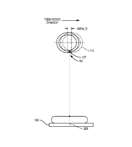
[0010] Figure 1 illustrates a breast tomosynthesis system 100 which
includes an x-ray tube
of the present invention;
[0011] Figure 2 is a cross section view of an x-ray tube provided to
illustrate an effective
focal spot of a prior art tomosynthesis system;
[0012] Figures 3A and 38 are cross section views of an x-ray tube and
breast compression
plate, and are used to illustrate motions of an static focal spot of an x-ray
tube of the
prior art and according to the present invention;
- 3a -
CA 02736592 2011-03-09
WO 2010/060007
PCT/US2009/065451
[0013] Figures 4A and 4B are cross section views of an X-ray tube provided
to illustrate the
motion of the static focal spot and resulting effective focal spot provided by
an x-ray
tube of the present invention;
[0014] Figures 5A and 5B are also cross section views of an x-ray tube and
breast
compression plate, and are used to illustrate reduced image blurring that can
be
achieved by increasing an static focal spot while decreasing the exposure time
for an x-
ray image;
[0015] Figure 6 is a diagram of an exemplary x-ray tube of the present
invention;
[0016] Figure 7 is a flow diagram that is used to describe an exemplary
process that may be
followed during a tomosynthesis image scan using an x-ray tube having a moving
focal
spot of the present invention; and
[0017] Figures 8A and 8B include diagrams of a target anode and an
exemplary motion
control element for controlling a movement of a focal spot on the anode; and
[0018] Figures 9A and 9B are diagrams illustrating the use of additional
filaments in a
focusing cup for increasing focal spot size..
[0019] Detailed Description
[0020] Figure 1 illustrates a tomosynthesis system 100 which includes an x-
ray tube 110,
upper and lower compression paddles 130, 135, an anti-scatter grid 140 and a
detector
160. The x-ray tube 110 includes a cathode 112, an anode 114 that is mounted
on a
shaft 116 and rotated by a motor 118, a tube port 120. Also shown attached to
the x-
ray tube are a filter 122 and a collimator 124.
- 4 -
CA 02736592 2011-03-09
WO 2010/060007
PCT/US2009/065451
[0021] The x-ray tube is a glass vacuum tube. Within the cathode 112 is a
heated filament.
When the x-ray tube is turned on, a current is passed through the filament,
heating the
filament and causing high energy electrons to be dislodged from the filament.
A high
voltage between cathode and anode causes the electrons to accelerate toward a
target
location 125 on the anode. The anode is made for example from tungsten and is
rotated by motor 118 to avoid local overheating of the target location 125 on
the
anode.
[0022] Electrons are focused to a specific target location by means of a
focusing cup (not
shown). The focusing cup is a separate control electrode that is cylindrical
in shape and
that is attached to the cathode, partially surrounding a filament of the
cathode.
[0023] The dislodged electrons collide with the tungsten atoms of the
anode, and x-ray
photons are generated having bremsstrahlung radiation and characteristic line
emission spectra. X-ray photons are emitted in all directions from the target
location
125. The x-ray photons which come out of the tube port 120 are used for
imaging. For
the purposes of this application, the x-ray photons which come out of the tube
port
define the static focal spot 127. The static focal spot size refers to the
focal spot size at
any given instantaneous time moment, as compared to the time-averaged focal
spot
size during an x-ray exposure of finite time period, which is referred to
herein as the
effective focal spot size of an x-ray exposure. The size of the static focal
spot 127
significantly affects the heat loading capacity of the x-ray tube. With larger
focal spots,
greater heat loading is possible which allows a higher tube current mA to be
safely
provided. The size of the focal spot is determined by a combination of factors
including
-5 -
CA 02736592 2011-03-09
WO 2010/060007
PCT/US2009/065451
the size and shape of the filament and the shape and bias voltage of the
focusing cup.
The angle of the target surface further defines a focal spot size along the so-
called
length direction.
[0024] The static focal spot 127 is therefore the focal spot as it appears
from directly
beneath the x-ray tube as seen by the breast, at near the chestwall position
of patient.
[0025] Focal spot characteristics are defined by International Standard CEI
IEC 60336. Focal
spots are generally rectangular in shape, and are stated for two normal
directions of
evaluation referred to as the length and width direction. The length direction
is
generally parallel to a longitudinal axis of the x-ray system, and the width
direction is
generally perpendicular to the longitudinal axis. The longitudinal axis of an
exemplary
tomosynthesis system is shown in Figure 1.
[0026] The size of the focal spot is a very important factor in a
diagnostic x-ray tube
because it affects the resolution of the radiography system; systems having
smaller
focal spots have better resolution. Thus it is often a design goal to minimize
the static
focal spot size. For example, mammography systems may be designed to provide a
0.3
mm focal spot for imaging ( 0.1 mm focal spot for high magnification images).
According to one aspect of the invention it is realized that the movement of
the x-ray
source during image exposure effectively stretches the width of the static
focal spot,
resulting in an effective focal spot which is much wider than the static focal
spot and
which decreases image sharpness. The size of the effective focal spot is
therefore
determined by the size of the static focal spot and the motion of the static
focal spot
- 6 -
CA 02736592 2011-03-09
WO 2010/060007
PCT/US2009/065451
during exposure. The effective focal spot is therefore the accumulation of the
static
focal spot over time, and may also be referred to as the dynamic focal spot.
[0027] For example, during an exemplary tomosynthesis image scan, an x-ray
tube may
move from a position of -7.5 to a position of +7.5 . During the tube
movement, a total
of 15 exposures are performed, each having duration between 30 - 60ms. During
each
exposure period, the x-ray tube continues to move along its path, thereby
effectively
'stretching' the width of the static focal spot during the exposure to provide
an
effective focal spot of increased size.
[0028] Figure 2 is a cross section of x-ray tube 110 of Figure 1 and
illustrates the movement
of x-ray tube 110 during one of the described tomosynthesis exposure periods.
The
static focal spot 127 of the x-ray tube is defined by a width Ws and length Ls
and
indicated by dashed box 127. At T = Exposure start, the x-ray tube is at a
position X'.
During the exposure period, the tube moves to a position X". The time-averaged
effective focal spot 190 is defined by a width We and a length Le. Although
the length
of the effective focal spot corresponds to the length of the static focal
spot, the width
has increased. For example, in an exemplary tomosynthesis system with a static
focal
spot width of 0.3mm, the effective focal spot width may increase to 1.5 mm
during an
exposure because the x-ray tube has moved 1.2 mm during the exposure period.
[0029] Figures 3A ¨ 3B also illustrate the motion of the x-ray source 110
over an exposure
period, and further highlight the effect of the increased effective focal spot
width on
the image detector plane. As shown in Figure 34, the result of the increase in
focal
spot width can be seen in the imaging plane 160, where the delta in positions
X' to X"
- 7 -
CA 02736592 2011-03-09
WO 2010/060007
PCT/US2009/065451
(delta_13) translates into a shift of the focus spot during exposure by the
amount
represented as shaded element 200 (delta_d) so that a point object on the
breast
surface will be elongated to a size of delta_d.
[0030] The present invention recognizes that there is a direct relationship
between the x-
ray exposure time and the increase in size of the focal spot for tomosynthesis
imaging.
The relationship is problematic for at least the reason that denser breasts
require
longer exposure times for image acquisition, yet increased exposure time
results in
increased effective focal spot size and reduced image clarity because of focal
spot
blurring. The present invention further appreciates that the effective focal
spot size is a
function of the static focal spot size and the exposure period. The exposure
period
cannot always be shortened sufficiently so as to mitigate the effect of tube
motion and
minimize the effective focal spot size, because the tube current cannot be
increased
arbitrarily. Several approaches may be taken to reduce the effective focal
spot size.
[0031] According to a first approach shown in Figure 38, the static focal
spot is moved in a
direction opposite to and generally synchronized with the directional movement
of the
x-ray source during the exposure period. In essence the movement of the static
focal
spot compensates for the movement of the x-ray tube, so that the effective
focal spot
appears to be fixed in space, relative to one of the breast and/or detector,
in one
position during the entire duration of the exposure. With such an arrangement
image
blurring may be reduced.
[0032] According to a second approach, the size of the static focal spot is
increased. This
allows higher x-ray tube current and thereby allows the exposure time to be
decreased.
- 8 -
CA 02736592 2011-03-09
WO 2010/060007
PCT/US2009/065451
Such an arrangement reduces the width of the effective focal spot while
increasing the
speed of a tomosynthesis scan and enabling full utilization of the x-ray tube
capacity.
[0033] A third approach combines the above two approaches, using a
relatively larger focal
point in combination with a shorter exposure period, and moving the larger
focal point
during the exposure period.
[0034] An x-ray tube designed using any of the above approaches may be
adapted to
support both 2D and 3D imaging for systems that provide a combination of
mammography and tomosynthesis imaging capability in a single system. For a 2D
acquisition, the x-ray tube may provide smaller sized focal spots to provide
standard
mammograms or magnified images. In a 3D mode, the static focal spot size,
position,
or both may be controlled during imaging or in response to breast density to
obtain
images with increased clarity.
[0035] The above described approaches are described in detail below.
However it is
important to distinguish these approaches from flying focal spot techniques
that have
historically been used in computed tomography.
[0036] For example, U.S. Patent 6,256,369 describes a system whereby a
focal spot is made
to oscillate in the longitudinal direction to improve scan throughput. In a
preferred
embodiment, the focal spot moves along a predefined path including a set of
multiple
positions displaced from one another in a longitudinal direction at each
successive
rotation angle. In effect the tube provides first and second fan beam planes.
By
alternating the focal spot between two longitudinal positions, the data are
effectively
-9-.
CA 02736592 2011-03-09
WO 2010/060007
PCT/US2009/065451
sampled in two different z positions using two different fan beams, thereby
doubling
the throughput rate of the scanner.
[0037] U.S. Patent 6,292,538 also describes an x-ray tube with flying
focus. The flying focus
technique of the '538 patent skips the focal point between two positions at
high speed,
effectively displacing images by a half pixel, in an attempt to improve
resolution during
CT scans.
[0038] In contrast, the present invention does not move the focal spot to
different discrete
positions to obtain multiple images during an exposure; rather, the object is
to keep
the effective focal spot in a fixed position in space, relative to either or
both of the
breast and/or detector, during the duration of the exposure through a slow
controlled
continuous movement of the focal point to enhance clarity in a single image.
[0039] The static focal spot is moved in a direction opposing the direction
of the movement
of the x-ray tube through space. As a result of the contrary movements of the
x-ray
tube and the static focal spot of the tube, the resulting effective focal spot
maintains a
fixed position relative to the breast and/or detector during each
tomosynthesis
exposure, minimizing the effective focal spot size and increasing image
clarity.
[0040] Figure 4A illustrates the movement of the static focal spot within
an x-ray tube 110
during an exposure. In this figure the motion of the x-ray tube is not shown
for clarity
purposes although the tube is moved from left to right during the exposure
period of
this example. At the start of the exposure, the static focal spot is in
position Y'. During
the exposure, the static focal spot is moved at a rate of speed that matches
or is
otherwise related to the speed of the x-ray tube, in a direction opposed to
the
- 10 -
CA 02736592 2011-03-09
WO 2010/060007
PCT/US2009/065451
directional movement of the x-ray tube and indicated by arrow 111, until the
static
focal spot is in position Y".
[004].] Figure 4B illustrates the position of the static focal spot 127
during a tomosynthesis
exposure when the static focal spot is moved in a manner shown in Figure 4A.
It can be
seen that as the x-ray tube 110 is moved from position X' to position X", the
resulting
effective focal spot 190 remains in a fixed or relatively fixed size and
position relative to
the detector 160 or imaged object; thus, although the x-ray tube is moving,
the
effective focal spot 190 appears to remain relatively fixed and minimally
sized. As a
result, there is no shift of focus at the detector, and the clarity of the
tomosynthesis
image is increased. Referring back to Figure 3B, it can be seen that a point
image on
the breast generated as a result of the motion controlled focal spot will not
experience
the severe blurring effect of the prior art.
[0042] Thus a method for increasing x-ray image clarity in the presence of
a moving x-ray
source has been shown and described. Although the above description describes
contrary movement of the x-ray tube and static focal spot in a single plane,
it should be
appreciated that the concepts of the present invention may be adapted to
facilitate
contrary movement of the focal spot from the x-ray tube in any dimension. In
addition,
the present invention is not limited to embodiments whereby the x-ray tube and
focal
spot are moved at the same speed, or over the same distance; it can be
appreciated
that the benefit of image clarity can be realized through any opposed motion
of the
static focal spot relative to the x-ray tube which minimizes the accumulation
of the
- 11 -
CA 02736592 2011-03-09
WO 2010/060007
PCT/US2009/065451
focal spot during x-ray exposure; thus the present invention is not limited to
a
particular range or directional speed for focal spot movement.
[00431 As described above, a second approach for reducing the effective
focal spot size
involves increasing the size of the static focal spot, but reducing the
exposure period.
For example, Figure 5A is similar to Figure 3A, where a relatively small
static focal spot
320 is provided. To obtain the desired exposure for the image, the x-ray tube
must be
activated for duration D1, causing a blur effect 300 at the detector plane
160. As
shown in Figure 5B, if a relatively larger static focal spot 330 is provided,
the total
exposure time D2 may be reduced, resulting in a concomitant reduction in image
blur
310. An advantage of providing a larger focal spot is that it permits full
utilization of
the x-ray tube generator. For example, current tomosynthesis x-ray tubes use a
200
rriA generator but the generator does not always operate at the 200 mA because
the
desired, smaller static focal spot (for image clarity) allows only 160 mA at
28 kV. The
present invention recognizes that the focal spot of a tomosynthesis image may
be
increased without sacrificing image clarity because the effect of an increased
focal spot
size is offset by the reduction in exposure time, so that a 'resulting'
effective focal spot
is smaller than previously obtained effective focal spots in the prior art.
[0044] The second approach may also have advantages for systems where the
effective
focal spot is larger than 1 cm due to physical constraints of focal spot
movement. In
such embodiments, enlarging the focal spot and reducing the exposure may yield
the
smallest focal spot for imaging.
- 12 -
CA 02736592 2011-03-09
WO 2010/060007
PCT/US2009/065451
[0045] A variety of techniques may be used to enlarge the focal spot size.
The techniques
include, but are not limited to: de-focusing the focus cup to allow larger
focal spot sizes
in the length or the width or both directions, incorporating additional
electrodes to
allow focal spot size change in one or both directions and incorporating a
third filament
or combination of the several filaments. In addition, sophisticated electron
or x-ray
optics techniques may be applied to enlarge focal spot sizes.
[0046] Accordingly, two methods for increasing x-ray image clarity in the
presence of a
moving x-ray source has been shown and described, wherein the first method
involves
moving the focal spot, and a second method involves increasing the size of the
focal
spot. It should be appreciated that either method may be used alone or in
combination; for example, it is realized that providing an x-ray tube with a
larger,
moveable effective focal spot will result in a system that fully utilizes the
x-ray tube
generator, provides a high quality image and, due to the decrease in exposure
time,
may scan the patient more quickly.
[0047] In some embodiments it may be desirable to enable either automatic
or manual
selection of the use of either method, or the combination thereof. Either
method may
be realized by a modification to existing x-ray tube, for example by providing
the ability
to move the target, adjust the focus cup of the cathode, utilize different
filaments or
otherwise focus the x-ray photons.
[0048] For example, Figure 6 illustrates an x-ray tube 110 of the present
invention, which
includes a vacuum tube 400 which encases an anode 114, a cathode 112 and an
anode
rotor 410. According to one aspect of the invention, the x-ray tube further
includes a
- 13 -
CA 02736592 2011-03-09
WO 2010/060007
PCT/US2009/065451
focal spot position controller 600. The focal spot controller may be coupled
to the
cathode 112 to deflect the electron trajectory in the 'width' direction. In
its simplest
form, the controller comprises two parallel metal plates located next to the
focusing
cup, with a bias voltage applied across the plates that can shift electron
motion
direction, and therefore the target location on the anode. The shift of the
focal spot is
therefore controlled via an application of a bias voltage across the plates.
In several
embodiments, the bias voltage can be dynamically or statically configured
prior to x-ray
exposure.
[0049] Referring briefly to Figures 8A and 8B, an exemplary mechanism for
controlling the
movement of a focal spot will now be described. In prior art, electrons
emitted from
the filament and cathode 112 travel along path 620 and hit the anode 114 at
location a.
This location a is the location of the focal spot.
[0050] In one embodiment of the present invention, motion control unit 600
is added. The
motion control unit includes metal plates 601 and a voltage source 602
controlled by
controller 603. Varying the voltage of the plates 601 varies the path of
travel of the
electrons to the target; for example, an electron will travel along one of the
paths 620
and hit the anode 114 at different locations a and b depending upon the
voltage
applied to 601, where locations a and b represent different focal spot
locations on the
anode 114.
[0051] In Figure 8B, the plates are shown aligned along a Y axis, and
modifying the voltage
of the plates changes the path of the electron along the Y axis. In the
present
invention, however, it is desirable to alter the path of the electron in the Z
plane
- 14 -
CA 02736592 2011-03-09
WO 2010/060007
PCT/US2009/065451
(normal to the page), and in such an embodiment, the motion controller would
include
plates aligned along the Z axis. Suffice it to say that the plates 601 can be
in different
geometric locations than shown in the figure, and there can be more than 1 set
of
plates. The number and arrangement of the plates, as well as the selection of
voltage
to be applied across the plates, be determined by the desired locations of the
focal
spots. Controller 603 will change the voltage over time to create the desired
effective
focal spot distribution, synchronized in an appropriate way with the tube
motion and
image receptor acquisition sequences.
[0052] In alternate embodiments, the focal spot controller controls a tilt
of the anode to
change the angle at which the electron stream from the cathode hits the anode
target.
In another embodiment, the controller moves the cathode focus cup so that the
electrons hit the anode at a different target location or electronically
deflects electron
trajectory by applying a bias voltage applied orthogonally to the trajectory.
Modifying
the angle of electron engagement with the anode is one technique that can be
used to
control the static focal spot size, although any method that modifies the
angle by which
the electron stream engages the anode may be substituted herein as an
equivalent.
For example it is envisioned that similar results may be achieve through
movement of
the cathode filament or by other means.
[0053] Changing the relative angle between the anode and the cathode, and
hence the
angular direction at which electrons hit the target, may also accomplish the
task of
moving the position of the focal spot during a tomosynthesis exposure.
- 15 -
CA 02736592 2011-03-09
WO 2010/060007
PCT/US2009/065451
[0054] There are a variety of other methods that may be used to move the x-
ray focal spot.
in one embodiment, the focal spot may be moved using an x-ray lens within the
tube or
by adjusting the size and/or location of the tube port 120. The shifting of
the electron
beam focal spot may be accomplished by electronically steering the electron
beam to a
different spot on the x-ray target, electronically shifting the target, or by
using an X-ray
tube with one or more electron beam sources and distributed focal spots on one
or
more x-ray targets.
[0055] For example, to shift the X-ray beam focal spot, a first electron
beam aimed at a first
spot on the x-ray target could be turned off and a second electron beam aimed
at a
different second spot on the first or second x-ray target turned on. Since the
electron
beam strikes a different spot on the x-ray target, the x-ray beam will be
emitted from a
different spot on the target, thus shifting the position of the x-ray beam
focal spot.
[0056] Alternatively, a fiber optic x-ray lens may also be used to shift
the x-ray beam focal
spot, by passing the beam through different fiberoptic strands of the bundle,
with the
strands directed towards slightly different directions. A shutter may be used
to steer
the beam into a particular strand or group of strands.
[0057] As mentioned above, it is also envisioned that the size of the focal
spot may be
changed to accommodate different modes of imaging. In an exemplary embodiment,
an x-ray tube may be provided with three focal spot sizes: a small focal spot
size: 0.1 x
0.1 mm2, a large focal spot size: 0.3 x 0.3 mm2, and an extra large focal spot
size: 0.5 x
0.5 mm2 or 0.6 x 0.3 mm2 (Width x Length). The small focal spot and large
focal spot
may be used to support various 2D modes of imaging. The x-ray tube focal spot
size
- 16 -
CA 02736592 2011-03-09
WO 2010/060007
PCT/US2009/065451
may be configured to the extra large focal spot size in the 3D pulse mode when
needed.
With such an arrangement, an extra large focal spot size is provided on the
anode track,
allowing much higher tube current mA than the static 2D mode. The effective
focal spot
size associated with the extra large focal spot is expected to be 0.5 x 0.5
mm2 or 0.6 x
0.3 mm2.
[0058] One method of changing the focal spot size involves adding filaments
into the
focusing cup. Figure 9A and 9B illustrates two embodiments of a focal spot cup
900
with different numbers of filaments 910 and 920. When one filament is lit, it
provides
an existing 0.3 x 0.3 mm focal spot. When both filaments are lit, the
resulting filament
emission capability is doubled, thus tube current is doubled, while focal spot
size
become 0.6 x 0.3 (width x length).
[0059] As mentioned above, the x-ray tube of the present invention may be
provided in an
imaging system which includes both 2D and 3D imaging capabilities. In such
systems,
the motion of the static focal spot and size of the static focal spot may
differ between
the two imaging modes. For example, the focal spot size may be increased for
each
tomosynthesis exposure, or during an entire scan, and reduced for modes, such
as
mammography, which require increased resolution and are not affected by x-ray
source movement. The x-ray tubes are therefore configurable based on an
imaging
mode.
[0060] Referring now to Figure 7, a process 500 of using an x-ray tube of
the present
invention in a system that supports 2D and 3D imaging will now be described.
In the
exemplary embodiment, the tomosynthesis system scans from -7.5 degrees to +7.5
- 17 -
CA 02736592 2011-03-09
WO 2010/060007
PCT/US2009/065451
degrees. A mammogram is taken following the tomosynthesis scan.At step 510 the
gantry of a tomosynthesis machine is positioned at a -7.5 degree location and
the
tomosynthesis sweep is initiated. At step 512 the x-ray tube reaches an
initial imaging
position, and at step 514 the x-ray tube is activated. In one embodiment, each
exposure takes less than 60ms. During the exposure, the gantry continues to
move
towards the +7.5 degree position, and the x-ray tube focal spot motion
controller sets
the focal spot to a starting position on the anode which is pre-calculated
based on the
x-ray technique and gantry scan speed of the intended tomosynthesis scan,
moves the
static focal spot in the opposite direction (in this example, clockwise
tomosynthesis
scan).
[0061] At step 516, when the exposure is complete and focal spot at the
same time
reaches the pre-calculated stop position, the x-ray tube is turned off and the
static focal
spot is re-centered within the x-ray tube. At step 518 it is determined
whether the end
point of the clock wise scan has been reached (gantry at the +7.5 degree
position). If
not, the steps of 512-516 are repeated until all tomosynthesis projection
images are
obtained. At step 520 the gantry is returned to a zero degree position and the
focal
spot is optimized for mammography imaging. If the focal spot size had been
increased
for tomosynthesis imaging, it is reduced to the range which provides desired
mammogram resolution. At step 522 the 2D image is obtained and the process is
complete. It should be noted that the above text has described a system
whereby the x-
ray tube is turned 'on' or 'off.' However, it should be noted that the present
invention
is not limited to such a system. In fact, many systems have x-ray tubes that
are
- 18 -
CA 02736592 2011-03-09
WO 2010/060007
PCT/US2009/065451
continuously on during the scan, with image capture being controlled by
capture of the
x-rays at the detector at select 'exposure times' times during the scan. In
such
instances, it can be appreciated that the focal spot motion is synchronized to
the
exposure start and exposure end times, regardless of whether the x-ray tube is
cycled
on or off, or is continuously on.
[00621 Accordingly, a system, method and process of the present invention
has been
shown and described whereby tomosynthesis image clarity is improved by static
or
dynamic management of focal spot size and position during an x-ray exposure.
It
should be noted that although the description has centered on the use of a
tomosynthesis system for breast imaging, the x-ray tubes of the present
invention are
not limited for use to any particular imaging modality. Rather it is
envisioned that the
x-ray tubes of the present invention may have utility in any system which
obtains
images while an x-ray source is in motion. For example, computed tomography
(CT)
systems experience focal spot blurring. The modified x-ray tube of the present
invention may advantageously be used with CT systems to reduce the FS blur,
making
the Modulation Transfer Function (MTF) across field of view isotropic. In a
breast CT
system, one benefit of such an improvement would be that the MTF at the breast
edge
would be as good as that in the breast center in the horizontal plane. The
above
specific examples and embodiments are illustrative, and many variations can be
introduced on these examples and embodiments without departing from the spirit
of
the disclosure or from the scope of the appended claims. For example, elements
and/or features of different illustrative embodiments may be combined with
each
- 19-
CA 02736592 2011-03-09
WO 2010/060007
PCT/US2009/065451
other and/or substituted for each other within the scope of this disclosure
and
appended claims.
[0063] Various embodiments of the invention include an x-ray tube arranged
to move
during an exposure period. The x-ray tube includes a cathode for providing an
electron
stream, an anode comprising a target for receiving the electron stream, the
anode for
generating a photon stream, a focusing cup which focuses the electron stream
on the
anode during the exposure period, a port for passing the photon stream out of
the x-
ray tube, wherein the cathode, anode and port together define an static focal
spot of
the x-ray tube, and a controller coupled to at least one of the anode, the
cathode and
focusing cup for modifying a characteristic of the static focal spot during
the exposure
period by performing at least one of modifying a static focal spot location or
size in
relation to a movement of the x-ray tube.
[0064] The x-ray tube may move in a first direction during the exposure
period and the
controller may move the static focal spot in a second direction, opposite to
the first
direction, during the exposure period such that an effective focal spot
remains
relatively fixed in space relative to the breast and/or detector during the
exposure
period to reduce image blur. The static focal spot size may be increased to
reduce the
exposure period and resulting image blur.
[0065] In accordance with another aspect of the invention, a method of
acquiring an x-ray
image using an x-ray tube that moves in a first direction during an exposure
period
includes the step of moving a focal spot of the x-ray tube in a second
direction opposite
- 20 -
CA 02736592 2011-03-09
WO 2010/060007
PCT/US2009/065451
to the first direction during the exposure period to reduce an effective focal
spot size
during the exposure period.
[0066] According to a further aspect of the invention, a method of
acquiring an x-ray image
by an imaging system capable of operating in two modes, the method including
the
step of varying a size of a focal spot of an x-ray tube in response to an
operating mode
of the imaging system. The method may include the step of varying the size of
the focal
spot increases the size of the focal spot in imaging modes wherein images are
acquired
using an x-ray tube which moves during an exposure period. In addition, the
step of
varying the size of the focal spot may decrease the size of the focal spot in
imaging
modes wherein images are acquired using an x-ray tube which remains in fixed
position
relative to the breast and/or detector during the exposure period.
[0067] The foregoing of preferred embodiments has been presented as an
illustration of
examples and is not intended to be exhaustive or to limit the claimed
inventions to the
specific examples. Those examples are intended to describe principles that
persons
skilled in the art may use to practice the claimed inventions, using
variations and
modifications of the disclosed examples that are suited to a particular
environment. It
is intended that the scope of the invention be defined by the appended claims
and their
equivalents.
- 21 -