Note: Descriptions are shown in the official language in which they were submitted.
CA 02736868 2011-03-10
WO 2010/030817 PCT/US2009/056563
A PHOTOACOUSTIC IMAGING DEVICE
CROSS-REFERENCE TO RELATED APPLICATIONS
This application claims benefit of U.S. Provisional Application No.
61/095,881, filed September 10, 2008, which is hereby incorporated by
reference.
FIELD OF THE INVENTION
The present invention relates generally to a system and method for
acoustic imaging and more particularly to a diagnostic photo-acoustic imaging
system and method for low volume imaging.
BACKGROUND OF THE INVENTION
Non-invasive small animal imaging
Low volume imaging relates to diagnostic imaging tailored to low
volume objects. Low volume imaging has applications in human diagnostic
imaging of smaller body parts, including wrist, hand, and foot. It has further
application in tissue specimen imaging and preclinical (i.e., non-human
animal) imaging.
Preclinical models of disease have become more available and
sophisticated. They are now a common tool used in the development and
evaluation of new therapeutics and treatments. The use of preclinical models
is a precursor and validation step prior to human clinical trials.
Non-invasive imaging is an important tool in preclinical studies;
computed tomography (CT), magnetic resonance imaging (MR), single photon
emission tomography (SPECT), positron emission tomography (PET), x-ray,
optical, and ultrasound are standard tools in studying disease and the
evaluation of new therapies. These imaging tools are being actively used for
understanding and assisting in therapy development of diseases, such as
cardiovascular, musculoskeletal, neoplasia, auto immune, and inflammation.
CA 02736868 2011-03-10
WO 2010/030817 PCT/US2009/056563
Clinical imaging devices are often sufficient for imaging of large animal
species such as primates, porcine, and canine. However, the vast majority of
preclinical studies involve the use of lower volume animals, such as rodents
and rabbits; where the murine model (mouse) is the most widely used
preclinical model.
In recent years specialized devices have been developed and
commercialized specifically for low volume animal imaging based on standard
non-invasive medical imaging technologies.
The aim of low volume imaging is to have the full functionality of clinical
imaging, but with sensitivity and resolution at the scale of the low volume
object of interest. Clinical imaging systems are not directly scalable to low
volume imaging systems. As such, in all of the above mentioned imaging
modalities, technical and scientific hurdles had to be overcome to achieve
systems that had proper functioning, often including achieving higher
resolution, the need for miniaturization of many aspects of the technology,
and proper placement and handling of low volume objects, specifically
animals.
As in clinical imaging, each of the preclinical imaging modalities helps
to visualize different aspects of an object and has different strengths. Some
of the desirable attributes include sensitivity, resolution, field of view,
minimal
required time to produce an image, contrast, cost, three-dimensional imaging,
and whether the system is capable of dynamic imaging. No one imaging
modality is sufficient for all applications. Furthermore, the current array of
low
volume imaging modalities still do not provide the full functionality in
visualization and quantification that is desired for current preclinical and
other
low volume needs.
Photo/thermo-acoustic imaging
Two relatively new imaging technologies are thermo-acoustic and
photo-acoustic imaging (collectively referred to herein as photo-acoustic
imaging). This new modality adds new insights into properties of tissues and
other objects, above those offered by established imaging modalities.
Specifically, it provides information related to the thermoelastic properties
of
2
CA 02736868 2011-03-10
WO 2010/030817 PCT/US2009/056563
tissue. More specifically, laser, radio frequency or other energy pulses are
delivered into an object. Some of the delivered energy will be absorbed and
converted into heat, leading to transient thermoelastic expansion and thus
ultrasonic emission. The generated ultrasonic waves are then detected by
ultrasonic transducers to form images (Bowen, Radiation-Induced
Thermoacoustic Soft Tissue Imaging, Proc. of IEEE Ultrasonic Symposium
2:817-822, June, 1981).
No system currently exists that is ideally suited for low volume photo-
acoustic imaging. Accordingly, there is a need for new low-volume
photoacoustic imaging systems.
SUMMARY OF THE INVENTION
In general, the invention features systems for imaging tissue and
methods of their use.
In one aspect, the invention features a system for imaging tissue
including (i) a source of electromagnetic radiation; (ii) an encasement h a
plurality of acoustic transducers (e.g., at least 128); (iii) a support
structure
having a portion for holding a tissue; and (iv) a chamber between the
encasement and support structure for housing an acoustic coupling medium.
In the system, electromagnetic radiation from the source is sufficient to
induce
a thermoacoustic response in the tissue positioned in the support structure,
and the plurality of acoustic transducers are positioned to receive ultrasound
from the thermoacoustic response of the tissue. In addition, the portion for
holding the tissue has a thickness of less than 250 pm, and the acoustic
impedance of the portion is matched to the tissue (i.e., within 50-150% of
that
of the tissue), or the portion allows for contact between the tissue and the
acoustic coupling medium.
The system may further include one or more of an optical camera (e.g.,
sensitive to light from 300 - 1064 nm) positioned to monitor the tissue in the
support structure; an electro-mechanical motion control system for rotation of
the encasement relative to the support structure (e.g., in movements of 1
degree or less); a digital acquisition system for acquiring and storing
thermoacoustic response signals received by the plurality of transducers;
CA 02736868 2011-03-10
WO 2010/030817 PCT/US2009/056563
a temperature monitor and control system for maintaining a specified
temperature (e.g., between 30 and 39 C) of acoustic coupling medium in the
chamber; and a pulse energy monitor for measuring the energy of the
electromagnetic radiation.
In another embodiment, a portion of the plurality of transducers is
capable of transmitting ultrasound into the tissue, and a portion of the
plurality
of transducers is capable of receiving ultrasound emitted from the tissue,
wherein the system is further capable of producing ultrasound images of the
tissue.
The encasement is optionally positioned between the source and the
support structure, with the encasement further including a window through
which the electromagnetic radiation from the source passes to the support
structure.
The system may also include a plurality of sources of electromagnetic
radiation, wherein the electromagnetic radiation from each source is
sufficient
to induce a thermoacoustic response in the tissue positioned in the support
structure, and wherein the plurality of sources are positioned to illuminate
different portions of the tissue.
The support structure may or may not separate the tissue from
acoustic coupling medium in the chamber. In certain embodiments, the
system includes an acoustic coupling medium disposed in the chamber and
having a speed of sound of 1450-1600 m/s.
Preferred transducers have a center frequency of 1 to 30 MHz and a
bandwidth of greater than 50%.
The encasement may include a spherical inner surface, e.g., wherein the
plurality of acoustic transducers is positioned on the inner surface of the
encasement so that the axis of maximum sensitivity of each transducer
intersects the centroid of the sphere. Such a surface may have a radius of
80-150 mm. The encasement may also include a cylindrical section
extending from the sphere equator to accommodate displacement of acoustic
coupling medium by the introduction of the tissue to the support structure.
An exemplary source produces a pulse sequence of one or more
pulses, each with an individual pulse length less than 500 nanoseconds, at a
4
CA 02736868 2011-03-10
WO 2010/030817 PCT/US2009/056563
pulse rate greater than 1 Hertz. The energy per pulse is optionally greater
than 0.03 mJ. The electromagnetic radiation is, for example, infrared,
visible,
UV, radio frequency, or microwave.
The system may also include a computer for generating an image or
volumetric representation of the tissue from the thermoacoustic response.
The support structure may include markings to show the field of view for
thermoacoustic imaging. The portion of the support structure holding the
tissue may or may not conform to the tissue. The portion of the support
structure holding the tissue may also or alternatively be shaped to maintain
the tissue in substantially the same orientation for thermoacoustic imaging.
The invention also features a method of producing a thermoacoustic
image of a tissue by (a) providing a system for imaging as described herein;
(b) placing the tissue in the support structure; (c) actuating the source to
induce a thermoacoustic response in the tissue; (d) receiving ultrasound from
the thermoacoustic response of the tissue at the plurality of acoustic
transducers; and (e) generating a thermoacoustic image or volume from the
received ultrasound.
Other features and advantages will be apparent from the following
description, the drawings, and the claims.
BRIEF DESCRIPTION OF THE DRAWINGS
Figure 1 is an isometric view of an exemplary encasement.
Figure 2 is a cut away section through an exemplary system, without
external covers.
Figure 3 shows an exemplary specimen-positioning tray.
Figure 4 shows a system with an E-chain cable management system.
Figure 5 shows the results of imaging a single absorbing point with the
system employing laser illumination.
Figure 6 shows a volume image derived from imaging an intact mouse
in the system employing laser illumination.
5
CA 02736868 2011-03-10
WO 2010/030817 PCT/US2009/056563
DETAILED DESCRIPTION OF THE INVENTION
A photo-acoustic system has been developed specifically for low
volume imaging (including small animal imaging) with the specific aim of
applications in the study of disease, the guidance of procedures, and the
monitoring of therapies in the fields of academic research, pharmaceutical
drug development, and clinical applications. Low volume imaging refers to
imaging of a single organ or focal volume of interest and is differentiated
from
the more common 'whole body' imaging available with modalities such as:
magnetic resonance (MR), x-ray computed tomography (CT), and positron
emission tomography (PET) where large scan volumes covering multiple
organs are available.
In its simplest embodiment, the system includes an electromagnetic
radiation source, acoustic transducers, a support structure, and an
encasement to which the transducers are attached. Figure 2 shows a cut
away section through a system without external covers. This system includes
both moving and stationary parts. A table top [1] is attached to structural
frame members [6] and provides a working surface that is stationary at all
times. The encasement and plurality of acoustic transducers [2] are located
beneath the table top and are attached to an electro-mechanical, computer
controlled rotation stage [8] by way of support struts [5] to form a rotating
assembly. The rotation stage has an unobstructed path through its axis of
rotation [7] that allows an unimpeded path for illumination of the tissue from
below the encasement. The tissue support structure [4] rests on the table top
and remains stationary at all times during the data acquisition procedure. The
support structure is attached to a handling apparatus [3] that allows for
removal and positioning of the tray.
The system may further include various additional elements as
described herein. The individual components of the system are discussed
below. It will be understood that the system is constructed to provide for
thermoacoustic imaging of a tissue located within it.
6
CA 02736868 2011-03-10
WO 2010/030817 PCT/US2009/056563
Electromagnetic Radiation Source
Any electromagnetic radiation source capable of producing a
thermoacoustic response in a particular tissue may be employed. The
radiation may be ionizing or non-ionizing, e.g., infrared, visible,
ultraviolet,
radio frequency (US 6,633,774), or microwave (such as 10MHz to 4GHz). An
exemplary source is a laser. The radiation may be pulsed, e.g., at greater
than 1 Hz, or continuous. Pulse length may be less than 500 ns, and the
energy per pulse may be less than 1 mJ, e.g., less than 0.03 mJ. The system
may further include a monitor to measure the pulse energy.
In one embodiment, the one or more sources may be employed. When
multiple sources are employed, they are typically positioned to illuminate a
tissue at different locations. Sources may pass radiation through a window in
the encasement. Alternatively, a source is positioned to illuminate from
within
or above the encasement. Combinations of sources directed from the bottom
and top results in more uniform light distribution along the tissue being
imaged. Multiple sources can be synchronized by using a common trigger
signal and trigger delay, for all individual sources. In this manner, the
cumulative energy of the individual sources will increase the thermoacoustic
signal response from the tissue. Increased signal is generally desirable,
particularly when increased sensitivity is required to detect trace materials
or
small changes in concentration of the absorber. Instead of using multiple
sources, a single source can be used with the radiation split into multiple
paths that will illuminate the tissue from multiple positions.
Encasement
The encasement is a structure to which acoustic transducers are
attached and which houses an acoustic coupling medium. The medium is
placed in the encasement to provide acoustic coupling between the
transducers and a tissue located in a support structure, as described in more
detail below.
The encasement may be of any shape suitable for transducers to
receive ultrasound emitted from a tissue placed within it, e.g., spherical;
7
CA 02736868 2011-03-10
WO 2010/030817 PCT/US2009/056563
For example, the transducers may be arranged in a spiral pattern within a
portion of the inner surface of a hemispherical encasement, e.g., with a
radius
of 80-150 mm.
The encasement is typically filled with an acoustic coupling medium
e.g., a liquid (such as water) or a gel. Acoustic coupling media are known in
the art. The speed of sound (SOS) in the medium can be closely matched to
SOS of the tissue being imaged. A medium having a speed of sound of 1450
to 1600 m/s is preferred. In one embodiment, water is combined with glycerol
to produce a medium with the desired SOS. In some embodiments, the
encasement includes a drainage hole in to allow for removal of liquids from
the encasing and to facilitate cleaning and disinfection. The drainage hole is
positioned so that it does not interfere with the detectors. The encasement
typically also includes a volume (e.g., a cylindrical extension of a
hemisphere)
into which coupling media can be displaced when a support structure is
inserted in the system, as discussed below.
The encasement may be constructed of conductive or non-conductive
materials (which are preferred for use with radio and microwave frequencies).
Engineered thermoplastics such as Delrin and Ultem are suitable encasing
materials as they are chemically inert and machinable and have low water
absorbance. As discussed above, the encasement may include a window (or
otherwise be transparent) to radiation emitted from a source.
A temperature probe may also be installed on the encasement (or
adjacent to it) to monitor and/or regulate the temperature of the acoustic
coupling medium. Maintaining consistent temperature will result in consistent
speed of sound through the coupling medium and may also reduce motion of
the tissue being imaged. This is particularly relevant for imaging of animals.
The temperature can be maintained through heaters located on or within the
encasement. Alternatively, medium, e.g., water, can be exchanged between
an external tank with constant temperature and the encasement. Preferably,
medium is only exchanged between the external tank and the encasement
between successive scans to prevent bubble formation during scans.
Preferably, the temperature of the medium is matched to the normal
physiological temperature of the tissue being imaged. In some embodiments,
8
CA 02736868 2011-03-10
WO 2010/030817 PCT/US2009/056563
a temperature below physiological is advantageous. For example, in some
small animal imaging applications (such as the mouse), a lower heart rate
may be preferred and may be achieved by lowering the temperature of the
liquid 1-5 degrees Celsius. Temperatures may also be lowered to maintain
the integrity of an isolated tissue. The normal range of temperature for in
vivo
imaging for the medium will be 30-39 degrees Celsius.
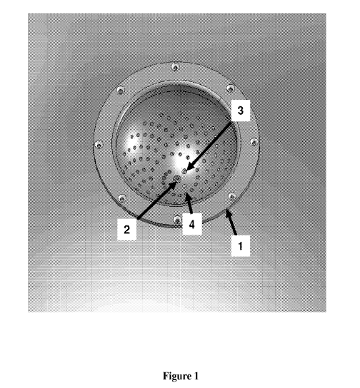
Figure 1 is an isometric view of an exemplary encasement. The
encasement [1] is machined, formed, or molded to provide the required
geometry. The figure illustrates the pattern of machined holes into which the
acoustic receivers are placed and form a spiral pattern as described in US
5,713,356 and US 6,102,857. A window [2], at the bottom of the encasement,
provides an entry port through which electromagnetic radiation may be
delivered to the tissue. A drainage hole [3] is also located in proximity of
the
lowest point of the encasing. A flexible hose, with a valve, is connected to
the
drainage hole by way of a fitting to allow the acoustic coupling media to be
removed from the encasing. An additional hole in the encasing provides
access for a temperature sensor to monitor the temperature of the acoustic
coupling media.
Support Structure
The support structure houses the tissue being imaged. The structure is
placed in contact with acoustic coupling medium held in the encasement.
Preferably, the support structure includes a portion that is able to conform
to
the shape of the tissue being imaged or is molded to hold the tissue in
substantially the same orientation for thermoacoustic imaging, e.g., by
approximating the shape of the tissue being imaged (Figure 3). The support
structure also positions the tissue appropriately in the system's field of
view,
i.e., the volume that can be thermoacoustically imaged. The height of the
support structure may be adjustable, e.g., to allow the tissue to be centered
vertically and/or horizontally in the system's field of view. Markings may be
included on the support structure to assist in localizing the tissue in the
field of
view. The support structure may be removable from the rest of the system or
may be hinged along one side of the system (or otherwise attached). Both of
9
CA 02736868 2011-03-10
WO 2010/030817 PCT/US2009/056563
these approaches will facilitate cleaning of the encasement. The portion
holding the tissue preferably prevents contact between the tissue and the
acoustic coupling medium. The support structure may also include molded
portions to accommodate non-imaged portions of a tissue, e.g., an arm, leg,
animal tail, etc. The structure may further allow for the connection of
catheters (e.g., arterial or venous) for delivery or removal of fluids to the
tissue
or other elements to the tissue, e.g., heart rate, breathing rate, or
temperature.
The portion of the support structure that holds the tissue (which may be
referred to herein as a cradle) may be removable and disposable. Alternately,
this portion may be sterilized after each use. The portion holding the tissue
can be rigid or deformable, preformed or flat. The acoustic impedance of the
material employed in the portion housing the tissue is matched to the tissue.
Additionally, the portion housing the tissue may have a high transmittance for
the radiation being employed. The thickness of the portion holding the tissue
is for example between 10 and 250 microns. Examples of suitable materials
for the portion holding the tissue are: polycarbonate (e.g., Lexan ),
polyethylene, perfluoroelastomer, polyethylene terephthalate, and plastic wrap
(e.g., Saran ).
In another embodiment, the support structure allows a portion of the
tissue in the path of illumination to be directly in contact with the coupling
medium. In this embodiment, the support structure is not required to be
transparent to the illuminating energy and the acoustic impedance of the
support structure does not need to approximate the acoustic impedance of the
tissue.
Figure 3 shows an exemplary support structure. The cradle [1] is
formed to approximate the geometry of the tissue of interest. The support
structure has a horizontal rim [2] and screw holes [3] that allow it to be
attached to the handling apparatus. Together with the handling apparatus,
the support structure is inserted into the table top for the scanning
procedure.
The geometry of the support structure and cradle are of appropriate
dimensions such that the tissue of interest is located at the effective field
of
view of each acoustic transducer in the encasement.
CA 02736868 2011-03-10
WO 2010/030817 PCT/US2009/056563
Acoustic Transducers
The system includes a plurality of acoustic transducers, e.g., at least
128, for receiving ultrasound produced thermoacoustically. The transducers
may be arranged on the encasement as is known in the art, e.g., in a spiral
pattern as disclosed in ( US 6,102,857). When the encasement has a
spherical surface, the transducers may be arranged so that the axis of
maximum sensitivity of each transducer intersects the centroid of the sphere.
Exemplary transducers have center frequencies of 1 to 30 MHz and
bandwidths of at least 50%.
One or more of the transducers may be used as an emitter of
ultrasound, while one or more of the others are used as receivers for the
production of an ultrasound image.
E-chains or other cable management systems may be used with the
transducers to connect them to data storage and/or analysis components.
Additional Components
The system may also include a cover to enclose the tissue in
conjunction with the encasement. Such a cover may also provide a structure
for mounting electromagnetic radiation sources or optics to direct radiation
to
a tissue. The system may also include a protective shield to shield portions
of
a tissue from electromagnetic radiÃation.
Additionally, an optical camera, e.g., having sensitivity from 300 to
1064 nm, may be included and used to monitor the tissue during
thermoacoustic imaging or to form an optical image based on: reflection,
transmission, or emission, e.g., fluorescence, during the imaging procedure.
The camera may be integrated into the cover above the tissue being scanned,
lateral to the tissue, or external to the imaging system with the optical
image
of the tissue obtained using relay optics.
The system may further include a rotation stage to move the
encasement relative to the tissue and/or radiation source. The stage rotates
to provide thermoacoustic waveforms from multiple views. The rotation stage
may have a hole through its vertical axis to provide an unimpeded light path
to
the window at the base of the encasing. The rotation stage may be manually
11
CA 02736868 2011-03-10
WO 2010/030817 PCT/US2009/056563
driven or driven by a computer controlled drive system that allows for
discrete
increments, e.g., of 1 degree or less, or continuous rotation. The rotation
stage may also include an encoder that allows for the recording of angular
position at any given time.
The system may further include data storage and/or data analysis
components. In one embodiment, the system includes a digital acquisition
system that acquires and stores thermoacoustic response signals received by
the transducers. The system may also include a computer that generates
two-dimensional images or three-dimensional volumetric representations of
the tissue based on the thermoacoustic responses received. The data
storage and acquisition components and/or computer may also be used to
storage and generate ultrasound images or volumes, when the transducers
are used to transmit and received ultrasound.
The system may further include a table top that tilts (hinged on struts)
so that the encasement surface may be accessed for cleaning and
disinfecting; an optically opaque cover placed over the imaging area to
provide shielding from stray laser light during imaging; or an interlock
switch
on the cover that connects to the laser to ensure no exposure to the imaging
area when the cover is open.
Methods of Use
The system of the invention may be employed to produce
thermoacoustic images and volumetric representations of a tissue, as is
known in the art. Tissues imaged may be entire organisms, e.g., a plant, a
mouse, rat, or rabbit; portions of an animal, e.g., a hand, foot, or breast;
or
material excised from an animal or grown in culture, e.g., a biopsy specimen
or tissue implant.
Examples
An exemplary system is described as follows. Any component
specifically descried below may be employed with other components of the
system and is generally applicable to the invention. Figure 4 illustrates a
system as viewed from above, without external covers. The acoustic
12
CA 02736868 2011-03-10
WO 2010/030817 PCT/US2009/056563
transducers in the encasement [1 ] rotate through 360 degrees to provide
multiple views of the thermoacoustic waveforms emitted from the tissue as it
is illuminated. Each acoustic transducer has a pair of electrical wires
(signal
and ground). The pairs of electrical wires from all acoustic transducers in
the
encasement come together to form a cable. The cable is guided through the
e-chain cable management system [2] between a fitting on the rotating portion
of the scanner [3] and a fitting on the stationary scanner frame [4] allowing
unencumbered motion of the cable within the photoacoustic scanner. An in-
flow tube [5] delivers temperature controlled acoustic coupling media into the
encasing, while an out-flow tube removes acoustic coupling media from the
encasing and transfers it to an external temperature control unit. The
combination of in-flow/out-flow tubes, an external pump, and temperature
control unit allow for the acoustic coupling media to be at a constant and
controlled temperature during the imaging procedure.
The energy source is a tunable OPO laser source capable of
generating 40mJ per pulse, at a wavelength of 300-1064nm, with pulse
duration < 1 Ons. The laser induces heating in the tissue being imaged. An
optical chain including lenses, diffusers, filters, prisms, mirrors, and fiber
optic
cables is employed to relay the light emitted from the laser to the tissue. A
beam splitter is used to provide two separate light sources for illuminating
the
tissue in the field of view. Alternatively, additional beam paths are
incorporated with an integrating sphere with a photodiode to monitor the
energy of each laser pulse. One beam path impinges on the animal, while the
other (<5% of the total) is relayed to the integrating sphere (or alternate
beam
monitoring device) to quantify the light output per pulse. The energy of each
pulse during a scan sequence, as measured by the beam monitor, is recorded
as part of an acquisition sequence on the computer.
128 acoustic transducers are arranged within a hemispherical
encasement (4" radius) with an optical window at the base (entry port for
light
illumination from the bottom). The transducers (unfocused, flat front surface)
are arranged in a spiral pattern. Each transducer has a pair of wires (signal
and ground, groups of the ground leads come together into one lead). The
signal and ground wires come together into a bundle with an outer sheath,
CA 02736868 2011-03-10
WO 2010/030817 PCT/US2009/056563
making up a cable. The cable is approx. 2 meters in length and terminates in
a 156 pin connector (standard ultrasound ITT/canon DL-1 connector).
The DL-1 connector mates to a digital acquisition system (DAS) with
128 channels digitizing the input signals from each of the 128 transducers.
The DAS has analog electronics with two amplifier stages providing gain 30
dB and digitizing at sample rates of 5, 10, 20; 40 MHz. An anti-aliasing
filter
employing a Hanning or Hamming window, with a user selectable cut-off
frequency, is available in the gain - A/D electronics to eliminate artifacts
resulting from under-sampling. The signal is digitized and stored into a field-
programmable gate array (FPGA) (24bits/sample) with up to 2048 samples
stored per transducer. Individual signals generated from multiple laser pulses
may be averaged in the FPGA to provide increased signal to noise. Multiple
DASs may be employed, e.g., with 256 or 512 detectors.
The number of pulses from the radiation source, the selection of the
anti-aliasing filter, digitizing rate, and amplifier gain may be set from
commands to the DAS from an acquisition computer through a universal serial
bus (USB) connection.
The DAS has a trigger input. A pulse from the laser triggers. the
digitization. The waveform is amplified, digitized, averaged with the
waveforms from other laser pulses, and stored in the FPGA. Once all of the
laser pulses for a given position of the transducer geometry have been
acquired and averaged, the resulting digitized waveform is transferred to the
acquisition computer.
An ultrasound image is formed by using a single transducer element in
the array as an emitter by placing an RF pulse on its signal line. The
resulting
signal returning from the tissue is recorded for all transducers in the
encasement. The ultrasound transmit process may be repeated for all
individual transducers in the array and for multiple rotational positions of
the
encasement. The recorded signals are used to form an ultrasound image of
the tissue being imaged.
The encasement rests on a rotation stage. The stage rotates to
provide thermoacoustic waveforms to be collected from multiple views. The
rotation stage has a hole through its vertical axis to provide an unimpeded
14
CA 02736868 2011-03-10
WO 2010/030817 PCT/US2009/056563
light path from the fiber optic to the glass entry window at the base of the
encasement. The rotation stage is driven by a computer controlled drive
system that allows for discrete increments or continuous rotation. The
rotation stage has an encoder that allows for the recording of angular
position
at any given time.
The imaging area (FOV) is centered at the iso-center of the
encasement. This iso-center can also be understood as the optimal point for
imaging, given the placements of the transducers. The transducers are
positioned in the encasement so that the central axes (perpendicular to the
front faces of the transducers) intersect at the iso-center,.
The encasement is hemi-spherical with vertical walls (cylindrical) rising
(1.5") from the equator rim. This provides capacity for coupling medium that
will fill the encasement for acoustic coupling between the tissue imaged and
each transducer.
The support structure holding the tissue is located above the
encasement and has a hole (-5" radius). A deformable plastic, molded cradle
(i.e., portion of the support structure that holds the tissue) is placed into
the
hole in the support structure. The deformable cradle is made of material with
acoustical impedance close to or matching that of the coupling medium, e.g.,
water. The shape and geometry of the cradle allow the tissue to be located
within the useful imaging FOV.
Light delivery is from the bottom of the encasement, through the
window with a beam size so that the area of the laser, pulse illuminating the
animal is 1 square centimeter. Alternatively, light may be delivered from
below and from above, wherein the light from above the specimen may
illuminate the opposite surface (relative to the light from below). The above
light is delivered by a fiber optic that may be manually positioned.
The height of the cradle may be adjusted vertically. A plane of laser
light coming horizontally from the side can be used to determine the optimal
height for the specimen. This optimal height can be identified by the laser
light pointing at the iso-center (or other area of interest) of the tissue.
Positioning the specimen in the horizontal plane is facilitated by markings on
the support structure and cradle, which show the center of the FOV and the
CA 02736868 2011-03-10
WO 2010/030817 PCT/US2009/056563
outer boundary of the FOV. The support structure and/or cradle portion has a
shaped feature to accommodate the tail of a rodent being imaged to facilitate
catheterization for injection or continuous infusion of contrast material. The
encasement is filled with a liquid, e.g., water, to provide acoustic coupling
from transducer to cradle. The tissue is coupled to the cradle with an
acoustic
coupling gel.
The system also includes a digital control unit having several functions:
monitoring the energy of each laser pulse; control of a mechanical shutter,
e.g., an electro-mechanical actuator to block the laser beam (the beam stop)
and allow the laser to be conditioned without exposing the imaging area;
rotation stage encoding to record the angular position of the stage; and
temperature monitoring of the liquid filling the encasement.
The system also includes an acquisition computer to control data
acquisition. A typical application sequence includes control over motion by
sending commands to a motion control device to determine the angular
position of the encasement; the laser by setting the pulse rate and wavelength
through serial communication; the DAS control to determine the digitizing
rate,
filter function, gain, and number of pulses to average per transducer position
through USB sets; and a micro-controller to control the beam stop monitor
liquid temperature, and read the pulse energy.
The impulse response for each transducer is recorded. The
characteristic functions are stored on the computer. Each signal that is
digitized is deconvolved with the corresponding filter function for that
transducer. The time derivative is computed (US 5,713,356).The data for
each transducer are back projected for each position of the transducer
geometry (Kruger et al., Photoacoustic ultrasound (PAUS) - reconstruction
tomography., Med. Phys. 22 (10), Oct. 1995, pp. 1605-1609). Image
reconstruction is possible with 128 transducers and rotation of less than 180
degrees. Use of 256 transducers allows for use of 180 degrees of rotation for
optimal sampling.
Figure 5 illustrates the results of imaging a 200 micron absorbing test
object, A 200 micron circle composed of highly absorbing printer ink was
printed onto a thin, transparent, sheet of Mylar. The circle was placed at
1
CA 02736868 2011-03-10
WO 2010/030817 PCT/US2009/056563
approximately the spherical center of the encasement and imaged with the
photoacoustic system. The printer ink dot was illuminated by 7 ns pulses of
light at a wavelength of 800 nm with 6 mJ of energy per pulse. The
thermoacoustic waveforms emitted from the light absorbing circle were
detected by 128 acoustic receivers in the encasing, digitized by a 128 channel
digital acquisition system sampling the waveform at 20 Mega-Hertz, and
stored on a computer. Thermoacoustic data were acquired for multiple views
at 64 equally distributed rotational positions of the encasing over 360
degrees.
The digitized data from all acoustic receivers, from all views, was
reconstructed using the methodology as described in Kruger et al.,
Photoacoustic ultrasound (PAUS) - reconstruction tomography., Med. Phys.
22 (10), Oct. 1995, pp. 1605-1609. The resulting intensity image representing
relative absorption is show in Figure 5(a). An intensity profile of the
reconstructed data through the center of the absorbing printer ink circle is
shown in Figure 5(b). The full width at half maximum for the profile plot is
280
microns.
Figure 6 illustrates a reconstructed photoacoustic volume derived from
imaging an intact mouse with 7 ns laser pulses of light at 800 nm.
Thermoacoustic waveforms were acquired at 64 equally spaced rotational
positions of the encasing over a span of 360 degrees. The image represents
a maximum intensity projection of a 3 mm coronal section through the
abdomen of the mouse. A number of abdominal organs along with the lumbar
vertebrae are clearly visible.
Other Embodiments
All publications, patents, and patent application publications mentioned
herein are hereby incorporated by reference. Various modifications and
variations of the described compounds of the invention will be apparent to
those skilled in the art without departing from the scope and spirit of the
invention. Although the invention has been described in connection with
certain embodiments, it should be understood that the invention as claimed
should not be unduly limited to such embodiments. Indeed, various
eA
CA 02736868 2011-03-10
WO 2010/030817 PCT/US2009/056563
modifications of the described modes for carrying out the invention that are
obvious to those skilled in the relevant art are intended to be within the
scope
of the invention.
What is claimed is: