Note: Descriptions are shown in the official language in which they were submitted.
CA 02737106 2011-03-14
WO 2010/040124 PCT/US2009/059526
COMPOSITIONS, KITS AND METHODS FOR THE DIAGNOSIS,
PROGNOSIS, AND MONITORING OF CANCER USING GOLPH3
Cross-Reference to Related Applications
This application claims the benefit of priority to U.S. Provisional
Application
No. 61/195,071, filed on October 3, 2008, U.S. Provisional Application No.
61/217,502, filed on June 1, 2009, U.S. Provisional Application No. 61/217,688
filed June 3, 2009 and U.S. Provisional Application No. 61/217,768, filed on
June 4,
2009; the contents of each application of which are hereby incorporated in
their
entirety.
Government Funding
Work described herein was supported, at least in part, by National Institutes
of
Health (NIH) under grant ROl CA93947 and P50 CA93683. The government may
therefore have certain rights to this invention.
Background of the Invention
Cancer represents the phenotypic end-point of multiple genetic lesions that
endow
cells with a full range of biological properties required for tumorigenesis.
Indeed, a
hallmark genomic feature of many cancers, including, for example, B cell
cancer, lung
cancer, breast cancer, ovarian cancer, pancreatic cancer, and colon cancer, is
the presence
of numerous complex chromosome structural aberrations-including non-reciprocal
translocations, amplifications and deletions.
Karyotype analyses (Johansson, B., et at. (1992) Cancer 69, 1674-8 1; Bardi,
G., et
at. (1993) Br J Cancer 67, 1106-12; Griffin, C. A., et at. (1994) Genes
Chromosomes
Cancer 9, 93-100; Griffin, C. A., et at. (1995) Cancer Res 55, 2394-9;
Gorunova, L., et at.
(1995) Genes Chromosomes Cancer 14, 259-66; Gorunova, L., et at. (1998) Genes
Chromosomes Cancer 23, 81-99), chromosomal CGH and array CGH (Wolf M et at.
(2004)
Neoplasia 6(3)240; Kimura Y, et at. (2004) Mod. Pathol. 21 May (epub); Pinkel,
et at.
(1998) Nature Genetics 20:211; Solinas-Toldo, S., et at. (1996) Cancer Res 56,
3803-7;
Mahlamaki, E. H., et at. (1997) Genes Chromosomes Cancer 20, 383-91;
Mahlamaki, E.
H., et at. (2002) Genes Chromosomes Cancer 35, 353-8; Fukushige, S., et at.
(1997) Genes
1
CA 02737106 2011-03-14
WO 2010/040124 PCT/US2009/059526
Chromosomes Cancer 19:161-9; Curtis, L. J., et al. (1998) Genomics 53, 42-55;
Ghadimi,
B. M., et at. (1999) Am J Pathol 154, 525-36; Armengol, G., et at. (2000)
Cancer Genet
Cytogenet 116, 133-41), fluorescence in situ hybridization (FISH) analysis
(Nilsson M et
at. (2004) Int J Cancer 109(3):363-9; Kawasaki K et at. (2003) Int J Mol Med.
12(5):727-
31) and loss of heterozygosity (LOH) mapping (Wang ZC et at. (2004) Cancer Res
64(1):64-71; Seymour, A. B., et at. (1994) Cancer Res 54, 2761-4; Hahn, S. A.,
et at.
(1995) Cancer Res 55, 4670-5; Kimura, M., et at. (1996) Genes Chromosomes
Cancer 17,
88-93) have identified recurrent regions of copy number change or allelic loss
in various
cancers.
A standard approach to identify such regions of copy number change or allelic
loss
associated with a specific cancer involves the identification of conserved
genetic elements
that are shared among samples having that particular cancer. While this
approach has led to
important insights into conserved amplifications or deletions that may serve
as an
independent predictor of cancer in a sample or subject, the presumed cancer-
relevant
targets relevant to the underlying disease initiation, progression and
maintenance, as well as
drug responsiveness in these loci harboring genomic alterations, require
significant work to
identify. That is, while recurrent chromosomal gains and losses have been
mapped in
numerous cancers, most of the presumed cancer-relevant targets in these loci
remain
unknown. In addition, the approach does not identify conserved genetic
elements among
samples derived from non-identical cancers (i.e., across multiple cancer
types). Thus, it is
clear that there is a need to discover the underlying genes responsible for
causing and
affecting cancer in order to provide improved diagnostic and prognostic
systems that will
guide clinical management and provide new therapeutic targets. In addition,
there remains
a need to identify such genes that are common across multiple cancer types.
Summary of the Invention
In order to address these deficiencies, oncogenomic analyses across multiple
tumor
types are described herein, which were conducted to more readily distinguish
causal events
governing the pathogenesis of a large fraction of human cancers. Herein,
GOLPH3 was
identified as the target of 5p13 amplification in numerous cancers (e.g.,
including at least
nine solid tumor types examined with an overall frequency range of 8 to 56%).
Subcellular
2
CA 02737106 2011-03-14
WO 2010/040124 PCT/US2009/059526
localization with gain- and loss-of-function studies in vitro and in vivo
validated GOLPH3
as a Golgi localized protein with potent oncogenic activity. Mechanistically,
GOLPH3
yeast-interaction analysis, coupled with observation of knockdown-associated
cell size
reduction phenotype, led to confirmatory biochemical and functional studies
establishing
that GOLPH3 activates mTOR-S6-Kinase signaling and confers sensitivity to mTOR
inhibitors (e.g., rapamycin).
mTOR, a serine/threonine protein kinase and "target of rapamycin," serves as a
primary regulator of protein synthesis and cell growth (Wullschleger et at.
(2006) Cell 124:
471-484). Genetic studies in Drosophila and mice (Shima et at. (1998) Embo J.
17: 6649-
6659; Montagne et at. (1999) Science 285: 2126-2129; Oldham et at. (2000)
Genes Dev.
14: 2689-2694; Zhang et at. (2000) Genes Dev. 14: 2712-2724) have shown that
mTOR
activity can influence cell size, a key parameter governing entry into the
cell cycle (Fingar
et at. (2004) Oncogene 23: 3151-3171). mTOR also integrates diverse upstream
signals
that include amino acid and energy stress sensing to regulate cell
proliferation, growth and
survival (Guertin et al. (2007) Cancer Cell 12: 9-22; Yang et al. (2007) Cell
Res. 17: 666-
68 1). mTOR is present in two separate signaling complexes, mTORCI and mTORC2,
which differ in subunit composition and their sensitivity to the bacterial
macrolide
rapamycin. Rapamycin inhibits mTOR activity when bound to the protein raptor,
leading
to reduced cell growth, cell size and proliferation (Abraham et at. (1996)
Annu. Rev.
Immunol. 14: 483-5 10; Sabatini et at. (2006) Nat. Rev. Cancer 6: 729-7341
Wullschleger et
at. (2006) Cell 124: 471-484).
GOLPH3 has also been shown herein to affect biosynthesis of lipid second
messengers that feed into cancer signaling pathways and to impact oncogenesis
through
regulation by cellular growth factors (e.g., EGF). Thus, one embodiment
described herein
relates to GOLPH3 as a first-in-class Golgi oncoprotein linked to key
signaling pathways
important for cancer diagnostics, prognostics and therapeutics.
Accordingly, in one aspect, a method of assessing whether a subject is
afflicted with
cancer or is at risk for developing cancer is provided, the method comprising
comparing the
copy number of a marker in a subject sample to the normal copy number of the
marker,
wherein said marker comprises region 5p 13 of human chromosome 5 or a fragment
thereof,
and wherein an altered copy number (e.g., germline and/or somatic) of the
marker in the
3
CA 02737106 2011-03-14
WO 2010/040124 PCT/US2009/059526
sample indicates that the subject is afflicted with cancer or at risk for
developing cancer. In
one embodiment, the copy number is assessed by fluorescent in situ
hybridization (FISH).
In another embodiment, the copy number is assessed by quantitative PCR (qPCR)
or single-
molecule sequencing. In still another embodiment, the normal copy number is
obtained
from a control sample. In yet another embodiment, the sample may be from
tissue, whole
blood, serum, plasma, buccal scrape, saliva, cerebrospinal fluid, urine,
stool, and bone
marrow.
In another aspect, the disclosure features a method of assessing whether a
subject is
afflicted with cancer or is at risk for developing cancer, the method
comprising comparing:
a) the amount, structure, subcellular localization, and/or activity of a
marker in a subject
sample, wherein the marker is selected from the group consisting of markers
which reside
within region 5p 13 of human chromosome 5, markers which reside within the MCR
consisting of 32.0 Mb to 32.8 Mb of human chromosome 5, and markers listed in
Table 3;
and b) the normal amount, structure, subcellular localization, and/or activity
of the marker,
wherein a significant difference in the amount, structure, subcellular
localization, and/or
activity of the marker in the sample and the normal amount, structure,
subcellular
localization, and/or activity is an indication that the subject is afflicted
with cancer or at
risk for developing cancer. In one embodiment, the marker is GOLPH3. In
another
embodiment, the GOLPH3 marker increases cellular phospholipids, modulates
retrograde
trafficking by the retromer, or modulates receptory recycling. In still
another embodiment,
the cellular phospholipids may be PIP2 and/or PA. In yet another embodiment,
the
GOLPH3 modulates the P13K pathway. In another embodiment, the GOLPH3 is
phosphorylated by ARF4 and/or GOLPH3 phosphorylation levels are reduced after
exposure of the subject sample to cellular growth factors (e.g., EGF) and/or
GOLPH3
translocates from the Golgi to the plasma membrane after exposure of the
subject sample to
cellular growth factors (e.g., EGF). In certain embodiments, the method can
compare the
amount and/or structure and/or subcellular localization and/or the activity of
the marker. In
one embodiment, the amount of the marker is determined by determining the
level of
expression of the marker and/or by determining copy number (e.g., germline
and/or
somatic) of the marker (e.g., wherein the copy number is assessed by
fluorescence in situ
hybridization (FISH) and/or quantitative PCR (qPCR) and/or single-molecule
sequencing
4
CA 02737106 2011-03-14
WO 2010/040124 PCT/US2009/059526
and/or comparative genomic hybridization (CGH), such as by array CGH). In
another
embodiment, the normal amount, subcellular localization, structure, and/or
activity is
obtained from a control sample. In yet another embodiment, the sample may be
from
tissue, whole blood, serum, plasma, buccal scrape, saliva, cerebrospinal
fluid, urine, stool,
and bone marrow. In another embodiment, the level of expression of the marker
in the
sample is assessed by detecting the presence in the sample of a protein
corresponding to the
marker (e.g., wherein the presence of the protein is detected using a reagent
which
specifically binds with the protein, such as from the group consisting of an
antibody, an
antibody derivative, and an antibody fragment). In still another embodiment,
the level of
expression of the marker in the sample is assessed by detecting the presence
in the sample
of a transcribed polynucleotide or portion thereof, wherein the transcribed
polynucleotide
comprises the marker (e.g., an mRNA and/or a cDNA) and/or wherein the step of
detecting
further comprises amplifying the transcribed polynucleotide. In yet another
embodiment,
the level of expression of the marker in the sample is assessed by detecting
the presence in
the sample of a transcribed polynucleotide which anneals with the marker or
anneals with a
portion of a polynucleotide wherein the polynucleotide comprises the marker,
under
stringent hybridization conditions.
In still another aspect, the disclosure features a method of assessing the
likelihood
of efficacy of an mTOR pathway inhibitor in a subject, the method comprising
comparing:
a) the amount, structure, subcellular localization, and/or activity of a
marker in a subject
sample, wherein the marker is selected from the group consisting of markers
which reside
within region 5p 13 of human chromosome 5, markers which reside within the MCR
consisting of 32.0 Mb to 32.8 Mb of human chromosome 5, and markers listed in
Table 3;
and b) the normal amount, structure, subcellular localization, and/or activity
of the marker,
wherein a significant difference in the amount, structure, subcellular
localization, and/or
activity of the marker in the sample and the normal amount, structure,
subcellular
localization, and/or activity is an indication that an mTOR pathway inhibitor
is likely to
have significant efficacy in the subject. In one embodiment, the marker is
GOLPH3. In
another embodiment, the GOLPH3 marker increases cellular phospholipids,
modulates
retrograde trafficking by the retromer, or modulates receptory recycling. In
still another
embodiment, the cellular phospholipids may be PIP2 and/or PA. In yet another
5
CA 02737106 2011-03-14
WO 2010/040124 PCT/US2009/059526
embodiment, the GOLPH3 modulates the P13K pathway. In another embodiment, the
GOLPH3 is phosphorylated by ARF4 and/or GOLPH3 phosphorylation levels are
reduced
after exposure of the subject sample to cellular growth factors (e.g., EGF)
and/or GOLPH3
translocates from the Golgi to the plasma membrane after exposure of the
subject sample to
cellular growth factors (e.g., EGF). In another embodiment, the mTOR pathway
inhibitor
is rapamycin. The method can compare the amount and/or structure and/or
subcellular
localization and/or the activity of the marker. In one embodiment, the amount
of the
marker is determined by determining the level of expression of the marker
and/or by
determining copy number (e.g., germline and/or somatic) of the marker (e.g.,
wherein the
copy number is assessed by fluorescence in situ hybridization (FISH) and/or
quantitative
PCR (qPCR) and/or single-molecule sequencing and/or comparative genomic
hybridization
(CGH), such as by array CGH). In another embodiment, the normal amount,
subcellular
localization, structure, and/or activity is obtained from a control sample. In
yet another
embodiment, the sample is may be from tissue, whole blood, serum, plasma,
buccal scrape,
saliva, cerebrospinal fluid, urine, stool, and bone marrow. In another
embodiment, the level
of expression of the marker in the sample is assessed by detecting the
presence in the
sample of a protein corresponding to the marker (e.g., wherein the presence of
the protein is
detected using a reagent which specifically binds with the protein, such as
from the group
consisting of an antibody, an antibody derivative, and an antibody fragment).
In still
another embodiment, the level of expression of the marker in the sample is
assessed by
detecting the presence in the sample of a transcribed polynucleotide or
portion thereof,
wherein the transcribed polynucleotide comprises the marker (e.g., an mRNA
and/or a
cDNA) and/or wherein the step of detecting further comprises amplifying the
transcribed
polynucleotide. In yet another embodiment, the level of expression of the
marker in the
sample is assessed by detecting the presence in the sample of a transcribed
polynucleotide
which anneals with the marker or anneals with a portion of a polynucleotide
wherein the
polynucleotide comprises the marker, under stringent hybridization conditions.
In still another aspect, the disclosure features a method for monitoring the
progression of cancer in a subject, the method comprising: a) detecting in a
subject sample
at a first point in time, the amount, subcellular localization, and/or
activity of a marker,
wherein the marker is selected from the group consisting of markers which
reside within
6
CA 02737106 2011-03-14
WO 2010/040124 PCT/US2009/059526
region 5p13 of human chromosome 5, markers which reside within the MCR
consisting of
32.0 Mb to 32.8 Mb of human chromosome 5, and markers listed in Table 3; b)
repeating
step a) at a subsequent point in time; and c) comparing the amount,
subcellular localization,
and/or activity detected in steps a) and b), thereby monitoring the
progression of cancer in
the subject. In one embodiment, the marker is GOLPH3. In another embodiment,
the
GOLPH3 marker increases cellular phospholipids, modulates retrograde
trafficking by the
retromer, or modulates receptor recycling. In still another embodiment, the
cellular
phospholipids may be PIP2 and/or PA. In yet another embodiment, the GOLPH3
modulates
the P13K pathway. In another embodiment, the GOLPH3 is phosphorylated by ARF4
and/or GOLPH3 phosphorylation levels are reduced after exposure of the subject
sample to
cellular growth factors (e.g., EGF) and/or GOLPH3 translocates from the Golgi
to the
plasma membrane after exposure of the subject sample to cellular growth
factors (e.g.,
EGF). The method may compare the amount and/or structure and/or subcellular
localization and/or the activity of the marker. In one embodiment, the amount
of the
marker is determined by determining the level of expression of the marker
and/or by
determining copy number (e.g., germline and/or somatic) of the marker (e.g.,
wherein the
copy number is assessed by fluorescence in situ hybridization (FISH) and/or
quantitative
PCR (qPCR) and/or single-molecule sequencing and/or comparative genomic
hybridization
(CGH), such as by array CGH). In another embodiment, the normal amount,
subcellular
localization, structure, and/or activity is obtained from a control sample. In
yet another
embodiment, the sample is may be from tissue, whole blood, serum, plasma,
buccal scrape,
saliva, cerebrospinal fluid, urine, stool, and bone marrow. In another
embodiment, the level
of expression of the marker in the sample is assessed by detecting the
presence in the
sample of a protein corresponding to the marker (e.g., wherein the presence of
the protein is
detected using a reagent which specifically binds with the protein, such as
from the group
consisting of an antibody, an antibody derivative, and an antibody fragment).
In still
another embodiment, the level of expression of the marker in the sample is
assessed by
detecting the presence in the sample of a transcribed polynucleotide or
portion thereof,
wherein the transcribed polynucleotide comprises the marker (e.g., an mRNA
and/or a
cDNA) and/or wherein the step of detecting further comprises amplifying the
transcribed
polynucleotide. In yet another embodiment, the level of expression of the
marker in the
7
CA 02737106 2011-03-14
WO 2010/040124 PCT/US2009/059526
sample is assessed by detecting the presence in the sample of a transcribed
polynucleotide
which anneals with the marker or anneals with a portion of a polynucleotide
wherein the
polynucleotide comprises the marker, under stringent hybridization conditions.
In another
embodiment, the sample comprises cells obtained from the subject. In another
embodiment, during the first point in time and the subsequent point in time,
the subject has
undergone treatment for cancer, has completed treatment for cancer, and/or is
in remission.
In still another aspect, the disclosure features a method of assessing the
efficacy of a
test compound for inhibiting cancer in a subject, the method comprising
comparing: a) the
amount, subcellular localization, and/or activity of a marker in a first
sample obtained from
the subject and maintained in the presence of the test compound, wherein the
marker is
selected from the group consisting of markers which reside within region 5p 13
of human
chromosome 5, markers which reside within the MCR consisting of 32.0 Mb to
32.8 Mb of
human chromosome 5, and markers listed in Table 3; and b) the amount,
subcellular
localization, and/or activity of the marker in a second sample obtained from
the subject and
maintained in the absence of the test compound, wherein a significant
difference in the
amount, subcellular localization, and/or activity of a marker in the first
sample relative to
the second sample, is an indication that the test compound is efficacious for
inhibiting
cancer in the subject. In one embodiment, the marker is GOLPH3. In another
embodiment,
the GOLPH3 marker increases cellular phospholipids, modulates retrograde
trafficking by
the retromer, or modulates receptor recycling. In still another embodiment,
the cellular
phospholipids may be PIP2 and/or PA. In yet another embodiment, the GOLPH3
modulates
the P13K pathway. In another embodiment, the GOLPH3 is phosphorylated by ARF4
and/or GOLPH3 phosphorylation levels are reduced after exposure of the subject
sample to
cellular growth factors (e.g., EGF) and/or GOLPH3 translocates from the Golgi
to the
plasma membrane after exposure of the subject sample to cellular growth
factors (e.g.,
EGF). In another embodiment, the first and second samples are portions of a
single sample
obtained from the subject. In still another embodiment, the first and second
samples are
portions of pooled samples obtained from the subject.
In yet another aspect, the disclosure features a method of assessing the
efficacy of a
therapy for inhibiting cancer in a subject, the method comprising comparing:
a) the amount,
subcellular localization, and/or activity of a marker in a first sample
obtained from the
8
CA 02737106 2011-03-14
WO 2010/040124 PCT/US2009/059526
subject prior to providing at least a portion of the therapy to the subject,
wherein the marker
is selected from the group consisting of markers which reside within region 5p
13 of human
chromosome 5, markers which reside within the MCR consisting of 32.0 Mb to
32.8 Mb of
human chromosome 5, and markers listed in Table 3, and b) the amount,
subcellular
localization, and/or activity of the marker in a second sample obtained from
the subject
following provision of the portion of the therapy, wherein a significant
difference in the
amount, subcellular localization, and/or activity of a marker in the first
sample relative to
the second sample, is an indication that the therapy is efficacious for
inhibiting cancer in
the subject. In one embodiment, the marker is GOLPH3. In another embodiment,
the
GOLPH3 marker increases cellular phospholipids, modulates retrograde
trafficking by the
retromer, or modulates receptor recycling. In still another embodiment, the
cellular
phospholipids may be PIP2 and/or PA. In yet another embodiment, the GOLPH3
modulates
the P13K pathway. In another embodiment, the GOLPH3 is phosphorylated by ARF4
and/or GOLPH3 phosphorylation levels are reduced after exposure of the subject
sample to
cellular growth factors (e.g., EGF) and/or GOLPH3 translocates from the Golgi
to the
plasma membrane after exposure of the subject sample to cellular growth
factors (e.g.,
EGF).
In another aspect, the disclosure features a method of selecting a composition
capable of modulating cancer, the method comprising: a) obtaining a sample
comprising
cancer cells; b) contacting said cells with a test compound; and c)
determining the ability of
the test compound to modulate the amount, subcellular localization, and/or
activity of a
marker, wherein the marker is selected from the group consisting of markers
which reside
within region 5p 13 of human chromosome 5, markers which reside within the MCR
consisting of 32.0 Mb to 32.8 Mb of human chromosome 5, and markers listed in
Table 3,
thereby identifying a modulator of cancer. In one embodiment, the marker is
GOLPH3. In
another embodiment, the GOLPH3 marker increases cellular phospholipids,
modulates
retrograde trafficking by the retromer, or modulates receptor recycling. In
still another
embodiment, the cellular phospholipids may be PIP2 and/or PA. In yet another
embodiment, the GOLPH3 modulates the P13K pathway. In another embodiment, the
GOLPH3 is phosphorylated by ARF4 and/or GOLPH3 phosphorylation levels are
reduced
after exposure of the subject sample to cellular growth factors (e.g., EGF)
and/or GOLPH3
9
CA 02737106 2011-03-14
WO 2010/040124 PCT/US2009/059526
translocates from the Golgi to the plasma membrane after exposure of the
subject sample to
cellular growth factors (e.g., EGF). In still another embodiment, said cells
are isolated from
an animal model of cancer. In yet another embodiment, said cells are from a
cancer cell
line. In another embodiment, said cells are from a subject suffering from
cancer. In
another embodiment, said cells may be from lung carcinoma, ovarian carcinoma,
melanoma, breast carcinoma, colon carcinoma, multiple myeloma, prostate
carcinoma,
pancreatic carcinoma, and liver carcinoma cell lines. In still another
embodiment, the
method further comprises administering the test compound to an animal model of
cancer.
In yet another embodiment, the modulator changes the subcellular localization
or inhibits
the amount and/or activity of a gene or protein corresponding to GOLPH3.
In another aspect, the disclosure features a method of selecting a composition
capable of modulating cancer, the method comprising: a) contacting a marker
with a test
compound, wherein the marker is selected from the group consisting of markers
which
reside within region 5p 13 of human chromosome 5, markers which reside within
the MCR
consisting of 32.0 Mb to 32.8 Mb of human chromosome 5, and markers listed in
Table 3;
and b) determining the ability of the test compound to modulate the amount,
subcellular
localization, and/or activity of a marker which resides in the MCR, thereby
identifying a
composition capable of modulating cancer. In one embodiment, the marker is
GOLPH3.
In another embodiment, the GOLPH3 marker increases cellular phospholipids,
modulates
retrograde trafficking by the retromer, or moedulates receptor recycling. In
still another
embodiment, the cellular phospholipids may be PIP2 and/or PA. In yet another
embodiment, the GOLPH3 modulates the P13K pathway. In another embodiment, the
GOLPH3 is phosphorylated by ARF4 and/or GOLPH3 phosphorylation levels are
reduced
after exposure of the subject sample to cellular growth factors (e.g., EGF)
and/or GOLPH3
translocates from the Golgi to the plasma membrane after exposure of the
subject sample to
cellular growth factors (e.g., EGF). In another embodiment, the method further
comprises
administering the test compound to an animal model of cancer. In still another
embodiment, the modulator changes the subcellular localization or inhibits the
amount
and/or activity of a gene or protein corresponding to GOLPH3.
In still another aspect, the disclosure features a method of treating a
subject afflicted
with cancer comprising administering to the subject a compound which changes
the
CA 02737106 2011-03-14
WO 2010/040124 PCT/US2009/059526
subcellular localization of or modulates the amount and/or activity of a gene
or protein
corresponding to a marker, wherein the marker is selected from the group
consisting of
markers which reside within region 5p 13 of human chromosome 5, markers which
reside
within the MCR consisting of 32.0 Mb to 32.8 Mb of human chromosome 5, and
markers
listed in Table 3. In one embodiment, the marker is GOLPH3. In another
embodiment, the
GOLPH3 marker increases cellular phospholipids, modulates retrograde
trafficking by the
retromer, or modulates receptor recycling. In still another embodiment, the
cellular
phospholipids may be PIP2 and/or PA. In yet another embodiment, the GOLPH3
modulates
the P13K pathway. In another embodiment, the GOLPH3 is phosphorylated by ARF4
and/or GOLPH3 phosphorylation levels are reduced after exposure of the subject
sample to
cellular growth factors (e.g., EGF) and/or GOLPH3 translocates from the Golgi
to the
plasma membrane after exposure of the subject sample to cellular growth
factors (e.g.,
EGF). In one embodiment, said compound is administered in a pharmaceutically
acceptable formulation. In another embodiment, said compound is an antibody or
an
antigen binding fragment thereof, which specifically binds to a protein
corresponding to
said marker (e.g., wherein the antibody is conjugated to a toxin and/or a
chemotherapeutic
agent). In yet another embodiment, said compound is an RNA interfering agent
which
inhibits expression of a gene corresponding to said marker (e.g., an siRNA
molecule or an
shRNA molecule). In another embodiment, said compound is an antisense
oligonucleotide
complementary to a gene corresponding to said marker. In still another
embodiment, said
compound is a peptide or peptidomimetic. In yet another embodiment, said
compound is a
small molecule which inhibits activity of said marker (e.g., a small molecule
that inhibits a
protein-protein interaction between a marker and a target protein). In yet
another
embodiment, said compound is an aptamer which inhibits expression or activity
of said
marker.
In another aspect, the disclosure features various kits. In one embodiment,
the
disclosure features a kit for assessing whether a subject is afflicted with
cancer, the kit
comprising a reagent for assessing the copy number of a marker, wherein the
marker
comprises region 5p 13 of human chromosome 5 or a fragment thereof. In another
embodiment, the disclosure features a kit for assessing the ability of a
compound to inhibit
cancer, the kit comprising a reagent for assessing the amount, structure,
subcellular
11
CA 02737106 2011-03-14
WO 2010/040124 PCT/US2009/059526
localization, and/or activity of a marker, wherein the marker is selected from
the group
consisting of markers which reside within region 5p 13 of human chromosome 5,
markers
which reside within the MCR consisting of 32.0 Mb to 32.8 Mb of human
chromosome 5,
and markers listed in Table 3. In still another embodiment, the disclosure
features a kit for
assessing whether a subject is afflicted with cancer, the kit comprising a
reagent for
assessing the amount, structure, subcellular localization, and/or activity of
a marker,
wherein the marker is selected from the group consisting of markers which
reside within
region 5p13 of human chromosome 5, markers which reside within the MCR
consisting of
32.0 Mb to 32.8 Mb of human chromosome 5, and markers listed in Table 3. In
yet another
embodiment, the disclosure features a kit for assessing the presence of human
cancer cells,
the kit comprising an antibody or fragment thereof, wherein the antibody or
fragment
thereof specifically binds with a protein corresponding to a marker, wherein
the marker is
selected from the group consisting of markers which reside within region 5p 13
of human
chromosome 5, markers which reside within the MCR consisting of 32.0 Mb to
32.8 Mb of
human chromosome 5, and markers listed in Table 3. In another embodiment, the
disclosure features a kit for assessing the presence of cancer cells, the kit
comprising a
nucleic acid probe wherein the probe specifically binds with a transcribed
polynucleotide
corresponding to a marker, wherein the marker is selected from the group
consisting of
markers which reside within region 5p 13 of human chromosome 5, markers which
reside
within the MCR consisting of 32.0 Mb to 32.8 Mb of human chromosome 5, and
markers
listed in Table 3. In another embodiment, the marker of any of the kits is
GOLPH3. In
another embodiment, the marker of any of the kits is GOLPH3 and wherein GOLPH3
increases cellular phospholipids, modulates retrograde trafficking by the
retromer, or
modulates receptor recycling (e.g., wherein the cellular phospholipids may be
PIP2 and/or
PA). In still another embodiment, the marker of any of the kits is GOLPH3 and
wherein
GOLPH3 modulates the P13K pathway. In yet another embodiment, the marker of
any of
the kits is GOLPH3 and wherein GOLPH3 is phosphorylated by ARF4. In another
embodiment, the marker of any of the kits is GOLPH3 and wherein GOLPH3
phosphorylation levels are reduced after exposure of the subject sample to
EGF. In another
embodiment, the marker of any of the kits is GOLPH3 and wherein GOLPH3
translocates
from the Golgi after exposure of the subject sample to EGF.
12
CA 02737106 2011-03-14
WO 2010/040124 PCT/US2009/059526
Brief Description of the Drawings
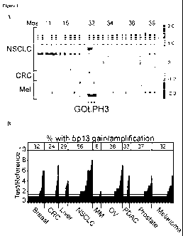
Figures IA-lE show genomic characterization of 5p13 amplification. Figure 1A
shows an array-CGH heat map detailing GOLPH3 amplification at 5p13 in
representative
tumor specimens and cell lines from malignant melanoma (Mel), colon
adenocarcinoma
(CRC) and non-small cell lung cancer (NSCLC). Regions of genomic amplification
and
deletion are denoted in red and blue, respectively. Mbs = position on
chromosome 5 in
megabases. Figure 1B shows a histogram summary of copy number status at 5p13
by
TMA-FISH analysis of 307 tumor cores of the indicated tumor types. CRC = colon
adenocarcinoma; NSCLC = small cell lung cancer; MM = multiple myeloma; OV =
ovarian
carcinoma; PDAC = pancreatic ductal adenocarcinoma. Figure 1C shows the
minimum
common region of the 5p13 amplicon defined by array-CGH from one
representative tumor
(melanoma C27) with focal amplification. Figure 1D shows delimitation of
chromosome
5p13 amplicon boundaries by genomic qPCR using four informative cell line and
tumor
specimens. Figure 1E shows a heat map depiction of Affymetrix expression data
for
NSCLC 5p13 amplified (AMP) and normal specimens. * = significant correlation
after
bonferroni correction for multiple testing.
Figures 2A-2E shows functional validation data of GOLPH3. Figure 2A shows the
results of the indicated cell lines treated with non-targeting siRNA (siNT) or
individual
siRNAs against GOLPH3 (si#1-si#4) for effect on anchorage-independent growth
in soft
agar (left panels) and cell proliferation (right panels). Bars indicate S.D.
Figure 2B
shows the results of A549 parental cells and those expressing either wild type
(WT) or
siRNA resistant GOLPH3 (siRES) treated with either non-targeting siRNA (siNT)
or
siRNA against GOLPH3 (siGOLPH3) for effect on cell proliferation. Bars
indicate S.D.
Shown are endpoint values for day 5. Figure 2C shows the results of primary
Ink4a/Arf-
deficient MEFs transfected with the indicated vectors expressing HRAS T12, MYC
and
GOLPH3. Vec = LacZ vector control; bars indicate S.D.; Two-tailed t-test:
HRAS T12 +
GOLPH3 vs. HRAS T12 + Vec, p=0.0018. Figure 2D shows the results of TERT-
immortalized human melanocytes (HMEL) expressing activated BRAFV600E
transduced
with either GOLPH3 or SUB1 to assay for effect on anchorage-independent growth
in soft
agar. Bars indicate S.D.; Two-tailed t-test for colony number: EV vs. GOLPH3,
p=0.0020;
13
CA 02737106 2011-03-14
WO 2010/040124 PCT/US2009/059526
EV vs. SUB 1, p=0.3739. Figure 2E shows the results of the indicated cell
lines transduced
with GOLPH3 to assay for effect on growth of mouse xenograft tumors.
Figures 3A-3D show that GOLPH3 interacts with VPS35 and influences cell size.
Figure 3A shows GOLPH3 positive endosome-like structures (arrows) in both
1205LU
melanoma cells stably-expressing GOLPH3 that were co-immunostained for HA
(GOLPH3HA; green) and TGN46 (Golgi marker; red) (top panel) and A549 cells co-
immunostained for GOLPH3 (green) and TGN46 (Golgi marker; red) (bottom panel).
DNA was labeled with DAPI. Figure 3B shows immunoblotting results of isolated
proteins immunoprecipitated (IP) with anti-HA (left panel) or anti-V5 (right
panel) for
immunoblotting with the indicated antibodies from 239T cells transiently
expressing the
indicated constructs. NS = non-specific band. Figure 3C shows GOLPH3 positive
co-
staining at endosome structures (arrows) in A549 cells co-immunostained for
GOLPH3
(green) and VPS35 (red). DNA was labeled with DAPI (blue). Figure 3D shows
Automated Quantitative Analysis (AQUA) of phospho-S6KT 389 (red) in two
representative lung adenocarcinomas. Cytokeratin (green) defines tumor and non-
nuclear
compartments. FISH ratio = 5pl3:reference ratio as determined by FISH on
consecutive
TMA sections. Magnification = 20x.
Figures 4A-4E show that GOLPH3 modulates phosphorylation status of mTOR
substrates. Figure 4A shows representative flow histograms for A549 cells
treated with
non-targeting (siNT, blue), siRNA against GOLPH3 (siGOLPH3, left panel, green)
or
rapamycin (Rap, middle panel, green). Peak FSC-H is indicated in the
histograms and the
right panel shows mean FSC-H for multiple experiments (n=3); Bars indicate
S.D. Figure
4B shows protein lysates extracted from 1205LU (left panel), A549 (middle
panel) and
HMEL-tet-GOLPH3 (right panel; with or without doxycycline (DOX)) cells
expressing
GOLPH3 immunoblotted with the indicated antibodies. Figure 4C shows the
results of
HMEL-tet-GOLPH3 cells that were serum depleted and propagated with or without
doxycycline (DOX), followed by treatment with or without EGF for 30 min for
immunoblot
analysis with the indicated antibodies. Figures 4D-4E shows the results of
A549 (Figure
4D) and CRL-5889 (Figure 4E) cells that were serum depleted and treated with
either non-
targeting (siNT) or siRNA against GOLPH3 (siGOLPH3), followed by growth factor
stimulation with EGF for immunoblot analysis with the indicated antibodies.
14
CA 02737106 2011-03-14
WO 2010/040124 PCT/US2009/059526
Figures 5A-5C shows that in vivo GOLPH3 growth advantage is abrogated by
treatment with rapamycin. Mice harboring tumors of the melanoma cell lines
WM239A
(Figure 5A) and 1205LU (Figure 5B) transduced with empty vector (EV; left
panels) or
GOLPH3 (right panels) were treated with vehicle or rapamycin (6.0 mg/kg) at
two-day
increments following treatment onset (tumor baseline volume -100 mm) . Growth
curves
were plotted as mean change in tumor volume relative to baseline starting
volume for each
group. Bars indicate S.E.M. for biological replicates. %TGI indicates
percent tumor
growth inhibition at time course endpoint. Figure 5C summarizes data from the
rapamycin
treatment xenograft studies shown in Figures 5A-5B at the same time point (day
8, post 4
doses). Veh indicates vehicle; Rap indicates rapamycin; %TGI indicates percent
tumor
growth inhibition. 1205LU-GOLPH3 tumors treated with vehicle grew 2.5X in size
during
the 8 days of treatment. In comparison, WM239A-GOLPH3 tumors grew 5.8X in size
during the same period of 8 days. The %TGI in these two cohorts of tumors was
similar, at
81.9% and 80.9% respectively, indicating that growth rate did not impact on
the response to
rapamycin.
Figures 6 shows the results of genomic identification of the 5p13 amplicon.
Representative images of TMA-FISH analysis are shown for 5p13 amplification in
tumor
core specimens: (i) benign compound nevus of right waist, (ii) malignant
melanoma of left
heel, (iii) normal lung and (iv) grade II lung adenocarcinoma. Regions of 5p13
and
centromere-specific ploidy reference are indicated by green and red,
respectively.
Figures 7A-7G show additional results of immunoblot and anchorage-
independence assays for GOLPH3. Figure 7A shows immunoblot analysis of GOLPH3
expression in 5p13 amplified (AMP) and normal (NL) melanoma and NSCLC cell
lines.
NHM = normal human melanocytes. Figure 7B shows confirmation of GOLPH3
knockdown in NSCLC CRL-5889 (top panel) and melanoma 1205LU (bottom panel) for
the anchorage-independent growth and proliferation assays presented in Figure
2A.
GOLPH3 knockdown was performed using the indicated siRNAs (si#1-si#4). siNT =
non-
targeting siRNA control; P=parental A549 lysate. Figure 7C shows confirmation
of
GOLPH3 expression in GOLPH3-transduced melanoma 1205LU used in Figure 7D and
xenograft assays. 1205LU lysates were extracted from cells used for GOLPH3
gain-of-
function experiment presented in Figure 4B and are therefore presented in both
figures.
CA 02737106 2011-03-14
WO 2010/040124 PCT/US2009/059526
Figure 7D shows that GOLPH3 enhances anchorage-independent growth of human
melanoma 1205LU cells (without 5p13 amplification). EV indicates empty vector;
Bars
indicate S.D.; Two-tailed t-test for colony number, p=0.0003. Figures 7E-7F
show
confirmation of GOLPH3 expression in the xenograft assays using GOLPH3-
transduced
melanoma WM239A cells (Figure 7E) and GOLPH3-transduced NSCLC A549 cells
(Figure 7F). Whole cell lysates were immunoblotted with the indicated
antibodies. Figure
7G shows confirmation of mTOR inhibition by rapamycin. Whole cell lysates
extracted
from representative empty vector (EV; n=3) or GOLPH3 (n=5) WM239A xenograft
tumors
that were harvested from mice treated with (+) or without (-) rapamycin were
immunoblotted with the indicated antibodies.
Figures 8A-8C show the results of yeast two-hybrid screening for GOLPH3-
interacting proteins. Figure 8A shows that yeast two-hybrid screening
identified VPS35 as
a GOLPH3 -interacting protein. The yeast reporter strain (AH109) co-expressing
VPS35
(prey) with either GOLPH3 (+) or empty vector (-) plated on SC-Leucine-
Histidine-
Adenine+XaGal (SC-L-H-A+XaGAL) reporter plates confirm reporter activation and
GOLPH3 bait-dependency. Figure 8B shows controls for the yeast two-hybrid
screen.
Negative control (-), AH109 expressing pGBKT7-Lam (bait; TRP) and pGADT7-T
(prey,
LEU); positive control (+), AH109 expressing pGBKT7-p53 (bait, TRP) and GADT7-
T
(prey, LEU); positive control (++), AH109 expressing pGBKT7 (empty vector,
TRP) and
pCLI (GAL4 activation domain, LEU); bait-independent false positive control
(++ -bait)
AH109 expressing pCL1 alone. Strains from SC-Leucine (SC-L) were replica
plated to
SC-Leucine-Trptophan (SC-LT) to confirm presence of bait and prey and to SC-
Leucine-
Histidine-Adenine+XaGal (SC-L-H-A+XaGAL) to confirm reporter activation
through the
cells ability to grow and express the a-galactosidase reporter (indicated by
blue
appearance). Controls were used as comparison for positive clone selection and
bait-
dependency testing. Figure 8C shows the interaction of endogenous GOLPH3 with
VPS35
as determined by co-immunoprecipitation analysis. NSCLC A549 protein extracts
were
immunoprecipitated (IP) with either control (C) or anti-GOLPH3 (G3) mouse
serum for
immunoblotting with the indicated antibodies.
Figure 9 shows the results of Western blot analysis for GOLPH3-dependent
changes in mTOR substrates and other MAPK-/PI3K-relevant proteins. The
indicated cell
16
CA 02737106 2011-03-14
WO 2010/040124 PCT/US2009/059526
lines were either transduced with empty vector (EV) or GOLPH3 (two left
panels, over-
expression) or transfected with non-targeting siRNA (siNT) or siRNA against
GOLPH3
(siGOLPH3) (two right panels, knockdown). Whole cell lysates were
immunoblotted with
the indicated antibodies.
Figures 1OA-10B show the results of endpoint analysis for rapamycin treatment
xenograft studies indicated by endpoint tumor volume measurements for tumors
presented
in Figure 5. Values are plotted as proportion of dose 4 or dose 6 endpoint
tumor volumes
over respective baseline starting volume for WM239A (Figure 10A) and 1205LU
(Figure
10B) xenografts, respectively. Rap indicates rapamycin; Two-tailed t-test for
rapamycin
treated WM239A and 1205LU EV vs. GOLPH3 xenografts, p=0.0677 and p=0.0268,
respectively.
Figure 11A-11C show reduction of lipid second messenger biosynthesis upon
GOLPH3 depletion. Figure 11A shows that depletion of GOLPH3 reduces cell
migration,
possibly through either an mTOR- or phospholipid-mediated pathway. Figure 11B
shows
that depletion of GOLPH3 reduces in vivo basal and growth factor simulated
biosynthesis
of lipid second messengers that feed into cancer signaling pathways. Figure
11C shows
quantitative results of the data presented in Figure 11B.
Figure 12A-12D show that growth factor signaling causes GOLPH3 mis-
localization via ARF4. Figure 12A shows that ARF4, a GTPase identified herein
as a
GOLPH3-interacting protein, co-localizes with GOLPH3 in the Golgi. Figure 12B
shows
that GTP-mediated phosphorylation of GOLPH3 is required for GOLPH3
localization to
the Golgi and that depletion of ARF4 causes mislocalization of GOLPH3 from the
Golgi to
other parts of the cell, including the cell periphery. Figure 12C shows the
kinetics of EGF
receptor phosphorylation upon stimulating with EGF in a representative cancer
cell line
(A549). Figure 12D shows that growth factor signaling (e.g., EGF) causes
redistribution of
GOLPH3 from the Golgi to other parts of the cell, suggesting that growth
factors might do
so by decreasing GOLPH3 phosphorylation by ARF4 since ARF4 is known to
relocalize to
the plasma membrane upon EGF administration.
Detailed Description of the Invention
The invention is based, in part, on the discovery that GOLPH3, a Golgi
localized
17
CA 02737106 2011-03-14
WO 2010/040124 PCT/US2009/059526
protein, was identified as a novel oncogene from within a conserved 5p13
amplification in
numerous cancers (e.g., at least nine solid tumor types). GOLPH3 yeast-
interaction
analysis, coupled with observation of knockdown-associated cell size reduction
phenotype,
led to confirmatory biochemical and functional studies establishing that
GOLPH3 activates
mTOR-S6-Kinase signaling and confers sensitivity to mTOR inhibitors (e.g.,
rapamycin).
GOLPH3 has also been shown herein to affect biosynthesis of lipid second
messengers that
feed into cancer signaling pathways and to impact oncogenesis through
regulation by
cellular growth factors (e.g., EGF). GOLPH3 polypeptides and fragments
thereof, e.g.,
biologically active or antigenic fragments thereof, are provided, as reagents
or targets in
assays applicable to diagnosis of cancer, e.g., lung, ovarian, pancreatic,
liver, breast,
prostate, and colon carcinomas, as well as melanoma and multiple myeloma. In
particular,
the methods and compositions of the present disclosure relate to detection of
expression
and/or activity of a GOLPH3 gene or fragment thereof, e.g., biologically
active fragments
thereof, as well as to the detection of expression and/or activity of gene
products or
fragments thereof encoded by the GOLPH3 gene, e.g., biologically active
fragments
thereof. The methods and compositions of the present disclosure can utilize
the GOLPH3
gene or gene sequence or fragments thereof, as well as gene products of the
GOLPH3 gene
and/or fragments thereof, e.g., antibodies which specifically bind to such
GOLPH3 gene
products.
In one aspect, methods are provided for detecting the presence, absence,
stage, and
other characteristics of cancers, e.g., lung, ovarian, pancreatic, liver,
breast, prostate, and
colon carcinomas, as well as melanoma and multiple myeloma, in a sample that
are relevant
to prognosis, diagnosis, monitoring, and characterization in a patient.
The disclosure also features compositions of matter, including antibodies
(e.g.,
antibodies which specifically bind to any one of the polypeptides described
herein) as well
as fusion polypeptides, including all or a fragment of a polypeptide described
herein.
1. Definitions
The articles "a" and "an" are used herein to refer to one or to more than one
(i.e. to
at least one) of the grammatical object of the article. By way of example, "an
element"
means one element or more than one element.
18
CA 02737106 2011-03-14
WO 2010/040124 PCT/US2009/059526
The term "altered amount" of a marker or "altered level" of a marker refers to
increased or decreased copy number (e.g., germline and/or somatic) of a marker
or
chromosomal region, e.g., MCR, and/or increased or decreased expression level
of a
particular marker gene or genes in a cancer sample, as compared to the
expression level or
copy number of the marker in a control sample. The term "altered amount" of a
marker
also includes an increased or decreased protein level of a marker in a sample,
e.g., a cancer
sample, as compared to the protein level of the marker in a normal, control
sample.
Furthermore, an altered amount of a marker may be determined by detecting the
methylation status of a marker, as described herein, which may affect the
expression or
activity of a marker.
The amount of a marker, e.g., expression or copy number of a marker or MCR, or
protein level of a marker, in a subject is "significantly" higher or lower
than the normal
amount of a marker or MCR, if the amount of the marker is greater or less,
respectively,
than the normal level by an amount greater than the standard error of the
assay employed to
assess amount, and preferably at least twice, and more preferably three, four,
five, ten or
more times that amount. Alternately, the amount of the marker or MCR in the
subject can
be considered "significantly" higher or lower than the normal amount if the
amount is at
least about two, and preferably at least about three, four, or five times,
higher or lower,
respectively, than the normal amount of the marker or MCR.
The term "altered level of expression" of a marker or MCR refers to an
expression
level or copy number of a marker in a test sample e.g., a sample derived from
a patient
suffering from cancer, that is greater or less than the standard error of the
assay employed
to assess expression or copy number, and is preferably at least twice, and
more preferably
three, four, five or ten or more times the expression level or copy number of
the marker or
MCR in a control sample (e.g., sample from a healthy subjects not having the
associated
disease) and preferably, the average expression level or copy number of the
marker or MCR
in several control samples. The altered level of expression is greater or less
than the
standard error of the assay employed to assess expression or copy number, and
is preferably
at least twice, and more preferably three, four, five or ten or more times the
expression level
or copy number of the marker or MCR in a control sample (e.g., sample from a
healthy
19
CA 02737106 2011-03-14
WO 2010/040124 PCT/US2009/059526
subjects not having the associated disease) and preferably, the average
expression level or
copy number of the marker or MCR in several control samples.
The term "altered activity" of a marker refers to an activity of a marker
which is
increased or decreased in a disease state, e.g., in a cancer sample, as
compared to the
activity of the marker in a normal, control sample. Altered activity of a
marker may be the
result of, for example, altered expression of the marker, altered protein
level of the marker,
altered structure of the marker, or, e.g., an altered interaction with other
proteins involved
in the same or different pathway as the marker or altered interaction with
transcriptional
activators or inhibitors, or altered methylation status.
The term "altered structure" of a marker refers to the presence of mutations
or
allelic variants within the marker gene or maker protein, e.g., mutations
which affect
expression or activity of the marker, as compared to the normal or wild-type
gene or
protein. For example, mutations include, but are not limited to substitutions,
deletions, or
addition mutations. Mutations may be present in the coding or non-coding
region of the
marker.
The term "altered subcellular localization" of a marker refers to the
mislocalization
of the marker within a cell relative to the normal localization within the
cell (e.g., within a
healthy and/or wild-type cell. An indication of normal localization of the
marker can be
determined through an analysis of subcellular localization motifs known in the
field that are
harbored by marker polypeptides.
Unless otherwise specified here within, the terms "antibody" and "antibodies"
broadly encompass naturally-occurring forms of antibodies (e.g. IgG, IgA, IgM,
IgE) and
recombinant antibodies such as single-chain antibodies, chimeric and humanized
antibodies
and multi-specific antibodies, as well as fragments and derivatives of all of
the foregoing,
which fragments and derivatives have at least an antigenic binding site.
Antibody
derivatives may comprise a protein or chemical moiety conjugated to an
antibody.
The term "antibody" as used herein also includes an "antigen-binding portion"
of an
antibody (or simply "antibody portion"). The term "antigen-binding portion",
as used
herein, refers to one or more fragments of an antibody that retain the ability
to specifically
bind to an antigen (e.g., GOLPH3 polypeptide or fragment thereof). It has been
shown that
the antigen-binding function of an antibody can be performed by fragments of a
full-length
CA 02737106 2011-03-14
WO 2010/040124 PCT/US2009/059526
antibody. Examples of binding fragments encompassed within the term "antigen-
binding
portion" of an antibody include (i) a Fab fragment, a monovalent fragment
consisting of the
VL, VH, CL and CH1 domains; (ii) a F(ab')2 fragment, a bivalent fragment
comprising two
Fab fragments linked by a disulfide bridge at the hinge region; (iii) a Fd
fragment
consisting of the VH and CH1 domains; (iv) a Fv fragment consisting of the VL
and VH
domains of a single arm of an antibody, (v) a dAb fragment (Ward et at.,
(1989) Nature
341:544-546), which consists of a VH domain; and (vi) an isolated
complementarity
determining region (CDR). Furthermore, although the two domains of the Fv
fragment, VL
and VH, are coded for by separate genes, they can be joined, using recombinant
methods,
by a synthetic linker that enables them to be made as a single protein chain
in which the VL
and VH regions pair to form monovalent polypeptides (known as single chain Fv
(scFv);
see e.g., Bird et at. (1988) Science 242:423-426; and Huston et at. (1988)
Proc. Natl. Acad.
Sci. USA 85:5879-5883; and Osbourn et at. 1998, Nature Biotechnology 16: 778).
Such
single chain antibodies are also intended to be encompassed within the term
"antigen-
binding portion" of an antibody. Any VH and VL sequences of specific scFv can
be linked
to human immunoglobulin constant region cDNA or genomic sequences, in order to
generate expression vectors encoding complete IgG polypeptides or other
isotypes. VH and
VL can also be used in the generation of Fab, Fv or other fragments of
immunoglobulins
using either protein chemistry or recombinant DNA technology. Other forms of
single
chain antibodies, such as diabodies are also encompassed. Diabodies are
bivalent,
bispecific antibodies in which VH and VL domains are expressed on a single
polypeptide
chain, but using a linker that is too short to allow for pairing between the
two domains on
the same chain, thereby forcing the domains to pair with complementary domains
of
another chain and creating two antigen binding sites (see e.g., Holliger, P.,
et at. (1993)
Proc. Natl. Acad. Sci. USA 90:6444-6448; Poljak, R. J., et at. (1994)
Structure 2:1121-
1123).
Still further, an antibody or antigen-binding portion thereof may be part of
larger
immunoadhesion polypeptides, formed by covalent or noncovalent association of
the
antibody or antibody portion with one or more other proteins or peptides.
Examples of
such immunoadhesion polypeptides include use of the streptavidin core region
to make a
tetrameric scFv polypeptide (Kipriyanov, S.M., et at. (1995) Human Antibodies
and
21
CA 02737106 2011-03-14
WO 2010/040124 PCT/US2009/059526
Hybridomas 6:93-101) and use of a cysteine residue, a marker peptide and a C-
terminal
polyhistidine tag to make bivalent and biotinylated scFv polypeptides
(Kipriyanov, S.M., et
at. (1994) Mol. Immunol. 31:1047-1058). Antibody portions, such as Fab and
F(ab')2
fragments, can be prepared from whole antibodies using conventional
techniques, such as
papain or pepsin digestion, respectively, of whole antibodies. Moreover,
antibodies,
antibody portions and immunoadhesion polypeptides can be obtained using
standard
recombinant DNA techniques, as described herein.
Antibodies may be polyclonal or monoclonal; xenogeneic, allogeneic, or
syngeneic;
or modified forms thereof (e.g. humanized, chimeric, etc.). Antibodies may
also be fully
human. Preferably, antibodies of the invention bind specifically or
substantially
specifically to GOLPH3 polypeptides or fragments thereof. The terms
"monoclonal
antibodies" and "monoclonal antibody composition", as used herein, refer to a
population of
antibody polypeptides that contain only one species of an antigen binding site
capable of
immunoreacting with a particular epitope of an antigen, whereas the term
"polyclonal
antibodies" and "polyclonal antibody composition" refer to a population of
antibody
polypeptides that contain multiple species of antigen binding sites capable of
interacting
with a particular antigen. A monoclonal antibody composition typically
displays a single
binding affinity for a particular antigen with which it immunoreacts.
The term "body fluid" refers to fluids that are excreted or secreted from the
body as
well as fluid that are normally not (e.g. amniotic fluid, aqueous humor, bile,
blood and
blood plasma, cerebrospinal fluid, cerumen and earwax, cowper's fluid or pre-
ejaculatory
fluid, chyle, chyme, stool, female ejaculate, interstitial fluid,
intracellular fluid, lymph,
menses, breast milk, mucus, pleural fluid, pus, saliva, sebum, semen, serum,
sweat,
synovial fluid, tears, urine, vaginal lubrication, vitreous humor, vomit).
The terms "cancer" or "tumor" refer to the presence of cells possessing
characteristics typical of cancer-causing cells, such as uncontrolled
proliferation,
immortality, metastatic potential, rapid growth and proliferation rate, and
certain
characteristic morphological features. Cancer cells are often in the form of a
tumor, but
such cells may exist alone within an animal, or may be a non-tumorigenic
cancer cell, such
as a leukemia cell. As used herein, the term "cancer" includes premalignant as
well as
malignant cancers. Cancers include, but are not limited to, B cell cancer,
e.g., multiple
22
CA 02737106 2011-03-14
WO 2010/040124 PCT/US2009/059526
myeloma, Waldenstrom's macroglobulinemia, the heavy chain diseases, such as,
for
example, alpha chain disease, gamma chain disease, and mu chain disease,
benign
monoclonal gammopathy, and immunocytic amyloidosis, melanomas, breast cancer,
lung
cancer, bronchus cancer, colorectal cancer, prostate cancer, pancreatic
cancer, stomach
cancer, ovarian cancer, urinary bladder cancer, brain or central nervous
system cancer,
peripheral nervous system cancer, esophageal cancer, cervical cancer, uterine
or
endometrial cancer, cancer of the oral cavity or pharynx, liver cancer, kidney
cancer,
testicular cancer, biliary tract cancer, small bowel or appendix cancer,
salivary gland
cancer, thyroid gland cancer, adrenal gland cancer, osteosarcoma,
chondrosarcoma, cancer
of hematological tissues, and the like.
The term "cellular growth factors" refers to cellular growth factors well
known in
the art, including, e.g., EGF, FGF, TGF-a, TGF-(3, PDGF, IGF-1, IGF-2 BNDF,
BMP,
GGRP, GDNF, GGF, HGF, KGF, mytotrophin, NGF, OSM, somatotrophin, and VEGF.
The term "cellular phospholipids" encompasses cellular phospholipids well
known
in the art, including, e.g., PIP2, PIP3, IP3, DAG, and PA).
As used herein, the term "coding region" refers to regions of a nucleotide
sequence
comprising codons which are translated into amino acid residues, whereas the
term
"noncoding region" refers to regions of a nucleotide sequence that are not
translated into
amino acids (e.g., 5' and 3' untranslated regions).
"Complementary" refers to the broad concept of sequence complementarity
between
regions of two nucleic acid strands or between two regions of the same nucleic
acid strand.
It is known that an adenine residue of a first nucleic acid region is capable
of forming
specific hydrogen bonds ("base pairing") with a residue of a second nucleic
acid region
which is antiparallel to the first region if the residue is thymine or uracil.
Similarly, it is
known that a cytosine residue of a first nucleic acid strand is capable of
base pairing with a
residue of a second nucleic acid strand which is antiparallel to the first
strand if the residue
is guanine. A first region of a nucleic acid is complementary to a second
region of the same
or a different nucleic acid if, when the two regions are arranged in an
antiparallel fashion, at
least one nucleotide residue of the first region is capable of base pairing
with a residue of
the second region. Preferably, the first region comprises a first portion and
the second
region comprises a second portion, whereby, when the first and second portions
are
23
CA 02737106 2011-03-14
WO 2010/040124 PCT/US2009/059526
arranged in an antiparallel fashion, at least about 50%, and preferably at
least about 75%, at
least about 90%, or at least about 95% of the nucleotide residues of the first
portion are
capable of base pairing with nucleotide residues in the second portion. More
preferably, all
nucleotide residues of the first portion are capable of base pairing with
nucleotide residues
in the second portion.
The "copy number of a gene" or the "copy number of a marker" refers to the
number
of DNA sequences in a cell (e.g., germline and/or somatic) encoding a
particular gene
product. Generally, for a given gene, a mammal has two copies of each gene.
The copy
number can be increased, however, by gene amplification or duplication, or
reduced by
deletion. For example, germline copy number changes include chagnes at one or
more
genomic loci, wherein said one or more genomic loci are not accounted for by
the number
of copies in the normal complement of germline copies in a control (e.g., the
normal copy
number in germline DNA for the same species as that from which the specific
germline
DNA and corresponding copy number were determined). Somatic copy number
changes
includechanges at one or more genomic loci, wherein said one or more genomic
loci are not
accounted for by the number of copies in germline DNA of a control (e.g., copy
number in
germline DNA for the same subject as that from which the somatic DNA and
corresponding
copy number were determined).
The "normal" copy number (e.g., germline and/or somatic) of a marker or MCR or
"normal" level of expression of a marker is the level of expression, copy
number of the
marker, or copy number of the MCR, in a biological sample, e.g., a sample
containing
tissue, whole blood, serum, plasma, buccal scrape, saliva, cerebrospinal
fluid, urine, stool,
and bone marrow, from a subject, e.g., a human, not afflicted with cancer.
As used herein, the term "diagnostic marker" includes markers listed herein
which
are useful in the diagnosis of cancer, e.g., over- or under- activity,
emergence, expression,
growth, remission, recurrence or resistance of tumors before, during or after
therapy. The
predictive functions of the marker may be confirmed by, e.g., (1) increased or
decreased
copy number (e.g., by FISH, FISH plus SKY, single-molecule sequencing, e.g.,
as
described in the art at least at J. Biotechnol., 86:289-30 1, or qPCR),
overexpression or
underexpression (e.g., by ISH, Northern Blot, or qPCR), increased or decreased
protein
level (e.g., by IHC), or increased or decreased activity (determined by, for
example,
24
CA 02737106 2011-03-14
WO 2010/040124 PCT/US2009/059526
modulation of a pathway in which the marker is involved), e.g., in more than
about 5%,
6%, 7%, 8%, 9%, l0%, l l%, l2%, l3%, l4%, l5%, 20%, 25%, or more of human
cancers
types or cancer samples; (2) its presence or absence in a biological sample,
e.g., a sample
containing tissue, whole blood, serum, plasma, buccal scrape, saliva,
cerebrospinal fluid,
urine, stool, or bone marrow, from a subject, e.g. a human, afflicted with
cancer; (3) its
presence or absence in clinical subset of patients with cancer (e.g., those
responding to a
particular therapy or those developing resistance).
Diagnostic markers also include "surrogate markers," e.g., markers which are
indirect markers of cancer progression.
A molecule is "fixed" or "affixed" to a substrate if it is covalently or non-
covalently
associated with the substrate such that the substrate can be rinsed with a
fluid (e.g. standard
saline citrate, pH 7.4) without a substantial fraction of the molecule
dissociating from the
substrate.
"Homologous" as used herein, refers to nucleotide sequence similarity between
two
regions of the same nucleic acid strand or between regions of two different
nucleic acid
strands. When a nucleotide residue position in both regions is occupied by the
same
nucleotide residue, then the regions are homologous at that position. A first
region is
homologous to a second region if at least one nucleotide residue position of
each region is
occupied by the same residue. Homology between two regions is expressed in
terms of the
proportion of nucleotide residue positions of the two regions that are
occupied by the same
nucleotide residue. By way of example, a region having the nucleotide sequence
5'-
ATTGCC-3' and a region having the nucleotide sequence 5'-TATGGC-3' share 50%
homology. Preferably, the first region comprises a first portion and the
second region
comprises a second portion, whereby, at least about 50%, and preferably at
least about
75%, at least about 90%, or at least about 95% of the nucleotide residue
positions of each
of the portions are occupied by the same nucleotide residue. More preferably,
all
nucleotide residue positions of each of the portions are occupied by the same
nucleotide
residue.
As used herein, the term "host cell" is intended to refer to a cell into which
a nucleic
acid of the invention, such as a recombinant expression vector of the
invention, has been
introduced. The terms "host cell" and "recombinant host cell" are used
interchangeably
CA 02737106 2011-03-14
WO 2010/040124 PCT/US2009/059526
herein. It should be understood that such terms refer not only to the
particular subject cell
but to the progeny or potential progeny of such a cell. Because certain
modifications may
occur in succeeding generations due to either mutation or environmental
influences, such
progeny may not, in fact, be identical to the parent cell, but are still
included within the
scope of the term as used herein.
The term "humanized antibody", as used herein, is intended to include
antibodies
made by a non-human cell having variable and constant regions which have been
altered to
more closely resemble antibodies that would be made by a human cell. For
example, by
altering the non-human antibody amino acid sequence to incorporate amino acids
found in
human germline immunoglobulin sequences. The humanized antibodies of the
invention
may include amino acid residues not encoded by human germline immunoglobulin
sequences (e.g., mutations introduced by random or site-specific mutagenesis
in vitro or by
somatic mutation in vivo), for example in the CDRs. The term "humanized
antibody", as
used herein, also includes antibodies in which CDR sequences derived from the
germline of
another mammalian species, such as a mouse, have been grafted onto human
framework
sequences.
An "inducible" promoter is a nucleotide sequence which, when operably linked
with
a polynucleotide which encodes or specifies a gene product, causes the gene
product to be
produced in a living human cell substantially only when an inducer which
corresponds to
the promoter is present in the cell.
As used herein, the term "inhibit" includes the decrease, limitation, or
blockage, of,
for example a particular action, function, or interaction.
Cancer is "inhibited" if at least one symptom of the cancer is alleviated,
terminated,
slowed, or prevented. As used herein, cancer is also "inhibited" if recurrence
or metastasis
of the cancer is reduced, slowed, delayed, or prevented.
As used herein, the term "interaction", when referring to an interaction
between two
molecules, refers to the physical contact (e.g., binding) of the molecules
with one another.
Generally, such an interaction results in an activity (which produces a
biological effect) of
one or both of said molecules.
An "isolated antibody", as used herein, is intended to refer to an antibody
that is
substantially free of other antibodies having different antigenic
specificities (e.g., an
26
CA 02737106 2011-03-14
WO 2010/040124 PCT/US2009/059526
isolated antibody that specifically binds GOLPH3 polypeptide or a fragment
thereof is
substantially free of antibodies that specifically bind antigens other than a
GOLPH3
polypeptide or a fragment thereof). Moreover, an isolated antibody may be
substantially
free of other cellular material and/or chemicals.
As used herein, an "isolated protein" refers to a protein that is
substantially free of
other proteins, cellular material, separation medium, and culture medium when
isolated
from cells or produced by recombinant DNA techniques, or chemical precursors
or other
chemicals when chemically synthesized. An "isolated" or "purified" protein or
biologically
active portion thereof is substantially free of cellular material or other
contaminating
proteins from the cell or tissue source from which the antibody, polypeptide,
peptide or
fusion protein is derived, or substantially free from chemical precursors or
other chemicals
when chemically synthesized. The language "substantially free of cellular
material"
includes preparations of GOLPH3 polypeptide or fragment thereof, in which the
protein is
separated from cellular components of the cells from which it is isolated or
recombinantly
produced. In one embodiment, the language "substantially free of cellular
material"
includes preparations of GOLPH3 protein or fragment thereof, having less than
about 30%
(by dry weight) of non-GOLPH3 protein (also referred to herein as a
"contaminating
protein"), more preferably less than about 20% of non-GOLPH3 protein, still
more
preferably less than about 10% of non-GOLPH3 protein, and most preferably less
than
about 5% non-GOLPH3 protein. When antibody, polypeptide, peptide or fusion
protein or
fragment thereof, e.g., a biologically active fragment thereof, is
recombinantly produced, it
is also preferably substantially free of culture medium, i.e., culture medium
represents less
than about 20%, more preferably less than about 10%, and most preferably less
than about
5% of the volume of the protein preparation.
A "kit" is any manufacture (e.g. a package or container) comprising at least
one
reagent, e.g. a probe, for specifically detecting the expression of a marker
of the invention.
The kit may be promoted, distributed, or sold as a unit for performing the
methods of the
present invention. The kit may comprise one or more reagents necessary to
express a
marker of the invention (e.g., GOLPH3). In certain embodiments, the kit may
further
comprise a reference standard, e.g., a nucleic acid encoding a protein that
does not affect or
regulate signaling pathways controlling cell growth, division, migration,
survival or
27
CA 02737106 2011-03-14
WO 2010/040124 PCT/US2009/059526
apoptosis. One skilled in the art can envision many such control proteins,
including, but
not limited to, common molecular tags (e.g., green fluorescent protein and
beta-
galactosidase), proteins not classified in any of pathway encompassing cell
growth,
division, migration, survival or apoptosis by GeneOntology reference, or
ubiquitous
housekeeping proteins. Reagents in the kit may be provided in individual
containers or as
mixtures of two or more reagents in a single container. In addition,
instructional materials
which describe the use of the compositions within the kit can be included.
A "marker" is a gene whose altered level of expression in a tissue or cell
from its
expression level in normal or healthy tissue or cell is associated with a
disease state, such as
cancer. A "marker nucleic acid" is a nucleic acid (e.g., mRNA, cDNA) encoded
by or
corresponding to a marker of the invention. Such marker nucleic acids include
DNA (e.g.,
cDNA) comprising the entire or a partial sequence of any of the nucleic acid
sequences set
forth in the Sequence Listing or the complement of such a sequence. The marker
nucleic
acids also include RNA comprising the entire or a partial sequence of any of
the nucleic
acid sequences set forth in the Sequence Listing or the complement of such a
sequence,
wherein all thymidine residues are replaced with uridine residues. A "marker
protein" is a
protein encoded by or corresponding to a marker of the invention. A marker
protein
comprises the entire or a partial sequence of any of the sequences set forth
in the Sequence
Listing. The terms "protein" and "polypeptide" are used interchangeably.
A "minimal common region (MCR)," as used herein, refers to a contiguous
chromosomal region which displays either gain and amplification (increased
copy number)
or loss and deletion (decreased copy number) in the genome of a cancer. An MCR
includes
at least one nucleic acid sequence which has increased or decreased copy
number and
which is associated with a cancer.
The "normal" level of expression of a marker is the level of expression of the
marker in cells of a subject, e.g., a human patient, not afflicted with a
cancer, e.g., lung,
ovarian, pancreatic, liver, breast, prostate, and colon carcinomas, as well as
melanoma and
multiple myeloma. An "over-expression" or "significantly higher level of
expression" of a
marker refers to an expression level in a test sample that is greater than the
standard error of
the assay employed to assess expression, and is preferably at least twice, and
more
preferably three, four, five or ten times the expression level of the marker
in a control
28
CA 02737106 2011-03-14
WO 2010/040124 PCT/US2009/059526
sample (e.g., sample from a healthy subjects not having the marker associated
disease) and
preferably, the average expression level of the marker in several control
samples. A
"significantly lower level of expression" of a marker refers to an expression
level in a test
sample that is at least twice, and more preferably three, four, five or ten
times lower than
the expression level of the marker in a control sample (e.g., sample from a
healthy subject
not having the marker associated disease) and preferably, the average
expression level of
the marker in several control samples.
An "overexpression" or "significantly higher level of expression or copy
number"
of a marker or MCR refers to an expression level or copy number in a test
sample that is
greater than the standard error of the assay employed to assess expression or
copy number,
and is preferably at least twice, and more preferably three, four, five or ten
or more times
the expression level or copy number of the marker or MCR in a control sample
(e.g.,
sample from a healthy subject not afflicted with cancer) and preferably, the
average
expression level or copy number of the marker or MCR in several control
samples.
The term "probe" refers to any molecule which is capable of selectively
binding to a
specifically intended target molecule, for example, a nucleotide transcript or
protein
encoded by or corresponding to a marker. Probes can be either synthesized by
one skilled
in the art, or derived from appropriate biological preparations. For purposes
of detection of
the target molecule, probes may be specifically designed to be labeled, as
described herein.
Examples of molecules that can be utilized as probes include, but are not
limited to, RNA,
DNA, proteins, antibodies, and organic molecules.
An "RNA interfering agent" as used herein, is defined as any agent which
interferes
with or inhibits expression of a target gene, e.g., a marker of the invention,
by RNA
interference (RNAi). Such RNA interfering agents include, but are not limited
to, nucleic
acid molecules including RNA molecules which are homologous to the target
gene, e.g., a
marker of the invention, or a fragment thereof, short interfering RNA (siRNA),
and small
molecules which interfere with or inhibit expression of a target gene by RNA
interference
(RNAi).
"RNA interference (RNAi)" is an evolutionally conserved process whereby the
expression or introduction of RNA of a sequence that is identical or highly
similar to a
target gene results in the sequence specific degradation or specific post-
transcriptional gene
29
CA 02737106 2011-03-14
WO 2010/040124 PCT/US2009/059526
silencing (PTGS) of messenger RNA (mRNA) transcribed from that targeted gene
(see
Coburn, G. and Cullen, B. (2002) J. of Virology 76(18):9225), thereby
inhibiting expression
of the target gene. In one embodiment, the RNA is double stranded RNA (dsRNA).
This
process has been described in plants, invertebrates, and mammalian cells. In
nature, RNAi
is initiated by the dsRNA-specific endonuclease Dicer, which promotes
processive cleavage
of long dsRNA into double-stranded fragments termed siRNAs. siRNAs are
incorporated
into a protein complex that recognizes and cleaves target mRNAs. RNAi can also
be
initiated by introducing nucleic acid molecules, e.g., synthetic siRNAs or RNA
interfering
agents, to inhibit or silence the expression of target genes. As used herein,
"inhibition of
target gene expression" or "inhibition of marker gene expression" includes any
decrease in
expression or protein activity or level of the target gene (e.g., a marker
gene of the
invention) or protein encoded by the target gene, e.g., a marker protein of
the invention.
The decrease may be of at least 30%, 40%, 50%, 60%, 70%, 80%, 90%, 95% or 99%
or
more as compared to the expression of a target gene or the activity or level
of the protein
encoded by a target gene which has not been targeted by an RNA interfering
agent.
"Short interfering RNA" (siRNA), also referred to herein as "small interfering
RNA" is defined as an agent which functions to inhibit expression of a target
gene, e.g., by
RNAi. An siRNA may be chemically synthesized, may be produced by in vitro
transcription, or may be produced within a host cell. In one embodiment, siRNA
is a
double stranded RNA (dsRNA) molecule of about 15 to about 40 nucleotides in
length,
preferably about 15 to about 28 nucleotides, more preferably about 19 to about
25
nucleotides in length, and more preferably about 19, 20, 21, or 22 nucleotides
in length,
and may contain a 3' and/or 5' overhang on each strand having a length of
about 0, 1, 2, 3,
4, or 5 nucleotides. The length of the overhang is independent between the two
strands,
i.e., the length of the over hang on one strand is not dependent on the length
of the
overhang on the second strand. Preferably the siRNA is capable of promoting
RNA
interference through degradation or specific post-transcriptional gene
silencing (PTGS) of
the target messenger RNA (mRNA).
In another embodiment, an siRNA is a small hairpin (also called stem loop) RNA
(shRNA). In one embodiment, these shRNAs are composed of a short (e.g., 19-25
nucleotide) antisense strand, followed by a 5-9 nucleotide loop, and the
analogous sense
CA 02737106 2011-03-14
WO 2010/040124 PCT/US2009/059526
strand. Alternatively, the sense strand may precede the nucleotide loop
structure and the
antisense strand may follow. These shRNAs may be contained in plasmids,
retroviruses,
and lentiviruses and expressed from, for example, the pol III U6 promoter, or
another
promoter (see, e.g., Stewart, et at. (2003) RNA Apr;9(4):493-501 incorporated
be reference
herein).
RNA interfering agents, e.g., siRNA molecules, may be administered to a
patient
having or at risk for having cancer, to inhibit expression of a marker gene of
the invention,
e.g., a marker gene which is overexpressed in cancer (such as the markers
listed in Table 3)
and thereby treat, prevent, or inhibit cancer in the subject.
A "constitutive" promoter is a nucleotide sequence which, when operably linked
with a polynucleotide which encodes or specifies a gene product, causes the
gene product
to be produced in a living human cell under most or all physiological
conditions of the cell.
As used herein, "subject" refers to any healthy animal, mammal or human, or
any
animal, mammal or human afflicted with a cancer, e.g., lung, ovarian,
pancreatic, liver,
breast, prostate, and colon carcinomas, as well as melanoma and multiple
myeloma. The
term "subject" is interchangeable with "patient".
The language "substantially free of chemical precursors or other chemicals"
includes preparations of antibody, polypeptide, peptide or fusion protein in
which the
protein is separated from chemical precursors or other chemicals which are
involved in the
synthesis of the protein. In one embodiment, the language "substantially free
of chemical
precursors or other chemicals" includes preparations of antibody, polypeptide,
peptide or
fusion protein having less than about 30% (by dry weight) of chemical
precursors or non-
antibody, polypeptide, peptide or fusion protein chemicals, more preferably
less than about
20% chemical precursors or non-antibody, polypeptide, peptide or fusion
protein chemicals,
still more preferably less than about 10% chemical precursors or non-antibody,
polypeptide, peptide or fusion protein chemicals, and most preferably less
than about 5%
chemical precursors or non- antibody, polypeptide, peptide or fusion protein
chemicals.
A "tissue-specific" promoter is a nucleotide sequence which, when operably
linked
with a polynucleotide which encodes or specifies a gene product, causes the
gene product
to be produced in a living human cell substantially only if the cell is a cell
of the tissue type
corresponding to the promoter.
31
CA 02737106 2011-03-14
WO 2010/040124 PCT/US2009/059526
A "transcribed polynucleotide" or "nucleotide transcript" is a polynucleotide
(e.g. an
mRNA, hnRNA, a cDNA, or an analog of such RNA or cDNA) which is complementary
to
or homologous with all or a portion of a mature mRNA made by transcription of
a marker
of the invention and normal post-transcriptional processing (e.g. splicing),
if any, of the
RNA transcript, and reverse transcription of the RNA transcript.
An "underexpression" or "significantly lower level of expression or copy
number"
of a marker or MCR refers to an expression level or copy number in a test
sample that is
greater than the standard error of the assay employed to assess expression or
copy number,
but is preferably at least twice, and more preferably three, four, five or ten
or more times
less than the expression level or copy number of the marker or MCR in a
control sample
(e.g., sample from a healthy subject not afflicted with cancer) and
preferably, the average
expression level or copy number of the marker or MCR in several control
samples.
As used herein, the term "vector" refers to a nucleic acid capable of
transporting
another nucleic acid to which it has been linked. One type of vector is a
"plasmid", which
refers to a circular double stranded DNA loop into which additional DNA
segments may be
ligated. Another type of vector is a viral vector, wherein additional DNA
segments may be
ligated into the viral genome. Certain vectors are capable of autonomous
replication in a
host cell into which they are introduced (e.g., bacterial vectors having a
bacterial origin of
replication and episomal mammalian vectors). Other vectors (e.g., non-episomal
mammalian vectors) are integrated into the genome of a host cell upon
introduction into the
host cell, and thereby are replicated along with the host genome. Moreover,
certain vectors
are capable of directing the expression of genes to which they are operatively
linked. Such
vectors are referred to herein as "recombinant expression vectors" or simply
"expression
vectors". In general, expression vectors of utility in recombinant DNA
techniques are often
in the form of plasmids. In the present specification, "plasmid" and "vector"
may be used
interchangeably as the plasmid is the most commonly used form of vector.
However, the
invention is intended to include such other forms of expression vectors, such
as viral
vectors (e.g., replication defective retroviruses, adenoviruses and adeno-
associated viruses),
which serve equivalent functions.
There is a known and definite correspondence between the amino acid sequence
of a
particular protein and the nucleotide sequences that can code for the protein,
as defined by
32
CA 02737106 2011-03-14
WO 2010/040124 PCT/US2009/059526
the genetic code (shown below). Likewise, there is a known and definite
correspondence
between the nucleotide sequence of a particular nucleic acid and the amino
acid sequence
encoded by that nucleic acid, as defined by the genetic code.
GENETIC CODE
Alanine (Ala, A) GCA, GCC, GCG, GCT
Arginine (Arg, R) AGA, ACG, CGA, CGC, CGG, CGT
Asparagine (Asn, N) AAC, AAT
Aspartic acid (Asp, D) GAC, GAT
Cysteine (Cys, C) TGC, TGT
Glutamic acid (Glu, E) GAA, GAG
Glutamine (Gln, Q) CAA, CAG
Glycine (Gly, G) GGA, GGC, GGG, GGT
Histidine (His, H) CAC, CAT
Isoleucine (Ile, I) ATA, ATC, ATT
Leucine (Leu, L) CTA, CTC, CTG, CTT, TTA, TTG
Lysine (Lys, K) AAA, AAG
Methionine (Met, M) ATG
Phenylalanine (Phe, F) TTC, TTT
Proline (Pro, P) CCA, CCC, CCG, CCT
Serine (Ser, S) AGC, AGT, TCA, TCC, TCG, TCT
Threonine (Thr, T) ACA, ACC, ACG, ACT
Tryptophan (Trp, W) TGG
Tyrosine (Tyr, Y) TAC, TAT
Valine (Val, V) GTA, GTC, GTG, GTT
Termination signal (end) TAA, TAG, TGA
An important and well known feature of the genetic code is its redundancy,
whereby, for most of the amino acids used to make proteins, more than one
coding
nucleotide triplet may be employed (illustrated above). Therefore, a number of
different
nucleotide sequences may code for a given amino acid sequence. Such nucleotide
sequences are considered functionally equivalent since they result in the
production of the
same amino acid sequence in all organisms (although certain organisms may
translate some
sequences more efficiently than they do others). Moreover, occasionally, a
methylated
33
CA 02737106 2011-03-14
WO 2010/040124 PCT/US2009/059526
variant of a purine or pyrimidine may be found in a given nucleotide sequence.
Such
methylations do not affect the coding relationship between the trinucleotide
codon and the
corresponding amino acid.
In view of the foregoing, the nucleotide sequence of a DNA or RNA coding for a
fusion protein or polypeptide of the invention (or any portion thereof) can be
used to derive
the fusion protein or polypeptide amino acid sequence, using the genetic code
to translate
the DNA or RNA into an amino acid sequence. Likewise, for fusion protein or
polypeptide
amino acid sequence, corresponding nucleotide sequences that can encode the
fusion
protein or polypeptide can be deduced from the genetic code (which, because of
its
redundancy, will produce multiple nucleic acid sequences for any given amino
acid
sequence). Thus, description and/or disclosure herein of a nucleotide sequence
which
encodes a fusion protein or polypeptide should be considered to also include
description
and/or disclosure of the amino acid sequence encoded by the nucleotide
sequence.
Similarly, description and/or disclosure of a fusion protein or polypeptide
amino acid
sequence herein should be considered to also include description and/or
disclosure of all
possible nucleotide sequences that can encode the amino acid sequence.
1. Description
The present disclosure relates to methods and compositions for the diagnosis,
prognosis, and monitoring of cancers, e.g., lung, ovarian, pancreatic, liver,
breast, prostate,
and colon carcinomas, as well as melanoma and multiple myeloma cancer.
In particular, the methods and compositions of the present disclosure relate
to
detection of expression and/or activity of a gene referred to herein as the
GOLPH3 gene or
a fragment thereof, e.g., a biologically active fragment thereof, as well as
to the detection of
expression and/or activity of gene products encoded by the GOLPH3 gene (i.e.,
a
"GOLPH3 gene product") or fragments thereof, e.g., biologically active
fragments thereof.
The methods and compositions of the present disclosure can utilize the GOLPH3
gene or
gene sequence or fragments thereof, as well as gene products of the GOLPH3
gene, e.g.,
antibodies which specifically bind to such GOLPH3 gene products, or fragments
thereof.
Sequences, splice variants, and structures of GOLPH3 gene and gene products
have been
described in the art. See, for example, the Gene Cards.com website and Bell et
al., (2001) J
34
CA 02737106 2011-03-14
WO 2010/040124 PCT/US2009/059526
Biol Chem 276: 5152-5165. GOLPH3 gene and gene products from many species are
known and include, for example, chimpanzee GOLPH3 (NCBI Accession XM517830.2),
rat GOLPH3 (NCBI Accession NM023977.2), mouse GOLPH3 (NM_02567.3), chicken
GOLPH3 (XM_424995.2), and human GOLPH3 (NM_022130 and NP_071413). Human
GOLPH3 sequences include those listed below.
GOLPH3 coding nucleic acid sequence:
1 atgacctcgc tgacccagcg cagctccggc ctggtgcagc ggcgcaccga ggcctcccgc
61 aacgccgccg acaaggagcg ggcggcgggc ggcggcgccg gcagcagcga
ggacgacgcg
121 cagagccgcc gcgacgagca ggacgacgac gacaagggcg actccaagga
aacgcggctg
181 accctgatgg aggaagtgct cctgctgggc ctcaaggacc gcgagggtta
cacatcattt
241 tggaatgact gtatatcatc tggattacgt ggctgtatgt taattgaatt
agcattgaga
301 ggaaggttac aactagaggc ttgtggaatg agacgtaaaa gtctattaac
aagaaaggta
361 atctgtaagt cagatgctcc aacaggggat gttcttcttg atgaagctct
gaagcatgtt
421 aaggaaactc agcctccaga aacggtccag aactggattg aattacttag
tggtgagaca
481 tggaatccat taaaattgca ttatcagtta agaaatgtac gggaacgatt
agctaaaaac
541 ctggtggaaa agggtgtatt gacaacagag aaacagaact tcctactttt
tgacatgaca
601 acacatcccc tcaccaataa caacattaag cagcgcctca tcaagaaagt
acaggaagcc
661 gttcttgaca aatgggtgaa tgaccctcac cgcatggaca ggcgcttgct
ggccctcatt
721 tacctggctc atgcctcgga cgtcctggag aatgcttttg ctcctcttct
ggacgagcag
781 tatgatttgg ctaccaagag agtgcggcag cttctcgact tagaccctga
agtggaatgt
841 ctgaaggcca acaccaatga ggttctgtgg gcggtggtgg cggcgttcac caagtaa
GOLPH3 protein sequence:
1 MTSLTQRSSG LVQRRTEASR NAADKERAAG GGAGSSEDDA QSRRDEQDDD
DKGDSKETRL
61 TLMEEVLLLG LKDREGYTSF WNDCISSGLR GCMLIELALR GRLQLEACGM
RRKSLLTRKV
121 ICKSDAPTGD VLLDEALKHV KETQPPETVQ NWIELLSGET WNPLKLHYQL
RNVRERLAKN
181 LVEKGVLTTE KQNFLLFDMT THPLTNNNIK QRLIKKVQEA VLDKWVNDPH
RMDRRLLALI
241 YLAHASDVLE NAFAPLLDEQ YDLATKRVRQ LLDLDPEVEC LKANTNEVLW AVVAAFTK
II. GOLPH3 Antibodies
An isolated GOLPH3 polypeptide or a fragment thereof (or a nucleic acid
encoding
such a polypeptide), can be used as an immunogen to generate antibodies that
bind to said
CA 02737106 2011-03-14
WO 2010/040124 PCT/US2009/059526
immunogen, using standard techniques for polyclonal and monoclonal antibody
preparation. A full-length GOLPH3 polypeptide can be used, or alternatively,
the
disclosure relates to antigenic peptide fragments of GOLPH3 polypeptide for
use as
immunogens. An antigenic peptide of GOLPH3 comprises at least 8 amino acid
residues
and encompasses an epitope present in the respective full length molecule such
that an
antibody raised against the peptide forms a specific immune complex with the
respective
full length molecule. Preferably, the antigenic peptide comprises at least 10
amino acid
residues. In one embodiment such epitopes can be specific for a given
polypeptide
molecule from one species, such as mouse or human (i.e., an antigenic peptide
that spans a
region of the polypeptide molecule that is not conserved across species is
used as
immunogen; such non conserved residues can be determined using an alignment
such as
that provided herein).
In one embodiment, an antibody binds substantially specifically to a GOLPH3
polypeptide, or a fragment thereof. In a preferred embodiment, an antibody
binds to a
GOLPH3 polypeptide, or a fragment thereof, and blocks the interaction between
a
GOLPH3 polypeptide or a fragment thereof and its natural binding partner(s) or
a
fragment(s) thereof.
A GOLPH3 immunogen typically is used to prepare antibodies by immunizing a
suitable subject (e.g., rabbit, goat, mouse or other mammal) with the
immunogen. An
appropriate immunogenic preparation can contain, for example, a recombinantly
expressed
or chemically synthesized molecule or fragment thereof to which the immune
response is to
be generated. The preparation can further include an adjuvant, such as
Freund's complete
or incomplete adjuvant, or similar immunostimulatory agent. Immunization of a
suitable
subject with an immunogenic preparation induces a polyclonal antibody response
to the
antigenic peptide contained therein.
Polyclonal antibodies can be prepared as described above by immunizing a
suitable
subject with a polypeptide immunogen. The polypeptide antibody titer in the
immunized
subject can be monitored over time by standard techniques, such as with an
enzyme linked
immunosorbent assay (ELISA) using immobilized polypeptide. If desired, the
antibody
directed against the antigen can be isolated from the mammal (e.g., from the
blood) and
further purified by well known techniques, such as protein A chromatography to
obtain the
36
CA 02737106 2011-03-14
WO 2010/040124 PCT/US2009/059526
IgG fraction. At an appropriate time after immunization, e.g., when the
antibody titers are
highest, antibody-producing cells can be obtained from the subject and used to
prepare
monoclonal antibodies by standard techniques, such as the hybridoma technique
originally
described by Kohler and Milstein (1975) Nature 256:495-497) (see also Brown et
at.
(1981) J. Immunol. 127:539-46; Brown et at. (1980) J. Biol. Chem. 255:4980-83;
Yeh et at.
(1976) Proc. Natl. Acad. Sci. 76:2927-3 1; and Yeh et at. (1982) Int. J.
Cancer 29:269-75),
the more recent human B cell hybridoma technique (Kozbor et at. (1983)
Immunol. Today
4:72), the EBV-hybridoma technique (Cole et at. (1985) Monoclonal Antibodies
and
Cancer Therapy, Alan R. Liss, Inc., pp. 77-96) or trioma techniques. The
technology for
producing monoclonal antibody hybridomas is well known (see generally Kenneth,
R. H. in
Monoclonal Antibodies: A New Dimension In Biological Analyses, Plenum
Publishing
Corp., New York, New York (1980); Lerner, E. A. (1981) Yale J. Biol. Med.
54:387-402;
Gefter, M. L. et al. (1977) Somatic Cell Genet. 3:231-36). Briefly, an
immortal cell line
(typically a myeloma) is fused to lymphocytes (typically splenocytes) from a
mammal
immunized with an immunogen as described above, and the culture supernatants
of the
resulting hybridoma cells are screened to identify a hybridoma producing a
monoclonal
antibody that binds to the polypeptide antigen, preferably specifically.
Any of the many well known protocols used for fusing lymphocytes and
immortalized cell lines can be applied for the purpose of generating an anti-
GOLPH3
monoclonal antibody (see, e.g., Galfre, G. et at. (1977) Nature 266:55052;
Gefter et at.
(1977) supra; Lerner (1981) supra; Kenneth (1980) supra). Moreover, the
ordinary skilled
worker will appreciate that there are many variations of such methods which
also would be
useful. Typically, the immortal cell line (e.g., a myeloma cell line) is
derived from the
same mammalian species as the lymphocytes. For example, murine hybridomas can
be
made by fusing lymphocytes from a mouse immunized with an immunogenic
preparation of
the present invention with an immortalized mouse cell line. Preferred immortal
cell lines
are mouse myeloma cell lines that are sensitive to culture medium containing
hypoxanthine,
aminopterin and thymidine ("HAT medium"). Any of a number of myeloma cell
lines can
be used as a fusion partner according to standard techniques, e.g., the P3-
NS1/1-Ag4-1, P3-
x63-Ag8.653 or Sp2/O-Ag14 myeloma lines. These myeloma lines are available
from the
American Type Culture Collection (ATCC), Rockville, Md. Typically, HAT-
sensitive
37
CA 02737106 2011-03-14
WO 2010/040124 PCT/US2009/059526
mouse myeloma cells are fused to mouse splenocytes using polyethylene glycol
("PEG").
Hybridoma cells resulting from the fusion are then selected using HAT medium,
which kills
unfused and unproductively fused myeloma cells (unfused splenocytes die after
several
days because they are not transformed). Hybridoma cells producing a monoclonal
antibody
of the invention are detected by screening the hybridoma culture supernatants
for antibodies
that bind a given polypeptide, e.g., using a standard ELISA assay.
As an alternative to preparing monoclonal antibody-secreting hybridomas, a
monoclonal antibody specific for one of the above described polypeptides can
be identified
and isolated by screening a recombinant combinatorial immunoglobulin library
(e.g., an
antibody phage display library) with the appropriate polypeptide to thereby
isolate
immunoglobulin library members that bind the polypeptide. Kits for generating
and
screening phage display libraries are commercially available (e.g., the
Pharmacia
Recombinant Phage Antibody System, Catalog No. 27-9400-0 1; and the Stratagene
SurJZAPT M Phage Display Kit, Catalog No. 240612). Additionally, examples of
methods
and reagents particularly amenable for use in generating and screening an
antibody display
library can be found in, for example, Ladner et al. U.S. Patent No. 5,223,409;
Kang et al.
International Publication No. WO 92/18619; Dower et al. International
Publication No.
WO 91/17271; Winter et al. International Publication WO 92/20791; Markland et
al.
International Publication No. WO 92/15679; Breitling et al. International
Publication WO
93/01288; McCafferty et al. International Publication No. WO 92/01047; Garrard
et al.
International Publication No. WO 92/09690; Ladner et al. International
Publication No.
WO 90/02809; Fuchs et al. (1991) Biotechnology (NY) 9:1369-1372; Hay et al.
(1992)
Hum. Antibod. Hybridomas 3:81-85; Huse et al. (1989) Science 246:1275-1281;
Griffiths et
al. (1993) EMBO J. 12:725-734; Hawkins et al. (1992) J. Mol. Biol. 226:889-
896; Clarkson
et al. (1991) Nature 352:624-628; Gram et al. (1992) Proc. Natl. Acad. Sci.
USA 89:3576-
3580; Garrard et al. (1991) Biotechnology (NY) 9:1373-1377; Hoogenboom et al.
(1991)
Nucleic Acids Res. 19:4133-4137; Barbas et al. (1991) Proc. Natl. Acad. Sci.
USA 88:7978-
7982; and McCafferty et al. (1990) Nature 348:552-554.
Additionally, recombinant anti-GOLPH3 polypeptide antibodies, such as chimeric
and humanized monoclonal antibodies, comprising both human and non-human
portions,
which can be made using standard recombinant DNA techniques, are within the
scope of
38
CA 02737106 2011-03-14
WO 2010/040124 PCT/US2009/059526
the invention. Such chimeric and humanized monoclonal antibodies can be
produced by
recombinant DNA techniques known in the art, for example using methods
described in
Robinson et at. International Patent Publication PCT/US86/02269; Akira et at.
European
Patent Application 184,187; Taniguchi, M. European Patent Application 171,496;
Morrison
et at. European Patent Application 173,494; Neuberger et at. PCT Application
WO
86/01533; Cabilly et at. U.S. Patent No. 4,816,567; Cabilly et at. European
Patent
Application 125,023; Better et at. (1988) Science 240:1041-1043; Liu et at.
(1987) Proc.
Natl. Acad. Sci. USA 84:3439-3443; Liu et at. (1987) J. Immunol. 139:3521-
3526; Sun et
at. (1987) Proc. Natl. Acad. Sci. 84:214-218; Nishimura et at. (1987) Cancer
Res. 47:999-
1005; Wood et at. (1985) Nature 314:446-449; and Shaw et at. (1988) J. Natl.
Cancer Inst.
80:1553-1559); Morrison, S. L. (1985) Science 229:1202-1207; Oi et al. (1986)
Biotechniques 4:214; Winter U.S. Patent 5,225,539; Jones et at. (1986) Nature
321:552-
525; Verhoeyan et at. (1988) Science 239:1534; and Beidler et at. (1988) J.
Immunol.
141:4053-4060.
In addition, humanized antibodies can be made according to standard protocols
such
as those disclosed in US patent 5,565,332. In another embodiment, antibody
chains or
specific binding pair members can be produced by recombination between vectors
comprising nucleic acid molecules encoding a fusion of a polypeptide chain of
a specific
binding pair member and a component of a replicable generic display package
and vectors
containing nucleic acid molecules encoding a second polypeptide chain of a
single binding
pair member using techniques known in the art, e.g., as described in US
patents 5,565,332,
5,871,907, or 5,733,743.
Additionally, fully human antibodies could be made against a GOLPH3
immunogen. Fully human antibodies can be made in mice that are transgenic for
human
immunoglobulin genes, e.g. according to Hogan, et at., "Manipulating the Mouse
Embryo:
A Laboratory Manuel," Cold Spring Harbor Laboratory. Briefly, transgenic mice
are
immunized with purified GOLPH3 immunogen. Spleen cells are harvested and fused
to
myeloma cells to produce hybridomas. Hybridomas are selected based on their
ability to
produce antibodies which bind to the GOLPH3 immunogen. Fully human antibodies
would
reduce the immunogenicity of such antibodies in a human.
39
CA 02737106 2011-03-14
WO 2010/040124 PCT/US2009/059526
In one embodiment, an antibody for use in the instant invention is a
bispecific
antibody. A bispecific antibody has binding sites for two different antigens
within a single
antibody polypeptide. Antigen binding may be simultaneous or sequential.
Triomas and
hybrid hybridomas are two examples of cell lines that can secrete bispecific
antibodies.
Examples of bispecific antibodies produced by a hybrid hybridoma or a trioma
are
disclosed in U.S. Pat. 4,474,893. Bispecific antibodies have been constructed
by chemical
means (Staerz et al. (1985) Nature 314:628, and Perez et al. (1985) Nature
316:354) and
hybridoma technology (Staerz and Bevan (1986) Proc. Natl. Acad. Sci. USA,
83:1453, and
Staerz and Bevan (1986) Immunol. Today 7:241). Bispecific antibodies are also
described
in U.S. patent 5,959,084. Fragments of bispecific antibodies are described in
U.S. patent
5,798,229.
Bispecific agents can also be generated by making heterohybridomas by fusing
hybridomas or other cells making different antibodies, followed by
identification of clones
producing and co-assembling both antibodies. They can also be generated by
chemical or
genetic conjugation of complete immunoglobulin chains or portions thereof such
as Fab
and Fv sequences. The antibody component can bind to a GOLPH3 polypeptide or a
fragment thereof. In one embodiment, the bispecific antibody could
specifically bind to
both a GOLPH3 polypeptide or a fragment thereof and its natural binding
partner(s) or a
fragment(s) thereof.
Yet another aspect of the invention pertains to anti-GOLPH3 antibodies that
are
obtainable by a process comprising, immunizing an animal with an immunogenic
GOLPH3
polypeptide or an immunogenic portion thereof unique to GOLPH3; and then
isolating from
the animal antibodies that specifically bind to the polypeptide or a fragment
thereof.
In another aspect of this invention, GOLPH3 polypeptide fragments or variants
can
be used. In one embodiment, a variegated library of GOLPH3 variants is
generated by
combinatorial mutagenesis at the nucleic acid level and is encoded by a
variegated gene
library. A variegated library of GOLPH3 variants can be produced, for
instance, by
enzymatically ligating a mixture of synthetic oligonucleotides into gene
sequences such
that a degenerate set of potential polypeptide sequences is expressible as
individual
polypeptides containing the set of polypeptide sequences therein. There are a
variety of
methods which can be used to produce libraries of polypeptide variants from a
degenerate
CA 02737106 2011-03-14
WO 2010/040124 PCT/US2009/059526
oligonucleotide sequence. Chemical synthesis of a degenerate gene sequence can
be
performed in an automatic DNA synthesizer, and the synthetic gene then ligated
into an
appropriate expression vector. Use of a degenerate set of genes allows for the
provision, in
one mixture, of all of the sequences encoding the desired set of potential
polypeptide
sequences. Methods for synthesizing degenerate oligonucleotides are known in
the art (see,
e.g., Narang, S. A. (1983) Tetrahedron 39:3; Itakura et at. (1984) Annu. Rev.
Biochem.
53:323; Itakura et at. (1984) Science 198:1056; Ike et at. (1983) Nucleic Acid
Res. 11:477.
In addition, libraries of fragments of a polypeptide coding sequence can be
used to
generate a variegated population of polypeptide fragments for screening and
subsequent
selection of variants of a given polypeptide. In one embodiment, a library of
coding
sequence fragments can be generated by treating a double stranded PCR fragment
of a
polypeptide coding sequence with a nuclease under conditions wherein nicking
occurs only
about once per polypeptide, denaturing the double stranded DNA, renaturing the
DNA to
form double stranded DNA which can include sense/antisense pairs from
different nicked
products, removing single stranded portions from reformed duplexes by
treatment with Si
nuclease, and ligating the resulting fragment library into an expression
vector. By this
method, an expression library can be derived which encodes N-terminal, C-
terminal and
internal fragments of various sizes of the polypeptide.
Several techniques are known in the art for screening gene products of
combinatorial libraries made by point mutations or truncation, and for
screening cDNA
libraries for gene products having a selected property. Such techniques are
adaptable for
rapid screening of the gene libraries generated by the combinatorial
mutagenesis of
polypeptides. The most widely used techniques, which are amenable to high
through-put
analysis, for screening large gene libraries typically include cloning the
gene library into
replicable expression vectors, transforming appropriate cells with the
resulting library of
vectors, and expressing the combinatorial genes under conditions in which
detection of a
desired activity facilitates isolation of the vector encoding the gene whose
product was
detected. Recursive ensemble mutagenesis (REM), a technique which enhances the
frequency of functional mutants in the libraries, can be used in combination
with the
screening assays to identify variants of GOLPH3 (Arkin and Youvan (1992) Proc.
Natl.
Acad. Sci. USA 89:7811-7815; Delagrave et al. (1993) Protein Eng. 6(3):327-
331). In one
41
CA 02737106 2011-03-14
WO 2010/040124 PCT/US2009/059526
embodiment, cell based assays can be exploited to analyze a variegated
polypeptide library.
For example, a library of expression vectors can be transfected into a cell
line which
ordinarily synthesizes GOLPH3. The transfected cells are then cultured such
that the full
length polypeptide and a particular mutant polypeptide are produced and the
effect of
expression of the mutant on the full length polypeptide activity in cell
supernatants can be
detected, e.g., by any of a number of functional assays. Plasmid DNA can then
be
recovered from the cells which score for inhibition, or alternatively,
potentiation of full
length polypeptide activity, and the individual clones further characterized.
Systematic substitution of one or more amino acids of a polypeptide amino acid
sequence with a D-amino acid of the same type (e.g., D-lysine in place of L-
lysine) can be
used to generate more stable peptides. In addition, constrained peptides
comprising a
polypeptide amino acid sequence of interest or a substantially identical
sequence variation
can be generated by methods known in the art (Rizo and Gierasch (1992) Annu.
Rev.
Biochem. 61:387, incorporated herein by reference); for example, by adding
internal
cysteine residues capable of forming intramolecular disulfide bridges which
cyclize the
peptide.
The amino acid sequences disclosed herein will enable those of skill in the
art to
produce polypeptides corresponding peptide sequences and sequence variants
thereof.
Such polypeptides can be produced in prokaryotic or eukaryotic host cells by
expression of
polynucleotides encoding the peptide sequence, frequently as part of a larger
polypeptide.
Alternatively, such peptides can be synthesized by chemical methods. Methods
for
expression of heterologous proteins in recombinant hosts, chemical synthesis
of
polypeptides, and in vitro translation are well known in the art and are
described further in
Maniatis et al. Molecular Cloning: A Laboratory Manual (1989), 2nd Ed., Cold
Spring
Harbor, N.Y.; Berger and Kimmel, Methods in Enzymology, Volume 152, Guide to
Molecular Cloning Techniques (1987), Academic Press, Inc., San Diego, Calif.;
Merrifield,
J. (1969) J. Am. Chem. Soc. 91:501; Chaiken I. M. (1981) CRC Crit. Rev.
Biochem. 11:
255; Kaiser et al. (1989) Science 243:187; Merrifield, B. (1986) Science
232:342; Kent, S.
B. H. (1988) Annu. Rev. Biochem. 57:957; and Offord, R. E. (1980)
Semisynthetic Proteins,
Wiley Publishing, which are incorporated herein by reference).
42
CA 02737106 2011-03-14
WO 2010/040124 PCT/US2009/059526
In one embodiment, the peptide has an amino acid sequence identical or similar
to
the GOLPH3 binding site of its natural binding partner(s) or a fragment(s)
thereof. In one
embodiment, the peptide competes with a GOLPH3 polypeptide or a fragment
thereof for
binding its natural binding partner(s) or a fragment(s) thereof.
Peptides can be produced, typically by direct chemical synthesis, and used
e.g., as
antagonists of the interactions between a GOLPH3 polypeptide or a fragment
thereof and
its natural binding partner(s) or a fragment(s) thereof. Peptides can be
produced as
modified peptides, with nonpeptide moieties attached by covalent linkage to
the N-terminus
and/or C-terminus. In certain preferred embodiments, either the carboxy-
terminus or the
amino-terminus, or both, are chemically modified. The most common
modifications of the
terminal amino and carboxyl groups are acetylation and amidation,
respectively. Amino-
terminal modifications such as acylation (e.g., acetylation) or alkylation
(e.g., methylation)
and carboxy-terminal-modifications such as amidation, as well as other
terminal
modifications, including cyclization, can be incorporated into various
embodiments of the
invention. Certain amino-terminal and/or carboxy-terminal modifications and/or
peptide
extensions to the core sequence can provide advantageous physical, chemical,
and
biochemical properties.
Peptidomimetics (Fauchere, J. (1986) Adv. Drug Res. 15:29; Veber and
Freidinger
(1985) TINS p.392; and Evans et at. (1987) J. Med. Chem. 30:1229, which are
incorporated herein by reference) are usually developed with the aid of
computerized
molecular modeling. Peptide mimetics that are structurally similar to peptides
useful for
diagnostic, prognostic, and/or clinical trial monitoring applications can be
used to produce
equivalent diagnostic, prognostic, and/or clinical trial monitoring
applications. Generally,
peptidomimetics are structurally similar to a paradigm polypeptide (i.e., a
polypeptide that
has a biological or pharmacological activity), such as a human GOLPH3
polypeptide or a
fragment thereof, but have one or more peptide linkages optionally replaced by
a linkage
selected from the group consisting of. -CH2NH-, -CH2S-, -CH2-CH2-, -CH=CH-
(cis and
trans), -COCH2-, -CH(OH)CH2-, and -CH2SO-, by methods known in the art and
further
described in the following references: Spatola, A. F. in "Chemistry and
Biochemistry of
Amino Acids, Peptides, and Proteins" Weinstein, B., ed., Marcel Dekker, New
York, p.
267 (1983); Spatola, A. F., Vega Data (March 1983), Vol. 1, Issue 3, "Peptide
Backbone
43
CA 02737106 2011-03-14
WO 2010/040124 PCT/US2009/059526
Modifications" (general review); Morley, J. S. (1980) Trends Pharm. Sci. pp.
463-468
(general review); Hudson, D. et at. (1979) Int. J. Pept. Prot. Res. 14:177-185
(-CH2NH-,
CH2CH2-); Spatola, A. F. et at. (1986) Life Sci. 38:1243-1249 (-CH2-S); Hann,
M. M.
(1982) J. Chem. Soc. Perkin Trans. I. 307-314 (-CH-CH-, cis and trans);
Almquist, R. G.
et al. (190) J. Med. Chem. 23:1392-1398 (-COCH2-); Jennings-White, C. et al.
(1982)
Tetrahedron Lett. 23:2533 (-COCH2-); Szelke, M. et at. European Appln. EP
45665
(1982) CA: 97:39405 (1982)(-CH(OH)CH2-); Holladay, M. W. et at. (1983)
Tetrahedron
Lett. (1983) 24:4401-4404 (-C(OH)CH2-); and Hruby, V. J. (1982) Life Sci.
(1982)
31:189-199 (-CH2-S-); each of which is incorporated herein by reference. A
particularly
preferred non-peptide linkage is -CH2NH-. Such peptide mimetics may have
significant
advantages over polypeptide embodiments, including, for example: more
economical
production, greater chemical stability, altered specificity (e.g., a broad-
spectrum of
biological activities), reduced antigenicity, and others. Labeling of
peptidomimetics
usually involves covalent attachment of one or more labels, directly or
through a spacer
(e.g., an amide group), to non-interfering position(s) on the peptidomimetic
that are
predicted by quantitative structure-activity data and/or molecular modeling.
Such non-
interfering positions generally are positions that do not form direct contacts
with the
macropolypeptides(s) to which the peptidomimetic binds. Derivitization (e.g.,
labeling) of
peptidomimetics should not substantially interfere with the desired diagnostic
and/or
prognostic utility of the peptidomimetic.
These peptides or peptidomimetic molecules can also be chimeric or fusion
proteins. As used herein, a "chimeric protein" or "fusion protein" comprises a
protein,
peptide, or peptidomimetic molecule or a fragment thereof operatively linked
to another
protein, peptide, or peptidomimetic molecule or a fragment thereof . A "GOLPH3
molecule" refers to a polypeptide having an amino acid sequence corresponding
to
GOLPH3 or a fragment thereof, whereas a "a non-GOLPH3 molecule" refers to a
polypeptide having an amino acid sequence corresponding to a protein which is
not
substantially homologous to the respective GOLPH3 molecule, e.g., a protein
which is
different from the GOLPH3 molecule, and which is derived from the same or a
different
organism. Within a GOLPH3 fusion protein, the GOLPH3 portion can correspond to
all or
a portion of a full length GOLPH3 molecule. Within the chimeric or fusion
protein, the
44
CA 02737106 2011-03-14
WO 2010/040124 PCT/US2009/059526
term "operatively linked" is intended to indicate that the independent
protein, peptide, or
peptidomimetic molecules or fragments thereof are fused in-frame to each other
in such a
way as to preserve functions exhibited when expressed independently of the
fusion.
Such a fusion protein can be produced by recombinant expression of a
nucleotide
sequence encoding the first peptide and a nucleotide sequence encoding the
second peptide.
The second peptide may optionally correspond to a moiety that alters the
solubility,
affinity, stability or valency of the first peptide, for example, an
immunoglobulin constant
region. Preferably, the first peptide consists of a portion of GOLPH3 that
comprises at least
one biologically active portion of a GOLPH3 molecule. In another preferred
embodiment,
the first peptide consists of a portion of a biologically active molecule. The
second peptide
can include an immunoglobulin constant region, for example, a human Cyl domain
or Cy4
domain (e.g., the hinge, CH2 and CH3 regions of human IgCyl, or human IgCy4,
see e.g.,
Capon et at. US patent 5,116,964; 5,580,756; 5,844,095 and the like,
incorporated herein by
reference). Such constant regions may retain regions which mediate effector
function (e.g.
Fc receptor binding) or may be altered to reduce effector function. A
resulting fusion
protein may have altered solubility, binding affinity, stability and/or
valency (i.e., the
number of binding sites available per polypeptide) as compared to the
independently
expressed first peptide, and may increase the efficiency of protein
purification. Fusion
proteins and peptides produced by recombinant techniques can be secreted and
isolated
from a mixture of cells and medium containing the protein or peptide.
Alternatively, the
protein or peptide can be retained cytoplasmically and the cells harvested,
lysed and the
protein isolated. A cell culture typically includes host cells, media and
other byproducts.
Suitable media for cell culture are well known in the art. Protein and
peptides can be
isolated from cell culture media, host cells, or both using techniques known
in the art for
purifying proteins and peptides. Techniques for transfecting host cells and
purifying
proteins and peptides are known in the art.
Preferably, a fusion protein of the invention is produced by standard
recombinant
DNA techniques. For example, DNA fragments coding for the different
polypeptide
sequences are ligated together in-frame in accordance with conventional
techniques, for
example employing blunt-ended or stagger-ended termini for ligation,
restriction enzyme
digestion to provide for appropriate termini, filling-in of cohesive ends as
appropriate,
CA 02737106 2011-03-14
WO 2010/040124 PCT/US2009/059526
alkaline phosphatase treatment to avoid undesirable joining, and enzymatic
ligation. In
another embodiment, the fusion gene can be synthesized by conventional
techniques
including automated DNA synthesizers. Alternatively, PCR amplification of gene
fragments can be carried out using anchor primers which give rise to
complementary
overhangs between two consecutive gene fragments which can subsequently be
annealed
and reamplified to generate a chimeric gene sequence (see, for example,
Current Protocols
in Molecular Biology, eds. Ausubel et al. John Wiley & Sons: 1992). A
polypeptide
encoding nucleic acid can be cloned into such an expression vector such that
the fusion
moiety is linked in-frame to the GOLPH3 encoding sequences.
In another embodiment, the fusion protein contains a heterologous signal
sequence
at its N-terminus. In certain host cells (e.g., mammalian host cells),
expression and/or
secretion of a polypeptide can be increased through use of a heterologous
signal sequence.
The fusion proteins of the invention can be used as immunogens to produce
antibodies in a subject. Such antibodies may be used to purify the respective
natural
polypeptides from which the fusion proteins were generated, or in screening
assays to
identify polypeptides which inhibit the interactions between a GOLPH3
polypeptide or a
fragment thereof and its natural binding partner(s) or a fragment(s) thereof.
In yet another aspect of the invention, GOLPH3 polypeptides or fragments
thereof
can be used as "bait proteins" in a two-hybrid assay or three-hybrid assay
(see, e.g., U.S.
Pat. No. 5,283,317; Zervos et al. (1993) Cell 72:223-232; Madura et al. (1993)
J. Biol.
Chem. 268:12046-12054; Bartel et al. (1993) Biotechniques 14:920-924; Iwabuchi
et al.
(1993) Oncogene 8:1693-1696; and Brent W094/10300), to identify other
polypeptides
which bind to or interact with GOLPH3 or fragments thereof ("GOLPH3-binding
proteins",
"GOLPH3 binding partners", or "GOLPH3-bp") and are involved in GOLPH3
activity.
Such GOLPH3-binding proteins are also likely to be involved in the propagation
of signals
by the GOLPH3 polypeptides or GOLPH3 natural binding partner(s) as, for
example,
downstream elements of a GOLPH3 -mediated signaling pathway. Alternatively,
such
GOLPH3-binding polypeptides may be GOLPH3 inhibitors.
The two-hybrid system is based on the modular nature of most transcription
factors,
which consist of separable DNA-binding and activation domains. Briefly, the
assay utilizes
two different DNA constructs. In one construct, the gene that codes for a
GOLPH3
46
CA 02737106 2011-03-14
WO 2010/040124 PCT/US2009/059526
polypeptide is fused to a gene encoding the DNA binding domain of a known
transcription
factor (e.g., GAL-4). In the other construct, a DNA sequence, from a library
of DNA
sequences, that encodes an unidentified polypeptide ("prey" or "sample") is
fused to a gene
that codes for the activation domain of the known transcription factor. If the
"bait" and the
"prey" polypeptides are able to interact, in vivo, forming a GOLPH3-dependent
complex,
the DNA-binding and activation domains of the transcription factor are brought
into close
proximity. This proximity allows transcription of a reporter gene (e.g., LacZ)
which is
operably linked to a transcriptional regulatory site responsive to the
transcription factor.
Expression of the reporter gene can be detected and cell colonies containing
the functional
transcription factor can be isolated and used to obtain the cloned gene which
encodes the
polypeptide which interacts with the GOLPH3 polypeptide.
III. Uses and Methods of the Invention
The GOLPH3 molecules, e.g., molecules comprising the GOLPH3 nucleic acid
molecules, polypeptides, polypeptide homologues, antibodies, and fragments
thereof,
described herein can be used in one or more of the following methods: a)
screening assays;
and b) predictive medicine (e.g., diagnostic assays, prognostic assays, and
monitoring
clinical trials).
The isolated nucleic acid molecules of the invention can be used, for example,
to
express a GOLPH3 polypeptide or a fragment thereof and to detect GOLPH3 mRNA
or a
fragment thereof (e.g., in a biological sample) or a genetic alteration in a
GOLPH3 gene, as
described further below. Moreover, the anti-GOLPH3 antibodies or fragments
thereof of
the invention can be used to detect and isolate GOLPH3 polypeptides or
fragments thereof.
A. Screening
In one aspect, the invention relates to a method for preventing in a subject,
a disease
or condition associated with an unwanted or less than desirable immune
response. Subjects
at risk for a disease that would benefit from treatment with the claimed
agents or methods
can be identified, for example, by any or a combination of diagnostic or
prognostic assays
known in the art and described herein.
B. Detection Assays
Portions or fragments of the cDNA sequences identified herein (and the
corresponding complete gene sequences) can be used in numerous ways as
polynucleotide
47
CA 02737106 2011-03-14
WO 2010/040124 PCT/US2009/059526
reagents. For example, these sequences can be used to: (i) map their
respective genes on a
chromosome; and, thus, locate gene regions associated with genetic disease;
(ii) identify an
individual from a minute biological sample (tissue typing); and (iii) aid in
forensic
identification of a biological sample. These applications are described in the
subsections
below.
1. Chromosome Ma
Once the sequence (or a portion of the sequence) of a gene has been isolated,
this
sequence can be used to map the location of the gene on a chromosome. This
process is
called chromosome mapping. Accordingly, portions or fragments of the GOLPH3
nucleotide sequences, described herein, can be used to map the location of the
GOLPH3
gene on a chromosome. The mapping of the GOLPH3 sequences to chromosomes is an
important first step in correlating these sequences with genes associated with
disease.
Briefly, GOLPH3 genes can be mapped to chromosomes by preparing PCR
primers (preferably 15-25 bp in length) from the GOLPH3 nucleotide sequences.
Computer analysis of the GOLPH3 sequences can be used to predict primers that
do not
span more than one exon in the genomic DNA, thus complicating the
amplification process.
These primers can then be used for PCR screening of somatic cell hybrids
containing
individual human chromosomes. Only those hybrids containing the human gene
corresponding to the GOLPH3 sequences will yield an amplified fragment.
Somatic cell hybrids are prepared by fusing somatic cells from different
mammals
(e.g., human and mouse cells). As hybrids of human and mouse cells grow and
divide, they
gradually lose human chromosomes in random order, but retain the mouse
chromosomes.
By using media in which mouse cells cannot grow, because they lack a
particular enzyme,
but human cells can, the one human chromosome that contains the gene encoding
the
needed enzyme will be retained. By using various media, panels of hybrid cell
lines can be
established. Each cell line in a panel contains either a single human
chromosome or a small
number of human chromosomes, and a full set of mouse chromosomes, allowing
easy
mapping of individual genes to specific human chromosomes (D'Eustachio, P. et
at. (1983)
Science 220:919-924). Somatic cell hybrids containing only fragments of human
chromosomes can also be produced by using human chromosomes with
translocations and
deletions.
48
CA 02737106 2011-03-14
WO 2010/040124 PCT/US2009/059526
PCR mapping of somatic cell hybrids is a rapid procedure for assigning a
particular sequence to a particular chromosome. Three or more sequences can be
assigned
per day using a single thermal cycler. Using the GOLPH3 nucleotide sequences
to design
oligonucleotide primers, sublocalization can be achieved with panels of
fragments from
specific chromosomes. Other mapping strategies which can similarly be used to
map a
GOLPH3 sequence to its chromosome include in situ hybridization (described in
Fan, Y. et
at. (1990) Proc. Natl. Acad. Sci. USA 87:6223-27), pre-screening with labeled
flow-sorted
chromosomes, and pre-selection by hybridization to chromosome specific cDNA
libraries.
Fluorescence in situ hybridization (FISH) of a DNA sequence to a metaphase
chromosomal spread can further be used to provide a precise chromosomal
location in one
step. Chromosome spreads can be made using cells whose division has been
blocked in
metaphase by a chemical such as colcemid that disrupts the mitotic spindle.
The
chromosomes can be treated briefly with trypsin, and then stained with Giemsa.
A pattern
of light and dark bands develops on each chromosome, so that the chromosomes
can be
identified individually. The FISH technique can be used with a DNA sequence as
short as
500 or 600 bases. However, clones larger than 1,000 bases have a higher
likelihood of
binding to a unique chromosomal location with sufficient signal intensity for
simple
detection. Preferably 1,000 bases, and more preferably 2,000 bases will
suffice to get good
results in a reasonable amount of time. For a review of this technique, see
Verma et at.,
Human Chromosomes: A Manual of Basic Techniques (Pergamon Press, New York
1988).
Reagents for chromosome mapping can be used individually to mark a single
chromosome or a single site on that chromosome, or panels of reagents can be
used for
marking multiple sites and/or multiple chromosomes. Reagents corresponding to
noncoding regions of the genes actually are preferred for mapping purposes.
Coding
sequences are more likely to be conserved within gene families, thus
increasing the chance
of cross hybridization during chromosomal mapping.
Once a sequence has been mapped to a precise chromosomal location, the
physical position of the sequence on the chromosome can be correlated with
genetic map
data (such data are found, for example, in McKusick, V., Mendelian Inheritance
in Man,
available on-line through Johns Hopkins University Welch Medical Library). The
relationship between a gene and a disease, mapped to the same chromosomal
region, can
49
CA 02737106 2011-03-14
WO 2010/040124 PCT/US2009/059526
then be identified through linkage analysis (co-inheritance of physically
adjacent genes),
described in, for example, Egeland, J. et at. (1987) Nature 325:783-787.
Moreover, differences in the DNA sequences between individuals affected and
unaffected with a disease associated with the GOLPH3 gene can be determined.
If a
mutation is observed in some or all of the affected individuals but not in any
unaffected
individuals, then the mutation is likely to be the causative agent of the
particular disease.
Comparison of affected and unaffected individuals generally involves first
looking for
structural alterations in the chromosomes, such as deletions or translocations
that are visible
from chromosome spreads or detectable using PCR based on that DNA sequence.
Ultimately, complete sequencing of genes from several individuals can be
performed to
confirm the presence of a mutation and to distinguish mutations from
polymorphisms.
2. Tissue Typing
The GOLPH3 sequences of the present invention can also be used to identify
individuals from minute biological samples. The United States military, for
example, is
considering the use of restriction fragment length polymorphism (RFLP) for
identification
of its personnel. In this technique, an individual's genomic DNA is digested
with one or
more restriction enzymes, and probed on a Southern blot to yield unique bands
for
identification. This method does not suffer from the current limitations of
"Dog Tags"
which can be lost, switched, or stolen, making positive identification
difficult. The
sequences of the present invention are useful as additional DNA markers for
RFLP
(described in U.S. Pat. No. 5,272,057).
Furthermore, the sequences of the present invention can be used to provide an
alternative technique which determines the actual base-by-base DNA sequence of
selected
portions of an individual's genome. Thus, the GOLPH3 nucleotide sequences
described
herein can be used to prepare two PCR primers from the 5' and 3' ends of the
sequences.
These primers can then be used to amplify an individual's DNA and subsequently
sequence
it.
Panels of corresponding DNA sequences from individuals, prepared in this
manner, can provide unique individual identifications, as each individual will
have a unique
set of such DNA sequences due to allelic differences. The sequences of the
present
invention can be used to obtain such identification sequences from individuals
and from
CA 02737106 2011-03-14
WO 2010/040124 PCT/US2009/059526
tissue. The GOLPH3 nucleotide sequences of the invention uniquely represent
portions of
the human genome. Allelic variation occurs to some degree in the coding
regions of these
sequences, and to a greater degree in the noncoding regions. It is estimated
that allelic
variation between individual humans occurs with a frequency of about once per
each 500
bases. Each of the sequences described herein can, to some degree, be used as
a standard
against which DNA from an individual can be compared for identification
purposes.
Because greater numbers of polymorphisms occur in the noncoding regions, fewer
sequences are necessary to differentiate individuals. The noncoding sequences
of GOLPH3
can provide positive individual identification with a panel of perhaps 10 to
1,000 primers
which each yield a noncoding amplified sequence of 100 bases. If predicted
GOLPH3
coding sequences are used, a more appropriate number of primers for positive
individual
identification would be 500-2000.
If a panel of reagents from GOLPH3 nucleotide sequences described herein is
used to generate a unique identification database for an individual, those
same reagents can
later be used to identify tissue from that individual. Using the unique
identification
database, positive identification of the individual, living or dead, can be
made from
extremely small tissue samples.
3. Use of GOLPH3 Sequences in Forensic Biology
DNA-based identification techniques can also be used in forensic biology.
Forensic biology is a scientific field employing genetic typing of biological
evidence found
at a crime scene as a means for positively identifying, for example, a
perpetrator of a crime.
To make such an identification, PCR technology can be used to amplify DNA
sequences
taken from very small biological samples such as tissues, e.g., hair or skin,
or body fluids,
e.g., blood, saliva, or semen found at a crime scene. The amplified sequence
can then be
compared to a standard, thereby allowing identification of the origin of the
biological
sample.
The sequences of the present invention can be used to provide polynucleotide
reagents, e.g., PCR primers, targeted to specific loci in the human genome,
which can
enhance the reliability of DNA-based forensic identifications by, for example,
providing
another "identification marker" (i.e., another DNA sequence that is unique to
a particular
individual). As mentioned above, actual base sequence information can be used
for
51
CA 02737106 2011-03-14
WO 2010/040124 PCT/US2009/059526
identification as an accurate alternative to patterns formed by restriction
enzyme generated
fragments. Sequences targeted to noncoding regions of GOLPH3 are particularly
appropriate for this use as greater numbers of polymorphisms occur in the
noncoding
regions, making it easier to differentiate individuals using this technique.
Examples of
polynucleotide reagents include the GOLPH3 nucleotide sequences or portions
thereof,
e.g., fragments derived from the noncoding regions of GOLPH3 having a length
of at least
20 bases, preferably at least 30 bases.
The GOLPH3 nucleotide sequences described herein can further be used to
provide polynucleotide reagents, e.g., labeled or labelable probes which can
be used in, for
example, an in situ hybridization technique, to identify a specific tissue,
e.g., lymphocytes.
This can be very useful in cases where a forensic pathologist is presented
with a tissue of
unknown origin. Panels of such GOLPH3 probes can be used to identify tissue by
species
and/or by organ type.
In a similar fashion, these reagents, e.g., GOLPH3 primers or probes can be
used
to screen tissue culture for contamination (i.e., screen for the presence of a
mixture of
different types of cells in a culture).
C. Predictive Medicine
The present invention also pertains to the field of predictive medicine in
which
diagnostic assays, prognostic assays, and monitoring clinical trials are used
for prognostic
(predictive) purposes to thereby treat an individual prophylactically.
Accordingly, one
aspect of the present invention relates to diagnostic assays for determining
GOLPH3
polypeptide and/or nucleic acid expression as well as GOLPH3 activity, in the
context of a
biological sample (e.g., blood, serum, cells, or tissue) to thereby determine
whether an
individual is afflicted with a disease or disorder, or is at risk of
developing a disorder,
associated with aberrant or unwanted GOLPH3 expression or activity. The
invention also
provides for prognostic (or predictive) assays for determining whether an
individual is at
risk of developing a disorder associated with GOLPH3 polypeptide, nucleic acid
expression
or activity. For example, mutations in a GOLPH3 gene can be assayed in a
biological
sample.
52
CA 02737106 2011-03-14
WO 2010/040124 PCT/US2009/059526
Such assays can be used for prognostic or predictive purpose to thereby
prophylactically treat an individual prior to the onset of a disorder
characterized by or
associated with GOLPH3 polypeptide, nucleic acid expression or activity.
Another aspect of the invention pertains to monitoring the influence of agents
(e.g., drugs, compounds) on the expression or activity of GOLPH3 in clinical
trials. These
and other agents are described in further detail in the following sections.
1. Diagnostic Assays
An exemplary method for detecting the presence or absence of GOLPH3
polypeptide or nucleic acid or fragments thereof in a biological sample
involves obtaining a
biological sample from a test subject and contacting the biological sample
with a compound
or an agent capable of detecting GOLPH3 polypeptide or nucleic acid that
encodes
GOLPH3 polypeptide (e.g., mRNA or genomic DNA) or fragments thereof such that
the
presence of GOLPH3 polypeptide or nucleic acid or fragments thereof is
detected in the
biological sample. A preferred agent for detecting GOLPH3 mRNA, genomic DNA,
or
fragments thereof is a labeled nucleic acid probe capable of hybridizing to
GOLPH3
mRNA, genomic DNA., or fragments thereof. The nucleic acid probe can be, for
example,
full length GOLPH3 nucleic acid, or a portion thereof, such as an
oligonucleotide of at least
15, 30, 50, 100, 250 or 500 nucleotides in length and sufficient to
specifically hybridize
under stringent conditions to GOLPH3 mRNA or genomic DNA. Other suitable
probes for
use in the diagnostic assays of the invention are described herein.
In one embodiment, the level of GOLPH3 protein is measured. It is generally
preferred to use antibodies, or antibody equivalents, to detect GOLPH3
protein. Methods
for the detection of protein are well known to those skilled in the art, and
include ELISA
(enzyme linked immunosorbent assay), RIA (radioimmunoassay), Western blotting,
and
immunohistochemistry. Immunoassays, such as ELISA or RIA, which can be
extremely
rapid, are more generally preferred. Antibody arrays or protein chips can also
be employed,
see for example U.S. Patent Application Nos: 20030013208A1; 20020155493A1,
20030017515 and U.S. Pat. Nos: 6,329,209; 6,365,418, herein incorporated by
reference in
their entirety.
ELISA and RIA procedures may be conducted such that a GOLPH3 standard is
labeled (with a radioisotope such as 125I or 35S, or an assayable enzyme, such
as horseradish
53
CA 02737106 2011-03-14
WO 2010/040124 PCT/US2009/059526
peroxidase or alkaline phosphatase), and, together with the unlabelled sample,
brought into
contact with the corresponding antibody, whereon a second antibody is used to
bind the
first, and radioactivity or the immobilized enzyme assayed (competitive
assay).
Alternatively, GOLPH3 in the sample is allowed to react with the corresponding
immobilized antibody, radioisotope- or enzyme-labeled anti-GOLPH3 antibody is
allowed
to react with the system, and radioactivity or the enzyme assayed (ELISA-
sandwich assay).
Other conventional methods may also be employed as suitable.
The above techniques may be conducted essentially as a "one-step" or "two-
step"
assay. A "one-step" assay involves contacting antigen with immobilized
antibody and,
without washing, contacting the mixture with labeled antibody. A "two-step"
assay
involves washing before contacting, the mixture with labeled antibody. Other
conventional
methods may also be employed as suitable.
In one embodiment, a method for measuring GOLPH3 levels comprises the steps
of:
contacting a biological specimen with an antibody or variant (e.g., fragment)
thereof which
selectively binds GOLPH3, and detecting whether said antibody or variant
thereof is bound
to said sample and thereby measuring the levels of GOLPH3.
Enzymatic and radiolabeling of GOLPH3 and/or the antibodies may be effected by
conventional means. Such means will generally include covalent linking of the
enzyme to
the antigen or the antibody in question, such as by glutaraldehyde,
specifically so as not to
adversely affect the activity of the enzyme, by which is meant that the enzyme
must still be
capable of interacting with its substrate, although it is not necessary for
all of the enzyme to
be active, provided that enough remains active to permit the assay to be
effected. Indeed,
some techniques for binding enzyme are non-specific (such as using
formaldehyde), and
will only yield a proportion of active enzyme.
It is usually desirable to immobilize one component of the assay system on a
support, thereby allowing other components of the system to be brought into
contact with
the component and readily removed without laborious and time-consuming labor.
It is
possible for a second phase to be immobilized away from the first, but one
phase is usually
sufficient.
It is possible to immobilize the enzyme itself on a support, but if solid-
phase
enzyme is required, then this is generally best achieved by binding to
antibody and affixing
54
CA 02737106 2011-03-14
WO 2010/040124 PCT/US2009/059526
the antibody to a support, models and systems for which are well-known in the
art. Simple
polyethylene may provide a suitable support.
Enzymes employable for labeling are not particularly limited, but may be
selected
from the members of the oxidase group, for example. These catalyze production
of
hydrogen peroxide by reaction with their substrates, and glucose oxidase is
often used for
its good stability, ease of availability and cheapness, as well as the ready
availability of its
substrate (glucose). Activity of the oxidase may be assayed by measuring the
concentration
of hydrogen peroxide formed after reaction of the enzyme-labeled antibody with
the
substrate under controlled conditions well-known in the art.
Other techniques may be used to detect GOLPH3 according to a practitioner's
preference based upon the present disclosure. One such technique is Western
blotting
(Towbin et at., Proc. Nat. Acad. Sci. 76:4350 (1979)), wherein a suitably
treated sample is
run on an SDS-PAGE gel before being transferred to a solid support, such as a
nitrocellulose filter. Anti-GOLPH3 antibodies (unlabeled) are then brought
into contact
with the support and assayed by a secondary immunological reagent, such as
labeled
protein A or anti-immunoglobulin (suitable labels including 125I, horseradish
peroxidase
and alkaline phosphatase). Chromatographic detection may also be used.
Immunohistochemistry may be used to detect expression of GOLPH3, e.g., human
GOLPH3, e.g., in a biopsy sample. A suitable antibody is brought into contact
with, for
example, a thin layer of cells, washed, and then contacted with a second,
labeled antibody.
Labeling may be by fluorescent markers, enzymes, such as peroxidase, avidin,
or
radiolabelling. The assay is scored visually, using microscopy.
Anti-GOLPH3 antibodies may also be used for imaging purposes, for example, to
detect the presence of GOLPH3 in cells and tissues of a subject. Suitable
labels include
radioisotopes, iodine (1251, 1211) carbon (14C) sulphur (35S), tritium (3H)
indium (112In) and
technetium (99mTc), fluorescent labels, such as fluorescein and rhodamine, and
biotin.
For in vivo imaging purposes, antibodies are not detectable, as such, from
outside
the body, and so must be labeled, or otherwise modified, to permit detection.
Markers for
this purpose may be any that do not substantially interfere with the antibody
binding, but
which allow external detection. Suitable markers may include those that may be
detected
by X-radiography, NMR or MRI. For X-radiographic techniques, suitable markers
include
CA 02737106 2011-03-14
WO 2010/040124 PCT/US2009/059526
any radioisotope that emits detectable radiation but that is not overtly
harmful to the
patient, such as barium or cesium, for example. Suitable markers for NMR and
MRI
generally include those with a detectable characteristic spin, such as
deuterium, which may
be incorporated into the antibody by suitable labeling of nutrients for the
relevant
hybridoma, for example.
The size of the subject, and the imaging system used, will determine the
quantity of
imaging moiety needed to produce diagnostic images. In the case of a
radioisotope moiety,
for a human subject, the quantity of radioactivity injected will normally
range from about 5
to 20 millicuries of technetium-99 m. The labeled antibody or antibody
fragment will then
preferentially accumulate at the location of cells which contain GOLPH3. The
labeled
antibody or antibody fragment can then be detected using known techniques.
Antibodies that may be used to detect GOLPH3 include any antibody, whether
natural or synthetic, full length or a fragment thereof, monoclonal or
polyclonal, that binds
sufficiently strongly and specifically to the GOLPH3 to be detected, e.g.,
human GOLPH3.
An antibody may have a Kd of at most about 10-6M, 10-7M, 10-8M, 10-9M, 10-10M,
10-11M,
10-12M. The phrase "specifically binds" refers to binding of, for example, an
antibody to an
epitope or antigen or antigenic determinant in such a manner that binding can
be displaced
or competed with a second preparation of identical or similar epitope, antigen
or antigenic
determinant. An antibody may bind preferentially to GOLPH3 relative to other
proteins,
such as related proteins.
Antibodies are commercially available or may be prepared according to methods
known in the art.
Antibodies and derivatives thereof that may be used encompass polyclonal or
monoclonal antibodies, chimeric, human, humanized, primatized (CDR-grafted),
veneered
or single-chain antibodies as well as functional fragments, i.e., GOLPH3
binding
fragments, of antibodies. For example, antibody fragments capable of binding
to GOLPH3
or portions thereof, including, but not limited to, Fv, Fab, Fab' and F (ab')
2 fragments can
be used. Such fragments can be produced by enzymatic cleavage or by
recombinant
techniques. For example, papain or pepsin cleavage can generate Fab or F (ab')
2
fragments, respectively. Other proteases with the requisite substrate
specificity can also be
used to generate Fab or F (ab') 2 fragments. Antibodies can also be produced
in a variety of
56
CA 02737106 2011-03-14
WO 2010/040124 PCT/US2009/059526
truncated forms using antibody genes in which one or more stop codons have
been
introduced upstream of the natural stop site. For example, a chimeric gene
encoding a F
(ab') 2 heavy chain portion can be designed to include DNA sequences encoding
the CH,
domain and hinge region of the heavy chain.
Synthetic and engineered antibodies are described in, e.g., Cabilly et al.,
U.S. Pat.
No. 4,816,567 Cabilly et al., European Patent No. 0,125,023 B1; Boss et al.,
U.S. Pat. No.
4,816,397; Boss et al., European Patent No. 0,120,694 B1; Neuberger, M. S. et
al., WO
86/01533; Neuberger, M. S. et al., European Patent No. 0,194,276 B1; Winter,
U.S. Pat.
No. 5,225,539; Winter, European Patent No. 0,239,400 B1; Queen et al.,
European Patent
No. 0451216 B1; and Padlan, E. A. et al., EP 0519596 Al. See also, Newman, R.
et al.,
BioTechnology, 10: 1455-1460 (1992), regarding primatized antibody, and Ladner
et al.,
U.S. Pat. No. 4,946,778 and Bird, R. E. et al., Science, 242: 423-426 (1988))
regarding
single-chain antibodies. Antibodies produced from a library, e.g., phage
display library,
may also be used.
In some embodiments, agents that specifically bind to GOLPH3 other than
antibodies are used, such as peptides. Peptides that specifically bind to
GOLPH3 can be
identified by any means known in the art. For example, specific peptide
binders of
GOLPH3 can be screened for using peptide phage display libraries.
Generally, agents which are capable of detecting GOLPH3 polypeptide, such that
the presence of GOLPH3 is detected and/or quantitated, may be used. As defined
herein,
an "agent" refers to a substance which is capable of identifying or detecting
GOLPH3 in a
biological sample (e.g., identifies or detects GOLPH3 mRNA, GOLPH3 DNA, GOLPH3
protein). In one embodiment, the agent is a labeled or labelable antibody
which specifically
binds to GOLPH3 polypeptide. As used herein, the phrase "labeled or labelable"
refers to
the attaching or including of a label (e.g., a marker or indicator) or ability
to attach or
include a label (e.g., a marker or indicator). Markers or indicators include,
but are not
limited to, for example, radioactive molecules, colorimetric molecules, and
enzymatic
molecules which produce detectable changes in a substrate.
In addition, the GOLPH3 protein may be detected using Mass Spectrometry such
as
MALDI/TOF (time-of-flight), SELDI/TOF, liquid chromatography-mass spectrometry
(LC-MS), gas chromatography-mass spectrometry (GC-MS), high performance liquid
57
CA 02737106 2011-03-14
WO 2010/040124 PCT/US2009/059526
chromatography-mass spectrometry (HPLC-MS), capillary electrophoresis-mass
spectrometry, nuclear magnetic resonance spectrometry, or tandem mass
spectrometry (e.g.,
MS/MS, MS/MS/MS, ESI-MS/MS, etc.). See for example, U.S. Patent Application
Nos:
20030199001, 20030134304, 20030077616, which are herein incorporated by
reference.
Mass spectrometry methods are well known in the art and have been used to
quantify and/or identify biomolecules, such as proteins (see, e.g., Li et al.
(2000) Tibtech
18, 151-160; Rowley et al. (2000) Methods 20, 383-397; Kuster and Mann (1998)
Curr.
Opin. Structural Biol. 8, 393-400). Further, mass spectrometric techniques
have been
developed that permit at least partial de novo sequencing of isolated proteins
(see, e.g.,
Chait et al. (1993) Science 262, 89-92; Keough et al. (1999) Proc. Natl. Acad.
Sci. USA. 96,
7131-7136; reviewed in Bergman (2000) EXS 88, 133-44).
In certain embodiments, a gas phase ion spectrophotometer is used. In other
embodiments, laser-desorption/ionization mass spectrometry is used to analyze
the sample.
Modem laser desorption/ionization mass spectrometry ("LDI-MS") can be
practiced in two
main variations: matrix assisted laser desorption/ionization ("MALDI") mass
spectrometry
and surface-enhanced laser desorption/ionization ("SELDI"). In MALDI, the
analyte is
mixed with a solution containing a matrix, and a drop of the liquid is placed
on the surface
of a substrate. The matrix solution then co-crystallizes with the biological
molecules. The
substrate is inserted into the mass spectrometer. Laser energy is directed to
the substrate
surface where it desorbs and ionizes the biological molecules without
significantly
fragmenting them. However, MALDI has limitations as an analytical tool. It
does not
provide means for fractionating the sample, and the matrix material can
interfere with
detection, especially for low molecular weight analytes (see, e.g., Hellenkamp
et al., U.S.
Pat. No. 5,118,937 and Beavis and Chait, U.S. Pat. No. 5,045,694).
In SELDI, the substrate surface is modified so that it is an active
participant in the
desorption process. In one variant, the surface is derivatized with adsorbent
and/or capture
reagents that selectively bind the protein of interest. In another variant,
the surface is
derivatized with energy absorbing molecules that are not desorbed when struck
with the
laser. In another variant, the surface is derivatized with molecules that bind
the protein of
interest and that contain a photolytic bond that is broken upon application of
the laser. In
each of these methods, the derivatizing agent generally is localized to a
specific location on
58
CA 02737106 2011-03-14
WO 2010/040124 PCT/US2009/059526
the substrate surface where the sample is applied (see, e.g., Hutchens and
Yip, U.S. Pat. No.
5,719,060 and Hutchens and Yip, WO 98/59361). The two methods can be combined
by,
for example, using a SELDI affinity surface to capture an analyte and adding
matrix-
containing liquid to the captured analyte to provide the energy absorbing
material.
For additional information regarding mass spectrometers, see, e.g., Principles
of
Instrumental Analysis, 3rd edition., Skoog, Saunders College Publishing,
Philadelphia,
1985; and Kirk-Othmer Encyclopedia of Chemical Technology, 4<sup>th</sup> ed. Vol.
15 (John
Wiley & Sons, New York 1995), pp. 1071-1094.
Detection of the presence of a marker or other substances will typically
involve
detection of signal intensity. This, in turn, can reflect the quantity and
character of a
polypeptide bound to the substrate. For example, in certain embodiments, the
signal
strength of peak values from spectra of a first sample and a second sample can
be compared
(e.g., visually or by computer analysis) to determine the relative amounts of
particular
biomolecules. Software programs such as the Biomarker Wizard program
(Ciphergen
Biosystems, Inc., Fremont, Calif.) can be used to aid in analyzing mass
spectra. The mass
spectrometers and their techniques are well known to those of skill in the
art.
Any person skilled in the art understands, any of the components of a mass
spectrometer (e.g., desorption source, mass analyzer, detect, etc.) and varied
sample
preparations can be combined with other suitable components or preparations
described
herein, or to those known in the art. For example, in some embodiments a
control sample
may contain heavy atoms (e.g. 13C) thereby permitting the test sample to be
mixed with the
known control sample in the same mass spectrometry run.
In one embodiment, a laser desorption time-of-flight (TOF) mass spectrometer
is
used. In laser desorption mass spectrometry, a substrate with a bound marker
is introduced
into an inlet system. The marker is desorbed and ionized into the gas phase by
laser from
the ionization source. The ions generated are collected by an ion optic
assembly, and then
in a time-of-flight mass analyzer, ions are accelerated through a short high
voltage field and
let drift into a high vacuum chamber. At the far end of the high vacuum
chamber, the
accelerated ions strike a sensitive detector surface at a different time.
Since the time-of-
flight is a function of the mass of the ions, the elapsed time between ion
formation and ion
detector impact can be used to identify the presence or absence of molecules
of specific
59
CA 02737106 2011-03-14
WO 2010/040124 PCT/US2009/059526
mass to charge ratio.
In some embodiments the relative amounts of one or more biomolecules present
in a
first or second sample is determined, in part, by executing an algorithm with
a
programmable digital computer. The algorithm identifies at least one peak
value in the first
mass spectrum and the second mass spectrum. The algorithm then compares the
signal
strength of the peak value of the first mass spectrum to the signal strength
of the peak value
of the second mass spectrum of the mass spectrum. The relative signal
strengths are an
indication of the amount of the biomolecule that is present in the first and
second samples.
A standard containing a known amount of a biomolecule can be analyzed as the
second
sample to provide better quantification of the amount of the biomolecule
present in the first
sample. In certain embodiments, the identity of the biomolecules in the first
and second
sample can also be determined.
In one embodiment, detecting or determining GOLPH3 levels comprises detecting
or determining GOLPH3 RNA levels. In one embodiment, one or more cells from
the
subject to be tested are obtained and RNA is isolated from the cells. In a
preferred
embodiment, a sample of breast tissue cells is obtained from the subject. When
obtaining
the cells, it is preferable to obtain a sample containing predominantly cells
of the desired
type, e.g., a sample of cells in which at least about 50%, preferably at least
about 60%, even
more preferably at least about 70%, 80% and even more preferably, at least
about 90% of
the cells are of the desired type. Tissue samples can be obtained according to
methods
known in the art.
It is also possible to obtain a cell sample from a subject, and then to enrich
it in the
desired cell type. For example, cells can be isolated from other cells using a
variety of
techniques, such as isolation with an antibody binding to an epitope on the
cell surface of
the desired cell type. Where the desired cells are in a solid tissue,
particular cells can be
dissected out, e.g., by microdissection.
In one embodiment, RNA is obtained from a single cell. For example, a cell can
be
isolated from a tissue sample by laser capture microdissection (LCM). Using
this
technique, a cell can be isolated from a tissue section, including a stained
tissue section,
thereby assuring that the desired cell is isolated (see, e.g., Bonner et al.
(1997) Science 278:
1481; Emmert-Buck et al. (1996) Science 274:998; Fend et al. (1999) Am. J.
Path. 154: 61
CA 02737106 2011-03-14
WO 2010/040124 PCT/US2009/059526
and Murakami et al. (2000) Kidney Int. 58:1346). For example, Murakami et al.,
supra,
describe isolation of a cell from a previously immunostained tissue section.
It is also be possible to obtain cells from a subject and culture the cells in
vitro, such
as to obtain a larger population of cells from which RNA can be extracted.
Methods for
establishing cultures of non-transformed cells, i.e., primary cell cultures,
are known in the
art.
When isolating RNA from tissue samples or cells from individuals, it may be
important to prevent any further changes in gene expression after the tissue
or cells has
been removed from the subject. Changes in expression levels are known to
change rapidly
following perturbations, e.g., heat shock or activation with
lipopolysaccharide (LPS) or
other reagents. In addition, the RNA in the tissue and cells may quickly
become degraded.
Accordingly, in a preferred embodiment, the tissue or cells obtained from a
subject is snap
frozen as soon as possible.
RNA can be extracted from the tissue sample by a variety of methods, e.g., the
guanidium thiocyanate lysis followed by CsC1 centrifugation (Chirgwin et al.,
1979,
Biochemistry 18:5294-5299). RNA from single cells can be obtained as described
in
methods for preparing cDNA libraries from single cells, such as those
described in Dulac,
C. (1998) Curr. Top. Dev. Biol. 36, 245 and Jena et al. (1996) J. Immunol.
Methods
190:199. Care to avoid RNA degradation must be taken, e.g., by inclusion of
RNAsin.
The RNA sample can then be enriched in particular species. In one embodiment,
poly(A)+ RNA is isolated from the RNA sample. In general, such purification
takes
advantage of the poly-A tails on mRNA. In particular and as noted above, poly-
T
oligonucleotides may be immobilized within on a solid support to serve as
affinity ligands
for mRNA. Kits for this purpose are commercially available, e.g., the
MessageMaker kit
(Life Technologies, Grand Island, NY).
In a preferred embodiment, the RNA population is enriched in GOLPH3 sequences.
Enrichment can be undertaken, e.g., by primer-specific cDNA synthesis, or
multiple rounds
of linear amplification based on cDNA synthesis and template-directed in vitro
transcription (see, e.g., Wang et al. (1989) PNAS 86, 9717; Dulac et al.,
supra, and Jena et
al., supra).
61
CA 02737106 2011-03-14
WO 2010/040124 PCT/US2009/059526
The population of RNA, enriched or not in particular species or sequences, can
further be amplified. As defined herein, an "amplification process" is
designed to
strengthen, increase, or augment a molecule within the RNA. For example, where
RNA is
mRNA, an amplification process such as RT-PCR can be utilized to amplify the
mRNA,
such that a signal is detectable or detection is enhanced. Such an
amplification process is
beneficial particularly when the biological, tissue, or tumor sample is of a
small size or
volume.
Various amplification and detection methods can be used. For example, it is
within
the scope of the present invention to reverse transcribe mRNA into cDNA
followed by
polymerase chain reaction (RT-PCR); or, to use a single enzyme for both steps
as described
in U.S. Pat. No. 5,322,770, or reverse transcribe mRNA into cDNA followed by
symmetric
gap ligase chain reaction (RT-AGLCR) as described by R. L. Marshall, et al.,
PCR
Methods and Applications 4: 80-84 (1994). Real time PCR may also be used (see
Examples).
Other known amplification methods which can be utilized herein include but are
not
limited to the so-called "NASBA" or "3SR" technique described in PNAS USA 87:
1874-
1878 (1990) and also described in Nature 350 (No. 6313): 91-92 (1991); Q-beta
amplification as described in published European Patent Application (EPA) No.
4544610;
strand displacement amplification (as described in G. T. Walker et al., Clin.
Chem. 42: 9-13
(1996) and European Patent Application No. 684315; target mediated
amplification, as
described by PCT Publication W09322461; PCR; ligase chain reaction (LCR) (see,
e.g.,
Wu and Wallace, Genomics 4, 560 (1989), Landegren et al., Science 241, 1077
(1988));
self-sustained sequence replication (SSR) (see, e.g., Guatelli et al., Proc.
Nat. Acad. Sci.
USA, 87, 1874 (1990)); and transcription amplification (see, e.g., Kwoh et
al., Proc. Natl.
Acad. Sci. USA 86, 1173 (1989)).
Detection of RNA transcripts may be achieved by Northern blotting, for
example,
wherein a preparation of RNA is run on a denaturing agarose gel, and
transferred to a
suitable support, such as activated cellulose, nitrocellulose or glass or
nylon membranes.
Radiolabeled cDNA or RNA is then hybridized to the preparation, washed and
analyzed by
autoradiography.
In situ hybridization visualization may also be employed, wherein a
radioactively
62
CA 02737106 2011-03-14
WO 2010/040124 PCT/US2009/059526
labeled antisense RNA probe is hybridized with a thin section of a biopsy
sample, washed,
cleaved with RNase and exposed to a sensitive emulsion for autoradiography.
The samples
may be stained with haematoxylin to demonstrate the histological composition
of the
sample, and dark field imaging with a suitable light filter shows the
developed emulsion.
Non-radioactive labels such as digoxigenin may also be used.
Alternatively, mRNA expression can be detected on a DNA array, chip or a
microarray. Labeled nucleic acids of a test sample obtained from a patient may
be
hybridized to a solid surface comprising of GOLPH3 DNA. Positive hybridization
signal is
obtained with the sample containing GOLPH3 transcripts. Methods of preparing
DNA
arrays and their use are well known in the art (see, e.g., U.S. Pat. Nos:
6,618,6796;
6,379,897; 6,664,377; 6,451,536; 548,257; U.S. 20030157485 and Schena et al.
(1995)
Science 20, 467-470; Gerhold et al. (1999) Trends In Biochem. Sci. 24, 168-
173; and
Lennon et al. (2000) Drug Discovery Today 5, 59-65, which are herein
incorporated by
reference in their entirety). Serial Analysis of Gene Expression (SAGE) can
also be
performed (See for example U.S. Patent Application 20030215858).
To monitor mRNA levels, for example, mRNA is extracted from the biological
sample to be tested, reverse transcribed, and fluorescently-labeled cDNA
probes are
generated. The microarrays capable of hybridizing to GOLPH3 cDNA are then
probed
with the labeled cDNA probes, the slides scanned and fluorescence intensity
measured.
This intensity correlates with the hybridization intensity and expression
levels.
Types of probes that can be used in the methods described herein include cDNA,
riboprobes, synthetic oligonucleotides and genomic probes. The type of probe
used will
generally be dictated by the particular situation, such as riboprobes for in
situ hybridization,
and cDNA for Northern blotting, for example. In one embodiment, the probe is
directed to
nucleotide regions unique to the RNA. The probes may be as short as is
required to
differentially recognize GOLPH3 mRNA transcripts, and may be as short as, for
example,
15 bases; however, probes of at least 17, 18, 19 or 20 or more bases can be
used. In one
embodiment, the primers and probes hybridize specifically under stringent
conditions to a
DNA fragment having the nucleotide sequence corresponding to the GOLPH3 gene.
As
herein used, the term "stringent conditions" means hybridization will occur
only if there is
at least 95% identity in nucleotide sequences. In another embodiment,
hybridization under
63
CA 02737106 2011-03-14
WO 2010/040124 PCT/US2009/059526
"stringent conditions" occurs when there is at least 97% identity between the
sequences.
The form of labeling of the probes may be any that is appropriate, such as the
use of
radioisotopes, for example, 32P and 35S. Labeling with radioisotopes may be
achieved,
whether the probe is synthesized chemically or biologically, by the use of
suitably labeled
bases.
In one embodiment, a change in genomic GOLPH3 copy number (e.g., germline
and/or somatic) is detected. In one embodiment, the biological sample is
tested for the
presence of copy number changes in genomic loci (e.g., germline and/or
somatic)
containing GOLPH3. A copy number of at least 3, 4, 5, 6, 7, 8, 9, or 10 is
indicative of the
presence of cancer or the likelihood of developing cancer.
Methods of evaluating the copy number of a particular biomarker or chromosomal
region (e.g., GOLPH3 or chromosome 1g32) include, but are not limited to,
hybridization-
based assays. Hybridization-based assays include, but are not limited to,
traditional "direct
probe" methods, such as Southern blots, in situ hybridization (e.g., FISH and
FISH plus
SKY) methods, and "comparative probe" methods, such as comparative genomic
hybridization (CGH), e.g., cDNA-based or oligonucleotide-based CGH. The
methods can
be used in a wide variety of formats including, but not limited to, substrate
(e.g. membrane
or glass) bound methods or array-based approaches.
In one embodiment, evaluating the copy number of a GOLPH3 gene in a sample
involves a Southern Blot. In a Southern Blot, the genomic DNA (typically
fragmented and
separated on an electrophoretic gel) is hybridized to a probe specific for the
target region
(e.g., GOLPH3 or chromosome 1 q32). Comparison of the intensity of the
hybridization
signal from the probe for the target region with control probe signal from
analysis of
normal genomic DNA (e.g., a non-amplified portion of the same or related cell,
tissue,
organ, etc.) provides an estimate of the relative copy number of the target
nucleic acid.
Alternatively, a Northern blot may be utilized for evaluating the copy number
of encoding
nucleic acid in a sample. In a Northern blot, mRNA is hybridized to a probe
specific for the
target region. Comparison of the intensity of the hybridization signal from
the probe for the
target region with control probe signal from analysis of normal RNA (e.g., a
non-amplified
portion of the same or related cell, tissue, organ, etc.) provides an estimate
of the relative
copy number of the target nucleic acid. Alternatively, other methods well
known in the art
64
CA 02737106 2011-03-14
WO 2010/040124 PCT/US2009/059526
to detect GOLPH3 RNA can be used, such that higher or lower expression
relative to an
appropriate control (e.g., a non-amplified portion of the same or related cell
tissue, organ,
etc.) provides an estimate of the relative copy number of the target nucleic
acid.
An alternative means for determining genomic copy number is in situ
hybridization
(e.g., Angerer (1987) Meth. Enzymol 152: 649). Generally, in situ
hybridization comprises
the following steps: (1) fixation of tissue or biological structure to be
analyzed; (2)
prehybridization treatment of the biological structure to increase
accessibility of target
DNA, and to reduce nonspecific binding; (3) hybridization of the mixture of
nucleic acids
to the nucleic acid in the biological structure or tissue; (4) post-
hybridization washes to
remove nucleic acid fragments not bound in the hybridization and (5) detection
of the
hybridized nucleic acid fragments. The reagent used in each of these steps and
the
conditions for use vary depending on the particular application. In a typical
in situ
hybridization assay, cells are fixed to a solid support, typically a glass
slide. If a nucleic
acid is to be probed, the cells are typically denatured with heat or alkali.
The cells are then
contacted with a hybridization solution at a moderate temperature to permit
annealing of
labeled probes specific to the nucleic acid sequence encoding the protein. The
targets (e.g.,
cells) are then typically washed at a predetermined stringency or at an
increasing stringency
until an appropriate signal to noise ratio is obtained. The probes are
typically labeled, e.g.,
with radioisotopes or fluorescent reporters. In one embodiment, probes are
sufficiently
long so as to specifically hybridize with the target nucleic acid(s) under
stringent
conditions. Probes generally range in length from about 200 bases to about
1000 bases. In
some applications it is necessary to block the hybridization capacity of
repetitive
sequences. Thus, in some embodiments, tRNA, human genomic DNA, or Cot-I DNA is
used to block non-specific hybridization.
An alternative means for determining genomic copy number is comparative
genomic hybridization. In general, genomic DNA is isolated from normal
reference cells,
as well as from test cells (e.g., tumor cells) and amplified, if necessary.
The two nucleic
acids are differentially labeled and then hybridized in situ to metaphase
chromosomes of a
reference cell. The repetitive sequences in both the reference and test DNAs
are either
removed or their hybridization capacity is reduced by some means, for example
by
prehybridization with appropriate blocking nucleic acids and/or including such
blocking
CA 02737106 2011-03-14
WO 2010/040124 PCT/US2009/059526
nucleic acid sequences for said repetitive sequences during said
hybridization. The bound,
labeled DNA sequences are then rendered in a visualizable form, if necessary.
Chromosomal regions in the test cells which are at increased or decreased copy
number can
be identified by detecting regions where the ratio of signal from the two DNAs
is altered.
For example, those regions that have decreased in copy number in the test
cells will show
relatively lower signal from the test DNA than the reference compared to other
regions of
the genome. Regions that have been increased in copy number in the test cells
will show
relatively higher signal from the test DNA. Where there are chromosomal
deletions or
multiplications, differences in the ratio of the signals from the two labels
will be detected
and the ratio will provide a measure of the copy number. In another embodiment
of CGH,
array CGH (aCGH), the immobilized chromosome element is replaced with a
collection of
solid support bound target nucleic acids on an array, allowing for a large or
complete
percentage of the genome to be represented in the collection of solid support
bound targets.
Target nucleic acids may comprise cDNAs, genomic DNAs, oligonucleotides (e.g.,
to
detect single nucleotide polymorphisms) and the like. Array-based CGH may also
be
performed with single-color labeling (as opposed to labeling the control and
the possible
tumor sample with two different dyes and mixing them prior to hybridization,
which will
yield a ratio due to competitive hybridization of probes on the arrays). In
single color
CGH, the control is labeled and hybridized to one array and absolute signals
are read, and
the possible tumor sample is labeled and hybridized to a second array (with
identical
content) and absolute signals are read. Copy number difference is calculated
based on
absolute signals from the two arrays. Methods of preparing immobilized
chromosomes or
arrays and performing comparative genomic hybridization are well known in the
art (see,
e.g., U.S. Pat. Nos: 6,335,167; 6,197,501; 5,830,645; and 5,665,549 and
Albertson (1984)
EMBO J. 3: 1227-1234; Pinkel (1988) Proc. Natl. Acad. Sci. USA 85: 9138-9142;
EPO
Pub. No. 430,402; Methods in Molecular Biology, Vol. 33: In situ Hybridization
Protocols,
Choo, ed., Humana Press, Totowa, N.J. (1994), etc.) In another embodiment, the
hybridization protocol of Pinkel, et al. (1998) Nature Genetics 20: 207-211,
or of
Kallioniemi (1992) Proc. Natl Acad Sci USA 89:5321-5325 (1992) is used.
In still another embodiment, amplification-based assays can be used to measure
copy number. In such amplification-based assays, the nucleic acid sequences
act as a
66
CA 02737106 2011-03-14
WO 2010/040124 PCT/US2009/059526
template in an amplification reaction (e.g., Polymerase Chain Reaction (PCR).
In a
quantitative amplification, the amount of amplification product will be
proportional to the
amount of template in the original sample. Comparison to appropriate controls,
e.g. healthy
tissue, provides a measure of the copy number.
Methods of "quantitative" amplification are well known to those of skill in
the art.
For example, quantitative PCR involves simultaneously co-amplifying a known
quantity of
a control sequence using the same primers. This provides an internal standard
that may be
used to calibrate the PCR reaction. Detailed protocols for quantitative PCR
are provided in
Innis, et at. (1990) PCR Protocols, A Guide to Methods and Applications,
Academic Press,
Inc. N.Y.). Measurement of DNA copy number at microsatellite loci using
quantitative
PCR analysis is described in Ginzonger, et at. (2000) Cancer Research 60:5405-
5409. The
known nucleic acid sequence for the genes is sufficient to enable one of skill
in the art to
routinely select primers to amplify any portion of the gene. Fluorogenic
quantitative PCR
may also be used in the methods of the invention. In fluorogenic quantitative
PCR,
quantitation is based on amount of fluorescence signals, e.g., TaqMan and SYBR
green.
Other suitable amplification methods include, but are not limited to, ligase
chain
reaction (LCR) (see Wu and Wallace (1989) Genomics 4: 560, Landegren, et at.
(1988)
Science 241:1077, and Barringer et al. (1990) Gene 89: 117), transcription
amplification
(Kwoh, et at. (1989) Proc. Natl. Acad. Sci. USA 86: 1173), self-sustained
sequence
replication (Guatelli, et at. (1990) Proc. Nat. Acad. Sci. USA 87: 1874), dot
PCR, and linker
adapter PCR, etc.
Loss of heterozygosity (LOH) mapping (Wang, Z.C., et at. (2004) Cancer Res
64(1):64-71; Seymour, A. B., et al. (1994) Cancer Res 54, 2761-4; Hahn, S. A.,
et al.
(1995) Cancer Res 55, 4670-5; Kimura, M., et at. (1996) Genes Chromosomes
Cancer 17,
88-93) may also be used to identify regions of amplification or deletion.
In one embodiment, the biological sample contains polypeptide molecules from
the test subject. Alternatively, the biological sample can contain mRNA
molecules from
the test subject or genomic DNA molecules from the test subject. A preferred
biological
sample is a serum sample isolated by conventional means from a subject.
In another embodiment, the methods further involve obtaining a control
biological
sample from a control subject, contacting the control sample with a compound
or agent
67
CA 02737106 2011-03-14
WO 2010/040124 PCT/US2009/059526
capable of detecting GOLPH3 polypeptide, mRNA, genomic DNA, or fragments
thereof,
such that the presence of GOLPH3 polypeptide, mRNA, genomic DNA, or fragments
thereof, is detected in the biological sample, and comparing the presence of
GOLPH3
polypeptide, mRNA, genomic DNA, or fragments thereof, in the control sample
with the
presence of GOLPH3 polypeptide, mRNA, genomic DNA, or fragments thereof in the
test
sample.
The invention also encompasses kits for detecting the presence of a GOLPH3
nucleic acid, polypeptide, or fragments thereof, in a biological sample. For
example, the kit
can comprise a labeled compound or agent capable of detecting a GOLPH3 nucleic
acid,
polypeptide, or fragments thereof in a biological sample; means for
determining the amount
of the GOLPH3 nucleic acid, polypeptide, or fragments thereof in the sample;
and means
for comparing the amount of the GOLPH3 nucleic acid, polypeptide, or fragments
thereof
in the sample with a standard. The compound or agent can be packaged in a
suitable
container. The kit can further comprise instructions for using the kit to
detect the GOLPH3
nucleic acid, polypeptide, or fragments thereof.
2. Prognostic Assays
The diagnostic methods described herein can furthermore be utilized to
identify
subjects having or at risk of developing a disease or disorder associated with
aberrant or
unwanted GOLPH3 expression or activity. As used herein, the term "aberrant"
includes a
GOLPH3 expression or activity which deviates from the wild type GOLPH3
expression or
activity. Aberrant expression or activity includes increased or decreased
expression or
activity, as well as expression or activity which does not follow the wild
type
developmental pattern of expression or the subcellular pattern of expression.
For example,
aberrant GOLPH3 expression or activity is intended to include the cases in
which a
mutation in the GOLPH3 gene or regulatory sequence thereof causes the GOLPH3
gene to
be under-expressed or over-expressed and situations in which such mutations
result in a
non-functional GOLPH3 polypeptide or a polypeptide which does not function in
a wild-
type fashion, e.g., a polypeptide which does not interact with a GOLPH3
binding partner(s)
or one which interacts with a non-GOLPH3 binding partner(s). As used herein,
the term
"unwanted" includes an unwanted phenomenon involved in a biological response
such as
68
CA 02737106 2011-03-14
WO 2010/040124 PCT/US2009/059526
immune cell activation. For example, the term unwanted includes a GOLPH3
expression or
activity which is undesirable in a subject.
The assays described herein, such as the preceding diagnostic assays or the
following assays, can be utilized to identify a subject having or at risk of
developing a
disorder associated with a misregulation in GOLPH3 polypeptide activity or
nucleic acid
expression, such as a cancer, e.g., lung, ovarian, pancreatic, liver, breast,
prostate, and
colon carcinomas, as well as melanoma and multiple myeloma. Alternatively, the
prognostic assays can be utilized to identify a subject having or at risk for
developing a
disorder associated with a misregulation of GOLPH3 polypeptide activity or
nucleic acid
expression, such as a cancer, e.g., lung, ovarian, pancreatic, liver, breast,
prostate, and
colon carcinomas, as well as melanoma and multiple myeloma. Thus, the present
invention
provides a method for identifying a disease or disorder associated with
aberrant or
unwanted GOLPH3 expression or activity in which a test sample is obtained from
a subject
and GOLPH3 polypeptide or nucleic acid (e.g., mRNA or genomic DNA) is
detected,
wherein the presence of GOLPH3 polypeptide or nucleic acid is diagnostic for a
subject
having or at risk of developing a disease or disorder associated with aberrant
or unwanted
GOLPH3 expression or activity. As used herein, a "test sample" refers to a
biological
sample obtained from a subject of interest. For example, a test sample can be
a biological
fluid (e.g., cerebrospinal fluid or serum), cell sample, or tissue.
Furthermore, the prognostic assays described herein can be used to determine
whether a subject can be administered an agent (e.g., an agonist, antagonist,
peptidomimetic, polypeptide, peptide, nucleic acid, small molecule, or other
drug
candidate) to treat a disease or disorder associated with aberrant or unwanted
GOLPH3
expression or activity. For example, such methods can be used to determine
whether a
subject can be effectively treated with an agent for a cancer, e.g., lung,
ovarian, pancreatic,
liver, breast, prostate, and colon carcinomas, as well as melanoma and
multiple myeloma.
Thus, the present invention provides methods for determining whether a subject
can be
effectively treated with an agent for a disorder associated with aberrant or
unwanted
GOLPH3 expression or activity in which a test sample is obtained and GOLPH3
polypeptide or nucleic acid expression or activity is detected (e.g., wherein
the abundance
of GOLPH3 polypeptide or nucleic acid expression or activity is diagnostic for
a subject
69
CA 02737106 2011-03-14
WO 2010/040124 PCT/US2009/059526
that can be administered the agent to treat a disorder associated with
aberrant or unwanted
GOLPH3 expression or activity).
The methods of the invention can also be used to detect genetic alterations in
a
GOLPH3 gene, thereby determining if a subject with the altered gene is at risk
for a
disorder characterized by misregulation in GOLPH3 polypeptide activity or
nucleic acid
expression, such as cancer, e.g., lung, ovarian, pancreatic, liver, breast,
prostate, and colon
carcinomas, as well as melanoma and multiple myeloma. In preferred
embodiments, the
methods include detecting, in a sample of cells from the subject, the presence
or absence of
a genetic alteration characterized by at least one alteration affecting the
integrity of a gene
encoding a GOLPH3 polypeptide, or the mis-expression of the GOLPH3 gene. For
example, such genetic alterations can be detected by ascertaining the
existence of at least
one of 1) a deletion of one or more nucleotides from a GOLPH3 gene, 2) an
addition of one
or more nucleotides to a GOLPH3 gene, 3) a substitution of one or more
nucleotides of a
GOLPH3 gene, 4) a chromosomal rearrangement of a GOLPH3 gene, 5) an alteration
in the
level of a messenger RNA transcript of a GOLPH3 gene, 6) aberrant modification
of a
GOLPH3 gene, such as of the methylation pattern of the genomic DNA, 7) the
presence of
a non-wild type splicing pattern of a messenger RNA transcript of a GOLPH3
gene, 8) a
non-wild type level of a GOLPH3 polypeptide, 9) allelic loss or gain of a
GOLPH3 gene,
and 10) inappropriate post-translational modification of a GOLPH3 polypeptide.
As
described herein, there are a large number of assays known in the art which
can be used for
detecting alterations in a GOLPH3 gene. A preferred biological sample is a
tissue or serum
sample isolated by conventional means from a subject.
In certain embodiments, detection of the alteration involves the use of a
probe/primer in a polymerase chain reaction (PCR) (see, e.g., U.S. Pat. Nos.
4,683,195 and
4,683,202), such as anchor PCR or RACE PCR, or, alternatively, in a ligation
chain
reaction (LCR) (see, e.g., Landegran et al. (1988) Science 241:1077-1080; and
Nakazawa
et at. (1994) Proc. Natl. Acad. Sci. USA 91:360-364), the latter of which can
be particularly
useful for detecting point mutations in a GOLPH3 gene (see Abravaya et at.
(1995) Nucleic
Acids Res. 23:675-682). This method can include the steps of collecting a
sample of cells
from a subject, isolating nucleic acid (e.g., genomic, mRNA or both) from the
cells of the
sample, contacting the nucleic acid sample with one or more primers which
specifically
CA 02737106 2011-03-14
WO 2010/040124 PCT/US2009/059526
hybridize to a GOLPH3 gene under conditions such that hybridization and
amplification of
the GOLPH3 gene (if present) occurs, and detecting the presence or absence of
an
amplification product, or detecting the size of the amplification product and
comparing the
length to a control sample. It is anticipated that PCR and/or LCR may be
desirable to use
as a preliminary amplification step in conjunction with any of the techniques
used for
detecting mutations described herein.
Alternative amplification methods include: self sustained sequence replication
(Guatelli, J. C. et at. (1990) Proc. Natl. Acad. Sci. USA 87:1874-1878),
transcriptional
amplification system (Kwoh, D. Y. et at. (1989) Proc. Natl. Acad. Sci. USA
86:1173-1177),
Q-Beta Replicase (Lizardi, P. M. et at. (1988) Bio-Technology 6:1197), or any
other
nucleic acid amplification method, followed by the detection of the amplified
molecules
using techniques well known to those of skill in the art. These detection
schemes are
especially useful for the detection of nucleic acid molecules if such
molecules are present in
very low numbers.
In an alternative embodiment, mutations in a GOLPH3 gene from a sample cell
can be identified by alterations in restriction enzyme cleavage patterns. For
example,
sample and control DNA is isolated, amplified (optionally), digested with one
or more
restriction endonucleases, and fragment length sizes are determined by gel
electrophoresis
and compared. Differences in fragment length sizes between sample and control
DNA
indicates mutations in the sample DNA. Moreover, the use of sequence specific
ribozymes
(see, for example, U.S. Pat. No. 5,498,531) can be used to score for the
presence of specific
mutations by development or loss of a ribozyme cleavage site.
In other embodiments, genetic mutations in GOLPH3 can be identified by
hybridizing a sample and control nucleic acids, e.g., DNA or RNA, to high
density arrays
containing hundreds or thousands of oligonucleotide probes (Cronin, M. T. et
at. (1996)
Hum. Mutat. 7:244-255; Kozal, M. J. et at. (1996) Nat. Med. 2:753-759). For
example,
genetic mutations in GOLPH3 can be identified in two dimensional arrays
containing light-
generated DNA probes as described in Cronin et at. (1996) supra. Briefly, a
first
hybridization array of probes can be used to scan through long stretches of
DNA in a
sample and control to identify base changes between the sequences by making
linear arrays
of sequential, overlapping probes. This step allows the identification of
point mutations.
71
CA 02737106 2011-03-14
WO 2010/040124 PCT/US2009/059526
This step is followed by a second hybridization array that allows the
characterization of
specific mutations by using smaller, specialized probe arrays complementary to
all variants
or mutations detected. Each mutation array is composed of parallel probe sets,
one
complementary to the wild-type gene and the other complementary to the mutant
gene.
Such genetic mutations in GOLPH3 can be identified in a variety of contexts,
including, for
example, germline and somatic mtuations.
In yet another embodiment, any of a variety of sequencing reactions known in
the
art can be used to directly sequence the GOLPH3 gene and detect mutations by
comparing
the sequence of the sample GOLPH3 with the corresponding wild-type (control)
sequence.
Examples of sequencing reactions include those based on techniques developed
by Maxam
and Gilbert (1977) Proc. Natl. Acad. Sci. USA 74:560 or Sanger (1977) Proc.
Natl. Acad
Sci. USA 74:5463. It is also contemplated that any of a variety of automated
sequencing
procedures can be utilized when performing the diagnostic assays (Naeve, C. W.
(1995)
Biotechniques 19:448-53), including sequencing by mass spectrometry (see,
e.g., PCT
International Publication No. WO 94/16101; Cohen et al. (1996) Adv.
Chromatogr. 36:127-
162; and Griffin et al. (1993) Appl. Biochem. Biotechnol. 38:147-159).
Other methods for detecting mutations in the GOLPH3 gene include methods in
which protection from cleavage agents is used to detect mismatched bases in
RNA/RNA or
RNA/DNA heteroduplexes (Myers et at. (1985) Science 230:1242). In general, the
art
technique of "mismatch cleavage" starts by providing heteroduplexes formed by
hybridizing (labeled) RNA or DNA containing the wild-type GOLPH3 sequence with
potentially mutant RNA or DNA obtained from a tissue sample. The double-
stranded
duplexes are treated with an agent which cleaves single-stranded regions of
the duplex such
as which will exist due to basepair mismatches between the control and sample
strands.
For instance, RNA/DNA duplexes can be treated with RNase and DNA/DNA hybrids
treated with SI nuclease to enzymatically digest the mismatched regions. In
other
embodiments, either DNA/DNA or RNA/DNA duplexes can be treated with
hydroxylamine
or osmium tetroxide and with piperidine in order to digest mismatched regions.
After
digestion of the mismatched regions, the resulting material is then separated
by size on
denaturing polyacrylamide gels to determine the site of mutation. See, for
example, Cotton
et at. (1988) Proc. Natl. Acad. Sci. USA 85:4397 and Saleeba et at. (1992)
Methods
72
CA 02737106 2011-03-14
WO 2010/040124 PCT/US2009/059526
Enzymol. 217:286-295. In a preferred embodiment, the control DNA or RNA can be
labeled for detection.
In still another embodiment, the mismatch cleavage reaction employs one or
more
proteins that recognize mismatched base pairs in double-stranded DNA (so
called "DNA
mismatch repair" enzymes) in defined systems for detecting and mapping point
mutations
in GOLPH3 cDNAs obtained from samples of cells. For example, the mutY enzyme
of E.
coli cleaves A at G/A mismatches and the thymidine DNA glycosylase from HeLa
cells
cleaves T at G/T mismatches (Hsu et at. (1994) Carcinogenesis 15:1657-1662).
According
to an exemplary embodiment, a probe based on a GOLPH3 sequence, e.g., a wild-
type
GOLPH3 sequence, is hybridized to a cDNA or other DNA product from a test
cell(s). The
duplex is treated with a DNA mismatch repair enzyme, and the cleavage
products, if any,
can be detected from electrophoresis protocols or the like. See, for example,
U.S. Pat. No.
5,459,039.
In other embodiments, alterations in electrophoretic mobility will be used to
identify
mutations in GOLPH3 genes. For example, single strand conformation
polymorphism
(SSCP) may be used to detect differences in electrophoretic mobility between
mutant and
wild type nucleic acids (Orita et at. (1989) Proc Natl. Acad. Sci USA 86:2766;
see also
Cotton (1993) Mutat. Res. 285:125-144 and Hayashi (1992) Genet. Anal. Tech.
Appl. 9:73-
79). Single-stranded DNA fragments of sample and control GOLPH3 nucleic acids
will be
denatured and allowed to renature. The secondary structure of single-stranded
nucleic
acids varies according to sequence, the resulting alteration in
electrophoretic mobility
enables the detection of even a single base change. The DNA fragments may be
labeled or
detected with labeled probes. The sensitivity of the assay may be enhanced by
using RNA
(rather than DNA), in which the secondary structure is more sensitive to a
change in
sequence. In a preferred embodiment, the subject method utilizes heteroduplex
analysis to
separate double stranded heteroduplex molecules on the basis of changes in
electrophoretic
mobility (Keen et al. (1991) Trends Genet. 7:5).
In yet another embodiment the movement of mutant or wild-type fragments in
polyacrylamide gels containing a gradient of denaturant is assayed using
denaturing
gradient gel electrophoresis (DGGE) (Myers et at. (1985) Nature 313:495). When
DGGE
is used as the method of analysis, DNA will be modified to ensure that it does
not
73
CA 02737106 2011-03-14
WO 2010/040124 PCT/US2009/059526
completely denature, for example by adding a GC clamp of approximately 40 bp
of high-
melting GC-rich DNA by PCR. In a further embodiment, a temperature gradient is
used in
place of a denaturing gradient to identify differences in the mobility of
control and sample
DNA (Rosenbaum and Reissner (1987) Biophys. Chem. 265:12753).
Examples of other techniques for detecting point mutations include, but are
not
limited to, selective oligonucleotide hybridization, selective amplification,
or selective
primer extension. For example, oligonucleotide primers may be prepared in
which the
known mutation is placed centrally and then hybridized to target DNA under
conditions
which permit hybridization only if a perfect match is found (Saiki et at.
(1986) Nature
324:163; Saiki et at. (1989) Proc. Natl. Acad. Sci. USA 86:6230). Such allele
specific
oligonucleotides are hybridized to PCR amplified target DNA or a number of
different
mutations when the oligonucleotides are attached to the hybridizing membrane
and
hybridized with labeled target DNA.
Alternatively, allele specific amplification technology which depends on
selective
PCR amplification may be used in conjunction with the instant invention.
Oligonucleotides
used as primers for specific amplification may carry the mutation of interest
in the center of
the molecule (so that amplification depends on differential hybridization)
(Gibbs et at.
(1989) Nucleic Acids Res. 17:2437-2448) or at the extreme 3' end of one primer
where,
under appropriate conditions, mismatch can prevent, or reduce polymerase
extension
(Prossner (1993) Tibtech 11:238). In addition it may be desirable to introduce
a novel
restriction site in the region of the mutation to create cleavage-based
detection (Gasparini et
at. (1992) Mol. Cell Probes 6:1). It is anticipated that in certain
embodiments amplification
may also be performed using Taq ligase for amplification (Barany (1991) Proc.
Natl. Acad.
Sci USA 88:189). In such cases, ligation will occur only if there is a perfect
match at the 3'
end of the 5' sequence making it possible to detect the presence of a known
mutation at a
specific site by looking for the presence or absence of amplification.
The methods described herein may be performed, for example, by utilizing pre-
packaged diagnostic kits comprising at least one probe nucleic acid or
antibody reagent
described herein, which may be conveniently used, e.g., in clinical settings
to diagnose
patients exhibiting symptoms or family history of a disease or illness
involving a GOLPH3
gene.
74
CA 02737106 2011-03-14
WO 2010/040124 PCT/US2009/059526
Furthermore, any cell type or tissue in which GOLPH3 is expressed may be
utilized
in the prognostic assays described herein.
In another embodiment, a method is provided to assess the likelihood of
efficacy of
an mTOR pathway inhibitor in a subject. GOLPH3 has been found herein to confer
increased sensitivity to rapamycin, which is an inhibitor of the mTOR pathway,
such that
GOLPH3 expression level or DNA copy number status comprises a positive
predictive
biomarker for mTOR pathway inhibitors (e.g., rapamycin, CCI-779, everolimus,
CC-5013,
AP23573, TAFA93, deforolimus, etc.) in cancers, including, e.g., lung,
ovarian, pancreatic,
liver, breast, prostate, and colon carcinomas, as well as melanoma and
multiple myeloma.
Without being bound by theory, GOLPH3 over-expressing cells display the well-
described
phenomenon of "oncogene addiction," wherein cells become "addicted" to an
oncogene
(e.g., GOLPH3) and inhibiting the targeted pathway (e.g., the mTOR pathway)
attenuates
growth of the cells and/or tumor in question.
3. Monitoring of Effects During Clinical Trials
Monitoring the influence of agents (e.g., drugs) on the expression or activity
of a
GOLPH3 polypeptide or a fragment thereof (e.g., the modulation of cell
proliferation
and/or migration) can be applied not only in basic drug screening, but also in
clinical trials.
For example, the effectiveness of an agent determined by a screening assay as
described
herein to increase GOLPH3 gene expression, polypeptide levels, or upregulate
GOLPH3
activity, can be monitored in clinical trials of subjects exhibiting decreased
GOLPH3 gene
expression, polypeptide levels, or downregulated GOLPH3 activity.
Alternatively, the
effectiveness of an agent determined by a screening assay to decrease GOLPH3
gene
expression, polypeptide levels, or downregulate GOLPH3 activity, can be
monitored in
clinical trials of subjects exhibiting increased GOLPH3 gene expression,
polypeptide
levels, or GOLPH3 activity. In such clinical trials, the expression or
activity of a GOLPH3
gene, and preferably, other genes that have been implicated in, for example, a
GOLPH3-
associated disorder can be used as a "read out" or marker of the phenotype of
a particular
cell.
For example, and not by way of limitation, genes, including GOLPH3, that are
modulated in cells by treatment with an agent (e.g., compound, drug or small
molecule)
which modulates GOLPH3 activity (e.g., identified in a screening assay as
described
CA 02737106 2011-03-14
WO 2010/040124 PCT/US2009/059526
herein) can be identified. Thus, to study the effect of agents on GOLPH3-
associated
disorders (e.g., disorders characterized by dysregulated GOLPH3 activity), for
example, in
a clinical trial, cells can be isolated and RNA prepared and analyzed for the
levels of
expression of GOLPH3 and other genes implicated in the GOLPH3-associated
disorder,
respectively. The levels of gene expression (e.g., a gene expression pattern)
can be
quantified by Northern blot analysis or RT-PCR, as described herein, or
alternatively by
measuring the amount of polypeptide produced, by one of the methods as
described herein,
or by measuring the levels of activity of GOLPH3 or other genes. In this way,
the gene
expression pattern can serve as a marker, indicative of the physiological
response of the
cells to the agent. Accordingly, this response state may be determined before,
and at
various points during treatment of the individual with the agent.
In a preferred embodiment, the present invention provides a method for
monitoring
the effectiveness of treatment of a subject with an agent (e.g., an agonist,
antagonist,
peptidomimetic, polypeptide, peptide, nucleic acid, small molecule, or other
drug candidate
identified by the screening assays described herein acting directly or
indirectly on
GOLPH3) including the steps of (i) obtaining a pre-administration sample from
a subject
prior to administration of the agent; (ii) detecting the level of expression
of a GOLPH3
polypeptide, mRNA, genomic DNA, or fragments thereof in the preadministration
sample;
(iii) obtaining one or more post-administration samples from the subject; (iv)
detecting the
level of expression or activity of the GOLPH3 polypeptide, mRNA, genomic DNA,
or
fragments thereof in the post-administration samples; (v) comparing the level
of expression
or activity of the GOLPH3 polypeptide, mRNA, genomic DNA, or fragments thereof
in the
pre-administration sample with the GOLPH3 polypeptide, mRNA, or genomic DNA in
the
post administration sample or samples; and (vi) altering the administration of
the agent to
the subject accordingly. For example, increased administration of the agent
may be
desirable to increase the expression or activity of GOLPH3 directly or
indirectly to higher
levels than detected, i.e., to increase the effectiveness of the agent.
Alternatively, decreased
administration of the agent may be desirable to decrease expression or
activity of GOLPH3
directly or indirectly to lower levels than detected, i.e., to decrease the
effectiveness of the
agent. According to such an embodiment, GOLPH3 expression or activity may be
used as
76
CA 02737106 2011-03-14
WO 2010/040124 PCT/US2009/059526
an indicator of the effectiveness of an agent, even in the absence of an
observable
phenotypic response.
D. GOLPH3-based therapeutics for treating cancers
Based at least on the observation that high GOLPH3 levels in primary tumors is
associated with the presence of cancer, it may be possible to diminish the
likelihood of
developing cancer, halt the progression of cancer or prevent cancer altogether
by inhibiting
or reducing the expression level of GOLPH3 in the tumor or tissue of the
subject. In one
embodiment, a method for treating or preventing cancer, such as lung, ovarian,
pancreatic,
liver, breast, prostate, and colon carcinomas, as well as melanoma and
multiple myeloma,
comprises reducing the level of expression of GOLPH3. A method may include
reducing
the expression of a GOLPH3 gene, reducing the amount of GOLPH3 protein, or
inhibiting
the activity of a GOLPH3 protein. In a method for treatment, one may reduce
GOLPH3
levels or activity in a tumor, e.g., a primary tumor. In a method for
preventing cancer, one
may reduce GOLPH3 levels or activity in tissue likely to develop cancer, e.g.,
tissue that
exhibits high levels of GOLPH3 expression.
Prophylaxis may be appropriate even at very early stages of the disease, to
prevent
tumorigenesis or metastasis. Thus, administration of an agent that reduces
GOLPH3 levels
or activity may be effected as soon as cancer is diagnosed, and treatment
continued for as
long as is necessary, generally until the threat of the disease has been
removed. Such
treatment may also be used prophylactically in individuals at high risk for
development of
certain cancers, e.g., breast cancer.
1. RNAi Technology
In one embodiment, GOLPH3 levels are decreased by administration of or
expression in a subject, e.g., in cells or a tissue of the subject, of one or
more siRNAs.
Isolated RNA molecules specific to GOLPH3 mRNA, which mediate RNAi, are
antagonists useful in the method of the present invention (see, e.g., U.S.
Patent Application
Nos: 20030153519A1; 20030167490A1; and U.S. Pat. Nos: 6,506,559; 6,573,099,
which
are herein incorporated by reference in their entirety).
In one embodiment, the RNA is comprised of, or capable of being cleaved to,
short
interfering or small interfering RNAs (siRNAs). The term "short interfering
RNAs
77
CA 02737106 2011-03-14
WO 2010/040124 PCT/US2009/059526
(siRNA)" as used herein is intended to refer to any nucleic acid molecule
capable of
mediating RNAi or gene silencing. The term siRNA is intended to encompass
various
naturally generated or synthetic compounds, with RNAi function. Such compounds
include, without limitation, duplex synthetic oligonucleotides, of about 21 to
23 base pairs
with terminal overlaps of 2 or 3 base pairs; hairpin structures of one
oligonucleotide chain
with sense and complementary, hybridizing, segments of 21-23 base pairs joined
by a loop
of 3-5 base pairs; and various genetic constructs leading to the expression of
the preceding
structures or functional equivalents. Such genetic constructs are usually
prepared in vitro
and introduced in the test system, but can also include siRNA from naturally
occurring
siRNA precursors coded by the genome of the host cell or animal.
It is not a requirement that the siRNA be comprised solely of RNA. In one
embodiment, the siRNA comprises one or more chemical modifications and/or
nucleotide
analogues. The modification and/or analogue may be any modification and/or
analogue,
respectively, that does not negatively affect the ability of the siRNA to
inhibit GOLPH3
expression. The inclusion of one or more chemical modifications and/or
nucleotide
analogues in an siRNA may be used to prevent or slow nuclease digestion, and
in turn,
create a more stable siRNA for practical use. Chemical modifications and/or
nucleotide
analogues which stabilize RNA are known in the art. Phosphorothioate
derivatives, which
include the replacement of non-bridging phosphoryl oxygen atoms with sulfur
atoms, are
one example of analogues showing increased resistance to nuclease digestion.
Sites of the
siRNA which may be targeted for chemical modification include the loop region
of a
hairpin structure, the 5' and 3' ends of a hairpin structure (e.g. cap
structures), the 3'
overhang regions of a double-stranded linear siRNA, the 5' or 3' ends of the
sense strand
and/or antisense strand of a linear siRNA, and one or more nucleotides of the
sense and/or
antisense strand.
As used herein, the term siRNA is intended to be equivalent to any term in the
art
defined as a molecule capable of mediating sequence-specific RNAi. Such
equivalents
include, for example, double-stranded RNA (dsRNA), microRNA (mRNA), short
hairpin
RNA (shRNA), short interfering oligonucleotide, and post-transcriptional gene
silencing
RNA (ptgsRNA).
siRNAs may be introduced into cells to suppress gene expression for
therapeutic or
78
CA 02737106 2011-03-14
WO 2010/040124 PCT/US2009/059526
prophylactic purposes as described in International Publication Number WO
0175164.
Such molecules may be introduced into cells to suppress gene expression for
therapeutic or
prophylactic purposes as described in various patents, patent applications and
papers.
Publications herein incorporated by reference, describing RNAi technology
include, but are
not limited to, the following: U.S. Pat. No. 6,686,463, U.S. Pat. No.
6,673,611, U.S. Pat.
No. 6,623,962, U.S. Pat. No. 6,506,559, U.S. Pat. No. 6,573,099, and U.S. Pat.
No.
6,531,644; International Publication Numbers W004061081; W004052093;
W004048596; W004048594; W004048581; W004048566; W004046320; W004044537;
W004043406; W004033620; W004030660; W004028471; WO 0175164. Papers which
describe the methods and concepts for the optimal use of these compounds
include, but are
not limited to, the following: Brummelkamp Science 296: 550-553 (2002); Caplen
Expert
Opin. Biol. Ther. 3:575-86 (2003); Brummelkamp, Science Express 21 Mar. 3 1-6
(2003);
Yu Proc Natl Acad Sci USA 99:6047-52 (2002); Paul, Nature Biotechnology 29:505-
8
(2002); Paddison, Proc Natl Acad Sci USA 99:1443-8 (2002); Brummelkamp, Nature
424:
797-801 (2003); Brummelkamp, Science 296: -550-3 (2003); Sui, Proc Natl Acad
Sci USA
99: 5515-20 (2002); Paddison, Genes and Development 16:948-58 (2002).
A composition comprising an siRNA effective to inhibit GOLPH3 expression may
include an RNA duplex comprising a sense sequence of GOLPH3. In this
embodiment, the
RNA duplex comprises a first strand comprising a sense sequence of GOLPH3 and
a
second strand comprising a reverse complement of the sense sequence of GOLPH3.
In one
embodiment the sense sequence of GOLPH3 comprises of from 10 to 25 nucleotides
in
length. In another embodiment, the sense sequence of GOLPH3 comprises of from
19 to 25
nucleotides in length. In yet another embodiment, the sense sequence of GOLPH3
comprises of from 21 to 23 nucleotides in length. The sense sequence of GOLPH3
can
comprises a sequence of GOLPH3 containing a translational start site or a
portion of
GOLPH3 sequence within the first 400 nucleotides of the human GOLPH3 mRNA.
In another embodiment, a composition comprising an siRNA effective to inhibit
GOLPH3 expression may comprise in a single molecule a sense sequence of
GOLPH3, the
reverse complement of the sense sequence of GOLPH3, and an intervening
sequence
enabling duplex formation between the sense and reverse complement sequences.
The
79
CA 02737106 2011-03-14
WO 2010/040124 PCT/US2009/059526
sense sequence of GOLPH3 may comprise 10 to 25 nucleotides in length, 19 to 25
nucleotides in length, or 21 to 23 nucleotides in length.
It will be readily apparent to one of skill in the art that an siRNA of the
present
invention may comprise a sense sequence of GOLPH3 or the reverse complement of
the
sense sequence of GOLPH3 which is less than perfectly complementary to each
other or to
the targeted region of GOLPH3. In other words, the siRNA may comprise
mismatches or
bulges within the sense or reverse complement sequence. In one aspect, the
sense sequence
or its reverse complement may not be entirely contiguous. The sequence or
sequences may
comprise one or more substitutions, deletions, and/or insertions. The only
requirement of
the present invention is that the siRNA sense sequence possess enough
complementarity to
its reverse complement and to the targeted region of GOLPH3 to allow for RNAi
activity.
It is an object of the present invention, therefore, to provide for sequence
modifications of
an siRNA of the present invention that retain sufficient complementarity to
allow for RNAi
activity. One of skill in the art may predict that a modified siRNA
composition of the
present invention will work based on the calculated binding free energy of the
modified
sequence for the complement sequence and targeted region of GOLPH3. Methods
for
calculating binding free energies for nucleic acids and the effect of such
values on strand
hybridization are known in the art.
A wide variety of delivery systems are available for use in delivering an
siRNA of
the present invention to a target cell in vitro and in vivo. An siRNA of the
present
invention may be introduced directly or indirectly into a cell in which GOLPH3
inhibition
is desired. An siRNA may be directly introduced into a cell by, for example,
injection. As
such, it is an object of the invention to provide for a composition comprising
an siRNA
effective to inhibit GOLPH3 in injectable, dosage unit form. An siRNA of the
present
invention may be injected intravenously or subcutaneously, as an example, for
therapeutic
use in conjunction with the methods and compositions of the present invention.
Such
treatment may include intermittent or continuous administration until
therapeutically
effective levels are achieved to inhibit GOLPH3 expression in the desired
tissue.
Indirectly, an expressible DNA sequence or sequences encoding the siRNA may be
introduced into a cell and the siRNA, thereafter, transcribed from the DNA
sequence or
sequences. It is an object of the present invention, therefore, to provide for
compositions
CA 02737106 2011-03-14
WO 2010/040124 PCT/US2009/059526
comprising a DNA sequence or sequences which encode an siRNA effective to
inhibit
GOLPH3 expression.
A DNA composition of the present invention comprises a first DNA sequence
which encodes a first RNA sequence comprising a sense sequence of GOLPH3 and a
second DNA sequence which encodes a second RNA sequence comprising the reverse
complement of the sense sequence of GOLPH3. The first and second RNA
sequences,
when hybridized, form an siRNA duplex capable of forming an RNA-induced
silencing
complex, the RNA-induced silencing complex being capable of inhibiting GOLPH3
expression. The first and second DNA sequences may be chemically synthesized
or
synthesized by PCR using appropriate primers to GOLPH3. Alternatively, the DNA
sequences may be obtained by recombinant manipulation using cloning
technology, which
is well known in the art. Once obtained, the DNA sequences may be purified,
combined,
and then introduced into a cell in which GOLPH3 inhibition is desired.
Alternatively, the
sequences may be contained in a single vector or separate vectors and the
vector or vectors
introduced into the cell in which GOLPH3 inhibition is desired.
Delivery systems available for use in delivering a DNA composition of the
present
invention to a target cell include, for example, viral and non-viral systems.
Examples of
suitable viral systems include, for example, adenoviral vectors, adeno-
associated virus,
lentivirus, poxvirus, retroviral vectors, vaccinia, herpes simplex virus, HIV,
the minute
virus of mice, hepatitis B virus and influenza virus. Non-viral delivery
systems may also be
used, for example using, uncomplexed DNA, DNA-liposome complexes, DNA-protein
complexes and DNA-coated gold particles, bacterial vectors such as salmonella,
and other
technologies such as those involving VP22 transport protein, Co-X-gene, and
replicon
vectors. A viral or non-viral vector in the context of the present invention
may express the
antigen of interest.
2. Antisense Technology
In another embodiment, the level of GOLPH3 is reduced or decreased by
administration or the expression of antisense molecules in a subject or tissue
or cell thereof.
Gene expression can be controlled through triple-helix formation or antisense
DNA
or RNA, both of which methods are based on binding of a polynucleotide to DNA
or RNA.
An antisense nucleic acid molecule which is complementary to a nucleic acid
molecule
81
CA 02737106 2011-03-14
WO 2010/040124 PCT/US2009/059526
encoding GOLPH3 can be designed based upon the isolated nucleic acid molecules
encoding GOLPH3. An antisense nucleic acid molecule can comprise a nucleotide
sequence which is complementary to a coding strand of a nucleic acid, e.g.
complementary
to an mRNA sequence, constructed according to the rules of Watson and Crick
base
pairing, and can hydrogen bond to the coding strand of the nucleic acid. The
antisense
sequence complementary to a sequence of an mRNA can be complementary to a
sequence
in the coding region of the mRNA or can be complementary to a 5' or 3'
untranslated region
of the mRNA. Furthermore, an antisense nucleic acid can be complementary in
sequence to
a regulatory region of the gene encoding the mRNA, for instance, a
transcription initiation
sequence or regulatory element. In one embodiment, an antisense nucleic acid
complementary to a region preceding or spanning the initiation codon or in the
3'
untranslated region of an mRNA is used. An antisense nucleic acid can be
designed based
upon the nucleotide sequence of GOLPH3. A nucleic acid is designed which has a
sequence
complementary to a sequence of the coding or untranslated region of the shown
nucleic
acid. Alternatively, an antisense nucleic acid can be designed based upon
sequences of the
GOLPH3 gene, which can be identified by screening a genomic DNA library with
an
isolated nucleic acid of the invention. For example, the sequence of an
important
regulatory element can be determined by standard techniques and a sequence
which is
antisense to the regulatory element can be designed.
The antisense nucleic acids and oligonucleotides of the invention can be
constructed
using chemical synthesis and enzymatic ligation reactions using procedures
known in the
art. The antisense nucleic acid or oligonucleotide can be chemically
synthesized using
naturally occurring nucleotides or variously modified nucleotides designed to
increase the
biological stability of the molecules or to increase the physical stability of
the duplex
formed between the antisense and sense nucleic acids. For example,
phosphorothioate
derivatives and acridine substituted nucleotides can be used. Alternatively,
the antisense
nucleic acids and oligonucleotides can be produced biologically using an
expression vector
into which a nucleic acid has been subcloned in an antisense orientation (i.e.
nucleic acid
transcribed from the inserted nucleic acid will be of an antisense orientation
to a target
nucleic acid of interest). The antisense expression vector is introduced into
cells in the
form of a recombinant plasmid, phagemid or attenuated virus in which antisense
nucleic
82
CA 02737106 2011-03-14
WO 2010/040124 PCT/US2009/059526
acids are produced under the control of a high efficiency regulatory region,
the activity of
which can be determined by the cell type into which the vector is introduced.
For a
discussion of the regulation of gene expression using antisense genes, see
Weintraub, H. et
al., Antisense RNA as a molecular tool for genetic analysis, Reviews--Trends
in Genetics,
Vol. 1 (1)1986.
In addition, ribozymes can be used to inhibit in vitro expression of GOLPH3.
For
example, the nucleic acids of the invention can further be used to design
ribozymes which
are capable of cleaving a single-stranded nucleic acid encoding a GOLPH3
protein, such as
a GOLPH3 mRNA transcript. A catalytic RNA (ribozyme) having ribonuclease
activity
can be designed which has specificity for an mRNA encoding GOLPH3 based upon
the
sequence of a nucleic acid of the invention. For example, a derivative of a
Tetrahymena L-
19 IVS RNA can be constructed in which the base sequence of the active site is
complementary to the base sequence to be cleaved in a GOLPH3-encoding mRNA
(see,
e.g., Cech et al., U.S. Pat. No. 4,987,071; Cech, et al., U.S. Pat. No.
5,116,742).
Alternatively, a nucleic acid of the invention could be used to select a
catalytic RNA having
a specific ribonuclease activity from a pool of RNA molecules (see, e.g.,
Bartel and Szostak
(1993) Science 261, 1411-1418). RNA-mediated interference (RNAi) (Fire et al.
(1998)
Nature 391, 806-811) may also be used.
3. GOLPH3 Blocking Antibodies or Aptamers
In yet another embodiment, GOLPH3 levels are reduced by administration to or
expression in a subject or a cell or tissue thereof, of GOLPH3 blocking
antibodies or
aptamers.
Antibodies, or their equivalents and derivatives, e.g., intrabodies, or other
GOLPH3
antagonists, may be used in accordance with the present invention for the
treatment or
prophylaxis of cancers. Administration of a suitable dose of the antibody or
the antagonist
may serve to block the activity of the protein and this may provide a crucial
time window in
which to treat malignant growth.
A method of treatment involves attachment of a suitable toxin to the
antibodies
which then target the area of the tumor. Such toxins are well known in the
art, and may
comprise toxic radioisotopes, heavy metals, enzymes and complement activators,
as well as
such natural toxins as ricin which are capable of acting at the level of only
one or two
83
CA 02737106 2011-03-14
WO 2010/040124 PCT/US2009/059526
molecules per cell. It may also be possible to use such a technique to deliver
localized
doses of suitable physiologically active compounds, which may be used, for
example, to
treat cancers.
In addition to using antibodies to inhibit GOLPH3, it may also be possible to
use
other forms of inhibitors. For example, it may be possible to identify
antagonists that
functionally inhibit GOLPH3. In addition, it may also be possible to interfere
with the
binding of GOLPH3 to target proteins. Other suitable inhibitors will be
apparent to the
skilled person.
The antibody (or other inhibitors or intrabody) can be administered by a
number of
methods. One method is set forth by Marasco and Haseltine in PCT W094/02610,
which is
incorporated herein by reference. This method discloses the intracellular
delivery of a gene
encoding the antibody. In one embodiment, a gene encoding a single chain
antibody is
used. In another embodiment, the antibody would contain a nuclear localization
sequence
(e.g. an SV40 nuclear localization signal). By this method, one can
intracellularly express
an antibody, which can block GOLPH3 functioning in desired cells.
Where the present invention provides for the administration of, for example,
antibodies to a patient, then this may be by any suitable route. If the tumor
is still thought to
be, or diagnosed as, localized, then an appropriate method of administration
may be by
injection direct to the site. Administration may also be by injection,
including
subcutaneous, intramuscular, intravenous and intradermal injections.
Aptamers can be produced using the methodology disclosed in a U.S. Pat. No.
5,270,163 and WO 91/19813.
4. Other GOLPH3 inhibitors
Compounds that inhibit the activity of GOLPH3 may also be used. Such
compounds include small molecules, e.g., molecules that interact with the
active site or a
binding site of the protein, e.g., an RNA binding site. Such compounds may be
identified
according to methods known in the art.
E. Pharmaceutical formulations
Formulations may be any that are appropriate to the route of administration,
and
will be apparent to those skilled in the art. The formulations may contain a
suitable carrier,
such as saline, and may also comprise bulking agents, other medicinal
preparations,
84
CA 02737106 2011-03-14
WO 2010/040124 PCT/US2009/059526
adjuvants and any other suitable pharmaceutical ingredients. Catheters
constitute another
mode of administration.
The term "pharmaceutically acceptable" refers to compounds and compositions
which may be administered to mammals without undue toxicity. Exemplary
pharmaceutically acceptable salts include mineral acid salts such as
hydrochlorides,
hydrobromides, phosphates, sulfates, and the like; and the salts of organic
acids such as
acetates, propionates, malonates, benzoates, and the like.
The antibodies, nucleic acids or antagonists of the invention may be
administered
orally, topically, or by parenteral means, including subcutaneous and
intramuscular
injection, implantation of sustained release depots, intravenous injection,
intranasal
administration, and the like. Accordingly, antibodies or nucleic acids of the
invention may
be administered as a pharmaceutical composition comprising the antibody or
nucleic acid
of the invention in combination with a pharmaceutically acceptable carrier.
Such
compositions may be aqueous solutions, emulsions, creams, ointments,
suspensions, gels,
liposomal suspensions, and the like. Suitable carriers (excipients) include
water, saline,
Ringer's solution, dextrose solution, and solutions of ethanol, glucose,
sucrose, dextran,
mannose, mannitol, sorbitol, polyethylene glycol (PEG), phosphate, acetate,
gelatin,
collagen, Carbopol Registered TM, vegetable oils, and the like. One may
additionally
include suitable preservatives, stabilizers, antioxidants, antimicrobials, and
buffering
agents, for example, BHA, BHT, citric acid, ascorbic acid, tetracycline, and
the like.
Cream or ointment bases useful in formulation include lanolin, Silvadene
Registered TM
(Marion), Aquaphor Registered TM (Duke Laboratories), and the like. Other
topical
formulations include aerosols, bandages, and other wound dressings.
Alternatively, one
may incorporate or encapsulate the compounds in a suitable polymer matrix or
membrane,
thus providing a sustained-release delivery device suitable for implantation
near the site to
be treated locally. Other devices include indwelling catheters and devices
such as the Alzet
Registered TM minipump. Ophthalmic preparations may be formulated using
commercially available vehicles such as Sorbi-care Registered TM (Allergan),
Neodecadron Registered TM (Merck, Sharp & Dohme), Lacrilube Registered TM, and
the
like, or may employ topical preparations such as that described in U.S. Pat.
No. 5,124,155,
incorporated herein by reference. Further, one may provide an antagonist in
solid form,
CA 02737106 2011-03-14
WO 2010/040124 PCT/US2009/059526
especially as a lyophilized powder. Lyophilized formulations typically contain
stabilizing
and bulking agents, for example human serum albumin, sucrose, mannitol, and
the like. A
thorough discussion of pharmaceutically acceptable excipients is available in
Remington's
Pharmaceutical Sciences (Mack Pub. Co.).
The amount of antibody, nucleic acid or inhibitor required to treat any
particular
disorder will of course vary depending upon the nature and severity of the
disorder, the age
and condition of the subject, and other factors readily determined by one of
ordinary skill in
the art.
1. Immunotherapy
In further aspects, the present invention provides methods for using GOLPH3 or
an
immunoreactive polypeptide thereof (or DNA encoding the protein or
polypeptides) for
immunotherapy of cancer in a patient. As used herein, a "patient" refers to
any warm-
blooded animal, preferably a human. A patient may be afflicted with a disease,
or may be
free of detectable disease. Accordingly, GOLPH3 or an immunoreactive
polypeptide
thereof, may be used to treat cancer or to inhibit the development of cancer.
In accordance with this method, the protein, polypeptide or DNA is generally
present within a pharmaceutical composition and/or a vaccine. Pharmaceutical
compositions may comprise the full length protein or one or more immunogenic
polypeptides, and a physiologically acceptable carrier. The vaccines may
comprise the full
length protein or one or more immunogenic polypeptides and a non-specific
immune
response enhancer, such as an adjuvant, biodegradable microsphere (PLG) or a
liposome
(into which the polypeptide is incorporated).
Alternatively, a pharmaceutical composition or vaccine may contain DNA
encoding
GOLPH3 or an immunogenic polypeptide thereof, such that the full length
protein or
polypeptide is generated in situ. In such pharmaceutical compositions and
vaccines, the
DNA may be present within any of a variety of delivery systems known to those
of ordinary
skill in the art, including nucleic acid expression systems, bacteria and
viral expression
systems. Appropriate nucleic acid expression systems contain the necessary DNA
sequences for expression in the patient (such as a suitable promoter).
Bacterial delivery
systems involve the administration of a bacterium (such as Bacillus-Calmette-
Guerrin) that
expresses an epitope of a breast cell antigen on its cell surface. In one
embodiment, the
86
CA 02737106 2011-03-14
WO 2010/040124 PCT/US2009/059526
DNA may be introduced using a viral expression system (e.g., vaccinia or other
pox virus,
retrovirus, or adenovirus), which may involve the use of a non-pathogenic
(defective),
replication competent virus. Suitable systems are disclosed, for example, in
Fisher-Hoch et
al., PNAS 86:317-321, 1989; Flexner et al., Ann. N.Y. Acad. Sci. 569:86-103,
1989;
Flexner et al., Vaccine 8:17-21, 1990; U.S. Pat. Nos. 4,603,112, 4,769,330,
and 5,017,487;
WO 89/01973; U.S. Pat. No. 4,777,127; GB 2,200,651; EP 0,345,242; WO 91/02805;
Berkner, Biotechniques 6:616-627,1988; Rosenfeld et al., Science 252:431-434,
1991;
Kolls et al., PNAS 91:215-219, 1994; Kass-Eisler et al., PNAS 90:11498-11502,
1993;
Guzman et al., Circulation 88:2838-2848, 1993; and Guzman et al., Cir. Res.
73:1202-
1207, 1993. Techniques for incorporating DNA into such expression systems are
well
known to those of ordinary skill in the art. The DNA may also be "naked," as
described, for
example, in published PCT application WO 90/11092, and Ulmer et al., Science
259:1745-
1749 (1993), reviewed by Cohen, Science 259:1691-1692 (1993).
Routes and frequency of administration, as well as dosage, will vary from
individual
to individual and may parallel those currently being used in immunotherapy of
other
diseases. In general, the pharmaceutical compositions and vaccines may be
administered by
injection (e.g., intracutaneous, intramuscular, intravenous or subcutaneous),
intranasally
(e.g., by aspiration) or orally. Between 1 and 10 doses may be administered
over a 3-24
week period. In one embodiment, 4 doses are administered, at an interval of 3
months, and
booster administrations may be given periodically thereafter. Alternative
protocols may be
appropriate for individual patients. A suitable dose is an amount of
polypeptide or DNA
that is effective to raise an immune response (cellular and/or Immoral)
against tumor cells,
e.g., kidney tumor cells, in a treated patient. A suitable immune response is
at least 10-50%
above the basal (i.e. untreated) level. In general, the amount of polypeptide
present in a
dose (or produced in situ by the DNA in a dose) ranges from about 1 pg to
about 100 mg
per kg of host, from about 10 pg to about 1 mg, or from about 100 pg to about
1 g.
Suitable dose sizes will vary with the size of the patient, but will typically
range from about
0.01 mL to about 5 ml.
GOLPH3 or an immunogenic polypeptide thereof can be used in cell based
immunotherapies, i.e., stimulation of dendritic cells with GOLPH3 or fusion
with GOLPH3
expressing cells. The modified dendritic cells, once injected into the
patient, are a cellular
87
CA 02737106 2011-03-14
WO 2010/040124 PCT/US2009/059526
vaccine, where the dendritic cells activate an immune response against the
GOLPH3
expressing cancer.
While any suitable carrier known to those of ordinary skill in the art may be
employed in the pharmaceutical compositions of this invention, the type of
carrier will vary
depending on the mode of administration. For parenteral administration, such
as
subcutaneous injection, the carrier can comprise water, saline, alcohol, a
fat, a wax and/or a
buffer. For oral administration, any of the above carriers or a solid carrier,
such as
mannitol, lactose, starch, magnesium stearate, sodium saccharine, talcum,
cellulose,
glucose, sucrose, and/or magnesium carbonate, may be employed. Biodegradable
microspheres (e.g., polyleptic galactide) may also be employed as carriers for
the
pharmaceutical compositions of this invention. Suitable biodegradable
microspheres are
disclosed, for example, in U.S. Pat. Nos. 4,897,268 and 5,075,109.
Any of a variety of non-specific immune response enhancers may be employed in
the vaccines of this invention. For example, an adjuvant may be included. Most
adjuvants
contain a substance designed to protect the antigen from rapid catabolism,
such as
aluminum hydroxide or mineral oil, and a nonspecific stimulator of immune
response, such
as lipid A, Bordella pertussis or Mycobacterium tuberculosis. Such adjuvants
are
commercially available as, for example. Freund's Incomplete Adjuvant and
Complete
Adjuvant (Difco Laboratories. Detroit, Mich.) and Merck Adjuvant 65 (Merck and
Company, Inc., Rahway, N.J.).
This invention is further illustrated by the following examples which should
not be
construed as limiting. The contents of all references, patents and published
patent
applications cited throughout this application, as well as the Figures, are
incorporated
herein by reference.
EXAMPLE S
Example 1: Materials and Methods used in Examples 2-9
A. Cell lines
All cell lines were propagated at 37 C and 5% CO2 in humidified atmosphere in
RPMI 1640 Medium (Invitrogen, Carlsbad, CA) supplemented with 10% heat-
inactivated
fetal bovine serum (FBS). CRL-5889, SK-MEL-5, 1205LU, A549 and 293T were
obtained
88
CA 02737106 2011-03-14
WO 2010/040124 PCT/US2009/059526
from the American Type Culture Collection. MALME-3M was obtained from the NCI
cell
line panel of the National Cancer Institute-Division of Cancer Treatment and
Diagnosis
repository. Sbcl2, WM239A and hTERT/CDK4(R24C)/p53DD/BRAFV600E melanocytes
(HMEL) have been described before (Satyamoorthy et at. (1997) Melanoma Res 7
Suppl 2:
S35-S42; Garraway et at., (2005) Nature 436: 117-122).
B. Plasmids, retroviral transduction, and siRNA transfection
The retroviral HA-GOLPH3 expression construct, pBABE-HA-GOLPH3, was
constructed by subcloning PCR-generated GOLPH3 ORF (NM_022130) into pBABE-puro-
HA (Addgene). The GOLPH3 siRNA resistant construct pBABE-HA-GOLPH3(siRes),
which encodes wild-type GOLPH3 protein with nucleotide sequence mutated to
resist
siRNA against GOLPH3 (si#3), was constructed through site-directed mutagenesis
of the
pBABE-HA-GOLPH3 vector using primers 5'
tgtatgttaattgaattagcattgaggggtagattgcaactagaggcttgtggaatgagacg and 5'
cgtctcattccacaagcctctagttgcaatctacccctcaatgctaattcaattaacataca. pEF-Dest5l-
GOLPH3,
pLenti4/TO/V5-DEST-GOLPH3 and pLenti6/V5/DEST-VPS35 were constructed via
Gateway recombination cloning (Invitrogen) into pEF-Dest5 1, pLenti4/TO/V5-
DEST and
pLenti6/V5/DEST (Invitrogen), respectively, using a pDONR223-GOLPH3 and
pDONR223-VPS35 entry clone (CCSB, DFCI, Boston, MA) according to the
manufacturer's protocol. The yeast bait construct, pGBKT7-GOLPH3, was
constructed by
inserting the GOLPH3 fragment from pBABE-HA-GOLPH3 into the pGBKT7 bait vector
(Clontech). The tet-inducible GOLPH3 cell line, HMEL-tet-GOLPH3, was created
using
the T-Rex lentiviral expression system (Invitrogen) and pLenti4/TO/V5-DEST-
GOLPH3
according to the manufacturer's protocol. Lentivirus and retrovirus were
prepared by co-
transfecting 293T cells with the above-mentioned vector backbones and standard
virus
packaging systems for subsequent collection of viral supernatants. All over-
expression
studies were performed with newly-transduced stable cells lines. In HMEL-tet-
GOLPH3,
GOLPH3 expression was stimulated by the addition of 2 g/ml doxycycline for 48
hrs for
all assays.
For small interfering RNA (siRNA) experiments, cells were seeded to approach
80% confluence at the time of with Lipofectamine 2000 (Invitrogen) according
to the
manufacturer's protocol. Transfections were performed with Block-It
(Invitrogen) or
89
CA 02737106 2011-03-14
WO 2010/040124 PCT/US2009/059526
siRNA (Dharmacon, Lafayette, CO) targeting GOLPH3 (denoted siGOLPH3: siRNA
pool,
M-006414-00; si#l, D-006414-01, si#2, D-006414-02, si#3, D-006414-03; si#4, D-
006414-
04), SUB1 (denoted siSUB1: siRNA pool, M-009815-00) or non-targeting control
(denoted
siNT: D-001210-02-20). Cells were incubated 48-72 hrs prior to harvest for all
assays.
Unless otherwise indicated, GOLPH3 knockdown experiments were conducted using
siRNA #3. For soft agar colony formation, cells were plated 24 hrs following
siRNA
transfection.
C. Cross-tumor aCGH and expression analysis
Cross-tumor aCGH analysis of malignant melanoma, non-small cell lung cancer
(NSCLC) and colon adenocarcinoma (CRC) was performed as described (Maser et
at.,
(2007) Nature 447: 966-97 1) using melanoma and CRC data previously submitted
to GEO
(accession numbers GSE7606 and GSE7604, respectively). The NSCLC dataset has
been
previously described (Tonon et at., (2005) Proc Natl Acad Sci U.S.A. 102: 9625-
9630) and
can be found on the world wide web at genomic.dfci.harvard.edu. The number of
5p13
amplifications identified in array-CGH profiles were as follows: Melanoma, 6
(2 focal)
present among 88 tumor profiles; Non-Small Cell Lung Cancer, 18 (1 focal)
present among
67 profiles (15 cell lines and 52 tumors); and Colorectal cancer, 4 present
among 81
profiles (38 cell lines and 43 tumors). For representative 5p13-containing
tumor specimen
C27 depicted in Figure 1 C, Y-axis is loge ratio compared to reference sample
(pooled
normal human DNA) and X-axis denotes position on chromosome 5. For expression
analysis of MCR resident genes, NSCLC expression data accompanying array-CGH
profiles was analyzed. Among the 42 samples with both array-CGH 22K profiles
and
Affymetrix HGUl33plus2 profiles available, 14 contained the 5p13 amplification
event.
Expression values for each gene (values from multiple probes for same gene
were
averaged) from two groups (with amplification or without amplification) were
compared by
two-sample t test and the significance level was adjusted with Boferroni
correction.
D. TMA-FISH
The following tissue microarrays were purchased from Cybrdi (Frederick, MD):
0004-01-004 lung carcinoma, CC11-11-005 ovarian carcinoma, 0008-01-002 breast
carcinoma, 0005-21-001 colon adenocarcinoma, 0003-01-003 liver carcinoma and
CC19-
11-007 prostate carcinoma. Multiple myeloma tissue microarray was from TriStar
CA 02737106 2011-03-14
WO 2010/040124 PCT/US2009/059526
(Rockville, MD). PA802 pancreatic carcinoma and ME1001 melanoma tissue
microarrays
were from US Biomax. Fluorescence in situ hybridization was prepared following
standard
protocols. BAC RP11-437P15 was used to mark the region of gain at 32MB on
chromosome 5 (5p13). Centromere-specific CEP1 probe (Abbott Laboratories) was
served
as a ploidy reference. FISH signals evaluation and acquisition were performed
manually
using filter sets and software developed by Applied Spectral Imaging. Signal
to reference
ratio greater than 1.5 was considered as gain; ratio above >_2.5 as a high
amplification level.
E. TMA-IHC and Automated Quantitative Analysis (AQUA )
Arrays were deparaffinized with xylene, rehydrated and antigen-retrieved by
pressure cooking for 20 minutes in citrate buffer (pH=6). Slides were pre-
incubated with
0.3% bovine serum albumin (BSA) in O.1M tris-buffered saline (TBS, pH=8) for
30
minutes at room temperature. Lung cancer TMAs were then incubated overnight
with a
cocktail of either the mTOR primary antibody diluted 1:200 (rabbit monoclonal,
clone
7C10, Cell Signaling Technology) and a mouse monoclonal anti-human cytokeratin
antibody (clone AE1/AE3, M3515, Dako) diluted 1:100 in BSA/TBS or the phospho-
S6KT389 primary antibody diluted 1:200 (mouse monoclonal, clone 1A5, Cell
Signaling
Technology) and a wide-spectrum rabbit anti-cow cytokeratin antibody (Z0622,
Dako)
diluted 1:100 in BSA/TBS. For the melanoma cohort a mouse monoclonal 5100
antibody
(15E2E2, BoGenex) and a rabbit polyclonal 5100 antibody (Z0311, Dako) both
diluted
1:100 in BSA/TBST were used instead of the mouse and rabbit cytokeratin
respectively.
This was followed by an 1-hour incubation with Alexa 546-conjugated goat anti-
mouse
secondary antibody (A11003, Molecular Probes) diluted 1:100 in rabbit EnVision
reagent
(K4003, Dako) and Alexa 546-conjugated goat anti-rabbit secondary antibody (Al
1010,
Molecular Probes) diluted 1:100 in mouse EnVision reagent (K400 1, Dako) for
mTOR and
phospho S6K respectively. Cyanine 5 (Cy5) directly conjugated to tyramide
(FP1117,
Perkin-Elmer) at a 1:50 dilution was used as the fluorescent chromagen for
target detection.
Prolong mounting medium (ProLong Gold, P36931, Molecular Probes) containing
4',6-
Diamidino-2-phenylindole (DAPI) was used to identify tissue nuclei. Serial
sections of a
smaller TMA consisting of 30 lung cancer specimens (NSCLC "test" array) were
stained
aside both cohorts to confirm assay reproducibility. H1299 and A431 cells were
used as
91
CA 02737106 2011-03-14
WO 2010/040124 PCT/US2009/059526
positive controls as indicated by the manufacturer. Negative control sections,
in which the
primary antibody was omitted, were used for each immunostaining run.
Automated Quantitative Analysis (AQUA) allows exact measurement of protein
concentration within subcellular compartments, as described in Camp et at.,
(2002) Nat.
Med. 8, 1323-1327. In brief, a series of high resolution monochromatic images
were
captured by the PM-2000 microscope. For each histospot, in- and out-of-focus
images
were obtained using the signal from the DAPI, cytokeratin and S100-Alexa 546
(for lung
cancer and melanoma respectively) and mTOR/phospho S6K-Cy5 channel. mTOR and
phospho S6K were measured using a channel with emission maxima above 620nm, in
order
to minimize tissue autofluorescence. Tumor was distinguished from stromal and
non-
stromal elements by creating a tumor "mask" from the cytokeratin and S 100
signal for lung
cancer and melanoma specimens respectively. This created a binary mask (each
pixel
being either "on" or "off') on the basis of an intensity threshold set by
visual inspection of
histospots. AQUA score of the protein of interest in each subcellular
compartment was
calculated by dividing the signal intensity (scored on a scale from 0-255) by
the area of the
specific compartment. Specimens with less that 5% tumor area per spot were not
included
in automated quantitative analysis for not being representative of the
corresponding tumor
specimen.
For statistical analysis, Pearson's correlation coefficient (R) was used to
assess the
correlation between log normalized mTOR and pS6K AQUA scores as well as the
same
cores on serial cuts of the NSCLC "test" array. Evaluation of the inter-array
reproducibility
did not reveal significant differences between serial sections of the NSCLC
"test" array
(Pearson's R=0.95, p<0.0001). Ratios of cytoplasmic to nuclear expression for
mTOR and
pS6K were calculated in order to normalize for individual variation between
groups and
compared to GOLPH3 gene copy number. The association between mTOR, pS6K AQUA
scores and GOLPH3 gene copy number (as determined by 5p13 FISH) and
clinicopathologic parameters (age, gender, histological type and tumor
differentiation) were
analyzed using Spearman rank test. GOLPH3 copy number status was also
binarized based
on FISH signals as normal (<1.5) and gain (>= 1.5) and correlated as a non-
continuous
variable with all other parameters using Pearson's correlation.
92
CA 02737106 2011-03-14
WO 2010/040124 PCT/US2009/059526
F. Quantitative PCR and copy number
Genomic DNA was prepared from cell lines and melanomas with the Wizard kit
(Promega), and total RNA was isolated using Trizol reagent (Invitrogen) and
RNeasy
columns (Qiagen). DNA copy numbers and relative expression levels were
determined by
real-time PCR using SYBR green I (Qiagen) detection chemistry and the
Stratagene
MX3000p detection system according to the manufacturer's protocol. The
comparative
cycle threshold method was used to quantify target gene or mRNA copy numbers
in the
samples. For quantification of gene copy numbers, copy numbers in tumor DNA
were
compared to copies in normal human control DNA (Promega). The DNA copy number
normalization reference was Line-1 DNA copy number. For delimitation of
chromosome
5p13 amplicon boundaries by genomic qPCR presented in Figure 1D, samples
include
tumor specimens C27 (primary melanoma, red), Cl (primary melanoma, blue), CRL-
5889
(NSCLC cell line, green) and Sbc12 (melanoma cell line, yellow).
G. Anchorage independent growth
Soft-agar assays were performed on 6-well plates in triplicate. For each well,
1x10⁴
cells were mixed thoroughly in cell growth medium containing 0.4% SeaKem LE
agarose
(Fisher) in RPMI + 10% FBS, followed by plating onto bottom agarose prepared
with
0.65% agarose in RPMI + 10% FBS. Each well was allowed to solidify and
subsequently
covered in 1 ml RPMI + 10% FBS, which was refreshed every 4 days. Colonies
were
stained with 0.05% (wt/vol) iodonitrotetrazolium chloride (Sigma) and scanned
at 1200 dpi
using a flatbed scanner, followed by counting and two-tailed t-test
calculation using Prism
4 (Graphpad).
H. Proliferation assays
Proliferation assays were performed on 12-well plates in triplicate using
7x10³ (A549) or
1x10⁴ (CRL-5889 and 1205LU) cells per well. Cells were fixed in 10% formalin
in PBS
and stained with crystal violet at 24 hr increments starting after cell
adherence (TO). At the
conclusion of the assay, crystal violet was extracted using 10% acetic acid
and measured
and assessed for absorbance at 595nm (ABS595)=
1. Focus formation assay
Ink4a/Arf-deficient primary MEFs were plated in DMEM containing 10% FBS at a
density of 8x10⁵ cells/10-cm 16 hrs prior to transfection. For RAS
cooperation, 1.5 ug
93
CA 02737106 2011-03-14
WO 2010/040124 PCT/US2009/059526
HRAS T12 vector was co-transfected with 6.5 ug pEF-DestS l -LacZ control
vector, MYC or
pDest51-GOLPH3 using Lipofectamine2000 (Invitrogen) following the
manufacturer's
instructions. The total amount of transfected DNA was kept constant at 7.5 ug
using
pDest51-LacZ, and transfections were done in duplicate three times. At 48 hrs
post-
transfection, each transfected 10-cm plate was equally split into three 10-cm
plates and
incubated for 10 days during which media was refreshed twice. Cells were
washed, fixed
in 10% formalin and stained with Giemsa solution (Sigma) for 10 min. at room
temperature
for foci quantitation. Two-tailed t-test calculations were performed using
Prism 4
(Graphpad).
J. Yeast two-hybrid interaction screening
A pre-transformed human fetal brain cDNA library (Clontech) was screened
(1x10⁶
clones) using the AH109 yeast reporter strain and the MATCHMAKER Two-Hybrid
System 3 (Clontech) according to the manufacturer's instructions. Plasmid DNA
from 119
potential positive clones was isolated after transformation into Escherichia
coli strain
DH5a, followed by DNA sequencing using the provided prey vector-specific
primers.
Informative sequencing data was obtained for 102 of the 118 clones, 44 of
which contained
partial to full-length coding sequence and were further considered for
downstream analysis.
GOLPH3-dependency was assessed by serial loss passaging the positive clones in
the
absence of vector selection, followed by replica plating VPS3 5 -expressing
AH109 cells to
SC-LT (to confirm absence of the GOLPH3 bait vector) and to SC-L-H-A+XaGAL (to
confirm loss of reporter activation).
K. Xenograft studies
WM239A and A549 cells transduced with either empty vector or HA-GOLPH3
retrovirus (pBABE-HA-GOLPH3) were stably selected and subcutaneously implanted
in
female nude animals (Taconic) at 1.0x10⁶ and 2.5x10⁶ cells/site on both
flanks,
respectively, mixed 1:1 with Matrigel (BD Bioscience). WM239A- and A549-
derived
tumors were isolated at 22 days and 45 days, respectively, and measured for
tumor volume.
Two-tailed t-test calculations were performed using Prism 4 (Graphpad).
For in vivo rapamycin studies, melanoma 1205LU and WM239A cells verified to
stably-express empty vector or GOLPH3 were subcutaneously implanted into
female nude
animals (Taconic) at 5.0x10⁵ cells/site on both flanks. Tumors were allowed to
reach
94
CA 02737106 2011-03-14
WO 2010/040124 PCT/US2009/059526
approximately 100 mm3, after which time animals were randomized into separate
cohorts
for treatment with vehicle [5% (vol/vol) PEG400. 5% (vol/vol) Tween-80) or
rapamycin (6
mg/kg; LC Laboratories; 50 mg/ml stock prepared in 100% ETOH and diluted fresh
in
vehicle solvent for treatment) by intraperitoneal injection every other day.
Tumor volumes
and body weights were measured upon drug administration. Tumor volume was
determined by measuring in two directions with vernier calipers and formulated
as tumor
volume = (length x width )/2. Growth curves were plotted as mean change in
tumor
volume for each group, where mean change indicates change in tumor volume at a
given
time point minus tumor volume at time of initial dose administration. Endpoint
scatter
plots were plotted as proportion of final dose endpoint tumor volume over
respective tumor
baseline starting volume. Percent tumor growth inhibition was determined as (1-
(T/N)) x
100, where T is the mean change in tumor volume of the treated group and N is
the mean
change in tumor volume of the control group at the assay endpoint. Two-tailed
t-test
calculations were performed using Prism 4 (Graphpad).
L. Cell Size Determination
A Becton Dickinson FACScan flow cytometer with Cell Quest software was used to
determine relative changes in cell size as assessed by forward scatter
differences. A549
cells were seeded to 60-mm dishes for siRNA transfection the following day as
indicated
above, followed by incubation overnight. Cells were split to 10-cm dishes with
or without
25 nM rapamycin (EMD Bioscience) and harvested for flow cytometry by ethanol
fixation
following 60 hrs post-transfection. For FACS analysis, 10,000 single cells
were collected
and assessed for forward scatter.
M. Immunoblotting, immunofluorescence and co-immunoprecipitation anal
For EGF stimulation assays, cells were serum starved for 24 hrs followed by
treatment as indicated with 100 ng/ml EGF (Invitrogen). For immunoblotting,
cells were
washed in 2x in PBS and lysed using RIPA buffer (Boston BioProducts)
containing 1 mM
PMSF, lx Protease Inhibitor Cocktail (Sigma) and lx Phosphatase inhibitor
(Calbiochem)
for separation on NuPAGE 4-12% Bis-Tris gels (Invitrogen) and blotted onto
PVDF
(Millipore). The following antibodies were used for immunoblotting following
the
manufacture's recommendations: phospho-p70 S6K (Thr389; 1:1000; Cell
Signaling), p70
S6K (1:1000; Cell Signaling), phospho-AKT (Ser473, 1:1000; Cell Signaling),
AKT
CA 02737106 2011-03-14
WO 2010/040124 PCT/US2009/059526
(1:1000; Cell Signaling), mTOR (Ser2481, 1:1000; Cell Signaling), alpha-
tubulin
(1:20,000; Sigma), vinculin (1:5,000; Santa Cruz), phospho-4E-BP1 (Thr37/46,
1:1000;
Cell Signaling), phospho-PDK1 (Ser241, 1:1000; Cell Signaling), PTEN (1:750;
Cell
Signaling), phospho-p27Kip (Thr157; 1:500; R&D Systems), phospho-MEK1/2
(Ser2l7/221; 1:1000; Cell Signaling), phospho-p44/42 (Erkl/2; Thr202/Tyr204,
1:1000;
Cell Signaling), V5 (1:5,000; Invitrogen), and HA (1:1000; Cell Signaling).
Rabbit anti-
GOLPH3 (JJB; 1:1000) used in Figure 7B was obtained as a gift from JJ
Bergeron, McGill
University, Montreal, Quebec. All other GOLPH3 immunoblotting was performed
using
mouse anti-GOLPH3 (C 19; 1:1000) prepared by the Dana Farber/Harvard Cancer
Center
Monoclonal Antibody Core Facility.
For co-immunoprecipitation studies, parental A549 cells or 293T cells co-
transfected with the combinations of pBABE-GOLPH3-HA, pLenti6/V5/DEST-VPS35 or
the corresponding empty vectors were used. Cytosolic lysates from these A549
cells or
transfectants were prepared with hypotonic buffer {10 mM Tris (pH 7.6), 10 mM
KC1, 5
mM MgC12, 1% NP40 and protease inhibitors} 48 hrs post-transfection (for 293T
cells),
and protein lysates were adjusted to 50 mM Tris (pH 7.6) and 150 mM NaCl for
immunoprecipitation using either anti-V5, anti-HA or anti-GOLPH3 antibody
overnight at
4 C with rocking. Protein G beads (Roche) were added to the lysate-antibody
mix
following the manufacture's recommendations and incubated for an additional 3
hours at
4 C with rocking. Immunoprecipitants were washed 3x for 20 min with either low
stringent buffer for anti-V5 IP (PBS plus 0.1% NP40 and 0.05% Nadeoxycholate)
or high
stringency buffer for anti-HA and anti-GOLPH3 IP (RIPA buffer with 500 mM
NaCl).
Immunoprecipitation complexes were eluted by the addition of SDS loading
buffer after
centrifugation and resolved on NuPAGE 4-12% Bis-Tris gels (Invitrogen) for
immunoblotting analysis for Golph3 (anti-HA or anti-GOLPH3) and Vps35 (anti-
V5).
For immunofluorescence studies, cells were cultured on cover slips, followed
by
fixation for 15 min at RT in 4% paraformaldehyde in PBS, permeabilization for
10 min RT
in 0.1% Triton X-100/PBS and blocking 1 hr RT in 10% goat or donkey serum/PBS.
Slides
were incubated 1 hr RT with the following antibodies: mouse anti-HA (1:100,
Cell
Signaling); rabbit anti-TGN46 (1:500; Abeam), goat anti-VPS35 (1:200; Abeam)
and
mouse anti-GOLPH3 (1:300). Slides were stained for 1 hour RT with the
corresponding
96
CA 02737106 2011-03-14
WO 2010/040124 PCT/US2009/059526
Alexa Flour secondary antibodies (Invitrogen). Cover slips were stained with
DAPI
(Sigma) and mounted with mounting medium. Microscopic images were obtained
with a
Nikon inverted Ti microscope equipped with Yokogawa spinning disk
confocal/TIRF
system and a Hamamatsu Orca ER firewire digital CCD camera using constant
exposure
times for each channel in individual experiments. Images were compiled and
false-colored
with Adobe Photoshop using identical settings for each color. Magnification is
100x unless
otherwise indicated.
Example 2: GOLPH3 copy number aberrations in various tumors
The human cancer genome harbors numerous chromosomal alterations resulting in
irreversible numerical and structural aberrations affecting a plethora of
genetic elements,
including causal events that can activate oncogenes and inactivate tumor
suppressor genes,
as well as genomic bystanders that are biologically neutral. Distinguishing
the causal
events from noise is a central challenge facing genomic science today.
Triangulation across
model systems has proven to be a powerful filter for prioritizing
evolutionarily conserved
syntenic events likely to be biologically important (Kim et at, (2006) Cell
125: 1269-128 1;
Zender et at., (2006) Cell 125: 1253-1267; Maser et at., (2007) Nature 447:
966-971). By
that same logic, it was hypothesized that genomic alterations observed in
cancers of
different tissue lineages are more likely to be pathogenetically relevant and
functionally
robust.
Array-CGH analysis of 83 melanoma specimens revealed a focal amplification
within a larger 5p 13 regional copy number gain, which was also present in non-
small cell
lung cancer and colon adenocarcinomas (Figure IA), prompting a broad survey by
fluorescence in situ hybridization (FISH). Analysis on tumor tissue
microarrays (TMA)
containing 307 cores of diverse tumor types showed that 5p13 gain was present
significantly in all tumor types surveyed, including 56% (27 of 48) of non-
small cell lung
carcinoma (NSCLC) cores, 38% (18 of 48) of ovarian carcinoma cores, 37% (16 of
43) of
prostate cancer cores as well as 32% (12 of 38) of melanoma cores (Figure 1B,
Figure 6,
Table 1). Quantitative real-time PCR across the 5p13 region in informative
tumor samples
delimited a 0.8 MB minimal common region (MCR) encompassing four resident
annotated
genes (Figures 1C and 1D). Given that copy number aberration (CNA) is a
mechanism to
97
CA 02737106 2011-03-14
WO 2010/040124 PCT/US2009/059526
drive deregulated gene expression, we next investigated the expression pattern
of these
resident genes in a NSCLC collection with matched expression and array-CGH
profiles
were also investigated. As shown in Figure 1 E, only GOLPH3 and SUB 1, but not
MTMR12 and ZFR, showed statistically significant correlation between
expression level
and copy number status, thereby pointing to GOLPH3 and SUB 1 as viable
candidate
target(s) of this amplification. Moreover, increased copy number variation was
further
observed in germline DNA of several cohorts, including the presence of GOLPH3
copy
number variation in approximately 1.8% of the HapMap cohort (see the world
wide web at
hapmap.ncbi.nlm.nih.gov) and in approximately 4% of the cancer patients in the
Cancer
Genome Atlas (see the world wide web at cancergenome.nih.gov). Accordingly,
somatic
and/or germline copy number variations in GOLPH3 (e.g., increases in GOLPH3
copy
number) is believed to be predictive of increased risk for cancer (see, as non-
limiting
examples, the cancer types listed in Table 1).
Table 1: Summary of TMA-FISH analysis for 5p13 amplification
Number 5p13 Status
Cancer Type Informative
Cores
GaiEn* Amplifi tion'
Lung Carcinoma 48 27(56.3%) 16(33.3%)
Ovarian Carcinoma 48 18(37,5%) 12 (250%)
-------------------------
Pancreatic Carcinoma 12 4(33.3%) 3(25.0%)
Liver Carcinoma 17 5 (29A%): 4(23.5)
Breast Carcinoma 31 10(32,3%) 6(19.4%)
Prostate Carcinoma 43 16(37.2%) 8(18.6%)
Melanoma 38 12 (31.3%) 7(18.4%)
Colon Carcinoma 33 8 (24.2%): 4 (12.1%)
11 ultiple l yelorna 37 3(8,1%) 0
*Gain = signal to reference ratio 1.5 to 2.5
"Amplification = signal to reference ratio > 2.5
98
CA 02737106 2011-03-14
WO 2010/040124 PCT/US2009/059526
Example 3: GOLPH3 effects on cellular transformation as assessed by loss-of-
function analysis
To assess the cancer-relevance of GOLPH3, SUB1 or both, knockdown assays
using pooled siRNAs (Table 2) were performed to gauge the dependence of human
tumor
(NSCLC and melanoma) cell lines on either gene for their transformed phenotype
relative
to the underlying copy number status and overall protein expression level
(Figure 7A).
Knockdown of GOLPH3 resulted in significant loss of anchorage independent
growth in
CRL-5889 (NSCLC), Sbcl2 and SK-MEL-S (melanoma), three human cancer cell lines
with 5p13 amplification and high expression level. However, a similar level of
knockdown
in 1205LU, a melanoma cell line without the 5p13 CNA and with low protein
expression,
resulted in minimal effect on anchorage independence (Table 2). In contrast,
equally
effective knockdown of SUB I in the Sp l 3-amplified tumor lines had either no
or relatively
modest effects on anchorage independence.
To confirm that the observed knockdown activity was not due to an off-target
effect
of the GOLPH3 siRNA, the siRNA pool was deconvoluted and verified that two of
the four
independent siRNA duplexes (siRNAs #3 and #4) were effective at knocking down
GOLPH3 (Figure 7B), which led to potent suppression of soft agar growth and
inhibition of
proliferation in 5p13-amplified CRL-5889 cells (Figure 2A). Similarly
effective
knockdown in 1205LU without 5p13 gain and low GOLPH3 expression showed minimal
effect, indicating that acute GOLPH3 depletion was not generally toxic to all
cells (Figure
2A and Figure 7B). Importantly, specificity of siRNA#3 against GOLPH3 was
further
documented by rescue of A459 proliferation by a GOLPH3 cDNA engineered to be
insensitive to siRNA#3 (siRES) (Figure 2B). Together, these genetic loss-of-
function
studies using RNAi-mediated knockdown pointed to GOLPH3 as the likely
functionally
active target of this amplification.
Table 2: Summary of soft agar colony counts (SA#) and corresponding siRNA KD
(%KD)
of GOLPH3 and SUB 1 in the indicated cell lines with amplified (AMP) or normal
(NL)
GOLPH3 copy number. ND = not determined.
99
CA 02737106 2011-03-14
WO 2010/040124 PCT/US2009/059526
às T siGOLP:H3 sà B1
Cell l
SM % D ?+ % KD
L-5889 183 2 19 1 :9:5 1 ;120 2:: 99 1
SK-MFL-5 32,_~: 12 5 90 3 31+_5:: 90: 3
be . 117 2 9 1 92 5 ND
- - -------- --------------------
1' L 7 1'1 80 3 85 1 45 1 9
Example 4: GOLPH3 effects on cellular transformation as assessed by Cain-of-
function analysis
To reinforce the GOLPH3 loss-of-function studies, the impact of ectopic GOLPH3
expression in a number of model systems was assessed. GOLPH3 was capable of
effecting
malignant transformation of both primary non-transformed mouse and human
cells.
Specifically, in the classical co-transformation assay, GOLPH3 cooperated with
activated
HRAS T12 to increase transformed focus formation in Ink4a/Arf-deficient
primary mouse
embryonic fibroblasts (MEF) (Figure 2C; 3.4-fold increase relative to HRAS T12
alone). In
primary human cells, GOLPH3 cooperated with oncogenic BRAFv600E in TERT-
immortalized human melanocytes (hereafter referred to as "HMEL") (Garraway et
at.
(2005) Nature 436: 117-122) to confer anchorage independent growth in soft
agar, whereas
SUB 1 showed no transforming activity in this system (Figure 2D). Similar
activity was
also observed in the 1205LU melanoma cell line (no 5p13 amplification, low
GOLPH3
expression), wherein GOLPH3 over-expression (Figure 7C) enhanced anchorage
independent growth and cell proliferation in vitro (Figure 7D). Lastly, GOLPH3
over-
expression (Figures 7E-7F) significantly enhanced xeno-transplanted tumor
growth (Figure
2E) of human melanoma (WM239A) and NSCLC (A549) cell lines, both without 5p13
amplification. This series of reinforcing knockdown and over-expression
studies
demonstrates that GOLPH3 is a bona fide oncogene with potent transforming
activity.
Example 5: GOLPH3 localizes to the Golgi
GOLPH3 (alias GPP34; GMx33) was initially identified as a peripheral membrane
protein localized to the trans-Golgi Network (TGN) (Wu et at. (2000) Traffic
1: 963-975;
Bell et al., (2001) JBiol Chem 276: 5152-5165). Subsequent work with the rat
homolog,
100
CA 02737106 2011-03-14
WO 2010/040124 PCT/US2009/059526
GMx33, revealed that the protein is dynamically associated with the trans-
Golgi matrix,
rapidly moving from the TGN to the cytosol with localization in endosomes and
at the
plasma membrane (Snyder et at. (2006) Mol. Biol. Cell 17: 511-524). As a
class, TGN-
localizing proteins have not been directly implicated in cancer pathogenesis.
Thus, it was
first confirmed herein by confocal microscopy that both exogenously-expressed
and
endogenous human GOLPH3 indeed co-localized to the Golgi apparatus via co-
immunofluorescence with the TGN marker, TGN46, and to endosome-like structures
(Figure 3A; Snyder et at. (2006) Mol. Biol. Cell 17: 511-524).
Example 6: GOLPH3-interactors identified by yeast-2-hybrid screening
To gain mechanistic insights into the biological functions of GOLPH3, GOLPH3-
interacting proteins were screened using the yeast two-hybrid system (Table
3). Most
notable among the GOLPH3-interacting proteins was VPS35, a highly conserved
member
of the cargo-recognition complex of the retromer, which regulates retrograde
transport of
proteins that include transmembrane receptors from endosomes to the TGN
(Bonifacino et
at. (2008) Curr. Opin. Cell. Biol. 20: 427-436). After documenting bait-
dependent
interaction of GOLPH3 with VPS35 in yeast (Figures 8A-8B), physical
interaction of
VPS35 with both exogenously-expressed and endogenous GOLPH3 in human cells was
demonstrated by co-immunoprecipitation (Figure 3B and Figure 8C). Confocal co-
immunofluorescence studies further confirmed co-localization of endogenous
VPS35 and
GOLPH3 at endosome-like structures (Figure 3C).
A large-scale chemical genomic profiling screen in S. cerevisiae (Xie et at.
(2005)
Proc. Natl. Acad. Sci. USA 102: 7215-7220) has found that deletion mutants of
VPS35 and
VPS29 exhibited altered sensitivity to rapamycin, an inhibitor of TOR
signaling, suggesting
that the retromer complex might function in the TOR signaling pathway in
budding yeast.
Thus, it was postulated that GOLPH3 might regulate the mammalian ortholog of
TOR
(mTOR) thereby contributing to the pro-tumorigenic effects of GOLPH3. This
hypothesis
is supported by the observation that, in human NSCLC tumor specimens, high
5p13 copy
number was associated with increased mTOR expression and elevated
phosphorylation of
the mTOR substrate, S6 Kinase (S6K) by AQUA quantitative immunofluorescence
(Camp
et at. (2002) Nat. Med. 8: 1323-1327; Figure 3D and Tables 4-5). Specifically,
mTOR
101
CA 02737106 2011-03-14
WO 2010/040124 PCT/US2009/059526
expression level was associated with cytoplasmic, but not nuclear nor total,
phospho-
S6KThr389 (pS6K) level (Pearson's R=0.42, p=0.001). When the 5p13 copy number
status
was binarized into normal (with FISH-determined signal <1.5) and gained (FISH
signal
>=1.5), a significant correlation with increased mTOR (Spearman's rho=0.475,
p=0.04) and
cytoplasmic pS6K (Spearman's rho=0.724, p<0.0001) was observed, signifying
that 5p13
CNA is positively correlated with mTOR-pS6K activity in the adenocarcinoma
subtype of
NSCLC. Importantly, even when the 5p13 copy number was treated as a continuous
variable, significant correlation was still observed with cytoplasmic pS6K in
this subtype of
NSCLC (Pearson's R=0.513, p<0.025; Table 4). Taken together, this correlative
relationship in human cancers, coupled with the yeast genetic interaction
data, supports the
hypothesis that GOLPH3 regulates mTOR activity in mammalian cells.
Table 3: Summary of GOLPH3-interactors identified by yeast-2-hybrid screening
Gene Description Gene Accession ID
ID
ACTG1 Actin, beta 71 NM_001101
AP1G1 Adaptor-related protein complex 1, gamma 1 subunit 164 NM_001030007
ARF4 Ad -ribos lation factor 4 378 NM_001660
CAMLG Calcium modulating ligand 819 NM_001745
COL1A2 Collagen, type i, alpha 2 1278 NM_000089
AF322220 1915
Eukaryotic translation elongation factor 1 alpha 1 NM_001402
EEF2 Eukaryotic translation elongation factor 2 1938 NM_001961
EIF4A2 Eukaryotic translation initiation factor 4a, isoform 2 1974 NM_001967
FHL2 Four and a half lim domains 2 2274 NM_001039492
FLNA Filamin a, alpha (actin binding protein 280) 2316 NM_001456
FTLL1 Ferritin, light polypeptide 2512 NM_000146
GPM6A GI coprotein m6a 2823 NM_005277
HSBP1 Heat shock factor binding protein 1 3281 NM_001537
IFRD1 Interferon-related developmental regulator 1 3475 NM _001007245
LGALS1 Lectin, galactoside-binding, soluble, 1 (galectin 1) 3956 NM_002305
NDUFA4 Nadh dehydrogenase (ubiquinone) 1 alpha subcomplex, 4697
4, 9kda NM_002489
NGFR Nerve growth factor receptor (tnfr superfamily, member 4804
16) N M002507
NKX2-2 Nk2 transcription factor related, locus 2 (drosophila) 4821 NM_002509
PEX10 Peroxisome biogenesis factor 10 5192 NM_153818
PSAP Prosaposin (variant gaucher disease and variant 5660 NM 002778
102
CA 02737106 2011-03-14
WO 2010/040124 PCT/US2009/059526
metachromatic leukodystrophy)
S100B S100 calcium binding protein, beta (neural) 6285 NM_006272
SYT4 S naptota min iv 6860 NM_020783
TTC3 Tetratricopeptide repeat domain 3 7267 NM_001001894
GTF3C5 General transcription factor iiic, polypeptide 5, 63kda 9328 NM_012087
MAGED1 Melanoma antigen family d, 1 9500 NM_006986
ELMO1 Engulfment and cell motility 1 9844 NM_001039459
RANBP9 Ran binding protein 9 10048 NM_005493
HAX1 Hcls1 associated protein x-1 10456 NM_006118
CCT7 Chaperonin containing tcpl, subunit 7 (eta) 10574 NM_001009570
CIpX Clpx caseinolytic peptidase x homolog E. coli) 10845 NM_006660
GABARAP 11337
Gaba a receptor-associated protein NM_007278
KCNH3 Potassium voltage-gated channel, subfamily h (eag- 23416
related), member 3 NM_012284
WSB1 Wd repeat and socs box-containing 1 26118 NM_015626
CARD10 Caspase recruitment domain family, member 10 29775 NM_014550
INTS8 Chromosome 8 open reading frame 52 55656 NM_017864
VPS35 Vacuolar protein sorting 35 (yeast) 55737 NM_018206
HACE1 Hect domain and ankyrin repeat containing, e3 ubiquitin 57531
protein ligase 1 NM_020771
C19orf29 Chromosome 19 open reading frame 29 58509 XM_944939
SCNM1 Sodium channel modifier 1 79005 NM_024041
FBXL10 F-box and leucine-rich repeat protein 10 84678 NM_032590
MGC12966 84792
Hypothetical protein 1oc84792 NM_001037163
MGC33212 255758
Hypothetical protein m c33212 NM_152773
TIPRLI Tip4l, tor signalling pathway regulator-like S. cerevisiae) 261726
NM_001031800
LOC38861 38861
0 Hypo hetical loc388610 0 NM 001013642
N.B. Bold = Interactors identified 2x or more
103
CA 02737106 2011-03-14
WO 2010/040124 PCT/US2009/059526
DFS-078.25
Table 4: Correlation of GOLPH3 gene copy number with clinicopathologic and
molecular
profiles of NSCLC
Correlations AV en r- AC histoto Grade niT R pS6K
5P13 C1 ,11f 0.18 0.1 0.5 (p<0.0001) 0.26 0.08 0.25 (p=0.09)
niTOR 0 043 .00.13 0.18 0.038 1 0.42 (4.001)
ipS Ai -0.09 0.17 -027 (x0.027.) -0.22 0.42 (p=0.O0'1) 1
5P13 ,Fit 0.321 0.037 -0.286 0.343 (p=O. 0.51 (1)=OA12 5)
mTOR 0179 0.236 -0.252 1 0.447 (p=O.0 5
p.~61t -0.247 0.405 - -0414 0447 (p=0.055) 1
5p13 Sttatust -0.114 -0.023 0.1104 0.475 (it O.04) 0.724 (i)O.1W 1
f 5p13 CN = copy number (continuous tanables) as determined by FISH scores.
j 5p'13 Status Copy number status ninarized to Normal (<1.5) or Gain (71.5)
mTORandpS6K signals based on AQUA scores (see Methods)
Pearson R coefficient is given for parametric correlations between age mTOR,
pS6K protein levels
and GOLPH3gene copy number (cuunfinuousvanables)
Spearman's rho coefficient is gin for non-parametric correlations bete +een
conti nuous variables and
GOLFH3 oinarized scores, gender, histotype and grade.
Significant correlations vvrth p<0.05 are in boldface. Trending p values are
indicated in parenthesis.
Not significant p values are not shown.
TABLE 5: AQUA pS6K signals and FISH Ratio on NSCLC TMA.
pS6K
FISH in pS6K pS6K
No. Age Sex Organ Pathology Diagnosis Ratio Mask Nuclear Cytoplasm
15 63 M Lung Adenosquamous carcinoma 1 159.8 268.3 110.3
16 67 F Lung Adenosquamous carcinoma 1 428.2 564.4 370.9
18 60 F Lung Adenocarcinoma (grade III) 1 101.9 127.5 94.2
20 63 M Lung Squamous cell carcinoma (grade II) 1 81.4 104.6 73.1
22 60 M Lung Adenocarcinoma (grade II) 7 177.1 249.1 139.3
23 61 M Lung Adenocarcinoma (grade II) 1 108.8 214.2 79.5
24 54 F Lung Adenocarcinoma (grade III) 3 95.0 174.8 84.8
104
CA 02737106 2011-03-14
WO 2010/040124 PCT/US2009/059526
DFS-078.25
25 48 M Lung Gland of tracheal mucous membrane 1 124.0 219.1 81.1
26 76 M Lung Squamous cell carcinoma (grade II) 6 84.5 143.0 68.3
27 71 M Lung Squamous cell carcinoma (grade II) 6 177.4 291.3 152.7
28 46 F Lung Small cell carcinoma 1 280.6 529.7 173.8
30 54 M Lung Squamous cell carcinoma (grade II) 1 117.8 190.1 98.8
31 70 M Lung Papillary adenocarcinoma 6 212.1 584.6 164.9
32 65 M Lung Adenocarcinoma (grade I) 3 173.9 267.0 137.8
33 48 M Lung Squamous cell carcinoma (grade II) 1 200.6 313.1 137.4
34 60 M Lung Squamous cell carcinoma (grade II) 4 148.6 253.3 115.8
35 40 M Lung Squamous cell carcinoma (grade II) 1 266.8 233.5 291.5
36 67 M Lung Adenocarcinoma (grade II) 1 54.3 55.2 54.9
37 61 M Lung Tracheal mucous membrane 1 186.3 215.1 167.6
38 79 F Lung Large cell carcinoma (giant cells) 1 74.7 96.0 70.3
39 42 F Lung Papillary adenocarcinoma 2 567.7 503.5 589.5
41 51 M Lung Adenocarcinoma (grade III) 3 126.4 219.3 105.7
42 62 M Lung Large cell carcinoma 1 194.5 335.5 155.4
43 59 M Lung Adenocarcinoma (grade II) 3 124.4 234.9 83.3
44 54 M Lung Squamous cell carcinoma 7 159.3 265.7 106.8
45 60 F Lung Adenocarcinoma (grade I) 6 533.9 335.0 582.0
46 65 M Lung Squamous cell carcinoma (grade III) 1 178.3 329.9 140.4
47 60 M Lung Adenocarcinoma (grade II) 6 214.3 293.7 189.1
48 54 M Lung Squamous cell carcinoma (grade II) 2 206.0 417.6 122.5
49 62 F Lung Adenocarcinoma 2 244.0 462.2 183.1
50 50 M Lung Adenocarcinoma (grade II) 2 636.0 527.1 689.0
51 55 M Lung Adenocarcinoma (grade II) 1 441.9 533.8 337.0
52 60 M Lung Squamous cell carcinoma (grade III) 2 155.6 295.4 96.9
53 44 M Lung Squamous cell carcinoma (grade II) 7 114.0 168.2 94.6
55 65 M Lung Bronchioloalveolar carcinoma 1 106.3 186.8 72.0
56 24 M Lung Squamous cell carcinoma (grade II) 2 144.1 223.9 118.4
58 54 F Lung Squamous cell carcinoma (grade III) 1 250.2 351.6 200.7
59 25 M Lung Small cell carcinoma 1 145.1 239.9 95.3
60 66 F Lung Adenocarcinoma (grade II) 8 524.8 359.3 625.1
61 59 M Lung Adenocarcinoma (grade II) 6 128.6 178.5 116.8
62 47 M Lung Squamous cell carcinoma (grade II) 2 178.5 286.3 139.8
64 68 M Lung Squamous cell carcinoma (grade II) 1 197.0 266.4 195.8
66 50 M Lung Squamous cell carcinoma (grade II) 2 98.6 159.6 98.2
67 63 M Lung Small cell carcinoma 1 121.2 149.7 114.0
68 60 M Lung Squamous cell carcinoma (grade II) 1.5 300.9 378.5 285.6
69 57 M Lung Squamous cell carcinoma (grade II 2 187.9 242.5 168.9
Example 7: GOLPH3 activates mTOR signaling
To test the hypothesis that GOLPH3 activates mTOR signaling, the biological
consequences of GOLPH3 modulation was first examined. Consistent with mTOR's
role in cell
size regulation, RNAi-mediated GOLPH3 depletion led to a significant cell size
reduction in
A549, an effect that was comparable to treatment with rapamycin (Fingar et at.
(2002) Genes
105
CA 02737106 2011-03-14
WO 2010/040124 PCT/US2009/059526
DFS-078.25
Dev 16: 1472-1487; Figure 4A). Next, the biochemical consequences of GOLPH3
modulation
was assayed.
Since the mTOR substrate S6K is a kinase effector of cell size that is
phosphorylated at
Thr389 by mTOR (Burnett et al. (1998) Proc. Natl. Acad. Sci. USA 95: 1432-
1437; Isotani et
al. (1999) J. Biol. Chem. 274: 34493-34498), phospho-S6K status was
investigated as a readout
of the mTORC1 axis. Consistent with the human tumor data showing elevated pS6K
in 5p13
amplified NSCLC specimens, GOLPH3 over-expression resulted in elevated pS6K in
tumor cell
lines (1205LU and A549) as well as in HMEL-tet-GOLPH3, a TERT-immortalized
human
melanocyte cell line engineered with a tet-regulated GOLPH3 expression
construct (Figure 4B).
Substantiating these observations using the inducible system, GOLPH3 induction
further
enhanced pS6K accumulation in response to growth factor stimulation by
epidermal growth
factor (EGF; Figure 4C). At the same time, monitored phosphorylation of AKT
(pAKT) was
monitored at Ser473, a direct substrate of mTORC2 (Hresko et al. (2005) J.
Biol. Chem. 280:
40406-40416; Sarbassov et al. (2005) Science 307: 1098-1101). Similar to
mTORC1-mediated
phosphorylation of S6K, a comparable increase in pAKT phosphorylation was
observed in
GOLPH3 over-expressing cells (Figure 4B), suggesting that GOLPH3 can enhance
signaling
through both mTOR-associated complexes. Moreover, AKT and S6K phosphorylation
was
significantly abrogated in siGOLPH3-treated NSCLC A549 and CRL-5889 cells
compared to
control cells in response to EGF (Figures 4D-4E). Additional biochemical
analyses showed
altered phosphorylation of the mTOR substrates S6K ir389 p4E-BP1Thr37/46 and
AKTser473 with
little to no affect on other signaling proteins including PTEN, MEK1/2 and
p44/42 (Erkl/2)
among others (Figure 9). Collectively, these data biochemically indicate that
GOLPH3 activates
mTOR signaling through phosphorylation of both mTORC1- and mTORC2-specific
substrates.
Example 8: GOLPH3 modulates rapamycin sensitivity
Complementing the described genetic studies, it was next asked whether GOLPH3
expression levels affected tumor cell sensitivity to pharmacological mTOR
inhibition in vivo.
Here, two human melanoma cells, 1205LU and WM239A, were selected based on
their normal
GOLPH3 copy-number and low protein expression as well as their ability to
readily form
106
CA 02737106 2011-03-14
WO 2010/040124 PCT/US2009/059526
DFS-078.25
subcutaneous (SQ) tumors in vivo. Parental cells were engineered to stably
express either empty
vector (EV) or GOLPH3 for orthotopic subcutaneous transplantation into
immunodeficient
animals for tumor growth. Next, the degree of tumor growth inhibition (%TGI)
was compared in
GOLPH3-expressing versus EV-control tumors upon rapamycin treatment.
Consistent with above (Figure 2E), 1205LU-GOLPH3 cells exhibited a significant
growth advantage compared to 1205LU-EV control cells in vivo (1.9-fold
increase in tumor
volume at 36 days post-injection in vehicle control cohort, p-value=0.0148).
Upon tumors
reaching a baseline volume of -100 mm3, the animals were randomized into
control and
treatment cohorts for intraperitoneal injection of either vehicle or rapamycin
(6.0 mg/kg) every
other day. The treatment trial was terminated when one animal in any cohort
had to be sacrificed
for tumor burden according to IACUC regulations. The inhibitory effect of
rapamycin on mTOR
activity of treated tumors was verified by Western analysis (Figure 7G). The
efficacy of
rapamycin treatment was then calculated as %TGI of treated versus non-treated
cohorts after 4
doses (day 8 of trial) for WM239A and 6 doses (day 12 of trial) for 1205LU.
Indeed, GOLPH3-
expressing tumors were significantly more sensitive to rapamycin in vivo
(Figures 5A-5C and
Figures l0A-lOB). Therefore, GOLPH3's biochemical effect on mTOR signaling is
a critical
aspect of its oncogenic function, as inhibition by rapamycin effectively
blocked the growth
advantage conferred by GOLPH3 in vivo.
Thus, integrative analyses of genome-wide copy number and expression data
coupled
with reinforcing knockdown and over-expression assays in vitro and in vivo led
to the
identification of GOLPH3 as a bona fide oncoprotein frequently targeted for
copy number
gain/amplification in diverse human cancers. A suspect role for the Golgi
apparatus in
regulating cancer-relevant signaling has been speculated based on observation
that some
cytoplasmic membrane oncoproteins, such as RAS, can functionally signal when
temporally
present at the Golgi apparatus (Chiu et at. (2002) Nat. Cell. Biol. 4: 343-
350). However,
proteins such as GOLPH3 that are predominantly localized to the TGN have not
been directly
linked on a genetic level to cancer; therefore, GOLPH3 represents a first-in-
class Golgi
oncoprotein. Mechanistically, enhanced activation of mTOR signaling represents
a molecular
basis for GOLPH3's oncogenic activity. In this light, enhanced and sustained
mTOR activation
107
CA 02737106 2011-03-14
WO 2010/040124 PCT/US2009/059526
DFS-078.25
in vivo would be expected to confer a significant growth advantage to cancer
cells, a likely basis
for increased GOLPH3 gene copy number or expression in a large fraction of
human cancers.
The molecular data on the physical interaction between GOLPH3 and the retromer
complex, which is responsible for protein trafficking between endosomes and
the TGN
(Bonifacino et al. (2008) Curr. Opin. Cell. Biol. 20: 427-436), for the first
time genetically
implicates this biological process in cancer. This is consistent with recent
reports on the
essential role of the retromer and retrograde transport in regulation of the
Wntless receptor and
proper secretion of the WNT morphogen (Eaton (2008) Dev. Cell 14: 4-6), which
is important in
both normal and neoplastic development. Along the same line, depletion of
VPS35 in
Drosophila inhibited endocytosis of RTKs with concomitant alterations in
downstream signaling
(Korolchuk et al. (2007) J. Cell Sci. 120: 4367-4376). Taken together, GOLPH3
might function
with VPS35 and the retromer to regulate receptor recycling of key molecules
thereby influencing
downstream signaling through mTOR.
It has recently been discovered that Vps74, the yeast homolog of GOLPH3, is
required
for proper docking and localization of glycosyltransferases to the Golgi
apparatus (Schmitz et al.
(2008) Dev. Cell 14: 523-534; Tu et al. (2008) Science 321: 404-407). Protein
glycosylation is
one of the most prevalent forms of post-translational modification, and
altered glycosylation is a
hallmark feature of cancers (Ohtsubo et al. (2006) Cell 126: 855-867). It is
noteworthy that
glycosylation is known to be important for growth factor-activation of
transmembrane receptors,
since glycosylation mediates receptor sorting, ligand binding and endocytosis
(Ohtsubo et al.
(2006) Cell 126: 855-867; Takahashi et al. (2004) Glycoconj. J. 20: 207-212).
Thus, it is
plausible that human GOLPH3 might serve a similar function in
glycosyltransferase docking as
in S. cerevisiae and therefore might influence the downstream mTOR signaling
response through
its effect on membrane RTKs.
The PI3K-AKT-mTOR signaling cascade is activated in nearly all cancers and
hence
represents an intense focus for cancer drug development. However, the clinical
response to
rapamycin and its analogs has been feeble (Sabatini et al. Nat Rev Cancer 6
(9), 729-734 (2006).
GOLPH3's role in activating mTOR signaling and conferring increased
sensitivity to rapamycin
in preclinical setting, as described herein, indicates that GOLPH3 expression
level or copy
108
CA 02737106 2011-03-14
WO 2010/040124 PCT/US2009/059526
DFS-078.25
number status may predict sensitivity to mTOR inhibitors. Indeed, endpoint
analysis of the
described preclinical treatment studies showed that rapamycin was
significantly more effective
against xenograft tumors expressing high level of GOLPH3 (p = 0.0268; 1205LU-
GOLPH3 vs.
1205LU-EV tumor volumes at endpoint; Figure lOB), thereby indicating that
GOLPH3 levels
may be a positive predictor of rapamycin sensitivity.
Example 9: GOLPH3 depletion reduces lipid second messenger production and
modulates the P13K pathway
Depletion of GOLPH3 was shown to reduce cell migration, possibly through
either an
mTOR- or phospholipid-mediated pathway (Figure 11A). GOLPH3 depletion was also
shown to
reduce in vivo basal and growth factor stimulated biosynthesis of lipid second
messengers that
feed into cancer signaling pathways (Figures 11B-11C).
Example 10: Growth factor signaling causes GOLPH3 mis-localization via ARF4
Using immunofluorescence assays in lung A549 cells, ARF4 was shown to co-
localize
with GOLPH3 (Figure 12A). In order to determine whether or not ARF4 might
regulate
GOLPH3 localization at the Golgi apparatus, ARF4 siRNA were transfected into
A549 cells.
GOLPH3 was shown to leave the Golgi with ARF4 siRNA, demonstrating that ARF4
is the
GTPase required for GOLPH3 phosphorylation (Figure 12B). Lastly, to
demonstrate whether
GOLPH3 localization is altered upon EGFR stimulation, A549 cells were treated
with EGF at
specific time points and GOLPH3 localization was monitored via
immunofluorescence. It was
shown that EGF caused redistribution of GOLPH3 from the Golgi (Figures 12C-
12D). Taken
together, these results demonstrate that growth factors stimulate ARF4
movement to the
membranes (Kim et al. (2003) J. Biol. Chem. 278:2661-2668), thus, regulating
GOLPH3
redistribution from the Golgi.
Without being bound by theory, one mechanism by which GOLPH3 likely regulates
signaling relates to its association with VPS35, a central component of the
retromer complex that
plays a key role in recycling of transmembrane receptors. The retromer complex
is important in
cancer as in, for example, the ability of the retromer complex to regulate
secretion of Writ family
109
CA 02737106 2011-03-14
WO 2010/040124 PCT/US2009/059526
DFS-078.25
proteins, which provide important roles in developmental processes and cancer
pathogenesis
((Belenkaya et al. (2008) Dev. Cell 14: 120-13 1; Franch-Marro et al. (2008)
Nat. Cell Biol.
10:170-177; Pan et al. (2008) Dev. Cell 14:132-139; Port et al. (2008) Nat.
Cell Biol. 10:178-
185; Yang et al. (2008) Dev. Cell 14:140-147); Clevers (2006) Cell 3:469-480).
Inhibiting
retromer function through depletion of VPS35 destabilizes Wntless, the
transmembrane protein
that regulates Wnt secretion, by impeding its recycling and further use
following internalization.
In another example, a genetic screen in Drosophila discovered a role for VPS35
in regulating
Racl-dependent actin polymerization (Korolchuk et al. (2007) J. Cell Sci.
120:4367-4376).
Depletion of VPS35 was found to inhibit endocytosis of several transmembrane
proteins that
include the Toll receptor, EGFR and the PDGF and VEGF-receptor-related
receptor (PVR) with
concomitant increase in plasma membrane localization and correspondingly
increase in signaling
via downstream components. These combined data indicate that GOPLH3 functions
with
VPS35 to regulate receptor recycling of key molecules thus influencing
downstream signaling
through AKT/mTOR.
Based upon the examples described above (e.g., through the yeast two-hybrid
screen), it
was found that GOLPH3 interacts with the ARF4 GTPase that regulates retrograde
protein
transport of cell surface receptors and other proteins. ARF4 cycles from the
Golgi apparatus to
the plasma membrane where it binds receptor tyrosine kinases upstream of
Akt/mTOR (e.g.,
EGFR) (Kim et al. (2003) J. Biol. Chem. 278:2661-2668). In addition, the
examples described
above demonstrate that 1) ARF4 and GOLPH3 colocalize at the Golgi, 2)
depletion of ARF4
with RNAi causes GOLPH3 to relocalize outside the Golgi (indicating that the
GTPase activity
is required for GOLPH3 localization, which is phosphorylation-dependent, at
the Golgi), and 3)
GOLPH3 localization is altered upon EGFR stimulation with EGF. These data
provide a model
whereby EGFR stimulation causes ARF4 redistribution from the Golgi to the
plasma membrane
(Kim et al. (2003) J. Biol. Chem. 278:2661-2668), and the resulting loss of
ARF4 GTPase
activity at the Golgi phenocopies ARF4 KD resulting in GOLPH3 redistribution
to the
cytoplasm. Taken together with the fact that GOLPH3 has been shown to
dynamically associate
with the Golgi, moving to and from the cytoplasm in a GOLPH3 phosphorylation-
dependent
manner (Snyder et al. (2006) Mol. Biol. Cell 17:511-524), these results
indicate that GOLPH3,
110
CA 02737106 2011-03-14
WO 2010/040124 PCT/US2009/059526
DFS-078.25
ARF4 and the retromer complex cooperated to regulate receptor recycling in
response to growth
factor stimulation.
Without being bound by theory, GOLPH3 also likely regulates signaling similar
to
VPS74, the yeast homolog of which regulates glycosyltransferase localization
to the Golgi
apparatus (Schmitz et al. (2008) Dev. Cell 14:523-534; Tu et al. (2008)
Science 321:404-407).
Protein glycosylation is one of the most prevalent forms of post-translational
modification, and
altered glycosylation is a hallmark feature of tumorigenesis (Ohtsubo et al.
(2006) Cell 126:855-
867). Glycan structures are well-known markers for tumor progression and are
associated with
numerous pathological events in cancer that include cell growth, adhesion,
migration and
invasion, immune recognition and signal transduction. In the case of signal
transduction,
glycosylation has been proven important for growth factor-activation of
transmembrane
receptors (Takahashi et al. (2004) Glycoconj. J. 20:207-212). For example,
EGFR contains 12
N-glycosylation consensus sites (Carpenter and Cohen (1990) J. Biol. Chem.
265:7709-7712),
and glycosylation at these residues is necessary for both EGFR sorting and
subsequent ligand
binding (Soderquist and Carpenter (1984) J. Biol. Chem. 259:12586-12594; Gamou
and Shimizu
(1988) J. Biochem. 104:388-396). Moreover, Golgi glycosyltransferase activity
can alter
endocytosis of transmembrane receptors, which can lead to altered sensitivity
to receptor ligand
(Ohtsubo et al. (2006) Cell 126:855-867). In the case of EGFR, modified N-
glycosylation
sequesters EGFR at the plasma membrane by resisting internalization thereby
resulting in
prolonged responsiveness to growth factor (Partidge et al. (2004) Science
306:120-124).
Incorporation by Reference
All publications, patents, and patent applications mentioned herein are hereby
incorporated by reference in their entirety as if each individual publication,
patent or patent
application was specifically and individually indicated to be incorporated by
reference. In case
of conflict, the present application, including any definitions herein, will
control.
Also incorporated by reference in their entirety are any polynucleotide and
polypeptide
sequences which reference an accession number correlating to an entry in a
public database, such
as those maintained by The Institute for Genomic Research (TIGR) on the world
wide web at
111
CA 02737106 2011-03-14
WO 2010/040124 PCT/US2009/059526
DFS-078.25
tigr.org and/or the National Center for Biotechnology Information (NCBI) on
the world wide
web at ncbi.nlm.nih.gov.
Equivalents
Those skilled in the art will recognize, or be able to ascertain using no more
than routine
experimentation, many equivalents to the specific embodiments of the invention
described
herein. Such equivalents are intended to be encompassed by the following
claims.
112