Note: Descriptions are shown in the official language in which they were submitted.
CA 02737180 2015-12-11
MODULATION OF BCL11A FOR TREATMENT OF HEMOGLOBINOPATHIES
UNITED STATES GOVERNMENT SUPPORT
[0002] This invention was made with Government Support under T32 GM07726,
T32
GM07753-27, 5P01 HL32262-26, and 5R01 HL32259-27, all awarded by the National
Institutes
of Health. The United States Government has certain rights in the invention.
BACKGROUND OF THE INVENTION
[0003] Normal adult hemoglobin comprises four globin proteins, two of which
are alpha
(a) proteins and two of which are beta (13) proteins. During mammalian fetal
development,
particularly in humans, the fetus produces fetal hemoglobin, which comprises
two gamma (y)-
globin proteins instead of the two 13-globin proteins. At some point during
fetal development or
infancy, depending on the particular species and individual, a globin switch
occurs, referred to
as the "fetal switch", at which point, erythrocytes in the fetus switch from
making
predominantly y-globin to making predominantly 13-globin. The developmental
switch from
production of predominantly fetal hemoglobin or HbF (a2y2) to production of
adult hemoglobin
or HbA (a2132) begins at about 28 to 34 weeks of gestation and continues
shortly after birth until
HbA becomes predominant. This switch results primarily from decreased
transcription of the
gamma-globin genes and increased transcription of beta-globin genes. On
average, the blood of
a normal adult contains only about 2% HbF, though residual HbF levels have a
variance of over
20 fold in healthy adults (Atweh, Semin. Hematol. 38(4):367-73 (2001)).
[0004] Hemoglobinopathies encompass a number of anemias of genetic origin
in which
there is a decreased production and/or increased destruction (hemolysis) of
red blood cells
(RBCs). These also include genetic defects that result in the production of
abnormal
hemoglobins with a concomitant impaired ability to maintain oxygen
concentration. Some such
disorders involve the failure to produce normal 13-globin in sufficient
amounts, while others
involve the failure to produce normal 13-globin entirely. These disorders
associated with the 13-
globin protein are referred to generally as 13-hemoglobinopathies. For
example,I3-thalassemias
result from a partial or complete defect in the expression of the 13-globin
gene, leading to
CA 02737180 2011-03-11
WO 2010/030963 PCT/US2009/056770
deficient or absent HbA. Sickle cell anemia results from a point mutation in
the 13-globin
structural gene, leading to the production of an abnormal (sickled) hemoglobin
(HbS). HbS
RBCs are more fragile than normal RBCs and undergo hemolysis more readily,
leading
eventually to anemia (Atweh, Semin. Hematol. 38(4):367-73 (2001)).
[0005] Recently, the search for treatment aimed at reduction of globin
chain imbalance
in patients with f3-hemoglobinopathies has focused on the pharmacologic
manipulation of fetal
hemoglobin (a2y2; HbF). The therapeutic potential of such approaches is
suggested by
observations of the mild phenotype of individuals with co-inheritance of both
homozygous 13-
thalassemia and hereditary persistence of fetal hemoglobin (HPFH), as well as
by those patients
with homozygous 13 -thalassemia who synthesize no adult hemoglobin, but in
whom a reduced
requirement for transfusions is observed in the presence of increased
concentrations of fetal
hemoglobin. Furthermore, it has been observed that certain populations of
adult patients with p
chain abnormalities have higher than normal levels of fetal hemoglobin (HbF),
and have been
observed to have a milder clinical course of disease than patients with normal
adult levels of
HbF. For example, a group of Saudi Arabian sickle-cell anemia patients who
express 20-30%
HbF have only mild clinical manifestations of the disease (Pembrey, et al.,
Br. J. Haematol. 40:
415-429 (1978)). It is now accepted that hemoglobin disorders, such as sickle
cell anemia and
the 13-thalassemias, are ameliorated by increased HbF production. (Reviewed in
Jane and
Cunningham Br. J. Haematol. 102: 415-422 (1998) and Bunn, N. Engl. J. Med.
328: 129-131
(1993)).
[0006] As mentioned earlier, the switch from fetal hemoglobin to adult
hemoglobin
(a2y2; HbA) usually proceeds within six months after parturition. However, in
the majority of
patients with 13-hemoglobinopathies, the upstream y globin genes are intact
and fully functional,
so that if these genes become reactivated, functional hemoglobin synthesis
could be maintained
during adulthood, and thus ameliorate disease severity (Atweh, Semin. Hematol.
38(4):367-73
(2001)). Unfortunately, the in vivo molecular mechanisms underlying the globin
switch are not
well understood.
[0007] Evidence supporting the feasibility of reactivation of fetal
hemoglobin production
comes from experiments in which it was shown that peripheral blood, containing
clonogenic
cells, when given the appropriate combination of growth factors, produce
erythroid colonies and
bursts in semisolid culture. Individual cells in such colonies can accumulate
fetal hemoglobin
(HbF), adult hemoglobin (HbA) or a combination of both. In cultures from adult
blood,
nucleated red cells accumulate either HbA (F-A+) only, or a combination of HbF
and HbA
2
CA 02737180 2011-03-11
WO 2010/030963 PCT/US2009/056770
(F+A+) (Papayannopoulou, et al.. Science 199: 1349-1350 (1978); Migliaccio, et
al., Blood 76:
1150-1157 (1990)). Importantly, individual colonies contain both F+ and F-
cells, indicating that
both types are progeny from the same circulating stem cells. Thus, during the
early stages of
development in culture, cells execute an option, through currently unknown
mechanisms,
whether or not to express HbF. The proportion of adult F+ cells developing in
culture does not
appear to be preprogrammed in vivo, but appears to depend on culture
conditions: A shift into
the combined HbF and HbA expression pathway can, for example, be achieved in
vitro by high
serum concentrations, due to the activity of an unidentified compound that can
be absorbed on
activated charcoal (Bohmer, et al., Prenatal Diagnosis 19: 628-636 (1999);
Migliaccio, et al.,
Blood 76: 1150 (1990); Rosenblum, et al., in: Experimental Approaches for the
Study of
Hemoglobin 397 (1985)).
[0008] Overall, identification of molecules that play a role in the globin
switch is
important for the development of novel therapeutic strategies that interfere
with adult
hemoglobin and induce fetal hemoglobin synthesis. Such molecules would provide
new targets
for the development of therapeutic interventions for a variety of
hemoglobinopathies in which
reactivation of fetal hemoglobin synthesis would significantly ameliorate
disease severity and
morbidity.
SUMMARY OF THE INVENTION
[0009] The invention relates to methods and uses of modulating fetal
hemoglobin
expression (HbF) via BCL11A.
[0010] The invention is based, in part, upon identification of a function
for the BCLI
protein, namely that the BCLI IA protein acts as a stage specific regulator of
fetal hemoglobin
expression.
[0011] Accordingly, the invention provides a method for increasing fetal
hemoglobin
levels in a cell, comprising the steps of contacting a hematopoietic
progenitor cell with an
effective amount of a composition comprising an inhibitor of BCLI1A, whereby
fetal
hemoglobin expression is increased in the hematopoietic progenitor cell, or
its progeny, relative
to the cell prior to contacting.
[0012] The hematopoietic progenitor cell is contacted ex vivo, in vitro,
or in vivo. In a
further embodiment, the hematopoietic progenitor cell being contacted is of
the erythroid
lineage.
3
CA 02737180 2011-03-11
WO 2010/030963 PCT/US2009/056770
[0013] In one embodiment, the composition inhibits BCL11A expression. In
one
embodiment, the inhibitor of BCL11A expression is selected from a small
molecule and a
nucleic acid. In a preferred embodiment, the inhibitor is a nucleic acid
comprising a BCL11A
specific RNA interference agent or a vector encoding a BCL11A specific RNA
interference
agent. In a preferred embodiment, the RNA interference agent comprises one or
more of the
nucleotide sequences of SEQ ID NO:1-6.
[0014] In one embodiment, the composition inhibits BCL1l A activity. In one
embodiment, the inhibitor of BCL11 A activity is selected from the group
consisting of an
antibody against BCL11 A or an antigen-binding fragment thereof, a small
molecule, and a
nucleic acid. In a more preferred embodiment, the nucleic acid is a BCL11 A
specific RNA
interference agent, a vector encoding a RNA interference agent, or an aptamer
that binds
BCLI 1A. In a preferred embodiment, the RNA interference agent comprises one
or more of the
nucleotide sequences of SEQ ID NO: 1-6.
[0015] Accordingly, the invention provides a method for increasing fetal
hemoglobin
levels in a mammal in need thereof, comprising the step of contacting a
hematopoietic
progenitor cell in the mammal with an effective amount of a composition
comprising an
inhibitor of BCL11A, whereby fetal hemoglobin expression is increased in the
mammal, relative
to expression prior to the contacting.
[0016] In one embodiment, the mammal has been diagnosed with a
hemoglobinopathy.
In a further embodiment, the hemoglobinopathy is a I3-hemoglobinopathy. In
another
embodiment, the hemoglobinopathy is a sickle cell disease. The sickle cell
disease can be sickle
cell anemia, sickle-hemoglobin C disease (HbSC), sickle beta-plus-thalassaemia
(HbS/13+) and
sickle beta-zero-thalassaemia (HbS/130). In another embodiment, the
hemoglobinopathy is 13-
thalassemia.
[0017] In one embodiment, the hematopoietic progenitor cell is contacted
with the
composition ex vivo or in vitro, and the cell or its progeny is administered
to the mammal. In a
further embodiment, the hematopoietic progenitor cell being contacted is of
the erythroid
lineage.
[0018] In one embodiment, the hematopoietic progenitor cell is contacted
with a
composition comprising an inhibitor of BCL11A and a pharmaceutically
acceptable carrier or
diluent. In a further embodiment, the composition comprising a BCL11A
inhibitor is
administered by injection, infusion, instillation, or ingestion.
4
CA 02737180 2011-03-11
WO 2010/030963 PCT/US2009/056770
[0019] In one embodiment, the composition comprising a BCL11A inhibitor
inhibits the
expression of BCL11A. In another embodiment, the inhibitor of BCL11A
expression is selected
from a small molecule and a nucleic acid. In a preferred embodiment, the
nucleic acid is a
BCL11A specific RNA interference agent or a vector encoding a RNA interference
agent, or an
aptamer that binds BCL11A. In a preferred embodiment, the RNA interference
agent comprises
one or more of the nucleotide sequences of SEQ ID NO: 1-6.
[0020] In one embodiment, the composition comprising a BCL11A inhibitor
inhibits the
activity of BCL11A. In another embodiment, the inhibitor of BCL11A activity is
selected from
the group consisting of an antibody against BCL11A or an antigen-binding
fragment thereof, a
small molecule, and a nucleic acid. In a preferred embodiment, the nucleic
acid inhibitor of
BCL11A activity is a BCL11 A specific RNA interference agent, a vector
encoding a RNA
interference agent, or an aptamer that binds BCL11A. In another embodiment,
the RNA
interference agent comprises one or more of the nucleotide sequences of SEQ ID
NO: 1-6.
[0021] Accordingly, the invention provides a method for identifying a
modulator of
BCL11A activity or expression, the method comprising contacting a
hematopoietic progenitor
cell with a composition comprising a test compound, and measuring the level of
fetal
hemoglobin or fetal hemoglobin mRNA in the hematopoietic progenitor cell or
its progeny,
wherein an increase in fetal hemoglobin is indicative that the test compound
is a candidate
inhibitor of BCL11A activity or expression.
[0022] In one embodiment, the hematopoietic progenitor cell is contacted
in vivo, ex
vivo, or in vitro. In one embodiment, the cell is of human, non-human primate,
or mammalian
origin. In one embodiment, the test compound is a small molecule, antibody or
nucleic acid. In a
preferred embodiment, the composition causes an increase in fetal hemoglobin
expression.
BRIEF DESCRIPTION OF DRAWINGS
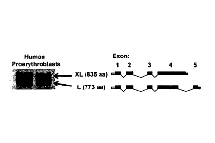
[0023] Figures 1A-1B shows the expression of BCL11 A in human erythroid
progenitors.
[0024] Figure 1A shows the major BCL11A isoforms present in nuclear
extracts of
human erythroid cells.
[0025] Figure 1B compares the expression of BCL11A and fetal hemoglobin in
erythroid
cells at different stages of human ontogeny.
[0026] Figure 2A demonstrates that the common variant rs4671393 is
associated with
BCL11A expression in human lymphoblastoid cell lines from the HapMap European
(CEU) and
African (YRI) populations.
CA 02737180 2011-03-11
WO 2010/030963 PCT/US2009/056770
[0027] Figure 2B are Western blots of lysates of primary human bone marrow
(BM)
erythroblasts, second trimester fetal liver (FL) erythroblasts, first
trimester circulating primitive
erythroblasts, and K562 cells. Primary human stage-matched erythroblasts were
isolated by
sorting for the CD235 and CD7I double-positive population. The XL and L bands
migrate
together here as a result of reduced separation on this blot.
[0028] Figures 3A-3D depict the proteomic affinity screen methodology used
to identify
BCLI I A partner proteins in erythroid cells.
[0029] Figure 3A depicts the scheme used for affinity purification in mouse
erythroleukemia (MEL) cells.
[0030] Figure 3B tabulates the results of the subtractive screen.
[0031] Figure 3C displays the results of the analyses of the Affymetrix
arrays.
[0032] Figure 3D highlights the motif found in BCLI1A and several other
proteins
suggested to mediate interactions with the NuRD repressor complex.
[0033] Figures 4A-4E show confirmations of the BCLI1A interactions with
GATA-I,
FOG-I, and the NuRD complex in erythroid cells.
[0034] Figure 4A shows immunoprecipitation data that confirms the
interactions of
BCL11A with GATA-1, FOG-I, MTA2, and RBBP7 in erythroid (MEL) cells.
[0035] Figure 4B depicts the interactions of BCLI IA with MTA2, GATA-1, and
FOG-1
using gel filtration fractions from erythroid nuclear extracts.
[0036] Figures 4C and 4D show immunoprecipitation data that confirm the
interactions
of BCLI IA with GATA-land FOG-I respectively by exogenous expression in Cos7
cells.
[0037] Figure 4E shows immunoprecipitation data to maps the interaction of
BCLIIA
on the GATA-1 molecule.
[0038] Figures 5A-5E demonstrate that BCLI1A acts as a repressor of the y-
globin gene.
[0039] Figure 5A demonstrates that siRNA-mediated knockdown of BCL11A
results in
elevations of y-globin mRNA levels in human erythroid progenitor cells.
[0040] Figure 5B depicts that global gene expression is not modified
greatly in cells
targeted with BCL11A siRNA.
[0041] Figure 5C shows that lentiviral-mediated shRNA delivery to human
erythroid
progenitors results in a 60%- 97% knockdown.
6
CA 02737180 2011-03-11
WO 2010/030963 PCT/US2009/056770
[0042] Figure 5D depicts that the shRNA targeted cells are morphologically
indistinguishable from control treated cells.
[0043] Figure 5E shows the induction of y-globin mRNA in cells in response
to
knockdown of BCL11A.
[0044] Figure 5F shows the hemolysates prepared from cells on day 12 of
differentiation
show the presence of mature HbF.
[0045] Figure 6A-H show that human y-globin is primarily expressed in
primitive
erythroid cells of 13-locus mice.
[0046] Figure 6A is a representative FACS plot showing FSC (linear scale)
versus SSC
(log scale) for E13.5 embryonic blood. Gating is shown to allow for the
enrichment of primitive
and definitive lineages.
[0047] Figure 6B is a histogram showing the relative expression of murine
ry globin
gene, human embryonic c gene, and human y-globin genes in the primitive
population (P), as
compared with the definitive population (D). Results are shown as mean
standard deviation
(n>3 per group). P=0.98 for a two-sided t-test comparing the relative
enrichment of Ey with y-
globin.
[0048] Figure 6C-H are representatives immunohistochemical staining with an
anti-HbF
antibody from human and murine E13.5 fetal livers. All images are taken with a
60X objective.
[0049] Figure 6C shows human fetal livers contain numerous erythroblasts,
which all
stain positive for y-globin expression.
[0050] Figure 6D and 6E shows that murine fetal liver definitive
erythroblasts do not
show major y-globin staining and only occasional cells with megaloblastic
primitive morphology
show staining (arrows).
[0051] Figure 6E and 6F shows many megaloblastic primitive cells in the
circulation
having highly positive staining (arrowheads in Fig. 6E; arrows in Fig. 6F),
while smaller
definitive erythrocytes are negative (in Fig. 6F as smaller light grey
circles).
[0052] Figure 6G and 6H show staining performed on the single copy YAC
lines A20
and A858 showed similar staining patterns. Positive staining was determined in
comparison with
background staining from transgene negative littermate controls.
[0053] Figure 7A-D show PT-FISH analyses revealing that y -globin
expression parallels
the murine embryonic globins in primitive erythroid cells. Two independent
lines of transgenic
7
CA 02737180 2011-03-11
WO 2010/030963 PCT/US2009/056770
YAC mice, A85 (Fig. 7A and 7C) and A20 (Fig. 7B and 7D) were analysed using
four color
primary transcript RNA fluorescence in situ hybridization (PT-FISH). For the
first set of
experiments, probes were made to target murine a-globin (ma), human 13-g1obin
(hp), and
human y-globin (by). Additionally DAPI was used to identify nuclei of cells.
[0054] Figure 7A and B show the expression of y-globin predominates within
the two
lines in the primitive populations seen circulating in primitive blood cells
(PBC) from embryos
El 1.5 and E13.5. Minor expression is seen in the mature definitive
populations from fetal liver
(FL) at El 3.5. Many of these cells may represent primitive cells found within
the FL
parenchyma.
[0055] Figure 7C and 7D show a parallel expression of my and hy for PBC at
E13.5 and
FL at E13.5, respectively. The graphs depict the percentage of active loci and
are measured for
>100 nuclei per probe set at each time point.
[0056] Figure 8A-8B show that BCL11A expression varies between humans and
mice,
indicating a model for trans-acting variation in 3-globin gene expression.
[0057] Figure 8A shows that in human cells full-length proteins of BCL11A
(XL/L
isoforrns) are reduced within cell populations that express high levels of y-
globin, including
primitive and fetal liver cells.
[0058] Figure 8B is a schematic model summarizes the ontogeny of 13-like
globin gene
regulation in humans, mice, and 13-locus mice. The ontogeny of mammalian
erythropoiesis and
progenitor populations is shown at the top. Progenitor populations, including
primitive erythroid
populations (EryP-CFC), definitive hematopoietic stem cells (HSC), and
definitive erythroid
burst-forming unit cells (BFU-E) are depicted. The aorto gonado-mesonephros
(AGM) and
placenta are sites of definitive hematopoiesis. The patterns of 3-like globin
and BCL11A
expression seen in the two species are shown below.
[0059] Figure 9A-9F shows that BCL11A -/- mice fail to silence expression
of mouse
embryonic f3-like globins and human 3-globin genes.
[0060] Figure 9A shows that he CD71/Ter119 expression pattern for fetal
liver cells
from E14.5 embryos, revealing grossly normal erythropoiesis with these
phenotypic markers.
The mean percentages for the populations in each quadrant are shown in red
(n=6 for fl/+
controls and n=4 for -/- mutants). The P > 0.1 by a two-sided t-test for all
gated populations
analyzed.
8
CA 02737180 2011-03-11
WO 2010/030963 PCT/US2009/056770
[0061] Figure 9B shows that the expression of the embryonic globins as a
percentage of
total mouse 13-like globins for control mice (fl/+), BCLI1A heterozygous (+/-
), and null mice (-
/-) at E14.5 (n=10, 14, 11 respectively).
[0062] Figure 9C shows that the expression of the embryonic globins as a
percentage of
total mouse 13-like globins at E18.5 (n=9, 9, 7 respectively).
[0063] Figure 9D shows the immunohistochemistry was performed on E14.5 FLs
from
BCLI1A fl/+ and -/- animals for the embryonic globin Ey. Representative
sections at 40X
magnification with a 10X objective lens are shown.
[0064] Figure 9E shows similar IHC staining was performed for 13h1 globin.
In both
cases robust expression is seen in the scattered erythroblasts of the FL in -/-
, but not control
mice.
[0065] Figure 9F shows the expression of human 13-globin locus genes for
animals with
the various BCL1 IA genotypes in the presence of the 13-locus YAC transgene
(YAC+) at E14.5
(n=4, 6, 4 for the fl/+, +/-, and -/- animals, respectively) and E18.5 (n=4,
7, 4). All y- and 13-
globin levels for the different genotypes are significantly different (P <
1X10⁻⁵ by a two-sided t-
test). All data are plotted as the mean the standard deviation of the
measurement.
[0066] Figure 10 shows the inability to recapitulate stress responses in
adult P-YAC
mice. Adult 13-locus mice were induced with a variety of-globin stimulating
responses.
[0067] Figure 11 shows that BCL1 IA RNA is expressed in mouse definitive
cells, but
not primitive cells.
[0068] Figure 12 shows that BCL1 IA -/- mice are morphologically normal and
completely lack BCL1 lA protein expression in the fetal liver.
[0069] Figure 12A are examples of control (fl/+) and mutant mice (-/-) from
the same
litter at E18.5. Mice were obtained in expected Mendelian ratios at E18.5 and
the mutants were
morphologically indistinguishable from control littermates.
[0070] Figure 12B are protein expression of BCL1 IA data assessed in E18.5
fetal livers
and showed reduced expression in heterozygous animals, with absent expression
in null animals.
GAPDH was analyzed as a loading control.
[0071] Figure 13 shows that BCL1 IA -/- mice have normal phenotypic
erythropoiesis at
E18.5. Erythroid maturation was assessed using the markers CD7I and Ter-I 19
in the fetal
9
CA 02737180 2011-03-11
WO 2010/030963 PCT/US2009/056770
livers of E18.5 animals (Sankaran, V.G., et al., 2008, Genes Dev 22, 463-475).
The mean values
in each quadrant are shown (n=9 for the controls and 7 for the null animals).
[0072] Figure 14 shows that BCL11A -/- mice have normal erythroid
morphology.
Example cytospin preparations from single cell suspensions of the fetal liver
stained with May-
Griinwald-Giemsa stain are shown from E14.5 and E18.5. All images were viewed
with a 10X
objective and with the lens magnifications shown.
[0073] Figure 15A and B are histological analyses of fetal livers from BCL1
1A -/- mice
revealing normal gross histology and morphological erythropoiesis.
[0074] Figure 15A shows saggital sections are shown at low resolution and
show that
there are no gross histological abnormalities seen in these mice (at 5X
magnification).
[0075] Figure 15B shows histological sections stained with hematoxylin and
eosin
(H&E) are shown at two magnifications (10X objective with a 40X lens) from
E14.5 and E18.5
fetal livers. These sections reveal clusters of erythroblasts within the fetal
liver that appear to be
similar in quantity and morphologically normal.
[0076] Figure 16A and 16B show that BCL1 IA -/- have an upregulation of
embryonic
globins in the fetal liver.
[0077] Figure 16A shows the relative RNA expression of the 13-like globin
genes is
shown for controls (BCL11A fl/+), heterozygous animals (BCL11A -/+), and null
animals
(BCL11A -/-) at E14.5 (n=10, 14, 11 for these groups, respectively).
Additionally the relative
expression of BCL11A RNA is shown. The relative expression is normalized with
respect to
GAPDH (with GAPDH set to a value of 1). All data is shown as the mean the
standard error of
the measurement.
[0078] Figure 16B shows the relative RNA expression (normalized to GAPDH)
of the 13-
like globin genes for controls, heterozygous animals, and null animals at
E18.5 (n=9, 9, 7 for
these groups, respectively). All data is shown as the mean the standard
deviation.
[0079] Figure 17 is the immunohistochemistry of BCL1 IA -/- mice showing an
upregulation of embryonic globins in the fetal liver. Immunohistochemistry was
performed on
E18.5 FLs from BCL11 A fl/+ and -/- animals for the embryonic globinI3h1.
Representative
sections at 40X magnification with a 10X objective lens are shown. Similar IHC
staining was
performed for Ey globin as labeled in the figure.
CA 02737180 2011-03-11
WO 2010/030963 PCT/US2009/056770
[0080] Figure 18A displays the percentages for all the human 13-like globin
genes the
standard deviation at E14.5 in I3-Locus mice crosses with BCL11A mutant mice.
[0081] Figure 18A displays the percentages for all the human 13-like globin
genes the
standard deviation at E18.5, in I3-Locus mice crosses with BCL11A mutant mice.
All y- and 13-
globin levels for the different genotypes are significantly different (P <
1X10⁻⁵ by a two-sided t-
test).
[0082] Figure 19 show that BCLI1A occupies discrete regions in the human f3-
globin
locus in adult erythroid progenitors. The human f3-globin locus is depicted at
the top with
regions showing significant binding shaded in gray in the histogram plot
below. The results are
depicted as the mean with the standard deviation as error bars (n=3 per
group).
DETAILED DESCRIPTION OF THE INVENTION
[0083] The present invention provides for novel methods for the regulation
of fetal
hemoglobin (HbF) synthesis for the treatment of 13- hemoglobinopathies and
screening methods
therein.
[0084] The invention is based upon identification of a novel function for
the BCLI1A
protein, namely that the BCLIIA protein acts as a stage specific regulator of
fetal hemoglobin
expression and that expression of BCL11 A represses y-globin induction.
Accordingly, the
invention provides novel methods for the regulation of y-globin expression in
eythroid cells.
More specifically, these activities can be harnessed in methods for the
treatment ofI3-
hemoglobinopathies by induction of y-globin via inhibition of the BCL11A gene
product.
[0085] Fetal hemoglobin (HbF) is a tetramer of two adult a-globin
polypeptides and two
fetal 13-like y-globin polypeptides. During gestation, the duplicated y-globin
genes constitute the
predominant genes transcribed from the 13-globin locus. Following birth, y-
globin becomes
progressively replaced by adult 13-globin, a process referred to as the "fetal
switch" (3). The
molecular mechanisms underlying this switch have remained largely undefined
and have been a
subject of intense research. The developmental switch from production of
predominantly fetal
hemoglobin or HbF (a2q2) to production of adult hemoglobin or HbA (002) begins
at about 28
to 34 weeks of gestation and continues shortly after birth at which point HbA
becomes
predominant. This switch results primarily from decreased transcription of the
gamma-globin
genes and increased transcription of beta-globin genes. On average, the blood
of a normal adult
contains only about 2% HbF, though residual HbF levels have a variance of over
20 fold in
healthy adults (Atweh, Semin. Hematol. 38(4):367-73 (2001)).
11
CA 02737180 2015-12-11
[0086] Hemoglobinopathies encompass a number of anemias of genetic origin
in which
there is a decreased production and/or increased destruction (hemolysis) of
red blood cells
(RBCs). These disorders also include genetic defects that result in the
production of abnormal
hemoglobins with a concomitant impaired ability to maintain oxygen
concentration. Some such
disorders involve the failure to produce normal f3-globin in sufficient
amounts, while others
involve the failure to produce normal P-globin entirely. These disorders
specifically associated
with the P-globin protein are referred to generally as P-hemoglobinopathies.
For example, p-
thalassemias result from a partial or complete defect in the expression of the
P-globin gene,
leading to deficient or absent HbA. Sickle cell anemia results from a point
mutation in the p-
globin structural gene, leading to the production of an abnormal (sickled)
hemoglobin (HbS).
HbS RBCs are more fragile than normal RBCs and undergo hemolysis more readily,
leading
eventually to anemia (Atweh. Semin. Hematol. 38(4):367-73 (2001)). Moreover,
the presesnce
of a BCL11A genetic variant, HBSIL-MYB variation, ameliorates the clinical
severity in beta-
thalassemia. This variant has been shown to be associated with HbF levels.
Here, it was shown
that there is an odds ratio of 5 for having a less severe form of beta-
thalassemia with the high-
HbF variant.
[0087] Recently, the search for treatment aimed at reduction of globin
chain imbalance
in patients with P-hemoglobinopathies has focused on the pharmacologic
manipulation of fetal
hemoglobin (a2y2; HbF). The important therapeutic potential of such approaches
is suggested
by observations of the mild phenotype of individuals with co-inheritance of
both homozygous p-
thalassemia and hereditary persistence of fetal hemoglobin (HPFH), as well as
by those patients
with homozygous f3 -thalassemia who synthesize no adult hemoglobin, but in
whom a reduced
requirement for transfusions is observed in the presence of increased
concentrations of fetal
hemoglobin. Furthermore, it has been observed that certain populations of
adult patients with P
chain abnormalities have higher than normal levels of fetal hemoglobin (HbF),
and have been
observed to have a milder clinical course of disease than patients with normal
adult levels of
HbF. For example, a group of Saudi Arabian sickle-cell anemia patients who
express 20-30%
HbF have only mild clinical manifestations of the disease (Pembrey, et al.,
Br. J. Haematol. 40:
415-429 (1978)). It is now accepted that 13-hemoglobinopathies, such as sickle
cell anemia and
the p-thalassemias, are ameliorated by increased HbF production. (Reviewed in
Jane and
Cunningham Br. J. Haematol. 102: 415-422 (1998) and Bunn, N. Engl. J. Med.
328: 129-131
(1993)).
12
CA 02737180 2011-03-11
WO 2010/030963 PCT/US2009/056770
[0088] While the molecular mechanisms controlling the in vivo developmental
switch
from y- to 13-globin gene expression are currently unknown, there is
accumulating evidence that
external factors can influence y-globin gene expression. The first group of
compounds
discovered having HbF reactivation activity were cytotoxic drugs. The ability
to cause de novo
synthesis of HbF by pharmacological manipulation was first shown using 5-
azacytidine in
experimental animals (DeSimone, Proc Natl Acad Sci U S A. 79(14):4428-31
(1982)).
Subsequent studies confirmed the ability of 5-azacytidine to increase HbF in
patients with13-
thalassemia and sickle cell disease (Ley, et al., N. Engl. J. Medicine, 307:
1469-1475 (1982),
and Ley, et al., Blood 62: 370-380 (1983)). Additional experiments
demonstrated that baboons
treated with cytotoxic doses of arabinosylcytosine (ara-C) responded with
striking elevations of
F-reticulocytes (Papayannopoulou et al., Science. 224(4649):617-9 (1984)), and
that treatment
with hydroxyurea led to induction of y-globin in monkeys or baboons (Letvin
et. al., N Engl J
Med. 310(14):869-73 (1984)).
[0089] The second group of compounds investigated for the ability to cause
HbF
reactivation activity was short chain fatty acids. The initial observation in
fetal cord blood
progenitor cells led to the discovery that y-aminobutyric acid can act as a
fetal hemoglobin
inducer (Perrine et al., Biochem Biophys Res Commun.148(2):694-700 (1987)).
Subsequent
studies showed that butyrate stimulated globin production in adult baboons
(Constantoulakis et
al., Blood. Dec; 72(6):1961-7 (1988)), and it induced y-globin in erythroid
progenitors in adult
animals or patients with sickle cell anemia (Perrine et al.. Blood. 74(1):454-
9 (1989)).
Derivatives of short chain fatty acids such as phenylbutyrate (Dover et al.,
Br J Haematol.
88(3):555-61 (1994)) and valproic acid (Liakopoulou et al., 1: Blood.
186(8):3227-35 (1995))
also have been shown to induce HbF in vivo. Given the large number of short
chain fatty acid
analogs or derivatives of this family, there are a number of potential
compounds of this family
more potent than butyrate. Phenyl acetic and phenylalkyl acids (Torkel son et
al., Blood Cells
Mol Dis. 22(2):150-8. (1996)), which were discovered during subsequent
studies, were
considered potential HbF inducers as they belonged to this family of
compounds. Presently,
however, the use of butyrate or its analogs in sickle cell anemia and 13-
thalassemia remains
experimental and cannot be recommended for treatment outside of clinical
trials.
[0090] Clinical trials aimed at reactivation of fetal hemoglobin synthesis
in sickle cell
anemia and 13 -thalassemia have included short term and long term
administration of such
compounds as 5-azacytidine, hydroxyurea, recombinant human erythropoietin, and
butyric acid
analogs, as well as combinations of these agents. Following these studies,
hydroxyurea was used
for induction of HbF in humans and later became the first and only drug
approved by the Food
13
CA 02737180 2011-03-11
WO 2010/030963 PCT/US2009/056770
and Drug Administration (FDA) for the treatment of hemoglobinopathies.
However, varying
drawbacks have contraindicated the long term use of such agents or therapies,
including
unwanted side effects and variability in patient responses. For example, while
hydroxyurea
stimulates HbF production and has been shown to clinically reduce sickling
crisis, it is
potentially limited by myelotoxicity and the risk of carcinogenesis. Potential
long term
carcinogenicity would also exist in 5-azacytidine-based therapies.
Erythropoietin-based
therapies have not proved consistent among a range of patient populations. The
short half-lives
of butyric acid in vivo have been viewed as a potential obstacle in adapting
these compounds for
use in therapeutic interventions. Furthermore, very high dosages of butyric
acid are necessary
for inducing y-globin gene expression, requiring catheritization for
continuous infusion of the
compound. Moreover, these high dosages of butyric acid can be associated with
neurotoxicity
and multiorgan damage (Blau, et al., Blood 81: 529-537 (1993)). While even
minimal increases
in HbF levels are helpful in sickle cell disease, 13-thalassemias require a
much higher increase
that is not reliably, or safely, achieved by any of the currently used agents
(Olivieri, Seminars in
Hematology 33: 24-42 (1996)).
[0091] Identifying natural regulators of HbF induction and production could
provide a
means to devise therapeutic interventions that overcome the various drawbacks
of the
compounds described above. Recent genome-wide association studies have yielded
insights into
the genetic basis of numerous complex diseases and traits (McCarthy et al.,
Nat Rev Genet 9,
356 (2008) and Manolio et. al. J Clin Invest 118, 1590 (2008)). However, in
the vast majority of
instances, the functional link between a genetic association and the
underlying pathophysiology
remains to be uncovered. The level of fetal hemoglobin (HbF) is inherited as a
quantitative trait
and clinically important, given its above-mentioned and well-characterized
role in ameliorating
the severity of the principal P-hemoglobinopathies, sickle cell disease and 13-
thalassemia
(Nathan et. al., Nathan and Oski's hematology of infancy and childhood ed.
6th, pp. 2 v. (xiv,
1864, xli p.) 2003)). Two genome-wide association studies have identified
three major loci
containing a set of five common single nucleotide polymorphisms (SNPs) that
account for ¨20%
of the variation in HbF levels (Lettre et al., Proc Natl Acad Sci U S A
(2008); Uda et al., Proc
Natl Acad Sci U S A 105, 1620 (2008); Menzel et al.. Nat Genet 39, 1197
(2007)). Moreover,
several of these variants appear to predict the clinical severity of sickle
cell disease (Lettre et al.,
Proc Natl Acad Sci U S A (2008)) and at least one of these SNPs may also
affect clinical
outcome inI3-thalassemia (Uda et al., Proc Natl Acad Sci U S A 105, 1620
(2008)). The SNP
with the largest effect size, explaining over 10% of the variation in HbF, is
located in the second
intron of a gene on chromosome 2, BCL11A. Whereas BCL11A, a C2H2-type zinc
finger
14
CA 02737180 2011-03-11
WO 2010/030963 PCT/US2009/056770
transcription factor, has been investigated for its role in lymphocyte
development (Liu et al., Nat
Immunol 4, 525 (2003) and Liu et al., Mol Cancer 5, 18 (2006)), its role in
red blood cell
production or globin gene regulation has not been previously assessed.
[0092] At the onset of the recombinant DNA era, studies of globin gene
structure
provided a strong molecular foundation for interrogating the fetal globin
switch. Considerable
effort has focused on delineating the cis-elements within the J3-globin locus
necessary for proper
regulation of the genes within the f3-like globin cluster. These studies
relied on naturally
occurring mutations and deletions that dramatically influence HbF levels in
adults, and have
been complemented by generation of transgenic mice harboring portions of the
cluster (Nathan
et. al., Nathan and Oski's hematology of infancy and childhood ed. 6th, pp. 2
v. (xiv, 1864, xli
p.) 2003) and G. Stamatoyannopoulos, Exp Hematol 33. 259 (2005)). Although the
precise cis-
elements required for globin switching remain ill-defined, findings in
transgenic mice have
strongly indicated that the y-globin genes are autonomously silenced in the
adult stage, a finding
that is most compatible with the absence of fetal-stage specific activators or
the presence of a
stage-specific repressor. The results of recent genetic association studies
provide candidate
genes to interrogate for their involvement in control of the y-globin genes,
such as BCL11A.
[0093] We identified a novel stage-specific repressor of the y-globin
genes, namely
BCL11A, wherein the expression of the BCL11A protein acts as a negative
regulator of
expression from the y-globin genes.
Methods of Increasing Fetal Hemoglobin in a Cell
[0094] The present invention provides improved methods for increasing
fetal
hemoglobin production in a cell, by the administration of compositions
containing inhibitors of
BCL11A. The data demonstrate that inhibition of BCL11A leads to increased
expression from
the'y-globin genes, and agents wherein to achieve this inhibition.
[0095] As disclosed herein, it is an object of the present invention to
provide a method
for increasing fetal hemoglobin levels in a cell.
[0096] Accordingly, one aspect of the invention provides a method for
increasing fetal
hemoglobin levels expressed by a cell, comprising the steps of contacting a
hematopoietic
progenitor cell with an effective amount of a composition comprising an
inhibitor of BCL11A,
whereby fetal hemoglobin expression is increased in the cell, or its progeny,
relative to the cell
prior to such contacting.
CA 02737180 2011-03-11
WO 2010/030963 PCT/US2009/056770
[0097] In connection with contacting a cell with an inhibitor of BCL11A,
"increasing the
fetal hemoglobin levels" in a cell indicates that fetal hemoglobin is at least
5% higher in
populations treated with a BCL11A inhibitor, than in a comparable, control
population, wherein
no BCL11A inhibitor is present. It is preferred that the percentage of fetal
hemoglobin
expression in a BCL11A inhibitor treated population is at least 10% higher, at
least 20% higher,
at least 30% higher, at least 40% higher, at least 50% higher, at least 60%
higher, at least 70%
higher, at least 80% higher, at least 90% higher, at least 1-fold higher, at
least 2-fold higher, at
least 5-fold higher, at least 10 fold higher, at least 100 fold higher, at
least 1000-fold higher, or
more than a control treated population of comparable size and culture
conditions. The term
"control treated population" is used herein to describe a population of cells
that has been treated
with identical media, viral induction, nucleic acid sequences, temperature,
confluency, flask
size, pH, etc., with the exception of the addition of the BCL11A inhibitor.
[0098] An "inhibitor" of BCL11A, as the term is used herein, can function
in a
competitive or non-competitive manner, and can function, in one embodiment, by
interfering
with the expression of the BCL11A protein. Any of a number of different
approaches can be
taken to inhibit BCL11A expression or activity. A BCL11A inhibitor includes
any chemical or
biological entity that, upon treatment of a cell, results in inhibition of the
biological activity
caused by activation of BCL11A in response to cellular signals. BCL11A
inhibitors, include, but
are not limited to, small molecules, antibodies or antigen-binding antibody
fragments,
intrabodies, aptamers, antisense constructs, RNA interference agents, and
ribozymes.
Antibody Inhibitors of BCL114
[0099] Antibodies that specifically bind BCL11A can be used for the
inhibition of the
factor in vivo. Antibodies to BCL11A are commercially available and can be
raised by one of
skill in the art using well known methods. The BCL11A inhibitory activity of a
given antibody,
or, for that matter, any BCL11A inhibitor, can be assessed using methods known
in the art or
described herein ¨ to avoid doubt, an antibody that inhibits BCL11A will cause
an increase in
fetal hemoglobin expression. Antibody inhibitors of BCL11A can include
polyclonal and
monoclonal antibodies and antigen-binding derivatives or fragments thereof.
Well known
antigen binding fragments include, for example, single domain antibodies
(dAbs; which consist
essentially of single VL or VH antibody domains), Fv fragment, including
single chain Fv
fragment (scFv), Fab fragment, and F(ab')2 fragment. Methods for the
construction of such
antibody molecules are well known in the art.
Nucleic Acid Inhibitors of BCL11A Expression
16
CA 02737180 2011-03-11
WO 2010/030963 PCT/US2009/056770
[0100] A powerful approach for inhibiting the expression of selected target
polypeptides
is through the use of RNA interference agents. RNA interference (RNAi) uses
small interfering
RNA (siRNA) duplexes that target the messenger RNA encoding the target
polypeptide for
selective degradation. siRNA-dependent post-transcriptional silencing of gene
expression
involves cleaving the target messenger RNA molecule at a site guided by the
siRNA. "RNA
interference (RNAi) 'is an evolutionally conserved process whereby the
expression or
introduction of RNA of a sequence that is identical or highly similar to a
target gene results in
the sequence specific degradation or specific post-transcriptional gene
silencing (PTGS) of
messenger RNA (mRNA) transcribed from that targeted gene (see Coburn, G. and
Cullen. B.
(2002) J. of Virology 76(18):9225), thereby inhibiting expression of the
target gene. In one
embodiment, the RNA is double stranded RNA (dsRNA). This process has been
described in
plants, invertebrates, and mammalian cells. In nature, RNAi is initiated by
the dsRNA-specific
endonuclease Dicer, which promotes processive cleavage of long dsRNA into
double-stranded
fragments termed siRNAs. siRNAs are incorporated into a protein complex
(termed "RNA
induced silencing complex," or "RISC") that recognizes and cleaves target
mRNAs. RNAi can
also be initiated by introducing nucleic acid molecules, e.g., synthetic
siRNAs or RNA
interfering agents, to inhibit or silence the expression of target genes. As
used herein, "inhibition
of target gene expression" includes any decrease in expression or protein
activity or level of the
target gene or protein encoded by the target gene as compared to a situation
wherein no RNA
interference has been induced. The decrease will be of at least 10%, 20%, 30%,
40%, 50%, 60%,
70%, 80%, 90%, 95% or 99% or more as compared to the expression of a target
gene or the
activity or level of the protein encoded by a target gene which has not been
targeted by an RNA
interfering agent.
[01101] The terms "RNA interference agent" and "RNA interference" as they
are used
herein are intended to encompass those forms of gene silencing mediated by
double-stranded
RNA, regardless of whether the RNA interfering agent comprises an siRNA,
miRNA, shRNA or
other double-stranded RNA molecule. "Short interfering RNA" (siRNA), also
referred to herein
as "small interfering RNA" is defined as an RNA agent which functions to
inhibit expression of
a target gene, e.g.. by RNAi. An siRNA may be chemically synthesized, may be
produced by in
vitro transcription, or may be produced within a host cell. In one embodiment,
siRNA is a
double stranded RNA (dsRNA) molecule of about 15 to about 40 nucleotides in
length,
preferably about 15 to about 28 nucleotides, more preferably about 19 to about
25 nucleotides in
length, and more preferably about 19, 20, 21, 22, or 23 nucleotides in length,
and may contain a
3' and/or 5' overhang on each strand having a length of about 0, 1, 2, 3, 4,
or 5 nucleotides. The
17
length of the overhang is independent between the two strands, i.e., the
length of the overhang
on one strand is not dependent on the length of the overhang on the second
strand. Preferably the
siRNA is capable of promoting RNA interference through degradation or specific
post-
transcriptional gene silencing (PTGS) of the target messenger RNA (mRNA).
[0102] siRNAs also include small hairpin (also called stern loop) RNAs
(shRNAs). In
one embodiment, these shRNAs are composed of a short ( e.g., about 19 to about
25 nucleotide)
antisense strand, followed by a nucleotide loop of about 5 to about 9
nucleotides, and the
analogous sense strand. Alternatively, the sense strand may precede the
nucleotide loop structure
and the anti sense strand may follow. These shRNAs may be contained in
plasmids, retroviruses,
and lentiviruses and expressed from, for example, the pol III U6 promoter, or
another promoter (
see, e.g., Stewart, et al. (2003) RNA Apr;9(4):493- 501).
The target gene or sequence of the RNA interfering agent may be a cellular
gene or
genomic sequence, e.g. the BCL11A sequence. An siRNA may be substantially
homologous to
the target gene or genomic sequence, or a fragment thereof. As used in this
context, the term
"homologous" is defined as being substantially identical, sufficiently
complementary, or similar
to the target mRNA, or a fragment thereof, to effect RNA interference of the
target. In addition
to native RNA molecules, RNA suitable for inhibiting or interfering with the
expression of a
target sequence include RNA derivatives and analogs. Preferably, the siRNA is
identical to its
target. The siRNA preferably targets only one sequence. Each of the RNA
interfering agents,
such as siRNAs, can be screened for potential off-target effects by, for
example, expression
profiling. Such methods are known to one skilled in the art and are described,
for example, in
Jackson et al. Nature Biotechnology 6:635-637, 2003. In addition to expression
profiling, one
may also screen the potential target sequences for similar sequences in the
sequence databases to
identify potential sequences which may have off-target effects. For example,
according to
Jackson et al. (Id.), 15, or perhaps as few as 11 contiguous nucleotides, of
sequence identity are
sufficient to direct silencing of non-targeted transcripts. Therefore, one may
initially screen the
proposed siRNAs to avoid potential off-target silencing using the sequence
identity analysis by
any known sequence comparison methods, such as BLAST. siRNA sequences are
chosen to
maximize the uptake of the antisense (guide) strand of the siRNA into RISC and
thereby
maximize the ability of RISC to target human GGT mRNA for degradation. This
can be
accomplished by scanning for sequences that have the lowest free energy of
binding at the 5'-
terminus of the antisense strand. The lower free energy leads to an
enhancement of the
unwinding of the 5'- end of the antisense strand of the siRNA duplex, thereby
ensuring that the
antisense strand will be taken up by RISC and direct the sequence-specific
cleavage of the
18
CA 2737180 2018-11-13
CA 02737180 2011-03-11
WO 2010/030963 PCT/US2009/056770
human BCL11A mRNA. siRNA molecules need not be limited to those molecules
containing
only RNA, but, for example, further encompasses chemically modified
nucleotides and non-
nucleotides, and also include molecules wherein a ribose sugar molecule is
substituted for
another sugar molecule or a molecule which performs a similar function.
Moreover, a non-
natural linkage between nucleotide residues can be used, such as a
phosphorothioate linkage.
The RNA strand can be derivatized with a reactive functional group of a
reporter group, such as
a fluorophore. Particularly useful derivatives are modified at a terminus or
termini of an RNA
strand, typically the 3' terminus of the sense strand. For example, the 2'-
hydroxyl at the 3'
terminus can be readily and selectively derivatizes with a variety of groups.
Other useful RNA
derivatives incorporate nucleotides having modified carbohydrate moieties,
such as 2'0-
alkylated residues or 2'-0-methyl ribosyl derivatives and 2'-0-fluoro ribosyl
derivatives. The
RNA bases may also be modified. Any modified base useful for inhibiting or
interfering with
the expression of a target sequence may be used. For example, halogenated
bases, such as 5-
bromouracil and 5-iodouracil can be incorporated. The bases may also be
alkylated, for example,
7-methylguanosine can be incorporated in place of a guanosine residue. Non-
natural bases that
yield successful inhibition can also be incorporated. The most preferred siRNA
modifications
include 2'-deoxy-2'-fluorouridine or locked nucleic acid (LAN) nucleotides and
RNA duplexes
containing either phosphodiester or varying numbers of phosphorothioate
linkages. Such
modifications are known to one skilled in the art and are described, for
example, in Braasch et
al., Biochemistry, 42: 7967-7975, 2003. Most of the useful modifications to
the siRNA
molecules can be introduced using chemistries established for antisense
oligonucleotide
technology. Preferably, the modifications involve minimal 2'-0-methyl
modification, preferably
excluding such modification. Modifications also preferably exclude
modifications of the free 5'-
hydroxyl groups of the siRNA. The Examples herein provide specific examples of
RNA
interfering agents, such as shRNA molecules that effectively target BCL11A
mRNA.
[0103] In a preferred embodiment, the RNA interference agent is delivered
or
administered in a pharmaceutically acceptable carrier. Additional carrier
agents, such as
liposomes, can be added to the pharmaceutically acceptable carrier. In another
embodiment, the
RNA interference agent is delivered by a vector encoding small hairpin RNA
(shRNA) in a
pharmaceutically acceptable carrier to the cells in an organ of an individual.
The shRNA is
converted by the cells after transcription into siRNA capable of targeting,
for example,
BCL11 A.
[0104] In one embodiment, the vector is a regulatable vector, such as
tetracycline
inducible vector. Methods described, for example. in Wang et al. Proc. Natl.
Acad. Sci. 100:
19
CA 02737180 2011-03-11
WO 2010/030963 PCT/US2009/056770
5103-5106, using pTet-On vectors (BD Biosciences Clontech, Palo Alto, CA) can
be used. In
one embodiment, the RNA interference agents used in the methods described
herein are taken up
actively by cells in vivo following intravenous injection, e.g., hydrodynamic
injection, without
the use of a vector, illustrating efficient in vivo delivery of the RNA
interfering agents. One
method to deliver the siRNAs is catheterization of the blood supply vessel of
the target organ.
Other strategies for delivery of the RNA interference agents, e.g., the siRNAs
or shRNAs used
in the methods of the invention, may also be employed, such as, for example,
delivery by a
vector, e.g., a plasmid or viral vector, e.g., a lentiviral vector. Such
vectors can be used as
described, for example, in Xiao-Feng Qin et al. Proc. Natl. Acad. Sci. U.S.A.,
100: 183-188.
Other delivery methods include delivery of the RNA interfering agents, e.g.,
the siRNAs or
shRNAs of the invention, using a basic peptide by conjugating or mixing the
RNA interfering
agent with a basic peptide, e.g., a fragment of a TAT peptide, mixing with
cationic lipids or
formulating into particles. The RNA interference agents, e.g., the siRNAs
targeting BCL11A
mRNA, may be delivered singly, or in combination with other RNA interference
agents, e.g.,
siRNAs, such as, for example siRNAs directed to other cellular genes. BCL11A
siRNAs may
also be administered in combination with other pharmaceutical agents which are
used to treat or
prevent diseases or disorders associated with oxidative stress, especially
respiratory diseases,
and more especially asthma. Synthetic siRNA molecules, including shRNA
molecules, can be
obtained using a number of techniques known to those of skill in the art. For
example, the
siRNA molecule can be chemically synthesized or recombinantly produced using
methods
known in the art, such as using appropriately protected ribonucleoside
phosphoramidites and a
conventional DNA/RNA synthesizer (see, e.g., Elbashir, S.M. et al. (2001)
Nature 411:494-498;
Elbashir, S.M., W. Lendeckel and T. Tuschl (2001) Genes & Development 15:188-
200;
Harborth, J. et al . (2001) J. Cell Science 114:4557-4565; Masters, J.R. et
al. (2001) Proc. Natl.
Acad. Sci., USA 98:8012-8017; and Tuschl. T. et al. (1999) Genes & Development
13:3191-
3197). Alternatively, several commercial RNA synthesis suppliers are available
including, but
not limited to, Proligo (Hamburg, Germany), Dharmacon Research (Lafayette, CO,
USA),
Pierce Chemical (part of Perbio Science , Rockford, IL, USA), Glen Research
(Sterling, VA,
USA), ChemGenes (Ashland, MA, USA), and Cruachem (Glasgow, UK). As such, siRNA
molecules are not overly difficult to synthesize and are readily provided in a
quality suitable for
RNAi. In addition, dsRNAs can be expressed as stem loop structures encoded by
plasmid
vectors, retroviruses and lentiviruses (Paddison, P.J. et al. (2002) Genes
Dev. 16:948-958;
McManus, M.T. et al. (2002) RNA 8:842-850; Paul, C.P. et al. (2002) Nat.
Biotechnol. 20:505-
508; Miyagishi, M. et al. (2002) Nat. Biotechnol. 20:497-500; Sui, G. et al.
(2002) Proc. Natl.
CA 02737180 2011-03-11
WO 2010/030963 PCT/US2009/056770
Acad. Sci., USA 99:5515-5520; Brummelkamp, T. et al. (2002) Cancer Cell 2:243;
Lee, N.S., et
al. (2002) Nat. Biotechnol. 20:500-505; Yu, J.Y., et al. (2002) Proc. Natl.
Acad. Sci., USA
99:6047-6052; Zeng, Y., et al. (2002) Mol. Cell 9:1327-1333; Rubinson, D.A.,
et al. (2003) Nat.
Genet. 33:401-406; Stewart, S.A., et al. (2003) RNA 9:493-501). These vectors
generally have a
pollll promoter upstream of the dsRNA and can express sense and antisense RNA
strands
separately and/or as a hairpin structures. Within cells, Dicer processes the
short hairpin RNA
(shRNA) into effective siRNA. The targeted region of the siRNA molecule of the
present
invention can be selected from a given target gene sequence, e.g. .. a BCL11A
coding sequence,
beginning from about 25 to 50 nucleotides, from about 50 to 75 nucleotides, or
from about 75 to
100 nucleotides downstream of the start codon. Nucleotide sequences may
contain 5' or 3'
UTRs and regions nearby the start codon. One method of designing a siRNA
molecule of the
present invention involves identifying the 23 nucleotide sequence motif
AA(N19)TT (SEQ. ID.
NO. 21) (where N can be any nucleotide) and selecting hits with at least 25%,
30%, 35%, 40%,
45%, 50%, 55%, 60%, 65%, 70% or 75% G/C content. The "TT" portion of the
sequence is
optional. Alternatively, if no such sequence is found, the search may be
extended using the motif
NA(N21), where N can be any nucleotide. In this situation, the 3' end of the
sense siRNA may
be converted to TT to allow for the generation of a symmetric duplex with
respect to the
sequence composition of the sense and antisense 3' overhangs. The antisense
siRNA molecule
may then be synthesized as the complement to nucleotide positions 1 to 21 of
the 23 nucleotide
sequence motif. The use of symmetric 3' TT overhangs may be advantageous to
ensure that the
small interfering ribonucleoprotein particles (siRNPs) are formed with
approximately equal
ratios of sense and antisense target RNA-cleaving siRNPs (Elbashir et al.,
(2001) supra and
Elbashir et al., 2001 supra). Analysis of sequence databases, including but
not limited to the
NCBI, BLAST, Derwent and GenSeq as well as commercially available
oligosynthesis
companies such as OLIGOENGINE , may also be used to select siRNA sequences
against EST
libraries to ensure that only one gene is targeted.
Delivery of RNA Interfering Agents
[0105] Methods of delivering RNA interference agents, e.g., an siRNA, or
vectors
containing an RNA interference agent, to the target cells, e.g., lymphocytes
or other desired
target cells, for uptake include injection of a composition containing the RNA
interference
agent, e.g., an siRNA, or directly contacting the cell, e.g., a lymphocyte,
with a composition
comprising an RNA interference agent, e.g., an siRNA. In another embodiment,
RNA
interference agent, e.g., an siRNA may be injected directly into any blood
vessel, such as vein,
artery, venule or arteriole, via, e.g., hydrodynamic injection or
catheterization. Administration
21
CA 02737180 2011-03-11
WO 2010/030963 PCT/US2009/056770
may be by a single injection or by two or more injections. The RNA
interference agent is
delivered in a pharmaceutically acceptable carrier. One or more RNA
interference agent may be
used simultaneously. In one preferred embodiment, only one siRNA that targets
human
BCL11A is used. In one embodiment, specific cells are targeted with RNA
interference, limiting
potential side effects of RNA interference caused by non-specific targeting of
RNA interference.
The method can use, for example, a complex or a fusion molecule comprising a
cell targeting
moiety and an RNA interference binding moiety that is used to deliver RNA
interference
effectively into cells. For example, an antibody-protamine fusion protein when
mixed with
siRNA, binds siRNA and selectively delivers the siRNA into cells expressing an
antigen
recognized by the antibody, resulting in silencing of gene expression only in
those cells that
express the antigen. The siRNA or RNA interference-inducing molecule binding
moiety is a
protein or a nucleic acid binding domain or fragment of a protein, and the
binding moiety is
fused to a portion of the targeting moiety. The location of the targeting
moiety can be either in
the carboxyl-terminal or amino-terminal end of the construct or in the middle
of the fusion
protein. A viral-mediated delivery mechanism can also be employed to deliver
siRNAs to cells
in vitro and in vivo as described in Xia, H. et al. (2002) Nat Biotechnol
20(10):1006). Plasmid-
or viral-mediated delivery mechanisms of shRNA may also be employed to deliver
shRNAs to
cells in vitro and in vivo as described in Rubinson, D.A., et al. ((2003) Nat.
Genet. 33:401-406)
and Stewart, S.A., et al. ((2003) RNA 9:493-501). The RNA interference agents,
e.g., the
siRNAs or shRNAs, can be introduced along with components that perform one or
more of the
following activities: enhance uptake of the RNA interfering agents, e.g.,
siRNA, by the cell, e.g.,
lymphocytes or other cells, inhibit annealing of single strands, stabilize
single strands, or
otherwise facilitate delivery to the target cell and increase inhibition of
the target gene, e.g.,
BCL11A. The dose of the particular RNA interfering agent will be in an amount
necessary to
effect RNA interference, e.g., post translational gene silencing (PTGS), of
the particular target
gene, thereby leading to inhibition of target gene expression or inhibition of
activity or level of
the protein encoded by the target gene.
[0106] In one embodiment, the hematopoietic progenitor cell is contacted ex
vivo or in
vitro. In a specific embodiment, the cell being contacted is a cell of the
erythroid lineage. In one
embodiment, the composition inhibits BCL11A expression.
[0107] "Hematopoietic progenitor cell" as the term is used herein, refers
to cells of a
stem cell lineage that give rise to all the blood cell types including the
myeloid (monocytes and
macrophages, neutrophils, basophils, eosinophils, erythrocytes,
megakaryocytes/platelets,
dendritic cells), and the lymphoid lineages (T-cells, B-cells, NK-cells). A
"cell of the erythroid
22
CA 02737180 2011-03-11
WO 2010/030963 PCT/US2009/056770
lineage" indicates that the cell being contacted is a cell that undergoes
erythropoeisis such that
upon final differentiation it forms an erythrocyte or red blood cell (RBC).
Such cells belong to
one of three lineages, erythroid, lymphoid, and myeloid, originating from bone
marrow
haematopoietic progenitor cells. Upon exposure to specific growth factors and
other components
of the haematopoietic microenvironment, haematopoietic progenitor cells can
mature through a
series of intermediate differentiation cellular types, all intermediates of
the erythroid lineage,
into RBCs. Thus, cells of the "erythroid lineage", as the term is used herein,
comprise
hematopoietic progenitor cells, rubriblasts, prorubricytes, erythroblasts,
metarubricytes,
reticulocytes, and erythrocytes.
[0108] In some embodiment, the haematopoietic progenitor cell has at least
one of the
cell surface marker characteristic of haematopoietic progenitor cells: CD34+,
CD59+,
Thy1/CD90+,CD3810/-, and C-kit/CD117+. Preferably, the haematopoietic
progenitor cells have
several of these marker.
[0109] In some embodiment, the haematopoietic progenitor cells of the
erythroid lineage
have the cell surface marker characteristic of the erythroid lineage: CD71 and
Ten 19.
[0110] Stem cells, such as hematopoietic progenitor cells, are capable of
proliferation
and giving rise to more progenitor cells having the ability to generate a
large number of mother
cells that can in turn give rise to differentiated, or differentiable daughter
cells. The daughter
cells themselves can be induced to proliferate and produce progeny that
subsequently
differentiate into one or more mature cell types, while also retaining one or
more cells with
parental developmental potential. The term "stem cell" refers then, to a cell
with the capacity or
potential, under particular circumstances, to differentiate to a more
specialized or differentiated
phenotype, and which retains the capacity, under certain circumstances, to
proliferate without
substantially differentiating. In one embodiment, the term progenitor or stem
cell refers to a
generalized mother cell whose descendants (progeny) specialize, often in
different directions, by
differentiation, e.g., by acquiring completely individual characters, as
occurs in progressive
diversification of embryonic cells and tissues. Cellular differentiation is a
complex process
typically occurring through many cell divisions. A differentiated cell may
derive from a
multipotent cell which itself is derived from a multipotent cell, and so on.
While each of these
multipotent cells may be considered stem cells, the range of cell types each
can give rise to may
vary considerably. Some differentiated cells also have the capacity to give
rise to cells of greater
developmental potential. Such capacity may be natural or may be induced
artificially upon
treatment with various factors. In many biological instances, stem cells are
also "multipotent"
23
CA 02737180 2011-03-11
WO 2010/030963 PCT/US2009/056770
because they can produce progeny of more than one distinct cell type, but this
is not required for
"stem-ness." Self-renewal is the other classical part of the stem cell
definition, and it is essential
as used in this document. In theory, self-renewal can occur by either of two
major mechanisms.
Stem cells may divide asymmetrically, with one daughter retaining the stem
state and the other
daughter expressing some distinct other specific function and phenotype.
Alternatively, some of
the stem cells in a population can divide symmetrically into two stems, thus
maintaining some
stem cells in the population as a whole, while other cells in the population
give rise to
differentiated progeny only. Generally, "progenitor cells" have a cellular
phenotype that is more
primitive (i.e., is at an earlier step along a developmental pathway or
progression than is a fully
differentiated cell). Often, progenitor cells also have significant or very
high proliferative
potential. Progenitor cells can give rise to multiple distinct differentiated
cell types or to a single
differentiated cell type, depending on the developmental pathway and on the
environment in
which the cells develop and differentiate.
[0111] In the context of cell ontogeny, the adjective "differentiated". or
"differentiating"
is a relative term. A "differentiated cell" is a cell that has progressed
further down the
developmental pathway than the cell it is being compared with. Thus, stem
cells can differentiate
to lineage-restricted precursor cells (such as a hematopoietic progenitor
cell), which in turn can
differentiate into other types of precursor cells further down the pathway
(such as an erthyrocyte
precursor), and then to an end-stage differentiated cell, such as an
erthyrocyte, which plays a
characteristic role in a certain tissue type, and may or may not retain the
capacity to proliferate
further.
[0112] In one embodiment, the inhibitor of BCL11A expression is selected
from a small
molecule and a nucleic acid. Alternatively and preferably, the inhibitor of
BCL11A expression is
a BCL11A specific RNA interference agent, or a vector encoding said BCL11A
specific RNA
interference agent. In one specific embodiment, the RNA interference agent
comprises one or
more of the nucleotide sequences of SEQ ID NO:1-6.
[0113] As used herein, the term "small molecule" refers to a chemical agent
including,
but not limited to, peptides, peptidomimetics, amino acids, amino acid
analogs, polynucleotides,
polynucleotide analogs, aptamers, nucleotides, nucleotide analogs, organic or
inorganic
compounds (i.e., including heteroorganic and organometallic compounds) having
a molecular
weight less than about 10,000 grams per mole, organic or inorganic compounds
having a
molecular weight less than about 5,000 grams per mole, organic or inorganic
compounds having
a molecular weight less than about 1,000 grams per mole, organic or inorganic
compounds
24
CA 02737180 2011-03-11
WO 2010/030963 PCT/US2009/056770
having a molecular weight less than about 500 grams per mole, and salts,
esters, and other
pharmaceutically acceptable forms of such compounds.
[0114] A "nucleic acid", as described herein, can be RNA or DNA, and can
be single or
double stranded, and can be selected, for example, from a group including:
nucleic acid
encoding a protein of interest, oligonucleotides, nucleic acid analogues, for
example peptide-
nucleic acid (PNA), pseudo-complementary PNA (pc-PNA), locked nucleic acid
(LNA) etc.
Such nucleic acid sequences include, for example, but are not limited to,
nucleic acid sequence
encoding proteins, for example that act as transcriptional repressors,
antisense molecules,
ribozymes, small inhibitory nucleic acid sequences, for example but are not
limited to RNAi,
shRNAi, siRNA, micro RNAi (mRNAi), antisense oligonucleotides etc.
[0115] As disclosed herein, it is an object of the present invention to
provide a method
for increasing fetal hemoglobin levels in a mammal.
[0116] Accordingly, one aspect of the present invention provides a method
for
increasing fetal hemoglobin levels in a mammal in need thereof, the method
comprising the step
of contacting a hematopoietic progenitor cell in the mammal with an effective
amount of a
composition comprising an inhibitor of BCL11A, whereby fetal hemoglobin
expression is
increased, relative to expression prior to such contacting.
[0117] In connection with contacting a cell in a mammal with an inhibitor
of BCL11A,
"increasing fetal hemoglobin levels in a mammal" indicates that fetal
hemoglobin in the
mammal is at least 5% higher in populations treated with a BCL11A inhibitor,
than a
comparable, control population, wherein no BCL11A inhibitor is present. It is
preferred that the
fetal hemoglobin expression in a BCL11A inhibitor treated mammal is at least
10% higher, at
least 20% higher, at least 30% higher, at least 40% higher, at least 50%
higher, at least 60%
higher, at least 70% higher, at least 80% higher, at least 90% higher, at
least 1-fold higher, at
least 2-fold higher, at least 5-fold higher, at least 10 fold higher, at least
100 fold higher, at least
1000-fold higher, or more than a comparable control treated mammal. The term
"comparable
control treated mammal" is used herein to describe a mammal that has been
treated identically,
with the exception of the addition of the BCL11A inhibitor.
[0118] The term "mammal" is intended to encompass a singular "mammal" and
plural
"mammals," and includes, but is not limited to humans; primates such as apes,
monkeys,
orangutans, and chimpanzees; canids such as dogs and wolves; felids such as
cats, lions, and
tigers; equids such as horses, donkeys, and zebras; food animals such as cows,
pigs, and sheep;
CA 02737180 2011-03-11
WO 2010/030963 PCT/US2009/056770
ungulates such as deer and giraffes; rodents such as mice, rats, hamsters and
guinea pigs; and
bears. In some preferred embodiments, a mammal is a human.
[0119] Accordingly, in one embodiment, the mammal has been diagnosed with
a
hemoglobinopathy. In a further embodiment, the hemoglobinopathy is a P-
hemoglobinopathy.
In one preferred embodiment, the hemoglobinopathy is a sickle cell disease. As
used herein,
"sickle cell disease" can be sickle cell anemia, sickle-hemoglobin C disease
(HbSC), sickle beta-
plus-thalassaemia (HbS/I3+), or sickle beta-zero-thalassaemia (HbS/I30). In
another preferred
embodiment, the hemoglobinopathy is a 13-thalassemia.
[0120] As used herein, the term "hemoglobinopathy" means any defect in the
structure
or function of any hemoglobin of an individual, and includes defects in the
primary, secondary,
tertiary or quaternary structure of hemoglobin caused by any mutation, such as
deletion
mutations or substitution mutations in the coding regions of the 13-globin
gene, or mutations in,
or deletions of, the promoters or enhancers of such genes that cause a
reduction in the amount of
hemoglobin produced as compared to a normal or standard condition. The term
further includes
any decrease in the amount or effectiveness of hemoglobin, whether normal or
abnormal, caused
by external factors such as disease, chemotherapy, toxins, poisons, or the
like.
[0121] The term "effective amount", as used herein, refers to the amount
that is safe and
sufficient to treat, lesson the likelihood of, or delay the development of a
hemoglobinopathy.
The amount can thus cure or result in amelioration of the symptoms of the
hemoglobinopathy,
slow the course of hemoglobinopathy disease progression, slow or inhibit a
symptom of a
hemoglobinopathy, slow or inhibit the establishment of secondary symptoms of a
hemoglobinopathy or inhibit the development of a secondary symptom of a
hemoglobinopathy.
The effective amount for the treatment of the hemoglobinopathy depends on the
type of
hemoglobinopathy to be treated, the severity of the symptoms, the subject
being treated, the age
and general condition of the subject, the mode of administration and so forth.
Thus, it is not
possible or prudent to specify an exact "effective amount". However, for any
given case, an
appropriate -effective amount" can be determined by one of ordinary skill in
the art using only
routine experimentation.
[0122] The treatment according to the present invention ameliorates one or
more
symptoms associated with the disorder by increasing the amount of fetal
hemoglobin in the
individual. Symptoms typically associated with a hemoglobinopathy, include for
example,
anemia, tissue hypoxia, organ dysfunction, abnormal hematocrit values,
ineffective
erythropoiesis, abnormal reticulocyte (erythrocyte) count, abnormal iron load,
the presence of
26
CA 02737180 2011-03-11
WO 2010/030963 PCT/US2009/056770
ring sideroblasts, splenomegaly, hepatomegaly, impaired peripheral blood flow,
dyspnea,
increased hemolysis, jaundice, anemic pain crises, acute chest syndrome,
splenic sequestration,
priapism, stroke, hand-foot syndrome, and pain such as angina pectoris.
[0123] In one embodiment, the hematopoietic progenitor cell is contacted ex
vivo or in
vitro, and the cell or its progeny is administered to said mammal. In a
further embodiment, the
hematopoietic progenitor cell is a cell of the erythroid lineage.
[0124] In one embodiment, the hematopoietic progenitor cell is contacted
with a
composition comprising of an inhibitor of BCL11A and a pharmaceutically
acceptable carrier or
diluent. In one embodiment, said composition is administered by injection,
infusion, instillation,
or ingestion.
[0125] As used herein, the term "pharmaceutically acceptable", and
grammatical
variations thereof, as they refer to compositions, carriers, diluents and
reagents, are used
interchangeably and represent that the materials are capable of administration
to or upon a
mammal without the production of undesirable physiological effects such as
nausea, dizziness,
gastric upset and the like. Each carrier must also be "acceptable" in the
sense of being
compatible with the other ingredients of the formulation. A pharmaceutically
acceptable carrier
will not promote the raising of an immune response to an agent with which it
is admixed, unless
so desired. The preparation of a pharmacological composition that contains
active ingredients
dissolved or dispersed therein is well understood in the art and need not be
limited based on
formulation. The pharmaceutical formulation contains a compound of the
invention in
combination with one or more pharmaceutically acceptable ingredients. The
carrier can be in the
form of a solid, semi-solid or liquid diluent, cream or a capsule. Typically
such compositions are
prepared as injectable either as liquid solutions or suspensions, however,
solid forms suitable for
solution, or suspensions, in liquid prior to use can also be prepared. The
preparation can also be
emulsified or presented as a liposome composition. The active ingredient can
be mixed with
excipients which are pharmaceutically acceptable and compatible with the
active ingredient and
in amounts suitable for use in the therapeutic methods described herein.
Suitable excipients are,
for example, water, saline, dextrose, glycerol, ethanol or the like and
combinations thereof. In
addition, if desired, the composition can contain minor amounts of auxiliary
substances such as
wetting or emulsifying agents, pH buffering agents and the like which enhance
the effectiveness
of the active ingredient. The therapeutic composition of the present invention
can include
pharmaceutically acceptable salts of the components therein. Pharmaceutically
acceptable salts
include the acid addition salts (formed with the free amino groups of the
polypeptide) that are
27
CA 02737180 2011-03-11
WO 2010/030963 PCT/US2009/056770
formed with inorganic acids such as, for example, hydrochloric or phosphoric
acids, or such
organic acids as acetic, tartaric, mandelic and the like. Salts formed with
the free carboxyl
groups can also be derived from inorganic bases such as, for example, sodium,
potassium,
ammonium, calcium or ferric hydroxides, and such organic bases as
isopropylamine,
trimethylamine, 2-ethylamino ethanol, histidine, procaine and the like.
Physiologically tolerable
carriers are well known in the art. Exemplary liquid carriers are sterile
aqueous solutions that
contain no materials in addition to the active ingredients and water, or
contain a buffer such as
sodium phosphate at physiological pH value, physiological saline or both, such
as phosphate-
buffered saline. Still further, aqueous carriers can contain more than one
buffer salt, as well as
salts such as sodium and potassium chlorides, dextrose, polyethylene glycol
and other solutes.
Liquid compositions can also contain liquid phases in addition to and to the
exclusion of water.
Exemplary of such additional liquid phases are glycerin, vegetable oils such
as cottonseed oil,
and water-oil emulsions. The amount of an active agent used in the invention
that will be
effective in the treatment of a particular disorder or condition will depend
on the nature of the
disorder or condition, and can be determined by standard clinical techniques.
The phrase
"pharmaceutically acceptable carrier or diluent" means a pharmaceutically
acceptable material,
composition or vehicle, such as a liquid or solid filler, diluent, excipient,
solvent or
encapsulating material, involved in carrying or transporting the subject
agents from one organ,
or portion of the body, to another organ, or portion of the body.
[0126] As used herein, "administered" refers to the placement of an
inhibitor of
BCL1-1 A into a subject by a method or route which results in at least partial
localization of the
inhibitor at a desired site. An agent which inhibits BCH 1 A can be
administered by any
appropriate route which results in effective treatment in the subject, i.e.
administration results in
delivery to a desired location in the subject where at least a portion of the
composition delivered,
i.e. at least one agent which inhibits BCL1-1 A, is active in the desired site
for a period of time.
The period of time the inhibitor is active depends on the half life in viva
after administration to a
subject, and can be as short as a few hours, e. g. twenty-four hours, to a few
days, to as long as
several years. Modes of administration include injection, infusion,
instillation, or ingestion.
"Injection" includes, without limitation, intravenous, intramuscular,
intraarteri al, intrathecal,
intraventricular, intracapsular, intraorbital, intracardiac, intradermal,
intraperitoneal,
transtracheal, subcutaneous, subcuticular, intraarticular, sub capsular,
subarachnoid, intraspinal,
intracerebro spinal, and intrastemal injection and infusion.
[0127] In one embodiment, the hematopoietic progenitor cell from a mammal
needing
treatment is contacted with a composition that inhibits BCL11A expression.
28
CA 02737180 2011-03-11
WO 2010/030963 PCT/US2009/056770
[0128] By "inhibits BCL11A expression" is meant that the amount of
expression of
BCL11A is at least 5% lower in populations treated with a BCL11A inhibitor,
than a
comparable, control population, wherein no BCL11A inhibitor is present. It is
preferred that the
percentage of BCL11A expression in a BCL11A inhibitor treated population is at
least 10%
lower, at least 20% lower, at least 30% lower, at least 40% lower, at least
50% lower, at least
60% lower, at least 70% lower, at least 80% lower, at least 90% lower, at
least 1-fold lower, at
least 2-fold lower, at least 5-fold lower, at least 10 fold lower, at least
100 fold lower, at least
1000-fold lower, or more than a comparable control treated population in which
no BCL11A
inhibitor is added.
[0129] In one embodiment, the inhibitor of BCL11A expression is selected
from a small
molecule and a nucleic acid. In a preferred embodiment, the nucleic acid is a
BCL11 A specific
RNA interference agent or a vector encoding said RNA interference agent, or an
aptamer that
binds BCL11A. In a preferred embodiment, said RNA interference agent comprises
one or more
of the nucleotide sequences of SEQ ID NO:1 -6.
[0130] In one embodiment, the hematopoietic progenitor cell from a mammal
needing
treatment is contacted with a composition that inhibits BCL11A activity.
[0131] By "inhibits BCL11A activity" is meant that the amount of functional
activity of
BCL11A is at least 5% lower in populations treated with a BCL11A inhibitor,
than a
comparable, control population, wherein no BCL11A inhibitor is present. It is
preferred that the
percentage of BCL11A activity in a BCL11A-inhibitor treated population is at
least 10% lower,
at least 20% lower, at least 30% lower, at least 40% lower, at least 50%
lower, at least 60%
lower, at least 70% lower, at least 80% lower, at least 90% lower, at least 1-
fold lower, at least
2-fold lower, at least 5-fold lower, at least 10 fold lower, at least 100 fold
lower, at least 1000-
fold lower, or more than a comparable control treated population in which no
BCL11A inhibitor
is added. At a minimum, BCL11A activity can be assayed by determining the
amount of
BCL11A expression at the protein or mRNA levels, using techniques standard in
the art.
Alternatively, or in addition, BCL11A activity can be determined using a
reporter construct,
wherein the reporter construct is sensitive to BCL1lA activity. The y-globin
locus sequence is
recognizable by the nucleic acid-binding motif of the BCL11A construct.
Alternatively, or in
addition, BCL11A activity can be assayed by measuring fetal hemoglobin
expression at the
mRNA or protein level following treatment with a candidate BCL11A inhibitor.
An increase in
fetal hemoglobin expression of at least 10% is indicative of a compound being
a candidate
BCL11A inhibitor.
29
CA 02737180 2011-03-11
WO 2010/030963 PCT/US2009/056770
[0132] In one embodiment, the inhibitor of BCL11A activity is selected from
the group
consisting of an antibody against BCL11A or an antigen-binding fragment
thereof, a small
molecule, and a nucleic acid. In one preferred embodiment, the nucleic acid is
a BCL11A
specific RNA interference agent, a vector encoding the RNA interference agent,
or an aptamer
that binds BCL11A. In another preferred embodiment, the RNA interference agent
comprises
one or more of the nucleotide sequences of SEQ ID NO:1-6.
[0133] An "antibody" that can be used according to the methods described
herein
includes complete immunoglobulins, antigen binding fragments of
immunoglobulins, as well as
antigen binding proteins that comprise antigen binding domains of
immunoglobulins. Antigen
binding fragments of immunoglobulins include, for example, Fab, Fab', F(ab')2.
scFv and dAbs.
Modified antibody formats have been developed which retain binding
specificity, but have other
characteristics that may be desirable, including for example, bi specificity,
multivalence (more
than two binding sites), and compact size (e.g., binding domains alone).
Single chain antibodies
lack some or all of the constant domains of the whole antibodies from which
they are derived.
Therefore, they can overcome some of the problems associated with the use of
whole antibodies.
For example, single-chain antibodies tend to be free of certain undesired
interactions between
heavy-chain constant regions and other biological molecules. Additionally,
single-chain
antibodies are considerably smaller than whole antibodies and can have greater
permeability
than whole antibodies, allowing single-chain antibodies to localize and bind
to target antigen-
binding sites more efficiently. Furthermore, the relatively small size of
single-chain antibodies
makes them less likely to provoke an unwanted immune response in a recipient
than whole
antibodies. Multiple single chain antibodies, each single chain having one VH
and one VL
domain covalently linked by a first peptide linker, can be covalently linked
by at least one or
more peptide linker to form multivalent single chain antibodies, which can be
monospecific or
multispecific. Each chain of a multivalent single chain antibody includes a
variable light chain
fragment and a variable heavy chain fragment, and is linked by a peptide
linker to at least one
other chain. The peptide linker is composed of at least fifteen amino acid
residues. The
maximum number of linker amino acid residues is approximately one hundred. Two
single chain
antibodies can be combined to form a diabody, also known as a bivalent dimer.
Diabodies have
two chains and two binding sites, and can be monospecific or bispecific. Each
chain of the
diabody includes a VH domain connected to a VL domain. The domains are
connected with
linkers that are short enough to prevent pairing between domains on the same
chain, thus driving
the pairing between complementary domains on different chains to recreate the
two antigen-
binding sites. Three single chain antibodies can be combined to form
triabodies, also known as
CA 02737180 2011-03-11
WO 2010/030963 PCT/US2009/056770
trivalent trimers. Triabodies are constructed with the amino acid terminus of
a VL or VH
domain directly fused to the carboxyl terminus of a VL or VH domain, i.e.,
without any linker
sequence. The triabody has three Fv heads with the polypeptides arranged in a
cyclic, head-to-
tail fashion. A possible conformation of the triabody is planar with the three
binding sites
located in a plane at an angle of 120 degrees from one another. Triabodies can
be monospecific,
bispecific or trispecific. Thus, antibodies useful in the methods described
herein include, but are
not limited to, naturally occurring antibodies, bivalent fragments such as
(Fab')2, monovalent
fragments such as Fab, single chain antibodies, single chain Fv (scFv), single
domain antibodies,
multivalent single chain antibodies, diabodies, triabodies, and the like that
bind specifically with
an antigen.
[0134] Antibodies can also be raised against a polypeptide or portion of a
polypeptide
by methods known to those skilled in the art. Antibodies are readily raised in
animals such as
rabbits or mice by immunization with the gene product, or a fragment thereof.
Immunized mice
are particularly useful for providing sources of B cells for the manufacture
of hybridomas, which
in turn are cultured to produce large quantities of monoclonal antibodies.
Antibody manufacture
methods are described in detail, for example, in Harlow et al., 1988. While
both polyclonal and
monoclonal antibodies can be used in the methods described herein, it is
preferred that a
monoclonal antibody is used where conditions require increased specificity for
a particular
protein.
[0135] In one embodiment, the inhibitor of BCL11A activity interferes with
BCL11A
interactions with BCL11A binding partners. In one embodiment, the binding
partners are
GATA-1, FOG-1, and components of the NuRD complex. In another embodiment, the
binding
partners are matrin-3, MTA2 and RBBP7.
[0136] By "interferes with BCL11A interactions with BCL11A binding
partners" is
meant that the amount of interaction of BCL11A with the BCL11A binding partner
is at least
5% lower in populations treated with a BCL11A inhibitor, than a comparable,
control
population, wherein no BCL11A inhibitor is present. It is preferred that the
amount of
interaction of BCL11A with the BCL11A binding partner in a BCL11A-inhibitor
treated
population is at least 10% lower, at least 20% lower, at least 30% lower, at
least 40% lower, at
least 50% lower, at least 60% lower, at least 70% lower, at least 80% lower,
at least 90% lower,
at least 1-fold lower, at least 2-fold lower, at least 5-fold lower, at least
10 fold lower, at least
100 fold lower, at least 1000-fold lower, or more than a comparable control
treated population in
which no BCL11A inhibitor is added. At a minimum, BCL11A interaction can be
assayed by
31
CA 02737180 2011-03-11
WO 2010/030963 PCT/US2009/056770
determining the amount of BCL11A binding to the BCL11A binding partner using
techniques
standard in the art, including, but not limited to, mass spectrometry,
immunoprecipitation, or gel
filtration assays. Alternatively, or in addition, BCL11A activity can be
assayed by measuring
fetal hemoglobin expression at the mRNA or protein level following treatment
with a candidate
BCL11A inhibitor.
[0137] In one embodiment, BCL11 A activity is the interaction of BCL1-1 A
with its
binding partners: GATA-1, FOG-I, components of the NuRD complex, matrin-3,
MTA2 and
RBBP7. Accordingly, any antibody or fragment thereof, small molecule, chemical
or compound
that can block this interaction is considered an inhibitor of BCL1-1 A
activity.
[0138] In one embodiment, any method known it he art can be used to measure
an
increase in fetal hemoglobin expression, e. g. Western Blot analysis of fetal
y-globin protein and
quantifiying mRNA of fetal y-globin.
[0139] As disclosed herein, also encompassed within the objects of the
present invention
are methods for screening for modulators of BCL11A activity or expression for
the
identification of inhibitors of BCL11A.
[0140] Accordingly, one aspect of the present invention provides for a
method for
identifying a modulator of BCL11 A activity or expression, the method
comprising contacting a
hematopoietic progenitor cell with a composition comprising a test compound,
and measuring
the level of fetal hemoglobin or fetal hemoglobin mRNA in said cell or its
progeny, wherein an
increase in fetal hemoglobin is indicative that said test compound is a
candidate inhibitor of
BCL11 A activity or expression.
[0141] In one embodiment, the hematopoietic progenitor cell is contacted in
vivo, ex
vivo, or in vitro. In one embodiment, the cell is of human, non-human primate,
or mammalian
origin. In one embodiment, the test compound is a small molecule, antibody or
nucleic acid. In
one embodiment, the composition causes an increase in fetal hemoglobin
expression.
Definitions
[0142] For convenience, certain terms employed in the entire application
(including the
specification, examples, and appended claims) are collected here. Unless
defined otherwise, all
technical and scientific terms used herein have the same meaning as commonly
understood by
one of ordinary skill in the art to which this invention belongs.
[0143] As used herein, the term "vector" refers to a nucleic acid molecule
capable of
transporting another nucleic acid to which it has been linked. One type of
vector is a "plasmid",
32
CA 02737180 2011-03-11
WO 2010/030963 PCT/US2009/056770
which refers to a circular double stranded DNA loop into which additional
nucleic acid
segments can be ligated. Another type of vector is a viral vector, wherein
additional nucleic acid
segments can be ligated into the viral genome. Certain vectors are capable of
autonomous
replication in a host cell into which they are introduced (e.g., bacterial
vectors having a bacterial
origin of replication and episomal mammalian vectors). Other vectors (e.g.,
non-episomal
mammalian vectors) are integrated into the genome of a host cell upon
introduction into the host
cell, and thereby are replicated along with the host genome. Moreover, certain
vectors are
capable of directing the expression of genes to which they are operatively
linked. Such vectors
are referred to herein as "recombinant expression vectors", or more simply
"expression vectors."
In general, expression vectors of utility in recombinant DNA techniques are
often in the form of
plasmids. In the present specification, "plasmid" and "vector'' can be used
interchangeably as
the plasmid is the most commonly used form of vector. However, the invention
is intended to
include such other forms of expression vectors, such as viral vectors (e.g.,
replication defective
retroviruses, lentiviruses, adenoviruses and adeno-associated viruses), which
serve equivalent
functions. In one embodiment, lentiviruses are used to deliver one or more
siRNA molecule of
the present invention to a cell.
[0144] Within an expression vector, "operably linked" is intended to mean
that the
nucleotide sequence of interest is linked to the regulatory sequence(s) in a
manner which allows
for expression of the nucleotide sequence (e.g., in an in vitro
transcription/translation system or
in a target cell when the vector is introduced into the target cell). The term
"regulatory
sequence" is intended to include promoters, enhancers and other expression
control elements
(e.g., polyadenylation signals). Such regulatory sequences are described, for
example, in
Goeddel; Gene Expression Technology: Methods in Enzymology 185, Academic
Press, San
Diego, CA (1990). Regulatory sequences include those which direct constitutive
expression of a
nucleotide sequence in many types of host cell and those which direct
expression of the
nucleotide sequence only in certain host cells (e.g., tissue-specific
regulatory sequences).
Furthermore, the RNA interfering agents may be delivered by way of a vector
comprising a
regulatory sequence to direct synthesis of the siRNAs of the invention at
specific intervals, or
over a specific time period. It will be appreciated by those skilled in the
art that the design of
the expression vector can depend on such factors as the choice of the target
cell, the level of
expression of siRNA desired, and the like.
[0145] The expression vectors of the invention can be introduced into
target cells to
thereby produce siRNA molecules of the present invention. In one embodiment, a
DNA
template, e.g., a DNA template encoding the siRNA molecule directed against
the mutant allele.
33
may be ligated into an expression vector under the control of RNA polymerase
III (Pol III), and
delivered to a target cell. Pol III directs the synthesis of small, noncoding
transcripts which 3'
ends are defined by termination within a stretch of 4-5 thymidines.
Accordingly, DNA
templates may be used to synthesize, in vivo, both sense and antisense strands
of siRNAs which
effect RNAi (Sui, et al. (2002) PNAS 99(8):5515).
[0146] As used in this specification and the appended claims, the
singular forms "a,"
"an," and "the" include plural references unless the context clearly dictates
otherwise. Thus for
example, references to "the method" includes one or more methods, and/or steps
of the type
described herein and/or which will become apparent to those persons skilled in
the art upon
reading this disclosure and so forth. It is understood that the foregoing
detailed description and
the following examples are illustrative only and are not to be taken as
limitations upon the scope
of the invention. Various changes and modifications to the disclosed
embodiments, which will
be apparent to those of skill in the art, may be made without departing from
the spirit and scope
of the present invention. Further, all patents, patent applications, and
publications identified are
expressly for the purpose of describing and
disclosing, for
example, the methodologies described in such publications that might be used
in connection
with the present invention. These publications are provided solely for their
disclosure prior to
the filing date of the present application. Nothing in this regard should be
construed as an
admission that the inventors are not entitled to antedate such disclosure by
virtue of prior
invention or for any other reason. All statements as to the date or
representation as to the
contents of these documents are based on the information available to the
applicants and do not
constitute any admission as to the correctness of the dates or contents of
these documents.
[0147] The present invention can be defined in any of the following
alphabetized
paragraphs:
[A] A method for increasing fetal hemoglobin levels in a cell, the method
comprising
the steps of contacting a hematopoietic progenitor cell with an effective
amount of a
composition comprising an inhibitor of BCL11A, whereby fetal hemoglobin
expression
is increased in said cell, or its progeny, relative to the cell prior to said
contacting.
[B] The method of paragraph [A], wherein the hematopoietic progenitor cell
is a cell
of the erythroid lineage.
[C] The method of paragraph [A], wherein the hematopoietic progenitor cell
is
contacted ex vivo or in vitro.
34
CA 2737180 2018-11-13
CA 02737180 2011-03-11
WO 2010/030963 PCT/US2009/056770
[D] The method of paragraph [A], wherein the composition comprising an
inhibitor
of BCLI IA inhibits BCL11A expression.
[E] The method of paragraph [D], wherein the inhibitor of BCLI I A
expression is
selected from a small molecule and a nucleic acid.
[F] The method of paragraph [E], wherein the nucleic acid is a BCLI1A
specific
RNA interference agent, or a vector encoding a BCLI1A specific RNA
interference
agent.
[G] The method of paragraph [F], wherein the RNA interference agent
comprises one
or more of the nucleotide sequences of SEQ ID NO:1-6.
[H] The method of paragraph [A], wherein the composition comprising an
inhibitor
of BCLI IA inhibits BCL11A activity.
[I] The method of paragraph [H], wherein the inhibitor of BCLI1A activity
is
selected from the group consisting of an antibody against BCLI IA or an
antigen-binding
fragment thereof, a small molecule, and a nucleic acid.
[J] The method of paragraph [I], wherein the nucleic acid is a BCLI IA
specific
RNA interference agent, a vector encoding a RNA interference agent, or an a
tamer that
binds BCLI
[K] The method of paragraph [J], wherein the RNA interference agent
comprises one
or more of the nucleotide sequences of SEQ ID NO:1-6.
[L] A method for increasing fetal hemoglobin levels in a mammal in need
thereof,
the method comprising the step of contacting a hematopoietic progenitor cell
in said
mammal with an effective amount of a composition comprising an inhibitor of
BCLI1A,
whereby fetal hemoglobin expression is increased in said mammal, relative to
expression
prior to said contacting.
[M] The method of paragraph [L], wherein the mammal has been diagnosed with
a
hemoglobinopathy.
[N] The method of paragraph [M], wherein the hemoglobinopathy is a (3-
hemoglobinopathy.
[0] The method of paragraph [M], wherein the hemoglobinopathy is sickle
cell
disease.
[P] The method of paragraph [M], wherein the hemoglobinopathy is 13-
thalassemia.
CA 02737180 2011-03-11
WO 2010/030963 PCT/US2009/056770
[Q] The method of paragraph [L], wherein the hematopoietic progenitor cell
is
contacted ex vivo or in vitro, and said cell or its progeny is administered to
said mammal.
[R] The method of paragraph [L], wherein the contacting comprises
contacting said
cell with a composition comprising of an inhibitor of BCLI IA and a
pharmaceutically
acceptable carrier or diluent.
[S] The method of paragraph [L], wherein the composition is administered by
injection, infusion, instillation, or ingestion.
[T] The method of paragraph [L], wherein the composition comprising an
inhibitor
of BCL11A inhibits BCL11A expression.
[U] The method of paragraph [T], wherein the inhibitor of BCLI IA
expression is
selected from a small molecule and a nucleic acid.
[V] The method of paragraph [U], wherein the nucleic acid is a BCLIIA
specific
RNA interference agent or a vector encoding a RNA interference agent, or an a
tamer
that binds BCL11A.
[W] The method of paragraph [V], wherein the RNA interference agent
comprises one
or more of the nucleotide sequences of SEQ ID NO:1-6.
[X] The method of paragraph [L], wherein the composition comprising an
inhibitor
of BCL11A inhibits BCL11A activity.
[Y] The method of paragraph [X], wherein the inhibitor of BCLI IA activity
is
selected from the group consisting of an antibody against BCLI IA or an
antigen-binding
fragment thereof, a small molecule, and a nucleic acid.
[Z] The method of paragraph [Y], wherein the nucleic acid is a BCLI IA
specific
RNA interference agent, a vector encoding said RNA interference agent, or an a
tamer
that binds BCLI IA.
[AA] The method of paragraph [Z], wherein the RNA interference agent comprises
one
or more of the nucleotide sequences of SEQ ID NO:1-6.
[BB] A method for identifying a modulator of BCLI IA activity or expression,
the
method comprising contacting a hematopoietic progenitor cell with a
composition
comprising a test compound, and measuring the level of fetal hemoglobin or
fetal
hemoglobin mRNA in said cell or its progeny, wherein an increase in fetal
hemoglobin is
36
=
indicative that said test compound is a candidate inhibitor of BCL11 A
activity or
expression.
[CC] The method of paragraph [AA], wherein the hematopoietic progenitor cell
is
contacted in vivo, ex vivo, or in vitro.
[DD] The method of paragraph [AA], wherein the cell is of human, non-human
primate, or mammalian origin.
[EE] The method of paragraph [AA], wherein the test compound is a small
molecule,
antibody or nucleic acid.
[FF] The method of paragraph [AA], wherein the composition causes an increase
in
fetal hemoglobin mRNA or protein expression.
[0148] This invention is further illustrated by the following example
which should not be
construed as limiting.
EXAMPLE 1
Materials and Methods
Cell Culture
[0149] Mouse erythroleukemia (MEL) cells were cultured and subclones
carrying the
BirA enzyme and tagged versions of BCL11A were created as previously described
(Woo et al.,
Mol Cell Biol 28, 2675 (2008)). All constructs were created using standard
recombinant DNA
techniques. MEL cells were maintained in Dulbecco's modified Eagle's medium
(DMEM) with
10% fetal calf serum (FCS) and 2% penicillin-streptomycin (PIS). Appropriate
antibiotics were
added to the medium as necessary for selection or maintenance of clones, as
described (Woo et
al., Mol Cell Biol 28, 2675 (2008)).
[0150] COS-7 and 293T cells were maintained in DMEM with 10% FCS. These
cells
were transfected with the FuGene 6 (Roche) reagent according to manufacturer's
protocol.
[0151] Primary human CD34+ cells were obtained from magnetically-sorted
mononuclear samples of G-CSF mobilized peripheral blood from donors and were
frozen down
after isolation. Cells were obtained from the Yale Center of Excellence in
Molecular
Hematology (YCEMH). Cells were thawed and washed into RPMI 1640 with 10% FCS,
and
then seeded in StemSpan SFEM Medium (StemCell Technologies Inc.) with 1X CC100
cytokine mix (StemCell Technologies Inc.) and 2% P/S. Cells were maintained in
this
37
CA 2737180 2018-11-13
CA 02737180 2011-03-11
WO 2010/030963 PCT/US2009/056770
expansion medium at a density of 0.1-1 X 10⁶ cells/ ml with media changes
every other or every
third day as necessary. Cells were kept in expansion medium for a total of 6
days. On day 6,
cells were reseeded into StemSpan SFEM Medium with 2% P/S, 20 ng/ml SCF, 1
U/ml Epo, 5
ng/ml IL-3, 2 micromolar dexamethasone, and 1 micromolar13-estradiol. Cells
were maintained
in differentiation medium, with media changes every other or every third day
as needed. Cells
were maintained at a density of 0.1-1 X 10⁶ cells/ ml. By day 3 of
differentiation, homogeneous
larger blasts were present in the culture. By day 5, the majority of cells had
proerythroblast
morphology and on day 7 the majority of the cells had basophilic erythroblast
morphology. By
day 12 of differentiation, the majority of cells were of orthochromatophilic
and
polychromatophilic erythroblast morphology.
[0152] Hemolysates were prepared from cells on day 12 of differentiation,
as has been
described (Sankaran et al., Genes Dev. 22:463 (2008)), using osmotic lysis in
water and three
rapid freeze-thaw cycles. Debris were cleared by centrifugation and the
lysates were stored at -
80 C or for a few days at 4 C. Hemoglobin electrophoresis with cellulose
acetate and high
performance liquid chromatography (HPLC) were carried out in the clinical
laboratories of the
Brigham and Women's Hospital using clinically-calibrated standards for the
human
hemoglobins.
RNA Extraction and qRT-PCR
[0153] Isolation of RNA was performed using the Trizol reagent (Sigma) or
with the
RNeasy Mini Kit (Qiagen). RNA obtained using the Trizol reagent method was
subsequently
treated with the RQ1 DNase (Promega) before cDNA synthesis occurred. An on-
column DNase
(Qiagen) digestion was performed according to manufacturer's instructions with
the RNeasy
Mini Kit. cDNA was synthesized with the iScript cDNA synthesis Kit (Bio-Rad).
Real-time
PCR was performed using the iQ SYBR Green Mastermix (Bio-Rad), as described
previously
(Sankaran et al., Genes Dev. 22:463 (2008)). Relative expression was
quantitated using the
AACt method as described previously (Sankaran et al., Genes Dev 22, 463
(2008)). Sequences
of primers used for RT-PCR are available on request. Preparation of samples
for expression
microanay analysis was done as previously described (Sankaran et al., Genes
Dev 22, 463
(2008)) and microarrays were process by the Dana-Farber Cancer Institute
Microanay Core
Facility. Data processing was performed using dChip at the Harvard University
World Wide
Web site computer lab and at the r-project organization World Wide Web site)
with filtering
performed as described previously (Sankaran et.al., Genes Dev 22, 463 (2008);
Mootha et al.,
Nat Genet 34, 267 (2003); Su et al., Proc. Natl. Acad. Sci. U. S. A. 101:6062
(2004); and Su et
38
CA 02737180 2011-03-11
WO 2010/030963 PCT/US2009/056770
al., Proc. Natl. Acad. Sci. U. S. A. 99:4465 (2002)). Since prior work had
suggested that
Affymetrix average difference levels of <100 for at least one sample represent
RNAs that are
unlikely to be expressed (Su et al., Proc. Natl. Acad. Sci. U. S. A. 99:4465
(2002)), this was
used as the filtering criteria for all analyses performed here.
Proteomic analysis
[0154] Analysis of protein interaction partners using affinity tagged
versions of BCL11A
was performed as previously described (Woo et al., Mol. Cell. Biol. 28:2675
(2008)). Mass
spectrometric analysis was performed at the Taplin Biological Mass
Spectrometry Facility at
Harvard Medical School. Following identification of peptides in individual
samples (with three
samples submitted per gel lane), redundancy was collapsed. A subtractive
approach was then
employed to identify proteins that were specifically purified in the BCL11A
pulldowns and not
in the control pulldowns in the parental MEL cell lines containing the BirA
enzyme. Data were
then consolidated by identifying proteins that were common in independent
experiments. All
nuclear extract (NE) preparations, candidate immunoprecipitations (IPs), gel
filtration of NEs,
transient exogenous expression with IPs, and mapping studies were carried out
using methods
that have been described previously (Woo et al., Mol Cell Biol 28, 2675
(2008)).
siRNA and shRNA knockdown
[0155] Pooled siRNAs samples were obtained from Dharmacon. This included a
non-
targeting pool (D-001810-10) and a BCL11A targeting pool (L-006996-00). The
BCL11A
siRNA target sequences used are presented in Table 1.
Table 1:
SEQ ID NO 1 GAGCACAAACGGAAACAAU
SEQ ID NO 2 GCCACAGGAUGACGAUUGU
SEQ ID NO 3 GCACUUAAGCAAACGGGAA
SEQ ID NO 4 ACAGAACACUCAUGGAUUA
These siRNAs were prepared as 100 M stocks, as recommended by the
manufacturer. Aliquots
were stored at -80oC until use. siRNAs were introduced into expanded and
differentiating
CD34 cells using the Microporator-Mini (Digital Bio Technology).
Manufacturer's protocols
were followed and after screening a number of conditions, it was found that
with a single pulse
of 1800 V (using a pulse width of 20 ms) the best transduction efficiency was
obtained, as
39
CA 02737180 2011-03-11
WO 2010/030963 PCT/US2009/056770
assessed using a GFP reporter plasmid. Transduction efficiency was estimated
to be ¨50-60% of
viable cells. Typically, ¨250,000 cells were transduced with 4 [t1 of siRNAs
in a volume of ¨15
1,11. Cells were then seeded into fresh differentiation medium.
[0156] shRNA clones in the pLKO vector were obtained from a large
collection of
shRNAs that has been previously described (Moffat et al., Cell 124:1283
(2006)). Two shRNAs
targeting BCL11A were obtained with the sequences presented Table 2:
SEQ ID NO 5 CCGGCGCACAGAACACTCATGGATTCTCGAGAATCCATGAGTGTT
CTGTGCGTTTTTG
SEQ ID NO 6 CCGGCCAGAGGATGACGATTGTTTACTCGAGTAAACAATCGTCAT
CCTCTGGTTTTTG
[0157] These shRNAs were chosen, since they target both of the major
isoforms of
BCL11A found in erythroid cells. Lentiviruses were prepared and infection of
cells was carried
out as described (Moffat et al., Cell 124:1283 (2006)). The cells were washed
twice with PBS
and media was changed 24 hours after the infection. Selection with puromycin
was initiated at
48 hours following infection, which generally corresponded to the time when
the cells were
seeded into differentiation medium.
Results
Inverse correlation of BCL11A and HbF levels
[0158] As a first step in seeking how variation at the BCL11A locus might
relate to
globin expression, expression of BCL11A in erythroid cells was examined. In
primary adult
human erythroid cells, BCL11A is expressed as two major isoforms at the
protein and RNA
levels (Fig. 1A). These isoforms have been previously designated isoforms 1
and 2 or XL and L
(Liu et al., Mol Cancer 5, 18 (2006)). The XL and L isoforms differ only in
usage of the 3'
terminal exon and appear to bind one another and function similarly in other
settings (Liu et al.,
Mol. Cancer 5, 18 (2006)). A western blot shows the major isoforms, XL and L,
from nuclear
extracts of human erythroid cells (A). These two isoforms, which could also be
confirmed by
RT-PCR of all known and predicted exons, are depicted on the right hand side
of this panel with
the appropriate exon numbers shown above the diagram. Interrogation of the
expression pattern
of BCL11A in a collection of expression data from human cells (Su et al.,
Proc. Natl. Acad. Sci.
U. S. A. 101:6062 (2004)) reveals an inverse correlation between the
expression of BCL11A and
that of the 13-globin gene in cells of the erythroid lineage (Fig. 1B). The
expression of BCLI1A
CA 02737180 2011-03-11
WO 2010/030963 PCT/US2009/056770
in erythroid cells at different stages of human ontogeny and with varying
patterns of globin gene
expression are shown (Fig. 1B), as assessed from a large collection of
expression data in human
tissues and cell types (Su et al.. Proc. Natl. Acad. Sci. U. S. A. 101:6062
(2004)). The top panel
shows the normalized expression (across a panel of 79 human cell types,
performed at least in
duplicate for each cell type) of BCL11A in the different erythroid cell types
and stages listed at
the bottom from probe 219497_s_at. Similar results were seen with BCL11A
probes
219498_s_at and 210347_s_at. The bottom panel shows the normalized levels of
fetal and
embryonic human globins from this dataset. The data were normalized as for the
top panel and
then relative percentages were calculated based upon all of the humanI3-globin
genes (including
the E-, 7-, 6- and 13-globin genes). Notably, BCL11A expression is very low in
fetal liver
erythroid cells and in the embryonic erythroid cell line K562. The inverse
correlation indicates
that BCL11A expression is developmentally stage-restricted. Furthermore, the
temporal pattern
is consistent with BCL11A acting as a potential repressor of y-globin
expression.
[0159] Genetic variants in intron 2 of the BCL11A gene are significantly
associated with
HbF levels in normal individuals and patients with hemoglobin disorders
(Lettre et al., Proc.
Natl. Acad. Sci. U. S. A. (2008); Uda et al., Proc. Natl. Acad. Sci. USA
105:1620 (2008); and
Menzel et al., Nat. Genet. 39, 1197 (2007)). The association signal has been
recently finely
mapped to a single variant that is in close linkage disequilibrium (LD) with
the SNP rs4671393
(Lettre et al., supra (2008)). Since this association has been confirmed in
multiple independent
European and African diasporic populations, expression of BCL11A as a function
of the
genotype at rs4671393 in lymphoblastoid cell lines from the HapMap European
(CEU) and
African (YRI) groups was examined. As shown in Fig. 2A, the common variant
rs4671393 is
associated with BCL11A expression in human lymphoblastoid cell lines from the
HapMap
European (CEU) and African (YRI) populations. qRT-PCR was performed on RNA
from these
cell lines and normalized to the level of human [3-actin. Two separate PCR
reactions were
performed that could individually assess levels of the XL (Top) and L (Bottom)
isoforms based
on differences at the 3' end of these genes. Similar results were obtained by
analyzing common
5' sequences using qRT PCR. Results are depicted as the mean with the standard
error shown by
error bars. Differences between genotypes were calculated using the Student t-
test. The pattern
of increase in HbF levels for each of these genotypes is shown at the top
(Lettre et al., Proc Natl
Acad Sci U S A (2008)).
[0160] A striking difference was observed in expression for both the XL
and L isoforms
between individuals homozygous for the low HbF allele (GG), heterozygous for
both alleles, or
41
CA 02737180 2011-03-11
WO 2010/030963 PCT/US2009/056770
homozygous for the minor allele associated with high HbF levels (AA) (Fig.
2A). Cells
homozygous for the "high HbF" alleles expressed a lower level of BCL11A
transcripts than
those homozygous for "low HbF" alleles or heterozygous for both alleles. Thus,
expression of
BCL11A at the different human variants is inversely correlated with the
associated HbF levels.
The difference in expression between the "high" and "low" HbF associated
BCL11A alleles is
roughly 3-fold. Hence, relatively modest differences in BCL11A expression
appear to associate
with changes in HbF expression. Taken together with the developmental pattern
of expression of
BCL11A, these results provide independent, yet indirect, support for a model
in which BCL11A
might act as a repressor of y-globin expression.
[0161] Surprisingly, the embryonic erythroleukemia cell line K562 was
observed to
express very little, if any, of the XL and L isoforms, but instead expressed
shorter variant
proteins (Fig. 2B). To assess whether the difference between adult
erythroblasts and K562 cells
reflected developmental stage-specific control of BCL11A or the malignant
nature of these cells,
stage-matched, CD71+/CD235+ erythroblasts isolated from adult bone marrow were
examined,
second trimester fetal liver (FL), and circulating first-trimester primitive
cells. FL and primitive
erythroblasts, which both robustly express y-globin (C. Peschle et al., 1985,
Nature 313, 235),
expressed predominantly shorter BCL11A variants (Fig. 2B) While we are
currently
investigating the structure of these variant proteins, the findings herein
indicate that the
BCL11A locus is developmentally regulated, such that full-length XL and L
isoforms are
expressed almost exclusively in adult stage erythroblasts. Independently, the
genetic data
strongly argue that the level of XL and L isoforms is influenced by sequence
variants in the
BCL11A gene.
BCL11A binds the NuRD repressor complex, GA TA-1, and FOG-1 in erythroid
cells.
[0162] To better understand the mechanism of action of BCL11A in erythroid
cells, the
proteins with which BCL11A interacts were characterized. First, affinity
tagged versions of
BCL11A in mouse erythroleukemia (MEL) cells were prepared (Fig. 3A).These
cells represent a
convenient model of adult-type erythroid cells that express exclusively adult
globins
(Papayannopoulou et.al., Cell 46:469 (1986)). The scheme used for the affinity
purification in
mouse erythroleukemia (MEL) cells is depicted in this diagram. Once FLAG
peptide elution
was performed, whole-lane mass spectrometry from acrylamide gels was done as
described
above. To identify specific interactions, a subtractive approach involving a
simultaneous
pulldown in parental Mel-BirA (MB) cells was used. The results of this
subtractive screen are
shown (Fig. 3B) with the number of peptides obtained in each trial listed
adjacent to the
42
CA 02737180 2011-03-11
WO 2010/030963 PCT/US2009/056770
identified protein. The various components of the NuRD complex are shown in
blue in this
table. (Fig. 3D)
No major global transcriptional changes by microarray analysis were observed
upon expression
of tagged versions of BCL11A in these cells (Fig. 3C). The log, normalized
intensity of filtered
probes from Affymetrix 430 2.0 arrays on the parental MB cells and a
collection of four clones
containing FLAG-Biotag versions of BCL11A (EBB clones) are shown in red. A
linear
regression is shown as a black line (r2 = 0.9753). The microarray analysis and
filtering were
performed as described herein. The overall correlation coefficient (r2) was
0.9753 for the log)
normalized intensity of probes from the parental cell line compared to a
collection of tagged
BCL1-1A-expressing clones, indicating a close similarity in the
transcriptional activity of these
cells (with r2 values of 0.9678, 0.9445, 0.9667, and 0.9736 for individual
clones showing,
respectively, 1, 1, 4, and 9-fold expression of tagged BCL11A compared with
endogenous
levels). Following affinity purification of protein complexes containing
tagged BCLIIA and
mass spectrometric peptide sequencing, we identified numerous peptides of
BCL11A, consistent
with the observation that BCL11A can self-associate and these complexes appear
to involve
multiple isoforms (Liu et al., Mol Cancer 5, 18 (2006)) (Fig. 3B). All
components of the
nucleosome remodeling and histone deacetylase (NuRD) repressive complex were
retrieved,
suggesting a physical association between BCL11A and the complex in erythroid
cells (Fig. 3B,
blue), consistent with prior observations of BCL11A in B-cells and the
homologue BCL11B in
T-cells (Cismasiu et al., Oncogene 24:6753 (2005)). Compatible with this
observed interaction,
BCLli A contains an N-terminal motif that is believed in other proteins to
recruit the NuRD
complex (Fig. 3D) (Lauberth et. al., J. Biol. Chem. 281:23922 (2006) and Hong
et al., Embo J.
24:2367 (2005)).
[0163] It was also found that the nuclear matrix protein, matrin-3
(Nakayasu et.al., Proc.
Natl. Acad. Sci. U. S. A. 88:10312 (1991)), consistently co-purified with
BCLIIA, which may
be responsible in part for the localization of BCLI1A to the nuclear matrix
(Liu et al., Mol.
Cancer 5, 18 (2006)) (Fig. 3B). Prior work has shown that the 13 globin locus
is closely
associated with the nuclear matrix until later stages of erythropoeisis when
high level globin
gene transcription occurs (Ragoczy et. al., Genes Dev. 20:1447 (2006)).
Additionally. BCLI1A
complexes contain peptides derived from GATA-1, the principal erythroid
transcription factor
(Martin, Nature 338:435 (1989)) (Fig. 3B).
[0164] This interaction was further characterized and validated. By
immunoprecipitation
(IP), it was confirmed that GATA-1 specifically associates with BCL11A in
erythroid cells (Fig.
43
CA 02737180 2011-03-11
WO 2010/030963 PCT/US2009/056770
4A). Immunoprecipitations (IPs) were performed with M2-agarose beads.
Moreover, it was
found that the GATA-1 cofactor FOG-1 (Tsang et al., Cell 90:109 (1997)) also
specifically
associates with BCL11A and the interaction with NuRD components in erythroid
cells was
additionally confirmed (Fig. 4A). Prior work has shown that FOG-1 also binds
to the NuRD
complex (Hong et al.. Embo J. 24:2367 (2005)) and these results suggest that
BCL11A may
synergize with this interaction in the context of specific loci.
[0165] Gel filtration fractions (every 4th fraction of 1 ml fractions is
shown on the blot)
from erythroid nuclear extracts are shown and blotted for BCL11A, MTA2, GATA-
1, and FOG-
1. On size fractionation of erythroid nuclear extracts, considerable overlap
between NuRD
components and BCL11A in large megadalton complexes was observed (Fig. 4B).
Overlap of
BCL11A with GATA-1 and FOG-I polypeptides was less extensive (Fig. 4B). There
is
significant overlap between BCL11A and MTA2, with a small peak of GATA-1 and
FOG-1
seen here as well. BCL11A interactions with GATA-1 (Fig. 4C) and FOG-1 (Fig.
4D) could be
confirmed by exogenous expression in Cos7 cells using FLAG-tagged versions of
GATA-1 or
FOG-1 and VS tagged versions of BCL11A. Using this same strategy, fragments of
GATA--1
(all of which show robust expression here) could be used to map the
interaction with BCL11A
(Fig. 4E).Without wishing to be bound by a theory, it is possible that only a
minor fraction of
these factors are bound within the BCL11A and the NuRD complexes.
Alternatively, in vivo
association might be greater but dissociation of the components of protein
complexes occurs
during extract preparation and size fractionation. GATA-] and FOG-1
immunoprecipitated with
BCL1-1A upon exogenous expression in non-erythroid cells, which suggests that
these proteins
directly interact (Fig. 4C and 4D). This approach was used to map the
determinants mediating
association of GATA-1 with BCL11A (Fig. 4E). It was found that BCL11A
interacts with the
zinc-fingers of GATA-1 (amino acids 200-338) and this interaction appears to
be partially
inhibited by the N-terminal region of GATA-1. The N-terminal region of GATA-1
is known to
be important for normal erythropoiesis in humans (Hollanda et al., Nat. Genet.
38:807 (2006))
and is somatically mutated in an infantile myeloproliferative disorder and
leukemia arising in
patients with Down syndrome (Wechsler et al., Nat. Genet. 32:148 (2002); and
Vyas et. al., Curr
Opin Pediatr 19:9 (2007)). Together, the proteomic data indicate that BCL11A
binds the NuRD
complex along with GATA-1 and FOG-1 in erythroid cells. These associated
factors are likely
to be critical for the action of BCL11A as a transcriptional repressor in
erythroid cells.
Functional assessment of BCL11A as a repressor of HbF expression.
44
CA 02737180 2011-03-11
WO 2010/030963 PCT/US2009/056770
[0166] The results presented thus far provide genetic, developmental, and
biochemical
evidence in support of a potential role for BCL1 IA as repressor of y-globin
gene expression. To
test this hypothesis, modulation of the level of BCL11A in primary human
erythroid cells was
attempted. As a cellular system in which to perform experiments, erythroid
precursors from
purified CD34+ human hematopoietic progenitors were expanded and
differentiated. The effect
of transient introduction of siRNAs that target BCL11A mRNA was examined. When
siRNAs
were introduced into erythroid progenitors at day 0 of differentiation, 40-45%
knockdown of
BCL11A mRNA levels was achieved, as assessed on day 4 of differentiation. With
this
knockdown, a ¨2.3-fo1d increase in the level of y-globin by qRT-PCR at the
basophilic
erythroblast stage on day 7 of differentiation was observed (Fig. 5A). With
this knockdown, a
2.3-fold increase in the level of y-globin RNA was observed (from an average
of 7 to 15.7 %) at
the basophilic erythroblast stage on day 7 of differentiation (Fig. 5A). It
was found that as these
siRNAs were introduced at later time points during erythroid differentiation,
lower induction of
the y-globin gene was observed (with 1.7 and 1.4-fold average y-globin
induction seen by
adding siRNAs on days 1 and 2 of differentiation).
[0167] The results observed from siRNA knockdown of BCLI IA could be due to
a
broad effect on the cellular differentiation state, which has been shown to
alter y-globin
expression (Nathan et. al., Nathan and Oski's hematology of infancy and
childhood. 6th, pp. 2 v.
(x9) (2003) and Stamatoyannopoulos, Exp. Hematol. 33:259 (2005)), or reflect
more direct
action at a limited number of targets, including the y-globin gene. To
distinguish these
possibilities, micromay expression profiling of the cells following knockdown
of BCL11A and
subsequent differentiation was performed. Microarray profiling of these cells
using the
Affymetrix U133 Plus 2.0 array reveals that there is close similarity in the
expression profile of
non-targeting and BCL11A siRNA treated cells (r2 = 0.9901). The plot is shown
with log)
normalized probe intensities. The transcriptional profiles of genes in the
quantitative range of the
array (which excluded the globins) were remarkably similar between cells on
day 7 after
treatment with BCL11A siRNAs and non targeting (NT) siRNAs on day 0, with an
r2 of 0.9901
for the log) normalized intensities (Fig. 5B). Additionally, the morphology of
these two groups
of cells was indistinguishable throughout differentiation. Together, these
results suggest that
knockdown of BCL11A is able to alter globin expression without causing global
changes in the
differentiation state of the cells.
[0168] To examine the effects of more persistent reduction in BCL11A
expression,
lentiviral shRNA mediated knockdown of BCLI IA expression with selection of
transduced cells
CA 02737180 2011-03-11
WO 2010/030963 PCT/US2009/056770
was utilized (Moffat et al., Cell 124, 1283 (2006)). Two independent shRNA
constructs were
chosen for this purpose. When cells were infected with the two BCL11A shRNA
lentiviruses
and drug selection was imposed upon the initiation of differentiation, an
average of 97 and 60
percent knockdown BCL11A at the protein level by day 5 of erythroid
differentiation was
observed, based upon densitometry of western blots (Fig. 5C). At day 6 of
differentiation
(proerythroblast to basophilic erythroblast stage), the cells appear to be
indistinguishable, as
occurs morphologically at other stages of differentiation as well. No
morphological differences
between the groups of cells could be noted during the course of
differentiation, suggesting that
as in the case of the siRNA experiments, BCLI IA knockdown was not perturbing
overall
erythroid differentiation (Fig. 5D).
[0169] The level of y-globin at day 7 of differentiation was dramatically
elevated by 6.5
and 3.5-fold (from an average of 7.4 to 46.8 and 26 %) in the two sets of
shRNA-mediated
knockdown of BCLI IA treated cells compared with the control infected cells
(Fig. 5E). This
robust effect is likely to be the result of both the selection for transduced
cells, as well as the
continuous expression of the shRNAs following viral transduction. Induction of
y-globin RNA
was accompanied by corresponding levels of mature HbF, as shown by hemoglobin
electrophoresis and high performance liquid chromatography (HPLC) (Fig. SF).
Hemolysates
prepared from cells on day 12 of differentiation show the presence of mature
HbF. This could be
assessed using cellulose acetate hemoglobin electrophoresis, with the smear of
HbF shown in
the top panels and the average corresponding measurement from densitometry
shown below
these panels. This could also be more accurately quantified by hemoglobin
HPLC, as shown at
the bottom. The HbF peaks are labeled with an arrow in each chromatogram, with
the first peak
corresponding to acetylated HbF and the second unmodified HbF. The HPLC
revealed that a
substantial fraction of the mature hemoglobin in these cells was HbF (with an
average level of
35.9 and 23.6 %, compared with undetectable levels in the control). Based on
the variation in the
extent of knockdown of BCLI IA from the siRNA and shRNA experiments and the
concomitant
degree of y-globin induction seen, it appears that BCLI1A may function as a
molecular rheostat
to regulate the silencing of the y-globin gene.
[0170] The molecular studies of globin switching during ontogeny have
served as a
paradigm for the developmental control of mammalian genes. Despite extensive
study, the exact
molecular mechanisms underlying this process remain to be uncovered. Without
wishing to be
bound by theory, the results described herein suggest that BCLI1A is itself a
developmentally-
regulated and critical modulator of this process. We have shown that BCL11A
represses 7-
46
CA 02737180 2011-03-11
WO 2010/030963 PCT/US2009/056770
globin gene expression in primary adult human erythroid cells. Our protein
data suggest that
BCL11A functions in concert with the NuRD repressor complex, GATA-1, and FOG-
1. Of note,
inhibitors of histone deacetylases (HDACs) appear to induce some HbF in
patients with
hemoglobin disorders (Perrine, Hematology Am. Soc. Hematol. Educ. Program, 38
(2005)).
HDAC1 and HDAC2 are both core components of the NuRD complex and this
association with
BCL11A suggests that this complex may be the molecular target of these
therapies. It is evident
from the work on human genetics that modulation of BCL11A can elevate HbF
levels and
ameliorate the severity of these diseases (Lettre et al., Proc. Natl. Acad.
Sci. U. S. A. (2008);
Uda et al., Proc Natl Acad Sci U S A 105, 1620 (2008) and Menzel et al., Nat
Genet 39, 1197
(2007)). As a stage-specific component involved in repression of y-globin
expression, BCLI1A
emerges as a new therapeutic target for reactivation of HbF in sickle cell
disease and the 13-
thalassemias. It is likely that the further study of BCL11A and its associated
factors in globin
gene regulation will lead to an improved mechanistic understanding of the
fetal switch and
targeted manipulation of HbF in humans.
EXAMPLE 2
Materials and Methods
Experimental Animals
[0171] All experiments performed with the f3-locus, K-RasG12D, BCL1 1A -/-,
GATA1-
Cre, and Mxl-Cre mice were approved by the Children's Hospital Boston animal
ethics
committee and the ethics committee of the Fred Hutchinson Cancer Research
Center.
[0172] The wild-type 13-globin locus YAC transgenic (I3-YAC) mouse strains
that were
used in this study display a similar pattern of human globin gene expression
and are
representative of the various strains of transgenic mice harboring the entire
human 13-globin
locus (Peterson, K.R. et al. 1993, Proc. Natl. Acad. Sci. U. S. A. 90:7593-7;
Peterson, K.R., et
al. 1998. Hum. Mol.Genet. 7:2079-88; Harju, S., et al., 2005, Mol. Cell Biol.
25:8765-78; Porcu,
S. et al. 1997, Blood 90:4602-9; Gaensler, K.M., et al., 1993, Proc. Natl.
Acad. Sci. U. S. A.
90:11381-5; Strouboulis, J., et al., 1992, Genes Dev. 6:1857-64). One
transgenic mouse line was
kindly provided by K. Peterson and was created with the insertion of a 213 kb
YAC containing
the entire intact human 3-globin locus and has been described and
characterized previously
(Peterson, K.R. et al. 1993; Peterson, K.R., et al. 1998; Harju, S.. et al.,
2005, supra). This 13-
YAC line contains three intact copies of the human 13-globin locus integrated
at a single genomic
locus. Two I3-YAC lines (A20 and A85) harboring a single copy of an ¨150 kb 13-
globin locus
47
CA 02737180 2011-03-11
WO 2010/030963 PCT/US2009/056770
YAC were also used in this study and have been described previously (Porcu, S.
et al. 1997,
supra) (kindly provided by K. Gaensler). These transgenes were maintained in
the hemizygous
state. The animals were maintained on a pure C57B1/6 background for all
experiments involving
adult hematopoietic analysis. A juvenile myelomonocytic leukemia-type
myeloproliferative
disorder was induced by crossing the Mxl-Cre line with the K-rasG12D
conditional allele
(Chan, I.T. et al., J. 2004, Clin. Invest. 113:528-38; Braun, B.S. et al.,
2004, Proc. Natl. Acad.
Sci. U. S. A. 101:597-602), along with the I3-YAC transgene from K. Peterson.
Congenic
B6.SJL-PtprcaPep3b/BoyJ (Ptprca or CD45.1) mice were purchased from Taconic
Farms or The
Jackson Laboratory. Mice containing a BCL11A foxed allele (with loxP sites
flanking exon 1)
were created through gene targeting approaches and will be described in future
work (G.C.I.,
S.D.M, and P.W.T., unpublished). To obtain the BCL11A null allele, these mice
were crossed
with GATA1-Cre mice and screened for germline deletion (Garrick, D. et al.
2006, PLoS Genet.
2:e58; Jasinski, M., et al., 2001, Blood 98:2248-55).
Adult Hematopoietic Analysis
[0173] Analyses of adult hematology, bone marrow transplants, and 5-
fluorouracil (5-
FU) induction were performed as described previously (Sankaran, V.G., et al.,
2008, Genes Dev.
22:463-475; Walkley, C.R., et al., 2005, Nat. Cell Biol. 7:172-8). Whole PB
was analyzed on a
Beckman Coulter AcT (Jasinski, M., et al., 2001, supra) hematological
analyzer. Recipient
(CD45.1) mice were irradiated with a total of 10.5Gy y-radiation (5Gy and
5.5Gy, 3 hours apart)
on the day of transplantation. Whole BM was isolated and pooled from13-YAC
mice. A total of
2X10⁶ cells/mouse were retro-orbitally injected into recipients. RNA was
obtained from blood
using the QiaAmp Blood Mini Kit (Qiagen Inc., Valencia, CA) and quantitative
RT-PCR (qRT-
PCR) was performed as described (Sankaran, V.G., et al., 2008, supra;
Sankaran, V.G. et al.,
2008, Science 322:1839-42) (using the human globin gene primers listed below
or previously
reported murine primers (Kingsley, P.D. et al., 2006. B!ood 107:1665-72). The
human globin
gene primers were E-globin exon 1 forward 5'-GAGAGGCAGCAGCACATATC-3' (SEQ. ID.
NO. 7), E-globin exon 2 reverse 5'-CAGGGGTAAACAACGAGGAG-3' (SEQ. ID. NO. 8), y-
globin exon 2 forward 5'- TGGATGATCTCAAGGGCAC-3' (SEQ. ID. NO. 9), y-globin
exon
3 reverse 5'-TCAGTGGTATCTGGAGGACA-3' (SEQ. ID. NO. 10),13-globin exon 1
forward
5'-CTGAGGAGAAGTCTGCCGTTA-3' (SEQ. ID. NO. 11), and 3-globin exon 2 reverse 5'-
AGCATCAGGAGTGGACAGAT-3' (SEQ. ID. NO. 12). The mouse globin gene primers used
were Ey globin exon 1 forward 5'-TGGCCTGTGGAGTAAGGTCAA-3' (SEQ. ID. NO. 13),
Ey
globin exon 2 reverse 5'-GAAGCAGAGGACAAGTTCCCA-3' (SEQ. ID. NO. 14), 13h1
globin
48
CA 02737180 2011-03-11
WO 2010/030963 PCT/US2009/056770
exon 2 forward 5'-TGGACAACCTCAAGGAGACC-3' (SEQ. ID. NO. 15), 13111 globin exon
3
reverse5'-ACCTCTGGGGTGAATTCCTT-3' (SEQ. ID. NO. 16),13major/13 minor globins
exon
2 forward 5'-TTTAACGATGGCCTGAATCACTT-3' (SEQ. ID. NO. 17), and13-major/13-
minor globins exon 3 reverse 5'- CAGCACAATCACGATCATATTGC-3' (SEQ. ID. NO. 18).
The mouse BCL11A qRT-PCR primers were forward 5'- AACCCCAGCACTTAAGCAAA-
3'(SEQ. ID. NO. 19) and reverse 5'- ACAGGTGAGAAGGTCGTGGT-3' (SEQ. ID. NO. 20).
Developmental Hematopoietic Analysis
[0174] Embryos were obtained from timed matings, bled, and Ten 19 positive
cells were
sorted based upon forward and side scatter similar to what has been previously
described
(Kingsley, P.D. et al., 2006, supra). Cells were maintained in phosphate
buffered saline (PBS)
with 5% fetal calf serum (FCS). Unfractionated heparin in PBS was added to
this solution to a
final concentration of 12.51_1g/till. Immunohistochemistry using an anti-HbF
polyclonal antibody
was performed on fixed paraffin-embedded sections as described (Choi, J.W., et
al., 2001, Int. J.
Hematol. 74:277-80). The fetal livers of E13.5 murine embryos were dissected
and a single cell
suspension was created. Similarly, bone marrow cells were harvested as has
been described
previously from mice (Sankaran, V.G., et al., 2008, Genes Dev. 22:463-475). In
both cases, the
cells were labeled with Ter-1 19 and CD71, as well as 7-AAD. The Ter-
119+/CD71+
populations were sorted as described previously (Sankaran, V.G., et al., 2008,
supra). Stage-
matched human samples were obtained and sorted as previously described
(Sankaran, V.G. et
al., 2008, Science 322:1839-42). These human samples were kindly provided by
H. Mikkola and
B. Van Handel.
Western Blot Analysis of BGL1 lA
[0175] Expression of BCL11A was performed using antibody 14B5 (Abcam Inc.,
ab19487), as described previously (Sankaran, V.G. et al., 2008, Science
322:1839-42).
Expression of GAPDH was assessed as a standard using rabbit polyclonal
antibody FL-335
(Santa Cruz Biotechnology Inc., sc-25778).
RNA Primary Transcript FISH
[0176] Primary transcript RNA FISH was largely performed as previously
described
(Wijgerde, M., et al., 1995, Nature 377:209-13; Ragoczy, T., et. al., 2006,
Genes Dev. 20, 1447-
57) with some modifications. Prior to hybridization, the slides were
equilibrated in 50%
formamide/2X SSC, pH 7Ø Single-stranded DNA probes against the introns of
the murine a-
and Ey- and human y- and13-globin genes were generated by in vitro
transcription of cloned
49
CA 02737180 2011-03-11
WO 2010/030963 PCT/US2009/056770
intron fragments followed by reverse transcription and inclusion of DIG-11-
dUTP, biotin-16-
dUTP (Roche) or DNP-11-dUTP (Perkin Elmer) in the reactions as described
(Bolland, D.J. et
al. 2004, Nat. Immunol. 5:630-7). Labeled probes were hybridized to the cells
in 50%
formamide/ 10% dextran sulfate/ 2XSSC/ 5 mM ribonucleotide vanadate complex/
0.05% BSA/
0.1 mg/ml Cot-1 DNA/ 1p,g/plE.coli tRNA. The probes were heat denatured at 80
C for 5
minutes, preannealed at 37 C, and then hybridized overnight at 37 C in a humid
chamber. Slides
were washed in 50% formamide/ 2X SSC, pH 7 at 37 C, rinsed in 2X SSC and
blocked in 145
mM NaCl / 0.1M Tris pH 7.5/ 2% BSA/ 2 mM ribonucleotide vanadate complex.
Primary
transcript foci were detected by indirect immunofluorescence with Cy3-, Alexa
Fluor 488- and
647-conjugated antibodies including one or two layers of signal amplification,
as described
(Trimborn, T., et al., 1999, Genes Dev. 13, 112-24).
FISH Image Acquisition and Analysis
[0177] Image stacks (Z sections spaced 0.25 lam apart) were captured on an
Olympus
IX71 microscope (Olympus objective 100X/1.40, UPLS Apo) equipped with a cooled
CCD
camera using Deltavision SoftWorx software (Applied Precision). The presence
of the globin
gene primary transcripts was determined in 2D projections of the Z stacks
using Photoshop
(Adobe). About 100-200 nuclei were analyzed for each probe set and maturation
stage.
Chromatin immunoprecipitation (ChIP) of primary erythroid cells
[0178] Human CD34-derived erythroid progenitors were harvested on day 5 of
differentiation (proerythroblast stage). The cells were fixed using a 1% final
concentration of
formaldehyde and cross-linking was allowed to proceed for 10 minutes. Glycine
to a final
concentration of 125 mM was then introduced to stop the cross-linking. Cells
were washed twice
in PBS and cell pellets were stored at -80 C. Typically ¨15-20 X106 cells were
used per ChIP
reaction. The ChIP assays were performed in a similar manner to what has
previously been
described in J. Kim,et al., 2008, Cell 132:1049. The sonication buffer was
modified with the use
of 0.5 % SDS, instead of 0.1 %. The sonication procedure was modified with the
use of 4 to 6
pulses of 30 seconds, each involving constant sonication. This exact procedure
typically
produces fragments in the range of 300-1000 base pairs with this procedure.
The following
antibodies were used for the ChIP procedure: BCL11A [14B51 (Abeam, ab19487),
BCLI1A
[15E3AC 1 1] (Abeam, ab18688), BCL11A (Novus Biologicals, Inc. NB600-261), and
Rabbit
IgG (Upstate, 12-370). Similar results were obtained with all BCL11A
antibodies in all the
regions tested.
CA 02737180 2011-03-11
WO 2010/030963 PCT/US2009/056770
[0179] The ChIP samples were analyzed by real-time quantitative PCR
(BioRad). All
primers were tested for PCR efficiency as recommended by the manufacturer
(BioRad). A
standard curve was prepared for each set of primers using serial titration of
the input DNA. The
relative amount of precipitated chromatin (percent of input) was calculated
from primer-specific
standard curves using the iCycler Data Analysis Software. The specific primers
were designed
to amplify sequences at the H53; HBG1 promoter region; HBG1 downstream region
(+ 3 kb);
HBD upstream region (-1 kb); and HBB promoter region of the human f3-globin
locus.
Additionally, a degenerate primer set that bound to the promoters of both HBG2
and HBG1 was
used and showed similar results to the HBG1 promoter primer set (with no
enrichment detected).
Results
[0180] The contribution of changes in cis-regulatory elements or trans-
acting factors to
interspecies differences in gene expression is not well understood. The
mammalian 13-globin loci
have served as a paradigm for gene regulation during development. Transgenic
mice harboring
the human 13-globin locus, consisting of the linked embryonic (8), fetal (y)
and adult (3) genes,
have been used as a model system to study the temporal switch from fetal to
adult hemoglobin,
as occurs in humans. The inventors show that the human y-globin genes in these
mice behave as
murine embryonic globin genes, revealing a limitation of the model and
demonstrating that
critical differences in the trans-acting milieu have arisen during mammalian
evolution. The
inventors show that the expression of BCL11A, a repressor of human y-globin
expression
identified through genome-wide association studies, differs between mouse and
human.
Developmental silencing of the mouse embryonic globin and human y-globin genes
fails to
occur in mice in the absence of BCL11A. Thus, BCL11A is a critical mediator of
species-
divergent globin switching. By comparing the ontogeny of [3-globin gene
regulation in mice and
humans, the inventors have shown that alterations in expression of a trans-
acting factor
constitute a critical driver of gene expression changes during evolution.
[0181] The extent to which changes in cis-regulatory elements or the trans-
acting
environment account for differences in gene expression in closely related
species is the subject
of debate (Carroll, S.B., 2008,Cell 134:25-36; Hoekstra, H.E. and Coyne, J.A.,
2007, Evolution
61:995-1016). Some studies suggest that changes in cis-regulatory elements are
largely
responsible for many interspecies differences in gene expression (Wallace,
H.A. et al., 2007,
Cell 128:197-209;Wilson, M.D. et al., 2008, Science 322:434-8). The
contribution of alterations
in the trans-acting milieu is less established. With their temporal switches
of globin expression,
mammalian 13-globin loci serve as a paradigm for developmental gene regulation
(McGrath, K.
51
CA 02737180 2011-03-11
WO 2010/030963 PCT/US2009/056770
& Palis, J., 2008, Curr. Top. Dev. Biol. 82:1-22). To study the regulation of
human cis-elements
in a mouse trans-acting environment, the inventors employed human 13-globin
locus transgenic
mice (13-locus mice). The regulation of the human J3-globin locus has been
widely studied using
such mouse models (Wijgerde, M., et al., 1995, Nature 377:209-13; Peterson,
K.R., et al., 1998,
Hum. Mol. Genet. 7:2079-88; Porcu, S. et al., 1997, Blood 90:4602-9). It is
generally accepted
that these mice provide a valid system for evaluating human developmental
globin gene
regulation, though some differences have been noted between humans and these
mice. For
example, the onset of 'y-globin expression occurs during the embryonic, yolk
sac stage of
erythropoiesis in the mouse, while high-level expression of this gene occurs
during the fetal liver
stage in man. Moreover, the switch from y-globin to adult 13-globin occurs
during early fetal
liver erythropoiesis in these mice (Wijgerde, M., et al., 1995; Peterson,
K.R., et al., 1998, Porcu,
S. et al., 1997, supra), whereas it occurs around the time of birth in humans
(Peschle, C. et al.,
1985, Nature 313, 235-8). In addition, differences have been noted in the
capacity of these mice
to respond to fetal hemoglobin (HbF) eliciting responses that are active in
humans (Sloane-
Stanley, J., 2006, Br. J. Haematol. 135:735-7; Pace, B., et al., 1994. Blood
84:4344-53). The
inventors began by evaluating whether these mice respond to stimuli that
consistently increase
the level of HbF in humans (Papayannopoulou, T., et al., 1984, Science 224:617-
9). The
inventors found that these mice have much lower basal levels of y-globin
expression than adult
humans and fail to respond to stimuli that result in elevated levels of HbF in
humans (Fig. 10).
The graph shows, respectively, the baseline measurement in adult mice (n=10),
bone marrow
transplants with 2X10⁶ donor (13-locus mice) marrow cells (Alter, B.P., et
al., 1976, Blood
48:843-53) (n=10) at days 10 and 17 post-transplant, 5-FU treatment when
cytopenias are at
their nadir on day 7 (n=10), and a juvenile myelomonocytic leukemia (JMML)-
type of
myeloproliferative disorder from activation of K-ras (Braun, B.S. et al. 2004,
Proc. Natl. Acad.
Sci. U. S. A. 101:597-602; Chan, I.T. et al. 2004, J. Clin. Invest .113:528-
38) (n=3). Data is
plotted as percentage of i-globin over total human 13-like globin gene levels
calculated based
upon qRT-PCR results. Results are shown as the mean standard deviation of
the mean. Of
note, the baseline level of y-globin is 50 to 100-fold lower than in human
adults (Oneal. P.A. et
al. 2006. Blood 108:2081-6; Nathan, D. G., et al., 2003, in Hematology of
infancy and
childhood, 2 v. (xiv, 1864, xli p.) (Saunders, Philadelphia, Pa.,). Also, in a
model of juvenile
myelomonocytic leukemia created in these mice, no elevation in y-globin levels
was observed,
in contrast to the high levels of y-globin seen in humans with this syndrome
(Weatherall, D.J. et
al., 1975, Nature 257:710-2).
Human fetal y-globin genes behave as embryonic genes in the mouse
52
CA 02737180 2011-03-11
WO 2010/030963 PCT/US2009/056770
[0182] To pursue the underlying basis of these species differences, the
inventors
reassessed the ontogeny of human y-globin expression during mouse development.
The
inventors first isolated circulating blood from embryos at a time when Tglobin
expression is
observed (E13.5) (Wijgerde, M., et al., 1995, Nature 377:209-13; Peterson,
K.R., et al., 1998,
Hum. Mol. Genet. 7:2079-88; Porcu, S. et al., 1997, Blood 90:4602-9). Using
differences in cell
size that permit separation of circulating primitive and definitive lineage
cells using flow
cytometry (Kingsley, P.D. et al., 2006, Blood, 107:1665-72; Fraser, S.T., et
al., 2007, Blood,
109:343-52), the inventors enriched the erythroid cells in blood from
embryonic day 13.5 (El
3.5) f3-locus mice (Fig. 6A). As anticipated, expression of the mouse
embryonic gene ey globin,
a gene confined to the primitive erythroid lineage along with mouse 13h1
globin (Kingsley. P.D.
et al., 2006, Blood, 107:1665-72; Fraser, S.T., et al., 2007, Blood, 109:343-
52), was enriched
(approximately 5-fold) in the primitive population relative to the definitive
population (Fig. 6B).
Consistent with this distribution, human embryonic c-gobin transcripts were
similarly enriched
in the primitive population (Fig. 6B). Surprisingly, there was no difference
observed between
the relative enrichment of the embryonic genes and the degree of enrichment of
human y-globin
transcripts in the primitive erythroid population compared to the definitive
cells (Fig. 6B). This
finding indicates that the human y-gobin genes are not robustly expressed in
early definitive
erythroid cells in I3-locus mice.
[0183] The inventors then used immunohistochemistry (IHC) of y-globin in
E13.5
embryos to examine its cellular distribution. IHC of human fetal liver (FL)
revealed positive
labeling of all erythroblasts (Fig. 6C). In contrast, the majority of
erythroblasts present in the
murine FL of 3-locus mice failed to stain for y-globin. The inventors observed
occasional large
nucleated, megaloblastic cells in FL positive for y -globin (Fig. 6D and 6E).
Morphologically
these cells resemble primitive cells that continue to circulate in substantial
numbers during this
period of gestation (McGrath, K. & Pails, J., 2008, Curr. Top. Dev. Biol. 82:1-
22). Consistent
with this interpretation, the numerous y -globin positive cells seen in the
circulation were all
megaloblastic primitive cells, whereas enucleate, smaller definitive cells
were uniformly
negative (Fig. 6E and 6F). To generalize these findings, the inventors
performed similar
immunohistochemical staining in other independently-derived lines of 3-locus
mice (Fig. 6G
and 6H) (Porcu, S. et al., 1997, Blood 90:4602-9). In all lines, y -globin
expression (as indicated
by positive IHC) was confined to circulating megaloblastic cells that were
infrequent in FL
parenchyma. As similar observations have been made in independently derived 13-
locus mice,
the inventors findings demonstrate a characteristic feature ofn-locus mice.
53
CA 02737180 2011-03-11
WO 2010/030963 PCT/US2009/056770
Single cell analysis confirms the divergent behavior of human II-globin loci
in mice
[0184] To gain additional insight at the single cell level, the inventors
employed primary
transcript RNA fluorescence in situ hybridization (PT-FISH) to examine the
relative expression
of the endogenous mouse and human globin genes at different stages of ontogeny
(Ragoczy, T.,
et al., 2006, Genes Dev. 20:1447-57; Trimborn, T., 1999, Genes Dev. 13:112-
24). First, the
inventors examined the relative expression of human y- and (3-globin (with
murine a-globin as a
control) in El 1.5 primitive erythroid cells from two independent transgenic
lines (A20 and
A85). Consistent with prior analyses demonstrating high-level expression of y-
globin at the
primitive erythroid stage in J3-locus mice, the inventors noted relatively
high expression of y-
glob in by PT-FISH, with low or absent expression of human 3-globin (Fig. 7A
and 2B). Among
circulating primitive cells from a later stage of development (E13.5) a
similar pattern was
observed, although more human 3-globin expression was seen and an overall
reduction in the
percentage of cells with a PT-FISH signal (using the murine sa-globin control)
was noted, with
only a fraction of cells (-1/3) showing transcriptionally active loci at a
single time point (Fig.
7A and 7B). Examples of the cells used in this analysis are shown (data not
shown). An
interesting observation made with concomitant PT-FISH analysis of human y- and
3-globin is
the extent of cotranscription, which represents the concomitant presence of
two primary
transcript signals within the same gene locus (data not shown).
Analysis of cotranscription by primary transcript fluorescence in situ
hybridization (PT-
FISH) analysis
[0185] Cotranscription is defined as the simultaneous presence of two
primary transcript
signals from the same gene locus in a single cell. High frequency of
cotranscription is seen in
this analysis, particularly at stages when little of the mature y-globin gene
is expressed. In the
peripheral blood cells (circulating primitive cells) from embryonic day 13.5
(E13.5), 19 and
21% (in the A85 and A20 lines, respectively) of cells expressing y-globin show
cotranscription
of 3- globin (data not shown). In the fetal liver cells from El 3.5, this
degree of overlap increases
dramatically, with 52 and 55% of cells expressing y-globin showing
cotranscription of 3-globin
(data not shown). The nature of such cotranscription is unclear. It has
previously been ascribed
to a rapid flip-flop mechanism of the locus control region (LCR) with the
downstream globin
genes (Wijgerde, M., et al., 1995, Nature 377:209-13). The results suggest
that primary
transcripts may be generated by cotranscription even in the absence of robust
transcription (as
indicated for y-globin in E13.5 FL cells). Since PT-FISH is limited to a
single snap-shot of
transcription, it is unclear whether the rate of transcription at
cotranscribed loci is comparable.
54
CA 02737180 2011-03-11
WO 2010/030963 PCT/US2009/056770
These findings suggest that the rate and/ or efficiency at these concomitantly
transcribed loci
likely vary and therefore the presence of primary transcripts, particularly in
the context of
cotranscription, may not indicate efficient production of mature transcripts.
[0186] Comparison of mouse embryonic ey-globin with y-globin revealed
similar
expression of the mouse embryonic gene with human g-globin in circulating
primitive cells from
E13.5 (Fig. 7C and 7D). This finding indicates that expression of the human y-
globin genes
parallels that of mouse embryonic 13-like genes in the mouse trans-acting
environment. FL cells
from E13.5 were analyzed in a similar manner, by examining the expression of
mouse ey and
human y-globin by PT-FISH in these cells. Only a low percentage of cells
showed staining for
either Ey or y-globin (Fig. 7C and 7D), compared with robust transcription of
human -globin at
the same stage (Fig. 7A and 7B). Consistent with prior developmental analyses
in mice
(Kingsley, P.D. et al., 2006, Blood, 107:1665-72; Trimborn, T., 1999, Genes
Dev. 13:112-24),
cells positive for mouse 7 represent circulating primitive cells present
within the mouse fetal
liver. The cells that are positive for human y-globin expression are also
likely to be primitive
erythroid cells, and it is important to recognize that in these cells only a
fraction (-1/3) of loci
are active at any single time point, thereby limiting the degree of
concomitant expression seen.
Of note, 45 and 54% (in the A85 and A20 lines, respectively) of the primitive
cells from E13.5
(PBC) with y-globin transcript showed expression of Ey globin, supporting the
notion that y-
globin is treated as an embryonic gene in the mouse trans-acting environment.
Interestingly, an
early analysis of very low expressing transgenes lacking critical locus region
regulatory
sequences had suggested that y-globin indeed behaved as an embryonic gene, as
we have shown
for mice containing the entire robustly expressed human 3-locus (Chada, K., et
al., 1986, Nature
319:685-9).
BCL11A restricts mouse embryonic /3-like globin expression to the primitive
lineage
[0187] From these results it was concluded that the homologous mouse
erythroid trans-
acting environment differs from that of the human, presumably with respect to
the composition
or regulation of critical transcriptional regulators. It has recently been
shown that the gene
BCL11A, which harbors genetic variants that affect HbF levels in humans (Uda,
M. et al., 2008,
Proc. Natl. Acad. Sci. U. S. A. 105:1620-5; Lettre, G. et al., 2008, Proc.
Natl. Acad. Sci. U. S.
A. 105:11869-74; Menzel, S. et al., 2007, Nat. Genet. 39:1197-9; Sedgewick,
A.E. et al., 2008,
Blood Cells Mol. Dis. 41:255-8 ), encodes a developmental stage-specific
repressor of the
human y-globin genes (Sankaran, V.G. et al., 2008, Science 322:1839-42). The
prior findings
were confined to an analysis of human erythroid cells, where we found that
forms of full-length
CA 02737180 2011-03-11
WO 2010/030963 PCT/US2009/056770
BCL11A were expressed robustly in adult bone marrow erythroblasts, at
substantially lower
levels in FL, and absent within primitive erythroblasts. Moreover, shorter
variant forms of
BCL11A are expressed in human primitive and FL erythroblasts, both of which
express y -
globin. To investigate potential species differences in BCL11A protein
expression, we examined
stage-matched, FACS-sorted populations of mouse and human erythroid cells.
Remarkably,
comparison of BCL11A expression in mouse and human samples reveal striking
differences
(Fig. 8A, Fig. 11). The expression of BCL11A RNA measured by qRT-PCR in sorted
stage-
matched cell populations (sorted for CD71 and Ter-119) from different
developmental stages in
mice demonstrates that BCL1 1A RNA is expressed at similar levels in all
definitive populations
of murine cells, but was absent or expressed at markedly reduced levels in
primitive cells
(normalized to GAPDH). First. BCL11A protein and RNA transcripts are absent in
primitive
erythroid cells of mice. Second, full-length forms of BCL1 1A are expressed at
similar levels in
definitive erythroid cells of both mouse FL and bone marrow, whereas no
shorter variant forms
could be identified in mice (Fig. 8A). Additionally, short variant forms are
present at these
earlier developmental stages. All human cells were sorted for CD235 and CD71
expression. In
contrast, in murine cells, full-length BCL1 1A protein expression is evident
in all definitive
progenitor populations, including sorted stage-matched E13.5 fetal liver and
bone marrow
erythroid cells (all populations were sorted for Ter119+/CD71+). No expression
of BCL11A
within murine primitive cell populations was detected. These results highlight
important
interspecies differences that could potentially play a role in mediating
divergent globin gene
regulation. A model based upon our findings of the developmental expression of
the 13-like
globin genes in humans, mice, and 13-locus mice is shown, along with a summary
of BCL11A
expression in these two species (Fig. 8B).
[0188] The inventors demonstrated that expression of the human y-globin
genes strictly
parallels that of the mouse embryonic genes, cy and Phi, in the context of the
mouse trans-acting
environment. Moreover, the pattern of BCL1 1A expression suggests a role
throughout
definitive erythropoiesis in mice, as opposed to its predominant role after
birth in humans. Thus
it was hypothesize that changes in expression of BCL1 1A may be responsible,
at least in part,
for the observed interspecies divergent expression of 13-like globin genes. To
test directly a
potential role for BCL11A in silencing the endogenous embryonic genes in the
definitive
erythroid lineage, BCL11A knockout mice was examined. As described previously
(Liu, P. et al.
2003, Nat. Immunol. 4:525-32), BCL11A -/- mice die in the perinatal period
from unknown
causes. BCL11A -/- mice at E14.5 and E18.5 during gestation were examined when
robust
definitive erythropoiesis is taking place within the FL (Fig. 12). By
phenotypic and morphologic
56
CA 02737180 2011-03-11
WO 2010/030963 PCT/US2009/056770
approaches (Sankaran, V.G. et al., 2008, Genes Dev. 22:463-475; Zhang, J., et
al., 2003, Blood,
102:3938-46), erythropoiesis appeared ostensibly normal within these embryos
(Fig. 9A, Figs.
13-15). Then, the expression of the mouse globin genes was assessed. In strong
support of the
inventors' hypothesis, it was observed that silencing of expression of mouse
embryonic globin
genes fails to occur in E14.5 and E18.5 FL erythroid cells (Fig. 9B-E, Fig.
16). Restriction of
embryonic globin expression to the primitive lineage is lost. Expression of
the ey and Ohl globin
genes was up-regulated by 70 and 350-fold, respectively, at E14.5 (Fig. 9B).
Together these
embryonic globin genes account for 50 percent of the total 13-like globin
genes at this stage,
compared with 0.4 percent in the controls. At E18.5, while the contribution of
their transcripts to
total I3-like globin transcripts was somewhat reduced, ey and phi globin
transcripts were 2600
and 7600-fold elevated compared to controls (Fig. 9C). To determine the
cellular distribution of
the mouse embryonic globins, immunohistochemistry was performed. Using this
approach we
found that Phi and sy globins were robustly expressed in definitive erythroid
cells (Fig. 9D and
9E, Fig. 17), whereas normally these embryonic globins are confined to the
primitive erythroid
lineage (McGrath, K. & Palis, J., 2008, Curr. Top. Dev. Biol. 82:1-22) (Fig.
8B).
Silencing of human 7-globin expression depends on BC1,11A
[0189] We then examined the consequence of BCL1 1A loss on regulation of
human
globin genes in the 13-locus mice. By introducing the 13-locus transgene into
the knockout
environment, we found that in the absence of BCL1 1A developmental silencing
of the y-globin
genes is markedly impaired in the definitive erythroid lineage (Fig 9F, Fig.
18). In BCL11A -/-,
+/-, and littermate control mice y-globin RNA comprised 76, 20, and 0.24
percent of total 13-like
globin gene RNA at E18.5, respectively (Fig 9F, Fig. 18). Relaxation of y-
globin gene silencing
in BCL11A +/- heterozygotes is consistent with the genetic association of BCL1
1A and HbF
levels and extends our prior observations using knockdown approaches in human
cells
(Sankaran, V.G. et al., 2008, Science 322:1839-42) that together point to
BCL11A as a
quantitative regulator of y-globin expression. The failure of y-globin gene
silencing in the face
of otherwise ostensibly normal erythropoiesis provides compelling evidence
that BCL11A is a
major regulator of the globin switches in mouse and human ontogeny.
[0190] In principle, BCL11A might influence globin gene expression either
directly by
interacting with cis-regulatory elements within the 13-globin cluster or
indirectly by affecting cell
cycle or other pathways that ultimately impinge on HbF expression. To
discriminate these
possibilities, chromatin immunoprecipitation (Ch1P) was utilized to study
primary human
erythroid progenitors. Occupancy of neither the y- or (3-globin proximal
promoters was detected.
57
CA 02737180 2011-03-11
WO 2010/030963 PCT/US2009/056770
Rather, robust binding in several other regions of the 13-globin cluster was
observed (Fig. 19).
These include the third hypersensitivity site (HS3) of the locus control
region (LCR) (P. A.
Navas et al., 1998, Mol. Cell. Biol. 18:4188), the region of the high HbF-
associated Corfu
deletion upstream of the 6-globin gene (A. Bank, 2006, Blood 107:435), and
another region
downstream of the Ay-globin gene that is commonly deleted in certain forms of
hereditary
persistence of fetal hemoglobin (A. Bank, 2006, Blood 107:435). Of particular
note, all of these
cis-elements have been suggested to play a role in 'y-globin silencing. The
present results
strongly argue that BCL11A acts within the J3-globin cluster. Shorter BCL11A
variants present
in cells that actively express y-globin may participate in others aspects of
transcriptional
regulation within the 13-globin cluster. Thus BCL11A, at different levels and
in its variant forms,
reconfigures the 3-globin locus at different development stages.
Conclusion
[0191] Taken together, the findings here demonstrate how changes in
expression of a
single transacting factor over the course of evolution may lead to altered
developmental gene
expression. Shown herein is that cis-elements within the human 13-globin locus
are insufficient to
recapitulate proper developmental regulation in a mouse context. Previously it
has been
postulated that the evolution of 13 -like globin gene expression is largely
mediated through
changes in cis-elements (Johnson, R.M. et al. 2006, Proc. Natl. Acad. Sci .U.
S. A. 103:3186-
91). The findings herein argue persuasively that changes in transacting
factors may exert striking
effects on gene switching during development. BCLI1A serves to silence the
embryonic genes
in mouse definitive erythroid cells, in contrast to its role in humans where
it acts to silence y-
globin expression after birth. Moreover, we show that BCLI1A is a powerful
regulator of the
species divergent globin switches by demonstrating that the y-globin gene
escapes proper
developmental silencing in a mouse transacting BCLI1A -/- environment. The
findings herein
indicate a model in which one (or more) trans-acting silencers of the
embryonic globin genes,
initially expressed throughout definitive erythropoiesis, have been altered
during primate
evolution, such that their expression is shifted to a later phase of
definitive erythropoiesis,
allowing for the evolution of a unique fetal hemoglobin expression stage.
Here, it is shown that
BCLI1A represents one of the major factors regulating this switch. These
findings allow for
simplification of molecular models accounting for this critical developmental
transition. This
work provides not only unique insights into how alterations in gene expression
occur in the
course of evolution, but also reveals additional mechanistic clues to the
clinically important
fetal-to-adult hemoglobin switch in humans.
58