Note: Descriptions are shown in the official language in which they were submitted.
CA 02737379 2011-03-15
WO 2010/039714 PCT/US2009/058797
BIOLOGICAL MARKERS PREDICTIVE OF RHEUMATOID ARTHRITIS
RESPONSE TO LYMPHOTOXIN ANTAGONISTS
CROSS-REFERENCE TO RELATED APPLICATIONS
[001] The present application claims the benefit of and priority to US
Provisional
Application Ser. No. 61/194,850, filed September 30, 2008 (Attorney Docket No.
PR4254)
and US Provisional Application Ser. No. 61/176,406, filed May 7, 2009
(Attorney Docket
No. PR4254-1), the entire disclosures of which are incorporated herein by
reference.
FIELD OF THE INVENTION
[002] The present invention relates to a soluble lymphotoxin (solLT) and
methods of
using the solLT as a biomarker in the treatment of autoimmune disease. More
particularly,
the present invention relates to soluble lymphotoxin alpha-beta (so1LTa(3) and
methods of
using this so1LTa(3 as a biomarker in the treatment of rheumatoid arthritis
(RA).
BACKGROUND OF THE INVENTION
[003] Autoimmune diseases remain clinically important diseases in humans. As
the
name implies, autoimmune diseases act through the body's own immune system.
While the
pathological mechanisms differ among individual types of autoimmune diseases,
one general
mechanism involves the generation of antibodies (referred to herein as self-
reactive
antibodies or autoantibodies) directed against specific endogenous proteins.
Physicians and
scientists have identified more than 70 clinically distinct autoimmune
diseases, including RA,
multiple sclerosis, vasculitis, immune-mediated diabetes, and lupus such as
SLE. While
many autoimmune diseases are rare-affecting fewer than 200,000 individuals-
collectively,
these diseases afflict millions of Americans, an estimated five percent of the
population, with
women disproportionately affected by most diseases. The chronic nature of
these diseases
leads to an immense social and financial burden.
[004] Inflammatory arthritis is a prominent clinical manifestation in diverse
autoimmune
disorders including rheumatoid arthritis (RA), psoriatic arthritis (PsA),
systemic lupus
erythematosus (SLE), Sjogren's syndrome, and polymyositis. Most of these
patients develop
joint deformities on physical examination but typically only RA and PsA
patients manifest
bone erosions on imaging studies.
CA 02737379 2011-03-15
WO 2010/039714 PCT/US2009/058797
[005] RA is a chronic inflammatory disease that affects approximately 0.5 to
I% of the
adult population in northern Europe and North America, and a slightly lower
proportion in
other parts of the world (Alamanosa and Drosos, Autoimmun. Rev., 4:130-136
(2005)). It is a
systemic inflammatory disease characterized by chronic inflammation in the
synovial
membrane of affected joints, which ultimately leads to loss of daily function
due to chronic
pain and fatigue. The majority of patients also experience progressive
deterioration of
cartilage and bone in the affected joints, which may eventually lead to
permanent disability.
The long-term prognosis of RA is poor, with approximately 50% of patients
experiencing
significant functional disability within 10 years from the time of diagnosis
(Keystone,
Rheumatology, 44 (Suppl. 2):ii8-iil2 (2005)). Life expectancy is reduced by an
average of 3-
years (Alamanosa and Drosos, supra). Patients with a high titer of rheumatoid
factor (RF)
(approximately 80% of patients) have more aggressive disease (Bukhari et at.,
Arthritis
Rheum. 46:906-912 (2002)), with a worse long-term outcome and increased
mortality over
those who are RF negative (Heliovaara et at., Ann. Rheum. Dis., 54:811-814
(1995)).
[006] The pathogenesis of chronic inflammatory bone diseases, such as RA, is
not fully
elucidated. Such diseases are accompanied by bone loss around affected joints
due to
increased osteoclastic resorption. This process is mediated largely by
increased local
production of pro-inflammatory cytokines (Teitelbaum, Science, 289:1504-1508
(2000);
Goldring, Arthritis Res. 2(1):33-37 (2000)). These cytokines can act directly
on cells in the
osteoclast lineage or indirectly by affecting the production of the essential
osteoclast
differentiation factor, receptor activator of NFKB ligand (RANKL), and/or its
soluble decoy
receptor, osteoprotegerin (OPG), by osteoblast/stromal cells (Hossbauer et
at., J. Bone Miner.
Res., 15(1):2-12 (2000)). Tumor necrosis factor-alpha (TNF-a) is a major
mediator of
inflammation, whose importance in the pathogenesis of various forms of bone
loss is
supported by several lines of experimental and clinical evidence (Feldmann et
at., Cell,
85(3):307-310 (1996)). However, TNF-a is not essential for osteoclastogenesis
(Douni et at.,
J. Inflamm., 47:27-38 (1996)), erosive arthritis (Campbell et at., J. Clin.
Invest.,
107(12):1519-1527 (2001)), or osteolysis (Childs et al., J. Bon. Min. Res.,
16:338-347
(2001)), as these can occur in the absence of TNF-a.
[007] Tumor Necrosis Factor (TNF)-related proteins are recognized in the art
as a large
family of proteins having a variety of activities ranging from host defense to
immune
regulation to apoptosis. Many tumor necrosis factor superfamily (TNF-SF)
members are
among those elevated. The TNF-SF is a large family of eighteen identified
members that
exhibit a variety of activities ranging from host defence to immune regulation
to apoptosis
2
CA 02737379 2011-03-15
WO 2010/039714 PCT/US2009/058797
(Locksley et al., Cell 2001; 104(4):487-501). Members of the TNF-SF exist in
membrane-
bound forms that act locally through cell-cell contact, or as secreted
proteins. A family of
TNF-SF receptors (TNFR-SF) bind these proteins and triggers a variety of
signalling
pathways including apoptosis, cell proliferation, tissue differentiation, and
pro-inflammatory
responses. TNF-a by itself has been implicated in inflammatory diseases;
autoimmune
diseases; viral, bacterial, and parasitic infections, malignancies, and
neurodegenerative
diseases and is a specific therapeutic target in autoimmune diseases such as
RA and Crohn's
disease (Feldmann et al., 2001, supra).
[008] In RA specifically, an immune response is thought to be
initiated/perpetuated by
one or several antigens presenting in the synovial compartment, producing an
influx of acute
inflammatory cells and lymphocytes into the joint. Successive waves of
inflammation lead to
the formation of an invasive and erosive tissue called pannus. This contains
proliferating
fibroblast-like synoviocytes and macrophages that produce proinflammatory
cytokines such
as TNF-a and interleukin-1 (IL-1). Local release of proteolytic enzymes,
various
inflammatory mediators, and osteoclast activation contribute to much of the
tissue damage.
There is loss of articular cartilage and the formation of bony erosions.
Surrounding tendons
and bursa may become affected by the inflammatory process. Ultimately, the
integrity of the
joint structure is compromised, producing disability.
[009] The precise contributions of B cells to the immunopathogenesis of RA are
not
completely characterized. However, there are several possible mechanisms by
which B cells
may participate in the disease process (Silverman and Carson, Arthritis Res.
Ther., 5 Suppl.
4: S 1-6 (2003)).
[010] Historically, B cells were thought to contribute to the disease process
in RA
predominantly by serving as the precursors of autoantibody-producing cells. A
number of
autoantibody specificities have been identified including antibodies to Type
II collagen, and
proteoglycans, as well as rheumatoid factors. The generation of large
quantities of antibody
leads to immune complex formation and the activation of the complement
cascade. This in
turn amplifies the immune response and may culminate in local cell lysis.
Increased RF
synthesis and complement consumption has been correlated with disease
activity. The
presence of RF itself is associated with a more severe form of RA and the
presence of extra-
articular features.
[011] Recent evidence (Janeway and Katz, J. Immunol., 138:1051 (1998); Rivera
et at.,
Int. Immunol., 13:1583-1593 (2001)) shows that B cells are highly efficient
antigen-
presenting cells (APC). RF-positive B cells may be particularly potent APCs,
since their
3
CA 02737379 2011-03-15
WO 2010/039714 PCT/US2009/058797
surface immunoglobulin would readily allow capture of any immune complexes
regardless of
the antigens present within them. Many antigens may thus be processed for
presentation to T
cells. In addition, it has been recently suggested that this may also allow RF-
positive B cells
to self-perpetuate (Edwards et at., Immunology, 97:188-196 (1999)).
[012] For activation of T cells, two signals need to be delivered to the cell;
one via the T-
cell receptor (TCR), which recognizes the processed peptide in the presence of
major
histocompatibility complex (MHC) antigen, and a second, via co-stimulatory
molecules.
When activated, B cells express co-stimulatory molecules on their surface and
can thus
provide the second signal for T-cell activation and the generation of effector
cells.
[013] B cells may promote their own function as well as that of other cells by
producing
cytokines (Harris et at., Nat. Immunol., 1:475-482 (2000)). TNF-a and IL-1,
lymphotoxin-
alpha (LTa), interleukin-6 (IL-6), and interleukin-l0 (IL-10) are amongst some
of the
cytokines that B cells may produce in the RA synovium.
[014] Although T-cell activation is considered to be a key component in the
pathogenesis
of RA, recent work using human synovium explants in severe combined
immunodeficiency
disorders (SCID) mice has demonstrated that T-cell activation and retention
within the joint
is critically dependent on the presence of B cells (Takemura et at., J.
Immunol., 167:4710-
4718 (2001)).
[015] The precise role of B cells in this is unclear, since other APCs did not
appear to
have the same effect on T cells.
[016] Structural damage to joints is an important consequence of chronic
synovial
inflammation. Between 60% and 95% of patients with RA develop at least one
radiographic
erosion within 3-8 years of disease onset (Paulus et at., J. Rheumatol.,
23:801-805 (1996);
Hulsmans et at., Arthritis Rheum., 43:1927-1940 (2000)). In early RA, the
correlation
between radiographic damage scores and functional capacity is weak, but after
8 years of
disease, correlation coefficients can reach as high as 0.68 (Scott et at.,
Rheumatology,
39:122-132 (2000)). In 1,007 patients younger than age 60 years who had RA for
at least
four years, Wolfe et at. (Arthritis Rheum, 43 Suppl. 9:S403 (2000)) found a
significant
association between the rate of progression of the Larsen radiographic damage
score (Larsen
et at., Acta Radiol. Diagn. 18:481-491 (1977)), increasing social security
disability status,
and decreasing family income.
[017] Prevention or retardation of radiographic damage is one of the goals of
RA
treatment (Edmonds et at., Arthritis Rheum., 36:336-340 (1993)). Controlled
clinical trials of
6 or 12 months' duration have documented that the progression of radiographic
damage
4
CA 02737379 2011-03-15
WO 2010/039714 PCT/US2009/058797
scores was more rapid in the placebo group than in groups that received
methotrexate (MTX)
(Sharp et at., Arthritis Rheum., 43:495-505 (2000)), leflunomide (Sharp et
at., supra),
sulfasalazine (SSZ) (Sharp et at., supra), prednisolone (Kirwan et at., N.
Engl. J. Med.,
333:142-146 (1995); Wassenburg et at., Arthritis Rheum, 42:Suppl 9:S243
(1999)),
interleukin-1 receptor antagonist (Bresnihan et at., Arthritis Rheum, 41:2196-
2204 (1998)), or
an infliximab/MTX combination (Lipsky et at., N. Eng. J. Med., 343:1594-1604
(2000)), and
that radiographic progression following treatment with etanercept was less
rapid than that
following treatment with MTX (Bathon et at., N. Engl. J. Med., 343:1586-1593
(2000)).
Other studies have evaluated radiographic progression in patients treated with
corticosteroids
(Joint Committee of the Medical Research Council and Nuffield Foundation, Ann
Rheum.
Dis., 19:331-337 (1960); Van Everdingen et at., Ann. Intern. Med., 136:1-12
(2002)),
cyclosporin A (Pasero et at., J. Rheumatol., 24:2113-2118 (1997); Forre,
Arthritis Rheum.,
37:1506-1512 (1994)), MTX versus azathioprine (Jeurissen et at., Ann. Intern.
Med.,
114:999-1004 (1991)), MTX versus auranofin (Weinblatt et at., Arthritis
Rheum., 36:613-619
(1993)), MTX (meta-analysis) (Alarcon et al., J. Rheumatol., 19:1868-1873
(1992)),
hydroxychloroquine (HCQ) versus SSZ (Van der Heijde et al., Lancet, 1:1036-
1038), SSZ
(Hannonen et at., Arthritis Rheum., 36:1501-1509 (1993)), the COBRA
(Combinatietherapei
Bij Reumatoide Artritis) combination of prednisolone, MTX, and SSZ (Boers et
at., Lancet,
350:309-318 (1997); Landewe et at., Arthritis Rheum., 46:347-356 (2002)),
combinations of
MTX, SSZ, and HCQ (O'Dell et al., N. Engl. J. Med., 334:1287-1291 (1996);
Mottonen et
at., Lancet, 353:1568-1573 (1999)), the combination of cyclophosphamide,
azathioprine, and
HCQ (Csuka et at., JAMA, 255:2115-2119 (1986)), and the combination of
adalimumab with
MTX (Keystone et at., Arthritis Rheum., 46 Suppl. 9:S205 (2002)).
[018] The FDA has now approved labeling claims that certain medications, e.g.,
leflunomide, etanercept, and infliximab, slow the progression of radiographic
joint damage.
These claims are based on the statistically significant differences in
progression rates
observed between randomly assigned treatment groups and control groups.
However, the
progression rates in individuals within the treatment and control groups
overlap to a
considerable extent; therefore, despite significant differences between
treatment groups, these
data cannot be used to estimate the probability that a patient who is starting
a treatment will
have a favorable outcome with respect to progression of radiographic damage.
Various
methods have been suggested to categorize paired radiographs from individual
patients as not
progressive, e.g., damage scores of 0 at both time points, no increase in
damage scores, no
new joints with erosions, and a change in score not exceeding the smallest
detectable
CA 02737379 2011-03-15
WO 2010/039714 PCT/US2009/058797
difference (i.e., 95% confidence interval for the difference between repeated
readings of the
same radiograph) (Lassere et at., J. Rheumatol., 26:731-739 (1999)).
[019] Determining whether there has been increased structural damage in an
individual
patient during the interval between paired radiographs obtained at the
beginning and end of a
6- or 12-month clinical trial has been difficult, for several reasons. The
rate of radiographic
damage is not uniform within a population of RA patients; a few patients may
have rapidly
progressing damage, but many may have little or no progression, especially if
the tie interval
is relatively short. The methods for scoring radiographic damage, e.g., Sharp
(Sharp et at.,
Arthritis Rheum., 14:706-720 (1971); Sharp et at., Arthritis Rheum., 28:1326-
1335 (1985)),
Larsen (Larsen et at., Acta Radiol. Diagn., 18:481-491 (1977)), and
modifications of these
methods (Van der Heijde, J. Rheumatol., 27:261-263 (2000)), depend on the
judgment and
the interpretation of the reader as to whether an apparent interruption of the
subchondral
cortical plate is real, or whether a decrease in the distance between the
cortices on opposite
sides of a joint is real or is due to a slight change in the position of the
joint relative to the
film and the radiographic beam, to a change in radiographic exposure, or to
some other
technical factor.
[020] Therefore, the recorded score is an approximation of the true damage,
and for
many subjects, the smallest detectable difference between repeat scores of the
same
radiographs is larger than the actual change that has occurred during the
interval between the
baseline and final radiographs. If the reader is blinded to the temporal
sequence of the films,
these unavoidable scoring errors may be in either direction, leading to
apparent "healing"
when the score decreases or to apparent rapid progression when reading error
increases the
difference between films. When the study involves a sufficiently large
population of patients
who have been randomly assigned to receive an effective treatment as compared
with
placebo, the positive and negative reading errors offset each other, and small
but real
differences between treatment groups can be detected.
[021] The imprecision of the clinical measures that are used to quantitate RA
disease
activity has caused a similar problem; statistically significant differences
between certain
outcome measures from clinical trials were not useful for estimating the
probability of
improvement for an individual who was starting the treatment (Paulus et at.,
Arthritis
Rheum., 33:477-484 (1990)). Attribution of individual improvement became
practical with
the creation of the American College of Rheumatology (ACR) 20% composite
criteria for
improvement (ACR20), which designated a patient as improved if there was 20%
improvement in the tender and swollen joint counts and 20% improvement in at
least 3 of 5
6
CA 02737379 2011-03-15
WO 2010/039714 PCT/US2009/058797
additional measures (pain, physical function, patient global health
assessment, physician
global health assessment, and acute-phase reactant levels) (Felson et at.,
Arthritis Rheum.,
38:727-735 (1995)). All of these measures have large values for the smallest
detectable
difference, but by requiring simultaneous improvement in 5 of the 7 aspects of
the same
process (disease activity), the randomness of the 7 measurement errors is
constrained and it is
easier to attribute real improvement to the individual.
[022] In RA, joint damage is a prominent feature. Radiologic parameters of
joint
destruction are seen as a key outcome measure in descriptions of disease
outcome. In the
recent OMERACT (Outcome Measures in Rheumatology Clinical Trials) consensus
meeting,
radiology was chosen as part of the core set of outcome measures for
longitudinal
observational studies (Wolfe et at., Arthritis Rheum., 41 Supp 9:S204 (1998)
abstract).
Radiology is also part of the WHO/ILAR (World Health
Organization/International League
of Associations for Rheumatology) required core set of measures for long-term
clinical trials
(Tugwell and Boers, J. Rheumatol., 20:528-530 (1993)).
[023] Available data on the outcome of radiologic damage in RA have been
obtained in
both short-term and long-term studies. In short-term studies of RA patients
with recent-onset
disease, radiographs obtained every 6 months showed that after an initial
rapid progression,
there was diminution of the progression rate of radiologic damage in the hands
and feet after
2-3 years (Van der Heijde et at., Arthritis Rheum., 35:26-34 (1992); Fex et
at., Br. J.
Rheumatol., 35:1106-1055 (1996)). In long-term studies with radiographs taken
less
frequently, a constant rate of progression was found, with relentless
deterioration of damage
up to 25 years of disease duration (Wolfe and Sharp, Arthritis Rheum., 41:1571-
1582 (1998);
Graudal et al., Arthritis Rheum., 41:1470-1480 (1998); Plant et al., J.
Rheumatol., 25:417-
426 (1998); Kaarela and Kautiainen, J. Rheumatol., 24:1285-1287 (1997)).
Whether these
differences in radiographic progression pattern are due to differences in the
scoring
techniques is not clear.
[024] The scoring systems used differ in the number of joints being scored,
the presence
of independent scores for erosions (ERO) and joint space narrowing (JSN), the
maximum
score per joint, and the weighing of a radiologic abnormality. As yet, there
is no consensus
on the scoring method of preference. During the first 3 years of follow-up in
a cohort study
of patients with early arthritis, JSN and ERO were found to differ in their
contribution to the
measured progression in radiologic damage of the hands and feet (Van der
Heijde et at.,
Arthritis Rheum., 35:26-34 (1992)). Furthermore, methods that independently
score ERO
and JSN, such as the Sharp and Kellgren scores, were found to be more
sensitive to change in
7
CA 02737379 2011-03-15
WO 2010/039714 PCT/US2009/058797
early RA than methods using an overall measure, such as the Larsen score
(Plant et at., J.
Rheumatol., 21:1808-1813 (1994); Cuchacovich et at., Arthritis Rheum., 35:736-
739 (1992)).
The Sharp score is a very labor-intensive method (Van der Heijde, Baillieres
Clin.
Rheumatol., 10:435-533 (1996)). In late or destructive RA, the Sharp and the
Larsen
methods were found to provide similar information. However, the sensitivity to
change of
the various scoring methods late in the disease has not yet been investigated
and it can be
argued that the scoring methods that independently measure ERO and JSN provide
useful
information (Pincus et at., J. Rheumatol., 24:2106-2122 (1997)). See also
Drossaers-Bakker
et at., Arthritis Rheum., 43:1465-1472 (2000), which compared the three
radiologic scoring
systems for the long-term assessment of RA.
[025] Paulus et at., Arthritis Rheum., 50:1083-1096 (2004) categorized
radiographic joint
damage as progressive or non-progressive in individuals with RA participating
in clinical
trials, and concluded that RA joint damage in an observational cohort can be
classified as
progressive or non-progressive with the use of a composite definition that
includes a number
of imprecise and related, but distinct, measures of structural joint damage.
It appears that in
day-to-day clinical management of an RA patient, an interval change between a
pair of
radiographs of at least five Sharp radiographic damage score units should be
present before
one considers the structural change to be real and uses it as the basis for a
treatment decision.
[026] Over the past 10 years there have been major advances in the treatment
of RA.
Combination use of existing disease-modifying anti-rheumatic drugs (DMARDs),
together
with new biologic agents, have provided higher levels of efficacy in a larger
proportion of
patients, while the early diagnosis and treatment of the disease has also
improved outcomes.
[027] Etanercept is a fully human fusion protein that inhibits TNF and the
subsequent
inflammatory cytokine cascade. Etanercept has been shown to be safe and
effective in
rapidly reducing disease activity in adults with RA and in sustaining that
improvement
(Bathon et at., N. Eng. J. Med., 343:1586-1593 (2000); Moreland et at., N.
Engl. J. Med.,
337:141-147 (1997); Moreland et at., Ann. Intern. Med., 130:478-486 (1999);
Weinblatt et
at., N. Engl. J. Med., 340:253-259 (1999); Moreland et at., J. Rheumatol.,
28:1238-1244
(2001)). It is equally effective in children with polyarticular juvenile RA
(Lovell et at., N.
Engl. J. Med., 342:763-769 (2000)). Etanercept is approved for use as
monotherapy, as well
as combination therapy with MTX, for the treatment of RA. US 2007/0071747
discloses use
of a TNF-alpha inhibitor for treatment of erosive polyarthritis.
[028] Loss of function and radiographic change occur early in the course of
the disease.
These changes can be delayed or prevented with the use of certain DMARDs.
Although
8
CA 02737379 2011-03-15
WO 2010/039714 PCT/US2009/058797
several DMARDs are initially clinically effective and well tolerated, many of
these drugs
become less effective or exhibit increased toxicity over time. Based on its
efficacy and
tolerability, MTX has become the standard therapy by which other treatments
are measured
(Bathon et al., N. Eng. J. Med., 343:1586-1593 (2000); Albert et al., J.
Rheumatol., 27:644-
652 (2000)).
[029] Recent studies have examined radiographic progression in patients with
late-
stage RA who have taken leflunomide, MTX, or placebo (Strand et at., Arch.
Intern. Med.,
159:2542-2550 (1999)) as well as patients who have taken infliximab plus MTX
or placebo
plus MTX following a partial response to MTX (Lipsky et at., N. Engl. J. Med.,
343:1594-
1602 (2000); Maini et at., Lancet, 354:1932-1939 (1999)). In the first year of
the
ENBRELTM ERA (early RA) trial, etanercept was shown to be significantly more
effective
than MTX in improving signs and symptoms of disease and in inhibiting
radiographic
progression (Bathon et at., N. Eng. J. Med., 343:1586-1593 (2000)). Genovese
et al.,
Arthritis Rheum. 46:1443-1450 (2002) reports results from the second year of
the study,
concluding that etanercept as monotherapy was safe and superior to MTX in
reducing disease
activity, arresting structural damage, and decreasing disability over 2 years
in patients with
early, aggressive RA.
[030] Further, reduction in radiographic progression in the hands and feet was
observed
in patients with early RA after receiving infliximab in combination with MTX
(Van der
Heijde et at., Annals Rheumatic Diseases 64:418-419 (2005)). Patients with
early RA
achieved a clinically meaningful and sustained improvement in physical
function after
treatment with infliximab (Smolen et at., Annals Rheumatic Diseases, 64:418
(2005)). The
effect of infliximab and MTX on radiographic progression in patients with
early RA is
reported in Van der Heijde et at., Annals Rheumatic Diseases, 64:417 (2005).
Infliximab
treatment of patients with ankylosing spondylitis leads to changes in markers
of inflammation
and bone turnover associated with clinical efficacy (Visvanathan et at.,
Effects of infliximab
on markers of inflammation and bone turnover and associations with bone
mineral density in
patients with ankylosing spondylitis, Ann Rheum Dis, Feb 2009; 68: 175 - 182.
[031] The effect of infliximab therapy on bone mineral density in patients
with
ankylosing spondylitis (AS) resulting from a randomized, placebo-controlled
trial named
ASSERT is reported by Van der Heijde et at., Efficacy and safety of infliximab
in patients
with ankylosing spondylitis: results of a randomized, placebo-controlled trial
(ASSERT).
Arthritis Rheum 2005;52:582-91. Infliximab was found to improve fatigue and
pain in
patients with AS, in results from ASSERT. Further, the efficacy and safety of
infliximab in
9
CA 02737379 2011-03-15
WO 2010/039714 PCT/US2009/058797
patients with AS as a result of ASSERT are described by van der Heijde et at.,
Arthritis
Rheum., 52:582-591 (2005). The authors conclude that infliximab was well
tolerated and
effective in a large cohort of patients with AS during a 24-week study period.
In addition, the
effect of infliximab therapy on spinal inflammation was assessed by magnetic
resonance
imaging in a randomized, placebo-controlled trial of 279 patients with AS (Van
der Heijde et
at., Arthritis Rheum., 52:582-591 (2005). The manner in which the treatment
effect on spinal
radiographic progression in patients with AS should be measured is addressed
by van der
Heijde et at., Arthritis Rheum. 52(7):1979-1985 (2005).
[032] The results of radiographic analyses of the infliximab multinational
psoriatic
arthritis controlled trial (IMPACT) after one year are reported by Antoni et
at., The
Infliximab Multinational Psoriatic Arthritis Controlled Trial (IMPACT):
results of
radiographic analyses after 1 year, Ann Rheum Dis, Aug 2006; 65: 1038 - 1043.
Evidence of
radiographic benefit of treatment with infliximab plus MTX in RA patients who
had no
clinical improvement, with a detailed subanalysis of data from the anti-TNF
factor trial in RA
with concomitant therapy study, is reported by Smolen et at., Arthritis Rheum.
52:1020-1030
(2005). Radiographic progression as measured by mean change in modified
Sharp/van der
Heijde score was much greater in patients receiving MTX plus placebo than in
patients
receiving infliximab plus MTX. The authors conclude that even in patients
without clinical
improvement, treatment with infliximab plus MTX provided significant benefit
with regard to
the destructive process, suggesting that in such patients these 2 measures of
disease are
dissociated. The association between baseline radiographic damage and
improvement in
physical function after treatment of patients having RA with infliximab is
described by
Breedveld et at., Annals Rheumatic Diseases, 64:52-55 (2005). Structural
damage was
assessed using the van der Heijde modification of the Sharp score. The authors
conclude that
greater joint damage at baseline was associated with poorer physical function
at baseline and
less improvement in physical function after treatment, underlining the
importance of early
intervention to slow the progression of j oint destruction.
[033] TNF was first identified as a serum-derived factor that was cytotoxic
for several
transformed cell lines in vitro and caused necrosis of certain tumors in vivo.
A similar factor
in the superfamily was identified and referred to as lymphotoxin ("LT"). Due
to observed
similarities between TNF and LT in the early 1980's, it was proposed that TNF
and LT be
referred to as TNF-a and TNF-(3, respectively. Scientific literature thus
makes reference to
both nomenclatures. As used in the present application, the term "TNF" refers
to TNF-a.
Later research revealed two forms of LT, referred to as LTa and LT(3. US 2005-
0129614
CA 02737379 2011-03-15
WO 2010/039714 PCT/US2009/058797
describes another polypeptide member of the TNF ligand super-family based on
structural
and biological similarities, designated TL-5.
[034] Members of the TNF family of proteins exist in membrane-bound forms that
act
locally through cell-cell contact, or as secreted proteins. A family of TNF-
related receptors
react with these proteins and trigger a variety of signalling pathways
including apoptosis, cell
proliferation, tissue differentiation, and proinflammatory responses. TNF-a by
itself has been
implicated in inflammatory diseases; autoimmune diseases; viral, bacterial,
and parasitic
infections, malignancies, and neurodegenerative diseases and is a useful
target for specific
biological therapy in diseases such as RA and Crohn's disease.
[035] Cloning of the TNF and LTa proteins and further characterization of
their
respective biological activities reveal that the proteins differ in many
aspects. Aggarwal et
at., Cytokines and Lipocortins in Inflammation and Differentiation, Wiley-
Liss, Inc. 1990,
pp. 375-384. For instance, LTa is a secreted, soluble protein of approximately
20 kDa (25
kDa if N- and 0-glycosylated). TNF, in contrast, has no site for glycosylation
and is
synthesized with an apparent transmembrane domain that results in the original
protein being
cell associated. Proteolysis of the cell-associated TNF protein results in the
release of the
soluble form of the protein having a molecular weight of approximately 19 kDa.
TNF is
produced primarily by activated macrophages, whereas LT is produced by
activated
lymphocytes. Wong et at., Tumor Necrosis Factors: The Molecules and their
Emerging Role
in Medicine, Beutler, B., ed., Raven Press (1991), pp. 473-484. The sequences
encoding
TNF and LTa also differ. TNF and LTa share only approximately 32% amino acid
sequence
identity. Regarding the different biological activities of TNF and LTa, TNF
increases
production of endothelial-cell interleukin-1 ("IL-1"), whereas LTa has little
effect thereon.
Further, TNF induces production of macrophage-colony-stimulating factor from
macrophages, whereas LTa has no effect thereon. These and other biological
activities are
discussed in Aggarwal, Tumor Necrosis Factors: Structure, Function and
Mechanism of
Action, Aggarwal and Vicek, eds. (1992), pp. 61-78.
[036] TNF and LTa are described further in the review articles by Spriggs,
"Tumor
Necrosis Factor: Basic Principles and Preclinical Studies," Biologic Therapy
of Cancer,
DeVita et at., eds., J.B. Lippincott Company (1991) Ch. 16, pp. 354-377;
Ruddle, Current
Opinion in Immunology, 4:327-332 (1992); Wong et at., "Tumor Necrosis Factor,"
Human
Monocytes, Academic Press (1989), pp. 195-215; and Paul et at., Ann. Rev.
Immunol., 6:407-
438 (1988).
11
CA 02737379 2011-03-15
WO 2010/039714 PCT/US2009/058797
[037] In non-tumor cells, TNF and TNF-related cytokines are active in a
variety of
immune responses. Both TNF and LTa ligands bind to and activate TNF receptors
(p55 or
p60 and p75 or p80; herein called "TNF-R").
[038] Cell-surface LT complexes have been characterized in CD4+ T cell
hybridoma
cells (II-23.D7), which express high levels of LT (Browning et at., J.
Immunol., 147: 1230-
1237 (1991); Androlewicz et at., J. Biol. Chem., 267: 2542-2547 (1992)). The
expression
and biological roles of LT(3-R, LT subunits, and surface LT complexes are
reviewed in Ware
et at., "The ligands and receptors of the lymphotoxin system", in Pathways for
Cytolysis,
Current Topics Microbiol. Immunol., Springer-Verlag, pp. 175-218 (1995).
[039] Lymphotoxin-a (LTa), which is also known as tumour necrosis factor-(3
(TNF-(3),
is produced after mitogenic stimulation by a variety of cells, including B
cells. It lacks a
transmembrane domain and is expressed on the cell surface as a heterotrimeric
complex
together with the transmembrane protein LT-(3, a member of the TNF family. LT-
a(3
membrane complexes have been found on activated T, B and natural killer (NK)
cells and
differ in subunit composition, with the major form consisting of LT-al (32. LT-
a (TNF-(3) is
mitogenic for B cells and appears to play an important role in lymphocyte
homing and
formation of spleen and lymph nodes, as mice with disrupted LT-a (TNF-(3)
genes fail to
develop peripheral lymph nodes and Peyer's patches.
[040] LTa and LT(3 are members of the TNF-SF. LTa expression is induced and
LTa
secreted primarily by activated T and B lymphocytes and natural killer (NK)
cells. Among
the T helper cell subclasses, LTa appears to be produced by Thl but not Th2
cells. LTa has
also been detected in melanocytes. LT(3 (also called p33) has been identified
on the surface
of T lymphocytes, T cell lines, B cell lines and lymphokine-activated killer
(LAK) cells.
Studies have shown that LT(3 is not functional in the absence of LTa.
[041] LTa exists either as a homotrimer (LTa3) or a heterotrimer with LT(3.
These
heterotrimers contain either two subunits of LTa and one subunit of LT(3
(LTa2(31), or one
subunit of LTa and two of LT(3 (LTal(32). LTa is secreted from cells as the
homotrimer
(LTa3) or complexed on the cell surface with transmembrane LT(3 predominantly
as a
LTa1(32 heterotrimer (Gramaglia I, et al., J Immunol 1999;162(3):1333-8).
[042] The two trimeric LT forms bind distinct receptors: LTa3 binds TNFRI and
TNFRII; whereas LTal (32 binds LT(3(3R. The heterotrimeric form LTa2(31 likely
binds TNF
receptors. Signaling through the LT(3R pathway is critical for the development
of germinal
center (GC) architecture and regulating normal development of secondary lymph
nodes (LN)
12
CA 02737379 2011-03-15
WO 2010/039714 PCT/US2009/058797
(Ware CF., Annu Rev Immunol 2005;23:787-819). It has been implicated in the
development of tertiary lymphoid structures in chronically-inflamed tissue
associated with
autoimmune disease (Weyland et al. J Rheumatol Suppl 2007;79:9-14). Elevated
LTa, LT(3
and LT(3R transcripts have been observed in synovial tissues of RA patients,
and point to a
role for the LT pathway in the pathogenesis of disease (Takemura et al., 2001,
supra).
Moreover, LT(3-R expression is increased in fibroblast-like synoviocytes in RA
patients
(Braun et al., Arthritis Rheum 2004;50(7):2140-50).
[043] LT(3-R has a well-described role both in the development of the immune
system
and in the functional maintenance of a number of cells in the immune system,
including
follicular dendritic cells and a number of stromal cell types (Matsumoto et
at., Immunol. Rev.
156:137 (1997)). Known ligands to the LT(3-R include not only LTal (32, but
also a second
ligand called LIGHT (Mauri et at., Immunity 8:21 (1998)). Activation of LT(3-R
has been
shown to induce the apoptotic death of certain cancer cell lines in vivo (US
6,312,691).
Humanized antibodies to LT(3-R and methods of use thereof are provided in US
2004-
0058394 and stated as being useful for treating or reducing the advancement,
severity, or
effects of neoplasia in humans. Further, EP 1585547 (WO 2004/058183) (LePage
and Gill)
discloses combination therapies that include a composition that activates LT(3-
R signaling in
combination with one or more other chemotherapeutic agents, as well as
therapeutic methods
and screening methods for identifying agents that in combination with a LT(3-R
agonist agent
have an additive effect on tumor inhibition.
[044] LT is important for lymphoneogenesis, as evident from knockout mice. See
Futterer et at. Immunity, 9 (1): 59-70 (1998), showing that mice deficient in
LT(3-R lacked
lymph nodes and Peyer's patches and also showing impaired antibody affinity
maturation.
Rennert et at., Immunity, 9 (1): 71-9 (1998) reported that an agonist
monoclonal antibody
against LT(3-R restored the ability to make lymph nodes in LTa knockout mice.
See also Wu
et at., J. Immunology, 166 (3): 1684-9 (2001) and Endres et at., J. Exp. Med.,
189 (1): 159-68
(1999); Dohi et at., J. Immunology, 167 (5): 2781-90 (2001); and Matsumoto et
at., J.
Immunology, 163 (3): 1584-91(1999). Komer et at. Eur. J. Immun., 27 (10): 2600-
9 (1997)
reported that mice lacking both TNF and LT showed retarded B-cell maturation
and serum
immunoglobulin deficiencies, whereas mice lacking only TNF showed no such
deficiencies.
[045] In addition, LT is important for inflammation. LTa is overexpressed in
the
pancreas of RIP.LTa transgenic mice, which have shown inflammation, increased
chemokine
expression, and a lymphoid-like structure, and in which overexpression of LT(3
alone has
13
CA 02737379 2011-03-15
WO 2010/039714 PCT/US2009/058797
demonstrated no additional inflammation. Further, LTa-deficient mice exhibit
impaired
TNF-a production, and defective splenic architecture and function are restored
when such
mice are crossed to TNF-transgene (Kollias, J. Exp. Med., 188:745 (1998);
Chaplin, Ann Rev
Imm 17:399 (1999)), and decreased TNF levels are restored after pathogenic
challenge
(Eugster, Eur. J.Immun. 31:1935 (2001)).
[046] When TNF-a or LTa3 interacts with the TNF receptors TNFRI and/or TNFRII,
the
result is proinflammatory responses and/or apoptosis. When LTal (32 interacts
with the
receptor LT(3-R, the result is lymphoneogenesis and induction of chemokines
and adhesion
molecules. Autoimmune diseases are associated with lymphoneogenesis and
inflammatory
responses, and there is increased LT expression in patients with autoimmune
disease,
including MS, inflammatory bowel disease (IBD), and RA (Weyand et at., Curr.
Opin.
Rheumatol., 15: 259-266 (2003); Selmaj et at., J. Clin.Invest., 87: 949-954
(1991);
Matusevicius et at., J. Neuroimm., 66: 115-123 (1996); Powell et at.,
International
Immunology, 2 (6): 539-44 (1990); Zipp et at., Annals of Neurology, 38/5: 723-
730 (1995);
Voskuhl et at., Autoimmunity 15 (2): 137-43 (1993); Selmaj et at., J.
Immunology, 147: 1522-
29 (1991); Agyekum et at., Journal Pathology, 199 (1): 115-21 (2003); and
Takemura et at.,
J. Immunol., 167: 1072 (2001)).
[047] As to RA specifically, levels of human LTa3 and TNF-a in RA patients are
elevated over those of normal donors (Stepien, Eur Cytokine Net 9: 145
(1998)). The roles of
LTa in RA include: serum LTa is present in some RA patients, increased LTa
protein is
present in synovium, the LT pathway is associated with ectopic
lymphoneogenesis in
synovium, and there is increased LT(3-R expression on fibroblast-like
synoviocytes in RA
patients. In addition, a case report discloses that neutralizing LTa3 is
beneficial for an
infliximab-resistant RA patient (Buch et at., Ann. Rheum.Dis., 63: 1344-46
(2004)). Also,
Han et at., Arthrit. Rheumat., 52: 3202-3209 (2005) describes that blockading
the LT
pathway exacerbates autoimmune arthritis by enhancing the Thl response.
[048] Preclinical efficacy for prevention and treatment with LT(3R-Fc in
collagen-
induced arthritis (CIA) is shown in Fava et at., J. Immunology, 171 (1): 115-
26 (2003).
Further, LTa-deficient mice are resistant to experimental autoimmune
encephalomyelitis
(EAE) (Suen et at., J. Exp. Med, 186: 1233-40 (1997); Sean Riminton et at., J.
Exp. Med, 187
(9): 1517-28 (1998)). There is also published efficacy of LT(3R-Fc in EAE
(Gommerman et
at., J. Clin.Invest,112 (5): 755-67 (2003)). Also, LT(3R-Fc disrupts
lymphogenesis in mice.
Mackay et at., Europ. J. Immunol. 27 (8): 2033-42 (1997)). Further,
administration of LT(3R-
14
CA 02737379 2011-03-15
WO 2010/039714 PCT/US2009/058797
Fc decreases insulin-dependent diabetes mellitus (IDDM) in non-obese diabetic
mice (Wu et
at., J. Exp. Med, 193 (11): 1327-32 (2001)). The role of LT in lymphogenesis
in non-human
primates was investigated by Gommerman et at., J. Clin. Invest.110 (9): 1359-
69 (2002)
using LT(3R-Fc. Further, LTa-deficient mice are less susceptible to M. bovis
BCG than TNF-
a-deficient mice. Eugster et at., Europ. J. Immunol., 31: 1935 (2001).
[049] Antagonists directed to interfere with the LT pathway have been
identified as
potential therapeutic agents for the treatment of autoimmune diseases. One
such molecule in
the pathway is lymphotoxin alpha (LTa), which is an attractive target because
it has been
shown to be capable of more interactions with various receptor in the pathway
than other
cytokines involved in the pathway, such as TNF-alpha or lymphotoxin beta
(LT(3). LTa
antagonistic antibodies have shown potential as therapeutic agents for the
treatment of
autoimmune diseases, such as rheumatoid arthritis (RA) (see Adams et al.
WO/2008/06377,
hereby incorporated by reference in its entirety). However, for any given RA
arthritis patient
one frequently cannot predict or prognosticate which patient is likely to
respond to a
particular treatment, even with newer LT antagonist therapies, thus
necessitating considerable
trial and error, often at considerable risk and discomfort to the patient, in
order to find the
most effective therapy.
[050] Thus, there is a need for more effective means for determining which
patients will
respond to which treatment and for incorporating such determinations into more
effective
treatment regimens for RA patients with LT antagonist therapies, whether used
as single
agents or combined with other agents to treat RA.
[051] The entire contents of all references cited herein are hereby
incorporated by
reference.
SUMMARY OF THE INVENTION
[052] The present invention provides soluble LTalpha-beta (so1LTa(3)
compositions and
methods for use as a biomarker in the treatment autoimmune diseases, e.g.
rheumatoid
arthritis.
[053] In one aspect, the present invention provides a method of assessing
whether a
patient with rheumatoid arthritis (RA) is responsive to treatment with a
lymphotoxin (LT)
antagonist, the method comprising assessing the amount of so1LTa(3 in the
patient treated
with the LT antagonist, wherein an increase in the amount of so1LTa(3 in the
treated patient,
CA 02737379 2011-03-15
WO 2010/039714 PCT/US2009/058797
as compared to the amount of so1LTa(3 in the untreated patient, indicates that
the patient is
responsive to treatment with the LT antagonist.
[054] In another aspect, the present invention provides a method of monitoring
the
efficacy of treatment for rheumatoid arthritis (RA) in a patient, wherein the
patient is treated
with a LT antagonist, the method comprising monitoring the amount of so1LTa(3
in the
patient treated with the LT antagonist, wherein an increase in the amount of
so1LTa(3 in the
treated patient, as compared to the amount of so1LTa(3 in the untreated
patient, is indicative of
the efficacy of the treatment with the LT antagonist.
[055] In another aspect, the present invention provides a method of
identifying a
therapeutic agent effective to treat rheumatoid arthritis in a patient
subpopulation, the method
comprising correlating efficacy of the agent with the presence of an amount of
so1LTa(3 in the
patient subpopulation treated with the agent, wherein the amount of so1LTa(3
indicates that
the patient subpopulation is responsive to the treatment with the agent,
thereby identifying the
agent as effective to treat rheumatoid arthritis in the patient subpopulation.
[056] In another aspect, the present invention provides a method of predicting
responsiveness of a patient, with rheumatoid arthritis, to treatment with a LT
antagonist,
comprising comparing the amount of so1LTa(3 in a sample obtained from the
patient after
treatment with the LT antagonist, to a sample obtained from the patient before
the treatment,
wherein an increased amount of the so1LTa(3 after treatment is indicative of
responsiveness to
treatment with the LT antagonist.
[057] In another aspect, the present invention provides a method of monitoring
responsiveness of a patient, with rheumatoid arthritis, to treatment with a LT
antagonist,
comprising comparing the amount of so1LTa(3 in a sample obtained from the
patient after
treatment with the LT antagonist, to a sample obtained from the patient before
the treatment,
wherein an increased amount of the so1LTa(3 after treatment is indicative of
responsiveness to
treatment with the LT antagonist.
[058] In another aspect, the present invention provides a method of modifying
a
treatment of a patient with rheumatoid arthritis with a LT antagonist,
comprising adjusting
the amount of a LT antagonist administered to the patient based on a
comparison of the
amount of so1LTa(3 in the patient serum or synovial fluid before and after
treatment with the
LT antagonist, wherein an increased amount of so1LTa(3 is indicative of
responsiveness to
treatment with the LT antagonist.
[059] In another aspect, the present invention provides a method of designing
a treatment
with a LT antagonist for a patient with rheumatoid arthritis, comprising
determining the
16
CA 02737379 2011-03-15
WO 2010/039714 PCT/US2009/058797
effective dosage of a LT antagonist administered to the patient based on a
comparison of the
amount of so1LTa(3 in the patient serum or synovial fluid before and after
treatment with the
LT antagonist, wherein the amount of so1LTa(3 is indicative of responsiveness
to treatment
with the LT antagonist.
[060] In another aspect, the present invention provides a method of predicting
prognosis
of an autoimmune disease in a patient, comprising modifying the amount of a LT
antagonist
to be administered to the patient based on a comparison of the amount of
so1LTa(3 in the
patient serum or synovial fluid before and after treatment with the LT
antagonist, wherein the
amount of so1LTa(3 is indicative of the prognosis of the disease.
[061] In another aspect, the present invention provides a method of monitoring
responsiveness of patient with rheumatoid arthritis, to treatment with a LT
antagonist,
comprising comparing the amount of so1LTa(3 in a sample obtained from the
patient after
treatment with the LT antagonist, to a sample obtained from the patient before
the treatment,
wherein a sustained increased amount of the so1LTa(3 after treatment is
indicative of
responsiveness to treatment with the LT antagonist
[062] In another aspect, the present invention provides a method of modifying
a
treatment of patient with rheumatoid arthritis with a LT antagonist,
comprising adjusting the
amount of a LT antagonist administered to the patient based on a comparison of
the amount
of so1LTa(3 in a sample obtained from the patient after treatment with the LT
antagonist, to a
sample obtained from the patient before the treatment, wherein an increased,
and/or sustained
increased, amount of the so1LTa(3 after treatment is indicative of
responsiveness to treatment
with the LT antagonist, and wherein the amount of LT antagonist is adjusted to
obtain and/or
sustain an increased amount of so1LTa(3 in the patient.
[063] In some aspects of the above methods, the amount of so1LTa(3 can be in a
range of
1-10,000 pg/mL in the patient serum. In one embodiment, the so1LTa(3 can be in
a range of
25-800 pg/mL in the patient serum. In another embodiment, the amount of
so1LTa(3 can be in
the range of 20-400 pg/ml in the patient synovial fluid or tissue.
[064] In another aspect, the present invention provides a method of diagnosing
or
predicting an autoimmune disease in a patient, comprising assessing the amount
of so1LTa(3
in a sample obtained from the patient, wherein an amount of the so1LTa(3 is
indicative of the
disease. In one aspect, the patient is treated with a LT antagonist. In
another aspect, the
amount of so1LTa(3 is in the range of 10-500 pg/mL. In one aspect the sample
is a serum
sample.
17
CA 02737379 2011-03-15
WO 2010/039714 PCT/US2009/058797
[065] In another aspect, the present invention provides a method of diagnosing
or
predicting a patient at risk for an autoimmune disease, comprising assessing
the amount of
so1LTa(3 in a sample obtained from the patient, wherein an amount of the
so1LTa(3 is
indicative of the disease. In one aspect, the patient is treated with a LT
antagonist. In
another aspect, the amount of so1LTa(3 is in the range of 10-500 pg/mL. In one
aspect the
sample is a serum sample.
[066] In some aspects of the above methods, the amount of so1LTa(3 can be
measured
within 24 hours, 50 days or 100 days after receiving a first dose of the LT
antagonist. In one
embodiment, the antagonist can be an antibody or immunoadhesin (e.g., the
antibody can be a
chimeric, humanized, or human antibody). In another embodiment, the antibody
can be an
anti-lymphotoxin alpha (anti-LTa) antibody. In other embodiments, the
antagonist is not
conjugated with a cytotoxic agent or the antagonist can be conjugated with a
cytotoxic agent.
[067] In some embodiments, the LT antagonist can be administered intravenously
or the
LT antagonist can be administered subcutaneously. In another, the LT
antagonist can be
administered into an affected joint.
[068] In some embodiments, the patient may have never been previously
administered a
medicament for the rheumatoid arthritis, the patient may have been previously
administered
at least one medicament for the rheumatoid arthritis, or the patient may not
be responsive to
the at least one medicament that was previously administered. In another
embodiment, the
previously administered medicament or medicaments can be an immunosuppressive
agent,
cytokine antagonist, integrin antagonist, corticosteroid, analgesic, a disease-
modifying anti-
rheumatic drug (DMARD), or a non-steroidal anti-inflammatory drug (NSAID).
[069] In one embodiment, the LT antagonist treatment can further comprise
administering an effective amount of one or more second medicaments with the
LT
antagonist, wherein the LT antagonist is a first medicament. In other
embodiments, the
second medicament can be more than one medicament. In other embodiments, the
second
medicament can be an immunosuppressive agent, a DMARD, a pain-control agent,
an
integrin antagonist, a NSAID, a cytokine antagonist, a bisphosphonate, or a
combination
thereof.
[070] In one embodiment, the immunosuppressive agent can be selected from the
group
consisting of etanercept, infliximab, adalimumab, leflunomide, anakinra,
azathioprine, and
cyclophosphamide;
[071] In one embodiment, the second medicament is a DMARD selected from the
group
consisting of auranofin, chloroquine, D-penicillamine, injectable gold, oral
gold,
18
CA 02737379 2011-03-15
WO 2010/039714 PCT/US2009/058797
hydroxychloroquine, sulfasalazine, myocrisin and methotrexate. In one
embodiment, the
second medicament is a NSAID selected from the group consisting of fenbufen,
naprosyn,
diclofenac, etodolac, indomethacin, aspirin and ibuprofen.
[072] In one embodiment, the second medicament is a corticosteroid selected
from the
group consisting of prednisone, prednisolone, methylprednisolone,
hydrocortisone, or
dexamethasone.
[073] In one embodiment, the second medicament can be selected from the group
consisting of anti-alpha4, etanercept, infliximab, etanercept, adalimumab,
kinaret,
efalizumab, osteoprotegerin (OPG), anti-receptor activator of NFKB ligand
(anti-RANKL),
anti-receptor activator of NFKB-Fc (RANK-Fc), pamidronate, alendronate,
actonel,
zolendronate, clodronate, methotrexate, azulfidine, hydroxychloroquine,
doxycycline,
leflunomide, sulfasalazine (SSZ), prednisolone, interleukin-1 receptor
antagonist, prednisone,
and methylprednisolone.
[074] In another embodiment, the second medicament can be selected from the
group
consisting of infliximab, an infliximab/methotrexate (MTX) combination, MTX,
etanercept, a
corticosteroid, cyclosporin A, azathioprine, auranofin, hydroxychloroquine
(HCQ),
combination of prednisolone, MTX, and SSZ, combinations of MTX, SSZ, and HCQ,
the
combination of cyclophosphamide, azathioprine, and HCQ, and the combination of
adalimumab with MTX.
[075] In one other embodiment, the second medicament can be MTX. In another
embodiment, the MTX can be administered perorally or parenterally.
[076] In one embodiment, the methods pertain to a patient having rheumatoid
arthritis
(RA). In another embodiment, the RA can be early rheumatoid arthritis or
incipient
rheumatoid arthritis. In other embodiments, the patient can have exhibited an
inadequate
response to one or more anti-tumor necrosis factor (anti-TNF) inhibitors.
[077] In one embodiment, the amount of the so1LTa(3 is measured within 24
hours, 50
days or 100 days after receiving a first dose of the LT antagonist.
[078] In another embodiment, the previously administered medicament(s) can be
administered at least about three months before the LT antagonist treatment.
In another
embodiment, the LT antagonist can be administered without any other medicament
to treat
the RA.
[079] In one embodiment, the method of monitoring responsiveness of an RA
patient to
treatment with a LT antagonist comprises the use of a test. In one embodiment,
the test is an
imaging test that measures a reduction in bone or soft tissue joint damage as
compared to a
19
CA 02737379 2011-03-15
WO 2010/039714 PCT/US2009/058797
baseline prior to the treatment. In another embodiment, the test can measure a
total modified
Sharp score.
[080] In one other embodiment, the amount of the LT antagonist administered is
effective in achieving a reduction in the joint damage.
[081] In another embodiment, the method can further comprise re-treating the
patient by
administering an effective amount of the LT antagonist to the patient. In
other embodiments,
the re-treatment is commenced at least about 24 weeks after the first
administration of the
antagonist. In yet another embodiment, the amount of the LT antagonist
administered upon
each administration thereof can be effective to achieve a continued or
maintained reduction in
joint damage. In other embodiments, the method can comprise a further re-
treatment
commenced with an effective amount of the LT antagonist. In another
embodiment, the
further re-treatment can be commenced at at least about 24 weeks after the
second
administration of the antagonist. In one embodiment, the joint damage can have
been
reduced after the re-treatment. In another embodiment, no clinical improvement
can be
observed in the patient at the time of the testing after the re-treatment.
[082] In another aspect, the present invention provides a method of treating
rheumatoid
arthritis in a patient comprising first administering an effective amount of a
LT antagonist to
the patient to treat the rheumatoid arthritis, provided that a sample from the
patient contains
an amount of a LT (e.g., so1LTa(3 and LTa3) that is greater than the amount of
LT in a
control wherein the greater amount is indicative of responsiveness of the
patient to the LT
antagonist treatment and at least about 24 weeks after the first
administration of the LT
antagonist re-treating the patient by administering an effective amount of the
LT antagonist to
the patient, wherein no clinical improvement is observed in the patient at the
time of the
testing after the first administration of the LT antagonist.
[083] In one aspect, the test sample is serum, synovial tissue or synovial
fluid. In one
aspect the control sample is a synovial fluid sample from an osteoarthritis
patient's affected
joint or from the RA patient's affected joint prior to treatment. In another
aspect the control
sample is from a normal donor serum sample or a pre-treatment sample from the
RA patient.
[084] In one embodiment, the testing is implemented using an apparatus adapted
to
determine the level of so1LTa(3. In another embodiment, the testing is
performed by using a
software program executed by a suitable processor. In certain embodiments, the
program is
embodied in software stored on a tangible medium. In certain other
embodiments, the
tangible medium is selected from the group consisting of a CD-ROM, a floppy
disk, a hard
drive, a DVD, and a memory associated with the processor.
CA 02737379 2011-03-15
WO 2010/039714 PCT/US2009/058797
[085] In certain embodiments, the methods of the invention further include a
step of
preparing a report recording the results of the testing or the diagnosis. In
one embodiment,
the report is recorded or stored on a tangible medium. In a specific
embodiment, the tangible
medium is paper. In another embodiment, the tangible medium is selected from
the group
consisting of a CD-ROM, a floppy disk, a hard drive, a DVD, and a memory
associated with
the processor.
[086] In certain other embodiments, the methods of the invention further
include a step
of communicating the results of the diagnosis to an interested party. In one
embodiment, the
interested party is the patient or the attending physician. In another
embodiment, the
communication is in writing, by email, or by telephone.
BRIEF DESCRIPTION OF THE DRAWINGS
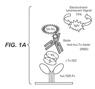
[087] Figure IA shows a schematic for a specific electrochemiluminescent assay
(ECLA) for human LTa(3 heterotrimers. Figure lB shows the specificity of this
assay for
detecting only human LTa(3 and not other TNF family ligands. The assay using
huLT(3R-Fc
capture and anti-LTa detection specifically recognizes LTal (32 and LTa201;
but not LTa3,
TNFc or LIGHT. In Figure 1 C 293 cells stably transfected with LTa and LT(3
constructs
were stained with huLT(3R-Fc-Alexa-647 and analyzed for surface LTa(3
expression by
FACS (open histogram); untransfected cells or cells stained with a control
antibody are also
shown. In Figure 1D culture supernatants from untransfected 293 cells and
cells transfected
with LTa and LT(3 constructs were analyzed using specific LTa3, LTa(3, and
TNFa assays to
measure levels of soluble cytokines (bars show average and SD of 4 cultures).
The lowest
detection limit for each assay is indicated with dashed line.
[088] Figure 2 illustrates that activated human T cells shed LTa(3 by ADAM 17
protease
cleavage. (A) Culture supernatants from polarized human T cells 2 days post-
reactivation
were analyzed using specific LTa3, LTa(3, and TNFa assays to measure levels of
soluble
cytokines (bar graphs show average and SD of 3 blood donors). (B) Culture
supernatants
from Thl human T cells treated -/+ 10 or 50 gM TNFa protease inhibitor-1 (TAPI-
1) for 1
day post reactivation were analyzed as in panel A for levels of soluble
cytokines (bars show
average and SD of 3 blood donors). (C) RNA was isolated from the cell
populations in panel
B and quantified by PCR using LTa, LT(3 and TNFa specific DNA probes. (D)
Supernatant
from pooled polarized Thl cells was immunoprecipitated with anti-LTa-
conjugated or LT(3R-
Fc-conjugated agarose beads, denatured proteins separated by gel
electrophoresis, and
21
CA 02737379 2011-03-15
WO 2010/039714 PCT/US2009/058797
Western blotted using fluorescent dye labeled probes specific for LTa (red)
and LT(3 (green).
Recombinant human LTal 02 was used as a reference. Two glycosylated forms each
are seen
for LTa and LT(3
[089] Figure 3 shows elevated so1LTa(3 levels in serum of experimental
autoimmune
encephalomyelitis (EAE) mice dosed with muLT(3R-Fc.
[090] Figure 4 shows (A) elevated so1LTa(3 levels in serum of collagen induced
arthritis
(CIA) mice dosed with muLT(3R-Fc, and (B) elevated soluble TNF-a levels in
serum of CIA
mice dosed with TNFRII-Fc.
[091] Figure 5 shows levels of soluble human LTa(3 in serum of human SCID mice
(transplanted with human peripheral blood mononuclear cells) which developed
severe graft
versus host disease. Levels of soluble human LTa(3 were elevated in mice
treated with
control antibody (Herceptin) but greatly reduced in mice treated with CTLA-4-
Fc (anti-
inflammatory therapeutic).
[092] Figure 6 shows peripheral so1LTa(3 in serum and synovial fluid of RA
patients.
(A) Sera collected from normal human donors and RA patients were analyzed
using specific
LTa3, LTa(3, and TNFa assays for levels of soluble cytokines (horizontal lines
depict
averages). (B) Synovial fluid collected from swollen joints of RA and OA
patients was
analyzed using specific LTa3, LTa(3, and TNFa assays for levels of soluble
cytokines
(horizontal lines depict averages).
[093] Figure 7 shows soluble LTa(3 and LTa3 induce the expression of
proinflammatory
cytokines, chemokines and adhesion molecules in primary RA fibroblast-like
synoviocytes
(FLS). (A) Primary RA FLS lines were simulated with 300ng/mL LTa(3 or media
alone for
6h. Total RNA was purified from the cells and quantitative PCR performed for
the genes
shown. (B) FLS were simulated with 100ng/mL LTa3 or media alone for 6h. Total
RNA was
purified from the cells and quantitative PCR performed for the genes shown.
Data are shown
as mean SEM and all differences between control and cytokines were highly
significant by
paired t test (p values<0.04). (C) FLS were stimulated with LTa(3 or LTa3
alone or in the
presence of 25 g/mL LT(3R-Fc or TNFRII-Fc. Total RNA was purified from the
cells and
quantitative PCR performed for the genes shown.
22
CA 02737379 2011-03-15
WO 2010/039714 PCT/US2009/058797
DETAILED DESCRIPTION
1. Abbreviations
The following abbreviations apply unless indicated otherwise:
Abbreviation Definition
LT Lymphotoxin
LTa or LTa or LTalpha Lymphotoxin-alpha
LTb or LT(3 or LTbeta Lymphotoxin-beta
LTab or LTa(3 or LTalpha-beta Lymphotoxin alpha-beta
soluble LTalpha-beta or solLTab or Soluble Lymphotoxin alpha-beta
so1LTa(3 or solLTab
huLTa(3 Human Lymphotoxin alpha-beta
OA Osteoarthritis
RA Rheumatoid arthritis
LT(3R Lymphotoxin-beta receptor
LT(3R-Fc or LT(3R-Ig Lymphotoxin-beta receptor conjugated to an
immunoglobulin Fc region
TNF Tumor Necrosis Factor
TNFR Tumor Necrosis Factor receptor
TNFR-Fc or TNFR-Ig Tumor Necrosis Factor receptor conjugated
to an immunoglobulin Fc region
FLS Fibroblast-like Synoviocytes
PAb Polyclonal antibodies
IL Interleukin
DMEM Dulbecco's Modified Eagle Medium
CXCL1 (GROa) Chemokine (C-X-C motif) ligand 1
previously called GRO1 oncogene, GROa,
KC, Neutrophil-activating protein 3 (NAP-3)
and melanoma growth stimulating activity,
alpha (MSGA-a).
CXCL2 (GROG) Chemokine (C-X-C motif) ligand 2 also
called macrophage inflammatory protein 2-
alpha (MIP2a), Growth-regulated protein
23
CA 02737379 2011-03-15
WO 2010/039714 PCT/US2009/058797
beta (Gro(3) and Gro oncogene-2 (Gro-2).
VCAM-l Vascular cell adhesion molecule 1 also
known as CD 106
ICAM-1 Inter-Cellular Adhesion Molecule 1 also
known as CD54
RPL19 Ribosomal protein L19
ECLA Electrochemiluminescent Assay
II. Definitions
[094] The practice of the present invention will employ, unless otherwise
indicated,
conventional techniques of molecular biology (including recombinant
techniques),
microbiology, cell biology, biochemistry, and immunology, which are within the
skill of the
art. Such techniques are explained fully in the literature, such as Molecular
Cloning: A
Laboratory Manual, second edition (Sambrook et al., 1989); Oligonucleotide
Synthesis (M. J.
Gait, ed., 1984); Animal Cell Culture (R. I. Freshney, ed., 1987); Methods in
Enzymology
(Academic Press, Inc.); Current Protocols in Molecular Biology (F. M. Ausubel
et al., eds.,
1987, and periodic updates); PCR: The Polymerase Chain Reaction, (Mullis et
al., ed., 1994);
A Practical Guide to Molecular Cloning (Perbal Bernard V., 1988); Phage
Display: A
Laboratory Manual (Barbas et al., 2001).
[095] Unless defined otherwise, technical and scientific terms used herein
have the same
meaning as commonly understood by one of ordinary skill in the art to which
this invention
belongs. Singleton et al., Dictionary of Microbiology and Molecular Biology
2nd ed., J.
Wiley & Sons (New York, NY 1994), and March, Advanced Organic Chemistry
Reactions,
Mechanisms and Structure 4th ed., John Wiley & Sons (New York, NY 1992),
provide one
skilled in the art with a general guide to many of the terms used in the
present application.
[096] One skilled in the art will recognize many methods and materials similar
or
equivalent to those described herein, which could be used in the practice of
the present
invention. Indeed, the present invention is in no way limited to the methods
and materials
described. For purposes of the present invention, the following terms are
defined below.
[097] "Lymphotoxin-alpha" or "LTa" or "LTa" is defined herein as a monomeric
protein
having a relative molecular mass of 25,000. The protein has the sequence shown
in Figure
2A of US Pat. No. 5,824,509 (and identified herein as SEQ ID NO: 1) or the
leu+1 (also
24
CA 02737379 2011-03-15
WO 2010/039714 PCT/US2009/058797
called the leucyl amino-terminal lymphotoxin species) or his+24 (also called
the histidyl
amino-terminal lymphotoxin species) as disclosed in US Pat. No. 5,824,509.
MTPPERLFLPRVCGTTLHLLLLGLLLVLLPGAOGLPGVGLTPSAAQTARQH
PKMHLAHSTLKPAAHLIGDPSKQNSLLWRANTDRAFLQDGFSLSNNSLLVPTS
GIYFVYSQVVFSGKAYSPKATSSPLYLAHEVQLFSSQYPFHVPLLSSQKMVYPG
LQEPWLHSMYHGAAFQLTQGDQLSTHTDGIPHLVLSPSTVFFGAFAL (SEQ ID
NO:1)
[098] Specifically, LTa is a member of the TNF superfamily and is secreted
from cells as
the homotrimer LTa3 (defined below), or complexed on the cell surface together
with LT(3
(defined below) as LTa(3 (defined below), predominantly as the LTal (32
heterotrimer. LTa is
defined to specifically exclude human TNF-a or its natural animal analogues
(Pennica et at.,
Nature 312:20/27: 724-729 (1984) and Aggarwal et at., J. Biol. Chem. 260: 2345-
2354
(1985)). LTa is defined to specifically exclude human LT(3 as defined, for
example, in US
5,661,004.
[099] "Lymphotoxin-beta" or "LT(3" or "LTb" is defined herein as a
biologically active
polypeptide having the amino acid sequence shown as SEQ ID NO:2 in U.S. Patent
No. US
5,661,004. LT(3 is defined to specifically exclude human LTa as defined, for
example, in US
5,824,509.
[0100] "Lymphotoxin-alpha3" or "Lymphotoxin-a3 trimer" or "LT0" or "LTa3"
refers to a
homotrimer of LTa monomers. It is a glycoprotein with a relative molecular
mass (Mr) of
55,000-70,000 and is formed by the association of three LTa monomers.
[0101] "Lymphotoxin-alpha-beta" or "Lymphotoxin-a(3" or "LTa(3" or "LTa(3
complex"
or "LTab" refers to a membrane bound heterotrimer of LTa with LT(3. These
heterotrimers
contain either two subunits of LTa and one subunit of LT(3 (LTa2(31), or one
subunit of LTa
and two of LT(3 (LTal (32). The term encompasses LTa2(31 or LTal (32,
individually, or a
mixture thereof.
[0102] The term "soluble Lymphotoxin-alpha-beta" or "so1LTa(3" refers to a
LTa(3 in
solution, not associated or bound to a cell. The so1LTa(3 are defined by the
LT(3 having been
cleaved at any point between the end of the transmembrane region (i.e., at
about amino acid
44 of SEQ ID NO:2 in U.S. Patent No. 5,661,004) and about amino acid 95.
[0103] "Tumor necrosis factor receptor-I" or "TNFRI" and "tumor necrosis
factor
receptor-II" or "TNFRII" refer to cell-surface TNF receptors for the LTa3
homotrimer, also
known as p55 and p75, respectively.
CA 02737379 2011-03-15
WO 2010/039714 PCT/US2009/058797
[0104] "Lymphotoxin-beta receptor" or "Lymphotoxin-(3 receptor" or "LTP-R" or
"LTV
refers to the receptor to which the LTa(3 heterotrimers bind. As used herein,
the term "a
lymphotoxin receptor" refers to the lymphotoxin-(3 receptor.
[0105] "Regulatory cytokines" are cytokines the abnormal levels of which
indicate the
presence of an autoimmune disorder in a patient. Such cytokines include, for
example,
interleukin-1 (IL-1), IL- 2, IL-3, IL-4, IL-5, IL-6, IL-7, IL-8, IL-10, IL-12,
IL-13, IL-14, IL-
15, IL-18, IL-23, IL-24, IL-25, IL-26, BLyS/April, TGF-a, TGF-(3, interferon-a
(IFN-a),
IFN-(3, IFN-y, MIP-1, MIF, MCP-1, M-CSF or G-CSF, a lymphotoxin, LIGHT, 4-1BB
ligand, CD27 ligand, CD30 ligand, CD40 ligand, Fas ligand, GITR ligand, OX40
ligand,
RANK ligand, THANK, TRAIL, TWEAK and VEG1. This group includes TNF family
members, which include but are not limited to, TNF-a, lymphotoxins (LTs) such
as LTa,
LT(3, and LIGHT. For a review of the TNF superfamily, see MacEwan, Br. J.
Pharmacology
135: 855-875 (2002). Preferably, the regulatory cytokine is a lymphotoxin such
as a TNF
family member.
[0106] "Inflammatory cytokines associated with rheumatoid arthritis" refer to
lymphotoxins, such as LTa, associated with RA pathology, which can be
inhibited
systemically and/or in the joints or in an in vitro collagen-induced arthritis
assay.
[0107] "LTa(3-expressing cells" are cells that express and/or shed the LTa(3
heterotrimers.
[0108] The expression "modulates LTa(3-expressing cells" refers to depleting
or altering
proteins made by the cells such as cytokines, chemokines, or growth factors,
with the cells
including, for example, monocytes, dendritic cells, T cells, and B cells.
[0109] A "lymphotoxin antagonist" or "LT antagonist" is a molecule that
reduces or
prevents the binding of a LT to its corresponding lymphotoxin receptor (LTR)
in a mammal
and/or interferes with one or more LTR expressing cell functions, e.g., by
reducing or
preventing a proinflammatory response elicited by the LTR-expressing cell. The
LT
antagonist can decrease, block, inhibit, abrogate, modulate and/or otherwise
interfere with LT
activity in vitro, in situ, and/or in vivo. Such an agent can inhibit a
biological function or
activity of a LT, e.g., through binding to a LT and neutralizing its activity.
For example, a
LT antagonist can decrease block, abrogate, modulate, interfere, prevent
and/or inhibit
lymphotoxin RNA, DNA, or protein synthesis, lymphotoxin release, lymphotoxin
receptor
signaling, membrane lymphotoxin cleavage, lymphotoxin activity, and
lymphotoxin
production and/or synthesis. Examples of LT antagonists include, but are not
limited to, anti-
LT antibodies, antigen-binding fragments thereof, specified mutants or domains
thereof that
26
CA 02737379 2011-03-15
WO 2010/039714 PCT/US2009/058797
bind specifically to a LT that, upon binding to a LT, interfere with one or
more functions of
cells expressing a receptor for the LT, a soluble lymphotoxin receptor or
fragment, fusion
polypeptides thereof, a small-molecule LT antagonist, e.g., TNF binding
protein I or II (TBP-
I or TBP-II), nerelimonmab, CDP-571, infliximab (REMICADE ), etanercept
(ENBREL ),
adalimulab (HUMIRATM), CDP-571, CDP-870, afelimomab, lenercept, and the like),
antigen-binding fragments thereof, and receptor molecules that bind
specifically to a LT,
compounds that prevent and/or inhibit lymphotoxin synthesis, LT release, or
its action on
target cells, such as thalidomide, tenidap, phosphodiesterase inhibitors (e.g,
pentoxifylline
and rolipram), A2b adenosine receptor agonists, and A2b adenosine receptor
enhancers,
compounds that prevent and/or inhibit lymphotoxin receptor signaling, such as
mitogen-
activated protein (MAP) kinase inhibitors, compounds that block and/or inhibit
membrane
lymphotoxin cleavage, such as metalloproteinase inhibitors, compounds that
block and/or
inhibit lymphotoxin activity, such as angiotensin-converting enzyme (ACE)
inhibitors (e.g.,
captopril), and compounds that block and/or inhibit lymphotoxin production
and/or synthesis,
such as MAP kinase inhibitors. A preferred antagonist comprises an antibody or
an
immunoadhesin. Examples of LT antagonists contemplated herein are etanercept
(ENBREL ), infliximab (REMICADE ), and adalimumab (HUMIRATM). In one
embodiment, the LT antagonist is an antagonist of LTa, e.g., an anti-LTa
antibody, and more
particularly a humanized, monoclonal anti-LTa antibody.
[0110] As used herein, "antagonist" includes any molecule that partially or
fully blocks,
inhibits, or neutralizes a biological activity of a native polypeptide
disclosed herein. Suitable
antagonist molecules specifically include antagonist antibodies or antibody
fragments,
fragments or amino acid sequence variants of native polypeptides, peptides,
antisense
oligonucleotides, and small organic molecules, as non limiting examples.
Methods for
identifying antagonists may comprise contacting such a polypeptide, including
a cell
expressing it, with a candidate agonist or antagonist molecule and measuring a
detectable
change in one or more biological activities normally associated with such
polypeptide.
[0111] A "blocking" antibody or an "antagonist" antibody is one that inhibits
or reduces
biological activity of the antigen it binds. Preferred blocking antibodies or
antagonist
antibodies substantially or completely inhibit the biological activity of the
antigen.
[0112] An "anti-lymphotoxin antibody antagonist" or "anti-LT antibody
antagonist" as
used herein is an antibody that is a LT antagonist. For example, in some
embodiments the
anti-lymphotoxin antibody antagonist reduces or prevents the binding of a LT
to its
corresponding lymphotoxin receptor in a mammal and/or interferes with one or
more
27
CA 02737379 2011-03-15
WO 2010/039714 PCT/US2009/058797
lymphotoxin receptors (LTR) expressing cell functions, e.g. by reducing or
preventing a
proinflammatory response elicited by the LTR-expressing cell. The antibody
that binds a LT
may be designated as follows: an antibody that binds to lymphotoxin alpha
(LTa), an "anti-
lymphotoxin alpha antibody", or an "anti-LTa antibody". Antagonists can be
screened by
various methods known in the art for anti-inflammatory effects. For example, a
method of
screening can be employed as described in the following references: Lu, Y., et
al., Current
Opinion in Pharmacology 7, 571-546 (2007); Han, S., et at. Arthritis Rheum 52,
3202-3209
(2005); Fava, R.A., et al., Jlmmunol 171, 115-126 (2003); Mackay, F., et al.,
Gastroenterology 115, 1464-1475 (1998); Gommerman, J.L., et at., Nat Rev
Immunol 2003
supra); Wu, Q., et at., JExp Med 193, 1327-1332 (2001); and Ettinger, R., et
at., JExp Med
193, 1333-1340 (2001).
[0113] The term "modulate" as used herein refers to up or down regulation or
change e.g.,
in an activity or function of a biological molecule. For example, modulate can
refer to the
change or up or down regulation of expression of a gene, level of RNA molecule
or
equivalent RNA molecules encoding one or more proteins or protein subunits, or
activity of
one or more proteins or protein subunits, such that expression, level, or
activity is greater than
or less than that observed in the absence of the modulator. For example, a LT
antagonist may
act as a modulator upon the activity of a LT polypeptide and/or a lymphotoxin
receptor
polypeptide.
[0114] The terms "antibody" and "immunoglobulin" are used interchangeably in
the
broadest sense and include monoclonal antibodies (e.g., full-length or intact
monoclonal
antibodies), polyclonal antibodies, multivalent antibodies, and multispecific
antibodies (e.g.,
bispecific antibodies so long as they exhibit the desired biological
activity), and may also
include certain antibody fragments (as described in greater detail herein). An
antibody can be
chimeric, human, humanized, and/or affinity matured.
[0115] An "isolated" antibody is one which has been identified and separated
and/or
recovered from a component of its natural environment. Contaminant components
of its
natural environment are materials which would interfere with research,
diagnostic or
therapeutic uses for the antibody, and may include enzymes, hormones, and
other
proteinaceous or nonproteinaceous solutes. In some embodiments, an antibody is
purified (1)
to greater than 95% by weight of antibody as determined by, for example, the
Lowry method,
and in some embodiments, to greater than 99% by weight; (2) to a degree
sufficient to obtain
at least 15 residues of N-terminal or internal amino acid sequence by use of,
for example, a
spinning cup sequenator, or (3) to homogeneity by SDS-PAGE under reducing or
28
CA 02737379 2011-03-15
WO 2010/039714 PCT/US2009/058797
nonreducing conditions using, for example, Coomassie blue or silver stain.
Isolated antibody
includes the antibody in situ within recombinant cells since at least one
component of the
antibody's natural environment will not be present. Ordinarily, however,
isolated antibody
will be prepared by at least one purification step.
[0116] "Native antibodies" are usually heterotetrameric glycoproteins of about
150,000
daltons, composed of two identical light (L) chains and two identical heavy
(H) chains. Each
light chain is linked to a heavy chain by one covalent disulfide bond, while
the number of
disulfide linkages varies among the heavy chains of different immunoglobulin
isotypes. Each
heavy and light chain also has regularly spaced intrachain disulfide bridges.
Each heavy
chain has at one end a variable domain (VH) followed by a number of constant
domains.
Each light chain has a variable domain at one end (VL) and a constant domain
at its other end;
the constant domain of the light chain is aligned with the first constant
domain of the heavy
chain, and the light-chain variable domain is aligned with the variable domain
of the heavy
chain. Particular amino acid residues are believed to form an interface
between the light-
chain and heavy-chain variable domains.
[0117] The "variable region" or "variable domain" of an antibody refers to the
amino-
terminal domains of the heavy or light chain of the antibody. The variable
domain of the
heavy chain may be referred to as "VH." The variable domain of the light chain
may be
referred to as "VL." These domains are generally the most variable parts of an
antibody and
contain the antigen-binding sites.
[0118] The term "variable" refers to the fact that certain portions of the
variable domains
differ extensively in sequence among antibodies and are used in the binding
and specificity of
each particular antibody for its particular antigen. However, the variability
is not evenly
distributed throughout the variable domains of antibodies. It is concentrated
in three
segments called hypervariable regions (HVRs) both in the light-chain and the
heavy-chain
variable domains. The more highly conserved portions of variable domains are
called the
framework regions (FR). The variable domains of native heavy and light chains
each
comprise four FR regions, largely adopting a beta-sheet configuration,
connected by three
HVRs, which form loops connecting, and in some cases forming part of, the beta-
sheet
structure. The HVRs in each chain are held together in close proximity by the
FR regions
and, with the HVRs from the other chain, contribute to the formation of the
antigen-binding
site of antibodies (see Kabat et at., Sequences of Proteins of Immunological
Interest, Fifth
Edition, National Institute of Health, Bethesda, MD (1991)). The constant
domains are not
29
CA 02737379 2011-03-15
WO 2010/039714 PCT/US2009/058797
involved directly in the binding of an antibody to an antigen, but exhibit
various effector
functions, such as participation of the antibody in antibody-dependent
cellular toxicity.
[0119] The "light chains" of antibodies (immunoglobulins) from any vertebrate
species
can be assigned to one of two clearly distinct types, called kappa (K) and
lambda (X), based
on the amino acid sequences of their constant domains.
[0120] Depending on the amino acid sequences of the constant domains of their
heavy
chains, antibodies (immunoglobulins) can be assigned to different classes.
There are five
major classes of immunoglobulins: IgA, IgD, IgE, IgG, and IgM, and several of
these may be
further divided into subclasses (isotypes), e.g., IgG1, IgG2, IgG3, IgG4,
IgA1, and IgA2. The
heavy-chain constant domains that correspond to the different classes of
immunoglobulins
are called a, 6, E, y, and , respectively. The subunit structures and three-
dimensional
configurations of different classes of immunoglobulins are well known and
described
generally in, for example, Abbas et al. Cellular and Mol. Immunology, 4th ed.
(W. B.
Saunders, Co., 2000). An antibody may be part of a larger fusion molecule,
formed by
covalent or non-covalent association of the antibody with one or more other
proteins or
peptides.
[0121] The terms "full-length antibody," "intact antibody," and "whole
antibody" are used
herein interchangeably to refer to an antibody in its substantially intact
form, not antibody
fragments as defined below. The terms particularly refer to an antibody with
heavy chains
that contain an Fc region.
[0122] A "naked antibody" for the purposes herein is an antibody that is not
conjugated to
a cytotoxic moiety or radiolabel.
[0123] "Antibody fragments" comprise a portion of an intact antibody,
preferably
comprising the antigen-binding region thereof. Examples of antibody fragments
include Fab,
Fab', F(ab')2, and Fv fragments; diabodies; linear antibodies; single-chain
antibody
molecules; and multispecific antibodies formed from antibody fragments.
[0124] Papain digestion of antibodies produces two identical antigen-binding
fragments,
called "Fab" fragments, each with a single antigen-binding site, and a
residual "Fc" fragment,
whose name reflects its ability to crystallize readily. Pepsin treatment
yields an F(ab')2
fragment that has two antigen-combining sites and is still capable of cross-
linking antigen.
[0125] "Fv" is the minimum antibody fragment which contains a complete antigen-
binding site. In one embodiment, a two-chain Fv species consists of a dimer of
one heavy-
and one light-chain variable domain in tight, non-covalent association. In a
single-chain Fv
CA 02737379 2011-03-15
WO 2010/039714 PCT/US2009/058797
(scFv) species, one heavy- and one light-chain variable domain can be
covalently linked by a
flexible peptide linker such that the light and heavy chains can associate in
a "dimeric"
structure analogous to that in a two-chain Fv species. It is in this
configuration that the three
HVRs of each variable domain interact to define an antigen-binding site on the
surface of the
VH-VL dimer. Collectively, the six HVRs confer antigen-binding specificity to
the antibody.
However, even a single variable domain (or half of an Fv comprising only three
HVRs
specific for an antigen) has the ability to recognize and bind antigen,
although at a lower
affinity than the entire binding site.
[0126] The Fab fragment contains the heavy- and light-chain variable domains
and also
contains the constant domain of the light chain and the first constant domain
(CH1) of the
heavy chain. Fab' fragments differ from Fab fragments by the addition of a few
residues at
the carboxy terminus of the heavy chain CH1 domain including one or more
cysteines from
the antibody-hinge region. Fab'-SH is the designation herein for Fab' in which
the cysteine
residue(s) of the constant domains bear a free thiol group. F(ab')z antibody
fragments
originally were produced as pairs of Fab' fragments which have hinge cysteines
between
them. Other chemical couplings of antibody fragments are also known.
[0127] "Single-chain Fv" or "scFv" antibody fragments comprise the VH and VL
domains
of an antibody, wherein these domains are present in a single polypeptide
chain. Generally,
the scFv polypeptide further comprises a polypeptide linker between the VH and
VL domains
that enables the scFv to form the desired structure for antigen binding. For a
review of scFv,
see, e.g., Pluckthun, in The Pharmacology of Monoclonal Antibodies, vol. 113,
Rosenburg
and Moore eds. (Springer-Verlag, New York: 1994), pp 269-315.
[0128] The term "diabodies" refers to antibody fragments with two antigen-
binding sites,
which fragments comprise a heavy-chain variable domain (VH) connected to a
light-chain
variable domain (VL) in the same polypeptide chain (VH-VL). By using a linker
that is too
short to allow pairing between the two domains on the same chain, the domains
are forced to
pair with the complementary domains of another chain and create two antigen-
binding sites.
Diabodies may be bivalent or bispecific. Diabodies are described more fully
in, for example,
EP 404,097; WO 1993/01161; Hudson et al., Nat. Med., 9:129-134 (2003); and
Hollinger et
al., Proc. Natl. Acad. Sci. USA, 90:6444-6448 (1993). Triabodies and
tetrabodies are also
described in Hudson et al., Nat. Med., 9:129-134 (2003).
[0129] The term "monoclonal antibody" as used herein refers to an antibody
obtained
from a population of substantially homogeneous antibodies, i.e., the
individual antibodies
comprising the population are identical except for possible mutations, e.g.,
naturally
31
CA 02737379 2011-03-15
WO 2010/039714 PCT/US2009/058797
occurring mutations, that may be present in minor amounts. Thus, the modifier
"monoclonal" indicates the character of the antibody as not being a mixture of
discrete
antibodies. In certain embodiments, such a monoclonal antibody typically
includes an
antibody comprising a polypeptide sequence that binds a target, wherein the
target-binding
polypeptide sequence was obtained by a process that includes the selection of
a single target
binding polypeptide sequence from a plurality of polypeptide sequences. For
example, the
selection process can be the selection of a unique clone from a plurality of
clones, such as a
pool of hybridoma clones, phage clones, or recombinant DNA clones. It should
be
understood that a selected target binding sequence can be further altered, for
example, to
improve affinity for the target, to humanize the target-binding sequence, to
improve its
production in cell culture, to reduce its immunogenicity in vivo, to create a
multispecific
antibody, etc., and that an antibody comprising the altered target binding
sequence is also a
monoclonal antibody of this invention. In contrast to polyclonal antibody
preparations,
which typically include different antibodies directed against different
determinants (epitopes),
each monoclonal antibody of a monoclonal-antibody preparation is directed
against a single
determinant on an antigen. In addition to their specificity, monoclonal-
antibody preparations
are advantageous in that they are typically uncontaminated by other
immunoglobulins.
[0130] The modifier "monoclonal" indicates the character of the antibody as
being
obtained from a substantially homogeneous population of antibodies, and is not
to be
construed as requiring production of the antibody by any particular method.
For example, the
monoclonal antibodies to be used in accordance with the present invention may
be made by a
variety of techniques, including, for example, the hybridoma method (e.g.,
Kohler and
Milstein., Nature, 256:495-97 (1975); Hongo et at., Hybridoma, 14(3):253-260
(1995),
Harlow et at., Antibodies: A Laboratory Manual, (Cold Spring Harbor Laboratory
Press, 2d
ed. 1988); Hammerling et at., in: Monoclonal Antibodies and T-Cell Hybridomas,
563-681
(Elsevier, N.Y., 1981)), recombinant DNA methods (see, e.g., U.S. 4,816,567),
phage-
display technologies (see, e.g., Clackson et at., Nature, 352: 624-628 (1991);
Marks et at., J.
Mol. Biol., 222:581-597 (1992); Sidhu et al., J. Mol. Biol., 338(2):299-310
(2004); Lee et al.,
J. Mol. Biol., 340(5):1073-1093 (2004); Fellouse, Proc. Natl. Acad. Sci. USA,
101(34):12467-12472 (2004); and Lee et at., J. Immunol. Methods, 284(1-2):119-
132(2004),
and technologies for producing human or human-like antibodies in animals that
have parts or
all of the human immunoglobulin loci or genes encoding human immunoglobulin
sequences
(see, e.g., WO 1998/24893; WO 1996/34096; WO 1996/33735; WO 1991/10741;
Jakobovits
et at., Proc. Natl. Acad. Sci. USA, 90: 2551 (1993); Jakobovits et at.,
Nature, 362: 255-258
32
CA 02737379 2011-03-15
WO 2010/039714 PCT/US2009/058797
(1993); Bruggemann et at., Year in Immunol., 7:33 (1993); U.S. 5,545,807;
5,545,806;
5,569,825; 5,625,126; 5,633,425; and 5,661,016; Marks et at., Bio/Technology,
10: 779-783
(1992); Lonberg et at., Nature, 368:856-859 (1994); Morrison, Nature, 368:812-
813 (1994);
Fishwild et at., Nature Biotechnol., 14:845-851 (1996); Neuberger, Nature
Biotechnol.,
14:826 (1996); and Lonberg and Huszar, Intern. Rev. Immunol., 13:65-93 (1995).
[0131] The monoclonal antibodies herein specifically include "chimeric"
antibodies in
which a portion of the heavy and/or light chain is identical with or
homologous to
corresponding sequences in antibodies derived from a particular species or
belonging to a
particular antibody class or subclass, while the remainder of the chain(s) is
identical with or
homologous to corresponding sequences in antibodies derived from another
species or
belonging to another antibody class or subclass, as well as fragments of such
antibodies, so
long as they exhibit the desired biological activity (e.g., U.S. 4,816,567 and
Morrison et at.,
Proc. Natl. Acad. Sci. USA, 81:6851-6855 (1984)). Chimeric antibodies include
PRIMATIZED antibodies wherein the antigen-binding region of the antibody is
derived
from an antibody produced by, e.g., immunizing macaque monkeys with the
antigen of
interest.
[0132] "Humanized" forms of non-human (e.g., murine) antibodies are chimeric
antibodies that contain minimal sequence derived from non-human
immunoglobulin. In one
embodiment, a humanized antibody is a human immunoglobulin (recipient
antibody) in
which residues from a HVR of the recipient are replaced by residues from a HVR
of a non-
human species (donor antibody) such as mouse, rat, rabbit, or nonhuman primate
having the
desired specificity, affinity, and/or capacity. In some instances, FR residues
of the human
immunoglobulin are replaced by corresponding non-human residues. Furthermore,
humanized antibodies may comprise residues that are not found in the recipient
antibody or in
the donor antibody. These modifications may be made to further refine antibody
performance. In general, a humanized antibody will comprise substantially all
of at least one,
and typically two, variable domains, in which all or substantially all of the
hypervariable
loops correspond to those of a non-human immunoglobulin, and all, or
substantially all, of
the FRs are those of a human immunoglobulin sequence. The humanized antibody
optionally
will also comprise at least a portion of an immunoglobulin constant region
(Fc), typically that
of a human immunoglobulin. For further details, see, e.g., Jones et at.,
Nature, 321:522-525
(1986); Riechmann et at., Nature, 332:323-329 (1988); and Presta, Curr. Op.
Struct. Biol.,
2:593-596 (1992). See also, for example, Vaswani and Hamilton, Ann. Allergy,
Asthma &
33
CA 02737379 2011-03-15
WO 2010/039714 PCT/US2009/058797
Immunol., 1:105-115 (1998); Harris, Biochem. Soc. Transactions, 23:1035-1038
(1995);
Hurle and Gross, Curr. Op. Biotech., 5:428-433 (1994); and U.S. 6,982,321 and
7,087,409.
[0133] A "human antibody" is one which possesses an amino-acid sequence which
corresponds to that of an antibody produced by a human and/or has been made
using any of
the techniques for making human antibodies as disclosed herein. This
definition of a human
antibody specifically excludes a humanized antibody comprising non-human
antigen-binding
residues. Human antibodies can be produced using various techniques known in
the art,
including phage-display libraries. Hoogenboom and Winter, J. Mol. Biol.,
227:381 (1991);
Marks et at., J. Mol. Biol., 222:581 (1991). Also available for the
preparation of human
monoclonal antibodies are methods described in Cole et at., Monoclonal
Antibodies and
Cancer Therapy, Alan R. Liss, p. 77 (1985); Boerner et at., J. Immunol.,
147(1):86-95
(1991). See also van Dijk and van de Winkel, Curr. Opin. Pharmacol., 5: 368-74
(2001).
Human antibodies can be prepared by administering the antigen to a transgenic
animal that
has been modified to produce such antibodies in response to antigenic
challenge, but whose
endogenous loci have been disabled, e.g., immunized xenomice (see, e.g., U.S.
6,075,181 and
6,150,584 regarding XENOMOUSETM technology). See also, for example, Li et at.,
Proc.
Natl. Acad. Sci. USA, 103:3557-3562 (2006) regarding human antibodies
generated via a
human B-cell hybridoma technology.
[0134] The term "hypervariable region," "HVR," or "HV," when used herein
refers to the
regions of an antibody-variable domain which are hypervariable in sequence
and/or form
structurally defined loops. Generally, antibodies comprise six HVRs; three in
the VH (H1,
H2, H3), and three in the VL (L1, L2, L3). In native antibodies, H3 and L3
display the most
diversity of the six HVRs, and H3 in particular is believed to play a unique
role in conferring
fine specificity to antibodies. See, e.g., Xu et at., Immunity, 13:37-45
(2000); Johnson and
Wu in Methods in Molecular Biology, 248:1-25 (Lo, ed., Human Press, Totowa,
NJ, 2003).
Indeed, naturally occurring camelid antibodies consisting of a heavy chain
only are functional
and stable in the absence of light chain. See, e.g., Hamers-Casterman et at.,
Nature, 363:446-
448 (1993) and Sheriff et at., Nature Struct. Biol., 3:733-736 (1996).
[0135] A number of HVR delineations are in use and are encompassed herein. The
HVRs
that are Kabat complementarity-determining regions (CDRs) are based on
sequence
variability and are the most commonly used (Kabat et at., Sequences of
Proteins of
Immunological Interest, 5th Ed. Public Health Service, National Institutes of
Health,
Bethesda, MD (1991)). Chothia refers instead to the location of the structural
loops (Chothia
and Lesk, J. Mol. Biol., 196:901-917 (1987)). The AbM HVRs represent a
compromise
34
CA 02737379 2011-03-15
WO 2010/039714 PCT/US2009/058797
between the Kabat CDRs and Chothia structural loops, and are used by Oxford
Molecular's
AbM antibody-modeling software. The "contact" HVRs are based on an analysis of
the
available complex crystal structures. The residues from each of these HVRs are
noted below.
Loop Kabat AbM Chothia Contact
L1 L24-L34 L24-L34 L26-L32 L30-L36
L2 L50-L56 L50-L56 L50-L52 L46-L55
L3 L89-L97 L89-L97 L91-L96 L89-L96
Hl H31-H35B H26-H35B H26-H32 H30-H35B (Kabat Numbering)
Hl H31-H35 H26-H35 H26-H32 H30-H35 (Chothia Numbering)
H2 H50-H65 H50-H58 H53-H55 H47-H58
H3 H95-H102 H95-H102 H96-H101 H93-H101
[0136] HVRs may comprise "extended HVRs" as follows: 24-36 or 24-34 (L1), 46-
56 or
50-56 (L2), and 89-97 or 89-96 (L3) in the VL, and 26-35 (H1), 50-65 or 49-65
(H2), and 93-
102, 94-102, or 95-102 (H3) in the VH. The variable-domain residues are
numbered
according to Kabat et at., supra, for each of these extended-HVR definitions.
[0137] "Framework" or "FR" residues are those variable-domain residues other
than the
HVR residues as herein defined.
[0138] The expression "variable-domain residue-numbering as in Kabat" or
"amino-acid-
position numbering as in Kabat," and variations thereof, refers to the
numbering system used
for heavy-chain variable domains or light-chain variable domains of the
compilation of
antibodies in Kabat et at., supra. Using this numbering system, the actual
linear amino acid
sequence may contain fewer or additional amino acids corresponding to a
shortening of, or
insertion into, a FR or HVR of the variable domain. For example, a heavy-chain
variable
domain may include a single amino acid insert (residue 52a according to Kabat)
after residue
52 of H2 and inserted residues (e.g. residues 82a, 82b, and 82c, etc.
according to Kabat) after
heavy-chain FR residue 82. The Kabat numbering of residues may be determined
for a given
antibody by alignment at regions of homology of the sequence of the antibody
with a
"standard" Kabat numbered sequence.
[0139] An "affinity-matured" antibody is one with one or more alterations in
one or more
HVRs thereof which result in an improvement in the affinity of the antibody
for antigen,
compared to a parent antibody which does not possess those alteration(s). In
one
CA 02737379 2011-03-15
WO 2010/039714 PCT/US2009/058797
embodiment, an affinity-matured antibody has nanomolar or even picomolar
affinities for the
target antigen. Affinity-matured antibodies are produced by procedures known
in the art.
For example, Marks et at., Bio/Technology, 10:779-783 (1992) describes
affinity maturation
by VH- and VL-domain shuffling. Random mutagenesis of HVR and/or framework
residues
is described by, for example: Barbas et al., Proc Nat. Acad. Sci. USA, 91:3809-
3813 (1994);
Schier et at., Gene, 169:147-155 (1995); Yelton et at., J. Immunol., 155:1994-
2004 (1995);
Jackson et at., J. Immunol., 154(7):3310-3319 (1995); and Hawkins et at., J.
Mol. Biol.,
226:889-896 (1992).
[0140] "Growth-inhibitory" antibodies are those that prevent or reduce
proliferation of a
cell expressing an antigen to which the antibody binds. For example, the
antibody may
prevent or reduce proliferation of B cells in vitro and/or in vivo.
[0141] Antibodies that "induce apoptosis" are those that induce programmed
cell death,
e.g. of a B cell, as determined by standard apoptosis assays, such as binding
of annexin V,
fragmentation of DNA, cell shrinkage, dilation of endoplasmic reticulum, cell
fragmentation,
and/or formation of membrane vesicles (called apoptotic bodies). Antibody
"effector
functions" refer to those biological activities attributable to the Fc region
(a native-sequence
Fc region or amino-acid-sequence-variant Fc region) of an antibody, and vary
with the
antibody isotype. Examples of antibody effector functions include: C l q
binding and
complement- dependent cytotoxicity (CDC); Fc-receptor binding; antibody-
dependent cell-
mediated cytotoxicity (ADCC); phagocytosis; down-regulation of cell-surface
receptors (e.g.
B-cell receptor); and B-cell activation.
[0142] The term "Fc region" herein is used to define a C-terminal region of an
immunoglobulin heavy chain, including native-sequence Fc regions and variant
Fc regions.
Although the boundaries of the Fc region of an immunoglobulin heavy chain
might vary, the
human IgG heavy-chain Fc region is usually defined to stretch from an amino
acid residue at
position Cys226, or from Pro230, to the carboxyl-terminus thereof. The C-
terminal lysine
(residue 447 according to the EU numbering system) of the Fc region may be
removed, for
example, during production or purification of the antibody, or by
recombinantly engineering
the nucleic acid encoding a heavy chain of the antibody. Accordingly, a
composition of
intact antibodies may comprise antibody populations with all K447 residues
removed,
antibody populations with no K447 residues removed, and antibody populations
having a
mixture of antibodies with and without the K447 residue.
36
CA 02737379 2011-03-15
WO 2010/039714 PCT/US2009/058797
[0143] Unless indicated otherwise herein, the numbering of the residues in an
immunoglobulin heavy chain is that of the EU index as in Kabat et at., supra.
The "EU index
as in Kabat" refers to the residue numbering of the human IgGl EU antibody.
[0144] A "functional Fc region" possesses an "effector function" of a native-
sequence Fc
region. Exemplary "effector functions" include Cl q binding; CDC; Fc-receptor
binding;
ADCC; phagocytosis; down-regulation of cell-surface receptors (e.g. B-cell
receptor; BCR),
etc. Such effector functions generally require the Fc region to be combined
with a binding
domain (e.g. an antibody-variable domain) and can be assessed using various
assays as
disclosed, for example, in definitions herein.
[0145] A "native-sequence Fc region" comprises an amino acid sequence
identical to the
amino acid sequence of an Fc region found in nature. Native-sequence human Fc
regions
include a native-sequence human IgGI Fc region (non-A and A allotypes); native-
sequence
human IgG2 Fc region; native-sequence human IgG3 Fc region; and native-
sequence human
IgG4 Fc region, as well as naturally occurring variants thereof.
[0146] A "variant Fc region" comprises an amino acid sequence which differs
from that of
a native- sequence Fc region by virtue of at least one amino acid
modification, preferably one
or more amino acid substitution(s). Preferably, the variant Fc region has at
least one amino
acid substitution compared to a native-sequence Fc region or to the Fc region
of a parent
polypeptide, e.g. from about one to about ten amino acid substitutions, and
preferably from
about one to about five amino acid substitutions in a native- sequence Fc
region or in the Fc
region of the parent polypeptide. The variant Fc region herein will preferably
possess at least
about 80% homology with a native-sequence Fc region and/or with an Fc region
of a parent
polypeptide, and most preferably at least about 90% homology therewith, more
preferably at
least about 95% homology therewith.
[0147] The term "Fc-region-comprising antibody" refers to an antibody that
comprises an
Fc region. The C-terminal lysine (residue 447 according to the EU numbering
system) of the
Fc region may be removed, for example, during purification of the antibody or
by
recombinant engineering the nucleic acid encoding the antibody. Accordingly, a
composition
comprising an antibody having an Fc region according to this invention can
comprise an
antibody with K447, with all K447 removed, or a mixture of antibodies with and
without the
K447 residue.
[0148] "Fc receptor" or "FcR" describes a receptor that binds to the Fc region
of an
antibody. In some embodiments, an FcR is a native-human FcR. In some
embodiments, an
FcR is one which binds an IgG antibody (a gamma receptor) and includes
receptors of the
37
CA 02737379 2011-03-15
WO 2010/039714 PCT/US2009/058797
FcyRI, FcyRII, and FcyRIII subclasses, including allelic variants and
alternatively spliced
forms of those receptors. FcyRII receptors include FcyRIIA (an "activating
receptor") and
FcyRIIB (an "inhibiting receptor"), which have similar amino acid sequences
that differ
primarily in the cytoplasmic domains thereof. Activating receptor FcyRIIA
contains an
immunoreceptor tyrosine-based activation motif (ITAM) in its cytoplasmic
domain.
Inhibiting receptor FcyRIIB contains an immunoreceptor tyrosine-based
inhibition motif
(ITIM) in its cytoplasmic domain. (see, e.g., Daeron, Annu. Rev. Immunol.
15:203-234
(1997)). FcRs are reviewed, for example, in Ravetch and Kinet, Annu. Rev.
Immunol 9:457-
92 (1991); Capel et at., Immunomethods 4:25-34 (1994); and de Haas et at., J.
Lab. Clin.
Med. 126:330-41 (1995). Other FcRs, including those to be identified in the
future, are
encompassed by the term "FcR" herein.
[0149] The term "Fc receptor" or "FcR" also includes the neonatal receptor,
FcRn, which
is responsible for the transfer of maternal IgGs to the fetus (Guyer et at.,
J. Immunol. 117:587
(1976) and Kim et at., J. Immunol. 24:249 (1994)) and regulation of
homeostasis of
immunoglobulins. Methods of measuring binding to FcRn are known (see, e.g.,
Ghetie and
Ward, Immunology Today, 18 (12):592-8 (1997); Ghetie et at., Nature
Biotechnology, 15
(7):637-40 (1997); Hinton et at., J. Biol. Chem., 279(8):6213-6 (2004); WO
2004/92219
(Hinton et al.).
[0150] Binding to human FcRn in vivo and serum half-life of human FcRn high-
affinity
binding polypeptides can be assayed, e.g., in transgenic mice or transfected
human cell lines
expressing human FcRn, or in primates to which the polypeptides with a variant
Fc region are
administered. WO 2000/42072 (Presta) describes antibody variants with improved
or
diminished binding to FcRs. See, also, for example, Shields et at., J. Biol.
Chem., 9(2): 6591-
6604 (2001).
[0151] "Human effector cells" are leukocytes which express one or more FcRs
and
perform effector functions. In certain embodiments, the cells express at least
FcyRIII and
perform ADCC effector function(s). Examples of human leukocytes which mediate
ADCC
include peripheral blood mononuclear cells (PBMC), natural-killer (NK) cells,
monocytes,
cytotoxic T cells, and neutrophils. The effector cells may be isolated from a
native source,
e.g., from blood.
[0152] "Antibody-dependent cell-mediated cytotoxicity" or "ADCC" refers to a
form of
cytotoxicity in which secreted Ig bound onto Fc receptors (FcRs) present on
certain cytotoxic
cells (e.g., NK cells, neutrophils, and macrophages) enables these cytotoxic
effector cells to
38
CA 02737379 2011-03-15
WO 2010/039714 PCT/US2009/058797
bind specifically to an antigen-bearing target cell and subsequently kill the
target cell with
cytotoxins. The primary cells for mediating ADCC, NK cells, express FcyRIII
only, whereas
monocytes express FcyRI, FcyRII, and FcyRIII. FcR expression on hematopoietic
cells is
summarized in Table 3 on page 464 of Ravetch and Kinet, Annu. Rev. Immunol.,
9:457-492
(1991). To assess ADCC activity of a molecule of interest, an in vitro ADCC
assay, such as
that described in U.S. 5,500,362 or 5,821,337 or U.S. 6,737,056 (Presta), may
be performed.
Useful effector cells for such assays include PBMC and NK cells.
Alternatively, or
additionally, ADCC activity of the molecule of interest may be assessed in
vivo, e.g., in an
animal model such as that disclosed in Clynes et at., Proc. Natl. Acad. Sci.
(USA), 95:652-
656 (1998).
[0153] "Complement-dependent cytotoxicity" or "CDC" refers to the lysis of a
target cell
in the presence of complement. Activation of the classical complement pathway
is initiated
by the binding of the first component of the complement system (C l q) to
antibodies (of the
appropriate subclass), which are bound to their cognate antigen. To assess
complement
activation, a CDC assay, e.g. as described in Gazzano-Santoro et at., J.
Immunol. Methods,
202:163 (1996), may be performed. Polypeptide variants with altered Fc region
amino acid
sequences (polypeptides with a variant Fc region) and increased or decreased
Clq binding
capability are described, e.g., in U.S. 6,194,551 and WO 1999/51642. See,
also, e.g.,
Idusogie et at., J. Immunol. 164:4178-4184 (2000).
[0154] "Binding affinity" generally refers to the strength of the sum total of
noncovalent
interactions between a single binding site of a molecule (e.g., an antibody)
and its binding
partner (e.g., an antigen). Unless indicated otherwise, as used herein,
"binding affinity"
refers to intrinsic binding affinity which reflects a 1:1 interaction between
members of a
binding pair (e.g., antibody and antigen). The affinity of a molecule X for
its partner Y can
generally be represented by the dissociation constant (Kd). Affinity can be
measured by
common methods known in the art, including those described herein. Low-
affinity antibodies
generally bind antigen slowly and tend to dissociate readily, whereas high-
affinity antibodies
generally bind antigen faster and tend to remain bound longer. A variety of
methods of
measuring binding affinity are known in the art, any of which can be used for
purposes of the
present invention. Specific illustrative and exemplary embodiments for
measuring binding
affinity are described in the following.
[0155] In one embodiment, the "Kd" or "Kd value" according to this invention
is
measured by a radiolabeled antigen-binding assay (RIA) performed with the Fab
version of
39
CA 02737379 2011-03-15
WO 2010/039714 PCT/US2009/058797
an antibody of interest and its antigen as described by the following assay.
Solution-binding
affinity of Fabs for antigen is measured by equilibrating Fab with a minimal
concentration of
(125I)-labeled antigen in the presence of a titration series of unlabeled
antigen, then capturing
bound antigen with an anti-Fab antibody-coated plate (see, e.g., Chen et at.,
J. Mol. Biol.,
293:865-881 (1999)). To establish conditions for the assay, microtiter plates
(DYNEX
Technologies, Inc.) are coated overnight with 5 g/ml of a capturing anti-Fab
antibody
(Cappel Labs) in 50 mM sodium carbonate (pH 9.6), and subsequently blocked
with 2%
(w/v) bovine serum albumin in PBS for two to five hours at room temperature
(approximately
23 C). In a non-adsorbent plate (Nunc #269620), 100 pM or 26 pM [125I]-antigen
are mixed
with serial dilutions of a Fab of interest (e.g., consistent with assessment
of the anti-VEGF
antibody, Fab-12, in Presta et at., Cancer Res., 57:4593-4599 (1997)). The Fab
of interest is
then incubated overnight; however, the incubation may continue for a longer
period (e.g.,
about 65 hours) to ensure that equilibrium is reached. Thereafter, the
mixtures are transferred
to the capture plate for incubation at room temperature (e.g., for one hour).
The solution is
then removed and the plate washed eight times with 0.1 % TWEEN-20TM surfactant
in PBS.
When the plates have dried, 150 l/well of scintillant (MICROSCINT-20TM;
Packard) is
added, and the plates are counted on a TOPCOUNTTM gamma counter (Packard) for
ten
minutes. Concentrations of each Fab that give less than or equal to 20% of
maximal binding
are chosen for use in competitive binding assays.
[0156] According to another embodiment, the Kd or Kd value is measured by
using
surface-plasmon resonance assays using a BIACORE -2000 or a BIACORE -3000
instrument (BlAcore, Inc., Piscataway, NJ) at 25 C with immobilized antigen
CM5 chips at
-10 response units (RU). Briefly, carboxymethylated dextran biosensor chips
(CM5,
BlAcore Inc.) are activated with N-ethyl-N'- (3-dimethylaminopropyl)-
carbodiimide
hydrochloride (EDC) and N-hydroxysuccinimide (NHS) according to the supplier's
instructions. Antigen is diluted with 10 mM sodium acetate, pH 4.8, to 5 g/ml
(-0.2 M)
before injection at a flow rate of 5 l/minute to achieve approximately ten
response units
(RU) of coupled protein. Following the injection of antigen, 1 M ethanolamine
is injected to
block unreacted groups. For kinetics measurements, two-fold serial dilutions
of Fab (0.78
nM to 500 nM) are injected in PBS with 0.05% TWEEN 20TM surfactant (PBST) at
25 C at a
flow rate of approximately 25 l/min. Association rates (k n) and dissociation
rates (k ff) are
calculated using a simple one-to-one Langmuir binding model (BIAcore
Evaluation
Software version 3.2) by simultaneously fitting the association and
dissociation sensorgrams.
The equilibrium dissociation constant (Kd) is calculated as the ratio k ff/k
n. See, e.g., Chen
CA 02737379 2011-03-15
WO 2010/039714 PCT/US2009/058797
et at., J. Mol. Biol., 293:865-881 (1999). If the on-rate exceeds 106 M-1s 1
by the surface-
plasmon resonance assay above, then the on-rate can be determined by using a
fluorescent
quenching technique that measures the increase or decrease in fluorescence-
emission
intensity (excitation = 295 nm; emission = 340 nm, 16 nm band-pass) at 25 C of
a 20 nM
anti-antigen antibody (Fab form) in PBS, pH 7.2, in the presence of increasing
concentrations
of antigen as measured in a spectrometer, such as a stop-flow-equipped
spectrophotometer
(Aviv Instruments) or a 8000-series SLM-AMINCOTM spectrophotometer
(ThermoSpectronic) with a stirred cuvette.
[0157] An "on-rate," "rate of association," "association rate," or "k n"
according to this
invention can also be determined as described above using a BIACORE -2000 or a
BIACORE -3000 system (BlAcore, Inc., Piscataway, NJ).
[0158] The term "substantially similar" or "substantially the same," as used
herein,
denotes a sufficiently high degree of similarity between two numeric values
(for example,
one associated with an antibody of the invention and the other associated with
a
reference/comparator antibody), such that one of skill in the art would
consider the difference
between the two values to be of little or no biological and/or statistical
significance within the
context of the biological characteristic measured by said values (e.g., Kd
values). The
difference between said two values is, for example, less than about 50%, less
than about 40%,
less than about 30%, less than about 20%, and/or less than about 10% as a
function of the
reference/comparator value.
[0159] The phrase "substantially reduced," or "substantially different," as
used herein,
denotes a sufficiently high degree of difference between two numeric values
(generally one
associated with a molecule and the other associated with a
reference/comparator molecule)
such that one of skill in the art would consider the difference between the
two values to be of
statistical significance within the context of the biological characteristic
measured by said
values (e.g., Kd values). The difference between said two values is, for
example, greater than
about 10%, greater than about 20%, greater than about 30%, greater than about
40%, and/or
greater than about 50% as a function of the value for the reference/comparator
molecule.
[0160] In certain embodiments, the humanized antibody useful herein further
comprises
amino acid alterations in the IgG Fc and exhibits increased binding affinity
for human FcRn
over an antibody having wild-type IgG Fc, by at least 60 fold, at least 70
fold, at least 80 fold,
more preferably at least 100 fold, preferably at least 125 fold, even more
preferably at least
150 fold to about 170 fold.
41
CA 02737379 2011-03-15
WO 2010/039714 PCT/US2009/058797
[0161] The N-glycosylation site in IgG is at Asn297 in the CH2 domain.
Included for use
in therapy herein are compositions of any humanized antibodies having an Fc
region, wherein
about 80-100% (and preferably about 90-99%) of the antibody in the composition
comprises
a mature core carbohydrate structure that lacks fucose, attached to the Fc
region of the
glycoprotein, or has reduced fucose content.
[0162] As used herein, "rheumatoid arthritis" or "RA" refers to a recognized
disease state
that may be diagnosed according to the 2000 revised American Rheumatoid
Association
criteria for the classification of RA, or any similar criteria. The term
includes not only active
and early RA, but also incipient RA, as defined below. Physiological
indicators of RA
include, symmetric joint swelling which is characteristic though not
invariable in RA.
Fusiform swelling of the proximal interphalangeal (PIP) joints of the hands as
well as
metacarpophalangeal (MCP), wrists, elbows, knees, ankles, and
metatarsophalangeal (MTP)
joints are commonly affected and swelling is easily detected. Pain on passive
motion is the
most sensitive test for joint inflammation, and inflammation and structural
deformity often
limits the range of motion for the affected joint. Typical visible changes
include ulnar
deviation of the fingers at the MCP joints, hyperextension, or hyperflexion of
the MCP and
PIP joints, flexion contractures of the elbows, and subluxation of the carpal
bones and toes.
The subject with RA may be resistant to DMARDs, in that the DMARDs are not
effective or
fully effective in treating symptoms. Further candidates for therapy according
to this
invention include those who have experienced an inadequate response to
previous or current
treatment with TNF inhibitors such as etanercept, infliximab and/or adalimumab
because of
toxicity or inadequate efficacy (for example, etanercept for 3 months at 25 mg
twice a week
or at least 4 infusions of infliximab at 3 mg/kg).
[0163] A patient with "active rheumatoid arthritis" means a patient with
active and not
latent symptoms of RA. Subjects with "early active rheumatoid arthritis" are
those subjects
with active RA diagnosed for at least 8 weeks but no longer than four years,
according to the
revised 1987 ACR criteria for the classification of RA.
[0164] Subjects with "early rheumatoid arthritis" are those subjects with RA
diagnosed for
at least eight weeks but no longer than four years, according to the revised
1987 ACR criteria
for classification of RA. RA includes, for example, juvenile-onset RA,
juvenile idiopathic
arthritis (JIA), or juvenile RA (JRA).
[0165] Patients with "incipient RA" have early polyarthritis that does not
fully meet ACR
criteria for a diagnosis of RA, in association with the presence of RA-
specific prognostic
biomarkers such as anti-CCP and shared epitope. They include patients with
positive anti-
42
CA 02737379 2011-03-15
WO 2010/039714 PCT/US2009/058797
CCP antibodies who present with polyarthritis, but do not yet have a diagnosis
of RA, and are
at high risk for going on to develop bona fide ACR criteria RA (95%
probability).
[0166] "Joint damage" is used in the broadest sense and refers to damage or
partial or
complete destruction to any part of one or more joints, including the
connective tissue and
cartilage, where damage includes structural and/or functional damage of any
cause, and may
or may not cause joint pain/arthalgia. It includes, without limitation, joint
damage associated
with or resulting from inflammatory joint disease as well as non-inflammatory
joint disease.
This damage may be caused by any condition, such as an autoimmune disease,
especially
arthritis, and most especially RA. Exemplary such conditions include acute and
chronic
arthritis, RA including juvenile-onset RA, JIA, or JRA, and stages such as
rheumatoid
synovitis, gout or gouty arthritis, acute immunological arthritis, chronic
inflammatory
arthritis, degenerative arthritis, type II collagen-induced arthritis,
infectious arthritis, septic
arthritis, Lyme arthritis, proliferative arthritis, psoriatic arthritis,
Still's disease, vertebral
arthritis, osteoarthritis, arthritis chronica progrediente, arthritis
deformans, polyarthritis
chronica primaria, reactive arthritis, menopausal arthritis, estrogen-
depletion arthritis, and
ankylosing spondylitis/rheumatoid spondylitis), rheumatic autoimmune disease
other than
RA, and significant systemic involvement secondary to RA (including but not
limited to
vasculitis, pulmonary fibrosis or Felty's syndrome). For purposes herein,
joints are points of
contact between elements of a skeleton (of a vertebrate such as an animal)
with the parts that
surround and support it and include, but are not limited to, for example,
hips, joints between
the vertebrae of the spine, joints between the spine and pelvis (sacroiliac
joints), joints where
the tendons and ligaments attach to bones, joints between the ribs and spine,
shoulders, knees,
feet, elbows, hands, fingers, ankles and toes, but especially joints in the
hands and feet.
[0167] "Treatment" of a subject herein refers to both therapeutic treatment
and
prophylactic or preventative measures. Those in need of treatment include
those already with
RA or joint damage as well as those in which the RA or joint damage or the
progress of RA
or joint damage is to be prevented. Hence, the subject may have been diagnosed
as having
the RA or joint damage or may be predisposed or susceptible to the RA or joint
damage, or
may have RA or joint damage that is likely to progress in the absence of
treatment.
Treatment is successful herein if the RA or joint damage is alleviated or
healed, or
progression of RA or joint damage, including its signs and symptoms and
structural damage,
is halted or slowed down as compared to the condition of the subject prior to
administration.
Successful treatment further includes complete or partial prevention of RA or
of the
development of joint or structural damage. For purposes herein, slowing down
or reducing
43
CA 02737379 2011-03-15
WO 2010/039714 PCT/US2009/058797
RA or joint damage or the progression of joint damage is the same as arrest,
decrease, or
reversal of the RA or joint damage.
[0168] As used herein, the term "patient" refers to any single animal, more
preferably a
mammal (including such non-human animals as, for example, dogs, cats, horses,
rabbits, zoo
animals, cows, pigs, sheep, and non-human primates) for which treatment is
desired. Most
preferably, the patient herein is a human.
[0169] A "subject" herein is any single human subject, including a patient,
eligible for
treatment who is experiencing or has experienced one or more signs, symptoms,
or other
indicators of RA or joint damage, whether, for example, newly diagnosed or
previously
diagnosed and now experiencing a recurrence or relapse, or is at risk for RA
or joint damage,
no matter the cause. Intended to be included as a subject are any subjects
involved in clinical
research trials not showing any clinical sign of disease, or subjects involved
in
epidemiological studies, or subjects once used as controls. The subject may
have been
previously treated with a medicament for RA or joint damage, including a
lymphotoxin
receptor antagonist, or not so treated. The subject may be naive to a second
medicament
being used when the treatment herein is started, i.e., the subject may not
have been previously
treated with, for example, an immunosuppressive agent such as MTX at
"baseline" (i.e., at a
set point in time before the administration of a first dose of antagonist in
the treatment
method herein, such as the day of screening the subject before treatment is
commenced).
Such "naive" subjects are generally considered to be candidates for treatment
with such
second medicament.
[0170] "Clinical improvement" refers to prevention of further progress of RA
or joint
damage or any improvement in RA or joint damage as a result of treatment, as
determined by
various testing, including radiographic testing. Thus, clinical improvement
may, for
example, be determined by assessing the number of tender or swollen joints,
the Psoriasis
Assessment Severity Index, a global clinical assessment of the subject,
assessing erythrocyte
sedimentation rate, or assessing the amount of C-reactive protein level.
[0171] For purposes herein, a subject is in "remission" if he/she has no
symptoms of RA
or active joint damage, such as those detectable by the methods disclosed
herein, and has had
no progression of RA or joint damage as assessed at baseline or at a certain
point of time
during treatment. Those who are not in remission include, for example, those
experiencing a
worsening or progression of RA or joint damage. Such subjects experiencing a
return of
symptoms, including active RA or joint damage, are those who have "relapsed"
or had a
"recurrence."
44
CA 02737379 2011-03-15
WO 2010/039714 PCT/US2009/058797
[0172] A "symptom" of RA or joint damage is any morbid phenomenon or departure
from
the normal in structure, function, or sensation, experienced by the subject
and indicative of
RA or joint damage, such as those noted above, including tender or swollen
joints.
[0173] The expression "effective amount" refers to an amount of a medicament
that is
effective for treating RA or j oint damage. This would include an amount that
is effective in
achieving a reduction in RA or joint damage as compared to baseline prior to
administration
of such amount as determined, e.g., by radiographic or other testing. An
effective amount of
a second medicament may serve not only to treat the RA or joint damage in
conjunction with
the antagonist herein, but also serve to treat undesirable effects, including
side-effects or
symptoms or other conditions accompanying RA or joint damage, including a
concomitant or
underlying disease or disorder.
[0174] "Total modified Sharp score" means a score obtained for assessment of
radiographs using the method according to Sharp, as modified by Genant, Am. J.
Med.,
30:35-47 (1983). The primary assessment will be the change in the total Sharp-
Genant score
from screening. The Sharp-Genant score combines an erosion score and a joint
space
narrowing score of both hands and feet. Joint damage is measured in this test
scoring by a
mean change of less than the score at baseline (when patient is screened or
tested before first
administration of the antagonist herein).
[0175] The term "immunosuppressive agent" as used herein for adjunct therapy
refers to
substances that act to suppress or mask the immune system of the mammal being
treated
herein. This would include substances that suppress cytokine production, down-
regulate or
suppress self-antigen expression, or mask the MHC antigens. Examples of such
agents
include 2-amino-6-aryl-5 -substituted pyrimidines (see U.S. 4,665,077);
NSAIDs; ganciclovir,
tacrolimus, glucocorticoids such as cortisol or aldosterone, anti-inflammatory
agents such as
a cyclooxygenase inhibitor, a 5-lipoxygenase inhibitor, or a leukotriene
receptor antagonist;
purine antagonists such as azathioprine or mycophenolate mofetil (MMF);
alkylating agents
such as CTX; bromocryptine; danazol; dapsone; glutaraldehyde (which masks the
MHC
antigens, as described in U.S. 4,120,649); anti-idiotypic antibodies for MHC
antigens and
MHC fragments; cyclosporin A; steroids such as corticosteroids or
glucocorticosteroids or
glucocorticoid analogs, e.g., prednisone, methylprednisolone, including SOLU-
MEDROL
methylprednisolone sodium succinate, and dexamethasone; dihydrofolate
reductase inhibitors
such as MTX (oral or subcutaneous); anti-malarial agents such as chloroquine
and
hydroxychloroquine; sulfasalazine; leflunomide; cytokine antagonists such as
cytokine
antibodies or cytokine receptor antibodies including anti-interferon-a, -0, or
-y antibodies,
CA 02737379 2011-03-15
WO 2010/039714 PCT/US2009/058797
anti-TNFa antibodies (infliximab (REMICADE ) or adalimumab), anti-TNF-alpha
immunoadhesin (etanercept), anti-TNF(3 antibodies, anti-IL-2 antibodies and
anti-IL-2
receptor antibodies, and anti- IL-6 receptor antibodies and antagonists (such
as
ACTEMRATM (tocilizumab)); anti-LFA-1 antibodies, including anti-CD 11 a and
anti-CD 18
antibodies; anti-L3T4 antibodies; heterologous anti-lymphocyte globulin; pan-T
antibodies,
preferably anti-CD3 or anti-CD4/CD4a antibodies; soluble peptide containing a
LFA-3
binding domain (WO 1990/08187); streptokinase; transforming growth factor-beta
(TGF(3);
streptodomase; RNA or DNA from the host; FK506; RS-61443; , chlorambucil;
deoxyspergualin; rapamycin; T-cell receptor (Cohen et at., U.S. 5,114,721); T-
cell receptor
fragments (Offner et al., Science, 251:430-432 (1991); WO 1990/11294; laneway,
Nature,
341:482 (1989); and WO 1991/01133); BAFF antagonists such as anti-BAFF
antibodies and
anti-BR3 antibodies and zTNF4 antagonists (for review, see Mackay and Mackay,
Trends
Immunol., 23:113-115 (2002)); biologic agents that interfere with T cell
helper signals, such
as anti-CD40 receptor or anti-CD40 ligand (CD154), including blocking
antibodies to CD40-
CD40 ligand (e.g., Durie et at., Science, 261:1328-1330 (1993); Mohan et at.,
J. Immunol.,
154:1470-1480 (1995)) and CTLA4-Fc (Finck et at., Science, 265:1225-1227
(1994)); and T-
cell receptor antibodies (EP 340,109) such as T10B9. Some immunosuppressive
agents
herein are also DMARDs, such as MTX. Examples of preferred immunosuppressive
agents
herein include CTX, chlorambucil, azathioprine, leflunomide, MMF, or MTX.
[0176] The term "cytokine" is a generic term for proteins released by one cell
population
that act on another cell as intercellular mediators. Examples of such
cytokines are
lymphokines, monokines, and traditional polypeptide hormones. Included among
the
cytokines are growth hormone such as human growth hormone, N-methionyl human
growth
hormone, and bovine growth hormone; parathyroid hormone; thyroxine; insulin;
proinsulin;
relaxin; prorelaxin; glycoprotein hormones such as follicle stimulating
hormone (FSH),
thyroid stimulating hormone (TSH), and luteinizing hormone (LH); hepatic
growth factor;
fibroblast growth factor; prolactin; placental lactogen; tumor necrosis factor-
a and -0;
mullerian-inhibiting substance; mouse gonadotropin-associated peptide;
inhibin; activin;
vascular endothelial growth factor; integrin; thrombopoietin (TPO); nerve
growth factors
such as NGF-(3; platelet-growth factor; transforming growth factors (TGFs)
such as TGF-a
and TGF-(3; insulin-like growth factor-I and -II; erythropoietin (EPO);
osteoinductive factors;
interferons such as interferon -a, -0, and -y; colony stimulating factors
(CSFs) such as
macrophage-CSF (M-CSF); granulocyte-macrophage-CSF (GM-CSF); and granulocyte-
CSF
(G-CSF); interleukins (ILs) such as IL-1, IL- la, IL-lb, IL-2, IL-3, IL-4, IL-
5, IL-6, IL-7, IL-
46
CA 02737379 2011-03-15
WO 2010/039714 PCT/US2009/058797
8, IL-9, IL-11, IL-12, IL-15, including PROLEUKIN rIL-2; a tumor necrosis
factor such as
TNF-a or TNF-0 (lymphotoxin); and other polypeptide factors including LIF and
kit ligand
(KL). As used herein, the term cytokine includes proteins from natural sources
or from
recombinant cell culture and biologically active equivalents of the native-
sequence cytokines,
including synthetically produced small-molecule entities and pharmaceutically
acceptable
derivatives and salts thereof. A "cytokine antagonist" is a molecule that
inhibits or
antagonizes such cytokines by any mechanism, including, for example,
antibodies to the
cytokine, antibodies to the cytokine receptor, and immunoadhesins.
[0177] Examples of "disease-modifying anti-rheumatic drugs" or "DMARDs"
include
hydroxycloroquine, sulfasalazine, MTX, leflunomide, etanercept, infliximab
(plus oral and
subcutaneous MTX), azathioprine, D-penicillamine, gold salts (oral), gold
salts
(intramuscular), minocycline, cyclosporine including cyclosporine A and
topical
cyclosporine, staphylococcal protein A (Goodyear and Silverman, J. Exp. Med.,
197(9):1125-
1139 (2003)), including salts and derivatives thereof, etc. A preferred DMARD
herein is
MTX.
[0178] Examples of "non-steroidal anti-inflammatory drugs" or "NSAIDs" include
aspirin, acetylsalicylic acid, ibuprofen, naproxen, indomethacin, sulindac,
tolmetin, COX-2
inhibitors such as celecoxib (CELEBREX ; 4-(5-(4-methylphenyl)-3-
(trifluoromethyl)-1H-
pyrazol-l-yl) benzenesulfonamide and valdecoxib (BEXTRA ), and meloxicam
(MOBIC ),
including salts and derivatives thereof, etc. Preferably, they are aspirin,
naproxen, ibuprofen,
indomethacin, or tolmetin.
[0179] "Corticosteroid" refers to any one of several synthetic or naturally
occurring
substances with the general chemical structure of steroids that mimic or
augment the effects
of the naturally occurring corticosteroids. Examples of synthetic
corticosteroids include
prednisone, prednisolone (including methylprednisolone, such as SOLU-MEDROL
methylprednisolone sodium succinate), dexamethasone or dexamethasone
triamcinolone,
hydrocortisone, and betamethasone. The preferred corticosteroids herein are
prednisone,
methylprednisolone, hydrocortisone, or dexamethasone.
[0180] A "medicament" is an active drug to treat RA or joint damage or the
signs or
symptoms or side effects of RA or joint damage.
[0181] The term "pharmaceutical formulation" refers to a sterile preparation
that is in such
form as to permit the biological activity of the medicament to be effective,
and which
contains no additional components that are unacceptably toxic to a subject to
which the
formulation would be administered.
47
CA 02737379 2011-03-15
WO 2010/039714 PCT/US2009/058797
[0182] A "sterile" formulation is aseptic or free from all living
microorganisms and their
spores.
[0183] A "package insert" is used to refer to instructions customarily
included in
commercial packages of therapeutic products or medicaments, that contain
information about
the indications, usage, dosage, administration, contraindications, other
therapeutic products to
be combined with the packaged product, and/or warnings concerning the use of
such
therapeutic products or medicaments, etc.
[0184] A "kit" is any manufacture (e.g a package or container) comprising at
least one
reagent, e.g., a medicament for treatment of RA or joint damage, or a probe
for specifically
detecting a biomarker gene or protein of the invention. The manufacture is
preferably
promoted, distributed, or sold as a unit for performing the methods of the
present invention.
[0185] A "target audience" is a group of people or an institution to whom or
to which a
particular medicament is being promoted or intended to be promoted, as by
marketing or
advertising, especially for particular uses, treatments, or indications, such
as individual
patients, patient populations, readers of newspapers, medical literature, and
magazines,
television or internet viewers, radio or internet listeners, physicians, drug
companies, etc.
[0186] The term "sample" or "test sample" as used herein generally refers to a
biological
sample. For example, a biological sample obtained from an individual. Examples
of a
biological sample are body fluid, body tissue, cells, tissue, cell culture, or
other biological
material. Body fluids are, e.g., lymph, sera, whole fresh blood, peripheral
blood mononuclear
cells, frozen whole blood, plasma (including fresh or frozen), urine, saliva,
semen, synovial
fluid and spinal fluid. Samples also include e.g., synovial tissue, skin, hair
follicle, and bone
marrow. Methods for obtaining tissue biopsies and body fluids from mammals are
well
known in the art. "Sample" and "biological sample" are used herein
interchangeably.
[0187] For example, "serum sample" as used herein is e.g., a serum sample
obtained from
an individual. Methods for obtaining sera from mammals are well known in the
art.
[0188] The term "synovial sample" as used herein is e.g., a synovial sample
(e.g., fluid
and/or tissue) obtained from an individual. Methods for obtaining synovial
sample from
mammals are well known in the art.
[0189] The term "biomarker" as used herein refers to an indicator of e.g., a
normal and/or
pathological state of a patient, which can be in response to therapeutic
intervention.
Examples of biomarkers include, but are not limited to a DNA, RNA, protein,
carbohydrate,
or glycolipid-based molecular marker, the expression or presence of in a
biological sample
can be detected by standard methods (or methods disclosed herein) and can be
e.g., predictive
48
CA 02737379 2011-03-15
WO 2010/039714 PCT/US2009/058797
and/or prognostic of the responsiveness of an RA patition to treatment with a
LT antagonist.
Such biomarkers contemplated by the present invention include, but are not
limited to
so1LTa(3. In some embodiments, a biomarker (e.g., a specific mutation and/or
SNP) is
present in a test sample, and is not in a control or reference sample, or is
present at a
particular amount or level in the test sample that differs from the control or
reference sample.
In another embodiment, the expression of such a biomarker may be determined to
be higher
than that observed for a control sample. The terms "marker" and "biomarker"
are used herein
interchangeably. The terms "predictive" and "prognostic" as used herein, in
the sense of
meaning that the methods for prediction or prognostication are to allow the
person practicing
the method to select patients that are deemed likely to respond to treatment
with a LT
receptor antagonist and/or a LT antagonist.
[0190] The term "marker" or "biomarker" can also refer to an identifiable
physical
location on a chromosome, such as a restriction endonuclease recognition site
or a gene,
whose inheritance can be monitored. The marker may be an expressed region of a
gene
referred to as a "gene expression marker", or some segment of DNA with no
known coding
function.
[0191] A "pharmacodynamic biomarker" or "PDB" as used herein refers to a
biomarker
that is detectable before, during, and/or after the administration of a
therapeutic agent to a
patient in need. Pharmacodynamic markers can e.g., provide the basis for a
clinical trial or
non-clinical trial assay which aid in determining the dosing and regimen,
identifying patient
subgroups or phenotypes that are responsive to the therapeutic agent, or
selecting and
developing a lead therapeutic agent. For example, lymphotoxin alpha-beta
(LTa(3) may be
used as a pharmacodynamic biomarker for identifying an RA patient subphenotype
that is
responsive to a LT antagonist, such as an anti-lymphotoxin alpha (LTa)
antibody.
Additionally, the PDBs can be used to monitor treatment with the drug.
[0192] The verbs "determine" and "assess" shall have the same meaning and are
used
interchangeably throughout the application.
[0193] An "effective response" of a patient or a patient's "responsiveness" to
treatment
with a lymphotoxin receptor antagonist and/or a LT antagonist and similar
wording refers to
the clinical or therapeutic benefit imparted to a patient at risk for or
suffering from RA from
or as a result of the treatment with the antagonist. Such benefit includes
cellular or biological
responses, a complete response, a partial response, a stable disease (without
progression or
relapse), or a response with a later relapse of the patient from or as a
result of the treatment
with the antagonist. For example, an effective response can be observed in a
patient
49
CA 02737379 2011-03-15
WO 2010/039714 PCT/US2009/058797
diagnosed with a lower amount of at least one of the biomarkers herein versus
a patient not
diagnosed with lower amounts of one or more of the biomarkers. The incidence
of
biomarker(s) herein effectively predicts, or predicts with high sensitivity,
such effective
response.
[0194] The expression "not responsive to," as it relates to the reaction of
subjects or
patients to one or more of the medicaments that were previously administered
to them,
describes those subjects or patients who, upon administration of such
medicament(s), did not
exhibit any or adequate signs of treatment of the disorder for which they were
being treated,
or they exhibited a clinically unacceptably high degree of toxicity to the
medicament(s), or
they did not maintain the signs of treatment after first being administered
such
medicament(s), with the word treatment being used in this context as defined
herein. The
phrase "not responsive" includes a description of those subjects who are
resistant and/or
refractory to the previously administered medication(s), and includes the
situations in which a
subject or patient has progressed while receiving the medicament(s) that he or
she is being
given, and in which a subject or patient has progressed within 12 months (for
example, within
six months) after completing a regimen involving the medicament(s) to which he
or she is no
longer responsive. The non-responsiveness to one or more medicaments thus
includes
subjects who continue to have active disease following previous or current
treatment
therewith. For instance, a patient may have active disease activity after
about one to three
months of therapy with the medicament(s) to which they are non-responsive.
Such
responsiveness may be assessed by a clinician skilled in treating the disorder
in question.
[0195] By "reducing the risk of a negative side effect" is meant reducing the
risk of a side
effect resulting from treatment with the antagonist herein to a lower extent
than the risk
observed resulting from treatment of the same patient or another patient with
a previously
administered medicament. Such side effects include those set forth above
regarding toxicity,
and are preferably infection, cancer, heart failure, or demyelination.
[0196] By "correlate" or "correlating" is meant comparing, in any way, the
performance
and/or results of a first analysis or protocol with the performance and/or
results of a second
analysis or protocol. For example, one may use the results of a first analysis
or protocol in
carrying out a second protocols and/or one may use the results of a first
analysis or protocol
to determine whether a second analysis or protocol should be performed. With
respect to
various embodiments herein, one may use the results of an analytical assay to
determine
whether a specific therapeutic regimen using a a lymphotoxin receptor
antagonist and/or a LT
antagonist, such as an anti-LTa antibody, should be performed.
CA 02737379 2011-03-15
WO 2010/039714 PCT/US2009/058797
[0197] The "amount" or "level" of a biomarker associated with an increased
clinical
benefit to a RA patient or patient with joint damage is a detectable level in
a biological
sample. These can be measured by methods known to the expert skilled in the
art and also
disclosed by this invention. The expression level or amount of biomarker
assessed can be
used to determine the response to the treatment.
[0198] The terms "level of expression" or "expression level" in general are
used
interchangeably and generally refer to the amount of a polynucleotide or an
amino acid
product or protein in a biological sample. "Expression" generally refers to
the process by
which gene-encoded information is converted into the structures present and
operating in the
cell. Therefore, according to the invention "expression" of a gene may refer
to transcription
into a polynucleotide, translation into a protein, or even posttranslational
modification of the
protein. Fragments of the transcribed polynucleotide, the translated protein,
or the post-
translationally modified protein shall also be regarded as expressed whether
they originate
from a transcript generated by alternative splicing or a degraded transcript,
or from a post-
translational processing of the protein, e.g., by proteolysis. "Expressed
genes" include those
that are transcribed into a polynucleotide as mRNA and then translated into a
protein, and
also those that are transcribed into RNA but not translated into a protein
(for example,
transfer and ribosomal RNAs).
[0199] An "algorithm" as used in the methods and systems herein is a specific
set of
instructions or a definite list of well-defined instructions for carrying out
a procedure,
typically proceeding through a well-defined series of successive states, and
eventually
terminating in an end-state, in this case, a binary answer of yes or no to the
amount(s) of the
cytokine(s).
[0200] As used herein, the term "covariate" refers to certain variables or
information
relating to a patient. The clinical endpoints are frequently considered in
regression models,
where the endpoints represent the dependent variable and the biomarkers
represent the main
or target independent variables (regressors). If additional variables from the
clinical data
pool are considered, they are denoted as (clinical) covariates.
[0201] The term "clinical covariate" is used herein to describe all clinical
information
about the patient, which is in general available at baseline. These clinical
covariates
comprise demographic information like sex, age, etc., other anamnestic
information,
concomitant diseases, concomitant therapies, results of physical examinations,
common
laboratory parameters obtained, known properties of the RA or joint damage,
information
quantifying the extent of RA disease, clinical performance scores like ECOG or
Karnofsky
51
CA 02737379 2011-03-15
WO 2010/039714 PCT/US2009/058797
index, clinical disease staging, timing and result of pretreatments, disease
history, as well as
all similar information that may be associated with the clinical response to
treatment.
[0202] As used herein, the term "raw analysis" or "unadjusted analysis" refers
to
regression analyses, wherein besides the considered biomarkers, no additional
clinical
covariates are used in the regression model, neither as independent factors
nor as stratifying
covariate.
[0203] As used herein, the term "adjusted by covariates" refers to regression
analyses,
wherein besides the considered biomarkers, additional clinical covariates are
used in the
regression model, either as independent factors or as stratifying covariate.
[0204] As used herein, the term "univariate" refers to regression models or
graphical
approaches wherein, as an independent variable, only one of the target
biomarkers is part of
the model. These univariate models can be considered with and without
additional clinical
covariates.
[0205] As used herein, the term "multivariate" refers to regression models or
graphical
approaches wherein, as independent variables, more than one of the target
biomarkers is part
of the model. These multivariate models can be considered with and without
additional
clinical covariates.
[0206] The term "polynucleotide," when used in singular or plural, generally
refers to any
polyribonucleotide or polydeoxribonucleotide, which may be unmodified RNA or
DNA or
modified RNA or DNA. Thus, for instance, polynucleotides as defined herein
include,
without limitation, single- and double-stranded DNA, DNA including single- and
double-
stranded regions, single- and double-stranded RNA, and RNA including single-
and double-
stranded regions, hybrid molecules comprising DNA and RNA that may be single-
stranded
or, more typically, double-stranded or include single- and double-stranded
regions. In
addition, the term "polynucleotide" as used herein refers to triple-stranded
regions
comprising RNA or DNA or both RNA and DNA. The strands in such regions may be
from
the same molecule or from different molecules. The regions may include all of
one or more
of the molecules, but more typically involve only a region of some of the
molecules. One of
the molecules of a triple-helical region often is an oligonucleotide. The term
"polynucleotide" specifically includes cDNAs. The term includes DNAs
(including cDNAs)
and RNAs that contain one or more modified bases. Thus, DNAs or RNAs with
backbones
modified for stability or for other reasons are "polynucleotides" as that term
is intended
herein. Moreover, DNAs or RNAs comprising unusual bases, such as inosine, or
modified
bases, such as tritiated bases, are included within the term "polynucleotides"
as defined
52
CA 02737379 2011-03-15
WO 2010/039714 PCT/US2009/058797
herein. In general, the term "polynucleotide" embraces all chemically,
enzymatically and/or
metabolically modified forms of unmodified polynucleotides, as well as the
chemical forms
of DNA and RNA characteristic of viruses and cells, including simple and
complex cells.
[0207] The term "oligonucleotide" refers to a relatively short polynucleotide,
including,
without limitation, single-stranded deoxyribonucleotides, single- or double-
stranded
ribonucleotides, RNA:DNA hybrids and double-stranded DNAs. Oligonucleotides,
such as
single-stranded DNA probe oligonucleotides, are often synthesized by chemical
methods, for
example using automated oligonucleotide synthesizers that are commercially
available.
However, oligonucleotides can be made by a variety of other methods, including
in vitro
recombinant DNA-mediated techniques and by expression of DNAs in cells and
organisms.
[0208] The phrase "gene amplification" refers to a process by which multiple
copies of a
gene or gene fragment are formed in a particular cell or cell line. The
duplicated region (a
stretch of amplified DNA) is often referred to as "amplicon." Usually, the
amount of the
messenger RNA (mRNA) produced, i.e., the level of gene expression, also
increases in the
proportion of the number of copies made of the particular gene expressed.
[0209] "Stringency" of hybridization reactions is readily determinable by one
of ordinary
skill in the art, and generally is an empirical calculation dependent upon
probe length,
washing temperature, and salt concentration. In general, longer probes require
higher
temperatures for proper annealing, while shorter probes need lower
temperatures.
Hybridization generally depends on the ability of denatured DNA to reanneal
when
complementary strands are present in an environment below their melting
temperature. The
higher the degree of desired homology between the probe and hybridizable
sequence, the
higher the relative temperature which can be used. As a result, it follows
that higher relative
temperatures would tend to make the reaction conditions more stringent, while
lower
temperatures less so. For additional details and explanation of stringency of
hybridization
reactions, see Ausubel et al., Current Protocols in Molecular Biology, Wiley
Interscience
Publishers, (1995).
[0210] "Stringent conditions" or "high stringency conditions", as defined
herein, typically:
(1) employ low ionic strength and high temperature for washing, for example
0.015 M
sodium chloride/0.0015 M sodium citrate/0.1% sodium dodecyl sulfate at 50 C;
(2) employ
during hybridization a denaturing agent, such as formamide, for example, 50%
(v/v)
formamide with 0.1 % bovine serum albumin/0.1 % Ficoll/0.1 %
polyvinylpyrrolidone/50mM
sodium phosphate buffer at pH 6.5 with 750 mM sodium chloride, 75 mM sodium
citrate at
53
CA 02737379 2011-03-15
WO 2010/039714 PCT/US2009/058797
42 C; or (3) employ 50% formamide, 5 x SSC (0.75 M NaCl, 0.075 M sodium
citrate), 50
mM sodium phosphate (pH 6.8), 0.1 % sodium pyrophosphate, 5 x Denhardt's
solution,
sonicated salmon sperm DNA (50 g/ml), 0.1% SDS, and 10% dextran sulfate at 42
C, with
washes at 42 C in 0.2 x SSC (sodium chloride/sodium citrate) and 50%
formamide, followed
by a high-stringency wash consisting of 0.1 x SSC containing EDTA at 55 C.
[0211] "Moderately stringent conditions" may be identified as described by
Sambrook et
al., Molecular Cloning: A Laboratory Manual, New York: Cold Spring Harbor
Press, 1989,
and include the use of washing solution and hybridization conditions (e.g.,
temperature, ionic
strength and %SDS) less stringent that those described above. An example of
moderately
stringent conditions is overnight incubation at 37 C in a solution comprising:
20%
formamide, 5 x SSC (150 mM NaCl, 15 mM trisodium citrate), 50 mM sodium
phosphate
(pH 7.6), 5 x Denhardt's solution, 10% dextran sulfate, and 20 mg/ml denatured
sheared
salmon sperm DNA, followed by washing the filters in 1 x SSC at about 37-50 C.
The
skilled artisan will recognize how to adjust the temperature, ionic strength,
etc. as necessary
to accommodate factors such as probe length and the like.
[0212] The terms "splicing" and "RNA splicing" are used interchangeably and
refer to
RNA processing that removes introns and joins exons to produce mature mRNA
with
continuous coding sequence that moves into the cytoplasm of an eukaryotic
cell.
[0213] In theory, the term "exon" refers to any segment of an interrupted gene
that is
represented in the mature RNA product (B. Lewin. Genes IV Cell Press,
Cambridge Mass.
1990). In theory the term "intron" refers to any segment of DNA that is
transcribed but
removed from within the transcript by splicing together the exons on either
side of it.
Operationally, exon sequences occur in the mRNA sequence of a gene as defined
by Ref.
SEQ ID numbers. Operationally, intron sequences are the intervening sequences
within the
genomic DNA of a gene, bracketed by exon sequences and having GT and AG splice
consensus sequences at their 5' and 3' boundaries.
[0214] The word "label" when used herein refers to a compound or composition
that is
conjugated or fused directly or indirectly to a reagent such as a nucleic acid
probe or an
antibody and facilitates detection of the reagent to which it is conjugated or
fused. The label
may itself be detectable (e.g., radioisotope labels or fluorescent labels) or,
in the case of an
enzymatic label, may catalyze chemical alteration of a substrate compound or
composition
which is detectable. The term is intended to encompass direct labeling of a
probe or antibody
by coupling (i.e., physically linking) a detectable substance to the probe or
antibody, as well
54
CA 02737379 2011-03-15
WO 2010/039714 PCT/US2009/058797
as indirect labeling of the probe or antibody by reactivity with another
reagent that is directly
labeled. Examples of indirect labeling include detection of a primary antibody
using a
fluorescently labeled secondary antibody and end-labeling of a DNA probe with
biotin such
that it can be detected with fluorescently labeled streptavidin.
[0215] The term "diabodies" refers to small antibody fragments with two
antigen-binding
sites, which fragments comprise a variable heavy domain (VH) connected to a
variable light
domain (VL) in the same polypeptide chain (VH - VL). By using a linker that is
too short to
allow pairing between the two domains on the same chain, the domains are
forced to pair
with the complementary domains of another chain and create two antigen-binding
sites.
Diabodies are described more fully in, for example, EP 404,097; WO 93/1116 1;
and
Hollinger et at., Proc. Natl. Acad. Sci. USA, 90:6444-6448 (1993).
[0216] A "naked antibody" is an antibody that is not conjugated to a
heterologous
molecule, such as a small molecule or radiolabel.
[0217] An "isolated" antibody is one which has been identified and separated
and/or
recovered from a component of its natural environment. Contaminant components
of its
natural environment are materials which would interfere with diagnostic or
therapeutic uses
for the antibody, and may include enzymes, hormones, and other proteinaceous
or
nonproteinaceous solutes. In preferred embodiments, the antibody will be
purified (1) to
greater than 95% by weight of antibody as determined by the Lowry method, and
most
preferably more than 99% by weight, (2) to a degree sufficient to obtain at
least 15 residues
of N-terminal or internal amino acid sequence by use of a spinning cup
sequenator, or (3) to
homogeneity by SDS-PAGE under reducing or nonreducing conditions using
Coomassie blue
or, preferably, silver stain. Isolated antibody includes the antibody in situ
within recombinant
cells since at least one component of the antibody's natural environment will
not be present.
Ordinarily, however, isolated antibody will be prepared by at least one
purification step. The
basic 4-chain antibody unit is a heterotetrameric glycoprotein composed of two
identical light
(L) chains and two identical heavy (H) chains (an IgM antibody consists of 5
of the basic
heterotetramer unit along with an additional polypeptide called J chain, and
therefore contain
antigen binding sites, while secreted IgA antibodies can polymerize to form
polyvalent
assemblages comprising 2-5 of the basic 4-chain units along with J chain). In
the case of
IgGs, the 4-chain unit is generally about 150,000 daltons. Each L chain is
linked to an H
chain by one covalent disulfide bond, while the two H chains are linked to
each other by one
or more disulfide bonds depending on the H chain isotype. Each H and L chain
also has
regularly spaced intrachain disulfide bridges. Each H chain has at the N-
terminus, a variable
CA 02737379 2011-03-15
WO 2010/039714 PCT/US2009/058797
domain (VH) followed by three constant domains (CH) for each of the a and y
chains and four
CH domains for and r, isotypes. Each L chain has at the N-terminus, a
variable domain (VL)
followed by a constant domain (CL) at its other end. The VL is aligned with
the VH and the
CL is aligned with the first constant dmain of the heavy chain (CH1).
Particular amino acid
residues are believed to form an interface between the light chain and heavy
chain variable
domains. The pairing of a VH and VL together forms a single antigen-binding
site. For the
structure and properties of the different classes of antibodies, see, e.g.,
Basic and Clinical
Immunology, 8th edition, Daniel P. Stites, Abba I. Terr and Tristram G.
Parslow (eds.),
Appleton & Lange, Norwalk, CT, 1994, page 71 and Chapter 6.
[0218] The L chain from any vertebrate species can be assigned to one of two
clearly
distinct types, called kappa and lambda, based on the amino acid sequences of
their constant
domains. Depending on the amino acid sequence of the constant domain of their
heavy
chains (CH), immunoglobulins can be assigned to different classes or isotypes.
There are five
classes of immunoglobulins: IgA, IgD, IgE, IgG, and IgM, having heavy chains
designated a,
6, r,, y, and , respectively. The y and a classes are further divided into
subclasses on the
basis of relatively minor differences in CH sequence and function, e.g.,
humans express the
following subclasses: IgGl, IgG2, IgG3, IgG4, IgAl, and IgA2.
[0219] An "affinity matured" antibody is one with one or more alterations in
one or more
hypervariable regions thereof which result an improvement in the affinity of
the antibody for
antigen, compared to a parent antibody which does not possess those
alteration(s). Preferred
affinity matured antibodies will have nanomolar or even picomolar affinities
for the target
antigen. Affinity matured antibodies are produced by procedures known in the
art. Marks et
at. Bio/Technology 10:779-783 (1992) describes affinity maturation by VH and
VL domain
shuffling. Random mutagenesis of CDR and/or framework residues is described
by: Barbas
et at. Proc Nat. Acad. Sci, USA 91:3809-3813 (1994); Schier et al. Gene
169:147-155 (1995);
Yelton et at. J. Immunol. 155:1994-2004 (1995); Jackson et at., J. Immunol.
154(7):3310-9
(1995); and Hawkins et at, J. Mol. Biol. 226:889-896 (1992).
[0220] An "amino acid sequence variant" antibody herein is an antibody with an
amino
acid sequence which differs from a main species antibody. Ordinarily, amino
acid sequence
variants will possess at least about 70% homology with the main species
antibody, and
preferably, they will be at least about 80%, more preferably at least about
90% homologous
with the main species antibody. The amino acid sequence variants possess
substitutions,
deletions, and/or additions at certain positions within or adjacent to the
amino acid sequence
of the main species antibody. Examples of amino acid sequence variants herein
include an
56
CA 02737379 2011-03-15
WO 2010/039714 PCT/US2009/058797
acidic variant (e.g. deamidated antibody variant), a basic variant, an
antibody with an amino-
terminal leader extension (e.g. VHS-) on one or two light chains thereof, an
antibody with a
C-terminal lysine residue on one or two heavy chains thereof, etc., and
includes combinations
of variations to the amino acid sequences of heavy and/or light chains. The
antibody variant
of particular interest herein is the antibody comprising an amino-terminal
leader extension on
one or two light chains thereof, optionally further comprising other amino
acid sequence
and/or glycosylation differences relative to the main species antibody.
[0221] A "glycosylation variant" antibody herein is an antibody with one or
more
carbohydrate moieities attached thereto which differ from one or more
carbohydrate moieties
attached to a main species antibody. Examples of glycosylation variants herein
include
antibody with a G1 or G2 oligosaccharide structure, instead a GO
oligosaccharide structure,
attached to an Fc region thereof, antibody with one or two carbohydrate
moieties attached to
one or two light chains thereof, antibody with no carbohydrate attached to one
or two heavy
chains of the antibody, etc., and combinations of glycosylation alterations.
[0222] Where the antibody has an Fc region, an oligosaccharide structure may
be attached
to one or two heavy chains of the antibody, e.g. at residue 299 (298, Eu
numbering of
residues). For pertuzumab, GO was the predominant oligosaccharide structure,
with other
oligosaccharide structures such as GO-F, G-1, Mans, Man6, G1-1, G1(1-6), G1(1-
3) and G2
being found in lesser amounts in the pertuzumab composition.
[0223] Unless indicated otherwise, a "G1 oligosaccharide structure" herein
includes G-1,
G1-1, G1(1-6) and G1(1-3) structures.
[0224] An "amino-terminal leader extension" herein refers to one or more amino
acid
residues of the amino-terminal leader sequence that are present at the amino-
terminus of any
one or more heavy or light chains of an antibody. An exemplary amino-terminal
leader
extension comprises or consists of three amino acid residues, VHS, present on
one or both
light chains of an antibody variant.
[0225] A "deamidated" antibody is one in which one or more asparagine residues
thereof
has been derivatized, e.g. to an aspartic acid, a succinimide, or an iso-
aspartic acid.
[0226] Administration "in combination with" one or more further therapeutic
agents
includes simultaneous (concurrent) and consecutive administration in any
order.
[0227] "Carriers" as used herein include pharmaceutically acceptable carriers,
excipients,
or stabilizers which are nontoxic to the cell or mammal being exposed thereto
at the dosages
and concentrations employed. Often the physiologically acceptable carrier is
an aqueous pH
buffered solution. Examples of physiologically acceptable carriers include
buffers such as
57
CA 02737379 2011-03-15
WO 2010/039714 PCT/US2009/058797
phosphate, citrate, and other organic acids; antioxidants including ascorbic
acid; low
molecular weight (less than about 10 residues) polypeptide; proteins, such as
serum albumin,
gelatin, or immunoglobulins; hydrophilic polymers such as
polyvinylpyrrolidone; amino
acids such as glycine, glutamine, asparagine, arginine or lysine;
monosaccharides,
disaccharides, and other carbohydrates including glucose, mannose, or
dextrins; chelating
agents such as EDTA; sugar alcohols such as mannitol or sorbitol; salt-forming
counterions
such as sodium; and/or nonionic surfactants such as TWEEN , polyethylene
glycol (PEG),
and PLURONICS .
[0228] By "solid phase" or "solid support" is meant a non-aqueous matrix to
which a
polypeptide, nucleic acid, antibody or Ihh, DefA5 and/or DefA6 binding agent-
of the present
invention can adhere or attach. Examples of solid phases encompassed herein
include those
formed partially or entirely of glass (e.g., controlled pore glass),
polysaccharides (e.g.,
agarose), polyacrylamides, polystyrene, polyvinyl alcohol and silicones. In
certain
embodiments, depending on the context, the solid phase can comprise the well
of an assay
plate; in others it is a purification column (e.g., an affinity chromatography
column). This
term also includes a discontinuous solid phase of discrete particles, such as
those described in
U.S. Patent No. 4,275,149.
[0229] A "liposome" is a small vesicle composed of various types of lipids,
phospholipids
and/or surfactant which is useful for delivery of a drug to a mammal. The
components of the
liposome are commonly arranged in a bilayer formation, similar to the lipid
arrangement of
biological membranes.
[0230] A "small molecule" or "small organic molecule" is defined herein to
have a
molecular weight below about 500 Daltons.
[0231] The expression "effective amount" refers to an amount of a medicament
that is
effective for treating RA or j oint damage. This would include an amount that
is effective in
achieving a reduction in RA or joint damage as compared to baseline prior to
administration
of such amount as determined, e.g., by radiographic or other testing. An
effective amount of
a second medicament may serve not only to treat the RA or joint damage in
conjunction with
the antagonist herein, but also serve to treat undesirable effects, including
side-effects or
symptoms or other conditions accompanying RA or joint damage, including a
concomitant or
underlying disease or disorder. An "effective amount" may be determined
empirically and in
a routine manner, in relation to this purpose.
[0232] The terms "level of expression" or "expression level" are used
interchangeably and
generally refer to the amount of a polynucleotide or an amino acid product or
protein in a
58
CA 02737379 2011-03-15
WO 2010/039714 PCT/US2009/058797
biological sample. "Expression" generally refers to the process by which gene-
encoded
information is converted into the structures present and operating in the
cell. Therefore,
according to the invention "expression" of a gene may refer to transcription
into a
polynucleotide, translation into a protein, or even posttranslational
modification of the
protein. Fragments of the transcribed polynucleotide, the translated protein,
or the post-
translationally modified protein shall also be regarded as expressed whether
they originate
from a transcript generated by alternative splicing or a degraded transcript,
or from a post-
translational processing of the protein, e.g., by proteolysis. "Expressed
genes" include those
that are transcribed into a polynucleotide as mRNA and then translated into a
protein, and
also those that are transcribed into RNA but not translated into a protein
(for example,
transfer and ribosomal RNAs).
[0233] The term "overexpression" as used herein, refers to cellular gene
expression levels
of a tissue that is higher than the normal expression levels for that tissue.
The term
"underexpression" as used herein, refers to cellular gene expression levels of
a tissue that is
lower than the normal expression levels for that tissue. In either case, the
higher or lower
expression is significantly different from normal expression under controlled
conditions of
the study.
[0234] A "control" includes a sample obtained from an individual for use in
determining
base-line or normal expression of a gene or activity of a protein in a
patientmammal.
Accordingly, a control sample may be obtained by a number of means including
from
individuals not affected by a rheumatoid arthritis (as determined by standard
techniques);
e.g., a control sample of a subject not experiencing RA; a control sample from
a subject not
having RA; or a control sample from a subject not suspected of being at risk
for RA. A
control also includes a previously established standard. Accordingly, any test
or assay
conducted according to the invention may be compared with the established
standard and it
may not be necessary to obtain a control sample for comparison each time.
III. General Description of the Invention
[0235] The practice of the present invention will employ, unless otherwise
indicated,
conventional techniques of molecular biology (including recombinant
techniques),
microbiology, cell biology, and biochemistry, which are within the skill of
the art. Such
techniques are explained fully in the literature, such as, "Molecular Cloning:
A Laboratory
Manual", 2d edition (Sambrook et al., 1989); "Oligonucleotide Synthesis" (M.J.
Gait, ed.,
1984); "Animal Cell Culture" (R.I. Freshney, ed., 1987); "Methods in
Enzymology"
59
CA 02737379 2011-03-15
WO 2010/039714 PCT/US2009/058797
(Academic Press, Inc.); "Handbook of Experimental Immunology", 4th edition
(D.M. Weir &
C.C. Blackwell, eds., Blackwell Science Inc., 1987); "Gene Transfer Vectors
for Mammalian
Cells" Q.M. Miller & M.P. Calos, eds., 1987); "Current Protocols in Molecular
Biology"
(F.M. Ausubel et al., eds., 1987); and "PCR: The Polymerase Chain Reaction",
(Mullis et al.,
eds., 1994).
[0236] The present invention relates to a soluble lymphotoxin (solLT) and
methods of
using the solLT as a biomarker in the treatment of autoimmune disease. More
particularly,
the present invention relates to soluble lymphotoxin alpha-beta (so1LTa(3) and
methods of
using this so1LTa(3 as a biomarker in the treatment of rheumatoid arthritis
(RA).
[0237] The compositions and methods of the present invention provide for
convenient,
efficient, and potentially cost-effective means to obtain information that
aids in patient
treatment decisions for autoimmune diseases such as RA. For example, the
present invention
provides methods of using the amount of so1LTa(3 in a patient with RA to
assess or identify
appropriate or effective therapies for treating the patient.
A. Soluble LTalpha-beta (solLTap) Compositions and Methods
[0238] The present invention provides soluble LTalpha-beta (so1LTa(3)
compositions and
methods for use in obtaining information regarding the treatment of autoimmune
diseases,
e.g. rheumatoid arthritis (RA). For example, the present invention provides
methods of
assessing the responsiveness of a patient, having an autoimmune disease, to
treatment with an
LT antagonist, the method comprising assessing or determining the level of a
soluble LTa(3
(solLTa(3) in the patient, where an increased amount of the so1LTa(3 in the
treated patient, as
compared to the amount in the untreated patient, is indicative of
responsiveness to treatment
with the LT antagonist.
[0239] The amount or level of so1LTa(3 may be determined using a variety of
standard
assay formats, including assays for detecting protein or nucleic acids. In
some emodiments,
the assay format detects the amount of so1LTa(3 protein or RNA, and an
activity thereof.
[0240] In one embodiment, the present invention provides a method of
predicting whether
a patient with RA will respond to treatment with a LT antagonist, comprising
assessing the
amount of so1LTa(3 in the patient, where the amount of so1LTa(3 is predictive
of whether the
patient will respond to treatment with the LT antagonist. In one embodiment,
serum and/or
synovial fluid is obtained from the patient and subjected to an assay to
assess the amount of
biomarkers present in the patient. In some embodiments, the threshold or
baseline amount
may be determined based upon a control sample. In one aspect the control
sample is a
CA 02737379 2011-03-15
WO 2010/039714 PCT/US2009/058797
synovial fluid sample from an osteoarthritis patient's affected joint or from
the RA patient's
affected joint prior to treatment. In another aspect the control sample is
from a normal donor
serum sample or a pre-treatment sample from the RA patient.
[0241] In another embodiment, the invention provides a method of specifying a
LT
antagonist for use in a RA patient subpopulation, the method comprising
providing
instruction to administer the LT antagonist to a patient subpopulation having
an amount of
so1LTa(3 that correlates with or is indicative of RA.
[0242] In a further aspect, the invention provides a system for analyzing
responsiveness of
a patient with RA to treatment with a LT antagonist comprising: reagents to
detect in a
sample from the patient an amount of so1LTa(3; hardware to perform detection
of the
biomarkers; and computational means to perform an algorithm to determine if
the patient is
susceptible or responsive to the treatment.
[0243] The reagents to detect the so1LTa(3 may be, for example, antibodies,
polynucleotides, and other molecules that bind to so1LTa(3. The hardware is
preferably a
machine or computer to perform the detection step, and the computational means
may be by,
for example, computer or machine.
[0244] The invention further provides a method for selecting a therapy for a
patient or a
patient population with RA comprising assessing the amount of so1LTa(3 in the
patient or
patient population, wherein the amount of so1LTa(3 indicates the patient will
be responsive to
the therapy. In one embodiment, the amount of so1LTa(3 in the patient serum or
synovial
fluid or tissue is assessed. In one embodiment, the method further comprises
administering
the LT antagonist to the patient. In a further embodiment, the antagonist is
an anti-LTa
antibody.
[0245] In another embodiment, the invention provides a method for selecting a
patient
with RA for treatment with a LT antagonist comprising assessing the amount of
so1LTa(3 in
the patient, wherein the amount of so1LTa(3 indicates the patient will be
responsive to
treatment with the LT antagonist. In one embodiment, the amount of so1LTa(3 in
the patient
serum or synovial fluid or tissue is assessed. In one embodiment, the method
further
comprises administering the LT antagonist to the patient. In a further
embodiment, the
antagonist is an anti-LTa antibody.
[0246] In another embodiment, the invention provides a method for identifying
a patient
with RA for treatment with a LT antagonist comprising assessing the amount of
so1LTa(3 in
the patient, wherein the amount of so1LTa(3 indicates the patient will be
responsive to
treatment with the LT antagonist. In one embodiment, the amount of so1LTa(3 in
the patient
61
CA 02737379 2011-03-15
WO 2010/039714 PCT/US2009/058797
serum or synovial fluid or tissue is assessed. In one embodiment, the method
further
comprises administering the LT antagonist to the patient. In a further
embodiment, the
antagonist is an anti-LTa antibody.
[0247] In another embodiment, the invention provides a method for monitoring
the
responsiveness of an RA patient to treatment with a LT antagonist, comprising
assessing the
amount of so1LTa(3 in the patient, wherein the amount of so1LTa(3 is
indicative of the
responsiveness of the patient to treatment with the LT antagonist. In one
embodiment, the
amount of so1LTa(3 in the patient serum or synovial fluid or tissue is
assessed. In a one
embodiment, the antagonist is an anti-LTa antibody.
[0248] One of skill in the medical arts, particularly pertaining to the
application of
diagnostic tests and treatment with therapeutics, will recognize that
biological systems are
somewhat variable and not always entirely predictable, and thus many good
diagnostic tests
or therapeutics are occasionally ineffective. Thus, it is ultimately up to the
judgment of the
attending physician to determine the most appropriate course of treatment for
an individual
patient, based upon test results, patient condition and history, and his or
her own experience.
There may even be occasions, for example, when a physician will choose to
treat a patient
with a LT antagonist even when a patient is not predicted to be particularly
sensitive to LT
antagonists, based on data from diagnostic tests or from other criteria,
particularly if all or
most of the other obvious treatment options have failed, or if some synergy is
anticipated
when given with another treatment.
[0249] The present invention further provides a method of identifying a
biomarker whose
expression level is predictive of the effective responsiveness of a particular
patient with RA
to a LT antagonist comprising: (a) measuring the expression level of a
candidate biomarker in
a panel of cells that displays a range of sensitivities to a LT antagonist,
and (b) identifying a
correlation between the expression level of or presence of said candidate
biomarker in the
cells and the sensitivity of a patient with RA to effective responsiveness to
the LT antagonist,
wherein the correlation indicates that the expression level or presence of
said biomarker is
predictive of the responsiveness of the patient to treatment by a LT
antagonist. In one
embodiment of this method the panel of cells is a panel of RA samples prepared
from
samples derived from patients or experimental animal models. In an additional
embodiment
the panel of cells is a panel of cell lines in mouse xenografts, wherein
responsiveness can, for
example, be determined by monitoring a molecular marker of responsiveness.
[0250] The present invention also provides a method of identifying a biomarker
that is
diagnostic for more effective treatment of RA with a LT antagonist comprising:
(a)
62
CA 02737379 2011-03-15
WO 2010/039714 PCT/US2009/058797
measuring the level of a candidate biomarker in samples from patients with RA,
and (b)
identifying a correlation between the expression level of or presence of said
candidate
biomarker in the sample from the patient with the effectiveness of treatment
of the RA with a
LT antagonist, wherein the correlation indicates that said biomarker is
diagnostic for more
effective treatment of the RA with a LT antagonist.
[0251] In another aspect, the present invention provides a method of
identifying a
biomarker that is diagnostic for prolonged symptom-free status of a patient
with RA when
treated with a LT antagonist comprising: (a) measuring the level of the
candidate biomarker
in samples from patients with RA, and (b) identifying a correlation between
the expression
level, seropositivity, or presence of said candidate biomarker in the sample
from the patient
with prolonged symptom-free status of that patient when treated with a LT
antagonist,
wherein the correlation of a biomarker with prolonged symptom-free status in
said patients
indicates said biomarker is diagnostic for prolonged symptom-free status of a
patient with RA
when treated with a LT antagonist.
[0252] The effectiveness of treatment in the preceding methods can, for
example, be
determined by using the ACR and/or European League Against Rheumatism (EULAR)
clinical response parameters in the patients with RA, or by assaying a
molecular determinant
of the degree of RA in the patient.
[0253] In all the methods described herein the sample is taken from a patient
who is
suspected to have, or is diagnosed to have RA, and hence is likely in need of
treatment. For
assessment of marker expression, patient samples, such as those containing
cells, or proteins
or nucleic acids produced by these cells, may be used in the methods of the
present invention.
In the methods of this invention, the level of a biomarker can be determined
by assessing the
amount (e.g. absolute amount or concentration) of the markers in a sample,
preferably
assessed in bodily fluids or excretions containing detectable levels of
biomarkers. In some
embodiments, synovial fluid, synovial tissue, and/or serum is used for
assessment of the
amount of so1LTa(3. "Blood" as used herein includes, whole blood, plasma,
serum, or any
derivative of blood. Other bodily fluids or secretions are useful as samples
in the present
invention including, e.g., urine, saliva, stool, pleural fluid, lymphatic
fluid, sputum, ascites,
prostatic fluid, cerebrospinal fluid (CSF), or any other bodily secretion or
derivative thereof.
Assessment of a biomarker in such bodily fluids or excretions can sometimes be
preferred in
circumstances where an invasive sampling method is inappropriate or
inconvenient.
However, the sample to be tested herein is preferably synovial tissue,
synovial fluid,
blood/serum, or any combination thereof.
63
CA 02737379 2011-03-15
WO 2010/039714 PCT/US2009/058797
[0254] The sample may be frozen, fresh, fixed (e.g. formalin fixed),
centrifuged, and/or
embedded (e.g. paraffin embedded), etc. The cell sample can, of course, be
subjected to a
variety of well-known post-collection preparative and storage techniques
(e.g., nucleic acid
and/or protein extraction, fixation, storage, freezing, ultrafiltration,
concentration,
evaporation, centrifugation, etc.) prior to assessing the amount of the marker
in the sample.
Likewise, biopsies may also be subjected to post-collection preparative and
storage
techniques, e.g., fixation.
[0255] In one embodiment, where one or more of the biomarkers described herein
are
found to be present in a sample from the patient at level(s) no greater than
predetermined
threshold level(s) for each biomarker, the patient from whom the sample was
procured is
concluded to be a candidate for therapy with a LT antagonist as disclosed
herein. The level
of biomarker protein can be determined using methods well known to those
skilled in the art.
[0256] As to physical and quantitative tests for detection of protein
biomarkers, such as
LTa(3 and/or LTa for example, various protein assays are available. For
example, the sample
may be contacted with an antibody specific for said biomarker under conditions
sufficient for
an antibody-biomarker complex to form, and then detecting said complex. The
presence of
the protein biomarker may be accomplished in a number of ways, such as by
Western blotting
(with or without immunoprecipitation), 2-dimensional SDS-PAGE,
immunoprecipitation,
fluorescence activated cell sorting (FACS), flow cytometry, and ELISA
procedures for
assaying a wide variety of tissues and samples, including plasma or serum. A
wide range of
immunoassay techniques using such an assay format are available, see, e.g.,
U.S. Pat. Nos.
4,016,043, 4,424,279, and 4,018,653. These include both single-site and two-
site or
"sandwich" assays of the non-competitive types, as well as in the traditional
competitive
binding assays. These assays also include direct binding of a labeled antibody
to a target
biomarker.
[0257] Sandwich assays are among the most useful and commonly used assays. A
number of variations of the sandwich assay technique exist, and all are
intended to be
encompassed by the present invention. Briefly, in a typical forward assay, an
unlabelled
antibody is immobilized on a solid substrate, and the sample to be tested
brought into contact
with the bound molecule. After a suitable period of incubation, for a period
of time sufficient
to allow formation of an antibody-antigen complex, a second antibody specific
to the antigen,
labeled with a reporter molecule capable of producing a detectable signal is
then added and
incubated, allowing time sufficient for the formation of another complex of
antibody-antigen-
labeled antibody. Any unreacted material is washed away, and the presence of
the antigen is
64
CA 02737379 2011-03-15
WO 2010/039714 PCT/US2009/058797
determined by observation of a signal produced by the reporter molecule. The
results may
either be qualitative, by simple observation of the visible signal, or may be
quantitated by
comparing with a control sample containing known amounts of biomarker.
[0258] Variations on the forward assay include a simultaneous assay, in which
both
sample and labeled antibody are added simultaneously to the bound antibody.
These
techniques are well known to those skilled in the art, including any minor
variations as will
be readily apparent. In a typical forward sandwich assay, a first antibody
having specificity
for the biomarker is either covalently or passively bound to a solid surface.
The solid surface
is typically glass or a polymer, the most commonly used polymers being
cellulose,
polyacrylamide, nylon, polystyrene, polyvinyl chloride, or polypropylene. The
solid supports
may be in the form of tubes, beads, discs of microplates, or any other surface
suitable for
conducting an immunoassay. The binding processes are well-known in the art and
generally
consist of cross-linking covalently binding or physically adsorbing, the
polymer-antibody
complex is washed in preparation for the test sample. An aliquot of the sample
to be tested is
then added to the solid phase complex and incubated for a period of time
sufficient (e.g. 2-40
minutes or overnight if more convenient) and under suitable conditions (e.g.,
from room
temperature to 40 C such as between 25 C and 32 C inclusive) to allow binding
of any
subunit present in the antibody. Following the incubation period, the antibody
subunit solid
phase is washed and dried and incubated with a second antibody specific for a
portion of the
biomarker. The second antibody is linked to a reporter molecule which is used
to indicate the
binding of the second antibody to the molecular marker.
[0259] An alternative method involves immobilizing the target biomarkers in
the sample
and then exposing the immobilized target to specific antibody which may or may
not be
labeled with a reporter molecule. Depending on the amount of target and the
strength of the
reporter molecule signal, a bound target may be detectable by direct labeling
with the
antibody. Alternatively, a second labeled antibody, specific to the first
antibody is exposed to
the target-first antibody complex to form a target-first antibody-second
antibody tertiary
complex. The complex is detected by the signal emitted by the reporter
molecule. By
"reporter molecule", as used in the present specification, is meant a molecule
which, by its
chemical nature, provides an analytically identifiable signal which allows the
detection of
antigen-bound antibody. The most commonly used reporter molecules in this type
of assay
are either enzymes, fluorophores or radionuclide containing molecules (i.e.,
radioisotopes)
and chemiluminescent molecules.
CA 02737379 2011-03-15
WO 2010/039714 PCT/US2009/058797
[0260] In the case of an enzyme immunoassay, an enzyme is conjugated to the
second
antibody, generally by means of glutaraldehyde or periodate. As will be
readily recognized,
however, a wide variety of different conjugation techniques exist, which are
readily available
to the skilled artisan. Commonly used enzymes include horseradish peroxidase,
glucose
oxidase, beta-galactosidase, and alkaline phosphatase, amongst others. The
substrates to be
used with the specific enzymes are generally chosen for the production, upon
hydrolysis by
the corresponding enzyme, of a detectable color change. Examples of suitable
enzymes
include alkaline phosphatase and peroxidase. It is also possible to employ
fluorogenic
substrates, which yield a fluorescent product rather than the chromogenic
substrates noted
above. In all cases, the enzyme-labeled antibody is added to the first
antibody-molecular
marker complex, allowed to bind, and then the excess reagent is washed away. A
solution
containing the appropriate substrate is then added to the complex of antibody-
antigen-
antibody. The substrate will react with the enzyme linked to the second
antibody, giving a
qualitative visual signal, which may be further quantitated, usually
spectrophotometrically, to
give an indication of the amount of biomarker which was present in the sample.
Alternately,
fluorescent compounds, such as fluorescein and rhodamine, may be chemically
coupled to
antibodies without altering their binding capacity. When activated by
illumination with light
of a particular wavelength, the fluorochrome-labeled antibody adsorbs the
light energy,
inducing a state to excitability in the molecule, followed by emission of the
light at a
characteristic color visually detectable with a light microscope. As in the
EIA, the
fluorescent labeled antibody is allowed to bind to the first antibody-
molecular marker
complex. After washing off the unbound reagent, the remaining tertiary complex
is then
exposed to the light of the appropriate wavelength, the fluorescence observed
indicates the
presence of the molecular marker of interest. Immunofluorescence and EIA
techniques are
both very well established in the art. However, other reporter molecules, such
as
radioisotope, chemiluminescent or bioluminescent molecules, may also be
employed.
[0261] An enzyme immunoassay (EIA) and serological assay, including a second-
generation ELISA (IMMUNOSCAN RATM), as well as an agglutination assay (Latex
and
Waaler-Rose) and specific ELISA (IgM, IgG and IgA) may also be used.
Commercially
available ELISAs can be used, including IMMUNOSCAN RATM (Eurodiagnostica, The
Netherlands), Inova Diagnostics and Axis-Shield Diagnostics. Detection can be
using 3
synthetic citrullinated peptide variants.
[0262] Abreu et al., "Multiplexed immunoassay for detection of rheumatoid
factors by
FIDIS Technology" Annals of the New York Academy of Sciences
1050(Autoimmunity), 357-
66
CA 02737379 2011-03-15
WO 2010/039714 PCT/US2009/058797
363 (2005) compares FIDIS RHEUMATM, a multiplexed immunoassay designed for
simultaneous detection of IgM class RF directed against Fc determinants of IgG
from humans
and animals, with agglutination and ELISA and evaluates the clinical
sensitivity and
specificity of biological markers for RA. FIDIS technology was employed using
the
LUMINEXTM system and consisted of distinct color-coded microsphere sets, a
flow
cytometer, and digital signal processing hardware and software. Agglutination
and ELISA
tests can be performed with commercial kits. For human specificity, FIDIS was
compared
with latex agglutination and ELISA. For animal specificity, FIDIS was compared
with
Waaler-Rose and ELISA. Detection of IgG anti-CCP by ELISA by
immunofluorescence was
also determined. Dubois-Galopin et at., "Evaluation of a new fluorometric
immunoassay for
the detection of anti-cyclic citrullinated peptide autoantibodies in
rheumatoid arthritis"
Annales de Biologie Clinique, 64(2):162-165 (2006) evaluated the measurement
of anti-CCP
antibodies by a new fluorescent-enzyme immunoassay, called E1iA CCPTM, fully
automated
onto UniCAP 100E. This compares well with an ELISA method (Euroimmun).
[0263] Methods for detecting genetic biomarkers desired to be assessed in
addition to the
protein biomarker(s) (for example, polymorphisms) include protocols that
examine the
presence and/or expression of a SNP, for example, in a sample. Tissue or cell
samples from
mammals can be conveniently assayed for, e.g., genetic-marker mRNAs or DNAs
using
Northern, dot-blot, or polymerase chain reaction (PCR) analysis, array
hybridization, RNase
protection assay, or using DNA SNP chip microarrays, which are commercially
available,
including DNA microarray snapshots. For example, real-time PCR (RT-PCR) assays
such as
quantitative PCR assays are well known in the art. In an illustrative
embodiment of the
invention, a method for detecting a SNP mRNA in a biological sample comprises
producing
cDNA from the sample by reverse transcription using at least one primer;
amplifying the
cDNA so produced using a SNP polynucleotide as sense and antisense primers to
amplify
SNP cDNAs therein; and detecting the presence of the amplified SNP cDNA. In
addition,
such methods can include one or more steps that allow one to determine the
levels of SNP
mRNA in a biological sample (e.g., by simultaneously examining the levels a
comparative
control mRNA sequence of a "housekeeping" gene such as an actin family
member).
Optionally, the sequence of the amplified SNP cDNA can be determined.
[0264] In one specific embodiment, genotyping of a polymorphism can be
performed by
RT-PCR technology, using the TAQMANTM 5'-allele discrimination assay, a
restriction
fragment-length polymorphism PCR-based analysis, or a PYROSEQUENCERTM
instrument.
In addition, the method of detecting a genetic variation or polymorphism set
forth in U.S.
67
CA 02737379 2011-03-15
WO 2010/039714 PCT/US2009/058797
7,175,985 may be used. In this method a nucleic acid is synthesized utilizing
the hybridized
3'- end, which is synthesized by complementary strand synthesis, on a specific
region of a
target nucleotide sequence existing as the nucleotide sequence of the same
strand as the
origin for the next round of complementary strand synthesis.
[0265] Probes used for PCR may be labeled with a detectable marker, such as,
for
example, a radioisotope, fluorescent compound, bioluminescent compound, a
chemiluminescent compound, metal chelator, or enzyme. Such probes and primers
can be
used to detect the presence of a SNP in a sample and as a means for detecting
a cell expressing
SNP-encoded proteins. As will be understood by the skilled artisan, a great
many different
primers and probes may be prepared based on known sequences and used
effectively to amplify,
clone, and/or determine the presence and/or levels of SNP mRNAs.
[0266] Other methods include protocols that examine or detect mRNAs in a
tissue or cell
sample by microarray technologies. Using nucleic acid microarrays, test and
control mRNA
samples from test and control tissue samples are reverse transcribed and
labeled to generate
cDNA probes. The probes are then hybridized to an array of nucleic acids
immobilized on a
solid support. The array is configured such that the sequence and position of
each member of
the array is known. For example, a selection of genes that have potential to
be expressed in
certain disease states may be arrayed on a solid support. Hybridization of a
labeled probe
with a particular array member indicates that the sample from which the probe
was derived
expresses that gene. Differential gene expression analysis of disease tissue
can provide
valuable information. Microarray technology utilizes nucleic acid
hybridization techniques
and computing technology to evaluate the mRNA expression profile of thousands
of genes
within a single experiment (see, e.g., WO 2001/75166). See, for example, U.S.
5,700,637,
U.S. 5,445,934, and U.S. 5,807,522, Lockart, Nature Biotechnology, 14:1675-
1680 (1996);
and Cheung et at., Nature Genetics, 21(Suppl):15-19 (1999) for a discussion of
array
fabrication.
[0267] In addition, the DNA profiling and SNP detection method utilizing
microarrays
described in EP 1,753,878 may be employed. This method rapidly identifies and
distinguishes between different DNA sequences utilizing short tandem repeat
(STR) analysis
and DNA microarrays. In an embodiment, a labeled STR target sequence is
hybridized to a
DNA microarray carrying complementary probes. These probes vary in length to
cover the
range of possible STRs. The labeled single-stranded regions of the DNA hybrids
are
selectively removed from the microarray surface utilizing a post-hybridization
enzymatic
68
CA 02737379 2011-03-15
WO 2010/039714 PCT/US2009/058797
digestion. The number of repeats in the unknown target is deduced based on the
pattern of
target DNA that remains hybridized to the microarray.
[0268] One example of a microarray processor is the Affymetrix GENECHIP
system,
which is commercially available and comprises arrays fabricated by direct
synthesis of
oligonucleotides on a glass surface. Other systems may be used as known to one
skilled in
the art.
[0269] Other methods for determining the level of the biomarker besides RT-PCR
or
another PCR-based method include proteomics techniques, as well as
individualized genetic
profiles that are necessary to treat RA based on patient response at a
molecular level. The
specialized microarrays herein, e.g., oligonucleotide microarrays or cDNA
microarrays, may
comprise one or more biomarkers having expression profiles that correlate with
either
sensitivity or resistance to one or more anti-CD20 antibodies. Additionally,
SNPs can be
detected using electronic circuitry on silicon microchips, as disclosed, for
example, in WO
2000/058522.
[0270] Identification of biomarkers that provide rapid and accessible readouts
of efficacy,
drug exposure, or clinical response is increasingly important in the clinical
development of
drug candidates. Embodiments of the invention include measuring changes in the
levels of
secreted proteins, or plasma biomarkers, which represent one category of
biomarker. In one
aspect, plasma samples, which represent a readily accessible source of
material, serve as
surrogate tissue for biomarker analysis.
[0271] Many references are available to provide guidance in applying the above
techniques (Kohler et al., Hybridoma Techniques (Cold Spring Harbor
Laboratory, New
York, 1980); Tijssen, Practice and Theory of Enzyme Immunoassays (Elsevier,
Amsterdam,
1985); Campbell, Monoclonal Antibody Technology (Elsevier, Amsterdam, 1984);
Hurrell,
Monoclonal Hybridoma Antibodies: Techniques and Applications (CRC Press, Boca
Raton,
FL, 1982); and Zola, Monoclonal Antibodies: A Manual of Techniques, pp. 147-
158 (CRC
Press, Inc., 1987)). Northern blot analysis is a conventional technique well
known in the art
and is described, for example, in Molecular Cloning, a Laboratory Manual,
second edition,
1989, Sambrook, Fritch, Maniatis, Cold Spring Harbor Press, 10 Skyline Drive,
Plainview,
NY 11803-2500. Typical protocols for evaluating the status of genes and gene
products are
found, for example in Ausubel et al. eds., 1995, Current Protocols In
Molecular Biology,
Units 2 (Northern Blotting), 4 (Southern Blotting), 15 (Immunoblotting) and 18
(PCR
Analysis).
69
CA 02737379 2011-03-15
WO 2010/039714 PCT/US2009/058797
[0272] For use in detection of the biomarkers, kits or articles of manufacture
are also
provided by the invention. Such kits can be used to determine if a subject
with RA will be
effectively responsive to a LT antagonist. These kits may comprise a carrier
means being
compartmentalized to receive in close confinement one or more container means
such as
vials, tubes, and the like, each of the container means comprising one of the
separate
elements to be used in the method. For example, one of the container means may
comprise a
probe that is or can be detestably labeled. Such probe may be an antibody or
polynucleotide
specific for a protein or autoantibody marker or a gene or message,
respectively. Where the
kit utilizes nucleic acid hybridization to detect the target nucleic acid, the
kit may also have
containers containing nucleotide(s) for amplification of the target nucleic
acid sequence
and/or a container comprising a reporter-means, such as a biotin-binding
protein, e.g., avidin
or streptavidin, bound to a reporter molecule, such as an enzymatic,
florescent, or
radioisotope label.
[0273] Such kit will typically comprise the container described above and one
or more other
containers comprising materials desirable from a commercial and user
standpoint, including
buffers, diluents, filters, needles, syringes, and package inserts with
instructions for use. A label
may be present on the container to indicate that the composition is used for a
specific
application, and may also indicate directions for either in vivo or in vitro
use, such as those
described above.
[0274] The kits of the invention have a number of embodiments. A typical
embodiment is
a kit comprising a container, a label on said container, and a composition
contained within
said container, wherein the composition includes a primary antibody that binds
to a protein or
autoantibody biomarker, and the label on said container indicates that the
composition can be
used to evaluate the presence of such proteins or antibodies in a sample, and
wherein the kit
includes instructions for using the antibody for evaluating the presence of
biomarker proteins
in a particular sample type. The kit can further comprise a set of
instructions and materials for
preparing a sample and applying antibody to the sample. The kit may include
both a primary
and secondary antibody, wherein the secondary antibody is conjugated to a
label, e.g., an
enzymatic label.
[0275] Another embodiment is a kit for detecting the biomarker(s) along with a
genetic
polymorphism biomarker that comprises a first container, a label on said
container, and a
composition contained within said container, wherein the composition includes
a reagent to
detect the biomarker(s) as noted above, a second container, a label on said
container, and a
composition contained within said second container, wherein the composition
includes one or
CA 02737379 2011-03-15
WO 2010/039714 PCT/US2009/058797
more polynucleotides that hybridize to a complement of the polynucleotide
polymorphism
being detected under stringent conditions, and the label on said first
container indicates that
the composition can be used to evaluate the presence of one or more of the
biomarkers
described herein in a sample, and the label on said second container indicates
that the
composition can be used to evaluate the presence of a SNP in a sample (the
sample being the
same or different from the one containing the cytokine(s)), and wherein the
kit includes
instructions for using the reagent for detecting the amount(s) of biomarker(s)
in a particular
sample and instructions for using the polynucleotide(s) for evaluating the
presence of the
SNP RNA or DNA in a particular sample type.
[0276] Other optional components of the kit include one or more buffers (e.g.,
block
buffer, wash buffer, substrate buffer, etc.), other reagents such as substrate
(e.g., chromogen)
that is chemically altered by an enzymatic label, epitope retrieval solution,
control samples
(positive and/or negative controls), control slide(s), etc. Kits can also
include instructions for
interpreting the results obtained using the kit.
[0277] In further specific embodiments, for antibody-based kits, the kit can
comprise, for
example: (1) a first antibody (e.g., attached to a solid support) that binds
to a biomarker
protein; and, optionally, (2) a second, different antibody that binds to
either the protein or the
first antibody and is conjugated to a detectable label.
[0278] For kits that also detect genes (oligonucleotide-based kits), the kit
can also
comprise, for example: (1) an oligonucleotide, e.g., a detectably labeled
oligonucleotide,
which hybridizes to a nucleic acid sequence encoding a biomarker protein or
(2) a pair of
primers useful for amplifying a biomarker nucleic acid molecule. The kit can
also comprise,
e.g., a buffering agent, a preservative, or a protein stabilizing agent. The
kit can further
comprise components necessary for detecting the detectable label (e.g., an
enzyme or a
substrate). The kit can also contain a control sample or a series of control
samples that can be
assayed and compared to the test sample. Each component of the kit can be
enclosed within
an individual container and all of the various containers can be within a
single package, along
with instructions for interpreting the results of the assays performed using
the kit.
B. Statistics
[0279] As used herein, the general form of a prediction rule consists in the
specification of
a function of one or multiple biomarkers potentially including clinical
covariates to predict
response or non-response, or more generally, predict benefit or lack of
benefit in terms of
suitably defined clinical endpoints.
71
CA 02737379 2011-03-15
WO 2010/039714 PCT/US2009/058797
[0280] The simplest form of a prediction rule consists of a univariate model
without
covariates, wherein the prediction is determined by means of a cutoff or
threshold. This can
be phrased in terms of the Heaviside function for a specific cutoff c and a
biomarker
measurement x, where the binary prediction A or B is to be made, then
[0281] If H (x-c)=O, then predict A.
[0282] If H (x-c)=1, then predict B.
[0283] This is the simplest way of using univariate biomarker measurements in
prediction
rules. If such a simple rule is sufficient, it allows for a simple
identification of the direction
of the effect, i.e., whether high or low expression levels are beneficial for
the patient.
[0284] The situation can be more complicated if clinical covariates need to be
considered
and/or if multiple biomarkers are used in multivariate prediction rules. The
two hypothetical
examples below illustrate the issues involved:
[0285] Covariate Adjustment (Hypothetical Example): For a biomarker X it is
found in a
clinical trial population that high expression levels are associated with a
worse clinical
response (univariate analysis). A closer analysis shows that there are two
types of RA
clinical response in the population, one of which possesses a worse response
than the other
one and at the same time the biomarker expression for this overall RA group is
generally
higher. An adjusted covariate analysis reveals that for each of the RA types
the relation of
clinical benefit and clinical response is reversed, i.e., within the RA types,
lower expression
levels are associated with better clinical response. The overall opposite
effect was masked by
the covariate RA type--and the covariate adjusted analysis as part of the
prediction rule
reversed the direction.
[0286] Multivariate Prediction (Hypothetical Example): For a biomarker X it is
found in a
clinical trial population that high expression levels are slightly associated
with a worse
clinical response (univariate analysis). For a second biomarker Y a similar
observation was
made by univariate analysis. The combination of X and Y revealed that a good
clinical
response is seen if both biomarkers are low. This makes the rule to predict
benefit if both
biomarkers are below some cutoffs (AND--connection of a Heaviside prediction
function).
For the combination rule, a simple rule no longer applies in a univariate
sense; for example,
having low expression levels in X will not automatically predict a better
clinical response.
[0287] These simple examples show that prediction rules with and without
covariates
cannot be judged on the univariate level of each biomarker. The combination of
multiple
biomarkers plus a potential adjustment by covariates does not allow assigning
simple
relationships to single biomarkers. Since the marker genes, in particular in
serum, may be
72
CA 02737379 2011-03-15
WO 2010/039714 PCT/US2009/058797
used in multiple-marker prediction models potentially including other clinical
covariates, the
direction of a beneficial effect of a single marker gene within such models
cannot be
determined in a simple way, and may contradict the direction found in
univariate analyses,
i.e., the situation as described for the single marker gene.
C. Methods of Diagnosis Using Soluble LTalpha-beta (solLTap) as a Biomarker
[0288] The methods of the present invention are valuable tools for providing
information
concerning methods of treating autoimmune diseases, e.g., rheumatoid arthritis
(RA).
[0289] In one embodiment, the methods provided herein include the step of
determining
the amount of so1LTa(3 in a sample from an RA patient. The methods of the
present
invention may further include the step of manipulating or testing a sample
from an RA
patient. In one embodiment, the manipulating step includes contacting a sample
with a
reagent to detect the amount of solLTa(3. In another embodiment, the reagent
is a nucleic
acid, a polypeptide, an antibody or a so1LTa(3-reactive fragment thereof, a
recombinant.
[0290] For example, a method of detecting the differential expression of an
IBD marker in
a biological sample comprises first contacting the sample with an anti-IBD
marker antibody,
an IBD marker-reactive fragment thereof, or a recombinant protein containing
an antigen-
binding region of an anit-IBD marker antibody; and then detecting the binding
of an IBD
marker protein in the sample.
[0291] Measurement of biomarker expression or protein levels may be performed
by using
a software program executed by a suitable processor. Suitable software and
processors are
well known in the art and are commercially available. The program may be
embodied in
software stored on a tangible medium such as CD-ROM, a floppy disk, a hard
drive, a DVD,
or a memory associated with the processor, but persons of ordinary skill in
the art will readily
appreciate that the entire program or parts thereof could alternatively be
executed by a device
other than a processor, and/or embodied in firmware and/or dedicated hardware
in a well
known manner.
[0292] Following the measurement or obtainment of the level of so1LTa(3, the
assay
results, findings, diagnoses, predictions and/or treatment recommendations are
typically
recorded and communicated to technicians, physicians and/or patients, for
example. In
certain embodiments, computers will be used to communicate such information to
interested
parties, such as, patients and/or the attending physicians. In some
embodiments, the assays
will be performed or the assay results analyzed in a country or jurisdiction
which differs from
the country or jurisdiction to which the results or diagnoses are
communicated.
73
CA 02737379 2011-03-15
WO 2010/039714 PCT/US2009/058797
[0293] In a preferred embodiment, a diagnosis, prediction and/or treatment
recommendation based on the so1LTa(3 level in a patient is communicated to the
patient as
soon as possible after the assay is completed and the diagnosis and/or
prediction is generated.
The results and/or related information may be communicated to the patient by
the patient's
treating physician. Alternatively, the results may be communicated directly to
a patient by
any means of communication, including writing, such as by providing a written
report,
electronic forms of communication, such as email, or telephone. Communication
may be
facilitated by use of a computer, such as in case of email communications. In
certain
embodiments, the communication containing results of a diagnostic test and/or
conclusions
drawn from and/or treatment recommendations based on the test, may be
generated and
delivered automatically to the subject using a combination of computer
hardware and
software which will be familiar to artisans skilled in telecommunications. One
example of a
healthcare-oriented communications system is described in U.S. Pat. No.
6,283,761, the
entire contents of which are incorporated by reference herein; however, the
present invention
is not limited to methods which utilize this particular communications system.
In certain
embodiments of the methods of the invention, all or some of the method steps,
including the
assaying of samples, diagnosing of diseases, and communicating of assay
results or
diagnoses, maybe carried out in diverse (e.g., foreign) jurisdictions.
[0294] To facilitate diagnosis, the level of so1LTa(3 can be displayed on a
display device,
contained electronically, or in a machine-readable medium, such as but not
limited to, analog
tapes like those readable by a VCR, CD-ROM, DVD-ROM, USB flash media, among
others.
Such machine-readable media can also contain additional test results, such as,
without
limitation, measurements of clinical parameters and traditional laboratory
risk factors.
Alternatively or additionally, the machine-readable media can also comprise
subject
information such as medical history and any relevant family history.
[0295] The methods of this invention, when practiced for commercial diagnostic
purposes
generally produce a report or summary of the normalized levels of one or more
of the
biomarkers described herein. The methods of this invention will produce a
report comprising
one or more predictions concerning a patient and a LT antagonist treatment
including, but not
limited to, suitability for treatment, responsiveness to treatment,
therapeutic efficacy of
treatment, safety of treatment, or any combination thereof. In another
embodiment, the
reports may concern a prediction regarding a patient who has not been
administered a LT
antagonist treatment or a prediction regarding a patient who has been
administered a LT
antagonist treatment.
74
CA 02737379 2011-03-15
WO 2010/039714 PCT/US2009/058797
[0296] The methods and reports of this invention can further include storing
the report in a
database. Alternatively, the method can further create a record in a database
for the subject
and populate the record with data. In one embodiment the report is a paper
report, in another
embodiment the report is an auditory report, in another embodiment the report
is an
electronic record. It is contemplated that the report is provided to a
physician and/or the
patient. The receiving of the report can further include establishing a
network connection to a
server computer that includes the data and report and requesting the data and
report from the
server computer. The methods provided by the present invention may also be
automated in
whole or in part.
D. RA Patients - Use of Soluble LTalpha-beta (solLTa(3) as a Biomarker
[0297] The present invention provides methods of providing information about
RA
patients who have been treated or are undergoing treatment with a
therapeutically effective
amount of a LT antagonist by detecting or determining in a patient sample the
amount of
solLTa(3, wherein the amount of so1LTa(3 indicates that the patient is
responsive or likely to
be responsive to treatment with the LT antagonist. An example of such an
amount of
so1LTa(3 is 20-800 pg/ml in patient serum or 20-400 pg/ml in patient synovial
fluid.
[0298] In another embodiment, the invention provides a method wherein the
detected
amount of so1LTa(3 in a patient sample is: diagnostic, predictive or
prognositic of RA or
progression of RA or risk of RA. An example of such an amount of so1LTa(3 is
at least 50
pg/ml, at least 100 pg/ml, at least 200 pg/ml, at least 300 pg/ml, at least
400 pg/ml, or at least
500 pg/ml.
[0299] The effectiveness of LT antagonist treatment in the preceding methods
can, for
example, be determined by using the ACR and/or EULAR clinical response
parameters in the
patients with RA, or by assaying a molecular determinant of the degree of RA
in the patient.
Thus, for example, a clinician may use any of several methods known in the art
to measure
the effectiveness of a particular dosage scheme of a LT antagonist. For
example, x-ray
technology can be used to determine the extent of joint destruction and damage
in the patient,
and the scale of ACR20, ACR50, and ACR70 can be used to determine relative
effective
responsiveness to the therapy. Dosage regimens may be adjusted to provide the
optimum
desired response (e.g., a therapeutic response). For example, a dose may be
administered,
several divided doses may be administered over time, or the dose may be
proportionally
reduced or increased as indicated by exigencies of the therapeutic situation.
CA 02737379 2011-03-15
WO 2010/039714 PCT/US2009/058797
[0300] Once the patient population most responsive to treatment with the
antagonist has
been identified, treatment with the antagonist herein, alone or in combination
with other
medicaments, results in an improvement in the RA or joint damage, including
signs or
symptoms thereof. For instance, such treatment may result in an improvement in
ACR
measurements relative to a patient treated with the second medicament only
(e.g., an
immunosuppressive agent such as MTX), and/or may result in an objective
response (partial
or complete, preferably complete) as measured by ACR. Moreover, treatment with
the
combination of an antagonist herein and at least one second medicament
preferably results in
an additive, more preferably synergistic (or greater than additive)
therapeutic benefit to the
patient. Preferably, in this method the timing between at least one
administration of the
second medicament and at least one administration of the antagonist herein is
about one
month or less, more preferably, about two weeks or less.
[0301] It will be appreciated by one of skill in the medical arts that the
exact manner of
adjusting or modifying the administration to the patient a therapeutically
effective amount of
a LT antagonist following a diagnosis of a patient's likely responsiveness to
the antagonist
will be at the discretion of the attending physician. The mode of
administration, including
dosage, combination with other anti-RA agents, timing and frequency of
administration, and
the like, may be affected by the extent of the diagnosis of the patient's
likely responsiveness
to such antagonist, as well as the patient's condition and history.
[0302] The composition comprising an antagonist will be formulated, dosed, and
administered in a fashion consistent with good medical practice. Factors for
consideration in
this context include the particular type of RA being treated, the particular
mammal being
treated, the clinical condition of the individual patient, the cause of the
RA, the site of
delivery of the antagonist, possible side-effects, the type of antagonist, the
method of
administration, the scheduling of administration, and other factors known to
medical
practitioners. The effective amount of the antagonist to be administered will
be governed by
such considerations.
[0303] A physician having ordinary skill in the art can readily determine and
prescribe the
effective amount of the pharmaceutical composition required, depending on such
factors as
the particular antagonist type and safety profile. For example, the physician
could start with
doses of such antagonist, such as an anti-LT alpha antibody, employed in the
pharmaceutical
composition at levels lower than that required to achieve the desired
therapeutic effect to
assess safety, and gradually increase the dosage until the desired effect
(without
compromising safety) is achieved. The effectiveness of a given dose or
treatment regimen of
76
CA 02737379 2011-03-15
WO 2010/039714 PCT/US2009/058797
the antagonist can be determined, for example, by assessing signs and symptoms
in the
patient using the standard RA measures of efficacy.
a. Dosage.
[0304] For the prevention or treatment of disease, the appropriate dosage of
an antibody of
the invention (when used alone or in combination with a second medicament as
noted below)
will depend, for example, on the type of disease to be treated, the type of
antibody, the
severity and course of the disease, whether the antibody is administered for
preventive or
therapeutic purposes, previous therapy, the patient's clinical history and
response to the
antibody, and the discretion of the attending physician. The dosage is
preferably efficacious
for the treatment of that indication while minimizing toxicity and side
effects.
[0305] The antibody is suitably administered to the patient at one time or
over a series of
treatments. Depending on the type and severity of the disease, about 1 gg/kg
to 500 mg/kg
(preferably about 0.1 mg/kg to 400 mg/kg) of antibody is an initial candidate
dosage for
administration to the patient, whether, for example, by one or more separate
administrations,
or by continuous infusion. One typical daily dosage might range from about 1
gg/kg to 500
mg/kg or more, depending on the factors mentioned above. For repeated
administrations
over several days or longer, depending on the condition, the treatment is
sustained until a
desired suppression of disease symptoms occurs. One exemplary dosage of the
antibody
would be in the range from about 0.05 mg/kg to about 400 mg/kg. Thus, one or
more doses
of about 0.5 mg/kg, 2.0 mg/kg, 4.0 mg/kg or 10 mg/kg or 50 mg/kg or 100 mg/kg
or 300
mg/kg or 400 mg/kg (or any combination thereof) may be administered to the
patient. Such
doses may be administered intermittently, e.g., every week or every three
weeks (e.g., such
that the patient receives from about two to about twenty, e.g., about six
doses of the
antibody). An initial higher loading dose, followed by one or more lower
doses, may be
administered. An exemplary dosing regimen comprises administering an initial
loading dose
of about 4 to 500 mg/kg, followed by a weekly maintenance dose of about 2 to
400 mg/kg of
the antibody. However, other dosage regimens may be useful. The progress of
this therapy
is easily monitored by conventional techniques and assays.
[0306] For the treatment of an autoimmune disorder, the therapeutically
effective dosage
will typically be in the range of about 50 mg/m2 to about 3000 mg/m2,
preferably about 50 to
1500 mg/m2, more preferably about 50-1000 mg/m2. In one embodiment, the dosage
range is
about 125-700 mg/m2. For treating RA, in one embodiment, the dosage range for
the
humanized antibody is about 50 mg/m2 or 125 mg/m2 (equivalent to about 200
mg/dose) to
about 1000 mg/m2, given in two doses, e.g., the first dose of about 200 mg is
administered on
77
CA 02737379 2011-03-15
WO 2010/039714 PCT/US2009/058797
day one followed by a second dose of about 200 mg on day 15. In different
embodiments,
the dosage is about any one of 50 mg/dose, 80 mg/dose, 100 mg/dose, 125
mg/dose, 150
mg/dose, 200 mg/dose, 250 mg/dose, 275 mg/dose, 300 mg/dose, 325 mg/dose, 350
mg/dose,
375 mg/dose, 400 mg/dose, 425 mg/dose, 450 mg/dose, 475 mg/dose, 500 mg/dose,
525
mg/dose, 550 mg/dose, 575 mg/dose, or 600 mg/dose, or 700 mg/dose, or 800
mg/dose, or
900 mg/dose, or 1000 mg/dose, or 1500 mg/dose.
[0307] In treating disease, the LTalpha-binding antibodies of the invention
can be
administered to the patient chronically or intermittently, as determined by
the physician of
skill in the disease.
[0308] A patient administered a drug by intravenous infusion or subcutaneously
may
experience adverse events such as fever, chills, burning sensation, asthenia,
and headache.
To alleviate or minimize such adverse events, the patient may receive an
initial conditioning
dose(s) of the antibody followed by a therapeutic dose. The conditioning
dose(s) will be
lower than the therapeutic dose to condition the patient to tolerate higher
dosages.
[0309] The antibodies herein may be administered at a frequency that is within
the
skill and judgment of the practicing physician, depending on various factors
noted above, for
example, the dosing amount. This frequency includes twice a week, three times
a week, once
a week, bi-weekly, or once a month, In a preferred aspect of this method, the
antibody is
administered no more than about once every other week, more preferably about
once a
month.
b. Route of administration
[0310] The antibodies used in the methods of the invention (as well as any
second
medicaments) are administered to a subject or patient, including a human
patient, in accord
with suitable methods, such as those known to medical practitioners, depending
on many
factors, including whether the dosing is acute or chronic. These routes
include, for example,
parenteral, intravenous administration, e.g., as a bolus or by continuous
infusion over a period
of time, by subcutaneous, intramuscular, intra-arterial, intraperitoneal,
intrapulmonary,
intracerebrospinal, intra-articular, intrasynovial, intrathecal,
intralesional, or inhalation routes
(e.g., intranasal). Parenteral infusions include intramuscular, intravenous,
intraarterial,
intraperitoneal, or subcutaneous administration. In addition, the antibody is
suitably
administered by pulse infusion, particularly with declining doses of the
antibody. Preferred
routes herein are intravenous or subcutaneous administration, most preferably
subcutaneous.
[0311] In one embodiment, the antibody herein is administered by intravenous
infusion,
and more preferably with about 0.9 to 20% sodium chloride solution as an
infusion vehicle.
78
CA 02737379 2011-03-15
WO 2010/039714 PCT/US2009/058797
[0312] As noted above, however, these suggested amounts of antagonist and
frequency of
dosing are subject to a great deal of therapeutic discretion. The key factor
in selecting an
appropriate dose and schedule is the result obtained, as indicated above. For
example,
relatively higher doses may be needed initially for the treatment of ongoing
and acute RA.
To obtain the most efficacious results, once antagonist therapy is predicted
by the biomarkers
herein the antagonist is administered as close to the first sign, diagnosis,
appearance, or
occurrence of the RA as possible or during remissions of the RA.
[0313] In all the inventive methods set forth herein, the antagonist (such as
an antibody
that binds to a LT or a lymphotoxin receptor) may be unconjugated, such as a
naked
antibody, or may be conjugated with another molecule for further
effectiveness, such as, for
example, to improve half-life. In one embodiment, the antagonist is a LTa
antagonist. In
another embodiment, the LT antagonist is an anti-LTa antibody, and more
particularly a
humanized anti-LTa antibody.
[0314] In a further embodiment of the methods herein, the subject has never
been
previously treated with one or more drugs, such as with a TNF-a inhibitor,
e.g., TNFR-Fc or
an anti-TNF-a or anti-TNF-a receptor antibody, to treat, for example, RA, or
with
immunosuppressive agent(s) to treat joint damage or an underlying cause such
as an
autoimmune disorder, has never been previously treated with a LT antagonist
(e.g., an
antibody to a LT). In another embodiment, the subject has never been
previously treated with
an integrin antagonist such as anti-a4 integrin antibody or co-stimulation
modulator, an
immunosuppressive agent, a cytokine antagonist, an anti-inflammatory agent
such as a
NSAID, a DMARD other than MTX, except for azathioprine and/or leflunomide, a
cell-
depleting therapy, including investigational agents (e.g., CAMPATH, anti-CD4,
anti-CD5,
anti-CD3, anti-CD 19, anti-CD 1 la, anti-CD22, or BLys/BAFF), a
live/attenuated vaccine
within 28 days prior to baseline, or a corticosteroid such as an intra-
articular or parenteral
glucocorticoid within 4 weeks prior to baseline. More preferably, the subject
has never been
treated with an immunosuppressive agent, cytokine antagonist, integrin
antagonist,
corticosteroid, analgesic, a DMARD, or a NSAID. Still more preferably, the
subject has
never been treated with an immunosuppressive agent, cytokine antagonist,
integrin
antagonist, corticosteroid, DMARD, or NSAID.
[0315] In a further aspect, the subject may have had a relapse with the RA or
joint damage
or suffered organ damage such as kidney damage before being treated in any of
the methods
above, including after the initial or a later antagonist or antibody exposure.
However,
79
CA 02737379 2011-03-15
WO 2010/039714 PCT/US2009/058797
preferably, the subject has not relapsed with the RA or joint damage and more
preferably has
not had such a relapse before at least the initial treatment.
[0316] In a further embodiment, the subject does not have a malignancy,
including a B-
cell malignancy, solid tumors, hematologic malignancies, or carcinoma in situ
(except basal
cell and squamous cell carcinoma of the skin that have been excised and
cured). In a still
further embodiment, the subject does not have rheumatic autoimmune disease
other than RA,
or significant systemic involvement secondary to RA (including but not limited
to vasculitis,
pulmonary fibrosis, or Felty's syndrome). In another embodiment, the subject
does have
secondary Sjogren's syndrome or secondary limited cutaneous vasculitis. In
another
embodiment, the subject does not have functional class IV as defined by the
ACR
Classification of Functional Status in RA. In a further embodiment, the
subject does not have
inflammatory joint disease other than RA (including, but not limited to, gout,
reactive
arthritis, psoriatic arthritis, seronegative spondyloarthropathy, or Lyme
disease), or other
systemic autoimmune disorder (including, but not limited to, SLE, inflammatory
bowel
disease, scleroderma, inflammatory myopathy, mixed connective tissue disease,
or any
overlap syndrome). In another embodiment, the subject does not have juvenile
idiopathic
arthritis (JIA), juvenile RA (JRA), and/or RA before age 16. In another
embodiment, the
subject does not have significant and/or uncontrolled cardiac or pulmonary
disease (including
obstructive pulmonary disease), or significant concomitant disease, including
but not limited
to, nervous system, renal, hepatic, endocrine or gastrointestinal disorders,
nor primary or
secondary immunodeficiency (history of, or currently active), including known
history of
HIV infection. In another aspect, the subject does not have any neurological
(congenital or
acquired), vascular or systemic disorder that could affect any of the efficacy
assessments, in
particular, joint pain and swelling (e.g., Parkinson's disease, cerebral
palsy, or diabetic
neuropathy). In a still further embodiment, the subject does not have MS. In a
yet further
aspect, the subject does not have lupus or Sjogren's syndrome. In still
another aspect, the
subject does not have an autoimmune disease other than RA. In yet another
aspect of the
invention, any joint damage in the subject is not associated with an
autoimmune disease or
with an autoimmune disease other than RA, or with a risk of developing an
autoimmune
disease or an autoimmune disease other than RA.
[0317] For purposes of these lattermost statements, an "autoimmune disease"
herein is a
disease or disorder arising from and directed against an individual's own
tissues or organs or
a co-segregate or manifestation thereof or resulting condition therefrom. In
many of these
autoimmune and inflammatory disorders, a number of clinical and laboratory
markers may
CA 02737379 2011-03-15
WO 2010/039714 PCT/US2009/058797
exist, including, but not limited to, hypergammaglobulinemia, high levels of
autoantibodies,
antigen-antibody complex deposits in tissues, benefit from corticosteroid or
immunosuppressive treatments, and lymphoid cell aggregates in affected
tissues.
"Autoimmune disease" can be an organ-specific disease (i.e., the immune
response is
specifically directed against an organ system such as the endocrine system,
the hematopoietic
system, the skin, the cardiopulmonary system, the gastrointestinal and liver
systems, the renal
system, the thyroid, the ears, the neuromuscular system, the central nervous
system, etc.) or a
systemic disease that can affect multiple organ systems (for example, SLE, RA,
polymyositis,
etc.). Preferred such diseases include autoimmune rheumatologic disorders
(such as, for
example, RA, Sjogren's syndrome, scleroderma, lupus such as SLE and lupus
nephritis,
polymyositis/dermatomyositis, cryoglobulinemia, anti-phospholipid antibody
syndrome, and
psoriatic arthritis), autoimmune gastrointestinal and liver disorders (such
as, for example,
inflammatory bowel diseases (e.g., ulcerative colitis and Crohn's disease),
autoimmune
gastritis and pernicious anemia, autoimmune hepatitis, primary biliary
cirrhosis, primary
sclerosing cholangitis, and celiac disease), vasculitis (such as, for example,
ANCA-negative
vasculitis and ANCA-associated vasculitis, including Churg-Strauss vasculitis,
Wegener's
granulomatosis, and microscopic polyangiitis), autoimmune neurological
disorders (such as,
for example, MS, opsoclonus myoclonus syndrome, myasthenia gravis,
neuromyelitis optica,
Parkinson's disease, Alzheimer's disease, and autoimmune polyneuropathies),
renal disorders
(such as, for example, glomerulonephritis, Goodpasture's syndrome, and
Berger's disease),
autoimmune dermatologic disorders (such as, for example, psoriasis, urticaria,
hives,
pemphigus vulgaris, bullous pemphigoid, and cutaneous lupus erythematosus),
hematologic
disorders (such as, for example, thrombocytopenic purpura, thrombotic
thrombocytopenic
purpura, post-transfusion purpura, and autoimmune hemolytic anemia),
atherosclerosis,
uveitis, autoimmune hearing diseases (such as, for example, inner ear disease
and hearing
loss), Behcet's disease, Raynaud's syndrome, organ transplant, and autoimmune
endocrine
disorders (such as, for example, diabetic-related autoimmune diseases such as
insulin-
dependent diabetes mellitus (IDDM), Addison's disease, and autoimmune thyroid
disease
(e.g., Graves' disease and thyroiditis)). More preferred such diseases
include, for example,
RA, ulcerative colitis, ANCA-associated vasculitis, lupus, MS, Sjogren's
syndrome, Graves'
disease, IDDM, pernicious anemia, thyroiditis, and glomerulonephritis.
[0318] In another preferred aspect of the above-described method, the subject
was
administered MTX prior to the baseline or start of treatment. More preferably,
the MTX was
administered at a dose of about 10-25 mg/week. Also, preferably, the MTX was
81
CA 02737379 2011-03-15
WO 2010/039714 PCT/US2009/058797
administered for at least about 12 weeks prior to the baseline, and still more
preferably the
MTX was administered at a stable dose the last four weeks prior to the
baseline. In other
embodiments, the MTX was administered perorally or parenterally.
[0319] In a particularly preferred embodiment of the above-identified methods,
the subject
has exhibited an inadequate response to one or more TNF-a inhibitors or to
MTX.
[0320] In another preferred aspect, MTX is administered to the subject along
with the LT
antagonist, for example, an anti-LTa antibody.
[0321] Also included herein, after the diagnosis step, is a method of
monitoring the
treatment of bone or soft tissue joint damage in a subject comprising
administering an
effective amount of a LT antagonist (such as an antibody thereto, including an
anti- LTa
antibody) to the subject and measuring by imaging techniques such as MRI or
radiography
after at least about three months, preferably about 24 weeks, from the
administration whether
the bone or soft tissue joint damage has been reduced over baseline prior to
the
administration, wherein a decrease versus baseline in the subject after
treatment indicates the
antagonist such as an anti- LTa antibody is having an effect on the joint
damage. Preferably,
the degree of reduction versus baseline is measured a second time after the
administration of
the antagonist such as an antibody or immunoadhesin.
[0322] In other aspects, at least about three months after the administration,
an imaging
test (radiographic and/or MRI) is given that measures a reduction in bone and
soft tissue joint
damage as compared to baseline prior to the administration, and the amount of
antagonist
administered is effective in achieving a reduction in the joint damage.
Preferably, the test
measures a total modified Sharp score. In other preferred embodiments, the
method further
comprises an additional administration to the patient of a LT antagonist in an
amount
effective to achieve a continued or maintained reduction in joint damage as
compared to the
effect of a prior administration of the antagonist. In preferred aspects, the
antagonist is
additionally administered to the patient even if there is no clinical
improvement in the patient
at the time of the radiographic testing after a prior administration.
Preferably, the clinical
improvement is determined by assessing the number of tender or swollen joints,
conducting a
global clinical assessment of the patient, assessing erythrocyte sedimentation
rate, assessing
the amount of C-reactive protein level, or using composite measures of disease
activity
(disease response), such as the DAS-28, ACR-20, -50, or -70 scores.
[0323] In yet another aspect, the invention provides, after the diagnosis
step, a method of
determining whether to continue administering a LT antagonist (such as an anti-
LTa
antibody) to a subject with bone or soft tissue joint damage comprising
measuring reduction
82
CA 02737379 2011-03-15
WO 2010/039714 PCT/US2009/058797
in joint damage in the subject, using imaging techniques, such as radiography
and/or MRI,
after administration of the antagonist a first time, measuring reduction in
joint damage in the
subject, using imaging techniques such as radiography and/or MRI after
administration of the
antagonist a second time, comparing imaging findings in the subject at the
first time and at
the second time, and if the score is less at the second time than at the first
time, continuing
administration of the antagonist.
[0324] In a still further embodiment, a step is included in the treatment
method to test for
the subject's response to treatment after the administration step to determine
that the level of
response is effective to treat the bone or soft tissue joint damage. For
example, a step is
included to test the imaging (radiographic and/or MRI) score after
administration and
compare it to baseline imaging results obtained before administration to
determine if
treatment is effective by measuring if, and by how much, it has been changed.
This test may
be repeated at various scheduled or unscheduled time intervals after the
administration to
determine maintenance of any partial or complete remission. Alternatively, the
methods
herein comprise a step of testing the subject, before administration, to see
if one or more
biomarkers or symptoms are present for joint damage, as set forth above. In
another method,
a step may be included to check the subject's clinical history, as detailed
above, for example,
to rule out infections or malignancy as causes, for example, primary causes,
of the subject's
condition, prior to administering the antagonist to the subject. Preferably,
the joint damage is
primary (i.e., the leading disease), and is not secondary, such as secondary
to infection or
malignancy, whether solid or liquid tumors.
[0325] In one embodiment of all the methods herein, the antagonist (for
example, an anti-
LTa antibody) is the only medicament administered to the subject to treat the
RA, i.e., no
other medicament than the antagonist is administered to the subject to treat
the RA.
[0326] In any of the methods herein, preferably the antagonist is one of the
medicaments
used to treat the RA. Thus, one may administer to the subject along with the
LT antagonist
an effective amount of a second medicament (where the B-LT antagonist (e.g.,
an anti- LTa
antibody) is a first medicament). The second medicament may be one or more
medicaments,
and includes, for example, an immunosuppressive agent, a cytokine antagonist
such as a
cytokine antibody, an integrin antagonist (e.g., antibody), a corticosteroid,
or any
combination thereof. The type of such second medicament depends on various
factors,
including the type of RA and/or joint damage, the severity of the RA and/or
joint damage, the
condition and age of the subject, the type and dose of the first medicament
employed, etc.
83
CA 02737379 2011-03-15
WO 2010/039714 PCT/US2009/058797
[0327] Examples of such additional medicaments include an immunosuppressive
agent
(such as mitoxantrone (NOVANTRONE ), MTX, cyclophosphamide, chlorambucil,
leflunomide, and azathioprine), intravenous immunoglobulin (gamma globulin),
lymphocyte-
depleting therapy (e.g., mitoxantrone, cyclophosphamide, CAMPATHTM antibodies,
anti-
CD4, cladribine, a polypeptide construct with at least two domains comprising
a de-
immunized, autoreactive antigen or its fragment that is specifically
recognized by the Ig
receptors of autoreactive B-cells (WO 2003/68822), total body irradiation, and
bone marrow
transplantation), integrin antagonist or antibody (e.g., an LFA-1 antibody
such as
efalizumab/RAPTIVA commercially available from Genentech, or an alpha 4
integrin
antibody such as natalizumab/ANTEGREN available from Biogen, or others as
noted
above), drugs that treat symptoms secondary or related to RA and/or joint
damage such as
those noted herein, steroids such as corticosteroid (e.g., prednisolone,
methylprednisolone
such as SOLU-MEDROLTM methylprednisolone sodium succinate for injection,
prednisone
such as low-dose prednisone, dexamethasone, or glucocorticoid, e.g., via joint
injection,
including systemic corticosteroid therapy), non-lymphocyte-depleting
immunosuppressive
therapy (e.g., MMF or cyclosporine), a TNF-a inhibitor such as an antibody to
TNF-a or its
receptor or TNFR-Fc (e.g., etanercept), DMARD, NSAID, plasmapheresis or plasma
exchange, trimethoprim-sulfamethoxazole (BACTRIMTM, SEPTRATM), MMF, H2-
blockers
or proton-pump inhibitors (during the use of potentially ulcerogenic
immunosuppressive
therapy), levothyroxine, cyclosporin A (e.g., SANDIMMUNE ), somatostatin
analogue, a
DMARD or NSAID, or a cytokine antagonist such as antibody, anti-metabolite,
immunosuppressive agent, rehabilitative surgery, radioiodine, thyroidectomy,
or an anti-IL-6
receptor antagonist/antibody (e.g., ACTEMRATM (tocilizumab)).
[0328] Preferred such medicaments include gamma globulin, an integrin
antagonist, anti-
CD4, cladribine, trimethoprimsulfamethoxazole, an H2-blocker, proton-pump
inhibitor,
cyclosporine, TNF-a inhibitor, DMARD, NSAID (to treat, for example,
musculoskeletal
symptoms), levothyroxine, cytokine antagonist (including cytokine-receptor
antagonist), anti-
metabolite, immunosuppressive agent such as MTX or a corticosteroid,
bisphosphonate.
[0329] The more preferred such medicaments are an immunosuppressive agent such
as
MTX or a corticosteroid, a DMARD, an integrin antagonist, a NSAID, a cytokine
antagonist,
a bisphosphonate, or a combination thereof.
[0330] In one particularly preferred embodiment, the second medicament is a
DMARD,
which is preferably selected from the group consisting of auranofin,
chloroquine, D-
84
CA 02737379 2011-03-15
WO 2010/039714 PCT/US2009/058797
penicillamine, injectable gold, oral gold, hydroxychloroquine, sulfasalazine,
myocrisin, and
MTX.
[0331] In another such embodiment, the second medicament is a NSAID, which is
preferably selected from the group consisting of. fenbufen, naprosyn,
diclofenac, etodolac
and indomethacin, aspirin, and ibuprofen.
[0332] In a further such embodiment, the second medicament is an
immunosuppressive
agent, which is preferably selected from the group consisting of etanercept,
infliximab,
adalimumab, leflunomide, anakinra, azathioprine, MTX, and cyclophosphamide.
[0333] In other preferred aspects, the second medicament is selected from the
group
consisting of anti-a4, etanercept, infliximab, etanercept, adalimumab,
kinaret, efalizumab,
OPG, RANK-Fc, anti-RANKL, pamidronate, alendronate, actonel, zolendronate,
clodronate,
MTX, azulfidine, hydroxylchloroquine, doxycycline, leflunomide, SSZ,
prednisolone, IL-1
receptor antagonist, prednisone, and methylprednisolone.
[0334] In still preferred embodiments, the second medicament is selected from
the group
consisting of infliximab, an infliximab/ MTX combination, etanercept, a
corticosteroid,
cyclosporin A, azathioprine, auranofin, hydroxychloroquine (HCQ), a
combination of
prednisolone, MTX, and SSZ, a combination of MTX, SSZ, and HCQ, a combination
of
cyclophosphamide, azathioprine, and HCQ, and a combination of adalimumab with
MTX. If
the second medicament is a corticosteroid, preferably it is prednisone,
prednisolone,
methylprednisolone, hydrocortisone, or dexamethasone. Also, preferably, the
corticosteroid
is administered in lower amounts than are used if the antagonist is not
administered to a
subject treated with a corticosteroid as standard-of-care therapy. Most
preferably, the second
medicament is MTX.
[0335] All these second medicaments may be used in combination with each other
or by
themselves with the first medicament, so that the expression "second
medicament" as used
herein does not mean it is the only medicament besides the first medicament,
respectively.
Thus, the second medicament need not be one medicament, but may constitute or
comprise
more than one such drug.
[0336] These second medicaments as set forth herein are generally used in the
same
dosages and with administration routes as used hereinbefore or about from I to
99% of the
heretofore-employed dosages. If such second medicaments are used at all,
preferably, they
are used in lower amounts than if the first medicament were not present,
especially in
subsequent dosings beyond the initial dosing with the first medicament, so as
to eliminate or
reduce side effects caused thereby.
CA 02737379 2011-03-15
WO 2010/039714 PCT/US2009/058797
[0337] The combined administration of a second medicament includes co-
administration
(concurrent administration), using separate formulations or a single
pharmaceutical
formulation, and consecutive administration in either order, wherein
preferably there is a time
period while both (or all) active agents (medicaments) simultaneously exert
their biological
activities.
[0338] The LT antagonists described herein are administered by any suitable
means,
including parenteral, topical, intraperitoneal, intrapulmonary, intranasal,
and/or intralesional
administration. Parenteral infusions include intramuscular, intravenous
(i.v.), intraarterial,
intraperitoneal, or subcutaneous (s.c.) administration. Intrathecal
administration is also
suitable. Also the antagonist may suitably be administered by pulse infusion,
e.g., with
declining doses of the antagonist. Preferably if the antagonist is an
antibody, the dosing is
given by i.v. or s.c. means, and more preferably by i.v. infusion(s) or
injection(s).
[0339] Aside from administration of antagonists to the patient by traditional
routes as
noted above, the present invention includes administration by gene therapy.
Such
administration of nucleic acids encoding the antagonist is encompassed by the
expression
"administering an effective amount of an antagonist". See, for example, WO
1996/07321
concerning the use of gene therapy to generate intracellular antibodies.
[0340] There are two major approaches to getting the nucleic acid (optionally
contained in
a vector) into the patient's cells, in vivo and ex vivo. For in vivo delivery
the nucleic acid is
injected directly into the patient, usually at the site where the antagonist
is required. For ex
vivo treatment, the patient's cells are removed, the nucleic acid is
introduced into these
isolated cells, and the modified cells are administered to the patient either
directly or, for
example, encapsulated within porous membranes that are implanted into the
patient (see, e.g.
US 4,892,538 and 5,283,187). There are a variety of techniques available for
introducing
nucleic acids into viable cells. The techniques vary depending upon whether
the nucleic acid
is transferred into cultured cells in vitro or in vivo in the cells of the
intended host.
Techniques suitable for the transfer of nucleic acid into mammalian cells in
vitro include the
use of liposomes, electroporation, microinjection, cell fusion, DEAE-dextran,
the calcium
phosphate precipitation method, etc. A commonly used vector for ex vivo
delivery of the
gene is a retrovirus.
[0341] The currently preferred in vivo nucleic acid transfer techniques
include transfection
with viral vectors (such as adenovirus, Herpes simplex I virus, or adeno-
associated virus) and
lipid-based systems (useful lipids for lipid-mediated transfer of the gene are
DOTMA, DOPE
and DC-Chol, for example). In some situations it is desirable to provide the
nucleic acid
86
CA 02737379 2011-03-15
WO 2010/039714 PCT/US2009/058797
source with an agent specific for the target cells, such as an antibody
specific for a cell-
surface membrane protein on the target cell, a ligand for a receptor on the
target cell, etc.
Where liposomes are employed, proteins that bind to a cell-surface membrane
protein
associated with endocytosis may be used for targeting and/or to facilitate
uptake, e.g. capsid
proteins or fragments thereof tropic for a particular cell type, antibodies
for proteins that
undergo internalization in cycling, and proteins that target intracellular
localization and
enhance intracellular half-life. The technique of receptor-mediated
endocytosis is described,
for example, by Wu et at., J. Biol. Chem., 262:4429-4432 (1987) and Wagner et
at., Proc.
Natl. Acad. Sci. USA, 87:3410-3414 (1990). Gene-marking and gene-therapy
protocols are
described, for example, in Anderson et at., Science, 256:808-813 (1992) and WO
1993/25673.
[0342] In another embodiment, a method is provided for treating joint damage
in a subject
eligible for treatment based on the biomarker analysis herein comprising
administering a LT
antagonist, such as an antibody thereto, for example, anti- LTa antibody, to
the subject, and
giving the subject, at least about 52 weeks after the administration, an
imaging test that
measures a reduction in the joint damage as compared to baseline prior to the
administration,
wherein the amount of antagonist such as an anti- LTa antibody administered is
effective in
achieving a reduction in the joint damage, indicating that the subject has
been successfully
treated.
[0343] In this method, preferably the test measures a total modified Sharp
score. In
another preferred embodiment of this joint-treatment method, the antagonist is
an anti- LTa
antibody.
[0344] In another preferred embodiment, the joint damage is caused by
arthritis,
preferably RA, and more preferably early or incipient RA. In all the methods
herein, the RA
is preferably early or incipient RA. The subject herein may be RF negative or
positive.
[0345] In another aspect, such method further comprises re-treating the
subject by
providing an additional administration to the subject of the antagonist such
as an anti- LTa
antibody in an amount effective to treat RA or achieve a continued or
maintained reduction in
joint damage as compared to the effect of a prior administration of the
antagonist. The re-
treatment may be commenced at least about 24 weeks (preferably at about 24
weeks) after the
first administration of the antagonist, and one or more further re-treatments
is optionally
commenced. In another embodiment, the further re-treatment is commenced at
least about 24
weeks after the second administration of the antagonist.
87
CA 02737379 2011-03-15
WO 2010/039714 PCT/US2009/058797
[0346] In one aspect the antagonist is additionally administered to the
subject even if there
is no clinical improvement in the subject at the time of RA testing or another
imaging testing
after a prior administration.
[0347] In a further preferred aspect, RA or joint damage has been reduced
after the re-
treatment as compared to the extent of RA or joint damage after the first
assessment such as
imaging assessment.
[0348] If multiple exposures of antagonist are provided as in re-treatment,
each exposure
may be provided using the same or a different administration means. In one
embodiment,
each exposure is by i.v. administration. In another embodiment, each exposure
is given by
s.c. administration. In yet another embodiment, the exposures are given by
both i.v. and s.c.
administration.
[0349] Preferably the same antagonist, such as an anti- LTa antibody is used
for at least
two antagonist exposures, and preferably for each antagonist exposure. Thus,
the initial and
second antagonist exposures are preferably with the same antagonist, and more
preferably all
antagonist exposures are with the same antagonist, i.e., treatment for the
first two exposures,
and preferably all exposures, is with one type of LT antagonist, e.g., an
antagonist that binds
to a LT, such as an anti- LTa antibody.
[0350] Preferably, in this re-treatment method, a second medicament is
administered in an
effective amount, wherein the antagonist is a first medicament. In one aspect,
the second
medicament is more than one medicament. In another aspect, the second
medicament is one
of those set forth above, including an immunosuppressive agent, a DMARD, an
integrin
antagonist, a NSAID, a cytokine antagonist, a bisphosphonate, or a combination
thereof, most
preferably MTX.
[0351] For the re-treatment methods described herein, where a second
medicament is
administered in an effective amount with an antagonist exposure, it may be
administered with
any exposure, for example, only with one exposure, or with more than one
exposure. In one
embodiment, the second medicament is administered with the initial exposure.
In another
embodiment, the second medicament is administered with the initial and second
exposures.
In a still further embodiment, the second medicament is administered with all
exposures. It is
preferred that after the initial exposure, such as of steroid, the amount of
such second
medicament is reduced or eliminated so as to reduce the exposure of the
subject to an agent
with side effects such as prednisone, prednisolone, methylprednisolone, and
cyclophosphamide.
88
CA 02737379 2011-03-15
WO 2010/039714 PCT/US2009/058797
[0352] In one embodiment of the re-treatment method, the subject has never
been
previously administered any drug(s), such as immunosuppressive agent(s), to
treat the RA or
joint damage. In another aspect, the subject or patient is responsive to
previous therapy for
the RA or joint damage.
[0353] In another aspect of re-treatment, the subject or patient has been
previously
administered one or more medicaments(s) to treat the RA or joint damage. In a
further
embodiment, the subject or patient was not responsive to one or more of the
medicaments
that had been previously administered. Such drugs to which the subject may be
non-
responsive include, for example, chemotherapeutic agents, immunosuppressive
agents,
cytokine antagonists, integrin antagonists, corticosteroids, analgesics, or LT
antagonists such
as antagonists to a LT or a lymphotoxin receptor, for example, an anti- LTa
antibody. More
particularly, the drugs to which the subject may be non-responsive include
immunosuppressive agents or LT antagonists such as an anti-LTa antibodies.
Preferably,
such antagonists are not antibodies or immunoadhesins, and are, for example,
small-molecule
inhibitors, or anti-sense oligonucleotides, or antagonistic peptides, as
noted, for example, in
the background section. In a further aspect, such antagonists include an
antibody or
immunoadhesin, such that re-treatment is contemplated with one or more
antibodies or
immunoadhesins of this invention to which the subject was formerly non-
responsive. Most
preferably, the subject or patient is not responsive to previous therapy with
MTX or a TNF-a
inhibitor.
[0354] In another embodiment, a method is provided for treating joint damage
in a subject
comprising administering a LT antagonist, such as an antibody thereto, for
example, an anti-
LTa antibody, to the subject, and giving the subject, at least about 52 weeks
after the
administration, an imaging test that measures a reduction in the joint damage
as compared to
baseline prior to the administration, wherein the amount of LT antagonist
administered is
effective in achieving a reduction in the joint damage, indicating that the
subject has been
successfully treated.
[0355] In this method, preferably the test measures a total modified Sharp
score. In
another preferred embodiment of this joint-treatment method, the antagonist is
an anti- LTa
antibody.
[0356] Preferably, in this method regarding the about 52-week assessment, a
second
medicament is administered in an effective amount, wherein the antagonist such
as anti- LTa
antibody is a first medicament. In one aspect, the second medicament is more
than one
medicament. In another aspect, the second medicament is one of those set forth
above,
89
CA 02737379 2011-03-15
WO 2010/039714 PCT/US2009/058797
including an immunosuppressive agent, a DMARD, an integrin antagonist, a
NSAID, a
cytokine antagonist, a bisphosphonate, or a combination thereof, most
preferably MTX.
[0357] In a further aspect, the invention involves a method of reducing the
risk of a
negative side effect in a subject (e.g., selected from the group consisting of
an infection,
cancer, heart failure, and demyelination) comprising administering to the
subject an effective
amount of a LT antagonist if the subject has one or more of the biomarkers
herein.
[0358] A discussion of methods of producing, modifying, and formulating such
antagonists follows.
E. Production of Antagonists
[0359] The methods and articles of manufacture of the present invention use,
or
incorporate, a LT antagonist such as an antibody. Methods for screening for
such antagonists
are noted above. Methods for generating such antagonists are well within the
skill of the art,
and include chemical synthesis, recombinant production, hybridoma production,
peptide
synthesis, oligonucleotide synthesis, phage-display, etc., depending on the
type of antagonist
being produced.
[0360] While the preferred antagonist is an antibody, other antagonists are
contemplated
herein. For example, the antagonist may comprise a small-molecule antagonist
optionally
fused to, or conjugated with, a cytotoxic agent. Libraries of small molecules
may be screened
against LT antigens of interest herein to identify a small molecule that binds
to that antigen.
The small molecule may further be screened for its antagonistic properties
and/or conjugated
with a cytotoxic agent.
[0361] The antagonist may also be a peptide generated by rational design or by
phage
display (see, e.g., WO 1998/35036). In one embodiment, the molecule of choice
maybe a
"CDR mimic" or antibody analogue designed based on the CDRs of an antibody.
While such
peptides may be antagonistic by themselves, the peptide may optionally be
fused to a
cytotoxic agent so as to add or enhance antagonistic properties of the
peptide.
[0362] A description follows as to exemplary techniques for the production of
the
antibody antagonists used in accordance with the present invention.
a. Polyclonal antibodies
[0363] Polyclonal antibodies are preferably raised in animals by multiple
subcutaneous
(sc) or intraperitoneal (ip) injections of the relevant antigen and an
adjuvant. It may be useful
to conjugate the relevant antigen to a protein that is immunogenic in the
species to be
immunized, e.g., keyhole limpet hemocyanin, serum albumin, bovine
thyroglobulin, or
CA 02737379 2011-03-15
WO 2010/039714 PCT/US2009/058797
soybean trypsin inhibitor using a bifunctional or derivatizing agent, for
example,
maleimidobenzoyl sulfosuccinimide ester (conjugation through cysteine
residues), N-
hydroxysuccinimide (through lysine residues), glutaraldehyde, succinic
anhydride, SOC12, or
RIN=C=NR, where R and RI are different alkyl groups.
[0364] Animals are immunized against the antigen, immunogenic conjugates, or
derivatives by combining, e.g., 100 g or 5 g of the protein or conjugate
(for rabbits or
mice, respectively) with 3 volumes of Freund's complete adjuvant and injecting
the solution
intradermally at multiple sites. One month later the animals are boosted with
1/5 to 1/10 the
original amount of peptide or conjugate in Freund's complete adjuvant by
subcutaneous
injection at multiple sites. Seven to 14 days later the animals are bled and
the serum is
assayed for antibody titer. Animals are boosted until the titer plateaus.
Preferably, the
animal is boosted with the conjugate of the same antigen, but conjugated to a
different protein
and/or through a different cross-linking reagent. Conjugates also can be made
in recombinant
cell culture as protein fusions. Also, aggregating agents such as alum are
suitably used to
enhance the immune response.
b. Monoclonal antibodies
[0365] Monoclonal antibodies are obtained from a population of substantially
homogeneous antibodies, i.e., the individual antibodies comprising the
population are
identical and/or bind the same epitope except for possible variants that arise
during
production of the monoclonal antibody, such variants generally being present
in minor
amounts. Thus, the modifier "monoclonal" indicates the character of the
antibody as not
being a mixture of discrete or polyclonal antibodies.
[0366] For example, the monoclonal antibodies may be made using the hybridoma
method
first described by Kohler et at., Nature, 256:495 (1975), or may be made by
recombinant
DNA methods (U.S. 4,816,567).
[0367] In the hybridoma method, a mouse or other appropriate host animal, such
as a
hamster, is immunized as hereinabove described to elicit lymphocytes that
produce or are
capable of producing antibodies that will specifically bind to the protein
used for
immunization. Alternatively, lymphocytes may be immunized in vitro.
Lymphocytes then
are fused with myeloma cells using a suitable fusing agent, such as
polyethylene glycol, to
form a hybridoma cell (Goding, Monoclonal Antibodies: Principles and Practice,
pp. 59-103
(Academic Press, 1986)).
[0368] The hybridoma cells thus prepared are seeded and grown in a suitable
culture
medium that preferably contains one or more substances that inhibit the growth
or survival of
91
CA 02737379 2011-03-15
WO 2010/039714 PCT/US2009/058797
the unfused, parental myeloma cells. For example, if the parental myeloma
cells lack the
enzyme hypoxanthine guanine phosphoribosyl transferase (HGPRT or HPRT), the
culture
medium for the hybridomas typically will include hypoxanthine, aminopterin,
and thymidine
(HAT medium), which substances prevent the growth of HGPRT-deficient cells.
[0369] Preferred myeloma cells are those that fuse efficiently, support stable
high-level
production of antibody by the selected antibody-producing cells, and are
sensitive to a
medium such as HAT medium. Among these, preferred myeloma cell lines are
murine
myeloma lines, such as those derived from MOPC-21 and MPC-11 mouse tumors
available
from the Salk Institute Cell Distribution Center, San Diego, California USA,
and SP-2 or
X63-Ag8-653 cells available from the American Type Culture Collection,
Rockville,
Maryland USA. Human myeloma and mouse-human heteromyeloma cell lines also have
been described for the production of human monoclonal antibodies (Kozbor, J.
Immunol.,
133:3001 (1984); Brodeur et at., Monoclonal Antibody Production Techniques and
Applications, pp. 51-63 (Marcel Dekker, Inc., New York, 1987)).
[0370] Culture medium in which hybridoma cells are growing is assayed for
production of
monoclonal antibodies directed against the antigen. Preferably, the binding
specificity of
monoclonal antibodies produced by hybridoma cells is determined by
immunoprecipitation or
by an in vitro binding assay, such as radioimmunoassay (RIA) or enzyme-linked
immunoabsorbent assay (ELISA).
[0371] The binding affinity of the monoclonal antibody can, for example, be
determined
by the Scatchard analysis of Munson et at., Anal. Biochem., 107:220 (1980).
[0372] After hybridoma cells are identified that produce antibodies of the
desired
specificity, affinity, and/or activity, the clones may be subcloned by
limiting dilution
procedures and grown by standard methods (Goding, Monoclonal Antibodies:
Principles and
Practice, pp.59-103 (Academic Press, 1986)). Suitable culture media for this
purpose
include, for example, D-MEM or RPMI-1640 medium. In addition, the hybridoma
cells may
be grown in vivo as ascites tumors in an animal.
[0373] The monoclonal antibodies secreted by the subclones are suitably
separated from
the culture medium, ascites fluid, or serum by conventional immunoglobulin
purification
procedures such as, for example, protein A-Sepharose, hydroxylapatite
chromatography, gel
electrophoresis, dialysis, or affinity chromatography.
[0374] DNA encoding the monoclonal antibodies is readily isolated and
sequenced using
conventional procedures (e.g., by using oligonucleotide probes that are
capable of binding
specifically to genes encoding the heavy and light chains of murine
antibodies). The
92
CA 02737379 2011-03-15
WO 2010/039714 PCT/US2009/058797
hybridoma cells serve as a preferred source of such DNA. Once isolated, the
DNA may be
placed into expression vectors, which are then transfected into host cells
such as E. coli cells,
simian COS cells, Chinese Hamster Ovary (CHO) cells, or myeloma cells that do
not
otherwise produce immunoglobulin protein, to obtain the synthesis of
monoclonal antibodies
in the recombinant host cells. Review articles on recombinant expression in
bacteria of DNA
encoding the antibody include Skerra et at., Curr. Opinion in Immunol., 5:256-
262 (1993)
and Pluckthun, Immunol. Revs., 130:151-188 (1992).
[0375] In a further embodiment, antibodies or antibody fragments can be
isolated from
antibody phage libraries generated using the techniques described in
McCafferty et at.,
Nature, 348:552-554 (1990). Clackson et at., Nature, 352:624-628 (1991) and
Marks et at.,
J. Mol. Biol., 222:581-597 (1991) describe the isolation of murine and human
antibodies,
respectively, using phage libraries. Subsequent publications describe the
production of high
affinity (nM range) human antibodies by chain shuffling (Marks et at.,
Bio/Technology,
10:779-783 (1992)), as well as combinatorial infection and in vivo
recombination as a
strategy for constructing very large phage libraries (Waterhouse et at., Nuc.
Acids. Res.,
21:2265-2266 (1993)). Thus, these techniques are viable alternatives to
traditional
monoclonal antibody hybridoma techniques for isolation of monoclonal
antibodies.
[0376] The DNA also may be modified, for example, by substituting the coding
sequence
for human heavy- and light-chain constant domains in place of the homologous
murine
sequences (U.S. 4,816,567; Morrison, et al., Proc. Natl Acad. Sci. USA,
81:6851 (1984)), or
by covalently joining to the immunoglobulin coding sequence all or part of the
coding
sequence for a non-immunoglobulin polypeptide.
[0377] Typically such non-immunoglobulin polypeptides are substituted for the
constant
domains of an antibody, or they are substituted for the variable domains of
one antigen-
combining site of an antibody to create a chimeric bivalent antibody
comprising one antigen-
combining site having specificity for an antigen and another antigen-combining
site having
specificity for a different antigen.
c. Humanized antibodies
[0378] Methods for humanizing non-human antibodies have been described in the
art.
Preferably, a humanized antibody has one or more amino acid residues
introduced into it
from a source which is non-human. These non-human amino acid residues are
often referred
to as "import" residues, which are typically taken from an "import" variable
domain.
Humanization can be essentially performed following the method of Winter and
co-workers
(Jones et at., Nature, 321:522-525 (1986); Riechmann et at., Nature, 332:323-
327 (1988);
93
CA 02737379 2011-03-15
WO 2010/039714 PCT/US2009/058797
Verhoeyen et at., Science, 239:1534-1536 (1988)), by substituting
hypervariable region
sequences for the corresponding sequences of a human antibody. Accordingly,
such
"humanized" antibodies are chimeric antibodies (U.S. 4,816,567) wherein
substantially less
than an intact human variable domain has been substituted by the corresponding
sequence
from a non-human species. In practice, humanized antibodies are typically
human antibodies
in which some hypervariable region residues and possibly some FR residues are
substituted
by residues from analogous sites in rodent antibodies.
[0379] The choice of human variable domains, both light and heavy, to be used
in making
the humanized antibodies is very important to reduce antigenicity. According
to the so-called
"best-fit" method, the sequence of the variable domain of a rodent antibody is
screened
against the entire library of known human variable-domain sequences. The human
sequence
which is closest to that of the rodent is then accepted as the human framework
region (FR)
for the humanized antibody (Sims et at., J. Immunol., 151:2296 (1993); Chothia
et at., J. Mol.
Biol., 196:901 (1987)). Another method uses a particular framework region
derived from the
consensus sequence of all human antibodies of a particular subgroup of light
or heavy chain
variable regions. The same framework may be used for several different
humanized
antibodies (Carter et at., Proc. Natl. Acad. Sci. USA, 89:4285 (1992); Presta
et at., J.
Immunol., 151:2623 (1993)).
[0380] It is further important that antibodies be humanized with retention of
high affinity
for the antigen and other favorable biological properties. To achieve this
goal, according to a
preferred method, humanized antibodies are prepared by a process of analysis
of the parental
sequences and various conceptual humanized products using three-dimensional
models of the
parental and humanized sequences. Three-dimensional immunoglobulin models are
commonly available and are familiar to those skilled in the art. Computer
programs are
available which illustrate and display probable three-dimensional
conformational structures
of selected candidate immunoglobulin sequences. Inspection of these displays
permits
analysis of the likely role of the residues in the functioning of the
candidate immunoglobulin
sequence, i.e., the analysis of residues that influence the ability of the
candidate
immunoglobulin to bind its antigen. In this way, FR residues can be selected
and combined
from the recipient and import sequences so that the desired antibody
characteristic, such as
increased affinity for the target antigen(s), is achieved. In general, the
hypervariable region
residues are directly and most substantially involved in influencing antigen
binding.
94
CA 02737379 2011-03-15
WO 2010/039714 PCT/US2009/058797
d. Human antibodies
[0381] As an alternative to humanization, human antibodies can be generated.
For
example, it is now possible to produce transgenic animals (e.g., mice) that
are capable, upon
immunization, of producing a full repertoire of human antibodies in the
absence of
endogenous immunoglobulin production. For example, it has been described that
the
homozygous deletion of the antibody heavy-chain joining region (JH) gene in
chimeric and
germ-line mutant mice results in complete inhibition of endogenous antibody
production.
Transfer of the human germ-line immunoglobulin gene array in such germ-line
mutant mice
will result in the production of human antibodies upon antigen challenge. See,
e.g.,
Jakobovits et at., Proc. Natl. Acad. Sci. USA, 90:2551 (1993); Jakobovits et
at., Nature,
362:255-258 (1993); Bruggermann et at., Year in Immuno., 7:33 (1993); and U.S.
5,591,669,
5,589,369 and 5,545,807.
[0382] Alternatively, phage display technology (McCafferty et at., Nature,
348:552-553
(1990)) can be used to produce human antibodies and antibody fragments in
vitro, from
immunoglobulin variable (V) domain gene repertoires from unimmunized donors.
According
to this technique, antibody V domain genes are cloned in-frame into either a
major or minor
coat protein gene of a filamentous bacteriophage, such as M13 or fd, and
displayed as
functional antibody fragments on the surface of the phage particle. Because
the filamentous
particle contains a single-stranded DNA copy of the phage genome, selections
based on the
functional properties of the antibody also result in selection of the gene
encoding the antibody
exhibiting those properties. Thus, the phage mimics some of the properties of
the B cell.
Phage display can be performed in a variety of formats; for their review see,
e.g., Johnson
and Chiswell, Current Opinion in Structural Biology, 3:564-571 (1993). Several
sources of
V-gene segments can be used for phage display. Clackson et al., Nature,
352:624-628 (1991)
isolated a diverse array of anti-oxazolone antibodies from a small random
combinatorial
library of V genes derived from the spleens of immunized mice. A repertoire of
V genes
from unimmunized human donors can be constructed and antibodies to a diverse
array of
antigens (including self-antigens) can be isolated essentially following the
techniques
described by Marks et at., J. Mol. Biol., 222:581-597 (1991), or Griffith et
at., EMBO J.,
12:725-734 (1993). See, also, U.S. 5,565,332 and 5,573,905.
[0383] Human antibodies may also be generated by in vitro activated B cells
(see U.S.
5,567,610 and 5,229,275).
CA 02737379 2011-03-15
WO 2010/039714 PCT/US2009/058797
e. Antibody fragments
[0384] Various techniques have been developed for the production of antibody
fragments.
Traditionally, these fragments were derived via proteolytic digestion of
intact antibodies (see,
e.g., Morimoto et at., J. Biochem. Biophys. Methods, 24:107-117 (1992) and
Brennan et at.,
Science, 229:81 (1985)). However, these fragments can now be produced directly
by
recombinant host cells. For example, the antibody fragments can be isolated
from the
antibody phage libraries discussed above. Alternatively, Fab'-SH fragments can
be directly
recovered from E. coli and chemically coupled to form F(ab')2 fragments
(Carter et at.,
Bio/Technology, 10:163-167 (1992)). According to another approach, F(ab')2
fragments can
be isolated directly from recombinant host cell culture. Other techniques for
the production
of antibody fragments will be apparent to the skilled practitioner. In other
embodiments, the
antibody of choice is a single chain Fv fragment (scFv). See WO 1993/16185;
U.S.
5,571,894; and U.S. 5,587,458. The antibody fragment may also be a "linear
antibody", e.g.,
as described in US Patent 5,641,870 for example. Such linear antibody
fragments may be
monospecific or bispecific.
f. Bispecific antibodies
[0385] Bispecific antibodies are antibodies that have binding specificities
for at least two
different epitopes. Exemplary bispecific antibodies may bind to two different
epitopes of a
LTa antigen. Bispecific antibodies can be prepared as full-length antibodies
or antibody
fragments (e.g. F(ab')2 bispecific antibodies).
[0386] Methods for making bispecific antibodies are known in the art.
Traditional
production of full length bispecific antibodies is based on the co-expression
of two
immunoglobulin heavy chain-light chain pairs, where the two chains have
different
specificities (Millstein et at., Nature, 305:537-539 (1983)). Because of the
random
assortment of immunoglobulin heavy and light chains, these hybridomas
(quadromas)
produce a potential mixture of 10 different antibody molecules, of which only
one has the
correct bispecific structure. Purification of the correct molecule, which is
usually done by
affinity chromatography steps, is rather cumbersome, and the product yields
are low. Similar
procedures are disclosed in WO 1993/08829, and in Traunecker et at., EMBO J.,
10:3655-
3659 (1991).
[0387] According to a different approach, antibody variable domains with the
desired
binding specificities (antibody-antigen combining sites) are fused to
immunoglobulin
constant domain sequences. The fusion preferably is with an immunoglobulin
heavy chain
constant domain, comprising at least part of the hinge, CH2, and CH3 regions.
It is preferred
96
CA 02737379 2011-03-15
WO 2010/039714 PCT/US2009/058797
to have the first heavy-chain constant region (CH1) containing the site
necessary for light
chain binding, present in at least one of the fusions. DNAs encoding the
immunoglobulin
heavy chain fusions and, if desired, the immunoglobulin light chain, are
inserted into separate
expression vectors, and are co-transfected into a suitable host organism. This
provides for
great flexibility in adjusting the mutual proportions of the three polypeptide
fragments in
embodiments when unequal ratios of the three polypeptide chains used in the
construction
provide the optimum yields. It is, however, possible to insert the coding
sequences for two or
all three polypeptide chains in one expression vector when the expression of
at least two
polypeptide chains in equal ratios results in high yields or when the ratios
are of no particular
significance.
[0388] In a preferred embodiment of this approach, the bispecific antibodies
are composed
of a hybrid immunoglobulin heavy chain with a first binding specificity in one
arm, and a
hybrid immunoglobulin heavy chain-light chain pair (providing a second binding
specificity)
in the other arm. It was found that this asymmetric structure facilitates the
separation of the
desired bispecific compound from unwanted immunoglobulin chain combinations,
as the
presence of an immunoglobulin light chain in only one half of the bispecific
molecule
provides for a facile way of separation. This approach is disclosed in WO
1994/04690. For
further details of generating bispecific antibodies see, for example, Suresh
et at., Methods in
Enzymology, 121:210 (1986).
[0389] According to another approach described in U.S. 5,731,168, the
interface between
a pair of antibody molecules can be engineered to maximize the percentage of
heterodimers
which are recovered from recombinant cell culture. The preferred interface
comprises at least
a part of the CH3 domain of an antibody constant domain. In this method, one
or more small
amino acid side chains from the interface of the first antibody molecule are
replaced with
larger side chains (e.g. tyrosine or tryptophan). Compensatory "cavities" of
identical or
similar size to the large side chain(s) are created on the interface of the
second antibody
molecule by replacing large amino acid side chains with smaller ones (e.g.
alanine or
threonine). This provides a mechanism for increasing the yield of the
heterodimer over other
unwanted end-products such as homodimers.
[0390] Bispecific antibodies include cross-linked or "heteroconjugate"
antibodies. For
example, one of the antibodies in the heteroconjugate can be coupled to
avidin, the other to
biotin. Such antibodies have, for example, been proposed to target immune
system cells to
unwanted cells (U.S. 4,676,980), and for treatment of HIV infection (WO
1991/00360, WO
1992/200373, and EP 03089). Heteroconjugate antibodies may be made using any
97
CA 02737379 2011-03-15
WO 2010/039714 PCT/US2009/058797
convenient cross-linking methods. Suitable cross-linking agents are well known
in the art,
and are disclosed in U.S. 4,676,980, along with a number of cross-linking
techniques.
[0391] Techniques for generating bispecific antibodies from antibody fragments
have also
been described in the literature. For example, bispecific antibodies can be
prepared using
chemical linkage. Brennan et at., Science, 229:81 (1985) describe a procedure
wherein intact
antibodies are proteolytically cleaved to generate F(ab')2 fragments. These
fragments are
reduced in the presence of the dithiol complexing agent sodium arsenite to
stabilize vicinal
dithiols and prevent intermolecular disulfide formation. The Fab' fragments
generated are
then converted to thionitrobenzoate (TNB) derivatives. One of the Fab'-TNB
derivatives is
then reconverted to the Fab'-thiol by reduction with mercaptoethylamine and is
mixed with an
equimolar amount of the other Fab'-TNB derivative to form the bispecific
antibody. The
bispecific antibodies produced can be used as agents for the selective
immobilization of
enzymes.
[0392] Various techniques for making and isolating bispecific antibody
fragments directly
from recombinant cell culture have also been described. For example,
bispecific antibodies
have been produced using leucine zippers. Kostelny et at., J. Immunol.,
148(5):1547-1553
(1992). The leucine zipper peptides from the Fos and Jun proteins were linked
to the Fab'
portions of two different antibodies by gene fusion. The antibody homodimers
were reduced
at the hinge region to form monomers and then re-oxidized to form the antibody
heterodimers. This method can also be utilized for the production of antibody
homodimers.
The "diabody" technology described by Hollinger et at., Proc. Natl. Acad. Sci.
USA,
90:6444-6448 (1993) has provided an alternative mechanism for making
bispecific antibody
fragments. The fragments comprise a heavy-chain variable domain (VH) connected
to a light-
chain variable domain (VL) by a linker which is too short to allow pairing
between the two
domains on the same chain. Accordingly, the VH and VL domains of one fragment
are forced
to pair with the complementary VL and VH domains of another fragment, thereby
forming
two antigen-binding sites. Another strategy for making bispecific antibody
fragments by the
use of single-chain Fv (sFv) dimers has also been reported. See Gruber et at.,
J. Immunol.,
152:5368 (1994).
[0393] Antibodies with more than two valencies are contemplated. For example,
trispecific antibodies can be prepared. Tutt et at., J. Immunol., 147:60
(1991).
98
CA 02737379 2011-03-15
WO 2010/039714 PCT/US2009/058797
F. Modifications of the Antagonist
[0394] Modifications of the antagonist are contemplated herein. For example,
the
antagonist may be linked to one of a variety of nonproteinaceous polymers,
e.g., polyethylene
glycol (PEG), polypropylene glycol, polyoxyalkylenes, or copolymers of
polyethylene glycol
and polypropylene glycol. Antibody fragments, such as Fab', linked to one or
more PEG
molecules are a therapeutic embodiment of the invention.
[0395] The antagonists disclosed herein may also be formulated as liposomes.
Liposomes
containing the antagonist are prepared by methods known in the art, such as
described in
Epstein et at., Proc. Natl. Acad. Sci. USA, 82:3688 (1985); Hwang et at.,
Proc. Natl Acad.
Sci. USA, 77:4030 (1980); U.S. 4,485,045 and 4,544,545; and WO 1997/3873 1.
Liposomes
with enhanced circulation time are disclosed in U.S. 5,013,556.
[0396] Particularly useful liposomes can be generated by the reverse phase
evaporation
method with a lipid composition comprising phosphatidylcholine, cholesterol,
and PEG-
derivatized phosphatidylethanolamine (PEG-PE). Liposomes are extruded through
filters of
defined pore size to yield liposomes with the desired diameter. Fab' fragments
of an antibody
of the present invention can be conjugated to the liposomes as described in
Martin et at., J.
Biol. Chem., 257:286-288 (1982) via a disulfide interchange reaction. A
chemotherapeutic
agent is optionally contained within the liposome. See Gabizon et at., J.
National Cancer
Inst., 81(19):1484 (1989).
[0397] Amino acid sequence modification(s) of protein or peptide antagonists
described
herein are contemplated. For example, it may be desirable to improve the
binding affinity
and/or other biological properties of the antagonist. Amino acid sequence
variants of the
antagonist are prepared by introducing appropriate nucleotide changes into the
antagonist
nucleic acid, or by peptide synthesis. Such modifications include, for
example, deletions
from, and/or insertions into and/or substitutions of, residues within the
amino acid sequences
of the antagonist. Any combination of deletion, insertion, and substitution is
made to arrive
at the final construct, provided that the final construct possesses the
desired characteristics.
The amino acid changes also may alter post-translational processes of the
antagonist, such as
changing the number or position of glycosylation sites.
[0398] A useful method for identification of certain residues or regions of
the antagonist
that are preferred locations for mutagenesis is called "alanine scanning
mutagenesis" as
described by Cunningham and Wells, Science, 244:1081-1085 (1989). Here, a
residue or
group of target residues are identified (e.g., charged residues such as arg,
asp, his, lys, and
glu) and replaced by a neutral or negatively charged amino acid (most
preferably alanine or
99
CA 02737379 2011-03-15
WO 2010/039714 PCT/US2009/058797
polyalanine) to affect the interaction of the amino acids with antigen. Those
amino acid
locations demonstrating functional sensitivity to the substitutions then are
refined by
introducing further or other variants at, or for, the sites of substitution.
Thus, while the site
for introducing an amino acid sequence variation is predetermined, the nature
of the mutation
per se need not be predetermined. For example, to analyze the performance of a
mutation at
a given site, ala scanning or random mutagenesis is conducted at the target
codon or region
and the expressed antagonist variants are screened for the desired activity.
[0399] Amino acid sequence insertions include amino- and/or carboxyl-terminal
fusions
ranging in length from one residue to polypeptides containing a hundred or
more residues, as
well as intrasequence insertions of single or multiple amino acid residues.
Examples of
terminal insertions include an antagonist with an N-terminal methionyl residue
or the
antagonist fused to a cytotoxic polypeptide. Other insertional variants of the
antagonist
molecule include the fusion to the N- or C-terminus of the antagonist of an
enzyme, or a
polypeptide which increases the serum half-life of the antagonist.
[0400] Another type of variant is an amino acid substitution variant. These
variants have
at least one amino acid residue in the antagonist molecule replaced by
different residue. The
sites of greatest interest for substitutional mutagenesis of antibody
antagonists include the
hypervariable regions, but FR alterations are also contemplated. Conservative
substitutions
are shown in Table 1 under the heading of "preferred substitutions". If such
substitutions
result in a change in biological activity, then more substantial changes,
denominated
"exemplary substitutions" in Table 1, or as further described below in
reference to amino acid
classes, may be introduced and the products screened.
Table 1
Original Exemplary Preferred
Residue Substitutions Substitutions
Ala (A) val; leu; ile Val
Arg (R) lys; g1n; asn Lys
Asn (N) g1n; his; asp, lys; arg gln
Asp (D) glu; asn glu
Cys (C) ser; ala ser
100
CA 02737379 2011-03-15
WO 2010/039714 PCT/US2009/058797
Original Exemplary Preferred
Residue Substitutions Substitutions
Gln (Q) asn; glu asn
Glu (E) asp; gln asp
Gly (G) Ala ala
His (H) asn; g1n; lys; arg arg
Ile (I) leu; val; met; ala; leu
phe; norleucine
Leu (L) norleucine; ile; val; ile
met; ala; phe
Lys (K) arg; g1n; asn arg
Met (M) leu; phe; ile leu
Phe (F) leu; val; ile; ala; tyr tyr
Pro (P) Ala ala
Ser (S) Thr thr
Thr (T) Ser ser
Trp (W) tyr; phe tyr
Tyr (Y) trp; phe; thr; ser phe
Val (V) ile; leu; met; phe; leu
ala; norleucine
[0401] Substantial modifications in the biological properties of the
antagonist are
accomplished by selecting substitutions that differ significantly in their
effect on maintaining
(a) the structure of the polypeptide backbone in the area of the substitution,
for example, as a
sheet or helical conformation, (b) the charge or hydrophobicity of the
molecule at the target
site, or (c) the bulk of the side chain. Naturally occurring residues are
divided into groups
based on common side-chain properties:
101
CA 02737379 2011-03-15
WO 2010/039714 PCT/US2009/058797
hydrophobic: norleucine, met, ala, val, leu, ile;
neutral hydrophilic: cys, ser, thr;
acidic: asp, glu;
basic: asn, gln, his, lys, arg;
residues that influence chain orientation: gly, pro; and
aromatic: trp, tyr, phe.
[0402] Non-conservative substitutions will entail exchanging a member of one
of these
classes for another class.
[0403] Any cysteine residue not involved in maintaining the proper
conformation of the
antagonist also may be substituted, generally with serine, to improve the
oxidative stability of
the molecule and prevent aberrant crosslinking. Conversely, cysteine bond(s)
may be added
to the antagonist to improve its stability (particularly where the antagonist
is an antibody
fragment such as an Fv fragment).
[0404] A particularly preferred type of substitutional variant involves
substituting one or
more HVR residues of a parent antibody. Generally, the resulting variant(s)
selected for
further development will have improved biological properties relative to the
parent antibody
from which they are generated. A convenient way for generating such
substitutional variants
is affinity maturation using phage display. Briefly, several HVR sites (e.g. 6-
7 sites) are
mutated to generate all possible amino substitutions at each site. The
antibody variants thus
generated are displayed in a monovalent fashion from filamentous phage
particles as fusions
to the gene III product of M13 packaged within each particle. The phage-
displayed variants
are then screened for their biological activity (e.g. binding affinity) as
herein disclosed.
Alanine-scanning mutagenesis can be performed to identify candidate HVR
residues
contributing significantly to antigen binding for possible modification.
Alternatively, or in
addition, it may be beneficial to analyze a crystal structure of the antigen-
antibody complex
to identify contact points between the antibody and antigen. Such contact
residues and
neighboring residues are candidates for substitution according to the
techniques elaborated
herein. Once such variants are generated, the panel of variants is subjected
to screening as
described herein and antibodies with superior properties in one or more
relevant assays may
be selected for further development.
[0405] Another type of amino acid variant of the antagonist alters the
original
glycosylation pattern of the antagonist. Such altering includes deleting one
or more
carbohydrate moieties found in the antagonist, and/or adding one or more
glycosylation sites
that are not present in the antagonist.
102
CA 02737379 2011-03-15
WO 2010/039714 PCT/US2009/058797
[0406] Glycosylation of polypeptides is typically either N-linked or O-linked.
N-linked
refers to the attachment of the carbohydrate moiety to the side chain of an
asparagine residue.
The tripeptide sequences asparagine-X-serine and asparagine-X-threonine, where
X is any
amino acid except proline, are the recognition sequences for enzymatic
attachment of the
carbohydrate moiety to the asparagine side chain. Thus, the presence of either
of these
tripeptide sequences in a polypeptide creates a potential glycosylation site.
O-linked
glycosylation refers to the attachment of one of the sugars N-
aceylgalactosamine, galactose,
or xylose to a hydroxyamino acid, most commonly serine or threonine, although
5-
hydroxyproline or 5-hydroxylysine may also be used.
[0407] Addition of glycosylation sites to the antagonist is typically
accomplished by
altering the amino acid sequence such that it contains one or more of the
above-described
tripeptide sequences (for N-linked glycosylation sites). The alteration may
also be made by
the addition of, or substitution by, one or more serine or threonine residues
to the sequence of
the original antagonist (for O-linked glycosylation sites).
[0408] Where the antibody comprises an Fc region, the carbohydrate attached
thereto may
be altered. For example, antibodies with a mature carbohydrate structure that
lacks fucose
attached to an Fc region of the antibody are described in US 2003/0157108
(Presta). See also
US 2004/0093621 (Kyowa Hakko Kogyo Co., Ltd). Antibodies with a bisecting N-
acetylglucosamine (G1cNAc) in the carbohydrate attached to an Fc region of the
antibody are
referenced in WO 2003/011878, Jean-Mairet et at. and U.S. 6,602,684, Umana et
at.
Antibodies with at least one galactose residue in the oligosaccharide attached
to an Fc region
of the antibody are reported in WO 1997/30087, Patel et at. See, also, WO
1998/58964
(Raju) and WO 1999/22764 (Raju) concerning antibodies with altered
carbohydrate attached
to the Fc region thereof. See also US 2005/0123546 (Umana et al.); US
2004/0072290
(Umana et al.); US 2003/0175884 (Umana et al.); and WO 2005/044859 (Umana et
al.) on
antigen-binding molecules with modified glycosylation, including antibodies
with an Fc
region containing N-linked oligosaccharides.
[0409] The preferred glycosylation variant herein comprises an Fc region,
wherein a
carbohydrate structure attached to the Fc region lacks fucose. Such variants
have improved
ADCC function. Optionally, the Fc region further comprises one or more amino
acid
substitutions therein which further improve ADCC, for example, substitutions
at positions
298, 333, and/or 334 of the Fc region (Eu numbering of residues). Examples of
publications
related to "defucosylated" or "fucose-deficient" antibodies include: US
2003/0157108; WO
2000/61739; WO 2001/29246; US 2003/0115614; US 2002/0164328; US 2004/0093621;
US
103
CA 02737379 2011-03-15
WO 2010/039714 PCT/US2009/058797
2004/0132140; US 2004/0110704; US 2004/0110282; US 2004/0109865; WO
2003/085119;
WO 2003/084570; WO 2005/035586; WO 2005/035778; W02005/053742; US
2006/0063254; US 2006/006478 1; US 2006/0078990; US 2006/007899 1; Okazaki et
at., J.
Mol. Biol., 336:1239-1249 (2004); Yamane-Ohnuki et at., Biotech. Biogng.,
87:614 (2004).
Examples of cell lines producing defucosylated antibodies include Lee 13 CHO
cells deficient
in protein fucosylation (Ripka et at., Arch. Biochem. Biophys., 249:533-545
(1986); US
2003/0157108 Al (Presta) and WO 2004/056312 Al (Adams et at., especially at
Example
11), and knockout cell lines, such as alpha-l,6-fucosyltransferase gene,
FUT8,knockout CHO
cells (Yamane-Ohnuki et at., Biotech. Biogng., 87:614 (2004)). See also Kanda
et at.,
Biotechnol. Biogng., 94:680-688 (2006). US 2007/0048300 (Biogen-IDEC)
discloses a
method of producing aglycosylated Fc-containing polypeptides, such as
antibodies, having
desired effector function, as well as aglycosylated antibodies produced
according to the
method and as methods of using such antibodies as therapeutics.
[0410] See also US 2006/024304 (Gemgross et al.); U.S. 7,029,872 (Gerngross);
US
2004/018590 (Gemgross et al.); US 2006/034828 (Gerngross et al.); US
2006/034830
(Gerngross et al.); US 2006/029604 (Gemgross et al.); WO 2006/014679 (Gemgross
et al.);
WO 2006/014683 (Gerngross et al.); WO 2006/014685 (Gerngross et al.); WO
2006/014725
(Gerngross et al.); and WO 2006/014726 (Gemgross et al.) on recombinant
glycoproteins and
glycosylation variants.
[0411] Nucleic acid molecules encoding amino-acid-sequence variants of the
antagonist
are prepared by a variety of methods known in the art. These methods include,
but are not
limited to, isolation from a natural source (in the case of naturally
occurring amino acid
sequence variants) or preparation by oligonucleotide-mediated (or site-
directed) mutagenesis,
PCR mutagenesis, and cassette mutagenesis of an earlier prepared variant or a
non-variant
version of the antagonist.
[0412] To increase the serum half life of the antagonist, one may incorporate
a salvage
receptor binding epitope into the antagonist (especially an antibody fragment)
as described in
U.S. 5,739,277, for example. As used herein, the term "salvage receptor
binding epitope"
refers to an epitope of the Fc region of an IgG molecule (e.g., IgG1, IgG2,
IgG3, or IgG4) that
is responsible for increasing the in vivo serum half-life of the IgG molecule.
Antibodies with
substitutions in an Fc region thereof and increased serum half-lives are also
described in WO
2000/42072 (Presta, L.).
[0413] Wngineered antibodies with three or more (preferably four) functional
antigen
binding sites are also contemplated (US 2002/0004587, Miller et al.).
104
CA 02737379 2011-03-15
WO 2010/039714 PCT/US2009/058797
G. Pharmaceutical Formulations
[0414] Therapeutic formulations of the antagonists used in accordance with the
present
invention are prepared for storage by mixing the antagonist having the desired
degree of
purity with optional pharmaceutically acceptable carriers, excipients, or
stabilizers in the
form of lyophilized formulations or aqueous solutions. For general information
concerning
formulations, see, e.g., Gilman et al. , (eds.) (1990), The Pharmacological
Bases of
Therapeutics, 8th Ed., Pergamon Press; A. Gennaro (ed.), Remington's
Pharmaceutical
Sciences, 18th Edition, (1990), Mack Publishing Co., Eastori, Pennsylvania.;
Avis et at.,
(eds.) (1993) Pharmaceutical Dosage Forms: Parenteral Medications Dekker, New
York;
Lieberman et al.., (eds.) (1990) Pharmaceutical Dosage Forms: Tablets Dekker,
New York;
and Lieberman et al., (eds.) (1990), Pharmaceutical Dosage Forms: Disperse
Systems
Dekker, New York, Kenneth A. Walters (ed.) (2002) Dermatological and
Transdermal
Formulations (Drugs and the Pharmaceutical Sciences), Vol 119, Marcel Dekker.
[0415] Acceptable carriers, excipients, or stabilizers are non-toxic to
recipients at the
dosages and concentrations employed, and include buffers such as phosphate,
citrate, and
other organic acids; antioxidants including ascorbic acid and methionine;
preservatives (such
as octadecyldimethylbenzyl ammonium chloride; hexamethonium chloride;
benzalkonium
chloride, benzethonium chloride; phenol, butyl or benzyl alcohol; alkyl
parabens such as
methyl or propyl paraben; catechol; resorcinol; cyclohexanol; 3-pentanol; and
m-cresol); low
molecular weight (less than about 10 residues) polypeptides; proteins, such as
serum albumin,
gelatin, or immunoglobulins; hydrophilic polymers such as
polyvinylpyrrolidone; amino
acids such as glycine, glutamine, asparagine, histidine, arginine, or lysine;
monosaccharides,
disaccharides, and other carbohydrates including glucose, mannose, or
dextrins; chelating
agents such as EDTA; sugars such as sucrose, mannitol, trehalose or sorbitol;
salt-forming
counter-ions such as sodium; metal complexes (e.g. Zn-protein complexes);
and/or non-ionic
surfactants such as TWEENTM, PLURONICSTM, or polyethylene glycol (PEG).
[0416] Lyophilized formulations adapted for subcutaneous administration are
described,
for example, in US Pat No. 6,267,958 (Andya et al.). Such lyophilized
formulations may be
reconstituted with a suitable diluent to a high protein concentration and the
reconstituted
formulation may be administered subcutaneously to the mammal to be treated
herein.
[0417] Crystallized forms of the antagonist are also contemplated. See, for
example, US
2002/0136719A1 (Shenoy et al.).
[0418] The formulation herein may also contain more than one active compound
(a
second medicament as noted above), preferably those with complementary
activities that do
105
CA 02737379 2011-03-15
WO 2010/039714 PCT/US2009/058797
not adversely affect each other. The type and effective amounts of such
medicaments
depend, for example, on the amount and type of LT antagonist present in the
formulation, and
clinical parameters of the subjects. The preferred such second medicaments are
noted above.
[0419] The active ingredients may also be entrapped in microcapsules prepared,
for
example, by coacervation techniques or by interfacial polymerization, for
example,
hydroxymethylcellulose or gelatin-microcapsules and poly-(methylmethacylate)
microcapsules, respectively, in colloidal drug delivery systems (for example,
liposomes,
albumin microspheres, microemulsions, nano-particles and nanocapsules) or in
macroemulsions. Such techniques are disclosed in Remington's Pharmaceutical
Sciences
16th edition, Osol, A. Ed. (1980).
[0420] Sustained-release preparations may be prepared. Suitable examples of
sustained-
release preparations include semi-permeable matrices of solid hydrophobic
polymers
containing the antagonist, which matrices are in the form of shaped articles,
e.g. films, or
microcapsules. Examples of sustained-release matrices include polyesters,
hydrogels (for
example, poly(2-hydroxyethyl-methacrylate), or poly(vinylalcohol)),
polylactides (U.S.
3,773,919), copolymers of L-glutamic acid and y ethyl-L-glutamate, non-
degradable
ethylene-vinyl acetate, degradable lactic acid-glycolic acid copolymers such
as the LUPRON
DEPOTTM (injectable microspheres composed of lactic acid-glycolic acid
copolymer and
leuprolide acetate), and poly-D-(-)-3-hydroxybutyric acid.
[0421] The formulations to be used for in vivo administration must be sterile.
This is
readily accomplished by filtration through sterile filtration membranes.
H. Articles of Manufacture
[0422] Articles of manufacture containing materials useful for the treatment
of the RA
described above are provided herein. The article of manufacture comprises a
container and a
label or package insert on or associated with the container. In this aspect,
the package insert
is on or associated with the container. Suitable containers include, for
example, bottles, vials,
syringes, etc. The containers may be formed from a variety of materials such
as glass or
plastic. The container holds or contains the antagonist that is effective for
treating the RA or
joint damage and may have a sterile access port (for example, the container
may be an
intravenous solution bag or a vial having a stopper pierceable by a hypodermic
injection
needle). At least one active agent in the composition is the LT antagonist.
The label or
package insert indicates that the composition is used for treating joint
damage or RA in a
106
CA 02737379 2011-03-15
WO 2010/039714 PCT/US2009/058797
subject eligible for treatment with specific guidance regarding dosing amounts
and intervals
of antagonist and any other medicament being provided.
[0423] The article of manufacture may further comprise a second container
comprising a
pharmaceutically acceptable diluent buffer, such as bacteriostatic water for
injection (BWFI),
phosphate-buffered saline, Ringer's solution, and dextrose solution. The
article of
manufacture may further include other materials desirable from a commercial
and user
standpoint, including other buffers, diluents, filters, needles, and syringes.
[0424] The kits and articles of manufacture of the present invention also
include
information, for example in the form of a package insert or label, indicating
that the
composition is used for treating RA or joint damage where levels of one or
more of the three
cytokines herein no greater than predetermined threshold levels for each
cytokine are
detected in a serum sample from the patient with the disease. The insert or
label may take
any form, such as paper or electronic media, for example, a magnetically
recorded medium
(e.g., floppy disk) or a CD-ROM. The label or insert may also include other
information
concerning the pharmaceutical compositions and dosage forms in the kit or
article of
manufacture.
[0425] Generally, such information aids patients and physicians in using the
enclosed
pharmaceutical compositions and dosage forms effectively and safely. For
example, the
following information regarding the antagonist may be supplied in the insert:
pharmacokinetics, pharmacodynamics, clinical studies, efficacy parameters,
indications and
usage, contraindications, warnings, precautions, adverse reactions,
overdosage, proper dosage
and administration, how supplied, proper storage conditions, references and
patent
information.
[0426] In a preferred embodiment the article of manufacture herein further
comprises a
container comprising a second medicament, wherein the antagonist is a first
medicament, and
which article further comprises instructions on the package insert for
treating the patient with
the second medicament, in an effective amount. The second medicament may be
any of
those set forth above, with an exemplary second medicament being those set
forth above,
including an immunosuppressive agent, a corticosteroid, a DMARD, an integrin
antagonist, a
NSAID, a cytokine antagonist, a bisphosphonate, or a combination thereof, more
preferably a
DMARD, NSAID, cytokine antagonist, integrin antagonist, or immunosuppressive
agent.
Most preferably, the second medicament is MTX.
[0427] In another aspect, the invention provides a method for manufacturing a
LT
antagonist or a pharmaceutical composition thereof comprising combining in a
package the
107
CA 02737379 2011-03-15
WO 2010/039714 PCT/US2009/058797
antagonist or pharmaceutical composition and a label stating that the
antagonist or
pharmaceutical composition is indicated for treating patients with RA from
whom sample(s)
has/have been obtained showing levels of one or more of the biomarkers herein
no greater
than predetermined threshold levels for each biomarker by assessing the levels
of one or more
of the biomarkers. This can be alone or in combination with showing the
presence or
amounts of other biomarkers in the sample. The same method can apply to joint
damage.
[0428] The present invention further provides a method for treating RA in a
patient
comprising administering to the patient an effective amount of an anti-
arthritis therapy other
than a LT antagonist, such as a DMARD (including MTX), or cytokine- or
integrin-directed
biologic therapy, wherein a sample from the patient before administration of
the therapy
exhibits a level of one or more of the biomarkers described herein greater
than predetermined
threshold levels for each as defined herein. The sample may be assessed, for
example, by any
of the methods described herein for determining the levels of one or more of
such
biomarkers. The assessment of the sample identifies the patient as one who is
less likely or
not likely to demonstrate an effective response to treatment with a LT
antagonist.
1. Methods of Advertising
[0429] Advertising is generally paid communication through a non-personal
medium in
which the sponsor is identified and the message is controlled. Advertising for
purposes
herein includes publicity, public relations, product placement, sponsorship,
underwriting, and
sales promotion. This term also includes sponsored informational public
notices appearing in
any of the print communications media designed to appeal to a mass audience to
persuade,
inform, promote, motivate, or otherwise modify behavior toward a favorable
pattern of
purchasing, supporting, or approving the invention herein.
[0430] The advertising and promotion of the diagnostic method herein may be
accomplished by any means. Examples of advertising media used to deliver these
messages
include television, radio, movies, magazines, newspapers, the internet, and
billboards,
including commercials, which are messages appearing in the broadcast media.
Advertisements also include those on the seats of grocery carts, on the walls
of an airport
walkway, and on the sides of buses, or heard in telephone hold messages or in-
store PA
systems, or anywhere a visual or audible communication can be placed. More
specific
examples of promotion or advertising means include television, radio, movies,
the internet
such as webcasts and webinars, interactive computer networks intended to reach
simultaneous users, fixed or electronic billboards and other public signs,
posters, traditional
108
CA 02737379 2011-03-15
WO 2010/039714 PCT/US2009/058797
or electronic literature such as magazines and newspapers, other media
outlets, presentations
or individual contacts by, e.g., e-mail, phone, instant message, postal,
courier, mass, or carrier
mail, in-person visits, etc.
[0431] The type of advertising used will depend on many factors, for example,
on the
nature of the target audience to be reached, e.g., hospitals, insurance
companies, clinics,
doctors, nurses, and patients, as well as cost considerations and the relevant
jurisdictional
laws and regulations governing advertising of medicaments and diagnostics. The
advertising
may be individualized or customized based on user characterizations defined by
service
interaction and/or other data such as user demographics and geographical
location.
[0432] Many alternative experimental methods known in the art may be
successfully
substituted for those specifically described herein in the practice of this
invention, as for
example described in many of the excellent manuals and textbooks available in
the areas of
technology relevant to this invention (e.g. Using Antibodies, A Laboratory
Manual, edited by
Harlow, E. and Lane, D., 1999, Cold Spring Harbor Laboratory Press, (e.g. ISBN
0-87969-
544-7); Roe B. A. et. al. 1996, DNA Isolation and Sequencing (Essential
Techniques Series),
John Wiley & Sons (e.g. ISBN 0-471-97324-0); Methods in Enzymology: Chimeric
Genes
and Proteins, 2000, ed. J. Abelson, M. Simon, S. Emr, J. Thomer. Academic
Press;
Molecular Cloning: a Laboratory Manual, 2001, 3rd Edition, by Joseph Sambrook
and Peter
MacCallum, (the former Maniatis Cloning manual) (e.g. ISBN 0-87969-577-3);
Current
Protocols in Molecular Biology, Ed. Fred M. Ausubel, et. al. John Wiley & Sons
(e.g. ISBN
0-471-50338-X); Current Protocols in Protein Science, Ed. John E. Coligan,
John Wiley &
Sons (e.g. ISBN 0-471-11184-8); and Methods in Enzymology: Guide to protein
Purification,
1990, Vol. 182, Ed. Deutscher, M.P., Academic Press, Inc. (e.g. ISBN 0-12-
213585-7)), or as
described in the many university and commercial websites devoted to describing
experimental methods in molecular biology.
[0433] Further details of the invention are illustrated by the following non-
limiting
Examples. The disclosures of all citations in the specification are expressly
incorporated
herein by reference.
IV. EXAMPLES
[0434] The following examples show for the first time the cleavage of LTa(3
heterotrimers
from the cell membrane and release of the cleaved, so1LTa(3 into circulation,
and increased
levels of the so1LTa(3 found in RA synovial fluid. Also described herein is
the ability of
so1LTa(3 to activate fibroblast-like synoviocytes (FLS) isolated from RA
patients.
109
CA 02737379 2011-03-15
WO 2010/039714 PCT/US2009/058797
[0435] Lymphotoxin (LT) is a TNF superfamily member and is secreted from
activated
lymphocytes as a trimeric cytokine (LTa3) or complexed on the cell-surface
with
transmembrane bound LT(3 predominantly as LTal 02. The present invention
provides a
specific assay for human LTa(3 that detects both LTal 02 and LTa2(31
heterotrimers, but not
LTa3. Using this assay, the present inventors show here that surface LTa(3
complexes are
shed from the surface of activated human polarized Thl cells. The mechanism is
partially
dependent on matrix metalloproteinases, as a TACE (TNFa convertase enzyme)
inhibitor
reduced soluble LTa(3 levels shed into the culture fluid in the absence of
affecting cell surface
or mRNA expression. Circulating levels of so1LTa(3 were detected in serum from
normal
donors. So1LTa(3 was also detected in serum from RA patients and in synovial
fluid taken
from diseased joints. So1LTa(3 levels found in serum were similar between
healthy donors
and RA patients, however, synovial fluid from RA joints had significantly
higher levels of
so1LTa(3 than synovial fluid from patients with osteoarthritis. In addition,
so1LTa(3 activated
primary synovial fibroblasts.
[0436] Soluble LTa(3 may act as a proinflammatory mediator and be a relevant
biomarker
in RA patients.
Example 1. Assays.
[0437] This example describes electrochemiluminescent assays (ECLA) specific
for
human LTa3 and LTa(3. Other assays used in subsequent Examples are also
described.
[0438] Human LT(3R-Fc was constructed as follows: human LT(3R encompassing the
extracellular domain (position 1 through position 224) was cloned into a
modified pRK5
expression vector encoding the human IgGI Fc region downstream of the LT(3R
sequence.
Proteins were overexpressed in CHO cells and purified by protein A affinity
chromatography,
as previously described (Grogan/Spits manuscript in press Nature Immunology).
[0439] Murine LT(3R-Fc was constructed as follows: murine LT(3R encompassing
the
extracellular domain (position 1 through position 222) was cloned into a
modified pRK5
expression vector encoding the murine IgG2a Fc region downstream of the LT(3R
sequence.
Proteins were overexpressed in CHO cells and purified by protein A affinity
chromatography.
A. Mouse LT assays
Electrochemiluminescent assay for measurement of murine LTa3
[0440] Soluble mouse LTa3 was measured by coating a High Bind 96-Well
Microtiter
plate (Meso Scale Discovery) with 35 gg/ml of goat anti-mouse LTa Clone AF749
(R&D
110
CA 02737379 2011-03-15
WO 2010/039714 PCT/US2009/058797
Systems) diluted in PBS/0.05% Tween 20for one hour. The wells were then
blocked with
150 uL PBS containing 5% bovine serum albumin for 1-2 hours. The wells were
washed 6x
with PBS containing 0.05% Tween -20 (PBS/Tween ) and a titration curve of
recombinant
mouse LTa3 (R&D Systems), controls and unknown test samples diluted in assay
diluent
(AD: PBS, 0.5% BSA, 0.05% Tween 20, 10 ppm Proclin) were added at 25 gl/well
and
incubated for 2 hours. The wells were washed 6x with PBS/Tween and an anti-
LTa
monoclonal antibody (N3EV) (Genentech) labeled with SULFO-TAG NHS (Meso Scale
Discovery) was diluted to 3 ug/ml in assay diluent and added to the wells at
25 gl/well for 1
hour. The plate was washed with wash buffer 6x and 150 gl/well of Read Buffer
T (Meso
Scale Discovery) diluted 1:2 in H2O was added to the plate. The plate was
immediately read
on an MA6000 SECTORTM Imager (Meso Scale Discovery). A weighted 4-parameter
fit
curve was plotted using XLFit (Guildford, UK) from the resulting standard
curve values and
unknown concentrations were extrapolated.
Electrochemiluminescent assay for measurement of murine LTa(3
[0441] Soluble mouse LTa(3 was measured using streptavidin coated 96-Well
Microtiter
plates (Meso Scale Discovery). Wells were then blocked with 150 uL of PBS
containing 5%
bovine serum albumin for 1-2 hours. Twenty-five uL per well of 1 gg/ml murine
LT(3R-Fc-
biotin (R&D Systems) diluted in PBS/0.05% Tween was then incubated with the
blocked
plate for 30 minutes. The wells were washed 6x with PBS containing 0.05% Tween
-20
(PBS/Tween ) and a titration curve of recombinant mouse LTa1(32 (R&D Systems),
controls
and unknown test samples diluted in high salt assay diluent (PBS, 0.5% BSA,
0.05% Tween
20, 10 ppm Proclin, 5 mM EDTA, 0.2% BGG, 0.25% CHAPS + 3.5 mM NaCl pH 7.4)
were
added at 25 gl/well and incubated for 2 hours. The wells were washed 6x with
PBS/Tween
and an anti-LTa monoclonal antibody (N3EV) (Genentech) labeled with SULFO-TAG
NHS (Meso Scale Discovery) was diluted to 3 ug/ml in assay diluent and added
to the wells
at 25 gl/well for 1 hour. The plate was washed with wash buffer 6x and 150
gl/well of Read
Buffer T (Meso Scale Discovery) diluted 1:2 in H2O was added to the plate. The
plate was
immediately read on an MA6000 SECTORTM Imager (Meso Scale Discovery). A
weighted
4-parameter fit curve was plotted using XLFit (Guildford, UK) from the
resulting standard
curve values and unknown concentrations were extrapolated.
111
CA 02737379 2011-03-15
WO 2010/039714 PCT/US2009/058797
B. Human LT assays
Electrochemiluminescent assay for measurement of human LTa3
[0442] A human LTa3 standard was generated in house as follows: The coding
sequence
of the extra-cellular domain of human LTa (P.W. Gray et al., 1984, Nature
312:721-724),
was fused downstream of the trp promoter and ribosome binding site (DG Yansura
and DJ
Henner, 1990, Methods in Enzymology 185:54-60 ) in the plasmid pBR322 (JG
Sutcliffe,
1978, Cold Spring Harbor Symposium Quant. Biol. 43:77) to create an expression
construct
for this protein in E. coli. Small scale protein inductions were carried out
in shake flasks by
diluting overnight LB cultures (20X) into either M9 minimal media (J Sambrook
et al., 1989,
Molecular Cloning: A labatory Manual, Cold Spring Harbor Laboratory, Cold
Spring
Harbor, NY) or (100X) into CRAP media (LC Simmons et al., 2002, J. Immunol.
Methods
263:133-147). Inductions were then allowed to proceed overnight at 30 C with
shaking.
Samples were removed the next day for expression analysis by SDS PAGE while
the bulk of
the cell paste was centrifuged and frozen prior to purification. E coli paste
expressing LTa3
was extracted by microfluidization and the LTa3 purified by a combination of
ion exchange
and gel filtration steps as described alpha (P.W. Gray et al., 1984, Nature
312:721-724).
[0443] High Bind 96-well microtiter plates (Meso Scale Discovery (MSD),
Gaithersburg,
MD) were spotted with 5 uL goat anti-human LTa clone AF211 (R&D Systems)
diluted in
PBS/0.05% Tween 20 (PBS/Tween) to 2 gg/ml, and incubated for one hour at room
temperature. Wells were blocked with 150 uL PBS + 5% BSA for 1-2 hours with
agitation.
Plates were washed 6x with PBS/Tween and a titration curve of recombinant
human LTa3
(Genentech Inc.) controls, and test samples diluted in high salt assay diluent
(HSAD: PBS,
0.5% BSA, 0.05% Tween 20, 0.25% CHAPS, 5mM EDTA, 0.20% BgG, 0.35M NaCl, 10%
FBS) were added at 25 gl/well and incubated for 2 hours with agitation. Plates
were washed
6x with PBS/Tween, an anti-LTa biotinylated PAb (BAF-211, R&D Systems) diluted
to 2
ug/ml in AD was added at 25 gL/well, and plates were incubated 1 hour with
agitation. Plates
were washed, 500 ng/mL Strepavidin-Sulgo-TAG (MSD) diluted in AD was added at
25
gL/well, and plates were incubated for 30 minutes with agitation. Plates were
washed with
PBS/Tween 6x, and 150 gl/well of Read Buffer T (MSD) diluted 1:2 in H2O was
added to
wells. Plates were read on an MA6000 SectorTM Imager (MSD) according to
manufacturer's.
A weighted 4-parameter fit standard curve was plotted using XLFit (Guildford,
UK) and
values of unknowns were extrapolated.
[0444] An alternative protocol is as follows: High Bind 96-well microtiter
plates (MSD),
were coated with 4 ug/mL goat anti-human LTa AF211 in PBS/0.05% Tween 20
(Sigma) and
112
CA 02737379 2011-03-15
WO 2010/039714 PCT/US2009/058797
blocked with Human Serum Cytokine Assay Diluent (SCAD) (MSD). Plates were then
processed as above.
Electrochemiluminescent assay for measurement of human LTa(3
[0445] High Bind 96-well microtiter plates (MSD) were spotted with 5 uL of 25
gg/ml
recombinant human LT(3R-Fc fusion protein (Genentech Inc.) diluted in
PBS/Tween, and
incubated 1 hour at RT. Wells were blocked and washed as above, a titration
curve of
recombinant human LTal 02 (R&D Systems), controls, and test samples diluted in
AD were
added at 25 gl/well, ands plates were incubated 2 hours with agitation. Plates
were washed as
above, an anti-LTa biotinylated PAb (BAF-211, R&D Systems) diluted to 1 ug/ml
in AD was
added to the wells at 25 gL/well, and plates were incubated 1 hour with
agitation. Plates
were washed and incubated with Strepavidin-Sulfo-TAG (Meso Scale Discovery),
developed with MSD read buffer, read on a MA6000 SectorTM Imager (Meso Scale
Discovery), and plotted as above.
C. TNFa Assay
Electrochemiluminescent assay for measurement of human TNF-a
[0446] Soluble human TNF-a, was detected using a human 96-well TNF-a kit
(K111BHA-4, Meso Scale Discovery) according to manufacturer's instructions.
Electrochemiluminescent assay for measurement of murine TNF-a
[0447] Soluble murine TNF-a, was quantified using 96-well muTNF-a kit (K112BHA-
4,
MSD) according to manufacturer's instructions.
[0448] To develop an assay specific for huLTa(3, huLT(3R-Fc fusion protein was
used for
capture. Bound LTa(3 was detected with polyclonal anti-LTa-biotin antibody
(clone 211,
R&D Systems) (assay schematic shown in Figure IA). The lower limit of
quantitation
(LLOQ) for LTa(3 is 20 pg/mL in 50% human serum. The present inventors tested
the assay
for detection of human recombinant LTa(3 trimers, as well as other TSF ligands
LTa3, and
TNF-a (TNFalpha) which do not bind LT(3R. LIGHT binds to LT(3R, but is not
detected by
the anti-LTa detection antibody. Detection of various recombinant proteins in
this assay is
shown in Figure lB. TNF-a and LIGHT are not recognized. In addition, the assay
does not
recognize LTa3, since LT(3R requires an LT(3 subunit to bind. However, the
assay detects
both LTal 02 and LTa20 1.
113
CA 02737379 2011-03-15
WO 2010/039714 PCT/US2009/058797
[0449] To verify that this assay could detect native soluble LTa(3,
supernatant from a
stably transfected 293-huLTa(3 (human LTa(3) expressing cell line was
assessed. See
Example 2, below.
Example 2 - So1LTcd is shed from activated lymphocytes in vitro
[0450] This example shows that LTa(3 is shed from activated lymphocytes to
yield soluble
LTa(3 (so1LTa(3) in the periphery, e.g., in the serum or synovial fluid.
[0451] Total human CD4+ T cells were isolated from PBMC with a CD4+ T cell
isolation
kit (Miltenyi Biotec). Cells were cultured in complete DMEM media (DMEM
supplemented
with 10% FBS, 2 mM glutamine, 2 gM 2-ME, 1 mM sodium pyruvate, 100 U/ml
penicillin
and 100 gg/ml streptomycin) in presence of 5 gg/ml anti-CD3 mAb and 2 gg/ml
anti-CD28
mAb. Human Th subset polarization conditions were as follows: ThO: 5 gg/ml
anti-hIL-12, 5
gg/ml anti-hIFN-y 1 gg/ml anti-hIL-4; Thl : 1 ng/ml rhIL-12, 10 ng/ml rhIFN-y,
1 g/ml anti-
hIL-4; Th2: 5 ng/ml rhIL-4, 5 gg/ml anti-hIL-12, 6 gg/ml anti-hIFN-y; Th17: 10
ng/ml rhIL-
23, 10 gg/ml anti-hIL-12, 10 gg/ml anti-hIFN-y. T cells were re-stimulated
with 5 gg/mL
anti-CD3 and 2 gg/mL anti-CD28 with indicated amount of TNFa protease
inhibitor 1
(TAPI-1) (Peptides International) or DMSO as control.
[0452] At dayl and day2 after re-stimulation, cells were collected for FACS
and RNA
preparation; culture supernatants were collected for LT and TNF-a
quantitation.
[0453] Antibodies used for staining were as follows: FITC- or PerCP-anti-CD4,
PE-anti-
CD25 and Alexa-647-anti-mIgG2a purchased from BD Biosciences; anti-LTalpha,
and
LT(3R-Fc were Alexa-647-conjugated using Alexa Fluor 647 Protein Labeling Kit
(Invitrogen). Samples were acquired on a FACSCalibur flow cytometer using
CellQuest Pro
v.5.1.1 software (BD Biosciences) and data analysis was conducted using FlowJo
v6.4.2
software (Tree Star, Inc.). Viable cells were identified by gating based on
forward and side
scatter. For CFSE labeling of cells, cells were incubated with 5 uM CFSE for 5
min at RT,
followed by four washes with PBS. For determination of absolute cell numbers,
Ca1iBRITE
APC Beads (BD Biosciences) were added before analyzing samples by flow
cytometry, and
total cell numbers were determined according to manufacturer's instructions.
[0454] SOlLTa(3 is measured by electrochemiluminescent assays (ECLA) specific
for
human LTa3 and LTa(3. (See Example 1 (Assays), above.) For LTa(3, human LT(3R-
Fc
fusion protein was used for capture, as this receptor specifically binds LT(3
and exclusively
captures LT trimers containing one or more 0 subunits. The assay detected
human
recombinant LTa(3 complexes (LTal 02 and LTa2(31) similarly, but did not
recognize human
114
CA 02737379 2011-03-15
WO 2010/039714 PCT/US2009/058797
recombinant LTa3 or other TNF-SF members, TNFa and LIGHT (Figure 1B). To
verify that
the assay could detect native soluble LTa(3 supernatant from a stable 293-
huTa(3 expressing
cell line was assessed. Expresssion of human LTa(3 in 293 cells: 293 cells
were transfected
with full-length human LTa and human LT(3 (human LTa sequence GenBank Ref
NM_000595; human LT(3 sequence GenBank Ref NM002341) to generate stable human
LTa(3-expressing cell lines. The 293-huLTa(3 transfectants express surface
LTa(3 (Figure 1C)
and supernatants contained high levels of secreted LTa (287 + 18 ng/mL) and
lower levels of
soluble LTa(3 (5.5 + 0.51 ng/mL), but no TNFa was detectable as expected
(Figure 1D).
[0455] Activated T helper cells secrete soluble LTa3 homotrimers and express
LTal 02 on
their surface. LTal (32 is detected on the surface of cells polarized under
ThO, Thl, Th2 or
Th17 conditions 24 hours after reactivation, however levels on Th2 cell were
significantly
lower and completely absent 72 hours post-activation, consistent with previous
reports
(Gramaglia et at., Lymphotoxin alphabeta is expressed on recently activated
naive and Thl-
like CD4 cells but is down-regulated by IL-4 during Th2 differentiation. J
Immunol
1999;162(3):1333-8) (Grogan, submitted manuscript). To determine if primary
human CD4+
LTa(3- lymphocytes also shed soluble LTa(3, we examined supernatants from
activated
polarized T helper cells on day 2 (Figure 2A). Analysis of cell culture
supernatants for TNF-
a, IFN-y, IL-17, IL-22 and IL-4 confirmed the polarization of these cells
(Grogan, submitted
manuscript). Soluble LTa(3 was detected in culture supernatant whenever LTa3
and TNFa
were detected, albeit at lower levels. LTa(3 is actively cleaved from
activated lymphocytes.
Example 3 - Activated human T cells shed LTcd by ADAM17 protease cleavage
[0456] This example illustrates the role of metalloproteinase cleavage in the
shedding of
LTa(3 by activated T cells.
[0457] To determine if LTa(3 complexes were shed by metalloproteinase
cleavage, the
TNFa protease inhibitor 1 (TAPI-1), known to inhibit the cleavage of TNFa by
ADAM17
protease, was tested on human CD4+ Thl cells. Culture supernatants were
analyzed for
solLTa(3, LTa3 and TNFa one day post re-activation in the presence or absence
of TAPI-1
(Figure 2A & B). T cells were collected, cultured and polarized as in Example
2 above.
[0458] Culture supernatant from resting Thl polarized lymphocytes contained
low levels
of so1LTa(3 (0.10 ng/mL+/- 0.06), which increased approximately 8 fold after
one day of re-
activation (0.78 ng/mL+/- 0.40, n=3). Increases in supernatant LTa3 and TNFa
were also
observed in re-activated cells. TAPI-1 treatment decreased the levels of
soluble LTa(3 in
supernatants of re-activated cells in a dose dependent manner (2-7 fold).
Secreted LTa3 was
115
CA 02737379 2011-03-15
WO 2010/039714 PCT/US2009/058797
not affected by TAPI-1 treatment, whereas shed TNFa supernatant concentrations
were
decreased 3-8 fold as expected. Decreased LTa(3 in the culture supernatant of
TAPI-1 treated
cells was not accompanied by an increase in cell surface LT (data not shown),
or a change in
levels of intracellular LT mRNA (Figure 2C).
[0459] The present Example shows direct evidence that the metalloproteinase
(TACE,
ADAM 17) is one mechanism by which LTa(3 complexes are cleaved from the cell
surface in
a manner similar to other TNF-SF members such as TNF. TAPI-1 (an inhibitor of
ADAM 17), decreased the concentration of LTa(3 detected in supernatants of
activated
lymphocytes. Therefore, LTa(3 may also be cleaved by ADAM 17.
Example 4. Both LTa and LTD subunits are detected in activated human
lymphocyte
culture supernatant by Western blotting
[0460] To validate the appropriate size of the soluble LT(3 cleavage fragments
and
quantitate the relative ratios of LTa and LT(3 subunits, lymphotoxin trimers
were
immunoprecipitated from activated human lymphocyte culture supernatant using
either an
anti-LTa antibody or LT(3R-Fc.
[0461] T cells were collected, cultured and polarized as in Example 2 above.
[0462] Agarose beads were conjugated with goat anti-human LTa AF211 or LT(3R-
Fc
using an AminoLink Plus Immobilization Kit 44894 (Pierce, Rockford, IL)
according to
manufacturer's instructions. One mL each of polarized T cell culture
supernatants were
incubated over night with 100 gL conjugated beads at 4 C. Beads were washed 3x
with
PBS/0.05% Tween 20/0.5% BSA; immune complexes were denatured and liberated
from the
beads by incubating 5 minutes at 90 C in SDS sample buffer (Invitrogen) + 5% 0
mercaptoethanol (Sigma). Recombinant LTal 02 (R & D Systems, Minneapolis MN)
was
used as standard. Molecular weight markers (Invitrogen), standards, and
samples were
electrophoresed on a 1.5mM 4-20% tris-glycerine gel (Invitrogen). Proteins
were transferred
to a nitrocellulose membrane using iBot (Invitrogen). Membranes were blocked
with LI-
COR Biosciences (Lincoln, NE) block buffer and probed overnight in LI-COR
block buffer
containing 100 ng/mL anti- LTa AF211-biotin (R&D Systems) and 500 ng/mL anti-
LT(3
1684 (R&D Systems). Blots were washed in PBS + 0.1 % Tween-20 at room
temperature,
then probed with secondary reagents; anti-mulg-dye IR800 and streptavidin-dye
IR680 (LI-
COR), diluted 1:10,000 in LI-COR block buffer at room temperature for 1 hour
with
agitation, washed as before, and images acquired on LI-COR IR reader.
116
CA 02737379 2011-03-15
WO 2010/039714 PCT/US2009/058797
[0463] Blots containing transferred proteins were probed with a mixture of
anti-LTa and
anti-LT(3 specific antibodies and detected with red and green fluorescent
dyes, respectively.
LTa secreted from human lymphocytes migrated as several glycosylated forms of
MW 26-30
KDa, larger than the recombinant protein from R&D, which lacks the first N-
terminal 34
amino acids of the native protein. LT(3 shed from human lymphocytes also
migrated as two
glycosylated forms of 28-30 kDa, slightly larger than the recombinant soluble
ECD from
R&D, which lacks 53 N-terminal amino acids (4 of which are part of the ECD).
The size of
the LT(3 fragment in T cell supernatant is consistent with its cleavage at the
membrane
surface and release of a glycosylated ECD of 195 amino acids. As expected,
anti-LTa-
conjugated beads immunoprecipitated a larger amount of LTa than LT(3R-Fc-
conjugated
beads, since anti-LTa immunoprecipitated both homo- and hetero-trimeric
complexes from
the supernatant (Figure 2D).
[0464] LTa(3 is shed into culture superntants in vitro and the assays
described herein are
specific for LTa(3.
Example 5. Increased solLTcd in sera of murine inflammatory disease models
[0465] This example illustrates that LTa(3 is actively cleaved from activated
lymphocytes,
is found in the circulation in vivo in preclinical animal models of
inflammation and
autoimmune disease (CIA and EAE).
EAE model
[0466] Female SJL/J mice were immunized intradermally at the base of the tail
with 200
l of emulsion containing 150 g of peptide PLP 139-151 in 100 l of PBS and
100 l of
CFA. On Day 12, animals with a score of 2-4 were randomized into four
different treatment
groups. Mice were treated with 6mg/kg anti-ragweed IgG2a monoclonal antibody
(control
antibody), murine LT(3R-Fc or murine TNFRII-Fc in 100 l PBS, subcutaneously,
three times
a week for the duration of the study. Animals were evaluated daily for
clinical signs using
the same grading system as for the transgenic mice.
[0467] The severity of experimental autoimmune encephalomyelitis (EAE) in the
experimental mouse model, as measured by the clinical score, was reduced
versus isotype
control by administration of the anti-LTa antibody S5H3, comparable to that
seen with the
CTLA-4-Fc molecule. On day 70, serum was collected and muLTa was analysed by
ELISA.
The results thus suggest that the antibody treatment would be efficacious in
treating diseases
predicted from the EAE model, such as relapsing remitting MS.
117
CA 02737379 2011-03-15
WO 2010/039714 PCT/US2009/058797
Induction of arthritis and treatment: CIA model
[0468] In RA the synovial membrane of multiple joints can become inflamed,
leading to
destruction of j oint tissues including bone and cartilage. The synovium of RA
can be highly
inflammatory in nature and is typically characterized by lymphocyte and
mononuclear cell
infiltration, T-cell and antigen-pressing cell (APC) activation, B-cell
immunoglobulin (Ig)
secretion, and pro-inflammatory cytokine production (Potocnik et at., Scand.
J. Immunol.,
31:213 (1990); Wernick et at., Arthritis Rheum., 28:742 (1985); Ridley et at.,
Br. J.
Rheumatology, 29:84 (1990); Thomas et at., J. Immunol., 152:2613 (1994);
Thomas et at., J.
Immunol., 156:3074 (1996)). Chronically inflamed synovium is usually
accompanied by a
massive CD4 T- cell infiltration (Pitzalis et at., Eur. J. Immunol., 18:1397
(1988); Morimoto
et at., Am. J. Med., 84:817 (1988)).
[0469] Collagen-induced arthritis (CIA) is an animal model for human RA, which
resembles human disease, and can be induced in susceptible strains of mice by
immunization
with heterologous type-II collagen (CII) (Courtenay et at., Nature, 283:665
(1980); Cathcart
et at., Lab. Invest., 54:26 (1986)). Both CD4 T cells and antibodies to CII
are required to
develop CIA. Transfer of anti-CII to naive animals only leads to partial histo-
pathology that
is quite different from CIA, and complete symptoms of CIA do not develop
(Holmdahl et at.,
Agents Action, 19:295 (1986)). In contrast, adoptive transfer of both CD4 T
cells and anti-
CII antibodies from CII-immunized mice to naive recipients completely
reconstitutes the
symptoms of classical CIA (Seki et at., J. Immunol., 148:3093 (1992)).
Involvement of both
T cells and antibodies in CIA is also consistent with histo-pathological
findings of inflamed
joints in CIA. Thus, agents that block B- cell or T-cell functions, or inhibit
pro-inflammatory
cytokines induced by T cells, may be efficacious to prevent or treat
arthritis. Indeed,
depletion of CD4 T cells, blockade of CD40-CD40L interactions, neutralization
of TNF-a, or
blocking of IL-1 receptors can lead to prevention of CIA in mice (Maini et
at., Immunol.
Rev., 144:195 (1995); Joosten et at., Arthritis Rheum., 39:797 (1996); Durie
et at., Science,
261:1328 (1993)).
[0470] In the CIA model used herein, DBA-1J mice were immunized with 100 g
bovine
collagen type II in 100 l of Complete Freund's Adjuvant (CFA) on Day 0 and
Day 21
intradermally. At Day 24 post- immunization, mice were randomly divided into
treatment
groups. Animals were subcutaneously treated either with 6mg/kg anti-ragweed
IgG2a
monoclonal antibody (control antibody) or with murine LT(3R-Fc in 100 1 PBS.
Animals
were treated three times weekly for the duration of the study. Limbs of
animals were
118
CA 02737379 2011-03-15
WO 2010/039714 PCT/US2009/058797
examined daily for signs of joint infiltration using a grading system of 1-4
for each joint,
giving a maximum of score 16. Sera was collected on Day 35 for cytokine (e.g.,
LTa and
TNFa) analysis.
[0471] Elevated so1LTa(3 levels are detected in serum of mice treated for 11
days with
muLT(3R-Fc (a recombinant murine LT(3 receptor conjugated to an immunoglobulin
Fc
region), but not with isotype control antibody or TNFRII-Fc (EAE, Figure 3;
CIA, Figure
4A), This suggests the stabilization in the circulation of a ligand, LTa(3,
that binds to LT(3R-
Fc in both murine models. Circulating LT levels were undetectable in normal
mice (data not
shown) or diseased mice treated with an isotype control antibody. Similarly,
increased levels
of TNF-a were detected in mice treated with TNFRII-Fc (Figure 4B).
Example 6. Soluble huLTa(3 complexes are detected in serum of HuSCID GVHD
model
[0472] This example examines LTa(3 in a Graft-versus-host disease (GVHD) model
and
shows that activated human lymphocytes in immunocompetent mice express human
LTa(3.
GVHD occurs when immunocompetent cells are transplanted into immunosuppressed
or
tolerant patients. The donor T cells recognize host antigens and become
activated, secrete
cytokines, proliferate and differentiate into effector cells. This response is
known as graft-
versus-host-reaction (GVHR). The GVHR response is a multi-organ syndrome and
the
effects can vary from life- threatening severe inflammation to mild cases of
diarrhea and
weight loss. GVHD models in mice have been used to model the clinical
disorders of acute
and chronic GVHR that occur after bone marrow transplantation and autoimmune
diseases.
A general procedure is described in Current Protocols in Immunology, supra,
unit 4.3. In this
instance, human PBMCs were purified from LEUKOPACKTM of a normal donor by
FICOLTM gradient.
[0473] Human peripheral blood mononuclear cells produce graft vs host disease
when
transplanted into severe combined immune deficient (SCID) mice. SCID mice were
reconstituted with human peripheral blood mononuclear cell (PBMC) purified
from a
leukopack of normal donor. SCID mice transplanted with human leukocytes
develop severe
graft vs. host disease. All mice (n=10/group) were sub-lethally irradiated
with 350 rads using
Cesium 137 source. Two hours after irradiation, mice were injected with 50
million human
PBMC cells/mouse in 200 ul PBS intravenously. Immediately after cell injection
mice were
treated IP either with 300 ug of trastuzumab (humanIgGi isotype control Ab) or
CTLA-4-Fc
in 100ul saline 2 times/week. CTLA-4-Fc inhibits T cell activation and reduces
the graft vs.
host disease response. CTLA-4-Fc was generated in a similar manner as that
used to generate
119
CA 02737379 2011-03-15
WO 2010/039714 PCT/US2009/058797
mouse LT(3R-Fc in Example 1, with the extracellular domain of murine CTLA-4
(position 1
through position 160) cloned into a modified pRK5 expression vector encoding
the human
IgGI Fc region downstream of the CTLA-4 sequence. POLYMYXINTM B 1 l Omg/liter
and
Neomycin 1.1 g/liter were added to the drinking water for 5 days post
irradiation. Mice were
monitored for graft-versus-host-disease (GVHD) as indicated by survival.
[0474] Mice were bled and serum collected and analysed by huLTa(3 ECLA on day
1.
[0475] Soluble human LTa(3 was detected in serum of the mice at levels of
approximately
350 pg/mL, and was reduced at least 7 fold to undetectable levels in mice
treated with CTLA-
4-Fc, which suppressed human lymphocyte activation. Figure 5 shows that no
shed human
LTa(3 complexes were detected in serum of huSCID mice treated with CTLA-4-Fc,
however
high levels were detected in huSCID mice treated with control antibody
(Herceptin). The
human LTa(3 assay does not cross-react with murine LTa(3.
[0476] The huSCID in vivo model of human T cell activation shows elevated
circulating
LT levels.
Example 7 - Circulating peripheral So1LTcd in serum and synovial fluid of RA
patients
[0477] This example shows that LTa(3 is shed from activated lymphocytes to
yield soluble
LTa(3 (solLTa(3) and that so1LTa(3 can be identified in the serum and synovial
fluid of RA
patients.
[0478] Serum samples were collected from healthy human donors and patients
fulfilling
the 1987 American College of Rheumatology criteria for RA. Synovial fluid
samples were
collected from patients diagnosed with RA or with osteoarthritis (OA). All
healthy controls
and patients had given their written informed consent. Blood samples were
taken from
consenting healthy human donors. Serum was separated from the clotted cellular
portion by
centrifugation and frozen in aliquots at -80 C.
[0479] Sera from 23 normal donors and 100 RA patients were analyzed for levels
of
LTa3, solLTa(3, and TNFa. Using the assays described in Example 1 soluble
LTa(3 was
detected in normal and RA sera at approximately 20 fold higher levels than the
soluble LTa3
homotrimer (Figure 6A). Average solLTa(3, LTa3 and TNFa levels did not
significantly
differ between normal donors and RA patients, although TNFa levels were
elevated in
approximately 50% of RA patients.
[0480] Pro-inflammatory cytokines are elevated at sites of damaged tissue,
therefore,
synovial fluid from the inflamed joints of 31 RA patients and 33
osteoarthritis (OA) patients
was analyzed for solLTa(3, LTa3, and TNFa levels (Figure 6B). RA synovial
fluid contained
120
CA 02737379 2011-03-15
WO 2010/039714 PCT/US2009/058797
an average of 27, 59, and 52 pg/mL of LTa3, solLTa(3, and TNFa, respectively,
while OA
synovial fluid contained significantly less (average of 2.7, 21, and 5.9 pg/mL
of LTa3,
solLTa(3, and TNFa, respectively). There was a weak association between LTa3
and
so1LTa(3 levels in RA patient serum (R2 = 0.37), suggesting that an underlying
mechanism
may effect the levels of both. No correlation between LTa3 and LTa(3 was
apparent in
synovial fluids of the RA patients analyzed (R2 = 0.24).
[0481] LTa(3 is detected in human synovial fluid of arthritis patients.
Although LTa(3
levels in RA sera are only modestly higher than in healthy controls, levels in
synovial fluid
are significantly higher than in OA patients. Synovial fluid levels of other
inflammatory
chemokines and cytokines also tend to be higher in RA vs. OA patients.
Example 8. Soluble LTcd activates synovial fibroblasts from RA patients
[0482] This example examines the ability of the soluble LTa(3 heterotrimer to
act as a
functional proinflammatory cytokine in RA. We assessed its ability, together
with the LTa3
isoform, to induce expression of proinflammatory chemokines, cytokines, and
adhesion
molecules in primary fibroblast-like synoviocytes (FLS) isolated from RA
patients.
[0483] For this Example soluble LTa(3 heterotrimers were expressed in insect
cells as
follows: The 187 amino acid carboxy-terminal portion of the extra-cellular
coding region of
the human LT(3 gene (c-terminal his tagged) and the 162 amino acid carboxy-
terminal portion
of the extra-cellular coding region of the human LTa gene (N-terminal flag-
tagged) were
cloned into the pAcGP67B Baculovirus expression vector (Pharmingen), and
viruses were
generated with these two constructs. Insect Tni cells were co-infected with
both viruses for 3
days in protein free cell culture media at 27 C. The infected cell culture
media was purified
over Ni-NTA, anti-flag M2 column, then QHP and S200 size exclusion column to
purify the
LTal 02 protein.
[0484] Total RNA was isolated using the Qiagen RNeasy mini kit (Qiagen,
Valencia,
CA). Real-time RT-PCR was conducted on an ABI 7500 Real-Time PCR system
(Applied
Biosystems) with Taqman one-step RT-PCR master mix kit following
manufacturer's
protocol (Applied Biosystems, Foster City, CA). Primers and probes used are as
follows:
LTa
probe: 5'-CAA GGC CAC CTC CTC CCC AC-3' (FAM-TAMRA) (SEQ ID
NO:2);
forward: 5'-TCT TCT CTG GGA AAG CCT ACT C-3' (SEQ ID NO:3);
reverse: 5'-CCT CAT GGG CCA GGT AGA-3' (SEQ ID NO:4).
121
CA 02737379 2011-03-15
WO 2010/039714 PCT/US2009/058797
LTR
probe: 5'-ACG TAC ACC CTC TCG CCC CTC C-3' (FAM-TAMRA) (SEQ
ID NO:5);
forward: 5'-ACG GGC CTC TCT GGT ACA-3' (SEQ ID NO:6);
reverse: 5'-CAT ATC GGG GTG ACT GAT GTT-3' (SEQ ID NO:7).
TNF-a
probe: 5'-CTG AGG CCT CTG CTC CCC AGG-3' (FAM-TAMRA) (SEQ ID
NO:8);
forward: 5'-TGG TGA CCA ACT GTC ACT CAT-3' (SEQ ID NO:9);
reverse: 5'AAT AGT AGG CCG ATT ACA GAC ACA-3' (SEQ ID NO:10).
RPL19
probe: 5'-CAC AAG CTG AAG GCA GAC AAG GCC C-3' (FAM, TAMRA)
(SEQ ID NO: 11);
forward: 5'-GCG GAT TCT CAT GGA ACA-3' (SEQ ID NO:12);
reverse: 5'-GGT CAG CCA GGA GCT TCT TG-3' (SEQ ID NO:13);
IL-8
probe: 5- AAC TGC ACC TTC ACA CAG AGC TGC-3' (FAM-BHQ1) (SEQ
ID NO:14);
forward 5'- CTC TCT TGG CAG CCT TCC TG-3' (SEQ ID NO:15);
reverse 5'- CTA AGT TCT TTA GCA CTC CTT GGC-3' (SEQ ID NO:16).
[0485] The following human primer/probe sets were purchased from ABI (Applied
Biosystems, Foster City, CA): IL6 - Hs99999032ml; CXCL1 - Hs00236937_ml; CXCL2
-
Hs00236966_ml; ICAM1 - Hs99999152_ml; VCAM1 - Hs00365485_ml. All assays were
done in triplicate and data was normalized to RPL 19.
[0486] Isolation of primary synovial fibroblasts was performed as follows:
synovial tissue
obtained from RA patients fulfilling the 1987 ACR criteria was processed 24
hours post-
biopsy. Tissue was digested in 50 g/mL collagenase type VIII in RPMI media
for 90
minutes at 37 C with agitation. The digested tissue mixture was filtered over
a 70 m mesh
cell strainer and washed twice in DMEM. Cells were counted and plated at 1 x
106 cells/mL
in DMEM. After 24 hours of culture, non-adherent cells were aspirated and
fresh media was
replaced on adherent cells. Cells were passaged at 90% confluence and passage
number for
122
CA 02737379 2011-03-15
WO 2010/039714 PCT/US2009/058797
all experiments did not exceed 5. The synovial fibroblast cultures were >99%
pure and free
of macrophage contamination as assessed by CD 14 staining with flow cytometry.
[0487] Cultured FLS were incubated for 6 hours at 37 C with either 300ng/mL
LTa(3or
100ng/mL LTa3 or media alone (control). In a second experiment, FLS were
stimulated with
LTap or LTa3 alone or in the presence of 25 g/mL LT(3R-Fc or TNFRII-Fc. In
both
experiments, total RNA was purified from the cells and quantitative PCR
performed for the
genes shown in Figure 4 using the above nucleotide sequences.
[0488] Culture of FLS with LTap resulted in the rapid induction of transcripts
for CXCL1
(GROa), CXCL2 (GROG), IL-6, IL-8, VCAM-1 and ICAM-1 (Figure 7A). LTa3 was also
able to induce these genes, but with an increased biological potency (Figure
7B). To confirm
the specificity of these cytokine/cytokine receptor interactions, we also
performed stimulation
of RA FLS with LTap or LTa3 in the presence of LTGR-Fc or TNFRII-Fc. As shown
in
Figure 7C, LTGR-Fc but not TNFRII-Fc blocked proinflammatory gene expression
induced
by LTa4, while TNFRII-Fc but not LTGR-Fc blocked gene expression induced by
LTa3.
Therefore, both LTap and solLTa3 trimeric isoforms act as cytokines and drive
expression of
proinflammatory genes in primary RA FLS.
Example 9 - Statistical Methods
[0489] This example shows methods useful in biomarker prediction.
[0490] The statistical tasks can comprise the following steps:
[0491] 1. Pre-selection of candidate biomarkers
[0492] 2. Pre-selection of relevant clinical efficacy response predictive
covariates
[0493] 3. Selection of biomarker prediction functions at a univariate level
[0494] 4. Selection of biomarker prediction functions including clinical
covariates at a
univariate level
[0495] 5. Selection of biomarker prediction functions at a multivariate level
[0496] 6. Selection of biomarker prediction functions including clinical
covariates at a
multivariate level
[0497] The following text details the different steps:
[0498] 1: Pre-selection of candidate biomarkers: The statistical pre-selection
of candidate
biomarkers is oriented towards the strength of association with measures of
clinical benefit.
For this purpose the different clinical endpoints may be transformed in
derived surrogate
scores, as, e.g., an ordinal assignment of the degree of clinical benefit
scores regarding TTP
123
CA 02737379 2011-03-15
WO 2010/039714 PCT/US2009/058797
that avoid censored observations. These surrogate transformed measures can be
easily used
for simple correlation analysis, e.g. by the non-parametric Spearman rank
correlation
approach. An alternative is to use the biomarker measurements as metric
covariates in time-
to-event regression models, as, e.g., Cox proportional hazard regression.
Depending on the
statistical distribution of the biomarker values, this step may require some
pre-processing, as,
for example, variance-stabilizing transformations and the use of suitable
scales or,
alternatively, a standardization step such as using percentiles instead of raw
measurements.
A further approach is inspection of bivariate scatter plots, for example, by
displaying the
scatter of (x-axis=biomarker value, y-axis=measure of clinical benefit) on a
single-patient
basis. Some non-parametric regression line as achieved, for example, by
smoothing splines
can be useful to visualize the association of biomarker and clinical benefit.
[0499] The goal of these different approaches is the pre-selection of
biomarker candidates
that show some association with clinical benefit in at least one of the
benefit measures
employed, while results for other measures are not contradictory. When there
are available
control groups, then differences in association of biomarkers with clinical
benefit in the
different arms could be a sign of differential prediction that makes the
biomarker(s) eligible
for further consideration.
[0500] 2: Pre-selection of relevant clinical efficacy response predictive
covariates: The
statistical pre-selection of clinical covariates as defined herein parallels
the approaches for
pre-selecting biomarkers and is also oriented towards the strength of
association with
measures of clinical benefit. So in principle the same methods apply as
considered under 1
above. In addition to statistical criteria, criteria from clinical experience
and theoretical
knowledge may apply to pre-select relevant clinical covariates.
[0501] The predictive value of clinical covariates could interact with the
predictive value
of the biomarkers. They will be considered for refined prediction rules, if
necessary.
[0502] 3: Selection of biomarker prediction functions at a univariate level:
The term
"prediction function" will be used in a general sense to mean a numerical
function of a
biomarker measurement that results in a number scaled to imply the target
prediction.
[0503] A simple example is the choice of the Heaviside function for a specific
cutoff c and
a biomarker measurement x, where the binary prediction A or B is to be made,
then If H (x-
c)=0, then predict A. If H (x-c)=1, then predict B.
[0504] This is probably the most common way of using univariate biomarker
measurements in prediction rules. The definition of "prediction function" as
noted above
includes referral to an existing training data set that can be used to explore
the prediction
124
CA 02737379 2011-03-15
WO 2010/039714 PCT/US2009/058797
possibilities. Different routes can be taken to achieve a suitable cutoff c
from the training set.
First, the scatterplot with smoothing spline mentioned under 1 can be used to
define the
cutoff. Alternatively, some percentile of the distribution could be chosen,
e.g., the median or
a quartile. Cutoffs can also be systematically extracted by investigating all
possible cutoffs
according to their prediction potential with regard to the measures of
clinical benefit. Then,
these results can be plotted to allow for an either manual selection or to
employ some search
algorithm for optimality. This can be realized based on certain clinical
endpoints using a Cox
model, wherein at each test cutoff the biomarker is used as a binary
covariate. Then the
results for the clinical endpoints can be considered together to chose a
cutoff that shows
prediction in line with both endpoints.
[0505] Another uncommon approach for choosing a prediction function can be
based on a
fixed-parameter Cox regression model obtained from the training set with
biomarker values
(possibly transformed) as covariate. A further possibility is to base the
decision on some
likelihood ratio (or monotonic transform of it), where the target probability
densities are pre-
determined in the training set for separation of the prediction states. Then
the biomarker
would be plugged into some function of predictive criteria.
[0506] 4: Selection of biomarker prediction functions including clinical
covariates at a
univariate level: Univariate refers to using only one biomarker--with regard
to clinical
covariates, this can be a multivariate model. This approach parallels the
search without
clinical covariates, except that the methods should allow for incorporating
the relevant
covariate information. The scatterplot method of choosing a cutoff allows only
a limited use
of covariates, e.g., a binary covariate could be color coded within the plot.
If the analysis
relies on some regression approach, then the use of covariates (also many of
them at a time)
is usually facilitated. The cutoff search based on the Cox model described
under 3 above
allows for an easy incorporation of covariates and thereby leads to a
covariate adjusted
univariate cutoff search. The adjustment by covariates may be done as
covariates in the
model or via the inclusion in a stratified analysis.
[0507] Also the other choices of prediction functions allow for the
incorporation of
covariates.
[0508] This is straightforward for the Cox model choice as prediction
function. This
includes the option to estimate the influence of covariates on an interaction
level, which
means that, e.g., for different age groups different predictive criteria
apply.
[0509] For the likelihood ratio type of prediction functions, the prediction
densities must
be estimated including covariates. For this purpose, the methodology of
multivariate pattern
125
CA 02737379 2011-03-15
WO 2010/039714 PCT/US2009/058797
recognition can be used or the biomarker values can be adjusted by multiple
regression on the
covariates (prior to density estimation).
[0510] The CART technology (Classification and Regression Trees; Breiman et
at.
(Wadsworth, Inc.: New York, 1984) can be used for this purpose, employing a
biomarker
(raw measurement level) plus clinical covariates and utilizing a clinical
benefit measure as
response. Cutoffs are searched and a decision-tree type of function will be
found involving
the covariates for prediction. The cutoffs and algorithms chosen by CART are
frequently
close to optimal and may be combined and unified by considering different
clinical benefit
measures.
[0511] 5: Selection of biomarker prediction functions at a multivariate level:
When there
are several biomarker candidates that maintain their prediction potential
within the different
univariate prediction function choices, then a further improvement may be
achieved by
combinations of biomarkers, i.e., considering multivariate prediction
functions.
[0512] Based on the simple Heaviside function model, combinations of
biomarkers may
be evaluated, e.g., by considering bivariate scatterplots of biomarker values
where optimal
cutoffs are indicated. Then a combination of biomarkers can be achieved by
combining
different Heaviside function by the logical "AND" and "OR" operators to
achieve an
improved prediction.
[0513] The CART technology can be used for this purpose, employing multiple
biomarkers (raw measurement level) and a clinical benefit measure as response,
to achieve
cutoffs for biomarkers and decision-tree type of functions for prediction. The
cutoffs and
algorithms chosen by CART are frequently close to optimal and may be combined
and
unified by considering different clinical benefit measures.
[0514] The Cox-regression can be employed on different levels. A first way is
to
incorporate the multiple biomarkers in a binary way (i.e., based on Heaviside
functions with
some cutoffs). The other option is to employ biomarkers in a metric way (after
suitable
transformations), or a mixture of the binary and metric approach. The evolving
multivariate
prediction function is of the Cox type as described under 3 above.
[0515] The multivariate likelihood ratio approach is difficult to implement,
but presents
another option for multivariate prediction functions.
[0516] 6: Selection of biomarker prediction functions including clinical
covariates at a
multivariate level: When there are relevant clinical covariates, then a
further improvement
may be achieved by combining multiple biomarkers with multiple clinical
covariates. The
126
CA 02737379 2011-03-15
WO 2010/039714 PCT/US2009/058797
different prediction function choices will be evaluated with respect to the
possibilities to
include clinical covariates.
[0517] Based on the simple logical combinations of Heaviside functions for the
biomarkers, further covariates may be included to the prediction function
based on the
logistic regression model obtained in the training set.
[0518] The CART technology and the evolving decision trees can be easily used
with
additional covariates, which would include these in the prediction algorithm.
[0519] All prediction functions based on the Cox-regression can use further
clinical
covariates. The option exists to estimate the influence of covariates on an
interaction level,
which means that, e.g., for different age groups different predictive criteria
apply.
[0520] The multivariate likelihood ratio approach is not directly extendible
to the use of
additional covariates.
127