Note: Descriptions are shown in the official language in which they were submitted.
CA 02737628 2011-04-19
VASCULAR HOLE CLOSURE DEVICE
BACKGROUND
Technical Field
This application relates to a vascular device and more particularly to a
device for closing
openings in vessel walls.
Background of Related Art
During certain types of vascular surgery, catheters are inserted through an
incision in the
skin and underlying tissue to access the femoral artery in the patient's leg.
The catheter is then
inserted through the access opening made in the wall of the femoral artery and
guided through
the artery to the desired site to perform surgical procedures such as
angioplasty or plaque
removal. After the surgical procedure is completed and the catheter is removed
from the patient,
the access hole must be closed. This is quite difficult not only because of
the high blood flow
from the artery, but also because there are many layers of tissue that must be
penetrated to reach
the femoral artery.
Several approaches to date have been used to close femoral access holes. In
one
approach, manual compression by hand over the puncture site is augmented by a
sandbag or
weight until the blood coagulates. With this approach, it can take up to six
hours for the vessel
hole to close and for the patient to be able to ambulate. This inefficiency
increases the surgical
procedure time as well as the overall cost of the procedure since the hospital
staff must
physically maintain pressure and the patient's discharge is delayed because of
the inability to
ambulate.
In another approach to close the vessel puncture site, a clamp is attached to
the operating
table and the patient's leg. The clamp applies pressure to the vessel opening.
The patient,
however, must still be monitored to ensure the blood is coagulating, requiring
additional time of
the hospital staff and increasing the cost of the procedure.
1
CA 02737628 2011-04-19
To avoid the foregoing disadvantages of manual pressure approaches, suturing
devices
have been developed. One such suturing device, referred to as "the Closer" and
sold by
Perclose, advances needles adjacent the vessel wall opening and pulls suture
material outwardly
through the wall adjacent the opening. The surgeon then ties a knot in the
suture, closing the
opening. One difficulty with the procedure involves the number of steps
required by the surgeon
to deploy the needles, capture the suture, withdraw the suture, and tie the
knot and secure the
suture. Moreover, the surgeon cannot easily visualize the suture because of
the depth of the
femoral artery (relative to the skin) and essentially ties the suture knot
blindly or blindly slips a
pre-tied knot into position. Additionally, the ability to tie the knot varies
among surgeons;
therefore success and accuracy of the hole closure can be dependent on the
skill of the surgeon.
Yet another disadvantage of this suturing instrument is that the vessel
opening is widened for
insertion of the instrument, thus creating a bigger opening to close in the
case of failure to deliver
the closure system. It is also difficult to pass the needle through calcified
vessels.
U.S. Patent No. 4,744,364 discloses another approach for sealing a vessel
puncture in the
form of a device having an expandable closure member with a filament for
pulling it against the
vessel wall. The closure member is held in place by a strip of tape placed on
the skin to hold the
filament in place. However, the closure device is still subject to movement
which can cause
leakage through the puncture. Additionally, if the suture becomes loose, the
closure member is
not retained and can flow downstream in the vessel. Moreover, since the suture
extends through
the skin, a potential pathway for infection is created. The closure device in
U.S. Patent No.
5,545,178 includes a resorbable collagen foam plug located within the puncture
tract. However,
since coagulation typically takes up to twenty minutes and blood can leak in
between the plug
and tissue tract, manual pressure must be applied to the puncture for a period
of time, until the
collagen plug expands within the tract.
It would therefore be advantageous to provide a device which would more
quickly and
effectively close openings (punctures) in vessel walls. Such device would
advantageously avoid
the aforementioned time and expense of applying manual pressure to the
opening, simplify the
steps required to close the opening, avoid widening of the opening, and more
effectively retain
the closure device in the vessel.
2
CA 02737628 2011-04-19
Commonly assigned co-pending patent application no 10/847,141, filed May 17,
2004,
discloses effective vascular hole closure devices which have the foregoing
advantages. It would
be further advantageous to provide a vascular hole closure device which is
adjustable to
accommodate different tissue thicknesses and applies a more constant
clamping/retaining force
between the intravascular and extravascular components of the device
irrespective of tissue
thickness.
SUMMARY
The present invention provides a device for closing an aperture in a vessel
wall, the
aperture having an external opening in an external region of the vessel wall
and an internal
opening in an internal region of the vessel wall. The device comprises a
covering member
having a longitudinal axis and positionable inside the vessel against the
internal opening of the
aperture, and having a dimension to prevent egress of fluid through the
aperture. A securing
member is positionable external of the vessel. The securing member has a
plurality of pores
extending therethrough. A flexible connecting member operatively connects the
covering
member and the securing member and moves the securing member toward the
covering member.
In some embodiments, the covering member has a first opening, the first
opening
configured to restrict movement of the connecting member. A second flexible
connecting
member could be provided for moving the securing member toward the covering
member.
In some embodiments, the covering member is composed of a resorbable material.
In
some embodiments, the securing member is composed of a mesh material. In some
embodiments, the securing member is composed of a resorbable material. The
connecting
member can also be composed of a resorbable material.
In one embodiment, the securing member is substantially disc shaped in
configuration.
In another embodiment, the securing member is substantially rectangular in
configuration.
The device can further include one or more retainers positioned proximally of
the
securing member. The device can also include first and second retainers. The
retainer(s) can be
spherical shaped, bullet shaped, pill shaped or other configurations. The
connecting member can
3
CA 02737628 2011-04-19
be connected to the retainer(s) to move the retainer(s) and securing member
toward the covering
member.
In another aspect, the present invention provides a device for closing an
aperture in a
vessel wall, the aperture having an external opening in an external region of
the vessel wall and
an internal opening in an internal region of the vessel wall. The device
comprises a covering
member having a longitudinal axis and positionable inside the vessel against
the internal opening
of the aperture and having a dimension to prevent egress of fluid through the
aperture. First and
second retainers are positionable external of the vessel. A flexible
connecting member connects
the first retainer to the covering member. A porous material is positioned
between the retainers
and the covering member. Preferably, pulling of the connecting member advances
the first
retainer toward the covering member.
In some embodiments, the covering member has an opening configured to restrict
movement of the connecting member. In these embodiments, the connecting member
can extend
through first and second openings of the covering member and be connected to
the securing
member. The first opening can be configured to frictionally retain the
connecting member to
retain the position of the securing member with respect to the covering
member.
The device may include a second flexible connecting member connecting the
securing
member to the covering member.
Preferably, the covering member is pivotable between a more longitudinal
orientation for
delivery and a transverse position for placement.
BRIEF DESCRIPTION OF THE DRAWINGS
Preferred embodiment(s) of the present disclosure are described herein with
reference to
the drawings wherein:
Figure 1 is a perspective view of a first embodiment of the closure device of
the present
invention;
Figure 2 is a cross-sectional view illustrating the suture extending through
the covering
member of Figure 1;
4
CA 02737628 2011-04-19
Figure 3 is a perspective view of the embodiment of Figure 1 illustrating the
sutures
pulled proximally to move the securing member, toward the covering member
adjacent the outer
surface of the vessel wall;
Figure 4 is a perspective view of an alternate embodiment of the closure
device of the
present invention;
Figure 5 is a perspective view of another alternate embodiment of the closure
device of
the present invention;
Figure 6 is a perspective view of yet another alternate embodiment of the
closure device
of the present invention; and
Figure 7 is a perspective view of another alternate embodiment of the closure
device of
the present invention.
DETAILED DESCRIPTION OF PREFERRED EMBODIMENTS
Referring now in detail to the drawings where like reference numerals identify
similar or
like components throughout the several views, Figure 1 is a perspective view
of a first
embodiment of the vascular hole (aperture) closure device of the present
invention. The device
is intended to close an aperture in the vessel wall, typically formed after
removal of a catheter
previously inserted through the vessel wall into the vessel lumen for
performing angioplasty or
other interventional procedures. The aperture extends through the patient's
skin and underlying
tissue, through the external wall of the vessel, through the wall of the
vessel, and through the
internal wall of the vessel to communicate with the internal lumen of the
vessel. The closure
device of the present invention has an intravascular component to block blood
flow and an
extravascular component to retain the intravascular component.
More specifically, the closure device includes a covering member or patch
positioned
within the vessel against the internal wall of the vessel to block blood flow
and a securing
member with openings or pores positioned external of the vessel wall to retain
the covering
member in its blocking position. The securing member is fixedly attached to a
flexible
connecting member such as a suture such that pulling of the suture advances
the attached
5
CA 02737628 2011-04-19
securing member toward the covering member to ultimately position the securing
member either
against or adjacent the external surface of the vessel wall. The plurality of
pores in the securing
member facilitates movement toward the covering member as tissue can enter
between the pores.
Thus, the fat molecules can enter allowing the securing member to track down
easier through the
tissue tract.
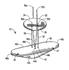
Turning to Figures 1-3, a first embodiment of the closure device of the
present invention
is illustrated. Hole (aperture) closure device 10 has a covering member or
patch 40 and a
securing member 20. The securing member 20 in this embodiment is in the form
of a disc
having substantially planar upper and lower surfaces 24, 26 respectively
although radiused
surfaces or irregular surfaces are also contemplated. Covering member or patch
40 is
dimensioned and configured for positioning inside the vessel on the internal
side of the vessel
aperture against the internal wall of the vessel; the securing member 20 is
configured to be
positioned outside the vessel wall adjacent or contiguous the external side of
the vessel aperture.
Covering member 40, preferably elongated in configuration as shown, is
retained in a
delivery sheath in a longitudinal position for delivery to the vessel, and
then pivots to a
transverse position within the vessel lumen (substantially perpendicular to an
axis extending
through the aperture) for orientation to cover (patch) the vessel aperture on
the internal side.
This movement is illustrated in Figures 37A-37D of commonly assigned co-
pending patent
application serial no. 10/847,141, filed May 17, 2004, issued as U.S. Patent
No. 7,662,661
(hereinafter the `141 application).
The elongated covering member 40 functions to cover (patch) the internal
opening in the
vessel wall to prevent the egress of blood. With reference to Figure 1, the
covering member 40
is preferably somewhat oval shaped with elongated substantially parallel side
walls 42a, 42b and
end walls 49a, 49b, illustratively curved, connecting the side walls 42a, 42b.
Other shapes of the
covering member are also contemplated. Covering member preferably has a
thicker region 43 in
the central region than the first and second end regions 45, 47. Other
dimensions are also
contemplated.
The longitudinal axis of covering member 40 defines a lengthwise dimension and
transverse axes define a shorter widthwise dimensions. The widthwise dimension
of the
6
CA 02737628 2011-04-19
covering member 40 can be for example about 2.5 mm (for a 6 Fr device). In a
preferred
embodiment, the covering member 40 is about 3.1 mm in widthwise dimension.
Other
dimensions are also contemplated. The width preferably is at least
substantially equal to the
dimension of the internal opening in the vessel wall to effectively cover the
opening. In a
preferred embodiment, the covering member 40 has a length of about 8.6 mm (in
a 6 French
system). Other dimensions are also contemplated.
It should be appreciated that alternatively the covering member could be
provided with
an enlarged width region as illustrated in the embodiment of Figure 1 of the
`141 application.
The covering member could also be configured asymmetrically so that the
enlarged region is off-
centered to accommodate widening of the aperture as the member is pulled at an
angle. The
covering member could also be configured in a paddle shape with a narrowed
region adjacent a
wider region as in Figures 9B-9E of the '141 application. Other covering
member configurations
including those disclosed in the `141 application could be utilized with the
securing members of
the present application.
The elongated covering member can be composed of materials such as
polycarbonate or
polyurethane. Preferably it is composed of resorbable materials such as
lactide/glycolide
copolymers that after a period of time resorb in the body. If composed of
resorbable material,
the covering member could optionally have regions of varying resorbability.
Varying degrees of
resorbability can be achieved for example by utilizing different materials
having differing
resorbable characteristics or by varying the mass of the covering member
(increased mass
increases resorbtion time).
Securing member 20 is preferably composed of resorbable material. The securing
member can be composed of a material having a plurality of pores extending
therethrough. This
can include a mesh, braid, or weave for example. It can also include a more
solid material
having pores formed therethrough. Materials include Polydioxanone (PDO),
Polylactic acid
(PLA), Polyglycolic Acid (PGA), Poly(lactic-co-glycolic acid) (PLGA),
Polyhydroxyalkanoates
(PHA), and Polycaprolactone (PCL), although other materials are contemplated.
It could also be
made of non-absorbable polymeric or metallic material.
7
CA 02737628 2011-04-19
When the securing member 20 is released from the delivery instrument, it is
spaced
further from the covering member 40. It is configured to then be advanced
toward the covering
member 40. More specifically, securing member 20 is fixedly secured to
flexible connecting
members illustratively in the form of suture 30 and 32. Sutures 30, 32 are
preferably made of
polymeric material and are preferably resorbable, composed of a material such
as
polydioxanome. It is also contemplated that alternatively a metallic material
could be utilized. It
is also contemplated that a single suture could be utilized to advance the
covering member.
As shown, suture 30 has a free end 30a and an opposite end 30b secured to
securing
member 20 by molding, gluing, forming a knot, or other methods. Similarly,
suture 32 has a free
end 32a and an opposite end 32b secured to securing member 22 in a similar
manner. The
sutures 30, 32 are looped through the covering member 40. Other methods of
attachment are
also contemplated. For example, the sutures can be attached to covering member
by a loop of
suture as shown for example in Figure 8 of co pending patent application
12/854,988, filed
August 12, 2010 (hereinafter the "'988 application").
To advance the securing member 20 toward the vessel wall W (and covering
member
40), the free end 30a, 32a of each suture is pulled proximally (in a direction
of the arrows of
Figure 3, thereby moving the securing member 20 in the opposite direction
closer to the aperture
and vessel wall. The pores of securing member 20 facilitate advancement toward
the covering
member 40 as tissue can enter between the pores as it is advanced. Once
tightened against the
tissue, a sufficient retention force is maintained, i.e. a proximal pulling
force on the covering
member 40 to pull it slightly upwardly (proximally) against the vessel wall.
The securing
member 20 therefore helps to prevent the covering member 40 from separating
from the vessel
wall (e.g. moving in the direction toward the opposing vessel wall) which
could create an
unwanted gap between the covering member 40 and the vessel opening to allow
blood flow. The
extent to which the securing member 20 moves toward the wall (and thus the
distance from the
vessel wall in its final placement position) will depend on the tissue
thickness. Thus, the closure
device 10 can adjust for different tissue thicknesses and apply a constant
retention force
regardless of tissue thickness.
8
CA 02737628 2011-04-19
The delivery instrument for inserting the closure device extends through an
opening in
the skin, through the tissue tract to the vessel, through an external opening
in the vessel wall,
through the aperture in the vessel wall, and through an internal opening on
the internal side of the
vessel wall into the vessel lumen.
The covering member 40 is outside a retainer tube and within a delivery sheath
in a tilted
position in a manner similar to Figures 2 and 3 of the `988 application. The
covering member 40
emerges from the sheath and moves from a tilted and preferably a somewhat
straightened
positioned, (substantially aligned with the longitudinal axis of the sheath)
to a transverse position
within the vessel (see the orientation of Figure 3). (Note the vessel wall is
shown in Figure 3 but
the rest of the vessel and tissue are removed for clarity.) The securing
member 20 remains
within the tube in a tilted somewhat straightened position. In some
embodiments, the securing
member 20 can be retained within the tube in a folded or compressed
configuration. Note the
covering member 40 can be ejected by a pusher (not shown) contacting the side
or top wall of the
covering member 40.
As shown in Figure 3, covering member 40 is pulled proximally to abut the
internal
opening on the internal side of the vessel W to cover (patch) the opening and
the sutures 30, 32,
extend through the opening A in the vessel wall. The securing member 20 is
ejected from the
sheath by advancing the securing member 20, retracting the sheath or relative
movement of both
to free the securing member 20 from the confines of the sheath. As noted
above, in the delivery
position, the securing member 20 is preferably in a tilted position (not
shown) to minimize the
transverse dimension of the delivery system and tilts to a transverse
deployment position when
exposed from the delivery sheath. As also noted above, the securing member 20
can
alternatively or additionally be held in a folded or compressed position.
Then, to retain the covering member 40 in position against the vessel wall to
block blood
flow therethrough, sutures 30 and 32 are pulled proximally from their free
ends 30a, 32a, in the
direction of arrows of Figure 3 thereby advancing the securing member 20
distally in the
direction toward the aperture A, vessel wall W and covering member 40. As
shown, the securing
member 20 can be moved to a position contiguous to the vessel wall, or
depending on tissue
thickness, may be adjacent the wall with some tissue interposed between the
securing member 20
9
CA 02737628 2011-04-19
and vessel wall. The securing member 20 in this position applies a proximal
(upward) force on
the elongated covering member 40 to limit movement of the covering member into
the vessel.
The covering member 40 has a first pair of holes 44a, 44b and a second pair of
holes 46a,
46b. The first pair of holes 44a, 44b receive suture 32 and the second pair of
holes 46a, 46b
receive suture 30. Holes 44b, 46b have a smaller diameter than holes 44a, 46a,
respectively.
The larger hole 46a is dimensioned to receive suture 30 for free unrestricted
movement of the
suture 30 therethrough and therefore easier application of securing member 20.
Similarly, the
larger hole 44a is dimensioned to receive suture 32 for free unrestricted
movement of the suture
32 therethrough. Smaller hole 46b is dimensioned to frictionally engage suture
30 so that tension
is applied to the suture 30. It is dimensioned so that the suture 30 can be
pulled through the hole
46b if sufficient force is applied by pulling on free end 30a, but if such
predetermined force is
not applied, the suture 30 will remain frictionally engaged within the wall of
the hole 46b and not
move, and thus securing member 20 will not move. Hole 44b operates similarly
with respect to
suture 32, allowing movement if a predetermined force is applied but remain
frictionally engaged
if such force is not applied. In this manner, when the user ceases pulling on
free ends 30a and
32a of sutures 30, 32 respectively, the securing member 20 will remain in
position. Figure 2
shows how the suture 30 is looped through the respective opening.
To enhance the retention of the suture of the present invention within the
smaller
diameter hole, a plurality of internal teeth can be provided. This is shown
for example in Figures
22 and 23 of the `988 application wherein hole 496a' has a plurality of teeth
497 formed on the
interior wall of the smaller opening. Engagement of the suture 430' by the
teeth 497 retains the
suture and retainer. Note that the teeth can be formed to angle inwardly so
the suture can be
moved in only one direction, i.e. proximally so the retainer is advanced
toward the covering
member.
The alternate embodiment of Figure 4 of the present invention is identical to
the
embodiment of Figure 1 except for the configuration of the securing member.
Thus, closure
device 100 has a covering member or patch 140 identical to patch 40, sutures
(flexible
connecting members) 130, 132 (with free ends 130a, 132a) identical to sutures
30, 32 and
openings 146a, 146b and 144a, 144b identical to openings 46a, 46b, 44a, and
44b. Therefore,
CA 02737628 2011-04-19
further detail of these components and their function, for brevity, will not
be repeated herein.
The securing member 120 differs from securing member 20 of Figure 1 in that it
is substantially
rectangular in shape. Securing member 120 can optionally have substantially
planar upper and
lower surfaces 124, 126, although other surfaces can be provided, e.g. curved,
concave, convex,
etc. The pores in securing member 120 facilitate movement in the same manner
as described
above. The securing member 120 can be made of the same materials as discussed
above with
respect to securing member 20. Sutures 130, 132 advance securing member 120
toward covering
member 140 in the same manner as sutures 30, 32 discussed above.
The alternate embodiment of Figure 6 is identical to the embodiment of Figure
1 except
for the configuration of the securing member. Thus, closure device 200 has a
covering member
or patch 240 identical to patch 40, sutures (flexible connecting members) 230,
232 (with free
ends 230a, 232a) identical to sutures 30, 32 and openings 246a, 246b and 244a,
244b identical to
openings 46a, 46b, 44a, and 44b. Therefore, further detail of these components
and their
function, for brevity, will not be repeated herein. The securing member 220 is
shaped similarly
to patch 240 (except inverted) with a thicker central region 225 and
substantially parallel side
walls 227, 229 connected by radiused walls 226a, 226b. An opening 223 in
central region 225
facilities advancement as tissue can enter the opening 223 as securing member
220 is advanced.
Additional openings or pores could also be provided to facilitate movement.
The securing
member 220 can be made of the same materials as discussed above with respect
to securing
member 20. Sutures 30, 32 advance securing member 220 toward covering member
240 in the
same manner as sutures 230, 232 discussed above.
In the embodiment of Figure 5, hole closure device 300 has a covering member
or patch
340 substantially identical to patch 40 and a securing member 320 with pores
similar to securing
member 120 of Figure 3 to facilitate advancement. Openings 346a, 344a are
larger than
openings 346b, 344b. The openings are dimensioned to receive sutures (flexible
connecting
members) 330, 332. That is, suture 330 extends through openings 346a and 344b
and suture 332
extends through openings 344a, 344b. Sutures 330, 332, have free ends 330a,
332a, respectively.
Openings 344b and 346b have a smaller dimension to frictionally engage the
suture as described
above with respect to openings 44b, 46b of Figure 1. First and second
retainers 360, 370 are
spherical shaped and positioned proximally of securing member 320, but it is
also contemplated
11
CA 02737628 2011-04-19
other shaped retainers could be utilized, e.g. cylindrical, pill shaped, etc.
Optionally one retainer
could be provided. Thus, securing member 320 is interposed between the
retainers 360, 370 and
patch 340. The sutures 330, 332 are attached at one end to retainers 360, 370,
respectively and
can extend through pores in the securing member 320 to loop through covering
member 340.
The securing member 320 remains external of the vessel opening and further
functions as an
extravascular component to block the retainers 360, 370 from extending through
the vessel
opening into the vessel. Proximal force applied to sutures 330, 332 at their
free ends 330a, 332a
advances retainers 360, 370 toward covering member 340 in the same manner as
sutures 30, 32
of Figure 1 advance securing member 20, due to their attachment to retainers
360, 370 at their
opposite end. As retainers 360, 370 are advanced toward covering member 340
they force
securing member 320 toward covering member 340 due to their engagement
(abutment) with the
proximal surface 321 of securing member 320.
In the embodiment of Figure 7, a single retainer 420 is provided in the form
of a
substantially cylindrical shaped member having pores to facilitate movement as
described above.
Member 420 is attached to a first portion of suture (flexible connecting
member) 430 by a suture
loop 432 extending through the pores or through openings in retainer 420 and
looped as shown.
Suture 430 extends through a channel in member 420 and extends through large
opening 444a in
covering member or patch 440, exiting smaller opening 444b, terminating in
free end 430a.
Thus, free end 430a of suture 430 is pulled proximally, pulling retainer 420
toward covering
member 440, with the smaller opening 444b frictionally retaining the suture
430 in the same
manner as opening 44b in Figure 1 to restrict movement.
While the above description contains many specifics, those specifics should
not be
construed as limitations on the scope of the disclosure, but merely as
exemplifications of
preferred embodiments thereof. Those skilled in the art will envision many
other possible
variations that are within the scope and spirit of the disclosure as defined
by the claims appended
hereto.
12