Note: Descriptions are shown in the official language in which they were submitted.
CA 02737653 2016-04-15
CA 02737653 2011-03-17
WO 2010/034021 PCT/IJS2009/057918
- 1 -
FLOW RESTORATION SYS l'E,MS AND METHODS FOR USE
RELATED APPLICATIONS
FIELD OF THE INVENTION
The present invention relates generally to apparatus for treating obstructive
material,
e.g., thrombus, stenosis, and/or unwanted material within a body lumen of a
patient, e.g.,
within a tubular graft, aorto-venous fistula, blood vessel, and the like. More
particularly,
the present invention relates to apparatus for removing or otherwise capturing
thrombus or
other obstructive material within a body lumen, and to methods for making and
using such
apparatus.
BACKGROUND
Flow within a blood vessel or other body lumen within a patient's vasculature
may
become constricted or ultimately interrupted for a variety of reasons. For
example, a vessel
may gradually narrow due to inflammation and/or cell proliferation. In
addition, thrombus
may faun due to such narrowing or other flow problems within a vessel.
One approach to removing unwanted material, e.g., thrombus, that is adherent
to a
vessel wall may involve advancing a device, e.g., a Fogarty embolectomy
balloon, to a point
beyond the adherent material blockage, expanding the device to the dimension
of the vessel
interior, and then withdrawing the expanded device back with the intent to
sweep the
adherent material out of the vessel. While this approach is frequently
successful, there are
some instances where the adherent material does not release from the vessel
wall and stays
within the vessel even after multiple passes.
Another approach to removing the adherent material is to advance a rotating
structure that can abrade the surface of the adherent material or become
entangled in the
adherent material, thereby forcing the adherent material to release from the
vessel wall. For
example, the Arrow Treratola device has several helical wires that expand
radially outward
to contact the vessel wall. These wires are spun at a high speed via a
driveshaft connected
CA 02737653 2011-03-17
WO 2010/034021
PCT/US2009/057918
- 2 -
to an electric motor in the hand piece of the device. During operation, the
Treratola device
rubs against the inside wall of the vessel as it is advanced. Upon engaging
adherent
material, the device abrades the inside surface of that material, and in many
cases, the
device may break through the interface between the adherent material and the
vessel wall.
In this event, the adherent material can be peeled off the vessel wall and
become wrapped
around the helical wires of the Treratola device.
While this may address the immediate goal of removing the adherent material
from
the vessel wall, it is often difficult to remove the material from the vessel
itself since the
Treratola device does not offer any method to unwind or aspirate the material.
As the
Treratola device is removed from the vessel, it typically passes through a
close-fitting
orifice such as an introducer sheath. Any material that is wound around the
Treratola
device is typically pushed off as it enters the sheath, and such material thus
remains in the
vessel.
Accordingly, apparatus and methods for removing material from aorto-venous
grafts, blood vessels, or other body lumens would be useful.
SUMMARY
The present invention is directed to apparatus for treating a body lumen of a
patient,
e.g., a tubular graft, aorto-venous fistula, blood vessel, and the like. More
particularly, the
present invention is directed to apparatus for removing or otherwise capturing
thrombus or
other obstructive material within a body lumen, and to methods for making and
using such
apparatus.
In accordance with one embodiment, a system is provided for removing
obstructive
material from a body lumen. The system includes an outer tubular member
comprising a
proximal end, a distal end, and a lumen extending between the proximal and
distal ends.
Optionally, an annular expandable occlusion member may be provided on the
outer tubular
member distal end. The system also includes a macerator device insertable
through the
lumen and comprising an elongate shaft, an expandable cage coupled to a distal
end of the
elongate shaft, and a constraint tube having a distal opening with a sharpened
edge, wherein
the shaft and the cage are axially moveable relative to the constraint tube.
Optionally, the
constraint tube may be fixedly coupled to an inner surface of the outer
tubular member or
the constraint tube may be movable independently of the outer tubular member.
CA 02737653 2011-03-17
WO 2010/034021 PCT/US2009/057918
- 3 -
The cage may include a plurality of apertures, and the distal sharpened edge
of the
constraint tube may be configured for shearing off material that protrudes
through the
apertures as the cage is proximally withdrawn into the constraint tube.
Optionally, the inner
surface of the cage may include a plurality of inwardly protruding barbs. The
cage may
include distal protruding structures and/or, thick struts and thin struts
connecting the thick
struts together. Optionally, one or more control wires may be coupled to the
distal
protruding structures of the cage, wherein the control wire(s) may be
configured for
drawing the protruding structures together into a closed configuration when
the cage is in an
expanded configuration. Optionally, a driveshaft may be operably coupled to
the cage for
causing the cage to rotate during advancement through the body lumen or the
system may
include an actuator for manually rotating the cage. In exemplary embodiments,
the distal
protruding structures may have a smooth edge, a slotted edge, or a serpentine
edge.
In an exemplary embodiment, the system may further include an elongate
treatment
member comprising an expandable treatment element selectively expandable for
directing
the obstructive material within the body lumen into the cage when the cage is
in an
expanded configuration. The elongate treatment member may be insertable
through a
lumen in the macerator device shaft. Alternatively, the elongate treatment
member may be
insertable through the outer tubular member lumen adjacent to the macerator
device shaft.
Optionally, in this alternative, the cage may have an uninterrupted path or
other opening
through which the elongate treatment member may pass.
In accordance with another embodiment, a method is provided for removing
obstructive material from a body lumen that includes introducing an outer
tubular member
into the body lumen, the outer tubular member including a lumen and a distal
opening. A
macerator device may be introduced through the outer tubular member lumen into
the body
lumen. In an exemplary embodiment, the macerator device may include an
elongate shaft,
an expandable cage coupled to a distal end of the elongate shaft, and a
constraint tube
having a distal opening. The expandable cage may be deployed out of the
constraint tube
distal opening by distally advancing the elongate shaft relative to the
constraint tube, and
expanded within the body lumen. Obstructive material may be captured within
the cage,
and then the cage may be proximally withdrawn into the constraint tube.
Material that
protrudes through apertures in the cage as the cage collapses may be sheared
off, e.g., by a
sharpened edge of the constraint tube distal opening, In addition or
alternatively, sheared off
material may be aspirated into the outer tubular member distal opening.
Optionally, the
CA 02737653 2011-03-17
WO 2010/034021
PCT/US2009/057918
- 4 -
method may also include expanding an occlusion element on the outer tubular
member
distal end, e.g., to prevent obstructive material from passing proximally
beyond the distal
end of the outer tubular member.
In an exemplary embodiment, the method may further include introducing an
elongate treatment member including a distal expandable treatment element
through the
outer tubular member lumen and into the body lumen such that the expandable
treatment
element, in a collapsed configuration, is positioned distal to the obstructive
material and the
cage is positioned proximal to the obstructive material. The expandable
treatment element
may be expanded and proximally withdrawn towards the expanded cage such that
obstructive material is withdrawn into the cage by the expandable treatment
element.
In another exemplary embodiment, the method may also include advancing the
expanded cage towards the obstructive material and rotating the cage during
advancement
until the obstructive material becomes entangled in a distal portion of the
cage. Rotation of
the cage may cause obstructive material to be separated from the vessel wall,
and the cage
may be withdrawn into the constraint tube. The withdrawal may release the
obstructive
material from the cage distal portion and/or withdraw the obstructive material
into the
constraint tube. Optionally, the cage may be re-deployed, expanded, and/or
withdrawn, one
or more additional times, e.g., to separate and/or withdraw obstructive
material into the
cage.
In accordance with another embodiment, an apparatus is provided for removing
obstructive material from a body lumen that includes an outer tubular member
including a
proximal end, a distal end sized for introduction into a body lumen, and a
lumen extending
between the proximal and distal ends; an elongate shaft including proximal and
distal ends
and movable axially within the tubular member lumen; and an expandable
macerator cage
including a first end attached to the distal end of the shaft and a second
free end. The cage
is expandable from a collapsed configuration when the cage is disposed within
the tubular
member lumen and an expanded configuration when the cage is deployed from the
tubular
member lumen.
In one embodiment, the cage includes a tubular structure including a wall
extending
between the first and second ends, the second end defining an opening
communicating with
an interior of the cage in the expanded configuration for capturing
obstructive material
within the interior of the cage. The wall may include a plurality of struts
and/or apertures
such that, when the cage is withdrawn back into the tubular member lumen after
capturing
CA 02737653 2011-03-17
WO 2010/034021
PCT/US2009/057918
- 5 -
obstructive material therein, the distal end of the tubular member slidably
engages the wall
of the cage or otherwise separates obstructive material captured by the cage
that extends
through the apertures and the cage is compressed back towards the collapsed
configuration.
In accordance with still another embodiment, a system is provided for removing
obstructive material from a body lumen that includes an outer tubular member
including a
proximal end, a distal end sized for introduction into a body lumen, and a
lumen extending
between the proximal and distal ends; an obstruction device deployable from
the tubular
member to a location beyond obstructive material intended to be removed, the
obstruction
device including an expandable member on a distal end thereof; and a macerator
device.
The macerator device may include an expandable cage carried on a distal end of
a shaft and
a constraint tube for maintaining the cage in a collapsed configuration, e.g.,
to allow the
macerator device to be introduced into the body lumen through the tubular
member lumen.
The cage may be deployable from a distal end of the constraint tube and
expandable to an
expanded configuration within a body lumen. In one embodiment, the cage may
include an
open end communicating with an interior of the cage in the expanded
configuration for
capturing obstructive material within the interior of the cage.
Other aspects and features of the present invention will become apparent from
consideration of the following description taken in conjunction with the
accompanying
drawings.
BRIEF DESCRIPTION OF THE DRAWINGS
It will be appreciated that the exemplary apparatus shown in the drawings are
not
necessarily drawn to scale, with emphasis instead being placed on illustrating
the various
aspects and features of the illustrated embodiments.
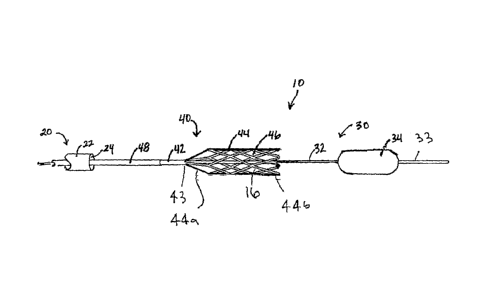
FIG. 1 is a side view of an exemplary embodiment of a flow restoration system
including an occlusion device and a macerator device deployable from an access
sheath.
FIG. 2A is a cross-sectional side view of an access sheath that may be
included in
the system of FIG. 1, including an expandable member on a distal end of the
access sheath.
FIG. 2B is a cross-sectional side view of an alternative embodiment of an
access
sheath that may be included in the system of FIG. 1, including an expandable
member on a
distal end of the access sheath.
FIG. 3 is a detail of an inner surface of a cage of a macerator device that
may be
included in the system of FIG. 1.
CA 02737653 2011-03-17
WO 2010/034021 PCT/US2009/057918
- 6 -
FIGS. 4A and 4B are cross-sectional side views of exemplary embodiments of a
constraint tube that may be included in a macerator device in the system of
FIG. 1.
FIG. 5 is a cross-sectional side view of an alternative embodiment of an
access
including an integral constraint tube that may be included in the system of
FIG. 1.
FIG. 6 is a side view of another exemplary embodiment of a flow restoration
system
including an occlusion device and a macerator device deployable from an access
sheath.
FIGS. 7-10 are cross-sectional views of a body lumen within a patient's body,
showing a method for removing obstructive material within the body lumen.
FIGS. 11A and 11B are perspective views of another exemplary embodiment of a
macerator device cage in expanded and collapsed configurations, respectively.
FIG. 12A is a top view of a flat pattern that may be incorporated into the
macerator
device cage of FIGS. 11A and 11B.
FIG. 12B is a detail of a portion of the flat pattern of FIG. 12A.
FIG. 13 is a perspective detail of the macerator device cage shown in FIGS.
11A and
11B being rotated in a direction A.
FIGS. 14A and 14B are cross-sectional views of another exemplary embodiment of
a macerator device cage that includes control wires for directing a distal end
of the cage
between an open configuration and a closed configuration, respectively.
FIG. 14C is a cross-sectional view of an alternative embodiment of the
macerator
device cage of FIGS. 14A and 14B that includes control wires and a sleeve
around the
control wires for directing the cage between an open configuration and a
closed
configuration.
FIGS. 15A-15C are side views of alternative embodiments of distal tips that
may be
provided on a macerator device cage, such as the cage shown in FIGS. 11A and
11B.
FIGS. 16A-16H are cross-sectional views of a body lumen within a patient's
body,
showing another method for removing obstructive material within the body
lumen.
DETAILED DESCRIPTION OF THE EXEMPLARY EMBODIMENTS
Turning to the drawings, FIG. 1 shows an exemplary embodiment of an apparatus
10
for treating a body lumen, e.g., for removing thrombus, clots, objects,
debris, and/or other
unwanted or obstructive material from within the body lumen, such as a blood
vessel, aorto-
venous fistula, tubular graft, and the like. Generally, the apparatus 10
includes an outer
access sheath or other tubular member 20, and, optionally, an obstruction
device 30 and a
CA 02737653 2011-03-17
WO 2010/034021 PCT/US2009/057918
- 7 -
macerator device 40, which together may provide a flow restoration system,
e.g., for
removing obstructive material from within body lumens in a patient's body. In
addition,
such a system may include one or more additional components not depicted,
e.g., one or
more guidewires, syringes or other sources of inflation media and/or vacuum,
and the like.
The sheath 20 may be an elongate tubular body, e.g., an introducer or
procedure
sheath, including a proximal end (not shown), a distal end 22 sized for
introduction into a
body lumen, and a lumen 24 extending between the proximal end and the distal
end 22. The
sheath 20 may be configured for percutaneous placement within a body lumen,
e.g.,
including a rounded or otherwise substantially atraumatic tip to facilitate
advancement into
and/or along body lumens within a patient's body.
The sheath 20 may have a substantially uniform construction along its length,
or
alternatively, the construction may be varied. For example, a proximal portion
of the sheath
may be substantially rigid or semi-rigid to facilitate advancement of the
apparatus 10 by
pushing or otherwise manipulating the proximal end. In addition or
alternatively, a distal
15 portion of the sheath 20 may be flexible, e.g., to facilitate bending
and/or advancement
through tortuous anatomy without substantial risk of kinking or buckling. In
exemplary
embodiments, the sheath 20 may be formed from materials such a metal, plastic,
e.g.,
PEEK, Grilamed L25, and the like, or composite materials. The sheath 20 may
have a
length between about five and one hundred thirty centimeters (5-130 cm) and an
outer
20 diameter between about 1.6 to 2.0 millimeters, and the lumen 24 may have
a diameter
between about 1.4 and 1.8 millimeters.
Optionally, the sheath 20 may include a handle or hub on the proximal end (not
shown). The handle may be shaped to facilitate holding or manipulating the
apparatus 10 or
individual components of the apparatus 10, as described further below. In
addition, the
handle may include a port communicating with the lumen 24, e.g., for infusing
fluid into the
lumen 24 and/or aspirating material from the lumen 24, e.g., around the
macerator device 40
and/or obstruction device 30. For example, a syringe, vacuum line, and the
like may be
coupled to the port for aspirating obstructive material received within the
lumen 24 of the
sheath 20 and/or disposed adjacent the distal end 22 within a body lumen, as
described
further below.
Optionally, the sheath 20 may include an expandable member or other occlusion
element carried on the distal end 22, e.g., to stabilize the sheath 20 within
a body lumen
and/or to seal the body lumen from fluid flow past the distal end 22 during a
procedure. For
CA 02737653 2011-03-17
WO 2010/034021 PCT/US2009/057918
- 8 -
example, FIG. 2A shows a sheath 20' including an expandable member 26' on its
distal end
22' within a body lumen 50. The expandable member 26' may be a compliant
balloon, e.g.,
formed from compliant material that may expand elastically proportional to the
amount of
inflation media delivered into the balloon 26,' a semi-compliant balloon, or a
non-compliant
balloon, e.g., a PTA balloon, if desired. In this embodiment, the sheath 20'
may include an
inflation lumen (not shown) extending from the proximal end of the sheath 20'
to the distal
end 22' and communicating with an interior of the balloon 26.' A source of
inflation media
and/or vacuum, e.g., a syringe with saline or other fluid (not shown), may be
coupled to the
proximal end of the sheath 20' for delivering inflation media into the balloon
26' via the
inflation lumen and/or aspirating fluid from the balloon 26,' e.g., to
facilitate collapsing the
balloon 26' after a procedure. Alternatively, the expandable member 26' may be
mechanically or otherwise expandable, e.g., including an expandable frame or
other
structure within or otherwise coupled to a membrane (not shown).
The expandable member 26' may be expandable from a low profile, collapsed
configuration, e.g., disposed against the outer surface of the sheath 20' to
facilitate
introduction of the sheath 20,' and a high profile, expanded configuration,
e.g., to engage or
otherwise contact an inner surface of a body lumen 50 within which the sheath
20' is
introduced. In the expanded configuration, the expandable member 26' may
provide a
substantially fluid tight seal within the body lumen 50, e.g., to prevent
substantial
physiologic flow along the body lumen 50, which may otherwise allow particles
of loose
material to move past the sheath 20 into other parts of the patient's body
where they may
cause harm. In addition or alternatively, the expandable member 26' may also
substantially
secure and/or stabilize the sheath within the body lumen 50, e.g., to prevent
inadvertent
movement of the sheath 20' within the body lumen 50 during treatment.
During use, the expandable member 26' may be maintained in the low profile
configuration when the sheath 20' is introduced, and then expanded to the high
profile
configuration once the sheath 20' is positioned within the body lumen 50 being
treated. The
expandable member 26' may remain expanded as obstructive material is removed
from the
body lumen 50 via the sheath 20' or other component of the apparatus 10, as
described
further below. Once the body lumen 50 is sufficiently treated, the expandable
member 26'
may be collapsed to restore physiologic flow within the body lumen 50.
In an alternative embodiment shown in FIG. 2B, an access sheath 20" may be
provided that includes an expandable member 26" that extends beyond the distal
end 22" of
CA 02737653 2016-04-15
- 9 -
the sheath 20" to provide an expandable distal tip for the sheath 20." Thus,
the expandable member
26" may provide a conical or other transition extending from an enlarged
distal tip towards the lumen
24" of the sheath 20," e.g., such that maceration of obstructive material may
be performed within or
adjacent the distal tip of the expandable member 26" if desired.
During maceration, particles of obstructive material may be liberated within
or adjacent the lumen 24"
of the sheath 20," thereby facilitating aspiration of the particles from the
body lumen 50 into the
lumen 24." Additional information on the access sheath 20" and/or expandable
member 26" may be
found in WO/2010/017537, filed August 8, 2009.
Referring back to FIG. 1, the obstruction device 30 generally includes a shaft
or other elongate
member 32 including a proximal end (not shown), a distal end 33 sized for
introduction through the
sheath 20, e.g., via the lumen 24, and carrying an expandable obstruction or
treatment member 34 on
the distal end 33. Generally, the obstruction member 34 is expandable from a
collapsed configuration
(not shown), e.g., sized for introduction through the lumen 24 of the sheath
20, and an expanded
configuration (shown in FIG. 1) for engaging or otherwise contacting a wall of
a body lumen within
which the obstruction member 34 is expanded. In an exemplary embodiment, the
obstruction
member 34 may be a balloon, e.g., a compliant, semi-compliant, or non-
compliant balloon,
expandable to a substantially spherical or cylindrical shape. In this
embodiment, the shaft 32 may
include an inflation lumen (not shown) in communication with an interior of
the obstruction member
34, e.g., for selectively expanding and collapsing the obstruction member 34.
Optionally, the
obstruction member 34 may include a core wire and/or helical structure (not
shown), e.g., such that
the obstruction member may adopt a helical shape in the expanded
configuration. Exemplary devices
that may be used for the obstructive device 30 are disclosed in Patent Serial
No. 8,043,313, filed July
2, 2009. Alternatively, the obstruction member 34 may include a frame or other
mechanically
expandable structure (not shown), if desired.
With continued reference to FIG. 1, the macerator device 40 generally includes
a shaft or other
elongate member 42 including a proximal end (not shown), a distal end 43 also
sized to fit
within the lumen 24 of the sheath 20, and an expandable cage 44 carried on the
distal end 43.
Optionally, the shaft 42 may include a lumen or other track (not shown) for
slidably receiving the
obstruction device 30 therethrough, as described further below. In
CA 02737653 2011-03-17
WO 2010/034021
PCT/US2009/057918
- 10 -
addition or alternatively, the shaft 42 may include one or more additional
lumens (not
shown), e.g., for receiving a guidewire or other rail (also not shown), one or
more actuator
wires or cables (also not shown), and the like, as described further below.
As shown, the cage 44 is an open or porous expandable structure including a
closed
proximal or first end 44a coupled to the shaft 42 and an open distal or second
end 44b, e.g.,
to accommodate receiving obstructive material within the cage 44, as described
further
below. Generally, the cage 44 includes a plurality of struts 44a extending
between the first
and second ends 44a, 44b and/or around a periphery of the cage 44, thereby
defining a
cylindrical or other tubular outer wall including a plurality of apertures 46,
e.g., at least
adjacent the first end 44a. The struts 44a and/or apertures 46 may be sized to
accommodate
expansion and/or collapse of the cage 44 and/or to define a desired pore size
that prevents
particles larger than the desired pore size from escaping once captured within
the cage 44,
as described further below.
The cage 44 is expandable from a low-profile, collapsed configuration (not
shown),
e.g., to accommodate introduction through the sheath 20, and a high-profile,
expanded
configuration (shown in FIG. 1) where the cage 44 expands radially outwardly,
e.g., to
contact the wall of a body lumen within which the cage 44 is deployed and/or
expanded.
Optionally, as shown in FIG. 3, the cage 44 may include a plurality of barbs
or other
features 41 projecting radially inwardly from the struts 44a, e.g., to engage
and/or provide
additional traction with obstructive material captured within the cage 44, as
discussed
further below.
The cage 44 may be formed from a variety of materials, e.g., capable of
elastically
or plastically moving between the collapsed and expanded configurations one or
more
times. For example, the cage 44 may be formed from elastic or superelastic
materials, e.g.,
metals, such as stainless steel, Nitinol, and the like, plastics, or composite
materials. In an
exemplary embodiment, the cage 44 may be formed from a tube with portions of
the tube
removed to define the struts 44a and/or apertures 46, e.g., by laser cutting,
etching,
mechanical cutting, and the like. Alternatively, the cage 44 may be formed
from a sheet
also with portions removed to define the struts 44a and/or apertures 46, e.g.,
by laser
cutting, etching, mechanical cutting, stamping, and the like, which may be
rolled into a
tubular shape with edges of the sheet attached together, e.g., by welding,
soldering, bonding
with adhesive, fusing, and the like.
CA 02737653 2011-03-17
WO 2010/034021 PCT/US2009/057918
- 11 -
The cage 44 may then be attached to the shaft 42, e.g., by substantially
permanently
attaching the closed end 44a around the distal end 43 of the shaft 42, e.g.,
by crimping,
bonding with adhesive, fusing, wrapping a collar, wire or other material
around the closed
end 44a, and the like. Thus, the closed end 43 may be fixed in the collapsed
configuration,
while the rest of the cage 44 may be free to expand from the collapsed
configuration to the
expanded configuration. In an exemplary embodiment, the cage 44 may be formed
from
superelastic material that may be heat treated to program the expanded
configuration into
the cage 44, while allowing the cage 44 to be resiliently compressed and
maintained in the
collapsed configuration.
Thus, in the embodiment shown in FIG. 1, the cage 44 may be a self-expanding
structure, e.g., resiliently compressible radially inwardly to the collapsed
configuration yet
biased to expand towards the expanded configuration. Alternatively, the cage
44 may be
mechanically expanded and collapsed, e.g., using an actuator (not shown) on
the proximal
end of the macerator device 40 coupled to the cage 44.
To maintain a self-expanding cage 44 in the collapsed configuration, e.g.,
during
introduction through or before deployment from the sheath 20, the macerator
device 40 may
include a constraint tube 48 slidably disposed around the shaft 42. The
constraint tube 48
may be an elongate tubular body including a proximal end (not shown), a distal
end 48a,
and a lumen 49 extending therebetween that is sized to receive the shaft 42
and cage 44 with
the cage 44 in the collapsed configuration. Alternatively, other removable
constraints may
be provided around the cage 44 to maintain the cage 44 in the collapsed
configuration until
it is desired to deploy and expand the cage 44 within a body lumen, e.g., one
or more
removable wires wound around the cage 44, a tear-away sleeve, and the like
(not shown).
The distal end 48a of the constraint tube 48 may be sized to be slidably
disposed
within the sheath 20, e.g., to accommodate introduction of the macerator
device 40 through
the lumen 24 of the sheath 20. The constraint tube 48 and the shaft 42 and
cage 44 may be
movable axially relative to one another, e.g., to allow the cage 44 to be
retracted within the
constraint tube 48 and/or deployed from the constraint tube 48. Thus, the
constraint tube 48
may maintain the cage 44 in the collapsed configuration, e.g., during
introduction into a
body lumen through the sheath 20, and allow the cage 44 to be deployed from
the constraint
tube 48 such that the cage 44 assumes the expanded configuration.
The proximal ends (not shown) of the shaft 42 and/or constraint tube 48 may
extend
or otherwise be coupled to the proximal end of the sheath 20 and may be
actuatable from
CA 02737653 2016-04-15
- 12 -
the proximal end of the sheath 20. For example, the sheath 20 may include a
handle or hub (not
shown) on its proximal end, which may include one or more actuators for
advancing the macerator
device 40 from the distal end 22 of the sheath 20 and/or for deploying the
cage 44 from and covering
the cage 44 with the constraint tube 48. The shaft 42 and/or constraint tube
48 may extend into the
handle, or one or more cables, wires, rods, or other actuator elements (not
shown) may be
coupled between the shaft 42 and/or constraint tube 48 and one or more
actuators on the handle.
For example, a first actuator, e.g., a slider, button, dial, and the like, may
be provided on the handle
(not shown) to advance and/or retract the entire macerator device 40 relative
to the sheath 20, e.g.,
to deploy the cage 44 from the distal end 22 of the sheath 20 while still
covered by the constraint
tube 48. A second actuator, e.g., another slider, button, dial, and the like
(also not shown), may then
be activated to expose the cage 44, e.g., by advancing the shaft 42 and cage
44 relative to the
constraint tube 48 or retracting the constraint tube 48 without substantial
movement of the cage 44.
Exemplary handles and/or actuators that may be provided on the apparatus 10
are disclosed in Patent
Serial No. 8,043,313.
Alternatively, the sheath 20 and macerator device 40 may be structurally
separate devices, and the
macerator device 40 may be introduced into the sheath 20, e.g., via a port or
other opening in the
proximal end of the sheath 20. For example, a handle or hub (not shown) may be
provided on the
proximal end of the sheath 20 that includes a port (also not shown)
communicating with the
lumen 24 that may accommodate introduction of the macerator device 40 and/or
other devices
therein. Optionally, the port may include one or more seals, e.g., a
hemostatic seal, that may
accommodate receiving the macerator device 40 therein while providing a
substantially fluid-tight
seal to prevent bodily fluids from escaping from the lumen 24. In this
alternative, the macerator
device 40 itself may include a handle or hub (not shown) on its proximal end
that includes one or
more actuators (also not shown) for manipulating the shaft 42 and cage
relative to the constraint
tube 48, similar to the actuators described above.
Optionally, in a similar manner, the obstruction device 30 may be coupled to
the sheath 20 and/or
macerator device 40, e.g., with one or more actuators (not shown) on a handle
of the apparatus 10
for deploying and/or withdrawing the obstruction device 30. Alternatively, the
obstruction device 30
may be a separate device from the sheath 20 and/or
CA 02737653 2011-03-17
WO 2010/034021 PCT/US2009/057918
- 13 -
macerator device 40, and the macerator device 40 may include a port for
receiving the
obstruction device 20, e.g., similar to the port described above.
Optionally, as shown in FIGS. 4A and 4B, the constraint tube 48 may be
configured
to facilitate removing excess material captured by the cage 44. For example,
the distal end
48a of the constraint tube 48 may include one or more features that slide or
otherwise
interact with the cage struts 44a, e.g., to trim excess material that extends
out of the cage
apertures 46 when the cage 44 is withdrawn back into the constraint tube 48
after capturing
obstructive material within the cage 44. Thus, as the cage 44 enters the
constraint tube 48
and is compressed towards the collapsed configuration, the features on the
distal end 48a of
the constraint tube 48 may shear or otherwise trim excess material that
protrudes out of the
cage apertures 46.
In an exemplary embodiment, shown in FIG. 4A, the distal end 48a of the
constraint
tube 48 may include a sharpened edge 47 extending around a distal opening 43
communicating with the lumen 49 that is suitable for cutting off excess
obstructive material
that may protrude through the apertures 46 of the cage 44. In the embodiment
shown in
FIG. 4A, the sharpened edge 47 may be a single-ground edge, e.g., that may
shear along the
outer surface of the cage 44 during withdrawal to cut or otherwise separate
obstructive
material extending through the apertures 46. Alternatively, as shown in FIG.
4B, a
constraint tube 48' may be provided that includes a double-ground sharpened
edge 47'
extending around distal opening 43.' The double-ground sharpened edge 47' may
cut
excess obstructive material similarly to the single-ground edge 47, but may be
more
resistant to damage as the cage 44 passes into the lumen 49' of the constraint
tube 48.' For
example, since the cutting edge 47' has a slightly larger diameter than the
lumen 49' of the
constraint tube 48' itself, the cutting edge 47' may not contact the outer
surface of the cage
44 during withdrawal of the cage 44 but remain spaced slightly apart from the
cage 44.
In an alternative embodiment, shown in FIG. 5, the constraint tube 48 shown in
FIGS. 4A and 4B may be omitted, and a sheath 60 may be used to constrain the
cage 44 (not
shown) in the collapsed configuration during introduction and/or to remove
excess material
during withdrawal of the cage 44. Similar to the previous embodiments, the
sheath 60
includes a proximal end (not shown), a distal end 62, and a lumen 64 extending
therebetween. Unlike the previous embodiments, the sheath 60 includes a
constraint tube
48" incorporated into the distal end 64, e.g., within the lumen 64. The
constraint tube 48"
may be a relatively short tubular body extending a short distance into the
lumen 64 and
CA 02737653 2011-03-17
WO 2010/034021 PCT/US2009/057918
- 14 -
including an exposed and sharpened edge 47" around a distal opening 43," e.g.,
a single-
ground or double-ground edge, similar to the previous embodiments. The
constraint tube
48" may be substantially permanently attached within the lumen 64 or otherwise
to the
distal end 62, e.g., by bonding with adhesive, interference fit, fusing, sonic
welding, and the
like, thereby providing a transition from the distal opening 43" into the
lumen 64.
During use, the cage 44 may be advanced from the lumen 64 and out of the
distal
opening 43" whereupon the cage 44 may freely expand towards the expanded
configuration.
After unwanted material is captured within the cage 44 (as described further
elsewhere
herein), the cage 44 may be withdrawn back into the sheath 60 through the
distal opening
43," whereupon excess obstructive material extending through the apertures 46
of the cage
44 may be cut or otherwise separated by the sharpened edge 47"as the cage 44
collapses.
In a further alternative, the sheath 20 shown in FIG. 1, may be used to
constrain the
cage 44 during introduction and/or withdrawal and the constraint tube 48 may
be omitted
entirely. Unlike previous embodiments, however, when the cage 44 is withdrawn
into the
sheath 20, the cage 44 may not be collapsed to as small a size due to the
relatively larger
diameter of the sheath 20 compared to a constraint tube 48 introduced through
the sheath
20. In this alternative, a portion of the obstructive material may remain
within the cage 44
after withdrawal into the sheath 20. In other words, the macerator device cage
44 may be
used to capture and remove the obstructive material without trimming off
excessive
obstructive material. Alternatively, the sheath 20 itself may include a
sharpened distal edge
(not shown) or a sharpened tip may be attached to the distal end 22 of the
sheath 20 (also
not shown), e.g., to cut off excess obstructive material during withdrawal of
the cage 44,
while obstructive material within the cage 44 is withdrawn into the sheath 20
within the
cage 44.
With further reference to FIG. 1, in the embodiment shown, the shaft 42 of the
macerator device 40 includes a lumen that slidably receives the shaft 32 of
the obstruction
device 30, e.g., such that the macerator device 40 and the obstruction device
30 have a
concentric, telescoping arrangement relative to one another. Alternatively, as
shown in
FIG. 6, an apparatus 10' may be provided in which a macerator device 40 and an
obstruction device 30 are provided in a side-by-side arrangement relative to
an outer sheath
20. In this alternative, the sheath 20, obstruction device 30, and macerator
device 40 may
be constructed similar to the embodiments described above. Optionally, the
macerator
device 40 may include a cage 44 that includes an uninterrupted path 45 defined
by struts
CA 02737653 2011-03-17
WO 2010/034021
PCT/US2009/057918
- 15 -
44a of the cage 44 that extends axially along at least a portion of the cage
44 to
accommodate the obstruction device 30 passing through the cage 44 while
allowing the
cage 44 to close substantially completely to the collapsed configuration.
In this embodiment, the obstruction device 30 and macerator device 40 may be
received in a common lumen 24 of the sheath 20, as shown in FIG. 6.
Alternatively, the
sheath 20 may include separate lumens (not shown) disposed adjacent one
another, each for
receiving one of the obstruction device 30 and the macerator device 40.
Otherwise structure
and operation of the apparatus 10' may be similar to that described with
reference to
apparatus 10 of FIG. 1.
Turning to FIGS. 7-10, an exemplary method is shown for removing material,
e.g.,
thrombus or other obstructive material 52, within a body lumen 50. The body
lumen 50
may be a blood vessel, aorto-venous fistula, tubular graft, xenograft, and the
like, e.g.,
within a patient's arm that communicates between an adjacent vein and artery.
Alternatively, the apparatus and methods described herein may be used to treat
other
locations within a patient's body, e.g., within the patient's vasculature or
other body
lumens. Although apparatus 10 shown in FIG. 1 is shown and described in
association with
FIGS. 7-10, it will be appreciated that the methods described herein may be
performed
using any of the apparatus and systems described herein.
Generally, the method may involve trapping thrombus or other obstructive
material
between an expanded obstruction member 34 and an expanded cage 44, e.g., such
that the
material may be captured by the cage 44, broken into smaller particles,
removed within the
cage 44, and/or aspirated from the body lumen 50 through sheath 20. Initially,
as shown in
FIG. 7, the sheath 20 may be introduced into a body lumen 50, e.g.,
percutaneously from an
entry site using conventional methods, and manipulated to position the distal
end 22 of the
sheath 20 within the body lumen 50 adjacent to and spaced apart from
obstructive material
52. Optionally, if the sheath 20 includes an expandable member, e.g., balloon
26, 26' as
shown in FIGS. 2A or 2B, on the distal end 22, the expandable member (not
shown) may be
expanded any time after the distal end 22 is placed at a desired position
within the body
lumen 50, e.g., to prevent subsequent movement of the sheath 20 and/or to
substantially seal
the body lumen 50 from fluid flow proximally past the sheath 20.
In addition or alternatively, aspiration may be applied to the lumen 24 of the
sheath
20, e.g., at any time after introducing the distal end 22 of the sheath 20
into the body lumen
50. For example, a syringe or vacuum line may be coupled to the proximal end
of the
CA 02737653 2011-03-17
WO 2010/034021 PCT/US2009/057918
- 16 -
sheath 20 and activated to apply a substantially continuous vacuum to the
lumen 24 to draw
loose material within the body lumen 50 into the lumen 24.
The obstruction device 30 may then be introduced into the body lumen 50 from
the
sheath 20 and advanced past the material 52 with the obstruction member 34 in
the low-
profile configuration (not shown). For example, the obstruction device 30 may
be loaded
into the lumen 24 of the sheath 20 and advanced through the length of the
sheath 20 into the
body lumen 50, or the obstruction device 30 may be integral with or preloaded
into the
sheath 20 before the procedure and merely deployed from the sheath 20.
Optionally, a
distal tip of the obstruction device 30 may be sufficiently small and/or sharp
to pass freely
through the material 52 and/or may be rounded or otherwise substantially
atraumatic to pass
along the wall of the body lumen 50 past the material 52. Once the obstruction
member 34
is positioned distally beyond the material 52, the obstruction member 34 is
expanded to the
high-profile condition, as shown in FIG. 7.
Next, with reference to FIG. 8, the macerator device 40 may be deployed from
the
sheath 20, e.g., over or adjacent the shaft 32 of the obstruction device 30.
The macerator
device 40 may be advanced until the cage 44 (maintained in the collapsed
configuration
within the constraint tube 48) is disposed proximal or adjacent to the
material 52 opposite
the expanded obstruction member 34. Thus, the obstructive material 52 may be
bounded by
the obstruction member 34 on one side and the macerator device 40 on the
other. Once
positioned within the body lumen 50, the cage 44 may be expanded within the
body lumen
50, e.g., by deploying the cage from the constraint tube 48, whereupon the
cage 44 may
resiliently expand radially outwardly to contact the wall of the body lumen,
as shown in
FIG. 8.
Turning to FIG. 9, the obstruction device 30 may then be retracted proximally
towards the cage 44 to pull material 52 within the body lumen 50 towards and
into the
macerator device cage 44, as shown. Alternatively, the cage 44 may be advanced
towards
the obstruction device 30 to capture material 52 within the cage 44, with the
obstruction
member 34 preventing distal migration of the material 52 away from the cage
44. As
described above, if the cage 44 includes barbs 41, e.g., as shown in FIG. 3,
the barbs 41 may
partially penetrate or otherwise engage with the material 52 captured within
the cage 44,
e.g., to prevent migration of the material 52 relative to the cage 44.
Optionally, the obstruction device 30 and/or macerator device 40 may include a
locking mechanism, e.g., one or more cooperating detents, tabs, or other
features (not
CA 02737653 2011-03-17
WO 2010/034021 PCT/US2009/057918
- 17 -
shown), that may substantially secure the obstruction device 30 relative to
the cage 44 when
the obstruction device 30 has been placed a predetermined distance from the
cage 44, e.g.,
substantially adjacent the cage 44 such that the obstruction device 30
substantially encloses
the material 52 within the cage 44, as shown in FIG. 9. Alternatively, a
locking mechanism
may be provided on a proximal end of the apparatus 10, e.g., on a handle (not
shown),
which may be locked and unlocked to selectively secure the obstruction device
30 relative
to the cage 44. With the locking mechanism engaged, the obstruction device 30
may not be
directed distally away from the cage 44, e.g., such that subsequent movement
of the
obstruction device 30 is coupled to movement of the cage 44.
Turning to FIG. 10, the macerator device cage 44 and expanded obstruction
member
34 may then be directed proximally towards the sheath 20, e.g., until the cage
44 enters the
constraint tube 48. If a vacuum has not been applied previously, a source of
vacuum may
be activated to aspirate material released within the body lumen 50 into the
lumen 24 of the
sheath 24, as shown. As the cage 44 is drawn into the constraint tube 48, the
cage 44 may
be compressed radially inwardly, thereby forcing portions of the material 52
through the
apertures 46 in the cage 44. The portions of the material 52 exposed through
the apertures
46 may be sheared off, e.g., by the sharpened distal edge 47, 47' (not shown,
see FIGS. 4A
and 4B) of the constraint tube 48, reducing the material 52 into many smaller
particles 53
within the body lumen 50. The loose particles 53 may be removed from the body
lumen 50,
e.g., by aspiration, through the sheath lumen 24, as shown. Notably, the
reduced particle
size may be a function of the size of the apertures 46 in the cage 44. Thus,
the size of the
cage apertures 46 may be chosen to reduce the particle size of the material 52
to a desired
maximum cross-section, e.g., such that the reduced diameter particles 53 may
be reliably
removed though the sheath 20 without substantial risk of occluding the sheath
lumen 24.
In one embodiment, the cage 44 may be compressed to a collapsed configuration
as
the cage 44 is withdrawn into the constraint tube 48 in which the interior
space of the cage
44 is minimized, thereby squeezing substantially all of the captured material
52 through the
apertures 46 of the cage 44. The extruded and/or sheared particles 53 may then
be aspirated
into the lumen 24 of the sheath 20. Alternatively, the cage 44 may have
sufficient interior
space in the collapsed configuration such that at least some captured material
may remain
within the cage 44 when the cage 44 is withdrawn fully into the constraint
tube 48.
With the cage 44 withdrawn fully into the constraint tube 48, the macerator
device
and obstruction device 30 may be withdrawn into the sheath 20 and the
apparatus 10
CA 02737653 2011-03-17
WO 2010/034021 PCT/US2009/057918
- 18 -
removed from the patient's body. Alternatively, the obstruction member 34 may
be
collapsed and the obstruction device 30 advanced through another section of
obstructive
material (not shown) within the body lumen 50. In this alternative, the
macerator device 40
may then be redeployed to capture and remove the material, e.g., by repeating
the steps
described above. Optionally, the entire apparatus 10 may be introduced into
another body
lumen (not shown), and the obstruction device 30 and macerator device 40
redeployed to
capture and/or remove obstructive material in other regions of the patient's
body, if desired.
Once sufficient material has been removed, the obstruction member 34 of the
obstruction device 30 may be collapsed and the obstruction device 30 may be
withdrawn
into the macerator device 40 or into the sheath 20 if the macerator device 40
has already
been withdrawn into the sheath 20. The aspiration within the sheath 20 may be
discontinued, the expandable member on the sheath 20 may be collapsed (if
provided on the
sheath 20), and the sheath 20 may be withdrawn from the body lumen 50.
Turning to FIGS. 11A-13, another embodiment of a macerator cage 144 is shown
that may be included in any of the apparatus and/or systems described herein.
Generally,
the macerator cage 144 includes a closed proximal or first end 144a and an
open distal or
second end 144b, similar to the previous cage 44. The cage 144 may include a
plurality of
struts 116, 118 extending between the first and second ends 144a, 144b and/or
around a
periphery of the cage 144, thereby defining a cylindrical or other tubular
outer wall
including a plurality of apertures 146, similar to the previous cage 44.
The closed end 144a of the cage 144 may include a collar portion 141, which
may be
attached to a macerator device shaft 42 (not shown, see, e.g., FIG. 1), while
the open distal
end 143 of the cage 144 may include a plurality of distally protruding
elements or distal tips
112. For example, FIG. 11A shows the cage 144 in an expanded configuration in
which the
collar portion 141 remains compressed, e.g., due to its attachment to a shaft
(not shown),
and the cage 144 defines a substantially continuous diameter extending from
the closed end
144a to the open end 144b. FIG. 11B shows the cage 144 in a compressed
configuration,
e.g., in which the cage 144 may be constrained or otherwise compressed around
the shaft
and/or within a constraint tube (also not shown).
Unlike the cage 44 of FIG. 1, as can be best seen in FIGS. 12A and 12B, the
cage
144 includes at least two different types of struts 116, 118. For example, the
cage 144 may
include a plurality of relatively thick struts 116 that extend substantially
continuously along
a length of the cage 144, e.g., in a first helical configuration between the
first and second
CA 02737653 2011-03-17
WO 2010/034021 PCT/US2009/057918
- 19 -
ends 144a, 144b. In addition, the cage 144 may include a plurality of
relatively thin struts
118, which may connect adjacent thick struts 116 together. As shown, the thin
struts 118
are not substantially continuous as are the thick struts 116, but may extend
in a
discontinuous pattern helically and/or circumferentially around the cage 44.
Optionally, the
thin struts 118 may also have bends or other features, e.g., relatively
thinned or perforated
portions, that allow the struts 118 to bend relatively easily compared to the
thick struts 116.
The apertures 146 may be defined by the spaces between the thick struts 116
and the thin
struts 118, thereby defining a desired pore size for the cage 144. The cage
144 may be
formed using similar materials and methods as those previously described
above.
The distal tips 112 on the open end 144b of the cage 144 may provide a
substantially
atraumatic distal end for the cage 144, e.g., to prevent puncture or other
damage to a wall of
a body lumen within which the cage 144 is deployed. In addition or
alternatively, the distal
tips 112 may be sufficiently flexible to allow the distal tips 112 to twist
helically and/or
interlock with one another during use. FIGS. 15A-15C show alternative
configurations of a
distal tip that may be provided on the cage 144, e.g., to facilitate engaging
and/or removing
obstructive material within a body lumen. For example, FIG. 15A shows an
exemplary
embodiment of a distal tip 112a that includes a substantially straight
configuration with a
smooth leading edge 125.
Alternatively, FIG. 15B shows another exemplary embodiment of a distal tip
112b
that includes a series of slots or indentations 126 spaced apart along a
length of the distal tip
112b, e.g., that may allow the distal tips 112b to entangle with each other
and/or with the
obstructive material captured or otherwise engaged by the distal tips 112b to
facilitate
removal, as described further below with reference to FIGS. 16A-16H. For
example, when
the cage 144 is rotated, the distal tips 112b and obstructive material may be
wound together,
e.g., such that portions of other distal tips 112b and/or obstructive material
may enter the
slots 126 and the distal tips 112b become interlocked with one another. FIG.
15C shows yet
another exemplary embodiment of a distal tip 112c that includes a serpentine
pattern 127.
In this embodiment, the internal bends 128 of the serpentine pattern 127 may
provide
regions where other distal tips 112c and/or obstructive material may become
entangled, e.g.,
compared to providing a smooth edge 125, as shown in the distal tip 112a of
FIG. 15A.
One advantage of the cage 144 shown in FIGS. 11A-13 is that the cage 144 may
facilitate deploying the cage 144 and/or advancing the cage 144 into or
through obstructive
material within a body lumen. In contrast, the cage 44 shown in FIG. 1 is
generally
CA 02737653 2011-03-17
WO 2010/034021 PCT/US2009/057918
- 20 -
maintained substantially stationary upon deployment within a body lumen, e.g.,
while the
obstruction device 40 is retracted to withdraw obstructive material into the
cage 44. For
example, the cage 144 of FIGS. 11A-13 may facilitate pulling material 52 back
into the
open end 144a of the cage 144 and/or separating obstructive material from a
wall of the
body lumen 50.
During distal advancement, the cage 144 may be concurrently advanced and
rotated,
e.g., manually or using a driveshaft connected to an electric motor in a
handle (not shown)
of the apparatus 10 (see, e.g., FIG. 1). This may cause the distal tips 112 of
the cage 144 to
track along the inside wall of the body lumen50, e.g., in a helical manner as
the cage 144 is
advanced. When thrombus or other obstructive material is encountered, the
distal tips 112
may pass between the material 52 and the wall of the body lumen 50, thereby
positioning
the material 50 inside the cage 144.
The distal tips 112 of the cage 144 may facilitate separation and/or capture
of
material within the cage 144. For example, the edges of the distal tips 112
may provide
distal leading edges of the cage 144 that are not a substantially smooth
cylinder but define
an undulating surface. Consequently, the distal tips 112 of the cage 144 may
act as a saw
by repeatedly making contact with the material 52 as the cage 144 is rotated,
which may
increase the chance of material 52 being dislodged from the wall of the body
lumen 50
and/or captured within the cage 144. To further ensure that the leading edge
of the cage 144
passes between the unwanted material and the wall of the body lumen 50, the
distal tips 112
and/or edges of the struts 116, 118 may also act as blades shearing along the
wall of the
body lumen 50 to draw adherent material into the cage 144. Thus, the struts
116, 118 may
cut or otherwise separate the interface between the body lumen 50 and the
obstructive
material 52.
The distal tips 112 may be formed such that they conform substantially to the
cylindrical shape of the cage 144, e.g., defining a diameter similar to the
rest of the
expanded cage 144, although alternatively the distal tips 112 may be biased
radially
outwardly, e.g., to ensure that the distal tips 112 pass between the wall of
the body lumen 50
and the obstructive material 52 and/or enhance engagement of the distal tips
112 against the
wall of the body lumen 50. Alternatively, the distal tips 112 may by biased to
extend
radially inwardly, e.g., laterally inwardly, relative to a central
longitudinal axis of the
apparatus 10, e.g., to prevent substantial risk of damage to the wall of the
body lumen 50.
CA 02737653 2011-03-17
WO 2010/034021 PCT/US2009/057918
- 21 -
In addition, the different thicknesses and/or shapes of struts 116, 118 may
provide a
cage 144 that responds in different ways depending upon which direction the
cage 40 is
rotated. For example, arrow "A" in FIG. 13 represents a first direction for
rotation of the
cage 144. If resistance is encountered in the portions of the cage 144
contacting the wall of
the body lumen 50 (not shown) during this rotation, torsion may occur. Because
the thick
struts 116 have a relatively high resistance to bending and the thin struts
118 are easily bent,
the torsion may not bend the thick struts 116, but may cause the thin struts
118 to bend to
define a greater angle between the adjacent struts 116, 118 and expand the
cage 144 radially
outwardly. This may increase the contact force between the macerator cage 144,
e.g.,
leading edges of the thick struts 116, and the wall of the body lumen 50,
which may
increase the chance that obstructive material being removed from the wall of
the body
lumen 50 and being captured within the cage 144. Optionally, the leading edges
of the thick
struts 116 may include sharpened edges or other features, which may enhance
cutting or
other engagement with adherent material within the body lumen 50.
If the cage 144 is rotated in a second direction opposite to "A," the torsion
may
cause the thin struts 118 to bend due to their low column strength to reduce
the angle
between the adjacent struts 116, 118, and the cage 144 may not expand radially
outwardly
in the same manner as the first direction. This anisotropy with respect to
rotational
direction may be useful because the cage 144 may be advanced and rotated in
the "A"
direction to engage and separate adherent obstructive material from a vessel
wall, e.g.,
causing the material to enter into the cage 144. The torsion may also cause
the cage 144 to
expand outwardly for better apposition or engagement with the vessel wall. If
the cage 144
encounters excessive resistance, the cage 144 may be rotated in the second
direction, e.g., to
disengage the resistance without causing radial expansion. Depending on the
stiffness
differential between the thin and thick struts 118, 116, rotation of the cage
144 in the second
direction may also cause the cage 144 to radially contract to further
facilitate
disengagement.
Turning to FIGS. 14A-14C, yet another embodiment of an expandable cage 144 is
shown, which may include an actuation mechanism for selectively opening and/or
closing
an open distal end 143 of the cage 144. For example, the distal end 143 of the
cage 144
may be substantially closed by contracting the distal tips 112 radially
inwardly, e.g., using
control elements, e.g., one or more wires or filaments 114, coupled to each of
the distal tips
112. Alternatively, a single control element (not shown) may be threaded
through each of
CA 02737653 2011-03-17
WO 2010/034021 PCT/US2009/057918
- 22 -
the distal tips 112, e.g., circumferentially and successively through holes in
the distal tips
(not shown), such that proximal tension on the control element may bend or
otherwise direct
the distal tips 122 radially inwardly. Optionally, a locking mechanism (not
shown) may be
provided for securing the distal tips 122 in the closed orientation, if
desired. Alternatively,
the distal tips 122 may interlock with one another, e.g., as described above,
to secure the
distal tips 122 in the closed orientation.
FIG. 14A shows the distal end 143 open with the cage 144 in the expanded
configuration, e.g., with the control elements 114 relaxed and the distal tips
112 biased to a
substantially axial, open configuration. FIG. 14B shows the distal tips 112
bent or
otherwise directed inwardly towards a closed configuration, e.g., after
proximal tension is
applied to the control elements 114. The structure of the distal tips 112 may
facilitate this
bending by providing one or more preferential bending features, e.g., thinned
strut widths,
thinned strut thicknesses, perforated portions, and the like (not shown), to
provide hinged
regions of the distal tips 112. In an alternative embodiment, shown in FIG.
14C, a tubular
member 113 may be advanced over the control elements 114 to cause the distal
tips 112 to
bend radially inwardly.
The closed configurations shown in FIGS. 14B and 14C may allow obstructive
material captured within the cage 114 to be substantially retained therein,
e.g., without the
need for an expandable obstruction member 34, as described elsewhere herein.
In these
alternatives, the cage 144 may simply be withdrawn into a constraint tube or
access sheath
(not shown), thereby compressing the cage 144 radially inwardly. With the
distal tips 112
closed, the captured material may not simply escape out the distal end 143 of
the cage 144,
but may remain within the cage 144 to be extruded through the apertures 146
(not shown in
FIGS. 14B and 14C) as the cage is compressed, e.g., to be aspirated, as
described above.
Alternatively, the captured material may be withdrawn into the constraint tube
or sheath
along with the cage 144, also as described above.
Turning to FIGS. 16A-16H, another exemplary method is shown for removing
obstructive material from within a vessel or other body lumen 150, e.g., using
the cage 144
shown in FIGS. 11A-13. The cage 144 may be introduced into the body lumen 150
via an
access sheath and/or constraint tube 148, similar to the previous embodiments.
As shown in
FIG. 16A, the cage 144 has been deployed and expanded within the body lumen
150 such
that the open distal end 143 is disposed adjacent to obstructive material 152
intended to be
CA 02737653 2011-03-17
WO 2010/034021 PCT/US2009/057918
- 23 -
removed. Once fully expanded, the cage 144 may be rotated and advanced within
the body
lumen 150 toward the obstructive material 152.
Turning to FIG. 16B, the distal end 143 of the cage 144 may engage the
material
152, e.g., such that the distal tips 112 pass between the material 152 and the
wall of the
body lumen 152 to some degree, but are also free to deform as they become
entangled in the
material 152. Optionally, an obstruction device (not shown) may be introduced
through the
material 152 and an obstruction member expanded beyond the material 152 before
the cage
144 is advanced. Thus, the obstruction member may prevent distal migration of
the
material 152 away from the cage 144 as the cage 144 is advanced.
With additional reference to FIG. 16C, upon further rotation of the cage 144,
the
distal tips 112 may wind at least partially around the material 152 and/or
around each other,
thereby creating a mechanical engagement between the cage 144 and the material
152.
Further rotation of the cage 144 may then be transmitted to the material 152,
which may
cause the material 152 to twist and/or otherwise detach from the wall of the
body lumen
150. After entanglement and further rotation, the entangled material 152 may
be completely
removed from the wall of the body lumen 150, as shown in FIG. 16D.
As shown in FIG. 16E, the cage 144 may then be withdrawn into the access
sheath
and/or constraint tube 148, similar to the previous embodiments, thereby
compressing the
cage 144 radially inwardly towards the collapsed configuration. Because the
distal tips 112
of the cage 144 are substantially straight and free on their distal ends (i.e.
the cage 144 is
open ended and not attached to a core wire on its distal end), the distal tips
112 may be
disengaged and the material 152 released as the cage 144 is withdrawn into the
access
sheath 20. Thus, the separated material 152 may remain loose within the body
lumen 150,
as shown.
Thereafter, as shown in FIG. 16F, the cage 144 may be redeployed from the
constraint tube 148 or sheath and expanded again with the open end 143
disposed adjacent
the loose material 152. The material 152 may then be pulled into the open end
143 of the
cage 144, e.g., using an obstruction device 134, which may be the same device
deployed
beyond the material 152 previously or a different device introduced into the
body lumen
150 and beyond the material 152 before or after redeploying the cage 144.
Turning to FIG. 16G, the cage 144 with the material 152 captured therein may
then
be withdrawn into the sheath 20 or constraint tube (not shown), similar to the
previous
embodiments. As discussed elsewhere herein, any material that protrudes
through the
CA 02737653 2011-03-17
WO 2010/034021 PCT/US2009/057918
- 24 -
apertures 146 in the cage 144 may be sheared off or otherwise separated as the
cage 144 is
compressed, e.g., to ensure that the cage 144 does not become lodged in the
tip of the sheath
20. The cage 144 may be withdrawn completely into the sheath 20, as shown in
FIG. 16H,
and any remaining loose particles of the material 152 may be aspirated through
the sheath
lumen, similar to the previous embodiments. For example, as described above, a
vacuum
may be applied by the sheath 20 within the body lumen 150 at any time to
aspirate loose
particles within the body lumen 150, e.g., released when the cage 144 is used
to separate
material from the wall of the body lumen 150 or thereafter. Any of these steps
may be
repeated as many times as desired to remove any remaining material.
It will be appreciated that elements or components shown with any embodiment
herein are exemplary for the specific embodiment and may be used on or in
combination
with other embodiments disclosed herein.
While the invention is susceptible to various modifications, and alternative
forms,
specific examples thereof have been shown in the drawings and are herein
described in
detail. It should be understood, however, that the invention is not to be
limited to the
particular forms or methods disclosed, but to the contrary, the invention is
to cover all
modifications, equivalents and alternatives falling within the scope of the
appended claims.