Note: Descriptions are shown in the official language in which they were submitted.
CA 02737873 2011-03-21
WO 2010/038020 PCT/GB2009/002332
- 1 -
IMMUNOGENIC PEPTIDES
Field of the Invention
The present invention relates to immunogenic peptides and their various
applications. In
particular the invention relates to immunogenic peptides derived from the
PASD1 protein and
their use in therapeutic, diagnostic and prognostic methods.
Background of the Invention
Tumour-associated antigens (TAAs), recognized by the immune system of a cancer
patient,
may represent important immunotherapeutic targets. Evidence in support of this
has been
provided by autologous bone marrow transplantation and donor lymphocyte
infusion studies,
demonstrating that donor cells can recognize and respond to TAAs in a variety
of
haematological malignancies such as multiple myeloma and myeloid leukaemia
(Bellucci et
al 2004, Porter et al 2006, Atanackovic et al 2007). Furthermore, vaccination
studies have
reported an increased immune response to TAAs (Rezvani et al 2007, Schmitt et
al 2008). It
is also of note that the immune response signature has been identified as
being of
importance in predicting survival in diffuse large B-cell lymphoma (DLBCL) and
follicular
lymphoma (FL) (Dave et al 2004, Monti et al 2005).
TAAs that are of current interest for improving treatment regimens are the
cancer testis
antigens (CTAs). Their restricted normal tissue distribution but widespread
expression in
tumours makes them attractive immunotherapeutic targets, while minimizing
potential
problems with autoimmunity (Scanlan et al 2004, Simpson et al 2005, Suri
2006). Initially,
studies of CTA expression focussed on solid tumours (Simpson et a! 2005), but
there are
increasing reports of CTAs being expressed in haematological malignancies such
as multiple
myeloma (Pellat-Deceunynck et al 2000, Chiriva-Internati et a/ 2001, Lim et al
2001, Sugita
et al 2004, van Rhee et al 2005, Goodyear et al 2005, Jungbluth et al 2005)
and myeloid
malignancies (Adams et al 2002, Zhang et al 2003, Andrade et a/ 2008, Tinguely
et a/ 2008).
Indeed, a gene expression profiling study reported transcripts of multiple
CTAs in myeloma
tumour cells (Condomines et a! 2007). Other studies have also reported the
presence of
cytotoxic T cells (CTLs), considered to be the major effector cells in
cellular immunity, to
CTAs such as NY-ESO-1 and Sp17 in the peripheral blood of multiple myeloma
patients,
thereby suggesting the presence of spontaneous immunity to these CTAs (van
Rhee et al
2005, Goodyear eta! 2005). There. is also accumulating evidence for a major
role for CD4+
CA 02737873 2011-03-21
WO 2010/038020 PCT/GB2009/002332
2 -
T-helper (TH) cells not only in the regulation and maintenance of the CTL and
humoural
responses but also in the ability of the TH themselves to control tumour cell
growth
(Oestrand-Rosenberg et a! 2005), Goodyear et al 2008). A subsequent
investigation has
shown that this immunity can be boosted through vaccination with antigens such
as NY-
ESO-1 (Baumgaertner et a/ 2006, Odunzi et al 2007) and clinical trials are
ongoing using
CTAs as vaccine targets (Szmania et a/ 2006, Odunzi et al 2007).
The present inventors previously used the SEREX technique, which exploits the
circulating
antibodies present in the serum of patients, to identify the PAS (Per ARNT
Sim) domain
containing 1 (PASD1) protein or CT63, encoded by a gene at Xq28, as a lymphoma-
associated antigen and candidate CTA (Liggins et a/ 2004a, Liggins et a/
2004b). Two splice
variants were identified, PASD1a (639 amino acids) and PASD1b (773 amino
acids). The
first 638 amino acids are common to both proteins (Liggins eta! 2004a). This
work is
described by International Patent Application Publication No. WO 03/082916,
which is
incorporated by reference in its entirety.
The production of monoclonal antibodies to PASD1 allowed confirmation of this
molecule as
a novel CTA with a highly restricted expression pattern in normal tissues and
more
specifically as a CT-X antigen expressed in a range of haematological
malignancies (Cooper
et a/ 2006, Sahota et a! 2006).
Summary of the Invention
The present invention relates to immunogenic peptides derived from PASD1. The
invention
thus provides an immunogenic peptide of from about 9 to about 25 amino acids
in length
comprising at least 9 consecutive amino acids of the amino acid sequence of
any of SEQ ID
Nos. 1 to 10 or 27.
In certain preferred embodiments, the immunogenic peptide is capable of
stimulating a T-cell
response. Preferably, the peptide is capable of producing a cytotoxic T
lymphocyte (CTL)
response.
In these embodiments, the immunogenic peptide may be between 9 and 12 and in
particular
either 9 or 10 amino acids in length. The peptide may comprise, consist
essentially of, or
consist of the amino acid sequence of any one of SEQ ID Nos. 1 to 5.
Preferably, the
CA 02737873 2011-03-21
WO 2010/038020 PCT/GB2009/002332
3 -
peptide comprises, consists essentially of, or consists of the amino acid
sequence of any one
of SEQ ID Nos. 1,2or5.
In other embodiments, the peptide is capable of producing a T helper (TH) cell
response.
In these embodiments, the peptide may be of from about 18 to about 25 amino
acids in
length. Preferably, the peptide is 20 amino acids in length. The peptide may
comprise,
consist essentially of, or consist of the amino acid sequence of any one of
SEQ ID Nos. 6 to
10. Preferably, the peptide comprises, consists essentially of, or consists of
the amino acid
sequence of any one of SEQ ID Nos. 6, 7 or 10.
In certain embodiments, the peptide may be capable of producing both a CTL and
a TH cell
response.
In other embodiments, the present invention relates to a nucleic acid encoding
an
immunogenic peptide of the invention as described herein. Preferably the
nucleic acid
comprises, consists essentially of, or consists of the nucleotide sequence of
any one of SEQ
ID Nos. 11 to 20.
The present invention also provides an expression vector comprising a nucleic
acid
described herein. A host cell or organism transformed or transfected with such
an
expression vector is also provided.
A transgenic non-human organism comprising a transgene encoding an immunogenic
peptide of the present invention is also provided.
A vaccine comprising an immunogenic peptide of the invention, a nucleic acid
of the
invention, an expression vector of the invention or a host cell of the
invention is also
provided.
The present invention also relates to an isolated T-cell specific for an
immunogenic peptide
as described herein. Furthermore, the present invention relates to an isolated
T-cell
produced by stimulating peripheral blood mononuclear cells (PBMCs) with an
immunogenic
peptide of the invention as described herein.
CA 02737873 2011-03-21
WO 2010/038020 PCT/GB2009/002332
4 -
The present invention also relates to the T-cell receptor (TCR) sequence
specific for an
immunogenic peptide of the invention as described herein.
In certain embodiments, the isolated T-cell is a cytotoxic T lymphocyte (CTL)
specific for an
immunogenic peptide of the invention as described herein.
In other embodiments, the isolated T-cell is a T helper (TH) cell specific for
an immunogenic
peptide of the invention as described herein.
The present invention also relates to pharmaceutical compositions comprising
an
immunogenic peptide of the invention, a nucleic acid, an expression vector or
a host cell
described herein and a pharmaceutically acceptable carrier or excipient.
The pharmaceutical compositions of the invention may comprise an immunogenic
peptide
capable of stimulating a CTL response and an immunogenic peptide capable of
stimulating a
TH response for simultaneous, sequential or separate administration.
The pharmaceutical compositions of the invention may comprise two or more of
an
immunogenic peptide, a nucleic acid, an expression vector or a host cell as
described herein
for simultaneous, sequential or separate administration.
In a further aspect, the present invention relates to an immunogenic peptide,
a nucleic acid,
an expression vector, a host cell, a vaccine, an isolated T-cell, or a
pharmaceutical
composition as described herein for use in therapy.
Preferably, the immunogenic peptide, nucleic acid, expression vector, host
cell, vaccine,
isolated T-cell, or pharmaceutical composition described herein is for use in
the treatment of
cancer.
The present invention also relates to the use of the immunogenic peptide,
nucleic acid,
expression vector, host cell, vaccine, isolated T-cell, or pharmaceutical
composition
described herein in the manufacture of a medicament for the treatment of
cancer.
In certain embodiments, the cancer is either a haematologically derived
malignancy selected
from multiple myeloma, mantle cell lymphoma, Hodgkin's lymphoma, T-cell
lymphomas,
follicular lymphoma (FL), Burkitt's lymphoma, T-cell rich B cell lymphoma,
diffuse large B-cell
CA 02737873 2011-03-21
WO 2010/038020 PCT/GB2009/002332
- 5 -
lymphoma (DLBCL) and acute and chronic myeloid leukaemia, or a non-
haematologically
derived malignancy selected from brain, melanoma, lung, breast, gastric,
kidney, prostate,
testicular, ovarian, uterine, colorectal and liver cancers and adenocarcinoma
of the colon.
In yet another aspect, the present invention relates to a method of treatment
of cancer,
comprising administering a therapeutically effective amount of an immunogenic
peptide, a
nucleic acid, an expression vector, a host cell, a vaccine, an isolated T-
cell, or a
pharmaceutical composition as described herein to a patient in need thereof.
The present invention further relates to a method of treatment of cancer,
comprising the
steps of:
(a) isolating a cell population containing or capable of producing CTLs and/or
TH
cells from a subject;
(b) treating the cell population with an immunogenic peptide(s) described
herein
optionally together with a proliferative agent;
(c) screening the cell population for CTLs and/or TH cells with specificity to
an
immunogenic peptide(s) described herein;
(d) administering the cell population to a patient suffering from cancer.
In certain embodiments, the CTLs and/or TH cells with specificity to an
immunogenic
peptide(s) described herein are isolated from the cell population and
administered to a
patient suffering from cancer.
In a further aspect, the present invention relates to a method of treatment of
cancer,
comprising the steps of:
(a) isolating a cell population containing or capable of producing CTLs and/or
TH
cells from a subject;
(b) treating the cell population with an immunogenic peptide(s) described
herein
optionally together with a proliferative agent;
(c) screening the cell population for CTLs and/or TH cells with specificity to
an
immunogenic peptide(s) described herein;
(d) cloning the T-cell receptor (TCR) genes from the CTLs and/or TH with
specificity
to the immunogenic peptide(s) described herein;
(e) transducing the TCR gene cloned in step (c) into either:
i. cells from the patient;
ii. cells from a donor; or
CA 02737873 2011-03-21
WO 2010/038020 PCT/GB2009/002332
6 -
iii. eukaryotic or prokaryotic cells for the generation of cell surface or
secreted
monoclonal TCRs (mTCRs); and
(f) administering the cells or mTCRs from step (e) to a patient suffering from
cancer.
In certain embodiments the subject from which the cell population is isolated
is the patient in
need of treatment (i.e. suffering from cancer). Alternatively, the cell
population may be
isolated from a normal subject or the mTCRs themselves may be administered.
Preferably the cancer is either a haematologically derived malignancy selected
from multiple
myeloma, mantle cell lymphoma, Hodgkin's lymphoma, T-cell lymphomas,
follicular
lymphoma, Burkitt's lymphoma, T-cell rich B cell lymphoma, diffuse large B-
cell lymphoma
(DLBCL) and acute and chronic myeloid leukaemia, or a non-haematologically
derived
malignancy selected from brain, melanoma, lung, breast, gastric, kidney,
prostate, testicular,
ovarian, uterine, colorectal and liver cancers and adenocarcinoma of the
colon.
In another aspect of the present invention, a method of diagnosing cancer is
provided. The
method comprises the steps of:
(a) obtaining a blood sample from a patient;
(b) screening for the presence of CTLs and/or TH cells specific for an
immunogenic
peptide described herein, wherein the presence of such cells indicates a
positive
diagnosis of cancer.
In a further aspect, the present invention relates to a method of predicting a
clinical outcome
for a patient with a cancer, comprising the steps of:
(a) isolating peripheral blood mononuclear cells (PBMCs) from a patient with a
cancer;
(b) screening said PBMCs for recognition of an immunogenic peptide described
herein;
(c) assigning a predicted positive clinical outcome to the patient where the
PBMCs
recognise the immunogenic peptide described herein or a predicted negative
clinical
outcome to the patient where the PBMCs do not recognise the immunogenic
peptide
described herein.
Preferably the cancer is either a haematologically derived malignancy selected
from multiple
myeloma, mantle cell lymphoma, Hodgkin's lymphoma, T-cell lymphomas,
follicular
lymphoma, Burkitt's lymphoma, T-cell rich B cell lymphoma, diffuse large B-
cell lymphoma
CA 02737873 2011-03-21
WO 2010/038020 PCT/GB2009/002332
7 -
(DLBCL) or acute and chronic myeloid leukaemia, or a non-haematologically
derived
malignancy selected from brain, melanoma, lung, breast, gastric, kidney,
prostate, testicular,
ovarian, uterine, colorectal and liver cancers and adenocarcinoma of the
colon.
Description of the Drawings
The present invention will be further understood by reference to the drawings.
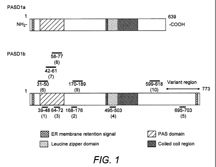
Figure 1. Schematic diagram of the PASD1 protein isoforms.
The positions of the PASD1 peptides are shown as horizontal lines : 1 =
PASD1(1); 2 =
PASD1(2); 3 = PASD1(3); 4 = PASD1(4);5 = PASD1(5); 6 = PASD1(6); 7 = PASD1(7);
8 =
PASD1(8); 9 = PASD1(9) and 10 = PASD1(10).
Figure 2. y-IFN responses of patients with de novo DLBCL (12) and transformed
DLBCL (37) to PASD1 peptides.
a) PBMCs obtained from patients 12 and 48 at time of diagnosis and after one
year from start
of treatment were maintained in short term culture. A significant y-IFN
response to peptides
PASD1(1), PASD1 (2) and PASD1(5) was observed in cells from both patients
obtained at
time of diagnosis and after one year from the start of treatment (p<0.05).
This suggests the
presence of memory T cells. No significant response was detected in cultures
stimulated by
the HIV peptide or containing medium only.
b) CTL cell lines generated after 6 weeks of culture were either enriched for
CD8-positive
cells using anti-CD8 antibody coated magnetic beads or incubated with an anti-
HLA-A2*0201
monoclonal antibody (BB7.2). A significant y-IFN response was observed only in
the culture
containing the CD8-positive cells (p<0.05). No significant responses were
detected in the
control cultures or the irrelevant peptides. The results are the mean +/- SD
and were
obtained from triplicate ELISPOT cultures.
Figure 3. Cytolytic activity of the PASDI -specific CTL cell lines derived
from patients
with DLBCL.
The functional activity of CTL cell lines obtained from patients 1, 12 (de
novo DLBCL) and
patient 48 (T-cell rich DLBCL) were studied in a conventional 51Cr release
assay on a range
of haematological cell lines. Significant dose dependent lysis of the HLA-
A*0201-positive
PASD1-positive Thiel (myeloma) cell line was observed by cells from all three
patients. In
contrast no significant lysis was observed of the SUDHL-6 (DLBCL; HLA-A*0201-
positive but
CA 02737873 2011-03-21
WO 2010/038020 PCT/GB2009/002332
8 -
PASD1-negative) or the OCI-Ly3 (DLBCL) and KM-H2 (HL; HLA-A*0201-negative but
PASD1-positive) cell lines. Results are the mean +/- SD from triplicate
cultures.
Figure 4. Immunoperoxidase labelling studies of biopsy sections from patients
with de
novo DLBCL.
a) Antibody PASD1-1 strongly stains the cytoplasm of tumour cells from HLA-
A*0201-positive
Patient 4 whose PBMCs exhibited a significant y-IFN response to PASD1
peptides. Antibody
PASD1-2 stained a subpopulation of nuclei (arrowed) as well as cytoplasm of
the tumour
cells.
b) and c) show the immunolabelling results obtained from two HLA-A*0201-
negative patients
in whom no PASD1 T-cell response was detected. Whereas the tumour cells from
Patient 27
were labelled strongly with antibody PASD1-1 b), no labelling was detected
with antibody
PASD1-2. Neither of the antibodies PASD1-1 c) or PASD1-2 (not shown) stained
the tumour
cells of Patient 17.
Figure 5. The TH y-IFN responses of patients with DLBCL to to PASDI peptides.
a) PBMCs from Patients 4 (de novo DLBCL) and 48 (T-cell rich DLBCL) were
obtained at
time of diagnosis and after one year from start of treatment were maintained
in short term
culture. A significant y-IFN response to peptides PASO1(6)and PASD1(7) was
observed in
cells from both patients obtained at time of diagnosis and after one year from
the start of
treatment (p<0.05). This suggests the presence of memory T cells. No
significant response
was detected in cultures stimulated by the HIV peptide or containing medium
only.
b) TH rich cell lines generated after 6 weeks of culture were either enriched
for CD4-positive
cells using anti-CD4 antibody coated magnetic beads or incubated with an anti-
HLA-DR
monoclonal antibody (WR18). A significant y-IFN response was observed only in
the culture
containing the CD4-positive cells (p<0.05). Abrogation of the y-IFN response
was observed
following the addition of anti-HLA-DR. No significant responses were detected
in the control
cultures or the irrelevant peptides. The results are the mean +/- SD and were
obtained from
triplicate ELISPOT cultures.
Figure 6. Cytolytic activity of the PASD1-specific TH cell lines derived from
Patient 1
with DLBCL.
The functional activity of TH cell lines specific for PASD1(6) and PASD1(7)
were studied in a
conventional 51Cr release assay on a range of haematological cell lines.
Significant dose
dependent lysis of the PASD1-positive Thiel (myeloma) and OCI-Ly3 cell lines.
In contrast no
CA 02737873 2011-03-21
WO 2010/038020 PCT/GB2009/002332
9 -
significant lysis was observed of the PASD1-negative SUDHL-6 (DLBCL) cell
line. Results
are the mean +/- SD from triplicate cultures.
Figure 7. Schematic representation of DNA fusion vaccine design.
Each vaccine contains at the NH2 terminus the leader sequence of the VH heavy
chain gene
from the BCL1 lymphoma followed by a sequence encoding the first domain (DOM1)
of
Fragment C of Tetanus toxin, including the p30 CD4+ Th epitope. The control
vaccine
contains no additional sequence whereas p.DOM-PASD1(1), p.DOM-PASD1(2) and
p.DOM-
PASD1 FL include DNA sequence encoding the HLA-A*02001-restricted CTL epitopes
PASD1(1), PASD1(2) or the full length sequence of PASD1 respectively, linked
to the COOH
terminus of DOM1.
Figure 8. DNA vaccination induces PASD138 and PASDI167 specific T-cell
responses
detectable ex vivo.
HHD mice were vaccinated with p.DOM-PASD1(1) (A), p.DOM-PASD1(2) (C), or p.DOM
DNA vaccines (B and D). Splenocytes from individual mice were harvested on day
14
following priming, and the numbers of spot-forming cells (SFCs) secreting IFNy
were
assessed ex vivo by ELISPOT assay after incubation without peptide, with an
irrelevant
peptide (1 NM), with p30 (1 NM), or with the relevant peptide (0.1 pM and 1
NM). A horizontal
bar represents group medians. Responses were considered significant if the
frequency of
IFNy-secreting cells was more than double the frequency detected in wells
without peptide.
Pooled data from two experiments with similar results.
Figure 9. DNA vaccination induced T cells are able to specifically kill in
vitro target
cells loaded with the relevant peptide.
HHD mice were vaccinated with p.DOM-PASD1(1) (A, mice 1-4), p.DOM-PASD1(2) (B,
mice
1-4), or p.DOM (A and B, Controls 1 and 2) DNA vaccines. Splenocytes were
harvested on
day 14 and cultured for 6 days with 0.1 pM of relevant peptide and IL-2 before
measuring
their CTL activity by 51Cr-release assay. The RMA-HHD target cells were either
non-loaded,
loaded with an irrelevant peptide, or with PASD1(1) or PASD1(2) peptides. The
YAC-1 cells
were used as a NK activity control target. Representative data of one of two
experiments with
the same results.
Figure 10. Boost with electroporation improves the peptide specific T-cell
responses.
HHD mice were vaccinated with p.DOM-PASD1(1) (A, E), p.DOM-PASD1(2) (C, F), or
p.DOM (B, D and controls in E and F) DNA vaccines and received a booster
injection
CA 02737873 2011-03-21
WO 2010/038020 PCT/GB2009/002332
- 10 -
immediately followed by electroporation on day 28. Splenocytes from individual
mice were
harvested 8 days later and the numbers of spot-forming cells (SFCs) secreting
IFNy were
assessed ex vivo by ELISPOT assay as described above (A-D). Splenocytes were
cultured
during 6 days with 0.1 pM of relevant peptide and IL-2 before measuring their
CTL activity by
51Cr-release assay (E and F). The target cells were the same as those used in
Figure 3. A-D
are pooled data from two experiments with similar results. E and F are
representative data of
one of two experiments with the same results.
Figure 11. Western blotting studies to show the presence of PASDI protein in
the
KMS-12-BM cell line.
Bands of a comparable size to that previously reported in the control Thiel MM
cell lysate
(Cooper, et a! 2006) are also observed in the KMS-1 2-BM cells using the
antibodies PASD1 -
2 (arrowhead) and PASD1-1 (not shown). Antibody PASD1-2 also recognised an
additional
higher molecular weight band in the KMS-1 2-BM cell line (arrowed). No stained
bands were
detectable in either the PASD1-negative Jurkat or SUDHL-10 cell line lysates.
Figure 12. DNA vaccination induced T cells are able to specifically kill in
vitro human
myeloma cell lines.
HHD mice were vaccinated with p.DOM-PASD1(1) (mice 1-4), p.DOM-PASD1(2) (mice
5-8),
or p.DOM (Controls 1 and 2) DNA vaccines. Splenocytes were harvested on day 14
and
cultured for 6 days with 0.1 pM of relevant peptide and IL-2 before measuring
their CTL
activity by 51Cr-release assay. The human KMS-12-HHD cells, either non-loaded,
loaded with
an irrelevant peptide, with PASD1(1) or PASD1(2) peptides, were used as target
cells. The
YAC-1 cells were used as a NK activity control target. Representative data of
one of two
experiments with the same results.
Figure 13 p.DOM-PASDIFL induces PASDI(1) specific T-cell responses in HHD
mice.
HHD mice were vaccinated with p.DOM-PASD1 FL or p.DOM DNA vaccines.
Splenocytes
from individual mice were harvested on day 14, and the numbers of spot-forming
cells
(SFCs) secreting IFNy were assessed ex vivo (A and B). Splenocytes were
cultured for 6
days with 0.1 pM of relevant peptide as indicated, and IL-2 before measuring
their CTL
activity by 51Cr-release assay (C). The target cells were KMS-12-HHD cells
expressing the
endogenous PASD1 protein. A and B are pooled data from two experiments with
similar
results. C is representative data of one of two experiments with the same
results.
Detailed Description
CA 02737873 2011-03-21
WO 2010/038020 PCT/GB2009/002332
- 11 -
The present inventors previously identified PASD1 as a novel immunogenic DLBCL-
associated CTA using the SEREX technique (Liggins et a/ 2004a, Liggins et a!
2004b). This
approach, which relies upon the presence of a co-ordinated cellular and
humoral response,
has been used to identify immunogenic CTAs and other molecules that represent
potential
immunotherapeutic targets (Scanlan et a/ 2004, Preuss et a! 2002). PASD1,
encoded by a
gene on Xq28, is a member of the CT-X group of CTAs (Scanlan et a/ 2004). Two
splice
variants were identified, PASD1a (639 amino acids) and PASD1b (773 amino
acids). The
first 638 amino acids are common to both proteins (Liggins et a/ 2004a).
Its restricted distribution in normal tissue but expression in a variety of
haematological
malignancies highlighted PASD1 as a potential immunotherapeutic target in both
DLBCL and
other hematological malignancies (Cooper et a12006, Sahota et a12006). This
was of
particular importance given previous reports of the paucity of CTA expression
in B-cell
lymphomas (Huang et a/ 2002, Xie eta! 2003). The potential of PASD1 as an
immunotherapeutic target was further supported by a study that reported PASD1
as a
SEREX antigen in patients with acute myeloid leukaemia and which also
demonstrated that
PASD1 mRNA elicited a proliferative CD4-positive T-cell response in normal
subjects (Guinn
eta! 2005).
The present invention is based upon the preparation of peptides derived from
the PASD1
protein which are capable of producing a T-cell response. Thus, in a first
aspect, the present
invention relates to novel immunogenic peptides generated from the PASD1
protein.
By "immunogenic peptide" is meant a peptide chain of amino acids capable of
stimulating an
immune response. Peptides of the invention are from about 9 to about 25 amino
acids in
length. Such an immune response may take the form of a T-cell response in
certain
embodiments. T-cell responses may be mediated by CD4+ T cells (T helper, TH
cells) or
CD8+ T cells (cytotoxic T lymphocytes, CTLs).
The peptides of the invention include at least 9 consecutive amino acids of
the amino acid
sequence of any of SEQ ID Nos. 1 to 10. The peptides may be up to 25 amino
acids long.
Additional amino acids, where the peptides are more than 9 amino acids long,
are preferably
as indicated in SEQ ID Nos 1 and 6 to 10. They may (for example where the
sequence
presented is only 9 amino acids long - such as SEQ ID Nos 2 to 5, or where the
peptide is
longer than the sequence indicated in SEQ ID Nos 1 to 10 respectively) be
derived from the
CA 02737873 2011-03-21
WO 2010/038020 PCT/GB2009/002332
- 12 -
amino acid sequence of the full length PASD1 protein as appropriate. They may,
however,
be derived from alternative sources provided that the minimum at least 9
consecutive amino
acid sequence is retained, together with the ability to elicit the appropriate
immunogenic
response.
Thus, variants of the peptides may form part of the present invention. In
particular, additional
flanking sequences may be added, for example to improve the generation of an
immunogenic response. Variant sequences preferably have at least 60%, at least
70%, at
least 80%, at least 88%, at least 89%, at least 90%, at least 91 %, at least
92%, at least 93%,
at least 94%, at least 95%, or at least 96% amino acid sequence identity with
the amino acid
sequence of any of SEQ ID Nos. 1 to 10. Thus, the peptides may incorporate
conservative
substitutions which change one or more amino acids but ensure the peptides
retain
functionality in terms of stimulating an immune response, as defined herein.
The peptides
may incorporate 1, 2, 3, 4 or 5 conservative substitutions in certain
embodiments. The
peptides may incorporate synthetic amino acid analogues or modified amino
acids as
appropriate.
The inventors have identified and characterised five 9-10 amino acid sequences
predicted to
be immunogenic in the context of the MHC Class I HLA-A*0201 allele. These
peptides
(PASD1(1) to (5)) were identified using a selection process involving a
combination of the
web-based BIMAS (Parker et a11994) and SYFPEITHI (Schuler et a12007)
programmes and
homology screening.
In addition, five 20 amino acid sequences were predicted to be immunogenic in
the context of
the MHC Class II alleles DRB1-0101, DRB1-0301, DRB1-0401 or DRB1-0701 using a
selection process involving a combination of the TEPITOPE predictive algorithm
(Rajapaskse
eta/2006) and the SYFPEITHI programme (PASD1(6) to (10)). The peptides
identified and
selected according to the criteria described herein were as follows:
= PASD1(1)39-48 (QLLDGFMITL)(SEQ ID No. 1);
= PASD1(2)168-176 (YLVGNVCIL)(SEQ ID No. 2);
= PASD1(3)64-72 (LLGHLPAEI)(SEQ ID No. 3);
= PASD1(4)495-503 (QLREQLQQL)(SEQ ID No. 4);
= PASD1(5)695a03 (ELSDSLGPV)(SEQ ID No. 5);
= PASD1(6)31-50 (DYFNQVTLQLLDGFMITLST)(SEQ ID No. 6);
= PASD1(7)42-61 (DGFMITLSTDGVIICVAENI)(SEQ ID No. 7);
CA 02737873 2011-03-21
WO 2010/038020 PCT/GB2009/002332
- 13 -
= PASD1(8)58-77 (AENISSLLGHLPAEIVGKKL)(SEQ ID No. 8);
= PASD1(9)170_189 (VGNVCILRTQLLQQLYTSKA)(SEQ ID No. 9);
= PASD1(10)599-818 (NHPVRFLQAQPIVPVQRAAE)(SEQ ID No. 10).
Of note is that PASD1(6) peptide also contains a CTL epitope YFNQVTLQL (SEQ ID
No. 27,
PASD132.40) predicted to be immunogenic in the context of HLA-A*2402 (BIMAS)
which is
one of the most common allele in Eastern Asia (including Japan) and the
northern tip of
South America population (http://www.pypop.org/popdata/2008/byfreq-A.php).
The peptide sequences of PASD1(1), PASD1(2), PASD1(3), PASD1(4), PASD1(6),
PASD1(7), PASD1(8), PASD1(9) and PASD1(10) are common to both PASD1a and
PASD1b
protein isoforms while PASD1(5) is specific for the PASD1 b isoform which
represents a
longer protein with a unique C-terminus that is absent in PASD1a. The
positions of the
peptide sequences of PASD1(1) to (10) in the PASID1 isoforms are shown in
Figure 1.
It should be noted that the prediction of peptides using web-based programmes
alone is
insufficient to identify immunogenic peptides that are correctly processed and
presented from
endogenous antigen in vivo. The ability of these peptides to stimulate an
immune response
must be confirmed in additional in vitro studies, as described below.
The sequence of the PASID1 gene has been deposited under GenBank accession
number
AY270020 and is included as SEQ ID No. 21. The amino acid sequence of PASD1a
is
available under Genpept accession number AAQ01136 and is included as SEQ ID
No. 22.
The amino acid sequence of PASID1 b is available under UniProt accession
number
NP_775764 and is set forth as SEQ ID No. 23. The cDNA sequence encoding PASD1
b is
available as GenBank accession number NM 173493 and is set forth as SEQ ID No.
26.
It is interesting to note that PASD1(6) and PASD1(7) encompass the PASD1(1)
CTL peptide,
while PASD1(8) encompasses the PASD1(3) peptide. This raises the possibility
of targeting
CD4+ and CD8+ T cells simultaneously, in particular using these particular
peptides
comprising, consisting essentially of or consisting of SEQ ID Nos. 6, 7 or 8.
The peptides were selected according to their combined scores in the
BIMAS/TEPITOPE
and SYFPEITHI algorithms. Furthermore, they were screened using a BLAST search
to
ensure that they did not share high homology with known proteins. This is
important to avoid
adverse autoimmune responses.
CA 02737873 2011-03-21
WO 2010/038020 PCT/GB2009/002332
- 14 -
In certain embodiments, the immunogenic peptides of the present invention
comprise,
consist essentially of or consist of at least 9 consecutive amino acids from
any of PASD1(1)
to (10) (SEQ ID Nos. 1 to 10) or SEQ ID No. 27.
In further embodiments, the immunogenic peptides comprise, consist essentially
of or consist
of the amino acid sequence of any of PASD1(1) to (10) (SEQ ID Nos 1 to 10) or
SEQ ID No
27.
In a further aspect, the present invention relates to nucleic acids encoding
immunogenic
peptides of the present invention. Such nucleic acids may generally be DNA or
RNA based,
but may also incorporate modified or synthetic nucleotides as appropriate.
They may be
single and double stranded as appropriate. In certain embodiments, the nucleic
acids
comprise, consist essentially of or consist of the nucleotide sequence of any
of SEQ ID Nos.
11 to 20.
The nucleic acid molecules according to the invention may, advantageously, be
included in a
suitable expression vector to express the peptides encoded therefrom in a
suitable host.
Incorporation of cloned DNA into a suitable expression vector for subsequent
transformation
of said cell and subsequent selection of the transformed cells is well known
to those skilled in
the art. Any suitable technique may be employed. Examples are provided in
Sambrook and
Russell (2001), Molecular cloning: A Laboratory Manual, Cold Spring Harbour
Laboratory.
An expression vector, according to the invention, includes a vector comprising
a nucleic acid
according to the invention operably linked to one or more regulatory
sequences, such as
promoter regions, that are capable of effecting expression of peptides encoded
by the nucleic
acid. A vector can include a large number of nucleic acids which can have a
desired
sequence inserted therein by, for example, using an appropriate restriction
enzyme and
ligating the sequence in the vector. The term "operably linked" refers to a
juxtaposition
wherein the components described are in a relationship permitting them to
function in their
intended manner. Such vectors may be transformed into a suitable host cell to
provide for
expression of a peptide according to the invention. The vectors may be capable
of
replicating within a host environment and may also comprise one or more
restriction sites for
nucleases which permits them to be restricted in a selective manner at a
particular location
for insertion of a new nucleic acid molecule or sequence therein. Thus, in a
further aspect,
the invention provides a process for preparing peptides according to the
invention, which
CA 02737873 2011-03-21
WO 2010/038020 PCT/GB2009/002332
- 15 -
comprises cultivating a host cell, transformed or transfected with an
expression vector as
described herein under conditions which facilitate or permit expression of the
peptide, andi
recovering the expressed peptide. Any suitable method of recovery, including
appropriate
purification techniques, may be employed.
In this regard, the nucleic acid molecule may encode a peptide of the
invention or a peptide
having a prosequence, including encoding a leader sequence on the prepeptide
which is
cleaved by the host cell to form the peptide of the invention.
The vectors may be, for example, plasmid, virus or phagemid vectors. They may
be
provided with an origin of replication, a promoter for the expression of the
peptide from the
nucleic acid and/or a regulator of the promoter for example. The vectors may
contain one or
more selectable markers, such as, for example, an antibiotic resistance gene.
Regulatory elements required for expression include promoter sequences to bind
RNA
polymerase and to direct an appropriate level of transcription initiation and
also translation
initiation sequences for ribosome binding. For example, a bacterial expression
vector may
include a promoter such as the lac promoter and for translation initiation the
Shine-Dalgarno
sequence and the start codon AUG. Similarly, a eukaryotic expression vector
may include a
heterologous or homologous promoter for RNA polymerase 11, a downstream
polyadenylation
signal, the start codon AUG, and a termination codon for detachment of the
ribosome.
However, the precise regulatory elements required for expression of a gene of
interest may
vary between different cell types but generally include 5' non-transcribing
and non-translating
regions which are required for initiation of translation and transcription.
Such vectors may be
obtained commercially or be assembled from known vectors using methods well
known in the
art.
Transcription of DNA encoding the peptides of the present invention by higher
eukaryotes
may be optimised by including an enhancer sequence in the vector. Enhancers
are cis-
acting elements of DNA that act on a promoter to increase the level of
transcription.
Nucleic acid molecules according to the invention may be inserted into a
suitable vector in an
antisense orientation in order to provide for the production of antisense RNA.
Antisense
RNA or other antisense nucleic acids, including antisense peptide nucleic acid
(PNA), may
be produced by synthetic means.
CA 02737873 2011-03-21
WO 2010/038020 PCT/GB2009/002332
- 16 -
In accordance with the present invention, a defined nucleic acid includes not
only the
identical nucleic acid but also any minor base variations including, in
particular, substitutions
in cases which result in a synonymous codon (a different codon specifying the
same amino
acid residue). The term "nucleic acid" also includes the complementary
sequence to any
single stranded sequence given regarding base variations.
A further aspect of the invention provides a host cell or organism,
transformed or transfected
with an expression vector according to the invention. The cell or organism may
be
transformed or transfected using any suitable technique. Many examples are
well known in
the art, such as electroporation and use of liposomes. The host cell or
organism may
advantageously be used in a method of producing peptides of the invention,
which comprises
recovering any expressed peptide from the host or organism transformed or
transfected with
the expression vector.
Any suitable host cell or organism may be used, for example a prokaryotic or
eukaryotic host
cell. Examples include but are not limited to bacteria, yeasts, higher plant
cells in culture,
insect cells in culture and mammalian cells in culture.
According to a further aspect of the invention there is also provided a
transgenic cell, tissue
or non-human organism comprising a transgene capable of expressing a peptide
according
to the invention. The term "transgene capable of expressing" as used herein
encompasses
any suitable nucleic acid which encodes and results in expression of a
peptide(s) having the
same function and/or activity as the peptides of the invention. The transgene,
may include,
for example, genomic nucleic acid isolated from human cells or synthetic
nucleic acid,
including DNA integrated into the genome or in an extrachromosomal state.
Preferably, the
transgene comprises a nucleic acid encoding a peptide according to the
invention as
described herein.
Transgenic non-human organisms may be utilised as model systems for studying
both
normal and disease cell processes. In general, to create such transgenic
animals an
exogenous gene with or without a mutation is transferred to the non-human
animal host
system and the phenotype resulting from the transferred gene is observed.
Other genetic
manipulations can be undertaken in the vector or host system to improve the
gene
expression leading to the observed phenotype (phenotypic expression). The gene
may be
transferred via a vector under the control of different inducible or
constitutive promoters, may
be overexpressed or the endogenous homologous gene may be rendered
unexpressible,
CA 02737873 2011-03-21
WO 2010/038020 PCT/GB2009/002332
- 17 -
and the like (WO 92/11358). The vector may be introduced by any suitable
method.
Examples include transfection or electroporation, for example, in embryonic
stem cells. The
cells that have the exogenous DNA incorporated into their genome, for example,
by
homologous recombination, may subsequently be injected into blastocytes for
generation of
the transgenic animals with the desired phenotype. Successfully transformed
cells
containing the vector may be identified by well known techniques such as
lysing the cells and
examining the DNA, by, for example, Southern blotting or using the polymerase
chain
reaction.
The peptide expressed by said transgenic cell, tissue or organism or a
functional equivalent
thereof also forms part of the present invention. Recombinant peptides may be
recovered
and purified from host cell cultures by any appropriate method known in the
art. Examples
include ammonium sulfate or ethanol precipitation, acid extraction, anion or
cation exchange
chromatography, phosphocellulose, chromatography, hydrophobic interaction
chromatography, affinity chromatography, hydroxyapatite chromatography and
lectin
chromatography.
In yet a further aspect, the present invention relates to a vaccine
composition including an
immunogenic peptide of the invention. Alternatively, the vaccine may comprise
a nucleic
acid, expression vector or host cell of the present invention. Also comprised
within the scope
of the invention are mimotopes which exhibit the same immune response
initiating
characteristics as the peptides of the invention. The invention also therefore
includes
peptides incorporating the epitopes or mimotopes described. A mimotope is
described as an
entity which is sufficiently similar to (the epitopes of) the peptides of the
invention so as to be
capable of producing a substantially identical immunogenic response. Suitable
techniques
for detecting and/or quantifying an immunogenic response induced by a peptide
are
described herein. They may be generated by addition, deletion or substitution
of selected
amino acids which, advantageously, means that the peptides of the invention
may be
modified, for example, for ease of delivery on a carrier.
Carriers which may be used with the immunogenic peptides of the present
invention will be
well known to those of skill in the art. The function of the carrier, such as
exosomes (Bianco
et a/ 2007), may be to provide cytokine help to facilitate the induction of an
immune response
following administration of the vaccine composition to an individual. Methods
for
immunisation, including formulating the vaccine composition and selecting
appropriate doses
are well known to those of skill in the art.
CA 02737873 2011-03-21
WO 2010/038020 PCT/GB2009/002332
- 18 -
In other embodiments, the vaccine compositions described herein may comprise
one or more
immunostimulants in addition to the immunogenic peptide, nucleic acid,
expression vector or
host cell of the present invention. An immunostimulant refers to essentially
any substance
that enhances or potentiates an immune response (antibody and/or cell-
mediated) to an
exogenous antigen. One preferred type of immunostimulant comprises an
adjuvant. Many
adjuvants contain a substance designed to protect the antigen from rapid
catabolism, such
as aluminium hydroxide or mineral oil. They may also incorporate a stimulator
of immune
responses, such as a lipid A, Bortadella pertussis or Mycobacterium
tuberculosis derived
protein. Certain adjuvants are commercially available such as, for example,
Freund's
Incomplete Adjuvant and Complete Adjuvant (Difco Laboratories, Detroit, MI);
Montanide
ISA-51 (Seppic, Fairfield, NJ); Merck Adjuvant 65 (Merck and Company, Inc.,
Rahway, NJ);
AS-2 (SmithKline Beecham, Philadelphia, PA); aluminum salts such as aluminum
hydroxide
gel (alum) or aluminum phosphate; salts of calcium, iron or zinc; an insoluble
suspension of
acylated tyrosine; acylated sugars; cationically or anionically derivatized
polysaccharides;
polyphosphazenes; biodegradable microspheres; monophosphoryl lipid A and quil
A.
Cytokines, such as GM-CSF, interleukin-2, -7, -12, and other like growth
factors, may also be
used as adjuvants.
Within certain embodiments of the invention, the adjuvant composition is one
that potentiates
an immune response predominantly of the Th1 type. High levels of Th1 type
cytokines (e.g.,
IFN-y, TNFa, IL-2 and IL-12) tend to favour the induction of cell mediated
immune responses
to an administered antigen. In contrast, high levels of Th2-type cytokines
(e.g., IL-4, IL-5, IL-
6 and IL10) tend to favour the induction of humoral immune responses.
Following application
of a vaccine as provided herein, a patient will support an immune response
that includes both
Th1- and Th2-type responses. Within a preferred embodiment, in which a
response is
predominantly Thl-type, the level of Thl-type cytokines will increase to a
greater extent than
the level of Th2-type cytokines. The levels of these cytokines may be readily
assessed using
standard assays. For a review of the families of cytokines, see Mosmann and
Coffman
(1989).
The present invention also provides a polyvalent vaccine composition
comprising a vaccine
of the invention in combination with other antigens, in particular antigens
useful for treating
cancers, autoimmune diseases and related conditions. Such a polyvalent vaccine
composition may include a Th-1 inducing adjuvant as hereinbefore described.
CA 02737873 2011-03-21
WO 2010/038020 PCT/GB2009/002332
- 19 -
The present invention further relates to isolated T cells specific for
immunogenic peptides of
the invention. Methods for generating and isolating such cells are available
to those of skill in
the art. Examples can be found in Xue et al (2005) and in Thomas et a/ (2007).
The present invention also provides pharmaceutical compositions which comprise
the
immunogenic peptides of the invention. In some embodiments, the pharmaceutical
compositions comprise the nucleic acid, expression vector or a host cell of
the invention.
In other embodiments, the pharmaceutical composition of the invention may
comprise an
immunogenic peptide capable of stimulating a CTL response and an immunogenic
peptide
capable of stimulating a TH response for simultaneous, sequential or separate
administration.
In further embodiments, the pharmaceutical compositions may include two or
more of an
immunogenic peptide, a nucleic acid, an expression vector or a host cell as
described herein
for simultaneous, sequential or separate administration.
In certain embodiments, the present invention relates to polytope compositions
which may
comprise more than one immunogenic peptide of the invention. In these
embodiments the
immunogenic peptides may be the same or different. They may be formulated for
simultaneous, sequential or separate administration.
The pharmaceutical compositions of the present invention may be formulated
with any
suitable carrier or excipient known in the art. Furthermore, the
pharmaceutical compositions
may be formulated into any suitable form. Examples known in the art include
nanoparticles,
ampoules, capsules, creams, elixirs, emulsions, microemulsions, fluids, drops,
injections,
solutions, lotions, sprays, powders, suspensions, syrups, tablets, tinctures
or ointments.
The pharmaceutical compositions of the present invention may be administered
by any
suitable route. Examples known in the art include intradermal, subcutaneous,
intramuscular,
intravenous, intraosseous, and intraperitoneal infusion or injection, oral or
sublingual
administration and inhalation.
In yet a further aspect, the present invention relates to methods of
diagnosing cancer. The
identification of peptides linked to certain tumours and lymphomas renders it
possible to
detect or identify patients suffering from cancer and will help in determining
the appropriate
course of treatment. The method involves screening patient samples for the
presence of T
CA 02737873 2011-03-21
WO 2010/038020 PCT/GB2009/002332
- 20 -
cells specific for the immunogenic peptides of the invention. Such methods of
screening are
available to those of skill in the art.
The diagnostic methods described herein may be carried out in vitro, starting
with a sample
isolated from a patient. Alternatively they may include the step of obtaining
the sample in
certain embodiments.
Patient samples may be of any suitable form. Examples include bodily fluids
such as blood,
saliva, urine, lymph, interstitial fluid or sputum, or a tissue or cell sample
obtained by biopsy.
A method of predicting a clinical outcome for patient with a haematologically
derived
malignancy is also contemplated by the current invention. The method comprises
the steps
of :
(a) isolating peripheral blood mononuclear cells (PBMCs) from a patient with a
haematologically derived malignancy;
(b) screening said PBMCs for recognition of an immunogenic peptide of the
invention;
(c) assigning a predicted positive clinical outcome to the patient where the
PBMCs
recognise the immunogenic peptide described herein or a predicted negative
clinical
outcome to the patient where the PBMCs do not recognise the immunogenic
peptide of
the invention.
The method of predicting a clinical outcome may be performed in vitro,
starting with a sample
isolated from a patient. This will include screening the sample for PASD1
expression using
immunolabelling, biochemical or molecular biological techniques. Alternatively
it may include
the step of obtaining the sample in certain embodiments.
By "screening" it is meant applying any suitable technique for determining
whether the PBMC
in question recognises an immunogenic peptide of the invention. In certain
embodiments this
may involve culturing the PBMCs with the immunogenic peptide or peptides and
monitoring
y-IFN release. Release of y-IFN by the PBMC in the presence of an immunogenic
peptide
indicates recognition. In other cases testing using peptide specific MHC
tetramers may be
utilised. Suitable controls may be employed.
CA 02737873 2011-03-21
WO 2010/038020 PCT/GB2009/002332
- 21 -
The term "recognition" as used herein refers to immunological recognition
resulting in an
immune response, for example CTL activation or y-IFN release and/or the
binding of cells to
MHC tetramers.
The present invention also relates to methods of treatment of cancer. These
methods may
involve administering a therapeutically effective amount of an immunogenic
peptide, a
nucleic acid, an expression vector, a host cell, a vaccine, an isolated T-
cell, or a
pharmaceutical composition of the invention as described herein to a patient
in need thereof.
The route of administration will vary depending on the particular cancer being
treated and
may be determined by one of skill in the art. Examples include, but are not
limited to,
intradermal, subcutaneous, intramuscular, intravenous, intraosseous, and
intraperitoneal
infusion or injection, oral or sublingual administration and inhalation.
Similarly, the effective dose will vary according to the severity of the
disease and other
patient-specific factors, such as height, age and weight of the patient. The
appropriate dose
can be readily determined by those of skill in the art.
The present invention further relates to a method of treatment of cancer,
comprising the
steps of:
(a) isolating a cell population containing or capable of producing CTLs and/or
TH cells from
a subject;
(b) treating the cell population with an immunogenic peptide(s) described
herein optionally
together with a proliferative agent;
(c) screening the cell population for CTLs and/or TH cells with specificity to
an
immunogenic peptide(s) described herein;
(d) administering the cell population to a patient suffering from cancer.
In certain embodiments, the CTLs and/or TH cells with specificity to an
immunogenic
peptide(s) described herein are isolated from the cell population and
administered to a
patient suffering from cancer.
The present invention also contemplates a method of treatment of cancer,
comprising the
steps of:
(a) isolating a cell population containing or capable of producing CTLs and/or
TH
cells from a subject;
CA 02737873 2011-03-21
WO 2010/038020 PCT/GB2009/002332
- 22 -
(b) treating the cell population with an immunogenic peptide(s) described
herein
optionally together with a proliferative agent;
(c) screening the cell population for CTLs and/or TH cells with specificity to
an
immunogenic peptide(s) described herein;
(d) cloning the T cell receptor (TCR) genes from the CTLs and/or TH with
specificity
to the immunogenic peptide(s) described herein;
(e) transducing the TCR gene cloned in step (d) into either:
i. cells from the patient; or
ii. cells from a donor; or
iii. prokaryotic or eukaryotic cells for the generation of monoclonal TCR
(mTCRs);
and
(f) administering the cells or mTCRs from step (e) to a patient suffering from
cancer.
Methods of cloning T-cell receptor genes have been described previously and
are available
to those of skill in the art (Ashfield and Jakobsen 2006, Xue & Stauss 2007,
Stauss et al
2007).
In certain embodiments the subject from which the cell population is isolated
is the patient in
need of treatment (i.e. suffering from cancer). Alternatively, the cell
population may be
isolated from a normal subject. The term "normal subject" is intended to mean
a subject
without cancer. In certain embodiments, the normal subject may be a subject
with particular
MHC (HLA) alleles. The particularly favourable HLA alleles may be:
= MHC Class I:
o HLA-A*0201
o HLA-A*2402
= MHC Class II:
o HLA-DRB1*0101
o HLA-DRB1*0301
o HLA-DRB1*0401
o HLA-DRB1*0701
A cell population is any group of cells that contains or is capable of
producing CTLs and/or
TH cells. This includes but is not limited to blood cells, in particular
peripheral blood
mononuclear cells (PBMCs), which may be stimulated to produce CTLs and/or TH
cells.
CA 02737873 2011-03-21
WO 2010/038020 PCT/GB2009/002332
- 23 -
The term "proliferative agents" is intended to encompass any compound or
composition that
causes cellular proliferation. Examples include but are not limited to
dendritic cells and
cytokines.
By "screening" it is meant applying any suitable technique for determining
whether the cell in
question recognises an immunogenic peptide of the invention. In certain
embodiments this
may involve culturing the cells with the immunogenic peptide or peptides and
monitoring y-
IFN release. Release of y-IFN in the presence of an immunogenic peptide
indicates
recognition. In other cases testing using peptide specific MHC tetramers may
be utilised.
Suitable controls may be employed. Where appropriate, screening may also
include
purification and/or isolation of cells that recognise immunogenic peptide(s)
of the present
invention. Methods of cell purification and/or isolation will be well known to
those of skill in
the art.
In certain embodiments, the immunogenic peptides, nucleic acids, expression
vectors, host
cells, vaccines, isolated T cells, pharmaceutical compositions and methods of
the present
invention may be particularly appropriate to a subgroup of patients carrying
particular MHC
(HLA) alleles. The particularly favourable HLA alleles may be:
= MHC Class I:
o H LA-A*0201
o H LA-A*2402
= MHC Class II:
o HLA-DRB1*0101
o HLA-DRB1*0301
o HLA-DRB1*0401
o HLA-DRB1*0701
Alternatively, the immunogenic peptides, nucleic acids, expression vectors,
host cells,
vaccines, isolated T cells, pharmaceutical compositions and methods of the
present invention
are useful with any HLA allele group.
Examples
The present invention will be further understood by reference to the following
experimental
examples.
CA 02737873 2011-03-21
WO 2010/038020 PCT/GB2009/002332
- 24 -
Cytolytic T-cell response to the PASDI cancer testis antigen in patients with
diffuse
large B-cell lymphoma
MATERIALS AND METHODS
Subjects
Peripheral blood was obtained from 50 patients with B-cell lymphoma attending
the
Haematology Departments of the John Radcliffe Hospital, Oxford (n=44) and
Milton Keynes
General Hospital (n=6). The patient cohort consisted of 36 patients with de
novo DLBCL, 11
patients with transformed DLBCL and 3 patients with T-cell rich B cell
lymphoma. The
patients presented with differing stages of disease and their clinical details
and treatment
protocols are summarized in Table 1.
Table 1. Clinical details of DLBCL cases.
ID Diagnosis Subtype# Stage IPI Sex Age Treatment Current
status
from time
of
diagnosis
1 DLBCL(dn) NGC I 1 F 23 CHOP-R CR (21
months)
2 DLBCL(dn) GCB 3 3 M 67 CHOP-R + CR (20
MTX+RX months)
3 DLBCL(dn) ND * 3 3 M 81 VIN/PRED PR (17
months)
4 DLBCL(dn) NGC 1 2 F 76 CHOP-R PR (29
months)
5 DLBCL(dn) GCB 1 0 M 52 CHOP-R + RX CR (12
months)
6* DLBCL (dn) NGC 2 1 M 21 CHOP-R Died (19
months)
7 DLBCL (dn) GCB 2 0 M 49 CHOP-R + PR (19
MTX months)
8 DLBCL(dn) GCB 1 0 M 63 CHOP-R CRU (24
months)
9 DLBCL(dn) NGC 3 2 F 71 CHOP-R PR (23
months)
10* DLBCL GCB 1 0 F 60 CHOP-R + CR (13
RICE + months)
ESHAP +
BEAM + TX
11 DLBCL(dn) GCB 1 1 M 38 CODOX-M + PR (17
RX months)
12 DLBCL(dn) GCB 1 0 F 59 CHOP-R CR (22
months)
13 DLBCL (dn) GCB 3 3 M 67 CHOP-R + PR (17
MTX months)
14 DLBCL(dn) NGC 3 2 M 63 CHOP-R + CR (12
CA 02737873 2011-03-21
WO 2010/038020 PCT/GB2009/002332
- 25 -
MTX months
relapse 2
months)
15 DLBCL(dn) NGC 3 3 M 85 VIN/PRED Died (6
months)
16 DLBCL(dn) GCB 2 2 M 59 CHOP-R CR (22
months)
17 DLBCL(dn) GCB 3 4 M 60 CHOP-R CR (17
months)
18 DLBCL(dn) NGC 4 4 M 74 CNOP-R CR (14
months)
19 DLBCL(dn) GCB 4 2 M 56 CHOP-R + RX Died (19
months)
20 DLBCL(dn) NGC 2 2 F 70 NONE Died (2
months)
21 DLBCL(dn) GCB 1 3 M 73 CHOP-R PR (23
months)
22 DLBCL(dn) GCB 3 1 M 53 CHOP-R PR (24
months)
23 DLBCL (dn) NGC 4 4 F 68 CHOP-R Died (2
weeks)
24 DLBCL(dn) NGC 1 1 F 62 CHOP-R CR (11
months)
25 DLBCL(dn) GCB 2 2 F 74 CHOP-R Died (6
months)
26 DLBCL(dn) GCB 2 2 F 62 CHOP-R PR (29
months)
27 DLBCL(dn) GCB 1/2 2 M >60 CHOP-R CR (28
months)
28 DLBCL(dn) GCB 1 2 M >60 CHOP-R CR (26
months)
29 DLBCL(dn) GCB 3 4 F 71 CHOP-R Died (7
months)
30 DLBCL(dn) NGC 3 3 M 62 CHOP-R CR (24
months)
31 DLBCL(dn) NGC 1 1 M 63 CHOP-R CR (23
months)
32 DLBCL(dn) GCB 1 3 M 75 CNOP-R CR (23
months)
33 DLBCL(dn) GCB 3 3 M 46 CHOP-R CR (22
months)
34 DLBCL(dn) NGC 2 1 M 61 CHOP-R CRU (21
months)
35 DLBCL(dn) GCB 3 2 M 45 CODOX-M + PR (15
CHOP + MTX months)
+ RX + IVAC
+ R +RICE +
ESHAP
36 DLBCL(dn) NGC 4 2 M 58 CHOP-R + PR (15
RICE + BEAM months)
+ TX
37 DLBCL (t) ND 1 2 M 59 CHOP-R + RX CR (22
months)
38 DLBCL (t) ND 3 2/3 M 71 PMitCEBO + CR (12
PRED + RX + months)
VIN
39 DLBCL (t) ND 4 2 F 39 CHOP-R Died (6
months)
40 DLBCL (t) ND M 64 CHOP-R Died (4
months)
41 DLBCL (t) ND 4 4 F 60 CHOP-R + CR (29
CNOP-R months)
CA 02737873 2011-03-21
WO 2010/038020 PCT/GB2009/002332
- 26 -
42 DLBCL (t) ND 2 1 F 54 CNOP-R PR (5
months)
43 DLBCL (t) ND 4 2 F 60 CHOP-R + RX CR (24
months)
44 DLBCL (t) ND 1 0 M 56 CHOP-R + CR (24
MTX ESHAP+ months)
BEAM + TX
45 DLBCL (t) ND 2 3 M 65 CHOP-R CR (21
months)
46 DLBCL (t) ND 4 4 F 47 CHOP-R CR (19
months)
47 DLBCL (t) ND 2 0 M 51 CHOP-R CR (29
months)
48 TCR ND 3 2 F 80 PMITCEBO-R CR (12 mo)
TO MARCH 06
49 TCR 4 4 F 39 CODOX+ IVAC CR (27
+ MTX + R months)
50 TCR - 3 4 M 74 CHOP-R PR (18
months)
= DLBCL(dn) Diffuse large B-cell lymphoma de novo;
= DLBCL (t) - Diffuse large B-cell lymphoma transformed;
= TCR - T-cell rich B cell lymphoma;
= # subtyped according to expression of CD10, BCL-6 and MUM1 according to Hans
et al.;
= GCB - Germinal center derived;
= NGC - Non-germinal center-derived;
= CHOP - R - Cyclophosphamide, doxorubicin, vincristine,
= prednisolone, Rituximab;
= MTX - Intrathecal methotrexate;
= RX - Radiotherapy;
= PRED - Prednisolone;
= VIN - Vinblastine;
= RICE - Rituximab, ifosfamide, carbplatin, etoposide;
= ESHAP - etoposide, methyprednisolone, cytarabine, cisplatin;
= TX - Autologous transplant;
= CODOX-M - Cyclophosphamide, vincristine, doxorubicin, methotrexate;
= BEAM - BCNU -(bis-chloro-ethyl nitrosourea), Etoposide, cytarabine,
melphalan; CNOP-
R- Cyclophosphamide, mitoxantrone, vincristine, prednisolone, Rituximab;
= PMitCEBO - Prednisolone, mitoxantrone, cyclophosphamide, etoposide,
bleomycin,
vincristine;
= CODOX - cyclophosphamide, doxorubicin, vincristine, methotrexate,
= IVAC - ifosfamide, etoposide, cytatabine.
= *Sample at relapse;
= CR - Complete response; PR - Partial response: CRU - Complete remission
unconfirmed.
Peptides
Five 9-10 amino acid sequences predicted to be immunogenic in the context of
the MHC
Class I HLA-A*0201 allele were identified using the web-based BIMAS (Parker et
al 1994)
and SYFPEITHI (Schuler et a/ 2007) programmes. BLAST analysis was performed to
exclude peptides that shared significant sequence identity with human proteins
other than
PASD1. The peptides identified and selected were as follows:
CA 02737873 2011-03-21
WO 2010/038020 PCT/GB2009/002332
- 27 -
= PASD1(1)39A8 (QLLDGFMITL)(SEQ ID No. 1);
= PASD1(2)168_16 (YLVGNVCIL) (SEQ ID No. 2);
= PASD1(3)64_72 (LLGHLPAEI) (SEQ ID No. 3);
= PASD1(4)495-503 (QLREQLQQL) (SEQ ID No. 4);
= PASD1(5)695-703 (ELSDSLGPV) (SEQ ID No. 5).
A control irrelevant peptide from HIV-1 reverse transcriptase (ILKEPVHGV)(SEQ
ID No. 24)
(Parker et a11992) predicted to bind to HLA-A" 0201 was also used. All
peptides were
synthesized by standard chemistry on a multiple peptide synthesizer
(Invitrogen, UK) and
were >90% pure. Lyophilized peptides were diluted in dimethyl sulfoxide and
stored at -20.
C.
The peptide sequences of PASD1(1), PASD1(2), PASD1(3) and PASD1(4) were common
to
both PASD1 a and PASD1 b protein isoforms while PASD1(5) was specific for the
PASID1 b
isoform which represents a longer protein with a unique C-terminus that is
absent in
PASD1 a.
In addition, five 20 amino acid sequences predicted to be immunogenic in the
context of the
MHC Class II alleles DRB1-0101, DRB1-0301, DRB1-0401 or DRB1-0701 were
identified
using a selection process involving a combination of the TEPITOPE predictive
algorithm
(Rajapaskse et a12006) and the SYFPEITHI programme (PASD1(6) to (10)). BLAST
analysis
was performed to exclude peptides that shared significant sequence identity
with human
proteins other than PASD1. The peptides identified were as follows:
= PASD1(6)31-50 (DYFNQVTLQLLDGFMITLST)(SEQ ID No. 6);
= PASD1(7)42-61 (DGFMITLSTDGVIICVAENI)(SEQ ID No. 7);
= PASD1(8)58_77 (AENISSLLGHLPAEIVGKKL)(SEQ ID No. 8);
= PASD1(9)170.189 (VGNVCILRTQLLQQLYTSKA)(SEQ ID No. 9);
= PASD1(10)599-618 (NHPVRFLQAQPIVPVQRAAE)(SEQ ID No. 10).
The positions of the peptide sequences in the PASD1 isoforms are shown in
Figure 1. A
control irrelevant peptide from HIV-1 reverse transcriptase was also used
(DESFRKYTAFTIPSMNNETP)(SEQ ID No. 25).
CA 02737873 2011-03-21
WO 2010/038020 PCT/GB2009/002332
- 28 -
Antibodies
Monoclonal antibodies: Both of the anti-PASD1 monoclonal antibodies, PASD1-1
(recognizing a region common to both PASD1a and PASD1b) and PASD1-2
(recognizing an
epitope in the C-terminus of PASD1 b) were produced in the inventors'
laboratory, as
previously described (Cooper et a/ 2006). Antibodies to BCL-6 and CD10 were
purchased
from DAKOCytomation (Ely, Cambridgeshire, UK) while anti-MUM1 was a kind gift
from Prof.
B. Falini (Perugia, Italy). The anti-HLA-A*0201 (BB7.2) was purchased from BD
BioSciences
(Oxford, UK).
Polyclonal antibodies: The Envision-HRP and Mach Three-HRP labeling kits were
obtained
from DAKOCytomation and BD Biosciences, respectively.
Cell lines
The following cell lines were obtained and cultured as described previously
(Cooper et al
2006): PASD1-positive, HLA-A*0201-positive and HLA-DRB1 *0401 -positive Thiel
(myeloma-
derived), the PASD1-positive, HLA-A*0201-negative and HLA-DR*0301-positive OCI-
Ly3
(DLBCL-derived) and KM-H2 (Hodgkin's lymphoma (HL)-derived and the PASD1-
negative,
HLA-A*0201-positive and HLA-DRB1 *0101 -positive SUDHL-6 (DLBCL-derived).
Preparation and culture of PBMCs
PBMCs were prepared in RPMI1640 containing 10% FCS (RPMI1640/FCS, Invitrogen
Ltd.,
Paisley, Scotland) as described previously (Ait-Tahar et al 2006). PBMCs (0.5
x 105) in 200
pl of RPMI1640/FCS were added to each well of a 96-well round-bottomed plate
and
incubated for 8-10 days with 1-10 pmol of one of the following: the PASD1(1),
PASD1(2),
PASD1(3), PASD1(4), PASD1(5), PASD1(6), PASD1(7), PASD1(8), PASDI(9),
PASD1(10)
or the control HIV peptides, 10 pl phytohaemagglutinin (PHA; Sigma-Aldrich Co.
Ltd, Dorset,
UK) or tissue culture media only. Recombinant interleukin-2 (IL-2: 20 IU/ml;
Roche
Diagnostics, Indianapolis, IN) and recombinant IL-7 (25 ng/ml; R&D Systems,
Minneapolis,
MN) were added on days 2,5 and 7.
ELISPOT assay
After 8-10 days of culture, cells were washed and incubated for 18 hours with
RPMI
1640/FCS at 37 C in 5% C02 with one of the PASD1 peptides, HIV control
peptides, PHA or
medium only. Peptides were used at 10 pmol and all cultures were carried out
in triplicate. y-
IFN release assays were performed according to manufacturer's instructions
(Mabtech,
Stockholm, Sweden). Spots were counted using an automated ELISPOT reader
(Autimmun-
Diagnostika, Strasberg, Germany). Results were considered highly positive if
the number of
CA 02737873 2011-03-21
WO 2010/038020 PCT/GB2009/002332
- 29 -
spots in the test wells were at least twice those present in the control
cultures and assays
were excluded if there were more than 25 spots per well in the absence of
peptides.
Generation of CTL and TH cell lines
PBMCs (2x10⁶) were cultured in RPMI-1640/FCS containing 10 pM of the
appropriate
PASD1 peptides. After 72 hours, an equal volume of RPMI1640/FCS containing 50
IU of rIL-
2/ml was added. Half of the medium was removed and replaced with fresh medium
every
three days. The cells were restimulated weekly for six weeks with PASD1
peptides before
being used in an ELISPOT assay. In some experiments, CD8-positive T cells were
enriched
from the CTL lines using magnetic beads coated with anti-human CD8 antibody or
CD4-
positive T cells were enriched from the TH cell lines using magnetic beads
coated with the
anti-human CD4 antibody according to manufacturer's instructions (Dynabeads,
Dynal, Oslo,
Norway), before assay. In other experiments, the anti-HLA-A*0201 antibody
(BB7.2) was
added at a concentration of 10 pg/ml to block the y-IFN release in CTL lines
while anti-HLA-
DR-specific antibody (WR18) was added to the TH cell lines. The remaining
cells were tested
in a cytolytic assay.
Cytolytic assays
A conventional 51Cr-labelling release assay was used to investigate the
ability of CTL and TH
cell lines generated from DLBCL patients to lyse PASD1-positive tumour target
cells. The
target cell lines, consisting of the OCI-Ly3, SUDHL-6, KM-H2 and Thiel, were
radiolabelled
with 100 NCi 51Cr for 90 minutes. After washing, the target cells were added
to the CTL lines
(at effector:target ratios of 1:3, 1:5 and 1:10) in 96-well microplates and
incubated for 4
hours at 37 C in a humidified atmosphere in 5% CO2. The incubation period of
the TH with
the target cells was increased to 18 hours. Maximum 51Cr release was
determined following
the addition of 10% Triton-X to the radiolabelled target cells and spontaneous
release was
assessed by adding RPMI1640/FCS to the target cells. The supernatant was
harvested and
counted in a gamma-counter (Beckmann, Heidelberg, Germany). The percentage of
specific
lysis was calculated as follows: (experimental cpm-spontaneous cpm)/(maximum
cpm-
spontaneous cpm) x 100.
Immunoperoxidase labeling studies
Paraffin embedded tissue sections were dewaxed and antigen retrieval was
carried out in 50
mM Tris: 2mM EDTA at pH 9.0 as previously described (Pulford et al 2006).
PASD1 protein
expression was detected after overnight incubation using the antibodies PASD1-
1 (diluted
1:50) and PASD1-2 (diluted 1:25) and the Mach Three detection kit following
the
CA 02737873 2011-03-21
WO 2010/038020 PCT/GB2009/002332
- 30 -
manufacturer's instructions. Subtyping of the DLBCL cases was performed using
antibodies
to MUM1, BCL6 and CD10 and the Envision detection system. Cases were
identified as
being of germinal center (GCB) or non-germinal center (NGC) origin according
to Hans et al
2004).
Statistical analysis
The student's t-test was used to analyze the results obtained in the ELISPOT
and cytolytic
assays.
RESULTS
The current study was performed in order to detect the presence of a CTL and
TH cell
responses to PASD1 peptides that would highlight PASD1 as a potential
candidate for
vaccine development in DLBCL. PASD1(1), PASD1(2), PASD1(3), PASD1(4) and
PASD1(5)
peptides were used to study the CTL response while peptides PASD1(6),
PASD1(7),
PASD1(8), PASD1(9) and PASD1(10) peptides were used to investigate the
presence of a
TH response. In a series of experiments the efficacy of peptides PASD1(6) and
PASD1(7) to
induce a CTL response was also investigated. In these cases cells cultured
with PASD1(6) or
PASD1(7) peptides for 8-10 days were tested in an ELISPOT assay for a y-IFN
release to
the PASD1(1) CTL peptide.
y-IFN release assay to PASDI peptides (PASDI (1) to PASDI(5))
The results of the gamma-interferon (y-IFN) response ELISPOT assay relating to
PASD1(1)
to (5) are summarized in Table 2. We have confirmed the presence of a
significant y-IFN
response in 21/28 (71%) H LA-A*0201 -positive DLBCL patients after short-term
culture with
PASD1 peptides compared to those results obtained from the control cultures
(cells
stimulated with the irrelevant HIV peptide or medium only, p<0.05). Of these,
18 patients
developed DLBCL de novo while in 2 patients the DLBCL developed via
transformation of
their follicular lymphoma and 1 patient had T-cell rich DLBCL. Thirteen
patients responded to
2 or more peptides and of these, 2 patients responded to all five peptides, 1
patient to 4
peptides and 5 patients to 3 peptides. In contrast, no significant y-IFN
responses were
obtained from the HLA-A*0201-negative patients with either de novo (n=10) or
transformed
(n= 8) DLBCL or T-cell rich B-cell lymphoma (n=2) (data not shown).
Furthermore, none of
the PBMCs obtained from the 4 HLA-A*0201-positive and 2 HLA-A*0201-negative
healthy
subjects recognized the PASD1 peptides. The frequencies of PASD1-responding T
cells
varied between patients, ranging from 1:600 PBMCs in patient 1 to 1:2000 in
patient 2. It is
CA 02737873 2011-03-21
WO 2010/038020 PCT/GB2009/002332
- 31 -
noteworthy that of those patients who were able to recognize the PASD1
peptides 13
achieved complete remission, 6 are currently in partial remission while 2
patients have died.
This is in contrast to the situation with the 7 HLA-A*0201-positive patients
who were unable
to recognize PASD1 peptides; only 1 achieved complete remission, 2 are in
partial remission
and 4 have died during the course of this study.
CA 02737873 2011-03-21
WO 2010/038020 PCT/GB2009/002332
Co N N N N m C) C) O N C) CD N m
-1 .-I -1 1-1 1-1 m 1-1 11 H .-1 .--1 CO ri N I m m .--I .-1
PHA
+i -H
q +I +I ii +I +I -H ii ii ii +H -H +I +I +I I +I +1
'~ lD N N N N m m N m lD lD m m m to w m O N
M r r 1f) m r Lo u') c' C' CD m r O N m 1n O O
d' CD N N N N N C C' N N N v' cr N +1 N d'
HIV-1 -H N N -H +1 -H +I +1 -H ii -H +I +1 -H -H +I -H +1 +I +I +i -H
Q co 0 C N N N N O O C N C kD kD N N N C)
(aU .--1 r-I H fi r-I r-I H H H H e-/ r-1 r-I r-I r-1 H .-C rl
No
A b N 0H
peptide 0 O C qW O OI N M O C' N O O lD O C '
H m m -i -A -4 H -1 -A m --1 - m -i ri H -i 1 H -1 4
41 N
7.)? 'T LZ T' ' - - N N lD N m Q' 'O' d~
0 PASD1 (5) +1 1 ~ - +I +I i-I +I +I
+I +I 1 1 I +I +I +I +I -H
N m Co m m N m
C :i 1 D l0 m N lD ~D N
4.) -1 N N -C N N N ) M ' N M N N N
U
b U)
{4 0 m C MTV C lD d' *' N OD C N C N ID
a a PASD1 (4)
SDJ ` -H (D `C -H l0 O CD m -H -H +' -H +' N +' -H
O m O c O co w ~Il N O N N ,- l + m
7 f"i m M M r1 M N N m M M N N N H N
d) LI
43 W `~7 d" V' ~D C' NC N C3 1 C l0 m N d' l0 (~ N m
~a H PASD1 (3) [_H +I ki~ +I 1 I +I +1 +i +1 +I +I -H +I +1
m kH +i +i
N C m N io m kD N N N N 4' m m W
N N I M N N M N M N Cmj .-~ M
rl
m 'w V' N lD N C' N ~O V' V~ C~ C N
PASD1 (2) ~ +' +' +' +I +I +I +I
m ~m m ]+fl -H m C m m N N N ID m + C C. N N m
r ill 7 M N N N M N N N N -4 M dw N
tP
0
41
ui N FN tD N NC v~ Ri C C N a iD N t(tl
+I +I (+1 +ri~ +i -H +I {i +1 -H +1 +I +1 +1
PASD1 (1) N C)
ago
4)
Id
a
+ + z I 1 Z +
- r) I ' + Z Z Z k I + +
a a
A ~ ,dam
A > 0
m 4 a I
a ' + C) Z z z + + I + Z i +
dp z
,..I 'I O Ln + + + +
v
Lr) 41 +
H HLA-A*0201 + + + + + + + + + + + + + + + + + + + +
}T1
41 0
0 U M CD U U U m m co m m m m U U m m U
U U H 0 U U U U U U U U U 0 0 U U (7 I I I
Sub-type z U' O z z z O O 0 U' U' U' 0 z z 0 U' z
44 U
O
a x 0 0 0 0 0 0 0 0 0 0 0 0 0 0 0 0 0 0
ro w U N 0 0 0 0 0 0 0 0 0 a 0 0 0 0 0 0 0 a CC CO
x
(~ C C C C C C C C C C -Ci C C C C C C C N 1%1 O
0
O Q a al a) a/ a) W N al N N a) a) al N al al al al a) F F
0 o a ca o 0 0 0 o a o o^ o 0 0 o x
4J
a
04 U
0 w
U U N M C' to kD r Co M O .--1 N M 7' 1n %D r m r m m ~C
r-1 r-1 r-I r-1 ri r-I r-I r-I r-1 M M C'
ro
H
CA 02737873 2011-03-21
WO 2010/038020 PCT/GB2009/002332
m N kD 'o C m m co V, O rl O m
PHA +i +I i"i -H -H -H -H +i +1 iii +{
.T d, m l0 N N co N 10
M m an C C N m
ri
N G' N C v l0 N N N N
HIV-1 -H -H +1 +1 -H +I +i +i -H -H -H +1 +1
m O C) C) C) IT C O O O m N
f-i -1 r1 -4 -4 r-1 1-1 1-1 -1 1-1 r-1
NO . N N N N N .--1 cr
-H -H
+I +1 -H -H -H -H -H _H * -H -H
=_ peptide rN rI*- m - m - m m m o ~o 1-1 4 1-1 a-)
/m
W w0 N N N 4' Q' N C N N N N
0 PASD1 (5) A +1 +I +i -H +I +I +i +1 +1 +i +I -H
Z N N \0 N N co k0 N N N OD C'
N rl r-I ~-I N rl r-I N fi rl r-1 N
43
w
U)
0 N m d' Q' Q' C' C' N N N D'
PASD1 (4) +1 +) -H -H +1 -H -H -H +i N +i -H
m m N m O l0 N N l0 m
N M N N 1-1 '+ m ,--1 .--1 1-4 1-1 N
64
W l0 N N C' N N N N N N 1-1 1-1 N
H PASD1 (3) +I +1 +I +i +I +I +I +i +i -H +1 +i -H
N m m m l0 m N O l0 O C C) ID
~0 O N l0 C N N N N O .--I (N
PASD1 (2) +1 +i +I +1 +I -H -H +I +4 +I +I -H
O D C N C' N m O 11 N C C
N M r-1 m -1 N N -4 N
q' d' C' m N C' d' V' N O N N
PASD1 (1) +1 +i +i +I +I +I +H +H +i +I +I -H
N m O N OD C_0 co N m
. m N OD
M N N M -I N N N rl N
N -1 N
N
I 1 I 1 1< I I I
=.i A
a
0
=d 43
0 Id
I 1 I I * + + I
+
HLA-A*0201 + + + + + + + + + + + I I
v
0 U U U U CU7 ( I I I I I I I
sub-type 0 2 Z 0 (7 z 2
1n
0
H
4J 0 0 0 0 0 0
U 0 0 0 0 0 0 > 0
4-97 V 000000 ro
N
A w O A0 A0 A0 O A E
0
U)
J.) 0
a) z
ri N M C 1f1 10
=,4 01 O ,-i c-1 m a) 10
.--I N N N N N N M A A A A A A r
b x x x x x x a,
w
a
CA 02737873 2011-03-21
WO 2010/038020 PCT/GB2009/002332
- 34 -
= GCB - Germinal center derived;
= NGC - Non-germinal center-derived;
= +/-, + and ++ denotes intensity of cytoplasmic labelling;
= * denotes nuclear labelling from 5-30% of tumor cells;
= # denotes labelling of some smaller lymphocytes and vessels in tumour;
= NA - Tissue not available;
= Underlining denotes significant y-IFN response.
= The results are from triplicate ELISPOT cultures. The SD was calculated
using standard
techniques.
= - Biopsy from time of relapse.
The results from the y-IFN release assay permitted the PASD1 peptides to be
listed in the
following order of immunogenicity for eliciting CTLs: PASD1(1), PASD1(2),
PASD1(5),
PASD1(3) and PASD1(4) with PASD1(1) being the most immunogenic. Subsequent
studies
on the CTL response have thus focused on the more immunogenic PASDI(1),
PASD1(2)
and PASD1(5) peptides. PASD1(1), PASD1(2), PASD1(3) and PASD1(4) lie within
the region
common to both PASD1a and PASD1b isoforms whilst PASD1(5) is within the unique
C-
terminus of PASD1 b.
Persistence of the y-IFN CTL response to PASD1.
Blood was collected from 3 HLA-A*0201-positive patients, two with de novo
DLBCL (patients
1 and 12) and one patient with T-cell rich DLBCL (patient 48) on their return
to clinic one year
after initial diagnosis. A y-IFN response to PASD1 peptides following short-
term culture was
detected in all three DLBCL patients after one year in remission. Results from
two patients
are shown in Figure 2a. This response suggested the presence of a pool of
memory T cells
to the PASID1 protein. Although the response was maintained in both patients,
a differential
can be seen with the intensity of the response of patient 12 increasing, but
that of patient 48
decreasing.
Generation of CTL lines specific for PASD I peptides.
PASD1-stimulated CTL lines from four HLA-A*0201-positive patients (3 with de
novo DLBCL
and one with T-cell rich DLBCL) were maintained in long-term culture to permit
further
analysis of their functional ability. PBMCs were re-stimulated weekly with rIL-
2 and with one
of the following: PASD1(1), PASD1(2), or PASD1(5) or the irrelevant HIV
peptide. After six
weeks, the cell lines were tested for their y-IFN secreting activity to the
PASD1 and control
peptides in an overnight ELISPOT assay. The CTL cell lines demonstrated a y-
IFN response
CA 02737873 2011-03-21
WO 2010/038020 PCT/GB2009/002332
- 35 -
to the PASD1 peptides which was abrogated by the removal of CD8-positive T
cells or the
addition of the anti-HLA-A*0201 monoclonal antibody 13137.2 (Figure 2b). These
results
demonstrate the CD8-positive MHC Class I restricted nature of the response.
Cytolytic activity of the CTL lines
Although the CTL cell lines can recognize the stimulating PASD1 peptides, it
is possible that
the cells may not recognize naturally presented peptides. Therefore, the
ability of the CD8-
positive CTL lines specific for PASD1(1), PASD1(2) and PASD1(5) to recognize
and lyse
tumor cells expressing endogenous PASD1 protein was tested in a standard 51Cr
release
assay. The CTL lines from four patients raised against the PASD1(1), PASD1(2)
and
PASD1(5) peptides demonstrated a dose dependent lysis of the HLA-A*0201-
positive
PASD1-positive Thiel cell line but not the PASD1-negative SUDHL-6 HLA-A*0201-
negative
or PASD1-positive OCI-Ly3 (DLBCL) or HLA-A*0201-negative PASD1-positive KM-H2
cell
lines. The cytolytic effect was significant even at an effector:target ratio
of 5:1 using cells
stimulated with PASD1(1) peptide in all four patients. The results obtained
from three
patients are shown in Figure 3.
Immunoperoxidase labeling of DLBCL
Results obtained from the DLBCL subtyping and PASD1 immunolabelling studies of
tumour
biopsies from patients are summarized in Tables 2 and 3. Tissue sections from
diagnostic
biopsies were available for 16 of the patients who mounted a significant y-IFN
response to
PASD1 peptides. Labelling with the PASD1 antibodies was detected in the tumour
cells of 13
of these patients. Examples of results are shown in Figure 4. Moderate to
strong labelling of
the cytoplasm of the tumour cells was observed in 8 patients using antibody
PASD1-1
(recognizing an epitope common to both PASD1 isoforms), while weaker labelling
was
present in 4 other cases. Nuclear labelling of a small number of tumour cells
was also seen
in biopsies from 5 patients using this reagent. Antibody PASD1-2 (recognizing
the unique
region of PASD1 b) stained either a subpopulation of nuclei or weakly labelled
the cytoplasm
of the tumour cells in 10 cases of DLBCL. Labelling using the PASD1-1 and
PASD1-2
antibodies was also observed in 8 patients whose cells did not mount a y-IFN
response. In
addition to the tumour cells, occasional smaller lymphoid cells and vessels
were also labelled
by antibody PASDI-1 in a case of de novo DLBCL and a case of T-cell rich
DLBCL.
y-IFN release assay to PASDI(6) to PASD 1(10) peptides
The results of the y-IFN response ELISPOT assay relating to PASD1 peptides (6)
to (10) are
summarized in Table 3.
CA 02737873 2011-03-21
WO 2010/038020 PCT/GB2009/002332
- 36 -
TH responses were examined in peripheral blood lymphocytes of 19 DLBCL
patients and five
healthy individuals. As shown in Table 3, 10 patients expressing the PASD1
protein
exhibited a significant y-IFN response to at least one of the five peptides
after short-term
culture with the PASD1 peptides compared to the control cultures (p<0.05). Of
these, 8 were
patients with de novo DLBCL, one had transformed DLBCL and the remaining
patient had T-
cell rich DLBCL. No significant response to any of the peptides was detected
in either the
PASD1-negative patients or the healthy donors. It is of interest that while
some patients, eg
patients 10 and 15, were able to mount a y-IFN response to some of the CTL
peptides (Table
2). These patients and patients 21 and 61 whose tumour cells were PASD1-
positive, failed to
mount a significant response to any of the five peptides PASD1(6) to
PASD1(10).
CA 02737873 2011-03-21
WO 2010/038020 PCT/GB2009/002332
37
co
N N O
ri m d' 00 O Q' N C) m lD N N C' O m
BFI N -H -H +1 +I 4 +1 ii co a +I +i +I (N +I
QI 04 lD N m m tD co m (N N 10 ~w r-
Lo w 11 O m C .--I 1- m N W 1o w 10 1o O
Q H
C/) N N N cw N N N N N d d N N N N N N
-H +I -H +I +i +I +I -H +i +I +I +I -H +i
ID N N ' N m w N O C N O O H N
N d' N N N N N C N N N C N N N N '1
O -H o +1 - o H w -i o +1 * -H +1 +1 +I +1 -H +I +1 +1 +1 +1
.~ y -w-I -zr O ,-w-I N ,I N CO ,-1, -I ~ 1-1 N ~
,-I - i
+J V
U m
s~ a
N C' N C v' N C C' N N N V' cl' N N N N N
r--' 0 PASD1 (1 0) +i +I -H -H +I -H -H -H -H +1 -H -H +I +I -H +I +i +1
O O N N m dN l0 C' m N co O N N CD co c)
cl' C' M M .-i ri M r-I M M M .--1 r-I N r-I ri ri ~-1
0
0
a PASD1(9) +I +I +I -H +i +1 +i +1 +I +I -H 1`1 N +i -H ~' -H +I
O C C' m m O O l0 m ID O +1 C m -H w
0 0 M N N r N N N N N .--1 m m N r m r N
r. 64
C." H PASD1 (8) -H + NH +I +I -H +I + +Ni +1 +I tl +1 +I -H c\j +1 -H -H c\j
N lD m m c) N C m CD O CD O C O m C C
M m
.-1 N N r-I N N .--I d' M ,-4 ri N N .-1 ri ri
-J
F-
d' m d' C N N C C l0 CD N N d' N N N N ~D
-ri PASD1(7) +1 +I +1 +i +1 +1 +I +1 +1 +I +i +1 +1 +1 -H +i +I +1
4) m 1D C C m m CD OD N lD CD N m lD CD d' O N
N d' N C 1-1 ri M M M CO N m 1-1 N N .-1 11 N
a
C_) -H +1 +4 m lD -H +1 +1 (N a) co +1 -H -H +1 N N N N -H
PASDl (6) (N +i IT CO -w -H +i C +i C) -H
m N N m N d' N m co O co c) N O m m
I-i CO M m 1o m M N C' N M Z1. M N N .1 N .l 1-1 N
.Q N
I
H x h k
,~ q I 1 I 1 I I I I I I I
0 bo a
a A A
A 43
4J V.
U ro I '
b .~ + +
a q O I + O 0 11 f + + + + I I I I + I
N +
CO +
H O O O O O O O O O O M 1-1 1-1
.~ H N C) C) N C) 0 0 0 M C) C) C) C) C) C)
O O O O O O O O
ri ri M 0 CO m d' M M r-I M M d' .--I M M N
O O O O r-I O O -1 0 O O O 1-1 O O .-I 1-1 O O
1 I I 1 I I 1 I I I
CA CL CO CO W CJ fA CO CO Cq r-1 r-I .-1 r-I r-I r 1 .-1 r-I
rl a a x x a a C4 Cx z x PO W Cl O fA [0 P P
0
Q Q Q Q Q Q Q Q Q Q m o 0 0 0 o A o A
+J m
O o U CO O U Co co co u m u co U U Co ao
a C7 U -1 0 U U CJ U' I I U C7 U C7 C7 U U I
dl m z C7 Q Z 0 0 C9 z 0 Z C9 Z Z 0 U'
U
>1 N y
p 1
0 0 0 0 0 0 0 0 -.U{ 0 0 0 0 0 0 0
> > > > > > > > m W > > > > > > > m
U 0 0 0 0 0 0 0 0 0 '1 '~ =.{ 0 0 0 0 0 0 0 0
r~ aI C. C.. C C: C: C: C: C: N V H (; C" C: C: C: C: 0 0 ro
Cl) q ~+ N U) U) U) U) U) N U) E F H , U) U) U) 0 U) U) N E.
.~ Q Q Q Q Q Q Q Q m Q Q Q Q Q Q Q
U) 0) 0
CV) 0) z
-0 4J
Q) -H m
r ^ r-I N M <T m Ol N C I' m O 1-) - m M O (N m
~'~~'JJJYYY 04 '-1 N M a' r-I .-1 '1 '1 r-I N N N
N a
a
H
CA 02737873 2011-03-21
WO 2010/038020 PCT/GB2009/002332
38
o v O ~o O
-H , -H -H -
-H +I -H -H +1
m l0 l0 d m
CO m l0 I- l0
+1 N N ri
N H -H + N
O -H
m l0 .--I r-1 m
N N N N N
m co
cqi
N N N N N
+I -H -H +1 +i -H
CD 00 ID
N N N N
-H +i -H +I N -H +I
+I m
ri r-I '~ m M r-I .1
N N N N N
-H -H -H N +1 +I
N co O O O
rl r-I r-I r-I 1
N N N N N
-H +1 -H +I +I
O +I O O N
N m N N
N N N N N
+1 +I +1 +1 +1 +1
N C' O C C
N rl ri -4 N
C) N r-1 ri
O O C> O a O CD CD
C C C M M
C. O O O CD CD
I I I I 1 I
'~ CI W cU Gl W
Q Q Q Q Q Q
U
0 I I I I I
z
0
o m
0 N
N
Q o
Q
0
(d -1 N M d' CO
r-I
x x m x x x
CA 02737873 2011-03-21
WO 2010/038020 PCT/GB2009/002332
- 39 -
= GCB - Germinal center derived;
= NGC - Non-germinal center-derived.
= +/-, + and ++ denotes intensity of cytoplasmic labelling
= * denotes nuclear labelling from 5-30%
= # denotes labelling of some smaller lymphocytes and vessels in
tumour
= NA - Tissue not available.
= The results +/- are from triplicate ELISPOT cultures. The SD was
calculated using standard techniques.
Patients responded differently to the PASD1 peptides. The frequencies of PASD1-
responding T cells varied among patients, ranging from 1:900 PBMCs in Patient
4 to 1:2000
in Patient 3. It is noteworthy that the PASD1(6)31_50 peptide (SEQ ID No 6)
and PASD1(7)42_
61 (SEQ ID No 7) encompassing the PASD1(1)39-48 epitope (SEQ ID No. 1) are
immunogenic
in the majority of patients studied here. Both PASD1(6) and PASD1(7) peptides
were also
able to elicit a comparable y-IFN response to the PASD1(1) CTL peptide (SEQ ID
No.1)
(Table 4). These results indicate that the PASD1(1) epitope when included in
either
PASD1(6) or PASD1(7) is processed correctly to retain its immunogenicity as a
CTL epitope.
Table 4. Comparison of the y-IFN response to the PASD1(1), PASD1(6) and
PASD1(7) peptides
by peripheral blood mononuclear cells from a DLBCL patient stimulated in
culture with PASD1(6)
or PASD1(7) peptides.
y-IFN response-to peptides -
PASD1(1) PASD1(6) PASD1(7) Medium Irrelevant PHA
only peptide
30+/-4 32+/-2 26+/-2 10+/-2 8+/-2 48+/-2
The results +/- are from triplicate ELISPOT cultures.
The SD was calculated using standard techniques.
The results from the y-IFN release assay permitted the PASD1 peptides to be
listed in the
following order of immunogenicity for eliciting TH cell responses: PASD1(6),
PASD1(7),
PASD1(10), PASD1(9) an PASD1(8) with PASD1(6) and PASD1(7) being the most
immunogenic. Subsequent studies on the TH cell response have thus focussed on
the more
immunogenic PAS1(6) and PASD1(7) peptides both of which lie within the region
common to
CA 02737873 2011-03-21
WO 2010/038020 PCT/GB2009/002332
- 40 -
both PASD1a and PASD1b. These results also demonstrate the presence, within
PASD1, of
numerous promiscuous MHC Class II epitopes; a situation previously reported
for other
CTAs such as NY-ESO-1 (Mandic et a! 2003). The presence of such epitopes,
recognisable
in the context of a variety of different MHC Class II molecules, expands the
population of
patients in which the peptides could be used.
Persistence of the TH y-IFN response to PASD1.
Blood was collected from 1 patient with de novo DLBCL (patient 12) and one
patient with T-
cell rich DLBCL (patient 48) on their return to clinic one year after initial
diagnosis. A y-IFN
response to PASD1 peptides following short-term culture was detected in all
three DLBCL
patients after one year in remission. Results from two patients are shown in
Figure 5a. This
response suggested the presence of a pool of memory T cells to the PASD1
protein.
The TH responses in two DLBCL patients (patients 10 and 12) at time of
diagnosis and one
year post-diagnosis (see Figure 5b). In both instances, a significant y-IFN
response to the
two PASD1 peptides PASD1(6) and PASD1(7) was sustained after one year post-
diagnosis.
This infers the presence of circulating memory TH cell populations able to
recognise the
PASDI protein.
Generation of TH lines specific for PASD 1 peptides.
PASD1-stimulated TH lines from a patient with de novo DLBCL (Patient 1) and
one patient
with T-cell rich DLBCL (patient 48) were maintained in long-term culture to
permit further
analysis of their functional ability. PBMCs were re-stimulated weekly with rIL-
2 and with one
of the following: PASD1(6), or. PASD1(7)-or-the-irrelevant HIV peptide. After
six weeks, the
cell lines were tested for their y-IFN secreting activity to the PASD1 and
control peptides in
an overnight ELISPOT assay. The cell lines demonstrated a y-IFN response to
the PASD1
peptides that was abrogated by the removal of CD4-positive T cells (Figure 5a)
or the
addition of the anti-HLA-DR monoclonal antibody WR18 (Figure 5b). These
results
demonstrate the CD4-positive MHC Class II restricted nature of the response.
Cytolytic activity of the TH cell lines
Although the TH cell lines can recognize the stimulating PASD1 peptides, it is
possible that
these cell lines are incapable of recognizing naturally presented peptides.
The ability of TH
lines (raised from Patients 1 and 48) specific for PASD1(6) and PASD1(7) to
recognize and
lyse tumor cells expressing endogenous PASDI protein was tested in a 51Cr
release assay.
The TH cell lines from these patients demonstrated a dose dependent lysis not
only of the
PASDI-positive Thiel but also of the PASD1-positive OCI-Ly3 cell lines. This
was despite the
CA 02737873 2011-03-21
WO 2010/038020 PCT/GB2009/002332
- 41 -
fact that the PASD1-positive cell lines express different HLA-DRB1 alleles
thus further
demonstrating the promiscuity of the HLA-DR epitopes chosen. (It is notable
that the lysis of
the OCI-Ly3 observed here with the TH cells was not observed with the CTL
lines, a finding in
keeping with the lack of the correct MHC Class I allele on OCI-Ly3). No lysis
was detected of
the PASD1-negative cell line SUDHL-6 despite the fact that this cell line
expressed a relevant
MHC Class II allele (Fig 6, P < 0.001).
DISCUSSION
This example describes the presence of circulating functional CTLs and TH
cells to PASD1
peptides in patients with either de novo, transformed or T-cell rich DLBCL
thus providing
experimental validation of PASD1 peptides as potential vaccine candidates that
are
recognized by a T-cell response in patients' with B-cell tumours. The
potential of PASD1 as
an immunotherapeutic target was further supported by a study reporting that
PASD1 not only
represented a SEREX antigen in acute myeloid leukemia but also that PASD1 mRNA
elicited
a proliferative CD4-positive T-cell response in normal subjects (Guinn et al
2005).
CTLs recognizing PASD1 peptides were detected after short-term culture in 71%
of HLA-
A*0201-positive DLBCL patients while TH cells recognizing PASD1 peptides were
detected
after short-term culture in 12/19 (63%) of DLBCL patients with relevant MHC
Class II alleles.
This result is suggestive of the presence of circulating PASD1-specific cells
in the DLBCL
patients. Such spontaneous immunity to CTAs, including NY-ESO-1, SP17 and MAGE-
A3,
has been previously reported in multiple myeloma (Chiriva-Internati et al
2002, van Rhee et
a/ 2005, Goodyear et-al 2005, Goodyear et-al 2008, Jackson et al 2006). The
presence of
CTLs recognizing CTAs has also been reported in patients following allogenic
transplantation
(Atanackovic et al 2007) and provides support for the use of CTAs as
immunotherapeutic
targets. The percentage of T-cells recognising PASD1 after short-term culture
varied from
0.16% to 0.05%, comparing favourably with the results obtained for NY-ESO-1,
MAGE-A(1-
4), MAGE-A3, LAGE-1 and NY-ESO-1 in haematological and non-haematological
malignancies (van Rhee et a/ 2005, Goodyear et al 2005, Jager et al 2000,
Inokuma et al
2007).
Correlations have been reported between antibody responses and prognosis in
myeloma
(van Rhee et al 2005, Goodyear et al 2005). Despite our previous finding that
antibody
responses to PASD1 were detected only in patients with poor prognosis DLBCL
identified
through immunolabelling techniques (Liggins et al 2004a, Liggins et al 2004b),
a y-IFN
response to PASD1 peptides was detected in 10 patients with GCB-derived DLBCL
in
CA 02737873 2011-03-21
WO 2010/038020 PCT/GB2009/002332
- 42 -
addition to the 12 patients with poor-prognosis DLBCL (8 with NGC-derived
DLBCL and 4
patients with transformed DLBCL, results obtained from Tables 2 and 3),
suggesting that
PASD1 may be applicable as a therapeutic target regardless of DLBCL subtype.
It is also of
interest that T-cell rich DLBCL, representing a variant of DLBCL with an
aggressive outcome
(Jaffe et al., 2001, El Weshi et a/ 2007) is characterised by the presence of
infiltrating
inflammatory cells suggestive of a 'host immune' response to the tumour
(Abramson et al
2007).
A study of sequential blood samples from DLBCL patients in the present example
demonstrates a CTL and TH cell response to PASD1 peptides that persisted over
a 12-month
period post-diagnosis. Sustained CTL responses to TAAs have been reported in
myeloma
(Goodyear et al 2005, Ait-Tahar et al 2006, Valmori et al 2000, Passoni et al
2006). All four
DLBCL patients were still in remission by the end of this study. The
persistence of the T-cell
responses in these patients suggests the presence of memory T cells which
might be
involved in protective immunity and which also represent potential populations
of T cells that
could be further stimulated following vaccination (Baumgaertner et a/ 2006).
The generation
and persistence of memory CTLs and TH cells is the aim of vaccination
therapies.
Since PASD1 constitutes a potential immunotherapeutic target it is important
to correlate the
presence of a y-IFN response to the expression of PASD1 in tumours. Van Rhee
et al. and
Goodyear et al. were previously able to confirm NY-ESO-1 and MAGE proteins in
those
patients who mounted a CTL response to NY-ESO-1 (van Rhee et al 2005, Goodyear
et al
2005). Immunohistochemical labelling with anti-PASD1 monoclonal antibodies
confirmed
PASD1 expression-in-the-majority of patients (13 of the sixteen for whom
biopsies were
available for study had circulating CTLs and/or TH cells recognising PASD1
peptides). As
previously described (Cooper et al 2006), variations in the labelling patterns
of the tumour
cells by the antibodies PASD1-1 and PASD1-2 recognising PASD1 isoforms were
observed
providing evidence for the possibility of differential expression of PASD1
isoforms in the
tumour cells. Furthermore, heterogeneity of labelling was observed in the
tumour cells.
Intratumoural variation of CTA expression has been previously described in
solid tumours
(Scanlan et a/ 2004, Barrow et a/ 2006, Theurillat et a/ 2007) and in myeloma
In addition to
the presence of different CTA isoforms (Nakagawa et al 2005), possible
explanations for
such heterogeneity include epigenetic phenomenon such as the silencing of CTA
expression
through hypermethylation (Simpson et al 2005, Coral et al 2002, Sigalotti et
al 2002) and
post-translational modifications (Corradi et al 1997, Heidebrecht et al 2006).
Increased
expression of CTAs being linked to the aggressiveness of the tumours (van Rhee
et al 2005,
Barrow et a/ 2006, Dhodapkar et al 2003).
CA 02737873 2011-03-21
WO 2010/038020 PCT/GB2009/002332
- 43 -
Heterogeneity in PASD1 expression may also explain the absence of labelling in
the 4 cases
of DLBCL in which a T-cell response was detected. Discrepancies in NY-ESO-1
expression
have also been linked to the size of the tissue sections studies indicating
that the presence of
CTAs may vary in different regions of the tumour (Theurillat eta! 2007). Only
needle biopsy
sections were available for two of these cases and it is possible that PASD1-
positive regions
of tumour were absent in the sections available for immunolabelling. It is
also possible that
immunolabelling may not constitute a sufficiently sensitive technique to
identify low levels of
PASD1 protein expression. This has been found to be the case in a study on CTA
expression
in haematological malignancies (paper submitted) and in breast tumours where
western
blotting, rather than immunolabelling techniques, was necessary to confirm CTA
expression
in the tumours (Sugita eta! 2004). Low levels of PASD1 antigen expression may,
however,.
not be a problem for the immune system. It has also been reported that it is
the high turnover
rate, rather than the presence of high or moderate levels of TAA in tumor
cells, that may be
important for T-cell recognition (Vierboom et a! 2000).
Labelling of scattered small lymphoid cells, that were unlikely to be tumour
cells, was noted
in two of the PASD1 -positive patients who responded to the PASD1 peptides.
Although
PASD1 transcripts and proteins were undetectable in normal non-reproductive
tissues in
previous studies (Liggins et a/ 2004b, Cooper et a/ 2006, Guinn et a/ 2005),
PASDI mRNA
was detected in histologically normal tissues present in a matched
tumour/normal expression
array (Liggins et a! 2004b). It is possible that PASD1 expression in these
normal tissues
could be due to early genetic changes occurring in the cells before
morphological
-abnormalities-become obvious. Such a situation may explain the current
result. It is also
noteworthy that CTA protein expression has been reported in benign
hyperplastic prostate
tissue (Hudolin et a! 2006).
A y-IFN response to the PASD1 peptides PASD1(1) to (5) was not detected in
those patients
who were HLA-A*0201-negative even though PASD1 protein was detected in their
tumour
cells. The abrogation of the y-IFN response through depletion of CD8-positive
cells or the
addition of an anti-MHC Class I reagent to CTL lines provided further evidence
for an MHC
Class I dependent PASD1 peptide response. The removal of CD4-positive cells
and the
addition of an anti-HLA-DR specific antibody resulted in the loss of the y-IFN
response of the
TH cells confirming this response to be CD4 and MHC Class II dependent.
It was possible that the y-IFN response of the expanded CTL and TH cell lines
investigated
here is limited to the recognition of the exogenous PASD1 peptides and that
endogenous
CA 02737873 2011-03-21
WO 2010/038020 PCT/GB2009/002332
- 44 -
PASD1 peptides may not be processed appropriately by the tumor cells for
recognition by the
effector CTLs (Luckey et al 1998) or TH cells. However, using cell lines
derived from a range
of haematological malignancies, we were able to confirm that the CTL and TH
cell lines
raised against PASD1 peptides were able to recognise endogenously expressed
PASD1
peptides and lyse PASD1-positive tumour cells in an MHC Class I and MHC Class
II
dependent manner respectively. The killing of target cell lines by the TH
could be explained
by the high degree of homology present between the DRB1 molecules and the
promiscuity of
the PASD1 peptides which enables them to recognise closely related DRB1
molecules
(Southwood et al., 1998). TH cells expressed different HLA-BRB1 alleles. These
results
suggest that PASD1 might be valuable as a candidate for vaccine development.
Previous
studies have also demonstrated the potential of using peptide epitopes binding
to both MHC
Class I and Class II to achieve optimal immune responses on vaccination (Zeng
1997,
Wagner'et al 2003). Our results provide additional evidence to support the
PASD1 peptides
PASD1(6) and PASD1 (7), both incorporating the PASD1 (1) CTL epitope, together
with their
recognition by memory T-cells, as representing attractive peptides for
inclusion in a vaccine
formulation.
Other studies have described the presence of more than one CTA antigen in
solid tumours
and in haematological malignancies such as myeloma and plasmacytoma
(Condomines et al
2007, Atanackovic et al 2006). The presence of more than one CTA within a
tumour,
combined with their loss and/or heterogeneity in their protein distribution,
provides support for
the inclusion of multiple CTAs in vaccine development. This approach should
further
maximize the eradication of the tumour cells while minimising the escape
variants of the
-tumour--(Atanackovic et al 2007, Mashino et al 2001, Jacobs et al 2007).
Previous gene expression profiling studies in DLBCL have identified the lymph
node and
MHC Class II signatures to be associated with improved prognosis (Rosenwald et
al 2002,
Rimsza et al 2004). In the case of FL, then the immune response signature of
genes
expressed by macrophages and T cells were linked with increased survival (Dave
et a/ 2004)
while an immunolabelling study identified the presence of FOXP3-positive T
regulatory cells
as being a good prognostic indicator (Carreras et al 2006). Such results
suggest the immune
microenvironment of the tumour cells and the infiltrating immune cells to be
of importance in
the outcome of these tumours.
This study is the first to define immunogenic PASD1 peptides and describe a
CTL and TH
response to PASD1 in DLBCL. It is also the first description of a T-cell
response to a CT-X
antigen in DLBCL. The current results support PASD1 as a potential
immunotherapeutic
CA 02737873 2011-03-21
WO 2010/038020 PCT/GB2009/002332
- 45 -
target for patients with PASD1-positive DLBCL and other malignancies that
express this
CTA. Since tumours may express more than one CTA, the inclusion of PASD1 in a
polyepitope vaccine should increase the chances of successful treatment of
malignancies.
Use of PASDI DNA vaccines in a pre-clinical transgenic murine model to show
selective in vivo processing and presentation of PASDI epitopes in multiple
myeloma
MATERIALS AND METHODS
Peptides
The HLA-A2*0201 restricted epitopes, PASD1(1) (QLLDGFMITL)(SEQ ID No 1) and
PASD1(2) (YLVGNVCIL)(SEQ ID No 2) (Ait-Tahar, et al 2009) together with the
HLA class II-
restricted p30 (Fragment C-derived: FNNFTVSFWLRVPKVSASHLE)(SEQ ID No 28)
peptide
were synthesized commercially and supplied at more than 95% purity (PPR Ltd,
Fareham,
United Kingdom).
DNA vaccine construction
Four vaccines were constructed as previously described (Rice, et al 2001) and
are shown in
Figure 7. The first domain of the Tetanus toxin fragment C (DOM) containing
the T-helper
epitope p30 was fused to sequences encoding one of the following: PASD1(1),
PASD1(2) or
full length (FL) PASD1 to produce pDOM-PASD1(1), pDOM-PASD1(2) and pDOM-FL,
respectively. p.DOM-PASD1(1) and p.DOM-PASD1(2) were obtained by polymerase
chain
reaction using p.DOM as template while PASD1 FL was obtained by PCR using the
clone
PASD1_v1 (Liggins, et a! 2004) as a template. The p.DOM vaccine contained the
DOM 1
only. The fusion genes were then inserted in pcDNA3 (Invitrogen, Paisley,
United Kingdom)
and their identities were confirmed by DNA sequencing and product size
determined by In
vitro Transcription and Translation using the TNT T7 coupled reticulocyte
lysate system
(Promega, Southampton, United Kingdom).
Cell lines
The RMA-HHD cell line (mouse lymphoma cell line stably transfected with HHD,
kindly
provided by Dr. Lemonnier F.A., Institut Pasteur, Paris, France), KMS-12-BM
(HLA-A*0201"
human myeloma cell line) and the YAC-1 (mouse lymphoma cell line sensitive to
NK cells
cytotoxic activity) cells were cultured in RPMI 1640 supplemented with 10%
heat-inactivated
FCS (Invitrogen Life Technologies, Paisley, U.K.), 1 mM sodium pyruvate, 2 mM
L-glutamine,
nonessential amino acids (1% of 100 stock) and 50 pM 2-ME. The Phoenix
Amphotropic
retroviral packaging cell line, kindly provided by Dr. P. Stevenson (Cambridge
University,
CA 02737873 2011-03-21
WO 2010/038020 PCT/GB2009/002332
- 46 -
UK), were cultured in DMEM media (Lonza, Verviers, Belgium), supplemented with
10%
heat-inactivated FCS, 1 mM sodium pyruvate, 2 mM L-glutamine, nonessential
amino acids
(1 % of 100 stock). The Thiel MM-derived, Jurkat (T-ALL derived) and SUDHL6
(DLBCL-
derived) cell lines were obtained as previously described (Cooper et al 2006).
Supernatant from the Phoenix Amphotropic packaging cells transiently
transfected with the
retroviral vector p.HHDmscvpuro (kindly provided by Dr Gisella Vittes) was
used transduce
the KMS-12-BM cell line to create KMS-12-HHD. The retroviral transfection
followed the
protocol from Dr. G. Nolan's laboratory (Stanford, USA), available online
(http://www.stanford.edu/group/nolan/protocols/prp_helper dep.html).
HLA-A"0201 transgenic mice
The HHD mouse strain expresses a transgenic chimeric monochain MHC class I
molecule in
which the COOH-terminus of human (32-microglobulin is covalently linked to the
NH2-
terminus of chimeric human HLA-A2 al and a 2 domains fused with the murine H-
2D b a3
domain (Pascolo, eta! 1997). These mice lack cell-surface expression of mouse
endogenous
H-2b class I molecules because of targeted disruption of the H-2D b and mouse
132-
microglobulin genes.
Vaccination protocol
6 to 10 week old HHD mice were injected intramuscularly in both quadriceps
with a total of
50 pg of DNA in 100 pl saline on day 0. Booster injections with the same DNA
vaccine
coupled with electroporation on day 28 were performed as described previously
(Buchan, et
a! 2005). Animal experimentation was conducted within local Ethical Committee
and UK
Coordinating Committee for Cancer Research (London, United Kingdom) guidelines
under a
Home Office License.
IFN-y ELISpot
Splenocytes from were obtained from immunised mice on day 14 or 36 (Rice, et
al 2004) and
incubated with the HLA-A" 0201-restricted PASD1(1) or PASD1(2) peptides for 22
hours.
Vaccine-specific interferon-y (IFN-y) release assays were carried out
according to the
manufacturer's instructions (BD Biosciences, San Diego, CA). The p30 peptide
(derived from
the fragment C fusion domain) was used to assess CD4+ T-cell responses and the
efficacy of
the DNA vaccine in inducing immune responses. Samples were tested in
triplicate with a
range of peptide concentration. Control samples were incubated without peptide
or with an
irrelevant HLA-A2-binding peptide.
CA 02737873 2011-03-21
WO 2010/038020 PCT/GB2009/002332
- 47 -
Cytotoxic T-cell expansion and detection
Splenocytes obtained from vaccinated mice at day 14 or 36 were cultured for 6
days in 10 to
15 mL complete medium with recombinant human interleukin-2 (IL-2; 20 IU/mL;
Perkin-
Elmer, Foster City, CA) and peptide (0.1 NM). Target cells (RMA-HHD, KMS-12-
HHD, YAC-
1) were 51chromium (51Cr) labelled during incubation with or without peptide,
as indicated.
The cytolytic activity of the cultured splenocytes was assessed by standard 5-
hour 51Cr-
release assay as previously described (Rice, et a! 2004). Specific lysis was
calculated by the
standard formula [release by CTL - release by targets alone] / [release by 4%
NP40 - release
by targets alone] x 100%.
Western blotting
Western blotting was performed as previously described (Cooper, et a! 2006)).
Briefly, cell
lysates prepared from the Thiel, KMS-12-BM, Jurkat and SUDHL-10 cell lines
were resolved
by SDS-PAGE and transferred to Immobilon membranes. The membranes were then
probed
with the monoclonal antibodies PASD1-1 or PASDI-2, washed, incubated with HRP
goat
anti-mouse IgG washed and the antigen/antibody complexes visualised using the
ECL
chemiluminescent substrate as previously described (Cooper, et a! 2006).
RESULTS
p.DOM-PASDI(1) and p.DOM-PASDI(2) induce y-IFN responses in HHD mice
A single priming dose of p.DOM-PASD1(1) or p.DOM-PASD1(2) DNA vaccine induced
significant peptide-specific responses in mice 14 days after vaccination. IFNy
release was
detected in 100% and 57% of the vaccinated HHD mice, respectively (Fig. 8A and
C). The
level of epitope specific T-cell response was more than 2 fold higher in mice
vaccinated with
p.DOM-PASD1(1) (median 117 SFCs/106 cells; 1pM peptide) than in those
vaccinated with
p.DOM-PASD1(2) (median 53 SFCs/106 cells; 1 NM). The p.DOM control vaccine
gave no
PASD1(1) or PASD1(2) specific T-cell response (Fig. 8B and D). All 3 vaccines
however
induced a p30-specific T-cell response, thus validating vaccine
immunogenicity.
p. DOM-PASD 1(1) and p. DOM-PASD 1(2) induce specific cytotoxic T lymphocytes
Cultured splenocytes obtained from mice vaccinated with p.DOM-PASD1(1) or
p.DOM-
PASD1(2) were able to specifically lyse peptide pulsed RMA-HHD cells (Figure
9). Cytolytic
activity was observed in 4/4 mice (8/8 when pooling both experiments)
vaccinated with
p.DOM-PASD1(1) (Fig. 9A) and 3/4 mice vaccinated with pDOM-PASD1(2) (Fig. 9B).
Similarly to p.DOM-PASD1(1), this cytolytic activity was PASD1(2) peptide
specific. No
significant cytolytic activity of any of the peptide pulsed target cells was
detected by cells
CA 02737873 2011-03-21
WO 2010/038020 PCT/GB2009/002332
- 48 -
from mice vaccinated with the control vaccine p.DOM (Fig. 9A and B). No
killing of the
PASD1-negative RMA-HHD cells, alone or loaded with an irrelevant peptide was
observed in
any of the experiments. The absence of killing of the YAC-1 cells confirmed
that the cytolytic
activity of cells from pDOM-PASD1(1) and p.DOM-PASD1(2) vaccinated mice was
not due to
NK cell activity (Fig. 9) and further confirmed that the cytolytic activity
observed after
vaccination was peptide specific.
DNA vaccination with electroporation boost improves specific T-cell responses
DNA vaccination and electroporation strategies in a homologous prime/boost
strategy
generate superior T-cell responses (Buchan, et al 2005) and this was
investigated for
PASD1. HHD mice immunised on day 0 received a booster injection followed
immediately by
electroporation on day 28 and their spleens were harvested 8 days later to
assess the T-cell
responses induced. After prime/boost vaccination with p.DOM-PASD1(1), the p30
specific T-
cell response was similar to the one observed after priming (Figure 10) but an
increase was
observed in the PASD1(1) specific response. This represents >100% increase in
the T-cell
response (Fig. 1 OA). An increase in peptide specific cytolysis of peptide-
pulsed RMA-HHD
cells at the highest E:T ratio was also detected (Fig 1 OE). With regard to
mice vaccinated
with p.DOM-PASD1(2), the y-IFN response showed a 42% increase following
boosting
compared to priming alone with (91 compared to 53 SFC/106 splenocytes) when
incubated
with 1 pM of PASD1(2) peptide (Fig 10C). However, no increase in was observed
in the
specific cytolytic activity of cells from the p.DOM-PASD1(2) mice (Fig 1OF).
Confirmation of PA SDI expression in the human KMS-12-BM MM cell line.
Although the KMS-12-BM cells had previously been shown to express PASDI mRNA
(Sahota, et al 2006) it was important to confirm the expression of PASD1
protein in these
cells. Western blotting studies using monoclonal antibodies specific for both
the PASD1a and
PASD1 b proteins confirmed the presence of PASD1 protein exhibiting a
molecular weight
consistent with that of the full length PASD1 b protein in these cells (Figure
11). Thus we
would predict that both PASD1(1) and PASD1(2) epitopes are present in the
endogenously
expressed protein in these cells. An additional higher molecular weight
protein band was
labelled using the antibody PASD1b in the KMS-12-BM cells suggesting the
presence of
additional PASD1 isoforms.
CTLs induced in vaccinated HHD mice are able to lyse human myeloma cells
A human MM cell line target (KMS-12-HHD) was generated by the transduction of
KMS-12-
BM cells with the hybrid HHD MHC molecule. This is commonly required to enable
epitope
recognition by CTLs from these mice, even in cell lines that express human HLA-
A2.
CA 02737873 2011-03-21
WO 2010/038020 PCT/GB2009/002332
- 49 -
Vaccination with p.DOM-PASD1(1) produced T-cells which mediated a specific
lysis of
PASD1(1) peptide loaded KMS-12-HHD cells (Fig 12), which was consistent with
the results
obtained with the peptide loaded PASD1-negative murine RMA-HHD cells (Fig 10).
More
importantly, the PASD1(1) specific CTLs induced by the vaccine were able to
lyse the KMS-
12-HHD cells in the absence of peptide loading or when loaded with an
irrelevant peptide
(Fig 12). This result suggests that the PASD1(1) epitope is naturally
processed and
presented in the human MM cell line KMS-12-HHD at a level permitting a
significant level (up
to 40%) of killing.
Cells from mice vaccinated with p.DOM-PASD1(2) demonstrated significant lytic
activity of
the KMS-12-HHD cells when loaded with the PASD1(2) peptide (Fig 12). However,
no lysis
was observed of the KMS-1 2-HHD cells in the absence of peptide loading or
when loaded
with the irrelevant peptide (Fig 12). One explanation for this is that the
PASD1(2) peptide
may not be naturally processed and presented effectively in this cell line.
Another would be
perhaps mutation or sequence polymorphisms affecting this region of the PASD1
protein.
Full length PASD1-encoding vaccine (p.DOM-PASDIFL) induces PASD138 specific T-
cell
responses in HHD mice
The p.DOM-PASD1 FL vaccine was used to examine which PASD1 epitopes are
processed
and presented in vivo. HHD mice were vaccinated with p.DOM-PASD1 FL and their
spleens
were harvested 14 days later to assess the PASD1(1) and PASD1(2) specific T-
cell
responses induced. A p30 specific response was detected in all vaccinated mice
(Fig 13B)
thus validating the immunogenicity of the p.DOM-PASD1FL vaccine. A PASD1(1)
specific y-
IFN response was observed in 8/8 mice with a median of 756 SFCs/106
splenocytes (Fig.
13A). In contrast only 1/8 mice showed a specific y-IFN response for PASD1(2)
and this was
comparably lower than the response to PASD1(1). However, in 1/8 mice both
PASD1(1) and
PASD1(2) epitopes were processed and presented after vaccination with the full
length
antigen. Hence, vaccination with p.DOM-PASD1 FL predominantly induced a strong
PASD1(1) specific T-cell response in vaccinated mice.
CTLs from the p.DOM-PASD1 FL vaccinated mice were re-stimulated with either
PASD1(1)
or PASD1(2) peptides before assessing their cytolytic activity towards the
endogenous
PASD1 protein in KMS-12-HHD target cells. With the PASD1(1) peptide, CTLs were
able to
lyse KMS-12-HHD cells expressing the endogenous PASD1 protein (Fig. 14C). In
marked
contrast, the PASD1(2) re-stimulated CTLs generated from mice immunised with
p.DOM-
PASD1 FL did not show any cytolytic activity against the target cells (Fig.
14C). CTLs from
CA 02737873 2011-03-21
WO 2010/038020 PCT/GB2009/002332
- 50 -
the mouse that presented a weak PASD1(2) specific T-cell response (Fig. 14A)
were even
unable to kill PASD1(2) peptide loaded KMS-12-HHD cells (data not shown).
DISCUSSION
This example describes the evaluation of PASD1 as a target for DNA fusion gene
vaccines
using the pre-clinical HHD A2 transgenic mouse model. The immunogenicity of
two PASD1
peptide epitopes, PASD1(1) and PASD1(2), identified as being the most
immunogenic in
DLBCL patients (Ait-Tahar, et al 2009) were examined individually and as
components of a
full length PASD1 p-DOM DNA vaccine in an in vivo pre-clinical model system.
Although
there is a murine PASD1 orthologue, this bears only 25% identity with the
human protein
(Liggins, et al 2004). This identity resides outside the PASD1 sequences
investigated in the
current study thus reducing the risk of autoimmune problems arising in the
current mouse
model or of tolerance to the human epitopes.
The use of the pDOM DNA vaccine system reduces problems of multiple
immunodominant
CTL epitopes in the Frag C backbone and includes a CD4 T-helper epitope. This
results in
the increased immunogenicity of the targeted antigen and the activation of
both the innate
and adaptive immune response of to provide long-lasting specific immune
responses, even in
a tolerised host (Rice, et al 2002, Rice, et al 2001). This approach has been
shown to
provide protection against tumour challenge in multiple murine tumour models
(King, et al
1998, Rice, et al 1999, Spellerberg, et al 1997) and is currently under
investigation in a
number of Phase 1/II trials in cancer.
PASD1 expression has been previously detected not only in MM-derived cell
lines but also
primary cases of MM (Cooper, et al 2006, Sahota, eta! 2006). This level of
PASD1
expression in MM cells is comparable to, or can exceed that of NY-ESO-1, an
important CTA
in MM reported in -25% of tumour cells (Dhodapkar, et al 2003). We have also
confirmed the
expression of endogenous PASD1 protein in the KMS-12-BM MM-derived cells that
were
used as vaccine targets in the present study. These results reinforce the
relevance of PASD1
as a target for immunotherapy in MM.
The p.DOM-PASD1(1) vaccine generated a robust T-cell response that was x2-fold
greater
than induced by the p.DOM-PASD1(2) vaccine following a single priming dose.
Comparable
levels of cytotoxicity were, however, obtained against murine PASD1-negative
RMA-HHD
target cells loaded with relevant PASD1 peptides with y-IFN secreting T cells
being induced
by both vaccines. The cytolytic activity of the CTLs indicated that both of
the PASD1 epitopes
CA 02737873 2011-03-21
WO 2010/038020 PCT/GB2009/002332
- 51 -
were efficiently presented in vivo through cross-presentation by antigen
processing cells
when delivered as a fusion protein via DNA vaccines (Radcliffe, et a/ 2006).
These data also
indicate that an A2-restricted T-cell repertoire is available to recognise
both PASD1-derived
epitopes. The delivery of epitopes using a prime/boost electroporation
strategy (permits
increased DNA uptake by the muscle cells at the injection site resulting in
increased antigen
expression (Aihara and Miyazaki 1998, Mathiesen 1999, Mir, et al 1999). This
augmented
MM cell line killing via PASD1(1) but had no effect on PASD1(2) lytic
activity, revealing a
variability in the potential to augment responses by some, but not all,
antigen-derived
epitopes.
Differences in the cytolytic effect of CTLs raised against the p-DOM-PASD 1
(1) and p.DOM-
PASD1(2) vaccines were observed on the chimeric KMS-12-HHD cells. While both
epitope-
specific DNA vaccines generated cytolytic cells which were able to lyse the
peptide loaded
KMS-12-HHD MM cells, only CTLs from mice vaccinated with p-DOM-PASD1(1) were
able to
kill KMS-12-HHD cells in the absence of exogenous relevant peptide, indicating
that
PASD1(1) and not PASD1(2) was naturally processed and presented at a
sufficient pMHC
density to allow direct killing of this cell line.
The difference between the PASD1(1) and PASD1(2) peptides was even more
pronounced
when the efficacy of the DNA vaccine encoding the full length PASD1 protein
was studied.
With p.DOM-PASD1 FL, a single priming dose invariably elicited high levels of
PASD1(1)
specific T cells but this vaccine elicited PASD1(2)-specific cells only
infrequently.
Furthermore, only the CTLs recognising PASD1(1) were cytolytic against the
endogenous
PASD1 protein in the highly relevant chimeric KMS-12-HHD MM cells. These data
confirm
that, of the two epitopes examined, only the PASD1(1) epitope is naturally
presented at a
level sufficient for tumour cell killing of this cell line. This is of
interest since neither this
difference in the presentation of the two PASD1 epitopes in this cell line nor
their differing
immunogenicity when processed and presented from the full length PASD1 vaccine
could
have been predicted using in silico epitope prediction programmes.
It is notable, however, that in our previous study investigating the immune
response to
PASD1 in DLBCL patients the PASD1 (2) epitope was presented and recognised by
patients'
CTLs on non-peptide loaded Thiel cells (another MM cell line expressing
endogenous
PASD1). Explanations for the difference in recognition of PASD1(2) between the
two studies
include a) differences in natural processing and presentation of PASD1 in
different tumour
cells, b) the presence of different PASD1 proteins which may not contain the
PASD1(2)
epitope in the tumour cells (although this is unlikely as both Thiel and KMS-
BM-12 cells
CA 02737873 2011-03-21
WO 2010/038020 PCT/GB2009/002332
- 52 -
express a comparable molecular weight PASD1 protein) c) differential
processing of
antigenic peptides between human and mouse cells and d) epitope dominance
(Palmowski,
et a! 2006). It is also possible that there may be mutations or naturally
occurring sequence
polymorphisms in the region encoding the PASD1(2) peptide that alter its
protein sequence.
Indeed we have evidence from both our previous and the present study (Cooper,
et a12006,
Sahota, et a12006) to support the expression of additional PASD1 isoforms in
different
tumour cell types.
The results from our previous study in DLBCL (Ait-Tahar, et a/ 2009) and the
in vivo DNA
vaccine study confirm that human DLBCL and MM cells retain an intact MHC class
I
processing and presentation machinery able to present PASD1 CTL epitopes at a
relevant
density. The findings also suggest that PASD1 is a suitable target to ablate
MM cells using
DNA vaccines. In view of the heterogeneity of CTA expression in tumour cells
(Dhodapkar, et
a12003, Goodyear, et a12005) it will be important to include sufficient
numbers of CTA
epitopes in a vaccine to target the maximum number of tumour cells whilst
minimising risks
with autoimmunity or problems caused by epitope dominance.
This study is the first to target a CTA with DNA vaccination in MM. The use of
PASD1-p.DOM
vaccine in a prime/boost electroporation strategy DNA vaccine represents a
potentially
important therapeutic approach not only for MM and DLBCL but also for a
variety of other
PASD1-positive cancers.
The present invention is not to be limited in scope by the specific
embodiments described
herein. Indeed, various modifications of the invention in addition to those
described herein
will become apparent to those skilled in the art from the foregoing
description and
accompanying figures. Such modifications are intended to fall within the scope
of the
appended claims. Moreover, all embodiments described herein are considered to
be broadly
applicable and combinable with any and all other consistent embodiments, as
appropriate.
Various publications are cited herein, the disclosures of which are
incorporated by reference
in their entireties.
CA 02737873 2011-03-21
WO 2010/038020 PCT/GB2009/002332
- 53 -
REFERENCES
1. Abramson JS. T-cell/histiocyte-rich B-cell lymphoma and the paradox of the
host immune
response. Leuk Lymphoma 2007;48(9):1670-1.
2. Adams SP, Sahota SS, Mijovic A, Czepulkowski B, Padua RA, Mufti GJ, et al.
Frequent
expression of HAGE in presentation chronic myeloid leukaemias. Leukemia
2002;16(11):2238-42.
3. Aihara H & Miyazaki J Gene transfer into muscle by electroporation in vivo.
Nat
Biotechnol. 1998;16:867-870.
4. Ait-Tahar K, Cerundolo V, Banham AH, Hatton C, Blanchard TJ, Kusec R, et
al. B and
CTL responses to the ALK protein in patients with ALK-positive ALCL. Int J
Cancer.
2006;118:688-95.
5. Ait-Tahar K, Liggins AP, Collins GP, Campbell A, Barnardo M, Lawrie C, Moir
D, Hatton
C, Banham AH, Pulford K Cytolytic T-cell response to the PASD1 cancer testis
antigen in
patients with diffuse large B-cell lymphoma. Br J Haematol, 2009;146:396-407.
6. Andrade VC, Vettore AL, Felix RS, Almeida MS, Carvalho F, Oliveira JS, et
al.
Prognostic impact of cancer/testis antigen expression in advanced stage
multiple myeloma
patients. Cancer Immun 2008;8:2.
7. Ashfield R, Jakobsen BK. Making high-affinity T-cell receptors: A new class
of targeted
therapeutics. Drugs 2006; 9: 554-559
8. Atanackovic D, Arfsten J, Cao Y, Gnjatic S, Schnieders F, Bartels K, et al.
Cancer-testis
antigens are commonly expressed in multiple myeloma and induce systemic
immunity
following allogeneic stem cell transplantation. Blood 2007;109(3):1103-12.
9. Atanackovic D, Blum I, Cao Y, Wenzel S, Bartels K, Faltz C, et al.
Expression of cancer-
testis antigens as possible targets for antigen-specific immunotherapy in head
and neck
squamous cell carcinoma. Cancer Biol Ther 2006;5(9):1218-25.
10. Barrow C, Browning J, MacGregor D, Davis ID, Sturrock S, Jungbluth AA, et
al. Tumor
antigen expression in melanoma varies according to antigen and stage. Clin
Cancer Res
2006;12(3 Pt 1):764-71.
11. Baumgaertner P, Rufer N, Devevre E, Derre L, Rimoldi D, Geldhof C, et al.
Ex vivo
detectable human CD8 T-cell responses to cancer-testis antigens. Cancer Res
2006;66(4):1912-6.
12. Bellucci R, Wu CJ, Chiaretti S, Weller E, Davies FE, Alyea EP, et al.
Complete response
to donor lymphocyte infusion in multiple myeloma is associated with antibody
responses to
highly expressed antigens. Blood 2004;103(2):656-63.
13. Bianco NR, Kim SH, Morelli AE, Robbins PD. Modulation of the immune
response using
dendritic cell-derived exosomes. Methods Mol Biol 2007;380:443-57.
CA 02737873 2011-03-21
WO 2010/038020 PCT/GB2009/002332
- 54 -
14. Buchan, S., Gronevik, E., Mathiesen, I., King, C.A., Stevenson, F.K. &
Rice, J. (2005)
Electroporation as a "prime/boost" strategy for naked DNA vaccination against
a tumor
antigen. J Immunol, 174, 6292-6298.
15. Carreras J, Lopez-Guillermo A, Fox BC, Colomo L, Martinez A, Roncador G,
et al. High
numbers of tumor-infiltrating FOXP3-positive regulatory T cells are associated
with improved
overall survival in follicular lymphoma. Blood 2006;108(9):2957-64.
16. Chiriva-Internati M, Wang Z, Salati E, Wroblewski D, Lim SH. Successful
generation of
sperm protein 17 (Sp17)-specific cytotoxic T lymphocytes from normal donors:
implication for
tumour-specific adoptive immunotherapy following allogeneic stem cell
transplantation for
Sp17-positive multiple myeloma. Scand J Immunol 2002;56(4):429-33.
17. Chiriva-Internati M, Wang Z, Xue Y, Bumm K, Hahn AB, Lim SH. Sperm protein
17
(Sp17) in multiple myeloma: opportunity for myeloma-specific donor T cell
infusion to
enhance graft-versus-myeloma effect without increasing graft-versus-host
disease risk. Eur J
Immunol 2001;31(8):2277-83.
18. Condomines M, Hose D, Raynaud P, Hundemer M, De Vos J, Baudard M, et al.
Cancer/testis genes in multiple myeloma: expression patterns and prognosis
value
determined by microarray analysis. J Immunol 2007;178(5):3307-15.
19. Cooper CD, Liggins AP, Ait-Tahar K, Roncador G, Banham AH, Pulford K.
PASD1, a
DLBCL-associated cancer testis antigen and candidate for lymphoma
immunotherapy.
Leukemia 2006.
20. Coral S, Sigalotti L, Altomonte M, Engelsberg A, Colizzi F, Cattarossi I,
et al. 5-aza-2'-
deoxycytidine-induced expression of functional cancer testis antigens in human
renal cell
carcinoma: immunotherapeutic implications. Clin Cancer Res 2002;8(8):2690-5.
21. Corradi JP, Yang C, Darnell JC, Dalmau J, Darnell RB. A post-
transcriptional-regulatory-
mechanism restricts expression of the paraneoplastic cerebellar degeneration
antigen cdr2 to
immune privileged tissues. J Neurosci 1997;17(4):1406-15.
22. Dave SS, Wright G, Tan B, Rosenwald A, Gascoyne RD, Chan WC, et al.
Prediction of
survival in follicular lymphoma based on molecular features of tumor-
infiltrating immune cells.
N Engl J Med 2004;351(21):2159-69.
23. Dhodapkar MV, Osman K, Teruya-Feldstein J, Filippa D, Hedvat CV, Iversen
K, et al.
Expression of cancer/testis (CT) antigens MAGE-A1, MAGE-A3, MAGE-A4, CT-7, and
NY-
ESO-1 in malignant gammopathies is heterogeneous and correlates with site,
stage and risk
status of disease. Cancer Immun 2003;3:9.
24. El Weshi A, Akhtar S, Mourad WA, Ajarim D, Abdelsalm M, Khafaga Y, et al.
T-
cell/histiocyte-rich B-cell lymphoma: Clinical presentation, management and
prognostic
factors: report on 61 patients and review of literature. Leuk Lymphoma
2007;48(9):1764-73.
CA 02737873 2011-03-21
WO 2010/038020 PCT/GB2009/002332
- 55 -
25. Goodyear 0, Piper K, Khan N, Starczynski J, Mahendra P, Pratt G, et al.
CD8+ T cells
specific for cancer germline gene antigens are found in many patients with
multiple myeloma,
and their frequency correlates with disease burden. Blood 2005;106(13):4217-
24.
26. Goodyear 0, Pratt G, McLarnon A, Cook M, Piper K, Moss P. Differential
pattern of CD4+
and CD8+ T cell immunity to MAGE-A1/A2/A3 in patients with monoclonal
gammapathy of
undetermined significance (MGUS) and multiple myeloma.Blood 2008;Epub ahead of
print.
27. Guinn BA, Bland EA, Lodi U, Liggins AP, Tobal K, Petters S, et al. Humoral
detection of
leukaemia-associated antigens in presentation acute myeloid leukaemia. Biochem
Biophys
Res Commun 2005;335(4):1293-304.
28. Hans CP, Weisenburger DD, Greiner TC, Gascoyne RD, Delabie J, Ott G, et
al.
Confirmation of the molecular classification of diffuse large B-cell lymphoma
by
immunohistochemistry using a tissue microarray. Blood 2004;103(1):275-82.
29. Heidebrecht HJ, Claviez A, Kruse ML, Pollmann M, Buck F, Harder S, et al.
Characterization and expression of CT45 in Hodgkin's lymphoma. Clin Cancer Res
2006;12(16):4804-11.
30. Huang S, Preuss KD, Xie X, Regitz E, Pfreundschuh M. Analysis of the
antibody
repertoire of lymphoma patients. Cancer Immunol Immunother 2002;51(11-12):655-
62.
31. Hudolin T, Juretic A, Pasini J, Tomas D, Spagnoli GC, Heberer M, et al.
Immunohistochemical expression of tumor antigens MAGE-Al, MAGE-A3/4, and NY-
ESO-1
in squamous cell carcinoma of the penis. Urology 2006;68(1):205-7.
32. Inokuma M, dela Rosa C, Schmitt C, Haaland P, Siebert J, Petry D, et al.
Functional T
cell responses to tumor antigens in breast cancer patients have a distinct
phenotype and
cytokine signature. J Immunol 2007;179(4):2627-33.
33. Jacobs JF, Brasseur F, Hulsbergen-van de Kaa CA, van de Rakt MW, Figdor
CG,-
2 5 Adema GJ, et al. Cancer-germline gene expression in pediatric solid tumors
using
quantitative real-time PCR. Int J Cancer 2007;120(1):67-74.
34. Jager E, Nagata Y, Gnjatic S, Wada H, Stockert E, Karbach J, et al.
Monitoring CD8 T
cell responses to NY-ESO-1: correlation of humoral and cellular immune
responses. Proc
Natl Acad Sci U S A 2000;97(9):4760-5.
35. Jackson H, Dimopoulos N, Mifsud NA, Tai TY, Chen Q, Svobodova S, Browning
J,
Luescher I, Stockert L, Old LJ, Davis ID, Cebon J, Chen W. Striking
immunodominance
hierarchy of naturally occurring CD8+ and CD4+ T cell responses to tumor
antigen Ny-ESO-
1. J Immunol 2006: 176:5908-17.
36. Jungbluth AA, Ely S, DiLiberto M, Niesvizky R, Williamson B, Frosina D, et
al. The
cancer-testis antigens CT7 (MAGE-C1) and MAGE-A3/6 are commonly expressed in
multiple
myeloma and correlate with plasma-cell proliferation. Blood 2005;106(1):167-
74.
CA 02737873 2011-03-21
WO 2010/038020 PCT/GB2009/002332
- 56 -
37. King, C.A., Spellerberg, M.B., Zhu, D., Rice, J., Sahota, S.S., Thompsett,
A.R., Hamblin,
T.J., Radl, J. & Stevenson, F.K. (1998) DNA vaccines with single-chain Fv
fused to fragment
C of tetanus toxin induce protective immunity against lymphoma and myeloma.
Nat Med, 4,
1281-1286.
38. Liggins AP, Guinn BA, Hatton CS, Pulford K, Banham AH. Serologic detection
of diffuse
large B-cell lymphoma-associated antigens. Int J Cancer 2004a;1 10:934.
39. Liggins AP, Brown PJ, Asker K, Pulford K, Banham AH. A novel diffuse large
B-cell
lymphoma-associated cancer testis antigen encoding a PAS domain protein. Br J
Cancer
2004b;91(1):141-9.
40. Lim SH, Wang Z, Chiriva-Internati M, Xue Y. Sperm protein 17 is a novel
cancer-testis
antigen in multiple myeloma. Blood 2001;97(5):1508-10.
41. Luckey CJ, King GM, Marto JA, Venketeswaran S, Maier BF, Crotzer VL, et
al.
Proteasomes can either generate or destroy MHC class I epitopes: evidence for
nonproteasomal epitope generation in the cytosol. J Immunol 1998;161(1):112-
21.
42. Mandic M, Alumunia C, Vicel S, Gillet D, Janjic B, Coval K, Maillere B,
Kirkwood JM,
Zarour HM. The alternative open reading frame of LAGE-1 gives rise oto
multiple promiscous
HLA-DR-restricted epitopes recognized by T-helper 1-type tumor-reacive CD4+ T
cells.
Cancer Res 2003; 63(19):6505-15.
43. Mashino K, Sadanaga N, Tanaka F, Yamaguchi H, Nagashima H, Inoue H, et al.
Expression of multiple cancer-testis antigen genes in gastrointestinal and
breast carcinomas.
Br J Cancer 2001;85(5):713-20.
44. Mathiesen, I. (1999) Electropermeabilization of skeletal muscle enhances
gene transfer
in vivo. Gene Ther, 6, 508-514.
45. Mir, L.M., Bureau, M.F., Gehl, J., Rangara, R., Rouy, D., Caillaud, J.M.,-
Delaere, P.,
Branellec, D., Schwartz, B. & Scherman, D. (1999) High-efficiency gene
transfer into skeletal
muscle mediated by electric pulses. Proc Natl Acad Sci U S A, 96, 4262-4267.
46. Monti S, Savage KJ, Kutok JL, Feuerhake F, Kurtin P, Mihm M, Wu B,
Pasqualucci L,
Neuberg D, Aguiar RC, Dal Cin P, Ladd C, Pinkus GS, Salles G, Harris NL, DAIa-
Favera R,
Habermann TM, Aster JC, Golub TR, Shipp MA. Molecular profiling of diffuse
large B-cell
lymphoma identifies robust subtypes including one characterized by host
inflammatory
response. Blood 2005; 105(5):1851-61.
47. Mosmann TR, Coffman RL. TH1 and TH2 cells: different patterns of
lymphokine
secretion lead to different functional properties. Ann. Rev. Immunol. 7:145-
173, 1989
48. Nakagawa K, Noguchi Y, Uenaka A, Sato S, Okumura H, Tanaka M, et al. XAGE-
1
expression in non-small cell lung cancer and antibody response in patients.
Clin Cancer Res
2005;11(15):5496-503.
CA 02737873 2011-03-21
WO 2010/038020 PCT/GB2009/002332
- 57 -
49. Odunzi K, Qian F, Matsuzaki J, Mhawech-Fauceglia P, Andrews C, Hoffman EW,
Pan L,
Ritter G, Villella J, Thomas B, Rodabaugh K, Lele S, Shrikant P, Old LJ,
Gnjatic S.
Vaccination with an NY-ESO-1 peptide of HLA class I/II specificities induces
integrated
humoral and T cell responses in ovarian cancer. Proc Nat[ Acad Sci
2007;104(31):12837-
12842.
50. Oestrand-Rosenberg S. CD4+ T [ymhocytes: a critical component of anti-
tumour
immunity. Cancer Invest 2005;23:413-419.
51. Palmowski, M.J., Gileadi, U., Salio, M., Gallimore, A., Millrain, M.,
James, E., Addey, C.,
Scott, D., Dyson, J., Simpson, E. & Cerundolo, V. (2006) Role of
immunoproteasomes in
cross-presentation. J Immunol, 177, 983-990.
52. Parker KC, Bednarek MA, Coligan JE. Scheme for ranking potential HLA-A2
binding
peptides based on independent binding of individual peptide side-chains. J
Immunol
1994;152(1):163-75.
53. Parker KC, Bednarek MA, Hull LK, Utz U, Cunningham B, Zweerink HJ, et al.
Sequence
motifs important for peptide binding to the human MHC class I molecule, HLA-
A2. J Immunol
1992;149(11):3580-7.
54. Pascolo, S., Bervas, N., Ure, J.M., Smith, A.G., Lemonnier, F.A. &
Perarnau, B. (1997)
HLA-A2. 1 -restricted education and cytolytic activity of CD8(+) T lymphocytes
from beta2
microglobulin (beta2m) HLA-A2.1 monochain transgenic H-2Db beta2m double
knockout
mice. J Exp Med, 185, 2043-2051.
55. Passoni L, Gallo B, Biganzoli E, Stefanoni R, Massimino M, Di Nicola M, et
al. In vivo T-
cell immune response against anaplastic lymphoma kinase in patients with
anaplastic large
cell lymphomas. Haematologica 2006;91(1):48-55.
56. Pellat-Deceunynck C, Mellerin MP, Labarriere-N, Jego G, Moreau-Aubry A,
Harousseau
JL, et al. The cancer germ-line genes MAGE-1, MAGE-3 and PRAME are commonly.
expressed by human myeloma cells. Eur J Immunol 2000;30(3):803-9.
57. Porter DL, Antin JH. Donor leukocyte infusions in myeloid malignancies:
new strategies.
Best Pract Res Clin Haematol 2006;19(4):737-55.
58. Preuss KD, Zwick C, Bormann C, Neumann F, Pfreundschuh M. Analysis of the
B-cell
repertoire against antigens expressed by human neoplasms. Immunol Rev
2002;188:43-50.
59. Pulford K, Banham AH, Lyne L, Jones M, Ippolito GC, Liu H, et al. The
BCL11AXL
transcription factor: its distribution in normal and malignant tissues and use
as a marker for
plasmacytoid dendritic cells. Leukemia 2006;20(8):1439-41.
60. Radcliffe, J.N., Roddick, J.S., Friedmann, P.S., Stevenson, F.K. &
Thirdborough, S.M.
(2006) Prime-boost with alternating DNA vaccines designed to engage different
antigen
presentation pathways generates high frequencies of peptide-specific CD8+ T
cells. J
Immunol, 177, 6626-6633.
CA 02737873 2011-03-21
WO 2010/038020 PCT/GB2009/002332
- 58 -
61. Rajapakse M, Schmidt B, Brusic V: Multi-Objective Evolutionary Algorithm
for
Discovering Peptide Binding Motifs. In Applications of Evolutionary Computing.
Volume 3907.
Lecture Notes in Computer Science, Springer; 2006:149-158
62. Rezvani K, Yong AS, Savani BN, Mielke S, Keyvanfar K, Gostick E, et al.
Graft-versus-
leukemia effects associated with detectable Wilms tumor-1 specific T
lymphocytes following
allogeneic stem cell transplantation for acute lymphoblastic leukemia (ALL).
Blood 2007.
63. Rice, J., Buchan, S., Dewchand, H., Simpson, E. & Stevenson, F.K. (2004)
DNA fusion
vaccines induce targeted epitope-specific CTLs against minor
histocompatibility antigens
from a normal or tolerized repertoire. J Immunol, 173, 4492-4499.
64. Rice, J., Buchan, S. & Stevenson, F.K. (2002) Critical components of a DNA
fusion
vaccine able to induce protective cytotoxic T cells against a single epitope
of a tumor antigen.
J Immunol, 169, 3908-3913.
65. Rice, J., Elliott, T., Buchan, S. & Stevenson, F.K. (2001) DNA fusion
vaccine designed to
induce cytotoxic T cell responses against defined peptide motifs: implications
for cancer
vaccines. J Immunol, 167, 1558-1565.
66. Rice, J., King, C.A., Spellerberg, M.B., Fairweather, N. & Stevenson, F.K.
(1999)
Manipulation of pathogen-derived genes to influence antigen presentation via
DNA vaccines.
Vaccine, 17, 3030-3038.
67. Rimsza LM, Roberts RA, Miller TP, Unger JM, LeBlanc M, Braziel RM, et al.
Loss of
MHC class II gene and protein expression in diffuse large B-cell lymphoma is
related to
decreased tumor immunosurveillance and poor patient survival regardless of
other
prognostic factors: a follow-up study from the Leukemia and Lymphoma Molecular
Profiling
Project. Blood 2004;103(11):4251-8.
68. Rosenwald A, Wright G, Chan WC,-Connors JM, Campo E, Fisher RI, et al. The
use of
molecular profiling to predict survival after chemotherapy for diffuse large-B-
cell lymphoma. N
Engl J Med 2002;346(25):1937-47.
69. Sahota SS, Goonewardena CM, Cooper CD, Liggins AP, Ait-Tahar K, Zojer N,
et al.
PASD1 is a potential multiple myeloma-associated antigen. Blood
2006;108(12):3953-5.
70. Sambrook and Russell, Molecular cloning: A Laboratory Manual, Cold Spring
Harbour
Laboratory (2001).
71. Scanlan MJ, Simpson AJ, Old LJ. The cancer/testis genes: review,
standardization, and
commentary. Cancer Immun 2004;4:1.
72. Schmitt M, Schmitt A, Rojewski MT, Chen J, Giannopoulos K, Fei F, et al.
RHAMM-R3
peptide vaccination in patients with acute myeloid leukemia, myelodysplastic
syndrome, and
multiple myeloma elicits immunologic and clinical responses. Blood
2008;111(3):1357-65.
73. Schuler MM, Nastke MD, Stevanovikc S. SYFPEITHI: database for searching
and T-cell
epitope prediction. Methods Mol Biol. 2007;409:75-93.
CA 02737873 2011-03-21
WO 2010/038020 PCT/GB2009/002332
- 59 -
74. Sigalotti L, Coral S, Altomonte M, Natali L, Gaudino G, Cacciotti P, et
al. Cancer testis
antigens expression in mesothelioma: role of DNA methylation and
bioimmunotherapeutic
implications. Br J Cancer 2002;86(6):979-82.
75. Simpson AJ, Caballero OL, Jungbluth A, Chen YT, Old U. Cancer/testis
antigens,
gametogenesis and cancer. Nat Rev Cancer 2005;5(8):615-25.
76. Southwood S, Sidney J, Kondo A, del Guercoa MF, Appella E, Hoffman S, Kubo
RT,
Chesnut RW, Grey HM, Sette A. Several common HLA-DR types share largely
overlapping
peptide binding repertoires. J Immunol 1998;160:3363-3373.
77. Spellerberg, M.B., Zhu, D., Thompsett, A., King, C.A., Hamblin, T.J. &
Stevenson, F.K.
(1997) DNA vaccines against lymphoma: promotion of anti-idiotypic antibody
responses
induced by single chain Fv genes by fusion to tetanus toxin fragment C. J
Immunol, 159,
1885-1892.
78. Stauss HJ, Cesco-Gaspere M, Thomas S, Hart DP, Xue SA, Holler A, Wright G,
Perro M,
Little AM, Pospori C, King J, Morris EC. Monoclonal T-cell receptors: new
reagents for
cancer therapy. Mol Ther. 2007 Oct;15(10):1744-50.
79. Sugita Y, Wada H, Fujita S, Nakata T, Sato S, Noguchi Y, et al. NY-ESO-1
expression
and immunogenicity in malignant and benign breast tumors. Cancer Res
2004;64(6):2199-
204.
80. Suri A. Cancer testis antigens--their importance in immunotherapy and in
the early
detection of cancer. Expert Opin Biol Ther 2006;6(4):379-89.
81. Szmania S, Tricot G, van Rhee F. NY-ESO-1 immunotherapy for multiple
myeloma. Leuk
Lymphoma 2006;47(10):2037-48.
82. Theurillat JP, Ingold F, Frei C, Zippelius A, Varga Z, Seifert B, et al.
NY-ESO-1 protein
expression in primary breast carcinoma-and-metastases: correlation with CD8+ T-
cell and
CD79a+ plasmacytic/B-cell infiltration. Int J Cancer 2007;120(11):2411-7.
83. Tinguely M, Jenni B, Knights A, Lopes B, Korol D, Rousson V, et al. MAGE-
C1/CT-7
expression in plasma cell myeloma: Sub-cellular localization impacts on
clinical outcome.
Cancer Sci 2008.
84. Thomas S, Xue SA, Cesco-Gaspere M, San Jose E, Hart DP, Wong V, et al.
Targeting
the Wilms tumor antien 1 by TCR gene transfer:TCR variants improve tetramer
bindng but
not the function of gene modified human T cells. J Immunol 2007;179(9):5803-
10.
85. Valmori D, Dutoit V, Lienard D, Rimoldi D, Pittet MJ, Champagne P, et al.
Naturally
occurring human lymphocyte antigen-A2 restricted CD8+ T-cell response to the
cancer testis
antigen NY-ESO-1 in melanoma patients. Cancer Res 2000;60(16):4499-506.
86. van Rhee F, Szmania SM, Zhan F, Gupta SK, Pomtree M, Lin P, et al. NY-ESO-
1 is
highly expressed in poor-prognosis multiple myeloma and induces spontaneous
humoral and
cellular immune responses. Blood 2005;105(10):3939-44.
CA 02737873 2011-03-21
WO 2010/038020 PCT/GB2009/002332
- 60 -
87. Vierboom MP, Zwaveling S, Bos GMJ, Ooms M, Krietemeijer GM, Melief CJ, et
al. High
steady-state levels of p53 are not a prerequisite for tumor eradication by
wild-type p53-
specific cytotoxic T lymphocytes. Cancer Res 2000;60(19):5508-13.
88. Wagner WM, Ouyang Q, Pawelec G. The abl/bcr gene product as a novel
leukemia-
specific antigen: peptides spanning the fusion region of abl/bcr can be
recognized by both
CD4+ and CD8+ T lymphocytes. Cancer Immunol Immunother 2003;52(2):89-96.
89. Xie X, Wacker HH, Huang S, Regitz E, Preuss KD, Romeike B, et al.
Differential
expression of cancer testis genes in histological subtypes of non-Hodgkin's
lymphomas. Clin
Cancer Res 2003;9(1):167-73.
90. Xue S, Gillmore R, Downs A, Tsallios A, Holler A, Gao L, et al. Exploiting
T cell receptor
genes for cancer immunotherapy. Clin Exp Immunol 2005 139(2):167-72.
91. Xue SA, Stauss HJ. Enhancing immune responses for cancer therapy. Cell Mol
Immunol.
2007 Jun;4(3):173-84.
92. Zeng G. MHC class 11-restricted tumor antigens recognized by CD4+ T cells:
new
strategies for cancer vaccine design. J Immunother (1997) 2001;24(3):195-204.
93. Zhang Y, Wang Z, Liu H, Giles FJ, Lim SH. Pattern of gene expression and
immune
responses to Semenogelin 1 in chronic hematologic malignancies. J Immunother
2003;26(6):461-7.