Note: Descriptions are shown in the official language in which they were submitted.
CA 02737938 2011-03-21
WO 2009/039428 PCT/US2008/077100
1/39
TITLE
Direct visualization Robotic Intra-Operative Radiation Therapy Applicator
Device
CONTINUATION DATA
This application claims benefit of and as may be required is a continuation-in-
part for any
national stage, including the United States, of U.S. Provisional application
60/973,545 entitled
"Direct visualization Robotic Intra-Operative Radiation Therapy Applicator
Device" filed on
September 19, 2007 and a U.S. provisional application 61/098,225 of the same
name filed on
September 18, 2008.
FIELD OF INVENTION
This invention relates to radiation cancer treatment by a mobile miniature
capsule or
cassette containing a radioactive source deployed internally to a patient
which is robotically
manipulated having an openable aperture to allow radiation emission to more
precisely destroy
tumors, especially those on organs, and to obtain a quality margin while not
destroying
underlying healthy, essential tissue. The invention enables close confines
radiation therapy. The
invention enables the practical use of intraoperative irradiation, with alpha,
beta and neutrons,
x-ray, gamma or a combination thereof.
SUMMARY
This invention proposes a robotic applicator device to be deployed internally
to a patient
having a capsule (also referred to as a cassette) and aperture with a means of
alternately
occluding and exposing a radioactive source through the aperture. The capsule
and aperture will
be integrated with a surgical robot to create a robotic IORT (intra-operative
radiation therapy)
applicator device as more fully described below. The capsule, radiation
source, and IORT
applicator arm would be integrated to enable a physician, physicist or
technician to interactively
internally view and select tissue for exposure to ionizing radiation in
sufficient quantities to
deliver therapeutic radiation doses to tissue, while avoiding exposure to
personnel. Via the
robotic manipulation device, the physician and physicist would remotely apply
radiation to not
only the tissue to be exposed, but also control the length of time of the
exposure. Control means
CA 02737938 2011-03-21
WO 2009/039428 PCT/US2008/077100
2/39
would be added to identify and calculate margin and depth of tissue to be
treated and the proper
radiation source or radioactive isotope (which can be any particle emitter,
including neutron, x-
ray, alpha, beta or gamma emitter) to obtain the desired therapeutic effects.
This invention described herein comprises the integration of a radiation
application
device with a surgical robotic machine for the purpose of allowing a novel
form of radiotherapy
treatment internally to a person having a cancer or other neoplasm consisting
of one or more
tumors by attaching and integrating a capsule containing a radiation producing
isotope or x-ray
or particle generator with an occlusive shielding mechanism to permit the
introduction,
visualization and aiming of a precise radiation field to expose the cancerous
and benign tumors
to a lethal dose of radiation under the remote guidance of the surgical robot
systems. This
invention will permit, under robotic control, the selection of a capsule,
attachment to the surgical
robotic arms and introduction of the radiation into the patient under direct
and imaging guided
visualization for the purpose of exposing cancerous tissues, intra-operatively
to doses of
radiation by exposing the tumor cells to a radiation field for an adequate
amount of time to
render them incapable of further growth and thus, limiting further growth of
the diseased tumor
cells.
As this invention is intended to be used intra-operatively, surgeons skilled
in the art of
cancer surgery, together with radiation oncologists and medical physicists
skilled in the art of
using and delivering radiation treatments will use the invention cooperatively
at the time of
surgical removal of the tumor and at subsequent intervals as may be necessary
to deliver
radiation treatments intra-operatively as part of a planned surgical procedure
to deliver curative
doses of radiation to tumors. The invention, using imaging techniques such as
ultrasound, MRI,
CT, PET or PET/CT or some combination of medical imaging guidance, a priori or
contemporaneously with the surgical procedure to guide and direct the
radiation oncologist in the
correct and accurate placement of the radiation field inside the patient and
timing of tissue
exposures to produce a curative dose of radiation without delivering doses to
uninvolved tissues
to minimize, to the greatest extent possible the complications associated with
radiation treatment
and delivery. The invention described herein will allow the operator to
identify neoplastic tissue
(benign or cancerous) of interest to the operator via medical imaging as
described above, real
time guidance via spatial depiction of the key anatomical landmarks at the
time of insertion of
CA 02737938 2011-03-21
WO 2009/039428 PCT/US2008/077100
3/39
the capsule for irradiation intra-operatively, realtime depiction in 3-
dimensions on the imaging
display system of the precise position of the applicator through the surgical
robots' positioning
reporting technologies and under direct visualization using visible light
techniques and permit
the operator to precisely position the intraoperative radiotherapy capsule in
such a way, within
the human body, using the surgical robotic manipulator arms under remote
control of the robot
by the physician, to deliver the proper type and exposure of radiation to the
neoplastic tumors,
thus enhancing the probability of curing and/or better managing the disease.
BACKGROUND
Traditionally, intraoperative radiation therapy has been delivered via large,
cumbersome linear accelerators and via injections of radioactive substances,
both of which can
cause substantial collateral damage and resultant morbidity and have not been
shown to
substantially improve outcomes. A significant and longstanding problem with
many cancers,
such as ovarian cancer, is that upon resection (surgery), it is difficult to
obtain what is referred to
as a clear margin, or optimal debulking, that is a complete surgical removal
of all cancer,
including microscopic cancer. As a result, residual cancer cells frequently
remain, and may (and
often do) break off from the primary cancer and migrate to other locations
which are difficult to
reach and destroy. Moreover, the other sites to which the cancer cells may
migrate (metastasize),
are often adjacent to and on sensitive organ tissue, even if they have not
invaded the organ at the
time of discovery. The metastatic cancer cells will then begin to grow using
the local blood
supply of the new site of involvement, eventually compromising organ function,
and ultimately
destroying the organ, frequently resulting in death.
Traditional external beam radiation therapy techniques frequently are
ineffective in
treating such localized metastases due to the relative toxicity of radiation
delivered to the
involved organ. A dose of radiation sufficient to destroy the cancer will be
likewise fatal to the
involved tissue or organ at issue due to the inability in the non-operative
setting to deliver a
specific dose to only the cancerous lesions. The inability of external beam
radiotherapy to
precisely target a small metastatic lesion is well documented and relates to
a.) inability to visualize small lesions on CT/MR/PET with high precision
b.) inability to identify and track organ motion in real time for the period
CA 02737938 2011-03-21
WO 2009/039428 PCT/US2008/077100
4/39
needed to precisely target a small cancerous lesion
c.) inability to restrict the external beam dose using conventional,
conformal,
IMRT, cyberknife or tomography techniques to the cancerous lesions enough to
deliver sufficient dose to the tumor without unacceptable normal organ damage.
The statistics supporting complete removal (i.e. optimal surgical excision)
are very
compelling. Research has demonstrated that for locally advanced ovarian
cancer, the prognosis
is dismal and for Stage III ovarian cancers, comprising 51% of all ovarian
cancer cases, as an
example, the five year survival rate for optimally debulked cancers (no gross
residual disease
apparent), is between 21% and 5%, and there has been little change in
mortality in the last 25
years, despite advances in chemotherapy and surgical techniques. [Gunderson]
The volume of residual disease is an important prognostic indicator supported
by
numerous studies demonstrating the value of cytoreductive surgery (ie the
complete removal of
all visible cancer cells), both in primary and secondary procedures. That is,
the larger the
volume of residual disease, the poorer the prognosis. Cytoreductive procedures
have been
shown to prolong progression free survival intervals and overall survival for
patients with
disease less than 1 cm remaining. For these patients, treatment with
chemotherapeutic agents has
been helpful, but ovarian cancer progression and death remains high. The value
of reducing
residual disease has been shown to be important. With no residual disease,
median survival was
39 months, with < 0.5 cm residual disease, median survival dropped to 29
months, with residual
disease between 0.5 cm and 1.5 cm, 18 months and less than 11 months for
residual disease
greater than 1.5 cm.[Griffiths]
Radiation therapy is a well known treatment modality for neoplastic
(cancerous) disease.
Radiation therapy has been tried without success in treating abdominal cancers
in general, due
the inability to deliver dose specifically to sites of residual disease
without producing
unacceptable morbidity and mortality due to the highly sensitive normal
tissues in the abdomen.
Intraoperative radiation therapy has not been widely adapted due to the
previous inability to
precisely deliver radiation to tumors while minimizing dose to normal tissues.
Other attempts at delivering radioactive seeds include placing catheters, but
absent a
robotic arm device and the dose delivery apparatus contemplated in this
invention and the real
CA 02737938 2011-03-21
WO 2009/039428 PCT/US2008/077100
5/39
time dosimetry and source selection during the surgical procedures, the
delivery methods are
inflexible and cannot be precisely guided in the way that the invention
proposes, and cannot be
rapidly repositioned during the course of the treatment. In other words, once
a catheter has been
placed, it is fixed and immobile absent a second operation, while the proposed
invention will
allow immediate and precise positioning at the time of the surgery, allowing
flexibility and
precision unobtainable with the traditional methods of catheter placement.
This invention proposes to be integrated with recent technologies developed
and owned
by Intuitive Surgical, Inc., called the DaVinci Robotic Surgery Device, a form
of intra-operative
robotic surgical device, and more generally to intra-operative robotic
surgical devices, including
a Bright Lase Ultra Laser(TM) surgical laser mad by QPC Lasers of Sylvan, CA.
. Examples of
technology related to intra-operative robotic surgical devices can be found in
"Performing
cardiac surgery without cardioplegia," Evans et al, U.S. Pat. 6,468,265, Oct.
22, 2002;
"Manipulator positioning linkage for robotic surgery," Blumenkranz et al, U.S.
Pat. 6,246,200,
June 12, 2001, "Master having redundant degrees of freedom," Salisbury, Jr. et
al, U.S. Pat.
6,684,129, Jan. 27, 2004; and devices illustrating automated control such as
"Minimally invasive
surgical training using robotics and telecollaboration," Wang et al, U.S. Pat.
7,413,565, August
19, 2008, the descriptions in which are adopted by reference to illustrate
surgical robotic intra-
operative surgical devices and integrated surgical robotic intra-operative
systems. The field of
radiation oncology has changed markedly with the introduction of imaging based
radiation
therapy treatment planning in the early 1990s for external beam radiation
therapy. An example
is the Mobitron(TM) now manufactured by Philips which uses a linear
acceleration radiation
system. The technologies that make this possible have allowed the design of
precision radiation
fields to treat cancers in ways that were previously not possible, but have a
clumsy aspect
because of their size. which renders them unable to be precisely manipulated
into a position
where the therapeutic beam can be optimally aimed to provide maximum
therapeutic advantage:
ie, the targeting of high risk tumor areas while avoiding dose to uninvolved
tissue. . There has
been a long felt need to be able to precisely target cancers and other tumors
in the intra-operative
setting as well. The development of the DaVinci style intra-operative surgical
device and like
devices (also more generically referred to as a "surgical robot") creates a
new avenue to exploit
in the pursuit of this goal, which avenue is the subject of this invention.
CA 02737938 2011-03-21
WO 2009/039428 PCT/US2008/077100
6/39
For the purposes of this invention, a device which proposes to stabilize the
patient and
then robotically undertake surgery and treatment with the physician operating
at least one robotic
device or arm shall be referred to as a surgical robot. For the purposes of
this invention, a
surgical robot which uses the radiotherapy capsule or cassette and related
guidance systems as an
attachment to a robotic manipulator arm shall be referred to as a surgical
robotic intra-operative
radiation therapy device, or SRIORT.
This invention is unique in that the device allows the physician to identify
and deliver a
lethal radiation dose to one or more tumor sites at the time of surgery in
real time under direct
visualization. By contrast, under the present art, an applicator is put in
place and at a later date
and time post-operatively deliver radiation using devices such as the
Mammosite 0
balloon/catheter type devices or a flat square of material containing
afterloading catheters
through which a radioactive source may be placed at a later date and time.
As previously stated, intraoperative radiation post-surgical therapy and
therapy during
surgery have been delivered via large, cumbersome linear accelerators and via
injections of
radioactive substances, both of which can cause substantial collateral damage
and resultant
morbidity and have not been shown to substantially improve outcomes.
Other approaches are inflexible and cannot be precisely guided in the way that
the
invention proposes, and cannot be rapidly repositioned during the course of
the treatment. In
other words, once a catheter has been placed, it is fixed and immobile absent
a second operation,
while the proposed invention will allow immediate and precise positioning at
the time of the
surgery, allowing flexibility and precision unobtainable with the traditional
methods of catheter
placement. An additional benefit is that the proposed invention will permit
the introduction of
intra-operative radiation therapy during a closed laparoscopic procedure
rather than requiring an
open procedure as is presently required with linear accelerator based intra-
operative techniques.
This invention proposes a new addition to TORT that enables a much more highly
specific
targeted treatment of cancerous tissue and can direct radiation from different
angles as needed to
minimize vital organ damage while applying lethal doses of radiation localized
to the cancerous
lesion.
The SRIORT device will overcome disadvantages in the present art by combining
the
ability to deliver precise, robotically performed surgery using a surgical
robot, followed by the
CA 02737938 2011-03-21
WO 2009/039428 PCT/US2008/077100
7/39
ability, in the operating room, using the same surgical robot, to attach the
SRIORT device
containing a radioisotope with high specific activity and energy
characteristics, combined with a
movable aperture, aiming device and dosing and timing logic which will enable
the delivery of
radiation in a highly localized manner to treat areas of known or suspected
residual disease while
sparing normal tissue radiation dose, thus creating a substantial therapeutic
advantage. This
device will combine PET/CT/MR and direct imaging modalities, including video
imaging,
intraoperative ultrasonic imaging, and tactile response sensors to precisely
identify the areas to
be treated, the depth of desired treatment and the radiation dose needed.
As the SRIORT device will permit the intra-operative placement of a radiation
field
directly on a tumor site, in real time, without the need for an open
laparotomy as is the case in
conventional intraoperative radiotherapy, and at the same time the robotic
component will permit
the surgeon and radiation oncologist to safely place the desired treatments in
real time in the
operating room with minimal to no personnel exposure to ionizing radiation,
this invention
represents a dramatic step forward in the art of radiation therapy. It will
eliminate the need for
open surgery, utilize minimally invasive surgery, and will reduce the need for
a second operation
for traditional catheter based brachytherapy.
The application of the invention also contemplates delivery of radiation to
what have
been viewed as "inoperable" cancers because of proximity to critical tissue.
This invention
enables stereotactical intervention by radiation in a precise manner adjacent
to radiosensitive
tissue not ordinarily amenable to radiation therapy without lethal or
undesired consequences.
OBJECTIVES OF THE INVENTION
A first objective of the invention is to enable non-surgical precise
improvement of
margins by intra-body irradiation which cannot be safely done by a human in
close proximity to
the capsule and tissue to be irradiated.
A second objective is to enable visual examination of tissue adjacent to
surgically
removed tissue, and on a real-time basis, irradiate tissue that needs to be
eliminated, or irradiate
tissue to increase the margin from removed tissue.
A third objective is to enable removal of tissue to precise depths by
irradiation inside the
patient's body, including while visually examining such tissue, so that
"inoperable," meaning
CA 02737938 2011-03-21
WO 2009/039428 PCT/US2008/077100
8/39
tissue that is radiosensitive, or dangerous to excise, can be precisely
removed or avoided.
A fourth objective is to enable visualization and removal of small lesions,
including those
detected on CT/MR/PET, with high precision.
A fifth objective is to identify and track organ or tissue motion in real time
for the period
needed to precisely target a small cancerous lesion, and adjust irradiation to
coordination with
organ or tissue motion.
A sixth objective is to restrict irradiation to benign, malignant, or
cancerous lesions
enough to deliver sufficient dose to the tumor without unacceptable normal
organ damage, and
avoid the imprecision and collateral damage from the inability to restrict the
external beam dose
using conventional, conformal, IIVIRT, cyberknife or tomography techniques to
the precise lesion
and desired margin.
A seventh objective is to use the increased velocity and accuracy with which a
surgical
robot can move to minimize invasive time that would be required and
simultaneously decrease
unnecessary time of exposure to radiation.
DESCRIPTION OF FIGURES
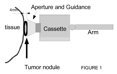
Figure 1 shows the relative positions of the body tissue (1) with the tumor
nodule (2) (an
example of 4 mm. depth is shown) which is being targeted disposed on said
tissue. A simplified
diagram of a shroud (3) containing a locator mechanism is shown over the
tissue, with the
cassette (4) containing the radioactive substance, and the general disposition
of the cassette on a
robotic arm (5).
DESCRIPTION OF THE INVENTION:
The preferred mode of invention proposes to first select an interchangeable
irradiating
capsule with a shutter as set forth below. Based on the depth and size of
tissue to be treated, a
radiation source will be selected for placement in the capsule and mounted on
the robotic arm of
the SRIORT. The arm would then be moved to the proper location for irradiation
of the tissue,
under direct visualization, with or without assistance from alternative
imaging modalities or any
combination of these.
CA 02737938 2011-03-21
WO 2009/039428 PCT/US2008/077100
9/39
Expanding on the above, the key invention components are:
= A radiation source
= A capsule/arm with an aperture opening to a cavity containing the
radiation source
with certain control electronics and devices designed to be connected to the
surgical robot and inserted into the patient's body through the
laparoscopic/surgical robotic incisions
= For a lesion, tumor, tissue, or organ, a mechanism for displaying pre-
operative
medical imaging, fused pre-operative medical imaging, including CT, MRI,
Ultrasound, functional MRI, PET, PET/CT and nuclear medical scanning in the
operating room in real time visible to the manipulation station of the
surgical
robot preferably on a video screen or computer monitor or other means for
display.
= A mechanism for identifying and tracking the real time coordinates of the
radiation source capsule within the body and displaying the 3-dimensional
location of the capsule on the pre-operative imaging with a projection of the
presently programmed radiation field distribution on the images and a control
means such as a general purpose computer to make real-time updates to the
tissue
position relative to the surgical robot, avoiding overdoses to desired tissue.
= A mechanism for tracking, visually, preferalby on a video screen,
computer
monitor, or means for display the internal position of the capsule within the
body
and for advancement and positioning under direct visualization using visible,
infrared and ultraviolet light or any combination of these.
= A mechanism for identifying the tumor, and tumor depth (using a
combination of
the above or ultrasonic echoes)
= A mechanism for setting an aperture size, accepting a desired dose and
calculating
the exposure time based on the selected radiation source physical parameters
and
characteristics.
= A mechanism for activating the now properly positioned radioactive source
in the
cavity to deliver the desired radiation dose, and field size and shape to the
desired
CA 02737938 2011-03-21
WO 2009/039428 PCT/US2008/077100
10/39
volume of the tumor while preventing exposure to the operating room personnel.
Normally this would mean an electromechanical actuator opening a closed
shutter
in the capsule. However, a mechanical connection could be made so that an
actuator, such as a pin, in the surgical robot arm actually activates the
shutter to
open. A spirally opening and closing iris shutter of the style used in a
camera, or a
simple door mechanism can provide an adjustable aperture.
= A mechanism for identifying and tracking the real time coordinates of the
radiation source capsule within the body and displaying the 3-dimensional
location of the capsule on the pre-operative imaging, a post-radiation report
to
show radiation field distribution on the images, on for instance, a video
screen,
computer monitor or means for display, and probable damage to irradiated
tissue.
These components and mechanisms will be described in detail below.
The application of the invention would be as follows for cancers:
The physician would have pre-imaged the patient's body according to standard
medical
procedures to locate the tumor and any other areas of suspected cancer
activity, sometimes
known as "hot spots". These are areas that are identifiable in a variety of
medical imaging
modalities, including PET, CT, MRI and nuclear medicine scans. The physicians
would have
visually identified any other areas of suspected cancer involvement during the
course of surgical
intervention.
The physician will then make an incision in the abdomen and the SRIORT is
activated.
The SRIORT has a television camera mounted on a robotic arm. The SRIORT has
accessories
mounted on a robotic arm and are controlled by remote control. The surgical
SRIORT is then
used to incise the interior membranes and a cutting implement is used to
perform a resection by
the physician. The surgeon can cauterize and clean as needed and ultimately
view the remaining
tissue through the camera on the SRIORT arm, and in conjunction with medical
imaging as
described above, determine what further areas need radiation treatment.
In the case of ovarian cancer, when the maximum surgical debulking possible
has been
obtained, frequently, studs of disease remain which involve the surface of the
liver, the
diaphragm and areas of the bowel. It is not possible to treat these areas
generally with external
CA 02737938 2011-03-21
WO 2009/039428 PCT/US2008/077100
11/39
beam (whole abdominal radiation therapy), conventional brachytherapy or loose
isotope therapy
or conventional intraoperative radiation therapy using accelerators due to the
inability to deliver
a precisely enough targeted and sufficient dose of radiation to eliminate
cancer metastases
without causing substantial morbidity and even mortality, or exposing
operating room personnel
to unacceptably high exposures to radiation.
Based on the depth of tissue desired to be penetrated and the desired dose to
be delivered,
a particular radiation source, which may be a radioisotope or device generated
radiation (x-rays),
of appropriate emission type, energy and strength would be selected for
placement in the capsule
on the SRIORT arm. This capsule would be either permanently mounted on the
SRIORT arm or
preferably would be an interchangeable module to accommodate differing
physical
characteristics of radiation sources. The capsule must be designed to balance
size of the device
with necessary shielding for both direction and size of radiation field and
personnel protection
from leakage radiation. The capsule would then be selected under robotic
control from its
storage location, mounted on the arm of the SRIORT and moved into the proper
position inside
the patient in the proper location for irradiation. The physician would then
move the capsule and
proposed beam location to the angle and desired beam angle to the lesion. The
SRIORT has a
camera enabling direct visualization of the lesion. An alternate imaging
device, appropriate for
the tumor could be used in addition to a camera, such as an ultrasound
transducer or probe. A
laser could be mounted to identify and illuminate the spot of radiation beam
application.
Traditional TORT using linear accelerators external to the body have used
doses in the
range of 10-20 Gy (Gy = gray = joule/kg energy deposited in matter by ionizing
radiation).
These doses can be delivered with a variety of devices and isotopes, most
commonly those with
high specific activity such as Jr-192 or Cs-137, or more recently x-ray diodes
and solid state x-
ray generators, can be used. In addition, other emitters such as Sr-90 (beta
emitter with energy
of 0.195 MeV). The table below gives examples of byproduct material and
typical energies and
half lives.
Typical Isotope Emission/Energy Half Life
Cs-137 Gamma/662 keV 30 years
Jr-192 Gamma/442keV 70.2 days
CA 02737938 2011-03-21
WO 2009/039428 PCT/US2008/077100
12/39
Sr-90 Beta/195 keV 29 years
Cf-252 Neutron/fissile spectrum 2.6 years
Dose calculations are given by the following formula for isotopes:
Dose = (TA)( ISF)2(Strength)(time of exposure)
These sources and other sources will generally have activity in the range of 5-
10 Ci (10
Ci=370 GBq). For example, to deliver 20 Gy to a depth of 5 mm (4 mm + 1 mm
margin) for the
4 mm tumor shown in Figure 1, from the applicator capsule, assuming a 10 Ci
source strength,
using Iridium-192, which has a specific air KERMA constant ( r AKR-( 1.115P Gy
x m2)
(GBq x hr)
used to convert activity into dose, the following exposure would be required:
2000 cGy=(370 GBq)(111.5cGy - cm2) , 1 2 1 hr
l
which yields an exposure time of 2000/11001 = 0.181 minutes or 10 seconds
exposure,
assuming the above parameters. The quantity .25 cm. was selected in order to
have a typical
source to surface distance. Therefore, each lesion could be treated in under 1
minute, with
precise control of exposures, field placement and size under real time
guidance in the operating
room using the SRIORT.
Due to the absolute criticality of distance in this exposure range, to
delivered dose per
unit time, the capsule will have an independent electronic distance measuring
device using
optical ranging.
Where organ motion is a concern, the device can be placed at an increased
distance such
as 0.5 cm from the tumor at the physician's discretion. Adjustments can be
made to
accommodate organ motion or relative motion of the patient. For this distance
the above
calculation would yield an exposure time of 0.67 minutes or 40.2 seconds.
CA 02737938 2011-03-21
WO 2009/039428 PCT/US2008/077100
13/39
The exposure time would be electronically controlled with a dual timer backup
system
whereby if the primary timer set time expires, then a backup secondary timer
will engage and
close the aperture to stop the radiation exposure. Both of these timers will
have a clearly visual
display at the operator's console with an alarm, both visual and audible when
the cassette has
radiation present and a second alarm both visual and audio if the cassette's
control electronics fail
to close the aperture (in the case of a radioactive source) or stop power to
the radiation generator
(in the case of an x-ray diode device).
The cassette's radiation "safe" chamber and aperture is constructed with
radiation
shielding in mind. Since the device is capable of using both high and low dose
rate sources,
shielding is mandatory for several reasons, the most important of which is to
protect patient
tissue from stray radiation emission from the device and to protect operating
room personnel
while the device or radiation source is in transit.
The shielding calculations are based on using either depleted uranium, lead or
tungsten.
Due to its superior shielding characteristics, the preferred shielding is
uranium since uranium
shielding will be thinner and allow for a more compact cassette which will be
easier to insert into
a laparoscopic wound (1-3 cm) and manipulate under robotic control, once it is
inserted into the
body. A typical source size (based on the Nucletron and Varian sources
presently in use), is 0.5
mm in diameter by 5 mm long. To reduce the dose to acceptable levels during
the time the
source is in the patient, for this proposed calculation example, an assumption
is made that a
procedure with the source in the patient could last up to an hour. During this
time the source will
be emitting radiation and in the medical therapeutic use of radiation 60 cGy
of exposure during a
treatment can be administered at low risk. Since operating room personnel
exposure must be
kept lower than this, additional external shielding will be placed around the
patient to meet
ALARA radiation safety limits. The robotic workstation can be placed
physically far from the
patient, further minimizing the need for external shielding. The shielding
calculation equation is
i x 37 GBq x x( 111.5 cGy cm 2 1 )2
1 0 C =412.55 cGy I hr
Ci GBq - hr 10 cm
The 10 Ci is selected as the source strength. The quantity 37GBq per Ci is a
conversion
factor. Ten centimeters is a typically selected distance to the patient body
surface for the purpose
CA 02737938 2011-03-21
WO 2009/039428 PCT/US2008/077100
14/39
of radiation shielding calculation because the average patient is
approximately 20cm. "thick." To
reduce this dose rate to an acceptable level, the dose would be reduced to
less than 60 cGy/hr or
by a factor of approximately 1 or 2 tenth value layers of shielding. The tenth
value layer of
depleted uranium for Jr-192 is 6.5 mm so, 1.3 cm of depleted uranium will
allow full shielding
and reduce the leakage exposure rate at 10 cm from 411 cGy/hr to 4.1 cGy/hr at
10 cm or 16
cGy/hr at 5 cm. If tungsten were chosen, the shielding thickness required will
be approximately
22 mm.
Given the source size, shielding requirements, and necessary electronics and
adaptors, the
preferred mode would be that the final dimensions of the cassette will be 4 cm
in diameter x 5
cm long or 4 cm x 3 cm x 5 cm. For the cone portion, if a cone is desired, the
divergence of the
cone should match the outer diameter of the tissue being irradiated. The cone
can be selected in
shape to correspond to the tumor shape. The cone can be very short, if used at
all, 3 to 4 mm.
The cassette can have varying cones mounted on it to conform to irregular
tumor shapes. This
will give adequate space to enclose a source, associated visualization,
measurement and control
electronics and mechanical safety apparatus. In SI units the shielding
calculation equation is:
O. 1 00 mCi 4.111 cGy cm2
Cz x x
x(
101cm)2 = 411 cGy I hr
Ci mCi- hr
The shutter would have a diameter of at least the maximum field size desired.
A cassette
designed with a shutter opening of up to two cm. would be the most that would
likely be
required. The collimation of the radiation is more likely determined by the
size of the source, but
the shutter size should be larger than the largest desired collimation for a
particular treatment
regime.
A second mode of invention would use the cassette device as a positioning
system only
and for the delivery of radiation the device would have a transfer tube
connector which would
allow the use of existing High Dose Rate Remote afterloading devices such as
the Nucletron
HDR or Varian HDR device to provide the radiation source. These devices have
an Jr-192
source similar to that described above which is attached to a cable and is
positioned via transfer
tubes which are attached to the HDR and the SRIORT cassette. This option would
be available
for institutions that have such a device available for interstitial
radiotherapy. Other than the
CA 02737938 2011-03-21
WO 2009/039428 PCT/US2008/077100
15/39
source delivery mechanism, in this case, the source is not an integrated part
of the cassette, but
rather delivered once the device is properly positioned. There are numerous
disadvantages with
this arrangement which make this less preferred than the self contained
system, most notably is
that the source is freely radiating while it traverses the transfer tubes,
which will require all
personnel to leave the operating room, thus dramatically increasing the time
it takes to do the
procedures.
The advantage of this device is that the device is small, easily manipulated
by the
SRIORT control systems, in real time, under direct visualization. This enables
the surgeon and
radiation oncologist to determine during the course of the operation areas of
residual and
unresectable disease and to deliver a dose of radiation precisely and
interactively to sterilize the
tumor. Because the capsule radiation source is orders of magnitude smaller
than the
conventional linear accelerator arms, it can be placed with high precision
within the body and
using articulating robotic "hands" holding the capsule in place, the field can
be directed at the
correct tumor site while inserted into the body through the robotic incisions.
Due to the potentially high activity sources in use, an emergency aperture
closing
mechanism incorporating both electronic and mechanical overrides would be used
in the device.
The system will also have fail safe mechanisms resulting in the aperture
defaulting to the closed
position absent electrical and mechanical signals to open the shutter or
expose the aperture. In
the case of x-ray generators, the fail safe will not permit current to flow to
the device except
under direct positive command.
In addition this device, by virtue of having a shielded capsule with a
controllable
aperture, together with the articulated robotic "wrist" or "hand" apparatus,
allows precise
positioning of the radiation source prior to opening the aperture and thus
protecting normal tissue
from radiation until the device is positioned and verified. This is a
substantial advance over the
current methods of applying intraoperative radiation therapy.
The purpose of using a shielded capsule is to minimize the damage to tissue
while the
capsule and the radiation source inside is in transit to the desired location.
The capsule would be
made of a high density shielding material such as lead, tungsten or uranium
and the capsule
would have a shutter covering an aperture through which radiation particles
would be emitted.
The shutter would also be of high density shielding material such as tungsten,
but materials can
CA 02737938 2011-03-21
WO 2009/039428 PCT/US2008/077100
16/39
be selected from those in the Berger & Seltzer handbook which contains data on
mass energy
attenuation coefficients sufficient to provide appropriate and necessary
radiation protection. The
capsule design will permit the adaptation of interchangeable shutters, much
like the
interchangeable lenses of a camera.
The interchangeable capsule would be stored in a shielded storage device,
could be
sterilized by steam or gas sterilization as is traditionally used in the
operating room
environment. The radiation source would be extracted from the storage pig,
which is a larger,
well shielded storage chamber used to transport and store radioactive source
material, usually
build of lead or tungsten, immediately adjacent to the patient in the
operating room which will
minimize the exposure of any personnel and the patient during the capsule
transit time. It would
be impractical to shield all gamma radiation from a source emitting gamma
rays, but the distance
allowed by the robotically assisted intraoperative radiation therapy
applicator coupled with a
reasonable amount of shielding would allow the device to be used while
minimizing exposure to
personnel to be in conformance with NCRP limits of exposures to radiation
workers. The device
will include adequate shielding in the form of mobile shielding units
installed in the operating
room to protect operating personnel in accordance with the ALARA ¨ as low as
reasonably
achievable ¨ philosophy of radiation protection and well below the accepted
occupational
exposure limits for the planned procedures. Survey instruments will be build
into the apparatus
and workstations to measure and record total in-room exposures. Mobile patient
shielding would
be available, depending on the radioisotope, to shield the patient, preferably
with an aperture for
the surgical entry site only so that any exposure of the patient is minimized.
That mobile patient
shielding could be in the form of one or a series of hooded containers such as
lead shields on
mobile casters, or a one or a series of lead aprons.
The cassette could be designed to either have contacts connected to internal
wiring that
meet control contacts on the robotic arm, or the internal wiring of the
cassette can be connected
by a wire harness to the robotic arm. An alternative preferred mode is a
wireless control
mechanism, but the level of ionizing radiation can be problematic.
For alpha or beta emitters, a lightweight capsule is possible. Under current
technology a
particle accelerator cannot be used for effective application of alpha
particles, protons, electrons
or light ions, which at energies useful therapeutically have a very short path
length, but within
CA 02737938 2011-03-21
WO 2009/039428 PCT/US2008/077100
17/39
that path length are devastating to the reproductive machinery of cancer cells
(DNA and cellular
ability to repair fractured DNA). Alpha particles and to a lesser extent, beta
particles emitted
from radioisotopes are readily obtained from a variety of isotopes, as are
gamma rays. [Berger
and Selzer, Affix]
Alpha particles are considered high linear energy transfer (LET) particles and
deliver
substantive damage to DNA in the form of double stranded DNA breaks, which are
very difficult
for cells to repair properly. Gamma rays, and x rays, in contrast are low LET
particles and
operate by the generation of radiolysis of water generating hydroxyl free
radicals in the vicinity
of DNA causing single strand and double stranded breaks following a linear-
quadratic curve of
cell survival v. dose, culminating in a loss of reproductive integrity of the
cancer cells. Likewise
beta particles, though low in linear energy transfer can cause double stranded
breaks and destroy
DNA through clusters of single stranded breaks which can be made permanent by
oxygen
fixation in non-hypoxic environments.
The capsule mounted on the SRIORT arm enables an alpha or beta emitter to be
completely shielded from healthy tissue and to minimize transient damage as
the radiation source
is positioned at its intended target. Only on setting the aperture to the
desired beam size,
positioning the aperture in the correct location and desired angle and opening
the shutter on the
capsule will a beam of radiation be emitted through the aperture in the
capsule in the desired
direction to irradiate the lesion. In the case of an x-ray generator, the x-
ray source will only be
turned on when the above parameters are met.
As particle path length in tissue is very predictable, cancerous tissue can be
destroyed
with a much finer precision while minimizing damage to normal tissue, such as
livers, kidneys
and bowel. Sr-90 is a typical beta emitter which would be deadly to tissue
without appropriate
shielding, but when used in the proposed capsule could be safely directed to
the targeted area.
Likewise isotopes that emit alpha particles, and gamma rays or a source
capable of developing x-
rays can be used with appropriate shielding design on the capsule. The
significant advantage of a
beta emitter is enablement by the invention of a new technology of a very
effective and
predictable radiating isotope, and the miniaturization of the capsule because
of reduction of bulk
because shielding is much simpler. Any metal, or plastic such as lucite, with
appropriate electron
stopping power as set out in tables for a source available to a reasonably
skilled practitioner, such
CA 02737938 2011-03-21
WO 2009/039428 PCT/US2008/077100
18/39
as the tables in Berger & Seltzer, can be used for the shielding. Much smaller
tumors in much
smaller and confined spaces can be treated.
The capsule shutter could be simply the equivalent of a door occluding a
radiation
aperture. A preferred mode is to use an iris type aperture with a clam shell
outer cover. The
aperture can be opened to various diameters allowing the physician to choose
the size of lesion to
be treated and the surface area of the volume. A light source can be disposed
on the exterior of
the cassette for illumination inside the patient of the tumor to be
irradiated. An alternate light
source to act a a field light behind the aperture through which radiation will
be emitted, but
behind the iris would enable the physician to continue visible inspection of a
lesion as he
positions the device for maximum coverage of the tumor before the radiation
source is opened by
the clamshell. In addition, this mode gives redundant protection should one or
the other of the
apertures fail while the device is in place, thus allowing the device to be
removed from the
patient and safely deposited in the shielded pig until repairs can safely be
made. A preferred light
source is an LED, fiber-optic or solid state light emitter.
Upon completion of the treatment procedures, the SRIORT arm and radiation
source can
be remotely stored in the pig or appropriate storage device where
sterilization and preparation for
the next case can take place. For convenience sake, the storage device is
preferably a table with
a shielding container or pig on it. The storage device would likely have
multiple pigs. The
storage device including a shielding pig is referred to as a shielded source
containment table,
even if a closet or storage cabinet is used. To insure radiologic safety, each
pig shall have a
means of detecting radiation presence to insure that a source is present or
absent from the pig.
By regulation, that would usually be a room detector in the room, and/or a
sensor inside the
shielded source containment table, such as ion chamber, electrometer or Geiger-
Mueller type
device.
In addition to a radiation source, other devices could also be mounted with
the unit,
including a laser or particle emission device and used adjuvantly for tissue
destruction. This
device is not limited to the carriage of radioactive sources, but can also be
used in conjunction
with x-ray diodes or other radiation sources.
Because a surgical robot can have more than one arm, the invention enables
more than
one capsule to stand ready in the shielded source containment table so that
should a physician
CA 02737938 2011-03-21
WO 2009/039428 PCT/US2008/077100
19/39
determine to select a different capsule during irradiation, the capsule in
present use can be
quickly withdrawn , its path of extraction memorized and an new capsule with
the preferred
radiation source inserted.
EXAMPLES OF APPLICATIONS OF PREFERRED MODE OF INVENTION
In the following two examples, a narrative description of how the SRIORT
device and system
will be used in actual practice. Several physicians will, of necessity be
directly involved in these
procedures due the the differences in training between the specialties. The
key players in each
case will be a surgeon and a radiation oncologist. The surgeon will be
specifically trained in a
pertinent area and the radiation oncologist is trained in the appropriate use,
application and
dosing of radiation for the treatment of tumors. In addition, a medical
physicist, specifically
trained in the use of radiation sources in conjunction with the radiation
oncologist must be
available for the planning of radiation delivery using the SRIORT device.
Example: Abdominal Tumor (Ovarian Cancer Stage Mb)
Initially, the patient will be informed of the nature of the procedures to be
performed in
the treatment of the cancer. After being informed and after the patient
acknowledges this
information and gives her consent, the patient will be taken to the operating
room and placed on
the operating table in the supine position. Following this the patient will be
anesthetized using
general anesthesia supplied by the anesthesiologist.
After adequate general anesthesia is instilled, the patient will be examined
under
anesthesia to determine, if possible, the extent of disease. Following this,
the patient will be
prepped and draped in the usual sterile fashion and a sub-umbilical transverse
incision will be
made extending approximately 1 ¨ 1.5 cm. Following this, a laparoscopic
trochar with a TV
camera in the bore will be advanced through the incision and under direct
visualization into the
peritoneal cavity. Following entry into the abdomen, the abdomen will be
insufflated with
carbon dioxide gas to distend the abdominal wall away from the intra-abdominal
organs.
Following this, again under direct visualization via the TV camera, a series
of similar incisions
will be made and trochars introduced into the abdomen which will allow the
placement of
robotic arms in the course of the surgery. Once these trochars are in place,
the robotic actuating
CA 02737938 2011-03-21
WO 2009/039428 PCT/US2008/077100
20/39
system will be placed into position at the operating table and the robotic
arms will be placed in
the ready position. The physicians will then move to the SRIORT control
station, which will be
located in the operating room behind a radiation shield of sufficient physical
characteristics to
provide as low as reasonably achievable radiation protection during the period
of time that the
intra-operative radiation device is in operation. The workstation will have
visualization system
originating from the robotic cameras placed in the patient, and selectable
views. The control
station will also have ergonomic robotic hand manipulators which will allow
the physicians to
move and manipulate the robotic arms in a natural way, under the control of
computer and
associated electronic circuitry.
The surgeon will then place the appropriate robotic arms into the patient via
the
previously placed trochars which will then be manipulated from the control
station to perform
the operation. The surgeon will generally use the robotic arms to place
suction into areas of
peritoneal fluid collections which will be sent to pathology for microscopic
analysis for
metastatic cancer cells. Following this, the abdomen will be washed with
sterile water and that
too will be collected and sent to pathology for analysis.
From this point, the surgeon will perform the hysterectomy, bilateral salpingo-
oopherectomy and pelvic and para-aortic lymph node dissections. Once this part
of the
procedure is complete, the surgeon will turn his attention to the remainder of
the abdomen.
Generally in locally advanced ovarian cancer, the omentum is also removed.
Following this, the
surgeon will inspect the remainder of the bowel using the robotic devices and
cameras for further
evidence of cancer. S/he will examine the bladder, rectum, bowel, peritoneal
surfaces, the liver
and the underside of the diaphragm. If lesions are found, the surgeon will
resect, to the greatest
extent possible, any visible disease within the peritoneum, using the robotic
surgery system.
During the debulking process, the surgeon using the SRIORT system will
activate a marking
device which will record the spatial coordinates of all sites of known or
suspected cancer that has
been identified and/or resected within the abdomen or surgical field. These
coordinates will then
be available to identify, post-operatively and in future procedures, potential
locations where
further radiation therapy might be considered for the treatment of microscopic
disease.
The marking device will consist of an electronic control which will signal the
control
computers to record the present spatial position and settings of the robotic
arm, viewing system
CA 02737938 2011-03-21
WO 2009/039428 PCT/US2008/077100
21/39
and controls to, in essence, create a stored anatomical "waypoint" allowing
the surgeon to select
the location at some point in the future, display the waypoint on the
operating room imaging
monitors either alone or overlaid on the pre-operative imaging. This will
allow the surgeon and
the radiation oncologist to return to the area of interest in the patient for
further study, irradiation
or procedures. In addition, the device will allow the surgeon to place a gold
seed marker in
tissue to identify the suspect tissue radiologically at a future point, post-
operatively. Adjustments
could be made to waypoints during surgery to accommodate changes in position.
Once the surgeon has completed his work, the radiation oncologist, in
cooperation with
the surgeon will place on monitors in the operating theatre the pre-operative
medical imaging,
including, but not limited to computed tomography scans (CT/CAT), positron
emission
tomography scans (PET or PET/CT), magnetic resonance imaging scans (MRI),
ultrasonic
imaging and any other imaging techniques which may be helpful in localizing
position and
radiation within the patient. Once the surgeon and the radiation oncologist
determine the sites to
be irradiated, the radiation oncologist, in consultation with the medical
physicist, the shielding
equipment will be moved into place in the operating theatre to protect
personnel necessary to the
operation from the radiation sources used in the treatment of the lesions.
Following this, a cart containing the SRIORT robotic applicator arms capable
of attaching
cassettes containing the radiation sources, along with the cassettes and
radiation sources will be
brought into the operating theatre.
Once the radiation oncologist has selected the appropriate radiation sources
and doses to
be used in the treatment of lesions, the medical physicist will pre-program
the SRIORT device
using a separate computer workstation to identify the sources to be used, the
beam size to be
used and the depth of irradiation and doses of radiation to be delivered. Once
these parameters
have been programmed into the device, the delivery of the radiation can then
proceed.
Typically, as is presently done, for instance in prostate seeding, once the
lesions are
marked, a simulation of the proposed procedure would be performed. Techniques
of radiation
simulation that are presently available would be incorporated in programming
of a general
purpose computer used in conjunction with the system.
The radiation oncologist will select the appropriate arm to be used and will,
using the
SRIORT device move the arm into position to extract the selected cassette from
the radiation
CA 02737938 2011-03-21
WO 2009/039428 PCT/US2008/077100
22/39
source storage cart (pig, in the case of a radionuclide source). The cassette
will have electrical
connections which will enable the cassette to identify itself to the SRIORT
manipulator and
hence back to the control station. The SRIORT will compare the cassette
identification with the
pre-programmed source selection and radiation dose planning previously done by
the physicist to
insure that the proper cassette has been mounted with the correct source. The
source, while still
in its shielded chamber (pig) will then have its aperture set to a specific
set of sizes and each size
will be measured to verify the accuracy of the aperture size controls prior to
extraction. The
shutter will then be opened, as well to expose a radiation detector to verify
the source
activity/strength matches the predicted values calculated and referenced in
the pre-programmed
controller. This will allow the radiation oncologist and the physicist to
resolve any discrepancies
prior to actually introducing the device into a patient.
Once verification of the planning and exposure parameters have taken place,
the SRIORT
control system will allow the physician to remove the cassette and manipulate
the robotic arm
carrying the cassette into position within the patient via the appropriate
trochar. The cassette will
also contain a locator transducer which will identify its precise spatial
location within the
operating theatre and more importantly within the patient. This location will
also be transmitted
to the imaging workstations containing the medical images and the location of
the radiation
source within the patient can be depicted on the operating room monitors, as
well as directly
visualized within the patient on the SRIORT vision system. While this will
generally be done
with visual spectrum of light, it will also be possible to map non-visual
spectrum such as infrared
spectra to the visible spectrum to allow the radiation oncologist to observe
physiologic activity
which might not be observable with ordinary visible light, thus enhancing the
physician's ability
to identify and treat areas of potential residual cancer and prevent
recurrences.
Under these visualization schemas, the physician from the SRIORT control
station will
advance the radiation cassette into the proper position to deliver the
radiation to the intended
target. The radiation oncologist will then set an aperture size appropriate to
treat the lesion, and
then visually identify this aperture by means of a self contained field light
which will replicate
the actual radiation field through the aperture. Comparing this field light
with the area of
interest, the physician, in real time will make fine adjustments to the
position of the source and
aperture size to conform precisely to the area to be irradiated. The field
light can be
CA 02737938 2011-03-21
WO 2009/039428 PCT/US2008/077100
23/39
supplemented with an aiming laser device attached to the cassette or the
SRIORT arm carrying
the cassette.
Once this is done, the SRIORT will perform final exposure rate and time
calculations and
the shutters will be opened, allowing the cassette's radiation source to
irradiate the lesion to the
dose and depth desired for proper disease control. The radiation oncologist
will have the ability
to review and examine directly by manipulation of the SRIORT to the previously
stored
coordinates of areas of interest, the imaging studies and via direct visible
and extra-visual
spectral mapping information.
This process will be repeated as many times as is necessary to properly treat
each and
every lesion identified for the best hope of permanent eradication of the
cancerous lesions. In
each case, the radiation oncologist and the medical physicist will have the
ability to select from a
variety of cassettes, the appropriate intra-operative radiation applicator for
each lesion to be
treated with radiation at the time of the surgery and to manipulate and
program the sources in
real time for the best possible chance of cure of cancer and neoplastic
diseases.
In the case of other sites, such as the head and neck, brain or chest, these
procedures
described above will be equally applicable, with appropriate modifications for
the site of disease.
This SRIORT device will permit the use of radiation to treat areas previously
untreatable
intraoperatively due to the inability to position accelerators precisely.
Other devices, such as
Med-Tech's brachytherapy intraoperative applicator, are incapable of the
precision necessary to
spot treat lesions of interest without causing unacceptable morbidity for
lesions located on or
adjacent to radiosensitive organs.
While the invention has focused on a procedure relating to incision surgery
and resection
of tissue, and follow-up by irradiation to achieve adequate margins, the
invention is applicable to
surgery where resection is deemed undesirable, such as so-called "inoperable
cancers." These
involve lesions which for instance are adjacent to the aorta where resection
has too high a risk of
mortality. This invention enables a stand-off from a critical vessel or organ,
and use of
irradiation, potentially in a step-by-step manner, to destroy tissue
iteratively, avoiding physical
contact with the radiosensitive tissue, and/or permitting healthy tissue to
grow back.
Another variation is to utilize a sensor on a moving organ or in conjunction
with a
moving organ and coordinate the output from that sensor with the opening and
closing of the
CA 02737938 2011-03-21
WO 2009/039428 PCT/US2008/077100
24/39
shutter and aperture, and the positioning of the capsule. Thus, for a lesion
on heart tissue, an
EKG lead could be connected and integrated with a general purpose computer so
that radiation
exposure would be timed to only occur at certain points in the relative
movement of tissue vis a
vis the capsule. Alternatively, a range finder, either visual, optical, or
ultrasonic, on the capsule
could be coordinated with the aperture so that radiation exposure occurred
only in certain
distance ranges. This would enable certain heart and pulmonary-aortic lesions
to be treated by a
stand-off tissue irradiation with considerably less danger to a patient. The
capsule could be
moved in conjunction with rhythmic tissue movement.
The invention can be used, for example, in conjunction with intraparenchymal
lesions in
the liver. The liver is radiosensitive tissue and the intraparenchymal lesions
are not ordinarily
amenable to radiation therapy without lethal consequences.
The invention enables stereotactical radiosurgery type techniques where the
physician
can, in real time, determine the depth of effect of irradiation, and make real
time adjustments in
dosages, hopefully eliminating another invasion of the patient's body.
The invention contemplates a means for positive attachment of the capsule by
which is
meant that the robot arm has a clasp, finger, bayonet, clamp or slide
mechanism to positively
lock the capsule, and further, has an electrical feedback mechanism that
operates only when
positive lock has occurred meaning the capsule is securely attached to the
robot arm. A means
for positive attachment also includes a surgical end effector as defined in
U.S. Pat. 6,246, 200
cited earlier.
The invention contemplates that other arms of the surgical robot may be
engaged in
surgery, or in tissue manipulation to facilitate entry of the capsule for
irradiation.
If multiple consoles are contemplated, prior art describes and this invention
would use an
arbitration mechanism to preferably give priority at all times to the handling
of the capsule
containing a radioactive substance absent a specific command to the contrary.
Potentially a speech interface could be included to assist in direction on pre-
defined axes,
but it is important to remember of radiologic safety reasons, close manual
override and control is
needed.
While the preferred mode of electrical communication and control is a physical
electrical
electrical connection and control by pins on the capsule against contacts on
the robot arm or vice
CA 02737938 2011-03-21
WO 2009/039428 PCT/US2008/077100
25/39
versa, another mode of invention is to use telecommunication between the
surgical robot, or to
the surgical robot, and/or telecommunication to the capsule.
The term means for imaging is intended to include CT (computer tomography),
MRI
(magnetic resonance imaging), ultrasound or ultrasonic imaging; functional
MRI, PET (positive
emission tomography), PET/CT and nuclear medical scanning.
The term means for direct visualization or direct visualization includes the
use of visible
infrared and ultraviolet light or any combination of those to enable direct
visualization.
Also proposed is the concept of placing two means of direct visualization
enabling true
internal stereoscopic visualization through more than one mounted means for
direct visualization
on the capsule.
The term means for direct visualization or direct visualization includes the
use of visible,
infrared and ultraviolet light or any combination of those to enable direct
visualization, including
an endoscope or a laparascope.
Also proposed is the concept of placing stereoscopic endoscope or stereoscopic
laparascope, meaning two means of direct visualization enabling true internal
stereoscopic
visualization through more than one mounted means for direct visualization on
the capsule.
The term "stand-off remote detection" includes radar and electric signaling
for
determining distance; in this invention the stand-off remote detection is
primarily intended to
determine the distance from the radiation source to the tissue being
irradiated, taking into
account the tare length of the radiation source to the edge of the capsule, or
the end of the shroud
if one is used. Other forms of stand-off remote detection are also discussed
such as ultrasound
and laser optical finders.
A fail-safe closed position means that if power is lost, particularly power to
operate the
shutter, the shutter closes occluding the aperture through which radiation is
being emitted into
the patient.
The embodiments represented herein are only a few of the many embodiments and
modifications that a practitioner reasonably skilled in the art could make or
use. The invention is
not limited to these embodiments. Alternative embodiments and modifications
which would
still be encompassed by the invention may be made by those skilled in the art,
particularly in
light of the foregoing teachings. Therefore, the following claims are intended
to cover any
CA 02737938 2015-08-19
WO 2009/039428 PCT/US2008/077100
26/39
alternative embodiments, modifications or equivalents which may be included
within the
scope of the invention as claimed.
REFERENCES
Attix, Frank H, Introduction to Radiologic Physics and Radiation Dosimetry,
John Wiley &
Sons, 1986
Berger & Seltzer, Tables of Energy Losses and Ranges of Electrons and
Positrons, NASA, 1964
Gunderson & Tepper, Clinical Radiation Oncology, 2' Edition, Chapter 15,
Intraoperative
Irradiation, pp 315-328
Haddock MG, Petersen IA Webb MJ: Intraoperative Radiotherapy for locally
advanced
gynecologic malignancies, Frontiers of Radiation Therapy Oncology, 31:356-259;
1997
Khan, Faiz: The Physics of Radiation Therapy, 1984, Williams & Wilkins,
Baltimore, ISBN
0-683-04501-6
Petersen, IA, Haddock, MG, Donohue, JH: Use of intraoperative Electron Beam
Radiotherapy in
the Management of Retroperitoneal Soft Tissue Sarcoma, Int. J. Radiat Oncol
Biol Phys
50:126-131, 2001
Ramsay J, Suit HD: Experimental Studies on the incidence of metastases after
failure of
radiation treatment and the effect of salvage surgery. Int. J. Radiat Oncol
Biol Phys
14:1165-1168;1988
Stump, KE, DeWerd, LA, Micka, JA, and Anderson, DR: Calibration of New HDR Ir-
192
Sources. Med Physics, Vol 29(7):1483-1488
Suit HD: Local control in patient survival. Int. J. Radiat Oncol Biol Phys
23:653-660, 1992
Suit HD: Potential for improving survival rates for the cancer patient by
increasing efficacy of
treatment of the primary lesion. Cancer 50:1227-1234, 1982
Swiss Society for Radiobiology and Physics, Dosimetry and Quality Assurance in
High Dose
Rate Brachytherapy with Iridium-192, Recommendation #13, January, 2005, ISBN
3908-125-36-7