Note: Descriptions are shown in the official language in which they were submitted.
CA 02738479 2016-05-05
WO 2010/042404 PCT/US2009/059374
1
-rim: OF THE INVENTION
100011 METHOD OF STIMULATING A IlYPOGLOSSAL NERVE FOR
CONTROLLING THE POSITION OF A PATIENT'S TONGUE
BACKGROUND OF THE INVENTION
100031 The present invention generally relates to a method of stimulating a
Ffypoglossal nerve
for controlling the position of a patient's tongue. In one embodiment, the
Ilypoglossal nerve is
stimulated to prevent obstructive sleep apnea.
100041 Sleep apnea is a sleep disorder characterized by pauses in breathing
during sleep. Those
affected by sleep apnea stop breathing during sleep numerous times during the
night. There are two
types of sleep apnea, generally described in medical literature as central and
obstructive sleep apnea.
Central sleep apnea is a failure of the nervous system to produce proper
signals for excitation of the
muscles involved with respiration. Obstructive sleep apnea (USA) is caused by
episodes of physical
obstruction of the upper airway channel (UAW) during sleep. The physical
obstruction is often
caused by changes in the position of the tongue 110 during sleep that results
in the closure of the
soft tissues 112 at the rear of the throat or pharynx 132 (See Figs. I and 2A
and 211).
100051 ()SA is characterized by the complete obstruction of the airway
causing breathing to
cease completely (Apnea) or partially (Hypopnea). The human airway (at the
level of the thorax) is
lined by soft tissue, any collapse of its walls results in the closure of the
airway which leads to
insufficient oxygen intake, thereby interrupting one's sleep (episodes or
micro-arousals).
100061 During sleep, the tongue muscles relax. In this relaxed state, the
tongue may lack
sufficient muscle tone to prevent the tongue from changing its normal tonic
shape and position.
When the base of the tongue and soft tissue of the upper airway collapse, the
upper airway channel
is blocked. causing an apnea event (See Fig. 2B). Blockage of the upper airway
prevents air from
CA 02738479 2011-03-24
WO 2010/042404 PCT/US2009/059374
2
flowing into the lungs, creating a decrease in blood oxygen level, which in
turn increases blood
pressure and heart dilation. This causes a reflexive forced opening of the
upper airway channel until
normal patency is regained, followed by normal respiration until the next
apneaic event. These
reflexive forced openings briefly arouse the patient from sleep.
[0007] OSA is a potentially life-threatening disease that often goes
undiagnosed in most patients
affected by sleep apnea. The severity of sleep apnea is determined by dividing
the number of
episodes of apneas and hypopneas lasting ten seconds or more by the number of
hours of sleep. The
resulting number is called the Apnea-Hypopnea Index, or AHI. The higher the
index the more
serious the condition. An index between 5 and 10 is low, between 10 and 15 is
mild to moderate,
over 15 is moderately severe, and anything over 30 indicates severe sleep
apnea.
[0008] Current treatment options range from drug intervention, non-invasive
approaches, to
more invasive surgical procedures. In many of these instances, patient
acceptance and therapy
compliance is well below desired levels, rendering the current solutions
ineffective as a long-term
solution.
[0009] Current treatment options for OSA have not been consistently
effective for all patients.
A standard method for treating OSA is Continuous Positive Airway Pressure
(CPAP) treatment,
which requires the patient to wear a mask through which air is blown into the
nostrils and mouth to
keep the airway open. Patient compliance is poor due to discomfort and side
effects such as
sneezing, nasal discharge, dryness, skin irritation, claustrophobia, and panic
attacks. A surgical
procedure where rigid inserts are implanted in the soft palate to provide
structural support is a more
invasive treatment for mild to moderate cases of OSA. Alternate treatments are
even more invasive
and drastic, including uvulopalatopharyngoplasty and tracheostomy. However,
surgical or
mechanical methods tend to be invasive or uncomfortable, are not always
effective, and many are
not tolerated by the patient.
[0010] Nerve stimulation to control the position of the tongue is a
promising alternative to these
forms of treatment. For example, pharyngeal dilation via Hypoglossal nerve
(XII) stimulation has
been shown to be an effective treatment method for OSA. The nerves are
stimulated using an
implanted electrode to move the tongue and open the airway during sleep. In
particular, the medial
XII nerve branch (i.e., in. Genioglossus), has demonstrated significant
reductions in UAW airflow
resistance (i.e., increased pharyngeal caliber). While electrical stimulation
of nerves has been
experimentally shown to remove or ameliorate certain conditions (e.g.,
obstructions in the UAW),
current implementation methods typically require accurate detection of a
condition (e.g., a muscular
obstruction of the airway or chest wall expansion), selective stimulation of a
muscle or nerve, and a
CA 02738479 2011-03-24
WO 2010/042404 PCT/US2009/059374
3
coupling of the detection and stimulation. These systems rely on detection of
breathing and/or
detection of apnea events as pre-conditions to control and deliver electrical
stimulation in order to
cause only useful tongue motions and to periodically rest the tongue muscles
and avoid fatigue. In
one system, for example, a voltage controlled waveform source is multiplexed
to two cuff electrode
contacts. A bio-signal amplifier connected to the contacts controls stimulus
based on breathing
patterns. In another system, a microstimulator uses an implanted single-
contact constant current
stimulator synchronized to breathing to maintain an open airway. A third
system uses an
implantable pulse generator (1PG) with a single cuff electrode attached to the
distal portion of the
Hypoglossal nerve, with stimulation timed to breathing. This last system uses
a lead attached to the
chest wall to sense breathing motions by looking at "bio-impedance" of the
chest wall. Still another
system monitors vagus nerve electroneurograms to detect an apnea event and
stimulate the
Hypoglossal nerve in response.
[0011] What is needed is a system and method of electrical stimulation of
the Hypoglossal nerve
for controlling tongue position that is not tied to breathing and/or detection
of an apnea event.
BRIEF SUMMARY OF THE INVENTION
[00121 A method of stimulating a Hypoglossal nerve for controlling the
position of a patient's
tongue according to some embodiments of the present invention includes
attaching at least one
electrode to the patient's Hypoglossal nerve and applying an electric signal
through the electrode to
at least one targeted motor efferent located within the Hypoglossal nerve to
stimulate at least one
muscle of the tongue. In one embodiment the at least one electrode is
programmable.
[00131 In a further embodiment, the method includes programming a threshold
amplitude and
pulse duration of the electric signal by attaching the at least one
programmable electrode to the
patient's Hypoglossal nerve while the patient is awake and applying the
electric signal to the
Hypoglossal nerve at a first frequency through the at least one programmable
electrode, and
increasing at least one of the amplitude and pulse duration of the electric
signal until one of the
tongue moves and the patient reports a sensation.
[00141 In a further embodiment, the method includes programming a target
amplitude and pulse
duration of the electric signal by applying the threshold amplitude and pulse
duration to the patient's
Hypoglossal nerve at a second frequency through the at least one programmable
electrode, the
second frequency being faster than the first frequency, and increasing at
least one of the amplitude
and pulse duration of the electric signal to a target level until the tongue
moves sufficiently to open
CA 02738479 2011-03-24
WO 2010/042404 PCT/US2009/059374
4
the patient's airway. In a further embodiment, the method includes decreasing
the second frequency
to a target frequency.
[0015] In some embodiments, the at least one electrode includes at least
first and second
contacts and the electric signal comprises at least first and second electric
signals, and the method
further comprises applying the first electric signal through the first contact
to a first targeted motor
efferent located within the Hypoglossal nerve to stimulate at least one muscle
of the tongue, and
applying the second electric signal through the second contact to a second
targeted motor efferent
located within the Hypoglossal nerve to stimulate at least one muscle of the
tongue. In one
embodiment, the at least first and second contacts include a plurality of
contacts forming a plurality
of functional groups. In one embodiment, each functional group stimulates a
different muscle. In
one embodiment, each functional group includes at least one of the plurality
of contacts. In one
embodiment, the first and second electric signals are applied at predetermined
intervals. In one
embodiment, the predetermined intervals of the at least one first and second
electric signals are out
of phase with each other. In one embodiment, the first and second electric
signals are generally
equal in level and frequency. In one embodiment, the first electric signal
stimulates a first muscle
and the second electric signal stimulates a second muscle. In one embodiment,
the first and second
electric signals are applied at predetermined cycles for alternatively resting
and stimulating first and
second muscles. In one embodiment, the cessation of the first electric signal
is coincident with the
initiation of the second electric signal.
[0016] In one embodiment, the at least one targeted motor efferent is a
protrusor motor efferent.
In one embodiment, the at least one targeted motor efferent is a muscle that
moves to improve
airway patency. In one embodiment, the electric signal is applied for a
predetermined duration. In
one embodiment, the electric signal is automatically applied after the patient
activates the electrode
and following a time delay sufficient to allow the patient to fall asleep. In
one embodiment, the
muscle is stimulated such that one of apnea and hypopnea is prevented. In one
embodiment, the
electric signal is applied via an open loop system. In one embodiment, the
electric signal is applied
continuously for an entire sleep period.
BRIEF DESCRIPTION OF THE SEVERAL VIEWS OF THE DRAWINGS
[0017] The foregoing summary, as well as the following detailed description
of exemplary
embodiments of a method of stimulating a Hypoglossal nerve for controlling a
position of a patient's
tongue, will be better understood when read in conjunction with the appended
drawings. It should
CA 02738479 2011-03-24
WO 2010/042404 PCT/US2009/059374
be understood, however, that the invention is not limited to the precise
arrangements and
instrumentalities shown.
[0018] In the drawings:
[0019] Fig. 1 is an illustration of the human airway;
[0020] Fig. 2A is an illustration of an open human airway;
[0021] Fig. 2B is an illustration of a closed human airway during an apnea
event;
[0022] Fig. 3 is an illustration of the human tongue;
[0023] Fig. 4 is a schematic illustration of the motor nerve organization
of the Hypoglossal
nerve;
[0024] Fig. 5 is an illustration of the Hypoglossal nerve shown in Fig. 4
with labeling of the
lateral and medial branch nerve fibers;
[0025] Fig. 5A is a cross sectional illustration of the Hypoglossal nerve
shown in Fig. 5;
[0026] Fig. 5B is an illustration of the motor neurons in the Hindbrain;
[0027] Fig. 6 is a schematic illustration of the Hypoglossal nerve shown in
Fig. 4 with labeling
of the lateral and medial branch nerve fibers;
[0028] Fig. 7 is a schematic illustration of the Hypoglossal nerve shown in
Fig. 4 with labeling
of the medial branch nerve fibers;
[0029] Fig. 8A is an illustration of a cross-section of a human Hypoglossal
nerve;
[0030] Fig. 8B is an illustration of a cross-section of a human Lingual
nerve;
[0031] Fig. 8C is an illustration of a cross-section of a rat Hypoglossal
nerve;
[0032] Fig. 9 is an exemplary set of fatigue curves of human quadriceps
muscle showing
maximum voluntary contraction, 50 Hz electrical stimulation and twitch
responses;
[0033] Fig. 10 is an exemplary illustration of an electrode attached to a
patient's Hypoglossal
nerve;
[0034] Fig. 11 is a perspective view of the electrode shown in Fig. 10;
[0035] Fig. 12 is a perspective view of the electrode shown in Fig. 11
showing the plurality of
contacts;
[0036] Fig. 13 is a graphical representation of an exemplary stimulation
strategy;
[0037] Fig. 14A is a graphical representation of an exemplary duty cycle
stimulation strategy;
[0038] Fig. 14B is a graphical representation of an exemplary interleaved
stimulation strategy;
[0039] Fig. 14C is a graphical representation of an exemplary synchronous
stimulation strategy;
[0040] Fig. 14D is a graphical representation of an exemplary asynchronous
or random
stimulation strategy; and
CA 02738479 2011-03-24
WO 2010/042404 PCT/US2009/059374
6
10041] Fig. 15 is an exemplary strength-duration curve.
DETAILED DESCRIPTION OF THE INVENTION
[004211Tongue Muscle Properties
[0043] Referring to Figs. 1 and 3, the tongue 110 has been described as a
hydrostat - a
specialized muscle able to move and change shape without the usual tendon
connections to bones
against which forces may be applied. Much like the trunk of an elephant, the
tongue 110 can change
shape and move within the oral cavity to aid in speaking, eating, and
breathing. The tongue muscles
include the Genioglossus muscle 114, the Styloglossus muscle 116, the
Hyoglossus muscle 118, the
Palatoglossus muslce (not shown), the Geniohyoid muscle 320 (the Geniohyoid
muscle 320 is not a
tongue muscle but it is an important protrusor and pharyngeal dilator) and
several muscles that lie
within the tongue, called the intrinsics. In a patient who is awake, the brain
supplies neural drive to
these muscles through the Hypoglossal nerve 322, to move the tongue 110 and to
change its shape.
The Hypoglossal nerve 322 includes a Styloglossus branch 316a, Hyoglossus
branches 318a,
Genioglossus branches 314a, and Geniohyoid branches 320a. In a patient who is
awake, the neural
drive to the tongue muscles act to maintain tongue shape and position,
preventing the tongue 110
from blocking the airway.
[0044] The tongue 110 comprises both intrinsic and extrinsic lingual
muscles. There are four
intrinsic ¨ i.e., origin and insertion within the tongue 110 ¨ lingual
muscles: Verticalis 124,
Transversalis 126, Superior Longitudinalis 128, and Inferior Longitudinalis
130. There are four
extrinsic ¨ i.e., external bony origin and insertion in to the tongue base ¨
lingual muscles (mentioned
above): Genioglossus 114, Styloglossus 116, Hyoglossus 118, and Palatoglossus.
The lingual
muscles are also functionally categorized as either retrusor or protrusor
muscles and both intrinsic
and extrinsic muscles fall into these category. The retrusor lingual muscles
include the intrinsic
Superior and Inferior Longitudinalis muscles 128, 130 and the extrinsic
Hyoglossus muscle 118 and
Styloglossus muscle 116. The protrusor lingual muscles include the intrinsic
Verticalis and
Transversalis muscles 124, 126 and the extrinsic Genioglossus muscle 114. The
elevation of the
tongue 110 is achieved by the contraction of the Styloglossus muscle 116 while
the depression is the
result of downward movements of Hyoglossus and Genioglossus muscles 118, 320.
100451Hypoglossal Nerve Efferents
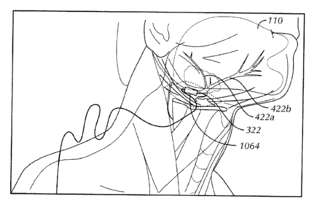
100461 Fig. 4 schematically illustrates the motor nerve organization of the
Hypodossal nerve
322 from its origin in the motor nuclei 444 in the Hindbrain 446¨ specifically
the location of the
retrusor and protrusor cell bodies 448, 450 ¨ extending via their axons to the
retrusor muscle 452
CA 02738479 2011-03-24
WO 2010/042404 PCT/US2009/059374
7
and protrusor muscle 454 innervated by the lateral 422a and medial 422b
branches, respectively of
the Hypoglossal nerve 322.
[00471 Referring to exemplary Figs. 5-7, the present invention's novel
method of mapping
Hypoglossal nerve efferents was demonstrated in a rat using dyes Dil 556 (for
example, l, l'-dioley1-
3,3,31,31-tetramethylindocarbocyanine methanesulfonate) and Di0 558 (for
example, 3,3'-
dilinoleyloxacarbocyanine perchlorate). In one embodiment, the fluorescent
dyes are manufactured
by Molecular Probes. The use of the dyes Dil 556 and Di0 558 disclosed a
surprising and
unexpected anatomical and topographical organization of the Hypoglossal nerve
322. This
anatomical and topographical organization permits targeted stimulation of
portions of the
Hypoglossal nerve 322 to maximize the efficacy of the stimulation as described
further below.
[0048] In a first experiment, efferents of the medial and lateral branches
422a, 422b were micro-
injected with dyes Dil 556 and Di0 558, respectively. Nerve branches were
exposed and the tips of
dye-loaded capillaries were pierced through the perineurium of each branch
422a, 422b. The dye
solution was iontophoresed using a current source (Kation Scientific,
Minneapolis, USA) at 4 A for
five seconds on and five seconds off duty cycle for five minutes.
[0049] In a second experiment, the Medial branch 422b and protrusor
musculature were
surgically exposed and injected with Dil 556. The tips of dye-loaded
capillaries were pierced into
the muscle bellies of selected protrusor muscles 454 and their innervating
branches. The dye
solution was iontophoresed using a current source (Kation Scientific,
Minneapolis, USA) at 4 A for
five seconds on and five seconds off duty cycle for five minutes.
[0050] Figs. 5 and 6 schematically show the effects of injecting the dyes
Dil 556 and Di0 558
into the lateral and medial branches 422a, 422b, respectively, of the
Hypoglossal nerve 322. The
dye Dil 556 injected into the lateral branch 422a of the Hypoglossal nerve 322
remains confined to
the Hypoglossal nerve efferents located within the lateral branch 422a and
spreads rostrally towards
the retrusor muscles 452 and anteriorly towards the location of the retrusor
cell bodies 448 in the
motor nuclei 444 in the hindbrain 446. The dye Di0 558 injected into the
medial branch 422b of the
Hypoglossal nerve 322 remains confined to the Hypoglossal nerve efferents
located within the
medial branch 422b and spreads rostrally towards the protrusor muscles 454 and
anteriorly towards
the location of the protrusor cell bodies 450 in the motor nuclei 444 in the
brain 446. Fig. 5B
illustrates the Di0 and Dil labeled neurons 556, 568.
100511 Referring to Fig. 5A, the magnified section of the lateral branch
422a of the Hypoglossal
nerve 22 demonstrated that it is almost exclusively comprised of the Dil
illuminated retrusor motor
efferents 560. Similarly, a magnified section of the medial branch 422b of the
Hypoglossal nerve
CA 02738479 2016-05-05
WO 2010/042404 PCT/US2009/059374
8
322 (not shown) demonstrated that is almost exclusively comprised of the Di0
illuminated protrusor
motor efferents 562. Consistent near segregation was found of the retrusor
motor efferents
dorsolaterally and the protrusor motor efferents ventromedially.
10052] This anatomical and topographical compartmentalization was confirmed
via a modified
labeling protocol. Fig. 7 illustrates that dye Di! 556 may be injected into
either the terminal end of
the medial branch 322b or into the protrusor musculature 454 and the dye Dil
556 will travel
anteriorly and ventromedially through the Hypoglossal nerve proper 322. A con
focal fluorescent
image of the entire Hypoglossal nerve 322 demonstrated the consistent
ventromedial localization of
the Dil labeled protrusor motor efferents 560 from the medial branch 422b
through the Hypoglossal
nerve proper 322 to the brain 446 and high magnification confocal images of
the Di I labeled axons.
100531 Figs. 8A, 8B and 8C demonstrate the organization structure of the I
luman Flypoglossal
nerve (Fig. 8A) and the Human Lingual Nerve (Fig. 8B), as well as the Rat
Hypoglossal Nerve (Fig.
8C). The Hypoglossal nerves in both Human and Rat are afascicular, lacking the
clear
organizational structure present in most peripheral nerves, and which is
present in the Human
Lingual Nerve.
100541 It is believed that the non-fascicular structure of the Hypoglossal
nerve in rats
approximates the structure of the Hypoglossal in humans. Moreover, the over-
all musculature
(organization of extrinsic and intrinsic muscles) in the rat tongue and the
human tongue, is nearly
identical. U.S. Provisional Patent Application No. 61/136,857 filed October
9,2008 entitled
"Method of Selectively Stimulating a Hypoglossal Nerve" discusses and
illustrates the
similarities between the rat and human tongues in further detail.
[0055] It has therefore been demonstrated that the surprising and
unexpected anatomical and
topographical compartmentalization forms the basis of the present invention
which relates to a
method of treating, controlling, or preventing a neurological disorder using
selective targeted
electrical nerve stimulation of the Hypoglossal nerve proper 22, and more
particularly to a method
of selective electrical stimulation of motor efferents (e.g., retrusor and
protrusor motor efferents) of
the Hypoglossal nerve 322. The words "selective" and "targeted" are used
interchangeably herein
meaning the use of electrodes and current sources to selectively activate
targeted nerve fibers within
a nerve bundle and hence their associated motor groups to achieve a specific
motor function. In the
case of obstructive sleep apnea, electrical stimulation of efferents of the
Hypoglossal nerve 322, and
more specifically, targeted stimulation of the protrusor motor efferents
located in Hypoglossal nerve
CA 02738479 2011-03-24
WO 2010/042404 PCT/US2009/059374
9
proper 322 and/or the medial branch 422b, for example, can open up the airway
and maintain the
patency of the upper airway channel.
[00561 The above described surprising anatomical and topographical
organization may help to
explain some of the failures and limitations of previous Hypoglossal nerve
stimulation applications.
Specifically, electrical stimulation of the whole Hypoglossal nerve proper
322¨ i.e., the section of
the Hypoglossal nerve 322 located proximal to its bifurcation into the medial
and lateral branches
422a, 422b ¨ resulted in combined (non-specific) contractions of both
intrinsic and extrinsic muscles
and both retrusor and protrusor muscles 452, 454. As both the retrusor and
protrusor muscles 452,
454 comprise intrinsic and extrinsic muscles, electrical stimulation of either
the medial or lateral
branches 422a, 422b alone results in recruitment of both intrinsic and
extrinsic muscles. Further,
stimulation of the Hypoglossal nerve proper 322 may excite sensory afferent
and motor efferent
fiber types. Grossly, the fused contractions of this non-selective stimulation
results not only in
undesirable sensory stimulation but also presents as a slight ipsilateral
deviation and retrusion of the
tongue 110.
[00571 Known stimulation of the Hypoglossal nerve proper has also resulted
in cases of
profound bradycardia which is believed to be related to secondary vagus nerve
stimulation: the
Hypoglossal nerve lies against the posterior surface of the vagus and superior
cervical sympathetic
ganglion where it exchanges branches of communication, and is united with the
inferior vagal
ganglion of the vagus by connective tissue. Common forms of electrical
stimulation elicit action
potentials in the nerve axon that propagate in two directions: towards the
desired muscle or end
organ, and in the antidromic direction towards the cell body, the same
direction that sensory fibers
would normally transfer their information. It is possible that this antidromic
activity could be
eliciting the secondary vagus nerve activation. A more distal site of
stimulation ¨ e.g., the medial
branch 22b ¨ may avoid unwanted vagal nerve reflex and muscle activation
because of its more
limited neural connections, but will still non-selectively recruit both
sensory afferent and motor
efferent fiber types if they both exist within the range of the stimulating
electrode. By discerning the
extent and myotopic organization of the Hypoglossal nerve motor neuron
subgroups and the
muscles(s) thereby innervated, such knowledge can be used to specify the
functional relevance of
diverging efferent systems and in elucidating mechanisms underlying tongue
control. Accordingly,
in one embodiment, the present invention is directed to a method of mapping
Hypoglossal nerve
fibers, thereby allowing the claimed method of selective recruitment of
specific nerve fibers, as well
as methods for selective stimulation. Understanding the neural organization
allows for selective
targeted stimulation that activates only those muscle groups that are
desirable, and avoids activating
CA 02738479 2011-03-24
WO 2010/042404 PCT/US2009/059374
those which are not. The knowledge gained from animal and cadaver studies
validate the methods
of selective stimulation described herein. The process of using selective
stimulation allows for the
selective activation of only the desired muscle functions.
[0058] It should be noted, additionally, that activation of a small
fraction of retrusor muscle or
muscles along with the activation of protrusor muscles can act to reduce
pharyngeal compliance
while not significantly leading to tongue retrusion, and may have a beneficial
effect in airway
patency.
[0059]Apparatus for Stimulation of Hypoglossal Nerve Efferents
[0060] Referring to Figs. 10-12, in one embodiment, an electrode 1064 is
attached to the
Hypoglossal nerve to apply at least one electric signal to a first targeted
motor efferent located
within the Hypoglossal nerve 322. The electrode 1064 may be programmable. The
electrode 1064
may include a plurality of contacts (e.g. contacts 1164a, 1264b, 1264c, 1264d)
each applying an
electric signal to a targeted motor efferent. In one embodiment, each contact
applies an electric
signal to a different targeted motor efferent. In one embodiment, more than
one contact applies an
electric signal to a single targeted motor efferent. In one embodiment, the
electrode 1064 includes a
first contact (e.g. contact 1164a) to apply a first electric signal to a first
targeted motor efferent, a
second contact (e.g. contact 1264b) to apply a second electric signal to a
second targeted motor
efferent, a third contact (e.g. contact 1264c) to apply a third electric
signal to a third targeted motor
efferent and a fourth contact (e.g. contact 1264d) to apply a fourth electric
signal to a fourth targeted
motor efferent. In one embodiment, the targeted motor efferents that are
stimulated stimulate at
least one muscle of the tongue 110 to control the position of the tongue 110.
In one embodiment,
the electrode 1064 is a biocompatible, soft material cuff electrode that
provides an intimate
connection to the nerve. In another embodiment, a lead wire connects the
programmable electrode
to the control system. In one embodiment, the apparatus does not require a
lead wire connecting the
programmable electrode to the control system. In one embodiment, the control
system includes a
battery, either primary or rechargeable, for powering the apparatus. In one
embodiment, the control
system includes a processor for setting up stimulation parameters to achieve
the desired outcome for
the individual patient or otherwise controlling the stimulation. In one
embodiment, stimulation
parameters are selected from the group consisting of, but not limited to,
stimulation amplitude,
stimulation frequency and stimulation duration. In one embodiment, the control
system includes a
mechanism that allows the patient to turn the apparatus on and off and
possibly make adjustments
within preprogrammed settings.
CA 02738479 2016-05-05
WO 2010/042404 PCT/US2009/059374
11
100611 The method provided by the present invention is not limited by the
design of the
apparatus used to carry it out except to the extent the point of contact with
the Hypoglossal nerve
proper 322 or its lateral and medial branches 422a, 422b is consistent with
the teachings herein.
Although an exemplary apparatus for selectively stimulating Hypoglossal nerve
efferents is shown,
equivalent alterations and modifications will occur to others skilled in the
art upon reading and
understanding this specification and annexed drawings. For example, U.S.
Patent Publication No.
2008/0046055, WO 2009/048580 and WO 2009/048581
can be modified in accordance with the teachings herein for
stimulating the Hypoglossal nerve 322. In particular regard to the various
functions performed by
the herein described exemplary apparatus, the terms used to describe the
exemplary apparatus are
intended to correspond to any apparatus that is functionally equivalent ¨
i.e., even though not
structurally equivalent, that performs the function in the herein illustrated
exemplary apparatus of
the present invention. For further information regarding an apparatus which
may be modified in
accordance to the teachings herein for practicing the method of the present
invention, refer to U.S.
Patent Nos. 6,456,866 and 6,587,725.
100621Fatigue
100631 Fig. 9 illustrates an exemplary fatigue curve. Fatigue is a common
phenomenon with
artificial activation by electrical stimulation of a muscle. In voluntary
muscle control, the human
brain recognizes, organizes, and selects the best muscle fibers to activate
for a particular activity. It
brings fibers in and out of activation to minimize or prevent fatigue and
maintain muscle output. In
artificial activation by electrical stimulation however, stimulation comes
from one or more electrode
contacts located in a relatively fixed position with respect to a targeted
nerve or nerve fiber bundle.
The same population of fibers are activated essentially every time that a
stimulus is applied because
of this fixed relationship.
100641 As is known in the art, excitation of a nerve fiber can occur along
a strength duration iso-
threshold curve - a nerve fiber will be excited as long as the amplitude is
above the curve or the
phase duration is to the right of the curve. An exemplary strength curve is
shown in Fig. 15. At
either end of the curve the shape of the curve is asymptotic - at a limiting
phase duration no amount
of stimulation current elicits a response, and at the other no phase duration
is long enough to elicit a
response either. The invention described here refers to the use of stimulus
amplitude fbr means of
modulating the recruitment of nerve fibers, hut it shall be understood that
many methods, including
CA 02738479 2011-03-24
WO 2010/042404 PCT/US2009/059374
12
phase duration and stimulus amplitude, can be utilized to the same ends of
activating nerve fibers
with electrical stimulation.
[0065] Nerve fibers are preferentially activated, or recruited, in the
order of their proximity to
the electrode contact and by their fiber diameter. As a general rule, the
closer a fiber is to the
cathodic contact, the more likely it will be activated (the general form of a
stimulating system is to
place the cathodal contact in close proximity to the target nerve axons; other
forms of stimulation
exist and shall be obvious to those skilled in the art). The larger the
diameter of a fiber, the more
likely it will be activated. The distance and size distribution in a nerve
bundle does not change
appreciably over time. Hence, the recruitment properties - which fibers will
be activated with a
particular amplitude pulse - do not change either. If the applied stimulus is
maintained at a
sufficiently high enough frequency, the recruited muscle fibers activated by
the stimulated nerve
fibers eventually fatigue. Muscle force and/or position then change to the
relaxed, inactivated
condition. The stimulation of skeletal muscle for postural control or limb
motion is often performed
at frequencies that would noimally be expected to cause fatigue in those
muscles along with the loss
of desired function if the stimulation were maintained continuously.
Stimulation may be modulated
by changing the stimulus amplitude, as described above, or by changing the
phase duration of the
pulse. Great care and tremendous effort are expended in avoidance of fatigue
in skeletal muscle
applications for fear of loss of desired functional effect, for example, for
patients suffering from
spinal cord injury or other neurological dysfunction.
[0066] Fatigue may be minimized or prevented by using a stimulation duty
cycle - that is,
stimulating for a certain amount of time before significant fatigue sets in,
then stopping to let the
muscle rest and regain its ability to contract. For obstructive sleep apnea
this is less than optimal
because without an applied stimulus during the off period of the electrical
stimulation duty cycle the
tongue would not be driven to maintain a desired position, and could fall back
against the rear of the
throat and allow an apnea event to occur. This is one of the reasons that many
OSA stimulation
systems rely on sensors to detect when to apply stimulation and when to leave
it off. The method of
using duty cycle to rhythmically apply stimulation has been proposed, also, to
do away with the
need to sense breathing events, in the hopes that by introducing rhythmic
stimulation to the
Hypoglossal nerve that somehow the breathing events would synchronize
automatically to the
stimulation timing. This has not been proven and the study by Davis. et al,
using microstimulators
in sheep demonstrated that manual timing of stimulation to the events of
breathing was required to
achieve a useful outcome in single point stimulation of the Hypog,lossal
nerve.
CA 02738479 2011-03-24
WO 2010/042404 PCT/US2009/059374
13
[0067] Another method of minimizing or preventing fatigue is to use one or
more independent
current sources to activate multiple portions of the desired muscle groups. In
certain exemplary
embodiments, one or more independent current sources drive one or more
contacts (1164a, 1264b,
1264c and 1264d for example shown in Figs. 11 and 12) that interface with the
Hypoglossal nerve
322. These contacts are optionally contained in a single cuff electrode 1064
as shown. Each contact
can be activated separately or in combination with other contacts.
[0068] In certain embodiments, each contact is assigned to one or more
functional groups.
Functional groups may in turn be used to select regions of fibers within the
nerve bundle that result
in a desired tongue movement. The effort of moving the tongue to the desired
position is thus
shifted from one functional group to another functional group so that no
single functional group is
required to work all of the time. Thus, the effort of moving the tongue is
shared among multiple
stimulated nerve fibers and their associated muscles, preventing or reducing
fatigue because none of
the groups is activated long enough to cause significant fatigue, and during
their off state they are
allowed to recover from the stimulation. In certain exemplary embodiments,
each group is active
until just before significant fatigue sets in. One or more other groups are
then activated to take its
place, allowing the former muscle group fibers to rest. In one embodiment, the
stimulation is spread
over more than one contact wherein the duty cycle of each contact is
overlapped (Fig. 13). In one
embodiment, the stimulation pulses may be generally random or pseudo random so
long as the
overall contractions per unit of time is limited (see Fig. 14D).
[0069] Another method of reducing or eliminating fatigue is to lower the
stimulation frequency.
The faster a nerve is stimulated, the faster it fatigues. Each pulse produces
a contraction, with each
contraction requiring a certain amount of work. The more contractions there
are, the more the
muscle works, and the more likely the muscle will become fatigued. Reducing
the stimulation
frequency to a rate just fast enough to achieve the desired response minimizes
the rate at which
muscle contractions occur. This minimizes the amount of work done by the
muscle, delaying or
minimizing muscle fatigue. In one embodiment, the stimulation is spread over
more than one
contact wherein each contact delivers a generally equal fraction of
stimulation frequency that is out
of phase with the other contacts (Fig. 14B). This method reduces the
stimulation rate for each of the
independent groups but results in a functional stimulation rate that is
essentially the sum of the rates
that are active. As shown in Figures 14A and 14B, the same effective force or
position is
maintained, but in Figure 12A fatigue is prevented by duty cycle method and in
Figure 14B it is
prevented by three groups running at one third the frequency of any one group
in Figure 14A,
resulting in the same muscle force or position and the same prevention of
fatigue. Stimulation
CA 02738479 2011-03-24
WO 2010/042404 PCT/US2009/059374
14
frequencies that have been used for activating skeletal muscle have often
required the use of a
frequency that results in tetanus, a smooth fusion of pulses fast enough to
maintain a near
continuous level of force or position. Tetanus is not required, per se, in the
artificial activation of
the tongue - the patient is asleep, and the cosmetic appearance of the tongue
while it is activated is
not nearly as important as the maintenance of airway patency. Experimental
evidence has shown
that stimulating at frequencies below 5 pulses per second have been adequate
to maintain airway
patency in patients with severe OSA.
[0070] Continuous or near continuous stimulation of a muscle is discouraged
in the art because
of fatigue problems. However, in view of the teaching herein, the tongue 110
is a fatigue resistant
muscle. Testing in both rats and humans has confirmed this finding. In limited
animal studies, it
was demonstrated that rat tongue muscle could be stimulated at very high
frequencies for extended
periods without observable changes in tongue position. In one study, rather
than stimulating at 15
pulses per second (pps), a frequency adequate to move the tongue sufficiently
to clear the rear of the
throat, stimulation was applied at supra-threshold levels at a frequency of
100 pps. The resulting
tongue response was maintained for more than one hour before any significant
change in tongue
position could be detected. If the stimulation frequency were dropped to 15
pps, it is likely that
stimulation may be applied more than five times longer before tongue position
change would be
expected to occur. In human trials, embodiments disclosed herein successfully
stimulated patients
with a fixed set of electrode contacts for many hours before the anti-apnea
effect was seen to
diminish. In one embodiment, using lower frequencies and multiple contacts on
a human tongue
increases the duration that stimulation could be applied before anti-apnea
effects diminish.
[0071]Preventing OSA By Open Loop Stimulation
[0072] Certain exemplary methods address this problem by applying constant,
or near-constant
electrical stimulation to the Hypoglossal nerve. The stimulation maintains a
sufficient muscle tone
by applying an artificial neural drive to the Hypoglossal nerve fibers that
preferentially move the
tongue to a position that clears the airway. In certain exemplary embodiments,
open loop
stimulation is used. The open loop stimulation in these embodiments achieves a
physical response
previously obtained using surgical procedures to make a long-term static
change in the airway
geometry during its employment.
100731 The presence or absence of tone is also associated with the
mechanism of the stiffening
of the airway walls, thereby making them less compliant or less easily
collapsible. Half of the
retroglossal airway is lined by the back of the tongue while the other half is
made up of mid-
pharyngeal wall. There is a close anatomical and functional relationship
between the Tranversalis
CA 02738479 2011-03-24
WO 2010/042404 PCT/US2009/059374
muscles (intrinsic lingual) and Superior Pharyngeal constrictor muscles 134 at
the base of the
posterior tongue (Seiji Niimi et. al., Clinical Anatomy, Volume 17(2), page
93). These two muscles
complement each other in maintaining the airway shape. Movement of the lingual
muscles
(protrusion or retrusion) not only results in the stiffening of the wall of
the posterior tongue but also
stretches and stiffens (imparts an indirect drag via Superior Pharyngeal
constrictor muscles) the
other parts of the pharyngeal wall, making it less compliant and thus causing
beneficial airway
changes that effect air flow.
100741 Thus, with the tongue and associated rear throat tissues
consistently driven in such a
manner as to clear the airway there is no need to detect apneas because they
simply will not be
allowed to occur. Rather than timing stimulation to breathing, or monitoring
for an apnea event
prior to initiating treatment, the exemplary embodiments stimulate the
Hypoglossal nerve in a
predetermined manner via an open loop system to activate targeted muscles in
the tongue to
maintain airway patency. With airway resistance decreased and/or the tongue
prevented from
falling back against the rear of the throat, and/or pharyngeal compliance
reduced, there is no need to
monitor for apneas, because they are prevented from occurring, nor monitor for
ventilation timing
because the stimulation is not timed or synchronized to breathing at all, it
is maintained
continuously during the entire sleeping period.
[0075] The activation of a protrusor that moves the tongue forward and away
from the oral-
pharyngeal junction, or the activation of a retrusor that acts to decrease the
compliance of the
pharyngeal wall are both desirable in preventing the occlusion of the airway.
The activation of
intrinsic muscles that change the shape of the tongue may also lead to
desirable motions even though
the actions of these muscles may not be clearly defined in terms of protrusor
or retrusor. It shall be
understood that the activation of any tongue muscle that achieves beneficial
motions or actions of
the tongue musculature is a potential target of the selective targeted methods
of electrical stimulation
as described by the methods of this patent and it shall not be the single
object of the described
method to only activate protrusors per se.
[00761 Since the tongue is a fatigue-resistant muscle, it can be
stimulated, using the techniques
described herein, for long durations without loss of force or movement. By
stimulating the
Hypoglossal nerve, tongue activation resembling normal daytime tongue muscle
tone is restored to
key muscles during sleep. The tongue does not fall into the throat, keeping
the airway open and
allowing the patient to breathe normally during sleep. Continuous or near-
continuous stimulation
maintains the tongue in a desired position, shaping the airway, without the
necessity of a
complicated closed loop stimulation strategy with the associated dependence
upon sensors and their
CA 02738479 2011-03-24
WO 2010/042404 PCT/US2009/059374
16
interpretation. While the tongue musculature is fatigue resistant, it is still
susceptible to fatigue in
general. Therefore methods employed herein are still directed at maintaining
therapeutic effect by
utilization of multiple groups to maintain desired function and other methods
such as frequency
control to minimize the work load of any single muscle group.
[00771 Problems with Detecting Changes in Respiration
100781 It is difficult to detect an event or a change in respiration and
use information such as
polysomnography data prior, during and after an apnea event, to control
delivery of stimulation in an
implanted system. With open loop stimulation, stimulation is not timed to
breathing activity, nor is
stimulation tied to detecting apnea activity. Detection of changes in
respiration requires the use of
sensors, electronic circuitry to condition the signals received from the
sensors, and processing
algorithms to analyze the data and make decisions about the data recorded.
Sensing often cannot
occur directly but by inference from other signals. Impedance plethysmography
depends upon the
fact that when the chest wall expands with an inspiration that the impedance
across the chest
changes accordingly. Pressure sensors monitoring thoracic pressures likewise
infer breathing
activity by correlating pressure to changes in the breathing cycle. Monitoring
the electroneurogram
of the vagal or Hypoglossal nerve to either detect breathing events or apneaic
events is likewise
extremely difficult. All of these sensors are subject to noise or disturbance
from other sources
making the clear distinction of events more difficult to detect or worse,
causing the false detection of
an event. The addition of sensors to an implanted system increases the
complexity of the leads and
header assembly of an implanted pulse generator and controller and increases
the likelihood for the
opportunity for system failure and makes the surgical implantation more
difficult. The added
electronic circuitry to condition the sensor signals adds complexity, cost,
and power consumption to
the implanted system. The requirement to process the conditioned data by a
microcontroller within
the implanted system adds further energy cost, software complexity, and the
opportunity for
misinterpretation of the acquired signals. The additional cost of sensing
increases the volume of the
implanted system and increases its power budget, requiring larger batteries
and longer recharge
times. All of these issues are favorably resolved using a system comparable to
the one described by
the invention herein ¨ no sensors are required, no sensor conditioning
electronics are required, no
analysis algorithms are required, and no additional energy or volume are
dedicated to sensing and
analysis functions.
[0079]Problems with Stimulating Whole Hypoglossal Nerve and its Distal
Branches
[0080] It was previously assumed by early investigators that stimulation of
the entire
Hypoglossal nerve would result in useful tongue motion despite the likelihood
that the Hypoglossal
CA 02738479 2016-05-05
WO 2019/04241)4 PCT/US2009/059374
17
nerve contains nerve fibers that innervate both the tongue's agonistic and
antagonistic muscles. The
stimulation of the entire Hypoglossal nerve resulted in only modest changes in
the airway, but which
were sufficient when they occurred at the right time in the breathing cycle.
This observation drove
the design of electrical stimulation systems for OSA that required detection
of the breathing cycle to
time the delivery of stimulation. Others have chosen to stimulate more distal
branches of the
Hypoglossal nerve in the hopes that if stimulation were applied to these more
differentiated branches
then only the desired tongue muscles would be activated. One problem with this
latter approach is
that the surgical approach to these distal branches is more difficult and the
branches are
progressively smaller the more distal the placement of the electrode, making
the design of an
appropriate electrode for such small branches more difficult and the systems
used to stimulate them
less robust and the opportunity for damage for these more delicate structures
more likely.
[00811Stimulating Non-Faseiculated Nerve Bundles
100821 Neurostimulation is often performed on peripheral motor nerves.
Peripheral motor
nerves emanate from the ventral horns of the spinal cord and travel in bundles
to various muscle
groups. A single motor nerve bundle may contain many sub-groups of neurons.
Some neuron sub-
groups are organized into separate sub-bundles called fascicles, which are
easily viewed in
histological cross section, and Mien connect to groups of muscle fibers within
the same muscle.
With these sub-groups, stimulation of the sub-group typically results in
activation of a group of
muscles working together to achieve a desired effect.
100831 Other peripheral nerves, such as the Hypoglossal nerve, have sub-
bundles that are not
organized into fascicles. Instead, these sub-bundles run in somewhat
controlled but less well
defined regions of the nerve, and are not easily recognizable in a cross-
sectional view. These sub-
groups often go to multiple muscle groups in different locations. An example
of such a nerve is the
Hypoglossal nerve, which has multiple sub-groups connecting to different
portions of the tongue. A
more detailed description of the nerve structure for the human tongue is
disclosed in U.S. Patent
Application No. 61/136,102, filed October 9, 2008.
100841 Not every muscle of the human tongue is involved in the opening of
the airway. Some
stimulated muscles act to block the airway. In the embodiments described, the
only nerves targeted
by the targeted selective electrical stimulation method described herein are
nerves that stimulate
muscles that activate the tongue resulting in the optimal opening of the
airway and suppression of
unwanted tongue movements. In contrast, whole nerve stimulation activates the
entire nerve
contents and nerve bundles containing nerve fibers to both desirable and non-
desirable groups of
contracting muscles are simultaneously activated. This not only leads to
suboptimal levels of
CA 02738479 2011-03-24
WO 2010/042404 PCT/US2009/059374
18
opening, but may also produce undesirable tongue motions. A surgical way to
avoid this problem
with less than optimal stimulation methods is to place stimulating electrodes
on distal branches of
the nerve that only innervate the desired muscle groups, a task that is
difficult and potentially
hazardous to the nerve.
[0085] In these cases, activation of the entire bundle from an artificial
electrical stimulus results
in activation of all of the muscles activated by the sub-groups within the
stimulated nerve group. In
the present invention, to target only the desired specific groups of fibers
within a nerve bundle,
exemplary embodiments use multiple nerve electrode contacts and multiple
independent controlled
current sources to activate only the desired sub-groups. This eliminates the
problem of delivering
stimulation to muscles not providing the desired tongue position.
[0086] The nerve in this region is non-fascicular, (proximal to the
Styloglossus/Hyoglossus
branches and distal to the ansa cervicalis branch) that is, the various nerve
groups that separate
distally are not isolated in the bundle as fascicles, but are present en masse
with all of the fibers of
the Hypoglossal nerve. As described in the rat dye studies above, and in
studies on human cadavers,
there appears, however, to be an organization to the bundle, with fibers
mostly innervating the
Genioglossus muscle residing in the medial region of the bundle. Studies
conducted in rats, an
animal model identified thus far that replicates the non-fascicular nature of
the human Hypoglossal
nerve, revealed an organization of the whole nerve, suggesting that targeted
activation of a sub-
population of neurons in the Hypoglossal nerve would be possible. Stimulation
studies in rats and
humans with multipolar electrodes and multiple independent current sources
verified this with the
result that multiple distinct motions and positions of the tongue could be
achieved using targeted
stimulation methods and devices. Placement of electrode contacts about the
perimeter of the
Hypoglossal nerve at this region has achieved targeted selective activation of
the tongue muscles.
The resulting airway changes elicited by stimulation depend upon which
electrode contacts are
activated.
[0087] In one exemplary system, an electrode 1064 is implanted around the
Hypoglossal nerve
at or near an approximately I cm length of 3.5 to 4.5 mm diameter nerve
bundles. This is typically
at the rear of and below the mandible, just underneath the sub-mandibular
gland, proximal to the
Styloglossus/Hyoglossus branches and distal to the ansa cervicalis branch. At
this point, the major
branches to the various tongue muscles are distal to the electrode site.
[0088]Targeted Selective Stimulation of Hypoglossal Nerve Efferents
[0089] In one embodiment, the present invention is directed to the targeted
selective stimulation
of Hypoglossal nerve efferents in animals. In one embodiment, the present
invention is directed to
CA 02738479 2011-03-24
WO 2010/042404 PCT/US2009/059374
19
the targeted selective stimulation of Hypoglossal nerve efferents in mammals.
In one embodiment,
the present invention is directed to the targeted selective stimulation of
Hypoglossal nerve efferents
in rats. In one embodiment, the present invention is directed to the targeted
selective stimulation of
Hypoglossal nerve efferents in humans.
[0090] In one embodiment, the present invention is directed to the targeted
selective stimulation
of Hypoglossal nerve efferents via electric signals emitted from at least one
programmable electrode
contact. In one embodiment, the targeted selective stimulation of Hypoglossal
nerve efferents
occurs via multiple electrode contacts. In one embodiment, the targeted
selective stimulation of
Hypoglossal nerve efferents is driven by multiple current sources. In one
embodiment, the multiple
electrode contacts are each driven by their own independent current source.
[0091] In one embodiment, the multiple electrode contacts each activate a
beneficial muscle
group and alternate in their operation such that the beneficial function is
maintained by at least one
group at all times. In one embodiment, the multiple electrode contacts each
activate a beneficial
muscle group and interleave their operation such that the patency of the
airway is maintained. In
one embodiment, the multiple electrode contacts each activate a beneficial
muscle, and alternate in
their operation such that the patency of the airway is maintained. In one
embodiment, the multiple
electrode contacts each activate one of a beneficial muscle, and interleave
their operation such that
the patency of the airway is maintained.
[0092] In one embodiment. the method includes activating the ipsilateral
Geniohyoid muscle. In
one embodiment, the method includes activating rostra( or caudal or both
compartments of the
ipsilateral Geniohyoid muscle. In one embodiment, the method includes
activating at least one
compartment or both compartments of ipsilateral or with the rostral
compartment of the contralateral
Geniohyoid muscles increasing the dilation (of the pharyngeal airway) and the
patency of the airway
channel.
100931 In one embodiment, the modulating electric signals have a frequency
sufficient for a
smooth tetanic contraction. In one embodiment, the modulating electric signals
have a stimulation
frequency of about 10 to about 40 pps. In one embodiment, the modulating
electric signals are of an
intensity from about 10 to about 3000 microamps ( A). In one embodiment, the
modulating electric
signals have a stimulation pulse width of about 10 to about 1000 microseconds
(us).
[0094] In one embodiment. the targeted selective stimulation of Hypoglossal
nerve efferents
activates at least one lingual muscle. In one embodiment, the targeted
selective stimulation of
Hypoglossal nerve efferents activates at least one upper airway channel
dilator muscle. In one
embodiment, at least one protrusor muscle is activated. In one embodiment, at
least one protrusor
CA 02738479 2011-03-24
WO 2010/042404 PCT/US2009/059374
muscle and at least one retrusor muscle are alternately activated. In one
embodiment, at least one
protrusor muscle and at least one retrusor muscle are co-activated. In one
embodiment, the at least
one protrusor muscle 400 activated is the genioglossus muscle. In one
embodiment, at least one
beneficial muscle group is activated. In one embodiment, at least two
beneficial muscle groups are
activated.
[0095]Method of Treating a Neurological Disorder Including Obstructive Sleep
Apnea
[0096] In one embodiment, the present invention is directed to a method of
treating, controlling,
or preventing a neurological disorder by attaching at least one programmable
electrode to a patient's
I Iypoglossal nerve proper 322; and selectively applying electric signals to
motor efferents located
within the Hypoglossal nerve proper 322 through the programmable electrode
1064 to selectively
stimulate at least one muscle. In one embodiment, the electric signals are
modulating. In one
embodiment, the method of treating, controlling, or preventing a neurological
disorder consists
essentially of the recruitment of retrusor motor efferents. In one embodiment,
the method comprises
the recruitment of protrusor motor efferents. In one embodiment, the method
comprises the
recruitment of a ratio of retrusor to protrusor motor efferents such as the
ratios described above to
treat a neurological disorder.
100971 In one embodiment, the neurological disorder suitable for treatment,
control, or
prevention by the present invention is selected from the group consisting of,
but not limited to oral
myofunctional disorders, atrophies, weakness, tremors, fasciculations, and
myositis.
[0098] In one embodiment, the neurological disorder is obstructive sleep
apnea. Other potential
applications of this method, in addition to treatment of obstructive sleep
apnea, include, for example,
supplemental nerve stimulation to keep the airway open for treatment of
snoring, hypopnea, or
countering motor activation of the tongue during a seizure. Other health
problems related to the
patency of a patient's airway may also be treated using methods provided by
the present invention.
100991 In one embodiment, the present invention provides a method of
treating, controlling, or
preventing obstructive sleep apnea including the steps of attaching at least
one programmable
electrode to a patient's Hypoglossal nerve proper 322; and selectively
applying electric signals to
motor efferents located within the patient's Hypoglossal nerve proper 322
through the
programmable electrode 1064 to selectively stimulate at least one muscle. In
one embodiment, at
least one programmable electrode 1064 provides a continuous, low level
electrical stimulation to
specific motor efferents to maintain the stiffness of the upper airway channel
throughout the
respiratory cycle. In one embodiment, at least one programmable electrode
provides intermittent
CA 02738479 2011-03-24
WO 2010/042404 PCT/US2009/059374
21
electrical stimulation to specific motor efferents at controlled,
predetermined intervals sufficiently
close to achieve a constantly opened airway.
[001001 In one embodiment, the method of treating, controlling, or
preventing obstructive sleep
apnea includes selectively activating one or more muscles in the upper airway
channel to effectively
reduce the severity of obstructive sleep apnea and improve airway patency. In
one embodiment, the
method includes targeted selective stimulation of motor efferents that
activate the geniohyoid
muscle, causing anterosuperior movement of the hyoid bone to increase the
patency of the upper
airway channel. In one embodiment, the method includes targeted selective
stimulation of
functionally opposite muscles that also effectively stiffen the upper airway
channel to reduce the risk
of collapse,
1001011 In one embodiment, the method of treating, controlling, or
preventing obstructive sleep
apnea consists essentially of the recruitment of protrusor motor efferents. In
one embodiment, the
method includes activating at least one protrusor muscle. In one embodiment,
the method includes
targeted selective stimulation of protrusor motor efferents located within the
Hypoglossal nerve
proper 22 that activate the genioglossus muscle, causing protrusion of the
tongue to increase the
patency of the upper airway channel.
1001021System Programming
[00103] System programming and stimulation of the exemplary embodiments do not
have to take
into account the timing of respiration. When electrical stimulation is applied
to a nerve bundle there
are essentially two factors that determine which fibers within the bundle will
be excited. The first is
distance of the fiber to the contact - the closer a fiber is to the contact,
the higher the current gradient
and the more likely that the fiber will be excited. The second is the diameter
of the fiber, which
determines the voltage changes across the membrane and hence the likelihood of
reaching the
threshold of generating an action potential - the larger the diameter, the
more likely that the fiber
will be excited. At a particular current amplitude of sufficient duration, all
of the fibers within a
certain distance or diameter of the stimulation will be excited. As current
amplitude increases, more
fibers will be excited. Since each fiber is associated with a muscle fiber or
fibers (jointly referred to
as a motor unit), as more nerve fibers are excited, more muscle fibers are
caused to contract, causing
a gradation in force production or position as the stimulation current or
phase duration is increased.
The point at which this force is first generated is referred to as the motor
threshold, and the point at
which all of the fibers are all recruited is the maximum stimulation level.
The comfort of this
activity to the patient is often exceeded before this maximum level is
attained, and it is important to
determine the threshold level and the level at which the useful level of force
or position is obtained
CA 02738479 2016-05-05
WO 2910/042494 PCT/US2009/059374
22
at a level that is not uncomfortable for the patient. The point at which the
optimal or best possible
force or position is obtained is the target level.
[001041 In certain exemplary embodiments, system programming entails
operatively connecting
at least one electrode with a motor efferent located within a nerve (for
example, the Hypoglossal
nerve). This connection need not be a physical connection. The connection can
be any connection
known to those skilled in the art where the connection is sufficient to
deliver a stimulus to the
targeted motor efferent of the targeted nerve. Once the electrode is
operatively connected with the
targeted nerve, two or more electrode contacts are activated to determine
their applicable stimulus
thresholds (i.e., the threshold at which a desired response is achieved). The
level of stimulation
comfortable to the patient can also be measured. The contacts may also be
assigned into functional
groups that provide tongue motions that are beneficial in maintaining airway
patency.
1001051 In certain exemplary embodiments, stimulation may be provided to
the nerve using at
least two functional groups. A functional group is defined as one or more
electrode contacts (for
example contacts 1164a, 1264b, 1264c and 1264d shown in Fig. 10) that deliver
a stimulus that
results in a tongue movement that maintains an open airway. Each functional
group may have a
single contact, or may have multiple contacts. For example, a functional group
with two contacts
could be used to excite a population of nerve fibers that lie between two
adjacent contacts. A non-
limiting example of how stimulation from the functional group can be delivered
is field or current
steering, described in International Patent PCT/US2008/011599 .
In another exemplary embodiment, two or more adjacent contacts may be used to
focus the
stimulation field to limit the area of excited neurons to a smaller area than
what might be achieved
with a single contact using a pulse generator case as a return contact. In
another exemplary
embodiment, two or more non-adjacent contacts may be used together to generate
a useful response
that is better than the response by the single contacts alone could produce.
The table below shows
various exemplary combinations of functional groups for an embodiment having
six contacts
numbered I - 6. A single contact can be a member of more than one functional
group. For example,
contact two could be in two different groups - one group made up of contact 1
and 2, and another
group made up of contact 2 and 3. Exemplary contact groups are shown below.
1001061 a. Single Contact Groups: 1,2,3,4,5.6
1001071 h. Double Contact Groups: 1&2,2&3,3&4,4&5,5&6,6&1
1001081 c. Triple Contact Groups: 1&2&3,2&3&4,3&4&5,4&5&6,5&6& 1,6&1&2
1001091 d. Non-Adjacent Contact Groups: &3, 2&4, 3&5, 4846, 5&l, l&3&5. 2&4&6,
3&5&1,
4&6&1, 1&2&4. etc.
CA 02738479 2011-03-24
WO 2010/042404 PCT/US2009/059374
23
[00110] Fig. Ii illustrates an exemplary stimulation strategy. As shown in
Fig. 11, functional
groups may be used to establish load sharing, amplitude ramping, and delayed
start of stimulation to
optimize the delivery of stimulation of the targeted nerve (the Hypoglossal
nerve, for example). In
the exemplary strategy of Fig. 13, stimulation is delayed after a patient
begins a sleep session,
allowing the patient to fall asleep before stimulation begins. Stimulation
from each of the functional
groups takes turns ramping up, holding the tongue in the desired position for
a period of time that is
sustainable without significant fatigue, before the next group starts and the
previous group stops
allowing muscle fibers associated with the previous group to relax, and which
helps to prevent
fatigue but which maintains desirable tongue position all the time.
[00111] The remaining effort in programming the two or more electrode contacts
is to select
electrode contacts and assign them to functional groups. During stimulation,
only a single
functional group will be on at a time or on at overlapping out of phase
intervals, but a group may
contain more than one contact. The effect of having more than one contact
should additionally be
tested to make sure that the sensation of the two contacts or groups on at the
same time does not
result in discomfort for the patient. Ostensibly, if a single contact results
in good airway opening
there is little reason to add another contact to the same targeted efferent.
If the use of two contacts
provides better opening then the pair should be tested together and assigned
to the same group.
[00112] In certain embodiments, at least two functional groups are defined,
so that the load of
maintaining tongue position is shared, prolonging the time until fatigue sets
in or preventing it
altogether. Stimulation starts with the first group, which ramps up in
amplitude to a target
amplitude, stays at the target level for a pre-determined amount of time and
then is replaced or
overlapped by the next group. This repeats through one or more of the
functional groups. The
pattern may repeat beginning with the first functional group, but need not
begin with the same
functional group each time. In certain exemplary embodiments, the groups may
be programmed to
ramp up in amplitude while the previous group is still on and at the target
level of the next group the
first group would be programmed to terminate. This would maintain a constant,
continuous level of
stimulation that is shared amongst the programmed groups. The cycle repeats
until the end of the
sleep session.
[00113] The load of maintaining muscle tone and position is shared by all
of the functional
groups. In one embodiment, each contact is pulsed at different or overlapping
intervals (Figs. 14A
and 14B). This prevents or minimizes fatigue by alternately resting and
stimulating targeted muscle
groups and thereby preventing the tongue from falling into a position that can
cause apnea or
hypopnea. The predetermined amount of time that a group is programmed to stay
on may be
CA 02738479 2011-03-24
WO 2010/042404 PCT/US2009/059374
24
determined by observing the tongue at a chosen stimulation frequency and
determining how long the
resulting contraction can be maintained before fatigue causes the resulting
position control to
degrade.
[00114] In another embodiment, each contact is pulsed at a fraction of the
total target frequency
(discussed below) and out of phase with each of the other contacts (Fig. 14B).
For example, if the
target frequency is 30 pps, each contact is pulsed at 10 pps with the other
contacts interleaved
between each pulse rather than pulsing each contact for an interval at 30 pps
as shown in Fig. 13. In
such an embodiment, the pulses are out of phase with one another so each
contact pulses
sequentially in a nearly continuous pattern to share the stimulation load of
the contacts. Spreading
the load over each of the contacts allows a much lower frequency to be used
that allows for near
constant muscle stimulation without or substantially without fatigue or
diminished positioning.
[00115] Using multiple functional groups, in either a staggered or
interleaved configuration,
allows the tongue to be continuously or near-continuously stimulated,
maintaining the tongue in a
desired position even though each functional group only stimulates its neural
population for a
portion of a stimulation cycle. This exemplary method maintains continuous or
near-continuous
stimulation by load sharing between multiple functional groups, with each
group - activating one or
more desired tongue muscle. This method has the additional feature that group
ramps would occur
once for a sleep session and that stimulation levels would be maintained at
their target levels,
reducing the complexity of stimulation control.
[001161Stimulus Ramping
[00117] Fig. 13 illustrates an exemplary stimulus ramp. In certain
exemplary embodiments, a
stimulus ramp is used to maximize patient comfort and/or for prevention of
arousal. With a patient
who is awake, stimulation producing a noticeable, smooth contraction is
important. In treating a
sleeping patient suffering from obstructive sleep apnea, however, achieving
the smallest contraction
necessary to treat the condition - without waking the patient - is important.
The contraction only
needs to be sufficient to move the tongue forward enough or make airway (the
pharyngeal wall)
tense/rigid enough to prevent an apnea event from occurring, and may not even
be visible to the
naked eye.
[00118] The sensation of the applied electrical pulses to the nerve, and
the accompanying
involuntary movement of the tongue generates is, at best, unnatural. In
certain exemplary
embodiments, the goal is to minimize sensation to a level acceptable to the
patient. In certain
exemplary embodiments, stimulus is gradually ramped up to ease the patient up
to a target stimulus
level. Stimulus starts at a threshold level. with stimulus magnitude slowly
increasing to the target
CA 02738479 2011-03-24
WO 2010/042404 PCT/US2009/059374
level. As is known to those skilled in the art, either stimulus magnitude or
phase duration may be
modulated to achieve control between the threshold and target levels.
[00119] If stimulation were immediately applied without a ramp, the
stimulation could awaken or
arouse the patient and adversely affect their sleep, just as an apnea event
would. The exemplary
embodiments of the present invention therefore employ the method of amplitude
magnitude ramps
at the start of stimulation to address this issue. The duration of this ramp
is often several seconds
long so that the change is gradual and the patient is able to adjust to the
delivery of stimulation to
the tissue.
[00120] In certain exemplary embodiments, an amplitude ramp of approximately 5
to 10 seconds
is selected, (i.e., where stimulus increases to a desired level in 5 to 10
seconds). Stimulation is
started at the threshold amplitude and slowly increased to the target
amplitude until significant
tongue movement is observed. Significant movement is defined as at least one
movement that
decreases airway resistance or results in increased airway air flow. The
movement of the tongue and
its affect on the airway can be observed with an endoscope placed in the nasal
cavity, by use of
fluoroscopy, or by observing the front of the oral cavity and the overall
position of the tongue.
Other ways of observing known to those skilled in the art can be used without
departing from the
scope of the invention. This is the operational point or targeted stimulation
level that will be used if
it is decided that this contact is to be included in the programmed
stimulation protocol designed to
affect the tongue during the sleeping session.
[001211Frequency Adjustment
[00122] Another factor affecting the perceived comfort for the patient is
the frequency of a
pulsatile waveform. Stimulating at a very low frequency, such as approximately
1 to 3 pps, allows
the easy identification of an amplitude threshold as distinct twitches or
brief contractions of the
muscle. These twitches or contractions are readily discernible, and often can
be felt by the patient.
Increasing the frequency to a sufficiently fast rate results in the fusion of
the twitches (referred to as
tetanus) and the relaxation between them into a smooth muscle contraction.
This also quite often
results in a sensation that is more comfortable for the patient, and is it is
generally more comfortable
for the patient as the frequency increases. Above a certain frequency,
however, the sensation may
again become uncomfortable, possibly associated with the level of work
associated with the
increased number of muscle contractions. This comfort level must be
experimentally determined
and it can vary from patient to patient. The amplitude is then increased to
the target amplitude to
sufficiently position the tongue as described above.
CA 02738479 2011-03-24
WO 2010/042404 PCT/US2009/059374
26
1001231Delayed Stimulation Onset
[00124] In certain embodiments, stimulation is delayed until after a
patient is asleep. By
monitoring a patient in a sleep laboratory and/or by interviewing a patient's
partner, it can be
determined how much time is necessary to delay stimulation onset. In certain
embodiments, this
delay is programmed into a pulse generator. When the patient initiates a sleep
session of the device,
the pulse generator then waits for the programmed delay period to complete
before applying
stimulation to the Hypoglossal nerve. The delay for stimulation onset may also
be associated with
the point at which sleep apnea begins to appear in the sleep cycle of the
patient. If apneas do not
begin to appear until the deepest stage of sleep (rapid eye movement or REM)
then it may be
advantageous to delay the onset of stimulation well past the point at which
the patient begins to
sleep and until just before the point at which apnea becomes apparent. The
stimulation may then be
applied for a predetermined period of time and/or until the pulse generator is
deactivated. In one
embodiment, the pulse generator is activated and deactivated via a wireless
remote.
[00125] Delaying stimulation onset, using frequency and/or amplitude
modulation for a gradual
ramp up or down to a desired stimulation all reduce the chances of arousing
the patient in the middle
of sleep, making tonic stimulation more likely to succeed. In certain
treatment methods, sleeping
medication for those patients who may be sensitive to the electrical
stimulation activated movement
may increase the chances of successful treatment.
[00126] In an exemplary embodiment, a stimulation amplitude threshold is
determined by
initially setting a low stimulation frequency between 1 and 3 pps. A typical
waveform such as 200
us cathodic phase duration, 50 ts interphase interval and 800 is anodic phase
duration is selected
(the andodic phase amplitude would then be one fourth the amplitude of the
cathodic phase
amplitude), and then waveform amplitude is slowly increased from approximately
0 A up to a level
at which the tongue muscle can be seen to twitch with each pulse, or when the
patient begins to feel
the pulsatile sensation. This is the point at which the electrical stimulation
is just enough to excite
fibers within the nerve bundle. This setting is noted as the threshold
amplitude and stimulation is
stopped.
[00127] Each contact may be further tested to see what frequency should be
used for initial
stimulation. Experience and literature evidence suggests that the higher the
frequency, the more
comfortable the sensation of electrical stimulation is for the patient. The
more comfortable the
stimulation, the less likely the patient will be awakened. In these exemplary
embodiments,
stimulation starts at a frequency above the target frequency, and gradually
decreases to the preferred
target frequency. A preferred frequency is a frequency comfortable to the
patient that produces a
CA 02738479 2011-03-24
WO 2010/042404 PCT/US2009/059374
27
desired stimulus response. In one embodiment, one or more contacts deliver the
target frequency at
different intervals (Figs. 13, 14A). In another embodiment, the target
frequency is generally divided
by the number of contacts and is spread or interleaved over the contacts (Fig.
I4C).
1001281 Determining the starting frequency is performed by setting the
contact stimulation
parameters to those determined for target stimulation and including an
amplitude ramp, typically 5
to 10 seconds. Stimulation is started and the frequency is slowly adjusted
upwards, checking with
the patient for comfort. It may be necessary to reduce amplitude with higher
frequency in order to
maintain comfort but if so, then the target frequency should be checked again
at the lower amplitude
to verify that it still produces a functional movement.
1001291 Once all of the contacts have been evaluated a common higher frequency
should be
selected which is the lowest of all of the contact frequencies. The frequency
is set to the lowest
contact frequency that achieves a response resulting in increased airway
airflow or decreased airway
resistance. Using the lowest frequency increases the time until fatigue
occurs. This frequency is
used as the startup frequency to be used after the delay from the beginning of
the session has
completed.
[00130]Exemplary Method of Use
[00131] The section below describes an exemplary method of patient use of the
system. In the
method described, the patient uses a remote control and charger (RCC) to
operate and maintain the
system. In this embodiment, the combination remote control and charger has a
mini-USB
connector, which charges an internal battery in the RCC. Optionally the RCC
may rest in a cradle
kept on the patient's nightstand. The cradle would have spring loaded
contacts, which make
connection to the RCC much like a cordless phone to charge the RCC battery.
The cradle may also
use a mini-USB connector to attach to a wall mounted power supply.
[00132] To start a sleep session the patient uses the RCC to activate the
implantable pulse
generator (IPG). In certain embodiments, the patient first activates the RCC,
which then attempts to
communicate to the IPG. If the RCC is unable to communicate with the IPG, the
RCC indicates to
the patient (by, for example, beeping three times and illuminating an LED)
that it could not
communicate with the IPG. This might mean that the IPG is so low in battery
power that it needs to
be charged, or that the RCC is not close enough to communicate to the IPG. If
the IPG needs
charging then the patient would attach a charge coil and cable to the RCC,
place the coil over the
IPG, press the charge switch on the RCC and charge the IPG until it has enough
energy to stimulate,
up to two or three hours for a completely depleted IPG.
CA 02738479 2016-05-05
WO 2010/042404 PCT/US2009/059374
28
[00133) If the IPG has enough energy to communicate and is in range of the
RCC, then the RCC
would acquire the stimulation status and battery level. Assuming that this is
the start of a normal
sleep session the IPG would have been in the "Stimulation Off' state. The RCC
then reports the
battery status by indicating the battery LED in the green state for full,
amber for medium and red for
low. If the battery level is full or medium then the IPG would be instructed
to start a sleep session
and the IPG On/Off LED would be set to green. If the battery were low then the
IPG would be
instructed to stay off and the IPG On/Off LED would be set to red. The patient
could then charge
the IPG to use for one or more sleep sessions.
[001341 Once a sleep session starts, the IPG initiates a startup delay period
allowing the patient to
fall asleep before stimulation starts. At the end of this delay, stimulation
starts with the first
functional group, ramping amplitude from threshold to target amplitude and
then holding for the
remainder of its On-Time duration. In interleaved or staggered mode, all
groups would start
simultaneously, utilizing their individual ramp up parameters, then maintain
stimulation levels at the
target levels for the duration of the sleep period. At the beginning of
stimulation, the stimulation
frequency is set to the startup frequency determined during programming. This
frequency would be
ramped downwards to the target frequency for a programmed duration after which
the target
frequency is used.
[00135] It will be appreciated by those skilled in the art that changes could
be made to the
exemplary embodiment shown and described above without departing from the
broad inventive
concept thereof. It is understood, therefore, that this invention is not
limited to the exemplary
embodiment shown and described, but it is intended to cover modifications
within the spirit and
scope of the present invention as defined by the claims. For example, "an
embodiment," and the
like, may be inserted at the beginning of every sentence herein where
logically possible and
appropriate such that specific features of the exemplary embodiment may or may
not be part of the
claimed invention and combinations of disclosed embodiments may be combined.
Unless
specifically set forth herein, the terms "a", "an" and "the" are not limited
to one element but instead
should be read as meaning "at least one".
[00136] Further, to the extent that the method does not rely on the particular
order of steps set
forth herein, the particular order of the steps should not be construed as
limitation on the claims.
One skilled in the art can readily appreciate that the steps may be varied.
The scope of the claims
should not be limited by the preferred embodiments or the examples, but should
be given the
broadest interpretation consistent with the description as a whole.