Note: Descriptions are shown in the official language in which they were submitted.
CA 02738766 2011-03-25
WO 2010/036961 PCT/US2009/058479
[0001] SPINAL CORD INJURY, INFLAMMATION, AND IMMUNE-
DISEASE: LOCAL CONTROLLED RELEASE OF
THERAPEUTIC AGENTS
[0002] FIELD OF INVENTION
[0003] The disclosure herein relates to therapeutic agents delivered to the
site of injury.
[0004] BACKGROUND
[0005] Nitric Oxide (NO) is a gaseous chemical messenger, involved in a
variety of physiological processes throughout the human body. It is found in
highest concentrations in the central nervous system (CNS). NO synthesis is
catalyzed by the enzyme NO synthase (NOS) (Conti, A., Miscusi, M., Cardali,
S.,
Germano, A., Suzuki, H., Cuzzocrea, S., and Tomasello, F. (2007) Nitric oxide
in
the injured spinal cord: Synthases cross-talk, oxidative stress and
inflammation.
Brain Research Reviews 54, 205-218). There are four isoforms of NOS in the
CNS. Two are expressed constitutively: neuronal (nNOS) and endothelial
(eNOS). A functionally active isoform is found in mitochondria (mtNOS), and
the
fourth is inducible under pathological conditions (iNOS).
[0006] Under normal conditions, nNOS is localized in neurons, perivascular
nerves, and at very low levels in astrocytes. eNOS can be found in
cerebrovascular endothelium. iNOS is expressed in astrocytes, microglia,
vascular smooth muscle and endothelial cells.
[0007] In addition to its role during normal function, however, NO can have
toxic affects. NO can outcompete superoxide dismutase for superoxide anion
radical (02' -), forming peroxynitrite anion. Peroxyintrite itself can be
toxic. In
addition, under physiological conditions, peroxynitrite decomposes into
hydroxyl
radical, carbonate radical, and nitrogen dioxide, all of which subject cells
to toxic
oxidative stress.
[0008] Oxidative stress due to peroxynitrite and its decomposition products
is implicated in a plethora of disease and injury states, including spinal
cord
-1-
CA 02738766 2011-03-25
WO 2010/036961 PCT/US2009/058479
injury (SCI), stroke, myocardial infarction, chronic heart failure, diabetes,
circulatory shock, chronic inflammatory diseases, cancer, and
neurodegenerative
disorders.
[0009] Following neuronal injury, nNOS is up-regulated for a short time
period (1 hour). Evidence suggests this contributes to ischemic damage. On the
other hand, eNOS produced NO may play a neuroprotective role by promoting
vasodilatation and inhibiting micro-vascular aggregation and adhesion. It is
hypothesized that NO in this context may have a protective function,
scavenging
reactive oxygen species (ROS) produced during ischemia. However, following the
initial up-regulation of nNOS, down-regulation of NO below constitutive levels
may contribute to oxidative stress and the hyper-induction of iNOS.
[0010] iNOS is expressed in virtually all cell types under pathological
conditions such as inflammation, immune response and trauma. Induction
requires inflammatory cytokines, leading to activation of transcription
factors
STAT-1 and (NF)-KB. Once expressed, iNOS produces spatiotemporally highly
concentrated NO. Although important in the phagocytic process, excess NO may
cause damage to tissues when released in an uncontrolled manner, as observed
during chronic inflammation, auto-immune disease, and trauma.
[0011] In SCI, iNOS mRNA is expressed in damaged tissue just 2 hours
after injury and continues for several days. Inflammatory cells do not invade
tissue prior to 3 hours post-injury. Therefore, early iNOS expression
following
SCI is likely due only to resident spinal cord cells, in particular microglia.
iNOS
expression after this time point is mainly due to infiltrated inflammatory
cells.
Neutrophils can be detected in the spinal cord 1 hour after injury, but are
mainly
intravascular. Extravasation occurs 3 to 4 hours post-injury. Neutrophil
prevalence reaches a maximum 1 to 3 days post-injury and is elevated for up to
days. Neutrophils release a number of substances including chemokines,
cytokines, enzymes, ROS and reactive nitrogen radicals.
[0012] NO is involved in neurotoxicity after ischemic and traumatic
injuries in the CNS (Xiong, Y, Rabchevsky, A.G. and Hall, E.D. (2007) Role of
peroxynitrite in secondary oxidative damage after spinal cord injury. J.
-2-
CA 02738766 2011-03-25
WO 2010/036961 PCT/US2009/058479
Neurochem. 100 (1). 639-649). NO as a free-radical can cause protein
nitrosylation. It can attenuate oxidative phosphorylation and inhibit
glycosylation via a number of mechanisms, resulting in energy depletion,
oxygen
starvation, and neuronal death. NO can promote mutagenic DNA deamination
and cause phospholipid peroxidation, damaging the structural and functional
integrity of cell membranes and leading to cell death.
[0013] After SCI, studies indicate sustained elevated levels of peroxynitrite
formation for at least one week post-injury, which coincides with protein
oxidation and lipid peroxidation. See Deng, Y., Thompson, B.M., Gao, X. and
Hall, E.D. (2007) Temporal relationship of peroxynitrite-induced oxidative
damage, calpain-mediated cytoskeletal degradation and neurodegeneration after
traumatic brain injury. Exp. Neurol. 205. 154-165. Further studies indicate
efficacy of a number of agents in mitigating secondary injury, including
penicillamine, tempol (Hillard, V.H., Peng, H., Zhang, Y., Das, K., Murali, R.
and
Etlinger, J.D. (2004) Tempol, a nitroxide antioxidant, improves locomotor and
histological outcomes after spinal cord contusion in rats, J Neurotrauma 21
(10).
1405-1414 ("Hillard et al.")), and uric acid (Scott, G. S., Cuzzocrea, S.,
Genovese,
T., Koprowski, H., and Hooper, D. C. (2005) Uric acid protects against
secondary
damage after spinal cord injury. Proc. Natl. Acad. Sci. 102 (9), 3483-3488
("Scott
et al.")). Additionally, studies suggest a neuroprotective effect of
clinically
administrated glucocorticoid steroids is in large part due to inhibition of
lipid
peroxidation, rather than receptor mediated anti-inflammation (Hall, E.D. and
Springer, J.E. (2004) Neuroprotection and Acute Spinal Cord Injury: A
Reappraisal. NeuroRx. 1, 80-100).
[0014] In 1990, high dose Methylprednisolone was adopted as the Standard
of Care for acute SCI. The administration of steroids for acute SCI is,
however,
controversial primarily due to risks of adverse side effects (e.g., infection,
pneumonia, septic shock, diabetic complications, and delayed wound healing)
and
dosage difficulties (e.g., a sharp biphasic dose-response curve and variations
over
the required treatment duration depending upon the initiation time-point).
Methylprednisolone has been administered locally. See Chvatal, S. A., Kim, Y.-
-3-
CA 02738766 2011-03-25
WO 2010/036961 PCT/US2009/058479
T., Bratt-Leal, A. M., Lee, H., and Bellamkonda, R. V. (2008) Spatial
distribution
and anti-inflammatory effects of Methylprednisolone after sustained local
delivery
to the contused spinal cord. Biomaterials, 1-9. The administration in Chvatal
et
al. required surgical exposure of the spinal cord through laminectomy.
Further,
the delivery medium, warm agarose, was applied outside of the dura.
[0015] Oxidative stress to spinal cord cells post SCI and peripheral nerves
post injury can be attributed to NO and ROS formed peroxynitrite and
peroxynitrite reactive decomposition products under physiological conditions.
The necrotic processes or apoptotic cascades resulting from oxidative stress
due
to peroxynitrite is characteristic of lesion expansion following spinal cord
injury
or injury to peripheral nerves.
[0016] SUMMARY
[0017] In an aspect, the invention relates to a method of treating injury at
a site of the injury in a patient. The method includes administering a drug
delivery system having a matrix and one or more therapeutic agents to the
patient at the site of injury.
[0018] In another aspect, the invention relates to a drug delivery system
having a matrix and one or more therapeutic agents.
[0019] BRIEF DESCRIPTION OF THE DRAWINGS
[0020] The following detailed description of the preferred embodiment of
the present invention will be better understood when read in conjunction with
the appended drawings. For the purpose of illustrating the invention, there
are
shown in the drawings embodiments which are presently preferred. It is
understood, however, that the invention is not limited to the precise
arrangements and instrumentalities shown. In the drawings:
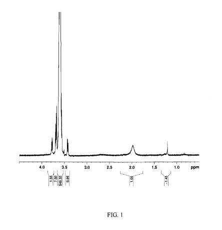
[0021] FIG. 1 shows an 1H-NMR spectrum for PEG-4000 (Fluka).
[0022] FIG. 2 shows an 1H-NMR spectrum for PLGA 50:50 Lactel.
[0023] FIG. 3 shows an 1H-NMR spectrum for CP-PLGA-pPEG-PLGA-1.
-4-
CA 02738766 2011-03-25
WO 2010/036961 PCT/US2009/058479
[0024] FIG. 4 shows an 1H-NMR spectrum used to detect the chain
transfer agent CP-PLGA-pPEG-PLGA-RAFT-funct.
[0025] FIG. 5 shows an 1H-NMR spectrum of S-(thiobenzoyl) thioglycolic
acid chloride DJS-CP-thiobenzoyl-thioglycolic acid chloride-1.
[0026] FIG. 6 shows an 1H-NMR spectrum for CP-PLGA-PEG-PLGA-CTA-
Cl-rxn-1.
[0027] FIG. 7 shows an 1H-NMR spectrum for CP-PGS-CTA poly(glycerol-
co-sebacic acid) functionalized with a S-thiobenzoyl-thioglycolic acid chain
transfer agent.
[0028] FIG. 8 shows a therapeutic agent release curve from hydrogel-
microparticles with 5 mg microparticles in 50 L hydrogel.
[0029] DETAILED DESCRIPTION OF THE PREFERRED
EMBODIMENTS
[0030] The words "a," and "one," as used in the claims and in the
corresponding portions of the specification, are defined as including one or
more
of the referenced item unless specifically stated otherwise.
[0031] As used herein, "matrix" refers to a hydrogel, particle, nanoparticle,
microparticle, or combinations thereof.
[0032] As used herein, "therapeutic agent" and "drug" are used
interchangeably.
[0033] As used herein, "injury" refers to injury caused by any means
including but not limited to physical trauma, disease, immune disease, or
inflammation.
[0034] As used herein, "patient" refers to a human or non-human animal
within the phylum chordata.
[0035] As used herein, "pharmaceutically acceptable salt" or
"pharmaceutically acceptable salts" means those salts of compounds that are
safe
and effective for use in a patient and that possess the desired biological
activity.
Pharmaceutically acceptable salts include salts of acidic or basic groups.
Pharmaceutically acceptable acid addition salts include but are not limited to
-5-
CA 02738766 2011-03-25
WO 2010/036961 PCT/US2009/058479
hydrochloride, hydrobromide, hydroiodide, nitrate, sulfate, bisulfate,
phosphate,
acid phosphate, isonicotinate, acetate, lactate, salicylate, citrate,
tartrate,
pantothenate, bitartrate, ascorbate, succinate, sodium succinate, maleate,
gentisinate, fumarate, gluconate, glucaronate, saccharate, formate, benzoate,
glutamate, methanesulfonate, ethanesulfonate, benzensulfonate, p-
toluenesulfonate and pamoate (i.e., 1,1'-methylene-bis-(2-hydroxy-3-
naphthoate))
salts. Pharmaceutically acceptable salts include salts with various amino
acids.
Pharmaceutically acceptable base salts include but are not limited to
aluminum,
calcium, lithium, magnesium, potassium, sodium, zinc, and diethanolamine
salts.
[0036] The embodiments herein provide a strategy for modulating post-
traumatic secondary injury by scavenging radicals in the injured site or site
of
inflammation through local administration of therapeutic agents. Due to
undesirable side-effects accompanying systemic administration of drugs (e.g.,
glucocorticoidal steroids), local administration of therapeutic agents,
including
anti-inflammatory drugs (e.g., minocycline or methylprednisolone), or free-
radical
scavengers (e.g., uric acid or tempol), to mitigate secondary injury could be
significant. To affect local administration of a therapeutic agent, a drug
delivery
system targeting processes responsible for nerve damage following injury is
provided. The processes targeted include oxidative stress resulting from
damage
caused by injury. Embodiments of the drug delivery system can be adapted for
treatment of the spinal cord after SCI. However, embodiments of the drug
delivery system may be used at any site of injury or inflammation. The site of
injury or inflammation may be the spinal cord or peripheral nerves. Methods of
treatment with the drug delivery system may be directed to intra-cellular
regions, extra-cellular regions, intravascular regions and/or cell membranes.
Embodiments of the drug delivery system and methods can address deleterious
effects of inflammation and these embodiments can be used for the treatment of
chronic inflammation, auto-immune disease, spinal cord injury (SCI), stroke,
myocardial infarction, chronic heart failure, diabetes, circulatory shock,
chronic
inflammatory diseases, cancer, neurodegenerative disorders, traumatic brain
injury, severing of peripheral nerves, nerve root impingment, and other
disorders
-6-
CA 02738766 2011-03-25
WO 2010/036961 PCT/US2009/058479
or traumatic injuries. The drug delivery system is a device that includes a
matrix and one or more therapeutic agents. The methods of treatment include
administering the drug delivery system.
[0037] The drug delivery system can include but is not limited to the
following combinations of matrix and therapeutic agent: 1) hydrogel plus
therapeutic agent; 2) hydrogel plus a combination of multiple therapeutic
agents;
3) particles and therapeutic agent; 4) particles plus multiple therapeutic
agents;
5) hydrogel plus particles plus therapeutic agent, where the agent is located
in
the hydrogel, particles or both; 6) hydrogel plus particles plus multiple
therapeutic agents, where the therapeutic agents are localized in the
hydrogel,
the particles (perhaps a distinct set of particles) within the hydrogel, or
both; and
7) hydrogel plus particles plus multiple therapeutic agents, where particular
therapeutic agents are localized in the hydrogel, the particles (perhaps a
distinct
set of particles) within the hydrogel, or both. The particles can be
microparticles
or nanoparticles. The therapeutic agent or therapeutic agents can be dissolved
or
dispersed in the hydrogel, the particles, or the hydrogel and particles for
controlled release kinetics via diffusion and/or dissolution. Preferably, the
matrix
is injectable and the drug delivery system can be delivered to a position at
the
site of injury through injection. The drug delivery system may, however, be
administered by other methods, which include but are not limited to surgical
implantation of the drug delivery device.
[0038] The hydrogel can be a temperature-sensitive biodegradable
composition. As used herein, "temperature sensitive" means that the hydrogel
exhibits a sol-gel phase transition between temperatures below patient body
temperature and the patient body temperature, or that mixtures of polymers
react to form a hydrogel at temperatures closer to patient body temperature
more
readily than at lower temperatures. Preferably, the patient is human, the body
temperature is 37 , and the lower temperature can be room temperature (e.g.,
22 C). Temperature-sensitive hydrogels may have a critical temperature closer
to the body temperature of the patient, preferably closer to 37 C.
-7-
CA 02738766 2011-03-25
WO 2010/036961 PCT/US2009/058479
[0039] A biodegradable, temperature-sensitive hydrogel in the drug
delivery system can include multi-block co-polymers. One or more of the
polymer
blocks can be biodegradeable, biocompatible, or biodegradeable and
biocompatible. Some of the polymer blocks can be biodegradable while others
are
biocompatible. Preferably, the hydrogel components, for example polymers,
monomers or breakdown products, are biodegradeable; biocompatible; or
biocompatible and excretable. The hydrogel may include biocompatible polymer
blocks that can be excreted by the body.
[0040] Ester links between monomer blocks are hydrolysable and can be
degraded in vivo to release polymer monomer blocks. Polymers containing ester
bonds are biodegradable. Amide, anhydride, and ether links may also be
hydrolysable. These links and others that can be degraded by action of
enzymes,
reducing conditions (e.g., thioester, thioether, disulfide links) or
conditions
present within the patient may also be used in the polymers contemplated for a
hydrogel or particle. The hydrogel may contain polymers blocks having ethylene
glycol with ether links, oligoethylene glycol, or polyethylene glycol, which
are
biocompatible and can be excreted. Polymers of glycolic acid, lactic acid,
glycerol,
and sebacic acid are biodegradeable hydrogels may include these polymers.
Lactide, glycolide, poly(glycerol-co-sebacic acid) polymers are biodegradeable
and
one or more of these polymers can be included in the hydrogel. Polymers
contemplated as part of the hydrogel include but are not limited to
poly(glycerol-
co-sebacic acid) acrylate; multiblock copolymers of poly(lactide-co-glycolide)
and
poly(ethylene glycol) or oligo (ethylene glycol) methyl methacrylate; and
graft
copolymers of poly(glycerol-co-sebacic acid) and poly(ethylene glycol), oligo
(ethylene glycol) methyl methacrylate or poly(N-isopropylacrylamide),
ethoxylated trimethylolpropane tri-3- mercaptopropionate, or poly(ethylene
glycol)
diacrylate. These polymers are temperature- sensitive.
[0041] Hydrogels can swell or shrink under changing physical conditions,
which are of physiologic significance. For example changes in temperature, pH
or ionic strength can cause a hydrogel to swell or shrink. Preferably, a
hydrogel
injected into the spinal cord or other site of injury does not swell
significantly
-8-
CA 02738766 2011-03-25
WO 2010/036961 PCT/US2009/058479
upon equilibrating to conditions within the injury site. A swelling hydrogel
may,
however, be included in the drug delivery system.
[0042] Hydrophilic polymers containing ethylene glycol monomer units
with reactive end groups, which include but are not limited to acrylate,
methacrylate,vinyl, dihydrazide or thiol groups, can be used as polymers in
either
a hydrogel or particle. Acrylochloride, methacrylochloride, vinyl chloride can
be
used to form the acrylate or methacrylate end groups. Thiol end groups can be
provided by mercaptopropionic acid, cysteine, and cystamine. Multiple
synthesis
methods, such as ring-opening-polymerization and living radical polymerization
may be employed to produce polymers for the matrix. The hydrogel may be
composed of a covalently or physically cross-linked network. Physical cross
links
refer to the aggregation of hydrophic blocks.
[0043] Polymers having acrylate, methacrylate, vinyl, dihydrazide or thiol
functionalized compounds are capable of reacting with other polymers having
compatible reactive end groups to form hydrogels. Acrylate, methacrylate and
vinyl reactive end groups are all compatible with each other and with thiols.
Thiol and acrylate functionalized polymers or polymer blocks are capable of
reacting to form thiol-ethers under mild conditions (heat or light).
Therefore,
thiol and acrylate functionalized water soluble polymers are suitable
candidates
for hydrogels in the drug delivery system. Some hydrogels can swell or shrink
upon equilibration following gelation, which may be due to incomplete
conversion
or to the high concentration of reactants required for a sufficiently rapid
reaction
compared to subsequent equilibrium concentrations in the gel under
physiological conditions (e.g., temperature, pH, ionic strength). The thiol-
acrylate functionalized polymers are attractive polymers in drug delivery
devices,
due to rapid reaction rates and high extents of conversion to hydrogel. A
hydrogel for the drug delivery system may be formed by mixing polymers with
compatible reactive end groups. Preferably, the mixture includes a thiol
containing polymer and an acrylate containing polymer. After mixing, the
formation of thiol-esters forms the hydrogel when the combination is exposed
to
sufficient temperatures or light. Preferably, thiol-ester formation occurs
more
-9-
CA 02738766 2011-03-25
WO 2010/036961 PCT/US2009/058479
rapidly at body temperature of the patient then at temperatures lower than the
body temperature of the patient. For example, the thiol-ester formation may
proceed more rapidly at or near 37-C than at or near 22 C. The polymers with
compatible reactive end groups, preferably thiol containing and acrylate
containing polymers, can be mixed prior to or during administration. When
mixed during administration, the polymers can be mixed while being implanted,
or mixed by serial or parallel injections of the different polymers. A non-
limiting
example of such a mixture includes ethoxylated trimethylolpropane tri-3-
mercaptopropionate in combination with poly(ethylene glycol) diacrylate.
[0044] A hydrogel in the drug delivery system preferably has a compressive
modulus similar to that of tissue surrounding the injury site. For example, a
drug delivery system designed to be delivered to the spinal cord can have a
compressive modulus similar to that of the spinal cord. The porosity of a
hydrogel in the drug delivery system can be matched to the size of the
therapeutic agent to be released. If a 500 dalton thereapeutic agent is part
of the
drug delivery system, the mesh of the hydrogel should allow a 500 dalton
therapeutic agent to migrate through the gel.
[0045] The matrix may include particles containing drug and the particles
may provide for controlled release kinetics of the drug. In a preferred
embodiment, the particles can be injected as a suspension into the area of
damage, which may be at a peripheral nerve or the spinal cord. The particles
can
be microparticles, ranging in size from about 1 micron to about 1000 microns.
The particles can be nanoparticles, ranging in size from about 1 nanometer to
about 1000 nanometers. The particle dimensions can, however, vary to suit the
particular application. The therapeutic agent can be present on or within the
particle at a concentration effective to achieve the effect of scavenging
radicals,
preventing the formation of radicals, or otherwise counteracting the toxic
effects
of nitric oxide associated oxidative stress. In preferred embodiments, the
particle
contains therapeutic agent at 0.1 - 30%, more preferably 1-30 % w/w ((weight
of
drug)/(weight of particle plus drug)). The therapeutic agent can be released
by
mechanisms of diffusion, dissolution, and particle degradation.
-10-
CA 02738766 2011-03-25
WO 2010/036961 PCT/US2009/058479
[0046] The particle may be a solid polymer or a gel. In a preferred
embodiment, particles are made of a biodegradable, biocompatible polymer,
which can be, for example, a polyester. One suitable polyester for a particle
is
poly(lactide-co-glycolide) (PLGA), which degrades by ester hydrolysis. Other
suitable materials include polylactide, polyglycolide, and poly(carboxyphenoxy
propane)-co-sebacic acid) (e.g., gliadel waferTM from MGI Pharmaceuticals).
Preferably, the particle components, for example polymers, monomers or
breakdown products, are biodegradeable; biocompatible; or biocompatible and
excretable.
[0047] A combination of hydrogel and particles can be provided to the
combination to provide the ability to decouple release kinetics from in-situ
gelling. Through decoupling of release kinetics, the release of therapeutic
agent
from particles versus hydrogel may occur at different rates. In a preferred
embodiment, different therapeutic agents may be included in the hydrogel or
particles, or different types of particles, such that each particular
therapeutic
agent is released at a different rate.
[0048] In an embodiment, hydrogel or particle material is functionalized
with a therapeutic agent. The therapeutic agent may be attached to by any type
of bond to functionalize the particle. Preferably, the attachment is through
carboxyl or hydroxyl groups of polymer repeating units through ester, amide,
ether, or acetal bonds. In a preferred embodiment, a functionalized hydrogel
includes therapeutic agent attached to a poly(glycerol-co-sebacate)acrylate
(PGSA).
[0049] Any therapeutic agent can be used to functionalize a hydrogel or
particles, but in a preferred embodiment the therapeutic agent is attached to
a
hydrogel and the therapeutic agent is an antioxidant. More preferably, the
antioxidants ascorbic acid (vitamin C) and alpha-tocopherol (vitamin E) are
attached to the hydrogel. Vitamin C and vitamin E antioxidants when present in
combination can recycle each other and antioxidant properties can be extended.
Other therapeutic combinations can be employed to effect recycling in the drug
delivery system.
-11-
CA 02738766 2011-03-25
WO 2010/036961 PCT/US2009/058479
[0050] PGSA can form elastomeric networks under mild conditions (radical
polymerization), which can protect the antioxidant from denaturing during
processing. In addition, a scaffold of arbitrary geometry can be formed from
PGSA using melt molding or solid free form rapid prototyping techniques. This
can be used to customize the drug delivery system to a particular lesion
cavity (or
spinal cord tumor cavity), resulting in an improvement of surgical
intervention to
treat injury, which may be SCI or peripheral nerve injury.
[0051] Table I, below, lists an exemplary formulation for the following
seven drug delivery system combinations: 1) hydrogel plus therapeutic agent;
2)
hydrogel plus a combination of multiple therapeutic agents; 3) particles and
therapeutic agent; 4) particles plus multiple therapeutic agents; 5) hydrogel
plus
particles plus therapeutic agent, where the agent is located in the hydrogel,
particles or both; 6) hydrogel plus particles plus multiple therapeutic
agents,
where the therapeutic agents are localized in the hydrogel, the particles
(perhaps
a distinct set of particles) within the hydrogel, or both; and 7) hydrogel
plus
particles plus multiple therapeutic agents, where particular therapeutic
agents
are localized in the hydrogel, the particles (perhaps a distinct set of
particles)
within the hydrogel, or both.
-12-
CA 02738766 2011-03-25
WO 2010/036961 PCT/US2009/058479
m
~ x x x x x
pq ct pq ct pq ct pq ct pq ct
O w~ w~ a~ a~ a ~
0 0 0 0 0 0 0 0 0 0 0
~, m m m m m m m
bA o + o +; o +; o +; o +; o +; o +;
D o D o D o D o D o D o D o
0 0 0 0
0 0 0 0 0
o o o o o
o o o o o
O U O U O U O U O
I-I = O O O O O
E-~
o o o o o 0 00 00 0 0
N N N W N
~ W PW~ W PW~ W PW~ W 0.' W PW~
0 0 0 0 0 0 0 0 0 0 0
LO LO LO LO LO LO LO LO LO LO
O x cq
~ 0 0 0 0 0 0 0
CD
X1.1 ..~ ..~ ..~ ..~ ..~ ..~ ..~
F O O O O O O O
I--~ U U U U U U U
CA 02738766 2011-03-25
WO 2010/036961 PCT/US2009/058479
[0052] In the examples of Table I, wt % is calculated as the weight of the
constituent divided by the weight of the combination, which includes the other
constituents. The PBS, pH 7.4 is phosphate buffered saline at pH 7.4, which
includes 144 mg/L (1.06 mM) potassium phosphate monobasic (KH2PO4, 136
g/mol), 9000 mg/L (155.17 mM) sodium chloride (NaCl, 58 g/mol), and 795 mg/L
(2.97 mM) sodium phosphate dibasic (Na2HPO4-7H20).
[0053] Any molecule acting as a radical scavenger or anti-inflammatory
agent is a candidate therapeutic agent for the drug delivery system.
Preferably,
the molecule is a small molecule. Therapeutic agent(s) can preferably reduce
the
number of free-radicals and/or reduce the production of free radicals at a
locale
within the body. The drug delivery system can include combinations of more
than one therapeutic agent. The therapeutic agent can be an antioxidant, a
steroid, or combinations thereof. In a preferred embodiment, the therapeutic
agent includes one or more substance selected from the group of an antioxidant
or antioxidants, tempol (4-hydroxy-2,2,6,6-tetramethylpiperidine-l-oxyl), uric
acid, minocycline, methylprednisolone, MnTBAP (Manganese (III) tetrakis (4-
benzoic acid)porphyrin), and dexamethasone. The antioxidant may be, but is not
limited to ascorbic acid or alpha-tocopherol. Combinations of therapeutic
agents
that can recycle each other may also be provided in the drug delivery system.
For
example, ascorbic acid (vitamin C) and alpha-tocopherol (vitamin E) can be
used
in combination to recycle each other and extend antioxidant properties.
[0054] Therapeutic agent(s) are not limited to those above. Non-limiting
examples of therapeutic agents that could be included in the drug delivery
system include inhibitors of NOS or NO production, antioxidants, spin traps,
and
peroxynitrite scavengers.
[0055] A non-limiting list of inhibitors of NOS or NO production that can be
provided in the drug delivery system includes 1400W (N-(3-
(aminomethyl)benzyl)acetamidine); actinomycin D; AET; ALLM; ALLN; NG-allyl-
L-arginine; aminoguanidine, hemisulfate; 1- amino- 2-hydroxyguanidine; p-
-14-
CA 02738766 2011-03-25
WO 2010/036961 PCT/US2009/058479
toluenesulfonate; 2-amino-4- methylpyridine; AMITU; AMT; S-benzylisothiourea;
bromocriptine mesylate; L-canavanine sulfate; canavalia ensiformis;
chlorpromazine, hydrochloride; curcumin; curcuma longa L; cycloheximide; high
purity cycloheximide; cyclosporine; dexamethasone, 2,4-diamino-6-
hydroxypyrimidine; NG,NG-dimethyl-L-arginine; NG,NG1-dimethyl-L-arginine;
diphenyleneiodonium; DMHP, S(-)- epigallocatechin gallate; S-ethyl-N-
phenylisothiourea; 2-ethyl-2-thiopseudourea; ETPI ; basic fibroblast growth
factor; bovine basic fibroblast growth factor; human recombinant basic
fibroblast
growth factor, GED; haloperidol; L-N6-(1 -lminoethyl)lysine, dihydrochloride;
L-
N5-(1-lminoethyl)ornithine; LY83583; LY231617; MEG; melatonin; S-
methylisothiourea sulfate; S-metlhyl-L-thiodtrulline, dihydrochloride; NG-
monoethyl-L-arginine; NG-monomethyl-D-arginine monoacetate; DiHABS (di-
hydroxyazobenzene-p'-sulfonate) salt of NG-monomethyl-L-argninine; NG-
monomethyl-L-argninine; monohydrate HABS salt of NG-monomethyl-L-
argninine; NG-monomethyl-L-homoarginine; mycophenolic acid, L-NIL; inducible
nitric oxide synthase inhibitor set (Calbiochem ); neuronal nitric oxide
synthase
inhibitor set (Calbiochem ); NG-nitro- D-arginine; NG -nitro -L- arginine; NG-
nitro-
D-arginine methyl ester; NG-nitro-L-arginine methyl ester; p-nitrolue
tetrazolium
chloride; 7-nitroindazole; sodium salt of 7-nitroindazole; 3-bromo-7-
nitroindazol;
sodium salt of 3-bromo-7-Nitroindazol; NOS inhibitor set (Calbiochem ), 1,3-
PBITU; pentamidine isethionate; PPM-18; NG-propyl-L-arginine; 1-
pyrrolidinecarbodithioic acid; SKF-525A ; SKF-96365; sodium salicylate;
spermidine; trihydrochloride spermidine; spermine; spermine
tetrahydrochioride;
L-thiocitrulline; Na-tosyl-Lys chloromethyl ketone; Na-tosyl-Phe chloromethyl
ketone; TRIM; and zinc (II) Protoporphyrin IX. Pharmaceutically acceptable
salts of an inhibitor or inhibitors of NOS or NO production can be included in
the
drug deliver system.
[0056] A non-limiting list of antioxidants that can be in the drug delivery
system includes N-acetyl-L-cysteine; N-acetyl-S-farnesyl-L-cysteine; AG 1714;
ambroxol hydrochloride; antioxidant set (Calbiochem ); L-ascorbic acid;
bilirubin,
bilirubin free acid, caffeic acid, CAPE; carnsol; (+)-catechin; ceruloplasmin;
-15-
CA 02738766 2011-03-25
WO 2010/036961 PCT/US2009/058479
human plasma ceruloplasmin; coelenterazine; copper diisopropylsalicylate;
deferoxamine mesylate; R-(-)-deprenyl hydrochloride; DMNQ; DTPA;
dianhydride; ebselen; ellagic acid; dehydrate ellagic acid; (-)-
epigallocatechin
gallate; L-ergothioneine; dihydrate EUK-8; apo-ferritin, equine spleen apo-
ferritin; cadmium free ferritin; equine spleen cadmium free ferritin; human
liver
ferritin; human recombinant ferritin H-chain; human recombinant ferritin L-
chain; formononetin; reduced glutathione; reduced glutathione free acid;
gluthathione monoethyl ester; a-lipoic acid; dihydro-DL-a-lipoic acid;
luteolin, LY
231617; penicillamine; MCI-186; MnTMPyP, morin hydrate; NCO-700; NDGA; p-
nitroblue tetrazolium chloride; 0-trensox; propyl gallate; resveratrol;
rosmarinic
acid; (+)-rutin hydrate; silymarin group; L-stepholidine; stephania
intermedica;
( )-taxifolin; tetrandrine; DL-thioctic acid; thioredoxin; human recombinant
low
endotoxin thioredoxon; thioredoxin II; yeast thioredoxin II; recombinant yeast
thioredoxin II; DL-a-tocopherol; tocopherol set (Calbiochem ); DL-a-tocopherol
acetate, tocotrienol set (Calbiochem ), Trolox ; U-74389G; U-83836E; uric
acid;
and vitamin E succinate. Pharmaceutically acceptable salts of an antioxidant
or
antioxidants can be included in the drug deliver system.
[0057] Spin trap agents that can be in the drug delivery system include but
are not limited to N-tert-butyl-a-phenylnitrone, tempol, and DTCS (Iron (II) N-
(dithiocarboxy) sarco sine Fe2+). Pharmaceutically acceptable salts of a spin
trap
or spin traps can be included in the drug deliver system.
[0058] Peroxynitrite Scavengers that can be in the drug delivery system
include but are not limited to ebselen; FeTMPyP; FeTPPS; reduced glutathione;
reduced glutiathione free acid; melatonin; MnTBAP; MnTMPyP; L-
selenomethionine; and Trolox . Pharmaceutically acceptable salts of a
peroxynitrite scavenger or peroxynitrite scavengers can be included in the
drug
deliver system.
[0059] Therapeutic agent(s) can be provided at any concentration effective
to scavenge radicals, prevent formation of radicals, or otherwise counteract
the
toxic effects of nitric oxide associated stress. Preferably, therapeutic
agent(s) are
present in the drug delivery system at a concentration of 0.1- 30% w/v (weight
of
-16-
CA 02738766 2011-03-25
WO 2010/036961 PCT/US2009/058479
drug/volume of drug delivery system). The concentration of selected
therapeutic
agents in a drug delivery device is provided in Table II, below.
Table II
Therapeutic Agent Suggested Concentration Suggested
Range Concentration.
Vitamin C 0.1 - 30% (w/v) 0.1% (w/v)
Vitamin C and vitamin E 0.1 - 30% (w/v) vitamin 0.2% (w/v) (0.1% vitamin
C and 0.1 - 30% (w/v) C and 0.1% vitamin E)
vitamin E
Tempol 0.1 - 30% (w/v) 0.1% (w/v)
Uric acid 0.1 - 30% (w/v) 0.1% (w/v)
Minocycline 0.1 - 30% (w/v) 0.1% (w/v)
Methylprednisolone 0.1 - 30% (w/v) 0.1% (w/v)
MnTBAP 0.1 - 30% (w/v) 0.1% (w/v)
Dexamethasone 0.1 - 30% (w/v) 0.1% (w/v)
[0060] The drug delivery system may include pharmaceutical additives
such as carriers and the like. The term "carrier" as used herein includes
acceptable adjuvants and vehicles. Pharmaceutically acceptable carriers can be
selected from but are not limited to those in the following list: ion
exchangers,
alumina, aluminum stearate, lecithin, serum proteins, human serum albumin,
buffer substances, phosphates, glycine, sorbic acid, potassium sorbate,
partial
glyceride mixtures of saturated vegetable fatty acids, water, salts or
electrolytes,
protamine sulfate, disodium hydrogen phosphate, potassium hydrogen
phosphate, sodium chloride, zinc salts, colloidal silica, magnesium
trisilicate,
polyvinyl pyrrolidone, cellulose-based substances, polyethylene glycol, sodium
carboxymethylcellulose, waxes, and polyethylene glycol.
[0061] The therapeutic agent can be provided in a small volume of diluent
as the carrier. For example, a one ml methylprednisolone sodium succinate
(i.e.,
pregna-1, 4-diene-3,20-dione,21-(3-carboxy-1-oxopropoxy)-11,17-dihydroxy-6-
methyl-monosodium salt, (6a, 11[3) (molecular weight 496.53)) therapeutic
agent
-17-
CA 02738766 2011-03-25
WO 2010/036961 PCT/US2009/058479
solution could include 40 mg methylprednisolone sodium succinate; 1.6 mg
monobasic sodium phosphate anhydrous; 17.46 mg dibasic sodium phosphate
dried; 25 mg lactose hydrous; and 8.8 mg benzyl alcohol as preservative. When
necessary, the pH of each formula can be adjusted. For example, sodium
hydroxide could be added so that the pH of the reconstituted solution is
within a
range of 7 to 8 and the tonicities are, for the 40 mg per mL
methylprednisolone
sodium succinate solution, 0.50 osmolar. The conditions within a matrix maybe
designed to be the same, similar, or different than a diluent or solution that
the
therapeutic agent exists in prior to its addition to matrix. For example, a
one ml
volume of matrix with methylprednisolone sodium succinate could be designed to
contain the same amount of methylprednisolone sodium succinate, monobasic
sodium phosphate, dibasic sodium phosphate, lactose hydrous, benzyl alcohol,
pH, and water as the diluent above. Other diluents may be utilized. The
therapeutic agent can be provided in any pharmaceutically acceptable carrier.
For example, the therapeutic agent can be provided in a buffered diluent, for
example phosphate buffer or phosphate buffered saline.
[0062] The drug delivery system can be assembled by dissolving or soaking
polymer in a therapeutic agent solution. When combinations of different
particles or particle and hydrogel are used, the matrix components can be
exposed to the therapeutic agent as a combination or separately. If different
therapeutic agents are intended for particles, different subsets of particles,
or
hydrogels, these components can be exposed to the respective therapeutic agent
solution prior to combining all of the polymer components.
[0063] Computational models of degradation, drug release and in vivo
distribution have been developed to predict the effect of various design
parameters on the spatial-temporal drug profile of the drug delivery systems.
Parameters include polymer composition, molecular weight, polydispersity, drug
type, drug size, drug-polymer interactions and geometry of the drug delivery
system. The parameters can be adjusted to optimize the drug delivery system.
[0064] As illustrated in example 7, below, ethoxylated trimethylolpropane
tri-3-mercaptopropionate with a MW = 1300 g/mol mixed with poly(ethylene
-18-
CA 02738766 2011-03-25
WO 2010/036961 PCT/US2009/058479
glycol) diacrylate with a MW 400 g/mol as a hydrogel and a PLGA polymer with a
MW 11,600 g/mol is a non-limiting example of a hydrogel and particles with
parameters suitable for an embodiment of the drug delivery system. Hyrdrogels
and particles with other parameters may be utilized in a drug delivery system.
[0065] The drug delivery system can be injected into a patient at an area at
the site of injury or inflammation, or deposited into or at the site following
surgery to expose the area. By injection, rather than surgical intervention,
the
drug delivery system can be administered in a minimally invasive manner. In a
preferred embodiment, only one administration would be necessary to maintain a
sustained dosage. The drug can thus be delivered directly to the point of
injury
or inflammation, thereby minimizing side-effects related to systemic
administration. Preferably, but not exclusively, a hydrogel made by a
combination of ethoxylated trimethylolpropane tri-3-mercaptopropionate with
poly(ethylene glycol) diacrylate may be utilized in this method. Non-limiting
examples of such a drug delivery device are provided in Table I.
[0066] One method of treatment is injection of the drug delivery system
into a contusion injury in the spinal cord. This can be accomplished by
intradural intramedullary injection. By injecting, or otherwise implanting,
the
drug delivery system into the spinal cord, neither the drug nor elements of
drug
delivery system has to cross the dura and the drug does not have to cross-the
blood brain barrier. Preferably, the drug delivery system is designed to
release
the therapeutic agent over a sustained period of time, synchronized with the
pathophysiological increased and temporally sustained levels of free radicals
and
free radical production at the injury site, which is due in part to microglial
activation and neutrophil infiltration. Preferably, but not exclusively, a
hydrogel
made by a combination of ethoxylated trimethylolpropane tri-3-
mercaptopropionate with poly(ethylene glycol) diacrylate may be utilized in
this
method. Non-limiting examples of such a drug delivery device are provided in
Table I.
[0067] In preferred embodiments, the drug delivery system is designed to
degrade during treatment by hydrolysis and be excreted by the body via normal
-19-
CA 02738766 2011-03-25
WO 2010/036961 PCT/US2009/058479
pathways, without the need for further surgical intervention. Preferably, but
not
exclusively, a hydrogel made by a combination of ethoxylated
trimethylolpropane
tri-3-mercaptopropionate with poly(ethylene glycol) diacrylate may be utilized
in
this method. Non-limiting examples of such a drug delivery device are provided
in Table I.
[0068] Potential Drug Delivery System Tests
[0069] Therapeutic agents, matrices and combinations thereof can be
tested by methods known in the art. A range of nitric oxide donors, such as
SIN-
1 hydrochloride, can be used in vitro to produce sustained levels of
peroxynitrite
and radicals due to peroxynitrite decomposition. Antioxidant activity can then
be
assayed by measuring nitrite using numerous methods, for example via modified
Griess Reagent. A typical commercial Griess reagent contains 0.2%
naphthylenediamine dihydrochloride, and 2% sulphanilamide in 5% phosphoric
acid. Cell integrity in vitro can be assayed using MTT or MTS assays for
mitochondrial activity of lactate dehydrogenase (LDH) for cell membrane
integrity. See Mosmann, T. (1983) Rapid Colorimetric Assay for Cellular Growth
and Survival: Application to Proliferation and Cytotoxicity Assays. J.
Immunol.
Meth. 65, 55-63; and Wilson, A. P. (2000) Cytotoxicity and Viability Assays in
Animal Cell Culture: A Practical Approach, 3rd ed. (ed. Masters, J. R. W.)
Oxford
University Press: Oxford 2000, Vol. 1, which are incorporated by reference as
if
fully set forth. A drug delivery system can also be tested for its ability to
reduce
cell death or cell membrane damage in vitro, and also elimination of nitrites
produced by donors such as SIN-1. The efficacy of a therapeutic agent, matrix,
or
drug delivery system can be assayed after in vivo studies via immunostaining
or
using markers for peroxinitrite oxidative stress. Markers include but are not
limited to 3-nitrotyrosine and 4-hydroxynonenal.
[0070] The spatial and temporal characteristics of peroxynitrite-derived
oxidative damage after a moderate contusion injury in rats was described in
Xiong, Y, Rabchevsky, A.G. and Hall, E.D. (2007) Role of peroxynitrite in
secondary oxidative damage after spinal cord injury. J. Neurochem. 100 (1),
639-
649 ("Xiong et al."), which is incorporated herein as if fully set forth.
Xiong et al.
-20-
CA 02738766 2011-03-25
WO 2010/036961 PCT/US2009/058479
showed that 3-nitrotyrosine, a specific marker for peroxynitrite, rapidly
accumulated at early time points (1 and 3 h) and significantly increases in 3-
nitrotyrosine were sustained out to 1 week after injury in comparison to sham
rats. Additionally, Xiong et al. showed a coincident and maintained increase
in
the levels of protein oxidation-related protein carbonyl and lipid
peroxidation-
derived 4-hydroxynonenal. The peak increases of 3-nitrotyrosine and 4-
hydroxynonenal were observed at 24 h post-injury. In immunohistochemical
results, Xiong et al. showed the co-localization of 3-nitrotyrosine and 4-
hydroxynonenal, indicating that peroxynitrite is involved in lipid
peroxidative as
well as protein nitrative damage. Another consequence of oxidative damage is
an
exacerbation of intracellular calcium overload, which activates the cysteine
protease calpain leading to the degradation of several cellular targets
including
cytoskeletal protein (a-spectrin). Xiong et al. also showed, through analysis
of a-
spectrin breakdown products, that the 145-kDa fragments of a-spectrin, which
are specifically generated by calpain, were significantly increased as soon as
1 h
following injury although the peak increase did not occur until 72 h post-
injury.
Xiong et al. concluded that the later activation of calpain was most likely
linked
to peroxynitrite-mediated secondary oxidative impairment of calcium
homeostasis. Candidate therapeutic agents, matrices and combinations may be
tested for markers described in Xiong et al. Numerous methods of assaying
markers may be employed, including the methods described by Xiong et al. The
methods described in Xiong et al. included a rat model of traumatic spinal
cord
contusion, immunobolotting analysis for 3-nitrotyrosine and 4-hydroxynonenal,
western blotting for a-spectrin breakdown products, and statistical analysis,
as
follows.
[0071] A rat model of traumatic spinal cord contusion: According to Xiong
et al., all studies described therein employed young adult female Sprague-
Dawley rats (Charles River, Portage, MI, USA) weighing between 200 and 225 g.
The animals were randomly cycling and were not tested for stage of the estrus
cycle. They were fed and watered ad libitum. Rats were anesthetized with
ketamine (80 mg/kg) and xylazine (10 mg/kg) before a laminectomy of the T10
-21-
CA 02738766 2011-03-25
WO 2010/036961 PCT/US2009/058479
vertebrae was performed. Spinal cord injury was performed using the Infinite
Horizon device (Scheff, S.W., Rabchevsky, A.G., Fugaccia, I., Main, J.A., and
Lumpp, J.E. Jr. (2003) Experimental modeling of spinal cord injury:
characterization of a force-defined injury device. J. Neurotrauma 20, 179-193,
which is incorporated by reference as if fully set forth), which creates a
reliable
contusion injury to the exposed spinal cord by rapidly applying a force-
defined
impact with a stainless steel-tipped impounder. Care was taken to perform
laminectomies that were slightly larger than the 2.5-mm impactor tip. The
vertebral column was stabilized by clamping the rostral T9 and caudal T11
vertebral bodies with forceps. The vertebral column and exposed spinal cord
were
carefully aligned in a level horizontal plane. During impact, the stepping
motor
drove the coupled rack toward the exposed spinal cord inflicting the contusion
injury. The force applied to spinal cord was 200 kdyn, which produced a
moderately severe injury. The impactor device was connected to a PC that
recorded the impounder velocity, actual force, and displacement of the spinal
cord.
[0072] At different time points following surgery (1, 3, 6, 24, 48, 72 h, and
1
week), animals in a first set (six rats per time point) were killed by sodium
pentobarbital overdose (150 mg/kg). A 20-mm segment of spinal cord containing
the impact epicenter was removed rapidly by laminectomy. The harvested tissue
was dissected on a chilled stage and immediately transferred to a centrifuge
tube
containing 800 pL Triton lysis buffer [20 mmol/L Tris-HC1, 150 mmol/L NaCl,
1% Triton X-100, 5 mmol/L EGTA, 10 mmol/L EDTA, 20 mmol/L HEPES, 10%
solution of glycerol, and protease inhibitor cocktail (Roche Inc., Nutley, NJ,
USA)]
and then briefly sonicated. Following dismembranation, the spinal cord tissue
samples were centrifuged at 15,000 rpm for 1 h at 4 C, the supernatant was
collected, protein levels were determined using the Protein Assay Kit (Pierce
Biotechnology, Inc., Rockford, IL, USA), the samples were then normalized to 1
pg/pL and stored at -80 C until assay. Oxidative damage was assessed by slot
immunoblotting. A 2-pg protein sample was loaded on slot-blot apparatus for
optimal antibody-binding sensitivity. For lipid peroxidation, rabbit
polyclonal
-22-
CA 02738766 2011-03-25
WO 2010/036961 PCT/US2009/058479
anti-HNE antibody was applied (1 : 5000; Alpha Diagnostics International,
Inc.,
San Antonio, TX, USA). For peroxynitrite-generated 3-nitrotyrosine, rabbit
polyclonal anti-nitrotyrosine antibody was employed (1 : 2000; Upstate USA,
Inc.,
Charlottesville, VA, USA). To detect protein oxidation, the oxy-blot technique
was used (Oxy-Blot Protein Oxidation Detection Kit; Chemicon International,
Temecula, CA, USA). The slot-blot analyses were analyzed using the Li-Cor
Odyssey Infrared Imaging System (LI-COR Biosciences, Lincoln, NE, USA),
which employs IRDye800-conjugated goat-anti-rabbit IgG (1 : 5000; Rockland,
Gilbertsville, PA, USA) as the secondary antibody. Preliminary studies were
conducted to determine the linear range of the densitometry curve for each of
the
oxidative markers and, thus, verify that the densitometric readings obtained
were not beyond the range of accurate quantification.
[0073] Immunohistochemistry for 3-nitrotyrosine and 4-hydroxynonenal:
According to Xiong et al., at different time points following surgery (1, 3,
6, and
24 h), animals in a second set were overdosed with sodium pentobarbital (150
mg/kg) and perfused with 150 mL of 0.1 mol/L phosphate buffered saline (PBS)
followed by 200 mL of 4% paraformaldehyde in PBS (pH = 7.4). For cross-
sections, a 5-mm spinal cord segment, centered on the injury epicenter, was
dissected at different time points. For longitudinal sections, a 15-mm spinal
cord
segment including the impact site was dissected 24 h after injury. After
harvesting, the spinal cords were immersed in 4% paraformaldehyde in PBS for 4
h. The tissues were then transferred to PBS overnight and cryopreserved in
phosphate-buffered 20% sucrose for 2 days. Spinal cords were sectioned at 20
pm
in a transverse or longitudinal plane, and every fifth section was transferred
directly onto Superfrost plus slides (Fisher Scientific International Inc.,
Hampton, NH, USA). After collecting all the spinal cord sections, the slides
were
placed on a tray and stored at 4 C to dehydrate overnight after which they
were
stored at -20 C until staining. On the day of staining, the frozen slides were
removed from -20 C and thawed at 20 C for 30 min. After rinsing in 0.2 mol/L
of
PBS, the sections were incubated in 3% hydrogen peroxide in 0.2 mol/L of PBS
for 30 min, followed by incubation in blocking buffer (5% goat serum, 0.25%
-23-
CA 02738766 2011-03-25
WO 2010/036961 PCT/US2009/058479
Triton-X, 1% dry milk in 0.2 mol/L PBS) for 1 h, followed by the exposure to
either the rabbit polyclonal anti-4-hydroxynonenal (1 : 5000) or anti-3-
nitrotyrosine antibody (1 : 2000) overnight. The following day, sections were
incubated for 2 h at 20 C with biotinylated goat-anti-rabbit secondary
antibody
(1 : 200, Vector ABC-AP Kit; Vector Labs, Burlingame, CA, USA). After rinsing,
the sections were incubated in VECTASTAIN ABC reagent (avidin DH plus
biotinylated horseradish peroxidase, Vector Labs) for 1 h followed by
development of the staining using the Vector blue method (Vector Blue Alkaline
Phosphatase Substrate Kit; Vector Labs) in the dark for 10-30 min. After
reaction, spinal cord sections were counterstained with nuclear fast red
(Vector
Labs), dehydrated and then photographed on an Olympus Provis A70 microscope
with an Olympus Magnafire digital camera (Olympus America, Inc., Melville,
NY, USA).
[0074] Western blotting for a-spectrin breakdown products: Xiong et al.
stated that fifteen micrograms of each sample were run on sodium dodecyl
sulfate-polyacrylamide gel electrophoresis [3-8% (w/v) acrylamide, Bio-Rad
Criterion XT precast gel] with a Tris-acetate running buffer system and then
transferred to nitrocellulose membranes using a semi-dry electro-transferring
unit (Bio-Rad Laboratories, Hercules, CA, USA) at 20 mA for 15 min. The blots
were probed with mouse monoclonal anti-a-spectrin antibody (1 : 5000,
Affiniti,
Inc., Ft. Lauderdale, FL, USA; now part of Biomol International, LP Plymouth,
PA, USA), which recognizes an epitope that is common to the 280 kDa parent a-
spectrin as well as each of the 150- and 145-kDa proteolytic fragments.
Exposure
to the primary antibody was followed by application of the secondary IRDye800-
conjugated goat-anti-mouse IgG (1 : 5000, Rockland) for 1 h in the dark.
Imaging analysis of western blots was performed using the Li-Cor Odyssey
Infrared Imaging System, to quantify the content of the 145 and 150 kDa a-
spectrin breakdown products (SBDP 145 and SBDP150). Each western blot
included a standardized protein loading control to allow for correction in
regard
to intensity differences from blot to blot. This quantitative method has been
employed in other studies (Kupina, N.C., Nath R., Bernath E.E., Inoue J.,
-24-
CA 02738766 2011-03-25
WO 2010/036961 PCT/US2009/058479
Mitsuyoshi A., Yuen, P.W., Wang, K.K., and Hall E.D. (2002)
Neuroimmunophilin ligand V-10,367 is neuroprotective after 24-hour delayed
administration in a mouse model of diffuse traumatic brain injury. J. Cereb.
Blood Flow Metab. 22, 1212-1221; Hall E.D., Sullivan, P.G., Gibson, T.R.,
Pavel,
K.M., Thompson, B.M., and Scheff, S.W. (2005) Spatial and temporal
characteristics of neurodegenerations after controlled cortical impact in
mice:
more than a focal brain injury. J. Neruotrauma 22, 252-265, both of which are
incorporated herein as if fully set forth).
[0075] Statistical analysis: Xiong et al. utilized quantitative densitometry
analysis for reading the slot-blot and western immunoblot analyses.
Statistical
analysis was performed using the STATVIEW software package (JMP Software,
Cary, NC, USA). All values were expressed as mean SEM. A two-way analysis
of variance was first performed. If the analysis of variance revealed a
significant
(p < 0.05) effect, post hoc testing was carried out to compare individual post-
traumatic time points to the sham, non-injured group by Fisher's protected
least
significant difference (PLSD) test. In all cases, a p < 0.05 was considered
significant.
[0076] Using the tests outlined in Xiong et al., and described above, any
therapeutic agent, matrix, or drug delivery system can be tested. The
therapeutic agent, matrix, or drug delivery system can be implanted surgically
or
through injection following SCI and then subsequent marker tests, such as
those
in Xiong et al., may be performed. Also, markers for immune response (e.g.,
Glial
Fibrilary Acidic Protein (GFAP)) can be monitored to track inflammatory
responses. Overall extent of lesion may be assessed hematoxylin or eosin
staining to monitor the affect of treatment.
[0077] The matrix, as described herein, may be used alone, seeded with
cells, in combination with drugs, or blended with other polymers for optimized
functionality. Functions that can be optimized include degradation rate,
mechanical properties, and small-scale features. Optimization includes the
formulation of particles, hydrogel, or particles and hydrogel as at least a
portion
of an injectable scaffold that may serve as a prosthetic or site for tissue
-25-
CA 02738766 2011-03-25
WO 2010/036961 PCT/US2009/058479
engineering. The hydrogel and/or particles may carry cells, drugs, or other
polymers useful for tissue engineering. The particle and/or hydrogel may
contain
peptide sequences to promote cell adhesion (e.g., RGB or IKVAV), which may be
incorporated in the polymer network by crosslinks, as part of the polymer
monomers, or physically constrained within the network. U.S. patent Nos.
5,759,830; 5,770,417; 5,770,193; 5,514,378; 6,689,608; 6,281,015; 6,095,148;
6,309,635; and 5,654,381 relate to synthesis of polymers, optimizing polymers,
seeding polymers with cells, and preparing tissue scaffolds and are
incorporated
by reference herein in their entirety as if fully set forth.
[0078] Although treatment of spinal cord injury represents a preferred
embodiment, the drug delivery system may be utilized to treat peripheral nerve
injury, stroke, myocardial infarction, chronic heart failure, diabetes,
circulatory
shock, chronic inflammatory diseases, cancer, and neurodegenerative disorders.
Additional maladies that the drug delivery system can be adapted to treat
include, but are not limited, to those described in Pacher, P., Beckman, J.
S.,
Liaudet, L. (2007) Nitric oxide and peroxynitrite in health and disease.
Physiol.
Rev. 87, 315-424, which is incorporated by reference as if fully set forth. To
treat
any of these conditions, including spinal cord injury, the drug delivery
system is
administered at the site of injury caused by the malady. Administration can be
by any means, which includes but is not limited to surgical implantation or
injection.
[0079] The skilled artisan will appreciate that two or more of the
embodiments described above may be compatible with one another and may be
implemented in combination with one another.
[0080] In further alternate embodiments, free radical scavengers, for
example, ascorbic acid, may be incorporated in a matrix as described above and
the combination may be utilized as a preservative in the food and packaging
industries. For example, a an edible matrix can be provide with an edible
antioxidant, preferably a poly lactic acid based polymer matrix is provided
with
vitamin C.
-26-
CA 02738766 2011-03-25
WO 2010/036961 PCT/US2009/058479
[0081] EXAMPLES
[0082] Example 1 - Multiblock Copolymer Synthesis - PGA-PEG-PGA
(poly(glycolide) -b-poly(ethylene glycol) -b-poly(glycolide))
[0083] This polymer is an example of a temperature-sensitive block
copolymer consisting of hydrophobic end groups polymerized on either side of a
hydrophilic polymer. It is an amphiphilic triblock copolymer for a temperature-
sensitive hydrogel. In this case ring opening polymerization is used to
construct
hydrophobic end chains.
[0084] Materials
1 gram poly(ethylene-glycol), MW 4000 = 0.00025 mol;
0.05 mol glycolide = 5.805 g glycolide; and
Stannous octanoate as a catalyst, 0.025 wt. % = 1.7 mg stannous octanoate,
density = 1.251 g/mL, 1.36 microliters.
[0085] Method
1. Dry PEG and glycolide were placed in an oven dried schlenk flask under
vacuum for 40 minutes with stirring via a stir bar.
2. The PEG and glycolide were melted at 150 C and a 15 microliter
droplet of the catalyst in acetone was added.
3. The reaction was allowed to proceed until the melt became amber and
viscous.
[0086] Example 2 - Multiblock Copolymer Synthesis - PLGA-PEG-PLGA
(poly(lactide-co-glycolide)-b-poly(ethylene glycol) -b-poly(lactide-co-
glycolide))
[0087] This is another example of an amphiphilic triblock copolymer for a
temperature- sensitive hydrogel.
[0088] Materials
PEG-4000, 0.000125 mol, 0.5 g;
Glycolide, 0.00625 mol, 0.725625 g;
D,L-Lactide, 0.00625 mol, 0.9008125 g; and
Stannous Octanoate as a catalyst, 0.05 % of total feed = 0.85 mg Stannous
Octanoate, Density = 1.251 g/mL, 0.68 microliters.
[0089] Method
-27-
CA 02738766 2011-03-25
WO 2010/036961 PCT/US2009/058479
1. PEG, glycolide and lactide were charged to the flask. The flask was
placed under vacuum and then filled with argon.
2. The PEG, glycolide and lactide were melted at 150 C and add a 15
microliter droplet of the catalyst in acetone was added
3. The reaction was allowed to proceed for 1 hour and 45 minutes
[0090] Figures 1, 2 and 3 demonstrate successful synthesis of CP-PLGA-
pPEG-PLGA-1 triblock copolymer. The methylene of PEG shows as a shift at 3.5-
3.7 ppm (4 Hydrogens), the methylene of PGA shows as a shift at 4.6-4.9 ppm (2
Hydrogens), and the methine of PLA shows as a shift at 5.2 ppm (1 Hydrogen).
[0091] These peaks had the following areas: PEG = 15.57/4 = 3.8925; PGA
= 3.6/2 = 1.8; and PGA = 1. PEG (Fluka) has a polymer molecular weight = 4000
g/mol, where the monomer MW = 44, and the degree of polymerization = 91. The
PLA monomer has a MW = 72, a degree of polymerization = 91/3.8925 = 23.38,
and a polymer MW = 1683.24. The PGA monomer MW = 58, the degree of
polymerization = 91/(3.8925/1.8) = 42.08, and the polymer has a MW = 2440.69.
The total PLGA-PEG-PLGA Molecular Weight: 8881.38 g/mol. The PEG block
has a molecular weight of approximately 4000 g/mol. Each PLGA block is around
4881.38 g/mol, with a PLGA ratio of lactic acid to glycolic acid monomers of
approximately 36:64.
[0092] Example 3 - Multiblock Copolymer Synthesis - CTA-CP-PLGA-
pPEG-PLGA-CTA
[0093] This polymer serves as an example of a macro-chain-transfer-agent
for reversible addition-fragmentation chain transfer (RAFT) polymerization of
a
multiblock copolymer. In this case, the macro-chain-transfer-agent is a tri-
block
and can be used to make amphiphilic multiblock copolymers with 5 or more
blocks.
[0094] Materials
CP-PLGA-pPEG-PLGA as previously described, or any other polymer with
hydroxyl end group difunctionality;
S-(thiobenzoyl)-thioglycolic acid (CTA) or another acid chain transfer
agent; and
-28-
CA 02738766 2011-03-25
WO 2010/036961 PCT/US2009/058479
Dicyclohexyl carbodiimide (DCC), to activate the chain transfer agent.
[0095] Methods
1. 100 mg CP-PLGA-pPEG-PLGA-1 (1.126 X 10⁻⁵ mol at 8881.38 g/mol)
was dissolved in 1 mL anhydrous dichloromethane.
2. 2x mol of DCC compared to CP-PLGA-pPEG-PLGA-1 (4.65 mg) and 5x
mol of CTA compared to CP-PLGA-pPEG-PLGA-1 (11.95 mg) were added to a
round bottom flask with a stirrer.
3. The flask was put under vacuum for 1 hour.
4. The vacuum was replaced with argon.
5. 1 mL of anhydrous dichloromethane was added to the flask.
6. The dissolved polymer was added dropwise to the flask and stirred (300
rpm) at room temperature overnight.
7. The resulting solution was precipitated in 100 mL ethyl ether.
8. The mixture was filtered by vacuum filter through filter paper and the
precipitate was dried.
[0096] Referring to FIG. 4, the presence of the chain transfer agent by 1H-
NMR analysis is not evident. However a refined process, Example 4, was
developed to increase the efficacy of coupling a chain transfer agent to a
polymer
with hydroxyl end groups.
[0097] Example 4 - Multiblock Copolymer Synthesis - DJS-CP-CTA-Cl
[0098] An acid chloride form of a RAFT chain transfer agent was developed
to increase reactivity with polymers with hydroxyl end groups. This can be
useful to facilitate the coupling of the acid chain transfer agent (CTA) as
previously described when it is undesirable to use a base catalyst, such as 4-
dimethylaminopyridine (DMAP), due to the risk of increasing base-catalyzed
ester hydrolysis of alpha-hydroxy-acid polymer blocks or other possible ester
blocks in the macromer.
[0099] Materials
S-(thiobenzoyl)-thioglycolic acid or other CTA
Oxalyl chloride
[00100] Method
-29-
CA 02738766 2011-03-25
WO 2010/036961 PCT/US2009/058479
1. 0.5 g S-(thiobenzoyl)-thioglycolic acid was dissolved in anhydrous
dichloromethane in a 50 mL dried round bottom flask with a stirrer and cooled
to
0 C by immersing in an ice-water bath.
2. 1.2 mol equivalent of oxalyl chloride was added slowly under nitrogen
and the solution was allowed to reach room temperature, stirring for three
hours
3. The solution was concentrated under reduced pressure to yield acid
chloride, or left in dichloromethane
[00101] FIG. 5 illustrates the 1H-NMR spectrum of the resulting S-
(thiobenzoyl) thioglycolic acid chloride DJS-CP-thiobenzoyl-thioglycolic acid
chloride-1.
[00102] Example 5 - Multiblock Copolymer Synthesis - Coupling of CTA-Cl
to CP-PLGA-pPEG-PLGA-1
[00103] This method demonstrates the success of using an acid chloride form
of a chain transfer agent for coupling to a polymer to create a macro chain
transfer agent for RAFT polymerization of blocks contributing to a thermo-
sensitive copolymer.
[00104] Method
1. 100 mg dry CP-PLGA-pPEG-PLGA-1 in 1 mL anhydrous
dichloromethane was placed in a Schlenk flask and then 7.84 microliters of
triethylamine were added.
2. The mixture was cooled to 0 C under inert gas.
3. DJS-CP-thiobenzoyl-thioglycolic acid chloride-1 (0.346 mL in
dichloromethane for 5x mol/mol of acid chloride compared to polymer) was
slowly
added.
4. The reaction was allowed to reach room temperature and react for 24
hours.
5. The solution was filtered to remove triethylamine salts.
6. The filtered solution was precipitated in ethyl ether to remove
unreacted acid chloride and triethylamine.
7. The precipitate was vacuum dried after filtering.
-30-
CA 02738766 2011-03-25
WO 2010/036961 PCT/US2009/058479
[00105] Figure 6 shows successful functionalization of the CP-PLGA-pPEG-
PLGA-1 copolymer with RAFT chain transfer agent end groups (7.6, 8.0 ppm) to
create a macro chain transfer agent for RAFT polymerization to add further
polymer blocks. The RAFT polymerization process can be used to add oligo
ethylene glycol methyl methacrylate to the tri-block to create a temperature-
sensitive a biocompatible biodegradable pentablock copolymer for an injectable
hydrogel drug delivery device or injectable tissue engineering scaffold.
[00106] Example 6 - Multiblock Copolymer Synthesis - PGS-CTA
[00107] Hydroxyl groups of Poly(glycerol-co-sebacic) acid can be
functionalized with a RAFT chain transfer agent as previously described for CP-
PLGA-pPEG-PLGA-1 using either the acid or acid chloride form of the chain
transfer agent. For example, hydroxyl groups of poly(glycerol-co-sebacic) acid
can
be functionalized via RAFT with oligo (ethylene glycol methyl methacrylate) to
create a graft copolymer that can form a temperature sensitive elastomeric
network.
[00108] Materials 1
Poly(glycerol-co-sebacic acid);
S-(Thiobenzoyl)-thioglycolic acid chain transfer agent (CTA) or another
acid CTA;
Dicyclohexyl carbodiimide (DCC), to activate the CTA; and
DMAP, as a base catalyst.
[00109] Method 1
1. 0.5 g PGS (- 1.95 mmol hydroxyl groups) were dissolved in 5mL
anhydrous dichloromethane.
2. Equimolar DCC (0.402 g) compared to hydroxyl groups in PGS and
excess CTA (0.414 g) compared to PGS were added to the round bottom flask
with a stirrer. Then 0.1 mol of DMAP compared to hydroxyl groups in PGS was
added
3. The flask was placed under vacuum for 1 hour.
4. The vacuum was replaced with argon.
-31-
CA 02738766 2011-03-25
WO 2010/036961 PCT/US2009/058479
5. Anhydrous dichloromethane was added to the flask to dissolve DCC,
CTA and DMAP
6. Dissolved PGS polymer from step 1 was added dropwise to the flask and
stirred at room temperature (300 rpm) overnight
7. The resulting solution was precipitated in 250 mL ethyl ether
8. The products were vacuum filtered through filter paper and the
precipitate was dried.
[00110] Materials 2
Poly(glycerol-co-sebacic acid)
DJS-CP thiobenzoyl thioglycolic acid chloride-1 or another acid chloride
chain transfer agent; and
Triethylamine, as a base catalyst
[00111] Method 2
1. 100 mg dry PGS in 1 mL anhydrous dichloromethane was placed in a
Schlenk flask. Then, triethylamine (equimolar to acid chloride) was added.
2. The mixture was cooled to 0 C under inert gas.
3. DJS-CP-thiobenzoyl-thioglycolic acid chloride-1 (2x mol/mol of acid
chloride compared to desired functionalization of polymer hydroxyl groups) was
slowly added.
4. The reaction was allowed to reach room temperature and react for 24
hours.
5. The solution was filtered to remove triethylamine salts.
6. The filtered solution was precipitated in ethyl ether to remove
unreacted acid chloride and triethylamine.
7. The precipitate was filtered and vacuum dried.
[00112] As shown in FIG. 7, the process yielded CP-PGS-CTA poly(glycerol-
co-sebacic acid) functionalized with a S-thiobenzoyl-thioglycolic acid chain
transfer agent.
[00113] Example 7 - Injectable hydrogel tested in vitro and in vivo
[00114] Injectable hydrogel tested in vitro
-32-
CA 02738766 2011-03-25
WO 2010/036961 PCT/US2009/058479
[00115] Water soluble polymer compounds were identified that gel rapidly
under physiologic conditions and also exhibit tunable swelling properties.
These
polymer compounds included the following:
1. Ethoxylated trimethylolpropane tri-3-mercaptopropionate
(ETTMP1300). CAS 345352-19-4. MW 1300 g/mol. Bruno Bock GmbH,
Marschacht, Germany.
2. Ethoxylated trimethylolpropane tri-3-mercaptopropionate (ETTMP700).
CAS 345352-19-4. MW 700 g/mol, Bruno Bock GmbH, Marschacht, Germany.
3. Poly(ethylene glycol) diacrylate (PEGDA400). CAS 26570-48-9. MW
400 g/mol. Polysciences, Warrington, PA, USA.
4. Poly(ethylene glycol) diacrylate (PEGDA400). CAS 26570-48-9. MW
4000 g/mol. Polysciences, Warrington, PA, USA.
[00116] Formulation
[00117] ETTMP1300 and ETTMP700 contain three thiol functional groups.
PEGDA400 and PEGDA4000 contain two acrylate functional groups. The
compounds were combined so that the ratio of thiol and acrylate functional
groups were equimolar. The compounds were then dissolved in phosphate
buffered saline (PBS) pH 7.4 (Gibco, Carlsbad, CA) at 20, 25, 30 w/v % total
polymer in solution. Any combination of acrylate and thiol containing polymers
may be used. But higher molecular weight acrylates led to greater swelling. To
decrease swelling, lower molecular weight acrylates (e.g., having a molecular
weight similar to that of PEGDA400) may be utilized.
[00118] Conversions rates from sol to gel
[00119] 200 microliters of polymer and saline solutions
(ETTMP1300/PEGDA400) of different concentrations were placed in 1.5 mL
Eppendorf tubes and incubated at 37 C or left at room temperature at 22 C in
absence of light. Table 1 shows conversion times from sol to gel, which was
considered to have occurred when the solution no longer flowed, monitored by
turning tubes upside-down and agitating manually (3 samples per group).
[00120] Table 1 Conversion rates as a function of thiol-acrylate
concentration in phosphate buffered saline pH 7.4
-33-
CA 02738766 2011-03-25
WO 2010/036961 PCT/US2009/058479
Temperature
Concentration (w/v %) 22 C 37 C
20 Did not gel after 11 hours 25 mins
25 25 mins 15 mins
30 15 mins 10 mins
[00121] Swelling tests
[00122] 1.5 mL gels (n = 6) were cured at 37 C, mass and volume
determined, and subsequently put into 300 mL phosphate buffered saline pH 7.4
at 37 C and allowed to equilibrate for 7 days.
[00123] A swelling ratio can be defined as the ratio of the volume of the
hydrogel at equilibrium to the initial volume of the hydrogel after curing.
Another informative measure is the ratio of initial hydrogel polymer weight
percent to the equilibrium hydrogel polymer weight percent. Initial polymer
weight percent is determined by formulation. Equilibrium polymer weight
percent is determined by measuring the hydrogel wet mass following
equilibration. Subsequently, the hydrogel is freeze dried and the dry mass is
measured. The equilibrium polymer weight percent is given by the ratio of dry
mass to wet mass. Comparing this to the initial polymer weight percent also
gives an indication of swelling. The extent of conversion and degradation of
the
hydrogel can be monitored by comparing the dry mass following equilibration to
the initial polymer mass added to the hydrogel.
[00124] Injectable hydrogel tested in vivo
[00125] A 25 w/v % solution of ETTMP1300 and PEGDA400 was made by
mixing from two stock solutions: Vial 1, 1720 mg ETTMP1300 in 3.28 mL PBS;
and Vial 2, 794 mg PEGDA400 in 4.21 mL PBS. Stock solutions were sterile
filtered (0.2 m Supor membrane Acrodisc syringe filter, PALL life sciences),
and
pipetted into separate 200 L aliquots under sterile conditions.
[00126] Contusion injuries were performed on 250 g rats under anesthesia
(isoflurane) using an Infinite Horizon impactor (250 kdynes) following
exposure
of the spinal cord at T8 by laminectomy.
[00127] At 6 hours (4 rats) or 3 days (4 rats) post-injury, rats were re-
anesthetized and the spinal cord re-exposed at the size of contusion. Using a
-34-
CA 02738766 2011-03-25
WO 2010/036961 PCT/US2009/058479
stereotaxic frame, a 25 L syringe (Hamilton 1802RN, 26 gauge blunt needle)
loaded with 5 L saline and 15 L of the thiol-acrylate gel solution (by
mixing of
two 200 L aliquots containing ETTMP1300 and PEGDA400 in PBS), was
inserted 1.1 mm into the spinal cord to the epicenter of the injury (measured
from the dorsal medial surface of the dura). Gel was injected at a rate of 3
L/min over 5 minutes. The additional saline in the syringe served to prevent
adhesion of the gel to the syringe, so that upon removal of the syringe the
gel was
not removed. The leftover hydrogel following mixing of the aliquots cured at
room temperature in approximately 20 minutes. Inside the spinal cord, the gel
is
was assumed to have cured completely in less than 7 minutes. Upon removal of
the syringe, small amounts of residual gel was observed when the injection
took
between 5 and 7 minutes.
[00128] Rats will be monitored for function over a period of 14 days using
Basso Beattie Bresnahan (BBB) scoring alongside controls receiving injuries
but
no injections. After two weeks, rats will be euthanized and spinal cords
collected
for tissue analysis to assess the size and characteristics of injury
(hematoxylin
and eosin staining) as well as inflammatory markers (GFAP, Ibal
immunohistochemistry).
[00129] Example 8 - Methylprednisolone microparticles
[00130] Fabrication
[00131] Instructions for preparing a single batch of single emulsion
microparticles (-250mg) are as follows.
[00132] Solution preparation
[00133] As a simple precaution against contamination, wash all beakers
with ethanol and acetone. Make an 800 mL aqueous solution using distilled
deionized water containing 0.25 wt. % poly (vinyl alcohol) (PVA) and 0.5 M
sodium chloride (NaCl). Dissolve the solids using a hot plate to speed the
process, and allow the solution to reach room temperature (or the
homogenization
may cause foaming). Make a 1 liter 0.5 M sodium chloride solution. Weigh
450mg poly(lactide-co-glycolide) (PLGA) (ex. Boehringer Ingelheim RG502H, MW
11,600 g/mol) and dissolve in 1.1 mL methylene chloride (DCM). Weigh 50mg
-35-
CA 02738766 2011-03-25
WO 2010/036961 PCT/US2009/058479
methylprednisolone sodium succinate (MPss) and dissolve in 400 1 methanol.
Combine methylene chloride and methanol solutions.
[00134] Homogenization
[00135] Prepare homogenizer with a middle-sized head by cleaning with
water, acetone, and then water again. Lower homogenizer head into 800 mL
PVA/NaCl solution and set speed to 6500rpm. Inject combined
PLGA/DCM/MPss/methanol mixture with a glass pipette and homogenize for 20
seconds. Pull head up and rinse off with water. Pour homogenized solutions
(approx. 800 mL) into 1 liter 0.5M NaCl solution. Stir for 1 hour at 400 rpm
on a
stir plate with a magnetic stirrer.
[00136] Filtration, washing, and lyophilization
[00137] Filter stirred 1.8 liter solution to remove PVA and DCM under
vacuum through and ethyl acetate filter. Rinse and collect microparticles from
filter with distilled water. Pour suspended microparticles into 50 mL Falcon
tubes. Centrifuge tubes at 1500 rcf for 3 minutes. Replace supernatant with
distilled deionized water and resuspend microparticles. Repeat three times.
After the final centrifugation step, remove supernatant and resuspend in 5 mL
distilled deionized water. Lyophilize by freezing the suspension in the Falcon
tubes with liquid nitrogen and placing under mTorr vacuum in a lyophilizer.
Aliquot dry microparticles into Eppendorf tubes and package for electron beam
sterilization (3 mRad).
[00138] Release of methylprednisolone from microparticles suspended in
thiol-acrylate hydrogels in vitro
[00139] Release study setup
[00140] Microparticles were suspended in hydrogels by vortexing 16 mg of
methylprednisolone PLGA microparticles (fabricated as described above) with
160 L of 25 w/v % of thiol-acrylate hydrogel solution. 50 L of the
suspension
was pipetted into a 15 mL falcon tube three times, and gels were cured at the
bottom of the tubes. 10 mL PBS was added to the tubes, on top of the
hydrogels,
-36-
CA 02738766 2011-03-25
WO 2010/036961 PCT/US2009/058479
and sealed tubes were placed on top of an orbital shaker at 37 C. At regular
time
intervals over 14 days, 300 L aliquots were collected from the supernatant.
[00141] Analysis of drug release by HPLC
[00142] Release rates of methylprednisolone from the hydrogel-microparticle
depots were measured by analyzing samples taken at various time points by high
pressure liquid chromatography (HPLC).
[00143] An Agilent 1100 HPLC system was used, with a UV detector at 238
nm. An Atlantis dC18 5 m 4.6 mm x 250 mm column (Waters, Ireland) was
used. The mobile phase contained acetonitrile, water and formic acid (60:40:1
ratio by volume), at a flow rate of 1 mL/min. The injection volume was 5 L.
Methylprednisolone sodium succinate had a retention time under these
conditions of 8.4 minutes. A standard curve based on peak areas was generated
using 6 samples diluted geometrically from 85 to 2.66 g/mL with a linear fit
with an r-squared value of 0.9997.
[00144] A release curve based on three hydrogel-microparticle with 5 mg
microparticles in 50 L hydrogel is shown in FIG. 8.
[00145] Example 9 - Dosage for the treatment of traumatic spinal cord
injury
[00146] In a rat spinal cord, it was feasible to inject 15 L of hydrogel into
the intradural intramedullary epicenter of a contusion injury. Based on the
formulation from example 8 (25 w/v % of thiol-acrylate hydrogel solution plus
1.5
mg metylprednisolone containing microparticles), this corresponds to a dosage
of
15 g of methylprednisolone sodium succinate released in a controlled manner
over 1-2 weeks. In a clinical setting, based on the fact the diameter of the
human
spinal cord is approximately 10 mm in diameter at T8 versus 2.8 mm in the rat,
it may be feasible to inject 150 L of the hydrogel into a human spinal cord
injury
with the same impact. This would correspond to a dose of 150 g of
methylprednisolone sodium succinate released directly at the injury site
during
the time course of secondary injury.
[00147] Example 10 - Peripheral nerve treatments
-37-
CA 02738766 2011-03-25
WO 2010/036961 PCT/US2009/058479
[00148] In the case of inflammation due to injury to peripheral nerves
caused by trauma or chronic degeneration (e.g., nerve root impingement), it
may
be feasible to inject the hydrogel close to the site of injury. In these
cases, the
dosage may vary depending on the available space surrounding the site of
inflammation. For example, if 1 mL of the hydrogel can be injected adjacent to
the nerve, a dose of 1 mg methylprednisolone sodium succinate, released over 1-
2
weeks may be administered.
[00149] It is understood, therefore, that this invention is not limited to the
particular embodiments disclosed, but is intended to cover all modifications
which are within the spirit and scope of the invention as defined by the
appended
claims; the above description; and/or shown in the attached drawings.
-38-