Note: Descriptions are shown in the official language in which they were submitted.
CA 02738983 2016-11-03
=
WO 2010/042225
PCT/US2009/005568
CHEMICAL MODULATORS OF PRO-APOPTOTIC BAX AND BCL-2
POLYPEPTIDES
BACKGROUND
Programmed cell death or apoptosis is an essential physiological process for
the normal development and homeostasis of multicellular organisms (Thompson,
C.
B. (1995) Apoptosis in the pathogenesis and treatment of disease, Science 267,
1456-
1462.; Jacobson, M. D., Weil, M., and Raff, M. C. (1997) Programmed cell death
in
animal development, Cell 88, 347-354). Apoptosis further functions as a
defense
mechanism for controlling cell proliferation and for eliminating abnormal,
misplaced,
dysfunctional, or harmful cells. Deregulation of apoptosis can change the
balance
between cell proliferation and cell death, contributing to a wide variety of
diseases
characterized by too much or too little cell death such as in cancer (Adams,
J. M., and
Cory, S. (2007) The Bc1-2 apoptotic switch in cancer development and therapy,
Oncogene 26, 1324-1337), autoimmunity (Krammer, P. H. (2000) CD95's deadly
mission in the immune system, Nature 407, 789-795), neurodegenerative diseases
(Yuan, J., and Yanlcner, B. A. (2000) Apoptosis in the nervous system, Nature
407,
802-809), and cardiovascular diseases (Kang, P. M., and Izumo, S. (2003)
Apoptosis
in heart: basic mechanisms and implications in cardiovascular diseases, Trends
Mol
Med 9,177-182). Intensive investigation of the apoptotic signaling pathway
over the
last two decades has identified the BCL-2 protein family as a signaling
network of
pro-apoptotic and anti-apoptotic proteins whose interactions maintain the
delicate
balance between cellular life and death (Dania], N. N., and Korsmeyer, S. J.
(2004)
Cell death: critical control points, Cell 116, 205-219; Youle, R. J., and
Strasser, A.
(2008) The BCL-2 protein family: opposing activities that mediate cell death,
Nat Rev
Mol Cell Biol 9, 47-59). Biochemical and genetic studies have revealed a
prominent
1
CA 02738983 2011-03-25
WO 2010/042225
PCT/US2009/005568
role for the BCL-2 protein family in regulating the "point of no return" for
apoptotic
cell death.
BCL-2 family members are evolutionary conserved and include pro- and anti-
apoptotic members that regulate apoptosis through protein interactions (Youle,
R. J.,
and Strasser, A. (2008) The BCL-2 protein family: opposing activities that
mediate
cell death, Nat Rev Mol Cell Biol 9, 47-59) (Figure 1). The anti-apoptotic
proteins
such as BCL-XL and BCL-2 protect against cell death by inhibiting pro-
apoptotic
proteins and share four BCL-2 homology domains (BH1-4) (Fig 2). Multidomain
pro-
apoptotic proteins such as BAX and BAK share three conserved domains (BH1-3)
and, upon activation, inflict irreversible damage to the mitochondrion (Wei,
M. C.,
Zong, W. X., Cheng, E. H., Lindsten, T., Panoutsakopoulou, V., Ross, A. J.,
Roth, K.
A., MacGregor, G. R., Thompson, C. B., and Korsmeyer, S. J. (2001)
Proapoptotic
BAX and BAK: a requisite gateway to mitochondrial dysfunction and death,
Science
(New York, N.Y 292, 727-730; Green, D. R. (2005) Apoptotic pathways: ten
minutes
to dead, Cell 121, 671-674). A subgroup of pro-apoptotic proteins share only
the
conserved BH3 domain. These "BH3-only" pro-apoptotic proteins function as
death
messengers that are positioned throughout the cell, poised to transmit death
signals to
multidomain members under conditions of physiological stress or cellular
injury
(Letai, A., Bassik, M. C., Walensky, L. D., Sorcinelli, M. D., Weiler, S., and
Korsmeyer, S. J. (2002) Distinct BH3 domains either sensitize or activate
mitochondrial apoptosis, serving as prototype cancer therapeutics, Cancer Cell
2,
183-192; Chen, L., Willis, S. N., Wei, A., Smith, B. J., Fletcher, J. I.,
Hinds, M. G.,
Colman, P. M., Day, C. L., Adams, J. M., and Huang, D. C. (2005) Differential
targeting of prosurvival Bc1-2 proteins by their BH3-only ligands allows
complementary apoptotic function, Mol Cell 17, 393-403). Depending upon the
nature of the apoptotic stimuli and the cellular context, the BH3-only
protein's death
signal will either be neutralized by anti-apoptotic proteins or delivered
directly to the
mitochondrial executioners BAX and BAK. BAX and BAK represent a gateway to
cell death for inducing permeabilization of the outer mitochondrial membrane
(Wei,
M. C., Zong, W. X., Cheng, E. H., Lindsten, T., Panoutsakopoulou, V., Ross, A.
J.,
Roth, K. A., MacGregor, G. R., Thompson, C. B., and Korsmeyer, S. J. (2001)
Proapoptotic BAX and BAK: a requisite gateway to mitochondrial dysfunction and
2
CA 02738983 2011-03-25
WO 2010/042225
PCT/US2009/005568
death, Science 292, 727-730). Once the outer mitochondrial membrane is
permeabilized, a number of mitochondrial factors is released into the cytosol.
One of
these apoptogenic factors, cytochrome c, is critical component of a cytosolic
complex
termed the apoptosome (Riedl, S. J., and Salvesen, G. S. (2007) The
apoptosome:
signalling platform of cell death, Nat Rev Mol Cell Biol 8, 405-413), which
activates
caspase-9, leading to the irreversible execution of the death program (Li, P.,
Nijhawan, D., Budihardjo, I., Srinivasula, S. M., Ahmad, M., Alnemri, E. S.,
and
Wang, X. (1997) Cytochrome c and dATP-dependent formation of Apaf-l/caspase-9
complex initiates an apoptotic protease cascade, Cell 91, 479-489; Luo, X.,
Budihardjo, I., Zou, H., Slaughter, C., and Wang, X. (1998) Bid, a Bc12
interacting
protein, mediates cytochrome c release from mitochondria in response to
activation of
cell surface death receptors, Cell 94, 481-490).
The discovery of the protein BCL-2 at the chromosomal breakpoint of t(14;18)
lymphomas unveiled a strategy that cancer cells exploit to resist cell death,
namely the
overcxpression of BCL-2 survival proteins and sequestration of the death
executioner
proteins BAX/BAK. BCL-2 family members operate at the crossroads of the
cellular
decision to live or die, and therefore, the development of targeted agents
that
modulate BCL-2 family protein activities may result in the capacity to
therapeutically
trigger or block cell death in diseases of unrestrained cell survival or
premature cell
death, respectively. We previously developed and applied a new technology
termed
"hydrocarbon stapling" to transform natural peptide segments of the BCL-2
family
into pharmacologic entities, termed Stabilized Alpha-Helices of CL-2 domains
(SAHBs) that can selectively identify and target BCL-2 family members within
cells
(Walensky, L. D., Kung, A. L., Escher, I., Malia, T. J., Barbuto, S., Wright,
R. D.,
Wagner, G., Verdine, G. L., and Korsmeyer, S. J. (2004) Activation of
apoptosis in
vivo by a hydrocarbon-stapled BH3 helix, Science (New York, N.Y 305, 1466-
1470).
We developed SAHBs with unique biophysical properties, including dramatically
enhanced a-helicity, proteolytic stability, cell permeability, and potent,
selective
target binding affinities. A discrete subset of the compounds demonstrated the
distinctive capacity to bind to the essential mitochondrial executioner
protein
BAX(Walensky, L. D., Pitter, K., Morash, J., Oh, K. J., Barbuto, S., Fisher,
J., Smith,
3
CA 02738983 2011-03-25
WO 2010/042225
PCT/US2009/005568
E., Verdine, G. L., & Korsmeyer, S. J. (2006) Molecular Cell 24, 199-210).
This
discovery prompted us to explore the structural basis for the interaction
between a
stapled BIM BH3 peptide and BAX using NMR spectroscopy. In pursuing these
studies, we identified for the first time an explicit binding site on BAX for
its direct
activation Gavathiotis, E., Suzuki, M., Davis, M. L., Pitter, K., Bird, G. H.,
Katz, S.
G., Tu, H.-C., Kim, H., Cheng, E. H.-Y., Tjandra, N., Walensky, L.D. (2008)
Nature,
in press). The trigger mechanism for BAX activation has been a longstanding
mystery
of the cell death field and the subject of intense debate. The location of
this
interaction site on BAX was unanticipated and defines both a new interaction
mechanism for BCL-2 family proteins and a novel therapeutic target for
modulating
cell death by direct BAX engagement. Whereas blockade of the novel site may
effectively repress BAX-induced cell death, ligand engagement may trigger BAX-
mediated apoptosis. Thus, our identification of a novel BAX activation site
has
important implications for the development of pharmacologic agents to
respectively
activate or inhibit apoptosis in human diseases characterized by unrestrained
cell
survival or pathologic cell death. Because BAX is only one of three known
homologous pro-apoptotic multidomain BCL-2 family members, the implications of
a
direct trigger site for BAX may likewise extend to pro-apopototic BAK and BOK.
SUMMARY OF THE INVENTION
In one aspect, the invention provides a method for identifying a compound
which modulates the activity of a BCL-2 family polypeptide, the method
comprising:
a) contacting said BCL-2 family polypeptide with a compound under
conditions suitable for modulation of the activity of said BCL-2 family
polypeptide;
and
b) detecting modulation of the activity of said BCL-2 family polypeptide by
the compound,
wherein the compound interacts with a binding site comprising one or more of
an al helix, a2 helix, a loop between al -a2, a6 helix, and select residues of
a4, a,5,
and a8 helices in said BCL-2 family polypeptide;
wherein the interaction of the compound with the binding site occurs at a
horizontal hydrophobic groove with or without a perimeter of charged and
4
CA 02738983 2011-03-25
WO 2010/042225
PCT/US2009/005568
hydrophilic residues, a superior juxta-loop, an inferior juxta-loop, or
combination
thereof.
In another aspect, the invention provides a method for identifying a compound
which activates the pro-apoptotic activity of a BAX polypeptide, the method
comprising:
a) contacting a binding site of said BAX polypeptide, wherein the binding site
comprises one or more of an al helix, a2 helix, a loop between al -a2, a6
helix, and
select residues of a4, a5, and a8 helices, with a compound under conditions
suitable
for activating the pro-apoptotic activity of said BAX polypeptide; and
b) detecting activation of said BAX polypeptide by said compound,
wherein said compound binds to one or more amino acid residues
corresponding to G1u17, G1n18, Met20, Lys21, Thr22, A1a24, Leu25, Leu27,
Gln28,
G1y29, 11e31, Gln 32, Asp33, Arg34, Ala35, G1y36, Arg37, Met38, G1y39, Gly40,
Glu41, Ala 42, Leu47, Asp48, Pro49, Va150, Pro51, Gln52, Asp53, A1a54, Ser55,
Thr56, Lys57, Lys58, Leu59, Ser60, 01u61, Lys64, Arg89, Phe92, Phe93, Leu122,
Leu125, Thr127, Lys128, Va1129, Pro130, G1u131, Leu132, Ile 133, Arg 134,
Thr135, Met137, Gly138, Trp139, Leu141, Asp142, Phe143, Arg145, G1u146, Arg
147, Leu149, G1y150, G1y156, G1y157, Trp158 Asp 159, Leu161, or Leu162 of SEQ
ID NO:1; and
wherein the interaction of the compound with the binding site occurs at a
horizontal hydrophobic groove with or without a perimeter of charged and
hydrophilic residues, a superior juxta-loop, an inferior juxta-loop, or
combination
thereof
In other aspects, the invention provides a method of identifying a candidate
modulator of a BCL-2 family polypeptide, comprising:
a. using a three dimensional structure of a binding site of said
BCL-2
family polypeptide, wherein said binding site comprises one or more of an al
helix,
a2 helix, a loop between al -a2, a6 helix, and select residues of a4, a5, and
a8
helices, to form a BCL-2 family polypeptide interaction template; and
5
CA 02738983 2011-03-25
WO 2010/042225
PCT/US2009/005568
b. employing said BCL-2 family polypeptide interaction template
to
select said BCL-2 family polypeptide candidate modulator, wherein said
candidate
modulator binds to said binding site;
wherein the interaction of the candidate modulator with the binding site
occurs
at a horizontal hydrophobic groove with or without a perimeter of charged and
hydrophilic residues, a superior juxta-loop, an inferior juxta-loop, or
combination
thereof.
In another aspect, the invention provides a method for identifying a candidate
compound which activates a BAX polypeptide's pro-apoptotic activity, the
method
comprising:
a. providing a three dimensional structure of a binding site of a
BAX
polypeptide, wherein said binding site comprises one or more of an al helix,
a2
helix, a loop between al-a2, a6 helix, and select residues of a4, a5, and a8
helices;
b. simulating a binding interaction between said binding site and a
compound, wherein the interaction of the compound with the binding site occurs
at a
horizontal hydrophobic groove with or without a perimeter of charged and
hydrophilic residues, a superior juxta-loop, an inferior juxta-loop, or
combination
thereof; and
c. determining whether said compound binds to an amino acid residue
selected from the group consisting of, G1u17, G1n18, Met20, Lys21, Thr22,
Ala24,
Leu25, Leu27, G1n28, G1y29, 11e31, Gln 32, Asp33, Arg34, Ala35, Gly36, Arg37,
Met38, 0ly39, G1y40, Glu41, Ala 42, Leu47, Asp48, Pro49, Va150, Pro51, G1n52,
Asp53, A1a54, Ser55, Thr56, Lys57, Lys58, Leu59, Ser60, G1u61, Lys64, Arg89,
Phe92, Phe93, Leu122, Leu125, Thr127, Lys128, Va1129, Pro130, G1u131, Leu132,
Ile 133, Arg 134, Thr135, Met137, G1y138, Trp139, Leu141, Asp142, Phe143,
Arg145, G1u146, Arg 147, Leu149, Gly150, G1y156, G1y157, Trp158 Asp 159,
Leu161, or Leu162 of SEQ ID NO:1 of said binding site, wherein said compound
which binds to said amino acid residue of the binding site is said candidate
compound.
6
CA 02738983 2011-03-25
WO 2010/042225
PCT/US2009/005568
In one aspect, the invention provides a method of treating a disorder in a
subject, comprising administering to said subject in need thereof, an
effective amount
of a compound identified by any one of the above methods, such that said
subject is
treated for said disorder.
In another aspect, the invention provides a method of treating a disorder in a
subject, wherein the subject has been identified as in need of treatment for
said
disorder, comprising
administering to said subject an effective amount of a compound identified by
the method of any one of claims 1-4, that binds to a binding site of a BCL-2
family
polypeptide or BAX, wherein said binding site comprises one or more of al
helix, a2
helix, a loop between al -a2, a6 helix, and select residues of a4, a5, and a8
helices,
wherein said compound modulates a BCL-2 family polypeptide or BAX, such that
said subject is treated for said disorder.
In certain asepcts, the invention provides for a method of treating cancer or
a
tumor in a subject, wherein the subject has been identified as in need of
treatment for
said disorder, comprising
administering to said subject an effective amount of a compound that binds to
a binding site of a BCL-2 family polypeptide, wherein said binding site
comprises one
or more of an al helix, a2 helix, a loop between al-a2, a6 helix, and select
residues
of a4, (x5, and a8 helices, wherein said compound activates the pro-apoptotic
activity
of a BAX polypeptide, wherein said compound binds to one or more amino acid
residues Glu17, Gln18, Met20, Lys21, Thr22, A1a24, Leu25, Leu27, G1n28, Gly29,
Ile31, Gin 32, Asp33, Arg34, Ala35, G1y36, Arg37, Met38, G1y39, G1y40, Glu41,
Ala
42, Leu47, Asp48, Pro49, Va150, Pro51, Gln52, Asp53, Ala54, Ser55, Thr56,
Lys57,
Lys58, Leu59, Ser60, G1u61, Lys64, Arg89, Phe92, Phe93, Leu122, Leu125,
Thr127,
Lys128, Va1129, Pro130, Glu131, Leu132, Ile 133, Arg 134, Thr135, Met137,
G1y138, Trp139, Leu141, Asp142, Phe143, Arg145, G1u146, Arg 147, Leu149,
Gly150, G1y156, Gly157, Trp158 Asp 159, Leu161, Leu162 of SEQ ID NO:1
wherein the binding site occurs at a horizontal hydrophobic groove with or
without a
7
CA 02738983 2011-03-25
WO 2010/042225
PCT/US2009/005568
perimeter of charged and hydrophilic residues, a superior juxta-loop, an
inferior juxta-
loop, or combination thereof.
In one aspect, the invention provides for a composition for treating a BCL-2
related disorder, wherein said composition comprises,
a compound that binds to a binding site of a BCL-2 family polypeptide,
wherein said binding site comprises one or more of an al helix, a2 helix, a
loop
between al -a2, a6 helix, and select residues of a4, a.5, and a8 helices,
wherein said
compound modulates the activity of a BCL-2 family polypeptide wherein the
compound interacts with the binding site at a horizontal hydrophobic groove
with or
without a perimeter of charged and hydrophilic residues, a superior juxta-
loop, an
inferior juxta-loop, or combination thereof.; and
a second compound selected from an organic compound, a polypeptide and a
nucleic acid or combinations thereof;
wherein the composition binds to a binding site of said BCL-2 family
polypeptide.
Also contemplated by the invention is a kit comprising a composition as
described above and instructions for use.
BRIEF DESCRIPTION OF THE DRAWINGS
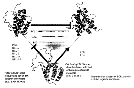
Figure 1 depicts how the three distinct classes of BCL-2 family proteins
interact to regulate apoptosis.
Figure 2 displays a listing of select BCL-2 family members, highlighting their
conserved BCL-2 homology (BH) domains.
Figure 3 depicts the BH3 binding pocket of anti-apoptotic BCL-2 family
members.
Figure 4 illustrates the continuum of events that is initiated by direct
activation
of BAX, culminating in BAX-mediated mitochondria] damage.
8
CA 02738983 2011-03-25
WO 2010/042225
PCT/US2009/005568
Figure 5 demonstrates the location of the newly identified BH3 interaction
site
on BAX, as deteremined by NMR analysis. Importantly, BIM SAHB engages BAX
at a structural location that is distinct from the canonical BH3 binding site
identified
for anti-apoptotic proteins and shown in Figure 3.
Figure 6 demonstrates the topography of the novel BH3 interaction site on
BAX (part A) and the orientation of BIM BH3 at the BAX binding site (part B)
as
determined by NMR analysis of the BIM SAHB-BAX interaction
Figure 7 indicates the amino acid sequence of BAX (part A) with residues of
the novel BH3 binding site on BAX highlighted. BAX residues engaged in BIM BH3
interactions are highlighted in ribbon (part B) and surface (part C) diagrams
of BAX.
Figure 8 lists the sequences of BAX activator BH3 peptides and hydrocarbon
stapled derivatives
Figure 9 shows how the identified molecules from a virtual screen of the novel
interaction site decorate the horizontal hydrophobic groove (part A), the
superior
juxta-loop region (part B), and the inferior juxta-loop region (part C).
Figure 10 indicates the amino acid sequence of BAX (part A) with those
residues involved with ligand interactions at or adjacent to the novel BH3
binding site
on BAX highlighted. BAX residues engaged in ligand interactions are
highlighted in
ribbon (part B) and surface (part C) diagrams of BAX.
Figure 11 demonstrates a competitive fluorescent polarization binding assay
revealing that compounds identified by the virtual screen effectively and dose-
responsively compete with FITC-BIM SAHB for BAX binding at the novel
interaction site.
Figure 12A demonstrates a BAX oligomerization assay involving the
application of a direct BAX-activating compound, such as BIM SAHB, and
monitoring the conversion of BAX from its monomeric to its oligomeric state by
size-
exclusion chromatography (SEC). Figure 12B depicts how compounds identified by
9
CA 02738983 2011-03-25
WO 2010/042225
PCT/US2009/005568
the virtual screen trigger BAX oligomerization as detected by this BAX
oligomerization assay.
Figure 13 depicts how compounds identified by the virtual screen trigger BAX
oligomerization as detected by conversion of the BAX monomer to its oligomeric
form using dynamic light scattering.
Figure 14 depicts how compounds identified by the virtual screen trigger
recombinant BAX-mediated cytochrome c release from Bax-I- Bak -I-
mitochondria, as
detected by ELISA assay.
Figure 15 depicts how identified compounds 5285738 (part A) and 5258079
(part B) selectively trigger dose-responsive apoptosis in BAX-reconstituted
BaxBak embryo fibroblasts (DKO MEF), but not in DKO MEFs.
DETAILED DESCRIPTION
I. Definitions
As used herein, the term, "BCL-2 family polypeptide" refers to an
evolutionary conserved family of proteins having as few as one to as many as
four
conserved BCL-2 homology domains (BH1, BH2, BH3 and/or BH4). The BH
domains are alpha-helical segments and are present in both the anti-apoptotic
and pro-
apoptotic polypeptides of the BCL-2 family. BCL-2 family polypeptides include
BCL-2, BCL-XL, BCL-w, MCL-1, BCL-B, A 1/BFL-1, BOO/DIVA, Nr-13, CED-9,
BAX, BAK, BOK/MTD, BID, BAD, BIK/NBK, BLK, HRK, BIM/BOD, BNIP3,
NIX, NOXA, PUMA, BMF, EGL-1, and viral homologues, including, but not limited
to Ml1L and E1B-19K.
The term "active site" refers to a region of a BCL-2 family polypeptide, as a
result of its shape, amino acid content, and charge potential, that favorably
interacts or
associates with another agent (including, without limitation, a protein,
polypeptide,
peptide, molecule, nucleic acid, compound, antibiotic or drug, or combination
thereof)
via various covalent and/or non-covalent binding forces. The "active site"
includes a
hydrophobic groove surrounded by a perimeter of charged and hydrophilic
residues
that is capable of binding a stabilized alpha helix of BCL-2 domain, such as
human
CA 02738983 2011-03-25
WO 2010/042225
PCT/US2009/005568
hydrocarbon-stapled BIM BH3 (SEQ ID NO:3), and which is formed by the
juxtaposition of alpha helices 1 and 6 of BAX. In one embodiment, the active
site
includes two or more amino acids corresponding to Glu17, Met20, Lys21, Thr22,
Ala24, Leu25, Leu27, G1n28, Gly29, 11e31, Gln 32, Leu47, Asp48, Pro49, Va150,
Pro51, G1n52, Asp53, Thr56, Arg89, Phe92, Phe93, Pro130, G1u131, Ile 133, Arg
134, Thr135, Met137, Gly138, Trp139, Leu141, Asp142, Phe143, Arg145, G1u146 of
SEQ ID NO:l.
The term "binding site" refers to a region of a BCL-2 family polypeptide, as a
result of its shape, amino acid content, and charge potential, that favorably
interacts or
associates with another agent (including, without limitation, a protein,
polypeptide,
peptide, molecule, compound, antibiotic or drug) via various covalent and/or
non-
covalent binding forces. A "bidning site" includes one or more amino acids
corresponding to Glu17, G1n18, Met20, Lys21, Thr22, Ala24, Leu25, Leu27,
G1n28,
Gly29, 11e31, Gin 32, Asp33, Arg34, A1a35, Gly36, Arg37, Met38, G1y39, Gly40,
Glu41, Ala 42, Leu47, Asp48, Pro49, Va150, Pro51, G1n52, Asp53, A1a54, Ser55,
Thr56, Lys57, Lys58, Leu59, Ser60, Glu61, Lys64, Arg89, Phe92, Phe93, Leu122,
Leu125, Thr127, Lys128, Va1129, Pro130, G1u131, Leu132, Ile 133, Arg 134,
Thr135, Met137, G1y138, Trp139, Leu141, Asp142, Phe143, Arg145, G1u146, Arg
147, Leu149, Gly150, G1y156, Gly157, Trp158 Asp 159, Leu161, Leu162 of SEQ ID
NO:l.
The term "BCL-2 family polypeptide variant" refers to polypeptides that vary
from a reference BCL-2 family polypeptide by the addition, deletion or
substitution of
at least one amino acid. It is well understood in the art that some amino
acids may be
changed to others with broadly similar properties without changing the nature
of the
activity of the polypeptide (conservative substitutions) as described
hereinafter.
Accordingly, BCL-2 family polypeptide variants as used herein encompass
polypeptides that have pro- or anti-apoptotic activity. The term "variant"
refers to a
protein having at least 30% amino acid sequence identity with a reference BCL-
2
homology domain within a protein or any other functional domain thereof More
specifically, the term "variant" includes, but is not limited to, a BCL-2
family
polypeptide comprising either 1) an active site characterized by a three
dimensional
structure comprising the relative structural coordinates of at least two BAX
amino
11
CA 02738983 2011-03-25
WO 2010/042225
PCT/US2009/005568
acid residues corresponding to Glul 7, Met20, Lys21, Thr22, A1a24, Leu25,
Leu27,
G1n28, G1y29, 11e31, Gin 32, Leu47, Asp48, Pro49, Va150, Pro51, G1n52, Asp53,
Thr56, Arg89, Phe92, Phe93, Pro130, G1u131, Ile 133, Arg 134, Thr135, Met137,
G1y138, Trp139, Leu141, Asp142, Phe143, Arg145, Glu146 of SEQ ID NO:1 or 2) a
binding site characterized by a three dimensional structure comprising the
relative
structural coordinates of at least one BAX amino acid residues corresponding
to
Glu17, Gln18, Met20, Lys21, Thr22, A1a24, Leu25, Leu27, G1n28, G1y29, 11e31,
Gin
32, Asp33, Arg34, A1a35, G1y36, Arg37, Met38, G1y39, G1y40, Glu41, Ala 42,
Leu47, Asp48, Pro49, Va150, Pro51, G1n52, Asp53, A1a54, Ser55, Thr56, Lys57,
Lys58, Leu59, Ser60, G1u61, Lys64, Arg89, Phe92, Phe93, Leu122, Leu125,
Thr127,
Lys128, Va1129, Pro130, G1u131, Leu132, Ile 133, Arg 134, Thr135, Met137,
G1y138, Trp139, Leu141, Asp142, Phe143, Arg145, G1u146, Arg 147, Leu149,
G1y150, G1y156, G1y157, Trp158 Asp 159, Leu161, Leu 162 of SEQ ID NO:1, in
each case, +/-a root mean square deviation from the conserved backbone atoms
of
those residues of not more than 1.1 angstroms, more preferably not more than
1.0
angstroms, and most preferably not more than 0.5 angstroms.
A "BCL-2 family polypeptide variant" further includes those polypeptides, or
their biologically active fragments, that comprise an amino acid sequence
which is at
least 30%, 40%, 50%, 60%, 70%, 80%, 90%, 95%, 96%, 97%, 98%, 99% or more
similar to an amino acid sequence of a BCL-2 homology domain (e.g., BH3
domain).
As used herein, the term "horizontal hydrophbic groove with or without a
perimeter of charged and hydrophilic residues" refers to that region of the
ligand
interaction on BAX that includes all or part of the BIM BH3 interaction site
on BAX,
as depicted in Figures 6 and 8A.
As used herein, the term "superior juxta-loop" refers to that portion of the
ligand interaction site that encompasses those residues located adjacent to
the al -a2
loop and extending from the midpoint of the horizontal hydrophobic groove
upward,
as depicted in Figure 8B.
As used herein, the term "inferior juxta-loop" refers to that portion of the
ligand interaction site that encompasses those residues located adjacent to
the al -a2
loop and extending from the midpoint of the horizontal hydrophobic groove
downward, as depicted in Figure 8B.
12
CA 02738983 2011-03-25
WO 2010/042225
PCT/US2009/005568
As used herein, the term "loop between al-a2" refers to those residues
located between a-helix 1 and a -helix 2 of BAX..
The term "hydrophobic patch" refers to the portion of the active site that
binds
a hydrophobic moiety. In one embodiment, the hydrophobic patch contains 1, 2,
3 or
more hydrophobic amino acid residues. In one particular embodiment, the
hydrophobic pocket contains amino acid residues corresponding to Met20, Ala24,
Leu25, Leu27, Gly29, 11e31, Leu47, Va150, Phe92, Phe93, Ile 133, Arg 134,
Met137,
G1y138, Trp139, Leu141, Phe143 of SEQ ID NO:l.
The term "charged/hydrophilic patch" refers to the portion of the active site
that binds a charged or hydrophilic moiety. In one embodiment, the
charged/hydrophilic patch contains 1, 2, 3 or more charged or hydrophilic
amino acid
residues. In one particular embodiment, the charged/hydrophilic patch contains
amino acid residues corresponding to G1u17, Lys21, Thr22, G1n28, Gln 32, Asp
33,
Asp48, Gln52, Asp53, Thr56, Arg89, G1u131, Arg 134, Thr135, Asp142, Arg145,
Glu146, Arg 147 of SEQ ID NO:l.
The term "hydrophobic amino acid" means any natural or non-natural amino
acid or mimetic thereof having an uncharged, non-polar side chain that is
relatively
insoluble in water. Examples of naturally occurring hydrophobic amino acids
are
alanine, leucine, isoleucine, valine, proline, phenylalanine, tryptophan and
methionine.
The term "hydrophilic amino acid" means any natural or non-natural amino
acid or mimetic thereof having an uncharged, polar side chain that is
relatively soluble
in water. Examples of naturally occurring hydrophilic amino acids are serine,
threonine, tyrosine, asparagine, glutamine, and cysteine.
The term "negatively charged amino acid" includes any naturally occurring or
unnatural amino acid or mimetic thereof having a negatively charged side chain
under
normal physiological conditions. Examples of negatively charged naturally
occurring
amino acids are aspartic acid and glutamic acid.
The term "positively charged amino acid" includes any naturally occurring or
unnatural amino acid or mimetic thereof having a positively charged side chain
under
normal physiological conditions. Examples of positively charged naturally
occurring
amino acids are arginine, lysine and histidine.
13
CA 02738983 2011-03-25
WO 2010/042225
PCT/US2009/005568
The term "anti-apoptotic polypeptide" refers to BCL-2 family polypeptides
characterized by having one or more amino acid homology domains, BH1, BH2,
BH3, and/or BH4, and that promote cell survival by attenuating or inhibiting
apoptosis. The "anti-apoptotic polypeptides" further include those proteins,
or their
biologically active fragments, that are at least 30%, 40%, 50%, 60%, 70%, 80%,
90%,
95%, 96%, 97%, 98%, 99% or more similar in amino acid sequence to an anti-
apoptotic BCL-2 homology domain within a BCL-2 family polypeptide. In a
preferred embodiment, the BCL-2 homology domain comprises one or more
conserved amino acid residues, such as amino acid residues corresponding to
Leu 97,
Gly 101 and Asp 102 of Bc1-2 (SEQ ID NO:8): Anti-apoptotic polypeptides
include
but are not limited to BCL-2, BCL-XL, BCL-w, MCL-1, A1/BFL-1, BCL-B,
BOO/DIVA, Nr-13 or CED-9.
The term "pro-apoptotic polypeptide" refers to BCL-2 family polypeptides
characterized by having one or more amino acid homology domains, BH1, BH2,
and/or BH3, and that promote cell death by activating apoptosis. The "pro-
apoptotic
polypeptides" further include those proteins, or their biologically active
fragments,
that are at least 30%, 40%, 50%, 60%, 70%, 80%, 90%, 95%, 96%, 97%, 98%, 99%
or more similar in amino acid sequence to a pro-apoptotic BCL-2 homology
domain
within a BCL-2 family polypeptide. In a preferred embodiment, the BCL-2
homology
domain comprises one or more conserved amino acid residue, such as amino acid
residues corresponding to Leu 92, Gly 96 and Asp 97 of BAX (SEQ ID NO: 1). Pro-
apoptotic polypeptides include but are not limited to BAX, BAK, BOK/MTD, BID
BAD, BIK/NBK, BLK, HRK, BIM/BOD, BNIP3, NIX, NOXA, PUMA, BMF AND
EGL-1.
As used herein, the term "apoptosis" refers to a regulated network of
biochemical events which lead to a selective form of cell death that is
characterized
by readily observable morphological and biochemical changes, such as the
fragmentation of the deoxyribonucleic acid (DNA), condensation of the
chromatin,
which may or may not be associated with endonuclease activity, chromosome
migration, margination in cell nuclei, the formation of apoptotic bodies,
mitochondrial
swelling, widening of the mitochondrial cristae, opening of the mitochondrial
permeability transition pores and/or dissipation of the mitochondrial proton
gradient.
14
CA 02738983 2011-03-25
WO 2010/042225
PCT/US2009/005568
The term "compound" is used herein to denote a chemical agent, polypeptide,
nucleic acid or combination thereof, or a mixture or synthetic combination of
chemical compounds and/or polypeptides and/or nucleic acids (e.g. DNA and/or
RNA
derivative), salts and solvates thereof, and the like. Preferably, a compound
of the
invention binds to the active site of a BCL-2 family polypeptide. A
"modulator" is a
compound which modulates the activity of a BCL-2 family polypeptide.
The term "candidate compound" is used herein to denote a chemical
compound, polypeptide, nucleic acid or combination thereof, or a mixture or
synthetic
combination of chemical compounds and/or polypeptides and/or nucleic acids,
salts
and solvates thereof, and the like, which is tested by a method of the
invention and is
found to bind to active site of a BCL-2 family polypeptide, and thus is
believed to
modulate the activity of the BCL-2 family polypeptide.
The term "modulate" as used herein with reference to a compound refers to the
activation or inhibition of anti-apoptotic or pro-apoptotic activity of a BCL-
2 family
polypeptide or other protein-protein interaction involving a BCL-2 family
member
that regulates a biochemical pathway (e.g. unfolded protein response, glucose-
stimulated insulin secretion). Methods for assaying both anti-apoptotic, pro-
apoptotic,
and other biochemical activities (e.g. unfolded protein response, glucose-
stimulated
insulin secretion) are well known in the art and described herein.
As used herein, the term "interacts" or "binds" refers to a condition of
proximity between a compound, or portions thereof, and the active site of a
BCL-2
family polypeptide or portions thereof. The interaction is between one or more
moieties on the compound and one or more moieties on amino acids of the active
site
region. The association may be non-covalent--wherein the juxtaposition is
energetically favored by hydrogen bonding or van der Waals or electrostatic
interactions--or it may be covalent.
The term, "activates" refers to an increase in the anti-apoptotic or pro-
apoptotic activity of a BCL-2 family polypeptide or other defined biochemical
activity based upon protein-protein interaction. A compound that activates a
pro-
apoptotic activity will bind to an active site of a BCL-2 family polypeptide
and cause
a 1.5x, 2x, 3x, 4x, 5x, 6x, 7x, 8x, 9x, 10x, 15x, 20x or more increase in the
pro-
apoptotic activity of the BCL-2 family polypeptide when compared with a
control
CA 02738983 2016-11-03
WO 2010/042225
PCT/US2009/005568
lacking the compound. In another embodiment, a compound that activates an anti-
apoptotic activity will bind to an active site of a BCL-2 family polypeptide
and cause
a 1.5x, 2x, 3x, 4x, 5x, 6x, 7x, 8x, 9x, 10x, 15x, 20x or more increase in the
anti-
apoptotic (survival) activity of the BCL-2 family polypeptide when compared
with a
control lacking the compound. Assays for assessing the activation of an anti-
apoptotic or pro-apoptotic activity are known in the art and described herein.
The term, "inhibits" refers to a decrease or blocking of the anti-apoptotic or
pro-apoptotic activity of a BCL-2 family polypeptide, or other defined
biochemical
activity based upon protein-protein interaction. For example, a compound that
inhibits a pro-apoptotic activity will bind to an active site of a BCL-2
family
polypeptide and prevent activation or reduce the activity of the BCL-2 family
polypeptide. Thus, the compound will inhibit or decrease the effects of a pro-
apoptotic activity. Thus, pro-apoptotic activity, e.g., cell death, will be
less than 75%,
70%, 60%, 50%, 40%, 30%, 20%, 10%, 5% or less in a population of cells in
which
an inhibitor is present than compared to a control cell population where the
compound
is not present.
A compound that inhibits an anti-apoptotic activity will bind to an active
site
or binding site of a BCL-2 family polypeptide and prevent or inhibit anti-
apoptotic
activity. Thus, anti-apoptotic activity, e.g., cell survival, will be less
than 75%, 70%,
60%, 50%, 40%, 30%, 20%, 10%, 5% or less in a population of cells in which the
inhibitor is present than compared to a control cell population where the
compound is
not present.
As used herein, the term "BH3 SAHB" refers to the BCL-2 homology domain
3 of a BCL-2 family polypeptide that has been hydrocarbon stapled so as to
form a
stabilized alpha helix. The amino acid sequence of numerous BH3 domains are
described herein. Methods of making I3H3 SAHBs are known in the art and
described
in U.S. Patent Publication No. US2005/0250680, filed November 5, 2004.
As used herein, the term "BIM BH3 polypeptide" refers to a polypeptide
having a BCL-2 homology domain 3 of BIM. In one embodiment, the BIM BH3
polypeptide has an amino acid sequence which is 30%, 40%, 50%, 60%, 70%, 80%,
90%, 95%, 96%, 97%, 98%, 99% or more identical to SEQ ID NO:3 (Figure 8) and
16
CA 02738983 2011-03-25
WO 2010/042225
PCT/US2009/005568
includes one or more of amino acid residues corresponding to Leul 52, Glyl 56,
and
Asp157 of SEQ ID NO:2 or conservative substitutions thereof. In a preferred
embodiment, the BIM BH3 polypeptide has the amino acid sequence of SEQ ID NO:3
or SAHB derivatives thereof (Figure 8).
As used herein, the term "BID BH3 polypeptide" refers to a polypeptide
having a BCL-2 homology domain 3 of BID. In one embodiment, the BID BH3
polypeptide has an amino acid sequence which is 30%, 40%, 50%, 60%, 70%, 80%,
90%, 95%, 96%, 97%, 98%, 99% or more identical to SEQ ID NO: 5 (Figure 8) and
includes one or more of amino acid residues corresponding to Leu90, G1y94, and
Asp95 of SEQ ID NO:4 or conservative substitutions thereof. In a preferred
embodiment, the BID BH3 polypeptide has the amino acid sequence of SEQ ID NO:5
or SAHB derivatives thereof (Figure 8).
As used herein, the term "PUMA BH3 polypeptide" refers to a polypeptide
having a BCL-2 homology domain 3 of PUMA. In one embodiment the PUMA BH3
polypeptide has an amino acid sequence which is 30%, 40%, 50%, 60%, 70%, 80%,
90%, 95%, 96%, 97%, 98%, 99% or more identical to SEQ ID NO: 7 (Figure 8) and
includes one or more of amino acid residues corresponding to Leu 141, Ala 145
and
Asp 146 of SEQ ID NO:6 or conservative substitutions thereof In a preferred
embodiment, the PUMA BH3 polypeptide has the amino acid sequence of SEQ ID
NO:7 or SAHB derivatives thereof (Figure 8).
As used herein, the term "BAX BH3 polypeptide" refers to a polypeptide
having a BCL-2 homology domain 3 of BAX. In one embodiment, the BAX BH3
polypeptide has an amino acid sequence which is 30%, 40%, 50%, 60%, 70%, 80%,
90%, 95%, 96%, 97%, 98%, 99% or more identical to SEQ ID NO: 8 (Figure 8) and
includes one or more of amino acid residues corresponding to Leu 63, Gly 67
and Asp
68 of SEQ ID NO:1 or conservative substitutions thereof In a preferred
embodiment,
the BAX BH3 polypeptide has the amino acid sequence of SEQ ID NO:8 or SAHB
derivatives thereof (Figure 8).
As used herein, the term "BAX activator BH3 consensus polypeptide" refers
to a polypeptide containing a consensus sequence for BAX binding at the new
interaction site. In one embodiment, the BAX activator BH3 consensus
polypeptide
has an amino acid sequence which is 30%, 40%, 50%, 60%, 70%, 80%, 90%, 95%,
17
CA 02738983 2011-03-25
WO 2010/042225
PCT/US2009/005568
96%, 97%, 98%, 99% or more identical to SEQ ID NO: 9 (Figure 8) or SAHB
derivatives thereof.
The terms "pharmacologically effective amount," "therapeutically effective
amount", "pharmacologically effective dose" or simply "effective amount"
refers to
that amount of an agent effective to produce the intended pharmacological,
therapeutic or preventive result. The pharmacologically effective amount
results in
the amelioration of one or more symptoms of a disorder, or prevents the
advancement
of a disorder, or causes the regression of the disorder. For example, with
respect to the
treatment of a disorder or excessive cellular survival or proliferation, a
therapeutically
effective amount preferably refers to the amount of a therapeutic agent that
decreases
the rate of tumor growth, decreases tumor mass, decreases the number of
metastases,
increases time to tumor progression, or increases survival time by at least
5%,
preferably at least 10%, at least 15%, at least 20%, at least 25%, at least
30%, at least
35%, at least 40%, at least 45%, at least 50%, at least 55%, at least 60%, at
least 65%,
at least 70%, at least 75%, at least 80%, at least 85%, at least 90%, at least
95%, or at
least 100%. For example, with respect to the treatment of a disorder
associated with
increased cellular death, e.g., ischemia, a therapeutically effective amount
preferably
refers to the amount of a therapeutic agent that prevents or limits tissue
and/or cellular
damage that would otherwise occur if treatment was not administered. The
therapeutic agent decreases tissue and/or cellular damage by at least 5%,
preferably at
least 10%, at least 15%, at least 20%, at least 25%, at least 30%, at least
35%, at least
40%, at least 45%, at least 50%, at least 55%, at least 60%, at least 65%, at
least 70%,
at least 75%, at least 80%, at least 85%, at least 90%, at least 95%, or at
least 100%
compared to damage that occurs without the administration of a therapeutic
agent of
the invention.
The terms "treat," and "treating," as used herein with reference to a disorder
(e.g., hyperpoliferative disorder, excessive cellular survival or
proliferation), refers to
a decrease in the occurrence of pathological cells (e.g., hyperproliferative
or
neoplastic cells) in an animal. The prevention may be complete, e.g., the
total absence
of pathological cells in a subject. The prevention may also be partial, such
that the
occurrence of pathological cells in a subject is less than that which would
have
18
CA 02738983 2011-03-25
WO 2010/042225
PCT/US2009/005568
occurred without the present invention. In some embodiments, such terms refer
to
one, two, three or more results following the administration of one or more
therapies:
(1) a stabilization, reduction or elimination of the cancer cell population,
(2) an
increase in the length of remission, (3) a decrease in the recurrence rate of
cancer, (4)
an increase in the time to recurrence of cancer, and (6) an increase in the
survival of
the patient.
The terms "treat," and "treating," as used herein with reference to a disorder
associated with increased cellular death, e.g., ischemia, refer to a decrease
in the
occurrence of tissue and/or cellular damage in an animal. The prevention may
be
complete, e.g., the total absence of tissue damage in a subject. The
prevention may
also be partial, such that the occurrence of tissue damage in a subject is
less than that
which would have occurred without the therapeutic agent.
As used herein, a "BCL-2 associated disorder", refers to a disorder associated
with a deregulated BCL-2 family member. BCL-2 associated disorders are
associated
with excessive cellular survival and/or proliferation, e.g., cancer, or
excessive cellular
death, e.g., Alzheimer's disease. BCL-2 associated disorders include those
described
herein.
As used herein, a "hyperproliferative disorder" means cancer, neoplastic
growth, hyperplastic or proliferative growth or a pathological state of
abnormal
cellular development or survival and includes solid tumors, non-solid tumors,
and any
abnormal cellular proliferation or accumulation, such as that seen in
leukemia.
The terms "anticancer agent" and "anticancer drug," as used herein, refer to
any therapeutic agents (e.g., chemotherapeutic compounds and/or molecular
therapeutic compounds), antisense therapies, nucleic acid therapies (e.g.
RNAi),
radiation therapies, used in the treatment of hyperproliferative diseases such
as
cancer. In one embodiment, the invention is directed to methods of treating a
BCL-2
associated disorder comprising administering an effective dose of an
anticancer agent
and a compound which binds to the active site, as described herein, of a BCL-2
family
peptide.
As used herein, the term "structural coordinates" refers to Cartesian
coordinates corresponding to an atom's spatial relationship to other atoms in
a
molecule or molecular complex. Structural coordinates may be obtained using x-
ray
19
CA 02738983 2011-03-25
WO 2010/042225
PCT/US2009/005568
crystallography techniques or NMR techniques, or may be derived using
molecular
replacement analysis or homology modeling. Various software programs allow for
the
graphical representation of a set of structural coordinates to obtain a three
dimensional
representation of a molecule or molecular complex. Structural coordinates for
the
BCL-2 family members are known in the art and are publicly available.
The term "interaction template" refers to a three dimensional model built
using
Cartesian coordinates corresponding to an atom's spatial relationship to other
atoms in
a molecule or molecular complex. Structural coordinates may be obtained using
x-ray
crystallography techniques or NMR techniques, or may be derived using
molecular
replacement analysis or homology modeling. Various software programs allow for
the
graphical representation of a set of structural coordinates to obtain a three
dimensional
representation of a molecule or molecular complex. The structural coordinates
of
BCL-2 family polypeptides are known in the art and can be found for example at
Protein Data Bank ("PDB") (Research Collaboratory for Structural
Bioinformatics;
http://www. rcsb.org). For example, known BCL-2 family structural coordinates
include BAX (PDB ID No. 1f16), BAK (PDB ID No. 2ims), BCL-2 (PDB ID No.
1g5m), BIM (PDB ID No. 2pqk) and BCL-XL (PDB ID No. 11x1), in addition to
those
associated with this invention: BIM BH3-BAX (PDB ID No. 2k7w), as well as
others
known in the art.
Preferably, the interaction template is of a BAX polypeptide having the amino
acids sequence set forth in SEQ ID NO:1, wherein the active site of the BAX
polypeptide is accessible to solvent and available for interaction with
modulators, e.g.,
activators. This three-dimensional form of BAX is used to facilitate the
identification
of compounds which bind in the active site. As used herein, the "interaction
template"
includes templates created by comparing the sequence/structual alignment of
BAX to
other BCL-2 family polypeptides. Identification of conserved and non-conserved
residues allows a skilled artisan to identify a corresponding active site in
other BCL-2
family polypeptides and design/screen modulators of the polypeptide.
As used herein in relation to the position of an amino acid, e.g., Ala 149 of
SEQ ID NO:1, the term "corresponding to" refers to an amino acid in a first
polypeptide sequence, e.g., BAX, that aligns with a given amino acid in a
reference
polypeptide sequence, e.g., BAK, when the first polypeptide and reference
CA 02738983 2011-03-25
WO 2010/042225
PCT/US2009/005568
polypeptide sequences are aligned by homology or other algorithms (e.g.,
structural
comparison). Alignment is performed by one of skill in the art using software
designed for this purpose, for example, BLASTP version 2.2.2 with the default
parameters for that version. Corresponding amino acids can also be identified
upon
structural comparisons of a first polypeptide sequence and a second
polypeptide
sequence. Such structural comparisons are known in the art and described
herein.
For example, Petros etal. Biochimica et Biophysica Acta 1644; 83-94 (2004) and
Suzuki et al., Cell. 103; 645-654 (2000) illustrated structural alignments
between
BCL-2 homology domains of BCL-2 family members.
The term "amino acid" refers to a molecule containing both an amino group
and a carboxyl group. Suitable amino acids include, without limitation, both
the D-
and L-isomers of the 20 common naturally occurring amino acids found in
peptides
(e.g., A, R, N, C, D, Q, E, G, H, I, L, K, M, F, P, S, T, W, Y, V (as known by
the one
letter abbreviations)) as well as the naturally occurring and unnaturally
occurring
amino acids prepared by organic synthesis or other metabolic routes.
A "non-essential" amino acid residue is a residue that can be altered from the
wild-type sequence of a polypeptide (e.g., BIM BH3) without abolishing or
substantially altering its BAX binding ability. An "essential" amino acid
residue is a
residue that, when altered from the wild-type sequence of the polypeptide,
results in
abolishing or substantially reducing the polypeptide's binding activity to a
BAX
active site or binding site. The essential and non-essential amino acid
residues of the
BH3 domains can readily be determined by methods well known in the art and
described herein. The term "essential" amino acid residue as used herein,
includes
conservative substitutions of the essential amino acid. Generally, the
"essential"
amino acid residues are found at the interacting face (residues interacting
with BAX)
of the BH3 polypeptide.
A "conservative amino acid substitution" is one in which the amino acid
residue is replaced with an amino acid residue having a similar side chain or
chemical
mimetic thereof. For example, families of amino acid residues having similar
side
chains have been defined in the art. These families include amino acids with
basic
side chains (e.g., lysine, arginine, histidine), acidic side chains (e.g.,
aspartic acid,
glutamic acid), uncharged polar side chains (e.g., glycine, asparagine,
glutamine,
21
CA 02738983 2011-03-25
WO 2010/042225
PCT/US2009/005568
serine, threonine, tyrosine, cysteine), nonpolar side chains (e.g., alanine,
valine,
leucine, isoleucine, proline, phenyl alanine, methionine, tryptophan), beta-
branched
side chains (e.g., threonine, valine, isoleucine) and aromatic side chains
(e.g.,
tyrosine, phenylalanine, tryptophan, histidine). Other conserved amino acid
substitutions can also occur across amino acid side chain families, such as
when
substituting an asparagine for aspartic acid in order to modify the charge of
a peptide.
Thus, a predicted nonessential amino acid residue in a BH3 domain polypeptide,
for
example, is preferably replaced with another amino acid residue from the same
side
chain family or homologues across families (e.g. asparagines for aspartic
acid,
glutamine for glutamic acid). In addition, individual substitutions, deletions
or
additions that alter, add or delete a single amino acid or a small percentage
of amino
acids in an encoded sequence are also considered "conservative substitutions".
The terms "identical" or "percent identity," in the context of two or more
nucleic acids or polypeptide sequences, refer to two or more sequences or
subsequences that are the same or have a specified percentage of nucleotides
or amino
acids that are the same, when compared and aligned for maximum correspondence,
as
measured using one of the following sequence comparison algorithms, or by
visual
inspection.
"Similarity" or "percent similarity" in the context of two or more polypeptide
sequences, refer to two or more sequences or subsequences that are the same or
have a
specified percentage of amino acid residues, or conservative substitutions
thereof, that
are the same when compared and aligned for maximum correspondence, as measured
using one of the following sequence comparison algorithms, or by visual
inspection.
By way of example, a first protein region can be considered similar to a
region of an
anti-apoptotic BCL-2 family member protein when the amino acid sequence of the
first region is at least 30%, 40%, 50%, 60%, 70%, 75%, 80%, 90%, or even 95%
identical, or conservatively substituted, to a region of the second anti-
apoptotic BCL-
2 family member protein when compared to any sequence of an equal number of
amino acids as the number contained in the first region, or when compared to
an
alignment of anti-apoptotic BCL-2 family member proteins that has been aligned
by a
computer similarity program known in the art, as discussed below. Preferably,
the
22
CA 02738983 2011-03-25
WO 2010/042225
PCT/US2009/005568
polypeptide region of the first protein and the second protein includes one or
more
conserved amino acid residues.
H. Description
In one aspect, the invention provides a method for identifying an organic
molecule which modulates the activity of a BCL-2 family polypeptide, the
method
comprising:
a) contacting said BCL-2 family polypeptide with a compound under
conditions suitable for modulation of the activity of said BCL-2 family
polypeptide;
and
b) detecting modulation of the activity of said BCL-2 family polypeptide by
the compound,
wherein the compound interacts with a binding site comprising one or more of
an al helix, a2 helix, a loop between al -a2, a6 helix, and select residues of
a4, a5,
and a8 helices in said BCL-2 family polypeptide;
wherein the interaction of the compound with the binding site occurs at a
horizontal hydrophobic groove with or without a perimeter of charged and
hydrophilic residues, a superior juxta-loop, an inferior juxta-loop, or
combination
thereof.
In a second aspect, the invention provides a method for identifying an organic
molecule compound which activates the pro-apoptotic activity of a BAX
polypeptide,
the method comprising:
a) contacting a binding site of said BAX polypeptide, wherein the binding site
comprises one or more of an al helix, a2 helix, a loop between al -a2, a6
helix, and
select residues of a4, cc5, and a8 helices, with a compound under conditions
suitable
for activating the pro-apoptotic activity of said BAX polypeptide; and
b) detecting activation of said BAX polypeptide by said compound,
wherein said compound binds to one or more amino acid residues
corresponding to G1u17, G1n18, Met20, Lys21, Thr22, A1a24, Leu25, Leu27,
G1n28,
Gly29, 11e31, Gln 32, Asp33, Arg34, A1a35, Gly36, Arg37, Met38, Gly39, G1y40,
23
CA 02738983 2011-03-25
WO 2010/042225
PCT/US2009/005568
G1u41, Ala 42, Leu47, Asp48, Pro49, Va150, Pro51, G1n52, Asp53, A1a54, Ser55,
Thr56, Lys57, Lys58, Leu59, Ser60, G1u61, Lys64, Arg89, Phe92, Phe93, Leu122,
Leu125, Thr127, Lys128, Va1129, Pro130, Glu131, Leu132, Ile 133, Arg 134,
Thr135, Met137, G1y138, Trp139, Leu141, Asp142, Phe143, Arg145, G1u146, Arg
147, Leu149, G1y150, G1y156, Gly157, Trp158 Asp 159, Leu161, Leu 162 of SEQ ID
NO:1; and
wherein the interaction of the compound with the binding site occurs at a
horizontal hydrophobic groove with or without a perimeter of charged and
hydrophilic residues, a superior juxta-loop, an inferior juxta-loop, or
combination
thereof.
In a third aspect, the invention provides a method of identifying a candidate
organic molecule modulator of a BCL-2 family polypeptide, comprising:
a) using a three dimensional structure of a binding site of said BCL-2 family
polypeptide, wherein said binding site comprises one or more of an al helix,
a2
helix, the loop between a1-a2, a6 helix, and select residues of a4, a5, and a8
helices, to form a BCL-2 family polypeptide interaction template; and
b) employing said BCL-2 family polypeptide interaction template to select
said BCL-2 family polypeptide candidate modulator, wherein said candidate
modulator binds to said binding site;
wherein the interaction of the candidate modulator with the binding site
occurs
at a horizontal hydrophobic groove with or without a perimeter of charged and
hydrophilic residues, a superior juxta-loop, an inferior juxta-loop, or
combination
thereof.
In a fourth aspect, the invention provides a method for identifying a
candidate
organic molecule compound which activates a BAX polypeptide pro-apoptotic
activity, the method comprising: a) providing a three dimensional structure of
a
binding site of a BAX polypeptide, wherein said binding site comprises one or
more
of an al helix, a2 helix, a loop between al -a2, a6 helix, and select residues
of a4,
a5, and a8 helices; b) simulating a binding interaction between said binding
site and a
compound, wherein the interaction of the compound with the binding site occurs
at a
horizontal hydrophobic groove with or without a perimeter of charged and
hydrophilic residues, a superior juxta-loop, an inferior juxta-loop, or
combination
24
CA 02738983 2011-03-25
WO 2010/042225
PCT/US2009/005568
thereof; and c) determining whether said compound binds to an amino acid
residue
selected from the group consisting of, G1u17, G1n18, Met20, Lys21, Thr22,
A1a24,
Leu25, Leu27, G1n28, G1y29, 11e31, Gin 32, Asp33, Arg34, Ala35, Gly36, Arg37,
Met38, Gly39, G1y40, G1u41, Ala 42, Leu47, Asp48, Pro49, Va150, Pro51, G1n52,
Asp53, A1a54, Ser55, Thr56, Lys57, Lys58, Leu59, Ser60, G1u61, Lys64, Arg89,
Phe92, Phe93, Leu122, Leu125, Thr127, Lys128, Va1129, Pro130, G1u131, Leu132,
Ile 133, Arg 134, Thr135, Met137, G1y138, Trp139, Leu141, Asp142, Phe143,
Arg145, G1u146, Arg 147, Leu149, G1y150, G1y156, G1y157, Trp158 Asp 159,
Leu161, Leu 162 of SEQ ID NO:1 of said binding site, wherein said compound
which
binds to said amino acid residue of the binding site is said candidate
compound.
In certain embodiments, said compound is an organic compound. In another
embodiment, the compound is selected from Table 1 and may further comprising a
derivative of a compound of Table 1, wherein said derivative improves binding
affinity, activity, solubility, or other pharmacologic properties.
In one embodiment, said BCL-2 family polypeptide is a pro-apoptotic
polypeptide.
In a further embodiment, said pro-apoptotic polypeptide is BAX. In another
further embodiment, said pro-apoptotic polypeptide is BOK or BAK.
In certain embodiments, said BCL-2 family polypeptide is an anti-apoptotic
polypeptide. In a further embodiment, said anti-apoptotic polypeptide is
selected
from the group consisting of: BCL-2, Bc1-Xl, Bcl-w, Mc1-1, BCL-B, A I/Bfl-1,
Boo/Diva, Nr-13, Ced-9, a viral homolog, MIlL, and E1B-19K.
In other embodiments, said activity is pro-apoptotic activity or anti-
apoptotic
activity. In another embodiment, said modulation is activation or inhibition
of said
pro-apoptotic activity. In another embodiment, modulation is activation or
inhibition
of said anti-apoptotic activity.
In one embodiment, the detection of step (b) comprises,
(A) an assay selected from the group consisting of, BCL-2 polypeptide
oligomerization, antibody-based detection of BCL-2 polypeptide conformers,
mitochondrial cytochrome c release, liposomal release, cell death,
mitochondria] or
cellular morphology, mitochondrial calcium flux, mitochondrial transmembrane
quantitation, and quantitation of caspase 3 activity or annexin V binding and
CA 02738983 2011-03-25
WO 2010/042225
PCT/US2009/005568
(B) using BCL-2 polypeptide protein specially prepared to maximize yield and
stability through optimized protein expression and purification conditions and
the
creation of polypeptide mutants that stabilize monomeric, dimeric or
oligomeric
conformers and species.
In certain embodiments, said compound binds to one or more amino acid
residues corresponding to G1u17, Met20, Lys21, Thr22, Ala24, Leu25, Leu27,
Gln28,
G1y29, 11e31, Gln 32, Asp 33, Leu47, Asp48, Pro49, Va150, Pro51, G1n52, Asp53,
Thr56, Arg89, Phe92, Phe93, Pro130, G1u131, Ile 133, Arg 134, Thr135, Met137,
G1y138, Trp139, Leu141, Asp142, Phe143, Arg145, Glu146 of SEQ ID NO:1 in the
binding site.
In another embodiment, said compound binds to one or more amino acid
residues corresponding to Met20, Lys21, A1a24, G1n28, G1n32, G1u131, Arg134,
Met137, Leu141, Asp142 of SEQ ID NO:1 in the binding site.
In another embodiment, said compound binds to an amino acid residue
corresponding to Lys21 of SEQ ID NO:1 in the binding site.
In certain embodiments, said compound binds to one or more amino acid
residues corresponding to Glu17, G1n18, Met20, Lys21, Thr22, Ala24, Leu25,
Leu27,
G1n28, G1y29, 11e31, Gin 32, Asp33, Arg34, A1a35, Gly36, Arg37, Met38, G1y39,
G1y40, Glu41, Ala 42, Leu47, Asp48, Pro49, Va150, Pro51, Gln52, Asp53, Ala54,
Ser55, Thr56, Lys57, Lys58, Leu59, Ser60, G1u61, Lys64, Arg89, Phe92, Phe93,
Leu122, Leu125, Thr127, Lys128, Va1129, Pro130, G1u131, Leu132, Ile 133, Arg
134, Thr135, Met137, G1y138, Trp139, Leu141, Asp142, Phe143, Arg145, Glu146,
Arg 147, Leu149, G1y150, G1y156, G1y157, Trp158 Asp 159, Leu161, Leu 162of
SEQ ID NO:1 in the binding site.
In one embodiment, said compound further comprises an organic compound, a
polypeptide, a nucleic acid or combinations thereof.
In a further embodiment, said compound comprises a compound of Table 1
linked to a second compound in Table 1, or comprises a combination of
compounds
or their chemical subcomponents listed in Table 1, and derivatives thereof.
In another embodiment, said second compound interacts with the binding site
at a horizontal hydrophobic groove with or without a perimeter of charged and
26
CA 02738983 2011-03-25
WO 2010/042225
PCT/US2009/005568
hydrophilic residues, a superior juxta-loop, an inferior juxta-loop, or
combination
thereof.
In certain embodiments, said compound further comprises a BIM polypeptide.
In another embodiment, said compound further comprises a BIM BH3 peptide
(SEQ ID NO:3) or SAHB derivative thereof.
In other embodiments, said compound further comprises an amino acid
sequence which is 30% or more identical with SEQ ID NO:3, and comprises an
amino
acid residue corresponding to 11e148, L152, Arg153, Arg154, Gly156, Asp157,
G1u158, or Asn160 of SEQ ID NO:2, or conservative natural or non-natural amino
acid substitutions thereof.
In another embodiment, said compound further comprises an amino acid
comprising residues 11e148, A1a149, L152, Arg153, Arg154, 11e155, G1y156,
Asp157,
G1u158, Asn160, A1a161, or Tyr163 of SEQ ID NO:2, or conservative natural or
non-
natural amino acid substitutions thereof.
In one embodiment, said compound further comprises a BID polypeptide.
In another embodiment, said compound further comprises a BID BH3 peptide
(SEQ ID NO: 5) or SAHB derivative thereof.
In still another embodiment, said compound further comprises a PUMA
polypeptide.
In certain embodiments, said compound further comprises a PUMA BH3
peptide (SEQ ID NO:7) or SAHB derivative thereof.
In another embodiment, said compound further comprises a BAX polypeptide.
In certain embodiments, said compound further comprises a BAX BH3
peptide (SEQ ID NO:8) or SAHB derivative thereof.
In one embodiment, said compound further comprises a polypeptide selected
from the group of BH3-only proteins, including but not limited to BID, BAD,
BIK/NBK, BLK, HRK, BIM/BOD, BNIP3, NIX, NOXA, PUMA, BMF and EGL-1
or a BH3 region thereof.
In one embodiment, said compound further comprises a polypeptide selected
from the group consisting of BCL-2, BCL-XL, BCL-w, Bc1-B, MCL-1, Al/BFL-1,
BOO/DIVA, NR-13, CED-9, a viral homolog, Ml1L, and E1B-19K.
27
=
CA 02738983 2011-03-25
WO 2010/042225
PCT/US2009/005568
In another embodiment, said compound further comprises a polypeptide
selected from the group consisting of BAX, BAK and BOK.
In other embodiments, said compound further comprises a BH3 region
polypeptide which is 30% identical to SEQ ID NO:3 and comprises amino acid
residues corresponding to Leu152, G1y156, and Asp157 of SEQ ID NO:2 or
conservative substitutions thereof.
In certain embodiments, said compound further comprises a BH3 region
polypeptide which is 30% identical to SEQ ID NO:5 and comprises amino acid
residues corresponding to Leu 90, Gly 94 and Asp 95 of SEQ ID NO:4 or
conservative substitutions thereof.
In another embodiment, said compound further comprises a BH3 region
polypeptide which is 30% identical to SEQ ID NO:7 and comprises amino acid
residues corresponding to Leu 141, Ala 145 and Asp 146 of SEQ ID NO:6 or
conservative substitutions thereof
In certain embodiments, said compound further comprises a BH3 region
polypeptide which is 30% identical to SEQ ID NO:8 and comprises amino acid
residues corresponding to Leu 63, Gly 67 and Asp 68 of SEQ ID NO:1 or
conservative substitutions thereof
In one embodiment, said compound further comprises a polypeptide which is
30% identical to a consensus sequence for BH3 binding to the BAX active site
as
identified in SEQ ID NO:9 and conservative substitutions thereof
In one embodiment, said compound binds to said binding site with an affinity
of <1 mM.
In another aspect, the invention provides a method of treating a disorder in a
subject, comprising administering to said subject in need thereof, an
effective amount
of a compound identified by the method described above, such that said subject
is
treated for said disorder.
In another aspect, the invention provides a method of treating a disorder in a
subject, wherein the subject has been identified as in need of treatment for
said
disorder, comprising
administering to said subject an effective amount of a compound identified by
the method of any one of claims 1-4, that binds to a binding site of a BCL-2
family
28
CA 02738983 2011-03-25
WO 2010/042225
PCT/US2009/005568
polypeptide or BAX, wherein said binding site comprises one or more of al
helix, a2
helix, a loop between al -a2, a6 helix, and select residues of a4, a5, and a8
helices,
wherein said compound modulates a BCL-2 family polypeptide or BAX, such that
said subject is treated for said disorder.
In one embodiment, said disorder is a disorder of cellular proliferation or
apoptotic blockade.
In another embodiment, said cellular proliferation or apoptotic blockage
disorder is cancer or an autoimmune disease.
In a further embodiment, said cancer is selected from the group consisting of
solid tumor, leukemia, and lymphoma. In a further embodiment, said cancer is a
chemoresistant cancer. In another further embodiment, said chemoresistant
cancer is
resistant to ABT-737, ABT-263, obatoclax, or other BCL-2 survival protein
inhibitors.
In one embodiment, said disorder is a disorder of cellular loss.
In another embodiment, the compound inhibits BAX activation.
In certain embodiments, said cellular loss disorder is neurodegeneration,
heart
attack, or stroke.
In another aspect, the invention provides a method of treating cancer or a
tumor in a subject, wherein the subject has been identified as in need of
treatment for
said disorder, comprising
administering to said subject an effective amount of a compound that binds to
a binding site of a BCL-2 family polypeptide, wherein said binding site
comprises one
or more of an al helix, a2 helix, a loop between al-a2, a6 helix, and select
residues
of a4, a5, and a8 helices, wherein said compound activates the pro-apoptotic
activity
of a BAX polypeptide, wherein said compound binds to one or more amino acid
residues G1u17, G1n18, Met20, Lys21, Thr22, A1a24, Leu25, Leu27, G1n28, G1y29,
11e31, Gln 32, Asp33, Arg34, A1a35, G1y36, Arg37, Met38, Gly39, Gly40, Glu41,
Ala
42, Leu47, Asp48, Pro49, Va150, Pro51, G1n52, Asp53, A1a54, Ser55, Thr56,
Lys57,
Lys58, Leu59, Ser60, G1u61, Lys64, Arg89, Phe92, Phe93, Leu122, Leu125,
Thr127,
Lys128, Va1129, Pro130, G1u131, Leu132, Ile 133, Arg 134, Thr135, Met137,
G1y138, Trp139, Leu141, Asp142, Phe143, Arg145, G1u146, Arg 147, Leu149,
G1y150, G1y156, G1y157, Trp158 Asp 159, Leu161, Leu162 of SEQ ID NO:1
29
CA 02738983 2011-03-25
WO 2010/042225
PCT/US2009/005568
wherein the binding site occurs at a horizontal hydrophobic groove with or
without a
perimeter of charged and hydrophilic residues, a superior juxta-loop, an
inferior juxta-
loop, or combination thereof.
In another aspect, the invention provides a composition for treating a BCL-2
related disorder, wherein said composition comprises,
a compound that binds to a binding site of a BCL-2 family polypeptide,
wherein said binding site comprises one or more of an al helix, a2 helix, a
loop
between al-a2, a6 helix, and select residues of a4, a5, and a8 helices,
wherein said
compound modulates the activity of a BCL-2 family polypeptide wherein the
compound interacts with the binding site at a horizontal hydrophobic groove
with or
without a perimeter of charged and hydrophilic residues, a superior juxta-
loop, an
inferior juxta-loop, or combination thereof; and
a second compound selected from an organic compound, a polypeptide and a
nucleic acid or combinations thereof;
wherein the composition binds to a binding site of said BCL-2 family
polypeptide.
In one embodiment, said compound is an organic compound.
In another embodiment, said compound is selected from a compound in Table
1.
In certain embodiments, said compound derives from a combination of
compounds selected from a compound in Table 1.
In another embodiment, said second compound is an organic compound.
In certain embodiments, said second compound is a polypeptide.
In other embodiments, said second compound is selected from the group
consisting of BIM, BID, BAX, PUMA, BAK and BOK.
In one embodiment, said BCL-2 family polypeptide is a pro-apoptotic
polypeptide.
In a further embodiment, said pro-apoptotic polypeptide is BAX.
In a further embodiment, said pro-apoptotic polypeptide is BOK or BAK.
In another embodiment, said BCL-2 family polypeptide is an anti-apoptotic
polypeptide.
CA 02738983 2011-03-25
WO 2010/042225
PCT/US2009/005568
In a further embodiment, said anti-apoptotic polypeptide is selected from the
group consisting of: BCL-2, BCL-XL, BCL-w, BCL-B, MCL-1, Bfl-1/A1,
BOO/DIVA, NR-13, CED-9, or viral homologs (e.g. Ml1L, E1B-19K, etc).
In another embodiment, said modulation is activation or inhbition of a pro-
apoptotic activity.
In another embodiment, said modulation is activation or inhibition of an anti-
apoptotic activity.
In another embodiment, said composition binds to one or more amino acid
residues selected from Glu17, Met20, Lys21, Thr22, A1a24, Leu25, Leu27, G1n28,
G1y29,11e31, Gln 32, Asp 33, Leu47, Asp48, Pro49, Va150, Pro51, G1n52, Asp53,
Thr56, Arg89, Phe92, Phe93, Pro130, G1u131, Ile 133, Arg 134, Thr135, Met137,
G1y138, Trp139, Leu141, Asp142, Phe143, Arg145, G1u146 of SEQ ID NO:1 in the
binding site.
In another embodiment, said composition binds to one or more amino acid
residues selected from Met20, Lys21, A1a24, Gln28, G1n32, G1u131, Arg134,
Met137, Leu141, Asp142 of SEQ ID NO:1 in the binding site.
In another embodiment, said composition binds to an amino acid residue
corresponding to Lys21 of SEQ ID NO:1 in the binding site.
In one embodiment, said composition binds to one or more amino acid
residues selected from G1u17, G1n18, Met20, Lys21, Thr22, Ala24, Leu25, Leu27,
G1n28, G1y29, 11e31, Gin 32, Asp33, Arg34, Ala35, Gly36, Arg37, Met38, G1y39,
G1y40, G1u41, Ala 42, Leu47, Asp48, Pro49, Va150, Pro51, G1n52, Asp53, A1a54,
Ser55, Thr56, Lys57, Lys58, Leu59, Ser60, G1u61, Lys64, Arg89, Phe92, Phe93,
Leu122, Leu125, Thr127, Lys128, Va1129, Pro130, G1u131, Leu132, Ile 133, Arg
134, Thr135, Met137, G1y138, Trp139, Leu141, Asp142, Phe143, Arg145, G1u146,
Arg 147, Leu149, G1y150, G1y156, G1y157, Trp158 Asp 159, Leu161, Leu 162of
SEQ ID NO:1 in the binding site.
In certain embodiments, said second compound is a BIM polypeptide.
In other embodiments, said second compound is a BIM BH3 polypeptide or
SAHB derivative thereof.
In another embodiment, said second compound is an amino acid comprising
an amino acid sequence which is 30% or more identical with SEQ ID NO:3, and
31
CA 02738983 2011-03-25
WO 2010/042225
PCT/US2009/005568
comprises an amino acid residue corresponding to 11e148, L152, Arg153, Arg154,
G1y156, Asp157, G1u158, or Asn160 of SEQ ID NO:2, or conservative natural or
non-natural amino acid substitutions thereof.
In another embodiment, said second compound is an amino acid comprising
amino acid residues 11e148, A1a149, L152, Arg153, Arg154, 11e155, G1y156,
Asp157,
G1u158, Asn160, A1a161, or Tyr163 of SEQ ID NO:2.
In another embodiment, said second compound is a BID polypeptide.
In certain embodiments, said second compound is a BID BH3 peptide or
SAHB derivative thereof.
In certain embodiments, said second compound is a PUMA polypeptide.
In a further embodiment, said second compound is a PUMA BH3 peptide or
SAHB derivative thereof.
In another embodiment, said second compound is a BAX polypeptide.
In a further embodiment, said second compound is a BAX BH3 peptide or
SAHB derivative thereof.
In one embodiment, said second compound is selected from the group of BH3-
only proteins, including but not limited to BID, BAD, BIK/NBK, BLK, HRK,
BIM/BOD, BNIP3, NIX, NOXA, PUMA, BMF and EGL-1 or a BH3 region thereof.
In another embodiment, said second compound comprises a polypeptide
selected from the group consisting of BCL-2, BCL-XL, BCL-w, BCL-B, MCL-1, Bfl-
1/A1, BOO/DIVA, NR-13, CED-9, or viral homologs (e.g. Ml1L, E1B-19K, etc).
In another embodiment, said second compound comprises a polypeptide
selected from the group consisting of BAX, BAK and BOK.
In one aspect, the invention provides a kit comprising a composition of claim
56 and instructions for use.
III. Structural insights into BCL-2 family function
The BCL-2 family of proteins includes both pro- and anti-apoptotic
polypeptides that provide the checks and balances that govern susceptibility
to cell
death. Deregulation of this pathway has been documented in the pathogenesis of
a
wide spectrum of human diseases, including many cancers.
32
CA 02738983 2011-03-25
WO 2010/042225
PCT/US2009/005568
Members of the evolutionarily conserved BCL-2 family are important
regulators of apoptotic cell death and survival. The proteins BCL-2, BCL-XL,
Bcl-w,
BFL1/A1 and MCL-1 are death antagonists while BAX, BAK, BAD, BCL-XS, BID,
BIM, and BIK are examples of death agonists (Kroemer et al., Nature Med. 6:614
20
(1997)).
The BCL-2 family is defined by the presence of up to four conserved "BCL-2
homology" (BH) domains designated BH1, BH2, BH3, and BH4, all of which include
alpha-helical segments (Chittenden et al. 1995 EMBO 14:5589; Wang et al. 1996
Genes Dev. 10:2859). Anti-apoptotic proteins, such as BCL-2 and BCL-XL,
display
sequence conservation in all BH domains. Pro-apoptotic proteins are divided
into
"multidomain" members (e.g. BAK, BAX, BOK), which possess homology in the
BH1, BH2, and BH3 domains, and the "BH3-domain only" members (e.g. BID, BAD,
BIM, BIK, NOXA, PUMA), that contain sequence homology exclusively in the BH3
amphipathic alpha-helical segment. BCL-2 family members have the capacity to
form
homo- and heterodimers, suggesting that competitive binding and the ratio
between
pro- and anti-apoptotic protein levels dictates susceptibility to death
stimuli. Anti-
apoptotic proteins function to protect cells from pro-apoptotic excess, i.e.,
excessive
programmed cell death. In certain cell types, death signals received at the
plasma
membrane trigger apoptosis via a mitochondrial pathway. The mitochondrial
apoptotic pathway can also be activated by internal cellular stresses and
signaling
pathways. The mitochondria can serve as a gatekeeper of cell death by
sequestering
cytochrome c, a critical component of a cytosolic complex which activates
caspase 9,
leading to fatal downstream proteolytic events. Multidomain proteins such as
BCL-
2/BCL-XL and BAK/BAX play dueling roles of guardian and executioner at the
mitochondrial membrane, with their activities further regulated by upstream
BH3-
only members of the BCL-2 family. For example, BID is a member of the "BH3-
domain only" subset of pro-apoptotic proteins, and transmits death signals
received at
the plasma membrane to effector pro-apoptotic proteins at the mitochondrial
membrane. Select BH3-only members, such as BID and BIM, have been termed
"activators" (Letai, A., et al. Cancer Cell 2, 183-192 (2002)), and have the
unique
capability of interacting with both pro- and anti-apoptotic proteins (Walensky
Mol
Cell 2006). Upon caspase 8 activation, BID is cleaved and the truncated
adduct,
33
CA 02738983 2011-03-25
WO 2010/042225
PCT/US2009/005568
tBID, triggers cytochrome c release and mitochondrial apoptosis through
engagement
of BCL-2 family proteins.
Deletion and mutagenesis studies determined that the amphipathic alpha-
helical BH3 segment of pro-apoptotic family members functions as a death
domain
and thus represents a critical structural motif for interacting with
multidomain
apoptotic proteins. Structural studies have demonstrated that the BH3 helix
interacts
with anti-apoptotic proteins by inserting into a hydrophobic groove formed by
the
interface of BH1, 2 and 3 domains. tBID and BIM can be bound and sequestered
by
anti-apoptotic proteins (e.g., BCL-2 and BCL-XL) and can trigger activation,
indirectly or directly, of the pro-apoptotic proteins BAX and BAK, leading to
cytochrome c release and a mitochondrial apoptosis program.
BCL-2-related ovarian killer (BOK) is the third member of the pro-apoptotic
multidomain subgroup and is also bound by activator SAHB ligands, such as BID
and
BIM SAHBs. BOK was cloned from an ovarian cDNA library and found to be highly
expressed in ovary, uterus, and testis. BOK mRNA species have since been
identified
in a broader distribution of tissues, including heart, spleen, liver, colon,
lung,
intestine, thyroid gland, adrenal, pancreas, and bone marrow, and select
cancer cell
lines.
A major breakthrough in BCL-2 biology was achieved by Fesik and co-
workers at Abbott Laboratories a decade ago when the first X-ray and NMR
structure
of a BCL-2 family protein was reported (Muchmore, S. W., Sattler, M., Liang,
H.,
Meadows, R. P., Harlan, J. E., Yoon, H. S., Nettesheim, D., Chang, B. S.,
Thompson,
C. B., Wong, S. L., Ng, S. L., and Fesik, S. W. (1996) X-ray and NMR structure
of
human Bc1-xL, an inhibitor of programmed cell death, Nature 381, 335-341). The
structure of BCL-XL is comprised of eight a-helices, two of which, (helices 5
and 6)
form a central hydrophobic core reminescent of the membrane insertion domains
of
the diphtheria and colicin pore-forming toxins (Muchmore, S. W., Sattler, M.,
Liang,
H., Meadows, R. P., Harlan, J. E., Yoon, H. S., Nettesheim, D., Chang, B. S.,
Thompson, C. B., Wong, S. L., Ng, S. L., and Fesik, S. W. (1996) X-ray and NMR
structure of human Bc1-xL, an inhibitor of programmed cell death, Nature 381,
335-
341). This structural homology provided the basis for studies that
demonstrated the
capacity of BCL-2 family members to form pores in liposomal and mitochondria]
34
CA 02738983 2011-03-25
WO 2010/042225
PCT/US2009/005568
systems (Nouraini, S., Six, E., Matsuyama, S., Krajewski, S., and Reed, J. C.
(2000)
The putative pore-forming domain of Bax regulates mitochondrial localization
and
interaction with Bc1-X(L), Mol Cell Biol 20, 1604-1615; Narita, M., Shimizu,
S., Ito,
T., Chittenden, T., Lutz, R. J., Matsuda, H., and Tsujimoto, Y. (1998) Bax
interacts
with the permeability transition pore to induce permeability transition and
cytochrome
c release in isolated mitochondria, Proc Nat! Acad Sci US A 95,14681-14686).
Subsequently, the structure of a BAK BH3 peptide in complex with BCL-XL
revealed
a paradigm for protein-protein interactions among pro- and anti-apoptotic BCL-
2
family members: the pro-apoptotic a-helical BH3 domain inserts into a
hydrophobic
groove formed by the juxtaposition of the BH1-3 domains (BH1: portions of
helices
a4 - a5; BH2: a7- a8; BH3: a2) of the anti-apoptotic protein (Figure 3). This
structural paradigm was confirmed in subsequent NMR structures of BCL-2
(Petros,
A. M., Nettesheim, D. G., Wang, Y., Olejniczak, E. T., Meadows, R. P., Mack,
J.,
Swift, K., Matayoshi, E. D., Zhang, H., Thompson, C. B., and Fesik, S. W.
(2000)
Rationale for Bc1-xL/Bad peptide complex formation from structure,
mutagenesis,
and biophysical studies, Protein Sci 9, 2528-2534), BCL-w (Denisov, A. Y.,
Chen,
G., Sprules, T., Moldoveanu, T., Beauparlant, P., and Gehring, K. (2006)
Structural
model of the BCL-w-BID peptide complex and its interactions with phospholipid
micelles, Biochemistry 45, 2250-2256), MCL-1 (Day, C. L., Chen, L.,
Richardson, S.
J., Harrison, P. J., Huang, D. C., and Hinds, M. G. (2005) Solution structure
of
prosurvival Mc-1 and characterization of its binding by proapoptotic BH3-only
ligands, J Biol Chem 280, 4738-4744), and BFL-1/A1 (Smits, C., Czabotar, P.
E.,
Hinds, M. G., and Day, C. L. (2008) Structural plasticity underpins
promiscuous
binding of the prosurvival protein Al, Structure 16, 818-829) in complex with
BH3-
only peptides, suggesting that communication among BCL-2 proteins is mediated
by
the network of homo- and hetero-complexes formed between hydrophobic grooves
and BH3 death helices. Preferences for hetero-associations are dictated by
discrete
differences in the amino acid composition of anti-apoptotic grooves and the
BH3
peptides of binding partners. The structural determinations of anti-apoptotic
complexes with BH3 peptides provided critical mechanistic insights into the
function
of BCL-2 family members and have led to the development of promising
pharmacologic modulators of BCL-2 regulated apoptotic pathways (Walensky, L.
D.,
CA 02738983 2011-03-25
WO 2010/042225
PCT/US2009/005568
et al. (2004) Activation of apoptosis in vivo by a hydrocarbon-stapled BH3
helix,
Science (New York, N.Y 305, 1466-1470; Oltersdorf, T., et al. (2005) An
inhibitor of
Bc1-2 family proteins induces regression of solid tumours, Nature 435, 677-
681;
Nguyen, M., etal. (2007) Small molecule obatoclax (GX15-070) antagonizes MCL-1
and overcomes MCL-1-mediated resistance to apoptosis, Proc Nat! Acad Sci USA
104, 19512-195). For example, selective BCL-2 inhibitors like the small
molecule
ABT-737 target the hydrophobic groove of anti-apoptotic BCL-2/BCL-XL and
reactivate apoptosis in select tumors (Oltersdorf, T., et al. (2005) An
inhibitor of Bel-
2 family proteins induces regression of solid tumours, Nature 435, 677-681).
Strikingly, the NMR structures of pro-apoptotic BAX (Suzuki, M., Youle, R.
J., and Tjandra, N. (2000) Structure of Bax: coregulation of dimer formation
and
intracellular localization, Cell 103, 645-654) and BH3-only BID (McDonnell, J.
M., et
al.(1999) Solution structure of the proapoptotic molecule BID: a structural
basis for
apoptotic agonists and antagonists, Cell 96, 625-634; Chou, J. J., et al.
(1999)
Solution structure of BID, an intracellular amplifier of apoptotic signaling,
Cell 96,
615-624) revealed structural similarities with the anti-apoptotic proteins.
BAX and
BID likewise possess two central core helices that are surrounded by 6 or 7
amphipathic helices, respectively. Another critical structural similarity was
revealed
at the N-terminal regions of BAX and BID. An unstructured loop between al and
a2
(BH3) is also present in select anti-apoptotic proteins such as BCL-2 (Petros,
A. M.,
et al. (2001) Solution structure of the antiapoptotic protein bc1-2, Proc Natl
Acad Sci
USA 98, 3012-3017) and BCL- XL (Muchmore, S. W., et al. (1996) X-ray and NMR
structure of human Bc1-xL, an inhibitor of programmed cell death, Nature 381,
335-
341). The loop regions of these proteins vary in primary sequence and in
length, and
are hypothesized to regulate their apoptotic functions. Indeed,
phosphorylation of the
BCL- XL /BCL-2 loop (Ito, T., Deng, X., Can, B., and May, W. S. (1997) Bc1-2
phosphorylation required for anti-apoptosis function, The Journal of
biological
chemistry 272, 11671-11673) inhibits anti-apoptotic function depending of the
cellular context, and caspase-8-mediated cleavage of the BCL-2 loop transforms
the
protein into a potent pro-apoptotic protein (Cheng, E. H., Kirsch, et al.
(1997)
Conversion of Bc1-2 to a Bax-like death effector by caspases, Science 278,
1966-
36
CA 02738983 2011-03-25
WO 2010/042225
PCT/US2009/005568
1968), presumably by exposing its BH3 death domain. The pro-apoptotic loop
region
of cytosolic BAX and BID undergoes enzymatic cleavage by calpain and caspase-8
respectively, producing truncated forms that exhibit enhanced mitochondrial
targeting
and pro-apoptotic activity (Li, H., et al. (1998) Cleavage of BID by caspase 8
mediates the mitochondrial damage in the Fas pathway of apoptosis, Cell 94,
491-
501; Cartron, P. F., et al. (2004) The p18 truncated form of Bax behaves like
a Bc1-2
homology domain 3-only protein, J Biol Chem 279, 11503-11512; Wood, D. E., et
al.(1998) Bax cleavage is mediated by calpain during drug-induced apoptosis,
Oncogene 17, 1069-1078).
Multidomain pro- and anti-apoptotic proteins contain a C-terminal
transmembrane region that is enriched in hydrophobic residues and functions as
an
anchor for mitochondrial outer membrane targeting and insertion (Suzuki, M.,
Youle,
R. J., and Tjandra, N. (2000) Structure of Bax: coregulation of dimer
formation and
intracellular localization, Cell 103, 645-654). Pro-apoptotic BAX contains a C-
terminal a9 helix, presumed to function as a transmembrane region once BAX is
deployed to the mitochondrion. In monomeric BAX, a9 engages the BAX
hydrophobic groove through complementary hydrophobic interactions, precluding
both access to the hydrophobic groove and exposure of its BH3 domain (Sattler,
M.,
et al. (1997) Structure of Bc1-xL-Bak peptide complex: recognition between
regulators of apoptosis, Science 275, 983-986). In healthy cells, BAK is
constitutively localized to the mitochondrial outer membrane; however, BAX
exists
as an inactive monomer in the cytosol (Suzuki, M., Youle, R. J., and Tjandra,
N.
(2000) Structure of Bax: coregulation of dimer formation and intracellular
localization, Cell 103, 645-654; Hsu, Y. T., and Youle, R. J. (1998) Bax in
murine
thymus is a soluble monomeric protein that displays differential detergent-
induced
conformations, J Biol Chem 273, 10777-1078). Upon receiving an apoptotic
stimulus,
BAX is believed to undergo a conformational change, leading to its
translocation to
the mitochondria, homo-oligomerization, and formation of a pore within the
outer
mitochondria' membrane (Hsu, Y. T., and Youle, R. J. (1998) Bax in murine
thymus
is a soluble monomeric protein that displays differential detergent-induced
conformations, J Biol Chem 273, 10777-1078; Soane, L., and Fiskum, G. (2005)
37
CA 02738983 2011-03-25
WO 2010/042225
PCT/US2009/005568
Inhibition of mitochondrial neural cell death pathways by protein transduction
of Bel-
2 family proteins, J Bioenerg Biomembr 37, 179-190; Tan, Y. J., Beerheide, W.,
and
Ting, A. E. (1999) Biophysical characterization of the oligomeric state of Bax
and its
complex formation with Bcl-XL, Biochem Biophys Res Commun 255, 334-339).
Previous studies suggested that release of the C-terminal transmembrane region
from
its groove may be required for translocation and membrane anchoring (Schinzel,
A.,
Kaufmann, T., Schuler, M., Martinalbo, J., Grubb, D., and Borner, C. (2004)
Conformational control of Bax localization and apoptotic activity by Pro168, J
Cell
Biol 164, 1021-1032). Additionally, a conformational change at the N-terminal
region, as detected by the monoclonal antibody 6A7, selectively detects a
conformationally activated form of BAX (Hsu, Y. T., and Youle, R. J. (1998)
Bax in
murine thymus is a soluble monomeric protein that displays differential
detergent-
induced conformations, J Biol Chem 273, 10777-10783; Yethon, J. A., Epand, R.
F.,
Leber, B., Epand, R. M., and Andrews, D. W. (2003) Interaction with a membrane
surface triggers a reversible conformational change in Bax normally associated
with
induction of apoptosis, J Biol Chem 278, 48935-48941) . Exposure of this N-
terminal
epitope is believed to be a prerequisite for mitochondrial targeting and a9
release
(Schinzel, A., Kaufrnann, T., Schuler, M., Martinalbo, J., Grubb, D., and
Borner, C.
(2004) Conformational control of Bax localization and apoptotic activity by
Pro168, J
Cell Biol 164, 1021-1032). Although a variety of models for BAX/BAK-mediated
apoptosis induction have been proposed, the explicit molecular trigger
mechanism for
BAX/BAK activation has remained unknown. Walensky and co-workers have now
identified a novel BH3 interaction site on pro-apoptotic BAX that triggers its
activation (Gavathiotis, E., Suzuki, M., Davis, M.L., Pitter, K., Bird, G.H.,
Katz, S.G.,
Tu, H.-C., Kim, H., Cheng, E. H.-Y. Tjandra, N. and Walensky, L.D. BAX
activation
is initiated at a novel interaction site. Nature, in press, 2008). Of note,
this new
interaction site is located in a distinct geographic region of the BCL-2
family protein,
as compared to the BH3 interaction site identified for anti-apoptotic proteins
(Figure
5). Just as solving the structure of the anti-apoptotic groove in complex with
the BH3
helix has led to targeted inhibitors of survival proteins for cancer therapy,
elucidating
the BH3 interaction site on BAX and defining BAX/BAK structures along their
38
CA 02738983 2011-03-25
WO 2010/042225
PCT/US2009/005568
activation continuum will provide new pharmacologic opportunities to modulate
apoptosis in human disease (Figure 4).
IV. A Novel Interaction Site on BAX that Triz2ers Its Activation
To investigate the initiating event for BAX activation, the interaction of BAX
with the BH3 ligand BIM SAHB was studied. BIM SAHB was previously
demonstrated to recapitulate the a-helical character of native death domains
and
bound directly to BAX (Walensky, L. D., et al. (2006) A stapled BID BH3 helix
directly binds and activates BAX, Mol Cell 24, 199-210). BIM SAHB binding to
BAX was monitored using Nuclear Magnetic Resonance (NMR) spectroscopy.
Compared to the 1H-15N correlation spectrum of BAX, the addition of BIM SAHB
broadened and shifted select NMR cross-peaks, indicating fast exchange between
the
bound and unbound conformations of BAX. The overall features of the NMR
spectra
were quite similar except for significant changes in the loop residues between
al and
a2 upon BIM SAHB binding. Chemical shift perturbation mapping of BAX with
BIM SAHB titration revealed interactions at a discrete subset of BAX residues.
The
largest changes were observed for residues localized in the al and a6 helices,
as well
as residues in the flexible loop between al and a2. Significant changes were
also
observed for the side-chain NH2 of Q28, Q32, and Q52. In the BAX structure
(Suzuki, M., Youle, R. J., and Tjandra, N. (2000) Structure of Bax:
coregulation of
dimer formation and intracellular localization, Cell 103, 645-654), the al and
a6
helices are positioned adjacent to one another, and the residues impacted by
BIM
SAHB binding localized to a discrete site at the juxtaposition of these
helices on one
side of the protein structure. Of note, no residues on the carboxy terminal
side of the
protein were affected by BIM SAHB titration under these conditions, thus
placing the
novel binding site on the completely opposite face of the protein from the
canonical
BH3 binding site of anti-apoptotic proteins (Figure 5). The binding site of
BIM
SAHB on BAX is thus defined by the two helices al and a6, with the
interhelical
junction forming a hydrophobic cleft surrounded by a perimeter of hydrophilic
and
charged residues (Figure 6).
39
CA 02738983 2011-03-25
WO 2010/042225
PCT/US2009/005568
Using the structurally-defined topography of the novel interaction site on
BAX, an in silico screening was undertaken and identified a series of small
molecules
predicted to engage the new BH3-binding groove on BAX. The identified
molecules
were then subjected to a series of biochemical assays to validate their
functional
activity, including competitive fluorescence polarization, BAX
oligomerization,
cytochrome c release, and cell-based apoptosis induction assays.
V. Computer based drum desizn
Identification of a binding site aids the development and identification of
compounds that are capable of modulating BAX and other BCL-2 family
polypeptides having a corresponding binding site. For example, using this
information, a three-dimensional computer generated interaction template of
BAX can
be generated by one of ordinary skill in the art and used to design activators
and
inhibitors specific for the BAX active site. In another embodiment, one of
ordinary
skill in the art can apply the BAX active site to identify corresponding
active sites in
other BCL-2 family members. This information may then be used to
identify/develop
compounds capable of modulating the other BCL-2 family polypeptides.
Determination of the three dimensional structure of the BCL-2 polypeptide
and specifically the binding site is critical to the rational identification
and/or design
of agents that may act as modulators of BCL-2 family polypeptide activity.
This is
advantageous over conventional drug assay techniques, in which the only way to
identify such an agent is to screen thousands of test compounds until an agent
having
the desired inhibitory effect on a target compound is identified. Necessarily,
such
conventional screening methods are expensive, time consuming, and do not
elucidate
the method of action of the identified agent on the target protein. Using such
a three
dimensional structure, researchers identify putative binding sites and then
identify or
design agents to interact with these binding sites. These agents are then
screened for a
modulating effect upon the target molecule. In this manner, not only are the
number
of agents to be screened for the desired activity greatly reduced, but the
mechanism of
action on the target compound is better understood.
It is contemplated that identification of the BAX binding site can be used to
computationally screen small molecule databases for compounds that can bind in
CA 02738983 2011-03-25
WO 2010/042225
PCT/US2009/005568
whole, or in part, to one or more of the regions of the BCL-2 family
polypeptide's
binding site. In one embodiment of this method, the quality or fit of the
compound
identified to the regions of the binding site can be judged either by shape
complementarity or by estimated interaction energy (Meng et al., J. Comp.
Chem.
13:505-524, 1992).
In a further embodiment, potential modulators that can be analyzed according
to the methods of the invention can be obtained using any of the numerous
approaches
in combinatorial library methods known in the art. In one embodiment,
potential
modulators are first identified for pro-apoptotic or anti-apoptotic activity
using the in
vitro assays described herein or known in the art. Once potential modulators
are
identified, and their structures determined, further optimization can be
carried out by
computational analyses using the structure information of the BAX binding site
described herein. In another embodiment, a potential modulator is first
identified in a
screen using an interaction template developed from the structure coordinates
of the
BCL-2 family binding site and further subjected to optimization by additional
computational analyses. Alternatively, further optimization can be carried out
by
determining the NMR structural coordinates of co-complexes of the potential
modulator and the BCL-2 family binding site using the methods described
herein.
Various combinatorial libraries that can be used in the methods of the
invention include, but are not limited to: biological libraries; spatially
addressable
parallel solid phase or solution phase libraries; synthetic library methods
requiring
deconvolution; the 'one-bead one-compound' library method; and synthetic
library
methods using affinity chromatography selection. The biological library
approach is
limited to peptide libraries, while the other four approaches are applicable
to peptide,
non-peptide oligomer or small molecule libraries of compounds (Lam (1997)
Anticancer Drug Des. 12:145).
In a preferred embodiment, the library of compounds is a digital library. The
binding interaction is performed with a database searching program which is
capable
of scanning a database of small molecules of known three-dimensional structure
for
candidates that fit into the binding site. Suitable software programs include
CATALYST (Molecular Simulations Inc., San Diego, CA), UNITY (Tripos Inc., St
Louis, MO), FLEXX (Rarey et al., J. Mol. Biol. 261: 470- 489 (1996)), CHEM-3-
41
CA 02738983 2011-03-25
WO 2010/042225
PCT/US2009/005568
DBS (Oxford Molecular Group, Oxford, UK), DOCK (Kuntz et al., I Mol. Biol 161:
269-288 (1982)), and MACCS-3-D (MDL Information Systems Inc., San Leandro,
CA) and LUDI (Boehm, I Comp. Aid. MoL Des. 6:61-78 (1992)), CAVEAT (Bartlett
et al. in "Molecular Recognition in Chemical and Biological Problems", special
publication of The Royal Chem. Soc., 78:182-196 (1989)) and MCSS (Miranker et
al.
Proteins 11: 29-34 (1991)), GLIDE (Schrodinger LLC, New York, NY), and PHASE
(Schrodinger LLC, New York, NY).
Further, examples of methods for the synthesis of molecular libraries can be
found in the art, for example in: DeWitt et al. (1993) Proc. Natl. Acad. Sci.
U.S.A.
90:6909; Erb et al. (1994) Proc. Natl. Acad. Sci. USA 91:11422; Zuckermann et
al.
(1994). J. Med. Chem. 37:2678; Cho et al. (1993) Science 261:1303; Carrell et
al.
(1994) Angew. Chem. Int. Ed. Engl. 33:2059; Carell et al. (1994) Angew. Chem.
Int.
Ed. Engl. 33:2061; and in Gallop etal. (1994) J. Med. Chem. 37:1233.
Libraries of compounds may be presented in solution (e.g., Houghten (1992)
Biotechniques 13:412-421), or on beads (Lam (1991) Nature 354:82-84), chips
(Fodor
(1993) Nature 364:555-556), bacteria (Ladner U.S. Pat. No. 5,223,409), spores
(Ladner U.S. Pat. No. '409), plasmids (Cu!! et al. (1992) Proc Natl Acad Sci
USA
89:1865-1869) or on phage (Scott and Smith (1990) Science 249:386-390);
(Devlin
(1990) Science 249:404-406); (Cwirla et al. (1990) Proc. Natl. Acad. Sci.
87:6378-
6382); (Felici (1991) J. Mol. Biol. 222:301-310); (Ladner supra.).
The potential modulator effect of a compound can be further analyzed prior to
its actual synthesis and testing by use of computer modeling techniques using
the
structural coordinates of the BAX active site. If the computer modeling
indicates an
interaction, the molecule can then be synthesized using standard methods known
to
those skilled in the chemical arts, and then tested for its ability to
modulate the
activity of a BCL-2 family polypeptide using the assays set forth herein.
A modulator or other binding compound of a BCL-2 family polypeptide may
be computationally evaluated and designed by means of a series of steps in
which
chemical entities or fragments are screened and selected for their ability to
associate
with the individual binding site. In other embodiments of the method of the
invention, potential modulator compounds can be examined for their ability to
associate with a BCL-2 family polypeptide's binding site and more particularly
with a
42
CA 02738983 2011-03-25
WO 2010/042225
PCT/US2009/005568
BAX binding site. This process can involve visual inspection of, for example,
the
binding site on a computer screen based on the structural coordinates of the
BAX
binding site. Selected compounds or chemical moieties can then be positioned
in a
variety of orientations, or docked, within an individual region of the binding
site as
defined herein. Docking can be accomplished using software such as Quanta and
SYBYL, followed by energy minimization and molecular dynamics with standard
molecular mechanics forcefields, such as CHARMM and AMBER.
In some embodiments, the invention involves the inputting of structural
coordinates of BCL-2 family polypeptides into an electronic storage medium to
generate a three-dimensional computer model of the polypeptide. In one
embodiment, the complete structural coordinates of a BCL-2 family polypeptide
are
input. In an alternative embodiment, a fragment, or less than the complete
structural
coordinates, but including the binding site are inputted. The structural
coordinates
may be known in the art or based on homology modeling. For example, known BCL-
2 family structural coordinates include BAX (PDB ID No. lfl 6), BAK (PDB ID
No.
2ims), BCL-2 (PDB ID No. 1g5m), BCL-XL (PDB ID No. 11x1), in addition to those
associated with this invention: BIM BH3-BAX (PDB ID No. 2k7w), as well as
others
known in the art. Structural coordinates for many known BCL-2 family
polypeptides
can be obtained from the Protein Data Bank ("PDB") (Research Collaboratory for
Structural Bioinformatics; http://www. rcsb.org).
The present invention further provides that the structural coordinates of the
present invention may be used with standard homology modeling techniques in
order
to determine the unknown three-dimensional structure of a molecule or
molecular
complex. Homology modeling involves constructing a model of an unknown
structure
using structural coordinates of one or more related protein molecules,
molecular
complexes or parts thereof (i.e., binding sites). Homology modeling may be
conducted by fitting common or homologous portions of the protein whose three
dimensional structure is to be solved to the three dimensional structure of
homologous
structural elements in the known molecule, specifically using the relevant
(i.e.,
homologous) structural coordinates. Homology may be determined using amino
acid
sequence identity, homologous secondary structure elements, and/or homologous
tertiary folds. Homology modeling can include rebuilding part or all of a
three
43
CA 02738983 2011-03-25
WO 2010/042225
PCT/US2009/005568
dimensional structure with replacement of amino acid residues (or other
components)
by those of the related structure to be solved.
Similar methods are known to those skilled in the art (Greer, 1985, Science
228, 1055; Bundell et al 1988, Eur. J. Biochem. 172, 513; Knighton et al.,
1992,
Science 258:130-135, http://biochem.vt.edu/courses/modeling/homology.htm).
Computer programs that can be used in homology modeling include Quanta and the
homology module in the Insight II modeling package (Accelrys, Inc., San Diego,
CA)
or MODELLER (Rockefeller University, www.iucr.ac:uk/sinris- top/logical/prg-
modeller.html, Sali's Modeller also from Accelrys, Inc., San Diego, CA).
Once an interaction template is prepared compounds which bind the BCL-2
family polypeptide's binding site can be identified. Specialized computer
programs
that can also be used in the process of selecting compounds or chemical
entities
include:
1. SYBYL Available from Tripos Inc., 1699 South Hanley Rd., St. Louis, Mo.,
63144, USA
2. UNITY Available from Tripos Inc., 1699 South Hanley Rd., St. Louis, Mo.,
63144,
USA
3. FlexX Available from Tripos Inc., 1699 South Hanley Rd., St. Louis, Mo.,
63144,
USA
4. GRID (Goodford, P. J., "A Computational Procedure for Determining
Energetically
Favorable Binding Sites on Biologically Important Macromolecules", J. Med.
Chem.,
28, pp. 849-857 (1985)). GRID is available from Oxford University, Oxford, UK.
5. MCSS (Miranker, A. and M. Karplus, "Functionality Maps of Binding Sites: A
Multiple Copy Simultaneous Search Method." Proteins: Structure. Function and
Genetics, 11, pp. 29-34 (1991)). MCSS is available from Molecular Simulations,
Burlington, Mass.
6. AUTODOCK (Goodsell, D. S. and A. J. Olsen, "Automated Docking of Substrates
to Proteins by Simulated Annealing", Proteins: Structure. Function, and
Genetics, 8,
pp. 195-202 (1990)). AUTODOCK is available from Scripps Research Institute, La
Jolla, Calif.
7. DOCK (Kuntz, I. D. et al., "A Geometric Approach to Macromolecule-Ligand
Interactions", J. Mol. Biol., 161, pp. 269-288 (1982)). DOCK is available from
44
CA 02738983 2011-03-25
WO 2010/042225
PCT/US2009/005568
University of California, San Francisco, Calif.
Once suitable compounds or chemical moieties have been selected, they can
be assembled into a single compound or inhibitor. Assembly may be proceed by
visual inspection of the relationship of the compounds or moieties to each
other on the
three-dimensional image displayed on a computer screen in relation to the
structure
coordinates of the BAX/BIM-BH3 NMR binding studies. This could then be
followed
by manual model building using software such as Quanta or SYBYL.
Other useful programs to aid one of skill in the art in connecting the
individual
compounds or chemical entities include:
1. CAVEAT (Bartlett, P. A. et al, "CAVEAT: A Program to Facilitate the
Structure-Derived Design of Biologically Active Molecules". In "Molecular
Recognition in Chemical and Biological Problems", Special Pub., Royal Chem.
Soc.,
78, pp. 182-196 (1989)). CAVEAT is available from the University of
California,
Berkeley, Calif.
2. 3D Database systems such as MACCS-3D (MDL Information Systems, San
Leandro, Calif.). This area is reviewed in Martin, Y. C., "3D Database
Searching in
Drug Design", J. Med. Chem., 35, pp. 2145-2154 (1992)).
3. HOOK (available from Molecular Simulations, Burlington, Mass.).
In other embodiments, BCL-2 family polypeptide modulators can be designed
as a whole or "de novo" using either an empty active site or optionally
including some
portion(s) of a known modulator(s). Programs which can aid in these methods
include:
1. LUDI (Bohm, H.-J., "The Computer Program LUDI: A New Method for the
De Novo Design of Enzyme Inhibitors", J. Comp. Aid. Molec. Design, 6, pp. 61-
78
(1992)). LUDI is available from Biosym Technologies, San Diego, Calif.
2. LEGEND (Nishibata, Y. and A. Itai, Tetrahedron, 47, p. 8985 (1991)).
LEGEND is available from Molecular Simulations, Burlington, Mass.
3. LeapFrog (available from Tripos Associates, St. Louis, Mo.).
Other molecular modeling techniques may also be employed in accordance
with this invention. See, e.g., Cohen, N. C. et al., "Molecular Modeling
Software and
Methods for Medicinal Chemistry", J. Med. Chem., 33, pp. 883-894 (1990). See
also,
CA 02738983 2011-03-25
WO 2010/042225
PCT/US2009/005568
Navia, M. A. and M. A. Murcko, "The Use of Structural Information in Drug
Design",
Current Opinions in Structural Biology, 2, pp. 202-210 (1992).
Once a compound has been designed or selected by the above methods, the
efficiency with which that compound modulates a BCL-2 family polypeptide can
be
tested and optimized by computational evaluation. An effective BCL-2 family
polypeptide modulator must preferably demonstrate a relatively small
difference in
energy between its bound and free states (i.e., a small deformation energy of
binding).
A compound designed or selected as a modulator of BCL-2 family
polypeptide can be further computationally optimized so that in its bound
state it
would preferably lack repulsive electrostatic interaction with the target
protein. Such
non-complementary (e.g., electrostatic) interactions include repulsive charge-
charge,
dipole-dipole and charge-dipole interactions. Specifically, the sum of all
electrostatic
interactions between the modulator and the enzyme when the modulator is bound
to
BCL-2 family polypeptide preferably make a neutral or favorable contribution
to the
enthalpy of binding.
Specific computer software is available in the art to evaluate compound
deformation energy and electrostatic interaction. Examples of programs
designed for
such uses include: Gaussian 92, revision C, M. J. Frisch, Gaussian, Inc.,
Pittsburgh,
Pa.; AMBER, version 4.0, P. A. Kollman, University of California at San
Francisco;
QUANTA/CHARMM, Molecular Simulations, Inc., Burlington, Mass.; and Insight
II/Discover (Biosysm Technologies Inc., San Diego, Calif.). These programs may
be
implemented, for instance, using a Silicon Graphics workstation, IRIS 4D/35 or
IBM
RISC/6000 workstation model 550. Other hardware systems and software packages
will be known to those skilled in the art.
Once a BCL-2 family polypeptide modulator has been optimally selected or
designed, as described herein, substitutions can then be made in some of its
atoms or
side groups in order to improve or modify its binding properties, again using
the
information provided by the interaction and specificity templates to identify
regions
amiable to modification. Generally, initial substitutions are conservative,
i.e., the
replacement group will have approximately the same size, shape, hydrophobicity
and
charge as the original group. It should, of course, be understood that
components
known in the art to alter conformation should be avoided. Such substituted
chemical
46
CA 02738983 2011-03-25
WO 2010/042225
PCT/US2009/005568
compounds may then be analyzed for efficiency of fit to BCL-2 family
polypeptides
by the same computer methods described in detail, above.
In certain embodiments the modulators have a Kd for BCL-2 family
polypeptides of less than 0.2 mM, less than 0.1 mM, less than 750 1AM, less
than 500
1.1M, less than 2501.1M, less than 100 viM, less than 50 M, less than 500 nM,
less than
250 nM, less than 50 nM, less than 30nm, less than 20nM, less than 10 nM, less
than
5 nM, less than 3 nM, less than 1 nM, or less than 0.5 nM.
Designed modulators can be further evaluated using in vitro or in vivo assays
known in the art and described herein.
VI. Frazment-Based Druz Desizn
Fragment-based drug discovery can also be used to identify compounds which
interact with the new active site of BAX or the corresponding site on other
BCL-2
polypeptides. These structural methods are known and computational tools for
their
use commercially available, for example "SAR by NMR" (Shukers, S. B., et al.,
Science, 1996, 274, 1531-1534), "SHAPES NMR" (Fejzo J, Lepre CA, Peng JW,
Bemis GW, Ajay, , Murcko MA, Moore JM., Chem Biol. 1999 Oct;6(10):755-69),
"Fragments of Active Structures" (www.stromix.com; Nienaber, V. L., et al.,
Nat.
Biotechnol., 2000, 18, 1105-1108), and "Dynamic Combinatorial X-ray
Crystallography" (e.g., permitting self-selection by the protein molecule of
self-
assembling fragments; Congreve, M. S., et al., Angew. Chem., Int. Ed., 2003,
42,
4479-4482), and other fragment-based NMR and x-ray crystallographic methods
(e.g.
Congreve et al., J. Med. Chem., 2008, 51(13), 3661-3680; Klagesa, J., Colesb,
M.,
Kesslera, H. NMR-based screening: a powerful tool in fragment-based drug
discovery
in Exploiting Chemical Diversity for Drug Discovery, 2006, ed. Bartlett, P.A.
and
Entzeroth, M. RSC Publishing, Cambridge, UK; Erlanson, D.A., Wells, J.A., and
Braisted, A.C. TETHERING: Fragment-Based Drug Discovery, Annual Review of
Biophysics and Biomolecular Structure, 2004, 33, 199-223; Zartler ER and
Shapiro
MJ, Fragonomics: fragment-based drug discovery, Curr Opin Chem Biol, 2005,
9(4):366-70.
47
CA 02738983 2011-03-25
WO 2010/042225
PCT/US2009/005568
VII. In vitro assays for assessin2 BCL-2 family peptide modulation and
compound
bindin2
Determining the ability of a compound, found to bind a binding site of a BCL-
2 family polypeptide based on computer modeling, can be evaluate further for
BCL-2
family polypeptide interaction by testing direct binding. Determining the
ability of a
test compound to bind to a BCL-2 family polypeptide can be accomplished, for
example, by coupling the BCL-2 family polypeptide or compound with a
radioisotope
or enzymatic label such that binding of the BCL-2 family polypeptide to the
potential
modulator can be determined by detecting the labeled BCL-2 family polypeptide
in a
complex. For example, a compound can be labeled with 125/3 35s,3
u or H, either
directly or indirectly, and the radioisotope detected by direct counting of
radioemmission or by scintillation counting. Alternatively, the compound can
be
enzymatically labeled with, for example, horseradish peroxidase, alkaline
phosphatase, or luciferase, and the enzymatic label detected by determination
of
conversion of an appropriate substrate to product. As a further example, the
compound can be labelled with fluorescent label such as fluorescein and
binding
interactions between ligand and BCL-2 family polypeptide quantitated using a
fluorescence polarization assay. Additionally, compound binding to the target
BCL-2
family polypeptide can be analyzed by NMR or x-ray crystallography of the
complex
using a variety of established methodologies known in the art, including SAR
by
NMR (Shukers, S. B., et al., Science, 1996, 274, 1531-1534).
In other embodiments, determining the ability of the modulator to bind to
BCL-2 family polypeptides can be determined by detecting induction of a
downstream event (e.g., polypeptide conformation change, apoptosis, release of
mitochondrial cytochrome c, etc.) or detecting another BCL-2 family-regulated
cellular response.
In another embodiment, the assay is a cell-free assay in which a BCL-2 family
protein or biologically active portion thereof containing a binding site is
contacted
with a test compound and the ability of the test compound to modulate the
activity of
the BCL-2 family protein or biologically active portion thereof is determined.
Determining the ability of the test compound to modulate the activity of a BCL-
2
family protein can be accomplished, for example, by determining the ability of
the
48
CA 02738983 2011-03-25
WO 2010/042225
PCT/US2009/005568
BCL-2 family protein to bind to another BCL-2 family target molecule (e.g.,
competition binding assay in which BAX binding to a hydrocarbon-stapled BIM
BH3
polypeptide is monitored).
Determining the ability of the BCL-2 family protein to bind to a target
molecule can also be accomplished using a technology such as real-time
Biomolecular Interaction Analysis (BIA). Sjolander, S. and Urbaniczky, C.
(1991)
Anal. Chem. 63:2338-2345 and Szabo et al. (1995) Curr. Opin. Struct. Biol.
5:699-
705. As used herein, "BIA" is a technology for studying biospecific
interactions in
real time, without labeling any of the interactants (e.g., BLAcore). Changes
in the
optical phenomenon of surface plasmon resonance (SPR) can be used as an
indication
of real-time reactions between biological molecules.
In an alternative embodiment, determining the ability of the test compound to
modulate the activity of a BCL-2 family protein can be accomplished by
determining
the ability of the BCL-2 family protein to modulate the activity of a
downstream
BCL-2 family target molecule. For example, the activity of the effector
molecule on
an appropriate target can be determined, or the binding of the effector to an
appropriate target can be determined as previously described.
In yet another embodiment, the cell-free assay involves contacting a BCL-2
family protein (e.g., BAX) or biologically active portion thereof containing a
binding
site, with a known compound which binds the BCL-2 family protein (e.g. a
hydrocarbon-stapled BIM BH3 polypeptide) to form an assay, and determining the
ability of the test compound to interact with the BCL-2 family protein,
wherein
determining the ability of the test compound to interact with the BCL-2 family
protein
comprises determining the ability of the test compound to preferentially bind
to or
modulate the activity of a BCL-2 family protein and displace the known
compound.
In more than one embodiment of the above assay methods of the present
invention, it may be desirable to immobilize either the BCL-2 family
polypeptide or
its target molecule to facilitate separation of complexed from uncomplexed
forms of
one or both of the proteins, as well as to accommodate automation of the
assay.
Binding of a test compound to a BCL-2 family protein, or interaction of a BCL-
2
family protein with a target molecule in the presence and absence of a
candidate
compound, can be accomplished in any vessel suitable for containing the
reactants.
49
CA 02738983 2011-03-25
WO 2010/042225
PCT/US2009/005568
Examples of such vessels include microtiter plates, test tubes, and
microcentrifuge
tubes. In one embodiment, a fusion protein can be provided which adds a domain
that
allows one or both of the proteins to be bound to a matrix. For example,
glutathione-
S-transferase/BCL-2 family fusion proteins or glutathione-S-transferase/target
fusion
proteins can be adsorbed onto glutathione sepharose beads (Sigma Chemical, St.
Louis, Mo.) or glutathione derivatized microtitre plates, which are then
combined
with the test compound or the test compound and either the non-adsorbed target
protein or BCL-2 family protein, and the mixture incubated under conditions
conducive to complex formation (e.g., at physiological conditions for salt and
pH).
Following incubation, the beads or microtitre plate wells are washed to remove
any
unbound components, the matrix immobilized in the case of beads, complex
determined either directly or indirectly, for example, as described above.
Alternatively, the complexes can be dissociated from the matrix, and the level
of
BCL-2 family binding or activity determined using standard techniques.
Other techniques for immobilizing proteins on matrices can also be used in the
assays of the invention. For example, either a BCL-2 family protein or a BCL-2
family target molecule can be immobilized utilizing conjugation of biotin and
streptavidin. Biotinylated BCL-2 family protein or target molecules can be
prepared
from biotin-NHS (N-hydroxy-succinimide) using techniques well known in the art
(e.g., biotinylation kit, Pierce Chemicals, Rockford, Ill.), and immobilized
in the wells
of streptavidin-coated 96 well plates (Pierce Chemical). Alternatively,
antibodies
reactive with BCL-2 family protein or target molecules but which do not
interfere
with binding of the BCL-2 family protein to its target molecule can be
derivatized to
the wells of the plate, and unbound target or BCL-2 family protein trapped in
the
wells by antibody conjugation. Methods for detecting such complexes, in
addition to
those described above for the GST-immobilized complexes, include
immunodetection
of complexes using antibodies reactive with the BCL-2 family protein or target
molecule, as well as enzyme-linked assays which rely on detecting an enzymatic
activity associated with the BCL-2 family protein or target molecule. For
example,
using a microtitre plate assay design, a specialized antibody that recognizes
the
activated or inhibited conformer of the target BCL-2 family polypeptide can be
coated
onto the microtitre plate to capture the compound-induced alteration of the
target
CA 02738983 2011-03-25
WO 2010/042225
PCT/US2009/005568
BCL-2 family polypeptide, which is then detected by applying a second BCL-2
family antibody that is either conjugated to a fluorophore or enzyme, or
recognized by
a secondary antibody conjugated to a fluorphore or enzyme, for rapid and
sensitive
detection (i.e. "sandwich" ELISA assay).
The compounds that bind a binding site of BCL-2 family polypeptides may be
demonstrated to inhibit tumor cell number in vitro or in vivo using a variety
of assays
known in the art, or described herein. Such assays can use cells of a cancer
cell line
or cells from a patient in the presence and absence of the compound of
interest.
Preferably the cell has a deregulated BCL-2 family polypeptide pathway. The
ability
of a compound or a regimen of the invention to reduce the number of cancer
cells or
inhibit their proliferation can be assessed by methods known in the art and
described
herein.
The invention provides methods (also referred to herein as "screening assays")
for identifying compounds which bind to a binding site and modulate the
activity of
one or more BCL-2 family proteins. Importantly, these assays can be used to
test and
validate compounds identified by computer-based screening, but also employed
in
non-computational, empiric compound screening from exhaustive libraries.
The binding affinity of polypeptides described herein can be determined using,
for example, a titration binding assay. A BCL-2 family polypeptide or
polypeptide
comprising a BH domain (e.g., BAX, etc.) can be exposed to varying
concentrations
of a candidate compound (e.g., 1 nM, 10 nM, 100 nM, 1 uM, 10 uM, 100 uM, 1 mM,
and 10 mM) in the presence of a substrate such as a fluorescently labeled BH3
containing polypeptide or a fragment thereof (e.g., BID, BAD, BAK, BAX, etc.),
or a
hydrocarbon stapled derivative thereof. The effect of each concentration of
candidate
compound is then analyzed to determine the effect of the candidate compound on
BCL-2 family polypeptide binding activity at varying concentrations, which can
be
used to calculate the Ki of the candidate compound. The candidate compound can
modulate BCL-2 type activity in a competitive or non-competitive manner.
Direct
binding assays can also be performed between BCL-2 family proteins and
fluorescently labeled candidate compounds to determine the Kd for the binding
interaction. Candidate compounds could also be screened for biological
activity in
vitro, for example, by measuring their dose-responsive efficacy in triggering
BCL-2
51
CA 02738983 2011-03-25
WO 2010/042225
PCT/US2009/005568
family protein conformational change as measured by conformation-specific
antibodies or HSQC NMR analysis, a change in BCL-2 family protein (e.g. BAX)
oligomerization state as monitored by size exclusion chromatography (SEC)-
based
analysis, fluorophore or protein-conjugated fluorophore-induced release from
liposomes, and/or cytochrome c release from purified mitochondria. Cell
permeability
screening assays are also envisioned, in which fluorescently or otherwise
labeled
candidate compounds are applied to intact cells, which are then assayed for
cellular
fluorescence by microscopy or FACS analysis as described (Walensky et al
Science
2004, Walensky et al Mol Cell 2006), or by high-throughput cellular
fluorescence
detection.
A compound, pharmaceutical composition, or regimen of the invention is
preferably tested in vitro and then in vivo for the desired therapeutic or
prophylactic
activity prior to use in humans. For example, assays which can be used to
determine
whether administration of a specific compound is effective include cell
culture assays
in which a patient tissue sample (e.g., cancer cell) is grown in culture and
exposed to,
or otherwise contacted with, a compound of the invention, and the effect of
such
compound upon the tissue sample is observed. The tissue sample can be obtained
by
biopsy or blood/bone marrow draw from the patient. This test allows the
identification of the therapeutically most effective therapy (e.g.,
prophylactic or
therapeutic agent) for each individual patient.
The assays described herein can be performed with individual candidate
compounds or can be performed with a plurality of candidate compounds. Where
the
assays are performed with a plurality of candidate compounds, the assays can
be
performed using mixtures of candidate compounds or can be run in parallel
reactions
with each reaction having a single candidate compound. The test compounds or
agents can be obtained using any of the numerous approaches in combinatorial
library
methods known in the art.
In a preferred embodiment, cell-based assay is performed on a compound
which is known to bind a binding site (e.g., identified via computer modeling,
direct
binding assay, NMR, or other method) of a BCL-2 family polypeptide in order to
determine whether the compound also modulates the activity of the BCL-2 family
polypeptide.
52
CA 02738983 2011-03-25
WO 2010/042225
PCT/US2009/005568
In one embodiment, an assay is a cell-based assay in which a cell that
expresses a BCL-2 family protein or biologically active portion thereof is
contacted
with a candidate compound, and the ability of the candidate compound to bind
to a
biding site and modulate BCL-2 type activity is determined (e.g., in some
instances
increase in apoptosis and in other instances decrease apoptosis, via intrinsic
or
extrinsic cell death pathways). Determining the ability of the test compound
to
modulate BCL-2 type activity within cells can be accomplished by monitoring,
for
example, release of cytochrome c from the mitochondria or other relevant
physiologic
readout (e.g., annexin V binding, MTT assay, caspase activity assay, TUNEL
assay).
For example, to assay for a compound's induction of specific and direct BAX-
mediated apoptosis, the response to compound of a cell that is genetically
deleted for
BAX/BAK (i.e. negative control cell) is compared to that same cell in which
BAX has
been replaced by transfection or retroviral infection (i.e. test cell), as
reported and
described herein (ref Gavathiotis et al. 2008, Nature, in press).
In vitro anti-tumor activity of the compounds found to bind to a binding site
of
a BCL-2 polypeptide can be assayed by measuring the ability of the compound to
kill
tumor cells. Examples of cell lines include: human lung (A549); resistant
human lung
with low topo II activity (A549-VP); murine melanoma (B16); human colon tumor
(HCT116); human clone tumor with elevated p170 levels (HCTVM); human colon
tumor with low topo II activity (HCTVP); P388 murine lymph leukemia cells; and
human colon carcinoma cell line (Moser), and many others known in the art.
Tumor inhibition assays are described, for example, in Kelly, et al., U.S.
Pat.
No. No. 5,166,208, and in Pandley, et al., J. Antibiot. 3(11):1389-401 (1981).
In one
assay, the cells are allowed to grow for a 24 hour period under standard
conditions.
After the cells are allowed to attach to the plate for 24 hours (e.g., a 96-
well flat
bottom plate), the cells are incubated for 72 hours with serially diluted
concentrations
of the BCL-2 family modulator compound. From these data, the concentration of
the
compound at which 50% of the cells are killed or growth inhibited (IC50) is
determined.
VIII. In vivo testing of compounds
53
CA 02738983 2011-03-25
WO 2010/042225
PCT/US2009/005568
The compounds of the invention can also be demonstrated to inhibit tumor
formation in vivo. The compounds, pharmaceutical compositions, and regimens of
the invention can be tested in suitable animal model systems prior to use in
humans.
Such animal model systems include, but are not limited to, rats, mice,
chicken, cows,
monkeys, pigs, dogs, rabbits, etc. Any animal system well-known in the art may
be
used. Several aspects of the procedure may vary; said aspects include, but are
not
limited to, the temporal regime of administering the therapeutic modalities
(e.g.,
prophylactic and/or therapeutic agents), whether such therapeutic modalities
are
administered separately or as an admixture, and the frequency of
administration of the
therapeutic modalities.
In vivo anti-tumor activity of BCL-2 family modulator compounds of the
invention can be assayed by a reduction of tumor cells in mammals (e.g., mice)
and a
resulting increase in survival time compared to untreated tumor bearing
animals. For
example, CDF1 mice are injected interperitoneally with a suspension of P388
murine
lymph leukemia cells, Ehrlich carcinoma cells, B16 melanoma cells, or Meth-A
fibrosarcoma cells. Some of the injected mice are then treated
interperitoneally with a
BCL-2 family modulator compound of the invention, and other mice are treated
with
saline or a control compound (e.g. enantiomer of small molecule, amino acid
mutant
of peptide). The in vivo activity of the compound is then determined in terms
of the %
T/C which is the ratio of the mean survival time of the treated group to the
mean
survival time of the saline treated group times 100. Yokoi, et al., U.S. Pat.
No.
4,584,377; Kelly, et al., U.S. Pat. No. 5,155,208; Warnick-Pickle, et al., J.
Antibiot.
34(11):1402-7 (1981); and Pandley et al., supra.
A vast number of animal models of hyperproliferative disorders, including
tumorigenesis and metastatic spread, are known in the art and are disclosed
herein
(see Chapter 317, "Principals of Neoplasia," in Harrison's: Principals of
Internal
Medicine, 13th Edition, Isselbacher et al., eds., McGraw-Hill, New York, p.
1814, and
Lovejoy et al., 1997, J. Pathol. 181:130-135). Hyperpoliferative disorders
include
cellular proliferation or apoptotic blockage disorders such as cancer and
autoimmune
disease. Examples of BCL-2 related cancers include, but are not limited to,
solid
tumors, leukemias, and lymphomas. In one embodiment, the disorder is a
chemoresistant cancer. In a more preferred embodiment, the chemoresistant
cancer is
54
CA 02738983 2011-03-25
WO 2010/042225
PCT/US2009/005568
resistant to ABT-737 or ABT-263 (available from Abbott; Abbott Park, Illinois)
or
obatoclax (available from Gemin X). Specific examples include for lung cancer,
transplantation of tumor nodules into rats (Wang et al., 1997, Ann. Thorac.
Surg.
64:216-219) or establishment of lung cancer metastases in SCID mice depleted
of NK
cells (Yono and Sone, 1997, Gan To Kagaku Ryoho 24:489-494); for colon cancer,
colon cancer transplantation of human colon cancer cells into nude mice
(Gutman and
Fidler, 1995, World J. Surg. 19:226-234), the cotton top tamarin model of
human
ulcerative colitis (Warren, 1996, Aliment. Pharmacol. Ther. Supp 12:45-47) and
mouse models with mutations of the adenomatous polyposis tumor suppressor
(Polakis, 1997, Biochim. Biophys. Acta 1332:F127-F147); for breast cancer,
kansgenic models of breast cancer (Dankort and Muller, 1996, Cancer Treat.
Res.
83:71-88; Amundadittir et al., 1996, Breast Cancer Res. Treat. 39:119-135) and
chemical induction of tumors in rats (Russo and Russo, 5 1996, Breast Cancer
Res.
Treat. 39:7-20); for prostate cancer, chemically-induced and transgenic rodent
models, and human xenograft models (Royal et al., 1996, Semin. Oncol. 23:35-
40),
for genitourinary cancers, induced bladder neoplasm in rats and mice (Oyasu,
1995,
Food Chem. Toxicol 33:747-755) and xenografts of human transitional cell
carcinomas into nude rats (Jarrett et al., 1995, J. Endourol. 9:1 -7); and for
hematopoietic cancers, transplanted allogeneic marrow in animals (Appelbaum,
1997,
Leukemia 11 (Suppl. 4):S15- S17). Further, general animal models applicable to
many types of cancer have been described, including, but not restricted to,
the p53-
deficient mouse model (Donehower, 1996, Semin. Cancer Biol. 7:269-278), the
Min
mouse (Shoemaker et al., 1997, Biochem. Biophys. Acta, 1332:F25-F48), and
immune responses to tumors in rat 15 (Frey, 1997, Methods, 12:173-188).
For example, a compound of the invention can be administered to a test
animal, in one embodiment a test animal predisposed to develop a type of
tumor, and
the test animal subsequently examined for a decreased incidence of tumor
formation
in comparison with an animal not administered the compound. Alternatively, a
compound can be administered to test animals having tumors (e.g., animals in
which
tumors have been induced by introduction of malignant, neoplastic, or
transformed
cells, or by administration of a carcinogen) and subsequently examining the
tumors in
the test animals for tumor regression in comparison to animals not
administered the
CA 02738983 2011-03-25
WO 2010/042225
PCT/US2009/005568
compound. A compound of the invention is considered effective in treating a
hyperpoliferative disorder when administration of a therapeutically effective
amount
increases time to tumor progression or increases survival time by at least 5%,
preferably at least 10%, at least 15%, at least 20%, at least 25%, at least
30%, at least
35%, at least 40%, at least 45%, at least 50%, at least 55%, at least 60%, at
least 65%,
at least 70%, at least 75%, at least 80%, at least 85%, at least 90%, at least
95%, or at
least 100%. Similarly, a compound of the invention is considered effective in
treating
a hyperpoliferative disorder when administration of a therapeutically
effective amount
decreases the rate of tumor growth, decreases tumor mass, decreases the number
of
metastases by at least 10%, at least 15%, at least 20%, at least 25%, at least
30%, at
least 35%, at least 40%, at least 45%, at least 50%, at least 55%, at least
60%, at least
65%, at least 70%, at least 75%, at least 80%, at least 85%, at least 90%, at
least 95%,
or at least 100%. Such results can be determined by one having ordinary skill
in the
relevant art, e.g., oncologist, cancer biologist.
Further, any assays known to those skilled in the art can be used to evaluate
the prophylactic and/or therapeutic utility of a compound or pharmaceutical
composition disclosed herein for disorder associated with excessive cellular
proliferation or cellular death or one or more symptoms thereof.
IX. Compounds identified by the in silico screen described herein
The following compounds were identified by computational screening using
the structural coordinates of the new BH3 interaction site on BAX, as
described
herein.
-0
40/1.
0
=
opF
0 * 0
0
56
CA 02738983 2011-03-25
WO 2010/042225
PCT/US2009/005568
1
I
0 0 NC -,%. 40 a 1, \ =
. -,--,
, a
*
\
kl,t
)
0
0
0-
0
II
ii'l S NH
a
11 0
etc
C CS
411 0 t.'H
Cu
0 0
0
fa
01 Ili d
HN
0 3 N\ /
0
:1
I
H 3C
40 ,0
CI
...'..)
0 0 40
Hri BP
57
CA 02738983 2011-03-25
WO 2010/042225
PCT/US2009/005568
0
41111
1110
0
01 F ,.k
F
..
0
U =
i
a ti
0'
= .
0 = =1
\7*
0
__ 01s
0 C:
0 i43 te=0
i
0'
C 0 2
. i
ri,_ C i ci
0
lic /
0
, N ,.........,/,...., p
"
r''..<
0 0 0
110 Cr = 0 0
Or
58
CA 02738983 2011-03-25
WO 2010/042225
PCT/US2009/005568
--- ,
---
0:-1 õ * m
hti, 0 * .0
r-------
c.,
....--c)
0 N.,
, ......
HO 7-7=-1
µ
-,....._z......y
Yi
=
F .0t....0 n'.:
/
I*.
()
i IõC
C 1
S N
u
I
/
H.0
H3C
=
59
CA 02738983 2011-03-25
WO 2010/042225 PCT/US2009/005568
cflõ
o
SI
'C'
-,----,,4 ,
. 011
0 0 --
N .o
I
0 Ilk CH
f a
P
*
C
/...- It
_I) CI
\........../
0 ,f 4
C,
(3.1,
0
a ,..õ....../.( 11
0
1
I
...fil.
1101
o
N
0
<
Cif:,
0
4
Nli
i 1
'1
1
A,
/C
N
0
+1õC
$
CA 02738983 2011-03-25
WO 2010/042225
PCT/US2009/005568
o
OH
_
0 i i
N
HC
0 Ilk CH 0411
0 \
r ciiõ
C H3
C) it 7--- \ \ o H H
1110
0
N.
'
=
\--/
. F
1.1
Br
'2
0 \
11 c
........
,
0
\
. ' .7 t
',..õ. 1 t ...,..., , ,,,..õ1 .
11,........."r4,
C
il.iC 0
le S S
1-
. ,s
11' ..
,
..., Si 6--",C.114
MC-
61
CA 02738983 2011-03-25
WO 2010/042225
PCT/US2009/005568
H
N
-'"-----) ¨
II
1
. . 0
0
0 HO 4}
...-- .
CH,
NOH
s
reLrl * .-ral F0 .
0
H
= 6
CH 3 0
I
o 0 1
...., 0
0,..0,, 0
it 0\40 0
*IN 0 r, C
'.=
11,C
..c.
411
. kIN
L..,=--
0
62
CA 02738983 2011-03-25
WO 2010/042225 PCT/US2009/005568
. H
0 AI
. CH
a
0
N'
0. ,..
4111
/
W, 0
PI
It
il
4I
11 0
0 0
SP / 40
III
c
1 -1-'-
' N 0
..,,, 1
*I:IC 0
A:1 f
0
. 41
./.. ic/.
(Tc 0
0
:.=N'
JLõ
.
. / \ 0 / \
CH,
63
CA 02738983 2011-03-25
WO 2010/042225
PCT/US2009/005568
,
N
411101. s 11.,
* C:.
O, MI . 11 lip
401 0 0
r--1--- 0
....,
1 . i
I. 0
,õ.................
.,
11
0
C`"t3
I-' PC
ar .
Ci4., 0 044
\--/ *
0 C
0 \ 0
0
CI 0 0 0 ==-,
"....õ
,
iiihiCH1 ..1
41101 õ.... ipP
N
64
CA 02738983 2011-03-25
WO 2010/042225 PCT/US2009/005568
eft.,
I
a ri a'-',',. cl\ fl:1C -.... 7-4
.....4,1
rt1 0
.
...,o 0,1
!i3c -----
\
õ , I 41110
,. ....õ...õ................c
11.1
is Ili C; 0
ri
1110
N
/1".: . . 0 0
'1
ri
c
HN
4i.
Le,
* 1.
tr4 s N S ,
0 .......V
1
0 a of"."--- 11.
of
CA 02738983 2011-03-25
WO 2010/042225 PCT/US2009/005568
0
IN-
I/ 0
* 0
N-.......
o' .
H,c 0 ,.............b 0,,,
CH,
= o
(-11 o
s -''''-- , --
o
--i
* =1 '.
Q FT4 C
1. 0
i
tic __o ca.,
\ ¨n
AP õ-- /
=0 y-L.----1 --\,0
,... ...
..,c___.µ cv--. = (..,
C.
...._.õ
0_..:,
Ho
01 0
Nit
* b(.........., N , 0
66
CA 02738983 2011-03-25
WO 2010/042225 PCT/US2009/005568
111 a.. \
CZ-4.4
NrS
re,/ N
0 b
HN
0
CH3
H,o
o
st=k"
+TN o N
sC 0
0
N
i
r30s `i
0 ;7:
M=I
1 -
67
CA 02738983 2011-03-25
WO 2010/042225 PCT/US2009/005568
. 7..
(110 0 0 N
o
o d'=;2i Id
=
Cr\ II
j.c. jsii 1/
N
.
t..;
\ /
/7.-----,
- ==.. --0....... % , \\ / '10
\ 1
r<r--- \C.) =N --- i 10
')___, i = 0
0 0
1 =
ca, Nti
0
it
0
(1I(
m 1111k / 1
q 1 0
..õ,
¨ N
68
CA 02738983 2011-03-25
WO 2010/042225 PCT/US2009/005568
. 4I õ...
.... .
.1õ
s v
0 0
, 4
ti \(r 411/
CA
1- = t 4 110 0
0
40 Fl a
013
Of,
11
01,
1 '
, ----IL
*
Ill 0111 li, C ".... 0
4111) 0 HIA
"--.... .
N".....$)
1101 0
N
H i
C H 3
0
69
CA 02738983 2011-03-25
WO 2010/042225 PCT/US2009/005568
isi ar
a,
Hic
di
0 *
FLN1
ki
ti: , FT 1 * .. 11-1 * "I
0 C,
at,
0
ci 0
i
.;.---1
i ..
0
0 aq,
0
0
.1),L\c,c.
1.1
1.1
0 * 0
en,
A
2 `,1
0
I
'1,: .'" *
(.11, 11,..,.. a in
-0'...":'''0
CA 02738983 2011-03-25
WO 2010/042225 PCT/US2009/005568
0 A
fi,,, * ji',../"'= C'
* 4 I
0 ri ri
H hrl 0
41111 \\o 1 .,.., o
...... 40
e/ N ,r1
>-- CH3 o o
0 0
0\ . /
0 0 i
o)
O
ti..F.
Le CIA,
...
0 ...... 1
4 - 1
71
CA 02738983 2011-03-25
WO 2010/042225 PCT/US2009/005568
P1H fit HH
"r. 0
S
,
0 0, 3E4,1
0
X. Methods of treatment
Agents of the present invention are useful for treating cells in which the
cell
death signal is downregulated and the affected cell has an inappropriately
diminished
propensity for cell death, which is referred to herein as being in a
"decreased
apoptotic state." The invention further provides methods for the
administration to a
subject of a therapeutically effective amount of an agent to treat an
apoptosis-
associated disease in which it is desirable to induce apoptosis in certain
types of cells,
such as virus-infected or autoantibody-expressing cells. Typically, the agent
is
substantially purified prior to administration. The subject can be an animal,
including
but not limited to, cows, pigs, horses, chickens, cats, dogs, and the like,
and is
typically a mammal, and in a particular embodiment human. In another specific
embodiment, a non-human mammal is the subject.
The present invention provides for both prophylactic and therapeutic methods
of treating a subject at risk of (or susceptible to) a disorder or having a
disorder
associated with aberrant (e.g., insufficient or excessive) BCL-2 family member
expression or activity (e.g., extrinsic or intrinsic apoptotic pathway
abnormalities). As
used herein, the term "treatment" is defined as the application or
administration of a
therapeutic agent to a patient, or application or administration of a
therapeutic agent to
an isolated tissue or cell line from a patient, who has a disease, a symptom
of disease
or a predisposition toward a disease, with the purpose to cure, heal,
alleviate, relieve,
alter, remedy, ameliorate, improve or affect the disease, the symptoms of
disease or
the predisposition toward disease. A therapeutic agent includes, but is not
limited to,
small molecules, peptides, antibodies, ribozymes, antisense oligonucleotides,
other
nucleic acid compositions, and combinations thereof.
72
CA 02738983 2011-03-25
WO 2010/042225
PCT/US2009/005568
BCL-2 type disorders can be caused, at least in part, by an abnormal level of
one or more BCL-2 family members (e.g., over or under expression of BCL-2), or
by
the presence of one or more BCL-2 family members exhibiting abnormal activity.
As
such, the invention is directed to the reduction in the level and/or activity
of the BCL-
2 family member or the enhancement of the level and/or activity of the BCL-2
family
member, which would bring about the amelioration of disorder symptoms. For
example, a tumor maintained by excessive levels of an anti-apoptotic protein
such as
BCL-2, can be treated with a BAX activating modulator compound in order to
circumvent apoptotic blockade and directly induce BAX-mediated apoptosis.
The compounds of the invention can be used to treat and prevent cancers and
neoplastic conditions. As used herein, the terms "cancer",
"hyperproliferative" and
"neoplastic" refer to cells having the capacity for autonomous growth and
defective
cell death, i.e., an abnormal state or condition characterized by rapidly
proliferating
cell growth and/or apoptotic blockade. Hyperproliferative and neoplastic
disease
states may be categorized as pathologic, i.e., characterizing or constituting
a disease
state, or may be categorized as non-pathologic, i.e., a deviation from normal
but not
associated with a disease state. The term is meant to include all types of
cancerous
growths or oncogenic processes, metastatic tissues or malignantly transformed
cells,
tissues, or organs, irrespective of histopathologic type or stage of
invasiveness.
"Pathologic hyperproliferative" cells occur in disease states characterized by
malignant tumor growth. Examples of non-pathologic hyperproliferative cells
include
proliferation of cells associated with wound repair.
Examples of cellular proliferative and/or differentiative disorders include
cancer, e.g., carcinoma, sarcoma, or metastatic disorders. The compounds can
act as
novel therapeutic agents for controlling breast cancer, ovarian cancer, colon
cancer,
lung cancer, metastasis of such cancers and the like. A metastatic tumor can
arise
from a multitude of primary tumor types, including but not limited to those of
breast,
lung, liver, colon and ovarian origin.
Examples of cancers or neoplastic conditions include, but are not limited to,
a
fibrosarcoma, myosarcoma, liposarcoma, chondrosarcoma, osteogenic sarcoma,
chordoma, angiosarcoma, endotheliosarcoma, lymphangiosarcoma,
lymphangioendotheliosarcoma, synovioma, mesothelioma, Ewing's tumor,
73
CA 02738983 2011-03-25
WO 2010/042225
PCT/US2009/005568
leiomyosarcoma, rhabdomyosarcoma, gastric cancer, esophageal cancer, rectal
cancer, pancreatic cancer, ovarian cancer, prostate cancer, uterine cancer,
cancer of
the head and neck, skin cancer, brain cancer, squamous cell carcinoma,
sebaceous
gland carcinoma, papillary carcinoma, papillary adenocarcinoma,
cystadenocarcinoma, medullary carcinoma, bronchogenic carcinoma, renal cell
carcinoma, hepatoma, bile duct carcinoma, choriocarcinoma, seminoma, embryonal
carcinoma, Wilm's tumor, cervical cancer, testicular cancer, small cell lung
carcinoma, non-small cell lung carcinoma, bladder carcinoma, epithelial
carcinoma,
glioma, astrocytoma, medulloblastoma, craniopharyngioma, ependymoma,
pinealoma, hemangioblastoma, acoustic neuroma, oligodendroglioma, meningioma,
melanoma, neuroblastoma, retinoblastoma, leukemia, lymphoma, or Kaposi
sarcoma.
Examples of proliferative disorders include hematopoietic neoplastic
disorders. As used herein, the term "hematopoietic neoplastic disorders"
includes
diseases involving hyperplastic/neoplastic cells of hematopoietic origin,
e.g., arising
from myeloid, lymphoid or erythroid lineages, or precursor cells thereof.
Preferably,
the diseases arise from poorly differentiated acute leukemias, e.g.,
erythroblastic
leukemia and acute megakaryoblastic leukemia. Additional exemplary myeloid
disorders include, but are not limited to, acute promyeloid leukemia (APML),
acute
myelogenous leukemia (AML) and chronic myelogenous leukemia (CML) (reviewed
in Vaickus, L. (1991) Crit Rev. in Oncol./Hemotol. 11:267-97); lymphoid
malignancies include, but are not limited to acute lymphoblastic leukemia
(ALL)
which includes B-lineage ALL and T-lineage ALL, chronic lymphocytic leukemia
(CLL), prolymphocytic leukemia (PLL), hairy cell leukemia (HLL) and
Waldenstrom's macroglobulinemia (WM). Additional forms of malignant lymphomas
include, but are not limited to non-Hodgkin lymphoma and variants thereof,
peripheral T cell lymphomas, adult T cell leukemia/lymphoma (ATL), cutaneous T-
cell lymphoma (CTCL), large granular lymphocytic leukemia (LGF), Hodgkin's
disease and Reed-Stemberg disease.
Examples of cellular proliferative and/or differentiative disorders of the
breast
include, but are not limited to, proliferative breast disease including, e.g.,
epithelial
hyperplasia, sclerosing adenosis, and small duct papillomas; tumors, e.g.,
stromal
tumors such as fibroadenoma, phyllodes tumor, and sarcomas, and epithelial
tumors
74
CA 02738983 2011-03-25
WO 2010/042225
PCT/US2009/005568
such as large duct papilloma; carcinoma of the breast including in situ
(noninvasive)
carcinoma that includes ductal carcinoma in situ (including Paget's disease)
and
lobular carcinoma in situ, and invasive (infiltrating) carcinoma including,
but not
limited to, invasive ductal carcinoma, invasive lobular carcinoma, medullary
carcinoma, colloid (mucinous) carcinoma, tubular carcinoma, and invasive
papillary
carcinoma, and miscellaneous malignant neoplasms. Disorders in the male breast
include, but are not limited to, gynecomastia and carcinoma.
Examples of cellular proliferative and/or differentiative disorders of the
lung
include, but are not limited to, bronchogenic carcinoma, including
paraneoplastic
syndromes, bronchioloalveolar carcinoma, neuroendocrine tumors, such as
bronchial
carcinoid, miscellaneous tumors, and metastatic tumors; pathologies of the
pleura,
including inflammatory pleural effusions, noninflammatory pleural effusions,
pneumothorax, and pleural tumors, including solitary fibrous tumors (pleural
fibroma)
and malignant mesothelioma.
Examples of cellular proliferative and/or differentiative disorders of the
colon
include, but are not limited to, non-neoplastic polyps, adenomas, familial
syndromes,
colorectal carcinogenesis, colorectal carcinoma, and carcinoid tumors.
Examples of cellular proliferative and/or differentiative disorders of the
liver
include, but are not limited to, nodular hyperplasias, adenomas, and malignant
tumors,
including primary carcinoma of the liver and metastatic tumors.
Examples of cellular proliferative and/or differentiative disorders of the
ovary
include, but are not limited to, ovarian tumors such as, tumors of coelomic
epithelium,
serous tumors, mucinous tumors, endometeriod tumors, clear cell
adenocarcinoma,
cystadenofibroma, brenner tumor, surface epithelial tumors; germ cell tumors
such as
mature (benign) teratomas, monodermal teratomas, immature malignant teratomas,
dysgerminoma, endodermal sinus tumor, choriocarcinoma; sex cord-stomal tumors
such as, granulosa-theca cell tumors, thecomafibromas, androblastomas, hill
cell
tumors, and gonadoblastoma; and metastatic tumors such as Krukenberg tumors.
The compounds described herein can also be used to treat or prevent
conditions charaterised by overactive cell death or cellular death due to
physiologic
insult etc. Some examples of conditions characterized by premature or unwanted
cell
deth are or alternatively unwanted or excessive cellular proliferation
include, but are
CA 02738983 2011-03-25
WO 2010/042225
PCT/US2009/005568
not limited to ischemia, hypocellular/hypoplastic, acellular/aplastic, or
hypercellular/hyperplastic conditions. Some examples include hematologic
disorders
including but not limited to fanconi anemia, aplastic anemia, thalaessemia,
congenital
neutropenia, myelodysplasia.
Compounds of the invention that act to decrease apoptosis can be used to treat
disorders associated with an undesirable level of cell death. Thus, the anti-
apoptotic
compounds of the invention can be used to treat disorders such as those that
lead to
cell death associated with viral infection, e.g., infection associated with
infection with
human immunodeficiency virus (HIV). A wide variety of neurological diseases
are
characterized by the gradual loss of specific sets of neurons, and the anti-
apoptotic
peptides of the infection can be used in the treatment of these disorders.
Such
disorders include Alzheimer's disease, Parkinson's disease, amyotrophic
lateral
sclerosis (ALS) retinitis pigmentosa, spinal muscular atrophy, and various
forms of
cerebellar degeneration. The cell loss in these diseases does not induce an
inflammatory response, and apoptosis appears to be the mechanism of cell
death. In
addition, a number of hematologic diseases are associated with a decreased
production of blood cells. These disorders include anemia associated with
chronic
disease, aplastic anemia, chronic neutropenia, and the myelodysplastic
syndromes.
Disorders of blood cell production, such as myelodysplastic syndrome and some
forms of aplastic anemia, are associated with increased apoptotic cell death
within the
bone marrow. These disorders could result from the activation of genes that
promote
apoptosis, acquired deficiencies in stromal cells or hematopoietic survival
factors, or
the direct effects of toxins and mediators of immune responses. Two common
disorders associated with cell death are myocardial infarctions and stroke. In
both
disorders, cells within the central area of ischemia, which is produced in the
event of
acute loss of blood flow, appear to die rapidly as a result of necrosis.
However,
outside the central ischemic zone, cells die over a more protracted time
period and
morphologically appear to die by apoptosis. The anti-apoptotic compounds of
the
invention can be used to treat all such disorders associated with undesirable
cell death.
Some examples of immunologic disorders that can be treated with the
compunds described herein include but are not limited to organ transplant
rejection,
arthritis, lupus, IBD, Crohn's disease, asthma, multiple sclerosis, diabetes
etc.
76
CA 02738983 2011-03-25
WO 2010/042225
PCT/US2009/005568
Some examples of neurologic disorders that can be treated with the
polypeptides described herein include but are not limited to Alzheimer's
Disease,
Down's Syndrome, Dutch Type Hereditary Cerebral Hemorrhage Amyloidosis,
Reactive Amyloidosis, Familial Amyloid Nephropathy with Urticaria and
Deafness,
Muckle-Wells Syndrome, Idiopathic Myeloma; Macroglobulinemia-Associated
Myeloma, Familial Amyloid Polyneuropathy, Familial Amyloid Cardiomyopathy,
Isolated Cardiac Amyloid, Systemic Senile Amyloidosis, Adult Onset Diabetes,
Insulinoma, Isolated Atrial Amyloid, Medullary Carcinoma of the Thyroid,
Familial
Amyloidosis, Hereditary Cerebral Hemorrhage With Amyloidosis, Familial
Amyloidotic Polyneuropathy, Scrapie, Creutzfeldt-Jacob Disease, Gerstmann
Straussler-Scheinker Syndrome, Bovine Spongiform Encephalitis, a Prion-
mediated
disease, and Huntington's Disease.
Some examples of endocrinologic disorders that can be treated with the
polypeptides described herein include but are not limited to diabetes,
hypthyroidism,
hyopituitarism, hypoparathyroidism, hypogonadism, etc.
Examples of cardiovascular disorders (e.g., inflammatory disorders) that can
be treated or prevented with the compounds and methods of the invention
include, but
are not limited to, atherosclerosis, myocardial infarction, stroke,
thrombosis,
aneurism, heart failure, ischemic heart disease, angina pectoris, sudden
cardiac death,
hypertensive heart disease; non-coronary vessel disease, such as
arteriolosclerosis,
small vessel disease, nephropathy, hypertriglyceridemia,
hypercholesterolernia,
hyperlipidemia, xanthomatosis, asthma, hypertension, emphysema and chronic
pulmonary disease; or a cardiovascular condition associated with
interventional
procedures ("procedural vascular trauma"), such as restenosis following
angioplasty,
placement of a shunt, stent, synthetic or natural excision grafts, indwelling
catheter,
valve or other implantable devices. Preferred cardiovascular disorders include
atherosclerosis, myocardial infarction, aneurism, and stroke.
XI. Administration of Modulators
In one embodiment, the compounds of the invention are administered as
monotherapy for the prevention, treatment, and/or management of a disorder
disclosed herein.
77
CA 02738983 2011-03-25
WO 2010/042225
PCT/US2009/005568
One aspect of the invention relates to a method of preventing, treating,
and/or
managing cancer in a patient (e.g., a human patient), the method comprising
administering to the patient a prophylactically effective regimen or a
therapeutically
effective regimen, the regimen comprising administering to the patient a
compound of
the invention or a composition of the invention, wherein the patient has been
diagnosed with cancer. The amount of a compound of the invention used in the
prophylactic and/or therapeutic regimens which will be effective in the
prevention,
treatment, and/or management of cancer can be based on the currently
prescribed
dosage of the compound as well as assessed by methods disclosed herein.
In one embodiment of this aspect, the patient has received or is receiving
another therapy. In another embodiment of this aspect, the patient has not
previously
received a therapy for the prevention, treatment, and/or management of the
cancer.
The medical practitioner can diagnose the patient using any of the
conventional cancer screening methods including, but not limited to physical
examination (e.g., prostate examination, breast examination, lymph nodes
examination, abdominal examination, skin surveillance), visual methods (e.g.,
colonoscopy, bronchoscopy, endoscopy), PAP smear analyses (cervical cancer),
stool
guaiac analyses, blood tests (e.g., complete blood count (CBC) test), blood
chemistries including liver function tests, prostate specific antigen (PSA)
test,
carcinoembryonic antigen (CEA) test, cancer antigen (CA)-125 test, alpha-
fetoprotein
(AFP)), karyotyping analyses, bone marrow analyses (e.g., in cases of
hematological
malignancies), histology, cytology, a sputum analysis and imaging methods
(e.g.,
computed tomography (CT), magnetic resonance imaging (MRI), ultrasound, X-ray
imaging, mammograph imaging, bone scans).
Another aspect of the invention relates to a method of preventing, treating,
and/or managing a solid tumor in a patient (e.g., a human patient), the method
comprising administering to a patient in need thereof a prophylactically
effective
regimen or a therapeutically effective regimen, the regimen comprising
administering
to the patient a compound or composition of the invention wherein the patient
has
been diagnosed with a solid tumor, and wherein the patient has undergone a
primary
therapy to reduce the bulk of the tumor.
78
CA 02738983 2011-03-25
WO 2010/042225
PCT/US2009/005568
Another aspect of the invention relates to a method of preventing, treating,
and/or managing cancer, the method comprising administering to a patient in
need
thereof a prophylactically effective regimen or a therapeutically effective
regimen, the
regimen comprising administering to the patient a compound of the invention
(as
described above), or a pharmaceutically acceptable salt thereof wherein the
patient
received another therapy. In some embodiments, the prior therapy is, for
example,
chemotherapy, radioimmunotherapy, toxin therapy, prodrug-activating enzyme
therapy, antibody therapy, surgical therapy, immunotherapy, radiation therapy,
targeted therapy or any combination thereof.
In some embodiments, the prior therapy has failed in the patient. In some
embodiments, the therapeutically effective regimen comprising administration
of a
compound of the invention is administered to the patient immediately after
patient has
. -
undergone the prior therapy. For instance, in certain embodiments, the outcome
of
the prior therapy may be unknown before the patient is administered a compound
of
the invention.
Another aspect of the invention relates to a method of preventing, treating,
and/or managing cancer in a patient (e.g., a human patient), the method
comprising
administering to a patient in need thereof a prophylactically effective
regimen or a
therapeutically effective regimen, the regimen comprising administering to the
patient
a compound or composition of the invention, wherein the compound or
composition
of the invention is administered at a dose that is lower than the human
equivalent
dosage (HED) of the no observed adverse effect level (NOAEL) over a period of
three
months, four months, six months, nine months, 1 year, 2 years, 3 years, 4
years or
more. The NOAEL, as determined in animal studies, is useful in determining the
maximum recommended starting dose for human clinical trials. For instance, the
NOAELs can be extrapolated to determine human equivalent dosages. Typically,
such extrapolations between species are conducted based on the doses that are
normalized to body surface area (i.e., mg/m2). In specific embodiments, the
NOAELs
are determined in mice, hamsters, rats, ferrets, guinea pigs, rabbits, dogs,
primates,
primates (monkeys, marmosets, squirrel monkeys, baboons), micropigs or
minipigs.
For a discussion on the use of NOAELs and their extrapolation to determine
human
equivalent doses, see Guidance for Industry Estimating the Maximum Safe
Starting
79
CA 02738983 2011-03-25
WO 2010/042225
PCT/US2009/005568
Dose in Initial Clinical Trials for Therapeutics in Adult Healthy Volunteers,
U.S.
Department of Health and Human Services Food and Drug Administration Center
for
Drug Evaluation and Research (CDER), Pharmacology and Toxicology, July 2005.
In certain embodiments, the regimens comprise administering a
prophylactically effective regimen and/or a therapeutically effective regimen,
wherein
the regimen results in a reduction in the cancer cell population in the
patient. In one
embodiment, the patient undergoing the regimen is monitored to determine
whether
the regimen has resulted in a reduction in the cancer cell population in the
patient.
Typically, the monitoring of the cancer cell population is conducted by
detecting the number or amount of cancer cells in a specimen extracted from
the
patient. Methods of detecting the number or amount of cancer cells in a
specimen are
known in the art. This monitoring step is typically performed at least 1, 2,
4, 6, 8, 10,
12, 14, 15, 16, 18, 20, or 30 days after the patient begins receiving the
regimen.
In some embodiments, the specimen may be a blood specimen, wherein the
number or amount of cancer cells per unit of volume (e.g., 1 mL) or other
measured
unit (e.g., per unit field in the case of a histological analysis) is
quantitated. The
cancer cell population, in certain embodiments, can be determined as a
percentage of
the total blood cells.
In other embodiments, the specimen extracted from the patient is a tissue
specimen (e.g., a biopsy extracted from suspected cancerous tissue), where the
number or amount of cancer cells can be measured, for example, on the basis of
the
number or amount of cancer cells per unit weight of the tissue.
The number or amount of cancer cells in the extracted specimen can be
compared with the numbers or amounts of cancer cells measured in reference
samples
to assess the efficacy of the regimen and amelioration of the cancer under
therapy. In
one embodiment, the reference sample is a specimen extracted from the patient
undergoing therapy, wherein the specimen from the patient is extracted at an
earlier
time point (e.g., prior to receiving the regimen, as a baseline reference
sample, or at
an earlier time point while receiving the therapy). In another embodiment, the
reference sample is extracted from a healthy, noncancer-afflicted patient.
In other embodiments the cancer cell population in the extracted specimen can
be compared with a predetermined reference range. In a specific embodiment,
the
CA 02738983 2011-03-25
WO 2010/042225
PCT/US2009/005568
predetermined reference range is based on the number or amount of cancer cells
obtained from a population(s) of patients suffering from the same type of
cancer as
the patient undergoing the therapy.
If the reduction in the cancer cell population is judged too small upon
comparing the number, amount, or percentage of cancer cells in the specimen
extracted from the patients undergoing therapy with the reference specimen,
then the
medical practitioner has a number of options to adjust the therapeutic
regimen. For
instance, the medical practitioner can then either increase the dosage of the
compound
or composition of the invention administered, the frequency of the
administration, the
duration of administration, or any combination thereof In a specific
embodiment,
after the determination is made, a second effective amount of a compound or
composition of the invention can be administered to the patient.
In other embodiments, the regimens comprise administering a compound or
composition of the invention, wherein the regimen results in a reduction in
the
number, amount, or percentage of cancer cells and a reduction in the number,
amount,
or percentage of cancer cells in the patient.
The amount of a compound of the invention used in the prophylactic and/or
therapeutic regimens which will be effective in the prevention, treatment,
and/or
management of cancer can be based on the currently prescribed dosage of the
compound as well as assessed by methods disclosed herein and known in the art.
The
frequency and dosage will vary also according to factors specific for each
patient
depending on the specific compounds administered, the severity of the
cancerous
condition, the route of administration, as well as age, body, weight,
response, and the
past medical history of the patient. For example, the dosage of a compound of
the
invention which will be effective in the treatment, prevention, and/or
management of
cancer can be determined by administering the compound to an animal model such
as,
e.g., the animal models disclosed herein or known to those skilled in the art.
In
addition, in vitro assays may optionally be employed to help identify optimal
dosage
ranges.
In some embodiments, the prophylactic and/or therapeutic regimens comprise
titrating the dosages administered to the patient so as to achieve a specified
measure
81
CA 02738983 2011-03-25
WO 2010/042225
PCT/US2009/005568
of therapeutic efficacy. Such measures include a reduction in the cancer cell
population in the patient.
In certain embodiments, the dosage of the compound of the invention in the
prophylactic and/or therapeutic regimen is adjusted so as to achieve a
reduction in the
number or amount of cancer cells found in a test specimen extracted from a
patient
after undergoing the prophylactic and/or therapeutic regimen, as compared with
a
reference sample. Here, the reference sample is a specimen extracted from the
patient
undergoing therapy, wherein the specimen is extracted from the patient at an
earlier
time point. In one embodiment, the reference sample is a specimen extracted
from the
same patient, prior to receiving the prophylactic and/or therapeutic regimen.
In
specific embodiments, the number or amount of cancer cells in the test
specimen is at
least 2%, 5%, 10%, 15%, 20%, 30%, 40%, 50%, 60%, 70%, 80%, 90%, 95% or 99%
lower than in the reference sample.
In some embodiments, the dosage of the compound of the invention in the
prophylactic and/or therapeutic regimen is adjusted so as to achieve a number
or
amount of cancer cells that falls within a predetermined reference range. In
these
embodiments, the number or amount of cancer cells in a test specimen is
compared
with a predetermined reference range.
In other embodiments, the dosage of the compound of the invention in
prophylactic and/or therapeutic regimen is adjusted so as to achieve a
reduction in the
number or amount of cancer cells found in a test specimen extracted from a
patient
after undergoing the prophylactic and/or therapeutic regimen, as compared with
a
reference sample, wherein the reference sample is a specimen is extracted from
a
healthy, noncancer-afflicted patient. In specific embodiments, the number or
amount
of cancer cells in the test specimen is at least within 60%, 50%, 40%, 30%,
20%,
15%, 10%, 5%, or 2% of the number or amount of cancer cells in the reference
sample.
In treating certain human patients having solid tumors, extracting multiple
tissue specimens from a suspected tumor site may prove impracticable. In these
embodiments, the dosage of the compounds of the invention in the prophylactic
and/or therapeutic regimen for a human patient is extrapolated from doses in
animal
models that are effective to reduce the cancer population in those animal
models. In
82
CA 02738983 2011-03-25
WO 2010/042225
PCT/US2009/005568
the animal models, the prophylactic and/or therapeutic regimens are adjusted
so as to
achieve a reduction in the number or amount of cancer cells found in a test
specimen
extracted from an animal after undergoing the prophylactic and/or therapeutic
regimen, as compared with a reference sample. The reference sample can be a
specimen extracted from the same animal, prior to receiving the prophylactic
and/or
therapeutic regimen. In specific embodiments, the number or amount of cancer
cells
in the test specimen is at least 2%, 5%, 10%, 15%, 20%, 30%, 40%, 50% or 60%
lower than in the reference sample. The doses effective in reducing the number
or
amount of cancer cells in the animals can be normalized to body surface area
(e.g.,
mg/m2) to provide an equivalent human dose.
The prophylactic and/or therapeutic regimens disclosed herein comprise
administration of compounds of the invention or pharmaceutical compositions
thereof
to the patient in a single dose or in multiple doses (e.g., 1, 2, 3,4, 5, 6,
7, 8, 10, 15, 20,
or more doses).
In one embodiment, the prophylactic and/or therapeutic regimens comprise
administration of the compounds of the invention or pharmaceutical
compositions
thereof in multiple doses. When administered in multiple doses, the compounds
or
pharmaceutical compositions are administered with a frequency and in an amount
sufficient to prevent, treat, and/or manage the condition. In one embodiment,
the
frequency of administration ranges from once a day up to about once every
eight
weeks. In another embodiment, the frequency of administration ranges from
about
once a week up to about once every six weeks. In another embodiment, the
frequency
of administration ranges from about once every three weeks up to about once
every
four weeks.
Generally, the dosage of a compound of the invention administered to a
subject to prevent, treat, and/or manage cancer is in the range of 0.01 to 500
mg/kg,
and more typically, in the range of 0.1 mg/kg to 100 mg/kg, of the subject's
body
weight. In one embodiment, the dosage administered to a subject is in the
range of
0.1 mg/kg to 50 mg/kg, or 1 mg/kg to 50 mg/kg, of the subject's body weight,
more
preferably in the range of 0.1 mg/kg to 25 mg/kg, or 1 mg/kg to 25 mg/kg, of
the
patient's body weight.
83
CA 02738983 2011-03-25
WO 2010/042225
PCT/US2009/005568
In a specific embodiment, the dosage of a compound of the invention
administered to a subject to prevent, treat, and/or manage cancer in a patient
is 500
mg/kg or less, preferably 250 mg/kg or less, 100 mg/kg or less, 95 mg/kg or
less, 90
mg/kg or less, 85 mg/kg or less, 80 mg/kg or less, 75 mg/kg or less, 70 mg/kg
or less,
65 mg/kg or less, 60 mg/kg or less, 55 mg/kg or less, 50 mg/kg or less, 45
mg/kg or
less, 40 mg/kg or less, 35 mg/kg or less, 30 mg/kg or less, 25 mg/kg or less,
20 mg/kg
or less, 15 mg/kg or less, 10 mg/kg or less, 5 mg/kg or less, 2.5 mg/kg or
less, 2
mg/kg or less, 1.5 mg/kg or less, or 1 mg/kg or less of a patient's body
weight.
In another specific embodiment, the dosage of a compound of the invention
administered to a subject to prevent, treat, and/or manage cancer in a patient
is a unit
dose of 0.1 to 50 mg, 0.1 mg to 20 mg, 0.1 mg to 15 mg, 0.1 mg to 12 mg, 0.1
mg to
10 mg, 0.1 mg to 8 mg, 0.1 mg to 7 mg, 0.1 mg to 5 mg, 0.1 to 2.5 mg, 0.25 mg
to 20
mg, 0.25 to 15 mg, 0.25 to 12 mg, 0.25 to 10 mg, 0.25 to 8 mg, 0.25 mg to 7 m
g, 0.25
mg to 5 mg, 0.5 mg to 2.5 mg, 1 mg to 20 mg, 1 mg to 15 mg, 1 mg to 12 mg, 1
mg to
10 mg, 1 mg to 8 mg, 1 mg to 7 mg, 1 mg to 5 mg, or 1 mg to 2.5 mg.
In a specific embodiment, the dosage of a compound of the invention
administered to a subject to prevent, treat, and/or manage cancer in a patient
is in the
range of 0.01 to 10 g/m2, and more typically, in the range of 0.1 g/m2 to 7.5
g/m2, of
the subject's body weight. In one embodiment, the dosage administered to a
subject
is in the range of 0.5 g/m2 to 5 g/m2, or 1 g/m2 to 5 g/m2 of the subject's
body's
surface area.
In other embodiments, the prophylactic and/or therapeutic regimen comprises
administering to a patient one or more doses of an effective amount of a
compound of
the invention, wherein the dose of an effective amount achieves a plasma level
of at
least 0.1 pg/mL, at least 0.5 ,ug/mL, at least 1 ,ug/mL, at least 2 ,ug/mL, at
least 5
g/mL, at least 6 yg/mL, at least 10 ,ug/mL, at least 15 g/mL, at least 20
,ug/mL, at
least 25 p.g/mL, at least 50 ,ug/mL, at least 100 ,ug/mL, at least 125 ,ug/mL,
at least
150 g/mL, at least 175 iug/mL, at least 200 ,ug/mL, at least 225 ,ug/mL, at
least 250
,ug/mL, at least 275 dug/mL, at least 300 dug/mL, at least 325 g/mL, at least
350
g/mL, at least 375 ,ug/mL, or at least 400 pg/mL of the compound of the
invention.
In other embodiments, the prophylactic and/or therapeutic regimen comprises
administering to a patient a plurality of doses of an effective amount of a
compound
84
CA 02738983 2016-11-03
WO 2010/042225
PCT/US2009/005568
of the invention, wherein the plurality of doses maintains a plasma level of
at least 0.1
pg/mL, at least 0.5 ,ug/mL, at least 1 pg/mL, at least 2 ,ug/mL, at least 5
,ug/mL, at
least 6 pg/mL, at least 10 ,ug/mL, at least 15 ,ug/mL, at least 20 ,ug/mL, at
least 25
pg/mL, at least 50 pg/mL, at least 100 pg/mL, at least 125 pg/mL, at least 150
,ug/mL, at least 175 ,ug/mL, at least 200 ,ug/mL, at least 225 ug/mL, at least
250
ug/mL, at least 275 ,ug/mL, at least 300 ,ug/mL, at least 325 ,ug/mL, at least
350
,ug/mL, at least 375 pg/mL, or at least 400 ,ug/mL of the compound of the
invention
for at least 1 day, 1 month, 2 months, 3 months, 4 months, 5 months, 6 months,
7
months, 8 months, 9 months, 10 months, 11 months, 12 months, 15 months, 18
months, 24 months or 36 months.
In some embodiments, the prophylactic and/or therapeutic regimen comprises
administration of a compound of the invention in combination with one or more
additional anticancer therapeutics. Preferably, the dosages of the one or more
additional anticancer therapeutics used in the combination therapy is lower
than those
which have been or are currently being used to prevent, treat, and/or manage
cancer.
The recommended dosages of the one or more additional anticancer therapeutics
currently used for the prevention, treatment, and/or management of cancer can
be
obtained from any reference in the art including, but not limited to, Hardman
etal.,
eds., Goodman & Gilman's The Pharmacological Basis Of Basis Of Therapeutics,
10th ed., Mc-Graw-Hill, New York, 2001; Physician's Desk Reference (60th ed.,
2006).
The compound of the invention and the one or more additional anticancer
therapeutics can be administered separately, simultaneously, or sequentially.
In
various embodiments, the compound of the invention and the additional
anticancer
therapeutic are administered less than 5 minutes apart, less than 30 minutes
apart, less
than 1 hour apart, at about I hour apart, at about I to about 2 hours apart,
at about 2
hours to about 3 hours apart, at about 3 hours to about 4 hours apart, at
about 4 hours
to about 5 hours apart, at about 5 hours to about 6 hours apart, at about 6
hours to
about 7 hours apart, at about 7 hours to about 8 hours apart, at about 8 hours
to about
9 hours apart, at about 9 hours to about 10 hours apart, at about 10 hours to
about 11
hours apart, at about 11 hours to about 12 hours apart, at about 12 hours to
18 hours
apart, 18 hours to 24 hours apart, 24 hours to 36 hours apart, 36 hours to 48
hours
CA 02738983 2011-03-25
WO 2010/042225
PCT/US2009/005568
apart, 48 hours to 52 hours apart, 52 hours to 60 hours apart, 60 hours to 72
hours
apart, 72 hours to 84 hours apart, 84 hours to 96 hours apart, or 96 hours to
120 hours
part. In preferred embodiments, two or more anticancer therapeutics are
administered
within the same patient visit.
In certain embodiments, the compound of the invention and the additional
anticancer therapeutic are cyclically administered. Cycling therapy involves
the
administration of one anticancer therapeutic for a period of time, followed by
the
administration of a second anticancer therapeutic for a period of time and
repeating
this sequential administration, i.e., the cycle, in order to reduce the
development of
resistance to one or both of the anticancer therapeutics, to avoid or reduce
the side
effects of one or both of the anticancer therapeutics, and/or to improve the
efficacy of
the therapies.
In a preferred embodiment, the anticancer therapeutics are administered
concurrently to a subject in separate compositions. The combination anticancer
therapeutics of the invention may be administered to a subject by the same or
different
routes of administration.
In a specific embodiment, cycling therapy involves the administration of a
first
anticancer therapeutic for a period of time, followed by the administration of
a second
anticancer therapeutic for a period of time, optionally, followed by the
administration
of a third anticancer therapeutic for a period of time and so forth, and
repeating this
sequential administration, i.e., the cycle in order to reduce the development
of
resistance to one of the anticancer therapeutics, to avoid or reduce the side
effects of
one of the anticancer therapeutics, and/or to improve the efficacy of the
anticancer
therapeutics.
When a compound of the invention and the additional anticancer therapeutic
are administered to a subject concurrently, the term "concurrently" is not
limited to
the administration of the anticancer therapeutics at exactly the same time,
but rather, it
is meant that they are administered to a subject in a sequence and within a
time
interval such that they can act together (e.g., synergistically to provide an
increased
benefit than if they were administered otherwise). For example, the anticancer
therapeutics may be administered at the same time or sequentially in any order
at
different points in time; however, if not administered at the same time, they
should be
86
CA 02738983 2011-03-25
WO 2010/042225
PCT/US2009/005568
administered sufficiently close in time so as to provide the desired
therapeutic effect,
preferably in a synergistic fashion. The combination anticancer therapeutics
of the
invention can be administered separately, in any appropriate form and by any
suitable
route. When the components of the combination anticancer therapeutics are not
administered in the same pharmaceutical composition, it is understood that
they can
be administered in any order to a subject in need thereof. For example, a
compound
of the invention can be administered prior to (e.g., 5 minutes, 15 minutes, 30
minutes,
45 minutes, 1 hour, 2 hours, 4 hours, 6 hours, 12 hours, 24 hours, 48 hours,
72 hours,
96 hours, 1 week, 2 weeks, 3 weeks, 4 weeks, 5 weeks, 6 weeks, 8 weeks, or 12
weeks before), concomitantly with, or subsequent to (e.g., 5 minutes, 15
minutes, 30
minutes, 45 minutes, 1 hour, 2 hours, 4 hours, 6 hours, 12 hours, 24 hours, 48
hours,
72 hours, 96 hours, 1 week, 2 weeks, 3 weeks, 4 weeks, 5 weeks, 6 weeks, 8
weeks,
or 12 weeks after) the administration of the additional anticancer
therapeutic, to a
subject in need thereof In various embodiments, the anticancer therapeutics
are
administered 1 minute apart, 10 minutes apart, 30 minutes apart, less than 1
hour
apart, 1 hour apart, 1 hour to 2 hours apart, 2 hours to 3 hours apart, 3
hours to 4 hours
apart, 4 hours to 5 hours apart, 5 hours to 6 hours apart, 6 hours to 7 hours
apart, 7
hours to 8 hours apart, 8 hours to 9 hours apart, 9 hours to 10 hours apart,
10 hours to
11 hours apart, 11 hours to 12 hours apart, no more than 24 hours apart or no
more
than 48 hours apart. In one embodiment, the anticancer therapeutics are
administered
within the same office visit. In another embodiment, the combination
anticancer
therapeutics of the invention are administered at 1 minute to 24 hours apart.
XII. Formulations
The present invention provides compositions that are suitable for veterinary
and/or human administration (e.g., pharmaceutical compositions). The
pharmaceutical compositions of the present invention can be in any form that
allows
for the composition to be administered to a subject, said subject preferably
being an
animal, including, but not limited to a human, mammal, or non-human animal,
such as
a cow, horse, sheep, pig, fowl, cat, dog, mouse, rat, rabbit, guinea pig,
etc., and is
more preferably a mammal, and most preferably a human.
87
CA 02738983 2011-03-25
WO 2010/042225
PCT/US2009/005568
The formulation of a compound of the invention used in the prophylactic
and/or therapeutic regimens which will be effective in the prevention,
treatment,
and/or management of cancer can be based on the currently available
formulation.
Alternatively the compounds can be reformulated based on knowledge within the
art
and the teachings herein. For example, the compound may be in the form of a
solid,
liquid or gas (aerosol). Typical routes of administration may include, without
limitation, oral, topical, parenteral, sublingual, rectal, vaginal, ocular,
intradermal,
intratumoral, intracerebral, intrathecal, and intranasal. Parenteral
administration
includes subcutaneous injections, intravenous, intramuscular, intraperitoneal,
intrapleural, intrasternal injection or infusion techniques. In a specific
embodiment,
the compositions are administered parenterally. In a more specific embodiment,
the
compositions are administered intravenously. Pharmaceutical compositions of
the
invention can be formulated so as to allow a compound of the invention to be
bioavailable upon administration of the composition to a subject. Compositions
can
take the form of one or more dosage units, where, for example, a tablet can be
a single
dosage unit, and a container of a compound of the invention in aerosol form
can hold
a plurality of dosage units.
Materials used in preparing the pharmaceutical compositions can be non-toxic
in the amounts used. It will be evident to those of ordinary skill in the art
that the
optimal dosage of the active ingredient(s) in the pharmaceutical composition
will
depend on a variety of factors. Relevant factors include, without limitation,
the type
of subject (e.g., human), the overall health of the subject, the type of
cancer the
subject is in need of treatment of, the use of the composition as part of a
multi-drug
regimen, the particular form of the compound of the invention, the manner of
administration, and the composition employed.
The pharmaceutically acceptable carrier or vehicle may be particulate, so that
the compositions are, for example, in tablet or powder form. The carrier(s)
can be
liquid, with the compositions being, for example, an oral syrup or injectable
liquid. In
addition, the carrier(s) can be gaseous, so as to provide an aerosol
composition useful
in, e.g., inhalatory administration.
The term "carrier" refers to a diluent, adjuvant or excipient, with which a
compound of the invention is administered. Such pharmaceutical carriers can be
88
CA 02738983 2011-03-25
WO 2010/042225
PCT/US2009/005568
liquids, such as water and oils, including those of petroleum, animal,
vegetable or
synthetic origin, such as peanut oil, soybean oil, mineral oil, sesame oil and
the like.
The carriers can be saline, gum acacia, gelatin, starch paste, talc, keratin,
colloidal
silica, urea, and the like. In addition, auxiliary, stabilizing, thickening,
lubricating and
coloring agents can be used. In one embodiment, when administered to a
subject, the
compounds of the invention and pharmaceutically acceptable carriers are
sterile.
Water is a preferred carrier when the compound of the invention is
administered
intravenously. Saline solutions and aqueous dextrose and glycerol solutions
can also
be employed as liquid carriers, particularly for injectable solutions.
Suitable
pharmaceutical carriers also include excipients such as starch, glucose,
lactose,
sucrose, gelatin, malt, rice, flour, chalk, silica gel, sodium stearate,
glycerol
monostearate, talc, sodium chloride, dried skim milk, glycerol, propylene,
glycol,
water, ethanol and the like. The present compositions, if desired, can also
contain
minor amounts of wetting or emulsifying agents, or pH buffering agents.
The composition may be intended for oral administration, and if so, the
composition is preferably in solid or liquid form, where semi-solid, semi-
liquid,
suspension and gel forms are included within the forms considered herein as
either
solid or liquid.
As a solid composition for oral administration, the composition can be
formulated into a powder, granule, compressed tablet, pill, capsule, chewing
gum,
wafer or the like form. Such a solid composition typically contains one or
more inert
diluents. In addition, one or more of the following can be present: binders
such as
ethyl cellulose, carboxymethylcellulose, microcrystalline cellulose, or
gelatin;
excipients such as starch, lactose or dextrins, disintegrating agents such as
alginic
acid, sodium alginate, Primogel, corn starch and the like; lubricants such as
magnesium stearate or Sterotex; glidants such as colloidal silicon dioxide;
sweetening
agents such as sucrose or saccharin, a flavoring agent such as peppermint,
methyl
salicylate or orange flavoring, and a coloring agent.
When the pharmaceutical composition is in the form of a capsule, e.g., a
gelatin capsule, it can contain, in addition to materials of the above type, a
liquid
carrier such as polyethylene glycol, cyclodextrin or a fatty oil.
89
CA 02738983 2011-03-25
WO 2010/042225
PCT/US2009/005568
The pharmaceutical composition can be in the form of a liquid, e.g., an
elixir,
syrup, solution, emulsion or suspension. The liquid can be useful for oral
administration or for delivery by injection. When intended for oral
administration, a
composition can comprise one or more of a sweetening agent, preservatives,
dye/colorant and flavour enhancer. In a composition for administration by
injection,
one or more of a surfactant, preservative, wetting agent, dispersing agent,
suspending
agent, buffer, stabilizer and isotonic agent can also be included.
The liquid compositions of the invention, whether they are solutions,
suspensions or other like form, can also include one or more of the following:
sterile
diluents such as water for injection, saline solution, preferably
physiological saline,
Ringer's solution, isotonic sodium chloride, fixed oils such as synthetic mono
or
digylcerides which can serve as the solvent or suspending medium, polyethylene
glycols, glycerin, cyclodextrin, propylene glycol or other solvents;
antibacterial agents
such as benzyl alcohol or methyl paraben; antioxidants such as ascorbic acid
or
sodium bisulfite; chclating agents such as ethylenediaminetetraacetic acid;
buffers
such as acetates, citrates or phosphates and agents for the adjustment of
tonicity such
as sodium chloride or dextrose. A parenteral composition can be enclosed in an
ampoule, a disposable syringe or a multiple-dose vial made of glass, plastic
or other
material. Physiological saline is a preferred adjuvant. An injectable
composition is
preferably sterile.
The pharmaceutical compositions comprise an effective amount of a
compound of the invention such that a suitable dosage will be obtained. The
pharmaceutical compositions may comprise the known effective amount of the
compounds as currently prescribed for their respective disorders.
Typically, the effective amount is at least 0.01% of a compound of the
invention by weight of the composition. When intended for oral administration,
this
amount can be varied to be between 0.1% and 80% by weight of the composition.
Preferred oral compositions can comprise from between 4% and 50% of the
compound of the invention by weight of the composition. Preferred compositions
of
the present invention are prepared so that a parenteral dosage unit contains
from
between 0.01% and 2% by weight of the compound of the invention.
CA 02738983 2011-03-25
WO 2010/042225
PCT/US2009/005568
The compounds of the invention can be administered by any convenient route,
for example, by infusion or bolus injection, by absorption through epithelial
or
mucocutaneous linings (e.g., oral mucosa, rectal and intestinal mucosa, etc.).
Administration can be systemic or local. Various delivery systems are known,
e.g.,
microparticles, microcapsules, capsules, etc., and may be useful for
administering a
compound of the invention. In certain embodiments, more than one compound of
the
invention is administered to a subject. Methods of administration may include,
but
are not limited to, oral administration and parenteral administration;
parenteral
administration including, but not limited to, intradermal, intramuscular,
intraperitoneal, intravenous, subcutaneous; intranasal, epidural, sublingual,
intranasal,
intracerebral, intraventricular, intrathecal, intravaginal, transdermal,
rectally, by
inhalation, or topically to the ears, nose, eyes, or skin. The preferred mode
of
administration is left to the discretion of the practitioner, and will depend
in-part upon
the site of the medical condition (such as the site of cancer, a cancerous
tumor or a
pre-cancerous condition).
In one embodiment, the compounds of the invention are administered
parenterally. In a specific embodiment, the compounds of the invention are
administered intravenously.
In specific embodiments, it can be desirable to administer one or more
compounds of the invention locally to the area in need of treatment (e.g.,
location of
the tumor or ischemic condition). This can be achieved, for example, and not
by way
of limitation, by local infusion during surgery; topical application, e.g., in
conjunction
with a wound dressing after surgery; by injection; by means of a catheter; by
means of
a suppository; or by means of an implant, the implant being of a porous, non-
porous,
or gelatinous material, including membranes, such as sialastic membranes, or
fibers.
In one embodiment, administration can be by direct injection at the site (or
former
site) of a cancer, tumor, or precancerous tissue.
Pulmonary administration can also be employed, e.g., by use of an inhaler or
nebulizer, and formulation with an aerosolizing agent, or via perfusion in a
fluorocarbon or synthetic pulmonary surfactant. In certain embodiments, the
compounds of the invention can be formulated as a suppository, with
traditional
binders and carriers such as triglycerides.
91
CA 02738983 2011-03-25
WO 2010/042225
PCT/US2009/005568
In yet another embodiment, the compounds of the invention can be delivered
in a controlled release system. In one embodiment, a pump can be used (see
Sefton,
CRC Crit. Ref Biomed. Eng. 1987, 14, 201; Buchwald etal., Surgery 1980, 88:
507;
Saudek etal., N Engl. J. Med. 1989, 321: 574). In another embodiment,
polymeric
materials can be used (see Medical Applications of Controlled Release, Langer
and
Wise (eds.), CRC Pres., Boca Raton, FL, 1974; Controlled Drug Bioavailability,
Drug Product Design and Performance, Smolen and Ball (eds.), Wiley, New York,
1984; Ranger and Peppas, J. Macromol. Sci. Rev. Macromol. Chem. 1983, 23, 61;
see
also Levy etal., Science 1985, 228, 190; During etal., Ann. Neurol., 1989, 25,
351;
Howard etal., J. Neurosurg., 1989, 7/, 105). In yet another embodiment, a
controlled-release system can be placed in proximity of the target of the
compounds
of the invention, e.g., the brain, thus requiring only a fraction of the
systemic dose
(see, e.g., Goodson, in Medical Applications of Controlled Release, supra,
vol. 2,
1984, pp. 115-138). Other controlled-release systems discussed in the review
by
Langer (Science 1990, 249, 1527-1533) can be used.
In another embodiment, polymeric materials can be used to achieve controlled
or sustained release of the compounds of the invention (see, e.g., U.S. Pat.
No.
5,679,377; U.S. Pat. No. 5,916,597; U.S. Pat. No. 5,912,015; U.S. Pat. No.
5,989,463;
U.S. Pat. No. 5,128,326; PCT Publication No. WO 99/15154; and PCT Publication
No. WO 99/20253. Examples of polymers used in sustained release formulations
include, but are not limited to, poly(2-hydroxy ethyl methacrylate),
poly(methyl
methacrylate), poly(acrylic acid), poly(ethylene-co-vinyl acetate),
poly(methacrylic
acid), polyglycolides (PLG), polyanhydrides, poly(N-vinyl pyrrolidone),
poly(vinyl
alcohol), polyacrylamide, poly(ethylene glycol), polylactides (PLA),
poly(lactide-co-
glycolides) (PLGA), and polyorthoesters. In a preferred embodiment, the
polymer
used in a sustained release formulation is inert, free of leachable
impurities, stable on
storage, sterile, and biodegradable.
Whether in solid, liquid or gaseous form, the compositions of the present
invention can comprise an additional active agent selected from among those
including, but not limited to, an additional prophylactic agent, an additional
therapeutic agent, an antiemetic agent, a hematopoietic colony stimulating
factor, an
adjuvant therapy, a vaccine or other immune stimulating agent, an
antibody/antibody
92
CA 02738983 2011-03-25
WO 2010/042225
PCT/US2009/005568
fragment-based agent, an anti-depressant and an analgesic agent. For instance
in a
particular embodiment, the pharmaceutical composition comprises a compound of
the
invention, an additional anticancer agent, and a pharmaceutically acceptable
carrier or
vehicle.
The invention also provides a pharmaceutical pack or kit comprising one or
more containers filled with one or more of the ingredients of the
pharmaceutical
compositions of the invention. Optionally associated with such container(s)
can be a
notice in the form prescribed by a governmental agency regulating the
manufacture,
use or sale of pharmaceuticals or biological products, which notice reflects
approval
by the agency of manufacture, use or sale for human administration.
The following examples are provided merely as illustrative of various aspects
of the invention and shall not be construed to limit the invention in any way.
EXAMPLES
Example 1: In silico Screen.
A diverse in silico library was generated from the following commercially
available
libraries: ACB Blocks, Asinex, Chembridge, Maybridge, Microsource, NCI,
Peakdale, FDA-approved drugs taken from the Zinc database (zinc.docking.org).
The
in silico library was converted in mol2 format and was filtered for drug-like
features,
ADME properties, and appropriate functional groups with Qikprop. Molecules
were
converted to 3D all-atom structures, generating a maximum of 4 stereoisomers,
ionization states for pH 7.0 and pH 2.0, different tautomers and chiralities,
and
optimized for their geometry with Ligprep and Macromodel. The database of in
silico
3D molecules totaled approximately 750,000 compounds. BAX structures for
docking
were prepared using an averaged BAX closed-loop structure and an averaged BAX
open-loop structure with GROMACS software. The two structures were generated
in
suitable format for docking with Maestro and charged residues were confirmed
for
their ionization states and hydrogen-bonding conflicts. The receptor grid on
BAX
structures was generated from coordinates representing the center of the BIM
SAHB
binding site, ligands docked within 20A, and their diameter midpoint
constrained to
12A from the supplied coordinates. No additional positional, hydrogen bond or
hydrophobic constraints were included. Docking was performed using Glide in
93
CA 02738983 2011-03-25
WO 2010/042225
PCT/US2009/005568
standard precision mode (SPVS) with the small molecule database for each BAX
structure. Flexible docking was performed, allowing flips of 5- and 6- member
rings,
keeping the best 400 poses for energy minimization for each ligand and scaling
the
ligands' nonploar atoms van der Waals radii by a scaling factor of 0.9. The
top
20,000 hits ranked based on Glidescore function for each BAX structural model
were
selected and re-docked to the BAX structures using extra precision docking
mode
(XPVS). The top 1000 hits from each docking calculation were visualized with
the
Glide pose viewer on the BAX structure and analyzed for their interactions
with key
BAX binding site residues and selected based on their favorable hydrogen
bonds,
hydrophobic contacts and molecular properties, leading to our identification
of the
compounds listed in Table 1 and depicted in Figure 9. Quickprop, Ligprep,
Macromodel, Maesto, Glide are part of Schrodinger Suite 2006.
Example 2: Synthesis of SAHBs that Directly Bind and Activate BAX.
Hydrocarbon-stapled peptides corresponding to the BH3 domains of BIM, BID,
PUMA, and BAX and their mutants and FITC-pAla derivatives were synthesized,
purified, and characterized using methodologies previously described. Examples
of
compositions of such SAHBs are listed in Figure 8 (Walensky, L. D., et al.
(2004)
Activation of apoptosis in vivo by a hydrocarbon-stapled BH3 helix, Science
(New
York, NY 305, 1466-1470; Walensky, L. D., et al. (2006) A stapled BID BH3
helix
directly binds and activates BAX, Mol Cell 24, 199-210; Bird, G.H., Bernal,
F., Pitter,
K., and Walensky, L.D. Synthesis and biophysical characterization of
stabilized
alpha-helices of BCL-2 domains. Methods in Enzymology, 446: 369-386, 2008).
Example 3: Preparation of BAX Suitable for NMR Analysis and Biological
Testing.
The composition and method of producing BAX was altered from prior reports
(Walensky, L. D., et al. (2006) A stapled BID BH3 helix directly binds and
activates
BAX, Mol Cell 24, 199-210; Suzuki, M., Youle, R. J., and Tjandra, N. (2000)
Structure of Bax: coregulation of dimer formation and intracellular
localization, Cell
103, 645-654) as necessary in order to optimize protein expression and
purification of
sufficiently stable and pure monomeric and other conformer species of BAX for
the
NMR analyses and biological assays described herein to identify and test BAX
94
CA 02738983 2011-03-25
WO 2010/042225
PCT/US2009/005568
modulators. Full-length human BAX-encoding cDNA (aa 1-192) was cloned into
pTYB1 plasmid (New England Biolabs) and fused at the N-terminus of chitin
protein
using restriction sites NdeI and SapI. Point mutagenesis of the BAX cDNA,
including
P168G, 1(21E, E69K, L45C, M137C, and combinations thereof, were generated by
employing the QuikChange II site-directed mutagenesis kit (Stratagene, CA).
The
mutations and their associated open reading frames were confirmed by DNA
sequence analysis. Fresh transformants in Escherichia coli BL21(DE3) cells
were
grown in Luria Broth (LB) media using flasks at OD: 0.8-1.0 or in enriched LB
media
(3.5g KH2PO4, 5.0g K2HPO4, 3.5g (NH4)2HPO4, 30g Glucose, 3.5g Tryptone, 5.0g
Yeast Extract) using a 5L fermentor at OD: 14, following the manufacturer's
protocol
(New Brunswick, NJ). Cells were grown at 37 C and induction of expression was
performed at 30 C with 1 mM IPTG for 4 hours. Cells were harvested by
centrifugation at 5000 rpm for 25 min at 4 C and then resuspended in cold
lysis buffer
containing 20 mM Tris-HC! pH 7.6, 500 mM NaCl, 1 mM EDTA, and Roche
protease inhibitor cocktail (50 ml buffer/25 g of pellet). Cells were
aliquoted in
Falcon tubes and frozen at -80 C. Once thawed, the cells were disrupted by
sonication and separated from pellet by ultracentrifugation at 45,000 rpm for
lh at
4 C. The supernatant was loaded onto a disposable gravity column (Bio-Rad)
containing chitin beads (New England Biolabs) pre-equilibrated in lysis buffer
at 4 C.
The beads were washed with 20 bed volumes of lysis buffer and then 3
additional bed
volumes of lysis buffer containing 50mM DT!'. The column was capped at the top
and bottom, and then chitin beads left overnight at 4 C for cleavage of the
chitin
fusion protein. BAX was eluted from the column with at least 10 bed volumes of
lysis
buffer. BAX protein was concentrated to 0.5 mL using a 10-KDa cut-off
Centricon
spin concentrator (Millipore) and then loaded onto a gel filtration column
(Superdex
75, 10/300 GL, GE Healthcare Life Sciences), which was pre-equilibrated with
gel
filtration buffer (20 mM Hepes, 150 mM KC1, pH 7.0) at 4 C. Separation of a
<0.25
mL injected sample volume yielded maximal BAX monomer at a flow rate of 0.25
mL/min with fractionation at 0.5mL intervals. Fractions containing BAX monomer
were eluted at ¨12 mL buffer volume, pooled, and then concentrated using a 10-
KDa
cut-off Centricon spin concentrator (Millipore) for prompt use in functional
assays.
CA 02738983 2011-03-25
WO 2010/042225
PCT/US2009/005568
Example 4: Identifying modulators of BAX by NMR Spectroscopy and Other
Structural Methods
Screening by NMR is conducted by recording 1H-13C filtered-1D experiments of
uniformly 13C-labeled BAX at 25-50 jiM concentrations, in the absence and
presence
of compounds at 50-1001AM concentrations. Binding is confirmed from changes in
methyl groups of Ala, Val, Leu, Ile or Thr in the chemical resonance region
between
1.0-0.2 p.p.m. The binding mode of test compounds is characterized by
performing
titrations of the molecules and recording 11-1, 15N-HSQC or 1H-13C HSQC
experiments
of BAX in the absence or presence of the compounds and by monitoring the
chemical
shifts of the BAX backbone amides or side chain methyl groups, respectively.
The
{(H)2 + (AN /5)2 ________________________________________________________ 1/2
weighted average chemical shift difference A is calculated as
in p.p.m. and plotted as a function of the residue number of BAX. Significant
chemical shift changes upon ligand titration are considered when induced A is
greater
than 0.1 p.p.pm. Uniformally labeled 15N or 15N/13C-labeled BAX samples are
prepared and purified as described above, except for the following
modifications to
achieve isotopic labeling. Transformed Escherichia coli BL21 (DE3) cells are
gown
at 37 C and induced with 1 mM IPTG in either LB media (for unlabeled protein)
or
M9-minimal media substituted with 15N-NH4C1 (1 g/l) with or without 13C-
glucose (2
g/1) to obtain uniformly 15N-labeled protein or 15N,13C-labeled protein,
respectively.
NMR samples of BAX are prepared in 20 mM sodium acetate, 50mM NaC1 buffer
(pH 6.0) in H20/D20 (9:1). Test compound stocks are prepared, for example, in
DMSO at 10 mM concentration. NMR experiments are processed with the NMRPipe
spectral analysis package and chemical shifts variations measured with NMR
View
software. NMR spectra are recorded, for example, at 30 C on Bruker Avance 600
MHz equipped with a z-shielded gradient, triple resonance cryoprobe.
Additional structural approaches for identifying and characterizing the
binding
properties of BAX interacting compounds include SAR by NMR, SHAPES NMR,
and other fragment-based drug discovery methodologies that employ NMR and x-
ray
crystallography, as described and referenced above.
96
CA 02738983 2011-03-25
WO 2010/042225
PCT/US2009/005568
Example 5: Competitive Fluorescence Polarization Binding Assay.
Fluorescinated BIM SAHB (25 nM) was incubated with recombinant wild-type BAX
or mutant derivatives thereof (100 nM) in binding buffer (140 mM NaC1, 50 mM
Tris-HC1 [pH 7.4]) at room temperature, in the presence or absence of
acetylated BIM
SAHB (1 1AM), to establish baseline FPA values for FITC-BIM SAHB binding to
BAX and competitive inhibition (Figure 11). To assess the BAX binding capacity
of
test compounds, serial dilutions starting from 501AM were added to FITC-BIM
SAHB
(25 nM) and BAX (100 nM) and fluorescence polarization measured over time
using
a POLARstar OPTIMA microplate reader (BMG labtech) to identify competitive
inhibitors of FITC-BIM SAHB/BAX binding. IC50 values were determined by
nonlinear regression analysis using Prism software 4.0 (Graphpad). This assay
can
also be employed for empiric screening of libraries to identify compounds with
BAX-
binding activity.
Example 6: Oligomerization Assay.
BIM SAHB was added to a 200 vIL solution (20 mM Hepes/KOH pH 7.2-7.4, 150
mM KC1) containing monomeric BAX (381AM) at a ratio of 0.5:1, 1:1, 2:1 and 4:1
BIM SAHB:BAX. The mixtures and a sample of BAX monomer alone were
incubated at 22 for 15 minutes and then subjected to analysis by size
exclusion
chromatography (SEC) using an SD75 column (Figure 12A). The chromatogram
demonstrates the monomeric and oligomeric peaks at -11.5 min and -6.5 min,
respectively. Protein standards (GE Healthcare) were used to calibrate the
molecular
weights of gel filtration peaks. For time-dependent analysis, BIM SAHB was
added
to monomeric BAX at a ratio of 1:1, and the mixtures were analyzed by SEC
after
incubation for 30, 60, and 90 minutes at 22 C. As a baseline for comparison,
Bax
monomer alone was analyzed at time 0 and the above time points. Test compounds
identified by the in silico screen were evaluated in this BAX oligomerization
assay,
and as demonstrated for select compounds, molecule-induced conversion of BAX
from its monomeric to its oligomeric form was observed (Figure 12B). An
alternative
method employed for high throughput detection of BAX oligomerization involves
dynamic light scattering (DLS) analysis (Wyatt Technologies) (Figure 13). The
97
CA 02738983 2011-03-25
WO 2010/042225
PCT/US2009/005568
oligomerization assay using SEC or DLS read-outs can also be employed for
empiric
screening of libraries to identify compounds that directly modulate (i.e.
activate or
inhibit) BAX oligomerization.
Example 7: Conformational Change Assay.
BIM SAHB was added to a 201AL PBS solution containing monomeric BAX (9 !AM)
at a ratio of 0.5:1, 1:1, 2:1 and 4:1 BIM SAHB:BAX. The mixtures (10 L) and a
BAX monomer sample (10 L) were incubated at 22 C for 15 minutes and then
added to a 3% BSA in PBS solution (250 L) containing 15 I, of 6A7 anti-BAX
antibody for 1 hour incubation at 4 C. Additionally, 1 lit of each input
sample (10%)
was mixed with 501AL of SDS-sample buffer to measure baseline BAX levels
across
specimens. Preclarified sepharose beads (50 JAL) were added to the BIM
SAHB:BAX
and BAX monomer solutions for an additional 2 hour incubation at 4 C. The
sepharose beads were spun down, washed 3 times with 1 mL of 3% BSA in PBS
solution, resuspended in 50 L of SDS-sample buffer and boiled at 95 C for 2
minutes. Ten microliters each of inputs and immunoprecipitation samples were
used
for analysis. Samples were separated on 12% SDS-PAGE Bis-Tris gel, blotted on
a
PVDF membrane, and Western analysis performed using the rabbit polyclonal N20
anti-BAX antibody (Santa Cruz Biotechnology) and chemiluminescence-based
detection (PerkinElmer). The capacity of test compounds to induce BAX
conformational change is evaluated using this assay. In addition, this
approach can be
employed for empiric screening of libraries to identify compounds that
directly
modulate (i.e. activate or inhibit) BAX conformational change.
Example 8: Crosslinking Assay.
BAX (10 nM) was added to serial dilutions of test compounds starting at 10 M,
in
the presence or absence of 100 nM BIM SAHB, for 1 hour at room temperature.
Subsequently, 1 mM BMH (Pierce) was added for an additional 30 minutes
incubation. The reaction was quenched with gel load buffer and samples were
analyzed by electrophoresis and Western analysis using the N20 anti-BAX
antibody
(Santa Cruz Biotechnology, Inc.) to determine the presence or absence of BAX
oligomeric forms. In the absence of BIM SAHB, the assay identifies compounds
that
98
CA 02738983 2011-03-25
WO 2010/042225
PCT/US2009/005568
trigger BAX oligomerization. When performed in the presence of BIM SAHB,
compounds that synergize with or block BIM SAHB-induced BAX oligomerization
can be identified.
Example 8: Liposomal Release Assay.
A liposomal release assay is used to measure the capacity of test ligands to
directly
activate the functional release activity of pro-apoptotic BAX in the absence
of factors
other than BAX, test ligand, and lipid vesicles). Large unilamellar vesicles
(LUVs)
with lipid composition resembling the mitochondrial outer-membrane contact
sites
(OMCT vesicles) (Ardail et al., 1990; Lutter et al., 2000) were generated and
entrapped with FITC-labeled dextran as previously described (Oh et al., 2005;
Oh et
al., 2006, Pitter et al., 2008). The fluorescence dequenching assay was
performed and
FITC release was quantitated as reported (Oh et al., 2005, Pitter et al.,
2008).
Experiments are conducted using a lipid concentration of 10 mg/ml, 15-50 nM
BAX,
and serial dilutions of BAX-activating peptides or test compounds. Negative
control
studies include testing BAX alone, compounds alone, control compounds (e.g.
enantiomers of small molecules, amino acid mutants of peptides), and the
capacity of
anti-apoptotic BCL-XL to block ligand-induced BAX activation, as previously
described (Walensky et al, 2006, Gavathiotis et al, 2008). Such liposomal
release
assays identify compounds that trigger BAX-mediated release and can be
conducted
in high throughput. When performed in the presence of BIM SAHB, compounds that
synergize with or block BIM SAHB-triggered, BAX-mediated liposomal release can
be identified. This assay can also be employed for empiric screening of
libraries to
identify compounds that directly modulate (i.e. activate or inhibit) BAX-
mediated
liposomal release.
Example 9: Cytochrome c Release Assay.
A mitochondrial cytochrome c release assay is used to measure the capacity of
test
ligands to directly activate the functional release activity of pro-apoptotic
BAX in the
context of the organelle (Figure 14). Bax-17Bak-1- mitochondria (0.5 mg/mL)
were
isolated and release assays performed as described (Pitter, K., Bernal, F.,
LaBelle,
J.L., and Walensky, L.D. Dissection of the BCL-2 family signaling network
using
99
CA 02738983 2011-03-25
WO 2010/042225
PCT/US2009/005568
stabilized alpha-helices of BCL-2 domains. Methods in Enzymology, 446: 387-
408,
2008). Mitochondria were incubated with BAX alone (50 nM), test compounds
alone
(e.g. serial dilution from 25 uM), and in combination (50 nM BAX + serial
dilution of
test compounds) in the presence or absence of BIM SAHB (250 nM). After 40
minutes, the pellet and supernatant fractions were isolated and cytochrome c
quantitated using a colorimetric ELISA assay (R&D Systems). Percent cytochrome
c
released into the supernatant (%cytocsup) from releasable mitochondrial pools
was
calculated according to the following equation: %cytoc=[(cytocsup-
c)'tocbackgr)/(c)'toctotarcytocbackgr)]*100, where background release
represents
cytochrome c detected in the supernatant of vehicle-treated (1% DMSO) samples
and
total release represents cytochrome c measured in 1% Triton-X 100 treated
samples.
In the absence of BIM SAHB, the assay identifies compounds that trigger BAX-
mediated cytochrome c release. When performed in the presence of BIM SAHB,
compounds that synergize with or block BIM SAHB-triggered, BAX-mediated
cytochrome c release can be identified. This assay can also be employed for
empiric
screening of libraries to identify compounds that directly modulate (i.e.
activate or
inhibit) BAX-mediated cytochrome c release.
Example 10: Cellular assay for detecting induction of BAX-mediated apoptosis
by a test ligand.
-/-
Bax Bak DKO MEFs were SV40 transformed and maintained in Dulbecco's
modified Eagles medium supplemented with 10% fetal bovine serum following
standard culture conditions and procedures. Reconstitution of BAX and its
mutants
into DKO cells was achieved by retroviral transduction of BAX¨IRES¨GFP as
previously described (Cheng, E. H., et al. BCL-2, BCL-X(L) sequester BH3
domain-
only molecules preventing BAX- and BAK-mediated mitochondrial apoptosis. Mol
Cell 8, 705-711 (2001); Kim, H., et al. Hierarchical regulation of
mitochondrion-
dependent apoptosis by BCL-2 subfamilies. Nat Cell Biol 8, 1348-1358 (2006)),
followed by MoFlo sorting for GFP positive cells. Expression of BAX protein
was
confirmed by anti-BAX Western analysis. BAX-reconstituted MEFs and control
Bax"
/-Balei- DKO MEFs were exposed to a series of concentrations of test compounds
and
cell death was quantified by Annexin-V-Cy3 (BioVision, Mountain View, CA)
100
CA 02738983 2016-11-03
WO 2010/042225
PCT/US2009/005568
staining according to the manufacturer's protocols, followed by flow
cytometric
analysis using a FACS Caliber (BD Bioscience, San Jose, CA) and CellQuest or
FlowJo software (Figure 15). To monitor for inhibition of BAX-mediated cell
death
by the test compounds, the identical experiment was performed in the presence
of
staurosporine (e.g. 1 1.1.M) or BIM SAHB (e.g. 10 piM), followed by annexin-V
analysis as described above. This assay can also be employed for empiric
screening
of libraries to identify compounds that directly modulate (i.e. activate or
inhibit)
BAX-mediated apoptosis induction in a cellular context.
Unless otherwise defined, all technical and scientific terms used herein are
accorded the meaning commonly known to one with ordinary skill in the art.
Those skilled in the art will recognize, or be able to ascertain using no more
than routine experimentation, many equivalents of the specific embodiments of
the
invention described herein. Such equivalents are intended with be encompassed
by
the following claims.
101