Note: Descriptions are shown in the official language in which they were submitted.
CA 02739017 2011-03-30
WO 2010/037861 PCT/EP2009/062854
1
HOLOGRAPHIC MICROSCOPY AND METHOD TO INVESTIGATE NANO-
SIZED OBJECTS
Field of the Invention
[0001] The present invention is related to a
holographic microscope and a method to investigate nano-
sized objects.
State of the Art
[0002] Digital holography microscopy (DHM), wherein
the hologram is recorded with a CCD camera, constitutes an
efficient tool to refocus, slice-by-slice, the depth images
of a thick sample by digital reconstruction. DHM also
provides quantitative phase contrast imaging with numerous
applications as the observation of biological samples. The
reconstruction in depth capability makes DHM powerful to
implement 3D velocimetry. As digital holography provides
the complex amplitude, powerful processes were implemented,
as the automated refocusing, the aberration compensation,
3D pattern recognition, segmentation and border processing.
The capability of DHM to record, in a short time, the
volume information that can further be processed makes it
possible to investigate fast phenomena with reduced time
distortions. This is a decisive advantage for 3D
velocimetry applications. For this purpose, several
approaches were investigated, including the use of in-line
holography. In bright field configurations, contrasted
particle reconstructions by DHM can be expected when the
CA 02739017 2011-03-30
WO 2010/037861 PCT/EP2009/062854
2
particle size exceeds the resolution of system. However, in
some circumstances, this condition is difficult to fulfil
for practical reasons. In the case of sub-micron sized
particles, the use of high numerical aperture lenses is
often unrealistic due to the short working distance and, in
some cases, the use of immersion oil lenses. Moreover, with
the increasing use of nanometric objects for numerous
applications, there are situations where the particles to
be detected will be anyway smaller than the optical
resolution limit that is about 300nm. Therefore, there is a
need for optical systems able to rapidly detect particles
or objects smaller than the resolution limit of optical
microscopy.
[0003] In the Ph.D. thesis of J.A. Dominguez
Caballero "Digital holographic images of aquatic species",
a dark filed digital holographic imaging system is
disclosed. The disclosed system does not give any
indication about the optimisation of the detection of
object smaller than the resolution of the optical system.
Aims of the Invention
[0004] The present invention aims to provide a
holographic microscope that overcomes drawbacks of prior
art holographic microscopes and a method to improve prior
art methods.
[0005] More particularly, the present invention aims
to provide a method of operating a transmission holographic
microscope (HM) to detect 3D objects smaller than the
classical limit of resolution of optical microscopy
(300nm).
Summary of the Invention
[0006] The present invention is related to a
holographic dark field microscope comprising:
CA 02739017 2011-03-30
WO 2010/037861 PCT/EP2009/062854
3
- a light source, said light source being at least
partially coherent, and being able to produce a first light
beam;
- a first beam splitter arranged to split said first light
beam into an object beam and a reference beam;
- a first microscope objective, in the optical path of the
object beam, able to produce an image of the light source
in a light source image plane;
- an object cell able to hold a specimen to be studied,
positioned in the optical path of the object beam, between
said first beam splitter and said first microscope
objective;
- optical means arranged to recombine said object beam and
said reference beam into a recombined beam;
- an optical stop located in said light source image plane
of said microscope objective on the optical axis of said
microscope objective;
- recording means capable of recording interferometric
signals produced by the interaction between the reference
beam and the object beam;
- focusing means for focusing said recombined beam onto
said recording means.
[0007] Preferably, said optical stop is larger than
the size of the image of the light source in the light
source image plane of said microscope objective.
[0008] The microscope of the present invention can
also preferably comprises an optical attenuator, inserted
on the optical path of the reference beam. Preferably, said
optical attenuator is a neutral density filter,
[0009] Advantageously, said optical means comprises
- a second beam splitter;
- a first mirror arranged to direct the
reference beam onto said second beam splitter;
CA 02739017 2011-03-30
WO 2010/037861 PCT/EP2009/062854
4
- a second mirror arranged to direct the object
beam on said second beam splitter.
[0010] Advantageously, the optical transfer function
of the optical path of the object beam without the specimen
and without the optical stop, and of the reference beam
without the neutral density filter are equivalent.
[0011] By optical transfer function, we mean in the
present invention, a function, which, applied to the phase
and amplitude in an input plane, calculates the phase and
amplitude in an output plane.
[0012] Advantageously, the total optical path length
of the object beam and of the reference beam are
essentially equal.
[0013] Preferably, the microscope comprises
compensation means arranged to compensate the difference of
optical paths between the reference beam and the object
beam, and to produce, in the absence of the specimen (9)
and of the optical stop, essentially the same spatial phase
dependency of object beam and reference beam on the
recording means.
[0014] Advantageously, said compensation means
comprises a second microscope objective placed in the
optical path of said reference beam.
[0015] Preferably, the compensation means comprises
a reference cell similar to the object cell but not
including the specimen to be studied, or a transparent
material of suitable thickness and suitable composition.
[0016] Advantageously, said light source is
spatially or / and temporally partially coherent.
[0017] Advantageously, said recording means is a
video camera, which can be preferably connected to computer
processing and image analysis means, for processing said
CA 02739017 2011-03-30
WO 2010/037861 PCT/EP2009/062854
interferometric signals. Advantageously, the camera is a
CCD camera or a CMOS camera
[0018] Preferably, the holographic microscope
further includes adjusting means able to align the
5 different elements in suitable positions and angles.
[0019] Another aspect of the invention is related to
a method for detecting a three dimensional object by means
of a holographic microscope, said three dimensional object
being smaller than the optical resolution of the
holographic microscope, the method comprising the steps of:
- providing a light source producing a first light beam,
said light source being at least partially coherent;
- splitting said first light beam into an object beam and
a reference beam by means of a first beam splitter;
- producing a image of the light source in a light source
image plane by means of a first microscope objective, in
the optical path of the object beam;
- positioning the three dimensional object to be detected
in an object cell in the optical path of the object beam ,
between said first beam splitter and said first microscope
objective ;
- recombining object beam and said reference beam into a
recombined beam by use of optical means;
- placing an optical stop in said light source image plane
of said microscope objective on the optical axis of said
microscope objective;
- focusing said recombined beam onto said recording means
with focusing means;
- recording interferometric signals produced by the
interaction between the reference beam and the object beam
with recording means,
reconstructing a three dimensional picture of the three
dimensional object to be detected from said interferometric
signal, thereby detecting said three dimensional object.
CA 02739017 2011-03-30
WO 2010/037861 PCT/EP2009/062854
6
[0020] By three dimensional picture, it is meant a
three dimensional representation of the three dimensional
object.
[0021] By optical resolution (OR) of the holographic
microscope, it is meant the optical resolution of the first
microscope objective. The optical resolution is defined as
the ability of an imaging system to resolve detail in the
object that is being imaged. The Rayleigh criterion is used
in the present description, which represents the smallest
distance between two points in the object plane for
remaining distinguishable in the image plane. For
microscopes, it can be estimated by the formula
OR= 0, 61.X/NA. Where NA is the numerical aperture of the
objective and X is the light wavelength.
[0022] According to particular preferred
embodiments, the method of the present invention further
discloses at least one or a suitable combination of the
following features:
- the optical stop is larger than the size of
the image of the light source in the light source
image plane of said microscope objective;
- the method comprises the step of inserting an
optical attenuator on the optical path of the
reference beam;
- the optical means comprises:
o a second beam splitter;
o a first mirror arranged to direct the
reference beam onto said second beam splitter;
o a second mirror arranged to direct the object
beam on said second beam splitter;
- the optical transfer function of the optical
path of the object beam without the specimen and
without the optical stop, and of the reference beam
CA 02739017 2011-03-30
WO 2010/037861 PCT/EP2009/062854
7
without the optical attenuator are essentially
equivalent;
- the total optical path length of the object
beam and of the reference beam are essentially equal.
- the method comprises the step of compensating
the difference of optical paths between the reference
beam and the object beam, and to produce, in the
absence of the specimen and of the optical stop,
essentially the same spatial phase dependency of
object beam and reference beam on the recording means;
- the method comprises the step of inserting a
second microscope objective in the optical path of
said reference beam;
- a reference cell similar to the object cell
but not including the specimen to be studied, or a
transparent material of suitable thickness and
suitable composition is inserted in the reference beam
optical path, preferably close to the front focal
plane of said second microscope objective;
- the light source is spatially partially
coherent;
- the light source is temporally partially
coherent;
- the recording means is a video camera,
preferably, said video camera is a CCD or a CMOS
camera;
- the video camera is connected to computer
processing and image analysis means, for processing
said interferometric signals;
CA 02739017 2011-03-30
WO 2010/037861 PCT/EP2009/062854
8
- the three dimensional picture reconstruction
is obtained by refocusing slice by slice the
experimental volume in which the three dimensional
object is distributed in order to be able to
reconstruct a three dimensional picture;
- the method is used for the detection of three
dimensional object smaller than the half of the
wavelength of the light source, preferably smaller
than 300nm.
Brief Description of the Drawings
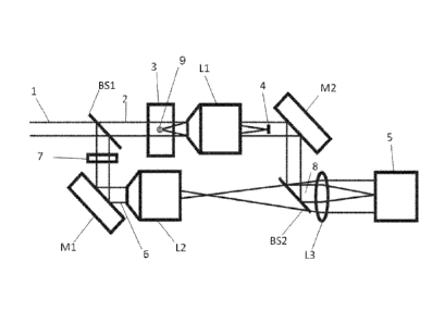
[0023] Fig. 1 is representing a dark field
holographic microscope according to the present invention.
[0024] Fig. la is representing a bright field
intensity image recorded in focus, according to prior art.
(100x100 cropped zone of the original image)
[0025] Fig. lb is representing a dark field
intensity image recorded in focus - Zone Corresponding to
la.
[0026] Fig. lc is representing a dark field
intensity image with a defocus distance of 60pm - Zone
corresponding to la.
[0027] Fig. 1d is representing a refocused dark
field intensity image by digital holography of the digital
hologram defocused by a distance of 60pm - Zone
corresponding to la.
CA 02739017 2011-03-30
WO 2010/037861 PCT/EP2009/062854
9
Detailed Description of the Invention
[0028] In order to outline the advantages provided
by the dark field integration in DHM, we consider first the
detection of an object smaller than the resolution limit of
an imaging system working in transmission.
[0029] To record a digital hologram, a reference
beam is interfering with an object beam on the sensor of a
camera. The best image of an object that can be expected
with a digital holographic reconstruction is the image of
the object that should be recorded when it is at the focus
distance.
[0030] Therefore, we analyse this imaging process.
We consider a circular opaque particle of diameter d
illuminated in transmission. It is imaged by a lens limited
with a circular aperture of diameter D, and we assume than
d is smaller than the resolution limit of the optical
system defined by the Rayleigh criterion.
[0031] The distance between the object plane and the
lens is denoted by a while the conjugated one between the
lens and the image plane is b. The optical axis is denoted
by Z while the x and y axis are perpendicular to z. The
position of the particle is located at the point (xo'YO) in
the object plane. For conciseness, we adopt the operator
notations of Fourier optics 25
[0032] Considering that the amplitude distribution
in the object plane uo(x'y), the amplitude distribution in
the imaging plane ul(x,y) is obtained by applying a linear
operator T, U1(x,y)=Tuo(x,y), in such a way that:
u~ = BV - a Q 1 w(x, y 1- D 2 s(Fcirc) (x - xo )D , (y - yo )D
bj Laj 21,aj 2)a 2)a j
(1)
CA 02739017 2011-03-30
WO 2010/037861 PCT/EP2009/062854
Where B involves all the terms that are unimportant for the
present discussion, VII is the scaling operator defined by
V[a19(x,y)=g(ax,ay) that expresses the magnification of the
optical system, s is the area of the particle, (Fcirc)
5 denotes the Fourier transformations of the lens aperture,
is the wavelength, Q[] represents a quadratic phase
factor defined by Q[P]=exp{j(kp/2)x2+y2)} with k=24) and
J- and where w(x,y) is the amplitude illumination of
the object plane.
10 [0033] Eq. (1) shows that the image of the object has
the shape, with a contrast reversal, of the impulse
response of the optical system on a bright background. We
see also that the modulation of the amplitude image is
multiplied by the area s of the object. It results that the
modulation is decreasing with this area and the available
contrast becomes rapidly weak for particle smaller than the
resolution limit. The modulation is further decreased when
the object is defocused. This has two consequences: the
available dynamics to record the object information is
reduced, and, if the background suffers from noise, as it
is almost always the case, the available signal is highly
corrupted.
[0034] Therefore, it is expected that objects
smaller than the resolution limit of the optical system
become rapidly undetectable. The actual experiences show
that the loss of detections happens very rapidly below the
resolution of the optical system. That is the motivation to
set a dark field system integrated in a digital holographic
microscope to improve the detection capabilities.
[0035] The system that we implemented is described
by the Fig. 1.
CA 02739017 2011-03-30
WO 2010/037861 PCT/EP2009/062854
11
[0036] A light beam, that can be coherent or of
partial coherence, is divided by the beam splitter BS1. The
transmitted beam, the object beam, illuminates the object 9
in transmission and is incident on the microscope lens L1.
An optical stop 4 is placed in such a way that, without
object 9, the transmitted beam is blocked. On the contrary,
when there is an object 9, a part of the diffracted light
is not blocked by the optical stop and is incident on the
camera sensor.
[0037] Therefore, the couple of lens L1-L3 performs
the dark field image of the front focal plane of L1 on the
CCD 5. The effect of the optical stop 4 is to remove the
constant term in Eq.(1). Assuming that the optical stop
weakly disturbs the imaged amplitude on the sensor, Eq. (1)
gives:
u =8(22a) sV[b] [a]w(x'YVVllFcirc 2,,a 2~a
(2)
As there is no more background, the important aspect is
that it becomes possible to adjust the sensitivity of the
detection system in such a way that the full dynamical
range of the recording system is exploited.
[0038] The beam reflected by BS1, the reference
beam, is also redirected on the sensor of the CCD in such a
way that we record the interference pattern between the
object and the reference beam. A neutral density filter 7
allows adjusting the beam ratio to obtain high contrast
fringe pattern.
[0039] The alignment of the system is set without
object and without the optical stop to obtain an
interference pattern to apply an off-axis holographic
method for the computation of the object complex amplitude
25,26 When the alignment procedure is achieved, the optical
stop 4 is placed.
CA 02739017 2011-03-30
WO 2010/037861 PCT/EP2009/062854
12
[0040] As the complex amplitude is available by DHM,
an object, represented by the amplitude U 0 that is recorded
out of focus, can be refocused by computing the Kirchhoff
Fresnel propagation equation over the defocus distance E:
U, (x', y')= exp(Jk)F ; y,Q[ ~2E]FF,vuo(x,y) (3)
[0041] In addition to the refocusing capabilities
provided by DHM, there is one additional advantage in using
the dark field configuration. Consider the dark field
amplitude distribution u'1 of an object smaller than the
resolution limit. For increasing defocus distance E, the
amplitude globally decreases as 1/Ewhile the intensity
decreases as 1/E . Therefore, the decrease is much faster
in intensity than in amplitude.
[0042] As it is the amplitude that is actually
provided by digital holography, regardless to its
refocusing capabilities, it is possible to detect defocus
objects over a range of distances that is significantly
increased in comparison to intensity imaging.
Example
[0043] To demonstrate the feasibility of the dark
field DHM to detect in 3D particles smaller than the
resolution limit, we inserted nanometric particles immersed
in deionised distilled water between a microscopic slide
and a cover-slit. The particles have an average size of
150nm with a width of 20nm. The DHM is equipped with X10
microscope lenses (NA=0.3) that provide a resolution limit
of 1.3pm.
[0044] The original field of view is 525pm x 420pm
that is imaged on a CCD sensor of 1280 x 1024 pixels. We
CA 02739017 2011-03-30
WO 2010/037861 PCT/EP2009/062854
13
note that the size peak of the particles is 8 times smaller
than the resolution limit. The particle intensity images
were recorded focus in bright and dark field (Fig. la, b).
The hologram was recorded in dark field with a defocus
distance of 60pm and reconstructed over this distance. Its
intensity image is provided by Fig. lc. Fig ld shows the
intensity of the refocused image by digital holography.
[0045] We observe, as expected that a large part of
the particles in the Fig. la have a poor contrast in bright
field. The better contrast obtained for some of them are
due to aggregation of the particles. On the dark field
image obtained in Fig. lb, the particles that are almost
invisible in the bright field image can be seen with a good
contrast with respect to the background.
[0046] That confirms the increased detection
capability provided by the dark field system. In Fig. lc,
the defocus makes impossible the detection of the
particles. The digital holographic reconstruction shows the
refocusing capability of the particles, and it can be seen
that this image is very similar to the one that has been
recorded in focus. That is demonstrating the feasibility of
the dark field digital holography for the detection in 3D
of particles that can be largely smaller than the
resolution limit.