Note: Descriptions are shown in the official language in which they were submitted.
CA 02739207 2011-03-25
WO 2010/037851 PCT/EP2009/062827
IMAGING NEUROINFLAMMATION
Technical Field of the Invention
The present invention concerns in vivo imaging and in particular in vivo
imaging of the
peripheral benzodiazepine receptor (PBR). A tetracyclic indole in vivo imaging
agent is
provided that binds with high affinity to PBR, has good uptake into the brain
following
administration, and which preferentially binds to tissues expressing higher
levels of
PBR. The present invention also provides a precursor compound useful in the
synthesis
of the in vivo imaging agent of the invention, as well as a method for
synthesis of said in
vivo imaging agent comprising use of said precursor compound, and a kit for
carrying
out said method. A cassette for the automated synthesis of the in vivo imaging
agent is
also provided. In addition, the invention provides a radiopharmaceutical
composition
comprising the in vivo imaging agent of the invention, as well as methods for
tire: use of
said in vivo imaging agent.
Description of Related Art
The peripheral benzodiazepine receptor (PBR), which is also known as
translocator
protein (TSPO), is known to be mainly localised in peripheral tissues and
glial cells but
its physiological function remains to be clearly elucidated. Subcellularly,
PBR is
known to localise on the outer mitochondrial membrane, indicating a potential
role in
the modulation of mitochondrial function and in the immune system. It has
furthermore
been postulated that PBR is involved in cell proliferation, steroidogenesis,
calcium now
and cellular respiration. PBR has been associated with a variety of conditions
including
acute and chronic stress, anxiety, depression, Parkinson's disease,
Alzheimer's disease,
brain damage, cancer (Gavish et al Pharm. Rev. 1999; 51 629), Huntington's
disease
(Mel3mer and Reynolds Neurosci. Lett. 1998; 241: 53-6), asthma (Pelaia et al
Gen.
Pharmacol. 1997; 28(4): 495-8), rheumatoid arthritis (Bribes et al Eur. J.
Pharmacol.
2002, 452(1): 111-22), atherosclerosis (Davies et at J. Nucl. Med. 2004; 45:
1898-1907)
and multiple sclerosis (Banati et al 2000 Brain; 123: 2321). PBR may also be
associated with neuropathic pain, Tsuda et at having observed activated
microglia in
subjects with neuropathic pain (2005 TINS 28(2) pplOl-7).
-1-
CA 02739207 2011-03-25
WO 2010/037851 PCT/EP2009/062827
Ligands having high affinity for PBR are known in the art. A class of indole
compounds having affinity for PBR (IC50 values for most active compounds of
between
0.2nM and 5.OnM) is disclosed in US 6451795. The compounds disclosed therein
are
stated to be useful for the prevention or treatment of peripheral neuropathies
and for the
treatment of central neurodegenerative diseases. Okubu et al (Bioorganic &
Medicinal
Chemistry 2004; 12: 3569-80) describe the design, synthesis and structure of a
group of
tetracyclic indole compounds having affinity for PBR (IC50 values as low as
about
0.4nM). No particular application of the compounds is discussed in this
publication by
Okubu et al.
In vivo imaging of PBR is also known in the art. Positron emission tomography
(PET)
imaging using the PBR selective ligand, (R)-[11C]PK11195 provides a generic
indicator
of central nervous system (CNS) inflammation. Despite the successful use of
(R)-
[' 1 C]PK11195, it has its limitations. It is known to have high protein
binding, and low
specific to non-specific binding (Lockhart et al. Nucl Med Biol. 30(2):199-
206). The
role of its radiolabelled metabolites is not known and quantification of
binding requires
complex modelling. There have been efforts to provide compounds having high
affinity
and selectivity for PBR to enable improved measurement of PBR in the CNS.
[I IC]DAA1106 and [18F]FEDAA1106 are PET radioligands based on aryloxyalinine
compounds and have been studied in humans (Ikomo et al J. Cereb. Blood Flow
Metab.
2007; 27: 173-84 and Fujimura et al J. Nuc. Med. 2006; 47: 43-50). However,
the
kinetic properties of these compounds are not ideal and may limit their
application to
quantitative studies. In an effort to improve further upon these radioligands,
another
aryloxyaniline derivative, PBR28, has been reported by Briard et al (J. Med.
Chem.
2008; 51: 17-30). An 11C-labelled version of PBR28 was injected into monkey to
assess its brain kinetics using PET. [11C]PBR28 showed high brain uptake, good
specific binding to PBR-expressing tissues and kinetic properties more
suitable for in
vivo imaging. PBR-binding pyrazolopyrimidine compounds have also been
evaluated
as PET radioligands for targeting PBR. James et al (J. Nuc. Med. 2008; 49(5):
814-22)
report that the PET radioligand [18F]-DPA-714 has high affinity for PBR, and
selective
uptake by PBR in baboon brain following intravenous administration. The
kinetics of
brain uptake of [' 8F]-DPA-714 was reported to be slower than, but similar in
nature to,
-2-
CA 02739207 2011-03-25
WO 2010/037851 PCT/EP2009/062827
[11C]DAA1106 and [18F]FEDAA1106. WO 2007/057705 discloses tetracyclic indole
compounds labelled with an imaging moiety, which are suitable for in vivo
imaging.
The in vivo imaging agents exemplified in WO 2007/057705 were shown to have
good
affinity to PBR, with K, values in a competition assay against ['H]-PK-1 1195
of
between 1.OnM and 0.1nM. However, the present inventors have now found that
the
selectivity of these compounds for PBR-expressing tissues is not ideal for in
vivo
imaging of PBR expression in the central nervous system.
There is scope to improve upon the known tetracyclic indole compounds in order
to
provide alternative in vivo imaging agents for evaluation of PBR expression in
the
central nervous system.
Summary of the Invention
The present invention provides in vivo imaging agents based on tetracyclic
indole
compounds. In comparison to known in vivo imaging agents based on tetracyclic
indole
compounds, the in vivo imaging agents of the present invention have better
properties
for in vivo imaging. The in vivo imaging agents of the present invention have
good
binding properties to the peripheral benzodiazepine receptor, as well as good
brain
uptake and in vivo kinetics following administration to a subject.
Detailed Description of the Invention
In Vivo Imaging Agent
In one aspect the present invention provides an in vivo imaging agent of
Formula I:
/N O
7 6
8 y 5
X
to N
1 I Q
R 3
2
-3-
CA 02739207 2011-03-25
WO 2010/037851 PCT/EP2009/062827
or a salt or solvate thereof wherein:
Q is hydrogen or fluorine;
X is hydrogen, fluoro, bromo, odo, hydroxy, C1_6 alkyl, Cl_6 haloalkyl, Cl_6
alkoxy,
or C 1 _6 alkyl amide;
Y is S, SO or S02; and,
R is hydrogen, CI_6 alkyl, or Cj_6 fluoroalkyl;
and wherein at least one atom of said in vivo imaging agent of Formula I is a
radioisotope suitable for in vivo imaging; wherein when said radioisotope is a
radioisotope of carbon, it is a carbonyl carbon;
with the proviso that, when Y is S, Q or X are not both hydrogen.
The term "in vivo imaging" as used herein refers to those techniques that non-
invasively
produce images of all or part of the internal aspect of the subject of the
invention.
Preferred in vivo imaging methods for use in the present invention are single
photon
emission computed tomography (SPECT) and positron emission tomography (PET),
with PET being especially preferred. The preference for PET in the method of
the
invention is due to its excellent sensitivity and resolution, so that even
relatively small
changes in a lesion can be observed over time. PET scanners routinely measure
radioactivity concentrations in the picomolar range. Micro-PET scanners now
approach
a spatial resolution of about Imm, and clinical scanners about 4-5mm.
The "in vivo imaging agent" of Formula I comprises a radioisotope suitable for
in vivo
imaging. This "radioisotope suitable for in vivo imaging" is a radioisotopic
form of one
of the atoms defined above for the in vivo imaging agent of Formula I. In
order to be
suitable for in vivo imaging as defined herein, the radioisotope is preferably
a gamma-
or a positron-emitter, thereby enabling detection of the in vivo imaging agent
external to
the subject following administration.
Suitable salts according to the invention include (i) physiologically
acceptable acid
addition salts such as those derived from mineral acids, for example
hydrochloric,
-4-
CA 02739207 2011-03-25
WO 2010/037851 PCT/EP2009/062827
hydrobromic, phosphoric, metaphosphoric, nitric and sulphuric acids, and those
derived
from organic acids, for example tartaric, trifluoroacetic, citric, malic,
lactic, fumaric,
benzoic, glycollic, gluconic, succinic, methanesulphonic, and para-
toluenesulphonic
acids; and (ii) physiologically acceptable base salts such as ammonium salts,
alkali
metal salts (for example those of sodium and potassium), alkaline earth metal
salts (for
example those of calcium and magnesium), salts with organic bases such as
triethanolamine, N-methyl-D-glucamine, piperidine, pyridine, piperazine, and
morpholine, and salts with amino acids such as arginine and lysine.
Suitable solvates according to the invention include those formed with
ethanol, water,
saline, physiological buffer and glycol.
Unless otherwise specified, the term "alkyl" alone or in combination, means a
straight-
chain or branched-chain alkyl radical containing preferably from 1 to 6 carbon
atoms,
most preferably 1 to 4 carbon atoms. Examples of such radicals include, but
are not
limited to, methyl, ethyl, n-propyl, isopropyl, n-butyl, isobutyl, see-butyl,
tert-butyl,
pentyl, iso-amyl, hexyl.
Unless otherwise specified, the term "alkoxy", alone or in combination, means
an alkyl
ether radical wherein the term alkyl is as defined above. Examples of suitable
alkyl
ether radicals include, but are not limited to, methoxy, ethoxy, n-propoxy,
isopropoxy,
n-butoxy, iso-butoxy, sec-butoxy, tert- butoxy.
"Alkyl amide" is an alkyl group as defined above linked to an amide, wherein
an amide
is the group -C(=O)-NR'R" wherein R' and R" are independently hydrogen or a
hydrocarbon radical.
The term "halogen" or "halo-" means a substituent selected from fluorine,
chlorine,
bromine or iodine. "Haloalkyl" is an alkyl group as defined above substituted
with one
or more halogens.
The term "h_ day" refers to the -OH radical.
In a preferred embodiment, Q is hydrogen.
-5-
CA 02739207 2011-03-25
WO 2010/037851 PCT/EP2009/062827
X is preferably hydrogen, fluoro, bromo, iodo, hydroxy, C14 alkyl, C1_4
haloalkyl, Cl-4
alkoxy, or C1-4 alkyl amide. X is most preferably hydrogen or C1-4 alkoxy.
Y is preferably S or 502. Y is most preferably S.
R is preferably hydrogen, C14 alkyl, or C14 fluoroalkyl. R is most preferably
C1_4
fluoroalkyl.
In a preferred embodiment of Formula I:
X is hydrogen, fluoro, bromo, iodo, hydroxy, C1-4 alkyl, C14 haloalkyl, C14
alkoxy,
or C1-4 alkyl amide;
Y is S or SO2; and,
R is hydrogen, C1-4 alkyl, or C1-4 fluoroalkyl.
In a most preferred embodiment of Formula I:
Q is hydrogen;
X is C1-4 alkoxy;
Y is S; and,
R is C 1-4 fluoroalkyl.
In an alternative preferred embodiment of Formula I:
Q is fluorine;
X is hydrogen;
Y is S; and,
R is C 14 fluoroalkyl.
Preferred radioisotopes suitable for in vivo imaging of the present invention
are gamma-
emitting radioactive halogens and positron-emitting radioactive non-metals.
-6-
CA 02739207 2011-03-25
WO 2010/037851 PCT/EP2009/062827
Examples of gamma-emitting radioactive halogens suitable for use in the
present
invention are 1231, 131I and 77Br. A preferred gamma-emitting radioactive
halogen is 123,
Examples of positron-emitting radioactive non-metal suitable for use in the
present
invention are 1 1c,13 N, 18F and 1241. A preferred positron-emitting
radioactive non-metal
is 18F; 18F is the most preferred radioisotope suitable for in vivo imaging of
the present
invention.
In a preferred embodiment, for the in vivo imaging agent of Formula I, X is
1231 124I or 131I,
18F or C1.4 [18F]-fluoroalkyl.
In an alternative preferred embodiment, for the in vivo imaging agent of
Formula I, R is C1,
[18F]-fluoroalkyl.
In a further alternative preferred embodiment, for the in vivo imaging agent
of Formula I
the carbonyl carbon is 11 C.
Non-limiting examples of some preferred in vivo imaging agents of the present
invention are
as follows:
,,,,N O -,,_,N O
o l~ S s
N O JN
18 18
F 1 F 2
,N O -~,N O
N S N S F
18 18
F F
-7-
CA 02739207 2011-03-25
WO 2010/037851 PCT/EP2009/062827
0 --~,N 0
O
N N
r) F r
18 F is
F 6
-,,_,N O
j I S
N
r) I
Is F 7
Out of the above in vivo imaging agents, imaging agent 2 is most preferred.
The synthetic methods used to obtain these in vivo imaging agents are
described in the
experimental section below. The potency of these non-radioactive versions of
the in
vivo imaging agents of the present invention was measured in an in vitro
assay, as
described in Example 10.
Examples 7-9 describe how to obtain the radio fluorinated in vivo imaging
agents 1-7.
The skilled person will know that when handling 18F the scale and the
conditions used
are different for safety and practical considerations. For a review of the
production of
18F PET tracers, see chapters 1 and 2 of "Principles and Practice of Positron
Emission
Tomography" (2002 Lippincott Williams & Wilkins; Wahl and Buchanan, Eds.). The
in vivo imaging agents were tested in an animal biodistribution model (Example
11),
and their biodistribution compared to that of the prior art compound [18F]FE-
PBR
(prepared according to Example 14 of WO 2007/057705):
-8-
CA 02739207 2011-03-25
WO 2010/037851 PCT/EP2009/062827
,,N O
11 S
N
r
1sF [18F]FE-PBR
Table 1 below provides data obtained in the in vitro affinity assay as well as
in the in
vivo biodistribution study. Non-radioactive analogues were tested in the in
vitro affinity
assay, and radiolabelled versions were evaluated in the biodistribution assay.
In Vivo Imaging Agent Ki OB:Striatum
nM 30 min
[' 8F]FE-PBR 0.68 1.42
1 0.37 2.07
2 0.40 3.50
3 0.93 2.00
4 0.31 2.92
0.32 2.26
6 0.52 2.67
7 1.09 2.42
5 Table 1: in vitro affinity data and in vivo specific uptake data for in vivo
imaging
agents 1-7 of the present invention as compared with the prior art compound
['8F]FE-PBR. OB = olfactory bulb.
The data illustrate that the potency of non-radioactive versions of in vivo
imaging agents
1-7 compares favourably with that of the prior art compound [18F]FE-PBR. In
addition,
the data show that in vivo imaging agents 1-7 of the invention are retained
significantly
more in the OB as compared with the striatum at 30 minutes post-injection
compared
with [18F]FE-PBR.] As it is known that PBR is highly expressed in the OB
compared
with other areas of the rat brain (see "Handbook of Substance Abuse" by
Tarter,
Ammerman and Ott; Springer 1998: 398-99) these data surprisingly demonstrate
that in
vivo imaging agents 1-7 have improved selectivity for PBR than the previously-
exemplified in vivo imaging agent, [18F]FE-PBR.
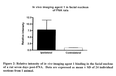
In vivo imaging agent 1 was further analysed in an autoradiography model, as
described
-9-
CA 02739207 2011-03-25
WO 2010/037851 PCT/EP2009/062827
in Example 12 below. Significantly higher levels of radioactivity were
detected in the
lesioned side of the facial nucleus (see Figures 1 and 2). Average intensity
in the
lesioned side was 7.75 +0.95 as compared to 3.73 0.36 in the non-lesioned
side. The
ratio between the two sides was 8.23 + 2.36. As the lesion has a higher
expression of
PBR compared with normal, these data support the conclusion from the
biodistribution
data that in vivo imaging agent 1 has good selectivity for PBR.
The in vivo imaging agents of the present invention therefore have
unexpectedly
superior properties for in vivo imaging of PBR in comparison to known
tetracyclic
indole PBR-binding in vivo imaging agents.
Precursor Compound
In another aspect, the present invention provides a precursor compound of
Formula II:
ZI
77 6
8 / I I Y5
X
9 4
l0 N I Q
R
`~
1 ~ 3
2 (II)
wherein one of R', X' or Z' comprises a chemical group that reacts with a
suitable
source of a radioisotope, where said radioisotope is as suitably and
preferably
defined herein, such that the in vivo imaging agent as suitably and preferably
defined herein is formed upon reaction of said precursor compound with said
suitable source of said radioisotope;
and wherein:
when R' does not comprise said chemical group it is as suitably and preferably
defined herein for R of Formula I, and optionally further comprises a
protecting
group;
when X1 does not comprise said chemical group it is as suitably and preferably
-10-
CA 02739207 2011-03-25
WO 2010/037851 PCT/EP2009/062827
defined herein for X of Formula I, and optionally further comprises a
protecting
group;
when Z' does not comprise said chemical group it is -C(=O)-N-(CH2-CH3)2, and
optionally further comprises a protecting group;
Q1 is as suitably and preferably defined herein for Q of Formula I; and,
Y' is as suitably and preferably defined herein for Y of Formula I, and
optionally
further comprises a protecting group.
A "precursor compound" comprises a derivative of a radiolabelled compound,
designed
so that chemical reaction with a convenient chemical form of the detectable
label occurs
site-specifically; can be conducted in the minimum number of steps (ideally a
single
step); and without the need for significant purification (ideally no further
purification),
to give the desired in vivo imaging agent. Such precursor compounds are
synthetic and
can conveniently be obtained in good chemical purity. The precursor compound
may
optionally comprise a protecting group for certain functional groups of the
precursor
compound.
By the term "protecting group" is meant a group which inhibits or suppresses
undesirable chemical reactions, but which is designed to be sufficiently
reactive that it
may be cleaved from the functional group in question under mild enough
conditions that
do not modify the rest of the molecule. After deprotection the desired product
is
obtained. Protecting groups are well known to those skilled in the art and are
suitably
chosen from, for amine groups: Boc (where Boc is tert-butyloxycarbonyl), Fmoc
(where
Fmoc is fluorenylmethoxycarbonyl), trifluoroacetyl, allyloxycarbonyl, Dde
[i.e. 1-(4,4-
dimethyl-2,6-dioxocyclohexylidene)ethyl] or Npys (i.e. 3-nitro-2-pyridine
sulfenyl); and
for carboxyl groups: methyl ester, tert-butyl ester or benzyl ester. For
hydroxyl groups,
suitable protecting groups are: methyl, ethyl or tent-butyl; alkoxymethyl or
alkoxyethyl;
benzyl; acetyl; benzoyl; trityl (Trt) or trialkylsilyl such as
tetrabutyldimethylsilyl. The
use of further protecting groups are described in `Protective Groups in
Organic
Synthesis', Theorodora W. Greene and Peter G. M. Wuts, (Third Edition, John
Wiley &
Sons, 1999).
-11-
CA 02739207 2011-03-25
WO 2010/037851 PCT/EP2009/062827
The term "a suitable source of a radioisotope" means the radioisotope in a
chemical
form that is reactive with a substituent of the precursor compound such that
the
radioisotope becomes covalently attached to the precursor compound.
For each particular radioisotope presented in the following section, one or
more suitable
sources of the radioisotope are discussed. The person skilled in the art of in
vivo
imaging agents will be familiar with these and other sources of radioisotopes
that are
suitable for application in the present invention.
When the radioisotope of the in vivo imaging agent is 18 F, the radiofluorine
atom may
form part of a fluoroalkyl or fluoroalkoxy group, since alkyl fluorides are
resistant to in
vivo metabolism. Alternatively, the radiofluorine atom may attach via a direct
covalent
bond to an aromatic ring such as a benzene ring.
Radio fluorination may be carried out via direct labelling using the reaction
of 18F-
fluoride with a suitable chemical group in the precursor compound having a
good
leaving group, such as an alkyl bromide, alkyl mesylate or alkyl tosylate.
18F can also be introduced by O-alkylation of hydroxyl groups with
18F(CH2)3OMs or
18 F(CH2)3Br.
For aryl systems, 18F-fluoride nucleophilic displacement from an aryl
diazonium salt,
aryl nitro compound or an aryl quaternary ammonium salt are suitable routes to
aryl-18F
derivatives. Such a strategy is suitable for example to introduce 18F at
positions 1-4 or
7-10 of Formula I.
Alternatively, labeling with 18F can be achieved by nucleophilic displacement
of a
leaving group from a derivative of Formula I. Suitable leaving groups include
Cl, Br, I,
tosylate (OTs), mesylate (OMs), and triflate (OTf). Such derivatives are
precursor
compounds for the preparation of in vivo imaging compounds of the invention.
Another strategy would be to have a suitable leaving group as defined above in
place on
an alkylamide group present on the precursor compound. In this way, the
precursor
compound may be labeled in one step by reaction with a suitable source of
[18F]-
fluoride ion (18F-), which is normally obtained as an aqueous solution from
the nuclear
-12-
CA 02739207 2011-03-25
WO 2010/037851 PCT/EP2009/062827
reaction 180(p,n)18F and is made reactive by the addition of a cationic
counterion and
the subsequent removal of water. For this method, the precursor compounds are
normally selectively chemically protected so that radio fluorination takes
place at a
particular site on the compound. Suitable protecting groups are those already
mentioned previously.
When the radioisotope is ' 8F, it is preferred that either X1 or R' comprises
either:
(i) an alkyl halide or an alkyl sulfonate (such as alkyl bromide, alkyl
mesylate or alkyl
tosylate) for neucleophilic substitution; or,
(ii) hydroxyl (for introduction of 18F by 0-alkylation of hydroxyl groups with
e.g.
' 8F(CH2)3OMs or ' 8F(CH2)3Br).
A generic reaction scheme to arrive at certain 18F in vivo imaging agents of
the
invention is illustrated below:
- 13 -
CA 02739207 2011-03-25
WO 2010/037851 PCT/EP2009/062827
7
~/N O
~/N O x
~ NNH' 7
H 6
S s - X 8 I I S 5
0 4 9 N \ 4
10 H
I / 3 1 / 3
2 * 2
Sodium hydride
O Acetonitrile
i RT
Br n
O
-,,,/N O 7
6
7 6
Tetrabutyl ammonium S
9\ I S 5 fluoride X N
X 9
4
I
to N 4 THE
11
In 1 )n 1 ' 3
O
OH
Methanesulphonylchloride -Si-
Pyridine Dichloromethane
-,,/N 0 N O
7 6 18F 7 6
8 / S5
Z I 8 / S5
X I Potassium carbonate x
9 N 4 Kryptofix 9\ N a
10 Acetonitrile 10 I
n z n 2
~ 1 / 3
OMs 18F
wherein X is as defined for Formula I, and n is between 0 and 5. RT stands for
room
temperature, and OMs stands for mesylate.
An alternative generic reaction scheme to arrive at certain 18 F in vivo
imaging agents of
5 the invention is illustrated below:
- 14-
CA 02739207 2011-03-25
WO 2010/037851 PCT/EP2009/062827
7
~/N O X ~/N O
6 9 / NNH' 7
H 6
f S 5 X s/ I I S s
0 4 9 N 4
to H
3 3
2
18F
TsO TsO 18 F Sodium hydride
OTs Potassium carbonate Acetonitrile
n
Kryptofix 80 C
Acetonitrile
O
7 6
s Ss
X
9 ~ ~ a
to
f / 3
In ~
I$F
wherein X is as defined for Formula I, and n is between 0 and 5, and OTs
stands for
tosylate.
11 C-labelled PET tracer compounds may be synthesised by reacting a precursor
5 compound with 11C methyl iodide. As the half-life of 1 1C is only 20.4
minutes, it is
important that the intermediate 11 C methyl iodide has high specific activity
and,
consequently, that it is produced using a reaction process which is as rapid
as possible.
A thorough review of such 11 C-labelling techniques may be found in Antoni et
al
"Aspects on the Synthesis of 1'C-Labelled Compounds" in Handbook of
10 Radiopharmaceuticals, Ed. M.J. Welch and C.S. Redvanly (2003, John Wiley
and
Sons).
When the in vivo imaging agent of the present invention is labeled with "C,
the 11 C is a
carbonyl carbon. This therefore means that 11 C can be present at the carbonyl
carbon of
-15-
CA 02739207 2011-03-25
WO 2010/037851 PCT/EP2009/062827
Formula I, or alternatively at X when X is a C1-6 alkyl amide.
A 11 C-labelled in vivo imaging agent of Formula I maybe obtained by employing
the
following reaction scheme:
Z C
3
6
3 s y3 s (i) strong base, e.g. BuLi or Pd catalyst 8 3 5
X
9\ I I 00 1 C] C02 Or Co X3 I I Y
0 1 3 (iii) (CHzCHz)zNH with or without condensing agent 110 1N 1 4
R 3 R3 I 3
2
9
IIb Id
5
wherein R3, X3, and Y3 of Formula IIb and Formula Id are as described for R,
X, and Y
of Formula I; and
Z3 is a substrate suitable for transition metal catalysts, e.g. hydrogen,
halide, boronic
acid, OTf, organotin.
10 Methods for the synthesis of 13N-labelled compounds are described by Clark
and
Aigbirhio ("Chemistry of Nitrogen-13 and Oxygen-15" in "Handbook of
Radiopharmaceuticals"; 2003 Wiley: Welch and Redvanly, Eds.). For example, an
in
vivo imaging agent of Formula I may be obtained by nucleophilic substitution
of a
halogen in a suitable precursor compound with 13N-labelled diethyl amine to
obtain the
15 desired amide.
Where the imaging moiety is radioiodine, preferred precursor compounds are
those
which comprise a derivative which either undergoes electrophilic or
nucleophilic
iodination or undergoes condensation with a labelled aldehyde or ketone.
Examples of
the first category are:
20 (a) organometallic derivatives such as a trialkylstannane (e.g.
trimethylstannyl or
tributylstannyl), or a trialkylsilane (e.g. trimethylsilyl) or an organoboron
compound (e.g. boronate esters or organotrifluoroborates);
-16-
CA 02739207 2011-03-25
WO 2010/037851 PCT/EP2009/062827
(b) aromatic rings activated towards electrophilic iodination (e.g. phenols)
and
aromatic rings activated towards nucleophilic iodination (e.g. aryl iodonium
salt
aryl diazonium, aryl trialkylammonium salts or nitroaryl derivatives).
For radioiodination, the precursor compound preferably comprises: an aryl
iodide or
bromide (to permit radioiodine exchange); an activated precursor compound aryl
ring
(e.g. a phenol group); an organometallic precursor compound (e.g. trialkyltin,
trialkylsilyl or organoboron compound); or an organic precursor compound such
as
triazenes or a good leaving group for nucleophilic substitution such as an
iodonium salt.
Precursor compounds and methods of introducing radioiodine into organic
molecules
are described by Bolton (J. Lab. Comp. Radiopharm. 2002; 45: 485-528).
Precursor
compounds and methods of introducing radioiodine into proteins are described
by
Wilbur (Bioconj. Chem. 1992; 3(6): 433-470). Suitable boronate ester
organoboron
compounds and their preparation are described by Kabalaka et al (Nucl. Med.
Biol.,
2002; 29: 841-843 and 2003; 30: 369-373). Suitable organotrifluoroborates and
their
preparation are described by Kabalaka et al (Nucl. Med. Biol., 2004; 31; 935-
938).
Preferred precursor compounds for radioiodination comprise an organometallic
precursor compound, most preferably a trialkyltin.
Examples of aryl groups to which radioactive iodine can be attached are given
below:
SnBu3 OH
Both contain substituents which permit facile radioiodine substitution onto
the aromatic
ring. Alternative substituents containing radioactive iodine can be
synthesised by direct
iodination via radiohalogen exchange, e.g.
1271 123
+ 127
+ 123
When the radioisotope is radioiodine, X1 of Formula II, together with the
aromatic group to
-17-
CA 02739207 2011-03-25
WO 2010/037851 PCT/EP2009/062827
which it is attached, forms:
(i) an aromatic ring substituted with either an organometallic derivative or
an
organoboron compound,
(ii) an aromatic ring activated towards electrophilic radioiodination (e.g.
phenols); or,
(iii) an aromatic ring activated towards nucleophilic radioiodination (e.g.
aryl
iodonium salt aryl diazonium, aryl trialkylammonium salts or nitroaryl
derivatives).
These precursor compounds are easily converted into radioiodinated in vivo
imaging
agents of the invention by radioiodine substitution.
Radiobromination can be achieved by methods similar to those described above
for
radioiodination. Kabalka and Varma have reviewed various methods for the
synthesis
of radiohalogenated compounds, including radiobrominated compounds
(Tetrahedron
1989; 45(21): 6601-21).
The precursor compound of the invention is ideally provided in sterile,
apyrogenic form.
The precursor compound can accordingly be used for the preparation of a
pharmaceutical composition comprising the in vivo imaging agent together with
a
biocompatible carrier suitable for mammalian administration. The precursor
compound
is also suitable for inclusion as a component in a kit for the preparation of
such a
pharmaceutical composition.
In a preferred embodiment, the precursor compound is provided in solution and
as part
of a kit or of a cassette designed for use in an automated synthesis
apparatus. These
aspects are discussed in more detail below in relation to additional aspects
of the
invention.
In another preferred embodiment, the precursor compound is bound to a solid
phase.
The precursor compound is preferably supplied covalently attached to a solid
support
matrix. In this way, the desired product forms in solution, whereas starting
materials
-18-
CA 02739207 2011-03-25
WO 2010/037851 PCT/EP2009/062827
and impurities remain bound to the solid phase. As an example of such a
system,
precursor compounds for solid phase electrophilic fluorination with 18F-
fluoride are
described in WO 03/002489, and precursor compounds for solid phase
nucleophilic
fluorination with 18F-fluoride are described in WO 03/002157.
Method for Preparation of In Vivo Imaging Agent
In a further aspect, the present invention provides a method for the
preparation of an in
vivo imaging agent of the invention, said method comprising:
(i) providing a precursor compound of Formula II as defined above;
(ii) providing a suitable source of said radioisotope as defined above;
(iii)reacting the precursor compound of step (i) with the radioisotope of step
(ii) to obtain an in vivo imaging agent of the invention.
In step (i), the precursor compound may be provided in solution in a kit or in
a cassette
suitable for use with an automated synthesis apparatus, or alternatively
attached to a
solid support, as described above in the description of the precursor
compound. The kit
and cassette form additional aspects of the invention and will be discussed in
more
detail below.
Suitable sources of radioisotope are as described above in relation to the
precursor
compound of the invention.
The step of "reacting" the precursor compound with the radioisotope involves
bringing
the two reactants together under reaction conditions suitable for formation of
the desired
in vivo imaging agent in as high a radiochemical yield (RCY) as possible.
Particular
synthetic routes for obtaining in vivo imaging agents of the present invention
are
presented in the experimental section below.
Kit for Preparation of In Vivo Imaging Agent
In a yet further aspect, the present invention provides a kit for the
preparation of an in
vivo imaging agent of the invention, said kit comprising a precursor compound
of
-19-
CA 02739207 2011-03-25
WO 2010/037851 PCT/EP2009/062827
Formula II as described above, so that reaction with a sterile source of a
radioisotope
gives the desired in vivo imaging agent with the minimum number of
manipulations.
Such considerations are particularly important where the radioisotope has a
relatively
short half-life, and for ease of handling and hence reduced radiation dose for
the
radiopharmacist. The precursor compound is preferably present in the kit in
lyophilized
form, and the reaction medium for reconstitution of such kits is preferably a
biocompatible carrier.
The "biocompatible carrier" is a fluid, especially a liquid, in which the in
vivo imaging
agent is suspended or dissolved, such that the composition is physiologically
tolerable,
i.e. can be administered to the mammalian body without toxicity or undue
discomfort.
The biocompatible carrier is suitably an injectable carrier liquid such as
sterile, pyrogen-
free water for injection; an aqueous solution such as saline (which may
advantageously
be balanced so that the final product for injection is either isotonic or not
hypotonic); an
aqueous solution of one or more tonicity-adjusting substances (e.g. salts of
plasma
cations with biocompatible counterions), sugars (e.g. glucose or sucrose),
sugar alcohols
(e.g. sorbitol or mannitol), glycols (e.g. glycerol), or other non-ionic
polyol materials
(e.g. polyethyleneglycols, propylene glycols and the like). The biocompatible
carrier
may also comprise biocompatible organic solvents such as ethanol. Such organic
solvents are useful to solubilise more lipophilic compounds or formulations.
Preferably
the biocompatible carrier is pyrogen-free water for injection, isotonic saline
or an
aqueous ethanol solution. The pH of the biocompatible carrier for intravenous
injection
is suitably in the range 4.0 to 10.5.
In the kit of the invention, the precursor compound is preferably presented in
a sealed
container which permits maintenance of sterile integrity and/or radioactive
safety, plus
optionally an inert headspace gas (e.g. nitrogen or argon), whilst permitting
addition and
withdrawal of solutions by syringe. A preferred sealed container is a septum-
sealed
vial, wherein the gas-tight closure is crimped on with an overseal (typically
of
aluminium). Such sealed containers have the additional advantage that the
closure can
withstand vacuum if desired e.g. to change the headspace gas or degas
solutions.
Preferred embodiments of the precursor compound when employed in the kit are
as
-20-
CA 02739207 2011-03-25
WO 2010/037851 PCT/EP2009/062827
described herein.
The precursor compound for use in the kit may be employed under aseptic
manufacture
conditions to give the desired sterile, non-pyrogenic material. The precursor
compound
may alternatively be employed under non-sterile conditions, followed by
terminal
sterilisation using e.g. gamma-irradiation, autoclaving, dry heat or chemical
treatment
(e.g. with ethylene oxide). Preferably, the precursor compound is provided in
sterile,
non-pyrogenic form. Most preferably the sterile, non-pyrogenic precursor
compound is
provided in the sealed container as described above.
Preferably, all components of the kit are disposable to minimise the
possibilities of
contamination between runs and to ensure sterility and quality assurance.
In a preferred embodiment, the kit may comprise a cassette which can be
plugged into a
suitably adapted automated synthesiser, described in more detail below. Such a
kit
typically includes means for fluorinating with fluoride ion and may also
comprise a
column to remove unwanted fluoride ion. The reagents, solvents and other
consumables required for the synthesis may also be included together with a
data
medium, such as a compact disc carrying software, which allows the automated
synthesiser to be operated in a way to meet the end user's requirements for
concentration, volumes, time of delivery etc.
[18F]-radiotracers for PET imaging are now often conveniently prepared on an
automated radiosynthesis apparatus. There are several commercially-available
examples of such apparatus, including Tracerlab and Fastlab (GE Heathcare).
Such
apparatus commonly comprises a "cassette", often disposable, in which the
radiochemistry is performed, which is fitted to the apparatus in order to
perform a
radiosynthesis. The cassette normally includes fluid pathways, a reaction
vessel, and
ports for receiving reagent vials as well as any solid-phase extraction
cartridges used in
post-radiosynthesic clean up steps.
The present invention therefore provides in another aspect a cassette for an
automated
synthesis apparatus comprising the precursor compound in a sealed container as
described hereinbefore. The present invention also provides a cassette for the
-21 -
CA 02739207 2011-03-25
WO 2010/037851 PCT/EP2009/062827
automated synthesis of an in vivo imaging agent as defined herein comprising:
(i) a vessel containing a precursor compound as defined herein; and
(ii) means for eluting the vessel with a suitable source of a radioisotope,
said
radioisotope as defined herein.
The cassette may additionally comprise:
(iii) an ion-exchange cartridge for removal of excess radiolabel; and
optionally,
(iv) a cartridge for deprotection of the resultant radiolabelled product to
form an
in vivo imaging agent as defined herein.
Radiopharmaceutical Composition
In another further aspect, the present invention provides a
"radiopharmaceutical
composition", which is a composition comprising the in vivo imaging agent of
the
invention, together with a biocompatible carrier in a form suitable for
mammalian
administration. The biocompatible carrier is as defined above in relation to
the kit of
the invention.
The radiopharmaceutical composition may be administered parenterally, i.e. by
injection, and is most preferably an aqueous solution. Such a composition may
optionally contain further ingredients such as buffers; pharmaceutically
acceptable
solubilisers (e.g. cyclodextrins or surfactants such as Pluronic, Tween or
phospholipids); pharmaceutically acceptable stabilisers or antioxidants (such
as ascorbic
acid, gentisic acid orpara-aminobenzoic acid). Where the in vivo imaging agent
of the
invention is provided as a radiopharmaceutical composition, the method for
preparation
of said in vivo imaging agent may further comprise the steps required to
obtain a
radiopharmaceutical composition, e.g. removal of organic solvent, addition of
a
biocompatible buffer and any optional further ingredients. For parenteral
administration, steps to ensure that the radiopharmaceutical composition is
sterile and
apyrogenic also need to be taken.
-22-
CA 02739207 2011-03-25
WO 2010/037851 PCT/EP2009/062827
In Vivo Imaging Method
In a yet further aspect, the present invention provides an in vivo imaging
method for
determining the distribution and/or the extent of PBR expression in a subject
comprising:
(i) administering to said subject an in vivo imaging agent as defined herein;
(ii) allowing said in vivo imaging agent to bind to PBR in said subject;
(iii) detecting by an in vivo imaging procedure signals emitted by the
radioisotope of said in vivo imaging agent;
(iv)generating an image representative of the location and/or amount of said
signals; and,
(v) determining the distribution and extent of PBR expression in said subject
wherein said expression is directly correlated with said signals emitted by
said
in vivo imaging agent.
For the in vivo imaging method of the invention, the in vivo imaging agent is
as defined
earlier in the specification.
"Administering" the in vivo imaging agent is preferably carried out
parenterally, and most
preferably intravenously. The intravenous route represents the most efficient
way to deliver
the in vivo imaging agent throughout the body of the subject, and therefore
also across the
blood-brain barrier (BBB) and into contact with PBR expressed in said subject.
The in vivo
imaging agent of the invention is preferably administered as the
pharmaceutical
composition of the invention, as defined herein.
Following the administering step and preceding the detecting step, the in vivo
imaging
agent is allowed to bind to PBR. For example, when the subject is an intact
mammal, the
in vivo imaging agent will dynamically move through the mammal's body, coming
into
contact with various tissues therein. Once the in vivo imaging agent comes
into contact
with PBR, a specific interaction takes place such that clearance of the in
vivo imaging agent
from tissue with PBR takes longer than from tissue without, or with less PBR.
A certain
point in time will be reached when detection of in vivo imaging agent
specifically bound to
-23-
CA 02739207 2011-03-25
WO 2010/037851 PCT/EP2009/062827
PBR is enabled as a result of the ratio between in vivo imaging agent bound to
tissue with
PBR versus that bound in tissue without, or with less PBR. An ideal such ratio
is around
2:1.
The "detecting" step of the method of the invention involves detection of
signals emitted by
the radioisotope by means of a detector sensitive to said signals. This
detection step can
also be understood as the acquisition of signal data. Single-photon emission
tomography
(SPECT) and positron-emission tomography (PET) are the most suitable in vivo
imaging
procedures for use in the method of the invention. PET is a preferred in vivo
imaging
procedures for use in the method of the invention.
The "generating" step of the method of the invention is carried out by a
computer which
applies a reconstruction algorithm to the acquired signal data to yield a
dataset. This
dataset is then manipulated to generate images showing the location and/or
amount of
signals emitted by said radioisotope. The signals emitted directly correlate
with the
expression of PBR such that the "determining" step can be made by evaluating
the
generated image.
The "subject" of the invention can be any human or animal subject. Preferably
the subject
of the invention is a mammal. Most preferably, said subject is an intact
mammalian body
in vivo. In an especially preferred embodiment, the subject of the invention
is a human.
The in vivo imaging method may be used to study PBR in healthy subjects, or in
subjects
known or suspected to have a pathological condition associated with abnormal
expression
of PBR (a "PBR condition"). Preferably, said method relates to the in vivo
imaging of a
subject known or suspected to have a PBR condition, and therefore has utility
in a method
for the diagnosis of said condition. Examples of such PBR conditions where in
vivo
imaging would be of use include neuropathologies such as Parkinson's disease,
multiple
sclerosis, Alzheimer's disease and Huntington's disease where
neuroinflammation is
present. Other PBR conditions that may be usefully imaged with the compounds
of the
invention include neuropathic pain, arthritis, asthma, atherosclerosis, as
well as malignant
diseases such as colorectal cancer and breast cancer. The in vivo imaging
agents of the
invention are particularly suited to in vivo imaging of the central nervous
system (CNS) due
to their good brain uptake.
-24-
CA 02739207 2011-03-25
WO 2010/037851 PCT/EP2009/062827
In an alternative embodiment, the in vivo imaging method of the invention
maybe carried
out repeatedly during the course of a treatment regimen for said subject, said
regimen
comprising administration of a drug to combat a PBR condition. For example,
the in vivo
imaging method of the invention can be carried out before, during and after
treatment with
a drug to combat a PBR condition. In this way, the effect of said treatment
can be
monitored over time. Preferably for this embodiment, the in vivo imaging
procedure is
PET. PET has excellent sensitivity and resolution, so that even relatively
small changes in
a lesion can be observed over time, which is advantageous for treatment
monitoring. PET
scanners routinely measure radioactivity concentrations in the picomolar
range. Micro-
PET scanners now approach a spatial resolution of about 1 mm, and clinical
scanners about
4-5mm.
In a further aspect, the present invention provides a method for diagnosis of
a PBR
condition. The method of diagnosis of the invention comprises the method of in
vivo
imaging as defined above, together with the further step (vi) of attributing
the distribution
and extent of PBR expression to a particular clinical picture, i.e. the
deductive medical
decision phase.
In another aspect, the present invention provides the in vivo imaging agent as
defined
herein for use in the method of diagnosis as defined herein.
In a yet further aspect, the present invention provides the in vivo imaging
agent as defined
herein for use in the manufacture of a radiopharmaceutical composition as
defined herein
for use in the method of diagnosis as defined herein.
Brief Description of the Examples
All reagents were obtained from Sigma Aldrich.
Examples 1-6 describe the synthesis of non-radioactive versions of various in
vivo
imaging agents of the invention.
Examples 7-9 describe how to obtain 18F-labelled in vivo imaging agents of the
invention.
-25-
CA 02739207 2011-03-25
WO 2010/037851 PCT/EP2009/062827
Example 10 describes the in vitro potency assay used to measure PBR affinity
of the
imaging agents of the invention.
Example 11 describes how the animal biodistribution studies were carried out.
Example 12 describes the facial nerve axotomy animal model and its use in an
autoradiography study.
List of Abbreviations used in the Examples
DCM dichloromethane
DMF dimethylformamide
DMSO dimethyl sulfoxide
EtOAc ethyl acetate
FNA: facial nerve axotomy
g gram(s)
h hour(s)
HRMS high resolution mass spectrometry
K222 Kryptofix 2.2.2
M molarity = moles of solute/litre of solution
MHz mega hertz
ml millilitre(s)
mmol milimole(s)
N normality = number of equivalents/1L of solution
NMR nuclear magnetic resonance
-26-
CA 02739207 2011-03-25
WO 2010/037851 PCT/EP2009/062827
PBR peripheral benzodiazepine receptor
RT room temperature
Examples
Example 1: Preparation of (+-)-11-(2-fluoroethyl)-8-methoxy-6,11-dilhydro-5-
thia-11-
aza-benzo,af fluorene-6-carboxylic acid diethyl amide (non-radioactive ima,-in
a entl
Example 1(i):' (+-)-4-Oxo-thiochroman-2-carboxylic acid diethyl amide
_,N 0
S
O
(+-)-4-Oxo-thiochroman-2-carboxylic acid (10.4 g, 50 mmol), prepared as
described in
T. Okubo et al (Bioorg. Med. Chem. 2004; 12-.3569-3580), in dry DCM (100 ml)
was
stirred under an atmosphere of nitrogen at room temperature with oxalyl
chloride (12.6
g, 100 mmole) and one drop of DMF for 18 h. The reaction was then evaporated
in
vacuo to a gum and then redissolved in DCM (100 ml), cooled to 0 C on an ice
bath,
stirred and treated dropwise with diethylamine (8.03 g, 110 mmol) in DCM (20
ml)
over a period of 1 h. The reaction was allowed to warm to room temperature
over 1 h
and 10% aqueous potassium carbonate solution (100 ml) was added and the
reaction
mixture vigorously stirred. The DCM solution was separated. The aqueous
solution
was extracted with two further batches of DCM (100 ml) and the combined
extracts
were dried over magnesium sulphate. The DCM solution was concentrated in vacuo
to
give a dark green oil that crystallized on standing. The crystalline solid was
triturated
with diethyl ether (50 ml) and filtered to give the title compound (8.57 g,
65%) as a pale
green solid.
1H NMR (300 MHz, CDC13) 6 1.06 (t, J=7.1 Hz, 3H), 1.23 (t, J=7.1 Hz, 3H), 3.0-
3.5
-27-
CA 02739207 2011-03-25
WO 2010/037851 PCT/EP2009/062827
(m, 6H), 4.25 (m, 1H), 7.15-7.21 (m, 2H), 7.32-7.39 (m, 1H), 8.10-8.14 (m,
1H).
13C NMR (75 MHz, CDC13) 6 12.9, 14.8, 40.1, 40.7, 42.3, 42.5, 125.8, 127.2,
128.7, 130.8, 133.4, 137.9, 167.9, 193.1
Example I(ii): (+-)-8-methoxy-6 11-dihydro-5-thia-11-aza-benzoTa J fluorene-6-
carboxylic acid diethyl amide
_,,N O
1 ~( O S
I~
H
To a solution of (+-)-4-Oxo-thiochroman-2-carboxylic acid diethyl amide (1.32
g, 5.0
mmol Example 1(i)) and 4-methoxyphenyl hydrazine hydrochloride (0.87 g, 5.0
mmol)
in ethanol (10 ml) was added concentrated sulphuric acid (0.73 ml, 1.35 g,
13.8 mmol)
under nitrogen. The reaction mixture was heated under reflux for 24 h. After
cooling,
the reaction mixture was filtered, the solid washed with ethanol, dried in
vacuo (45C)
to give the title compound (1.05 g, 57%) as a pale yellow solid.
1H NMR (300 MHz, DMSO-d6) 6 0.97 (t, J=6.8 Hz, 3H), 1.28 (t, J=6.8 Hz, 3H),
3.25 (m, 2H), 3.60 (m, 2H), 3.74 (s, 3H), 5.59 (s, 1H), 6.80 (m, 2H), 7.10-
7.35 (m,
4H), 7.75 (d, J=7.3 Hz, 1 H), 11.52 (s, 1 H, NH).
13C NMR (75 MHz, DMSO-d,) 6 10.5, 12.7, 32.7, 37.9, 39.5, 53.0, 97.6, 103.3,
109.87, 109.92, 120.3, 123.5, 123.8, 124.3, 124.7, 124.9, 127.8, 129.4, 131.8,
151.3, 166.2
m/z (ES) 367.1 (M+H).
-28-
CA 02739207 2011-03-25
WO 2010/037851 PCT/EP2009/062827
Example 1(iii). (+-)-11-(2 uoroethyl)-8-methoxy-611-dihydro-5-thia-11-aza-
benzofal
fuorene-6--carboxylic acid diethyl amide
-,~,N O
O S
N
F
To a solution of (+-)-8-methoxy-6,11-dihydro-5-thia-11-aza-benzo[a] fluorene-6-
carboxylic acid diethyl amide (150 mg, 0.41 mmol; Example 1(11)) in anhydrous
DMF
(4 ml) was added 2-fluoroethyl tosylate (166 mg, 0.82 mmol) ), prepared as
described in
L. Cronin et al (J. Org. Chem. 2004; 69: 5934-5946) followed by sodium hydride
60%
dispersion in mineral oil (34 mg, 0.82 mmol) under nitrogen. The reaction
mixture was
heated at 80C for 1 h. After cooling, the solvents were removed in vacuo, the
residue
quenched with water (30 ml), extracted with DCM (2 x 30 ml), dried (MgSO4) and
solvents removed in vacuo. The residue was purified by column chromatography
on
silica, eluting with 5-10% EtOAc/CH2C12. The crude solid was quenched with
ether/pet. spirit, filtered, dried in vacuo (45C) to give the title compound
(77 mg, 46%)
as a pale brown solid.
1H NMR (300 MHz, CDC13) 6 1.12 (t, J=7.0 Hz, 3H), 1.36 (t, J=7.0 Hz, 3H), 3.25-
3.70 (m, 4H), 3.83 (s, 3H), 4.45-4.70 (m, 2H), 4.80 (t, J=5.2 Hz, 1H), 4.96
(t, J=5.2
Hz, 1H), 5.09 (s, 1H), 6.84-6.93 (m, 2H), 7.13-7.32 (m, 3H), 7.46 (m, 1H),
7.58 (d,
J=8.0 Hz, 1H).
13C NMR (75 MHz, CDC13) 6 12.9, 14.9, 37.3, 41.1, 42.5, 45.5, 45.8, 55.9,
81.2,
83.5, 100.4, 110.1, 111.09, 111.12, 112.8, 124.31, 124.35, 125.2, 126.5,
127.1,
127.6, 128.8, 132.2, 134.4, 137.0, 154.8, 168.0
19F NMR (282 MHz, CDC13) 6 -219.4, -219.5, -219.6, -219.65, -219.73, -219.8,
-29-
CA 02739207 2011-03-25
WO 2010/037851 PCT/EP2009/062827
-219.9
m/z (ES) 413.1 (M+H).
Example 2: Preparation of (+-)-11-(2-fluoroethyl)-1O-methoxy-6,11-dihydro-5-
thia-
11-aza-benzofa/ fluorene-6-carboxylic acid diethyl amide (non-radioactive
imaging
agent 3
Example 2(i): (+-)-10-methoxy-6,11-dihydro-5-thia-11-aza-benzo f J fluorene-6-
carboxylic acid diethyl amide
--~,N O
S
H
This compound was prepared as described for Example 1(11) except that 2-
methoxyphenyl hydrazine hydrochloride was used instead of 4-methoxyphenyl
hydrazine hydrochloride. The compound was obtained in 40% yield.
m/z (ES) 367.0 (M+H).
Example 2(ii): (+-)-11-(2 uoroethyl)-10-methoxy-611-dihydro-5-thia-l l -aza-
benzo fa7 fluorene-6-carboxylic acid diethyl amide
-,,,N O
1 S
110
F
This compound was prepared as described for Example 1(iii) except that (+-)-10-
methoxy-6,11-dihydro-5-thia-11-aza-benzo[a] fluorene-6-carboxylic acid diethyl
amide
-30-
CA 02739207 2011-03-25
WO 2010/037851 PCT/EP2009/062827
(Example 2(i)) was used instead of (+-)-8-methoxy-6,11-dihydro-5-thia-11-aza-
benzo[a] fluorene-6-carboxylic acid diethyl amide. After recrystallisation
(ether), was
obtained in 10% yield as a white solid.
'H NMR (300 MHz, CDC13) 6 1.09 (t, J=7.0 Hz, )H), 1.35 (t, J=7.0 Hz, 3H), 3.25-
3.67 (m, 4H), 3.95 (s, 3H), 4.70-4.96 (m, 4H), 5.04 (s, I H), 6.67 (m, I H),
7.04 (m,
2H), 7.16 (m, I H), 7.29 (m, I H), 7.45 (m, I H), 7.77 (m, I H).
m/z (ES) 413.1 (M+H).
Example 3: Preparation of (+-)-4-fluoro-11-(2-fiuoroeth0-6,11-dihydro-5-thia-
11-
aza-benzo[a/ fluorene-6-carboxylic acid diethyl amide (non-radioactive imaj
inJ
agent 4
Example 3(i): (+-)-8-Fluoro-4-oxo-thiochrom ,,,: -2-carboxlic acid
0 OH
S
F
O 11
In a round bottom flask 2-fluorothiophenol (5.0 g, 39.0 mmol, 4.16 mL) and
furan-2,5-
dione (3.82 g, 39.0 mmol) in toluene (12 mL) were stirred at 50 C for 40
minutes.
Triethylamine (100 l) in toluene (5 mL) was then added over 10 minutes
ensuring the
reaction temperature did not increase over 60'C. The reaction was then heated
at 70'C
for 20 minutes. The reaction was then concentrated under high vacuum to obtain
the
crude product as an oil. This material was dissolved in DCM (75 mL), cooled on
an ice
bath and treated with aluminium trichloride (7.78 g, 58.5 mmol) in small
portions so as
to keep the temperature below 10 C. The reaction was warmed to RT and there
was a
vigorous evolution of hydrogen chloride gas and the reaction became very
viscous and
turned red. After stirring at RT for 1.5 hours the reaction mixture was then
diluted with
DCM (50 mL) to make it less viscous and slowly poured into vigorously stirred
concentrated hydrochloric acid (30 mL) and ice (30 g) in a 2L conical flask.
The
reaction was vigorously stirred and diluted with a further portion of DCM (500
mL) and
- 31 -
CA 02739207 2011-03-25
WO 2010/037851 PCT/EP2009/062827
isopropyl alcohol (50 mL) to dissolve any solid that had crystallized out. The
DCM
layer was separated, dried over magnesium sulfate and concentrated in vacuum
to give a
brown solid. The crude solid was purified by triturated with diethyl ether and
a cream
solid was collected by filtration to give 2.5 g (28%) of 8-Fluoro-4-oxo-
thiochromana-2-
carboxlic acid. 'H NMR (300 MHz; DMSO-d3): b 3.04-3.20 (2H, in, 3-H), 4.51
(1H,
dd, J= 4 and 6Hz, 2-H), 7.26-7.34 (1H, m, 6-H), 7.45- 7.52 (1H, in, 7-H), 7.82
(1H,
dd, J = 1 and 8 Hz, 5H). 13C NMR (75 MHz; DMSO-d3): 6 40.5, 40.7, 119.8,
120.1,
123.88, 123.92, 126.0, 126.1, 131.9, 156.1, 159.2, 171.5, 191.2, 191.3.
Example 3(ii): (+-)-8-Fluoro-4-oxo-thiochroman-2-carboxylic acid diethylamide
r
O N,_,
S
F
O
8-Fluoro-4-oxo-thiochromana-2-carboxlic acid (2.5 g, 11.1 mmol) in dry DCM
(50m1)
was stirred under an atmosphere of nitrogen at room temperature with oxalyl
chloride
(2.81 g, 22.1 mmo, 1.93 mL) and one drop of DMF to catalyse the reaction for
18h.
The acid was initially insoluble but dissolved as it reacted to give a orange
clear
solution after 2 hours and then turned black after 24h. The reaction was then
evaporated in vacuum to a gum to remove excess oxalyl chloride and 'H and 13 C
NMR
run in CDC13 to confirm complete reaction. The reaction was then redissolved
in DCM
(50m1) cooled to OTC on an ice bath stirred and treated dropwise with
diethylamine (1.66
g, 22.7 mmol, 2.05 mL) in DCM (20m1) over a period of lh. The reaction was
allowed
to warm to room temperature over a period of lh. The reaction was then
quenched by
the addition of 5% potassium carbonate solution (100ml) and the reaction
mixture
stirred vigorously. The DCM solution was separated and dried over magnesium
sulphate. Two further batches of DCM (100ml) were shaken with the aqueous
solution,
and then separated and dried over magnesium sulphate. The combined DCM
solutions
were concentrated in vacuum to give a brown solid. The crude solid was
purified by hot
recrystallisation from ethyl acetate and petrol to afford 1.73 g (56%) of 8-
Fluoro-4-oxo-
-32-
CA 02739207 2011-03-25
WO 2010/037851 PCT/EP2009/062827
thiochroman-2-carboxylic acid diethylamide as yellow crystals. 1H NMR (300
MHz;
CDC13): 6 1.07 (3H, t, J = 7 Hz, N(CH7CH3)2), 1.26 (3H, t, J = 7 Hz,
N(CH2CH3)2),
3.02-3.55 (6H, m, 2-H and N(CH2CH3)2), 4.24-4.27 (1H, in, 2-H), 7.15-7.19 (2H,
in, 6-
H and 7-H), 7.93-7.97 (1H, m, 5-H).
LC-MS: m/z calcd for C14HI6FNO2S 281.1; found, 282.0 (M+H)+
Example 3(iii): (+-)-4-Fluoro-6, 11-dihydro-5-thia-l l azabenzoIa J flu ?rene-
6-carl`)< ._ i s
acid diethylamide
r
0 Nom/
I S
F
H
8-Fluoro-4-oxo-thiochromana-2-carboxylic acid diethylamide (1..7 g, 6.0 mmol)
and
phenyl hydrazine 0.65 g, 6.0 mmol, 0.6 mL) in ethanol (10 mL) and sulphuric
acid
(conc., 0.8 mL) were stirred at reflux for overnight. After cooling the
reaction was
filtered and the white solid was collected to afford 1.4 g (80%) of crude
material (90%
pure). The crude solid (500 mg) was purified by hot re-crystallisation from
ethanol to
afford 277 mg (13%) of 4-Fluoro-6,11-dihydro-5-thia-ll-aza-benzo[a]fluorene-6-
carboxylic acid diethylamide as white crystals. The structure was confirmed by
IH
NMR (300 MHz; DMSO-d6): 6 0.96 (3H, t, J = 7 Hz, N(CH2CH3)2), 1.30 (3H, t, J =
9
Hz, N(CH2CH3)2), 3.19-3.25 (2H, m, N(CH2CH3)2), 3.56-3.66 (2H, m, N(CH CH3)2),
5.76 (1 H, s, 6-H), 7.02-7.45 (6H, in, ArH), 7.65 (1 H, dd, J = 1 and 6 Hz,
ArH), 11.8
(1 H, s, NH).
LC-MS: m/z calcd for C20H19FN2OS 354.2; found, 355.0(M+H)+,
-33-
CA 02739207 2011-03-25
WO 2010/037851 PCT/EP2009/062827
Example 3(iv): (+-)-4-fluoro-11-(2-fluoroethy) -611-dihydro-5-thia-11-aza-
benzofa/
fluorene-6-carboxylic acid diethyl amide
r
O N,_,
S F
N \ I
F
(+-)-4-Fluoro-6,11-dihydro-5-thia-11azabenzo[a] fluorene-6-carboxylic acid
diethylamide (0.10 g, 0.28 mmol) was dissolved in dry DMF (6 mL) at room
temperature under nitrogen. Fluoroethyl tosylate (0.12 g, 0.12 mmol) was added
and
then NaH (0.02 g, 0.56 mmol, 60% in oil). The reaction was heated to 80~C for
1 hour.
The solvent was removed under reduced pressure and the residue was dissolved
in
DCM and washed with water. The organics were dried over MgSO4, filtered and
evaporated to dryness. The crude material was crystallized from methanol to
afford
34.4 mg (30%) of4-Fluoro-l1-(2-fluoro-ethyl)-6,11-dihydro-5-thia-l1-aza-
benzo[a]fluorene-6-carboxylic acid diethylamide as a white solid. 'H NMR (300
MHz,
DMSO-d6) b 0.94 (3H, t, J = 7 Hz, N(CH2CH3)2), 1.29 (3H, t, J 7 Hz,
N(CH2CH3)2),
3.14-3.26 (2H, m, N(CH2CH3)2), 3.55-3.65 (2H, m, N(CH2CH3)2), 4.65-4.95 (4H,
m,
NCH2CH2F), 5.62 (1 H, s, 6-H), 7.12-7.37 (4H, m, ArH), 7.48 (1 H, d, J = 9 Hz,
ArH),
7.61-7.68 (2H, m, ArH).
LC-MS: m/z calcd for C22H22F2N20S 401:1; found, 401 .1 (M+H)+.
-34-
CA 02739207 2011-03-25
WO 2010/037851 PCT/EP2009/062827
Example 4: Preparation of (+-)3-fluoro-ll-(2-fluoroethyl)-6,11-dihydro-5-this-
11-
aza-benzofa/ fluorene-6-carboxylic acid diethyl amide (non-radioactive imaging
agent 5
Example 4(i)_(+-)-7-Fluoro-4-oxo-thiochromana-2-carboxlic acid
O OH
S
0 I
F
In a round bottom flask 3-Fluorothiophenol (10.0 g, 71.3 mmol, 8.85 mL) and
furan-
2,5-dione dione (7.0 g, 71.3 mmol) in toluene (12 mL) were stirred at 50C for
40
minutes. Triethylamine (26 l) in toluene (1 mL) was then added over 10
minutes
insuring the reaction temperature did not increase over 60 C. The reaction was
then
heated at 70~C for 20 minutes. The reaction was then concentrated under high
vacuum
to obtain the crude product as an oil. This material was dissolved in DCM (75
mL),
cooled on an ice bath and treated with aluminium trichloride (7.78 g, 58.5
mmol) in
small portions so as to keep the temperature below 10 C. The reaction was
warmed to
RT and there was a vigorous evolution of hydrogen chloride gas and the
reaction
became very viscous and turned red. After stirring at room temperature for 1.5
hours
the reaction mixture was then diluted with DCM (50 mL) to make it less viscous
and
slowly poured into vigorously stirred concentrated hydrochloric acid (30 mL)
and ice
(30 g) in a 2L conical flask. The reaction was vigorously stirred and diluted
with a
further portion of DCM (500 mL) and isopropyl alcohol (50 mL) to dissolve any
solid
that had crystallized out. The DCM layer was separated, dried over magnesium
sulfate
and concentrated in vacuum to give a brown solid. The solid was triturated
with diethyl
ether and then filtered to give 4.2g (48%) of 7-Fluoro-4-oxo-thiochroman-2-
carboxylic
acid as a cream solid. 'H NMR (300 MHz; DMSO-d3): 6 3.00-3.16 (2H, m, 3-H),
4.44
(1 H, dd, J =_= 5 and 10 Hz, 2-H), 7.08 (1 H, td, J, _. 3 and 9 Hz, 6-H), 7.30
(1 H, dd, J = 5
and 10 Hz, ArH), 8.01 (1H, dd, J, = 5 and 10 Hz, ArH). 13C NMR (75 MHz; DMSO-
d3): 6 38.0, 39.6, 111.1, 111.3, 111.5, 111.8, 125.0, 125.1, 129.0, 129.2,
139.6, 139.7,
-35-
CA 02739207 2011-03-25
WO 2010/037851 PCT/EP2009/062827
160.9, 164.3, 169.5, 188.9.
Example 4(ii) (+-)- 7-Fluoro-4-oxo-thiochromana-2-carboxlic acid diethylamide
r
0 N,_
S
0 11
F
7-Fluoro-4-oxo-thiochromana-2-carboxlic acid (4 g, 17.7 mmol) in dry DCM
(50m1)
was stirred under an atmosphere of nitrogen at room temperature with oxalyl
chloride
(4.49 g, 35.4 mmol, 3.1 mL) and one drop of DMF to catalyse the reaction for
18h. The
acid was initially insoluble but dissolved as it reacted to give a orange
clear solution
after 2 hours and then turned black after 18h. The reaction was then
evaporated in
vacuum to a gum to remove excess oxalyl chloride and 'H and 13 C NMR run in
CDC13
to confirm complete reaction. The reaction was then redissolved in DCM (50ml)
cooled
to 0~C on an ice bath stirred and treated dropwise with diethylamine in DCM
(10ml)
over a period of lh. The reaction was allowed to warm to room temperature over
a
period of lh. The reaction was then quenched by the addition of 5% potassium
carbonate solution (50m1) and the reaction mixture stirred vigorously. The DCM
solution was separated and dried over magnesium sulphate. Two further batches
of
DCM (50ml) were shaken with the aqueous solution, and then separated and dried
over
magnesium sulphate. The combined DCM solutions were concentrated in vacuum to
give a brown solid, which crystallised on standing to afford 5.03 g (quant) of
7-fluoro-4-
oxo-thiochroman-2-carboxylic acid diethylamide. The structure was confirmed by
1H
NMR (300 MHz; CDC13): 6 1.07 (3H, t, J = 7 Hz, N(CH2CH3)2), 1.24 (3H, t, J = 7
Hz,
N(CH2CH3)2), 2.99-3.50 (6H, in, 2-H and N(CH2CH3)2), 4.24-4.27 (1H, m, 2-H),
6.83-
6.94 (2H, m, 6-H and 8-H), 8.15 (1H, dd, J = 6 and 9 Hz, 5-H).
LC-MS: m/z calcd for C14H,66FNO2S 281.1; found, 282.0 (M+H)+
-36-
CA 02739207 2011-03-25
WO 2010/037851 PCT/EP2009/062827
Example 4(iii): (+-)-3-Fluoro-611-dihydro-5-thia-11-aza-benzo[aJfluorene-6-
carboxylic acid diethylamide
r
O N,_,
S
H
F
7-Fluoro-4-oxo-thiochromana-2-carboxylic acid diethylamide (2.5 g, 8.9 mmol)
and
phenyl hydrazine 0.96 g, 8.9 mmol, 0.9 mL) in ethanol (10 mL) and sulphuric
acid
(conc., 1.2 mL) were stirred at reflux for overnight. The crude solid was
purified by hot
re-crystallisation from ethanol to afford 1.49 g (47%) of 3-Fluoro-6,11-
dihydro-5-thia-
11-aza-benzo[a]fluorene-6-carboxylic acid diethylamide as white crystals. 'H
NMR
(300 MHz; DMSO-d6): 6 0.96 (3H, t, J = 6 Hz, N(CH2CH3)2), 1.29 (3H, t, J = 6
Hz,
N(CH2CH3)2), 3.19-3.25 (2H, m, N(CH2CH3)2), 3.55-3.61 (2H, m, N(CH2CH3)2),
5.66
(1 H, s, 6-H), 7.03 (1 H, td, J = 1 and 8 Hz, ArH), 7.09-7.18 (2H, m, ArH),
7.25 (1 H, dd,
J = 3 and 9 Hz, ArH), 7.3 5 (1 H, d, J = 8 Hz, ArH), 7.41 (1 H, d, J = 8 Hz,
ArH), 7.81
(1H, dd, J = 6 and 9 Hz, ArH), 11.68(1H, s, NH).
LC-MS: m/z calcd for C20H19FN20S 352.1; found, 353.2 (M+H)+.
Example 4(iv). (+-) 3-Fluoro-11-(2-fluoro-ethyl -611-dihydro-5-thia-11-aza-
benzo[aJfluorene-6-carboxylic acid diethylamide
O Nom/
S
N 11
F
F
3-Fluoro-6,11-dihydro-5-thia-1 l-aza-benzo[a]fluorene-6-carboxylic acid
diethylamide
(0.20 g, 0.56 mmol) was dissolved in dry DMF (6 mL) at room temperature under
-37-
CA 02739207 2011-03-25
WO 2010/037851 PCT/EP2009/062827
nitrogen. Fluoroethyl tosylate (0.25 g, 1.13 mmol) was added and then NaH
(0.05 g,
1.13 mmol, 60% in oil). The reaction was heated to 80 C for 1 hour. The
solvent was
removed under reduced pressure and the residue was dissolved in DCM and washed
with water. The organics were dried over MgSO4, filtered and evaporated to
dryness.
The crude material was purified by semi preparative HPLC eluting with water
(A) and
acetonitrile (B) (Gemini 5u, C18, 11 OA, 150 x 21mm, 5-95% B over 20 min, 21
mL/min) to afford 79.9 mg (35%) of 3-Fluoro-l 1-(2-fluoro-ethyl)-6,11-dihydro-
5-thia-
11-aza-benzo[a]fluorene-6-carboxylic acid diethylamide as a white solid. 'H
NMR
(300 MHz, DMSO-d6) 6 0.95 (3H, t, J = 9 Hz, N(CH2CH3)2), 1.88 (3H, t, J = 9
Hz,
N(CH2CH3)2), 3.14-3.26 (2H, m, N(CH2CH3)2), 3.51-3.67 (2H, m, N(CH2CH3)2),
4.58-
4.97 (4H, m, NCH2CH2F), 5.53 (1H, s, 6-H), 7.12-7.27 (3H, m, ArH), 7.38-4.47
(2H, m,
ArH), 7.61 (1 H, d, J = 9 Hz, ArH), 7.80-7.86 (1 H, m, ArH).
LC-MS: m/z calcd for C22H22F2N20S 401.1; found, 401.1 (M+H)+.
Example 5: Preparation of (+-)8-ethoxy-11-(2-fluoroethyl)-6,11-dihydro-5-thia-
11
aza-benzo[a/ fl'uorene-6-carboxylic acid diethyl amide (non-radioactive
ima.ginf
agent 6
Example 5(i)(+-) 11-[2-(tertbutyl-dimethyl-silanyloxy)]ethyl!-8-methox -6,11
dihydro-5-thia-1 l -aza-benzo[af fluorene-6-carboxylic acid diethylamide
r
O N,_,,-
O
N s
O
si,
p
To a solution of (+-)-8-methoxy-6,11-dihydro-5-thia-l l-aza-benzo[a] fluorene-
6-
carboxylic acid diethyl amide Example 1(ii) (2.0 g, 5.40 mmol) in anhydrous
DMF (20
ml) was added sodium hydride 60% dispersion in mineral oil (240 mg, 6.0 mmol)
and
the mixture stirred at room temperature for 5 min under nitrogen. 2-
(bromoethoxy)-tert-
-38-
CA 02739207 2011-03-25
WO 2010/037851 PCT/EP2009/062827
butyl-dimethylsilane (2.6 g, 10.8 mmol) was added and the mixture stirred for
4h. The
solvents were removed in vacuo, the residue quenched with water (30 ml),
extracted
with DCM (2 x 30 ml), dried (MgSO4) and solvents removed in vacuo. The residue
was purified by column chromatography on silica, eluting with 3% EtOAc/CH2C12
to
give the title compound (2.0g, 70 %) as a yellow solid.
Example 5(ii). (+-) 11-[2-hydroxyethylJ-8-hydroxy-611-dihydro-5-thia-11-aza-
benzo[aJfluorene-6-carboxylic acid diethylamide
r
O N,_,-
HO S
N
OH
To a solution of (+-) 11-[2-(tertbutyl-dimethyl-silanyloxy)]ethyl] -8-methoxy-
6,11-
dihydro-5-thia-11-aza-benzo[a]fluorene-6-carboxylic acid diethylamide (1.0 g,
1.91
mmol) in dry DCM (60 ml) at -78'C was added boron tribromide (11.5 ml, 1M in
DCM,
11.5 mmol). The solution was allowed to rise to RT and stirred for 24 h. The
solvents
were removed in vacuo, quenched with methanol (40 ml), and INHC1 (10 ml)
added,
refluxed for 1 h. The solvents were removed in vacuo, the mixture was
dissolved
methanol (5 ml), quenched with water (100 ml), filtered, dried in vacuo (45C)
to give
the title compound (0.77 g, 100%) as a light brown powder.
Example 5(iii)(+- 11-[2-hydroxyethylJ-8-ethoxy-6 11-dihydro-5-thia-J1-aza-
benzo[aJfluorene-6-carboxylic acid diethylamide
r
O N,_,,
O S
N
OH
To a solution of (+-) 11-[2-hydroxyethyl]-8-hydroxy-6,11-dihydro-5-thia-11-aza-
-39-
CA 02739207 2011-03-25
WO 2010/037851 PCT/EP2009/062827
benzo[a]fluorene-6-carboxylic acid diethylamide (400 mg, 1.01 mmol) in
anhydrous
DMF (4 ml) at 0C was added sodium hydride 60% dispersion in mineral oil (40
mg,
1.01 mmol). The mixture was stirred at 0C for 10 min under nitrogen. Ethyl
bromide
(218 mg, 2.0 mmol, 150u1) was added and the mixture stirred for 24h. The
solvents
were removed in vacuo, the residue quenched with water (30 ml), extracted with
DCM
(2 x 30 ml), dried (MgSO4) and solvents removed in vacuo. The residue was
purified
by column chromatography on silica, eluting with 40-60% EtOAc/CH2C12 to give
the
title compound (340 mg, 79 %) as a white solid.
Example 5(iv): (+-) ll -[2-methanesulphoxyethyl J-8-ethoxy-611-dihydro-5-thia-
11-aza-
benzo[a/fluorene-6-carboxylic acid diethylamide
r
0 N,_,,-
0 S
N
O; S-0
0
To a suspension of (+-) 11-[2-hydroxyethyl]-8-ethoxy-6,11-dihydro-5-thia-l1-
aza-
benzo[a]fluorene-6-carboxylic acid diethylamide (0.34 g, 0.80 mmol) in
anhydrous
DCM (15 ml) was added pyridine (0.63 g, 8.0 mmol, 0.65 ml). The reaction was
cooled
to 0C and methane sulfonyl chloride (0.37 g, 3.2 mmol, 0.25 ml) was added. The
reaction mixture was stirred at RT for 3h. The mixture was washed with 0.5M
HCl
(2x20 ml), then water 2x20 ml), dried (MgSO4) and the solvent removed under
reduced
pressure. The residue was purified by column chromatography on silica, eluting
with
20% EtOAc/CH2C12 The residue was quenched with ether/pet. spirit, filtered,
dried in
vacuo (45C) to give the title compound (0.38 g, 95 %) as a pale yellow solid.
-40-
CA 02739207 2011-03-25
WO 2010/037851 PCT/EP2009/062827
Example 5(v). (+) 11 -[2-fluoroethyll -8-ethoxy-6,11-dihydro-5-thia-11-aza-
benzo[a Jfluorene-6-carboxylic acid diethylamide
r
O N,_,-
0 S
N
F
To a solution of (+-) 11-[2-methanesulphoxyethyl]-8-ethoxy-6,11-dihydro-5-thia-
ll-
aza-benzo[a]fluorene-6-carboxylic acid diethylamide (100 mg, 0.20 mmol) in
anhydrous acetonitrile (5 ml) under nitrogen was added TBAF 1.0 M in THE (0.4
ml,
0.4 mmol). The mixture was heated to 80C for 2h. The solvents were removed in
vacuo and the residue purified by column chromatography on silica eluting with
5-10%
EtOAc/CH2CI2 to give the title compound (26 mg, 31 %) as a yellow solid.
Example 6: Preparation of (+-)7-methoxy-11-(2-fluoroethyl)-6,11-diliydro-5-
thia-11-
aza-benzofa/ fluorene-6-carboxylic acid diethyl amide (non-radioactive imaging
agent 2) and (+-)9-methoxy-11-(2-fluoroethyl)-6,11-dihydro-5-thia-11-aza-
benzofa/
fluorene-6-carboxylic acid diethyl amide (non-radioactive imaging agent 7)
Example 6(i) 7-methoxy-6,11-dihydro-5-thia-11-aza-benzo f l fluorene-6-
carboxylic
acid diethylamide and 9-methoxy-6,11-dihydro-5-thia-11-aza-benzo[al fluorene-6-
carboxylic acid diethylamide
I
Oi O Nom - 0 Nom -
I S + I S
To a solution of (+-)-4-Oxo-thiochroman-2-carboxylic acid diethyl amide (3.33
g, 12.6
mmol) (Example 1(i)) and 3-methoxyphenyl hydrazine hydrochloride (2.2 g, 12.6
mmol) in ethanol (30 ml) was added concentrated sulphuric acid (1.83 ml, 3.40
g, 11.5
-41-
CA 02739207 2011-03-25
WO 2010/037851 PCT/EP2009/062827
mmol) under nitrogen. The reaction mixture was heated under reflux for 24 h.
After
cooling, the reaction mixture was filtered, the solid washed with ethanol,
dried in vacuo
(45C) to give a mixture of 7-methoxy-6,11-dihydro-5-thia-1 l-aza-
benzo[a]fluorene-6-
carboxylic acid diethylamide and 9-methoxy-6,11-dihydro-5-thia-11-aza-benzo[a]-
fluorene-6-carboxylic acid diethylamide (3.2 g, 69%) as a pale white solid.
Example 601): 11-(2-fluoroethyl -7-methoxy-6 11-dihydro-5-thia-11-aza-
benzofaJfluorene-6-carboxylic acid diethylamide and 11-(2-fluoroethyl)-9-
methoxy-
6,11-dihvdro-5-thia-11-aza-benzo[a j fluorene-6-carboxylic acid diethylamide
r
O O N O N ,/
S
N O N
F F
To a solution of mixture isomers 7-methoxy-6,11-dihydro-5-thia-1 l-aza-
benzo[a] fluorene-6-carboxylic acid diethylamide and 9-methoxy-6,11-dihydro-5-
thia-
11-aza-benzo[a]-fluorene-6-carboxylic acid diethylamide (1.0 g, 2.73 mmol)
(prepared
according to Example 6(i)) in anhydrous DMF (10 ml) was added 2-fluoroethyl
tosylate
(1.2 g, 5.46 mmol) followed by sodium hydride 60% dispersion in mineral oil
(131 mg,
5.46 mmol) under nitrogen. The reaction mixture was heated at 80'C for 1 h.
After
cooling, the solvents were removed in vacuo, the residue quenched with water
(30 ml),
extracted with DCM (2 x 30 ml), dried (MgSO4) and solvents removed in vacuo.
The
residue was purified by column chromatography on silica, eluting with 5-10%
EtOAc/CH2CI2 to give the isomer mixture (1.0g, 89%). The material (400 mg) was
then purified by HPLC eluting with water (A) and methanol (B) (Gemini 5u, C18,
110A, 150 x 21mm, 70-95% B over 20 min, 21 mL/min) to afford 240 mg of 9-
methoxy-6,11-dihydro-5-thia-1 l-aza-benzo[a]fluorene-6-carboxylic acid
diethylamide
as a yellow solid and 100mg of 7-methoxy-6,11-dihydro-5-thia-1 l-aza-
benzo[a]fluorene-6-carboxylic acid diethylamide as a white solid.
-42-
CA 02739207 2011-03-25
WO 2010/037851 PCT/EP2009/062827
Example 7: Preparation of'8F-labelled Imaging Agents 2 and 7
Example 7(i): I1-[2-(tertbutvl-dimethyl-silanyloxy)7 ethyl!-7-methoxv-611-
dihydro-5-
thia-l1-aza-benzo[ajfluorene-6-carboxylic acid diethylamide and 11-[2-
(tertbutvl-
dimeth ly silanyloxy)Jethyl]-9-methoxv-6,11-dihydro-5-thia-11-aza-
benzo[ajfluorene-6-
carboxylic acid diethylamide
r
O N,_,,-
O O N,_,,-
S S
N
I \ I _O N
O O
To a solution of the mixture of isomers prepared according to Example 6(i)
(2.0 g, 5.46
mmol) in anhydrous DMF (20 ml) was added sodium hydride 60% dispersion in
mineral oil (240 mg, 6.0 mmol) and the mixture stirred at room temperature for
5 min
under nitrogen. 2-(bromoethoxy)-tert-butyl-dimethylsilane (2.6 g, 10.9 mmol)
was
added and the mixture stirred for 4h. The solvents were removed in vacuo, the
residue
quenched with water (30 ml), extracted with DCM (2 x 30 ml), dried (MgSO4) and
solvents removed in vacuo. The residue was purified by column chromatography
on
silica, eluting with 5% EtOAc/CH2C12 to give the isomer mixture 11-[2-
(tertbutyl-
dimethyl-silanyloxy)]ethyl] -7-methoxy-6,11-dihydro-5-thia-1 l-aza-
benzo[a]fluorene-6-
carboxylic acid diethylamide and 11-[2-(tertbutyl-dimethyl-silanyloxy)]ethyl] -
9-
methoxy-6,11-dihydro-5-thia-1 l-aza-benzo[a]fluorene-6-carboxylic acid
diethylamide
(2.53g, 88%) as a yellow solid.
-43-
CA 02739207 2011-03-25
WO 2010/037851 PCT/EP2009/062827
Example 7(ii): 11-[2-hydroxyethyl J-7-methoxy-6,11-dihydro-5-thia-11-aza-
benzo a uorene-6-carboxylic acid diethvlami rr s t , [2-(2-hydroxyoxy)lethyll-
7-
methoxy-611-dihydro-5-thia-11-aza-benzo fa J fluorene-6-carboxylic acid
diethylamide
r
O Nom/ O O N
S S -0 N N
OH OH
To a solution of mixture isomers prepared according to Example 7(i) (2.5 g,
4.76 mmol)
in anhydrous THE (40 ml) was added TBAF 1.0 M in THE (9.5 ml, 9.5 mmol) under
nitrogen. The mixture was stirred at room temperature for 4 h. The solvents
were
removed in vacuo, the residue was purified by column chromatography on silica,
eluting
with 40% EtOAc/CH2CI2 to give the isomer mixture 11-[2-hydroxyethyl]-7-methoxy-
6,11-dihydro-5-thia-1 l-aza-benzo[a]fluorene-6-carboxylic acid diethylamide
and 11-[2-
(2-hydroxyoxy)] ethyl] -7-methoxy-6,11-dihydro-5-thia- l l -aza-benzo [a]
fluorene-6-
carboxylic acid diethylamide (1.90 g, 97 %) as a pale yellow solid.
Example 7(iii): (+-) 1l-[2-methanesulphoxyethyll -7-methoxy-6,11-dihydro-5-
thia-11
aza-benzo[ajfluorene-6-carboxylic acid diethylamide and (+-) 11-2-
(methanesulphoxyethyl)-9-methoxy-6,11-dihydro-5-thia-1l -aza-benzo[al fluorene-
6-
carboxylic acid diethylamide
O O N ,/
I I'' S S
N _O N
O
O, .O
-1
0
-44-
CA 02739207 2011-03-25
WO 2010/037851 PCT/EP2009/062827
To a solution of mixture isomers prepared according to Example 7(11) (1.0 g,
2.4 mmol)
dissolved in anhydrous DCM (30 ml) was added pyridine (1.9 g, 24.0 mmol, 1.9
ml).
The reaction was cooled to 0C and methane sulfonyl chloride (1.1 g, 9.6 mmol,
0.74
ml) was added. The reaction mixture was stirred at RT for 4h. The mixture was
washed
with 0.5M HC1(2x20 ml), then water 2x20 ml), dried (MgSO4) and the solvent
removed under reduced pressure. The residue was purified by column
chromatography
on silica, eluting with 20% EtOAc/CH2C12 to give the isomer mixture (1.0g,
85%).
The material (400 mg) was then purified by HPLC eluting with water (A) and
methanol
(B) (Gemini 5u, C18, 11OA, 150 x 21 mm, 5-95% B over 30 min, 21 mL/min) to
afford
170 mg of 11-[2-methanesulphoxyethyl]-7-methoxy-6,11-dihydro-5-thia-11-aza-
benzo[a]fluorene-6-carboxylic acid diethylamide as a white solid and 60mg of
11-2-
(methanesulphoxyethyl)-9-methoxy-6,11-dihydro-5 -thia-I 1-aza-benzo [a]
fluorene-6-
carboxylic acid diethylamide as a white solid.
Example 7(iv): Direct labeling method
The precursor compounds 11-[2-methanesulphoxyethyl]-7-methoxy-6,11-dihydro-5-
thia-11-aza-benzo[a]fluorene-6-carboxylic acid diethylamide and 11-2-
(methanesulphoxyethyl)-9-methoxy-6,11-dihydro-5-thia- l l -aza-benzo[a]
fluorene-6-
carboxylic acid diethylamide, prepared according to Example 7(iii) were
radiolabelled
by a direct labeling method to obtain imaging agents 2 and 7, respectively.
18F water
was added to the reaction vessel followed by K222 (2mg) in acetonitrile
(500u1), and
KHCO3 (0.lmol dm-3 , 50ul) and dried at 100'C for 20-30mins. The precursor
(0.5-
1mg) in acetonitrile (1000ul) was added. The reaction vessel was sealed and
heated at
100C for l Omins. The reaction mixture was cooled, washed from the reaction
vessel
with water (1.5ml) and purified on a semi preperative HPLC. The fraction
containing
the main radioactive product was collected and diluted to a volume of l Oml
with H2O.
This was loaded onto a conditioned light C18 sep pak, flushed with H2O
(lx2ml), and
the product eluted with EtOH (0.5m1) into a P6 vial and PBS(5m1) was added.
-45-
CA 02739207 2011-03-25
WO 2010/037851 PCT/EP2009/062827
Example 8: Preparation 18F-labelled Imaging Agents 1, 3, 4 and 5
Example 8(i): Preparation of Precursor Compounds
The precursor compounds:
(a) (+-)-8-methoxy-6,11-dihydro-5-thia-11-aza-benzo[a] fluorene-6-
carboxylic acid diethyl amide (prepared according to Example 1(ii));
(b) (+-)-10-methoxy-6,11-dihydro-5-thia-l l-aza-benzo[a] fluorene-6-
carboxylic acid diethyl amide (prepared according to Example 2(i));
(c) (+-)-4-Fluoro-6,11-dihydro-5-thia-11 azabenzo[a] fluorene-6-carboxylic
acid diethylamide (prepared according to Example 3(iii)); and,
(d) (+-) 3-Fluoro-l1-(2-fluoro-ethyl)-6,11-dihydro-5-thia-l1-aza-
benzo[a]fluorene-6-carboxylic acid diethylamide (prepared according to
Example 4(iv))
were radiolabelled using the indirect labeling method described below to
obtain 18F-
labelled imaging agents 1, 3, 4 and 5, respectively.
Example 8(ii): Indirect labeling method
18F-/water was added to K222 (4mg), aqueous K2CO3 (50 l of a 0.1 molar
solution) and
acetonitrile (500 l) in a reaction vessel and dried for 20-30mins at 100C
under a stream
of nitrogen. Ethyl-1,2-ditosylate (4mg) in acetonitrile (1000ul) was added and
heated at
100'C for 1Omins. The reaction mixture was cooled and purified by semi
preperative
HPLC and the fraction containing i8F-fluoroethyl tosylate was collected. This
fraction
was diluted to a volume of ca. 20m1 with H2O, loaded onto a conditioned light
t-C 18 sep
pak, and flushed with H2O (lx2ml). The sep pak was dried on the N2 line with
high
flow, for 20mins. The 18F fluoroethyl tosylate was then eluted with DMF(500
1).
Separately, the precursor (13mg) in DMF(250u1) was added to a second reaction
vessel,
and purged with N2, for 5mins. NaH(1.3mg) in DMF(2x250u1) was then added under
nitrogen and the reaction vessel was heated at 45'C for 0.5-1h. To this was
then added
-46-
CA 02739207 2011-03-25
WO 2010/037851 PCT/EP2009/062827
the 18F fluoroethyl tosylate in DMF prepared above and heated at 100'C for l
Omins in
the N2 purged reaction vessel. The reaction was cooled and washed from the
reaction
vessel with water (lml). The solution was filtered through a syringe filter
and purified
on a preparative HPLC. The fraction containing the main radioactive peak was
collected. This was diluted to a volume of ca. l0ml with H2O, and loaded onto
a
conditioned light C18 sep pak, flushed with H2O (lx2ml), and eluted with EtOH
(0.5m1) into a P6 vial and Phosphate Buffered Saline (5m1) added.
Example 9: Preparation of 18F-labelled Imaj'inji Aj'ent 6
(+-) 11-[2-methanesulphoxyethyl]-8-ethoxy-6,11-dihydro-5-thia-11-aza
benzo[a]fluorene-6-carboxylic acid diethylamide (prepared according to Example
5(iv))
was radiolabelled using the direct labeling method described in Example 7(iv)
above.
Example 10: In Vitro Potency Assay
The compounds were screened for their affinity for PBR using a method adapted
from
Le Fur et al (Life Sci. 1983; USA 33: 449-57).
The compounds to be tested (dissolved in 50mM Tris-HC1, pH 7.4, 10mM MgCl2
containing 1%DMSO) competed for binding to Wistar rat heart PBR against 0.3 nM
[3H]-PK-11195. The reaction was carried out in 50mM Tris-HCI, pH 7.4 10mM
MgC12
for 15 minutes at 25C.
The compounds were screened at 6 different concentrations over a 300-fold
range of
concentrations around the estimated Ki. The results are presented in Table 1
above, and
demonstrate that the potency of the compounds of the invention favourably
compares
with that of the prior art compounds.
Example 11: In Vivo Biodistribution Method
In vivo imaging agents 1-7 as prepared in Examples 7, 8 and 9 above were
tested in the
in vivo biodistribution model and their biodistribution compared to that of
the prior art
compound [18F]FE-PBR (prepared according to Example 14 of WO 2007/057705).
-47-
CA 02739207 2011-03-25
WO 2010/037851 PCT/EP2009/062827
Adult male Wistar rats (200--300g) were injected with 1-3 MBq of each in vivo
imaging
agent via the lateral tail vein. At 2.. 10, 30 or 60 min (n = 3) after
injection, rats were
euthanised and tissues or fluids were sampled for radioactive measurement by
liquid
scintillation counting.
As compared with [ 18F] FE-PBR, the compounds of the invention demonstrated a
higher
olfactory bulb:striatum ratio of binding (see Table 1 above).
Example 12: AutoradioRraphy using Facial Nerve Axotomy (FNA) Model
For in vivo studies, male Wistar rats (180-200g) were used. Under Isoflurane
anaesthesia, the hair from the right side of the auricular region was removed.
An
infraauricular incision was made and the main trunk of the facial nerve
identified. The
facial nerve was severed behind the ear at the exit from the stylomastoid
foramen. The
wound was sutured and animals left to recover. Seven days post-surgery animals
were
injected with - 5 - IOMBq in vivo imaging agent 1 via the lateral tail vein.
Animals
were killed 30 minutes later and brain stem removed and frozen in isopentane.
Cryostat
sections (12 m) of brainstem containing both facial nuclei were mounted on
glass slides
and exposed to phosphor screen overnight.
Screens were then scanned on the Storm (GE Healthcare) phosphor-imager and the
resultant scan was analysed and quantified using ImageQuant TL (GE
Healthcare).
Figures 1 and 2 illustrate the data obtained in this experiment.
-48-