Note: Descriptions are shown in the official language in which they were submitted.
CA 02739391 2011-04-01
WO 2010/039955 PCT/US2009/059226
=
NEEDLE BIOPSY DEVICE
BACKGROUND
1. Technical Field
The present disclosure generally relates to the biopsy devices, and more
particularly, needle biopsy devices for collecting tissue, fluid, and cell
samples in
conjunction with procedures such as endoscopic ultrasound or endoscopic
bronchial
ultrasound.
2. Background of the Invention
Endoscopic ultrasounds have been used for more than twenty five years within
the
field of medicine. These procedures allow clinicians to scan, locate and
identify
individual layers of the gastrointestinal (GI) tract and determine the
location of individual
mucosal and submucosal layers. As a result, appropriate therapeutic modes of
treatment
for malignancies and various abnormalities may be determined.
An endoscopic ultrasound procedure consist of several steps. First, a
clinician
sedates a patient and inserts a probe via esophagogastroduodenoscopy into the
patient's
stomach and duodenum. Second, an endoscope is passed through the patient's
mouth and
advanced to the level of the duodenum. Third, from various positions between
the
esophagus and duodenum, organs or masses outside the gastrointestinal tract
are imaged
to determine abnormalities. Fourth, organs or masses can be biopsed through
the process
of "fine needle aspiration" (FNA) if any abnormalities are present.
Endoscopic ultrasounds and endoscopic bronchial ultrasounds through fine
needle
aspiration are presently the standard modes of treatment in the field of
gastrointestinal
endoscopy and bronchoscopy. These procedures traditionally result in high
yields of
sensitivity and specificity in the management of indications of diseases such
as
esophageal cancer, pancreatic cancer, liver mass, non-small cell lung cancer,
pancreatic
mass, endobronchial mass, and intra-abdominal lymph nodes.
CA 02739391 2011-04-01
WO 2010/039955 PCT/US2009/059226
An endoscopic ultrasound through fine needle aspiration requires a device that
is
attached to the luer port or working channel of a typical echoendoscope. Prior
art devices
utilize a series of push and pull handles to control the axial movement of the
catheter
shaft of the device and the depth of needle penetration. These devices,
however, suffer
from several drawbacks.
First, the means of attaching a device to an echoendoscope is cumbersome. For
example, these devices presently utilize male fitting adapters that must be
screwed onto a
female luer port of an endoscope. Second, prior art devices provide sub-
optimal
ergonomics of use. More specifically, a clinician must actuate a number of
handles
independently and lock respective handles in position via cap screw
arrangement to
secure the device. The cumulative actions required by a clinician result in
significantly
drawn out procedures. Third, needles commonly kink or deform during removal
from a
device causing numerous delays and failures. Fourth, multiple passes per
procedure are
required, which prolong the procedure and result in a clinician needing to
reconfirm the
location of a needle relative to a desired aspiration site with each new pass.
Additionally, prior art devices are not designed to individually accommodate
needles of various diameters. Specifically, a device must be removed from an
endoscope
during a procedure if a clinician chooses to utilize multiple needle sizes.
For example, a
clinician may begin an endoscopic ultrasound procedure with: 1) a device
having a
needle with a diameter of 19 AWG; 2.) aspirate; 3.) remove the needle housing
member
from the device; 4.) remove the device from the endoscope; 5.) attach a new
device to the
endoscope and insert a needle having a diameter of 22 AWG; and 6.) track the
needle
through the device's sheath lumen and continue the procedure. In this
instance, absent
removing the device from an endoscope, the difference in the clearance between
the inner
diameter of the sheath and the outer diameter of the needle will increase when
moving
from a large needle to a smaller needle. As a result, instability in the
ability of the needle
to puncture a desired lesion or cyst can result causing increased manipulation
time for the
clinician and loss of procedural efficiency.
Therefore, a need exists for an improved device for use in endoscopic
ultrasound
procedures.
2
CA 02739391 2011-04-01
WO 2010/039955 PCT/US2009/059226
SUMMARY
Accordingly, a device for needle biopsy is provided for collecting tissue,
fluid,
and cell samples in conjunction with procedures such as an endoscopic
ultrasound or
endoscopic bronchial ultrasound.
In a first aspect, a device for needle biopsy comprises a handle member having
proximal and distal portions, a proximal handle member disposed to the
proximal portion
of the handle member, and a distal handle member disposed to the distal
portion of the
handle member. A sheath lumen is disposed within the handle member and extends
from
the distal portion of the handle member. Additionally, a needle housing member
is
partially disposed to the proximal portion of the handle member. A needle is
also
disposed within the sheath lumen and a plurality of protrusions are disposed
thereon.
In one embodiment, a plurality of protrusions can be distributed along the
length
of the needle. Alternatively, the plurality of protrusions may be located at a
consistent
increment over the length of the needle. Additionally, the protrusions can be
distributed
on at least a portion of the length of the needle.
In another embodiment, at least a portion of the needle can include a tapered
region for increasing the overall dimension of the needle. The tapered region
and the
sheath lumen may provide interference for creating stability for the needle as
it passes
through the sheath lumen. The interference = can be a drag force creating
frictional
resistance between an outer diameter of the needle and an inner diameter of
the sheath
lumen. In another embodiment, at least a portion of the needle may also
include
materials or design features to enhance echogenicity and ultrasonic
visibility. In a further
embodiment, a stylet is disposed within the needle.
In a second aspect, a device for needle biopsy comprises a handle member
having
proximal and distal portions, a proximal handle member disposed to the
proximal portion
of the handle member, and a distal handle member disposed to the distal
portion of the
handle member. A sheath lumen is disposed within the handle member and extends
from
the distal portion of the handle member. Additionally, a needle housing member
is
3
CA 02739391 2011-04-01
WO 2010/039955 PCT/US2009/059226
partially disposed to the proximal portion of the handle member. Furthermore,
a needle
is disposed within the sheath lumen and at least a portion of the needle is
surrounded by a
polymer.
In one embodiment, a polymer may be comprised of lubricous materials. The
polymer may also increase the overall dimension of the needle to create
stability for the
needle as it passes through the sheath lumen. In another embodiment, at least
a portion of
the needle may also include materials or design features to enhance
echogenicity and
ultrasonic visibility. In a further embodiment, a stylet is disposed within
the needle.
In a third aspect, a device for needle biopsy comprises a handle member having
proximal, distal, and stop portions, a proximal handle member disposed to the
proximal
portion of the handle member, and a distal handle member disposed to the
distal portion
of the handle member. The proximal handle member is configured for slideable
engagement to the proximal portion of the handle member and includes a
friction
member. The friction member engages at least one indentation of a first series
of
.. indentations along the proximal portion of the handle member to limit
slideable
movement. The distal handle member is configured for slideable engagement to
the
distal portion of the handle member and includes friction members. The
friction
members engages at least one indentation of a second series of indentations
along the
distal portion of the handle member to limit slideable movement. A sheath
lumen is
disposed within the handle member and extends from the distal portion of the
handle
member. A needle housing member is partially disposed to the proximal portion
of the
handle member and includes a needle that is disposed within the sheath lumen.
In one embodiment, at least one indentation of the first series of
indentations may
represent the length by which the needle extends beyond a distal portion of
the sheath
member. Additionally, at least one indentation. of the second series of
indentations may
represent the length by which the sheath member extends beyond the distal
portion of the
distal handle member.
In another embodiment, a stop portion of the handle member is disposed between
the proximal and distal handle members. The stop portion can prevent axial
movement
4
CA 02739391 2011-04-01
WO 2010/039955 PCT/US2009/059226
of the proximal handle member into the distal handle member and axial movement
of the
distal handle member into the proximal handle member.
In another embodiment, friction members may include a male indentation having
a
mating end configured to engage to a female indentation. The friction member
may also
include a female indentation having a mating end configured to engage to a
male
indentation. In another embodiment, at least a portion of the needle may also
include
materials or design features to enhance echogenicity and ultrasonic
visibility. In a further
embodiment, a stylet is disposed within the needle.
In a fourth aspect, a device for needle biopsy comprises a handle member
having
proximal and distal portions, a proximal handle member disposed to the
proximal portion
of the handle member, and a distal handle member disposed to the distal
portion of the
handle member. The distal handle member includes a connector having a release
member
that connects axially to a medical device and engages and disengages to a
channel port of
the medical device. Additionally, a sheath lumen is disposed within the handle
member
and extends from the distal portion of the handle member. Furthermore, a
needle housing
member is partially disposed to the proximal portion of the handle member and
includes a
needle that is disposed within the sheath lumen.
In one embodiment, a channel port may be a luer port of the medical device. In
another embodiment, a connector may include at least two adaptations to
connect to the
medical device. The two adaptations may also connect relative to the
longitudinal axis of
the medical device. In another embodiment, the connector is disposed to the
distal
portion of the distal handle member. The release member may also be
depressible. In
another embodiment, at least a portion of the needle may also include
materials or design
features to enhance echogenicity and ultrasonic visibility. In a further
embodiment, a
stylet is disposed within the needle.
In a fifth aspect, a device for needle biopsy comprises a handle member having
proximal and distal portions, a proximal handle member disposed to the
proximal portion
of the handle member, and a distal handle member disposed to the distal
portion of the
handle member. The proximal handle member includes at least one adaptation
member.
5
CA 02739391 2011-04-01
WO 2010/039955 PCT/US2009/059226
A sheath lumen is disposed within the handle member and extends from the
distal portion
of the handle member. A needle housing member is partially disposed to the
proximal
portion of the handle member that is moveable in a substantially transverse
direction
relative to the longitudinal axis of the handle member. The needle housing
member
.. includes a needle that is disposed within the sheath lumen and a strain
relief member.
In one embodiment, a needle housing member may include at least one
indentation
for engaging to at least one adaptation member. In another embodiment, a
needle
housing member can detach from the proximal handle member by moving the needle
housing member in the substantially transverse direction. This movement can
cause at
least one indentation to disengage from at least one adaptation member.
In another embodiment, the proximal handle member can include a release member
that engages and disengages the needle housing member. Additionally, the
release
member may also be depressible. In other embodiments, the strain relief member
may
provide a semi-flexible transition between at least one adaptation member and
the needle
to reduce deformation of the needle during removal from the proximal handle
member.
In another embodiment, at least a portion of the needle may also include
materials or
design features to enhance echogenicity and ultrasonic visibility. In a
further
embodiment, a stylet is disposed within the needle.
In a sixth aspect, a device for needle biopsy comprises a handle member having
proximal and distal portions, a proximal handle member disposed to the
proximal portion
of the handle member, and a distal handle member disposed to the distal
portion of the
handle member. A sheath lumen is disposed within the handle member and extends
from
the distal portion of the handle member. A needle housing member is partially
disposed
to the proximal portion of the handle member that is moveable in a
substantially
transverse direction relative to the longitudinal axis of the handle member. A
needle
having a plurality of protrusions disposed thereon is disposed within the
sheath lumen.
In one embodiment, a plurality of protrusions may be distributed along the
length of
the needle. The plurality of protrusions may also be located at a consistent
increment
6
CA 02739391 2011-04-01
WO 2010/039955 PCT/US2009/059226
over the length of the needle. Additionally, the protrusions may be
distributed on at least
a portion of the length of the needle.
In another embodiment, at least a portion of the needle can include a tapered
region
for increasing the overall dimension of the needle. The tapered region and the
sheath
lumen can provide interference for creating stability for the needle as it
passes through
the sheath lumen. This interference may be a drag force creating frictional
resistance
between an outer diameter of the needle and an inner diameter of the sheath
lumen.
In another embodiment, a needle housing member can include at least one
indentation for engaging to at least one adaptation member. The needle housing
member
can detach from the proximal handle member by moving the needle housing member
in a
substantially transverse direction, thereby causing at least one indentation
to disengage
from at least one adaptation. The proximal handle member may also include a
release
member that engages and disengages the needle housing member. Additionally,
the
release member may be depressible. In another embodiment, a strain relief
member
reduces a drag force between the needle and sheath lumen as the needle is
removed from
the sheath lumen. In other embodiments, at least a portion of the needle may
also include
materials or design features to enhance echogenicity and ultrasonic
visibility. In a further
embodiment, a stylet is disposed within the needle.
In a seventh aspect, a device for needle biopsy comprises a handle member
having
proximal and distal portions, a proximal handle member disposed to the
proximal portion
of the handle member, and a distal handle member disposed to the distal
portion of the
handle member. The proximal handle member is configured for slideable
engagement to
the proximal portion of the handle member and includes a friction member. The
friction
member engages at least one indentation of a first series of indentations
along the
proximal portion of the handle member to limit slideable movement. The distal
handle
member is configured for slideable engagement to the distal portion of the
handle
member and includes a friction member. The friction member engages at least
one
indentation of a second series of indentations along the distal portion of the
handle
member to limit slideable movement. A sheath lumen is disposed within the
handle
7
CA 02739391 2011-04-01
WO 2010/039955 PCT/US2009/059226
member and extends from the distal portion of the handle member. A needle
housing
member is partially disposed to the proximal portion of the handle member. A
needle
including a plurality of protrusions disposed thereon is disposed within the
sheath lumen.
At least a portion of the needle is surrounded by a polymer.
BRIEF DESCRIPTION OF THE DRAWINGS
The objects and features of the present disclosure, which are believed to be
novel,
are set forth with particularity in the appended claims. The present
disclosure, both as to
its organization and manner of operation, together with further objectives and
advantages,
may be best understood by reference to the following description, taken in
connection
with the accompanying drawings as set forth below:
FIG. 1 is a perspective view of a needle biopsy device;
FIG. 2 is a perspective view of a handle member;
FIG. 3 is a cross-sectional view of a proximal portion of a handle member;
FIG. 4 is a cross-sectional view of a proximal portion of a handle member and
a
.. proximal handle member;
FIG. 5 is a cross-sectional view of an assembled proximal portion of a needle
biopsy device;
FIG. 6 is a partial cross-sectional view of an assembled distal portion of a
needle
biopsy device;
FIG. 7 is a perspective view of an assembled distal portion of a needle biopsy
device;
FIG. 8 is a cross-sectional view of a connector according to another
embodiment
of the invention;
FIG. 9 is a perspective view of a connector according to another embodiment of
the invention;
8
CA 02739391 2011-04-01
WO 2010/039955 PCT/US2009/059226
FIG. 10 is a cross-sectional view of a connector according to another
embodiment
of the invention;
FIG. 11 is a partial cross-sectional view of a disassembled distal portion of
the
invention;
FIG. 12 is a perspective view of a needle housing member;
FIG. 13 is a perspective view of a needle housing member according to another
embodiment of the invention;
FIG. 14 is a perspective view of a needle housing member according to another
embodiment of the invention;
FIG. 15 is a perspective view of a needle housing member according to another
embodiment of the invention;
FIG. 16 is a cross-sectional view of a needle housing member according to
another embodiment of the invention;
FIG. 17 is a perspective view of a sheath lumen;
FIG. 18 is a cross-sectional view of a needle according to another embodiment
of
the invention;
FIG. 19 is a cross-sectional view of a needle according to another embodiment
of
the invention;
FIG. 20 is a cross-sectional view of a needle according to another embodiment
of
the invention; and
FIG. 21 is a cross-sectional view of a needle according to another embodiment
of
the invention.
9
CA 02739391 2011-04-01
WO 2010/039955 PCT/US2009/059226
DETAILED DESCRIPTION OF EXEMPLARY EMBODIMENTS
The exemplary embodiments of the needle biopsy device and methods of
operation disclosed are discussed in terms of needle biopsy devices for
collecting tissue,
fluid, and cell samples from a body in conjunction with an endoscopic
ultrasound or
endoscopic bronchial ultrasound. It is envisioned that the present disclosure,
however,
finds application to a wide variety of biopsy devices for the collection of
samples from a
subject. It is also envisioned that the present disclosure may be employed for
collection
of body fluids including those employed during procedures relating to
phlebotomy,
digestive, intestinal, urinary, veterinary, etc. It is contemplated that the
needle biopsy
device may be utilized with other needle biopsy applications including, but
not limited to,
fluid collection, catheters, catheter introducers, spinal and epidural biopsy,
aphaeresis,
dialysis, etc.
In the discussion that follows, the term "proximal" refers to a portion of a
structure that is closer to a clinician, and the term "distal" refers to a
portion that is
further from the clinician. According to the present disclosure, the term
"clinician" refers
to an individual performing sample collection, installing or removing a needle
from a
needle biopsy device, and may include support personnel. Reference will now be
made
in detail to exemplary embodiments of the disclosure, which are illustrated in
the
accompanying figures.
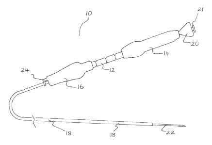
Referring to FIG. 1, a needle biopsy device 10 is provided for fine needle
aspiration during procedures such as endoscopic ultrasound. The device 10 is
generally
comprised of a handle 12, a proximal handle member 14, a distal handle member
16, a
sheath lumen 18, a needle housing member 20, a stylet 21, a needle 22, and a
connector
24.
In one embodiment, a clinician connects the device 10 to another medical
device
via the connector 24. The clinician subsequently inserts the needle housing
member 20,
which includes the stylet 21 and the needle 22, into the proximal portion of
the proximal
handle member 14. The stylet 21 may be, but is not limited to, a removable
coaxial thin
wire which is passed within the lumen of the needle 22. It is envisioned that
the stylet 21
CA 02739391 2011-04-01
WO 2010/039955 PCT/US2009/059226
may provide rigidity and stability to the needle 22. Additionally, it is
contemplated that
the stylet 21 can protect the needle 22 from damage or inadvertent collection
of samples.
Upon passing the needle 22 through the sheath lumen 18, the clinician may
slideably manipulate the proximal handle member 14 and the distal handle
member 16
along the axis of the handle 12. At this juncture, the clinician may lock the
proximal
handle member 14 and the distal handle member 16 at various depths along the
handle
12. Movement of the proximal handle member 14 causes the needle 22 to extend
from
the distal portion of the sheath 18. Additionally, movement of the distal
handle member
16 adjusts the depth of exposure of the sheath 18. A clinician may
subsequently withdraw
the stylet 21 from the needle housing member 20 and begin needle aspiration.
Referring to FIG. 2, the handle 12 includes a proximal portion 26, a distal
portion
30, and a stop portion 28. The handle 12 may be monolithically formed and
injection
molded from a rigid polymer such as acrylonitrile butadiene styrene,
polystyrene,
polyetherkeytone, polyamide, polyethersulfone, polyurethane, ether block amide
copolymers, polyacetal, and derivatives thereof. It is contemplated that the
handle 12 can
be integrally assembled of multiple sections and may be substantially
transparent,
opaque, etc. The handle 12 may also be variously configured and dimensioned
such as,
for example, rectangular, spherical, tapered etc.
The handle 12 can be joined by any appropriate process such as, for example,
snap fit, adhesive, solvent weld, thermal weld, ultrasonic weld, screw, rivet,
etc. In this
configuration, the handle 12 is presented wherein the proximal portion 26, the
distal
portion 30, and the stop portion 28 are joined through a snap fit process. In
one
embodiment, the handle 12 is assembled by inserting the stop portion 28 into
the
proximal portion 26, and subsequently inserting the distal portion 30 into the
stop portion
28. The stop portion 28 is disposed between the proximal portion 26 and the
distal
portion 30 to prevent axial movement of the proximal 14 and distal 16 handle
members,
as shown in FIG. 1, into one another.
The stop portion 28 takes the form of a circular ring with details 34 that are
incorporated into the molding. The details 34 facilitate the insertion of the
stop portion
11
CA 02739391 2011-04-01
WO 2010/039955 PCT/US2009/059226
28 into proximal portion 26 and the distal portion 30 of the handle 12. It is
envisioned
that the details 34 may create a permanent binding between the proximal
portion 26, the
stop portion 28, and the distal portion 30.
Referring to FIG. 3, an alternative embodiment is presented wherein the
details 36
.. consist of a male and female mating configuration. The details 36 consists
of a raised
circular male ridge that fits into a female type depression 38 in the proximal
portion of a
handle 40. It is envisioned that an identical configuration can exist between
the details 36
and the distal portion (not shown in Figure) of the handle 40. A configuration
is further
contemplated wherein a stop portion 42 includes details 36 that are female
type
depressions and the proximal and distal portions of the handle 40 includes a
raised
circular male ridge.
Turning to FIG. 4, a proximal portion of a handle 46 is presented wherein a
proximal handle member 44 is disposed thereon. The handle 46 includes
indentations 48
to facilitate slideable engagement along the axis of the handle 46. The
indentations 48
may take the form of ribs, ridges, or other forms of detents. In a preferred
embodiment,
the indentations 48 are located at approximately one centimeter intervals
along the handle
46.
In this configuration, the proximal handle member 44 incorporates a detail
member 50. The detail member 50 provides a means for the proximal handle
member 44
to engage the indentations 48. As previously presented in FIG. 3, the detail
member 50
similarly include a male mating configuration to facilitate a snap fit
engagement process.
The detail member 50 includes a male ridge member 52, which fits into a female
depression 54 and can form a permanent bond therebetween.
The detail member 50 includes friction members 56, which facilitate engagement
with at least one indentation 48 of a first series of indentations 48 along
the proximal
portion of the handle 46. A frictional drag force is created between the
friction members
56 engaging at least one indentation 48 of a first series of indentations 48.
It is
contemplated that the proximal handle member 44 and the detail member 50 may
be
12
CA 02739391 2011-04-01
WO 2010/039955 PCT/US2009/059226
joined via alternative processes such as adhesive, solvent weld, thermal weld,
ultrasonic
weld, etc.
The friction members 56 may be, but are not limited to, protrusions such as
semi-
circular barbs. In a preferred embodiment, the friction members 56 engage at
least one
indentation 48 of a first series of indentations 48 and provide a clinician
with a definitive
depth measurement of the proximal handle member 44. Additionally, the friction
members 56 serves to securely lock the proximal handle member 44 in place to
provide a
clinician with a consistent point of reference. It is contemplated that
multiple friction
members 56 may be employed. It is further contemplated that friction members
56 may
have flexible portions, which may be of varying flexibility according to the
particular
requirements of the handle 46.
Referring to FIG. 5, a proximal portion of a fully assembled handle 64 is
presented wherein a proximal handle member 60 can slideably advance a needle
66
within a sheath 68. In this configuration, friction members 58 are disposed to
a distal
portion of the proximal handle member 60 as semi-circular barbs. As presented,
the
friction member 58 allow the proximal handle member 60 to engage indentations
62 at
any of a plurality of positions along the axis of the handle member 64. It is
contemplated
that each of indentation 62 can represent a specific length by which the
needle 66 extends
relative to the sheath 68. More specifically, in an engaged position, a
clinician can set a
maximum length by which the needle 66 can extend beyond the distal end of the
sheath
68. A clinician may easily manipulate the position of the needle 66 by
applying pressure
to the distal portion of the proximal handle member 60. It is envisioned that
an excessive
level of pressure is not required to move the proximal handle member. However,
such
pressure must be sufficient to overcome the frictional resistance created
between the
friction member 58 and at least one indentation 62.
Referring to FIGS. 6-7, a distal handle member 70 is presented that is
identical to
the proximal handle member as described in .FIG. 5. The distal handle member
70
includes friction members 71, which facilitate engagement with at least one
indentation
73 of a second series of indentations 73 along the distal portion of a handle
74. A
13
CA 02739391 2011-04-01
WO 2010/039955 PCT/US2009/059226
frictional drag force is created between the friction members 71, which engage
at least
one indentation 73 of a second series of indentations 73 along the handle 74.
The proximal handle member (not shown in Figure) and the distal handle
member 70 further include a structural adaptation 72 that facilitates seamless
movement
along the handle 74. In the present configuration, the structural adaptation
72 has a larger
outer diameter than other portions of the distal handle member 70.
Additionally, the
structural adaptation 72 is ergonomically configured to serve as a resting
position for a
finger or thumb of a clinician. It is contemplated that the structural
adaptation 72 may
provide a surface that facilitates movement of the distal handle member 70
along the
handle 74. It is envisioned that the surface may be comprised of materials
such as a
rubber or other polymeric materials. The structural adaptation 72 may also
provide a
clinician with a tactile feel measurement system. for gauging the position of
the sheath 76
relative to the handle 74.
The distal handle member 70 also provides a means for engaging the needle
biopsy device to another medical device. Referring to FIG. 7, the distal
handle member
70 provides a connector 78 to facilitate attachment of the device to another
medical
device. The connector 78 is structurally capable of interacting with a
connector on
another medical device such as a channel or luer port. This interaction
between the
connector 78 and a connector on another medical device (not shown in Figure)
can be,
but is not limited to, a mating or locking connection.
Referring to FIG. 8, an alternative embodiment of a connector 78 is shown. The
connector 78 provides a mechanism for the quick connect and disconnect of a
needle
biopsy device 80 from a channel port 82 of a medical device 84. The connector
78
includes an adaptation that provides for connection relative to the
longitudinal axis of the
medical device. It is contemplated that the adaptation may be a female mating
configuration and may further provide for a side loading removal motion of the
device 80
from the channel port 82. It is further contemplated that the connector 78 is
sized such
that the device 80 is securely locked onto the channel port 82 in both an
axial and
perpendicular direction.
14
CA 02739391 2011-04-01
WO 2010/039955 PCT/US2009/059226
Referring to FIG. 9, another embodiment of a quick connect connector 86 is
shown. The connector 86 includes two adaptations 88 that provide for
connection
relative to the longitudinal axis of the medical device. It is envisioned that
the two
adaptations 88 may represent a male mating configuration engaging a female
mating
channel port of another medical device. It is further envisioned that the two
adaptations
provide a secure connection to the medical device.
Referring to FIG. 10, another embodiment of a connector 90 is shown. The
connector 90 is a spring loaded mechanism which facilitates connection to
other medical
devices with different channel ports. In the present configuration, a
clinician can quickly
load a device 96 axially onto a channel port 98 of another medical device 100.
A button
92 is provided to work in concert with a spring 94 to provide a spring loaded
tension
between the device 96 and another medical device 100. The button 92 may also
be
depressed to release the spring loading tension and disengage the device 96.
It is
contemplated that the button 92 may be situated in a position to allow the
clinician to
utilize their thumb or finger to depress the button 92 without disturbing the
desired
configuration of the device 96.
=
Referring to FIG. 11, a distal portion of a handle 102 is presented wherein a
connector 104 is joined via a snap fit process. It is contemplated that the
connector 104
may utilize a snap fit detail 106, which can be a male mating configuration
that engages a
female mating configuration 108. In one embodiment, the snap fit detail 106 is
permanently locked to the female mating member 108. It is further contemplated
that the
connector 104 may be adaptations in the form of two protruding male mating
adaptations,
a female mating adaptation, a spring loading mechanism, etc to satisfy the
need for a
quick connection mechanism.
Referring to FIGS. 1, 4, and 11, the needle biopsy device may also be
assembled
by engaging the connector 104 to the distal* handle member 110, and
subsequently
attaching the distal handle member 110 to a stop portion 112. The stop portion
112 may
be attached to the handle 46, as shown in FIG. 4, to complete the assembly of
the handle
12, as shown in FIG. 1.
=
CA 02739391 2011-04-01
WO 2010/039955 PCT/US2009/059226
Turning to FIGS. 12 and 13, assembly of the needle biopsy device may be
completed by inserting a needle housing member 114 into a proximal handle
member
122. The needle housing member 114 is designed to allow a clinician to quickly
and
seamlessly remove the needle 116 after an aspirating sample is taken at a site
of lesion or
abnormality.
The needle housing member 114 includes a needle 116, a hub 118, and a strain
relief 120. Due to the varying requirements of endoscopic ultrasound
procedures, the
needle 116 may be designed to range in length from fifty centimeters to two-
hundred and
fifty centimeters. Additionally, the needle 116 may be beveled via a single or
double
bevel at its distal end to aid a clinician in penetrating tissue in
preparation of collecting an
aspirated sample. It is contemplated that the needle 116 can be manufactured
from
several metallic based materials, such as stainless steel or alloys thereof
and nitinol or
alloys thereof. Alternatively, the needle 116 may be manufactured from
polymeric
materials including, but not limited to, polyetherkeytone, polyamide,
polyethersulfone,
polyurethane, ether block amide copolymers, polyacetal,
polytetrafluoroethylene and
derivatives thereof. Moreover, a combination of metallic based and polymeric
materials
may be suitable for this purpose. It is contemplated that one skilled in the
art will
realized that other materials suitable for manufacture in accordance with the
present
disclosure will also be appropriate.
The needle 116 requires a secure bond to the needle housing member 114. In one
embodiment, the needle is attached to the needle housing member 114 via
adhesive
bonding. Although adhesive bonding is suitable for this purpose, an
alternative and
preferred method, such as direct injection over-molding can be utilized.
The method of over-molding consists of a two step molding operation with two
constituent components. First, an inner component (not shown in the Figure)
consists of
a rigid polymer. The purpose of the inner component is to provide the primary
bond
between the hub 118 and the needle 116. It is contemplated that the inner
component
has shore hardness in the range of forty to eighty five Shore Durometer D.
However,
shore hardness in the range of seventy to eighty-five Shore Durometer D is
generally
=
16
CA 02739391 2011-04-01
WO 2010/039955 PCT/US2009/059226
preferable. It is contemplated that the shore hardness may include a scale of
Shore
Durometer A in addition to Shore Durometer D.
Second, the needle housing member 114 includes an outer component which
consists of a strain relief 120. A common issue associated with prior art
references is the
kinking and deformation of needles during insertion and removal from a device.
The
strain relief 120 is designed to address the issue by providing a smooth
transition and
bend radius for the needle housing member 114 upon insertion and removal from
the
proximal handle member 112. The strain relief 120 is comprised of a relatively
soft
polymer, having shore hardness in the range of ten to fifty-five durometer. It
is
contemplated, however, that shore hardness in the range of thirty to forty-
five durometer
is preferable.
Referring to FIG. 14, an alternative embodiment of the needle housing member
124 is shown. In the present configuration, the needle housing member 124 is
loaded
into an opening at the proximal portion of a proximal handle member 126. To
limit the
need for a clinician to remove their hand from the device, the needle housing
member
124 provides connecting details 128 that are immediately proximal to a strain
relief 130
to facilitate insertion and removal of the needle housing member 124. More
specifically,
the connecting details 128 provides a means for rapid connection and
disengagement of
the needle housing member 124 relative to the proximal handle member 126. Upon
inserting the needle housing member 124 into the proximal handle member 126,
female
connecting details 130 engage male connecting details 132 housed on the
proximal
handle member 126. The engagement of the female connecting details 130 and the
male
connecting details 132 provides the needle housing member 124 with a secure
lock in the
axial direction. This lock ensures that the needle subassembly can not move or
deform
while is use.
The present configuration is designed to allow a clinician to easily disengage
the
needle housing member 124 from the proximal handle member 126. For example,
once
the clinician has acquired the desired tissue or fluid sample through needle
aspiration,
they may apply force in a substantially traverse direction to the needle
housing member
17
CA 02739391 2011-04-01
WO 2010/039955 PCT/US2009/059226
124. The needle housing member 124 may be subsequently retracted for disposing
the
sample contained upon the needle. As a result, it is envisioned that a
clinician can
seamlessly acquire and insert another needle housing member 124 without
reconfiguring
the positions of the proximal handle member 126.
Referring to FIGS. 15 and 16, it is contemplated that a spring loaded
mechanism
may be provided to facilitate the removal of a needle housing member 134 from
a device
136. In the present configuration, a release member 138 is provided which
functions in
concert with a lever 140. The lever 140 operates under a spring loaded tension
142 to
securely fasten the needle housing member 134 to the device 136. The lever 140
is
.. operated by depressing the release member 138. Upon depressing the release
member
138, the tension released by a spring 142 causes the lever 140 to release the
needle
housing member 134 from the device 136.
Turning to FIG. 17, a sheath lumen 144 is provided to house the needle 22 from
the proximal handle member 14 through the distal handle member 16, as shown in
FIG.
1. The sheath lumen 144 is comprised of, but not limited to, thermoplastic
materials. It
is contemplated that the thermoplastic materials may be polyurethane,
polyamide and
derivatives thereof, ether block amide copolymers, polyimide, placental,
polyethylene
and derivates thereof, polytetrafluoroethyelene, and the like. In a preferred
embodiment,
the sheath lumen 144 is comprised of a heliacally braided configuration 146 of
outer
thermoplastic materials with a lubricious inner core 148.
The inner core 148 may be made from polytetrafluoroethyelene, fluorinated
ethylene propylene, or derivatives thereof, to provide a lubricous surface for
the needle
22, as shown in FIG. 1, as it is passed through the sheath lumen 144. It is
contemplated
that the sheath lumen 144 may have an outer diameter ranging from three French
to
twelve French. It is further contemplated that the sheath lumen 144 may have
an inner
diameter ranging from two French to ten French. In a preferred embodiment, the
inner
and outer diameter of the sheath 144 is between three French and six French.
18
CA 02739391 2011-04-01
WO 2010/039955 PCT/US2009/059226
Referring to FIG. 18, a taper 152 on the distal end of a needle 150 may be
provided to provide a level of interference between a sheath 154 and the
needle 150
during needle advancement. The taper 152 addresses the issue of needle
instability by
providing an enlarged portion that provides a frictional resistance in the
form of a drag
force. It is envisioned that the taper 152 may be incorporated onto the needle
150
through centerless grinding or cold-drawing techniques.
Referring to FIG. 19, an alternative embodiment is presented wherein a needle
156 comprises stabilizing bulbs 158 located at constant increments over the
length of the
needle 156. These bulbs 158 may be spaced anywhere from two millimeters to one
centimeter apart and may be located over the entire length of the needle 156
or over a
portion of the needle 156. It is contemplated that the bulbs 158 may be
circular or
elliptical in geometry and may be incorporated onto the needle 156 via
soldering or laser
welding or incorporating into the grind profile of the needle 156. It is
further
contemplated that the stabilizing bulbs 158 will provide sufficient frictional
resistance
between the needle 156 and a sheath 160.
Referring to FIG. 20, another embodiment is contemplated wherein a series of
barbs 162 are located at varying intervals along the length of a needle 164.
The purpose
of the barbs 162 is to reduce the effective clearance between the outer
diameter of the
needle 164 and the inner diameter of a sheath 1.66. It is contemplated that
the barbs 162
may be positioned at the distal end of the needle 164 or alternately, may be
spaced over
the entire length of the needle 164.
It is contemplated that all forms of protrusions, including the "taper",
"bulb" or
"barb" details, extend into the sheath 166 when the needle 164 is fully
extended relative
to the sheath 166. This ensures that at maximuni needle insertion depth, the
needle 164 is
kept stable in the assembly and achieves the desired design intent.
Referring to FIG. 21, a clinician may yield the benefit of improving the
echogenicity and ultrasonic visibility of a needle 168 during endoscopic
ultrasound, by
enhancing the definition of the needle 168 and the ability to discern needle
168 during the
procedure. It is contemplated that the needle 168 can be surrounded by
echogenic
19
CA 02739391 2011-04-01
WO 2010/039955 PCT/US2009/059226
materials such as a polymer impregnated with sonically reflective particles to
provide
ultrasonic visibility. It is further contemplated that ultrasonic visibility
may be, but is not
limited to, x-rays, ultrasounds, sonography, etc. It is envisioned that the
polymer may be,
but is not limited to, a thermoplastic or thermoset coating. It is further
contemplated that
the echogenic properties of the needle 168 may be enhanced through techniques
such as
sandblasting, laser etching, surface roughening, the introduction of various
patterned
geometries onto the surface of the needle, etc.
In the present configuration, an alternative configured is contemplated
wherein a
polymeric sleeve or jacket 170 covers the proximal portion of the needle 160,
which
extends distally from a sheath 172 back to a hub on a housing member 174. The
purpose
of the sheath 172 is to act as a "buffer-layer" between the outer diameter of
the needle
168 and the inner diameter of the sheath 172. In this way, the advancement of
smaller
diameter needles are stabilized as a result of frictional resistance between
the needle 168
and the sheath 172. The material used for the needle jacket 170 is preferably
extruded
from a thermoplastic material such as polyurethane, polyethylene,
polypropylene or
copolymers thereof, polyamide, polyimide, and polyether block amide or
copolymers
thereof. Alternately and more preferably, the jacket 170 may be extruded from
a highly
lubricious material such as polytetrafluoroethylene or fluorinated ethylene-
propylene. It
is contemplated that by utilizing low co-efficient of friction materials on
the outer wall of
the needle 168, the frictional drag or insertion force required to insert the
needle 168
through the sheath 172 to the desired anatomical location for aspiration is
minimized.
In the present configuration, the polymeric jacket or sleeve 170 is located to
commence at the needle housing member 174 and run the entire length of the
needle 168
to a specified location. This method ensures that the distal portion of the
needle 168,
which extends from the sheath 172, is bare and the polymeric jacket 170 does
not
interfere with passage of the needle 168 through the clinical anatomical mass
under
evaluation. The jacket 170 may be captured at the proximal end during insert
molding of
the needle housing member 174 or alternately may abut the needle housing
member 174.
The incorporation of such a polymeric jacket 170 to encase the proximal
portion
of the needle 168 also serves to provide the clinician with passive feedback
during
CA 02739391 2016-06-06
removal of the needle 168 from the proximal handle housing. During removal of
the
needle 168 from the device once the sample has been acquired, it is important
that the
clinician be made aware of when they are approaching the sharp end of the
needle 168.
With the polymeric jacket 170 being positioned at a constant distance from the
sharp
bevel of the needle 168, once the clinician observes the end of the polymeric
jacket 170
on the needle 168, they are passively made aware that a sharp bevel 176 is
located at a
specified distance from the end of the polymeric jacket 170. This passive
feedback is
important as the clinician can now exercise additional caution to ensure that
they do not
inadvertently pierce themselves with the needle 168 or cause the needle 168 to
become
entangled, endangering the diagnosing value of the collected sample.
It is contemplated that these concepts pertain to the maintenance of stability
during needle advancement, particularly in the case of a needle 168 with 22 or
25 AWG,
wherein the gap between outer diameter of the needle 168 and inner diameter of
the
sheath 172 is more appreciable. It is desirable to also incorporate the jacket
type
arrangement into the design for the 19 AWG needle portion. With a reduced
amount of
concentric clearance available between inner diameter of the sheath 172 and
the outer
diameter of the needle 168 in the case of a 19 AWG needle 168, the polymer
jacket 170
may take the form of polytetrafluoroethylene or other thermoplastic material
heat shrink
which is thermally laminated onto the outer diameter of the needle 168.
Alternately, it is
further contemplated that a 19 AWG needle 168 may be spray coated with a
lubricious
material such as teflon. At the distal end of the needle 168, the heat shrink
material or
coated material may terminate at specific distance from the sharp end of the
needle 168.
It is envisioned that this method will provide the clinician with feedback as
to when they
are approaching the sharp bevel at the distal end during extraction of the
needle 168.
It will be understood that various modifications may be made to the
embodiments
disclosed herein. Therefore, the above description should not be construed as
limiting,
but merely as exemplifications of the various embodiments of the invention.
Those
skilled in the art will envision other modifications within the scope of the
claims appended hereto.
21