Note: Descriptions are shown in the official language in which they were submitted.
CA 2739429
IMMUNOGLOBULIN VARIANTS AND USES THEREOF
RELATED APPLICATIONS
This application claims priority to United States application number
61/105,086 filed
October 14, 2008, United States application number 61/152,131 filed February
12, 2009, United
States application number 61/171,768 filed April 22, 2009, and United States
application number
61/220,514 filed June 25, 2009.
FIELD OF THE DISCLOSURE
The present invention relates generally to the field of molecular biology.
More specifically,
the present invention relates to IgG immunoglobulin variants with altered
biological properties and
methods of using the same.
BACKGROUND
Over the years the use of immunoglobulins as therapeutic agents has increased
dramatically. Immunoglobulin (Ig) molecules which constitute an important part
of the immune
system are of great interest because they (1) react with a diverse family of
ligands, (2) possess
different effector functions and (3) are of great biological importance. Today
uses of antibody
based drugs include treatment of cancer, autoimmune diseases as well as
various systemic and
infectious diseases. Also, immunoglobulins are useful as in viva diagnostic
tools, for example, in
diagnostic imaging procedures.
IgG is the most prevalent immunoglobulin class in humans and other mammals and
is utilized in
various types of immunotherapies and diagnostic procedures. Human IgGi is the
most commonly
used antibody for therapeutic purposes. Currently many antibodies in clinical
trials are directed
against tumor associated antigens. In particular, anti-VEGF neutralizing
antibodies have been
shown to suppress the growth of a variety of human tumor cell lines in nude
mice (Kim et al.
Nature 362:841-844 (1993); Warren et al. I Clin. Invest. 95:1789-1797 (1995);
Borgstrom et al.
Cancer Res. 56:4032-4039 (1996); and Melnyk et al. Cancer Res. 56:921-924
(1996)) and also
1
CA 2739429 2018-03-09
CA 02739429 2016-04-27
inhibit intraocular angiogenesis in models of ischemic retinal disorders
(Adamis etal. Arch.
Ophthalmol. 114:66-71 (1996)). Indeed, a humanized anti-VEGF antibody,
bevacizumab
(AVAST1N , Genentech, South San Francisco, CA) is the first U.S. FDA-approved
therapy
designed to inhibit angiogenesis.
Despite its potential, one of the problems with immunoglobulin immunotherapy
has been
the persistence of immunoglobulins in the circulation. The rate of
immunoglobulin clearance
directly affects the amount and frequency of dosage of the immunoglobulin.
Increased dosage and
frequency of dosage may cause adverse effects in the patient and also increase
medical costs.
The mechanism of IgG catabolism in the circulation has been elucidated through
studies
related to the transfer of passive immunity from mother to fetus/neonate
through the placenta or
yolk sac or through colostrum (maternofetal transfer of IgG via transcytosis)
in rodents (Brambell,
Lancet, ii:! 087-1093, 1966; Rodewald, J. Cell Biol., 71:666-670, 1976; Morris
et al., In: Antigen
Absorption by the Gut, pp. 3-22, 1978, University Park Press, Baltimore; Jones
et al., J. Clin.
Invest., 51:2916-2927, 1972). The neonatal Fc receptor (FcRn) plays an
important role in the
transcytosis and homeostasis of IgG in mammals. FcRn is structurally
homologous to major
histocompatibility complex (MHC) class I molecules and consists of a
transmembrane a chain and
132-microglobulin (132m). Previous studies in knockout mice illustrated that
the serum half-life of
IgG in FcRn- or 132m-deficient mice was greatly reduced (Roopenian et al., J
Immunol 170(7),
3528-3533, 2003; Israel et al., Immunology 89(4), 573-578, 1996),
demonstrating the protective
role of FcRn in regulating the level of circulating IgG. Various site-specific
mutagenesis
experiments in the Fe region of mouse IgGs have led to identification of
certain critical amino acid
residues involved in the interaction between IgG and FcRn (Kim etal., Eur. J.
Immunol., 24:2429-
2434, 1994; Medesan etal., Eur. immunol., 26:2533, 1996; Medesan etal., .1
Immunol.,
158:2211-2217, 1997). Additionally, various publications describe methods for
obtaining
physiologically active molecules whose half-lives are modified either by
introducing or modifying
an FcRn-binding region of the IgGs (WO 97/43316; U.S. Pat. No. 5,869,046; U.S.
Pat. No.
5,747,035; WO 96/32478; W02006053301; U.S. Pat. No. 7,083,784; U.S. Pat. No.
7,371,826).
At the molecular level, FcRn binds the Fe portion of IgG in the CH2-CH3 domain
region.
The Fe-FcRn interaction is highly pH dependent; IgGs bind FcRn with high
affinity at pH 6, but as
the pH is raised to 7.4, the binding affinity drops considerably. This pH
dependent interaction is
responsible for protecting IgG from degradation. Specifically, pinocytosed IgG
is captured by
2
CA 02739429 2016-04-27
FcRn in the acidic endosome, recycled back to the cell surface and then
released back into the
circulation at a physiological serum pH of 7.4 (Ober etal. Proc Natl Acad Sci
USA 101(30),
11076-11081, 2004; Ober et al. J Immunol 172(4), 2021-2029, 2004; Prabhat et
al. Proc Nati Acad
Sci USA 104(14), 5889-5894, 2007). IgG that is not bound by FcRn is targeted
to the lysosome
and degraded. As FcRn is important in regulating IgG homeostasis, modulating
the interaction
between Fc and FeRn through protein engineering is one method for improving
the
pharmacokinetics of therapeutic antibodies (Shields et al. J Biol Chem 276(9),
6591-6604, 2001;
Dall'Acqua etal. Nat Biotechnol 15(7), 637-640, 1997; Dall'Acqua et al., J
Immunol 169(9), 5171-
5180, 2002; Hinton etal. J Biol Chem 279(8), 6213-6216, 2004; Hinton etal. J
lmmunol 176(1),
346-356, 2006; Datta-Mannan et al. Drug Metab Dispos 35(1), 86-94, 2007; Datta-
Mannan et al. J
Biol Chem 282(3), 1709-1717, 2007). A number of studies in mice, rhesus and
cynomolgus
monkeys have demonstrated that increasing the pH-6 binding affinity of IgGs
can prolong half-life
(Dall'Acqua et al. Nat Biotechnol 15(7), 637-640, 1997; Dall'Acqua et al., J
Immunol 169(9), 5171-
5180, 2002; Hinton etal. J Biol Chem 279(8), 6213-6216, 2004; Hinton et al. J
Immunol 176(1),
.. 346-356, 2006). Furthermore, other studies have also demonstrated that FcRn
binding affinity at
pH 7.4 is an additional determinant of IgG pharmacokinetics. Specifically,
certain variants with
increased pH-7.4 binding affinity to mouse FcRn exhibited increased clearance
(i.e., decreased
half-life) in mice (Dall'Acqua et al., Immunol 169(9), 5171-5180, 2002).
Nevertheless, the
detailed relationship between FcRn affinity and half-life has not been
elucidated, as all of the
previous studies involved a small number of variants, within a limited range
of FcRn affinities.
The maximal half-life extension achievable through engineering the Fc:FcRn
interaction is unclear.
Despite the fact that adherence to (compliance with) drug treatment is
important, it is
estimated that half of those for whom medicines are prescribed do not take
them in the
recommended way. For example, a recent research showed that as many as one-
third of women
taking breast cancer drugs developed in the past 10 years do not complete
their recommended five-
year course. Some of the causes for poor compliance include forgetfulness,
physical difficulty in
complying (e.g. traveling to or moving away from place of treatment),
inconvenience, adverse side
effect, complicated regimen and cost of drugs. Poor adherence to drug
treatment can lead to
achieving less than the full health benefits medicines can provide to
patients. For example, not
completing the recommended course of cancer treatment could lead to a
recurrence of the disease
and a reduced chance of survival.
3
CA 02739429 2016-04-27
Strategies to improve drug compliance include making it more convenient for
patients to
finish the recommended course of treatment. One of the ways this may be
accomplished for
patients under immunotherapy treatment is by increasing the duration of time
that
immunoglobulins are in circulation. The rate of immunoglobulin clearance
directly affects the
amount and frequency of dosage of the immunoglobulin. Therefore, developing an
immunoglobulin that confers increased in vivo half-life may decrease the
amount and/or frequency
of dosage, thus minimizing the inconvenience as well as any additional medical
costs.
Accordingly, it would be highly advantageous to have modified immunoglobulins
that
confer increased in vivo half-life for therapeutic purposes. The present
disclosure relates to these
and other needs, as will be apparent upon review of the following disclosure.
SUMMARY
The disclosure provides novel IgG variants and uses thereof. A number of IgG
variants are
provided in the invention. For example, the present disclosure relates to
novel IgG variants
comprising a human IgG Fc region comprising two or more amino acid
substitutions relative to a
wild-type human IgG Fc region at two or more of amino acid residues 251, 252,
307, 308, 378,
428, 430, 434, and 436, numbered according to the EU index as in Kabat,
wherein the variant IgG
has an increased half-life compared to the half-life of an IgG having the wild-
type human IgG Fc
region, and wherein at least two of the amino acid substitutions are at amino
acid residue 251, 252,
307, 308, 378, 428, 430, 434, or 436, and an amino acid substitution at amino
acid residue 251 is a
substitution with aspartic acid or glutamic acid, an amino acid substitution
at amino acid residue
252 is a substitution with tyrosine, an amino acid substitution at amino acid
residue 307 is a
substitution with glutamine, an amino acid substitution at amino acid residue
308 is a substitution
with proline, an amino acid substitution at amino acid residue 378 is a
substitution with valine, an
amino acid substitution at amino acid residue 428 is a substitution with
leucine, an amino acid
.. substitution at amino acid residue 430 is a substitution with alanine or
lysine, an amino acid
substitution at amino acid residue 434 is a substitution with alanine, serine
or tyrosine, and an
amino acid substitution at amino acid residue 436 is a substitution with
isoleueine.
In certain embodiments, the human IgG Fc region comprises an amino acid
substitution at
amino acid 251, wherein the amino acid substitution at amino acid 251 is the
substitution with
aspartic acid or glutamic acid. In certain embodiments, the human IgG Fc
region comprises an
4
CA 02739429 2016-04-27
amino acid substitution at amino acid 307, wherein the amino acid substitution
at amino acid 307 is
the substitution with glutamine. In certain embodiments, the human IgG Fc
region comprises an
amino acid substitution at amino acid 308, wherein the amino acid substitution
at amino acid 308 is
the substitution with proline. In certain embodiments, the human IgG Fe region
comprises an
amino acid substitution at amino acid 378, wherein the amino acid substitution
at amino acid 378 is
the substitution with valine. In certain embodiments, the human IgG Fc region
comprises an amino
acid substitution at amino acid 436, wherein the amino acid substitution at
amino acid 436 is the
substitution with isoleucine. In certain embodiments, the variant IgGs have a
higher binding
affinity for FcRn than the IgG having the wild-type human IgG Fc region.
In one embodiment, the human IgG Fc region comprises the amino acid
substitution at
amino acid 307 with glutamine and the amino acid substitution at amino acid
434 with alanine. In
one embodiment, the human IgG Fe region comprises the amino acid substitution
at amino acid
307 with glutamine and the amino acid substitution at amino acid 434 with
serine. In one
embodiment, the human IgG Fc region comprises the amino acid substitution at
amino acid 308
with proline and the amino acid substitution at amino acid 434 with alanine.
In one embodiment,
the human IgG Fc region comprises the amino acid substitution at amino acid
252 with tyrosine
and the amino acid substitution at amino acid 434 with alanine. In one
embodiment, human IgG Fc
region comprises the amino acid substitution at amino acid 378 with valine and
the amino acid
substitution at amino acid 434 with alanine. In one embodiment, the human IgG
Fc region
comprises the amino acid substitution at amino acid 428 with leucine and the
amino acid
substitution at amino acid 434 with alanine. In one embodiment, the human IgG
Fc region
comprises the amino acid substitution at amino acid 434 with alanine and the
amino acid
substitution at amino acid 436 with isoleucine. In one embodiment, the human
IgG Fc region
comprises the amino acid substitution at amino acid 308 with proline and the
amino acid
substitution at amino acid 434 with tyrosine. In one embodiment, the human IgG
Fc region
comprises the amino acid substitution at amino acid 307 with glutamine and the
amino acid
substitution at amino acid 436 with isoleucine.
The present disclosure also relates to novel IgG variants comprising a human
IgG Fc region
comprising three or more amino acid substitutions relative to a wild-type
human IgG Fe region at
three or more of amino acid residues 251, 252, 307, 308, 378, 380, 428, 430,
434, and 436,
numbered according to the EU index as in Kabat, wherein the variant IgG has an
increased half-life
5
CA 02739429 2016-04-27
compared to the half-life of an IgG having the wild-type human IgG Fc region,
and wherein at least
three of the amino acid substitutions are at amino acid residue 251, 252, 307,
308, 378, 380, 428,
430, 434, or 436, and an amino acid substitution at amino acid residue 251 is
a substitution with
aspartic acid or glutamic acid, an amino acid substitution at amino acid
residue 252 is a substitution
with tyrosine, an amino acid substitution at amino acid residue 307 is a
substitution with glutamine,
an amino acid substitution at amino acid residue 308 is a substitution with
proline, an amino acid
substitution at amino acid residue 378 is a substitution with valine, an amino
acid substitution at
amino acid residue 380 is a substitution with alanine, an amino acid
substitution at amino acid
residue 428 is a substitution with leucine, an amino acid substitution at
amino acid residue 430 is a
substitution with alanine or lysine, an amino acid substitution at amino acid
residue 434 is a
substitution with alanine, serine, tyrosine or histidine, and an amino acid
substitution at amino acid
residue 436 is a substitution with isoleucine.
In certain embodiments, the human IgG Fc region comprises an amino acid
substitution at
amino acid 251, wherein the amino acid substitution at amino acid 251 is the
substitution with
aspartic acid or glutamic acid. In certain embodiments, the human IgG Fc
region comprises an
amino acid substitution at amino acid 307, wherein the amino acid substitution
at amino acid 307 is
the substitution with glutamine. In certain embodiments, the human IgG Fc
region comprises an
amino acid substitution at amino acid 308, wherein the amino acid substitution
at amino acid 308 is
the substitution with proline. In certain embodiments, the human IgG Fc region
comprises an
amino acid substitution at amino acid 378, wherein the amino acid substitution
at amino acid 378 is
the substitution with valine. In certain embodiments, the human IgG Fc region
comprises an amino
acid substitution at amino acid 436, wherein the amino acid substitution at
amino acid 436 is the
substitution with isoleucine. In certain embodiments, the variant IgGs have a
higher binding
affinity to Fcitn compared to the IgG having the wild-type human IgG Fc
region.
In one embodiment, the human IgG Fc region comprises the amino acid
substitution at
amino acid 307 with glutamine, the amino acid substitution at amino acid 380
with alanine and the
amino acid substitution at amino acid 434 with serine. In one embodiment, the
human IgG Fc
region comprises the amino acid substitution at amino acid 307 with glutamine,
the amino acid
substitution at amino acid 380 with alanine and the amino acid substitution at
amino acid 434 with
alanine. In one embodiment, the human IgG Fc region comprises the amino acid
substitution at
amino acid 252 with tyrosine, the amino acid substitution at amino acid 308
with proline and the
6
CA 02739429 2016-04-27
amino acid substitution at amino acid 434 with tyrosine. In one embodiment,
the human IgG Fe
region comprises the amino acid substitution at amino acid 251 with aspartic
acid, the amino acid
substitution at amino acid 307 with glutamine and the amino acid substitution
at amino acid 434
with histidine.
In certain embodiments, the present disclosure relates to variant IgGs or
fragments thereof
further comprising an amino acid substitution at position 297 to alanine.
In certain embodiments, the variant IgG of the present invention has a higher
binding
affinity for FcRn than the IgG having the wild-type human IgG Fe region. In
certain embodiments,
the variant IgG has a higher binding affinity for FcRn at pH 6.0 than at pH
7.4. In certain
embodiments, the variant IgG is a human or humanized IgG. In certain
embodiments, the variant
IgG is IgGi, IgG2, IgG3 or IgG4. In certain embodiments, the IgG Fe region of
the variant IgG is an
IgGI, IgG2, IgG3or IgG4 Fe region. In certain embodiments, the IgG Fe region
of the variant IgG is
an IgGIFc region.
In certain embodiments, the variant IgG is an anti-VEGF antibody. In certain
embodiments, the variant IgG is a variant of bevacizumab. In certain
embodiments, the IgG having
the wild-type human IgG Fe region is bevacizumab. In certain embodiments, the
wild-type human
IgG Fe region is the Fe region of bevacizumab. In certain embodiments, the
variant IgG comprises
the heavy chain variable domain (SEQ ID NO:1) and light chain variable domain
(SEQ ID NO:2).
In certain embodiments, the variant IgG comprises the heavy chain variable
domain (SEQ ID
NO:3) and light chain variable domain (SEQ ID NO:4). In certain embodiments,
the variant IgG
comprises the heavy chain variable domain (SEQ ID NO:7) and light chain
variable domain (SEQ
ID NO:8).
The present disclosure further relates to pharmaceutical compositions
comprising any of the
variant IgGs described herein and a pharmaceutically acceptable carrier. A kit
comprising any of
the variant IgGs described herein, in a container, and instructions for use is
also provided herein.
In certain embodiments, the half life of the variant IgG is increased by at
least 50%, 100%,
150%, 200%, 300% or greater compared to an IgG having the wild-type human IgG
Fc region. In
certain embodiments, the half life of the variant IgG is increased by at least
2 fold compared to an
IgG having the wild-type human IgG Fe region. In certain embodiments, the half
life of the variant
IgG is increased by at least 3 fold compared to an IgG having the wild-type
human IgG Fe region.
In certain embodiments, the half life of the variant IgG is increased by at
least 4 fold compared to
7
CA 02739429 2016-04-27
an IgG having the wild-type human IgG Fe region. In certain embodiments, the
IgG having the
wild-type human IgG Fe region is bevacizumab. In certain embodiments, the half
life of the variant
IgG is the mean half-life of bevacizumab. In certain embodiments, the mean
half-life of
bevacizumab is about 10 to 12 days as measured in cynomolgus monkeys, or about
3 weeks as
measured in humans.
In certain embodiments, variant IgGs comprising a human IgG Fe region are
provided,
wherein the human IgG Fe region comprises amino acid substitutions relative to
a wild-type human
IgG Fe region at amino acid residues 307 and 434, numbered according to the EU
index as in
Kabat, wherein the variant IgG has an increased half-life compared to the half-
life of an IgG having
the wild-type human IgG Fe region, and wherein the amino acid substitution at
amino acid residue
307 is a substitution with glutamine, and the amino acid substitution at amino
acid residue 434 is a
substitution with alanine. In one embodiment, the variant 1gG is variant IgGi
comprising the heavy
chain variable domain (SEQ ID NO:1) and light chain variable domain (SEQ ID
NO:2), and
comprising a human IgGi Fe region comprising amino acid substitutions relative
to a wild-type
human IgGi Fe region at amino acid residues 307 and 434, numbered according to
the EU index as
in Kabat, wherein the variant IgGi has an increased half-life compared to the
half-life of an IgGi
having the wild-type human IgG Fe region, and wherein the amino acid
substitution at amino acid
residue 307 is a substitution with glutamine, and the amino acid substitution
at amino acid residue
434 is a substitution with alanine.
In another embodiment, variant IgGs comprising a human IgG Fe region are
provided,
wherein the human IgG Fe region comprises amino acid substitutions relative to
a wild-type human
IgG Fe region at amino acid residues 307 and 434, numbered according to the EU
index as in
Kabat, wherein the variant IgG has an increased half-life compared to the half-
life of an IgG having
the wild-type human IgG Fe region, and wherein the amino acid substitution at
amino acid residue
307 is a substitution with glutamine, and the amino acid substitution at amino
acid residue 434 is a
substitution with serine.
In another embodiment, variant IgGs comprising a human IgG Fe region are
provided,
wherein the human IgG Fe region comprises amino acid substitutions relative to
a wild-type human
IgG Fe region at amino acid residues 308 and 434, numbered according to the EU
index as in
Kabat, wherein the variant IgGi has an increased half-life compared to the
half-life of an IgG
having the wild-type human IgG Fe region, and wherein the amino acid
substitution at amino acid
8
CA 02739429 2016-04-27
residue 308 is a substitution with proline, and the amino acid substitution at
amino acid residue 434
is a substitution with alanine.
In another embodiment, variant IgGs comprising a human IgG Fc region are
provided,
wherein the human IgG Fc region comprises amino acid substitutions relative to
a wild-type human
IgG Fc region at amino acid residues 307, 380 and 434, numbered according to
the EU index as in
Kabat, wherein the variant IgG has an increased half-life compared to the half-
life of an IgG having
the wild-type human IgG Fc region, and wherein the amino acid substitution at
amino acid residue
307 is a substitution with glutamine, the amino acid substitution at amino
acid residue 380 is a
substitution with alanine, and the amino acid substitution at amino acid
residue 434 is a
substitution with serine.
In certain embodiments, the variant IgG comprising a human IgG Fc region
described
above has a higher binding affinity for FcRn than the IgG having the wild-type
human IgG Fc
region. In certain embodiments, the variant IgG has a higher binding affinity
for FcRn at pH 6.0
than at pH 7.4. In certain embodiments, the variant IgG is a human or
humanized IgG. In certain
embodiments, the variant IgG is IgGi, IgG2, IgG3 or IgG4. In certain
embodiments, the IgG Fc
region of the variant IgG is an IgGi, IgG2, IgG3or IgG4 Fc region. In certain
embodiments, the IgG
Fe region of the variant IgG is IgGi Fc region. In certain embodiments, the
variant IgG is an anti-
VEGF antibody. In certain embodiments, the variant IgG is a variant of
bevacizumab. In certain
embodiments, the IgG having the wild-type human IgG Fc region is bevacizumab.
In certain
embodiments, the wild-type human IgG Fc region is the Fc region of
bevacizumab. In certain
embodiments, the variant IgG comprises the heavy chain variable domain (SEQ ID
NO: I) and light
chain variable domain (SEQ ID NO:2). In certain embodiments, a pharmaceutical
composition
comprising any of the variant IgGs comprising a human IgG Fc region and a
pharmaceutically
acceptable carrier are provided herein. A kit comprising any of the variant
IgGs comprising a
human IgG Fc region, in a container, and instructions for use is also provided
herein.
In certain embodiments, variant IgGs comprising a human IgGi Fc region are
provided,
wherein the variant IgGs comprise the heavy chain variable domain (SEQ ID
NO:!) and light chain
variable domain (SEQ ID NO:2) and wherein the human IgGi Fc region comprises
an amino acid
substitution relative to a wild-type human IgGi Fc region at amino acid
residue 434, numbered
according to the EU index as in Kabat, wherein the variant IgG has an
increased half-life compared
to the half-life of an IgG having the wild-type human IgGi Fc region, and the
variant IgG has a
9
CA 02739429 2016-04-27
higher binding affinity for FcRn compared to binding affinity for FcRn of the
IgG having the wild-
type human lgG1 Fc region, and wherein the amino acid substitution at amino
acid residue 434 is a
substitution with histidine. In certain embodiments, the variant IgG is
variant IgGI.
In certain embodiments, the half life of a variant IgG is increased by at
least 50, 55. 60, 65,
70, 75, 80, 85, 90. 95, or 100% compared to the half life of the IgG having
the wild-type human
IgG Fc region. In one embodiment, the half life of a variant IgG is increased
by at least 50%
compared to the half life of the IgG having the wild-type human IgG Fc region.
In another
embodiment, the half life of a variant IgG is increased by at least 75%
compared to the half life of
the IgG having the wild-type human IgG Fc region. In yet another embodiment,
the half life of a
variant IgG is increased by at least 100% compared to the half life of the IgG
having the wild-type
human IgG Fc region.
In certain embodiments, the half life of a variant IgG is at least about 15,
20, 25, 30, 35, or
40 days. In one embodiment, the half life of a variant IgG of the present
invention is at least about
days. In another embodiment, the half life of a variant IgG of the present
invention is at least
15 about 20 days. In another embodiment, the half life of a variant IgG of
the present invention is at
least about 25 days. In another embodiment, the half life of a variant IgG of
the present invention
is at least about 30 days. In another embodiment, the half life of a variant
IgG of the present
invention is at least about 35 days. In another embodiment, the half life of a
variant IgG of the
present invention is at least about 40 days. In certain embodiments, the
variant IgG is variant IgGI.
In certain embodiments, the half life of a variant IgG of the present
invention is the half life
as measured in humans. In certain embodiments, the half life of a variant IgG
of the present
invention is the half life as measured in eynomolgus monkeys. In certain
embodiments, the wild-
type IgG or the IgG having the wild-type human IgG Fc region is bevacizumab.
In certain
embodiments, the half-life of bevacizumab is about 10 to 12 days as measured
in cynomolgus
monkeys, or about 20 days as measured in humans.
A number of methods of using IgG variants are disclosed. Methods of treating
tumor in a
subject are provided. For example, methods comprise administering to the
subject an effective
amount of any variant IgGs described above and herein. In certain embodiments,
the variant IgG is
an anti-VEGF antibody. In certain embodiments, the variant IgG is a variant of
bevacizumab. In
certain embodiments, the methods further comprise administering to the subject
an effective
amount of a chemotherapeutic agent.
CA 02739429 2016-04-27
Methods of inhibiting VEGF activity in a subject are disclosed herein. For
example,
methods comprise administering to said subject an effective amount of any
variant IgGs described
above and herein. In certain embodiments, the VEGF activity is angiogenesis.
Methods of modulating vascular permeability in a subject are disclosed herein.
For
example, methods comprise administering to said subject an effective amount of
any variant IgGs
described above and herein.
Methods of treating a non-neoplastic disorder in a subject are disclosed
herein. For
example, methods comprise administering to said subject an effective amount of
any variant IgGs
described above and herein. In certain embodiments, the non-neoplastic
disorder is an autoimmune
disease. In certain embodiments, the non-neoplastic disorder is Alzheimer's
disease. In certain
embodiments, the subject is diagnosed with age-related macular degeneration.
Methods of treating a HER expressing tumor in a subject are disclosed herein.
For
example, methods comprise administering to said subject an effective amount of
any variant IgGs
described above and herein. In certain embodiments, the variant IgG comprises
the heavy chain
variable domain (SEQ ID NO:7) and light chain variable domain (SEQ ID NO:8).
Methods of inhibiting or preventing growth of cancer cells in a subject are
provided in the
invention. For example, methods comprise administering to said subject an
effective amount of
any variant IgGs described above and herein.
Methods of administering to a subject an effective amount of a variant IgG are
disclosed
herein. These methods of administration can be used in combination with other
methods (e.g.,
methods of treatments) described herein. In certain embodiments, methods
comprise administering
to said subject an effective amount of any variant IgGs described above and
herein, and wherein the
variant IgG is administered to the subject every 4 weeks or at longer
intervals. In certain
embodiments, the variant IgG is administered every 5 weeks or longer. In
certain embodiments,
the variant IgG is administered every 6 weeks or longer. In certain
embodiments, the variant IgG is
administered every 7 weeks or longer. In certain embodiments, the variant IgG
is administered
every 8 weeks or longer. In certain embodiments, the variant IgG is
administered every 9 weeks or
longer. In certain embodiments, the variant IgG is administered every 10 weeks
or longer. In
certain embodiments, the variant IgG is administered every 11 weeks or longer.
In certain
embodiments, the variant IgG is administered every 12 weeks or longer. In
certain embodiments,
methods comprise administering to said subject an effective amount of any
variant IgGs, wherein
11
CA 02739429 2016-04-27
the variant IgG is administered less frequently than the recommended or
prescribed dosage
frequency of the IgG having the wild-type human IgG Fc region. In certain
embodiments, the IgG
having the wild-type human IgG Fc region is bevacizumab. In certain
embodiments, the variant
IgG, e.g, a variant of bevacizumab, is administered less frequently than the
prescribed dosage
frequency of bevacizumab.
In certain embodiments, the variant IgG is initially administered every 2
weeks, and later
administered every 4 weeks or longer. In certain embodiments, the variant IgG
is initially
administered every 3 weeks, and later administered every 6 weeks or longer. In
certain
embodiments, the variant IgG is initially administered every 4 weeks, and
later administered every
8 weeks or longer. In certain embodiments, the variant 1gG is initially
administered every 5 weeks,
and later administered every 10 weeks or longer. In certain embodiments, the
variant IgG is
initially administered every 6 weeks, and later administered every 12 weeks or
longer. In certain
embodiments, the variant IgG, e.g., a variant of bevacizumab, is initially
administered with the
prescribed dosage frequency of bevacizumab, and later administered less
frequently than the
prescribed dosage of bevacizumab.
In certain embodiments of the methods described herein, the variant IgG is an
anti-VEGF
antibody. In one embodiment, the anti-VEGF antibody comprises the heavy chain
variable domain
(SEQ ID NO:1) and light chain variable domain (SEQ ID NO:2). In another
embodiment, the anti-
VEGF antibody is a variant of bevacizumab.
In certain embodiments, the variant IgGs are administered to the subject
intravenously. In
certain embodiments, the variant IgGs are administered to the subject
subcutaneously.
In certain embodiments of the methods described herein, the subject is human.
In certain
embodiments, the subject is diagnosed with cancer. In certain embodiments, the
cancer is selected
from the group consisting of non-small cell lung cancer, renal cell carcinoma,
ovarian cancer,
glioblastoma, breast cancer, and colorectal cancer.
Also provided herein are methods of treating a benign, pre-cancerous or non-
metastatic
cancer in a subject, which comprise administering to the subject an effective
amount of a variant
IgG. In certain embodiments, the administration of the variant IgG prevents
the benign, pre-
cancerous, or non-metastatic cancer from becoming an invasive or metastatic
cancer. For example,
the benign, pre-cancerous or non-metastatic cancer can be a stage 0, stage I,
or stage H cancer, and
in certain embodiments, the administration of the variant IgG prevents the
benign, pre-cancerous or
12
CA 02739429 2016-04-27
non-metastatic cancer from progressing to the next stage(s), e.g., a stage I,
a stage II, a stage III or
stage IV cancer. In certain embodiments, the variant IgG is administered for a
time and in an
amount sufficient to treat the benign, pre-cancerous, or non-metastatic tumor
in the subject or to
prevent the benign, pre-cancerous, or non-metastatic tumor from becoming an
invasive or
metastatic cancer. In certain embodiments, administering the variant IgG
reduces tumor size,
tumor burden, or the tumor number of the benign, pre-cancerous, or non-
metastatic tumor. The
variant IgG can also be administered in an amount and for a time to decrease
the vascular density in
the benign, pre-cancerous, or non-metastatic tumor.
As described herein, methods disclosed herein can be used to treat, e.g., a
stage 0 (e.g., a
carcinoma in situ), stage I, or stage H cancer. The methods of neoadjuvant and
adjuvant therapy
can be used to treat any type of cancer, e.g., benign or malignant. In certain
embodiments, the
cancer is a solid tumor, including, but not limited to, colon cancer, breast
cancer, prostate cancer,
renal cancer, lung cancer (e.g., non-small cell lung cancer), melanoma,
ovarian cancer, pancreatic
cancer, gastrointestinal cancer, head and neck cancer, liver cancer and soft
tissue cancers (e.g., B
cell lymphomas such as NHL and multiple myeloma and leukemias such as chronic
lymphocytic
leukemia). In another embodiment, the benign, pre-cancerous, or non-metastatic
tumor is a polyp,
adenoma, fibroma, lipoma, gastrinoma, insulinoma, chondroma, osteoma,
hemangioma,
lymphangioma, meningioma, leiomyoma, rhabdomyoma, squamous cell papilloma,
acoustic
neuromas, neurofibroma, bile duct cystanoma, leiomyomas, mesotheliomas,
teratomas, myxomas,
trachomas, granulomas, hamartoma, transitional cell papilloma, pleiomorphic
adenoma of the
salivary gland, desmoid tumor, dermoid cystpapilloma, cystadenoma, focal
nodular hyperplasia, or
a nodular regenerative hyperplasia. In another embodiment, the method is
desirably used to treat
an adenoma. Non-limiting examples of adenomas include liver cell adenoma,
renal adenoma,
metanephric adenoma, bronchial adenoma, alveolar adenoma, adrenal adenoma,
pituitary adenoma,
parathyroid adenoma, pancreatic adenoma, salivary gland adenoma,
hepatocellular adenoma,
gastrointestinal adenoma, tubular adenoma, and bile duct adenoma.
The disclosure also features methods that comprise administering to a subject
an effective
amount of a variant IgG to prevent occurrence or recurrence of a benign, pre-
cancerous, or non-
metastatic cancer in the subject. In certain embodiments, the subject is at
risk for cancer, polyps,
or a cancer syndrome. In one example, the subject has a family history of
cancer, polyps, or an
inherited cancer syndrome. In certain aspects, the subject is at risk of
developing a benign, pre-
13
CA 02739429 2016-04-27
cancerous, or non-metastatic tumor. In certain embodiments, the method
prevents occurrence or
recurrence of said benign, pre-cancerous or non-metastatic cancer in a subject
who has never had a
tumor, a subject who has never had a clinically detectable cancer, or a
subject who has only had a
benign tumor.
In another aspect, methods of preventing or reducing the likelihood of
recurrence of a
cancer in a subject that includes administering to the subject a variant IgG
for a time and in an
amount sufficient to prevent or reduce the likelihood of cancer recurrence in
the subject are
provided. The disclosure relates to a method of preventing the recurrence of a
cancer in a subject
having a tumor that includes the steps of removing the tumor (e.g., using
definitive surgery) and
thereafter administering to the subject a variant IgG. The disclosure relates
to methods of
preventing the regrowth of a tumor in a subject that includes the steps of
removing the tumor (e.g.,
using definitive surgery) and thereafter administering to the subject a
variant IgG. In a related
aspect, the disclosure relates to a method of preventing recurrence of cancer
in a subject or
reducing the likelihood of cancer recurrence in a subject that optionally
includes administering to
the subject an effective amount of a variant IgG prior to surgery, performing
definitive surgery, and
administering an effective amount of a variant IgG following the surgery
wherein the
administration of the variant IgG after the surgery prevents recurrence of the
cancer or reduces the
likelihood of cancer recurrence. In another related aspect, the disclosure
relates to a method of
preventing recurrence of cancer in a subject or reducing the likelihood of
cancer recurrence in a
subject that includes administering to the subject an effective amount of a
variant IgG in the
absence of any additional anti-cancer therapeutic agent, wherein the
administering prevents
recurrence of cancer in a subject or reduces the likelihood of cancer
recurrence in a subject.
For each of the above aspects, the tumor can be any type of tumor including
but not limited
to the solid tumors, and particularly the tumors and adenomas, described
herein. The subject can
have a dormant tumor or micrometastases, which may or may not be clinically
detectable. In one
embodiment of this aspect, the variant IgG is administered for a time and in
an amount sufficient to
reduce neovascularization of a dormant tumor or micrometastases. In another
embodiment, the
variant IgG is administered for a time and in an amount sufficient to prevent
occurrence of a
clinically detectable tumor, or metastasis thereof, or to increase the
duration of survival of the
subject.
14
CA 02739429 2016-04-27
In one embodiment, the variant IgG is a monotherapy. In another embodiment,
the subject
has been previously treated for the tumor, for example, using an anti-cancer
therapy. In one
example, the anti-cancer therapy is surgery. In another embodiment, the
subject can be further
treated with an additional anti-cancer therapy before, during (e.g.,
simultaneously), or after
administration of the variant IgG. Examples of anti-cancer therapies include,
without limitation,
surgery, radiation therapy (radiotherapy), biotherapy, immunotherapy,
chemotherapy, or a
combination of these therapies.
In embodiments where the subject has undergone definitive surgery, the variant
IgG is
generally administered after a period of time in which the subject has
recovered from the surgery.
This period of time can include the period required for wound healing or
healing of the surgical
incision, the time period required to reduce the risk of wound dehiscence, or
the time period
required for the subject to return to a level of health essentially similar to
or better than the level of
health prior to the surgery. The period between the completion of the
definitive surgery and the
first administration of the variant IgG can also include the period needed for
a drug holiday,
wherein the subject requires or requests a period of time between therapeutic
regimes. Generally,
the time period between completion of definitive surgery and the commencement
of the variant IgG
therapy can include less than one week, 1 week, 2 weeks, 3 weeks, 4 weeks (28
days), 5 weeks, 6
weeks, 7 weeks, 8 weeks, 3 months, 4 months, 5 months, 6 months, 7 months, 8
months. 9 months,
10 months, 11 months, 1 year, 2 years, 3 years, or more. In one embodiment,
the period of time
between definitive surgery and administering the variant IgG is greater than 2
weeks and less than 1
year. In one embodiment, the period of time between definitive surgery and
administering the
variant IgG is greater than 4 weeks (28 days).
In certain embodiments, each of the above aspects can further include
monitoring the
subject for recurrence of the cancer.
The disclosure also relates to methods of neoadjuvant therapy prior to the
surgical removal
of operable cancer in a subject, e.g., a human patient, comprising
administering to the patient an
effective amount of a variant IgG where the patient has been diagnosed with a
tumor or cancer.
The variant IgG can be administered alone or in combination with at least one
chemotherapeutic
agent.
The disclosure also relates to a method of treating a subject with operable
cancer that
includes administering to the subject an effective amount of a variant IgG
prior to surgery and
CA 02739429 2016-04-27
thereafter performing surgery whereby the cancer is resected. In one
embodiment, the method
further includes the step of administering to the subject an effective amount
of a variant IgG after
surgery to prevent recurrence of the cancer.
In another aspect, the disclosure concerns a method of neoadjuvant therapy
comprising
administering to a subject with operable cancer an effective amount of a
variant IgG and at least
one chemotherapeutic agent prior to definitive surgery.
In another aspect, the disclosure relates to a method of reducing tumor size
in a subject
having an unresectable tumor comprising administering to the subject an
effective amount of a
variant IgG wherein the administering reduces the tumor size thereby allowing
complete resection
.. of the tumor. In one embodiment, the method further includes administering
to the subject an
effective amount of a variant IgG after complete resection of the tumor.
In another aspect, the disclosure concerns a method of treating cancer in a
subject
comprising the following steps: a) a first stage comprising a plurality of
treatment cycles wherein
each cycle comprises administering to the subject an effective amount of a
variant IgG and at least
one chemotherapeutic agent at a predetermined interval; b) a definitive
surgery whereby the cancer
is removed; and c) a second stage comprising a plurality of maintenance cycles
wherein each cycle
comprises administering to the subject an effective amount of a variant IgG
without any
chemotherapeutic agent at a predetermined interval. In one embodiment, the
first stage comprises
a first plurality of treatment cycles wherein a variant IgG and a first
chemotherapy regimen are
administered followed by a second plurality of treatment cycles wherein a
variant IgG and a second
chemotherapy regimen are administered.
The disclosure relates to methods comprising administering to a subject with
metastatic or
nonmetastatie cancer, following definitive surgery, an effective amount of a
variant IgG. In certain
embodiments, the method further includes the use of at least one
chemotherapeutic agent.
In one aspect, the method comprises the following steps: a) a first stage
comprising a
plurality of treatment cycles wherein each cycle comprises administering to
the subject an effective
amount of a variant IgG and at least one chemotherapeutic agent at a
predetermined interval; and b)
a second stage comprising a plurality of maintenance cycles wherein each cycle
comprises
administering to the subject an effective amount of a variant IgG without any
chemotherapeutic
agent at a predetermined interval. In one embodiment, the first stage
comprises a first plurality of
treatment cycles wherein a variant IgG and a first chemotherapy regimen are
administered,
16
CA 02739429 2016-04-27
followed by a second plurality of treatment cycles wherein a variant IgG and a
second
chemotherapy regimen are administered.
In certain embodiments, the variant IgG is an anti-VEGF antibody that binds to
VEGF or
reduces VEGF expression or biological activity. The anti-VEGF antibody, or
antigen-binding
fragment thereof, can be a monoclonal antibody, a chimeric antibody, a fully
human antibody, or a
humanized antibody. In certain embodiments, exemplary antibodies useful in the
methods of the
invention include bevacizumab (AVASTIN ), G6-31, B20-4.1, B20-4.1.1, and
fragments thereof.
In certain embodiments, the variant IgG is humanized anti-HER2 monoclonal
antibody
HERCEPTIN . In certain embodiments, the variant IgG is chimeric anti-CD20
antibody Rituxan ,
anti-IgE antibody XOLAIR , anti-CD20 antibody, anti-CD I la antibody Raptiva ,
anti-Her2
antibody Omnitarg , an anti-oxLDL antibody, anti-CD4 antibody MTRX1011A, an
anti-HCV
antibody, an anti- IL-17A/F antibody, an anti-A-beta antibody, an anti-DR6
antibody, anti-human
cytomegalovirus (HCMV) antibody, anti-HER receptor family antibody, an anti-
tissue factor
antibody, MLN-02 antibody, humanized anti-CD 18 F(abe)2 antibody, or a
humanized anti-IgE IgGi
antibody rhuMab-E25. In certain embodiments, the variant IgG is a bispecific
antibody wherein
target antigens are IL-4 and IL-13. In certain embodiments, the variant IgG is
an antibody targeting
an epitope of staph aureus.
Although the subject can be treated in a number of different ways prior to,
during, or after
the administration of the variant IgG, in certain embodiments, the subject is
treated without surgery
or chemotherapy. In other embodiments, treatment with the variant IgG is a
monotherapy or a
monotherapy for the duration of the variant IgG treatment period, as assessed
by the clinician or
described herein.
In other embodiments, treatment with the variant IgG is in combination with an
additional
anti-cancer therapy, including but not limited to, surgery, radiation therapy,
chemotherapy,
differentiating therapy, biotherapy, immune therapy, an angiogenesis
inhibitor, and an anti-
proliferative compound. Treatment with the variant IgG can also include any
combination of the
above types of therapeutic regimens. In certain embodiments, cytotoxic agents,
anti-angiogenic
and anti-proliferative agents can be used in combination with the variant IgG.
In one embodiment,
the anti-cancer therapy is chemotherapy. In certain embodiments, the
chemotherapeutic agent and
the variant IgG are administered concurrently.
17
,
CA2739429
In certain embodiments, methods disclosed herein may be advantageous in
treating and
preventing early stage tumors, thereby preventing progression to the more
advanced stages resulting
in a reduction in the morbidity and mortality associated with advanced cancer.
The methods
disclosed herein may also be advantageous in preventing the recurrence of a
tumor or the regrowth of
a tumor, for example, a dormant tumor that persists after removal of the
primary tumor, or in
reducing or preventing the occurrence or proliferation of micrometastases.
For methods disclosed herein, the cancer may be a solid tumor, e.g., such as,
breast cancer,
colorectal cancer, rectal cancer, lung cancer, renal cell cancer, a glioma
(e.g., anaplastic astrocytoma,
anaplastic oligoastrocytoma, anaplastic oligodendroglioma, glioblastoma
multiforme), kidney cancer,
prostate cancer, liver cancer, pancreatic cancer, soft-tissue sarcoma,
carcinoid carcinoma, head and
neck cancer, melanoma, and ovarian cancer.
Methods disclosed herein may also include monitoring the subject for
recurrence of the
cancer or tumor.
Various embodiments of the claimed invention relate to variant IgG comprising
a human
IgG1 Fe region comprising amino acid substitutions relative to a wild-type
human IgG1 Fc region at
two or more of amino acid residues 251, 252, 307, 308, 378, 380, 428, 430,
434, and 436, numbered
according to the EU index as in Kabat, wherein the variant IgG has an
increased half-life compared
to the half-life of an IgG having the wild-type human IgG Fe region, and
wherein the Fe region
comprises: an amino acid substitution at amino acid 252 with tyrosine and an
amino acid
substitution at 434 with alanine; an amino acid substitution at amino acid 307
with glutamine and an
amino acid substitution at 434 with alanine; an amino acid substitution at
amino acid 307 with
glutamine and an amino acid substitution at 434 with serine; an amino acid
substitution at amino acid
307 with glutamine and an amino acid substitution at 378 with valine; an amino
acid substitution at
amino acid 307 with glutamine and an amino acid substitution at 436 with
isoleucine; an amino acid
substitution at amino acid 308 with proline and an amino acid substitution at
434 with alanine; an
amino acid substitution at amino acid 308 with proline and an amino acid
substitution at 434 with
tyrosine; an amino acid substitution at amino acid 378 with valine and an
amino acid substitution at
434 with alanine; an amino acid substitution at amino acid 434 with alanine
and an amino acid
substitution at 436 with isoleucine; an amino acid substitution at amino acid
252 with tyrosine, an
amino acid substitution at amino acid 308 with proline, and an amino acid
substitution at 434 with
tyrosine; an amino acid substitution at amino acid 307 with glutamine, an
amino acid substitution at
amino acid 380 with alanine, and an amino acid substitution at 434 with
serine; an amino acid
18
CA 2739429 2019-10-22
,
CA2739429
substitution at amino acid 307 with glutamine, an amino acid substitution at
amino acid 380 with
alanine, and an amino acid substitution at 434 with alanine; an amino acid
substitution at amino acid
307 with glutamine, an amino acid substitution at amino acid 378 with valine,
and an amino acid
substitution at 436 with isoleucine; or an amino acid substitution at amino
acid 251 with aspartic
acid, an amino acid substitution at amino acid 307 with glutamine, an amino
acid substitution at
amino acid 428 with leucine, an amino acid substitution at amino acid 434 with
histidine, and an
amino acid substitution at 436 with isoleucine.
Various embodiments of the claimed invention relate to a variant IgG1
comprising a human
IgG1 Fe region comprising amino acid substitutions relative to a wild-type
human IgG1 Fe region at
amino acid residues 308 and 434, numbered according to the EU index as in
Kabat, wherein the
variant IgG1 has an increased half- life compared to the half- life of an IgG1
having the wild- type
human IgG1 Fe region, and wherein the amino acid substitution at amino acid
residue 308 is a
substitution with proline, and the amino acid substitution at amino acid
residue 434 is a substitution
with alanine.
Other features and advantages of the disclosure will be apparent from the
following Detailed
Description, the drawings, and the claims.
Any embodiment described herein or any combination thereof applies to any and
all variant
IgGs and methods of the invention described herein.
18a
CA 2739429 2019-10-22
CA 02739429 2011-04-01
WO 2010/045193
PCT/US2009/060443
BRIEF DESCRIPTION OF THE FIGURES
Fig. 1 Panels A-B: Binding of anti-VEGF wild-type (WT) and anti-VEGF variants
to human FeRn at pH 6Ø Two separate experimental runs with different levels
of FcRn
coupled on the chips were performed. For each run, steady state response unit
is plotted as
a function of variant concentrations to estimate the dissociation constants.
Fig. 2: Dissociation constants of the anti-VEGF wild-type (WT) and anti-VEGF
variants against human FcRn at pH 6Ø KD was estimated from the two different
runs
shown in Figure 1.
Fig. 3: Binding of anti-VEGF wild-type (WT) and anti-VEGF variants to human
FcRn at pH 7.4. Steady state response unit is plotted as a function of anti-
VEGF variant
concentrations.
Fig. 4 Panels A-D: Binding of anti-VEGF wild-type and anti-VEGF variants to
(A) human FcRn at pH 6.0, (B) human FcRn at pH 7.4, (C) cyno FcRn at pH 6.0
and (D)
cyno FeRn at pH 7.4.
Fig. 5: Kinetics parameters and monovalent dissociation constants (Ku) of
various
anti-VEGF variants against human FeRn at pH 6.0 and 25 C. Results are
representative of
three independent experiments.
Fig. 6: Kinetics parameters and monovalent dissociation constants (Ku) of
various
anti-VEGF variants against cyno FcRn at pH 6.0 and 25 C. Results are
representative of
three independent experiments.
Fig. 7 Panels A-B: The dissociation rate (koll) of (A) human FcRn and (B) cyno
FcRn against different anti-VEGF variants at different pHs.
Fig. 8: Summary of the human FeRn affinity improvement of the anti-VEGF
variants over anti-VEGF wild-type. Data are summarized from Figures 4 and 5.
Fig. 9 Panels A-B: The VEGF binding of (1) anti-VEGF wildtype and anti-VEGF
variants (2) N434H, (3) T307Q/N434A, (4) T307Q/N434S, (5) T307Q/E380A/N4345
and
(6) V308P/N434A. (A) The VEGF binding of the antibodies determined by
injecting anti-
VEGF wildtype and anti-VEGF variants over a VEGF-A109 coated sensor chip at 37
C
using BIAcore0 3000. (B) Sensorgrams for the 50nM and 100nM injections. Each
sensorgram baseline was offset by 4RU for better viewing.
Fig. 10: The in-vitro HUVEC proliferation inhibition of AVAST1N , anti-VEGF
wildtype (bevacizumab) and anti-VEGF variants. Human umbilical vascular
endothelial
19
CA 02739429 2011-04-01
WO 2010/045193
PCT/US2009/060443
cells (HUVEC) were cultured in the presence of VEGF and various concentrations
of anti-
VEGF antibodies. Viability after 4 days of culture were assessed.
Fig. 11: Pharmacokinctic profiles of the anti-VEGF wild-type and five anti-
VEGF
variants in cynomolgus monkeys following a single IV dose of 5 mg/kg. Serum
concentrations of the antibodies were measured by ELISA. Data are represented
as the
mean standard deviation (n= 12 animals/group except for the V308P/N434A
group that
has 11 animals).
Fig. 12: Pharmacokinctics parameters for the anti-VEGF wild-type and five anti-
VEGF variants following a single IV dose of 5 mg/kg to cynomolgus monkeys.
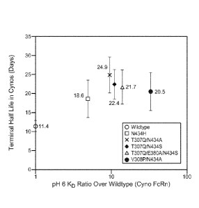
Fig. 13: Graph showing the relationship between terminal half-life in
cynomolgus
monkeys and pH 6.0 FcRn affinity for the anti-VEGF wild-type and five anti-
VEGF
variants. Error bars represent standard deviations of 11 or 12 animals per
group.
Fig. 14 Panels A-B: Pharmacokinetic profiles of the anti-VEGF wild-type and
anti-
VEGF variant T307Q/N434A in humanized VEGF transgenic mice following a single
IV
dose of 0.3 or 5 mg/kg. Serum concentrations of the antibodies were measured
using either
(A) VEGF capture ELISA or (B) human Fc capture ELISA. Data are represented as
the
mean + standard deviation.
Fig. 15: Pharmacokinetics parameters for the anti-VEGF wild-type and anti-VEGF
variant T307Q/N434A following a single intravenous dose of 0.3 or 5 mg/kg to
humanized
VEGF transgenic mice.
Fig. 16 Panels A-B: Pharmacokinetic profiles of anti-VEGF variant T307Q/N434A
in humanized VEGF transgcnic mice following a multi-dose of 0.3 or 5 mg/kg.
Antibody
was administered at day 0, 3, 6, and 9. Serum concentrations of the antibodies
were
measured using either (A) VEGF capture ELISA or (B) human Fc capture ELISA.
Data are
represented as the mean standard deviation.
Fig. 17: Pharmacokinetics parameters for the T307Q/N434A following four
intravenous doses of 0.3 or 5 mg/kg at day 0, 3, 6, and 9 to humanized VEGF
transgenic
mice.
Fig. 18 Panels A-C: Efficacy of anti-VEGF wildtype and T307Q/N434A (QA)
variant in treating HM-7 xenografts implanted s.c. into RAG2 KO; hum-X VEGF KI
double-homozygous mice. 5mg/kg and 0.5mg/kg of wildtype and T307Q/N434A
variant
and 5mg/kg of anti-ragweed control were administered intraperitoneal twice
weekly. (A)
Growth curves of HM-7 tumors. Data are represented as the mean standard
error. (B)
CA 02739429 2011-04-01
WO 2010/045193 PCT/US2009/060443
Growth curves of HM-7 tumors for 0.5 and 0.05mg/kg treatment groups. (C) Serum
anti-
VEGF antibody concentrations at the end of treatment (day 18 for anti-ragweed
and day 21
for the anti-VEGF treatment groups). Concentrations were measured using the
VEGF
capture ELSA. Data are represented as the mean + standard deviation.
Fig. 19 Panels A-E: A repeat efficacy study of anti-VEGF wildtype and
T307Q/N434A (QA) variant in treating HM-7 xenografts implanted s.c. into RAG2
KO;
hum-X VEGF K1 double-homozygous mice. 5, 0.5 and 0.05mg/kg of wildtype and
T307Q/N434A variant and 5mg/kg of anti-ragweed control were administered
intraperitoneal twice weekly. (A) Growth curves of HM-7 tumors. Data are
represented as
.. the mean standard error. (B) Growth curves of HM-7 tumors for 0.5 and
0.05mg/kg
treatment groups. Data are represented as the mean standard error. (C)
Terminal weights
of HM-7 tumors determined at the end of treatment (day 16 for anti-ragweed,
day 19 for
0.05mg/kg group, and day 22 of the remaining groups). Data are represented as
the mean +
standard error. (D) Serum anti-VEGF antibody concentrations at the end of
treatment.
.. Concentrations were measured using the human Fc capture ELISA. Data are
represented as
the mean standard deviation. (E) Ratio of antibody concentration in tumors
to that in
blood. Tumor antibody concentrations were determined by measuring the total
amount of
tumor lysates and the amount of anti-VEGF antibody in the tumor lysates. Data
are
represented as the mean f standard deviation.
Fig. 20 Panels A-D: The third efficacy study of anti-VEGF wildtype and
T307Q/N434A (QA) variant in treating HM-7 xenografts implanted s.c. into RAG2
KO;
hum-X VEGF KI double-homozygous mice. 5, 0.5 and 0.05mg/kg of wildtype and
T307Q/N434A and 5mg/kg of anti-ragweed control were administered
intraperitoneal twice
weekly. (A) Mean tumor volume for each group at the end of treatment (day 22).
Data are
.. represented as the mean standard error. (B) Terminal weights of HM-7
tumors. Data are
represented as the mean standard error. (C) Serum anti-VEGF antibody
concentrations at
the end of treatment. Concentrations were measured using the anti-human Fe
capture
EL1SA. Data are represented as the mean standard deviation. (D) Ratio of
antibody
concentration in tumors to that in blood. Tumor antibody concentrations were
determined
.. by measuring the total amount of tumor lysates and the amount of anti-VEGF
antibody in
the tumor lysates. Data are represented as the mean standard deviation.
Fig. 21 Panels A-D: Efficacy of anti-VEGF wildtype and T307Q/N434A (QA)
variant in treating HT-55 xenografts implanted s.c. into RAG2 KO; hum-X
VEGF K1
21
CA 02739429 2011-04-01
WO 2010/045193 PCT/US2009/060443
double-homozygous mice. 5, 0.5 and 0.05mg/kg of wildtype and T307Q/N434A and
5mg/kg of anti-ragweed control were administered intraperitoneal twice weekly.
(A)
Growth curves of HT-55 tumors. Data are represented as the mean standard
error. (B)
Terminal weights of HT-55 tumors determined at the end of treatment (day 35).
Data are
represented as the mean f standard error. (C) Serum anti-VEGF antibody
concentrations at
the end of treatment. Concentrations were measured using the human Fe capture
ELISA.
Data are represented as the mean standard deviation. (D) Ratio of antibody
concentration
in tumors to that in blood. Tumor antibody concentrations were determined by
measuring
the total amount of tumor lysates and the amount of anti-VEGF antibody in the
tumor
lysates. Data are represented as the mean standard deviation.
Fig. 22 Panels A-E: Efficacy of anti-VEGF wildtype and T307Q/N434A (QA)
variant in treating Colo-205 xenografts implanted s.c. into RAG2 KO; hum-X
VEGF KI
double-homozygous mice. 5, 0.5 and 0.05mg/kg of wildtype and T307Q/N434A and
5mg/kg of anti-ragweed control were administered intraperitoneal twice weekly.
(A)
.. Growth curves of Colo-205 tumors. Data are represented as the mean
standard error. (B)
Growth curves of Colo-205 tumors at 0.5 and 0.05mg/kg treatment groups. (C)
Terminal
weights of Colo-205 tumors determined at the end of treatment (day 38). Data
are
represented as the mean standard error. (D) Scrum anti-VEGF antibody
concentrations at
the end of treatment. Concentrations were measured using the human Fc capture
ELISA.
Data are represented as the mean standard deviation. (E) Ratio of antibody
concentration
in tumors to that in blood. Tumor antibody concentrations were determined by
measuring
the total amount of tumor lysates and the amount of anti-VEGF antibody in the
tumor
lysates. Data are represented as the mean standard deviation.
Fig. 23 Panels A-D: A repeat efficacy study of anti-VEGF wildtype and
T307Q/N434A (QA) variant in treating Colo-205 xenografts. 5, 0.5 and 0.05mg/kg
of
wildtype and T307Q/N434A and 5mg/kg of anti-ragweed control were administered
intraperitoneal twice weekly. (A) Growth curves of Colo-205 tumors. Data are
represented
as the mean standard error. (B) Growth curves of Colo-205 tumors at 0.5 and
0.05mg/kg
treatment groups. (C) Terminal weights of Colo-205 tumors determined at the
end of
treatment (day 32). Data are represented as the mean standard error. (D)
Serum anti-
VEGF antibody concentrations at the end of treatment. Concentrations were
measured
using the human Fe capture ELISA. Data are represented as the mean standard
deviation.
22
CA 02739429 2011-04-01
WO 2010/045193 PCT/US2009/060443
Fig. 24: Monovalent dissociation constants (KD) of anti-HER2 (traztuzumab)
IgGt
Fc variants to human FeRn at pH 6.0 and 25 C using BIAcore. Results are
representative of
two independent experiments.
Fig. 25: Expression levels of FcRn in different human tumor cell lines. Five
million cells of each cell line were used for the experiment. Raji cells
(human B-cell
lymphoma) were used as a negative control, while soluble human FeRn protein,
which is
missing the 7kDa transmembrane and cytoplasmic regions, was blotted as a
positive
control. Dilutions of soluble FeRn protein were used as the standard to
quantify the FeRn
expression level. Results shown here are representative of at least three
independent
experiments.
Fig. 26: pH-dependent binding of anti-HER2 (traztuzumab) IgGi Fe variants to
human FeRn. Variants were constructed with mutations at L251, L314, and E430.
The
binding was measured at pH ranging from 6 to 7.2 using BlAcore at 25 C. The
affinity
ratios of the variants relative to anti-HER2 IgGi wildtype were determined and
plotted as a
function of pH.
Fig. 27 Panels A-C: Binding of anti-HER2 (traztuzumab) IgGi wild-type, variant
T307Q/N434A, variant L251D/T307Q/N434H and variant
L251D/T307Q/M428LIN434H/Y4361 against human FeRn at (A) pH 6.0, (B) pH 7.1,
and
(C) pH 7.4. The binding was measured using BIAcore at 25 C. There was no
detectable
binding of variant L251D/T307Q,N434H to human FeRn at pH 7.4 in Fig. 27C.
Fig. 28: The dissociation rate (koff) of human FeRn against various anti-VEGF
and
anti-HER2 variants at different pHs. The anti-VEGF variants are T307Q/N434A,
T307Q/N434S. T307Q/E380A/N434S and V308P/N434A. The anti-HER2 variant is
L251D/T307Q/M428L/N434H/Y4361. The koff values at different pHs were fitted
against
pH for each variant to yield the slope of the best-fit line (equation:
log(koff) = slope x pH
+y-intercept).
DETAILED DESCRIPTION OF THE INVENTION
The present invention relates to novel variants of Fe domains, including those
found
in antibodies, Fe fusions, and immuno-adhesins, that have an increased in vivo
half-life.
These variants comprise a human IgG Fe region, or fragment thereof that binds
to an FeRn,
that contains one or more amino acid modifications relative to a wild type
human IgG Fe
23
CA 02739429 2011-04-01
WO 2010/045193 PCT/US2009/060443
region which modifications increase the affinity of the IgG Fc region, or
fragment thereof,
for the FcRn.
The techniques and procedures described or referenced herein are generally
well
understood and commonly employed using conventional methodology by those
skilled in
the art, such as, for example, the widely utilized methodologies described in
Sambrook et
al., Molecular Cloning: A Laboratory Manual 3rd. edition (2001) Cold Spring
Harbor
Laboratory Press, Cold Spring Harbor, N.Y. CURRENT PROTOCOLS IN MOLECULAR
BIOLOGY (F. M. Ausubel, et al. eds., (2003)); the series METHODS IN ENZYMOLOGY
(Academic Press, Inc.): PCR 2: A PRACTICAL APPROACH (M. J. MacPherson, B. D.
Hames and G. R. Taylor eds. (1995)), Harlow and Lane, eds. (1988) ANTIBODIES,
A
LABORATORY MANUAL, and ANIMAL CELL CULTURE (R. I. Freshney, ed. (1987));
Oligonucleotide Synthesis (M. J. Gait, ed., 1984); Methods in Molecular
Biology, Humana
Press; Cell Biology': A Laboratog Notebook (J. E. Cellis, ed., 1998) Academic
Press;
Animal Cell Culture (R. 1. Freshney), ed., 1987); Introduction to Cell and
Tissue Culture (J.
P. Mather and P. E. Roberts, 1998) Plenum Press; Cell and Tissue Culture:
Laboratory
Procedures (A. Doyle, J. B. Griffiths, and D. G. Newell, eds., 1993-8) J.
Wiley and Sons;
Handbook of Experimental Immunology (D. M. Weir and C. C. Blackwell, eds.);
Gene
Transfer Vectors for Mammalian Cells (J. M. Miller and M. P. Cabs, eds.,
1987); PCR:
The Polymerase Chain Reaction, (Mullis et al., eds., 1994); Current Protocols
in
Immunology (J. E. Coligan et al., eds., 1991); Short Protocols in Molecular
Biology (Wiley
and Sons, 1999); Immunobiology (C. A. Janeway and P. Travers, 1997);
Antibodies (P.
Finch, 1997); Antibodies: A Practical Approach (D. Catty., ed., IRL Press,
1988-1989);
Monoclonal Antibodies: A Practical Approach (P. Shepherd and C. Dean, eds.,
Oxford
University Press, 2000); Using Antibodies: A Laboratory Manual (E. Harlow and
D. Lane
(Cold Spring Harbor Laboratory Press, 1999); The Antibodies (M. Zanetti and J.
D. Capra,
eds., Harwood Academic Publishers, 1995); and Cancer: Principles and Practice
qf
Oncology (V. T. DeVita et al., eds., J.B. Lippincott Company, 1993).
Unless defined otherwise, technical and scientific terms used herein have the
same
meaning as commonly understood by one of ordinary skill in the art to which
this invention
belongs. Singleton et al., Dictionary of Microbiology and Molecular Biology
2nd ed., J.
Wiley & Sons (New York, N.Y. 1994), and March, Advanced Organic Chemistry
Reactions, Mechanisms and Structure 4th ed., John Wiley & Sons (New York, N.Y.
1992),
provide one skilled in the art with a general guide to many of the terms used
in the present
24
CA 02739429 2016-04-27
CA2739429
application.
Definitions
For purposes of interpreting this specification, the following definitions
will apply and
whenever appropriate, terms used in the singular will also include the plural
and vice versa. It is to be
understood that the terminology used herein is for the purpose of describing
particular embodiments
only, and is not intended to be limiting. In the event that any definition set
forth below conflicts with
any document incorporated herein by reference, the definition set forth below
shall control.
Throughout the present specification and claims, the numbering of the residues
in an
immunoglobulin heavy chain is that of the EU index as in Kabat et al.,
Sequences of Proteins of
Immunological Interest, 5th Ed. Public Health Service, National Institutes of
Health, Bethesda, Md.
(1991). The "EU index as in Kabat" refers to the residue numbering of the
human IgGi EU antibody.
The term "in vivo half-life" or "the half life of the antibody in vivo" as
used herein refers to a
biological half-life of a particular type of IgG molecule or its fragments
containing FeRn-binding sites
in the circulation of a given animal and is represented by the time required
for the circulating
concentration of a molecule to decrease by 50%. In certain embodiments, when
the concentration of a
given IgG is plotted as a function of time, the curve is usually biphasic with
a rapid a-phase which
represents an equilibration of the injected IgG molecules between the intra-
and extra-vascular space,
and a longer 0-phase which represents the elimination of the IgG molecules
from the intravascular
space. In certain embodiments, the term "in vivo half-life" corresponds to the
half life of the 1gG
molecules in the 0-phase. In certain embodiments, the concentration versus
time curve of a given IgG
is triphasic, with corresponding distribution into an a-phase and 0-phase, and
terminal elimination
represented by a '-phase. Therefore, in certain embodiments, in vivo half-life
corresponds to the half-
life of the terminal elimination phase, or the y -phase. In certain
embodiments, the concentration versus
time curve of a given IgG is monophasic, with a single elimination phase.
Therefore, in certain
embodiments, in vivo half-life corresponds to the half-life of the single
elimination phase.
By "parent polypeptide" or "wild-type polypeptide" as used herein is meant an
unmodified
polypeptide, a naturally occurring polypeptide, or an engineered modified
CA 02739429 2011-04-01
WO 2010/045193 PCT/US2009/060443
version of a naturally occurring polypeptide which lacks one or more of the Fc
region
amino acid modifications disclosed herein and which differs in effector
function compared
to variant IgG as herein disclosed. The parent polypeptide may comprise a
native sequence
Fc region or an Fc region with pre-existing amino acid sequence modifications
(such as
additions, deletions and/or substitutions). The parent polypeptide may also
comprise non-
natural amino acids as described below. Parent polypeptide may refer to the
polypeptide
itself, compositions that comprise the parent polypeptide, or the amino acid
sequence that
encodes it. Parent polypeptide, includes, without limitation, parent
immunoglobulin, wild-
type immunoglobulin, parent antibody and wild-type antibody.
Accordingly, by "parent immunoglobulin," "parent IgG," "wild-type
immunoglobulin" or "wild-type IgG" as used herein is meant an unmodified
immunoglobulin, a naturally occurring immunoglobulin, or an engineered
modified version
of a naturally occurring immunoglobulin which lacks one or more of the Fc
region amino
acid modifications disclosed herein and which differs in effector function
compared to
variant IgG as herein disclosed. The parent immunoglobulin may comprise a
native
sequence Fc region or an Fc region with pre-existing amino acid sequence
modifications
(such as additions, deletions and/or substitutions). The parent immunoglobulin
may also
comprise non-natural amino acids as described below. Parent immunoglobulin may
refer to
the immunoglobulin itself, compositions that comprise the parent
immunoglobulin, or the
amino acid sequence that encodes it.
By "parent antibody" or "wild-type antibody" as used herein is meant an
unmodified
antibody, a naturally occurring antibody, or an engineered modified version of
a naturally
occurring antibody which lacks one or more of the Fc region amino acid
modifications
disclosed herein and which differs in effector function compared to variant
IgG as herein
disclosed. The parent antibody may comprise a native sequence Fc region or an
Fc region
with pre-existing amino acid sequence modifications (such as additions,
deletions and/or
substitutions). The parent antibody may also comprise non-natural amino acids
as
described below. Parent antibody may refer to the antibody itself,
compositions that
comprise the parent antibody, or the amino acid sequence that encodes it.
In certain embodiments, "parent IgG," "parent antibody," "wild-type IgG," or
"wild-
type antibody" includes, but not limited to, known commercial, recombinantly
produced
antibodies as described herein. In certain embodiments, wild-type IgG is an
IgG having the
wild-type human IgG Fc region. In certain embodiments, the wild-type human IgG
Fc
26
CA 02739429 2011-04-01
WO 2010/045193
PCT/US2009/060443
region refers to Fc region which lacks one or more of the Fc region amino acid
modifications disclosed herein. In certain embodiments, the wild-type human
IgG Fc
region refers to Fc region with one or more Fc region amino acid modifications
not
disclosed herein. In certain embodiments, the wild-type IgG is bevacizumab. In
certain
embodiment, the wild-type antibody is an antibody fragment that does not
contain an Fc
region. In certain embodiments, the variant IgG of such wild-type antibody is
an Fc fusion
protein comprising Fab domain fragments of the wild-type antibody, or the
domain or
domains of a non-antibody protein, and an Fc domain fragment comprising one or
more of
the Fc region modifications disclosed herein. In certain embodiments, the wild-
type human
IgG Fc region refers to an IgG Fc region with Fc mutation L251D or L251D/434H,
but has
no other Fc region amino acid modifications disclosed herein.
By "variant," "variant protein" or "protein variant" as used herein is meant a
protein
that differs from that of a parent protein by virtue of at least one amino
acid modification.
Protein variant may refer to the protein itself, a composition comprising the
protein, or the
amino sequence that encodes it. In certain embodiments, the protein variant
has at least one
amino acid modification compared to the parent polypeptide, e.g. from about
one to about
ten amino acid modifications. In certain embodiments, the protein variant has
at least two
amino acid modifications in the IgG Fc region. In certain embodiments, the
protein variant
has at least three amino acid modifications in the IgG Fc region. The protein
variant
sequence herein will preferably possess at least about 80% homology with a
parent protein
sequence, and most preferably at least about 90% homology, more preferably at
least about
95% homology. Protein variants may also comprise non-natural amino acids, as
defined
below. The term "protein variant" includes immunoglobulin variant and antibody
variant as
described herein.
The term "immunoglobulin variant," "variant immunoglobulin," "variant IgG" or
"lgG variant" as used herein is meant an immunoglobulin sequence that differs
from that of
a parent or wild-type immunoglobulin sequence by virtue of at least one amino
acid
modification. In certain embodiments, variant IgG has at least two amino acid
modifications in the Fc region relative to wild-type IgG. In certain
embodiments, variant
IgG has at least three amino acid modifications in the Fc region relative to
wild-type IgG.
In certain embodiments, variant IgG is a variant antibody. In certain
embodiments, the
variant IgG is an anti-VEGF antibody. In one embodiment, the variant IgG is a
variant of
27
CA 02739429 2011-04-01
WO 2010/045193 PCT/US2009/060443
bevacizumab comprising one or more amino acid modification in the Fc region of
the
antibody.
By "antibody variant" or "variant antibody" as used herein is meant an
antibody that
differs from a parent antibody by virtue of at least one amino acid
modification. In certain
embodiments, the variant antibody has one or more amino acid modifications in
the Fc
region relative to wild-type antibody.
By "position" as used herein is meant a location in the sequence of a protein.
Positions may be numbered sequentially, or according to an established format,
for example
the EU index as in Kabat.
The term "Fc region-containing polypeptide" or "Fc polypeptide" refers to a
polypeptide that comprises all or part of an Fc region. For example, Fc
polypeptides
include antibodies, Fc fusions, isolated Fcs, and Fc fragments.
The term "Fc region-comprising antibody" refers to an antibody that comprises
an
Fc region. The C-terminal lysine (residue 447 according to the EU numbering
system) of
the Fc region may be removed, for example, during purification of the antibody
or by
recombinant engineering of the nucleic acid encoding the antibody.
Accordingly, a
composition comprising an antibody having an Fc region can comprise an
antibody with
K447, with all K447 removed, or a mixture of antibodies with and without the
K447
residue.
An "amino acid modification" refers to a change in the amino acid sequence of
a
predetermined amino acid sequence. Exemplary modifications include an amino
acid
substitution, insertion and/or deletion. In certain embodiments, the amino
acid
modification is a substitution.
An "amino acid modification at" a specified position, e.g. of the Fc region,
refers to
the substitution or deletion of the specified residue, or the insertion of at
least one amino
acid residue adjacent the specified residue. By insertion "adjacent" a
specified residue is
meant insertion within one to two residues thereof The insertion may be N-
terminal or C-
terminal to the specified residue.
An "amino acid substitution" refers to the replacement of at least one
existing amino
acid residue in a predetermined amino acid sequence with another different
"replacement"
amino acid residue. The replacement residue or residues may be "naturally
occurring amino
acid residues" (i.e. encoded by the genetic code) and selected from the group
consisting of:
alanine (Ala); arginine (Arg); asparagine (Asn); aspartic acid (Asp); cysteine
(Cys);
28
CA 02739429 2016-04-27
CA2739429
= glutamine (Gin); glutamic acid (Glu); glycine (Gly); histidine (His);
isoleucine (Ile): leucine (Leu);
lysine (Lys); methionine (Met); phenylalanine (Phe); proline (Pro); serine
(Ser); threonine (Thr);
tryptophan (Trp); tyrosine (Tyr); and valine (Val). In certain embodiments,
the replacement
residue is not cysteine. Substitution with one or more non-naturally occurring
amino acid residues
is also encompassed by the definition of an amino acid substitution herein.
A "non-naturally occurring amino acid residue" refers to a residue, other than
those
naturally occurring amino acid residues listed above, which is able to
covalently bind adjacent
amino acid residues(s) in a polypeptide chain. Examples of non-naturally
occurring amino acid
residues include norleucine, ornithine, norvaline, homoserine and other amino
acid residue
analogues such as those described in Ellman et al. Meth. Enzym. 202:301-336
(1991); US Patent
No, 6,586,207; WO 98/48032; WO 03/073238; U.S. Publication No. 2004-0214988A1;
WO
05135727A2; WO 05/74524A2; J. W. Chin et at., (2002), Journal of the American
Chemical
Society 124:9026-9027; J. W. Chin, & P. G. Schultz, (2002), ChemBioChern
11:1135-1137; and J.
W. Chin, et at., (2002), PICAS United States of America 99:11020-11024.
An "amino acid insertion" refers to the incorporation of at least one amino
acid into a
predetermined amino acid sequence. While the insertion will usually consist of
the insertion of one
or two amino acid residues, the present application contemplates larger
"peptide insertions", e.g.
insertion of about three to about five or even up to about ten amino acid
residues. The inserted
residue(s) may be naturally occurring or non-naturally occurring as disclosed
above.
An "amino acid deletion" refers to the removal of at least one amino acid
residue from a
predetermined amino acid sequence.
In certain embodiments, the term "increase," "increased half life" or
"increased in vivo half
life" refers to an overall increase of at least about 25%, 30%, 40%, 50%, 60%,
70%, 75%, 80%,
85%, 90%, 95%, 100%, 150%, 200%, 300% or greater, in the in vivo half life of
a variant IgG of
the invention detected by standard art known methods such as those described
herein, as compared
to a wild-type IgG or an 1gG having the wild-type human IgG Fc region. In
certain embodiments,
the term increase refers to the increased in vivo half life of the variant
IgG, wherein the increase is
at least about 1.25X, 1.5X, 1.75X, 2X, 3X, 4X, 5X, or 10X or greater as
compared to a wild-type
IgG or an IgG having the wild-type human IgG Fc region. In certain
embodiments, the wild-type
IgG or the IgG having the
29
CA 02739429 2011-04-01
WO 2010/045193 PCT/US2009/060443
wild-type human IgG Fe region is bevacizumab. In certain embodiments, the mean
half-life
of bevacizumab is about 10 to 12 days as measured in cynomolgus monkeys, or
about 20
days as measured in humans.
"Hinge region" is generally defined as stretching from Glu216 to Pro230 of
human
IgG1 (Burton, Molec. Imrnunol. 22:161-206 (1985)). Hinge regions of other IgG
isotypes
may be aligned with the IgG1 sequence by placing the first and last cysteine
residues
forming inter-heavy chain S¨S bonds in the same positions.
The "lower hinge region" of an Fe region is normally defined as the stretch of
residues immediately C-terminal to the hinge region, i.e. residues 233 to 239
of the Fe
region. Prior to the present invention, FcyR binding was generally attributed
to amino acid
residues in the lower hinge region of an IgG Fe region.
"Clq" is a polypeptide that includes a binding site for the Fe region of an
immunoglobulin. Clq together with two serine proteases, Clr and C Is, forms
the complex
Cl, the first component of the complement dependent cytotoxicity (CDC)
pathway. Human
Clq can be purchased commercially from, e.g. Quidel, San Diego, Calif.
The term "binding domain" refers to the region of a polypeptide that binds to
another molecule. In the case of an FcR, the binding domain can comprise a
portion of a
polypeptide chain thereof (e.g., the a chain thereof) which is responsible for
binding an Fe
region. One useful binding domain is the extracellular domain of an FcR a
chain.
The term "antibody" herein is used in the broadest sense and specifically
covers
monoclonal antibodies (including full length monoclonal antibodies),
polyclonal antibodies,
multispecific antibodies (e.g., bispecific antibodies), and antibody fragments
so long as they
exhibit the desired biological activity.
An "isolated" antibody is one which has been identified and separated and/or
recovered from a component of its natural environment. Contaminant components
of its
natural environment are materials which would interfere with research,
diagnostic or
therapeutic uses for the antibody, and may include enzymes, hormones, and
other
proteinaceous or nonproteinaceous solutes. In some embodiments, an antibody is
purified
(1) to greater than 95% by weight of antibody as determined by, for example,
the Lowry
method, and in some embodiments, to greater than 99% by weight; (2) to a
degree sufficient
to obtain at least 15 residues of N-terminal or internal amino acid sequence
by use of, for
example, a spinning cup sequenator, or (3) to homogeneity by SDS-PAGE under
reducing
or nonreducing conditions using, for example, Coomassie blue or silver stain.
Isolated
CA 02739429 2011-04-01
WO 2010/045193
PCT/US2009/060443
antibody includes the antibody in situ within recombinant cells since at least
one component
of the antibody's natural environment will not be present. Ordinarily,
however, isolated
antibody will be prepared by at least one purification step.
"Native antibodies" are usually heterotetrameric glycoproteins of about
150,000
daltons, composed of two identical light (L) chains and two identical heavy
(H) chains.
Each light chain is linked to a heavy chain by one covalent disulfide bond,
while the
number of disulfide linkages varies among the heavy chains of different
immunoglobulin
isotypes. Each heavy and light chain also has regularly spaced intrachain
disulfide bridges.
Each heavy chain has at one end a variable domain (VH) followed by a number of
constant
domains. Each light chain has a variable domain at one end (VI) and a constant
domain at
its other end; the constant domain of the light chain is aligned with the
first constant
domain of the heavy chain, and the light chain variable domain is aligned with
the variable
domain of the heavy chain. Particular amino acid residues are believed to form
an interface
between the light chain and heavy chain variable domains.
The term "constant domain" refers to the portion of an immunoglobulin molecule
having a more conserved amino acid sequence relative to the other portion of
the
immunoglobulin, the variable domain, which contains the antigen binding site.
The
constant domain contains the CHI, CH2 and CH3 domains of the heavy chain and
the CHL
domain of the light chain.
The "variable region" or "variable domain" of an antibody refers to the amino-
terminal domains of the heavy or light chain of the antibody. The variable
domain of the
heavy chain may be referred to as "VH." The variable domain of the light chain
may be
referred to as "VL." These domains are generally the most variable parts of an
antibody and
contain the antigen-binding sites.
The term "variable" refers to the fact that certain portions of the variable
domains
differ extensively in sequence among antibodies and are used in the binding
and specificity
of each particular antibody for its particular antigen. However, the
variability is not evenly
distributed throughout the variable domains of antibodies. It is concentrated
in three
segments called hypervariable regions (LIVRs) both in the light-chain and the
heavy-chain
variable domains. The more highly conserved portions of variable domains are
called the
framework regions (FR). The variable domains of native heavy and light chains
each
comprise four FR regions, largely adopting a beta-sheet configuration,
connected by three
HVRs, which form loops connecting, and in some cases forming part of, the beta-
sheet
31
CA 02739429 2011-04-01
WO 2010/045193
PCT/US2009/060443
structure. The HVRs in each chain are held together in close proximity by the
FR regions
and, with the HVRs from the other chain, contribute to the formation of the
antigen-binding
site of antibodies (see Kabat et al., Sequences of Proteins of Immunological
Interest, Fifth
Edition, National Institute of Health, Bethesda, MD (1991)). The constant
domains are not
.. involved directly in the binding of an antibody to an antigen, but exhibit
various effector
functions, such as participation of the antibody in antibody-dependent
cellular toxicity.
The "light chains" of antibodies (immunoglobulins) from any vertebrate species
can
be assigned to one of two clearly distinct types, called kappa (ic) and lambda
(X), based on
the amino acid sequences of their constant domains.
The term IgG "isotype: or "subclass" as used herein is meant any of the
subclasses
of immunoglobulins defined by the chemical and antigenic characteristics of
their constant
regions.
Depending on the amino acid sequences of the constant domains of their heavy
chains, antibodies (immunoglobulins) can be assigned to different classes.
There are five
major classes of immunoglobulins: IgA, IgD, IgE, IgG, and IgM, and several of
these may
be further divided into subclasses (isotypes), e.g., IgGI, IgG2, IgG3, IgG4,
IgAi, and IgA2.
The heavy chain constant domains that correspond to the different classes of
immunoglobulins are called a, 6, 8, 7, and 1,t, respectively. The subunit
structures and three-
dimensional configurations of different classes of immunoglobulins are well
known and
described generally in, for example, Abbas et al. Cellular and Mol.
Immunology, 4th ed.
(W.B. Saunders, Co., 2000). An antibody may be part of a larger fusion
molecule, formed
by covalent or non-covalent association of the antibody with one or more other
proteins or
peptides.
The term "IgG subclass modification" as used herein is meant an amino acid
modification that converts one amino acid of one IgG isotype to the
corresponding amino
acid in a different, aligned lgG isotype. For example, because lgGi comprises
a tyrosine
and lgG2 a phenylalanine at EU position 296, a F296Y substitution in IgG2 is
considered an
IgG subclass modification.
By "non-naturally occurring modification" as used herein is meant an amino
acid
modification that is not isotypic. For example, because none of the IgGs
comprise a
glutamic acid at position 332, substitution at position 332 with glutamic acid
(332E) in
IgGI, IgG2, IgG3, or 1gG4 is considered a non-naturally occurring
modification.
32
CA 02739429 2011-04-01
WO 2010/045193 PCT/US2009/060443
The terms "full length antibody," "intact antibody" and "whole antibody" are
used
herein interchangeably to refer to an antibody in its substantially intact
form. The terms
particularly refer to an antibody with heavy chains that contain an Fc region.
A "naked antibody" for the purposes herein is an antibody that is not
conjugated to a
cytotoxic moiety or radiolabel.
"Antibody fragments" comprise a portion of an intact antibody, preferably
comprising the antigen binding region thereof. In certain embodiments,
antibody fragments
comprise an Fe region or a portion of Fe region comprising one or more Fe
region
modification disclosed herein. Examples of antibody fragments include Fab,
Fab', F(ab')2,
and Fv fragments; diabodies; linear antibodies; single-chain antibody
molecules; and
multispecific antibodies formed from antibody fragments.
Papain digestion of antibodies produces two identical antigen-binding
fragments,
called "Fab" fragments, each with a single antigen-binding site, and a
residual "Fe"
fragment, whose name reflects its ability to crystallize readily. Pepsin
treatment yields an
F(a1:02 fragment that has two antigen-combining sites and is still capable of
cross-linking
antigen.
"Fv" is the minimum antibody fragment which contains a complete antigen-
binding
site. In one embodiment, a two-chain Fv species consists of a dimer of one
heavy- and one
light-chain variable domain in tight, non-covalent association. In a single-
chain Fv (scFv)
species, one heavy- and one light-chain variable domain can be covalently
linked by a
flexible peptide linker such that the light and heavy chains can associate in
a "dimeric"
structure analogous to that in a two-chain Fv species. It is in this
configuration that the
three HVRs of each variable domain interact to define an antigen-binding site
on the surface
of the VH-VL dimer. Collectively, the six HVRs confer antigen-binding
specificity to the
antibody. However, even a single variable domain (or half of an Fv comprising
only three
HVRs specific for an antigen) has the ability to recognize and bind antigen,
although at a
lower affinity than the entire binding site.
The Fab fragment contains the heavy- and light-chain variable domains and also
contains the constant domain of the light chain and the first constant domain
(CHI) of the
heavy chain. Fab' fragments differ from Fab fragments by the addition of a few
residues at
the carboxy terminus of the heavy chain CH1 domain including one or more
cysteines from
the antibody hinge region. Fab'-SH is the designation herein for Fab' in which
the cysteine
residue(s) of the constant domains bear a free thiol group. F(ab')2 antibody
fragments
33
CA 02739429 2011-04-01
WO 2010/045193 PCT/US2009/060443
originally were produced as pairs of Fab' fragments which have hinge cysteines
between
them. Other chemical couplings of antibody fragments are also known.
"Single-chain Fv" or "scFv" antibody fragments comprise the VH and VL domains
of antibody, wherein these domains are present in a single polypeptide chain.
Generally,
the scFv polypeptide further comprises a polypeptide linker between the VH and
VL
domains which enables the scFv to form the desired structure for antigen
binding. For a
review of scFv, see, e.g., Pluckthiin, in The Pharmacology of Monoclonal
Antibodies, vol.
113, Rosenburg and Moore eds., (Springer-Verlag, New York, 1994), pp. 269-315.
The term "diabodies" refers to antibody fragments with two antigen-binding
sites,
which fragments comprise a heavy-chain variable domain (VH) connected to a
light-chain
variable domain (VL) in the same polypeptide chain (VH-VL). By using a linker
that is too
short to allow pairing between the two domains on the same chain, the domains
are forced
to pair with the complementary domains of another chain and create two antigen-
binding
sites. Diabodies may be bivalent or bispecific. Diabodies are described more
fully in, for
example, EP 404,097; WO 1993/01161; Hudson et al., Nat. Med. 9:129-134 (2003);
and
Hollinger et al., Proc. Natl. Acad. Sci. USA 90: 6444-6448 (1993). Triabodies
and
tetrabodies are also described in Hudson et al., Nat. Med. 9:129-134 (2003).
The term "monoclonal antibody" as used herein refers to an antibody obtained
from
a population of substantially homogeneous antibodies, i.e., the individual
antibodies
comprising the population are identical except for possible mutations, e.g.,
naturally
occurring mutations, that may be present in minor amounts. Thus, the modifier
"monoclonal" indicates the character of the antibody as not being a mixture of
discrete
antibodies. In certain embodiments, such a monoclonal antibody typically
includes an
antibody comprising a polypeptide sequence that binds a target, wherein the
target-binding
polypeptide sequence was obtained by a process that includes the selection of
a single target
binding polypeptide sequence from a plurality of polypeptide sequences. For
example, the
selection process can be the selection of a unique clone from a plurality of
clones, such as a
pool of hybridoma clones, phage clones, or recombinant DNA clones. It should
be
understood that a selected target binding sequence can be further altered, for
example, to
improve affinity for the target, to humanize the target binding sequence, to
improve its
production in cell culture, to reduce its immunogenicity in vivo, to create a
multispecific
antibody, etc., and that an antibody comprising the altered target binding
sequence is also a
monoclonal antibody of this invention. In contrast to polyclonal antibody
preparations,
34
CA 02739429 2011-04-01
WO 2010/045193 PCT/US2009/060443
which typically include different antibodies directed against different
determinants
(epitopes), each monoclonal antibody of a monoclonal antibody preparation is
directed
against a single determinant on an antigen. In addition to their specificity,
monoclonal
antibody preparations are advantageous in that they are typically
uncontaminated by other
immunoglobulins.
The modifier "monoclonal" indicates the character of the antibody as being
obtained
from a substantially homogeneous population of antibodies, and is not to be
construed as
requiring production of the antibody by any particular method. For example,
the
monoclonal antibodies to be used in accordance with the present invention may
be made by
a variety of techniques, including, for example, the hybridoma method (e.g.,
Kohler and
Milstein, Nature, 256:495-97 (1975); Bongo et al., Hybridoma, 14(3): 253-260
(1995),
Harlow et al., Antibodies: A Laboratog Manual, (Cold Spring Harbor Laboratory
Press,
2nd ed. 1988); Hammerling etal., in: Monoclonal Antibodies and T-Cell
Hybridomas 563-
681 (Elsevier, N.Y., 1981)), recombinant DNA methods (see, e.g., U.S. Patent
No.
4,816,567), phage-display technologies (see, e.g., Clackson et al., Nature,
352: 624-628
(1991); Marks et al., J. Mol. Biol. 222: 581-597 (1992); Sidhu etal., J. Mol.
Biol. 338(2):
299-310 (2004); Lee etal., J. Ho!. Biol. 340(5): 1073-1093 (2004); Fellouse,
Proc. Natl.
Acad. Sci. USA 101(34): 12467-12472 (2004); and Lee etal., J. Inununol.
Methods 284(1-
2): 119-132(2004), and technologies for producing human or human-like
antibodies in
animals that have parts or all of the human immunoglobulin loci or genes
encoding human
immunoglobulin sequences (see, e.g., WO 1998/24893; WO 1996/34096; WO
1996/33735;
WO 1991/10741; Jakobovits etal., Proc. Natl. Acad. Sci. USA 90: 2551 (1993);
Jakobovits
etal., Nature 362: 255-258 (1993); Bruggemann et al., Year in Iminunol. 7:33
(1993); U.S.
Patent Nos. 5,545,807; 5,545,806; 5,569,825; 5,625,126; 5,633,425; and
5,661,016; Marks
etal., Bio/Technology 10: 779-783 (1992); Lonberg et al., Nature 368: 856-859
(1994);
Morrison, Nature 368: 812-813 (1994); Fishwild etal., Nature Biotechnol. 14:
845-851
(1996); Neuberger, Nature Biotechnol. 14: 826 (1996); and Lonberg and Huszar,
Intern.
Rev. Immunol. 13: 65-93 (1995).
The monoclonal antibodies herein specifically include "chimeric" antibodies in
which a portion of the heavy and/or light chain is identical with or
homologous to
corresponding sequences in antibodies derived from a particular species or
belonging to a
particular antibody class or subclass, while the remainder of the chain(s) is
identical with or
homologous to corresponding sequences in antibodies derived from another
species or
CA 02739429 2011-04-01
WO 2010/045193 PCT/US2009/060443
belonging to another antibody class or subclass, as well as fragments of such
antibodies, so
long as they exhibit the desired biological activity (see, e.g.,U U.S. Patent
No. 4,816,567; and
Morrison et al., Proc. Natl. Acad. Sci. USA 81:6851-6855 (1984)). Chimeric
antibodies
include PRIMATIZEDER) antibodies wherein the antigen-binding region of the
antibody is
derived from an antibody produced by, e.g., immunizing macaque monkeys with
the antigen
of interest.
"Humanized" forms of non-human (e.g., murine) antibodies are chimeric
antibodies
that contain minimal sequence derived from non-human immunoglobulin. In one
embodiment, a humanized antibody is a human immunoglobulin (recipient
antibody) in
which residues from a HVR of the recipient are replaced by residues from a HVR
of a non-
human species (donor antibody) such as mouse, rat, rabbit, or nonhuman primate
having the
desired specificity, affinity, and/or capacity. In some instances, FR residues
of the human
immunoglobulin are replaced by corresponding non-human residues. Furthermore,
humanized antibodies may comprise residues that are not found in the recipient
antibody or
in the donor antibody. These modifications may be made to further refine
antibody
performance. In general, a humanized antibody will comprise substantially all
of at least
one, and typically two, variable domains, in which all or substantially all of
the
hypervariable loops correspond to those of a non-human immunoglobulin, and all
or
substantially all of the FRs are those of a human immunoglobulin sequence. The
humanized antibody optionally will also comprise at least a portion of an
immunoglobulin
constant region (Fc), typically that of a human immunoglobulin. For further
details, see,
e.g., Jones et al., Nature 321:522-525 (1986); Riechmann et al., Nature
332:323-329
(1988); and Presta, Cum Op. Struct. Biol. 2:593-596 (1992). See also, e.g.,
Vaswani and
Hamilton, Ann. Allergy, Asthma & Immunol. 1:105-115 (1998); Harris, Biochem.
Soc.
Transactions 23:1035-1038 (1995); Hurle and Gross, Curr. Op. Biotech. 5:428-
433 (1994);
and U.S. Pat. Nos. 6,982,321 and 7,087,409.
A "human antibody" is one which possesses an amino acid sequence which
corresponds to that of an antibody produced by a human and/or has been made
using any of
the techniques for making human antibodies as disclosed herein. This
definition of a
human antibody specifically excludes a humanized antibody comprising non-human
antigen-binding residues. Human antibodies can be produced using various
techniques
known in the art, including phage-display libraries. Hoogenboom and Winter, J.
Mol. Biol.,
227:381 (1991); Marks et al., J. Mol. Biol., 222:581 (1991). Also available
for the
36
CA 02739429 2011-04-01
WO 2010/045193 PCT/US2009/060443
preparation of human monoclonal antibodies are methods described in Cole et
al.,
Monoclonal Antibodies and Cancer Therapy, Alan R. Liss, p. 77 (1985); Boerner
et al., J.
Immunol., 147(1):86-95 (1991). See also van Dijk and van de Winkel, Curr.
Opin.
Pharmacol., 5: 368-74 (2001). Human antibodies can be prepared by
administering the
antigen to a transgenic animal that has been modified to produce such
antibodies in
response to antigenic challenge, but whose endogenous loci have been disabled,
e.g.,
immunized xenomice (see, e.g., U.S. Pat. Nos. 6,075,181 and 6,150,584
regarding
XENOMOUSETm technology). See also, for example. Li et al., Proc. Natl. Acad.
Sci. LISA,
103:3557-3562 (2006) regarding human antibodies generated via a human B-cell
hybridoma technology.
A "species-dependent antibody" is one which has a stronger binding affinity
for an
antigen from a first mammalian species than it has for a homologue of that
antigen from a
second mammalian species. Normally, the species-dependent antibody "binds
specifically"
to a human antigen (i.e. has a binding affinity (Kd) value of no more than
about 1 x 10⁻⁷ M,
preferably no more than about 1 x 10⁻⁸ M and most preferably no more than
about 1 x 10⁻⁹
M) but has a binding affinity for a homologue of the antigen from a second
nonhuman
mammalian species which is at least about 50 fold, or at least about 500 fold,
or at least
about 1000 fold, weaker than its binding affinity for the human antigen. The
species-
dependent antibody can be any of the various types of antibodies as defined
above, but
preferably is a humanized or human antibody.
The term "hypervariable region," "HVR," or "HV," when used herein refers to
the
regions of an antibody variable domain which are hypervariable in sequence
and/or form
structurally defined loops. Generally, antibodies comprise six HVRs; three in
the VH (HI,
H2, H3), and three in the VL (L1, L2, L3). In native antibodies, H3 and L3
display the most
diversity of the six HVRs, and H3 in particular is believed to play a unique
role in
conferring fine specificity to antibodies. See, e.g., Xu et al., Immunity
13:37-45 (2000);
Johnson and Wu, in Methods in Molecular Biology 248:1-25 (Lo, ed., Human
Press,
Totowa, NJ, 2003). Indeed, naturally occurring camelid antibodies consisting
of a heavy
chain only are functional and stable in the absence of light chain. See, e.g.,
Hamers-
Casterman et al., Nature 363:446-448 (1993); Sheriff et al., Nature Struct.
Biol. 3:733-736
(1996).
A number of HVR delineations are in use and are encompassed herein. The Kabat
Complementarity Determining Regions (CDRs) are based on sequence variability
and are
37
CA 02739429 2011-04-01
WO 2010/045193 PCT/US2009/060443
the most commonly used (Kabat et al., Sequences of Proteins of Immunological
Interest,
5th Ed. Public Health Service, National Institutes of Health, Bethesda, MD.
(1991)).
Chothia refers instead to the location of the structural loops (Chothia and
Lesk J. Mot. Biol.
196:901-917 (1987)). The AbM HVRs represent a compromise between the Kabat
HVRs
and Chothia structural loops, and are used by Oxford Molecular's AbM antibody
modeling
software. The "contact" HVRs are based on an analysis of the available complex
crystal
structures. The residues from each of these HVRs are noted below.
Loop Kabat AbM Chothia Contact
Li L24-L34 L24-L34 L26-L32 L30-L36
L2 L50-L56 L50-L56 L50-L52 L46-L55
L3 L89-L97 L89-L97 L91-L96 L89-L96
H1 H31-H35B H26-H35B F126-H32 H30-H35B
(Kabat Numbering)
H1 H31-H35 H26-H35 H26-H32 H30-H35
(Chothia Numbering)
H2 H50-H65 H50-H58 H53-H55 H47-H58
H95-H102 H95-H102 H96-H101 H93-H101
HVRs may comprise "extended HVRs" as follows: 24-36 or 24-34 (L1), 46-56 or
50-56 (L2) and 89-97 or 89-96 (L3) in the VL and 26-35 (H1), 50-65 or 49-65
(H2) and 93-
102, 94-102, or 95-102 (H3) in the VH. The variable domain residues are
numbered
according to Kabat et al., supra, for each of these definitions.
"Framework" or "FR" residues are those variable domain residues other than the
HVR residues as herein defined.
The term "variable domain residue numbering as in Kabat" or "amino acid
position
numbering as in Kabat," and variations thereof, refers to the numbering system
used for
heavy chain variable domains or light chain variable domains of the
compilation of
antibodies in Kabat et al., supra. Using this numbering system, the actual
linear amino acid
sequence may contain fewer or additional amino acids corresponding to a
shortening of, or
insertion into, a FR or HVR of the variable domain. For example, a heavy chain
variable
domain may include a single amino acid insert (residue 52a according to Kabat)
after
residue 52 of H2 and inserted residues (e.g. residues 82a, 82b, and 82c, etc.
according to
38
CA 02739429 2011-04-01
WO 2010/045193
PCT/US2009/060443
Kabat) after heavy chain FR residue 82. The Kabat numbering of residues may be
determined for a given antibody by alignment at regions of homology of the
sequence of the
antibody with a "standard" Kabat numbered sequence.
The Kabat numbering system is generally used when referring to a residue in
the
variable domain (approximately residues 1-107 of the light chain and residues
1-113 of the
heavy chain) (e.g., Kabat et at., Sequences of Immunological Interest. 5th Ed.
Public Health
Service, National Institutes of Health, Bethesda, Md. (1991)). The "EU
numbering system"
or "EU index" is generally used when referring to a residue in an
immunoglobulin heavy
chain constant region (e.g., the EU index reported in Kabat et al., supra).
The "EU index as
in Kabat" refers to the residue numbering of the human IgGi EU antibody.
Unless stated
otherwise herein, references to residue numbers in the variable domain of
antibodies means
residue numbering by the Kabat numbering system. Unless stated otherwise
herein,
references to residue numbers in the constant domain of antibodies means
residue
numbering by the EU numbering system (see e.g., PCT Publication No.
W02006073941).
An "affinity matured" antibody is one with one or more alterations in one or
more
HVRs thereof which result in an improvement in the affinity of the antibody
for antigen,
compared to a parent antibody which does not possess those alteration(s). In
one
embodiment, an affinity matured antibody has nanomolar or even picomolar
affinities for
the target antigen. Affinity matured antibodies may be produced using certain
procedures
known in the art. For example, Marks et at. Bio/Technology 10:779-783 (1992)
describes
affinity maturation by VH and VL domain shuffling. Random mutagenesis of HVR
and/or
framework residues is described by, for example, Barbas et at. Proc Nat. Acad.
Sci. USA
91:3809-3813 (1994); Schier et al. Gene 169:147-155 (1995); Yelton et al. J.
Immunol.
155:1994-2004 (1995); Jackson et al.õ1. Immunol. 154(7):3310-9 (1995); and
Hawkins et
al, J. klol. Biol. 226:889-896 (1992).
A "blocking" antibody or an "antagonist" antibody is one which inhibits or
reduces
biological activity of the antigen it binds. Certain blocking antibodies or
antagonist
antibodies substantially or completely inhibit the biological activity of the
antigen.
An "agonist antibody," as used herein, is an antibody which partially or fully
mimics
at least one of the functional activities of a polypeptide of interest.
"Growth inhibitory- antibodies are those that prevent or reduce proliferation
of a
cell expressing an antigen to which the antibody binds.
39
CA 02739429 2011-04-01
WO 2010/045193 PCT/US2009/060443
Antibody "effector functions" refer to those biological activities
attributable to the
Fc region (a native sequence Fc region or amino acid sequence variant Fc
region) of an
antibody, and vary with the antibody isotype. Examples of antibody effector
functions
include: Clq binding and complement dependent cytotoxicity (CDC); Fc receptor
binding;
antibody-dependent cell-mediated cytotoxicity (ADCC); phagocytosis; down
regulation of
cell surface receptors (e.g. B cell receptor); and B cell activation.
The term "Fc region" herein is used to define a C-terminal region of an
immunoglobulin heavy chain, including native sequence Fc regions and variant
Fc regions.
Although the boundaries of the Fc region of an immunoglobulin heavy chain
might vary,
the human IgG heavy chain Fc region is usually defined to stretch from an
amino acid
residue at position Cys226, or from Pro230, to the carboxyl-terminus thereof.
The C-
terminal lysine (residue 447 according to the EU numbering system) of the Fc
region may
be removed, for example, during production or purification of the antibody, or
by
recombinantly engineering the nucleic acid encoding a heavy chain of the
antibody.
Accordingly, a composition of intact antibodies may comprise antibody
populations with all
K447 residues removed, antibody populations with no K447 residues removed, and
antibody populations having a mixture of antibodies with and without the K447
residue. In
certain embodiments, the Fc region of an immunoglobulin comprises two constant
domains,
CH2 and CH3.
The "CH2 domain" of a human IgG Fc region (also referred to as "Cy2" domain)
usually extends from about amino acid 231 to about amino acid 340. The C112
domain is
unique in that it is not closely paired with another domain. Rather, two N-
linked branched
carbohydrate chains are interposed between the two CH2 domains of an intact
native IgG
molecule. It has been speculated that the carbohydrate may provide a
substitute for the
.. domain-domain pairing and help stabilize the CH2 domain. Burton, Molec.
Inununol.
22:161-206 (1985).
The "CH3 domain" comprises the stretch of residues C-terminal to a CH2 domain
in
an Fc region (i.e. from about amino acid residue 341 to about amino acid
residue 447 of an
IgG).
A "functional Fc region" possesses an "effector function" of a native sequence
Fc
region. Exemplary "effector functions" include Fc receptor binding; Clq
binding; CDC;
ADCC; phagocytosis; down regulation of cell surface receptors (e.g. B cell
receptor; BCR),
CA 02739429 2011-04-01
WO 2010/045193 PCT/US2009/060443
etc. Such effector functions generally require the Fc region to be combined
with a binding
domain (e.g., an antibody variable domain) and can be assessed using various
assays.
A "native sequence Fc region" comprises an amino acid sequence identical to
the
amino acid sequence of an Fc region found in nature. Native sequence human Fc
regions
include a native sequence human IgGi Fc region (non-A and A allotypes); native
sequence
human IgG2 Fc region; native sequence human IgG3 Fc region; and native
sequence human
IgG4 Fc region as well as naturally occurring variants thereof (see e.g., SEQ
ID NO:11 to
SEQ ID NO:17).
A "variant Fc region" comprises an amino acid sequence which differs from that
of
a native sequence Fc region by virtue of at least one amino acid modification,
In certain
embodiments, variant Fc region comprises an amino acid sequence which differs
from that
of a native sequence Fc region by one or more amino acid substitution(s). In
certain
embodiments, the variant Fc region has at least one amino acid substitution
compared to the
Fc region of a wild-type IgG or a wild-type antibody. In certain embodiments,
the variant
Fc region has two or more amino acid substitutions in the Fc region of the
wild-type
antibody. In certain embodiments, the variant Fc region has three or more
amino acid
substitutions in the Fc region of the wild-type antibody. In certain
embodiments, the variant
Fc region has at least one, two or three or more Fc region amino acid
substitutions
described herein. In certain embodiments, the variant Fc region herein will
possess at least
about 80% homology with a native sequence Fc region and/or with an Fc region
of a parent
polypeptide. In certain embodiments, the variant Fc region herein will possess
at least
about 90% homology with a native sequence Fc region and/or with an Fc region
of a parent
polypeptide. In certain embodiments, the variant Fc region herein will possess
at least
about 95% homology with a native sequence Fc region and/or with an Fc region
of a parent
polypeptide.
"Fc receptor" or "FcR" describes a receptor that binds to the Fc region of an
antibody. In some embodiments, an FcR is a native human FcR. In some
embodiments, an
FcR is one which binds an IgG antibody (a gamma receptor) and includes
receptors of the
FcyRI, FcyRII, and FcyRIII subclasses, including allelic variants and
alternatively spliced
forms of those receptors. FcyRII receptors include FeyRIIA (an "activating
receptor") and
FcyRIIB (an "inhibiting receptor"), which have similar amino acid sequences
that differ
primarily in the cytoplasmic domains thereof. Activating receptor FcyRIIA
contains an
immunoreceptor tyrosine-based activation motif (ITAM) in its cytoplasmic
domain.
41
CA 02739429 2011-04-01
WO 2010/045193 PCT/US2009/060443
Inhibiting receptor FcyRIIB contains an immunoreceptor tyrosine-based
inhibition motif
(ITIM) in its cytoplasmic domain. (see, e.g., Daeron, Annu. Rev. Immunol.
15:203-234
(1997)). FcRs are reviewed, for example, in Ravetch and Kinet, Annu. Rev.
Immunol
9:457-92 (1991); Capel etal., Immunotnethods 4:25-34 (1994); and de Haas et
at., J. Lab.
Clin. lied. 126:330-41 (1995). Other FcRs, including those to be identified in
the future,
are encompassed by the term "FcR" herein.
The term "Fe receptor" or "FcR" also includes the neonatal receptor, FcRn,
which is
responsible for the transfer of maternal IgGs to the fetus (Guyer et al., J.
Immunol. 117:587
(1976) and Kim etal., J. Inununol. 24:249 (1994)) and regulation of
homeostasis of
immunoglobulins. Methods of measuring binding to FcRn are known (see, e.g.,
Ghetie and
Ward., Immunol. Today 18(12):592-598 (1997); Ghetie et al., Nature
Biotechnology,
15(7):637-640 (1997); Hinton etal., J. Biol. Chem. 279(8):6213-6216 (2004); WO
2004/92219 (Hinton etal.).
The in vivo or serum half life of human FeRn high affinity binding
polypeptides can
be assayed, e.g., in transgenic mice, in humans, or in non-human primates to
which the
polypeptides with a variant Fe region are administered. See also, e.g.,
Petkova etal.
International Immunology 18(12):1759-1769 (2006).
"Human effector cells" are leukocytes which express one or more FcRs and
perform
effector functions. In certain embodiments, the cells express at least FcyRIII
and perform
ADCC effector function(s). Examples of human leukocytes which mediate ADCC
include
peripheral blood mononuclear cells (PBMC), natural killer (NK) cells,
monocytes,
cytotoxic T cells, and neutrophils. The effector cells may be isolated from a
native source,
e.g., from blood.
"Antibody-dependent cell-mediated cytotoxicity" or "ADCC" refers to a form of
cytotoxicity in which secreted Ig bound onto Fe receptors (FcRs) present on
certain
cytotoxic cells (e.g. NK cells, neutrophils, and macrophages) enable these
cytotoxic effector
cells to bind specifically to an antigen-bearing target cell and subsequently
kill the target
cell with cytotoxins. The primary cells for mediating ADCC, NK cells, express
FcyRIII
only, whereas monocytes express FcyR1, FcyRII, and FcyRIII. FcR expression on
hematopoietic cells is summarized in Table 3 on page 464 of Ravetch and Kinet,
Annu. Rev.
Immunol 9:457-92 (1991). To assess ADCC activity of a molecule of interest, an
in vitro
ADCC assay, such as that described in US Patent No. 5,500,362 or 5,821,337 or
U.S.
Patent No. 6,737,056 (Presta), may be performed. Useful effector cells for
such assays
42
CA 02739429 2011-04-01
WO 2010/045193 PCT/US2009/060443
include PBMC and NK cells. Alternatively, or additionally, ADCC activity of
the molecule
of interest may be assessed in vivo, e.g., in an animal model such as that
disclosed in Clynes
et al. PNAS (USA) 95:652-656 (1998).
"Complement dependent cytotoxicity" or "CDC" refers to the lysis of a target
cell in
the presence of complement. Activation of the classical complement pathway is
initiated by
the binding of the first component of the complement system (Cl q) to
antibodies (of the
appropriate subclass), which are bound to their cognate antigen. To assess
complement
activation, a CDC assay, e.g., as described in Gazzano-Santoro et al., J.
Immunol. Methods
202:163 (1996), may be performed. Polypeptide variants with altered Fc region
amino acid
.. sequences (polypeptides with a variant Fc region) and increased or
decreased Clq binding
capability are described, e.g., in US Patent No. 6,194,551 B1 and WO
1999/51642. See
also, e.g., Idusogie et al. J. Inzmunol. 164: 4178-4184 (2000).
A polypeptide variant with "altered" FcR binding affinity or ADCC activity is
one
which has either enhanced or diminished FcR binding activity and/or ADCC
activity
compared to a parent polypeptide or to a polypeptide comprising a native
sequence Fc
region. The polypeptide variant which "displays increased binding" to an FcR
binds at least
one FcR with better affinity than the parent polypeptide. The polypeptide
variant which
"displays decreased binding" to an FcR, binds at least one FcR with lower
affinity than a
parent polypeptide. Such variants which display decreased binding to an FcR
may possess
little or no appreciable binding to an FcR, e.g., 0-20% binding to the FcR
compared to a
native sequence IgG Fc region.
The polypeptide variant which "mediates antibody-dependent cell-mediated
cytotoxicity (ADCC) in the presence of human effector cells more effectively"
than a parent
antibody is one which in vitro or in vivo is substantially more effective at
mediating ADCC,
when the amounts of polypeptide variant and parent antibody used in the assay
are
essentially the same. Generally, such variants will be identified using the in
vitro ADCC
assay as herein disclosed, but other assays or methods for determining ADCC
activity, e.g.
in an animal model etc., are contemplated.
"Binding affinity" generally refers to the strength of the sum total of
noncovalent
interactions between a single binding site of a molecule (e.g., an antibody)
and its binding
partner (e.g., an antigen). Unless indicated otherwise, as used herein,
"binding affinity"
refers to intrinsic binding affinity which reflects a 1:1 interaction between
members of a
binding pair (e.g., antibody and antigen). The affinity of a molecule X for
its partner Y can
43
CA 02739429 2011-04-01
WO 2010/045193 PCT/US2009/060443
generally be represented by the dissociation constant (Kd), the reciprocal of
the association
constant (Ka). Affinity can be measured by common methods known in the art,
including
those described herein. Low-affinity antibodies generally bind antigen slowly
and/or tend
to dissociate readily, whereas high-affinity antibodies generally bind antigen
faster and/or
.. tend to remain bound longer. A variety of methods of measuring binding
affinity are
known in the art, any of which can be used for purposes of the present
invention. Specific
illustrative and exemplary embodiments for measuring binding affinity are
described in the
following.
In certain embodiments, the "Kip," "Kd," "Kd" or "Kd value" according to this
.. invention is measured by using surface plasmon resonance assays using a
BIACOR0-2000
or a BIACORE -3000 (BIAcore, Inc., Piscataway, NJ) at 25 C with immobilized
antigen
CM5 chips at ¨10 response units (RU). Briefly, carboxymethylated dextran
biosensor chips
(CM5, BIACORE, Inc.) are activated with N-ethyl-N'- (3-dimethylaminopropy1)-
carbodiimide hydrochloride (EDC) and N-hydroxysuccinimide (NHS) according to
the
.. supplier's instructions. Antigen is diluted with 10 mM sodium acetate, pH
4.8, to 5 ug/m1
(-0.2 !LM) before injection at a flow rate of 5 ul/minute to achieve
approximately 10
response units (RU) of coupled protein. Following the injection of antigen, 1
M
ethanolamine is injected to block unreacted groups. For kinetics measurements,
serial
dilutions of polypeptide, e.g., full length antibody, are injected in PBS with
0.05%
TWEEN-20m1 surfactant (PBST) at 25 C at a flow rate of approximately 25
ul/min.
Association rates (kon) and dissociation rates (koff) are calculated using a
simple one-to-
one Langmuir binding model (BIACORE (8) Evaluation Software version 3.2) by
simultaneously fitting the association and dissociation sensorgrams. The
equilibrium
dissociation constant (Kd) is calculated as the ratio koff/kon. See, e.g.,
Chen et Mot.
.. Biol. 293:865-881 (1999). If the on-rate exceeds 106 M-1 5-1 by the surface
plasmon
resonance assay above, then the on-rate can be determined by using a
fluorescent quenching
technique that measures the increase or decrease in fluorescence emission
intensity
(excitation = 295 nm; emission = 340 nm, 16 nm band-pass) at 250C of a 20 nM
anti-
antigen antibody in PBS, pH 7.2, in the presence of increasing concentrations
of antigen as
measured in a spectrometer, such as a stop-flow equipped spectrophometer (Aviv
Instruments) or a 8000-series SLM-AMINCO TM spectrophotometer
(ThermoSpectronic)
with a stirred cuvette.
44
CA 02739429 2011-04-01
WO 2010/045193 PCT/US2009/060443
An "on-rate," "rate of association," "association rate," or "k." according to
this
invention can also be determined as described above using a BIACORE -2000 or
a
BIACORE -3000 system (BIAcore, Inc., Piscataway, NJ).
The term "substantially similar" or "substantially the same," as used herein,
denotes
a sufficiently high degree of similarity between two numeric values such that
one of skill in
the art would consider the difference between the two values to be of little
or no biological
and/or statistical significance within the context of the biological
characteristic measured by
said values (e.g., Kd values). In certain embodiments, the difference between
said two
values is, for example, less than about 50%, less than about 40%, less than
about 30%, less
than about 20%, and/or less than about 10% as a function of the
reference/comparator
value.
The phrase "substantially reduced," or "substantially different," as used
herein,
denotes a sufficiently high degree of difference between two numeric values
such that one
of skill in the art would consider the difference between the two values to be
of statistical
significance within the context of the biological characteristic measured by
said values
(e.g., Kd values). In certain embodiments, the difference between said two
values is, for
example, greater than about 10%, greater than about 20%, greater than about
30%, greater
than about 40%, and/or greater than about 50% as a function of the value for
the
reference/comparator molecule.
An "acceptor human framework" for the purposes herein is a framework
comprising
the amino acid sequence of a VL or VH framework derived from a human
immunoglobulin
framework or a human consensus framework. An acceptor human framework "derived
from" a human immunoglobulin framework or a human consensus framework may
comprise the same amino acid sequence thereof, or it may contain pre-existing
amino acid
sequence changes. In some embodiments, the number of pre-existing amino acid
changes
are 10 or less, 9 or less, 8 or less, 7 or less, 6 or less, 5 or less, 4 or
less, 3 or less, or 2 or
less. Where pre-existing amino acid changes are present in a VH, preferably
those changes
occur at only three, two, or one of positions 71H, 73H and 78H; for instance,
the amino acid
residues at those positions may be 71A, 73T and/or 78A. In one embodiment, the
VL
acceptor human framework is identical in sequence to the VL human
immunoglobulin
framework sequence or human consensus framework sequence.
A "human consensus framework" is a framework which represents the most
commonly occurring amino acid residues in a selection of human immunoglobulin
VL or
CA 02739429 2011-04-01
WO 2010/045193 PCT/US2009/060443
VH framework sequences. Generally, the selection of human immunoglobulin VL or
VH
sequences is from a subgroup of variable domain sequences. Generally, the
subgroup of
sequences is a subgroup as in Kabat et al., supra. In one embodiment, for the
VL, the
subgroup is subgroup kappa I as in Kabat et al., supra. In one embodiment, for
the VH, the
subgroup is subgroup III as in Kabat et al., supra.
A "VH subgroup III consensus framework" comprises the consensus sequence
obtained from the amino acid sequences in variable heavy subgroup III of Kabat
et al.
A "VL subgroup I consensus framework" comprises the consensus sequence
obtained from the amino acid sequences in variable light kappa subgroup I of
Kabat et al.
"Purified" means that a molecule is present in a sample at a concentration of
at least
95% by weight, or at least 98% by weight of the sample in which it is
contained.
An "isolated" nucleic acid molecule is a nucleic acid molecule that is
separated from
at least one other nucleic acid molecule with which it is ordinarily
associated, for example,
in its natural environment. An isolated nucleic acid molecule further includes
a nucleic
.. acid molecule contained in cells that ordinarily express the nucleic acid
molecule, but the
nucleic acid molecule is present extrachromosomally or at a chromosomal
location that is
different from its natural chromosomal location.
The term "vector," as used herein, is intended to refer to a nucleic acid
molecule
capable of transporting another nucleic acid to which it has been linked. One
type of vector
is a "plasmid," which refers to a circular double stranded DNA into which
additional DNA
segments may be ligated. Another type of vector is a phage vector. Another
type of vector
is a viral vector, wherein additional DNA segments may be ligated into the
viral genome.
Certain vectors are capable of autonomous replication in a host cell into
which they are
introduced (e.g., bacterial vectors having a bacterial origin of replication
and episomal
mammalian vectors). Other vectors (e.g., non-episomal mammalian vectors) can
be
integrated into the genome of a host cell upon introduction into the host
cell, and thereby
are replicated along with the host genome. Moreover, certain vectors are
capable of
directing the expression of genes to which they are operatively linked. Such
vectors are
referred to herein as "recombinant expression vectors," or simply, "expression
vectors." In
.. general, expression vectors of utility in recombinant DNA techniques are
often in the form
of plasmids. In the present specification, "plasmid" and "vector" may be used
interchangeably as the plasmid is the most commonly used form of vector.
46
CA 02739429 2011-04-01
WO 2010/045193 PCT/US2009/060443
"Polynucleotide," or "nucleic acid," as used herein, refer to polymers of
nucleotides
of any length, and include DNA and RNA. The nucleotides can be
deoxyribonucleotides,
ribonucleotides, modified nucleotides or bases, and/or their analogs, or any
substrate that
can be incorporated into a polymer by DNA or RNA polymerase or by a synthetic
reaction.
A polynueleotide may comprise modified nucleotides, such as methylated
nucleotides and
their analogs. If present, modification to the nucleotide structure may be
imparted before or
after assembly of the polymer. The sequence of nucleotides may be interrupted
by non-
nucleotide components. A polynucleotide may comprise modification(s) made
after
synthesis, such as conjugation to a label. Other types of modifications
include, for example,
"caps," substitution of one or more of the naturally occurring nucleotides
with an analog,
internucleotide modifications such as, for example, those with uncharged
linkages (e.g.,
methyl phosphonates, phosphotriesters, phosphoamidates, carbamates, etc.) and
with
charged linkages (e.g., phosphorothioates, phosphorodithioates, etc.), those
containing
pendant moieties, such as, for example, proteins (e.g., nucleases, toxins,
antibodies, signal
peptides, ply-L-lysine, etc.), those with intercalators (e.g., acridine,
psoralen, etc.), those
containing chelators (e.g., metals, radioactive metals, boron, oxidative
metals, etc.), those
containing alkylators, those with modified linkages (e.g., alpha anomeric
nucleic acids,
etc.), as well as unmodified forms of the polynucleotides(s). Further, any of
the hydroxyl
groups ordinarily present in the sugars may be replaced, for example, by
phosphonate
groups, phosphate groups, protected by standard protecting groups, or
activated to prepare
additional linkages to additional nucleotides, or may be conjugated to solid
or semi-solid
supports. The 5' and 3' terminal OH can be phosphorylated or substituted with
amines or
organic capping group moieties of from 1 to 20 carbon atoms. Other hydroxyls
may also be
derivatized to standard protecting groups. Polynucleotides can also contain
analogous
forms of ribose or deoxyribose sugars that are generally known in the art,
including, for
example, 2 '-0-methyl-, 2' -0-ally1-, 2 '-fluoro- or 2'-azido-ribose,
carbocyclic sugar analogs,
a-anomeric sugars, epimeric sugars such as arabinose, xyloses or lyxoses,
pyranose sugars,
furanose sugars, sedoheptuloses, acyclic analogs, and basic nucleoside analogs
such as
methyl riboside. One or more phosphodi ester linkages may be replaced by
alternative
linking groups. These alternative linking groups include, but are not limited
to,
embodiments wherein phosphate is replaced by P(0)S ("thioate"), P(S)S
("dithioate"),
(0)NR2("amidate"), P(0)R, P(0)OR', CO, or CH2 ("formacetal"), in which each R
or R'
is independently H or substituted or unsubstituted alkyl (1-20 C) optionally
containing an
47
CA 02739429 2011-04-01
WO 2010/045193 PCT/US2009/060443
ether (-0-) linkage, aryl, alkenyl, cycloalkyl, cycloalkenyl or araldyl. Not
all linkages in a
polynucleotide need be identical. The preceding description applies to all
polynucleotides
referred to herein, including RNA and DNA.
"Oligonucleotide," as used herein, generally refers to short, generally single-
stranded, generally synthetic polynucleotides that are generally, but not
necessarily, less
than about 200 nucleotides in length. The terms "oligonucleotide" and
"polynucleotide" are
not mutually exclusive. The description above for polynucleotides is equally
and fully
applicable to oligonucleotides.
The expression "control sequences" refers to DNA sequences necessary for the
expression of an operably linked coding sequence in a particular host
organism. The
control sequences that are suitable for prokaryotes, for example, include a
promoter,
optionally an operator sequence, and a ribosome binding site. Eukaryotic cells
are known
to utilize promoters, polyadenylation signals, and enhancers.
Nucleic acid is "operably linked" when it is placed into a functional
relationship
with another nucleic acid sequence. For example, DNA for a presequence or
secretory
leader is operably linked to DNA for a polypeptide if it is expressed as a
preprotein that
participates in the secretion of the polypeptide; a promoter or enhancer is
operably linked to
a coding sequence if it affects the transcription of the sequence; or a
ribosome binding site
is operably linked to a coding sequence if it is positioned so as to
facilitate translation.
Generally, "operably linked" means that the DNA sequences being linked are
contiguous,
and, in the case of a secretory leader, contiguous and in reading phase.
However, enhancers
do not have to be contiguous. Linking is accomplished by ligation at
convenient restriction
sites. If such sites do not exist, the synthetic oligonucleotide adaptors or
linkers are used in
accordance with conventional practice.
As used herein, the expressions "cell," "cell line," and "cell culture" are
used
interchangeably and all such designations include progeny. Thus, the words
"transformants" and "transformed cells" include the primary subject cell and
cultures
derived therefrom without regard for the number of transfers. It is also
understood that all
progeny may not be precisely identical in DNA content, due to deliberate or
inadvertent
mutations. Mutant progeny that have the same function or biological activity
as screened
for in the originally transformed cell are included. Where distinct
designations are
intended, it will be clear from the context.
48
CA 02739429 2011-04-01
WO 2010/045193 PCT/US2009/060443
As used herein, "codon set" refers to a set of different nucleotide triplet
sequences
used to encode desired variant amino acids. A set of oligonucleotides can be
synthesized,
for example, by solid phase synthesis, including sequences that represent all
possible
combinations of nucleotide triplets provided by the codon set and that will
encode the
desired group of amino acids. A standard form of codon designation is that of
the IUB
code, which is known in the art and described herein. A codon set typically is
represented
by 3 capital letters in italics, e.g., NNK, NNS, XYZ, DVK and the like. A "non-
random
codon set", as used herein, thus refers to a codon set that encodes select
amino acids that
fulfill partially, preferably completely, the criteria for amino acid
selection as described
herein. Synthesis of oligonucleotides with selected nucleotide "degeneracy" at
certain
positions is well known in that art, for example the TRIM approach (Knappek et
al. (1999)
J. Mol. Biol. 296:57-86); Garrard & Henner (1993) Gene 128:103). Such sets of
oligonucleotides having certain codon sets can be synthesized using commercial
nucleic
acid synthesizers (available from, for example, Applied Biosystems, Foster
City, CA), or
can be obtained commercially (for example, from Life Technologies, Rockville,
MD).
Therefore, a set of oligonucleotides synthesized having a particular codon set
will typically
include a plurality of oligonucleotides with different sequences, the
differences established
by the codon set within the overall sequence. Oligonucleotides, as used
according to the
invention, have sequences that allow for hybridization to a variable domain
nucleic acid
template and also can, but does not necessarily, include restriction enzyme
sites useful for,
for example, cloning purposes.
The expression "linear antibodies" refers to the antibodies described in
Zapata et al.
(1995 Protein Eng, 8(10):1057-1062). Briefly, these antibodies comprise a pair
of tandem
Fd segments (VH-CH1-VH-CH1) which, together with complementary light chain
polypeptides, form a pair of antigen binding regions. Linear antibodies can be
bispecific or
monospecific.
As used herein, "library" refers to a plurality of antibody or antibody
fragment
sequences (for example, variant IgGs of the invention), or the nucleic acids
that encode
these sequences, the sequences being different in the combination of variant
amino acids
that are introduced into these sequences according to the methods of the
invention.
"Phage display" is a technique by which polypeptides are displayed as fusion
proteins to at least a portion of coat protein on the surface of phage, e.g.,
filamentous phage,
particles. A utility of phage display lies in the fact that large libraries of
randomized protein
49
CA 02739429 2011-04-01
WO 2010/045193 PCT/US2009/060443
variants can be rapidly and efficiently sorted for those sequences that bind
to a target
antigen with high affinity. Display of peptide and protein libraries on phage
has been used
for screening millions of polypeptides for ones with specific binding
properties. Polyvalent
phage display methods have been used for displaying small random peptides and
small
proteins through fusions to either gene III or gene VIII of filamentous phage.
Wells and
Lowman (1992) Curr. Opin. Struct. Biol. 3:355-362, and references cited
therein. In a
monovalent phage display, a protein or peptide library is fused to a gene III
or a portion
thereof, and expressed at low levels in the presence of wild type gene III
protein so that
phage particles display one copy or none of the fusion proteins. Avidity
effects are reduced
relative to polyvalent phage so that sorting is on the basis of intrinsic
ligand affinity, and
phagemid vectors are used, which simplify DNA manipulations. Lowman and Wells
(1991) Methods: A companion to Methods in Enzymology 3:205-0216.
A "phagemid" is a plasmid vector having a bacterial origin of replication,
e.g.,
Co 1 El, and a copy of an intergenic region of a bacteriophage. The phagemid
may be used
on any known bacteriophage, including filamentous bacteriophage and lambdoid
bacteriophage. The plasmid will also generally contain a selectable marker for
antibiotic
resistance. Segments of DNA cloned into these vectors can be propagated as
plasmids.
When cells harboring these vectors are provided with all genes necessary for
the production
of phage particles, the mode of replication of the plasmid changes to rolling
circle
replication to generate copies of one strand of the plasmid DNA and package
phage
particles. The phagemid may form infectious or non-infectious phage particles.
This term
includes phagemids which contain a phage coat protein gene or fragment thereof
linked to a
heterologous polypeptide gene as a gene fusion such that the heterologous
polypeptide is
displayed on the surface of the phage particle.
The term "phage vector" means a double stranded replicative form of a
bacteriophage containing a heterologous gene and capable of replication. The
phage vector
has a phage origin of replication allowing phage replication and phage
particle formation.
The phage is preferably a filamentous bacteriophage, such as an Ml 3, fl, fd,
Pf3 phage or a
derivative thereof, or a lambdoid phage, such as lambda, 21, phi80, phi81, 82,
424, 434,
etc., or a derivative thereof.
As used herein, "solvent accessible position" refers to a position of an amino
acid
residue in the variable regions of the heavy and light chains of a source
antibody or antigen
binding fragment that is determined, based on structure, ensemble of
structures and/or
CA 02739429 2011-04-01
WO 2010/045193 PCT/US2009/060443
modeled structure of the antibody or antigen binding fragment, as potentially
available for
solvent access and/or contact with a molecule, such as an antibody-specific
antigen. These
positions are typically found in the CDRs and on the exterior of the protein.
The solvent
accessible positions of an antibody or antigen binding fragment, as defined
herein, can be
determined using any of a number of algorithms known in the art. In one
embodiment,
solvent accessible positions are determined using coordinates from a 3-
dimensional model
of an antibody, preferably using a computer program such as the InsightII
program
(Accelrys, San Diego, CA). Solvent accessible positions can also be determined
using
algorithms known in the art (e.g., Lee and Richards (1971)1. Mol. Biol. 55,
379 and
Connolly (1983) 1 Appl. Cryst. 16, 548). Determination of solvent accessible
positions can
be performed using software suitable for protein modeling and 3-dimensional
structural
information obtained from an antibody. Software that can be utilized for these
purposes
includes SYBYL Biopolymer Module software (Tripos Associates). Generally,
where an
algorithm (program) requires a user input size parameter, the "size" of a
probe which is
used in the calculation is set at about 1.4 Angstrom or smaller in radius. In
addition,
determination of solvent accessible regions and area methods using software
for personal
computers has been described by Pacios (1994) Comptit. Chem. 18(4): 377-386.
"Percent (%) amino acid sequence identity" with respect to a reference
polypeptide
sequence is defined as the percentage of amino acid residues in a candidate
sequence that
are identical with the amino acid residues in the reference polypeptide
sequence, after
aligning the sequences and introducing gaps, if necessary, to achieve the
maximum percent
sequence identity, and not considering any conservative substitutions as part
of the
sequence identity. Alignment for purposes of determining percent amino acid
sequence
identity can be achieved in various ways that are within the skill in the art,
for instance,
using publicly available computer software such as BLAST, BLAST-2, ALIGN or
Megalign (DNASTAR) software. Those skilled in the art can determine
appropriate
parameters for aligning sequences, including any algorithms needed to achieve
maximal
alignment over the full length of the sequences being compared. For purposes
herein,
however, % amino acid sequence identity values are generated using the
sequence
.. comparison computer program ALIGN-2. The ALIGN-2 sequence comparison
computer
program was authored by Genentech, Inc., and the source code has been filed
with user
documentation in the U.S. Copyright Office, Washington D.C., 20559, where it
is registered
under U.S. Copyright Registration No. TXU510087. The ALIGN-2 program is
publicly
51
CA 02739429 2011-04-01
WO 2010/045193 PCT/US2009/060443
available from Genentech, Inc., South San Francisco, California, or may be
compiled from
the source code. The ALIGN-2 program should be compiled for use on a UNIX
operating
system, preferably digital UNIX V4.0D. All sequence comparison parameters are
set by the
ALIGN-2 program and do not vary.
In situations where ALIGN-2 is employed for amino acid sequence comparisons,
the
% amino acid sequence identity of a given amino acid sequence A to, with, or
against a
given amino acid sequence B (which can alternatively be phrased as a given
amino acid
sequence A that has or comprises a certain % amino acid sequence identity to,
with, or
against a given amino acid sequence B) is calculated as follows:
100 times the fraction X/Y
where X is the number of amino acid residues scored as identical matches by
the sequence
alignment program ALIGN-2 in that program's alignment of A and B, and where Y
is the
total number of amino acid residues in B. It will be appreciated that where
the length of
amino acid sequence A is not equal to the length of amino acid sequence B, the
% amino
acid sequence identity of A to B will not equal the % amino acid sequence
identity of B to
A. Unless specifically stated otherwise, all % amino acid sequence identity
values used
herein are obtained as described in the immediately preceding paragraph using
the ALIGN-
2 computer program.
The term "VEGF" or "VEGF-A" as used herein refers to the 165-amino acid human
vascular endothelial cell growth factor and related 121-, 189-, and 206- amino
acid human
vascular endothelial cell growth factors, as described by Leung et al. (1989)
Science
246:1306, and Houck et al. (1991) Mol. Endocrin, 5:1806, together with the
naturally
occurring allelic and processed forms thereof. The term "VEGF" also refers to
VEGFs
from non-human species such as mouse, rat or primate. Sometimes the VEGF from
a
specific species are indicated by terms such as hVEGF for human VEGF, mVEGF
for
murine VEGF, and etc. The term "VEGF" is also used to refer to truncated forms
of the
polypeptide comprising amino acids 8 to 109 or Ito 109 of the 165-amino acid
human
vascular endothelial cell growth factor. Reference to any such forms of VEGF
may be
identified in the present application, e.g., by "VEGF (8-109)," "VEGF (1-
109)," "VEGF-
A109- or "VEGF165." The amino acid positions for a "truncated" native VEGF are
numbered as indicated in the native VEGF sequence. For example, amino acid
position 17
(methionine) in truncated native VEGF is also position 17 (methionine) in
native VEGF.
52
CA 02739429 2011-04-01
WO 2010/045193 PCT/US2009/060443
The truncated native VEGF has binding affinity for the KDR and Flt-1 receptors
comparable to native VEGF.
A "VEGF antagonist" refers to a molecule capable of neutralizing, blocking,
inhibiting, abrogating, reducing or interfering with VEGF activities
including, but not
limited to, its binding to one or more VEGF receptors. VEGF antagonists
include, without
limitation, anti-VEGF antibodies and antigen-binding fragments thereof,
receptor molecules
and derivatives which bind specifically to VEGF thereby sequestering its
binding to one or
more receptors, anti-VEGF receptor antibodies, VEGF receptor antagonists such
as small
molecule inhibitors of the VEGFR tyrosine kinases and immunoadhesins that
binds to
.. VEGF such as VEGF Trap. The term "VEGF antagonist," as used herein,
specifically
includes molecules, including antibodies, antibody fragments, other binding
polypeptides,
peptides, and non-peptide small molecules, that bind to VEGF and are capable
of
neutralizing, blocking, inhibiting, abrogating, reducing or interfering with
VEGF activities.
Thus, the term "VEGF activities" specifically includes VEGF mediated
biological activities
of VEGF.
The terms "biological activity" and "biologically active" with regard to VEGF
polypeptide or "VEGF activity" refer to physical/chemical properties and
biological
functions associated with full-length and/or truncated VEGF. In certain
embodiments,
VEGF activity is inducing and/or stimulating and/or promoting angiogenesis. In
certain
.. embodiments, VEGF activity is inducing and/or stimulating and/or promoting
neovascularization. In certain embodiments, VEGF activity is inducing and/or
modulating
vascular permeability. In certain embodiments, VEGF activity is inducing
and/or
stimulating and/or promoting endothelial cell migration and/or endothelial
cell
proliferation.
Anti-VEGF neutralizing antibodies suppress the growth of a variety of human
tumor
cell lines in nude mice (Kim et al., Nature 362:841-844 (1993); Warren et al.,
J. Clin.
Invest. 95:1789-1797 (1995); Borgstrom et al., Cancer Res. 56:4032-4039
(1996); Melnyk
et al., Cancer Res. 56:921-924 (1996)) and also inhibit intraocular
angiogenesis in models
of ischemic retinal disorders. Adamis et al., Arch. Ophthalnzol. 114:66-
71(1996).
The term "anti-VEGF antibody" or "an antibody that binds to VEGF" refers to an
antibody that is capable of binding to VEGF with sufficient affinity and
specificity that the
antibody is useful as a diagnostic and/or therapeutic agent in targeting VEGF.
For example,
the anti-VEGF antibody of the invention can be used as a therapeutic agent in
targeting and
53
CA 02739429 2011-04-01
WO 2010/045193 PCT/US2009/060443
interfering with diseases or conditions wherein the VEGF activity is involved.
See, e.g.,
U.S. Patents 6,582,959, 6,703,020; W098/45332; WO 96/30046; W094/10202,
W02005/044853; ; EP 0666868B1; US Patent Applications 20030206899,
20030190317,
20030203409, 20050112126, 20050186208, and 20050112126; Popkov et al., Journal
of
Immunological Methods 288:149-164 (2004); and W02005012359. The antibody
selected
will normally have a sufficiently strong binding affinity for VEGF. For
example, the
antibody may bind hVEGF with a Kd value of between 100 nM-1 pM. Antibody
affinities
may be determined by a surface plasmon resonance based assay (such as the
BIAcore assay
as described in PCT Application Publication No. W02005/012359); enzyme-linked
immunoabsorbent assay (ELISA); and competition assays (e.g. RIA's), for
example. The
antibody may be subjected to other biological activity assays, e.g., in order
to evaluate its
effectiveness as a therapeutic. Such assays are known in the art and depend on
the target
antigen and intended use for the antibody. Examples include the HUVEC
inhibition assay;
tumor cell growth inhibition assays (as described in WO 89/06692, for
example); antibody-
dependent cellular cytotoxicity (ADCC) and complement-mediated cytotoxicity
(CDC)
assays (US Patent 5,500,362); and agonistic activity or hematopoiesis assays
(see WO
95/27062). An anti-VEGF antibody will usually not bind to other VEGF
homologues such
as VEGF-B, VEGF-C, VEGF-D or VEGF-E, nor other growth factors such as P1GF,
PDGF
or bFGF. In one embodiment, anti-VEGF antibodies include a monoclonal antibody
that
binds to the same epitope as the monoclonal anti-VEGF antibody A4.6.1 produced
by
hybridoma ATCC HB 10709; a recombinant humanized anti-VEGF monoclonal antibody
(see Presta et al. (1997) Cancer Res. 57:4593-4599), including but not limited
to the
antibody known as "bevacizumab (BV)," also known as "rhuMAb VEGF" or
"AVASTIN." AVAST1N is presently commercially available. Bevacizumab comprises
mutated human IgGi framework regions and antigen-binding complementarity-
determining
regions from the murine antibody A.4.6.1 that blocks binding of human VEGF to
its
receptors. Approximately 93% of the amino acid sequence of bevacizumab,
including most
of the framework regions, is derived from human IgGi, and about 7% of the
sequence is
derived from A4.6.1. Bevacizumab has a molecular mass of about 149,000 daltons
and is
glycosylated. Bevacizumab and other humanized anti-VEGF antibodies are further
described in U.S. Pat. No. 6,884,879, issued February 26, 2005. Additional
anti-VEGF
antibodies include the G6 or B20 series antibodies (e.g., G6-23, G6-31, B20-
4.1), as
described in PCT Application Publication No. W02005/012359. For additional
preferred
54
CA 02739429 2016-04-27
CA2739429
antibodies see U.S. Pat. Nos. 7,060,269, 6,582,959, 6,703,020; 6,054,297;
W098/45332; WO
96/30046; W094/10202; EP 0666868B1; U.S. Patent Application Publication Nos.
2006009360,
20050186208, 20030206899, 20030190317, 20030203409, and 20050112126; and
Popkov et al.,
Journal of Immunological Methods 288:149-164 (2004).
The term "B20 series polypeptide" as used herein refers to a polypeptide,
including an
antibody that binds to VEGF. B20 series polypeptides includes, but not limited
to, antibodies
derived from a sequence of the B20 antibody or a B20-derived antibody
described in US
Publication No. 20060280747, US Publication No. 20070141065 and/or US
Publication No.
20070020267. In one embodiment, B20 series polypeptide is B20-4.1 as described
in US
Publication No. 20060280747, US Publication No. 20070141065 and/or US
Publication No.
20070020267. In another embodiment, B20 series polypeptide is B20-4.1.1
described in PCT
Publication No. WO 2009/073160.
The term "G6 series polypeptide" as used herein refers to a polypeptide,
including an
antibody that binds to VEGF. G6 series polypeptides includes, but not limited
to, antibodies
derived from a sequence of the G6 antibody or a G6-derived antibody described
in US Publication
No. 20060280747, US Publication No. 20070141065 and/or US Publication No.
20070020267. G6
series polypeptides, as described in US Publication No. 20060280747, US
Publication No.
20070141065 and/or US Publication No. 20070020267 include, but not limited to,
G6-8, G6-23
and G6-31.
An "angiogenic factor or agent" is a growth factor which stimulates the
development of
blood vessels, e.g., promote angiogenesis, endothelial cell growth, stabiliy
of blood vessels, and/or
vasculogenesis, etc. For example, angiogenic factors, include, but are not
limited to, e.g., VEGF
and members of the VEGF family (VEGF-B, VEGF-C and VEGF-D), PIGF, F'DGF
family,
fibroblast growth factor family (FGFs), TIE ligands (Angiopoietins), ephrins,
delta-like ligand 4
(DLL4), Del-1, fibroblast growth factors: acidic (aFGF) and basic (bFGF),
follistatin, granulocyte
colony-stimulating factor (G-CSF), hepatocyte growth factor (HGF) /scatter
factor (SF),
Interleukin-8 (IL-8), leptin, midkine, neuropilins, placental growth factor,
platelet-derived
endothelial cell growth factor (PD-ECGF), platelet-derived growth factor,
especially PDGF-BB or
PDGFR-beta, pleiotrophin (PTN), progranulin, proliferin, transforming growth
factor-alpha (TGF-
alpha), transforming growth factor-beta (TGF-beta), tumor necrosis factor-
alpha (TNF-alpha), etc.
It would also
CA 02739429 2011-04-01
WO 2010/045193 PCT/US2009/060443
include factors that accelerate wound healing, such as growth hormone, insulin-
like growth
factor-I (IGF-I), VIGF, epidermal growth factor (EGF), CTGF and members of its
family,
and TGF-alpha and TGF-beta. See, e.g., Klagsbrun and D'Amore (1991) Annu. Rev.
Physiol. 53:217-39; Streit and Detmar (2003) Oncogene 22:3172-3179; Ferrara &
Alitalo
(1999) Nature Medicine 5(12):1359-1364; Tonini et al. (2003) Oncogene 22:6549-
6556
(e.g., Table 1 listing known angiogenic factors); and, Sato (2003) Int. J.
Clin. Oncol. 8:200-
206.
An "anti-angiogenesis agent" or "angiogenesis inhibitor" refers to a small
molecular
weight substance, a polynucleotide (including, e.g., an inhibitory RNA (RNAi
or siRNA)),
a polypeptide, an isolated protein, a recombinant protein, an antibody, or
conjugates or
fusion proteins thereof, that inhibits angiogenesis, vasculogenesis, or
undesirable vascular
permeability, either directly or indirectly. It should be understood that the
anti-angiogenesis
agent includes those agents that bind and block the angiogenic activity of the
angiogenic
factor or its receptor. For example, an anti-angiogenesis agent is an antibody
or other
antagonist to an angiogenic agent as defined above, e.g., antibodies to VEGF-A
or to the
VEGF-A receptor (e.g., KDR receptor or Flt-1 receptor), anti-PDGFR inhibitors
such as
GLEEVEC(g) (Imatinib Mesylate), small molecules that block VEGF receptor
signaling
(e.g., PTK787/ZK2284, SU6668, SUTENT /SU11248 (sunitinib malate), AMG706, or
those described in, e.g., international patent application WO 2004/113304).
Anti-
angiogensis agents also include native angiogenesis inhibitors , e.g.,
angiostatin, endostatin,
etc. See, e.g., Klagsbrun and D'Amore (1991) Annu. Rev. Physiol. 53:217-39;
Streit and
Detmar (2003) Oncogene 22:3172-3179 (e.g., Table 3 listing anti-angiogenic
therapy in
malignant melanoma); Ferrara & Alitalo (1999) Nature Medicine 5(12):1359-1364;
Tonini
et al. (2003) Oncogene 22:6549-6556 (e.g., Table 2 listing known anti-
angiogenic factors);
and, Sato (2003) Int. J. Clin. dOncol. 8:200-206 (e.g., Table 1 listing anti-
angiogenic agents
used in clinical trials).
A "disorder" is any condition or disease that would benefit from treatment
with a
composition or method of the invention. This includes chronic and acute
disorders or
diseases including those pathological conditions which predispose the mammal
to the
disorder in question. Non-limiting examples of disorders that can be treated
using the
antibodies and antibody fragments of the invention include various diseases
and disorders
provided herein under "Definitions."
56
CA 02739429 2011-04-01
WO 2010/045193 PCT/US2009/060443
The terms "cell proliferative disorder" and "proliferative disorder" refer to
disorders
that are associated with some degree of abnormal cell proliferation. In one
embodiment, the
cell proliferative disorder is cancer.
"Tumor," as used herein, refers to all neoplastic cell growth and
proliferation,
whether malignant or benign, and all pre-cancerous and cancerous cells and
tissues. The
terms "cancer", "cancerous", "cell proliferative disorder", "proliferative
disorder" and
"tumor" are not mutually exclusive as referred to herein.
The tumor can be a solid tumor or a non-solid or soft tissue tumor. Examples
of
soft tissue tumors include leukemia (e.g., chronic myelogenous leukemia, acute
myelogenous leukemia, adult acute lymphoblastic leukemia, acute myelogenous
leukemia,
mature B-cell acute lymphoblastic leukemia, chronic lyrnphocytic leukemia,
polymphocytic
leukemia, or hairy cell leukemia), or lymphoma (e.g., non-Hodgkin's lymphoma,
cutaneous
T-cell lymphoma, or Hodgkin's disease). A solid tumor includes any cancer of
body tissues
other than blood, bone marrow, or the lymphatic system. Solid tumors can be
further
separated into those of epithelial cell origin and those of non-epithelial
cell origin.
Examples of solid tumors include tumors of colon, breast, prostate, lung,
kidney, liver,
pancreas, ovary, head and neck, oral cavity, stomach, duodenum, small
intestine, large
intestine, gastrointestinal tract, anus, gall bladder, labium, nasopharynx,
skin, uterus, male
genital organ, urinary organs, bladder, and skin. Solid tumors of non-
epithelial origin
.. include sarcomas, brain tumors, and bone tumors.
The terms "cancer" and "cancerous" refer to or describe the physiological
condition
in mammals that is typically characterized by unregulated cell growth.
Examples of cancer
include but are not limited to, carcinoma, lymphoma, blastoma, sarcoma, and
leukemia or
lymphoid malignancies. More particular examples of such cancers include, but
not limited
to, squamous cell cancer (e.g., epithelial squamous cell cancer), lung cancer
including
small-cell lung cancer, non-small cell lung cancer, adenocarcinoma of the lung
and
squamous carcinoma of the lung, cancer of the peritoneum, hepatocellular
cancer, gastric or
stomach cancer including gastrointestinal cancer and gastrointestinal stromal
cancer,
pancreatic cancer, glioblastoma, cervical cancer, ovarian cancer, liver
cancer, bladder
.. cancer, cancer of the urinary tract, hepatoma, breast cancer, colon cancer,
rectal cancer,
colorectal cancer, endometrial or uterine carcinoma, salivary gland carcinoma,
kidney or
renal cancer, prostate cancer, vulval cancer, thyroid cancer, hepatic
carcinoma, anal
carcinoma, penile carcinoma, melanoma, superficial spreading melanoma, lentigo
maligna
57
CA 02739429 2011-04-01
WO 2010/045193 PCT/US2009/060443
melanoma, acral lentiginous melanomas, nodular melanomas, multiple myeloma and
B-cell
lymphoma (including low grade/follicular non-Hodgkin's lymphoma (NHL); small
lymphocytic (SL) NHL; intermediate grade/follicular NHL; intermediate grade
diffuse
NHL; high grade immunoblastic NHL; high grade lymphoblastic NHL; high grade
small
non-cleaved cell NHL; bulky disease NHL; mantle cell lymphoma; AIDS-related
lymphoma; and Waldenstrom's Macroglobulinemia); chronic lymphocytic leukemia
(CLL);
acute lymphoblastic leukemia (ALL); hairy cell leukemia; chronic myeloblastic
leukemia;
and post-transplant lymphoproliferative disorder (PTLD), as well as abnormal
vascular
proliferation associated with phakomatoses, edema (such as that associated
with brain
tumors), Meigs' syndrome, brain, as well as head and neck cancer, and
associated
metastases. In certain embodiments, cancers that are amenable to treatment by
the variant
IgGs of the invention include breast cancer, colorectal cancer, rectal cancer,
non-small cell
lung cancer, glioblastoma, non-Hodgkins lymphoma (NHL), renal cell cancer,
prostate
cancer, liver cancer, pancreatic cancer, soft-tissue sarcoma, kaposi's
sarcoma, carcinoid
carcinoma, head and neck cancer, ovarian cancer, mesothelioma, and multiple
myeloma. In
some embodiments, the cancer is selected from the group consisting of small
cell lung
cancer, gliblastoma, neuroblastomas, melanoma, breast carcinoma, gastric
cancer,
colorectal cancer (CRC), and hepatocellular carcinoma. Yet, in some
embodiments, the
cancer is selected from the group consisting of non-small cell lung cancer,
colorectal
cancer, glioblastoma and breast carcinoma, including metastatic forms of those
cancers.
The term cancer embraces a collection of proliferative disorders, including
but not
limited to pre-cancerous growths, benign tumors, malignant tumors and dormant
tumors.
Benign tumors remain localized at the site of origin and do not have the
capacity to
infiltrate, invade, or metastasize to distant sites. Malignant tumors will
invade and damage
other tissues around them. They can also gain the ability to break off from
where they
started and spread to other parts of the body (metastasize), usually through
the bloodstream
or through the lymphatic system where the lymph nodes are located. Dormant
tumors are
quiescent tumors in which tumor cells are present but tumor progression is not
clinically
apparent. Primary tumors are classified by the type of tissue from which they
arise;
.. metastatic tumors are classified by the tissue type from which the cancer
cells are derived.
Over time, the cells of a malignant tumor become more abnormal and appear less
like
normal cells. This change in the appearance of cancer cells is called the
tumor grade and
cancer cells are described as being well-differentiated, moderately-
differentiated, poorly-
58
CA 02739429 2011-04-01
WO 2010/045193
PCT/US2009/060443
differentiated, or undifferentiated. Well-differentiated cells are quite
normal appearing and
resemble the normal cells from which they originated. Undifferentiated cells
are cells that
have become so abnormal that it is no longer possible to determine the origin
of the cells.
Epithelial cancers generally evolve from a benign tumor to a preinvasive stage
(e.g.,
carcinoma in situ), to a malignant cancer, which has penetrated the basement
membrane and
invaded the subepithelial stroma.
By "dysplasia" is meant any abnormal growth or development of tissue, organ,
or
cells. In certain embodiments, the dysplasia is high grade or precancerous.
By "metastasis" is meant the spread of cancer from its primary site to other
places in
the body. Cancer cells can break away from a primary tumor, penetrate into
lymphatic and
blood vessels, circulate through the bloodstream, and grow in a distant focus
(metastasize)
in normal tissues elsewhere in the body. Metastasis can be local or distant.
Metastasis is a
sequential process, contingent on tumor cells breaking off from the primary
tumor, traveling
through the bloodstream, and stopping at a distant site. At the new site, the
cells establish a
blood supply and can grow to form a life-threatening mass. Both stimulatory
and inhibitory
molecular pathways within the tumor cell regulate this behavior, and
interactions between
the tumor cell and host cells in the distant site are also significant.
By "micrometastasis" is meant a small number of cells that have spread from
the
primary tumor to other parts of the body. Micrometastasis may or may not be
detected in a
screening or diagnostic test.
By "non-metastatic" is meant a cancer that is benign or that remains at the
primary
site and has not penetrated into the lymphatic or blood vessel system or to
tissues other than
the primary site. Generally, a non-metastatic cancer is any cancer that is a
Stage 0, I, or II
cancer, and occasionally a Stage III cancer.
Reference to a tumor or cancer as a "Stage 0," "Stage I," "Stage II," "Stage
III," or
"Stage IV" indicates classification of the tumor or cancer using the Overall
Stage Grouping
or Roman Numeral Staging methods known in the art. Although the actual stage
of the
cancer is dependent on the type of cancer, in general, a Stage 0 cancer is an
in situ lesion, a
Stage I cancer is small localized tumor, a Stage II and III cancer is a local
advanced tumor
which exhibits involvement of the local lymph nodes, and a Stage IV cancer
represents
metastatic cancer. The specific stages for each type of tumor is known to the
skilled
clinician.
59
CA 02739429 2011-04-01
WO 2010/045193 PCT/US2009/060443
By "primary tumor" or "primary cancer" is meant the original cancer and not a
metastatic lesion located in another tissue, organ, or location in the
subject's body.
By "benign tumor" or "benign cancer" is meant a tumor that remains localized
at the
site of origin and does not have the capacity to infiltrate, invade, or
metastasize to a distant
site.
"Cancer recurrence" herein refers to a return of cancer following treatment,
and
includes return of cancer in the primary organ, as well as distant recurrence,
where the
cancer returns outside of the primary organ.
By "tumor dormancy" is meant a prolonged quiescent state in which tumor cells
are
present but tumor progression is not clinically apparent. A dormant tumor may
or may not
be detected in a screening or diagnostic test.
By "tumor burden" is meant the number of cancer cells, the size of a tumor, or
the
amount of cancer in the body. Tumor burden is also referred to as tumor load.
By "tumor number" is meant the number of tumors.
Non-neoplastic conditions that are amenable to treatment with antibodies and
antibody fragments of the invention include, but are not limited to, e.g.,
undesired or
aberrant hypertrophy, benign prostatic hypertrophy, arthritis, rheumatoid
arthritis (RA),
psoriatic arthritis, neurodegenerative diseases (e.g. Alzheimer's disease,
AIDS-related
dementia, Parkinson's disease, amyotrophic lateral sclerosis, retinitis
pigmentosa, spinal
muscular atrophy and cerebellar degeneration), autoimmune disease, psoriasis,
psoriatic
plaques, sarcoidosis, atherosclerosis, atherosclerotic plaques, Hashimoto's
thyroiditis,
angiogenic disorders, ocular disease such as presumed ocular histoplasmosis
syndrome,
retinal vascularization, diabetic and other proliferative retinopathies
including retinopathy
of prematurity, diabetic nephropathy, retrolental fibroplasia, neovascular
glaucoma, age-
related macular degeneration, diabetic macular edema, corneal
neovascularization, corneal
graft neovascularization, corneal graft rejection, retinal/choroidal
neovascularization,
neovascularization of the angle (rubeosis), ocular neovascular disease,
vascular disease,
conditions involving abnormal proliferation of vascular epithelial cells,
vascular restenosis,
Guillain-Barre Syndrome, polyps such as colon polyps, familial adenomatosis
polyposis,
nasal polyps or gastrointestinal polyps, gastrointestinal ulcers, infantile
hypertrophic pyloric
stenosis, urinary obstructive syndrome, Menetrier's disease, secreting
adenomas or protein
loss syndrome, fibroadenoma, respiratory disease, cholecystitis,
neurofibromatosis,
arteriovenous malformations (AVM), meningioma, hemangioma, angiofibroma,
thyroid
CA 02739429 2011-04-01
WO 2010/045193
PCT/US2009/060443
hyperplasias (including Grave's disease), corneal and other tissue
transplantation,
inflammatory diseases, chronic inflammation, lung inflammation, acute lung
injury/ARDS,
sepsis, chronic occlusive pulmonary disease, primary pulmonary hypertension,
malignant
pulmonary effusions, atheroma, edema following burns, trauma, radiation,
stroke, hypoxia
.. or ischemia, edema from myocardial infarction, ischemic injury, damage
following a
cerebral ischemic event, cerebral edema (e.g., associated with acute stroke/
closed head
injury/ trauma), thrombus caused by platelet aggregation. fibrotic or edemia
diseases such
as hepatic cirrhosis, lung fibrosis, carcoidosis, throiditis, hyperyiscosity
syndrome systemic,
synovial inflammation, pannus formation in RA, myositis ossificans,
hypertropic bone
formation, bone associated pathologies such as osteoarthritis, rickets and
osteoporosis,
refractory ascites, bone or joint inflammation, Myelodysplastic Syndrome,
aplastic anemia,
kidney or liver; T-cell mediated hypersensitivity disease, Paget's disease,
polycystic kidney
disease, 3rd spacing of fluid diseases (pancreatitis, compartment syndrome,
bums, bowel
disease), chronic inflammation such as IBD (Crohn's disease and ulcerative
colitis), renal
.. disorders, renal allograft rejection, graft versus host disease or
transplant rejection,
inflammatory bowel disease, acute and chronic nephropathies (including
proliferative
glomerulonephritis and diabetes-induced renal disease), nephrotic syndrome,
undesired or
aberrant tissue mass growth (non-cancer), obesity, adipose tissue mass growth,
hemophilic
joints, hypertrophic scars, inhibition of hair growth, Osler Weber-Rendu
Syndrome,
.. pyogenic granuloma retrolental fibroplasias, scleroderma, trachoma,
vascular adhesions,
synovitis, hypersensitivity reaction of the skin, skin disorders including
psoriasis and
dermatitis, eczema, photoaging (e.g. caused by UV radiation of human skin),
hypertrophic
scar formation, reproductive conditions such as endometriosis, ovarian
hyperstimulation
syndrome, polycystic ovarian disease, preeclampsia, dysfunctional uterine
bleeding, or
.. menometrorrhagia, uterine fibroids, premature labor, ascites, pericardial
effusion (such as
that associated with pericarditis), pleural effusion, endotoxic shock and
fungal infection,
certain microbial infections including microbial pathogens selected from
adenovirus,
hantaviruses, Borrelia burgdorferi, Yersinia spp., Bordetella pertussis and
psychiatric
disorders (e.g. schizophrenia, bipolar depression, autism, and attention
deficit disorder).
A "respiratory disease" involves the respiratory system and includes chronic
bronchitis, asthma including acute asthma and allergic asthma, cystic
fibrosis,
bronchiectasis, allergic or other rhinitis or sinusitis, .alpha.1-antitrypsin
deficiency, coughs,
61
CA 02739429 2011-04-01
WO 2010/045193 PCT/US2009/060443
pulmonary emphysema, pulmonary fibrosis or hyper-reactive airways, chronic
obstructive
pulmonary disease, and chronic obstructive lung disorder.
An "autoimmune disease" herein is a non-malignant disease or disorder arising
from
and directed against an individual's own tissues. Examples of autoimmune
diseases or
disorders include, but are not limited to, inflammatory responses such as
inflammatory skin
diseases including psoriasis and dermatitis (e.g. atopic dermatitis and
contact dermatitis);
systemic scleroderma and sclerosis; responses associated with inflammatory
bowel disease
(such as Crohn's disease and ulcerative colitis); respiratory distress
syndrome (including
adult respiratory distress syndrome; ARDS); dermatitis; meningitis;
encephalitis; uveitis;
.. colitis; glomerulonephritis; allergic conditions such as eczema and asthma
and other
conditions involving infiltration of T cells and chronic inflammatory
responses;
atherosclerosis; leukocyte adhesion deficiency; rheumatoid arthritis; systemic
lupus
erythematosus (SLE); diabetes mellitus (e.g. Type I diabetes mellitus or
insulin dependent
diabetes mellitis); multiple sclerosis; Reynaud's syndrome; autoimmune
thyroiditis; allergic
encephalomyelitis; Sjorgen's syndrome; juvenile onset diabetes; and immune
responses
associated with acute and delayed hypersensitivity mediated by cytokines and T-
lymphocytes typically found in tuberculosis, sarcoidosis, polymyositis,
granulomatosis and
vasculitis; pernicious anemia (Addison's disease); diseases involving
leukocyte diapedesis;
central nervous system (CNS) inflammatory disorder; multiple organ injury
syndrome;
hemolytic anemia (including, but not limited to cryoglobinemia or Coombs
positive
anemia); myasthenia gravis; antigen-antibody complex mediated diseases; anti-
glomerular
basement membrane disease; antiphospholipid syndrome; allergic neuritis;
Graves' disease;
Lambert-Eaton myasthenic syndrome; pemphigoid bullous; pemphigus; autoimmune
polyendocrinopathies; Reiter's disease; stiff-man syndrome; Behcet disease;
giant cell
arteritis; immune complex nephritis; IgA nephropathy; IgM polyneuropathies;
immune
thrombocytopenic purpura (ITP) or autoimmune thrombocytopenia etc.
The term "vascular disease or disorder" herein refers to the various diseases
or
disorders which impact the vascular system, including the cardiovascular
system. Examples
of such diseases include arteriosclerosis, vascular reobstruction,
atherosclerosis,
postsurgical vascular stenosis, restenosis, vascular occlusion or carotid
obstructive disease,
coronary artery disease, angina, small vessel disease, hypercholesterolemia,
hypertension,
and conditions involving abnormal proliferation or function of vascular
epithelial cells.
62
CA 02739429 2011-04-01
WO 2010/045193 PCT/US2009/060443
As used herein, "treatment" (and variations such as "treat" or "treating")
refers to
clinical intervention in an attempt to alter the natural course of the
individual or cell being
treated, and can be performed either for prophylaxis or during the course of
clinical
pathology. Desirable effects of treatment include preventing occurrence or
recurrence of
disease, alleviation of symptoms, diminishment of any direct or indirect
pathological
consequences of the disease, preventing metastasis, decreasing the rate of
disease
progression, amelioration or palliation of the disease state, and remission or
improved
prognosis. In some embodiments, variant IgGs of the invention are used to
delay
development of a disease or disorder or to slow the progression of a disease
or disorder.
An "individual," "subject," or "patient" is a vertebrate. In certain
embodiments, the
vertebrate is a mammal. Mammals include, but are not limited to, farm animals
(such as
cows), sport animals, pets (such as cats, dogs, and horses), primates, mice
and rats. In
certain embodiments, a mammal is a human.
The term "pharmaceutical formulation" or "pharmaceutical composition" refers
to a
preparation which is in such form as to permit the biological activity of the
active ingredient
to be effective, and which contains no additional components which are
unacceptably toxic
to a subject to which the formulation would be administered. Such formulations
may be
sterile. See also section entitled Dosages, Formulations, and Duration.
A "sterile" formulation is aseptic or free from all living microorganisms and
their
spores.
The term "effective amount" or "therapeutically effective amount" refers to an
amount of a drug effective to treat a disease or disorder in a subject. In
certain
embodiments, an effective amount refers to an amount effective, at dosages and
for periods
of time necessary, to achieve the desired therapeutic or prophylactic result.
A
therapeutically effective amount of a substance/molecule of the invention may
vary
according to factors such as the disease state, age, sex, and weight of the
individual, and the
ability of the substance/molecule, to elicit a desired response in the
individual. A
therapeutically effective amount encompasses an amount in which any toxic or
detrimental
effects of the substance/molecule are outweighed by the therapeutically
beneficial effects.
In the case of cancer, the effective amount of the drug may reduce the number
of cancer
cells; reduce the tumor size; inhibit (i.e., slow to some extent and typically
stop) cancer cell
infiltration into peripheral organs; inhibit (i.e., slow to some extent and
typically stop)
tumor metastasis; inhibit, to some extent, tumor growth; allow for treatment
of the tumor,
63
CA 02739429 2011-04-01
WO 2010/045193 PCT/US2009/060443
and/or relieve to some extent one or more of the symptoms associated with the
disorder. To
the extent the drug may prevent growth and/or kill existing cancer cells, it
may be cytostatic
and/or cytotoxic.
A "prophylactically effective amount" refers to an amount effective, at
dosages and
for periods of time necessary, to achieve the desired prophylactic result.
Typically, but not
necessarily, since a prophylactic dose is used in subjects prior to or at an
earlier stage of
disease, the prophylactically effective amount would be less than the
therapeutically
effective amount.
In the case of pre-cancerous, benign, early or late-stage tumors, the
therapeutically
effective amount of the angiogenic inhibitor may reduce the number of cancer
cells; reduce
the primary tumor size; inhibit (i.e., slow to some extent and preferably
stop) cancer cell
infiltration into peripheral organs; inhibit (i.e., slow to some extent and
preferably stop)
tumor metastasis; inhibit or delay, to some extent, tumor growth or tumor
progression;
and/or relieve to some extent one or more of the symptoms associated with the
disorder. To
the extent the drug may prevent growth and/or kill existing cancer cells, it
may be cytostatic
and/or cytotoxic.
The term "efficacy" is used herein in the broadest sense and refers to
immunoglobuin's, antibody's or Fc fusion protein's ability to produce a
desired effect. In
certain embodiments, efficacy refers to the maximal observed effect of an
immunoglobulin,
antibody or Fc fusion protein at saturating levels. In certain embodiments,
efficacy refers to
the EC50 of an immunoglobulin, antibody or Fc fusion protein. In certain
embodiments,
efficacy refers to the potency of an immunoglobulin, antibody or Fc fusion
protein. In
certain embodiments, efficacy refers to immunoglobulin' s, antibody's or Fc
fusion protein's
ability to produce beneficial effects on the course or duration of a disease,
including clinical
benefit as defined herein.
The term "EC50" refers to the concentration of an immunoglobulin, antibody or
Fc
fusion protein which induces a response halfway between the baseline and
maximum. In
certain embodiments, EC50 represents the concentration of an immunoglobulin,
antibody or
Fc fusion protein where 50% of its maximal effect is observed. In certain
embodiments,
EC50 represents the plasma or serum concentration required for obtaining 50%
of the
maximum effect in vivo.
Efficacy in treating cancer may be demonstrated by detecting the ability of an
antibody, a fusion protein, a conjugated molecule, or a composition of the
invention to
64
CA 02739429 2011-04-01
WO 2010/045193 PCT/US2009/060443
inhibit or reduce the growth or metastasis of cancerous cells or to ameliorate
or alleviate
one or more symptoms associated with cancer. The treatment is considered
therapeutic if
there is, for example, a reduction in the growth or metastasis of cancerous
cells,
amelioration of one or more symptoms associated with cancer, or a decrease in
mortality
and/or morbidity following administration of an antibody, a fusion protein, a
conjugated
molecule, or a composition of the invention. Antibodies, fusion proteins or
compositions of
the invention can be tested for their ability to reduce tumor formation in in
vitro, ex vivo,
and in vivo assays. For cancer therapy, efficacy in vivo can, for example, be
also measured
by assessing the duration of survival, time to disease progression (TTP), the
response rates
.. (RR), duration of response, and/or quality of life. See also section
entitled Efficacy of the
Treatment.
Efficacy in treating inflammatory disorders may be demonstrated by detecting
the
ability of an antibody, a fusion protein, a conjugated molecule, or a
composition of the
invention to reduce or inhibit the inflammation in an animal or to ameliorate
or alleviate
.. one or more symptoms associated with an inflammatory disorder. The
treatment is
considered therapeutic if there is, for example, a reduction is in
inflammation or
amelioration of one or more symptoms following administration of an antibody,
a fusion
protein, a conjugated molecule, or a composition of the invention.
Efficacy in treating or preventing viral infection may be demonstrated by
detecting
.. the ability of an antibody, a fusion protein, a conjugated molecule, or a
composition of the
invention to inhibit the replication of the virus, to inhibit transmission or
prevent the virus
from establishing itself in its host, or to prevent, ameliorate or alleviate
one or more
symptoms associated with viral infection. The treatment is considered
therapeutic if there
is, for example, a reduction is viral load, amelioration of one or more
symptoms or a
decrease in mortality and/or morbidity following administration of an
antibody, a fusion
protein, a conjugated molecule, or a composition of the invention. Antibodies,
fusion
proteins, conjugated molecules, or compositions of the invention can also be
tested for their
ability to inhibit viral replication or reduce viral load in in vitro and in
vivo assays.
Efficacy in treating or preventing bacterial infection may be demonstrated by
.. detecting the ability of an antibody, a fusion protein or a composition of
the invention to
inhibit the bacterial replication, or to prevent, ameliorate or alleviate one
or more symptoms
associated with bacterial infection. The treatment is considered therapeutic
if there is, for
example, a reduction is bacterial numbers, amelioration of one or more
symptoms or a
CA 02739429 2011-04-01
WO 2010/045193 PCT/US2009/060443
decrease in mortality and/or morbidity following administration of an
antibody, a fusion
protein or a composition of the invention.
Clinical benefit can be measured by assessing various endpoints, e.g.,
inhibition, to
some extent, of disease progression, including slowing down and complete
arrest; reduction
in the number of disease episodes and/or symptoms; reduction in lesion size;
inhibition
(i.e., reduction, slowing down or complete stopping) of disease cell
infiltration into adjacent
peripheral organs and/or tissues; inhibition (i.e. reduction, slowing down or
complete
stopping) of disease spread; decrease of auto-immune response, which may, but
does not
have to, result in the regression or ablation of the disease lesion; relief,
to some extent, of
one or more symptoms associated with the disorder; increase in the length of
disease-free
presentation following treatment, e.g., progression-free survival; increased
overall survival;
higher response rate; and/or decreased mortality at a given point of time
following
treatment.
To "reduce or inhibit" is to decrease or reduce an activity, function, and/or
amount
as compared to a reference. In certain embodiments, by "reduce or inhibit" is
meant the
ability to cause an overall decrease of 20% or greater. In another embodiment,
by "reduce
or inhibit" is meant the ability to cause an overall decrease of 50% or
greater. In yet
another embodiment, by "reduce or inhibit" is meant the ability to cause an
overall decrease
of 75%, 85%, 90%, 95%, or greater. Reduce or inhibit can refer to the symptoms
of the
disorder being treated, the presence or size of metastases, the size of the
primary tumor, or
the size or number of the blood vessels in angiogenic disorders.
"Operable" cancer is cancer which is confined to the primary organ and
suitable for
surgery.
"Survival" refers to the patient remaining alive, and includes disease free
survival
(DFS), progression free survival (PFS) and overall survival (OS). Survival can
be
estimated by the Kaplan-Meier method, and any differences in survival are
computed using
the stratified log-rank test.
-Disease free survival (DFS)" refers to the patient remaining alive, without
return of
the cancer, for a defined period of time such as about I year, about 2 years,
about 3 years,
about 4 years, about 5 years, about 10 years, etc., from initiation of
treatment or from initial
diagnosis. In one aspect of the invention, DFS is analyzed according to the
intent-to-treat
principle, i.e., patients are evaluated on the basis of their assigned
therapy. The events used
in the analysis of DFS can include local, regional and distant recurrence of
cancer,
66
CA 02739429 2011-04-01
WO 2010/045193 PCT/US2009/060443
occurrence of secondary cancer, and death from any cause in patients without a
prior event
(e.g., breast cancer recurrence or second primary cancer).
"Overall survival" refers to the patient remaining alive for a defined period
of time,
such as about 1 year, about 2 years, about 3 years, about 4 years, about 5
years, about 10
years, etc., from initiation of treatment or from initial diagnosis.
By "extending survival" is meant increasing DFS and/or OS in a treated patient
relative to an untreated patient, or relative to a control treatment protocol,
such as treatment
only with the chemotherapeutic agent. Survival is monitored for at least about
six months,
or at least about 1 year, or at least about 2 years, or at least about 3
years, or at least about 4
years, or at least about 5 years, or at least about 10 years, etc., following
the initiation of
treatment or following the initial diagnosis.
The term "concurrently" is used herein to refer to administration of two or
more
therapeutic agents, where at least part of the administration overlaps in
time. Accordingly,
concurrent administration includes a dosing regimen when the administration of
one or
more agent(s) continues after discontinuing the administration of one or more
other
agent(s).
By "monotherapy" is meant a therapeutic regimen that includes only a single
therapeutic agent for the treatment of the cancer or tumor during the course
of the treatment
period. In certain embodiments, monotherapy using a variant IgG means that the
variant
IgG is administered in the absence of an additional anti-cancer therapy during
that treatment
period.
By "maintenance therapy" is meant a therapeutic regimen that is given to
reduce the
likelihood of disease recurrence or progression. Maintenance therapy can be
provided for
any length of time, including extended time periods up to the life-span of the
subject.
.. Maintenance therapy can be provided after initial therapy or in conjunction
with initial or
additional therapies. Dosages used for maintenance therapy can vary and can
include
diminished dosages as compared to dosages used for other types of therapy.
-Neoadjuvant therapy" or "preoperative therapy" herein refers to therapy given
prior
to surgery. The goal of neoadjuvant therapy is to provide immediate systemic
treatment,
potentially eradicating micrometastases that would otherwise proliferate if
the standard
sequence of surgery followed by systemic therapy were followed. Neoadjuvant
therapy
may also help to reduce tumor size thereby allowing complete resection of
initially
unresectable tumors or preserving portions of the organ and its functions.
Furthermore,
67
CA 02739429 2011-04-01
WO 2010/045193 PCT/US2009/060443
neoadjuvant therapy permits an in vivo assessment of drug efficacy, which may
guide the
choice of subsequent treatments.
"Adjuvant therapy" herein refers to therapy given after surgery, where no
evidence
of residual disease can be detected, so as to reduce the risk of disease
recurrence. The goal
of adjuvant therapy is to prevent recurrence of the cancer, and therefore to
reduce the
chance of cancer-related death.
Herein, "standard of care" chemotherapy refers to the chemotherapeutic agents
routinely used to treat a particular cancer.
"Definitive surgery" is used as that term is used within the medical
community.
Definitive surgery includes, for example, procedures, surgical or otherwise,
that result in
removal or resection of the tumor, including those that result in the removal
or resection
of all grossly visible tumor. Definitive surgery includes, for example,
complete or curative
resection or complete gross resection of the tumor. Definitive surgery
includes procedures
that occurs in one or more stages, and includes, for example, multi-stage
surgical
procedures where one or more surgical or other procedures are performed prior
to resection
of the tumor. Definitive surgery includes procedures to remove or resect the
tumor
including involved organs, parts of organs and tissues, as well as surrounding
organs, such
as lymph nodes, parts of organs, or tissues.
Administration "in combination with" one or more further therapeutic agents
includes simultaneous (concurrent) and consecutive administration in any
order.
"Chronic" administration refers to administration of the agent(s) in a
continuous
mode as opposed to an acute mode, so as to maintain the initial therapeutic
effect (activity)
for an extended period of time. "Intermittent" administration is treatment
that is not
consecutively done without interruption, but rather is cyclic in nature.
"Carriers" as used herein include pharmaceutically acceptable carriers,
excipients, or
stabilizers which are nontoxic to the cell or mammal being exposed thereto at
the dosages
and concentrations employed. Often the physiologically acceptable carrier is
an aqueous
pH buffered solution. Examples of physiologically acceptable carriers include
buffers such
as phosphate, citrate, and other organic acids; antioxidants including
ascorbic acid; low
molecular weight (less than about 10 residues) polypeptide; proteins, such as
serum
albumin, gelatin, or immunoglobulins; hydrophilic polymers such as
polyvinylpyrrolidone;
amino acids such as glycine, glutamine, asparagine, arginine or lysine;
monosaccharides,
disaccharides, and other carbohydrates including glucose, mannose, or
dextrins; chelating
68
CA 02739429 2011-04-01
WO 2010/045193 PCT/US2009/060443
agents such as EDTA; sugar alcohols such as mannitol or sorbitol; salt-forming
counterions
such as sodium; and/or nonionic surfactants such as TWEENTm, polyethylene
glycol (PEG),
and PLURONICSTM.
A "liposome" is a small vesicle composed of various types of lipids,
phospholipids
and/or surfactant which is useful for delivery of a drug (such as a variant
IgG) to a mammal.
The components of the liposome are commonly arranged in a bilayer formation,
similar to
the lipid arrangement of biological membranes.
The term "anti-neoplastic composition" refers to a composition useful in
treating
cancer comprising at least one active therapeutic agent, e.g., "anti-cancer
agent." Examples
of therapeutic agents (anti-cancer agents) include, but are limited to, e.g.,
chemotherapeutic
agents, growth inhibitory agents, cytotoxic agents, agents used in radiation
therapy, anti-
angiogenesis agents, apoptotic agents, anti-tubulin agents, and other-agents
to treat cancer,
such as anti-HER-2 antibodies, anti-CD20 antibodies, an epidermal growth
factor receptor
(EGFR) antagonist (e.g., a tyrosine kinase inhibitor), HERVEGFR inhibitor
(e.g., erlotinib
(TARCEVA ), platelet derived growth factor inhibitors (e.g., GLEEVEC
(Imatinib
Mesylate)), a COX-2 inhibitor (e.g., celecoxib), interferons, cytokines,
antagonists (e.g.,
neutralizing antibodies) that bind to one or more of the following targets
ErbB2, ErbB3,
ErbB4, PDGFR-beta, BlyS, APRIL, BCMA or VEGF receptor(s), TRAIL/Apo2, and
other
bioactive and organic chemical agents, etc. Combinations thereof are also
included in the
invention.
The term "cytotoxic agent" as used herein refers to a substance that inhibits
or
prevents a cellular function and/or causes cell death or destruction. The term
is intended to
include radioactive isotopes (e.g., At211,1131,1125, y90, Re186, Re188, sm153,
Bi212, 1332, pb212
and radioactive isotopes of Lu), chemotherapeutic agents (e.g., methotrexate,
adriamicin,
vinca alkaloids (vincristine, vinblastine, etoposide), doxorubicin, melphalan,
mitomycin C,
chlorambucil, daunorubicin or other intercalating agents, enzymes and
fragments thereof
such as nucleolytic enzymes, antibiotics, and toxins such as small molecule
toxins or
enzymatically active toxins of bacterial, fungal, plant or animal origin,
including fragments
and/or variants thereof, and the various antitumor or anticancer agents
disclosed below.
Other cytotoxic agents are described below. A tumoricidal agent causes
destruction of
tumor cells. In certain embodiments, the variant IgG may be conjugated with a
cytotoxic
agent.
69
CA 02739429 2011-04-01
WO 2010/045193 PCT/US2009/060443
A "toxin" is any substance capable of having a detrimental effect on the
growth or
proliferation of a cell.
A "chemotherapeutic agent" is a chemical compound useful in the treatment of
cancer. Examples of chemotherapeutic agents include alkylating agents such as
thiotepa and
cyclosphosphamide (CYTOXANC); alkyl sulfonates such as busulfan, improsulfan
and
piposulfan; aziridines such as benzodopa, carboquone, meturedopa, and uredopa;
ethylenimines and methylamelamines including altretamine, triethylenemelamine,
triethylenephosphoramide, triethylenethiophosphoramide and trimethylomelamine;
acetogenins (especially bullatacin and bullatacinone); delta-9-
tetrahydrocannabinol
(dronabinol, MAR1NOL ); beta-lapachone; lapachol; colchicines; betulinic acid;
a
camptothecin (including the synthetic analogue topotecan (HYCAMTIN ')), CPT-11
(irinotecan, CAMPTOSAR ), acetylcamptothecin, scopolectin, and 9-
aminocamptothecin);
bryostatin; callystatin; CC-1065 (including its adozelesin, carzelesin and
bizelesin synthetic
analogues); podophyllotoxin; podophyllinic acid; teniposide; cryptophycins
(particularly
cryptophycin 1 and cryptophycin 8); dolastatin; duocarmycin (including the
synthetic
analogues, KW-2189 and CB1-TM1); eleutherobin; pancratistatin; a sarcodictyin;
spongistatin; nitrogen mustards such as chlorambucil, chlornaphazine,
chlorophosphamide,
estramustine, ifosfamide, mechlorethamine, mechlorethamine oxide
hydrochloride,
melphalan, novembichin, phenesterine, prednimustine, trofosfamide, uracil
mustard;
nitrosoureas such as carmustine, chlorozotocin, fotemustine, lomustine,
nimustine, and
ranimnustine; antibiotics such as the enediyne antibiotics (e.g.,
calicheamicin, especially
calicheamicin gamma 11 and calicheamicin omegaIl (see, e.g., Nicolaou et al.,
Angew.
Chem Intl. Ed. Engl., 33: 183-186 (1994)); CDP323, an oral alpha-4 integrin
inhibitor;
dynemicin, including dynemicin A; an esperamicin; as well as neocarzinostatin
chromophore and related chromoprotein enediyne antibiotic chromophores),
aclacinomysins, actinomycin, authramycin, azaserine, bleomycins, cactinomycin,
carabicin,
carminomycin, carzinophilin, chromomycins, dactinomycin, daunorubicin,
detorubicin, 6-
diazo-5-oxo-L-norleucine, doxorubicin (including ADRIAMYCIN , morpholino-
doxorubicin, cyanomorpholino-doxorubicin, 2-pyrrolino-doxorubicin, doxorubicin
HCI
liposome injection (DOXIL6), liposomal doxorubicin TLC D-99 (MYOCETc)),
peglylated
liposomal doxorubicin (CAELY)e), and deoxydoxorubicin), epirubicin,
esorubicin,
idarubicin, marcellomycin, mitomycins such as mitomycin C, mycophenolic acid,
nogalamycin, olivomycins, peplomycin, porfiromycin, puromycin, quelamycin,
rodorubicin,
CA 02739429 2011-04-01
WO 2010/045193 PCT/US2009/060443
streptonigrin, streptozocin, tubercidin, ubenimex, zinostatin, zorubicin; anti-
metabolites
such as methotrexate, gemcitabine (GEMZAR8), tegafur (UFTORAL8), capecitabine
(XELODA ), an epothilone, and 5-fluorouracil (5-FU); combretastatin; folic
acid analogues
such as denopterin, methotrexate, pteropterin, trimetrexate; purine analogs
such as
fludarabine, 6-mercaptopurine, thiamiprine, thioguanine; pyrimidine analogs
such as
ancitabine, azacitidine, 6-azauridine, carmofur, cytarabine, dideoxyuridine,
doxifluridine,
enocitabine, floxuridine; androgens such as calusterone, dromostanolone
propionate,
epitiostanol, mepitiostane, testolactone; anti-adrenals such as
aminoglutethimide, mitotane,
trilostane; folic acid replenisher such as frolinic acid; aceglatone;
aldophosphamide
glycoside; aminolevulinic acid; eniluracil; amsacrine; bestrabucil;
bisantrene; edatraxate;
defofamine; demecolcine; diaziquone; elfomithine; elliptinium acetate; an
epothilone;
etoglucid; gallium nitrate; hydroxyurea; lentinan; lonidainine; maytansinoids
such as
maytansine and ansamitocins; mitoguazone; mitoxantrone; mopidanmol;
nitraerine;
pentostatin; phenamet; pirarubicin; losoxantrone; 2-ethylhydrazide;
procarbazine; PSK
polysaccharide complex (JHS Natural Products, Eugene, OR); razoxane; rhizoxin;
sizoflran; spirogermanium; tenuazonic acid; triaziquone; 2,2',2'-
trichlorotriethylamine;
trichothecenes (especially T-2 toxin, verracurin A, roridin A and anguidine);
urethan;
vindesine (ELDISINEO, FILDESIN ); dacarbazine; mannomustine; mitobronitol;
mitolactol; pipobroman; gacytosine; arabinoside ("Ara-C"); thiotepa; taxoid,
e.g., paclitaxel
(TAXOLO), albumin-engineered nanoparticle formulation of paclitaxel
(ABRAXANETm),
and docetaxel (TAXOTERE ); chloranbucil; 6-thioguanine; mercaptopurine;
methotrexate;
platinum agents such as cisplatin, oxaliplatin (e.g., ELOXATIN ), and
carboplatin; vincas,
which prevent tubulin polymerization from forming microtubules, including
vinblastine
(VELBAN ), vincristine (ONCOVIN ), vindesine (ELDISINE , FILDESIN ), and
vinorelbine (NAVELBINE ); etoposide (VP-16); ifosfamide; mitoxantrone;
leucovorin;
novantrone; edatrexate; daunomycin; aminopterin; ibandronate; topoisomerase
inhibitor
RFS 2000; difluoromethylomithine (DMF0); retinoids such as retinoic acid,
including
bexarotene (TARGRETIN8); bisphosphonates such as clodronate (for example,
BONEFOS or OSTAC8), etidronate (DIDROCAC), NE-58095, zoledronic
acidizoledronate (ZOMETA8), alendronate (FOSAMAX ), pamidronate (AREDIA ),
tiludronate (SKELID(8), or risedronate (ACTONEC); troxacitabine (a 1,3-
dioxolane
nucleoside cytosine analog); antisense oligonucleotides, particularly those
that inhibit
expression of genes in signaling pathways implicated in aberrant cell
proliferation, such as,
71
CA 02739429 2011-04-01
WO 2010/045193 PCT/US2009/060443
for example, PKC-alpha, Raf, H-Ras, and epidermal growth factor receptor (EGF-
R) (e.g.,
erlotinib (TARCEVA )); and VEGF-A that reduce cell proliferation; vaccines
such as
THERATOPE vaccine and gene therapy vaccines, for example, ALLOVECT1N
vaccine,
LEUVECTIN vaccine, and VAXID vaccine; topoisomerase 1 inhibitor (e.g.,
LURTOTECANO); rmRH (e.g., ABARELIX0); BAY439006 (sorafenib; Bayer); SU-
11248 (sunitinib, SUTENT , Pfizer); perifosine, COX-2 inhibitor (e.g.
celecoxib or
etoricoxib), proteosome inhibitor (e.g. PS341); bortezomib (VELCADE ); CCI-
779;
tipifarnib (R11577); orafenib, ABT510; Bc1-2 inhibitor such as oblimersen
sodium
(GENASENSE ); pixantrone; EGFR inhibitors; tyrosine kinase inhibitors; serine-
threonine
kinase inhibitors such as rapamycin (sirolimus, RAPAMUNE );
farnesyltransferase
inhibitors such as lonafamib (SCH 6636, SARASARTm); and pharmaceutically
acceptable
salts, acids or derivatives of any of the above; as well as combinations of
two or more of the
above such as CHOP, an abbreviation for a combined therapy of
cyclophosphamide,
doxorubicin, vincristine, and prednisolone; and FOLFOX, an abbreviation for a
treatment
regimen with oxaliplatin (ELOXATINTm) combined with 5-FU and leucovorin, and
pharmaceutically acceptable salts, acids or derivatives of any of the above;
as well as
combinations of two or more of the above.
Chemotherapeutic agents as defined herein include "anti-hormonal agents" or
"endocrine therapeutics" which act to regulate, reduce, block, or inhibit the
effects of
hormones that can promote the growth of cancer. They may be hormones
themselves,
including, but not limited to: anti-estrogens and selective estrogen receptor
modulators
(SERMs), including, for example, tamoxifen (including NOLVADEXO tamoxifen),
raloxifene, droloxifene, 4-hydroxytamoxifen, trioxifene, keoxifene, LY117018,
onapristone, and FARESTON- toremifene; aromatase inhibitors that inhibit the
enzyme
aromatase, which regulates estrogen production in the adrenal glands, such as,
for example,
4(5)-imidazoles, aminogiutethimide, MEGASEO megestrol acetate, AROMASINO
exemestane, formestanie, fadrozole, RIVISORO vorozole, FEMARAO letrozole, and
ARIMIDEXO anastrozole; and anti-androgens such as flutamide, nilutamide,
bicalutamide,
leuprolide, and goserelin; as well as troxacitabine (a 1,3-dioxolane
nucleoside cytosine
analog); antisense oligonucleotides, particularly those which inhibit
expression of genes in
signaling pathways implicated in abherant cell proliferation, such as, for
example, PKC-
alpha, Raf and H-Ras; ribozymes such as a VEGF expression inhibitor (e.g.,
ANGIOZYME ribozyme) and a HER2 expression inhibitor; vaccines such as gene
72
CA 02739429 2011-04-01
WO 2010/045193
PCT/US2009/060443
therapy vaccines, for example, ALLOVECTINO vaccine, LEUVECTINO vaccine, and
VAXIDO vaccine; PROLEUKINO rIL-2; LURTOTECANO topoisomerase 1 inhibitor;
ABARELIXO rmRH; Vinorelbine and Esperamicins (see U.S. Pat. No. 4,675,187),
and
pharmaceutically acceptable salts, acids or derivatives of any of the above;
as well as
combinations of two or more of the above.
A "growth inhibitory agent" when used herein refers to a compound or
composition
which inhibits growth of a cell (such as a cell expressing VEGF) either in
vitro or in vivo.
Thus, the growth inhibitory agent may be one which significantly reduces the
percentage of
cells (such as a cell expressing VEGF) in S phase. Examples of growth
inhibitory agents
include agents that block cell cycle progression (at a place other than S
phase), such as
agents that induce G1 arrest and M-phase arrest. Classical M-phase blockers
include the
vincas (vincristine and vinblastine), taxanes, and topoisomerase II inhibitors
such as
doxorubicin, epirubicin, daunorubicin, etoposide, and bleomycin. Those agents
that arrest
G1 also spill over into S-phase arrest, for example, DNA alkylating agents
such as
tamoxifen, prednisone, dacarbazine, mechlorethamine, cisplatin, methotrexate,
5-
fluorouracil, and ara-C. Further information can be found in Mendelsohn and
Israel, eds.,
The Molecular Basis of Cancer, Chapter 1, entitled "Cell cycle regulation,
oncogenes, and
antineoplastic drugs" by Murakami et al. (W.B. Saunders, Philadelphia, 1995),
e.g., p. 13.
The taxanes (paclitaxel and docetaxel) are anticancer drugs both derived from
the yew tree.
Docetaxel (TAXOTEREt, Rhone-Poulenc Rorer), derived from the European yew, is
a
semisynthetic analogue of paclitaxel (TAXOLO, Bristol-Myers Squibb).
Paclitaxel and
docetaxel promote the assembly of microtubules from tubulin dimers and
stabilize
microtubules by preventing depolymerization, which results in the inhibition
of mitosis in
cells.
By "radiation therapy" is meant the use of directed gamma rays or beta rays to
induce sufficient damage to a cell so as to limit its ability to function
normally or to destroy
the cell altogether. It will be appreciated that there will be many ways known
in the art to
determine the dosage and duration of treatment. Typical treatments are given
as a one time
administration and typical dosages range from 10 to 200 units (Grays) per day.
The "pathology" of a disease includes all phenomena that compromise the well-
being of the patient. For cancer, this includes, without limitation, abnormal
or
uncontrollable cell growth, metastasis, interference with the normal
functioning of
73
CA 02739429 2011-04-01
WO 2010/045193
PCT/US2009/060443
neighboring cells, release of cytokines or other secretory products at
abnormal levels,
suppression or aggravation of inflammatory or immunological response, etc.
A "small molecule" is defined herein to have a molecular weight below about
500
Daltons.
The term "intravenous infusion" refers to introduction of a drug into the vein
of an
animal or human patient over a period of time greater than approximately 5
minutes,
preferably between approximately 30 to 90 minutes, although, according to the
invention,
intravenous infusion is alternatively administered for 10 hours or less.
The term "intravenous bolus" or "intravenous push" refers to drug
administration
into a vein of an animal or human such that the body receives the drug in
approximately 15
minutes or less, preferably 5 minutes or less.
The term "subcutaneous administration" refers to introduction of a drug under
the
skin of an animal or human patient, preferable within a pocket between the
skin and
underlying tissue, by relatively slow, sustained delivery from a drug
receptacle. The pocket
may be created by pinching or drawing the skin up and away from underlying
tissue.
The term "subcutaneous infusion" refers to introduction of a drug under the
skin of
an animal or human patient, preferably within a pocket between the skin and
underlying
tissue, by relatively slow, sustained delivery from a drug receptacle for a
period of time
including, but not limited to, 30 minutes or less, or 90 minutes or less.
Optionally, the
infusion may be made by subcutaneous implantation of a drug delivery pump
implanted
under the skin of the animal or human patient, wherein the pump delivers a
predetermined
amount of drug for a predetermined period of time, such as 30 minutes, 90
minutes, or a
time period spanning the length of the treatment regimen.
The term "subcutaneous bolus" refers to drug administration beneath the skin
of an
animal or human patient, where bolus drug delivery is preferably less than
approximately
15 minutes, more preferably less than 5 minutes, and most preferably less than
60 seconds.
Administration is preferably within a pocket between the skin and underlying
tissue, where
the pocket is created, for example, by pinching or drawing the skin up and
away from
underlying tissue.
The word "label" when used herein refers to a detectable compound or
composition
which is conjugated directly or indirectly to the polypeptide. The label may
be itself be
detectable (e.g., radioisotope labels or fluorescent labels) or, in the case
of an enzymatic
74
CA 02739429 2011-04-01
WO 2010/045193 PCT/US2009/060443
label, may catalyze chemical alteration of a substrate compound or composition
which is
detectable.
Antibodies
Antibodies are proteins which exhibit binding specificity to a specific
antigen.
Native antibodies are usually heterotetrameric glycoproteins of about 150,000
daltons,
composed of two identical light (L) chains and two identical heavy (H) chains.
Each light
chain is linked to a heavy chain by one covalent disulfide bond, while the
number of
disulfide linkages varies between the heavy chains of different immunoglobulin
isotypes.
Each heavy and light chain also has regularly spaced intrachain disulfide
bridges. Each
heavy chain has at one end a variable domain (VII) followed by a number of
constant
domains. Each light chain has a variable domain at one end (VI) and a constant
domain at
its other end; the constant domain of the light chain is aligned with the
first constant
domain of the heavy chain, and the light chain variable domain is aligned with
the variable
domain of the heavy chain. Particular amino acid residues are believed to form
an interface
between the light and heavy chain variable domains.
Depending on the amino acid sequence of the constant region of their heavy
chains,
antibodies or immunoglobulins can be assigned to different classes. There are
five major
classes of immunoglobulins: IgA, 1gD, IgE, IgG and 1gM, and several of these
may be
further divided into subclasses (isotypes), e.g. IgGi, IgG2, IgG3, and IgG4;
IgAi and IgA2. A
variety of human IgGi, IgG2, IgG3, and IgG4 allotypes have been described
(reviewed by
M.-P. LeFranc and G. LeFranc in: "The Human IgG Subclasses," F. Shakib (ed.),
pp. 43-78,
Pergamon Press, Oxford (1990)). The different isotypes of the IgG class,
including IgGi,
lgG2s, lgG3, and Igat, have unique physical, biological, and clinical
properties. Human
IgGi is the most commonly used antibody for therapeutic purposes, and the
majority of
engineering studies have been constructed in this context.
The present application is directed to variant IgG immunoglobulins that
include
amino acid modifications that alter the biological properties of the IgG. The
variant
immunoglobulins of the present application include antibodies that comprise a
modified Fe
region that display longer half lives in vivo compared to the wild-type
antibodies.
In certain embodiments, the half life of a variant IgG of the present
invention is
increased by at least 50% compared to the half life of the IgG having the wild-
type human
IgG Fe region. In certain embodiments, the half life of a variant IgG of the
present
CA 02739429 2011-04-01
WO 2010/045193
PCT/US2009/060443
invention is increased by at least 75% compared to the half life of the IgG
having the wild-
type human IgG Fc region. In certain embodiments, the half life of a variant
IgG of the
present invention is increased by at least 100% compared to the half life of
the IgG having
the wild-type human IgG Fc region.
In certain embodiments, the half life of a variant IgG of the present
invention is at
least about 15 days. In certain embodiments, the half life of a variant IgG of
the present
invention is at least about 20 days. In certain embodiments, the half life of
a variant IgG of
the present invention is at least about 25 days. In certain embodiments, the
half life of a
variant IgG of the present invention is at least about 30 days. In certain
embodiments, the
half life of a variant IgG of the present invention is at least about 35 days.
In certain
embodiments, the half life of a variant IgG of the present invention is at
least about 40 days.
In certain embodiments, the variant IgG is variant IgGi.
In certain embodiments, the half life of the variant IgG of the present
invention is
the half life as measured in humans. In certain embodiments, the half life of
the variant IgG
of the present invention is the half life as measured in cynomolgus monkeys.
Antibody Fragments
The present invention encompasses antibody fragments. Of particular interest
are
antibodies that comprise Fc regions, Fc fusions and the constant region of the
heavy chain.
In certain embodiments, the antibody fragments are the fragments of variant
immunoglobulins (IgGs) comprising Fc regions. Antibody fragments may be
generated by
traditional means, such as enzymatic digestion, or by recombinant techniques.
Various techniques have been developed for the production of antibody
fragments.
Traditionally, these fragments were derived via proteolytic digestion of
intact antibodies
(see, e.g., Mori moto et al., Journal of Biochemical and Biophysical Methods
24:107-117
(1992); and Brennan et al., Science, 229:81 (1985)). However, these fragments
can now be
produced directly by recombinant host cells. Fab, Fv and ScFy antibody
fragments can all
be expressed in and secreted from E. coli, thus allowing the facile production
of large
amounts of these fragments. Antibody fragments can be isolated from the
antibody phage
libraries. In certain embodiments. Fab'-SH fragments can be directly recovered
from E. coli
and chemically coupled to form F(ab1)2 fragments (Carter etal., Bio/Technology
10:163-167
(1992)). According to another approach, F(ab')2 fragments can be isolated
directly from
recombinant host cell culture. Other techniques for the production of antibody
fragments
76
CA 02739429 2011-04-01
WO 2010/045193 PCT/US2009/060443
will be apparent to the skilled practitioner. The antibody fragment may also
be a "linear
antibody", e.g., as described in U.S. Pat. No. 5,641,870, for example. Such
linear
antibodies may be monospecific or bispecific.
Humanized Antibodies
The invention encompasses humanized antibodies. In certain embodiments, the
humanized antibodies are humanized variant IgGs with one or more amino acid
modifications in the Fc region relative to wild-type IgG. Various methods for
humanizing
non-human antibodies are known in the art. For example, a humanized antibody
can have
one or more amino acid residues introduced into it from a source which is non-
human.
These non-human amino acid residues are often referred to as "import"
residues, which are
typically taken from an "import" variable domain. Humanization can be
essentially
performed following the method of Winter and co-workers (Jones et al. (1986)
Nature
321:522-525; Riechmann etal. (1988) Nature 332:323-327; Verhoeyen etal. (1988)
Science 239:1534-1536), by substituting hypervariable region sequences for the
corresponding sequences of a human antibody. Accordingly, such "humanized"
antibodies
are chimeric antibodies (U.S. Patent No. 4,816,567) wherein substantially less
than an intact
human variable domain has been substituted by the corresponding sequence from
a non-
human species. In practice, humanized antibodies are typically human
antibodies in which
some hypervariable region residues and possibly some FR residues are
substituted by
residues from analogous sites in rodent antibodies.
The choice of human variable domains, both light and heavy, to be used in
making
the humanized antibodies can be important to reduce antigenicity. According to
the so-
called "best-fit" method, the sequence of the variable domain of a rodent
antibody is
screened against the entire library of known human variable-domain sequences.
The human
sequence which is closest to that of the rodent is then accepted as the human
framework for
the humanized antibody. See, e.g., Sims etal. (1993) Immunol. 151:2296;
Chothia etal.
(1987) J. Mol. Biol. 196:901. Another method uses a particular framework
derived from
the consensus sequence of all human antibodies of a particular subgroup of
light or heavy
chains. The same framework may be used for several different humanized
antibodies. See,
.. e.g., Carter etal. (1992) Proc. Natl. Acad. Sci. USA, 89:4285; Presta et
al. (1993)J.
Immunol.,151:2623.
77
CA 02739429 2011-04-01
WO 2010/045193 PCT/US2009/060443
It is further generally desirable that antibodies be humanized with retention
of high
affinity for the antigen and other favorable biological properties. To achieve
this goal,
according to one method, humanized antibodies are prepared by a process of
analysis of the
parental sequences and various conceptual humanized products using three-
dimensional
models of the parental and humanized sequences. Three-dimensional
immunoglobulin
models are commonly available and are familiar to those skilled in the art.
Computer
programs are available which illustrate and display probable three-dimensional
conformational structures of selected candidate immunoglobulin sequences.
Inspection of
these displays permits analysis of the likely role of the residues in the
functioning of the
candidate immunoglobulin sequence, i.e., the analysis of residues that
influence the ability
of the candidate immunoglobulin to bind its antigen. In this way, FR residues
can be
selected and combined from the recipient and import sequences so that the
desired antibody
characteristic, such as increased affinity for the target antigen(s), is
achieved. In general,
the hypervariable region residues are directly and most substantially involved
in influencing
antigen binding.
Human Antibodies
In certain embodiments, the human antibodies of the present invention are
human
variant IgGs with one or more amino acid modifications in the Fe region
relative to wild-
type IgG. Human antibodies can be constructed by combining Fv clone variable
domain
sequence(s) selected from human-derived phage display libraries with known
human
constant domain sequences(s) as described above. Alternatively, human
monoclonal
antibodies can be made by the hybridoma method. Human myeloma and mouse-human
heteromyeloma cell lines for the production of human monoclonal antibodies
have been
described, for example, by Kozbor J. Inzmunol., 133: 3001 (1984); Brodeur et
al.,
Monoclonal Antibody Production Techniques and Applications, pp. 51-63 (Marcel
Dekker,
Inc., New York, 1987); and Boerner et al., J. immunol., 147: 86 (1991).
It is now possible to produce transgenic animals (e.g. mice) that are capable,
upon
immunization, of producing a full repertoire of human antibodies in the
absence of
endogenous immunoglobulin production. For example, it has been described that
the
homozygous deletion of the antibody heavy-chain joining region (JH) gene in
chimeric and
germ-line mutant mice results in complete inhibition of endogenous antibody
production.
Transfer of the human germ-line immunoglobulin gene array in such germ-line
mutant mice
78
CA 02739429 2011-04-01
WO 2010/045193 PCT/US2009/060443
will result in the production of human antibodies upon antigen challenge. See,
e.g.,
Jakobovits et at., Proc. Natl. Acad. Sci USA, 90: 2551(1993); Jakobovits et
al., Nature,
362: 255 (1993); Bruggermann et at., Year in Immunol., 7: 33 (1993).
Gene shuffling can also be used to derive human antibodies from non-human,
e.g.
rodent, antibodies, where the human antibody has similar affinities and
specificities to the
starting non-human antibody. According to this method, which is also called
"epitope
imprinting", either the heavy or light chain variable region of a non-human
antibody
fragment obtained by phage display techniques as described herein is replaced
with a
repertoire of human V domain genes, creating a population of non-human
chain/human
chain scFv or Fab chimeras. Selection with antigen results in isolation of a
non-human
chain/human chain chimeric scFy or Fab wherein the human chain restores the
antigen
binding site destroyed upon removal of the corresponding non-human chain in
the primary
phage display clone, i.e. the epitope governs (imprints) the choice of the
human chain
partner. When the process is repeated in order to replace the remaining non-
human chain, a
human antibody is obtained (see PCT WO 93/06213 published April 1, 1993).
Unlike
traditional humanization of non-human antibodies by CDR grafting, this
technique provides
completely human antibodies, which have no FR or CDR residues of non-human
origin.
Bispecific Antibodies
Bispecific antibodies are monoclonal antibodies that have binding
specificities for at
least two different antigens. In certain embodiments, the bispecific
antibodies are bispecific
antibodies with one or more amino acid modifications in the Fe region relative
to wild-type
antibody. In certain embodiments, bispecific antibodies are human or humanized
antibodies. In certain embodiments, one of the binding specificities is for
VEGF and the
other is for any other antigen. In certain embodiments, bispecific antibodies
may bind to
two different epitopes of VEGF. Bispecific antibodies may also be used to
localize
cytotoxic agents to cells which express VEGF. These antibodies possess a VEGF-
binding
arm and an arm which binds a cytotoxic agent, such as, e.g., saporin, anti-
interferon-a,
vinca alkaloid, ricin A chain, methotrexate or radioactive isotope hapten. In
certain
antibodies, the binding specificities are for IL-4 and IL-13. Bispecific
antibodies can be
prepared as full length antibodies or antibody fragments comprising Fe region.
Methods for making bispecific antibodies are known in the art. Traditionally,
the
recombinant production of bispecific antibodies is based on the co-expression
of two
79
CA 02739429 2011-04-01
WO 2010/045193 PCT/US2009/060443
immunoglobulin heavy chain-light chain pairs, where the two heavy chains have
different
specificities (Milstein and Cuello, Nature, 305: 537 (1983)). Because of the
random
assortment of immunoglobulin heavy and light chains, these hybridomas
(quadromas)
produce a potential mixture of 10 different antibody molecules, of which only
one has the
correct bispecific structure. The purification of the correct molecule, which
is usually done
by affinity chromatography steps, is rather cumbersome, and the product yields
are low.
Similar procedures are disclosed in WO 93/08829 published May 13, 1993, and in
Traunecker et cd., EMBO J., 10: 3655 (1991).
According to a different approach, antibody variable domains with the desired
binding specificities (antibody-antigen combining sites) are fused to
immunoglobulin
constant domain sequences. The fusion, for example, is with an immunoglobulin
heavy
chain constant domain, comprising at least part of the hinge, CH2, and CH3
regions. In
certain embodiments, the first heavy-chain constant region (CHI), containing
the site
necessary for light chain binding, is present in at least one of the fusions.
DNAs encoding
the immunoglobulin heavy chain fusions and, if desired, the immunoglobulin
light chain,
are inserted into separate expression vectors, and are co-transfected into a
suitable host
organism. This provides for great flexibility in adjusting the mutual
proportions of the three
polypeptide fragments in embodiments when unequal ratios of the three
polypeptide chains
used in the construction provide the optimum yields. It is, however, possible
to insert the
coding sequences for two or all three polypeptide chains in one expression
vector when the
expression of at least two polypeptide chains in equal ratios results in high
yields or when
the ratios are of no particular significance.
In one embodiment of this approach, the bispecific antibodies are composed of
a
hybrid immunoglobulin heavy chain with a first binding specificity in one arm,
and a hybrid
immunoglobulin heavy chain-light chain pair (providing a second binding
specificity) in the
other arm. It was found that this asymmetric structure facilitates the
separation of the
desired bispecific compound from unwanted immunoglobulin chain combinations,
as the
presence of an immunoglobulin light chain in only one half of the bispecific
molecule
provides for a facile way of separation. This approach is disclosed in WO
94/04690. For
further details of generating bispecific antibodies see, for example, Suresh
et al., Methods in
Enzymology, 121:210 (1986).
According to another approach, the interface between a pair of antibody
molecules
can be engineered to maximize the percentage of heterodimers which are
recovered from
CA 02739429 2011-04-01
WO 2010/045193 PCT/US2009/060443
recombinant cell culture. The interface comprises at least a part of the C113
domain of an
antibody constant domain. In this method, one or more small amino acid side
chains from
the interface of the first antibody molecule are replaced with larger side
chains (e.g. tyrosine
or tryptophan). Compensatory "cavities" of identical or similar size to the
large side
chain(s) are created on the interface of the second antibody molecule by
replacing large
amino acid side chains with smaller ones (e.g. alanine or threonine). This
provides a
mechanism for increasing the yield of the heterodimer over other unwanted end-
products
such as homodimers.
Bispecific antibodies include cross-linked or "heteroconjugate" antibodies.
For
example, one of the antibodies in the heteroconjugate can be coupled to
avidin, the other to
biotin. Such antibodies have, for example, been proposed to target immune
system cells to
unwanted cells (US Patent No. 4,676,980), and for treatment of HIV infection
(WO
91/00360, WO 92/00373, and EP 03089). Heteroconjugate antibodies may be made
using
any convenient cross-linking method. Suitable cross-linking agents are well
known in the
art, and are disclosed in US Patent No. 4,676,980, along with a number of
cross-linking
techniques.
The "diabody" technology described by Hollinger et al., Proc. Natl. Acad. Sci.
USA,
90:6444-6448 (1993) has provided an alternative mechanism for making
bispecific
antibody fragments. The fragments comprise a heavy-chain variable domain (VH)
connected to a light-chain variable domain (VL) by a linker which is too short
to allow
pairing between the two domains on the same chain. Accordingly, the VH and VL
domains
of one fragment are forced to pair with the complementary VL and VH domains of
another
fragment, thereby forming two antigen-binding sites. Another strategy for
making bispecific
antibody fragments by the use of single-chain Fv (sFv) dimers has also been
reported. See
Gruber et aL, J. Irnmunol., 152:5368(1994).
Antibodies with more than two valencies are contemplated. For example,
trispecific
antibodies can be prepared. Tutt et al. J. Immunol. 147: 60 (1991).
Multivalent Antibodies
A multivalent antibody may be internalized (and/or catabolized) faster than a
bivalent antibody by a cell expressing an antigen to which the antibodies
bind. The
antibodies of the present invention can be multivalent antibodies (which are
other than of
the IgM class) with three or more antigen binding sites (e.g. tetravalent
antibodies), which
81
CA 02739429 2011-04-01
WO 2010/045193 PCT/US2009/060443
can be readily produced by recombinant expression of nucleic acid encoding the
polypeptide chains of the antibody. The multivalent antibody can comprise a
dimerization
domain and three or more antigen binding sites. In certain embodiments, the
dimerization
domain comprises (or consists of) an Fc region or a hinge region. In this
scenario, the
antibody will comprise an Fc region and three or more antigen binding sites
amino-terminal
to the Fc region. In certain embodiments, a multivalent antibody comprises (or
consists of)
three to about eight antigen binding sites. In one such embodiment, a
multivalent antibody
comprises (or consists of) four antigen binding sites. The multivalent
antibody comprises at
least one polypeptide chain (for example, two polypeptide chains), wherein the
polypeptide
chain(s) comprise two or more variable domains. For instance, the polypeptide
chain(s)
may comprise VD1-(X1)n -VD2-(X2)n -Fc, wherein VDI is a first variable domain,
VD2 is
a second variable domain, Fc is one polypeptide chain of an Fc region, X1 and
X2 represent
an amino acid or polypeptide, and n is 0 or 1. For instance, the polypeptide
chain(s) may
comprise: VH-CH1 -flexib le linker-VH-CH1-Fc region chain; or VH-CH1-VH-CH1-Fc
region chain. The multivalent antibody herein may further comprise at least
two (for
example, four) light chain variable domain polypeptides. The multivalent
antibody herein
may, for instance, comprise from about two to about eight light chain variable
domain
polypeptides. The light chain variable domain polypeptides contemplated here
comprise a
light chain variable domain and, optionally, further comprise a CL domain.
Single-Domain Antibodies
In some embodiments, an antibody of the invention is a single-domain antibody
comprising Fc region. In certain embodiments, the single-domain antibody has
one or more
amino acid modifications in the Fc region relative to wild-type IgG. A single-
domain
antibody is a single polypeptide chain comprising all or a portion of the
heavy chain
.. variable domain or all or a portion of the light chain variable domain of
an antibody.
Antibody Modifications
In certain embodiments, amino acid sequence modification(s) of the
immunoglobulins described herein are contemplated. In certain embodiments,
modifications comprise one or more amino acid modifications to the variant
IgGs of the
present invention. In certain embodiments, it may be desirable to further
alter the binding
affinity, in vivo half-life and/or other biological properties of the variant
IgGs of the present
invention. In certain embodiments, amino acid modifications comprise one or
more amino
82
CA 02739429 2011-04-01
WO 2010/045193 PCT/US2009/060443
acid modifications in the Fc region not described herein. Modified amino acid
sequences of
the variant IgGs may be prepared by introducing appropriate changes into the
nucleotide
sequence encoding the antibody, or by peptide synthesis. Such modifications
include, for
example, deletions from, and/or insertions into and/or substitutions of,
residues within the
amino acid sequences of the antibody. Any combination of deletion, insertion,
and
substitution can be made to arrive at the final construct, provided that the
final construct
possesses the desired characteristics. The amino acid alterations may be
introduced in the
subject antibody amino acid sequence at the time that sequence is made.
A useful method for identification of certain residues or regions of the
antibody that
are preferred locations for mutagenesis is called "alanine scanning
mutagenesis" as
described by Cunningham and Wells (1989) Science, 244:1081-1085. Here, a
residue or
group of target residues are identified (e.g., charged residues such as arg,
asp, his, lys, and
glu) and replaced by a neutral or negatively charged amino acid (e.g., alanine
or
polyalanine) to affect the interaction of the amino acids with antigen. Those
amino acid
locations demonstrating functional sensitivity to the substitutions then are
refined by
introducing further or other modifications at, or for, the sites of
substitution. Thus, while
the site for introducing an amino acid sequence modification is predetermined,
the nature of
the mutation per se need not be predetermined. For example, to analyze the
performance of
a mutation at a given site, ala scanning or random mutagenesis is conducted at
the target
codon or region and the expressed immunoglobulins are screened for the desired
activity.
Amino acid sequence insertions include amino- and/or carboxyl-terminal fusions
ranging in length from one residue to polypeptides containing a hundred or
more residues,
as well as intrasequence insertions of single or multiple amino acid residues.
Examples of
terminal insertions include an antibody with an N-terminal methionyl residue.
Other
insertional modifications of the antibody molecule include the fusion to the N-
or C-
terminus of the antibody to an enzyme (e.g. for ADEPT) or a polypeptide which
increases
the serum half-life of the antibody.
In certain embodiments, variant IgG of the present invention is altered to
increase or
decrease the extent to which the antibody is glycosylated. Glycosylation of
polypepti des is
typically either N-linked or 0-linked. N-linked refers to the attachment of a
carbohydrate
moiety to the side chain of an asparagine residue. The tripeptide sequences
asparagine-X-
serine and asparagine-X-threonine, where X is any amino acid except proline,
are the
recognition sequences for enzymatic attachment of the carbohydrate moiety to
the
83
CA 02739429 2011-04-01
WO 2010/045193 PCT/US2009/060443
asparagine side chain. Thus, the presence of either of these tripeptide
sequences in a
polypeptide creates a potential glycosylation site. 0-linked glycosylation
refers to the
attachment of one of the sugars N-aceylgalactosamine, galactose, or xylose to
a
hydroxyamino acid, most commonly serine or threonine, although 5-
hydroxyproline or 5-
.. hydroxylysine may also be used.
Addition or deletion of glycosylation sites to the antibody is conveniently
accomplished by altering the amino acid sequence such that one or more of the
above-
described tripeptide sequences (for N-linked glycosylation sites) is created
or removed. The
alteration may also be made by the addition, deletion, or substitution of one
or more serine
or threonine residues to the sequence of the original antibody (for 0-linked
glycosylation
sites).
The carbohydrate attached to the Fc region of the variant IgGs may be altered.
Native antibodies produced by mammalian cells typically comprise a branched,
biantennary
oligosaccharide that is generally attached by an N-linkage to Asn297 of the
CH2 domain of
the Fe region. See, e.g., Wright et al. (1997) TIBTECH 15:26-32. The
oligosaccharide may
include various carbohydrates, e.g., mannose, N-acetyl glucosamine (GIcNAc),
galactose,
and sialic acid, as well as a fucose attached to a GIcNAc in the "stem" of the
biantennary
oligosaccharide structure. In some embodiments, modifications of the
oligosaccharide in a
variant IgG of the invention may be made in order to create variant IgGs with
certain
additionally improved properties. In certain embodiments, a variant IgG
further comprises
an amino substitution at position 297 to alanine.
For example, antibody modifications are provided having a carbohydrate
structure
that lacks fucose attached (directly or indirectly) to an Fe region. Such
modifications may
have improved ADCC function. See, e.g., US Patent Publication Nos. US
2003/0157108
.. (Presta, L.); US 2004/0093621 (Kyowa Hakko Kogyo Co., Ltd). Examples of
publications
related to "defucosylated" or "fucose-deficient" antibody modifications
include: US
2003/0157108; WO 2000/61739; WO 2001/29246; US 2003/0115614; US 2002/0164328;
US 2004/0093621; US 2004/0132140; US 2004/0110704; US 2004/0110282; US
2004/0109865; WO 2003/085119; WO 2003/084570; WO 2005/035586; WO
2005/035778; W02005/053742; W02002/031140; Okazaki et al. I Mol. Biol.
336:1239-
1249 (2004); Yamane-Ohnuki et al. Biotech. Bioeng. 87: 614 (2004). Examples of
cell
lines capable of producing defucosylated antibodies include Lec13 CHO cells
deficient in
protein fucosylation (Ripka et al. Arch. Biochem. Biophys. 249:533-545 (1986);
US Pat
84
CA 02739429 2011-04-01
WO 2010/045193 PCT/US2009/060443
Appl No US 2003/0157108 Al, Presta, L; and WO 2004/056312 Al, Adams et al.,
especially at Example 11), and knockout cell lines, such as alpha-1,6-
fucosyltransferase
gene, FUT8, knockout CHO cells (see, e.g., Yamane-Ohnuki et al. Biotech.
Bioeng. 87: 614
(2004); Kanda, Y. et al., Biotechnol. Bioeng., 94(4):680-688 (2006); and
W02003/085107).
Antibody modifications are further provided with bisected oligosaccharides,
e.g., in
which a biantennary oligosaccharide attached to the Fc region of the antibody
is bisected by
G1cNAc. Such antibody variants may have reduced fucosylation and/or improved
ADCC
function. Examples of such antibody modifications are described, e.g., in WO
2003/011878 (Jean-Mairet et al.); US Patent No. 6,602,684 (Umana et al.); and
US
2005/0123546 (Umana et al.). Antibody modifications with at least one
galactose residue
in the oligosaccharide attached to the Fc region are also provided. Such
antibody
modifications may have improved CDC function. Such antibody modifications are
described, e.g., in WO 1997/30087 (Patel et al.); WO 1998/58964 (Raju, S.);
and WO
1999/22764 (Raju, S.).
In certain embodiments, the invention contemplates an antibody modifications
that
possesses some but not all effector functions, which make it a desirable
candidate for many
applications in which the half life of the antibody in vivo is important yet
certain effector
functions (such as complement and ADCC) are unnecessary or deleterious. In
certain
embodiments, the Fc activities of the antibody are measured to ensure that
only the desired
properties are maintained. In vitro and/or in vivo cytotoxicity assays can be
conducted to
confirm the reduction/depletion of CDC and/or ADCC activities. For example, Fc
receptor
(FcR) binding assays can be conducted to ensure that the antibody lacks FcyR
binding
(hence likely lacking ADCC activity), but retains FcRn binding ability. The
primary cells
for mediating ADCC, NK cells, express FcyRIII only, whereas monocytes express
FcyRI,
FcyRII and FcyRIII. FcR expression on hematopoietic cells is summarized in
Table 3 on
page 464 of Ravetch and Kinet, Annu. Rev. Immunol. 9:457-92 (1991). Non-
limiting
examples of in vitro assays to assess ADCC activity of a molecule of interest
is described in
U.S. Patent No. 5,500,362 (see, e.g. Hellstrom, I., et al. Proc. Nat'l Acad.
Sci. USA
83:7059-7063 (1986)) and Hellstrom, let al., Proc. Nat'l Acad. Sci. USA
82:1499-1502
(1985); 5,821,337 (see Bruggemann, M. et al., J. Exp. Med. 166:1351-1361
(1987)).
Alternatively, non-radioactive assays methods may be employed (see, for
example, ACTITm
non-radioactive cytotoxicity assay for flow cytometry (CellTechnology, Inc.
Mountain
CA 02739429 2011-04-01
WO 2010/045193 PCT/US2009/060443
View, CA; and CytoTox 96 non-radioactive cytotoxicity assay (Promega,
Madison, WI).
Useful effector cells for such assays include peripheral blood mononuclear
cells (PBMC)
and Natural Killer (NK) cells. Alternatively, or additionally, ADCC activity
of the
molecule of interest may be assessed in vivo, e.g., in a animal model such as
that disclosed
in Clynes et al. Proc. Nat'l Acad. Sci. USA 95:652-656 (1998). Clq binding
assays may
also be carried out to confirm that the antibody is unable to bind Clq and
hence lacks CDC
activity. To assess complement activation, a CDC assay may be performed (see,
for
example, Gazzano-Santoro et al., J. Immunol. Methods 202:163 (1996); Cragg,
M.S. et al.,
Blood 101:1045-1052 (2003); and Cragg, M.S. and M.J. Glennie, Blood 103:2738-
2743
(2004)). FcRn binding and in vivo clearance/half life determinations can also
be perfoimed
using methods known in the art (see, for example, Petkova, S.B. et al., Int'l.
Immunol.
18(12):1759-1769 (2006)).
Other antibody modifications having one or more amino acid substitutions are
provided. Sites of interest for substitutional mutagenesis include the
hypervariable regions,
but FR alterations are also contemplated. Conservative substitutions are shown
in Table 1
under the heading of "preferred substitutions." More substantial changes,
denominated
"exemplary substitutions" are provided in Table 1, or as further described
below in
reference to amino acid classes. Amino acid substitutions may be introduced
into an
antibody of interest and the products screened, e.g., for a desired activity,
such as improved
antigen binding, decreased immunogenicity, improved ADCC or CDC, etc.
TABLE 1
Original Exemplary Preferred
Residue Substitutions Substitutions
Ala (A) Val; Leu; Ile Val
Arg (R) Lys; Gin; Asn Lys
Asn (N) Gin; His; Asp, Lys; Arg Gin
Asp (D) Glu; Asn Glu
Cys (C) Ser; Ala Ser
Gin (Q) Asn; Glu Asn
Glu (E) Asp; Gin Asp
Gly (G) Ala Ala
His (H) Asn; Gin; Lys; Arg Arg
86
CA 02739429 2011-04-01
WO 2010/045193 PCT/US2009/060443
Original Exemplary Preferred
Residue Substitutions Substitutions
Ile (I) Leu; Val; Met; Ala; Leu
Phe; Norleucine
Leu (L) Norleucine; Ile; Val; Ile
Met; Ala; Phe
Lys (K) Arg; Gin; Asn Arg
Met (M) Leu; Phe; Ile Leu
Phe (F) Trp; Leu; Val; Ile; Ala; Tyr Tyr
Pro (P) Ala Ala
Ser (S) Thr Thr
Thr (T) Val; Ser Ser
Trp (W) Tyr; Phe Tyr
Tyr (Y) Trp; Phe; Thr; Ser Phe
Val (V) Ile; Leu; Met; Phe; Leu
Ala; Norleucine
Modifications in the biological properties of an antibody may be accomplished
by
selecting substitutions that affect (a) the structure of the polypeptide
backbone in the area of
the substitution, for example, as a sheet or helical conformation, (b) the
charge or
hydrophobicity of the molecule at the target site, or (c) the bulk of the side
chain. Amino
acids may be grouped according to similarities in the properties of their side
chains (in A. L.
Lehninger, in Biochemistry, second ed., pp. 73-75, Worth Publishers, New York
(1975)):
(1) non-polar: Ala (A), Val (V), Leu (L), Ile (I), Pro (P), Phe (F), Trp (W),
Met (M)
(2) uncharged polar: Gly (G), Ser (S), Thr (T), Cys (C), Tyr (Y), Asn (N), Gin
(Q)
(3) acidic: Asp (D), Glu (E)
(4) basic: Lys (K), Arg (R), His(H)
Alternatively, naturally occurring residues may be divided into groups based
on
common side-chain properties:
(1) hydrophobic: Norleucine, Met, Ala, Val, Leu, Ile;
(2) neutral hydrophilic: Cys, Ser, Thr, Asn, Gin;
(3) acidic: Asp, Glu;
(4) basic: His, Lys, Arg;
(5) residues that influence chain orientation: Gly, Pro;
(6) aromatic: Trp, Tyr, Phe.
87
CA 02739429 2011-04-01
WO 2010/045193 PCT/US2009/060443
Non-conservative substitutions will entail exchanging a member of one of these
classes for another class. Such substituted residues also may be introduced
into the
conservative substitution sites or, into the remaining (non-conserved) sites.
One type of substitutional modification involves substituting one or more
hypervariable region residues of a parent antibody (e.g. a humanized or human
antibody).
In certain embodiments, the parent antibody is the wild-type counterpart
variant IgG (e.g., a
variant IgG of the invention without any additional alteration in its amino
acid sequence).
Generally, the resulting antibodies selected for further development will have
modified
(e.g., improved) biological properties relative to the parent antibody from
which they are
generated. An exemplary substitutional modification is an affinity matured
antibody, which
may be conveniently generated using phage display-based affinity maturation
techniques.
Briefly, several hypervariable region sites (e.g. 6-7 sites) are mutated to
generate all
possible amino acid substitutions at each site. The antibodies thus generated
are displayed
from filamentous phage particles as fusions to at least part of a phage coat
protein (e.g., the
gene III product of M13) packaged within each particle. The phage-displayed
antibodies
are then screened for their biological activity (e.g. binding affinity). In
order to identify
candidate hypervariable region sites for modification, scanning mutagenesis
(e.g., alanine
scanning) can be performed to identify hypervariable region residues
contributing
significantly to antigen binding. Alternatively, or additionally, it may be
beneficial to
analyze a crystal structure of the antigen-antibody complex to identify
contact points
between the antibody and antigen. Such contact residues and neighboring
residues are
candidates for substitution according to techniques known in the art,
including those
elaborated herein. Once such modified antibodies are generated, the panel of
antibodies is
subjected to screening using techniques known in the art, including those
described herein,
and antibodies with superior properties in one or more relevant assays may be
selected for
further development.
Nucleic acid molecules encoding amino acid sequence of the modified antibody
(e.g., modified variant IgG) are prepared by a variety of methods known in the
art. These
methods include, but are not limited to, isolation from a natural source (in
the case of
naturally occurring amino acid sequence modifications) or preparation by
oligonucleotide-
mediated (or site-directed) mutagenesis, PCR mutagenesis, and cassette
mutagenesis of an
earlier prepared modified antibody or a non-modified version of the antibody.
88
CA 02739429 2011-04-01
WO 2010/045193 PCT/US2009/060443
In accordance with this description and the teachings of the art, it is
contemplated
that in certain embodiments, an antibody modification of the invention may
comprise one
or more alterations as compared to the wild-type counterpart variant IgG
(e.g., a variant IgG
of the invention without any additional alteration in its amino acid
sequence). These
antibody modifications comprising additional alterations would nonetheless
retain
substantially the same characteristics required for therapeutic utility as
compared to the
wild-type counterpart variant IgG. In certain embodiments, the wild-type
counterpart
variant IgG is a variant of bevacizumab.
In another aspect, the invention provides antibody modifications comprising
modifications in the interface of Fe polypeptides comprising the Fc region,
wherein the
modifications facilitate and/or promote heterodimerization. These
modifications comprise
introduction of a protuberance into a first Fe polypeptide and a cavity into a
second Fe
polypeptide, wherein the protuberance is positionable in the cavity so as to
promote
complexing of the first and second Fe polypeptides. Methods of generating
antibodies with
these modifications are known in the art, e.g., as described in U.S. Pat. No.
5,731,168.
In yet another aspect, it may be desirable to create cysteine engineered
antibodies,
e.g., "thioMAbs," in which one or more residues of an antibody are substituted
with
cysteine residues. In certain embodiments, the substituted residues occur at
accessible sites
of the antibody. By substituting those residues with cysteine, reactive thiol
groups are
thereby positioned at accessible sites of the antibody and may be used to
conjugate the
antibody to other moieties, such as drug moieties or linker-drug moieties, as
described
further herein. In certain embodiments, any one or more of the following
residues may be
substituted with cysteine: V205 (Kabat numbering) of the light chain; A118 (EU
numbering) of the heavy chain; and S400 (EU numbering) of the heavy chain Fe
region.
Antibody Derivatives
In certain embodiments, the variant IgGs of the present invention can be
further
modified to contain additional nonproteinaceous moieties that are known in the
art and
readily available. In certain embodiments, the variant IgG may be conjugated
with a
cytotoxic agent. In certain embodiments, the variant IgG to which the
cytotoxic agent is
bound is internalized by the cell, resulting in increased therapeutic efficacy
of the conjugate
in killing the cancer cell to which it binds. In one embodiment, the cytotoxic
agent targets
or interferes with nucleic acid in the cancer cell.
89
CA 02739429 2011-04-01
WO 2010/045193 PCT/US2009/060443
In certain embodiments, the moieties suitable for derivatization of the
antibody are
water soluble polymers. Non-limiting examples of water soluble polymers
include, but are
not limited to, polyethylene glycol (PEG), copolymers of ethylene
glycol/propylene glycol,
carboxymethylcellulose, dextran, polyvinyl alcohol, polyvinyl pyrrolidone,
poly-1, 3-
dioxolane, poly-1,3,6-trioxane, ethylene/maleic anhydride copolymer,
polyaminoacids
(either homopolymers or random copolymers), and dextran or poly(n-vinyl
pyrrolidone)polyethylene glycol, propropylene glycol homopolymers,
prolypropylene
oxide/ethylene oxide co-polymers, polyoxyethylated polyols (e.g., glycerol),
polyvinyl
alcohol, and mixtures thereof. Polyethylene glycol propionaldehyde may have
advantages
in manufacturing due to its stability in water. The polymer may be of any
molecular
weight, and may be branched or unbranched. The number of polymers attached to
the
antibody may vary, and if more than one polymer are attached, they can be the
same or
different molecules. In general, the number and/or type of polymers used for
derivatization
can be determined based on considerations including, but not limited to, the
particular
properties or functions of the antibody to be improved, whether the antibody
derivative will
be used in a therapy under defined conditions, etc.
In another embodiment, conjugates of an antibody and nonproteinaceous moiety
that
may be selectively heated by exposure to radiation are provided. In one
embodiment, the
nonproteinaceous moiety is a carbon nanotube (Kam et al., Proc. Natl. Acad.
Sci. USA 102:
11600-11605 (2005)). The radiation may be of any wavelength, and includes, but
is not
limited to, wavelengths that do not harm ordinary cells, but which heat the
nonproteinaceous moiety to a temperature at which cells proximal to the
antibody-
nonproteinaceous moiety are killed.
Making Variant IgGs
The variant IgGs can be made by any method known in the art. In certain
embodiments, the variant IgG sequences are used to create nucleic acids that
encode the
member sequences, and that may then be cloned into host cells, expressed and
assayed, if
desired. These practices are carried out using well-known procedures, and a
variety of
methods that may find use in are described in Molecular Cloning--A Laboratory
Manual, 31d
.. Ed. (Maniatis, Cold Spring Harbor Laboratory Press, New York, 2001), and
Current
Protocols in Molecular Biology (John Wiley & Sons), both incorporated by
reference in
their entirety. The nucleic acids that encode the variant IgGs may be
incorporated into an
CA 02739429 2011-04-01
WO 2010/045193
PCT/US2009/060443
expression vector in order to express the protein. Expression vectors
typically include a
protein operably linked, that is, placed in a functional relationship, with
control or
regulatory sequences, selectable markers, any fusion partners, and/or
additional elements.
The variant IgGs may be produced by culturing a host cell transformed with
nucleic acid,
preferably an expression vector, containing nucleic acid encoding the variant
IgGs, under
the appropriate conditions to induce or cause expression of the protein. A
wide variety of
appropriate host cells may be used, including but not limited to mammalian
cells, bacteria,
insect cells, and yeast. For example, a variety of cell lines that may find
use are described
in the ATCC cell line catalog, available from the American Type Culture
Collection,
.. incorporated by reference herein in its entirety. The methods of
introducing exogenous
nucleic acid into host cells are well known in the art, and will vary with the
host cell used.
In certain embodiments, variant IgGs are purified or isolated after
expression.
Antibodies may be isolated or purified in a variety of ways known to those
skilled in the art.
Standard purification methods include chromatographic techniques,
electrophoretic,
immunological, precipitation, dialysis, filtration, concentration, and
chromatofocusing
techniques. As is well known in the art, a variety of natural proteins bind
antibodies, for
example bacterial proteins A, G, and L, and these proteins may find use in
purification.
Often, purification may be enabled by a particular fusion partner. For
example, proteins
may be purified using glutathione resin if a GST fusion is employed, Ni+2
affinity
chromatography if a His-tag is employed, or immobilized anti-flag antibody if
a flag-tag is
used. For general guidance in suitable purification techniques, see Antibody
Purification:
Principles and Practice, 3rd Ed., Scopes, Springer-Verlag, NY, 1994,
incorporated by
reference herein in its entirety.
Screening Variant IgGs
Variant IgGs of the present invention may be screened using a variety of
methods,
including but not limited to those that use in vitro assays, in vivo and cell-
based assays, and
selection technologies. Automation and high-throughput screening technologies
may be
utilized in the screening procedures. Screening may employ the use of a fusion
partner or
label, for example an immune label, isotopic label, or small molecule label
such as a
fluorescent or calorimetric dye.
91
CA 02739429 2011-04-01
WO 2010/045193
PCT/US2009/060443
In certain embodiment, the functional and/or biophysical properties of variant
IgGs
are screened in an in vitro assay. In certain embodiments, the protein is
screened for
functionality, for example its ability to catalyze a reaction or its binding
affinity to its target.
A subset of screening methods are those that select for favorable members of a
library. The methods are herein referred to as "selection methods," and these
methods find
use in the present invention for screening variant IgGs. When protein
libraries are screened
using a selection method, only those members of a library that are favorable,
that is which
meet some selection criteria, are propagated, isolated, and/or observed. A
variety of
selection methods are known in the art that may find use in the present
invention for
screening protein libraries. Other selection methods that may find use in the
present
invention include methods that do not rely on display, such as in vivo
methods. A subset of
selection methods referred to as "directed evolution" methods are those that
include the
mating or breading of favorable sequences during selection, sometimes with the
incorporation of new mutations.
In certain embodiments, variant IgGs are screened using one or more cell-based
or
in vivo assays. For such assays, purified or unpurified proteins are typically
added
exogenously such that cells are exposed to individual variants or pools of
variants
belonging to a library. These assays are typically, but not always, based on
the function of
the variant IgG; that is, the ability of the variant IgG to bind to its target
and mediate some
biochemical event, for example effector function, ligand/receptor binding
inhibition,
apoptosis, and the like. Such assays often involve monitoring the response of
cells to the
IgG, for example cell proliferation, cell migration, angiogenesis, cell
survival, cell death,
change in cellular morphology, or transcriptional activation such as cellular
expression of a
natural gene or reporter gene. For example, such assays may measure the
ability of IgG
variants to elicit ADCC, ADCP, or CDC. For some assays additional cells or
components,
that is in addition to the target cells, may need to be added, for example
serum complement,
or effector cells such as peripheral blood monocytes (PBMCs), NK cells,
macrophages, and
the like. Such additional cells may be from any organism, preferably humans,
mice, rat,
rabbit, and monkey. In certain embodiments, antibodies may inhibit
angiogenesis and
methods for monitoring such activity are well known in the art. In yet another
embodiment,
antibodies may cause apoptosis of certain cell lines expressing the target, or
they may
mediate attack on target cells by immune cells which have been added to the
assay.
Methods for monitoring cell death or viability are known in the art, and
include the use of
92
CA 02739429 2011-04-01
WO 2010/045193 PCT/US2009/060443
dyes, immunochemical, cytochemical, and radioactive reagents. Transcriptional
activation
may also serve as a method for assaying function in cell-based assays.
Alternatively, cell-
based screens are performed using cells that have been transformed or
transfected with
nucleic acids encoding the variants. That is, variant IgGs are not added
exogenously to the
cells.
The biological properties of the variant IgGs may be characterized in cell,
tissue,
and whole organism experiments. Drugs are often tested in animals, including
but not
limited to mice, rats, rabbits, dogs, cats, pigs, and monkeys, in order to
measure a drug's
efficacy for treatment against a disease or disease model, or to measure a
drug's
pharmacokinetics, toxicity, and other properties. The animals may be referred
to as disease
models. Therapeutics are often tested in mice, including but not limited to
nude mice,
SCID mice, xenograft mice, and transgenic mice (including knockins and
knockouts). Such
experimentation may provide meaningful data for determination of the potential
of the
protein to be used as a therapeutic. Any organism, preferably mammals, may be
used for
testing. For example because of their genetic similarity to humans, monkeys
can be suitable
therapeutic models, and thus may be used to test the efficacy, toxicity,
pharmacokinetics, or
other property of the variant IgGs. Tests of the in humans are ultimately
required for
approval as drugs, and thus of course these experiments are contemplated. Thus
the variant
IgGs may be tested in humans to determine their therapeutic efficacy,
toxicity,
immunogenicity, pharmacokinetics, and/or other clinical properties.
Therapeutic Uses of Variant IgGs
The variant IgGs may find use in a wide range of products. In certain
embodiments
the IgG variant is a therapeutic, a diagnostic, or a research reagent. The
variant IgG may
find use in an antibody composition that is monoclonal or polyclonal. In
certain
embodiments, the variant IgGs are used to block, antagonize or agonize the
target antigen,
such as VEGF. In certain embodiments, the variant IgGs are used to block or
neutralize
VEGF activity. In one embodiment, the VEGF activity is angiogenesis.
The variant IgGs may be used for various therapeutic purposes, including but
not
limited to treating patient with neoplastic and/or non-neoplastic diseases as
defined herein
under "Definitions." In certain embodiments, neoplastic disease is cancer. In
certain
embodiments, patients are first treated with wild-type IgG and later treated
with variant
IgG. In one embodiment, the patients are first treated with bevacizumab and
then later
93
CA 02739429 2016-04-27
CA2739429
treated with the variant IgG, e.g., a variant of bevacizumab. In certain
embodiments, the patients
are treated with bevacizumab for about 6 months and then later treated with
the variant IgG, e.g., a
variant of bevacizumab.
A number of antibodies and Fc fusions that are approved for use, in clinical
trials, or in
development are herein referred to as "clinical products and candidates." In
certain embodiments,
the variant IgGs of the present invention may find use in a range of clinical
products and
candidates. Examples of antibodies which may be modified include, but are not
limited to, an
antibody that can bind to a target antigen such as VEGF, EGFR (ErbB-1),
Her2/neu (ErbB-2), Her3
(ErbB-3), Her4 (ErbB-4), CD20, IgE, CD11, low density lipoprotein (LDL),
interleukin 4 (IL-4),
interleukin 13 (IL-13), an eptitope of hepatitis C, A-beta, IL-17A, IL-17F,
DR6, DR5, an epitope of
human cytomegalovirus, an epitope of staph aureus, tissue factor, alpha4beta7
integrin,
alpha5betal integrin, CTLA4, CD3, an epitope of the RSV, NFalpha, CD147, IL8,
MUC18,
MUC1, alpha4betal (VLA-4) integrin, lymphotoxin alpha receptor, lymphotoxin
beta receptor
(LTBR), TGF-132, IL-12, TGF[31, Eotaxinl, BAFF, TRAIL-R1, IL15, Heparanase I;
CD40, CD154,
CD80. CD23, macrophage migration factor (MIF), KDR, flk-1, VE cadherin,
carcinoembryonic
antigen (CEA), CD22, CTLA4, CD30, intercellular adhesion molecule-1, anti-
fibroblast growth
factor receptor 3 (FGFR-3), gamma interferon, IL-12, Ep-CAM antibody and beta2
integrin.
Examples of clinical products and candidates which may be modified include,
but are not
limited to, anti-VEGF antibody AVASTIN (bevacizumab, Genentech) (see for
example, U.S.
Patent No. 7,169,901); humanized anti-HER2 monoclonal antibody HERCEPTIN
(trastuzumab,
Genentech) (see for example, U.S. Patent No. 6,489,447); chimeric anti-CD20
antibody
RITUXAN (rituximab, IDEC/Genentech/Roche); anti-IgE antibody XOLAIR
(omalizumab,
Genentech); other anti-CD20 antibodies; anti-CD ha antibody RAPTIVA
(efalizumab,
Genentech/Xoma); anti-Her2 antibody OMNITARG (pertuzumab, Genentech); an anti-
oxLDL
antibody (see for example, U.S. Publication No. 20040202653 and WO
2004030607); anti-CD4
antibody MTRX1011A (see for example, WO 02/102853); bispecific antibodies
wherein target
antigens are IL-4 and IL-13; an anti-HCV antibody; an anti- IL-17A/F antibody;
an anti-A-beta
antibody; an anti-DR6 antibody; anti-human cytomegalovirus (HCMV) antibody;
anti-HER
receptor family antibodies; an anti-tissue factor antibody; MLN-02 antibody, a
humanized IgGi
monoclonal antibody to (14137
94
CA 02739429 2016-04-27
CA2739429
integrin (formerly LDP-02, Genentech/Millennium Pharmaceuticals); humanized
anti-CD 18
F(ab')2 antibody; and a humanized anti-IgE IgGI antibody rhuMab-E25
(Genentecli/Norvartis/Tanox Biosystems).
The additional clinical products and candidates which may be so modified
include, but
are not limited to, a chimeric anti-CD20 antibody approved to treat Non-
Hodgkin's lymphoma;
an anti-CD20 antibody HuMax-CD20 (Genmab); anti-CD20 antibody AME-133 (see for
example, U.S. Pat. No. 5,500,362, Applied Molecular Evolution); hA20
(Immunomedics, Inc.),
HumaLYM (Intracel), and PR070769 (PCT/US2003/040426, incorporated by reference
in its
entirety). A number of antibodies that target members of the family of
epidermal growth factor
receptors, including EGFR (ErbB-1), Her2/neu (ErbB-2), Her3 (ErbB-3), Her4
(ErbB-4), may
also benefit from the modifications to Fc region described in the present
invention. For
example the IgG variants may find use in an antibody that is substantially
similar to
ERBITUX (cetuximab, Imclone) (U.S. Pat. No. 4,943,533; WO 96/40210); a
chimeric anti-
EGFR antibody in clinical trials for a variety of cancers; ABX-EGF (U.S. Pat.
No. 6,235,883,
Abgenix/Immunex/Amgen); HuMax-EGFr (U.S. 2003/0091561, Genmab); 425, EMD55900,
EMD62000, and EMD72000 (Merck KGaA) (U.S. Pat. No. 5,558,864; Murthy et al.
1987,
Arch Biochem Biophys. 252(2):549-60; Rodeck et al., 1987, J Cell Biochem.
35(4):315-20;
Kettleborough et al., 1991, Protein Eng. 4(7):773-83); ICR62 (Institute of
Cancer Research)
(WO 95/20045; Modjtahedi etal., 1993,J. Cell Biophys. 1993, 22(1-3):129-46;
Modjtahedi et
al., 1993, Br. J Cancer. 1993, 67(2):247-53; Modjtahedi eta!, 1996, Br J
Cancer, 73(2):228-35;
Modjtahedi et al, 2003, Int J Cancer, 105(2):273-80); TheraCIM hR3 (YM
Biosciences,
Canada and Centro de Immunologia Molecular, Cuba (U.S. Pat. Nos. 5,891,996;
6,506,883;
Mateo et al, 1997, Immunotechnology, 3(1):71-81), mAb-806 (Ludwig Institute
for Cancer
Research, Memorial Sloan-Kettering) (Jungbluth et al. 2003, Proc Nail Acad Sci
USA.
100(2):639-44); KSB-102 (KS Biomedix); MR1-1 (IVAX, National Cancer Institute)
(WO
0162931A2); and SC100 (Scancell) (WO 01/88138). In certain embodiments, the
IgG variants
of the present invention may find use in CAMPATH (alemtuzumab, Genzyme
Corporation), a
CA 02739429 2011-04-01
WO 2010/045193 PCT/US2009/060443
humanized monoclonal antibody currently approved for treatment of B-cell
chronic
lymphocytie leukemia. The IgG variants of the present invention may find use
in a variety
of antibodies or Fc fusions that are substantially similar to other clinical
products and
candidates, including but not limited to VEGF-Trap (Regeneron); muromonab-CD3
.. (Orthoclone OKT3 ), an anti-CD3 antibody (Ortho Biotech/Johnson & Johnson);
anti-
CD20 antibody ZEVALIN (ibritumomab tiuxetan, IDEC/Schering AG); anti-CD33
antibody MYLOTARG , an (p67 protein) antibody gemtuzumab ozogamicin
(Celltech/Wyeth); an anti-LFA-3 Fc fusion antibody AMEVIVEO (alefacept,
Biogen);
REOPRO (abciximab, Centocor/Lilly); SIMULECT (basiliximab, Novartis);
SYNAGIS
(palivizumab, MedImmune); anti-TNFalpha antibody REMICADE (infliximab,
Centocor); anti-TNFalpha antibody HUMIRA , (adalimumab, Abbott); humanized
Igat
anti-TNF antibody HUMICADE (Celltech); anti-TNFalpha Fc fusion ENBREL
(etanercept, Immunex/Amgen); anti-CD147 antibody ABX-CBL (Abgenix); anti-IL8
antibody ABX-IL8 (Abgenix); anti-MUC18 antibody ABX-MA1 (Abgenix); an anti-
MUC1
antibody pemtumomab (R1549, 90Y-muHMFG1) (Antisoma); anti-MUC1 antibody therex
(R1550) (Antisoma); AngioMab (AS1405) (Antisoma); HuBC-1 (Antisoma);
Thioplatin
(AS1407) (Antisoma); TYSABRI (formally ANTEGREN , natalizumab), an anti-alpha-
4-
beta-1 (VLA-4) and alpha-4-beta-7 antibody (Biogen); anti-VLA-1 integrin
antibody VLA-
1 mAb (Biogen); anti-lymphotoxin beta receptor (LTBR) antibody LTBR mAb
(Biogen);
anti-TGF-132 antibody CAT-152 (Cambridge Antibody Technology); J695, an anti-
IL-12
antibody (Cambridge Antibody Technology/Abbott); CAT-192, an anti-TGFI31
antibody
(Cambridge Antibody Technology/Genzyme); CAT-213, an anti-Eotaxinl antibody
(Cambridge Antibody Technology); LYMPHOSTAT-B , an anti-Blys antibody
(Cambridge
Antibody Technology/ Human Genome Sciences Inc.; TRAIL-R1mAb, an anti-TRAIL-R1
antibody (Cambridge Antibody Technology/Human Genome Sciences, Inc.); HUMAX-
CD4, an anti-CD4 antibody (Genmab); HuMax-IL15, an anti-IL15 antibody
(Genmab/Amgen); HuMax-Inflam (Genmab/ Medarex); HuMax-Cancer, an anti-
Heparanase I antibody (Genmab/Medarex/Oxford GlycoSciences); HuMax-Lymphoma
(Genmab/Amgen); HuMax-TAC (Genmab); IDEC-131, an anti-CD4OL antibody (IDEC
Pharmaceuticals); IDEC-151 (clenoliximab), an anti-CD4 antibody (IDEC
Pharmaceuticals); IDEC-114, an anti-CD80 antibody (IDEC Pharmaceuticals); IDEC-
152,
an anti-CD23 antibody (IDEC Pharmaceuticals); anti-macrophage migration factor
(MIF)
antibodies (IDEC Pharmaceuticals); BEC2, an anti-idiotypic antibody (Imclone);
IMC-
96
CA 02739429 2016-04-27
CA2739429
1C11, an anti-KDR antibody (Imclone); DC101, an anti-flk-1 antibody (Imclone);
anti-VE cadherin
antibodies (Imclone); CEA-CIDEO (labetuzumab), an anti-carcinoembryonic
antigen (CEA)
antibody (Immunomedics); LYMPHOCIDE (Epratuzumab), an anti-CD22 antibody
(Immunomedics); AFP-Cide (Immunomedics); MyelomaCide (Immunomedics); LkoCide
(Immunomedics); ProstaCide (Immunomedics); MDX-010, an anti-CTLA4 antibody
(Medarex);
MDX-060, an anti-CD30 antibody (Medarex); MDX-070 (Medarex); MDX-018
(Medarex);
OSIDEMO (IDM-1), an anti-Her2 antibody (Medarex /Immuno-Designed Molecules);
CNTO 148,
an anti-TNRE antibody (Medarex/Centocor/J&J); CNTO 1275, an anti-cytokine
antibody
(Centocor/J&J); MORI 01 and M0R102, anti-intercellular adhesion molecule-1
(ICAM-1, also
known as CD54) antibodies (MorphoSys); M0R201, an anti-fibroblast growth
factor receptor 3
(FGFR-3) antibody (Morph Sys); NUVION (visilizumab), an anti-CD3 antibody
(Protein Design
Labs); HuZAFO, an anti-gamma interferon antibody (Protein Design Labs);
antibodies to 0131
integrin (Protein Design Labs); anti-IL-12 (Protein Design Labs); ING-1, an
anti-Ep-CAM antibody
(Xoma); and MLNOI, an anti-beta2 integrin antibody (Xoma).
The variant IgGs with modifications in the IgG Fc region of the present
invention may be
incorporated into the aforementioned clinical candidates and products, or into
antibodies and Fc
fusions that are substantially similar to them. The variant IgGs of the
present invention may also
be incorporated into versions of the aforementioned clinical candidates and
products that are
humanized, affinity matured, engineered, or modified in some other way.
In certain embodiments, the variant IgGs of the present invention may find use
in
the treatment of benign, pre-cancerous, or non-metastatic cancers; for the
treatment of
dormant tumors or micrometases; for the prevention of tumor recurrence or re-
growth; or
for treatment or prevention of cancer in a subject at risk for developing
cancer. For
example, variant IgGs comprising Fc modifications as described herein may be
used for
adjuvant therapy for the treatment of a subject with nonmetastatic cancer,
following
definitive surgery or for neoadjuvant therapy for the treatment of a subject
with an operable
cancer where the therapy is provided prior to the surgical removal of operable
cancer in the
subject. While the therapeutic applications are separated below prevention,
neoadjuvant
therapy, and
97
CA 02739429 2011-04-01
WO 2010/045193
PCT/US2009/060443
adjuvant therapy, it will be appreciated by the skilled artisan that these
categories
are not necessarily mutually exclusive.
Classification of Tumors
Cancer staging systems describe how far the cancer has spread anatomically and
attempt to put patients with similar prognosis and treatment in the same
staging group.
Several tests may be performed to help stage cancer including biopsy and
certain imaging
tests such as a chest x-ray, mammogram, bone scan, CT scan, and MRI scan.
Blood tests
and a clinical evaluation are also used to evaluate a patient's overall health
and detect
whether the cancer has spread to certain organs.
To stage cancer, the American Joint Committee on Cancer first places the
cancer,
particularly solid tumors, in a letter category using the TNM classification
system. Cancers
are designated the letter T (tumor size), N (palpable nodes), and/or M
(metastases). Ti, T2,
T3, and T4 describe the increasing size of the primary lesion; NO, Ni, N2, N3
indicates
progressively advancing node involvement; and MO and M1 reflect the absence or
presence
of distant metastases.
In the second staging method, also known as the Overall Stage Grouping or
Roman
Numeral Staging, cancers are divided into stages 0 to IV, incorporating the
size of primary
lesions as well as the presence of nodal spread and of distant metastases. In
this system,
cases are grouped into four stages denoted by Roman numerals I through IV, or
are
.. classified as "recurrent." For some cancers, stage 0 is referred to as "in
situ" or "Tis," such
as ductal carcinoma in situ or lobular carcinoma in situ for breast cancers.
High grade
adenomas can also be classified as stage 0. In general, stage I cancers are
small localized
cancers that are usually curable, while stage IV usually represents inoperable
or metastatic
cancer. Stage II and III cancers are usually locally advanced and/or exhibit
involvement of
local lymph nodes. In general, the higher stage numbers indicate more
extensive disease,
including greater tumor size and/or spread of the cancer to nearby lymph nodes
and/or
organs adjacent to the primary tumor. These stages are defined precisely, but
the definition
is different for each kind of cancer and is known to the skilled artisan.
Many cancer registries, such as the NCI's Surveillance, Epidemiology, and End
Results Program (SEER), use summary staging. This system is used for all types
of cancer.
It groups cancer cases into five main categories:
In situ is early cancer that is present only in the layer of cells in which it
began.
98
CA 02739429 2011-04-01
WO 2010/045193
PCT/US2009/060443
Localized is cancer that is limited to the organ in which it began, without
evidence
of spread.
Regional is cancer that has spread beyond the original (primary) site to
nearby
lymph nodes or organs and tissues.
Distant is cancer that has spread from the primary site to distant organs or
distant
lymph nodes.
Unknown is used to describe cases for which there is not enough information to
indicate a stage.
In addition, it is common for cancer to return months or years after the
primary
tumor has been removed. Cancer that recurs after all visible tumor has been
eradicated, is
called recurrent disease. Disease that recurs in the area of the primary tumor
is locally
recurrent, and disease that recurs as metastases is referred to as a distant
recurrence. A
dormant tumor is a tumor that exists in a quiescent state in which tumor cells
are present
but tumor progression is not clinically apparent. Micrometastases are a small
metastases or
.. a number of cells that have spread from the primary tumor to other parts of
the body.
Micrometastasis may or may not be detected in a screening or diagnostic test.
The methods
of the invention are useful for preventing the occurrence of dormant tumors or
micrometastases or the recurrence of the tumor, for example, in a setting
where a dormant
tumor or micrometastases is present but may or may not be clinically detected.
The methods of the invention are also useful for the treatment of early
cancers
including but not limited to benign, pre-cancerous, or non-metastatic tumors.
This includes
any stage 0, I, or II tumor; any non-metastatic stage II tumor; any condition
that typically
precedes or develops into a cancer, including but not limited to, dysplasia;
and any tumor
that remains localized at the site of origin and has not infiltrated, invaded,
or metastasized
to distant sites. Examples of benign, pre-cancerous, or non-metastatic tumors
include a
polyp, adenoma, fibroma, lipoma, gastrinoma, insulinoma, chondroma, osteoma,
hemangioma, lymphangioma, meningioma, leiomyoma, rhabdomyoma, squamous cell
papilloma, acoustic neuromas, neurofibroma, bile duct cystanoma, leiomyomas,
mesotheliomas, teratomas, myxomas, trachomas, granulomas, hamartoma,
transitional cell
.. papilloma, pleiomorphic adenoma of the salivary gland, desmoid tumor,
dermoid
cystpapilloma, cystadenoma, focal nodular hyperplasia, and nodular
regenerative
hyperplasia.
99
CA 02739429 2016-04-27
CA2739429
Because angiogenesis is involved in both primary tumor growth and metastasis,
the anti-
.
angiogenic treatment provided by the invention is capable of inhibiting the
neoplastic growth of
tumor at the primary site as well as preventing metastasis of tumors at the
secondary sites, therefore
allowing attack of the tumors by other therapeutics.
Additional information regarding adjuvant and neoadjuvant therapies and the
treatment of
early stage tumors are disclosed in U.S. Application No. 2008/0248033and
WO/2008/077077.
Prevention
In certain embodiments, variant IgGs can be used for the treatment of benign,
pre-
cancerous, or early stage cancers, or for the treatment or prevention of tumor
recurrence. In certain
embodiments, the variant IgG is an anti-VEGF antibody. In one embodiment, the
variant IgG is a
variant of bevacizumab. In one embodiment, the variant IgG comprises the
complementarity
determining regions of bevacizumab. In another embodiment, the variant IgG
comprises the heavy
chain variable domain (SEQ ID NO:1) and light chain variable domain (SEQ ID
NO:2). In yet
another embodiment, the variant IgG comprises the heavy chain variable domain
(SEQ ID NO:7)
and light chain variable domain (SEQ ID NO:8).
The methods can be used to treat the cancer itself or to prevent progression
of the cancer to
a metastatic or invasive stage or to a higher grade or stage. For example, the
methods of the
invention can be used to treat a subject with Stage 0 cancer or polyps in
order to prevent
progression to a Stage I or higher stage tumor. Similarly, in a patient having
Stage II cancer, the
methods can be used to prevent progression of the cancer to a Stage III or
Stage IV cancer.
Variant IgGs can also be used to prevent the recurrence of a tumor. For
example, if a tumor
has been identified and treated (e.g., with chemotherapy or surgically
removed), a variant IgG can
be used to prevent the recurrence of the colorectal tumor either locally or a
metastasis of the
colorectal tumor. For the prevention of the recurrence of the tumor, the
variant IgGs can be used,
for example, to treat a dormant tumor or micrometastases, or to prevent the
growth or re-growth of
a dormant tumor or micrometastases, which may or may not be clinically
detectable.
In certain embodiments, variant IgGs can be used for the prevention of cancer
in a subject
who has never had cancer or who is at risk for developing a cancer. There are
a
100
CA 02739429 2011-04-01
WO 2010/045193 PCT/US2009/060443
variety of risk factors known to be associated with cancer and many of them
are described
herein. In addition, a subject known to have an inherited cancer syndrome is
considered to
be at risk for developing a cancer.
Neoadjuvant Therapy
The invention provides methods of neoadjuvant therapy prior to the surgical
removal of operable cancer in a subject, e.g., a human patient, comprising
administering to
the patient (e.g., where the patient has been diagnosed with a tumor and/or
cancer) an
effective amount of a variant IgG. In certain embodiments, the variant IgG is
an anti-VEGF
antibody. In one embodiment, the variant IgG is a variant of bevacizumab. In
one
embodiment, the variant IgG comprises the complementarity determining regions
of
bevacizumab. In another embodiment, the variant IgG comprises the heavy chain
variable
domain (SEQ ID NO:1) and light chain variable domain (SEQ ID NO:2). In yet
another
embodiment, the variant IgG comprises the heavy chain variable domain (SEQ ID
NO:7)
and light chain variable domain (SEQ ID NO:8).
In certain embodiments, the variant IgG is administered in combination with at
least
one chemotherapeutic agent. The additional step of administering to the
subject an
effective amount of a variant IgG after surgery to prevent recurrence of the
cancer can also
be employed with the neoadjuvant therapies described herein. For the methods
that include
the additional step of administering to the subject an effective amount of a
variant IgG after
surgery, any of the adjuvant methods described herein can be used.
For example, one method includes treating cancer in a subject comprising the
following steps: a) a first stage comprising a plurality of treatment cycles
wherein each
cycle comprises administering to the subject an effective amount of a variant
IgG and,
optionally, at least one chemotherapeutic agent at a predetermined interval;
b) a definitive
surgery whereby the cancer is removed; and, optionally, c) a second stage
comprising a
plurality of maintenance cycles wherein each cycle comprises administering to
the subject
an effective amount of a variant IgG with or without any chemotherapeutic
agent at a
predetermined interval.
In one embodiment of an administration schedule, the neoadjuvant therapy
comprises a first step wherein a variant IgG and one or more chemotherapeutic
agents are
administered to the patients in a plurality of neoadjuvant cycles, followed by
a surgery to
101
CA 02739429 2011-04-01
WO 2010/045193 PCT/US2009/060443
definitively remove the tumor. In certain embodiments, the neoadjuvant therapy
lasts for
less than one year, in one embodiment, less than six months prior to surgery.
Adjuvant Therapy
The invention provides methods of adjuvant therapy comprising administering a
variant IgG to a subject with nonmetastatic cancer, following definitive
surgery. In certain
embodiments, the variant IgG is an anti-VEGF antibody. In one embodiment, the
variant
IgG is a variant of bevacizumab. In one embodiment, the variant IgG comprises
the
complementarity determining regions of bevacizumab. In another embodiment, the
variant
IgG comprises the heavy chain variable domain (SEQ ID NO:1) and light chain
variable
domain (SEQ ID NO:2). In yet another embodiment, the variant IgG comprises the
heavy
chain variable domain (SEQ ID NO:7) and light chain variable domain (SEQ ID
NO:8).
For example, a method can include following steps: a) a first stage comprising
a
plurality of treatment cycles wherein each cycle comprises administering to
the subject an
effective amount of a variant IgG and optionally, at least one
chemotherapeutic agent at a
predetermined interval; and b) a second stage comprising a plurality of
maintenance cycles
wherein each cycle comprises administering to the subject an effective amount
of a variant
IgG without any chemotherapeutic agent at a predetermined interval; wherein
the combined
first and second stages last for at least one year after the initial
postoperative treatment. In
one embodiment, the first stage comprises a first plurality of treatment
cycles wherein a
variant IgG and a first chemotherapy regimen are administered, followed by a
second
plurality of treatment cycles wherein a variant IgG and a second chemotherapy
regimen are
administered.
The variant IgG is generally administered after a period of time in which the
subject
has recovered from the surgery. This period of time can include the period
required for
wound healing or healing of the surgical incision, the time period required to
reduce the risk
of wound dehiscence, or the time period required for the subject to return to
a level of
health essentially similar to or better than the level of health prior to the
surgery. The
period between the completion of the definitive surgery and the first
administration of the
variant IgG can also include the period needed for a drug holiday, wherein the
subject
requires or requests a period of time between therapeutic regimes. Generally,
the time
period between completion of definitive surgery and the commencement of the
variant IgG
therapy can include less than one week, 1 week, 2 weeks, 3 weeks, 4 weeks (28
days), 5
102
CA 02739429 2011-04-01
WO 2010/045193 PCT/US2009/060443
weeks, 6 weeks, 7 weeks, 8 weeks, 3 months, 4 months, 5 months, 6 months, 7
months, 8
months, 9 months, 10 months, 11 months, 1 year, 2 years, 3 years, or more. In
one
embodiment, the period of time between definitive surgery and administering
the variant
IgG is greater than 4 weeks (28 days) and less than 1 year.
In one administration schedule, the adjuvant therapy comprises a first stage
wherein
a variant IgG and one or more chemotherapeutic agents are administered to the
patients in a
plurality of treatment cycles; and a second stage wherein a variant IgG is
used as a single
agent in a plurality of maintenance cycles. In certain embodiments, variant
IgG is variant of
bevacizumab and a treatment cycle can be eight weeks, which means patients
receive one
dose of chemotherapy and one dose of variant bevacizumab every eight weeks. In
certain
embodiments, treatment cycle can also be twelve weeks, which means patients
receive one
dose of chemotherapy and one dose of variant bevacizumab, every twelve week.
In certain
embodiments, the adjuvant therapy lasts for at least one year from the
initiation of the
treatment, and the subject's progress will be followed after that time. The
progress of the
therapy is easily monitored by conventional techniques and assays.
Dosages, Formulations, and Duration
The variant IgG composition will be formulated, dosed, and administered in a
fashion consistent with good medical practice. Factors for consideration in
this context
include, but not limited to, the particular disorder being treated, the
particular mammal
being treated, the clinical condition of the individual patient, the cause of
the disorder, the
site of delivery of the agent, the method of administration, the scheduling of
administration,
and other factors known to medical practitioners. For the prevention or
treatment of
disease, the appropriate dosage of a variant IgG, e.g., an antibody, of the
invention (when
used alone or in combination with one or more other additional therapeutic
agents) will
depend on the type of disease to be treated, the type of antibody, the
severity and course of
the disease, whether the antibody is administered for preventive or
therapeutic purposes,
previous therapy, the patient's clinical history and response to the antibody,
and the
discretion of the attending physician. The variant IgG is suitably
administered to the patient
at one time or over a series of treatments.
Pharmaceutical formulations herein may also contain more than one active
compound as necessary for the particular indication being treated, preferably
those with
103
CA 02739429 2011-04-01
WO 2010/045193
PCT/US2009/060443
complementary activities that do not adversely affect each other. Such
molecules are
suitably present in combination in amounts that are effective for the purpose
intended.
The "therapeutically effective amount" of the variant IgG, e.g., an antibody,
to be
administered will be governed by considerations discussed herein, and is the
minimum
amount necessary to prevent, ameliorate, or treat a disease or disorder. In
certain
embodiments, the "therapeutically effective amount" of the variant IgG to be
administered
is the minimum amount necessary to prevent, ameliorate, or treat, or
stabilize, a benign,
precancerous, or early stage cancer; or to treat or prevent the occurrence or
recurrence of a
tumor, a dormant tumor, or a micrometastases, for example, in the neoadjuvant
or adjuvant
.. setting. The variant IgG need not be, but is optionally formulated with one
or more agents
currently used to prevent or treat the disorder in question. The effective
amount of such
other agents depends on the amount of variant IgG present in the formulation,
the type of
disorder or treatment, and other factors discussed above. These are generally
used in the
same dosages and with administration routes as used hereinbefore or about from
1 to 99%
of the heretofore employed dosages. Generally, alleviation or treatment of a
disease or
disorder involves the lessening of one or more symptoms or medical problems
associated
with the disease or disorder. In the case of cancer, the therapeutically
effective amount of
the drug can accomplish one or a combination of the following: reduce the
number of
cancer cells; reduce the tumor size; inhibit (i.e., to decrease to some extent
and/or stop)
cancer cell infiltration into peripheral organs; inhibit tumor metastasis;
inhibit, to some
extent, tumor growth; and/or relieve to some extent one or more of the
symptoms
associated with the cancer. To the extent the drug may prevent growth and/or
kill existing
cancer cells, it may be cytostatic and/or cytotoxic. In some embodiments, a
composition of
this invention can be used to prevent the onset or reoccurrence of the disease
or disorder in
a subject or mammal.
In certain embodiments, the duration of therapy will continue for as long as
medically indicated or until a desired therapeutic effect (e.g., those
described herein) is
achieved. In certain embodiments, the variant IgG therapy is continued for 2
months, 4
months, 6 months, 8 months, 10 months, 1 year, 2 years, 3 years, 4 years, 5
years, 10 years
or for a period of years up to the lifetime of the subject.
Generally, alleviation or treatment of a benign, precancerous, or early stage
cancer
or the adjuvant or neoadjuvant therapy of a cancer (benign or malignant)
involves the
lessening of one or more symptoms or medical problems associated with the
cancer. The
104
CA 02739429 2011-04-01
WO 2010/045193 PCT/US2009/060443
therapeutically effective amount of the drug can accomplish one or a
combination of the
following to reduce (e.g., by 20%, 30%, 40%, 50%, 60%, 70%, 80%, 90%, 100% or
more)
the number of cancer cells in the tumor; to reduce the size of the tumor; to
reduce the tumor
burden; to inhibit (i.e., to decrease to some extent and/or stop) cancer cell
infiltration into
peripheral organs; to reduce vessel density in the tumor; to inhibit tumor
metastasis; to
reduce or inhibit tumor growth or tumor cell proliferation; to reduce or
prevent the growth
of a dormant tumor; to reduce or prevent the growth or proliferation of a
micrometastases;
to reduce or prevent the re-growth of a tumor after treatment or removal
(e.g., in adjuvant
therapy); to increase or extend the DFS or OS of a subject susceptible to or
diagnosed with
a benign, precancerous, or non-metastatic tumor or a malignant tumor; to
reduce the size of
a tumor to allow for surgery (e.g., in neoadjuvant therapy); and/or to relieve
to some extent
one or more of the symptoms associated with the cancer. In some additional
embodiments,
the variant IgG can be used to prevent the occurrence or recurrence of cancer
in the subject.
For the prevention or treatment of a disease, the appropriate dosage of a
variant IgG
of the invention (when used alone or in combination with one or more other
additional
therapeutic agents) will depend on the type of disease to be treated, the type
of antibody, the
severity and course of the disease, whether the variant IgG is administered
for preventive or
therapeutic purposes, previous therapy, the patient's clinical history and
response to the
variant IgG, and the discretion of the attending physician. In certain
embodiments, the
.. variant IgG is suitably administered to the patient at one time or over a
series of treatments.
Depending on the type and severity of the disease, about 1 p.g/kg to 20 mg/kg
(e.g.,
0.1mg/kg-15mg/kg) of variant IgG can be an initial candidate dosage for
administration to
the patient, whether, for example, by one or more separate administrations, or
by
continuous infusion. One typical daily dosage might range from about 1 jig/kg
to 100
.. mg/kg or more, depending on the factors mentioned above. For repeated
administrations
over several days or longer, depending on the condition, the treatment would
generally be
sustained until a desired suppression of disease symptoms occurs. In one
embodiment,
depending on the condition, the treatment is sustained until the cancer is
treated, as
measured by the methods described herein or known in the art. One exemplary
dosage of
.. the variant IgG would be in the range from about 0.05 mg/kg to about 20
mg/kg. Thus, one
or more doses of about 0.5 mg/kg, 2.0 mg/kg, 4.0 mg/kg, 7.5 mg/kg, 10 mg/kg or
15 mg/kg
(or any combination thereof) may be administered to the patient. Such doses
may be
administered intermittently, e.g., every three, every eight or every twelve
weeks (e.g., such
105
CA 02739429 2011-04-01
WO 2010/045193 PCT/US2009/060443
that the patient receives from about two to about twenty, or e.g., about six
doses of the
antibody). In certain embodiments, an initial higher loading dose, followed by
one or more
lower doses may be administered. In certain embodiments, dosing regimen
comprises
administering an initial loading dose of about 4 mg/kg, followed by a weekly
maintenance
dose of about 2 mg/kg of the antibody. However, other dosage regimens may be
useful.
The progress of this therapy is easily monitored by conventional techniques
and assays.
In certain embodiments, the variant IgG is an anti-VEGF antibody. In certain
embodiments, the variant IgG is a variant of bevacizumab.
In certain embodiments, the frequency of administration of the variant IgG is
reduced compared to the frequency of administration of the wild-type IgG due
to the
increased half life of the variant IgG. In certain embodiments, the variant
IgG is
administered less frequently than the recommended or prescribed dosage
frequency of the
wild-type IgG.
In certain embodiments, wherein the variant IgG is a variant of bevacizumab,
the
.. variant IgG may be administered every 4 weeks or at longer intervals. In
another
embodiment, the variant IgG may be administered every 6 weeks or longer. In
another
embodiment, the variant IgG may be administered every 8 weeks or longer. In
another
embodiment, the variant IgG may be administered every 10 weeks or longer. In
another
embodiment, the variant IgG may be administered every 12 weeks or longer.
In certain embodiments, the variant IgG may be administered initially every 2
weeks, and subsequently every 4 weeks or at longer intervals. In another
embodiment, the
variant IgG may be administered initially every 2-3 weeks, and subsequently
every 6 weeks
or longer. In another embodiment, the variant IgG may be administered
initially every 2-4
weeks, and subsequently every 8 weeks or longer. In another embodiment, the
variant IgG
may be administered initially every 2-5 weeks, and subsequently every 10 weeks
or longer.
In another embodiment, the variant IgG may be administered initially every 2-6
weeks, and
subsequently every 12 weeks or longer. In certain embodiments, the variant
IgG, e.g., a
variant of bevacizumab, is initially administered with the prescribed dosage
frequency of
bevacizumab, and later administered less frequently than the prescribed dosage
frequency of
bevacizumab. In certain embodiments, bevacizumab is initially administered at
the
prescribed dosage frequency and a variant of bevacizumab is later administered
less
frequently than the prescribed dosage frequency of bevacizumab.
106
CA 02739429 2011-04-01
WO 2010/045193 PCT/US2009/060443
In certain embodiments, the variant IgG is administered every 14 days or at
longer
intervals. In certain embodiments, the variant IgG is administered every 21
days or longer.
In certain embodiments, the variant IgG is administered every 28 days or
longer. In certain
embodiments, the variant IgG is administered every 60 days or longer. In
certain
embodiments, the variant IgG is administered every month or at longer
intervals. In certain
embodiments, the variant IgG is administered every two month or longer. In
certain
embodiments, the variant IgG is administered every three months or longer.
In certain embodiments, the patient is treated with a combination of the
variant IgG
and one or more other therapeutic agent(s). The combined administration
includes co-
administration or concurrent administration, using separate formulations or a
single
pharmaceutical formulation, and consecutive administration in either order,
wherein
optionally there is a time period while both (or all) active agents
simultaneously exert their
biological activities. The effective amounts of therapeutic agents
administered in
combination with a variant IgG will be at the physician's or veterinarian's
discretion.
Dosage administration and adjustment is done to achieve maximal management of
the
conditions to be treated. The dose will additionally depend on such factors as
the type of
therapeutic agent to be used and the specific patient being treated. In
certain embodiments,
suitable dosages for the variant IgG are those presently used for its wild-
type IgG and can
be lowered due to the increased half life and/or the combined action (synergy)
of the variant
IgG and the additional therapeutic agent used. In certain embodiments, the
combination of
the inhibitors potentiates the efficacy of a single inhibitor. The term
"potentiate" refers to
an improvement in the efficacy of a therapeutic agent at its common or
approved dose.
In certain embodiments, dosing regimens discussed herein are used in
combination
with a chemotherapy regimen. In certain embodiments, the chemotherapy regimen
involves
the traditional high-dose intermittent administration. In certain embodiments,
the
chemotherapeutic agents are administered using smaller and more frequent doses
without
scheduled breaks ("metronomic chemotherapy").
In certain embodiments, the patient is treated with a combination of the
variant IgG
and one or more chemotherapeutic agent(s). In certain embodiments, the
chemotherapeutic
agent may be administered prior to, or following, administration of the
variant IgG. In one
embodiment, the timing between at least one administration of the
chemotherapeutic agent
and at least one administration of the variant IgG is approximately 1 month or
less. In one
embodiment, the timing between at least one administration of the
chemotherapeutic agent
107
CA 02739429 2011-04-01
WO 2010/045193 PCT/US2009/060443
and at least one administration of the variant IgG is approximately 2 weeks or
less.
Alternatively, the chemotherapeutic agent and the variant IgG are administered
concurrently
to the patient, in a single formulation or separate formulations. Treatment
with the
combination of the chemotherapeutic agent and the variant IgG may result in a
synergistic,
.. or greater than additive, therapeutic benefit to the patient.
The chemotherapeutic agent, if administered, is usually administered at
dosages
known therefore, or optionally lowered due to combined action of the drugs or
negative side
effects attributable to administration of the antimetabolite chemotherapeutic
agent.
Preparation and dosing schedules for such chemotherapeutic agents may be used
according
to manufacturers' instructions or as deteimined empirically by the skilled
practitioner.
Various chemotherapeutic agents that can be combined are disclosed herein,
e.g.,
under Definitions. Examples of chemotherapeutic agents to be combined with the
variant
IgG include, but are not limited to, e.g., a taxoid (including docetaxel and
paclitaxel), vinca
(such as vinorelbine or vinblastine), platinum compound (such as carboplatin
or cisplatin),
aromatase inhibitor (such as letrozole, anastrazole, or exemestane), anti-
estrogen (e.g.
fulvestrant or tamoxifen), etoposide, thiotepa, cyclophosphamide,
methotrexate, liposomal
doxorubicin, pegylated liposomal doxorubicin, capecitabine, gemcitabine, COX-2
inhibitor
(for instance, celecoxib), or proteosome inhibitor (e.g. P5342). "Cocktails"
of different
chemotherapeutic agents may be administered.
The progress of the therapy of the invention is easily monitored by
conventional
techniques and assays.
In certain embodiments, treatment or prevention of the occurrence or
recurrence of a
tumor, a dormant tumor, or a micrometastases involves the prevention of tumor
or
metastases formation, generally after initial treatment or removal of a tumor
(e.g., using an
anti-cancer therapy such as surgery, chemotherapy, or radiation therapy).
Surgery can leave
behind residual tumor cells, or dormant micro-metastatic nodules, which have
the potential
to re-activate the "angiogenic program" and facilitate more exponential tumor
growth.
Although the presence of a dormant tumor or micrometastases is not necessarily
detectable
using clinical measurements or screens, a therapeutically effective amount is
one that is
.. sufficient to prevent or reduce detection of the dormant tumor,
micrometastases, metastases,
or tumor recurrence using techniques known to the clinician. In one example, a
subject
who is treated for a tumor by surgically removing the tumor is then treated
with a variant
IgG and monitored over time for the detection of a dormant tumor,
micrometastases, or
108
CA 02739429 2011-04-01
WO 2010/045193 PCT/US2009/060443
tumor recurrence. The variant IgG, e.g., an anti-VEGF antibody, can be
administered in
combination with another anti-cancer therapy (e.g., prior to, with, or after
the variant IgG)
and one or both therapies can be continued as a maintenance therapy.
Additional measurements of therapeutic efficacy in the treatment of cancers
are
described in U.S. Patent Application Publication No. 20050186208.
Variant IgG of the invention (and any additional therapeutic agent) can be
administered by any suitable means, including parenteral, subcutaneous,
intraperitoneal,
intracerobrospinal, intrapulmonary, and intranasal, and, if desired for local
treatment,
intralesional administration. Parenteral infusions include intramuscular,
intravenous,
intraarterial, intraperitoneal, or subcutaneous administration. In certain
embodiments, the
variant IgG, e.g., an antibody, is suitably administered by pulse infusion,
particularly with
declining doses of the variant IgG. Dosing can be by any suitable route, e.g.
by injections,
such as intravenous or subcutaneous injections, depending in part on whether
the
administration is brief or chronic. In certain embodiments, the variant IgG is
administered
to a subject intravenously, e.g., as a bolus or by continuous infusion over a
period of time.
The location of the binding target of a variant IgG, e.g., an antibody, of the
invention may be taken into consideration in preparation and administration of
the variant
IgG. When the binding target of a variant IgG is located in the brain, certain
embodiments
of the invention provide for the variant IgG to traverse the blood-brain
barrier. Several art-
known approaches exist for transporting molecules across the blood-brain
barrier,
including, but not limited to, physical methods, lipid-based methods, stem
cell-based
methods, and receptor and channel-based methods.
Physical methods of transporting a variant IgG, e.g., an antibody, across the
blood-
brain barrier include, but are not limited to, circumventing the blood-brain
barrier entirely,
or by creating openings in the blood-brain barrier. Circumvention methods
include, but are
not limited to, direct injection into the brain (see, e.g., Papanastassiou et
al., Gene Therapy
9: 398-406 (2002)), interstitial infusionlconvection-enhanced delivery (see,
e.g., Bobo et al.,
Proc. Natl. Acad. Sci. USA 91: 2076-2080 (1994)), and implanting a delivery
device in the
brain (see, e.g., Gill et al, Nature Med. 9: 589-595 (2003); and Gliadel
WafersTM,
Guildford Pharmaceutical). Methods of creating openings in the barrier
include, but are not
limited to, ultrasound (see, e.g., U.S. Patent Publication No. 2002/0038086),
osmotic
pressure (e.g., by administration of hypertonic mannitol (Neuwelt, E. A.,
Implication of the
Blood-Brain Barrier and its Manipulation,V ols 1 & 2, Plenum Press, N.Y.
(1989)),
109
CA 02739429 2011-04-01
WO 2010/045193 PCT/US2009/060443
permeabilization by, e.g., bradykinin or permeabilizer A-7 (see, e.g., U.S.
Patent Nos.
5,112,596, 5,268,164, 5,506,206, and 5,686,416), and transfection of neurons
that straddle
the blood-brain barrier with vectors containing genes encoding the variant IgG
(see, e.g.,
U.S. Patent Publication No. 2003/0083299).
Lipid-based methods of transporting a variant IgG, e.g., an antibody, across
the
blood-brain barrier include, but are not limited to, encapsulating the variant
IgG in
liposomes that are coupled to antibody binding fragments that bind to
receptors on the
vascular endothelium of the blood-brain barrier (see, e.g., U.S. Patent
Application
Publication No. 20020025313), and coating the variant IgG in low-density
lipoprotein
particles (see, e.g., U.S. Patent Application Publication No. 20040204354) or
apolipoprotein E (see, e.g., U.S. Patent Application Publication No.
20040131692).
Stem-cell based methods of transporting a variant IgG, e.g., an antibody,
across the
blood-brain barrier entail genetically engineering neural progenitor cells
(NPCs) to express
the antibody of interest and then implanting the stem cells into the brain of
the individual to
.. be treated. See Behrstock etal. (2005) Gene Ther. 15 Dec. 2005 advanced
online
publication (reporting that NPCs genetically engineered to express the
neurotrophic factor
GDNF reduced symptoms of Parkinson disease when implanted into the brains of
rodent
and primate models).
Receptor and channel-based methods of transporting a variant IgG, e.g., an
antibody, across the blood-brain barrier include, but are not limited to,
using glucocorticoid
blockers to increase permeability of the blood-brain barrier (see, e.g., U.S.
Patent
Application Publication Nos. 2002/0065259, 2003/0162695, and 2005/0124533);
activating
potassium channels (see, e.g., U.S. Patent Application Publication No.
2005/0089473),
inhibiting ABC drug transporters (see, e.g., U.S. Patent Application
Publication No.
2003/0073713); coating antibodies with a transferrin and modulating activity
of the one or
more transferrin receptors (see, e.g., U.S. Patent Application Publication No.
2003/0129186), and cationizing the antibodies (see, e.g., U.S. Patent No.
5,004,697).
Pharmaceutical formulations comprising a variant IgG, e.g., an antibody, of
the
invention are prepared for storage by mixing the variant IgG having the
desired degree of
.. purity with optional physiologically acceptable carriers, excipients or
stabilizers
(Remington: The Science and Practice of Pharmacy 20th edition (2000)), in the
form of
aqueous solutions, lyophilized or other dried formulations. Acceptable
carriers, excipients,
or stabilizers are nontoxic to recipients at the dosages and concentrations
employed, and
110
CA 02739429 2011-04-01
WO 2010/045193 PCT/US2009/060443
include buffers such as phosphate, citrate, histidine and other organic acids;
antioxidants
including ascorbic acid and methionine; preservatives (such as
octadecyldimethylbenzyl
ammonium chloride; hexamethonium chloride; benzalkonium chloride, benzethonium
chloride; phenol, butyl or benzyl alcohol; alkyl parabens such as methyl or
propyl paraben;
catechol; resorcinol; cyclohexanol; 3-pentanol; and m-cresol); low molecular
weight (less
than about 10 residues) polypeptides; proteins, such as serum albumin,
gelatin, or
immunoglobulins; hydrophilic polymers such as polyvinylpyrrolidone; amino
acids such as
glycine, glutamine, asparagine, histidine, arginine, or lysine;
monosaccharides,
disaccharides, and other carbohydrates including glucose, mannose, or
dextrins; chelating
agents such as EDTA; sugars such as sucrose, mannitol, trehalose or sorbitol;
salt-forming
counter-ions such as sodium; metal complexes (e.g., Zn-protein complexes);
and/or non-
ionic surfactants such as TWEENTm, PLURONICSTM or polyethylene glycol (PEG).
The active ingredients may also be entrapped in microcapsule prepared, for
example, by coacervation techniques or by interfacial polymerization, for
example,
hydroxymethylcellulose or gelatin-microcapsule and poly-(methylmethacylate)
microcapsule, respectively, in colloidal drug delivery systems (for example,
liposomes,
albumin microspheres, microemulsions, nano-particles and nanocapsulcs) or in
macroemulsions. Such techniques are disclosed in Remington: The Science and
Practice of
Pharmacy 20th edition (2000).
The formulations to be used for in vivo administration must be sterile. This
is
readily accomplished by filtration through sterile filtration membranes.
Sustained-release preparations may be prepared. Suitable examples of sustained-
release preparations include semipermeable matrices of solid hydrophobic
polymers
containing the immunoglobulin of the invention, which matrices are in the form
of shaped
articles, e.g., films, or microcapsule. Examples of sustained-release matrices
include
polyesters, hydrogels (for example, poly(2-hydroxyethyl-methacrylate), or
poly(vinylalcohol)), polylactides (U.S. Pat. No. 3,773,919), copolymers of L-
glutamic acid
and y ethyl-L-glutamate, non-degradable ethylene-vinyl acetate, degradable
lactic acid-
glycolic acid copolymers such as the LUPRON DEPOTTm (injectable microspheres
composed of lactic acid-glycolic acid copolymer and leuprolide acetate), and
poly-D-(+3-
hydroxybutyric acid. While polymers such as ethylene-vinyl acetate and lactic
acid-glycolic
acid enable release of molecules for over 100 days, certain hydrogels release
proteins for
shorter time periods. When encapsulated immunoglobulins remain in the body for
a long
111
CA 02739429 2011-04-01
WO 2010/045193 PCT/US2009/060443
time, they may denature or aggregate as a result of exposure to moisture at 37
C, resulting
in a loss of biological activity and possible changes in immunogenicity.
Rational strategies
can be devised for stabilization depending on the mechanism involved. For
example, if the
aggregation mechanism is discovered to be intermolecular S-S bond formation
through
thio-disulfide interchange, stabilization may be achieved by modifying
sulfhydryl residues,
lyophilizing from acidic solutions, controlling moisture content, using
appropriate
additives, and developing specific polymer matrix compositions.
Efficacy of the Treatment
Efficacy of variant IgGs can be measured in various ways, including but not
limited
to the methods described herein under "Definitions." For example, efficacy in
treating
tumor can be measured by detecting the ability of a variant IgG to inhibit or
reduce the
growth or metastasis of tumor. In certain embodiments, a variant IgG has
higher efficacy
compared to a wild-type IgG if the variant IgG is able to reduce the rate of
tumor growth
compared to the tumor growth achieved with treatment using wild-type IgG. In
certain
embodiments, a variant IgG has higher efficacy compared to the wild-type IgG
if the variant
IgG can achieve maximum inhibition of tumor growth at a lower IgG dose than
the dose
that is needed for the wild-type IgG to achieve the same maximum inhibition of
tumor
growth. In certain embodiments, a variant IgG has higher efficacy compared to
the wild-
type IgG if the variant IgG has the ability to inhibit or reduce the growth or
metastasis of
cancerous cells at a lower IgG dose than the dose required for the wild-type
IgG. In certain
embodiments, variant IgGs of the present invention has equal or higher
efficacy compared
to wild-type IgGs. In certain embodiments, variant IgGs of the present
invention does not
have lower efficacy compared to wild-type IgGs.
The efficacy of the treatment of the invention can also be measured by various
endpoints commonly used in evaluating neoplastic or non-neoplastic disorders.
For
example, cancer treatments can be evaluated by, e.g., but not limited to,
tumor regression,
tumor weight or size shrinkage, time to progression, duration of survival,
progression free
survival, overall response rate, duration of response, quality of life,
protein expression
and/or activity. Because certain agents described herein, such as the anti-
angiogenic agents,
target the tumor vasculature and not necessarily the neoplastic cells
themselves, they
represent a unique class of anti-cancer drugs, and therefore can require
unique measures and
definitions of clinical responses to drugs. For example, tumor shrinkage of
greater than
1J2
CA 02739429 2011-04-01
WO 2010/045193 PCT/US2009/060443
50% in a 2-dimensional analysis is the standard cut-off for declaring a
response. However,
the inhibitors of the invention may cause inhibition of metastatic spread
without shrinkage
of the primary tumor, or may simply exert a tumouristatic effect. Accordingly,
approaches
to determining efficacy of the therapy can be employed, including for example,
measurement of plasma or urinary markers of angiogenesis and measurement of
response
through radiological imaging.
Combination Therapies
Therapeutics described herein may be administered with other therapeutics
concomitantly, i.e., the therapeutics described herein may be co-administered
with other
therapies or therapeutics, including for example, small molecules, other
biologicals,
radiation therapy, surgery, etc.
In certain embodiments, an IgG variant is the only therapeutically active
agent
administered to a patient. In certain embodiments, the IgG variant is
administered in
combination with one or more other therapeutic agents, including but not
limited to anti-
angiogenic agents, chemotherapeutic agents, cytokines, growth inhibitory
agents, anti-
hormonal agents, kinase inhibitors, cytotoxic agents, cardioprotectants, or
other therapeutic
agents. The IgG variants may be administered concomitantly with one or more
other
therapeutic regimens. In certain embodiments, the IgG variant may be
administered in
conjunction with one or more antibodies, which may or may not be an IgG
variant. In
certain embodiments, the IgG variants can be employed in combination with
still other
therapeutic techniques such as surgery.
In certain embodiments, additional agents, e.g., anti-cancer agents or
therapeutics, or
anti-angiogenesis agents, can also be administered in combination with variant
IgG to treat
various neoplastic or non-neoplastic conditions. In one embodiment, the
neoplastic or non-
neoplastic condition is characterized by pathological disorder associated with
aberrant or
undesired angiogenesis. The variant IgGs of the invention can be administered
serially or in
combination with another agent that is effective for those purposes, either in
the same
composition or as separate compositions using the same or different
administration routes.
Anti-angiogenic therapy in relationship to cancer is a cancer treatment
strategy
aimed at inhibiting the development of tumor blood vessels required for
providing nutrients
to support tumor growth. In certain embodiments, because angiogenesis is
involved in both
primary tumor growth and metastasis, the anti-angiogenic treatment provided by
the
1J3
CA 02739429 2011-04-01
WO 2010/045193
PCT/US2009/060443
invention is capable of inhibiting the neoplastic growth of tumor at the
primary site as well
as preventing metastasis of tumors at the secondary sites, therefore allowing
attack of the
tumors by other therapeutics. In one embodiment of the invention, anti-cancer
agent or
therapeutic is an anti-angiogenic agent. In another embodiment, anti-cancer
agent is a
chemotherapeutic agent.
Many anti-angiogenic agents have been identified and are known in the arts,
including those listed herein, e.g., listed under Definitions, and by, e.g.,
Carmeliet and Jain,
Nature 407:249-257 (2000); Ferrara et at., Nature Reviews :Drug Discovery,
3:391-400
(2004); and Sato Int. .1. Clin. Oncol., 8:200-206 (2003). See also, US Patent
Publication
No. 20030055006. In certain embodiments, two or more angiogenesis inhibitors
may
optionally be co-administered to the patient in addition to variant IgG of the
invention.
In certain embodiments, other therapeutic agents that may be combined with the
variant IgG are VEGF antagonist or VEGF receptor antagonists. In certain
embodiments,
other therapeutic agents useful for combination tumor therapy with the variant
IgG include
antagonist of other factors that are involved in tumor growth, such as EGFR,
ErbB2 (also
known as Her2) ErbB3, ErbB4, or TNF. In certain embodiments, the variant IgG
can be
used in combination with small molecule receptor tyrosine kinase inhibitors
(RTKIs) that
target one or more tyrosine kinase receptors such as VEGF receptors, FGF
receptors, EGF
receptors and PDGF receptors. Many therapeutic small molecule RTKIs are known
in the
art, including, but are not limited to, vatalanib (PTK787), erlotinib (TARCEVA
), OSI-
7904, ZD6474 (ZACTIMA), ZD6126 (ANG453), ZD1839, sunitinib (SUTENT ),
semaxanib (SU5416), AMG706, AG013736, Imatinib (GLEEVEC ), MLN-518, CEP-701,
PKC- 412, Lapatinib (GSK572016), VELCADE , AZD2171, sorafenib (NEXAVAR ),
XL880, and CHIR-265.
The invention also features the use of a combination of two or more variant
IgGs of
the invention or the combination of at least one variant IgG with one or more
additional
anti-cancer therapies. Examples of anti-cancer therapies include, without
limitation,
surgery, radiation therapy (radiotherapy), biotherapy, immunotherapy,
chemotherapy, or a
combination of these therapies. In one embodiment, the anti-cancer therapy for
the prostate
cancer, ovarian cancer and breast cancer can be hormone therapy. In addition,
cytotoxic
agents, anti-angiogenic and anti-proliferative agents can be used in
combination with the
variant IgG. In certain embodiments, an IgG variant is administered to the
patient along
with chemotherapy, radiation therapy, or both chemotherapy and radiation
therapy.
1J4
CA 02739429 2011-04-01
WO 2010/045193 PCT/US2009/060443
In certain embodiments, the variant IgG is used as adjuvant therapy for the
treatment
of a nonmetastatic cancer following definitive surgery. In this example, the
variant IgG can
be provided with or without at least one additional chemotherapeutic agent.
In certain embodiments, the variant IgG is used as neoadjuvant therapy for the
treatment of an operable cancer prior to surgery. In this example, the variant
IgG can be
provided prior to surgery with or without at least one additional
chemotherapeutic agent.
In certain embodiments, the variant IgG and the one or more other therapeutic
agents can be administered simultaneously or sequentially in an amount and for
a time
sufficient to reduce or eliminate the occurrence or recurrence of a tumor, a
dormant tumor,
or a micrometastases. The variant IgG and the one or more other therapeutic
agents can be
administered as maintenance therapy to prevent or reduce the likelihood of
recurrence of the
tumor.
In certain embodiments, the invention features the use of a variant IgG with
one or
more chemotherapeutic agents (e.g., a cocktail). Non-limiting examples of
chemotherapeutic agents are described herein under Definitions. Preparation
and dosing
schedules for such chemotherapeutic agents may be used according to
manufacturers'
instructions or as determined empirically by the skilled practitioner, see
also, section
entitled Dosages, Formulations, and Duration.
Articles of Manufacture
In another aspect of the invention, an article of manufacture containing
materials useful
for the treatment, prevention and/or diagnosis of the disorders described
above is provided. The
article of manufacture comprises a container and a label or package insert on
or associated with
the container. Suitable containers include, for example, bottles, vials,
syringes, etc. The
containers may be formed from a variety of materials such as glass or plastic.
The container
holds a composition which is by itself or combined with another composition
effective for
treating, preventing and/or diagnosing the condition and may have a sterile
access port (for
example the container may be an intravenous solution bag or a vial having a
stopper pierceable
by a hypodermic injection needle). The label or package insert indicates that
the composition is
used for treating the condition of choice. In certain embodiments, the article
of manufacture
may comprise (a) a first container with a composition contained therein,
wherein the
composition comprises a variant IgG of the invention; and (b) a second
container with a
composition contained therein, wherein the composition comprises a further
cytotoxic or
115
CA 02739429 2011-04-01
WO 2010/045193 PCT/US2009/060443
otherwise therapeutic agent. The article of manufacture may further comprise a
package insert
indicating that the compositions can be used to treat a particular condition.
Alternatively, or
additionally, the article of manufacture may further comprise a second (or
third) container
comprising a pharmaceutically-acceptable buffer, such as bacteriostatic water
for injection
(BWFI), phosphate-buffered saline, Ringer's solution and dextrose solution. It
may further
include other materials desirable from a commercial and user standpoint,
including other
buffers, diluents, filters, needles, and syringes.
In certain embodiments, the variant IgG can be packaged alone or in
combination
with other therapeutic compounds as a kit. In one embodiment, the therapeutic
compound
is an anti-cancer agent. The kit can include optional components that aid in
the
administration of the unit dose to patients, such as vials for reconstituting
powder forms,
syringes for injection, customized IV delivery systems, inhalers, etc.
Additionally, the unit
dose kit can contain instructions for preparation and administration of the
compositions.
The kit may be manufactured as a single use unit dose for one patient,
multiple uses for a
particular patient (at a constant dose or in which the individual compounds
may vary in
potency as therapy progresses); or the kit may contain multiple doses suitable
for
administration to multiple patients ("bulk packaging"). The kit components may
be
assembled in cartons, blister packs, bottles, tubes, and the like.
EXAMPLES
The following are examples of methods and compositions of the invention. It is
understood that various other embodiments may be practiced, given the general
description
provided above.
Example 1: Production of anti-VEGF (Bevacizumab) variants
The Fv regions of wild-type anti-VEGF (Bevacizumab) IgGi heavy and light were
cloned separately into two pRK-based transient transfection plasmids
containing human
IgGi constant domains. Kunkel based site-directed mutagenesis was then used to
generate
all the anti-VEGF IgGi variants in which residues in the CH2 and CH3 domains
were
mutated. The anti-VEGF variants generated in this study are summarized in
Table 2 below.
Each variant contains either single, double and triple mutations in the CH2
and CH3
domains. Variants are numbered according to the EU index as in Kabat.
1.16
CA 02739429 2011-04-01
WO 2010/045193
PCT/US2009/060443
Table 2
1gGI Variant
T307Q
A378V
N434A
N434H
N434S
Y436I
T307Q/A378V
T307Q/N434A
T307QN434S
T307Q/Y4361
T307Q/A378V/Y4361
T307Q/E380A/N434S
V308P/N434A
N434A/Y4361
Plasmids containing the variants' heavy chain and wildtype light chain were co-
transfected into the adenovirus-transformed human embryonic kidney cell line
293 by
FUGENE (Roche, Basel, Switzerland) according to the manufacturing protocol.
After 24
hour of incubation with the transfection complexes, transfected cell were then
cultured with
either serum free media PS04 supplemented with 10mg/L of insulin and trace
elements for
5 days or 1.3X GEM N Medium with 5mM Glutamine. Supernatant were collected,
and
conditioned with 1M TRIS pH 8.0 and 5M sodium chloride (NaC1) to give a final
concentration of 30mM TRIS and 50mM NaCl. Conditioned supernatant were then
purified using Protein A chromatography. Bound IgGi was eluted from the
Protein A
column with 0.1M glycine buffer pH 3Ø Next, purified IgGi were concentrated
and
injected over a Superdex-200 size exclusion chromatography column to remove
any
aggregates. Monomeric IgGi fractions were pooled together and later used for
the binding
studies. Anti-VEGF wild-type and anti-VEGF variants IgGi concentrations were
calculated
using absorbance reading at 280nM, and an absorbance of 1.5 was estimated to
be 1mg/m1
of IgGi.
117
CA 02739429 2011-04-01
WO 2010/045193 PCT/US2009/060443
Example 2: Production of human and cynomolgus monkey FcRn
Human FcRn is a heterodimer of an alpha chain and a 132-microglobulin subunit.
These two subunits were cloned separately into two pRK based transient
transfection
plasmids. Plasmids containing both alpha chain and a 132-microglobulin were co-
transfected
into 293 cells using FUGENEO (Roche, Basel, Switzerland) according to
manufacturing
protocol. After 24 hour of incubation with the transfection complexes,
transfected cell were
then switched to serum free media PS04 supplemented with 10mg/L of insulin and
trace
elements for 5 days. Collected supernatant were filtered, and conditioned with
1M
hydrochloric acid and 5M NaCl to give a final pH of 6.0 and concentration of
50mM NaCI.
Conditioned supernatant were purified using IgG-sepharose chromatography.
Bound FcRn
was eluted from the column using a pH 8.0 buffer containing 30mM IRIS and
150mM
NaCI. Eluted FcRn were further purified using a Superdex-75 size exclusion
chromatography column to remove any aggregates. FcRn concentration was
calculated
using absorbance reading at 280nM, and an absorbance of 1.9 corresponded to be
lmg/m1
of FcRn. Cynomolgus monkey FeRn is produced and purified similarly as human
FcRn,
except plasmids containing the cyno alpha chain and cyno f32-microglobulin
were used for
the transfection.
Example 3: FcRn binding studies: Injection of IgGi variants over FeRn
The binding of anti-VEGF variants against human FcRn were studied by surface
plasmon resonance using a BIAcore 3000 instrument (GE healthcare, Piscataway,
NJ).
Human FeRn was coupled to the sensor chip using an amine coupling kit.
Specifically,
CM5 sensor chip was activated with EDC,NHS for 7min at 50/min. 100 g/m1 of
human
FcRn were injected for 30 sec to 2 min at a flow rate of 10t1/min over the
activated chip to
give a maximum binding response unit (RU) of 50 to 200. After conjugation,
FcRn
coupled chip was blocked by an injection of 35 1 of 1M ethanolamine
hydrochloride at
The anti-VEGF wildtype (WT) and anti-VEGF variants' binding to human FcRn at
pH 6.0 or pH 7.4 were determined. The running buffer for the binding
experiment is either
PBS pH 6.0 or pH 7.4 containing 0.01% P20 and 0.02% sodium azide. Anti-VEGF
(Bevacizumab) WT and anti-VEGF variants were buffer-exchanged into either pH
6.0 or
pH 7.4 running buffer. All the experiments were performed at 25 C. For the pH
6.0 run,
118
CA 02739429 2011-04-01
WO 2010/045193 PCT/US2009/060443
variants, with concentrations ranging from 15 M to 0.7nM, were flowed over a
FcRn
coated chip at 30,u1/min for various times to achieve steady state and then
were allowed to
dissociate from the chip for 5min. For the pH 7.4 run, variants, with
concentrations ranging
from 300/1 to 30nM, were injected over the FoRn coated chip at 20 1/min for
various times
to achieve steady state and then released for 2min. Variants were also flowed
over an
unconjugated spot on the sensor chip to allow subtraction of background non-
specific
binding from the binding to FcRn-coupled chip. Chip was regenerated with 30sec
pulse of
0.1M TR1S pH 8.3 in between injections. Steady state RU for each injection was
recorded
at the end of each injection phase, and apparent dissociation constants
(apparent KD) were
later estimated as the IgG concentrations that achieved 50% of maximum RU.
Results in Figure lA and 1B, resulting from two different runs, show that all
the
variants have improved FcRn affinity over wildtype at pH 6. Estimates of the
apparent
dissociation constants (KD) are shown in Figure 2. As the FcRn coupling
density differed
for the two runs, the avidity level was different, resulting in slightly
different apparent KD
values for the same variant. However, the affinity ranking of these variants
remained the
same for different runs. Figure 3 shows that all of the anti-VEGF variants
tested exhibit
higher neutral pH binding to human FcRn compared to the wildtype. The affinity
ranking
of the variants based on pH 7.4 binding corresponded with the affinity ranking
determined
using pH 6 binding.
Table 3
IgGi Variant
N434H
T307Q/N434A
T307Q,N4345
T307Q/E380A/N4345
V308P,N434A
The human and cyno FcRn binding of the anti-VEGF wild-type (WT) and anti-
VEGF variants shown in Table 3 were further evaluated using this assay format.
Human or
cyno FoRn was coated on sensor chips. Anti-VEGF wild-type and anti-VEGF
variants
were injected over the FcRn coated chips at 25 C in either pH 6.0 or pH 7.4
buffer. Steady
1.19
CA 02739429 2011-04-01
WO 2010/045193 PCT/US2009/060443
state response units were recorded and plotted as a function of injection
concentrations. All
the anti-VEGF variants showed improved binding to human and cyno FcRn over
anti-
VEGF wild-type at both pH 6.0 and pH 7.4 (see Fig. 4A-4D). The affinity
ranking for the
anti-VEGF variants determined in this assay was the same as the ranking
determined using
the monovalent KD.
The human FcRn binding of the anti-HER2 wild-type (WT) and anti-HER2 variants
shown in Figure 26 were also evaluated using this assay format. The variants
studied in
Figure 26 arc L251A, L314A, L314D, L314K, E430A, E430K, L251D/N434H and
L314D/N434H. Human FcRn was coated on sensor chips. Anti-HER2 wild-type and
anti-
HER2 variants were injected over the FcRn coated chips at 25 C in buffers with
pH ranging
from 6.0 to 7.2. Steady state response units were recorded and plotted as a
function of
injection concentrations for each pH. The affinities of wildtype and each
variant were then
estimated for each injection pH, and the affinity ratio of the variant to
wildtype was plotted
as a function of pH in Figure 26. The affinity ratios for variants E430A,
E430K and
L251D/N434H decrease with increasing pH; whereas the affinity ratios for all
of the other
variants increase with increasing pH.
The bindings of anti-HER2 (traztuzumab) IgGi wild-type, variant T307Q/N434A,
variant L251D/T307Q/N434H and variant L251D/T307Q/M4281,1N434H/Y4361 against
human FcRn at pH 6.0 (Fig. 27A), pH 7.1 (Fig. 27B), and pH 7.4 (Fig. 27C) were
also
evaluated using similar assay format. Anti-HER2 wild-type and anti-HER2
variants were
injected over the FeRn coated chips at 25 C in pH 6.0, pH 7.1 or pH 7.4
buffer. Steady
state response units were recorded and plotted as a function of injection
concentrations for
each variant. Results show that at pH 6.0 (Fig. 27A), variant
L251D/T307Q/N434H has
similar FcRn affinity as wildtype, and variant L251D/T307Q/M428L/N434H/Y4361
has
similar FcRn affinity as variant T307Q/N434A. At pH 7.1 (Fig. 27B), variant
L251D/T307Q/N434H has much lower FcRn affinity than wildtype; and variant
L251D/T307Q/M428L/N434H/Y4361 has similar FcRn affinity as wildtype and much
lower FeRn affinity than variant T307Q/N434A. At pH 7.4 (Fig. 27C), variant
L251D/T307Q/M428L/N434H/Y4361 has lower FeRn affinity than wildtype and
variant
T307Q/N434A.
120
CA 02739429 2011-04-01
WO 2010/045193 PCT/US2009/060443
Example 4: FeRn binding studies: Injection of human or cyno FeRn over IgGi
variants
In the binding format using a BIAcore 3000 instrument (GE healthcare,
Piscataway,
NJ), anti-VEGF wild-type (Bevacizumab) and anti-VEGF variants were conjugated
to
different flowcells of the sensor chip using an amine coupling kit.
Specifically, CMS
sensor chip was activated with EDC/NHS for 7 min at 5 1/min. 10 to 50 g/m1 of
antibodies
were injected for 30 sec to 2 min at a flow rate of 10p1/min over the
activated chip to give a
maximum binding response unit (RU) of 50 to 200. After conjugation, FcRn
coupled chip
was blocked by an injection of 35 1 of 1M ethanolamine hydrochloride at 5
1/min.
The running buffer for the binding experiments was PBS pH 6.0/0.01% P20/0.02%
sodium azide (NaN3). Soluble human or cyno FcRn dilutions from 20 i_tM to 0.15
nM were
injected at a flow rate of 30 1/min for 10 min over the antibody-coated sensor
chip at 25 C.
Steady-state RUs were recorded at the end of the injection. The chip was
regenerated with
a 30 sec pulse of 0.1 M TRIS pH 8.5/0.15 M NaCl. FcRn was also injected over
an
unconjugated surface for background subtraction. The kinetic parameters and
the
monovalent equilibrium binding constants (KD) were calculated using the
BIAevaluation
software (GE healthcare, Piscataway, NJ).
Results in Figure 5 and Figure 6 show that all of the anti-VEGF variants have
improved FcRn affinity over wildtype to both human and cyno FcRn at pH 6Ø
The affinity
improvements were due to both increases in association rate constants and
decreases in
dissociation rate constants. Overall, FcRn affinity improvements of the anti-
VEGF variants
over wildtype using different binding assays are summarized in Figure 8.
Figure 8 shows
that V308P/N434A variant has the highest FeRn affinity among the variants
listed in Table
3, followed by T307Q/E380A1N4345, T307Q/N434S, and T307Q/N434A. N434H variant
has the least amount of FcRn affinity improvement over wildtype.
Example 5: Dissociation rates of the anti-VEGF and anti-HER2 variants at
different
pHs
To measure the dissociation rates at various pH's, 200nM to 2 M of human or
cyno
FcRn were first injected over antibody-conjugated flowcell at 30 1/min in PBS
pH
6.0/0.01% P20/0.02% NaN3 for 5 min to achieve steady state. Then PBS buffer at
pHs
ranging from 6 to 7.4 were injected at 30n1/min over the flowcell for 8min to
allow the
121
CA 02739429 2016-04-27
CA2739429
complex to dissociate. FeRn was also injected over an unconjugated surface for
background
subtraction. Dissociation rate constants were determined by fitting the
dissociation phase of the
sensorgram using the BIAevaluation software (GE healthcare, Piscataway, NJ).
Results in
Figure 7 show that the koff's of the variants against both human FcRn (Fig.
7A) and cyno FcRn
.. (Fig. 7B) increase with increasing pII, and the rate of koff increase for
each variant was similar.
The dissociation rate (koff) of anti-HER2 variant
L251D/T307Q/M428L/N434H/Y4361
against human FcRn at different pHs was similarly measured. The koff of anti-
HER2 variant
L251D/T307Q/M428L/N434H/Y4361 against human FcRn was plotted as a function of
pH in
Figure 28. The koff of anti-VEGF variants T307Q/N434A, T307Q/N434S.
T307Q/E380A/N434S and V30813/N434A against human FcRn from Figure 7 were also
plotted in Figure 28 for comparison. The koff values at different pHs was
fitted against pH for
each variant to yield the slope of the best-fit line (equation: log(koff)
=slope x pH -1-y-intercept).
The slopes of the anti-VEGF variants range from 0.75 to 0.84; whereas the
slope of anti-HER2
variant L251D/T307Q/M428L/N4341-1/Y4361 is about 1.2.
Example 6: Binding against human VEGF
Recombinant form of VEGF-A109 was conjugated onto a CM5 chip using an amine
coupling kit. Specifically, CMS sensor chip was activated with EDC/NHS for 7
mm at
51.11/min. Ito 2 g/m1 of VEGF-A109 were injected for 30 sec at a flow rate of
101.11/min over the
activated chip to give a maximum binding response unit (RU) of 100 to 400.
After conjugation,
.. FeRn coupled chip was blocked by an injection of 35 1 of 1M ethanolamine
hydrochloride at
Sul/min. Two-fold dilutions of antibodies from 100nM to 6nM were injected over
the VEGF-
conjugated chip for 4 mm in PBS/0.05% TweenTm/0.02% NaN3 at 37 C. The
complexes were
allowed to dissociate for 18 min. The chip was regenerated with a 30 sec pulse
of 20mM
hydrochloric acid. Antibodies were also injected over an unconjugated surface
for background
.. subtraction. Results in Figure 9 show that the Fe mutations do not alter
the VEGF binding, and
that all of the variants have the same binding response as wildtype.
122
CA 02739429 2011-04-01
WO 2010/045193 PCT/US2009/060443
Example 7: In-vitro inhibition of cell proliferation
Various concentrations of anti-VEGF wildtype (Bevacizumab) and anti-VEGF
variants were pre-incubated with recombinant human VEGF at room temperature
for 1 hr.
The concentrations of anti-VEGF wildtype (Bevacizumab) and anti-VEGF variants
ranged
from 33nM to 0.05nM. The concentration of recombinant human VEGF was 0.26nM.
The
complexes were then presented to the human umbilical vascular endothelial
cells (HUVEC)
in culture at 37 C and 5% CO2. Viabilities of HUVEC after 4 days of culture
were assessed
by incubating the cells with 20% of ALAMAR BLUE dye (Trek Diagnostic Systems,
Cleveland, OH) for 6 hr at 37 C and 5% CO2. Fluorescence of ALAMAR BLUE was
.. then detected with a Molecular Devices (Sunnyvale, CA) microplate reader.
As shown in
Figure 10, all of the variants have the same level of proliferation inhibition
as the wildtype
and AVASTIN , again confirming that the Fe mutations do not affect the
variant's ability
to neutralize VEGF.
Example 8: Pharmacokinetic studies in cynomolgus monkeys
Thirty-six male and thirty-six female naïve cynomolgus monkeys, weighed 2 ¨ 5
kg
and were 2 to 7 years old at pre-study physical examination, were assigned to
six treatment
groups each consisting of six males and six females. Animals were assigned to
treatment
groups using a computerized blocking procedure designed to achieve body weight
balance
with respect to treatment groups. Only animals that appeared to be healthy and
that were
.. free of obvious abnormalities were used in the study. All animals received
a single
intravenous bolus dose via saphenous vein followed by a 0.9% saline flush on
day 1. The
dose level for all groups was 5 mg/kg. Blood samples (approximately 1.0 mL)
from the
femoral vein were collected pre-dose and post-dose at 0.5, 2, 4, 8 hours, 1,
2, 4, 7, 10, 14,
21, 28, 35, 42, 49, 56 and 70 days. The scrum concentration-time curves for
all groups
were constructed using the mean serum concentration of n=11 to 12 animals per
group.
Serum samples collected in this experiment were analyzed using an ELISA
protocol
described in Example 9.
123
CA 02739429 2016-04-27
Example 9: Detection of antibody concentration in cynomolgus monkey serum by
ELISA
Maxisorp ELISA plates (Thermo Fisher Scientific, Rochester, NY) were coated
with 0.5
ug/m1 of recombinant human VEGF in 50mM carbonate buffer, pH 9.6, at 4 C
overnight. Plates
were blocked with PBS, 0.5% BSA, 1 Oppm Proclin, pH 7.2 for 1 hr at room
temperature and then
washed with wash buffer (PBS/0.05% Tween 20/pH 7.2). Two folds serially
diluted standards
(anti-VEGF IgGi wildtype (Bevacizumab)) as well as 3-folds serially diluted
cyno serum samples
(starting at 1:10) in PBS buffer containing 0.5% bovine serum albumin, 0.05%
TweenTm 20, 5mM
EDTA pH 8.0, 0.25% CHAPS, 0.2% bovine gamma globulin, lOppm Proclin and 0.35M
NaCl
were added to the blocked plates and incubated for 2 hrs at room temperature
with shaking. Plates
were washed 6 times, and bound drug was detected with sheep anti-human IgG (Fe
specific)-HRP
(Jackson ImmunoResearch, West Grove, PA) diluted 1:10K in assay buffer (PBS,
pH 7.4, 0.5%
BSA, 0.05% TweenTm 20, lOppm Proclin) for 1 hr at room temperature with
shaking. Plates were
then washed 6 times again, followed by the addition of tetramethyl benzidine
substrate (Moss,
Pasadena, MD) for color development. The reaction was stopped after 20 minutes
by the addition
of 1M phosphoric acid (H3PO4). Plates were read on a Molecular Devices
microplate reader at a
wavelength of 450-620 nm. The serum profiles of the wildtype and five anti-
VEGF variants in
cynomolgus monkeys following a single IV dose of 5 mg/kg are shown in Figure
11. All five
variants exhibited reduced clearance and prolonged half-life compared to
wildtype.
Example 10: Pharmacokinetic data analysis
PK parameters were estimated using WinNonLin-Enterprise, version 5.1.1
(Pharsight
Corporation; Mountain View, CA). A two-compartment model with IV-bolus input,
first-order
elimination, and micro-rate constants (Model 7) was used to describe the
observed data.
Concentrations were weighted using iterative reweighting (predicted to power
n=-1) and the Gauss-
Newton minimization algorithm with Levenberg and Hartley modification. The
following PK
parameters were reported using WinNonLin Model 7: AUC.= total drug exposure
defined as area
under the concentration-time curve extrapolated to infinity; 11/2, a = half-
life of the alpha phase
(alpha half-life); 110, 3 = half-life of the beta phase (beta half-life)
124
CA 02739429 2011-04-01
WO 2010/045193 PCT/US2009/060443
of the beta phase (beta half-life); C.= maximum observed concentration; CL =
clearance;
Vi = volume of the central compartment; = volume of distribution at steady
state.
For all dose groups, model selection was based on goodness of fit by visual
inspection of the observed versus predicted serum concentration-time profile
for each
animal, examination of the weighted residuals sum of squares, and examination
of the
standard error and coefficient of variation for each parameter. PK parameters
were
presented as the mean standard deviation (SD) of each group.
Figure 12 shows the tabulated pharmacokinetics parameters for the anti-VEGF
wildtype and five variants, N434H, T307Q/N434A, T307Q/N434S, T307Q/E380A,N434S
and V308P,N434A, following a single IV dose of 5 mg/kg to cynomolgus monkeys.
The 13
(terminal) half-lives of variants are about 1.6- to 2.2- fold longer than the
half-life of
wildtype, with 1307Q/N434A variant having the longest half-life of 24.9 days.
To our
knowledge, the half-life of the variant T307Q/434A, about 25 days, represents
the longest half-life
of a human IgG in eynomolgus monkey yet reported.
The relationship between half-life and FcRn affinity is shown in Figure 13.
Modest
increase in pH 6.0 FcRn affinity results in prolonged terminal half-life, as
evidenced by
N434H and T307Q/N434A variants. However, additional increase in pH 6.0 FcRn
affinity
(T307Q/N434S, T307Q/E380A/N434A and V308P/N434A) does not further improved
half-life, as slower dissociation rate and increased affinity at neutral pH
may compensate
for the benefit brought forth from the acidic-pH affinity increase. Instead,
there is a trend
towards reduced half-life at higher FcRn affinities at pH 6Ø
Example 11: Pharmacokinetic studies in transgenic mice
The strain of mice used in the study is Mu.VEGFhuX.KI.R1.B6.129.
MuVEGFhuMUTX (+/+) knock-in, RAG2 (-/-) knock-out mice contain two alleles of
humanized forms of VEGF, which can be neutralized by wild-type anti-VEGF
antibody
(Bevacizumab). RAG2 (-/-) mice are immuno-deficient and do not generate
functional T
and B cells. Human tumors can be grown in these mice, which express humanized
forms of
VEGF in the absence of an overt immune response toward the tumor cells. Thus,
VEGF
derived from the human tumor and mouse stromal cell will be neutralized by
wild-type anti-
VEGF antibody (Bevacizumab), which does not neutralize murine VEGF. The PK of
anti-
VEGF wild-type and anti-VEGF variant T307Q/N434A were evaluated in these non-
tumor
bearing transgenic mice.
125
CA 02739429 2011-04-01
WO 2010/045193 PCT/US2009/060443
Two different PK studies were performed. The first study is a single-dose PK
study.
There are 4 groups with 8-9 animals per group, each receiving a single
intravenous dose of
0.3 or 5 mg/kg of wildtype and variant T307Q/N434A in PBS. Dose volume to be
administered varied from 5 to 15m1/kg depending on the concentration of the
dosing
solution and the weight of each animal. IV dosing was done via the tail vein.
Samples from
3 mice were bled at each time point and about 125uL of blood samples for PK
analysis
were collected at 15 minutes, 8 hours, 24 hours, 2, 4, 7, 10, 14, 21 and 28
days post-dose.
Blood samples were collected under anesthesia via periorbital sinus. At sac
time points,
mice were bled by cardiac puncture under isoflurane anesthesia.
The second study is a multi-dose PK study. There are 9 animals per group. Each
animal received 0.3 or 5 mg/kg of variant T307Q/N434A in PBS at day 0, 3, 6,
and 9. The
methods of injection and samples collection were similar to the single-dose PK
study.
However, blood samples were collected at 15 min post first dose, day 3 (pre-
dose), day 6
(pre-dose), day 9 (pre-dose), 15min post day-9 dose, day 11, day 14, day 21,
day 28 and day
35.
Serum samples collected from the PK studies were analyzed using ELISA
described
in Examples 13. Figures 14A and 14B show the pharmacokinetic profiles of the
wildtype
and variant T307Q/N434A determined using either VEGF capture (Fig. 14A) or
human Fe
capture (Fig. 14B) ELISA, respectively. Variant T307Q/N434A has similar PK
profiles are
wildtype following a single IV dose of 0.3 or 5 mg/kg. Figure 15 confirms that
the half-
lives of wildtype and T307Q/N434A variant in transgenic mice are comparable.
However,
non-linear PK responses were observed, as antibodies dosed at 0.3mg/kg have
shorter half-
lives than that dosed at 5mg/kg, possibly due to the antigen dependent
clearance. Figure 16
shows the pharmacokinetic profiles of variant T307Q/N434A in humanized VEGF
transgenic mice following a multi-dose of 0.3 or 5 mg/kg and the PK parameters
are
summarized in Figure 17. Results show that the serum concentrations measured
experimentally corresponded well with the concentrations predicted by a
simulation using
the single-dose PK parameters.
Example 12: In viva efficacy studies
Human HT-55, Colo-205 (colorectal carcinoma) and Calu-6 (lung carcinoma) cells
were obtained from the American Type Culture Collection (Manassas, VA). The
human
126
CA 02739429 2011-04-01
WO 2010/045193 PCT/US2009/060443
colorectal carcinoma HM-7 cell line is a derivative of LS 174T. The Calu-6 and
HM-7
were grown in Ham's F12, low glucose DMEM 1:1. Colo-205 and HT-55 were grown
in
RPMI 1640 medium. Both media were supplemented with 10% v/v FBS, 1% v/v
penicillin/streptomycin (lnvitrogen, Carlsbad, CA), 2 mM L-glutamine
(Invitrogen,
Carlsbad, CA) and 1 j..tg/m1FUNGIZONETm (Invitrogen, Carlsbad, CA). Cells were
grown
at 37 C in 5% CO2 until confluent, harvested, and resuspended in sterile
Matrigel at 50 x
106 cells per ml. Xenografts were established in 6- to 8-week-old RAG2 KO; hum-
X
VEGF KI double-homozygous mice (Genentech, South San Francisco, CA) by dorsal
flank
s.c. injection of 5 x10⁶ cells per mouse and allowed to grow. The treatment
with antibody
i.p. at the dose of 5, 0.5 and 0.05 mg/kg twice weekly were initiated 24 h
after tumor cell
inoculation. The transplanted tumors were measured twice weekly along the
longest axis
and the perpendicular axis as described. For each day on which tumors were
measured, the
tumor volume for each mouse was calculated, and the mean tumor volumes from
the control
antibody group (anti-Ragweed) and each anti-VEGF group were compared by
Student's t
test, at a level of P < 0.05. Mice were killed when tumor volume reached 2,000
mm/.
Results from the first HM-7 xenografts study are shown in Figure 18. Figures
18A
and 18B show that the variant T307Q/N434A was able to achieve maximum
inhibition of
tumor growth at both 0.5 mg/kg as well as 5 mg,/kg treatment groups. Although
the wild-
type did inhibit tumor growth at both doses, it did not achieve maximum
inhibition of
tumor growth at 0.5 mg/kg. These results suggest that the T307Q/N434A variant
is more
efficacious than wildtype at 0.5mg/kg in treating HM-7 xenografts, despite
similar levels of
serum IgG concentration (Fig. 18C). A repeat efficacy study of HM-7 xenografts
shown in
Figure 19 confirms that the T307Q/N434A variant is more efficacious than
wildtype at 0.5
and 0.05mg/kg treatment groups. Indeed, the T307Q/N434A variant showed greater
inhibition of tumor growth in all treatment groups compared to the wild-type
(Fig. 19A and
19B). The increased efficacy may be due to the higher blood-normalized
antibody
concentration in tumors for T307Q/N434A treated group compared to wildtype
(Fig. 19E).
The third efficacy study of HM-7 xenografts shown in Figure 20 further
validates the
superior efficacy of T307Q/N434A over wildtype at 0.5 and 0.05mg/kg treatment
groups.
For the HT-55 xenografts study, T307Q/N434A variant showed greater inhibition
of
tumor growth compared to wild-type in 0.05mg/kg treatment groups (Fig. 21)
suggesting
that T307Q/N434A is more efficacious than wildtype at 0.05mg/kg treatment
groups. For
the Colo-205 study, Figure 22B shows a significant difference in growth curves
between
127
CA 02739429 2011-04-01
WO 2010/045193 PCT/US2009/060443
0.5mg/kg wild-type and 0.5mg/kg T307Q/N434A variant. Figure 22A and 22B also
shows
an increase in inhibition of tumor growth by T307Q/N434A in 0.5 and 0.05mg/kg
treatment
groups, suggesting slightly higher efficacy for T307Q/N434A in 0.5 and
0.05mg/kg groups.
A repeat Colo-205 study shown in Figure 23 indicates that T307Q/N434A is
slightly more
efficacious than wildtype in treating Colo-205 xenografts. Finally, the
efficacies of
T307Q/N434A variant and wildtype in Calu-6 xenografts study were similar.
There may be several possible reasons for increased efficacy of the Fc
variants. For
example, the increased potency of a variant could be due to increased
retention and/or
recycling of the variant antibody mediated by the human FcRn expressed in
certain tumors
(e.g., HM-7). This may lead to an increased mass-action effect of blocking
locally
produced VEGF, or may provide a mechanism for enhanced degradation of VEGF in
the
tumor. However, we found that the increased concentration of variant detected
in the HM-7
tumors relative to the HT-55 and Calu-6 tumors is not directly correlated with
cellular FcRn
expression level, as HM-7 cells express lower amounts of FcRn than either HT-
55 or Calu-
6 cells (Figure 25). Other factors in the tumor microenvironment such as tumor
pH,
growth rate, and other tumor constituents may also play collaborative roles
with FcRn in
determining the distribution of IgGs. For example, the tumor microenvironment
is mostly
acidic, with pH ranging from 6.0 to 7.6 (median=7.1), while that of normal
tissues ranges
from 7.3 to 7.8 (median=7.55), see Song, C.W. et al., "Influence of Tumor pH
on
Therapeutic Response "in Cancer Drug Resistance, 21-42 (2007). Furthermore,
different
types of tumors can have a wide range of pH due to heterogeneous vascular
supply and
blood perfusion, see Song, C.W. et al., Cancer Drug Resistance, 21-42 (2007),
supra;
Gillies, R.J. et al., J Magn Reson Imaging 16, 430-450 (2002). Multiple in-
vitro studies indicate
that the amount of cell-associated Fc/IgG increases when cells were incubated
at acidic pH,
see Praetor, A. etal., Journal of cell science 112 (Pt 14), 2291-2299 (1999);
McCarthy, K.M.,
Yoong, Y. & Simister, N.E. Bidirectional transcytosis of IgG by the rat
neonatal Fc receptor
expressed in a rat kidney cell line: a system to study protein transport
across epithelia. Journal of
cell science 113 ( Pt 7), 1277-1285 (2000); Tesar, D.B. et al., Traffic 7,
1127-1142 (2006).
Therefore, it is conceivable that pH differences among the tumor lines tested
may affect the
accumulation level of antibody within each tumor. Additionally, the acidic
tumor
microenvironment also activates VEGF expression (see Song, C.W. etal., Cancer
Drug
Resistance, 21-42 (2007), supra), which could mediate the retention of these
anti-VEGF
antibodies specifically. Furthermore, HM-7 tumors in mice were previously
shown to have
128
CA 02739429 2016-04-27
CA2739429
sparse stroma (see Liang, W. C. et al., J Biol Chem 281, 951-961 (2006)),
while Calu-6 tumors
induced strong host stromal response and was relatively stroma-rich (see
Tejada, M. L. et al.,
Clin Cancer Res 12, 2676-2688 (2006)). The presence of mouse stromal cells,
which express
murine FcRn, may mask the improved recycling of an IgG variant by human tumor
cells. This
may lead to the lower concentrative effect in human xenografts having a
greater component of
murine stroma, for example, Calu-6.
Example 13: Detection of antibody concentrations in serums and tumors of
transgenic
mice by ELISA
Two different ELISA assay formats with either two different antibody capture
reagents
(VEGF or anti-human IgGI Fc) coated on plates were used to detect antibody
concentrations in
transgenic mice. Maxisorp ELISA plates (Thermo Fisher Scientific, Rochester,
NY) were
coated with either 0.5 )3,g/m1 of recombinant human VEGF or 0.25tig/m1 (Fab'2)
rabbit anti-
human IgGi Fc (Jackson ImmunoResearch, West Grove, PA) in 50mM carbonate
buffer, pH
9.6, at 4 C overnight. Plates were blocked with PBS, 0.5% BSA, I Oppm Proclin,
pH 7.2 for 1
hr at room temperature and then washed with wash buffer (PBS/0.05% Tween 20/pH
7.2).
Two folds serially diluted standards (Bevacizumab for VEGF format or human
IgGi for Fc
format) down to 12.5ng/m1 as well as 3-folds serially diluted cyno serum
samples (starting at
1:10) in PBS buffer containing 0.5% bovine serum albumin, 0.05% Tween 20, 5mM
EDTA pH
8.0, 0.25% CHAPS, 0.2% bovine gamma globulin, lOppm Proclin and 0.35M NaC1
were added
.. to the blocked plates and incubated for 2 hrs at room temperature with
shaking. Plates were
washed 6 times, and bound drug was detected with goat (Fab'2) anti-human IgG
(Fc specific)-
HRP conjugate (Jackson) diluted 1:20K to 1:60K in assay buffer (PBS, pH 7.4,
0.5% BSA,
0.05% TweenTm 20, lOppm Proclin) for lhr at room temperature with shaking.
Plates were
then washed 6 times again, followed by the addition of tetramethyl benzidine
substrate (Moss,
Pasadena, MD) for color development. The reaction was stopped after 20 minutes
by the
addition of 1M phosphoric acid (H3PO4). Plates were read on a Molecular
Devices microplate
reader at a wavelength of 450-620 nm.
129
CA 02739429 2011-04-01
WO 2010/045193 PCT/US2009/060443
Example 14: IgGi Fe variants with various affinity improvements over wildtype
and their in-vivo pharmacokinetic behaviors
Additional combinations of Fe mutations, shown in Figure 24, were incorporated
into human anti-HER2 (tratuzumab) to construct IgG variants. The IgGi variants
were
expressed using methods described in Example 1. The dissociation constants of
the wild-
type anti-HER2 IgGi and anti-HER2 IgGi variants are measured as described in
Example 4
and the results are shown in Fig. 24. Results show that by combining different
mutations,
we can construct an IgG variant such as M252Y/V308P/N434Y that is able to bind
human
FeRn with single-digit nanomolar affinity, representing an about 450-fold
improvement
over the wild-type IgGi.
However, high-affinity variants do not necessarily have improved
pharmacokinetic
behaviors in vivo. For example, two Fe mutations, N434A and N434W, were
incorporated
into a different human antibody to construct two IgGi variants. The two IgGi
variants,
N434A variant and N434W variant, resulted in approximately 3-fold and 40-fold
higher
FeRn affinity at pH 6.0 compared to the wildtype antibody, respectively, as
shown in Fig.
24. Their pharmacokinetic behavior was evaluated in cynomolgus monkeys, as
described in
Example 8 and 9, and compared to that of wildtype (approximately 6 to 9 days).
The half-
life of N434W (about 9.7 +/- 2.4 days) was less improved than that of the
modestly affinity-
improved variant, N434A (about 14.5 +/-2.2 days). These results suggest that
too much
increase in FeRn affinity may have a detrimental effect on the half-life of an
Fe variant, and
it is difficult to predict a priori whether an Fe variant with improved FeRn
affinity will
have improved half-life or not.
Example 15: FeRn immunoprecipitation using a high-affinity IgGi variant.
Five million cells/each of either HM-7, HT-55, Calu-6 or Raji (B-cell
lymphoma)
lines were lysed by incubating in 25mM sodium phosphate buffer pH 6.0
containing 1%
Nonidet P-40, 0.5% sodium deoxycholate, 0.1% SDS, 2mM EDTA, 150mM NaC1 and 1X
protease inhibitor (Pierce, Rockland, IL) for lhr at 4 C. Lysed cells were
centrifuged at
12,000g for 30min at 4 C, and then 50 nM of trastuzumab Fe variant
M252YN308P/N434Y (Yeung et al ., submitted) was added to the supernatant to
capture
the FeRn. After overnight incubation at 4 C, Protein-L (Pierce) resin was
added and
allowed to bind the complex for 4hr at 4 C. Resin was then washed five times
with the
130
CA 02739429 2011-04-01
WO 2010/045193 PCT/US2009/060443
lysis buffer and bound proteins were eluted with a 2X loading buffer
(Invitrogen, Carlsbad,
CA). Proteins were separated on a 4-12% BIS-TRIS gel (Invitrogen) and blotted
onto a
nitrocellulose membrane (Invitrogen). The membrane was blocked with 3% nonfat
milk in
PBS and probed with lng/m1 of rabbit anti-human FcRn antibody (Santa Cruz,
Santa Cruz,
CA) at room temperature for 1 hr, then with goat anti-rabbit IgG-peroxidase
conjugate at
1:104 dilution (Pierce) for 1 hr at room temperature. Membrane was washed with
PBS/0.05% Tween in between blocking and antibody incubation steps. The FcRn
protein
was visualized by the ECL detection kit (GE Healthcare, Piscataway, NJ). See
Figure 25.
RESULTS and DISCUSSION
Two separate binding experiments with different anti -VEGF variants were
performed at each pH, pH 6.0 and pH 7.4. The steady state binding response
unit (RU) as a
function of concentration for pH 6.0 and pH 7.4 was plotted in Figure 1 and
Figure 2,
respectively. The dissociation constants (KD) at pH 6.0 were estimated from
Figure 1 and
summarized in Figure 2. The dissociation constants of the same variant
calculated from
.. two different runs were slightly different. For instance, variant N434A had
a KD of 550nM
in the first run, but its KD from the second run was 250nM. The difference was
due to the
avidity effect in the assay format which involved flowing a bivalent antibody
over an FcRn-
coupled chip. The level of avidity contribution to the dissociation constant
depended on the
level of FcRn coupled onto the chips, with higher level of FcRn coupling
resulting in more
avidity. This might explain the higher affinity observed in the second run,
which had about
two fold higher RU than the first run. Although there was avidity effect in
the assay setup,
this format most resembled the natural binding process inside the cells, where
pinocytosed
bivalent antibodies arc allowed to bind to the membrane bound FeRn. While the
absolute
KD might differ from run to run, the affinity ranking of these variants were
consistent even
with different level of FeRn coupled. Both Figure 1 and Figure 2 showed that
V308P/N434A consistently had the highest affinity among the variants tested
and all of
variants tested have improved binding to FeRn at pH 6Ø
The affinities of the anti-VEGF variants to FeRn at pH 7.4 are much lower than
their affinities at pH 6Ø Since the binding affinity at pH 7.4 was very low,
the dissociation
constants of the variants at pH 7.4 were not determined. However, Figure 3
showed that
the affinity ranking of these variants at pH 7.4 was the same as the ranking
at pH 6.0,
131
CA 2739429
indicating the variants' binding to FcRn at pH 6.0 and pH 7.4 was coupled. As
the variants' pH
6.0 binding is improved, so is the binding at pH 7.4. There is a delicate
balance between how
the pH 6.0 and 7.4 binding to FcRn affects the variant's half-life. Improved
binding at pH 6.0
is suggested to have a beneficial role in variants' in vivo half-life, as
higher affinity variants can
bind FcRn better and hence be recycled more by the FcRn. On the other hand,
substantially
high binding at pH 7.4 is proposed to be unfavorable for variants' half-life,
as the FeRn-bound
antibodies may not be readily released back into circulation if they are bound
too tightly.
Increased affinity at high pH can negate the favorable effects of increased
affinity at
low pH. For example, Dall'Acqua et al., J Immunol 169(9), 5171-5180, 2002,
showed that
human IgGi variants such as M252Y/S254T/T256E and G385D/Q386P/N389S did not
have
improved half-lives in mice, apparently because of their high pH binding
affinity to murine
FeRn. Therefore, it has been unclear how much improvement in high-pH binding
can be
tolerated in IgGi variants while still improving their pharmacokinetic half-
lives.
Although the foregoing invention has been described in some detail by way of
illustration and example for purposes of clarity of understanding, the
descriptions and examples
should not be construed as limiting the scope of the invention.
SEQUENCE LISTING
This description contains a sequence listing in electronic form in ASCII text
format. A
copy of the sequence listing is available from the Canadian Intellectual
Property Office.
132
CA 2739429 2018-03-09