Note: Descriptions are shown in the official language in which they were submitted.
CA 02740008 2016-05-11
METHODS OF MAKING BIOCOMPOSITE MEDICAL CONSTRUCTS AND
RELATED CONSTRUCTS INCLUDING ARTIFICIAL TISSUES, VESSELS
AND PATCHES
FIELD OF THE INVENTION
100021 The invention relates to biomedical materials and products.
BACKGROUND OF THE INVENTION
100031 Koob et al. have described methods of producing
nordihydroguaiaretic acid (NDGA) polymerized collagen fibers for various
biomedical applications, some with tensile strengths similar to that of
natural tendon
(e.g., about 91 MPa). See, for example, Koob and Hernandez, Material
properties of
polymerized NDGA -collagen composite fibers: development of biologically based
tendon constructs, Biomaterials 2002 Jan; 23(1): 203-12; and U.S. Patent
Number
6,565,960.
SUMMARY OF EMBODIMENTS OF THE INVENTION
100041 Embodiments of the present invention are directed to methods and
systems for making biomaterials and/or collagen constructs for medical use and
related biomaterials and/or medical constructs.
100051 Embodiments of the present invention are directed to methods of
fabricating a medical construct. The methods include: (a) winding at least one
1
CA 02740008 2011-04-08
WO 2010/042205
PCT/US2009/005540
collagen fiber a number of revolutions about a length of a support member
having a
long axis to form the construct, the winding having at least one defined pitch
and/or
fiber angle relative to the long axis of the support member; and (b) applying
a non-
cytotoxic polymeric material (such as an acrylate emulsion) onto the at least
one
collagen fiber during the winding step.
[0006] The at least one collagen fiber can be provided as a spooled
supply
of the at least one collagen fiber. The length of the fiber(s) can be any
appropriate
length and may be, on average between about 1 m to about 100 m for the winding
step. Optionally, the liquid polymeric material can include antibiotics and/or
biologically active agents for additional function/utility.
[0007] The winding step may be carried out so that the at least one
collagen fiber defines a fiber mesh pattern with interstitial spaces and the
applying
step can be carried out so that the polymeric material, e.g., the acrylate,
enters the
interstitial spaces and forms a continuous solid film. The film may be
permeable to
small ions or low molecular weight (<150 g/mol) compounds, flexible and
optically
transmissive, e.g., translucent or transparent, or may be opaque. Optionally,
any heat
source can be used to aid in the polymeric (e.g., polyacrylate) application.
[0008] The method may optionally include spin-coating the elongate
construct with a liquid polymer (such as, for example, an acrylate emulsion)
after the
winding step, then incubating the spin-coated construct at a defined
temperature for a
defined time to form a dry polymeric coating (e.g., film) on the elongate
construct.
The spin-coating and incubation steps may be repeated at least once. Any heat
source
can be used to aid in the polyacrylate application and/or drying.
[0009] The at least one collagen fiber can have a diameter (dry) of
between
0.05 mm to about 0.2 mm (average) and a length between about 1 m to about 100
m
(average). The at least one fiber can be formed with multiple fibers joined
end- to-
end to form the desired length or can be a single fiber of a continuous length
to form
the desired length for the winding.
[0010] Other embodiments are directed to medical devices. The devices
include an elastic tube with a wall surrounding an axially extending cavity.
The wall
has at least one collagen fiber of a length (e.g., typically between about lm
to about
100m) arranged in a fiber mesh pattern of intersecting segments over at least
a major
length of the tube and a polymeric film that embeds or encases the at least
one
collagen fiber and extends over interstitial spaces defined by the fiber mesh
pattern.
2
I
CA 02740008 2011-04-08
WO 2010/042205 = PCT/US2009/005540
[0011] The medical device can be an artificial vessel and the at least
one
collagen fiber can be derived from extruded soluble dermal collagen and has a
length
that is between about 1 m to about 100 m. The at least one collagen fiber can
be
wound at an angle of between about 10 to 90 relative to a first plane normal
to a
longitudinal axis of the tube and the tube can include multiple overlying
layers of the
at least one collagen fiber.
[0012] The medical device can be a patch and the at least one fiber may
have a diameter when dry of between about 0.05 mm to about 0.2 mm (average).
The
at least one collagen fiber can be wound about a longitudinal axis of the tube
at an
angle of between about 5 to 55 relative to the first plane normal to the
longitudinal
axis. The at least one collagen fiber may optionally be a single collagen
fiber in
multiple stacked layers.
[0013] The medical devices can be artificial tissues, vessels (e.g.,
aortic
stents to vein or artery replacements or repairs), nerve guides or other
implantable
devices.
[0014] Other embodiments are directed to medical patches having at least
one collagen fiber, typically with a length of between about 1 m to about 100
m. The
at least one collagen fiber can be arranged in a mesh pattern with a plurality
of
overlying layers defining interstitial spaces. The patches may also include a
polymeric film with the fiber(s) embedded therein that extends over the
interstitial
spaces. The patches may be particularly suitable for dermal and/or epidermal
contusions, regions, repairs or disorders or other use.
[0015] The patches can include a greater density of the at least one
fiber on
end portions thereof. The patches can be wound at various angles, typically so
that
the fibers are arranged at one or more fiber angles between about 1-35
degrees.
[0016] Some embodiments are directed to artificial tissues such as
vessels.
The vessels include a tube with a wall surrounding an axially extending cavity
and at
least one wound collagen fiber arranged with a number of revolutions over at
least a
major length of the tube on at least one layer. The tube also includes a
polymeric
material and the fiber(s) are embedded in the polymeric material. Additional
coating
layers of polymeric material can be added to seal the fiber(s). One or a
plurality of
vessels can be formed on a rod having the desired tubular shape.
3
CA 02740008 2016-05-11
[0017] The vessel tube can be elastic with sufficient rigidity to be
able to elastically
deform in order to comply with expansion and contraction of blood flow and/or
pressure (e.g.,
able to mimic the natural environment of pulsatile flow) for vascular grafts.
The collagen fiber(s)
are embedded in the polymeric material. The polymeric material can be
configured to only allow
small molecular weight ions to penetrate the vessel.
[0018] The methods may be carried out using different formulations of
the emulsion,
including, for example, copolymer emulsions having: (a) about a 4:1 ratio of
ethyl acrylate to
methyl methacrylate; (b) about a 8:2 ratio of butyl acrylate to styrene; (c)
about a 7:3 ratio of
butyl acrylate to styrene; (d) about a 8:2 ratio of butyl acryl to methyl
methacrylate; or (e) about
a 7:3 ratio of butyl acryl to methyl methacrylate.
10018a1 In accordance with an aspect, there is provided a method of
fabricating a
medical construct, comprising:
winding at least one collagen fiber a number of revolutions about a length of
a support
member having a long axis to form the construct, the winding having at least
one defined pitch
and/or fiber angle relative to the long axis of the support member; and
applying a non-cytotoxic polymeric material onto the at least one collagen
fiber during
the winding step, wherein the applying step is carried out by applying an
acrylate emulsion of at
least one of the following: (a) about a 4:1 ratio of ethyl acrylate to methyl
methacrylate; (b)
about a 8:2 ratio of butyl acrylate to styrene; (c) about a 7:3 ratio of butyl
acrylate to styrene; (d)
about a 8:2 ratio of butyl acryl to methyl methacrylate; or (e) about a 7:3
ratio of butyl acryl to
methyl methacrylate.
10018b] In accordance with an aspect, there is provided a method of
fabricating a
medical construct, comprising:
providing a spooled supply of at least one collagen fiber in a length that is
between about
1 m to about 100 m;
winding the at least one collagen fiber from the spooled supply a number of
revolutions
about a length of a support member having a long axis to form the construct,
the winding having
at least one defined pitch and/or fiber angle relative to the long axis of the
support member; and
applying a non-cytotoxic polymeric material onto the at least one collagen
fiber during
the winding step, and wherein the applying step is carried out using a liquid
polymeric material.
4
CA 02740008 2016-05-11
10018e] In accordance with an aspect, there is provided a method of
fabricating a
medical construct, comprising:
winding at least one collagen fiber a number of revolutions about a length of
a support
member having a long axis to form the construct, the winding having at least
one defined pitch
and/or fiber angle relative to the long axis of the support member; and
applying a non-cytotoxic polymeric material onto the at least one collagen
fiber during
the winding step,
wherein the at least one collagen fiber comprises at least one fiber that has
a length that is
formed by connecting a series of collagen fibers in an end-to-end orientation.
[0018d] In accordance with an aspect, there is provided a method of
fabricating a
medical construct, comprising:
winding at least one collagen fiber a number of revolutions about a length of
a support
member having a long axis to form the construct, the winding having at least
one defined pitch
and/or fiber angle relative to the long axis of the support member; and
applying a non-cytotoxic polymeric material onto the at least one collagen
fiber during
the winding step,
wherein the winding step is carried out to create multiple adjacent overlying
layers of the
at least one fiber, the adjacent layers being coextensive for at least a major
portion of a length of
the elongate construct, and
wherein the winding is carried out so that the at least one fiber turns about
the support
member in one of a clockwise or counterclockwise direction along a first
lengthwise direction for
a first layer, then reverses to an opposing lengthwise direction and continues
to turn about the
support member in the same clockwise or counterclockwise direction for a
second adjacent
overlying layer.
[0018e] In accordance with an aspect, there is provided a method of
fabricating a
medical construct, comprising:
winding at least one collagen fiber a number of revolutions about a length of
a support
member having a long axis to form the construct, the winding having at least
one defined pitch
and/or fiber angle relative to the long axis of the support member; and
applying a non-cytotoxic polymeric material onto the at least one collagen
fiber during
the winding step,
4a
CA 02740008 2016-05-11
wherein the winding step is carried out to wind a continuous length of at
least one
collagen fiber at a first pitch on a first layer, then wind the at least one
collagen fiber at a second
smaller or greater pitch for a second layer.
[00181] In accordance with an aspect, there is provided a method of
fabricating a
medical construct, comprising:
winding at least one collagen fiber a number of revolutions about a length of
a support
member having a long axis to form the construct, the winding having at least
one defined pitch
and/or fiber angle relative to the long axis of the support member; and
applying a non-cytotoxic polymeric material onto the at least one collagen
fiber during
the winding step,
wherein the winding step is carried out so that the at least one collagen
fiber defines a
fiber mesh pattern with interstitial spaces, and wherein the polymeric
material comprises acrylate
emulsion, and wherein the applying step is carried out so that the acrylate
emulsion enters
interstitial spaces and forms a continuous coating over the at least one fiber
and the interstitial
spaces.
[0018g] In accordance with an aspect, there is provided a method of
fabricating a
medical construct, comprising:
winding at least one collagen fiber a number of revolutions about a length of
a support
member having a long axis to form the construct, the winding having at least
one defined pitch
and/or fiber angle relative to the long axis of the support member;
applying a non-cytotoxic polymeric material onto the at least one collagen
fiber during
the winding step; and
spin-coating the elongate construct with a liquid polymeric material after the
winding
step; then
incubating the spin-coated construct at a defined temperature for a defined
time to form a
dry polymeric coating on the elongate construct.
[0018h] In accordance with an aspect, there is provided a method of
fabricating a
medical construct, comprising:
winding at least one collagen fiber a number of revolutions about a length of
a support
member having a long axis to form the construct, the winding having at least
one defined pitch
and/or fiber angle relative to the long axis of the support member; and
4b
CA 02740008 2016-05-11
applying a non-cytotoxic polymeric material onto the at least one collagen
fiber during
the winding step,
wherein the applying step is carried out using a liquid polymeric material,
and wherein
the liquid polymeric material comprises an acrylate emulsion that adheres the
at least one fiber in
position on the support member.
[0018i] In accordance with an aspect, there is provided a method of
fabricating a
medical construct, comprising:
winding at least one collagen fiber a number of revolutions about a length of
a support
member having a long axis to form the construct, the winding having at least
one defined pitch
and/or fiber angle relative to the long axis of the support member;
applying a non-cytotoxic polymeric material onto the at least one collagen
fiber during
the winding step; and
polymerizing the at least one collagen fiber before the winding step using
NDGA.
[0018j] In accordance with an aspect, there is provided a method of
fabricating a
medical construct, comprising:
winding at least one collagen fiber a number of revolutions about a length of
a support
member having a long axis to form the construct, the winding having at least
one defined pitch
and/or fiber angle relative to the long axis of the support member;
applying a non-cytotoxic polymeric material onto the at least one collagen
fiber during
the winding step; and
cutting the construct in a longitudinal direction after the winding and
applying steps.
[0018k] In accordance with an aspect, there is provided a method of
fabricating a
medical construct, comprising:
winding at least one collagen fiber a number of revolutions about a length of
a support
member having a long axis to form the construct, the winding having at least
one defined pitch
and/or fiber angle relative to the long axis of the support member;
applying a non-cytotoxic polymeric material onto the at least one collagen
fiber during
the winding step; and
forming a medical patch using the at least one fiber after the winding and
applying steps.
[00181] In accordance with an aspect, there is provided a method of
fabricating a
medical construct, comprising:
4c
CA 02740008 2016-05-11
winding at least one collagen fiber a number of revolutions about a length of
a support
member having a long axis to form the construct, the winding having at least
one defined pitch
and/or fiber angle relative to the long axis of the support member; and
applying a non-cytotoxic polymeric material onto the at least one collagen
fiber during
the winding step,
wherein the winding step is carried out so that the construct has increased
collagen fiber
density at a plurality of axially spaced apart segments, at least some of
which define reinforced
segments for facilitating attachment of the construct to local tissue or
structure.
[0018m] In accordance with an aspect, there is provided a method of
fabricating a
medical construct, comprising:
winding at least one collagen fiber a number of revolutions about a length of
a support
member having a long axis to form the construct, the winding having at least
one defined pitch
and/or fiber angle relative to the long axis of the support member; and
applying a non-cytotoxic polymeric material onto the at least one collagen
fiber during
the winding step,
wherein the construct is an artificial vessel for vascular use, and wherein
the at least one
collagen fiber is a single collagen fiber that is wound in a first axial
direction relative to the
support member for a length of the construct then wound in a second opposing
axial direction
relative to the support member for at least a major portion of the length of
the vessel thereby
providing an anti-fray configuration.
[0018n] In accordance with an aspect, there is provided a method of
fabricating a
medical construct, comprising:
winding at least one collagen fiber a number of revolutions about a length of
a support
member having a long axis to form the construct, the winding having at least
one defined pitch
and/or fiber angle relative to the long axis of the support member; and
applying a non-cytotoxic polymeric material onto the at least one collagen
fiber during
the winding step,
wherein the at least one collagen fiber is a single fiber that is wound in a
first axial
direction for a length, then wound in a second opposing axial direction for a
length to form
multiple overlying layers of the at least one collagen fiber, and wherein the
applying step applies
a polyacrylate emulsion that defines a film that embeds the at least one fiber
and extends over
4d
CA 02740008 2016-05-11
interstitial spaces defined by the winding of the at least one fiber and
provides a smooth inner
surface and smooth outer surface.
1001801 In accordance with an aspect, there is provided a method of
fabricating a
medical construct, comprising:
winding at least one collagen fiber a number of revolutions about a length of
a support
member having a long axis to form the construct, the winding having at least
one defined pitch
and/or fiber angle relative to the long axis of the support member; and
applying a non-cytotoxic polymeric material onto the at least one collagen
fiber during
the winding step,
wherein the winding step is carried out to form multiple overlying layers of
the at least
one collagen fiber in one or more fiber angles so that the at least one fiber
intersects at different
locations along a length of the construct.
[0018p] In accordance with an aspect, there is provided a method of
fabricating a
medical construct, comprising:
winding at least one collagen fiber a number of revolutions about a length of
a support
member having a long axis to form the construct, the winding having at least
one defined pitch
and/or fiber angle relative to the long axis of the support member; and
applying a non-cytotoxic polymeric material onto the at least one collagen
fiber during
the winding step,
wherein the at least one collagen fiber is a plurality of fibers, wherein the
winding step
comprises winding the plurality of fibers substantially concurrently about the
support member.
[0018q] In accordance with an aspect, there is provided a method of
fabricating a
medical construct, comprising:
winding at least one collagen fiber a number of revolutions about a length of
a support
member having a long axis to form the construct, the winding having at least
one defined pitch
and/or fiber angle relative to the long axis of the support member; and
applying a non-cytotoxic polymeric material onto the at least one collagen
fiber during
the winding step,
wherein the at least one collagen fiber is a plurality of multiple-fiber
bundles, wherein the
winding step comprises winding the plurality of fibers substantially
concurrently about the
support member.
4e
CA 02740008 2016-05-11
[0018r] In accordance with an aspect, there is provided a method of
fabricating a
medical construct, comprising:
winding at least one collagen fiber a number of revolutions about a length of
a support
member having a long axis to form the construct, the winding having at least
one defined pitch
and/or fiber angle relative to the long axis of the support member; and
applying a non-cytotoxic polymeric material onto the at least one collagen
fiber during
the winding step,
wherein the winding step comprises winding of at least one layer of the at
least one
collagen fiber at a varying pitch for at least a major portion of a length
thereof.
[0018s] In accordance with an aspect, there is provided a method of
fabricating a
medical construct, comprising:
winding at least one collagen fiber a number of revolutions about a length of
a support
member having a long axis to form the construct, the winding having at least
one defined pitch
and/or fiber angle relative to the long axis of the support member; and
applying a non-cytotoxic polymeric material onto the at least one collagen
fiber during
the winding step,
wherein the construct defines a tubular vessel having sufficient strength and
elasticity to
expand and contract in position in a patient in response to blood flow and/or
pressure.
10018t] In accordance with an aspect, there is provided a method of
fabricating a
medical construct, comprising:
winding at least one collagen fiber a number of revolutions about a length of
a support
member having a long axis to form the construct, the winding having at least
one defined pitch
and/or fiber angle relative to the long axis of the support member;
applying a non-cytotoxic polymeric material onto the at least one collagen
fiber during
the winding step; and
separating the construct into a plurality of discrete pieces, wherein at least
some of the
pieces define medical patches for at least one of the following: a surgical
mesh; an implantable
wound or chronic ulcer bed patch; or topical covering for treating burns.
[0018u] In accordance with an aspect, there is provided a method of
fabricating a
medical construct, comprising:
4f
CA 02740008 2016-05-11
winding at least one collagen fiber a number of revolutions about a length of
a support
member having a long axis to form the construct, the winding having at least
one defined pitch
and/or fiber angle relative to the long axis of the support member; and
applying a non-cytotoxic polymeric material onto the at least one collagen
fiber during
the winding step,
wherein the polymeric material comprises an acrylate emulsion that comprises a
blood
thinner and/or anticoagulant.
[0018v] In accordance with an aspect, there is provided a medical
device, comprising:
an elastic tube with a wall surrounding an axially extending cavity, the wall
comprising a
collagen fiber of a continuous length, said collagen fiber arranged to form a
fiber mesh pattern of
intersecting segments over at least a major length of the tube and said
collagen fiber embedded in
a non-cytotoxic polymeric film that extends over interstitial spaces defined
by the fiber mesh
pattern,
wherein the non-cytotoxic polymeric film comprises an acrylate, wherein said
collagen
fiber is derived from extruded soluble dermal collagen and the continuous
length is in a range of
1 m to 100 m, wherein said collagen fiber is wound at an angle in a range of 1
to 90 relative to
a first plane normal to a longitudinal axis of the tube, and wherein the tube
comprises multiple
overlying layers of said collagen fiber to form the fiber mesh pattern of
intersecting segments.
[0018w] In accordance with an aspect, there is provided a medical patch
comprising a
continuous length collagen fiber, said continuous length collagen fiber
arranged to form a fiber
mesh pattern having a plurality of overlying layers defining interstitial
spaces, wherein the patch
comprises a polymeric film that extends over the interstitial spaces, and
wherein the polymeric
film comprises an acrylate.
[0018x] In accordance with an aspect, there is provided an artificial
blood vessel,
comprising:
a tube with a wall surrounding an axially extending cavity and comprising a
wound
collagen fiber of a continuous length arranged with a number of revolutions
over at least a major
length of the tube in a plurality of overlying stacked layers defining a fiber
mesh pattern of
intersecting segments with interstitial spaces, wherein the tube comprises a
non-cytotoxic
polymeric film, wherein the non-cytotoxic polymeric film is an acrylate film
that extends across
the interstitial spaces and over an external surface of said wound collagen
fiber, wherein said
4g
CA 02740008 2016-05-11
collagen fiber is derived from extruded soluble dermal collagen and the
continuous length is in a
range of 1 m to 100 m, and wherein said collagen fiber forming the fiber mesh
pattern of
intersecting segments is wound at an angle or angles for each overlying layer
in a range of 10 to
90 relative to a first plane normal to a longitudinal axis of the tube.
[0018y] In accordance with an aspect, there is provided a method of
fabricating a
medical construct, comprising:
winding at least one collagen fiber a number of revolutions about a length of
a support
member having a long axis to form the construct, the winding having at least
one defined pitch
and/or fiber angle relative to the long axis of the support member;
applying a non-cytotoxic acrylate emulsion onto the at least one collagen
fiber during the
winding step, and
spin-coating the elongate construct as it remains on the rotating support
member with a
liquid acrylate emulsion after the winding step so as to coat the wound
construct with said
emulsion thereby covering at least an outer surface of the construct in a film
that extends over
interstitial spaces of the at least one wound fiber.
[0018z] In accordance with an aspect, there is provided a medical
device, comprising
an elastic tube with a wall surrounding an axially extending cavity, the wall
having at least one
collagen fiber of a length arranged in a fiber mesh pattern of intersecting
segments over at least a
major length of the tube, the at least one collagen fiber embedded in a non-
cytotoxic acrylate
film that extends over interstitial spaces defined by the fiber mesh pattern.
10018aa] In accordance with an aspect, there is provided a medical device,
comprising a
medical patch having at least one collagen fiber, the at least one collagen
fiber arranged in a fiber
mesh pattern having a plurality of overlying layers defining interstitial
spaces, wherein the patch
comprises an acrylate film that extends over the interstitial spaces.
10018ab] In accordance with an aspect, the methods described herein further
comprise
separating the construct into a plurality of discrete pieces, wherein at least
some of the pieces
define medical patches suitable for at least one of the following: a surgical
mesh; an implantable
wound or chronic ulcer bed patch; or topical covering for treating burns.
[0019] It is noted that aspects of the invention described with
respect to one
embodiment, may be incorporated in a different embodiment although not
specifically described
relative thereto. That is, all embodiments and/or features of any embodiment
can be combined in
4h
CA 02740008 2016-05-11
any way and/or combination. Applicant reserves the right to change any
originally filed claim or
file any new claim accordingly, including the right to be able to amend any
originally filed claim
to depend from and/or incorporate any feature of any other claim although not
originally claimed
in that manner. These and other objects and/or aspects of the present
invention are explained in
detail in the specification set forth below.
BRIEF DESCRIPTION OF THE DRAWINGS
[0020] Figure 1A is a digital photograph of an exemplary collagen
fiber construct on
an exemplary support member according to embodiments of the present invention.
[0021] Figure 1B is a schematic end view illustration of the
cylindrical construct
shown in Figure IA according to some embodiments of the present invention.
[0022] Figure 2A is a top perspective digital photograph of a multi-
fiber construct
according to embodiments of the present invention.
[0023] Figure 2B is a top perspective digital photograph of a single
fiber construct
according to embodiments of the present invention.
4i
CA 02740008 2011-04-08
WO 2010/042205
PCT/US2009/005540
[0024] Figure 3A is a top perspective view of a system for producing a
wound fiber construct according to some embodiments of the present invention.
[0025] Figure 3B is a side perspective view of the system shown in
Figure 3A.
100261 Figure 3C is a side perspective view of the system shown in
Figures 3A and 3B but with a planar elongate support member according to some
embodiments of the present invention.
[0027] Figure 4 is a schematic illustration of different collagen fiber
configurations that may be used for winding a construct according to
embodiments of
the present invention.
[0028] Figure 5A is a schematic illustration of a tubular construct with
segments having increased fiber density according to embodiments of the
present
invention.
[0029] Figure 5B is a schematic illustration showing that the tubular
structure of Figure 5A can be separated or cut into multiple different
components
(shown as two) according to embodiments of the present invention.
[0030] Figure 6A is a schematic illustration of a substantially planar
construct with segments having increased fiber density according to
embodiments of
the present invention.
[0031] Figure 6B is a schematic illustration of the construct shown in
Figure 6A illustrating that the construct can be separated into multiple
components
(shown as four) according to embodiments of the present invention.
[0032] Figure 7 is a front view of a winding apparatus that can be used
to
wind collagen fiber according to embodiments of the present invention.
[0033] Figure 8 is a schematic illustration of an artificial vessel
according
to embodiments of the present invention.
100341 Figure 9 is a schematic illustration of a medical kit according
to
embodiments of the present invention.
[00351 Figure 10 is a flow chart of operations that can be used to
fabricate
a construct according to embodiments of the present invention.
100361 Figure 11 is a flow chart of an optional method step that may be
used to form constructs according to embodiments of the present invention.
100371 Figure 12A is a digital photograph of a prototype medical
construct made from NDGA-collagen fibers.
CA 02740008 2011-04-08
WO 2010/042205
PCT/US2009/005540
[0038] Figure 12B is a digital photograph of a prototype medical
construct
made from non-cross linked fibers according to other embodiments of the
present
invention.
100391 Figure 13 is a bar graph of force (N) and tensile strength (MPa)
for
three different versions of collagen fibers: (a) NDGA-cross linked fibers; (b)
non-
cross linked fibers; and (c) collagen fibers that were cross-linked with NDGA
after
the winding process.
[0040] Figure 14A illustrates a prototype construct that was cut and
hydrated prior to evaluation.
[0041] Figure 14B illustrates the prototype shown in Figure 14A mounted
in a load cell and with the fibers in a relaxed fiber state.
[0042] Figure 14C illustrates that the fibers of the construct shown in
Figures 14A and 14B align to a relaxed state after application of a uniaxial
load.
[0043] Figure 15 is a bar graph of force (N) and tensile strength (MPa)
for
six different prototype collagen fiber prototypes according to embodiments of
the
present invention.
[0044] Figure 16 is a bar graph of force (N) and tensile strength (MPa)
for
three different prototypes according to embodiments of the present invention.
[0045] Figure 17A is a graph of hoop stress, force (N) versus
displacement (mm), for three different prototypes (two single fiber and one
multi-
fiber device) according to embodiments of the present invention.
[0046] Figure 17B is a graph of hoop stress analysis of force (N) versus
displacement (mm), for collagen and gelatin and different prototypes according
to
embodiments of the present invention.
[0047] Figure 18A is a digital photograph of a flat polyacrylate fiber
patch
according to embodiments of the present invention.
[0048] Figure 18B is a digital photograph of the patch shown in Figure
18A that has been cut into two individual patches according to embodiments of
the
present invention.
[0049] Figures 19A-19C are digital photographs of single fiber patches
having different fiber angles according to embodiments of the present
invention.
[0050] Figure 20A is a digital photograph of a single fiber patch in a
pre-
hydration state according to embodiments of the present invention.
6
CA 02740008 2011-04-08
WO 2010/042205
PCT/US2009/005540
100511 Figure 20B is a digital photograph of the single fiber patch
shown
in Figure 20A in a post-hydration state according to embodiments of the
present
invention.
[0052] Figure 21 is a digital photograph of an un-crosslinked fiber-
polyaerylate biomaterial according to some embodiments of the present
invention.
[0053] Figures 22A-22D are enlarged digital photographs of flat
rectangular biomaterial with different fiber angles according to some
embodiments of
the present invention.
[0054] Figures 23A-23D are enlarged digital photographs of cylindrical
biomaterials with different fiber angles and/or numbers of fibers.
[0055] Figure 24 is a digital photograph of a single patch that was cut
into
sections, some transverse (trans) to the fiber alignment according to
embodiments of
the present invention.
[0056] Figure 25 is a bar graph of force (N) and tensile strength (MPa)
for
6 different patches with six different polyacrylate emulsion solutions
according to
some embodiments of the present invention.
[0057] Figure 26 is a graph of stress (MPa) versus strain for three
different samples according to embodiments of the present invention.
[0058] Figure 27 is a graph of stress (MPa) versus strain for three
different samples according to embodiments of the present invention.
[0059] Figure 28 is a graph of stress (MPa) versus strain for three
different samples according to embodiments of the present invention.
[0060] Figure 29 is a bar graph of tensile strength (MPa), Force (N) and
elastic modulus (MPa) for different fiber angles according to embodiments of
the
present invention.
[0061] Figures 30A and 3011 are bar graphs of force (N) and tensile
strength (MPa) for different fiber angles in dumbbell transverse sections
(Figure
30A) and dumbbell lateral sections (Figure 30B) according to embodiments of
the
present invention.
[0062] Figure 31A is a bar graph of tensile strength (MPa) versus fiber
angle for various samples according to embodiments of the present invention.
[0063] Figure 31B is a bar graph of force (N) versus fiber angle for the
various samples shown in Figure 31A according to embodiments of the present
invention.
7
CA 02740008 2011-04-08
WO 2910/042205
PCT/US2009/005540
[0064] Figure 32A is a digital photograph of a patch held in a punch
test
device according to embodiments of the present invention.
[0065] Figure 32B is a digital photograph of the punch test device in a
compression apparatus secured to a mechanical testing unit for shear strength
evaluation of the patches according to embodiments of the present invention.
[0066] Figure 33A is a digital photograph of a punched portion of the
patch sample using the test set-up shown in Figures 32A and 32B.
100671 Figure 33B is a digital photograph of various samples evaluated
for shear strength according to embodiments of the present invention.
[0068] Figure 34A is a bar graph of shear strength (MPa) by puncture
versus patch number according to embodiments of the present invention.
[0069] Figure 34B is a bar graph of shear strength (MPa) versus fiber
angle for angled patches according to embodiments of the present invention.
[0070] Figures 35 and 36 are bar graphs of shear strength (MPa) versus
patch type or number comparing prototype data with samples of commercially
available patches.
DETAILED DESCRIPTION
[0071] The present invention now is described more fully hereinafter
with
reference to the accompanying drawings, in which embodiments of the invention
are
shown. This invention may, however, be embodied in many different forms and
should not be construed as limited to the embodiments set forth herein;
rather, these
embodiments are provided so that this disclosure will be thorough and
complete, and
will fully convey the scope of the invention to those skilled in the art.
[0072] Like numbers refer to like elements throughout. In the figures,
the
thickness of certain lines, layers, components, elements or features may be
exaggerated for clarity. Broken lines illustrate optional features or
operations unless
specified otherwise.
10073] The terminology used herein is for the purpose of describing
particular embodiments only and is not intended to be limiting of the
invention. As
used herein, the singular forms "a", "an" and "the" are intended to include
the plural
forms as well, unless the context clearly indicates otherwise. It will be
further
understood that the terms "comprises" and/or "comprising," when used in this
specification, specify the presence of stated features, integers, steps,
operations,
8
,
CA 02740008 2011-04-08
WO 2010/042205 PCT/US2009/005540
elements, and/or components, but do not preclude the presence or addition of
one or
more other features, integers, steps, operations, elements, components, and/or
groups
thereof. As used herein, the term "and/or" includes any and all combinations
of one
or more of the associated listed items. As used herein, phrases such as
"between X
and Y" and "between about X and Y" should be interpreted to include X and Y.
As
used herein, phrases such as "between about X and Y" mean "between about X and
about Y." As used herein, phrases such as "from about X to Y" mean "from about
X
to about Y."
[0074] Unless otherwise defined, all terms (including technical and
scientific terms) used herein have the same meaning as commonly understood by
one
of ordinary skill in the art to which this invention belongs. It will be
further
understood that terms, such as those defined in commonly used dictionaries,
should be
interpreted as having a meaning that is consistent with their meaning in the
context of
the specification and relevant art and should not be interpreted in an
idealized or
overly formal sense unless expressly so defined herein. Well-known functions
or
constructions may not be described in detail for brevity and/or clarity.
100751 It will be understood that when an element is referred to as
being
"on", "attached" to, "connected" to, "coupled" with, "contacting", etc.,
another
element, it can be directly on, attached to, connected to, coupled with or
contacting
the other element or intervening elements may also be present. In contrast,
when an
element is referred to as being, for example, "directly on", "directly
attached" to,
"directly connected" to, "directly coupled" with or "directly contacting"
another
element, there are no intervening elements present. It will also be
appreciated by
those of skill in the art that references to a structure or feature that is
disposed
"adjacent" another feature may have portions that overlap or underlie the
adjacent
feature.
[0076] It will be understood that, although the terms first, second,
etc.
may be used herein to describe various elements, components, regions, layers
and/or
sections, these elements, components, regions, layers and/or sections should
not be
limited by these terms. These terms are only used to distinguish one element,
component, region, layer or section from another region, layer or section.
Thus, a
first element, component, region, layer or section discussed below could be
termed a
second element, component, region, layer or section without departing from the
teachings of the present invention. The sequence of operations (or steps) is
not
9
CA 02740008 2011-04-08
WO 2010/042205
PCT/US2009/005540
limited to the order presented in the claims or figures unless specifically
indicated
otherwise.
100771 Spatially relative terms, such as "under", "below", "lower",
"over",
"upper" and the like, may be used herein for ease of description to describe
one
element or feature's relationship to another element(s) or feature(s) as
illustrated in the
figures. It will be understood that the spatially relative terms are intended
to
encompass different orientations of the device in use or operation in addition
to the
orientation depicted in the figures. For example, if a device in the figures
is inverted,
elements described as "under" or "beneath" other elements or features would
then be
oriented "over" the other elements or features. Thus, the exemplary term
"under" can
encompass both an orientation of over and under. The device may be otherwise
oriented (rotated 90 degrees or at other orientations) and the spatially
relative
descriptors used herein interpreted accordingly. Similarly, the terms
"upwardly",
"downwardly", "vertical", "horizontal" and the like are used herein for the
purpose of
explanation only unless specifically indicated otherwise.
[0078] The term "implantable" and derivatives thereof means the device
can be inserted, embedded, grafted or otherwise acutely or chronically
attached or
placed in or on a patient. The term "construct" refers to a device and/or
material in a
final form for use or in a pre-final form. The term "pitch" means winding the
fiber at
an angle relative to a first plane normal to the longitudinal axis of a core
or cavity
and/or a wound fiber that is at an angle relative to a first plane normal to
the
longitudinal axis of a core or cavity. The term "incubate" and derivatives
thereof
means to heat the device for a desired time to dry the material and/or cause
the
material to solidify for facilitating cross-linking. The word "embedded" and
derivatives thereof mean that the at least one collagen fiber is held in a
polymeric
matrix and/or encased by the polymeric material (e.g., polymeric film).
10079] The term "patch" refers to a piece or segment of biomaterial
that
can be placed on and/or affixed to target anatomical structure, typically soft
tissue, to
treat, protect, repair and/or reinforce a target site. The patch can be any
geometric
shape but is typically substantially planar and may, in position, conform to
the shape
of underlying or overlying tissue.
100801 The terms "winding" and "wound" and derivatives thereof mean to
wrap about an object or center at least once, typically repeatedly in a
defined
direction or directions, e.g., to turn in a series of circular motions. In
some
CA 02740008 2011-04-08
WO 2010/042205
PCT/U52009/005540
embodiments, at least one collagen fiber (e.g., a single fiber, multiple
fibers, or one
or more fiber bundles) turns or rotates its circumferential position about a
centerline
or long axis. The winding may define a coil (e.g., a series of connected
typically
substantially concentric rings or spirals), woven and/or braided fiber
arrangement
with a number of revolutions or turns about a core and/or tube, typically in a
regular
pattern (but an irregular pattern may also be used) about a length of at least
one layer
of a tube or cylindrical shape.
100811 Embodiments of the present invention comprise collagen, typically
dermal collagen. However, the collagen can be of any form and from any origin.
The
collagen can be any of the identified collagen genotypes, for example, the
interstitial
fiber forming collagen types I, II and III, as well as any other substantially
fiber
forming types of collagen, for example collagen VI. The collagen can be acid
soluble
collagen or pepsin solubilized and/or soluble collagen. The collagen can be
from
mammalian cells synthesized in vitro. The collagen can be from molecularly
engineered constructs and synthesized by bacterial, yeast or any other
molecularly
manipulated cell type. For example, the collagen can be sea cucumber dermis
collagen, bovine, caprine, porcine, ovine or other suitable donor mammal,
marine
animal collagen such as chinoderms, molecularly engineered collagen, or
gelatin (e.g.,
in any suitable form including solid, gel, hydrogels, liquids, or foams). In
addition,
the collagen can be digested with a protease before, where used, oxidizing and
polymerizing steps. The collagen can be in the form of microfibrils, fibrils,
natural
fibers, or synthetic fibers.
100821 In some embodiments, the collagen can be solubilized, dissolved
or
otherwise transferred into an acid solution, for example, acetic acid (e.g.,
about 0.01M
to about 1.0M, typically about 0.5M), hydrochloric acid (between about pH 1 to
about
pH 3, typically about pH 2.0), or any other suitable acid at appropriate
concentration
(e.g., about pH 1.0 to about pH 3.0, typically about pH 2.0). Dialysis may
optionally
be used to neutralize a soluble collagen solution. The collagen can also or
alternatively be dissolved in a neutral buffered solution either with or
without salts,
e.g., phosphate buffer at about pH 7.0, or phosphate buffered saline at about
pH 7Ø
The phosphate buffer can be at any concentration of sodium phosphate between
about
0.01 and 0.5, but more typically between about 0.02 and about 0.1M. The buffer
can
also be any buffer, including, but not limited to, for example, sodium
acetate, HEPES,
or MOPS. The collagen can be present in a quantity that is at least about 0.1%
to
11
CA 02740008 2011-04-08
WO 2010/042205
PCT/US2009/005540
about 10%, typically between 0,1% to about 5% (e.g., about 0.1, 0.2, 0.3, 0.4,
1.0, 2.0,
4.0%) by weight per volume or by weight per volume in the neutral buffer
solution
before fibrillogenesis and fiber formation. In a dried fiber collagen,
collagen can be
present in an amount of weight by volume of between about 50-100% (e.g., at
least
about 75%, 90%, 95% or 100%) before crosslinking (where crosslinking is used).
100831 Collagen "microfibrils," "fibrils," "fibers," and "natural
fibers" refer
to naturally-occurring structures found in a tendon. Microfibrils are about
3.5 to 50
nm in diameter. Fibrils are about 50 nm to 50 gm in diameter. Natural fibers
are
above 50 gm in diameter. A "synthetic fiber" refers to any fiber-like material
that has
been formed and/or chemically or physically created or altered from its
naturally-
occurring state. For example, an extruded fiber of fibrils formed from a
digested
tendon is a synthetic fiber but a tendon fiber newly harvested from a mammal
is a
natural fiber.
100841 In some embodiments, other materials may be used with the
collagen fibers to form an elastic construct. For example, non-cytotoxic (and
typically non-inflammatory) polymers including thermoplastic materials and/or
polymers based on monomers such as acrylates, e.g., polymers which are
prepared by
copolymerizing two or more of the monomers such as alkyl acrylate monomers
(alkyl
moiety containing preferably 1 to 12, more preferably 1 to 6, carbon atoms)
(e.g.,
methyl acrylate, ethyl acrylate, butyl acrylate or octyl acrylate); alkyl
methacrylate
monomers (alkyl moiety containing preferably 1 to 6, more preferably 1 to 4,
carbon
atoms) (e.g., methyl methacrylate or ethyl methacrylate); acrylic acid or
methacrylic
acid; vinyl cyanide monomers (e.g., acrylonitrile or methacrylonitrile);
aromatic vinyl
monomers (e.g., styrene or a-methylstyrene); and vinyl halide monomers (e.g.,
vinyl
chloride or vinyl bromide). In addition to the monomers, cross-linking agents
such as
divinylbenzene, monoethylene glycol dimethacrylate and polyethylene glycol
dimethacrylate may be used alone or as a mixture of two or more. Of these
alkyl
acrylate monomers, alkyl methacrylate monomers and aromatic vinyl monomers may
be particularly suitable as the monomers, with a combination of an alkyl
acrylate
monomer and an alkyl methacrylate monomer. Combinations of an alkyl acrylate
monomer and an aromatic vinyl monomer for the biocompatible thermoplastic
material may be useful, including, but not limited to, a combination of butyl
acrylate
and methyl methacrylate and a combination of butyl acrylate and styrene.
12
CA 02740008 2016-05-11
[0085] The synthetic collagen fibers and/or polymeric and/or
thermoplastic
materials can include other non-collagenous components or biocompatible
materials,
such as therapeutic agents. The term "therapeutic agent" means biologically
active
agents, drugs and/or compounds for generating a clinical therapeutic effect.
Examples of such agents or drugs include, but are not limited to,
particulates,
hydroxyapatite and other mineral phases, or drugs that facilitate tissue
growth, inhibit
inflammation, treat infections, reduce pain, thin blood, inhibit coagulation,
blockage,
plaque build up or provide other desired therapies or effects, including, in
some
embodiments heparin and/or growth hormones. See also, U.S. Patent No.
6,821,530.
For example, the fibers and/or constructs
formed from same, can include compositions that can contain carbon nano-tubes,
zinc
nano-wires, nano-crystalline diamond, or other nano-scale particulates; and
larger
crystalline and non-crystalline particulates such as calcium phosphate,
calcium
sulfate, apatite minerals. For example, the compositions can also or
alternatively
contain therapeutic agents such as bisphosphonates, anti-inflammatory
steroids,
growth factors such as basic fibroblast growth factor, tumor growth factor
beta, bone
morphogenic proteins, platelet-derived growth factor, and insulin-like growth
factors;
chemotactic factors such fibronectin and hyaluronan; and extracellular matrix
molecules such as aggrecan, biglycan, decorin, fibromodulin, COMP, elastin,
and
fibrillin. In some embodiments, the fibers and/or fiber-derived constructs can
contain
cells, engineered cells, stem cells, and the like. Combinations of the above
or other
materials can be embedded, coated and/or otherwise directly or indirectly
attached to
the collagen fibers (such as in the liquid polymeric material used to apply
the film)
and/or construct formed of same.
[0086] The collagen fiber can be formed from a collagen gel that
includes
collagen fiber, fibrils and/or microfibrils, typically dermal collagen, that
has been
acid or pepsin solubilized (e.g., soluble collagen) and processed to maintain
the
collagen in its molecular form. The collagen concentration of the soluble
collagen
and/or resulting soluble collagen gel can be between about 0.1% to about 4%
weight
per volume. The soluble collagen gel may be formed to be in a cylindrical
shape of a
defined length and diameter, typically with a diameter of between about 0.1 to
1 cm,
and a length of between about 5 cm to about 100 m, more typically with a
length
between about 1 m to about 100m, such as a length between about 10 m to about
50
m, which is subsequently dried to form a collagen fiber.
13
CA 02740008 2016-05-11
[0087] The collagen fibers and collagen gel can be produced in batch or
continuous-type systems, including wet gel collagen extrusion systems, which
produce cylindrical lengths of gel that can be allowed to substantially dry
(actively or
passively) to obtain a suitable length of fiber. Examples of some collagen
fiber
production processes that can generate soluble collagen in suitable lengths
are
described in U.S. Patent No. 6,565,960, and pending U.S. Patent Application
Publication No. US-2008-0188933-Al.
[0088] The collagen fiber(s) can be spooled (e. g. , held wound on a
spool)
for supplying to an automated or semi-automated winder to form the biomedical
construct and/or biomaterial. The spooled fiber(s) can be in a dry state or
may be in a
hydrated or partially hydrated state. The collagen fiber(s) may be formed with
a
relatively thin diameter, such as, for example, between about .05 mm to about
0.2 mm
(average) (dry or wet), such as about .08 mm dry diameter (average) and/or
about a
0.13 mm wet diameter (average). The at least one fiber on the spool for the
winding
can be formed as a single continuous length or may be formed with multiple
fibers
joined end- to-end or a single length to form a desired length for the
winding.
[0089] It is noted that the present invention contemplates using various
thermoplastic materials to provide the desired elasticity and can be non-
cytotoxic (and
typically also anti-inflammatory). For discussion purposes, the specification
primarily describes acrylates but the invention is not intended to be limited
to
acrylates as the thermoplastic material. The use of acrylates are exemplary
embodiments of the present invention.
[0090] In some embodiments, biocomposite materials contemplated by
embodiments of the invention can be made from at least one collagen fiber and
a
non-cytotoxic polymeric material such as polyacrylate emulsions and/or other
thermoplastic materials, and the collagen fiber(s) can be either cross linked
or
uncrosslinked. The polymeric material can be applied in a liquid state to the
collagen
fiber. In some embodiments, the liquid polymeric material can be a
microemulsion.
The polymeric material can further include one or more additives including
surfactants, antioxidants, solvents, polymerization inhibitors, chain transfer
agents,
fillers, thickening agents, flow agents, polymerization initiators and
accelerators,
lubricants, air release agents, wetting agents, UV stabilizers,
compatibilizers, fire
retardants, urethane reaction catalysts, moisture scavengers, and shrink-
reducing
14
CA 02740008 2011-04-08
WO 2010/042205
PCT/US2009/005540
additives, and/or one or more therapeutic agent(s).
[00911 The acrylate emulsion can be homo or co-polymer based and may
include small molecular weight constituents and/or compounds (typically water
soluble). The biocomposite material can have multiple applications in the
medical
field as a biomaterial, such as for artificial tissue or other application
including wound
care and treatment. The resulting biomaterials can be an elastomeric material
with
structural integrity and/or sufficient strength for its target use. The
biomaterials can
have a controlled elasticity suitable for elastic tissue repairs, including,
but not limited
to, elastic vessel replacements, elastic skin or wound repairs or
replacements, lung
tissue repairs or reinforcements, and cardiac tissue repairs or
reinforcements.
Embodiments of the invention provide biomaterials that have a "memory shape"
structure so that after elastically deforming, the material substantially
returns to an
original shape or configuration without damaging the structural integrity and
functionality of the material. The biomaterials can be configured to cycle
through a
number of stress/relaxation cycles sufficient to provide the desired therapy
and
corresponding to the target use. The biomaterials can substantially simulate
or
correspond to the mechanical properties (elasticity) of natural "healthy" or
normal
tissue elasticity and structure.
[0092] The biomaterials can be provided and/or formed by any suitable
process or method into various arrays including but not limited to, braids,
weaves,
twists, knits, parallel arrays, and the like, with various patterns of
fiber(s) in various
orientations and fiber density (dense to sparse and tight to loose geometries)
to meet
the desired mechanical properties for the target use.
100931 The term "film" refers to a thin layer of a coating material. The
film is typically present in a thickness that is between about 5 microns to
about 5
mm. The film may embed the collagen fiber(s) so as define a combined
biocomposite material with a thickness of between about 0.5 mm to about 6 mm
thick, typically between about 1 mm to about 5 mm (average, dry). The film may
be
permeable and flexible. In some embodiments, the film may be permeable to only
small ions or low molecular weight (<150 g/mol) compounds. The film may be
optically transmissive, e.g., translucent or transparent, or may be opaque.
Several
layers of the same or different polymeric materials (e.g., one or more
polyacrylate
emulsions of the same or different formulations) can be applied to generate
the
desired coating thickness or coverage. The color or transmissve
characteristics may
CA 02740008 2011-04-08
WO 2010/042205
PCT/US2009/005540
change when hydrated. The coating can infuse into, permeate, migrate and/or
embed
a collagen fiber to form a collagen fiber laminate and/or to encase the
collagen fiber.
The coating can form a film that may prevent swelling and resulting
deformation of
the device upon hydration. The coating/film may provide a smooth (and
typically a
substantially constant diameter) dry surface over or under the fiber and
extend over
the interstitial space of the fiber(s) to close the outer and/or inner surface
of the
construct. For example, the coating can form a non-cytotoxic thermoplastic
material,
e.g., polyacrylate film that embeds the fiber(s) and extends as a solid film
over
interstitial spaces of the fiber mesh. The fluid polymeric material can help
the
fiber(s) retain its wound shape (e.g., inhibit unraveling) during and/or after
winding.
The film and collagen can give the construct reversible elasticity and
sufficient
mechanical properties such as modulus of elasticity and/or structural
strength.
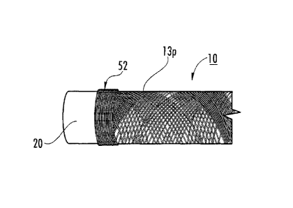
100941 Referring now to the figures, Figure 1A, an exemplary elongate
construct 10 is shown on a support member 20. As shown in Figure 1B, the
construct
includes an inner biocompatible thermoplastic material (e.g., polyacrylate)
coating
layer 11, an intermediate layer of at least one wound collagen fiber 13, and
an outer
biocompatible thermoplastic material (e.g., polyacrylate) coating layer 15.
The
thermoplastic material (e.g., polyacrylate) can embed and/or encapsulate
(seal) the
fiber(s) 13. In other embodiments, the construct 10 can be formed without one
of the
inner 11 and/or outer layer 15 and/or may optionally include other materials
or
constituents and/or layers. As shown in Figure 1B, the construct 10 can have a
wall
lOw with a suitable thickness defined by the at least one collagen fiber 13
and the
layers 11, 15 (where used) and/or other coatings or materials placed thereon.
The
construct 10 can have an open through cavity or may be filled or partially
filled with a
blood-thinning media and/or anticoagulant agent or other therapeutic material
(e.g., an
anti-inflammatory, antibiotic and/or the like).
100951 As also shown in Figure 1A, the at least one collagen fiber 13
has
an angular fiber pattern 13p (or fiber mesh) of repeating intersecting
collagen fiber
segments along its length. The angular pattern 13p can be defined by a number
of
revolutions of the at least one fiber 13 about the support member 20 at a
given pitch
or pitches for at least one layer (typically more than one layer). The at
least one
collagen fiber 13 is wrapped or wound about the support member 20 exterior
surface
to form a desired shape. The support member 20 can be any suitable shape
(shown as
cylindrical in Figure 1A) and may vary in shape and/or size over its length
(not
16
CA 02740008 2011-04-08
WO 2010/042205
PCT/US2009/005540
shown). As shown in Figure 1A, the at least one fiber 13 may be wrapped a
plurality
of times about one physical space to form a reinforced location 52, shown as a
reinforced end portion (and the reinforced portions can also be at any
intermediate or
internal locations). A clinician can secure a suture or other anchoring member
to the
reinforced end portion for attachment to local tissue. However, other
attachment
members and/or types may be used including, for example, biocompatible
adhesives,
staples, screws, nails, rivets, bone anchors and the like and combinations
thereof.
[0096] The polymeric material (e.g., polyacrylate emulsion) can be
applied
to the collagen fiber(s) 13 during fabrication (e.g., a winding, weaving or
braiding
operation). The polymeric material can be applied to the rod before the fiber
winding
step. The polymeric material can be applied in a fluid state. The combination
of the
polymeric material with the collagen fiber(s) 13 yields a composite
biomaterial with
controlled elasticity suitable for elastic vessel replacements or other
elastic repairs,
while the collagen fiber(s) can provide rigidity and/or strength suitable for
pressure-
loading applications.
[0097] Hydration of the composite biomaterial can generate a higher
degree of elasticity, typically without loss of structural integrity or
strength. The dry
biocomposite product is able to absorb a relatively large amount of liquid,
e.g., about
its body weight in water or exudates for wound bed applications.
[0098] Figure 2A illustrates an exemplary multi-fiber device 10c (as
shown, seven fibers) in a cylindrical shape. Figure 2B illustrates an
exemplary
single-fiber device 10c also in a cylindrical shape. As shown, both include
the
reinforced end portions 52. The cylindrical configurations may be particularly
suitable for artificial vessels and vascular tissue (see Figure 8).
[0099] The construct 10 can have reversible elasticity with sufficient
rigidity or strength to prevent collapsing under pressure while allowing
flexibility
sufficient to allow the construct 10 to expand and contract with changes in
blood
pressure. The vascular graft can be tailored to a wide range of inner
diameters to suit
multiple vascular replacements. The tubular construct 10 can be hydrated prior
to
surgical application as the dry construct is able to absorb a relatively
substantial
amount of water (typically about its body weight) in an aqueous (blood)
environment.
The dried tube can be used "as-is" (used in a non-cross-linked state and
hydrated
when in the body or prior to placement in the body). In other embodiments, the
collagen fiber(s) can be cross-linked with any agent or action that cross-
links the
17
CA 02740008 2016-05-11
collagen, typically prior to the fabrication (e.g., winding step or before the
liquid
polymer is added to the fiber(s)). The collagen fiber(s) may be cross-linked
with nor-
dihydroguaiaretic acid (NDGA), see, e.g., U.S. Patent No. 6,565,960, and U.S.
Patent
Application Publication No. US-2008-0161917-A 1.
[0100] Constructs of this and other embodiments can be used for other
repairs or treatments as will be discussed further below. The construct 10 is
non-
cytotoxic and may be biocompatible and, in particular embodiments can be
configured to provide a desired half-life or other suitable life for its
intended function.
101011 The construct 10 and/or the fiber 13 can optionally be cross-
linked
with a suitable polymerizing material, such as, but not limited to, NDGA, or
the
collagen fiber(s) may be used in the construct in a non-cross-linked state.
The NDGA
cross-linking of the collagen fiber(s) increases the strength of the device
10. In some
embodiments, the collagen fiber 13 is not cross-linked during the winding
process.
[0102] In some embodiments, the collagen fiber(s) can be cross-linked
with NDGA before the winding step. In particular embodiments, the winding can
be
carried out using both (a) one or more uncrosslinked collagen fibers and (b)
one or
more cross-linked collagen fibers, such as one or more NDGA cross-linked
collagen
fibers.
[0103] As shown in Figures 3A-3C, the construct 10 can be made by
winding at least one collagen fiber 13 about a support member 20 using a
computer-
guided and/or controlled lathe system 100. The support member 20 can be
tubular,
e.g., cylindrical, as shown in Figures 3A, 38 or may be substantially flat and
rectangular as shown in Figure 3C. Other geometries may also be used, such as,
for
example, a frustoconical or funnel shape. Typically, the support member 20 is
elongate and has a substantially circular, oval, polygonal or other cross-
sectional
shape.
[0104] The at least one collagen fiber 13 can be provided with one or
more
polymeric (e.g., thermoplastic) layers 15 before, during and/or after winding
the at
least one collagen fiber 13 to seal the fiber(s) 13 within the biocomposite
material
and/or to form a smooth inner and/or outer surface of the construct 10. An
example
of a small lathe, typically a micro or miniature lathe, suitable for
fabricating
embodiments of the constructs, is the Model 4410 lathe available from Sherline
Products, Inc., having a place of business in Vista, CA. The system 100 can
include
18
CA 02740008 2011-04-08
WO 2010/042205
PCT/US2009/005540
two user-selectable inputs to operate the lathe system: one controls the speed
at which
the support member spins and the other controls the pattern (fiber angle) in
which the
at least one fiber 13 is laid and/or fed onto the support member 20. The
winding
operation can be configured so that the fiber(s) 13 is self-pulling from a
spool of
collagen fiber(s) based on the speed of the spinning support member 20. The
feeder
head can have a channel that holds the fiber(s) and directs the fiber(s) to
wrap/wind
about the support member 20. The lathe can co-wind a plurality of fibers or
fiber
bundles substantially concurrently about the support member 20. In some
embodiments, a plurality of spools of collagen fibers can supply fibers that
can be
applied concurrently to the support member 20 as a single bundle of fibers or
as
separately wound fibers or fiber bundles.
101051 The winding can be performed so that at least one layer of the at
least one collagen fiber 13 has a substantially constant pitch for at least a
major
portion of a length thereof or so that at least one layer of the at least one
collagen fiber
13 has a variable pitch for at least a major portion of a length thereof.
10106] The support member 20 can include a lubricious and/or smooth
surface. The support member 20 can include an embossed surface that provides a
smaller contact surface area. The support member 20 can comprise or be formed
of a
polymer material. In other embodiments, the support member 20 can include an
anti-
slip surface with ridges or a sleeve can be placed over the support member
(not
shown) to contact the next layer (e.g., inner film 11 or fiber 13). In some
embodiments, the support member 20 comprises Teflon or other suitable low
friction and/or anti-stick material and the polymeric coating can adhere the
fiber (e.g.,
be a "sticky" substance) to the support member 20 during the winding operation
to
inhibit movement on the member 20 once applied.
101071 The support member 20 can be configured to facilitate removal of
the construct 10. For example, the construct 10 may be wound snugly and/or
tightly
against the outer surface of the support member 20 and allowed to dry. The
support
member 20 can be configured to reduce in cross-sectional size or disassemble
with the
construct 10 held thereon to allow easy removal of the elongate construct. In
some
embodiments, the support member 20 can be a multi-piece device that provides
this
size change. In other embodiments, the support member 20 may be cooled while
the
construct is heated to provide a size difference. In particular embodiments,
the
support member 20 can cooperate with an insert 201 (Figures 3A, 3B) that
provides
19
CA 02740008 2011-04-08
WO 2010/042205
PCT/US2009/005540
the desired size adjustability. The removable insert 201 can be placed in the
support
member 20 (e.g., Teflon rod) so that, when removed, a gap is formed between
the
rod and the construct to facilitate easy sliding removal of the construct 10
from the
support member 20. In other embodiments, the construct 10 can be removed from
the
support member without such a size adjustment, e.g., its inner surface may be
sufficiently lubricous or a suitable liquid or other material can be used to
slide the
construct off the support member. In some embodiments, the construct 10 can be
cut
in a lengthwise or longitudinal (e.g., "X") direction and taken off the
support member
20.
[0108] Figure 4 illustrates that different fiber 13 configurations may
be
used for the winding operation/method or to form the construct 10. Examples of
fiber
configurations include a single fiber 131, a plurality of fibers 131_ 13n
(typically n=2
to 100) that can be concurrently co-wound about the support member 20, a fiber
bundle 13b, a series of discrete shorter fibers joined to form a desired
length for
winding 13j, and a twisted, woven or braided fiber bundle 13t. For the fiber
bundles
13b, 13t, two or more fibers 13 can be grouped together to form the fiber
bundle 13b,
13t and that bundle 13b, 13t applied or wrapped about the support member 20,
similar
to a single fiber. One or more fiber bundles 13b, 13t may be used to form the
construct 10. Combinations of the different fiber types may also be used for
some
constructs 10. That is, for example, a twisted fiber 13t can be co-wound with
a single
fiber 131 and/or a single fiber 131 may be used to form one layer and a
twisted 13t to
form a different layer, and the like.
[0109] The collagen fiber(s) 13 can be wound using various fiber angles
(e.g., pitch angles), such as angles between about 1-90 degrees, typically
between
about 5-60 degrees, such as, for example, 5, 10, 15, 20, 25, 30, 35, 40, 45,
50, 54 and
55 degrees, or other odd or even numbers between 5-70. Where constructs of
multiple layers are used, one layer may have a first pitch and another layer
may have
a different pitch. The patches may be formed with winding angles of between
about
5-30 degrees while the tubular constructs may have winding angles of between
about
1-90 degrees, typically between about 5-90 degrees.
[0110] Figure 5A illustrates that a construct 10 can be wound with
increased fiber density 52 along certain segments, typically forming end rings
52r.
However, the increased fiber density 52 can also reside at other locations
along the
construct 10. This increased fiber density 52 can provide sufficient rigidity
to allow a
CA 02740008 2016-05-11
suture tc attach thereto. As shown in Figure 5A, the construct 10 is tubular
10t and
may optionally include an increased density segment 52 at an intermediate
location.
Figure 5B illustrates that the construct 10 can be used as formed, or may be
cut or
separated along a Y-axis into two components lOta, 10tb. For the latter, the
intermediate increased density ring 52 can form end rings for the separated
construct
lOta, 10tb.
101111 Figure 6A illustrates a construct 10 that has a wound fiber(s) 13
and is relatively flat 10f and/or rectangular. Again, the construct 101 can
optionally
include increased fiber density segments 52 that may be suitable for end rings
52r.
Figure 6B illustrates that the construct 10f can be cut along the X-axis and
separated
into at least two components that form biocompatible patches. The intermediate
increased density ring(s) 52, where used, can optionally form end rings 52 for
the
separated construct 10fa, 10113,10fc, 10fd, and the like.
101121 Figure 7 illustrates an example of another automated winding
system 100' that can be used to form the construct 10. This embodiment uses
several
fibers 13, each independently wound and/or wrapped to weave or braid the
fibers
about the support member 20 to form the construct 10. The system 100' includes
a
plate 122 supporting spindles 124, a forming plate 126, a support member
(shown as a
cylindrical mandrel) 20 that extends through an aperture in the forming plate
126, and
braid puller 128. An exemplary microbraider is believed to be available from
Kokubun Ltd of Japan. See also, Figure 2 and col. 2 of U.S. Patent No.
7,135,040.
[01131 The at least one fiber 13 can be wound after cross-linking and
the
fiber(s) may not be cross-linked at all. The fiber(s) 13 can, where desired,
be
polymerized with any suitable cross-linking materials, to promote collagen
organization, such as, for example, NDGA, but other cross-linking materials
may be
used, including, for example, glutaraldehyde. The collagen fiber can also be
treated
with other methods to improve the tensile properties of the fiber. The
(typically dry)
collagen fibers 13 can be cross-linked with agents such as glutaraldehyde,
formaldehyde, epoxy resins, tannic acid, or any other chemical agent that
produces
covalent cross-links between collagen molecules within fibrils or between
fibrils.
Alternatively, the at least one fiber 13 can be treated to induce cross-
linking between
collagen molecules such as, but not limited to, one or more of a carbodiimide
treatment, ultraviolet irradiation either with or without carbohydrates to
initiate
21
CA 02740008 2011-04-08
WO 2010/042205
PCT/US2009/005540
glycation adducts, and dehydrothermal treatment coupled with any of the
aforementioned methods and/or agents.
101141 Figure 8 illustrates that the cylindrical construct 10 may be
particularly suitable as a vessel 10v for vascular prosthesis such as for
aortic stents
and/or vessels for repairing or replacing veins or arteries. In some
embodiments, the
vessels 10v may be used for blood vessels such as coronary or other lumen
vessels.
The vessels 10v can have diameters between about 1 mm to about 12 mm. In some
embodiments, the blood vessels can be for repair, replacement or use of small
lumen
vascular vessels, typically about 6 mm or less in diameter. The construct 10
is tubular
with an open cavity and has a flexible elastic configuration to be able to
expand and
contract responsive to blood flow and pressure and may be able to mimic a
natural
behavior of normal "healthy" blood vessels in an environment of pulsatile
flow.
101151 The vessel 10v can be formed using at least one fiber 13. In
some
embodiments, the vessel 10v can be formed of a single fiber wound in multiple
overlying layers. The fiber 13 can be a continuous length of a single fiber or
the fiber
13 can have a length provided by a series of shorter fibers attached in an end-
to-end
orientation.
101161 As noted above, the collagen fiber length used for forming the
vessel 10v can be any suitable length, typically between about 1 cm to about
100 m,
and more typically between about 1 m to about 100 m. In some particular
embodiments, the vessel 10v can be formed with a fiber length that is between
about 5
m-20 m, such as between about 8-12 m. Each vessel type may use a different
length
of fiber. The vessel 10v can be formed using a single fiber 13 of a continuous
length
that is wrapped in several layers about the support member 20. Use of a single
fiber
13 can reduce the likelihood of any fraying associated with multiple fibers
(such as
those wound in one lengthwise direction). The vessel 10v can have a length
that is
between about 2 cm to about 8 cm (or more). The vessel 10v can have an inner
diameter that is between about 1-12 mm with the wall thickness (on average or
measured at a thickest part) being about 0.1 mm to about 2 mm. The vessel 10v
may
optionally have a slit lOs in a portion of a lengthwise direction to allow for
ease in
placement. One or both end portions of the vessel 10v may have an increased
density
of wound collagen fiber 52. An intermediate portion may also optionally
include an
increased density region 52. The vessel 10v can be formed by cutting or
otherwise
separating a longer tubular construct into a desired vessel length without
fraying.
22
CA 02740008 2011-04-08
WO 2010/042205
PCT/US2009/005540
[0117] Figure 9 illustrates a medical kit 250 that includes a construct
that
is medical device or implant 10 or 10'. The kit 250 may optionally include
other
components, such as, for example, a container of surgical adhesive, sutures
210,
suture anchors, and the like. The device or implant 10, 10' may be held
hydrated in a
flexible sealed package of sterile liquid 230. The kit 250 may include a
temperature
warning so that the construct 10, 10' is not exposed to unduly hot
temperatures that
may degrade the implant. A temperature sensor 252 may optionally be included
on
the package of the kit to alert the clinician as to any excessive or undue
temperature
exposure prior to implantation. For example, it may be desirable to hold or
store the
kit 250 (and implant or device 10, 10') at a temperature that is less than
about 37 C
and/or 100 F prior to implantation. The implant 10, 10' can be stored dry and
hydrated prior to use or may be packaged in a hydrated state. The kit 250 may
be
packaged in a housing with a temperature controlled or insulated chamber 250c
to
facilitate an appropriate temperature range.
101181 Figure 10 is a flow chart of operations that can be used to carry
out
embodiments of the present invention. In some embodiments, the at least one
collagen fiber is wound a number of revolutions about a length of a support
member
having a long axis. The winding can have a defined pitch and/or fiber angle
relative
to the long axis of the support member to form an elongate construct with at
least one
wound collagen fiber (block 150). The winding step can form multiple overlying
layers of the at least one collagen fiber in one or more fiber angles so that
the at least
one fiber intersects itself at different locations along a length of the
construct.
101191 A polymeric material can be applied to and/or placed on the at
least
one collagen fiber held on the support member during the winding step (block
155).
The polymeric material can be applied in a fluid (typically a liquid) state.
In some
embodiments, as the fiber(s) is wound about the support member, the (liquid)
polymeric material, e.g., acrylate emulsion, can be substantially continuously
applied
(e.g., wrapped, painted, sprayed, dripped, poured, brushed and the like) onto
the
fiber(s) so that the fiber(s) is wetted while one or more layers are wound on
the lathe.
Where used, the acrylate emulsion can act as a "sticky" substance that adheres
the
collagen fiber in position on the support member during the winding process.
The
polymeric material can optionally comprise a polyacrylate emulsion (block
158).
101201 The at least one collagen fiber can be supplied to the
winder/support member in a substantially dry state and may be provided as a
spooled
23
CA 02740008 2016-05-11
(dry) quantity of the at least one collagen fiber (block 152). The fiber(s)
can be
supplied and wound in a non-cross-linked state.
[0121] In some embodiments, the winding step can be carried out to
create
multiple adjacent overlying layers of the at least one fiber, the adjacent
layers being
coextensive for at least a major portion of a length of the construct (block
153). That
is, for example, the winding can wind back and forth over the support member
to
create overlapping layers of the at least one fiber and each layer can have
substantially
the same length (such that the layers are substantially coextensive with each
other) or
one or more of the different layers may have different lengths.
[0122] Optionally, additional layers of the same or a different (liquid)
polymeric material can be onto the at least one wound collagen fiber to cover
at least
the outer surface in a film that extends over the interstitial spaces of the
fiber(s) and
can provide a coating. The additional (e.g., aerylate) material can be applied
by spin
coating the wound construct as it remains on the support member 20 (typically
between two to three times), then incubating the construct at a desired
temperature,
such as between about 37-40 C, typically about 37 C, for a defined time,
typically
between 2-24 hours, such as about 4 hours (block 168). The spin-coating and
incubating steps can be repeated one or more times. After the final spin-
coating or
last outer layer is applied, a longer incubation time may be used, e.g., the
earlier
incubation times can be between 2-8 hours and the "final" incubation can be
longer,
e.g., between 8-24 hours, typically overnight (where multi-shift production is
not
used). Other post-winding coating methods may be used. The spin-coating and
winding steps and the outer layer formation step (where used if different from
the
spin-coating) can be carried out using the same material and/or emulsion
formulation
or different polymeric materials. In some embodiments, the applying and spin-
coating steps both use a polyacrylate emulsion.
[0123] Additionally or alternatively, an external heat source (e.g., a
heat
lamp) can be used to shorten the time used for drying the initial and/or
supplemental
polymeric coating(s) and/or thicker emulsions can be provided during the
winding
fabrication process to reduce the number of coats applied after the winding
process.
24
CA 02740008 2016-05-11
[0124] Optionally, liquid polymer can be placed onto the support member 20
before
and/or concurrently with the winding step (block 165). Optionally, the
collagen fiber can be
polymerized before the fiber is wound or the collagen fiber can be provided
and wound in an un-
crosslinked state (block 166). In addition, the collagen fiber(s) can be
coated with the same or a
different liquid polymeric material before the winding step and/or as a final
outer
24a
CA 02740008 2011-04-08
WO 2010/042205
PCT/US2009/005540
layer or emulsion or as an initial inner layer that can contain active
biomolecules (e.g.,
heparin, growth factors, etc... (block 167).
101251 As shown in Figure 11, the winding can be carried out so that the
at least one fiber turns about the support member in one of a clockwise or
counterclockwise direction along a first lengthwise direction for a first
layer, then
reverses to an opposing lengthwise direction and continues to turn about the
support
member in the same clockwise or counterclockwise direction for a second
adjacent
layer (block 180).
101261 In some embodiments, the winding step has a first pitch for the
winding of the at least one collagen fiber on the first layer and a second
smaller or
greater pitch for the winding of the at least one collagen fiber on the second
layer. In
some embodiments, the at least one fiber on the second layer resides between
gaps
defined by the at least one fiber wound with the defined pitch on the first
layer.
101271 The method can include cutting the construct in an axial
direction
to form a substantially flat collagen fiber patch. The method can include
winding the
collagen fibers in a plurality of axially spaced apart segments with increased
collagen
fiber density, at least some of which are provided as reinforced segments for
suturing.
The reinforced segments can be formed at end portions of the tube and
optionally at
one or more intermediate locations therebetween. The methods can produce an
artificial vessel with the ability to expand and contract in response to blood
flow
therethrough.
101281 Embodiments of the invention can be used for a number of
different medical applications, including, but not limited to, wound bed
patches,
muscle or organ patches, cardiac patches, valve replacements or repairs,
hernia
patches, skin patches, burn treatment patches, skin/tissue repair patches or
cuffs,
blood vessel (artery, vein, and the like) repairs, sleeves that can reside
about repairing
tendon to prevent or inhibit adhesions, indwelling tubes for delivery of
therapeutic
agents, ducts such as lymphatic, hepatic, pancreatic and cystic ducts, tubes
such as
ureter and urethra tubes and nerve guides.
(0129] The present invention is explained in greater detail in the
following
non-limiting Examples.
CA 02740008 2011-04-08
WO 2010/042205
PCT/US2009/005540
EXAMPLES
[0130] Thus far, over 40 different devices have been manufactured using
this new technique that combines collagen fibers with various water-based
polyacrylate emulsions synthesized through a microemulsion polymerization. The
combination of the NDGA-collagen fibers with the polyacrylate emulsion lends
the
novel properties observed for these devices for numerous biomedical
applications,
including, but not limited to, artificial elastic vessel and valve
replacements. The
polyacrylate emulsion has the ability to be synthesized in a one-step process
and can
optionally include covalently bound or encapsulated antibiotics and/or
biologically
active agents or compounds that may be incorporated into the devices during
post-
winding fabrication coating.
10131] The collagen fiber was derived from dermal collagen that is acid
or
pepsin soluble. The soluble collagen can be made by neutralizing acid soluble
collagen and keeping the soluble collagen at a desired low temperature to
maintain the
collagen in molecular form, (e.g., about 4 C). Collagen concentration of the
soluble
collagen can be from about 0.1-4% weight per volume. The gel cylinder can be
used
in the gel form or allowed to dry, actively or passively (suspended in air),
to form a
collagen fiber having a diameter between about 0.05 mm (average) to about 0.2
mm
(average).
101321 As discussed above, the devices can be manufactured using an
automated or semi-automated mechanical lathe. Some prototypes were made by
winding a single 30 m-50 m long fiber onto a Teflon rod with substantially
continuous application of the emulsion. Continuous addition of the emulsion
provides
a sticky adhesive for the fibers to adhere to the Teflon rod and remain where
set on
the rod and also can provide a water proof coating for the fiber that inhibits
or
prevents swelling and subsequent deformation of the device upon hydration.
[0133] The pitch of the fiber relative to the long axis of the tube can
be
specified. The thickness of the collagen winding can be adjusted, for example,
corresponding to the number of layers of fibers that are laid on (and/or the
number of
fibers bundled together for the winding). During the fiber winding process,
liquid
acrylate is applied (e.g., painted) onto the surface of the laid-on fibers.
10134] After the fiber has been wound for a sufficient time, such as
between about 20-60 minutes, depending on the size and thickness desired, the
mesh
26
CA 02740008 2011-04-08
WO 2010/042205
PCT/US2009/005540
fabric is spin-coated 2-3 times with the emulsion, incubated at 37 C for 4
hours, then
spin-coated an additional 2-3 times and incubated at 37 C overnight.
101351 Figures 1A, 2A and 2B illustrate polyacrylate-fiber devices. The
clear glassy look of the devices results from the polyacrylate coating, which
whitens
with hydration. The ability to utilize multiple fibers in the manufacturing
process
allows for precise engineering of the device to fit the specific needs of each
target
biomedical application. Coating of the device with emulsion embeds the fibers
within
the solid polyacrylate film and the film extends over the free space of the
fiber mesh
and gives the device reversible elasticity as well as prevents fluid leakage
through the
device (the device can absorb water).
[0136] Mechanical analysis has verified reversible elasticity of the
devices,
which increases with hydration. When under strain, the fibers within the
device align
and the device can stretch to an extent, yet when the strain is removed, the
device
returns to its original state without damage to the device establishing memory
for the
devices. A multi-fiber device (e.g., a 7-fiber yarn) results in a much
stronger and
thicker device (Figure 2A) than the single fiber device shown in Figure 1A
without
loss of elasticity as proved by mechanical analysis. Also, mechanical analysis
has
established that a multi-fiber cable is much stronger than a single fiber in
terms of
tensile strength, therefore, the resulting device will be more durable (e.g.,
have a
much larger tensile strength and require a higher maximum force for failure).
The
multi-fiber devices appear to be most suited for deep tissue hernia patches
and large
vascular replacements such as aortic replacements because there is less area
for
leakage and the increased strength can extend the lifetime of the device once
implanted. A prototype single fiber device was made having an 80 mm long
device
while a prototype multi-fiber device had a length of 20 mm. The prototypes
were
made using the lathe system shown in Figures 3A and 3B. The process includes
application of fiber(s) to the Teflonerod (support member 20) with continuous
emulsion application, coating of device with emulsion, and drying of coating.
The
time between passes of lathe was altered between the two devices to yield an
80 mm
long single fiber device (Figure 2B) and a 20mm long multi-fiber device (
e.g., 7
fiber device) (Figure 2A). In addition, the time spent at the end portions of
the device
(and other locations as appropriate) can control the thickness of the end
rings to keep
them consistent between devices or device types to comply with quality and
regulatory standards.
27
CA 02740008 2011-04-08
WO 2010/042205
PCT/US2009/005540
[0137] The dimensions of the prototype devices have been varied by
altering the time required during each pass of the lathe and the diameter of
the
Teflon rod used for making the device. The computer program used for
operating
the lathe permits altering of the time needed per pass, which allows the
system to
produce relatively short (20-30 mm) fiber devices or relatively long (80 mm)
fiber
devices. The total time for manufacturing can also be varied so that the
thickness of
the devices can be controlled. Typically, the multi-fiber devices are wound
for a
shorter period of time than the single fiber devices. Devices ranging from
about 1
mm to about 12 mm inner diameter have been made.
101381 The dimensions of the device can be adjusted to fit the needs of
the
medical field. A small diameter Teflon rod can be used for manufacturing
devices
for use in vein and artery replacements, while larger Teflon rods can be used
to
manufacture devices for aortic or large artery replacements and various
shunts. The
thickness of the device can be altered by extending the manufacturing time or
by
utilizing multiple fiber strands processed into a single cable or "yarn" for
winding.
This can allow both fiber and polyacrylate film to prevent leakage of any
fluids from
within or into the device in vivo.
[0139] Figure 12A is a photograph of a prototype device made from
NDGA-collagen fibers and Figure 128 is a photograph of a device with collagen
fibers not cross-linked.
101401 During production using non-NDGA crosslinked collagen fibers
(Figure 128), the collagen fiber would swell into a gel and become weak. This
caused some of the fiber to gel with the emulsion during application, forming
a novel
biocomposite material that forms a solid material easily during production
without the
need for excessive coating of the device with emulsion. These devices may be
particularly suited for application to superficial and acute epidermal wounds
and
disturbances where high strength is not required.
[0141] Figure 13 shows that the non-crosslinIced and post-crosslinked
devices were much weaker than the ones formed from NDGA cross-linked collagen
fibers in terms of maximum force required for failure, yet the tensile
strength
(normalized to the cross sectional area of the devices) was relatively
equivalent. The
non-crosslinked device yielded a softer material that felt more similar to the
currently
available wound care products than the NDGA-crosslinlced devices; however, the
strength of the device greatly decreases when the collagen is not crosslinlced
prior to
28
CA 02740008 2011-04-08
WO 2010/042205
PCT/US2009/005540
application (winding or introduction onto the support member). Both cross-
linked
and non-crosslinked patches displayed memory upon hydration and mechanical
analysis.
Mechanical Analysis of Manufactured Prototypes
10142] The combination of NDGA-collagen fibers with a polyacrylate
emulsion to form an elastic mesh device can be very beneficial to many
biomedical
applications, including artificial elastic tissue replacements. Mechanical
analysis has
verified that these materials possess reversible elasticity that increases
with hydration.
These devices can be considered "smart materials" since they can be
manipulated to
just below failure and, using memory, realign to their original confirmation
without
damage to the material. When under uniaxial strain, the polymer substrate
allows the
fibers to align within the device, at which point the device will not stretch
any further.
When the strain is removed, the device returns:to its original relaxed state
and the
stress/relaxation cycle can be repeated, similar to what is observed for the
majority of
mammalian elastic tissues. In order to determine tensile strength, the devices
were
taken past this alignment stage with continuous force application to cause
failure (see
below). The prototype devices were mounted in 1000lb load cell clamps and
mechanical analysis was performed under uniaxial load after hydration for a
minimum of 30 minutes in diH20 (deionized water).
101431 Figure 14A illustrates a device (prototype 5) that was cut in a
lengthwise direction and hydrated. Figure 14B illustrates a relaxed fiber
states and
Figure 14C illustrates the fibers align to the relaxed state. The devices were
cut
laterally along the device line then folded in half prior to placement in the
load cell
clamps as shown in Figures 14A, 14B. The cutting of the prototype device was
to
allow for removal of the device from the support member, before the Teflon
insert
had been engineered that allows the devices to be removed from the rod without
cutting. The prototypes no longer require cutting to remove and the devices
can be
analyzed in their intact cylindrical form, and this "intact" data is shown for
the multi-
fiber devices below (Figures 16, 17A, 17B). For prototypes/devices 1-6,
different
polyacrylate emulsion solutions were employed during the winding operation.
Each
emulsion used creates a polymer film with different physical and mechanical
properties as shown in Figure 15.
29
CA 02740008 2011-04-08
WO 2010/042205
PCT/US2009/005540
101441 Figure 15 illustrates the results of a mechanical analysis of 30
mm
single fiber devices manufactured using different polyacrylate emulsions. As
shown
in Figure 15, the strongest device formed (Device 4) used an emulsion
synthesized
from ethyl acrylate and methyl methacrylate in a 4:1 ratio respectively. This
emulsion
produces the strongest polymer films of all the emulsions synthesized and
analyzed by
the investigators. Device 1 and 2 both contained the same polyacrylate
emulsion of
butyl acrylate and styrene (7:3 ratio), but for device 2 the emulsion was
mixed in a 1:1
ratio of emulsion to soluble collagen suspension (3%) prior to device
production. By
mixing the emulsion with soluble collagen, the amount of solid polymer added
to the
device was reduced, which appears to have caused the overall tensile strength
and
maximum force of the device to decrease. Also, the elasticity of this device
was
greatly diminished from other devices analyzed due to the decrease in polymer
present in the device. Devices 5 and 6 were formulated using an emulsion that
yields
very weak films but possess the highest degree of elasticity of all the films
(8:2 butyl
acrylate to styrene). These devices were therefore not as stiff as the other
devices, but
possessed high maximum strain and deformation values and were highly elastic.
[0145] Mechanical analysis of the multi-fiber devices proved to be
difficult due to the exponential increase in strength for these devices,
especially when
the devices were analyzed without lateral sectioning of the device prior to
testing.
During uniaxial mechanical analysis, devices would slip out of the clamps
yielding a
false maximum force as the devices never reached failure. This allowed re-
testing of
the devices multiple times since little damage was done to the devices during
testing
and the testing produced an average tensile strength and maximum force for the
devices. An average of three tests were performed per device in an attempt to
achieve
an accurate tensile strength. Four devices were manufactured: Device 7, 12,
13, and
14. Devices 7 and 14 were manufactured using a 7:3 ratio emulsion of butyl
acrylate
to styrene. Devices 12 and 13 were both manufactured using a 4:1 ratio
emulsion of
ethyl acrylate to methyl methacrylate. Device 7 was manufactured using the
standard
device program with 20 minutes total production time where the lathe spent 5
seconds
between passes at the ends of the device. However, for devices 12-14, the time
spent
between passes where the end rings are wound was minimized after 10 minutes of
production in order to reduce the thickness of the end rings for these
devices. The
time spent at the ends of the devices were 5 seconds initially and was reduced
to 2
seconds for the remaining 10 minutes of production for devices 12-14. This
reduction
CA 02740008 2011-04-08
WO 2010/042205
PCT/US2009/005540
in time yielded a more even-distribution of fiber within the devices and
reduced the
bulkiness of the devices at the ends.
101461 As shown in Figure 16, the maximum force obtained for each
device was significantly greater than was observed for the single fiber
devices (shown
in Figure 15), yet the tensile strength was relatively equal. This is most
likely due to
the increased thickness of the multi-fiber devices (e.g., 7 fibers) factoring
into the
overall tensile strength calculated from the cross-sectional area of the
device.
However, the area of the devices drastically decreases during the tensile test
due to
the fiber alignment within the device, therefore the calculated tensile
strength for
device presented here are relative only to the initial cross sectional area
and the true
tensile strengths are likely significantly greater than what is presented
here.
[0147] Tensile strength was also determined for the devices by removing
them from the Teflon rod support member without lateral cutting, leaving the
device
in an intact cylinder and putting uniaxial tension on the device from the
inside. It is
believed that this test provides the most accurate data achievable under
uniaxial
mechanical analysis since the device is not able to slip from the clamps. It
is
currently contemplated that the tensile tests for analyzing the prototype
devices will
utilize this method of testing. Burst test data for cylindrical devices maxed
out at
about 75 psi (to burst using pressurized air from the inside) for fully coated
devices.
[0148] Tensile data for devices formed with various natural (collagen)
and
synthetic (polyacrylate) materials are shown in Figures 17A and 17B. Devices
15
and 16 were single fiber devices manufactured using butyl acrylate and methyl
methacrylate. Device 14 was a 7-fiber device manufactured using a butyl
acrylate and
styrene emulsion.
[0149] Figure 17A compares the mechanical properties of the single and
multi-fiber devices. Analysis demonstrated a maximum force of approximately
1770N for the seven fiber device (Device 14) as opposed to approximately 500N
maximum force for the single fiber devices (Devices 15, 16). This data
provided here
is believed to be the most accurate and depicts a higher load capacity than
was
observed for these same patches in the basic uniaxial tensile tests that
utilize a
clamping method (Figures 15, 16). The seven fiber device (Device 14) was over
three times stronger than the single fiber ones (Devices 15, 16), which was an
expected result for these highly reinforced devices. The maximum displacement
for
the seven fiber device (Device 14) was also greater than was observed for the
single
31
CA 02740008 2011-04-08
WO 2010/042205
PCT/US2009/005540
fiber ones (Devices 15, 16). The data shows that the elasticity appears to
have
increased for these devices, indicating retention of elasticity in the multi-
fiber devices
which appears to have increased concurrently with the increase in strength.
[01501 The data presented in Figure 17B shows that gelatin-fiber
devices
and the polyacrylate devices were very similar in terms of mechanical
behavior,
where both devices followed the same force/displacement trend. However, during
initial testing, the gelatin devices did not reach as high of a maximum force
or
displacement as the polyacrylate devices achieved. Incorporation of the
polyacrylate
film allowed the device to stretch until the mechanical strength of the fibers
within the
device was tested. The polyacrylate film and collagen fibers, which may
include
NDGA treated collagen fibers, can act to enhance the mechanical properties of
the
material.
101511 In addition to the tubular device structures discussed above,
thus
far, over 20 different patches have been manufactured using this new technique
that
combines collagen fibers with various water-based polyacrylate emulsions
synthesized through microemulsion polymerization. Again, these emulsions have
the
ability to be synthesized in a one-step process and can include covalently
bound
antibiotics that may be incorporated into the patches for additional utility.
The
patches were manufactured using a mechanical Sherline lathe.
[01521 Some prototypes were made by winding a single 30m-70m long
fiber onto a Teflon sheet (Figure 3C) with substantially continuous
application of a
polyacrylate emulsion. The application was by hand or manual but automated
devices
to apply the emulsions may be used. Substantially continuous addition of the
emulsion provides a sticky adhesive for the fibers to adhere to the Teflon
sheet and
remain where set on the sheet and also provides a water proof coating for the
fiber
that inhibits or prevents swelling and subsequent deformation of the device
upon
hydration. After the fiber has been wound for 20-60 minutes, depending on the
size
and thickness desired, the mesh fabric is spin coated 2-3 times with the
emulsion,
incubated at 37 C for 4 hours, then coated an additional 2-3 times and
incubated at
37 C overnight. This procedure produces a solid polyacrylate film in the
interstitial
space between fibers and also a polymer film coating the surface of the fiber
mesh
which gives the patch the reversible elasticity desirable for application to
wound beds
and other elastic tissues.
32
CA 02740008 2011-04-08
WO 2010/042205
PCT/US2009/005540
[0153] Figures 19A-19C show examples of single fiber (80mm long)
patches with varying fiber angles. Figure 19A shows a patch fiber angle of 25
.
Figure 19B shows a patch with a fiber angle of 150 and Figure 19C shows a
patch
with a fiber angle of 5 . Figures 22A-22D also illustrate varying fiber angles
using
NDGA cross-linked collagen fiber-polyacrylate biomaterial to form
substantially flat
patches. Figures 23A-23D illustrates cylindrical biomaterials with NDGA cross-
linked collagen fiber-polyacrylate biomaterials (Figure 23C illustrates a 7
fiber
biomaterial and Figure 23D illustrates a single fiber material).
[0154] The prototypes were made using a Teflon sheet and lathe (see,
e.g., Figure 3C). The finished patch is then removed from the Teflon sheet,
typically by cutting the fiber mesh laterally from end to end, revealing a
solid
substantially rectangular fabric that can then be packaged and sold in this
form,
substantially "as-is" (Figures 20A, 20B). The reinforced ends of the patch are
approximately three times stronger than the interior of the patch, making this
area
ideal for suturing of the device in a wound or chronic ulcer bed or for
application as a
surgical mesh (Figures 19A-C) and also prevent unraveling or fraying of the
fibers in
the patch. The shape of the Teflon sheet can also be adjusted so that two
rectangular patches are fabricated from a single manufacturing process
(Figures 18A,
18B). This provides double the manufacturing capability without increasing the
amount of fiber needed. Or, for instances where a double layer patch is
required
(such as for deep chronic wound treatment), the patch can be removed from the
Teflon sheet without cutting and the single device can be sutured into the
wound
bed.
[0155] Figures 18A and 18B illustrate flat polyacrylate-collagen fiber
biomaterial patches. In the example shown, the patch is cut along the length
of the
Teflon and peeled off to reveal a single patch that can then be cut into two
or more
individual patches. When the (rectangular) patches are held by the reinforced
end
rings and pulled away from one another, the patches display properties similar
to that
of a fiber-free polyacrylate film, namely it exhibits memory after
deformation.
However, when the cut ends of the patch are held and pulled away from one
another
the patch acts as a true fiber-reinforced composite where the fibers provide
intense
strength and little elasticity is observed at low amounts of strain.
[0156] The dimensions of the patches can vary. A smaller area Teflon
sheet can be used for manufacturing smaller patches while larger Teflon
sheets can
33
CA 02740008 2011-04-08
WO 2010/042205
PCT/US2009/005540
be used to produce larger patches to cover larger surfaces, such as high total
body
surface areas. The thickness of the patch can be varied by extending the
manufacturing time or by utilizing multiple fiber strands processed into a
single cable
for application or by using other multi-fiber devices, including multi-fiber
cables and
braids that can then be manufactured into patches.
101571 A multi-fiber cable using strands that are loosely wound into a
cable then applied to the rod for a patch results in a much stronger and
thicker patch.
It is also contemplated that the multiple fibers can be wound concurrently but
separately. Also, mechanical analysis has established that a multi-fiber cable
(e.g., 7
fiber) is stronger (e.g., 2-3 times stronger) than a single fiber in terms of
tensile
strength, therefore, when a cable is used for patch production, the resulting
patch
should be more durable.
[0158] Under standard manufacturing conditions, the internal surface of
the patch typically has only polyacrylate-coated collagen fibers whereas the
external
surface of the patch is coated with extra layers of polyacrylate emulsion to
provide a
polymer-based barrier to inhibit or prevent bacterial translocation into the
wound bed.
However, this feature can easily be modified to fit the specific needs of the
wound
bed where either or both surfaces (external or internal) can be coated with
emulsion
post-production sealing the collagen fibers within the patch and preventing
fiber
interaction with the surrounding tissue/fluid. The polyacrylate portion of the
patches
appears to absorb aqueous media (Figures 20A, 20B), making this formulation
appropriate for application to wound beds with high exudates. By controlling
the
amount of polymer in the patch composition, the dressing or covering can be
manufactured to fit the needs of highly exudating wound beds by coating both
surfaces of the patch.
[0159] Figures 20A and 20B illustrate a single collagen fiber patch
coated
with polyacrylate emulsion. Upon absorption of aqueous media, the polymer
transforms from translucent (Figure 20A) to solid white (Figure 20B) in the
interstitial space between fiber and on the surface of the patch.
[0160] Patches using NDGA crosslinked collagen fibers has also been
explored as shown in Figure 21. During manufacturing, the un-crosslinked
collagen
fiber would swell into a gel and become weak, which caused some of the fiber
to gel
with the emulsion during application forming a novel biocomposite material.
After
the collagen fiber patch was formed, the fibers were attempted to be cross-
linked with
34
CA 02740008 2011-04-08
WO 2010/042205
PCT/US2009/005540
NDGA prior to final coating of the patch with emulsion. The post-production
crosslinking was unsuccessful since the polyacrylate film that coats the
fibers during
manufacturing prevented NDGA interaction with the collagen. The non-
crosslinked
and post-crosslinked patches were weaker than the ones formed from NDGA-
collagen
fibers in terms of maximum force required for failure, yet the tensile
strength
(normalized to the cross sectional area of the patches) was relatively
equivalent.
Although the opaqueness of the non-crosslinked patch may be of utility to
wound
dressings where visibility of the wound bed is desired, the strength of the
patch
greatly decreases when the collagen is not crosslinked prior to application.
Mechanical Analysis of Manufactured Prototypes
101611 The combination of NDGA-collagen fibers with a polyacrylate
emulsion to form an elastic mesh material can be very beneficial to many
biomedical
applications, including topical wound dressings. Mechanical analysis has
verified that
these fabrics possess reversible elasticity that increases with hydration.
These fabrics
can be considered "smart materials" since they can be manipulated to just
below
failure and using memory realign to their original confirmation without damage
to the
material. When under uniaxial strain, the polymer substrate allows the fibers
to align
within the patch, at which point the patch will not stretch any further. When
the strain
is removed, the device returns to its original relaxed state and the
stress/relaxation
cycle can be repeated. In order to determine tensile strength, the patches
were taken
past this alignment stage with continuous force application to cause failure
(see
below). Patches were mounted in 1000lb load cell clamps and mechanical
analysis
was performed under uniaxial load after hydration for a minimum of 30 minutes
in
diH20.
101621 A single 80mm long patch was cut into sections relative to the
fiber
alignment within the patch as shown for example, in Figure 24. Six sections
were cut
transverse (trans) to the fiber alignment, and three sections were cut
longitudinally
(long). Also, the two reinforced end rings of the patch were sectioned and
analyzed
individually (ends). All sections were hydrated for 30 minutes in diH20 prior
to
mechanical analysis.
101631 It was observed that regardless of the direction of sectioning
(transverse or longitudinal), the wider the section of patch analyzed the
greater the
maximum force and tensile strength were for the section when the pitch angle
is
CA 02740008 2011-04-08
WO 2010/042205
PCT/US2009/005540
relatively high. However, for the reinforced end ring sections of patch, the
tensile
strength was an average of three times that of the internal sections due to
the high
maximum force observed for very narrow specimens. The stiffness of the
parallel
fiber ring at the terminal ends was much greater than for the internal portion
of the
patches, which caused a decrease in the amount of deformation and elasticity
observed for the specimens but a drastic increase in tensile strength. For
patches 8
and 9, the transverse sections were much wider than the longitudinal sections,
which
lead to the higher maximum force for these sections. However, other patches
(data
not shown) have exhibited higher maximum forces for the longitudinal sections
which
were wider than the transverse sections. Therefore, it is believed that fiber
alignment
does not materially affect (or affect at all) the tensile strength or maximum
force
observed for the patch sections and patch deformation in any direction should
yield
equivalent stiffness and strength.
[0164] Non-sectioned intact patches were also analyzed that were 30 mm
in length. The patches were cut laterally along the patch line, then folded in
half prior
to placement in the load cell clamps (see above). The force of the intact
patch was 3
times greater than that of the transverse/longitudinal sections cut from an
80mm
patch, and twice that of the reinforced end rings. This analysis is expected
to be more
representative of what the mechanical properties of the patch will be when
applied to
a wound bed or other applications where the entire patch is utilized un-
sectioned.
[0165] For patches 1-6, different polyacrylate emulsion solutions were
employed during the patch formation process, where each emulsion used creates
a
polymer film with different physical and mechanical properties as shown in
Figure
25. The strongest patch formed (Patch 4) used an emulsion synthesized from
ethyl
acrylate and methyl methacrylate in a 4:1 ratio respectively. This emulsion
produces
the strongest polymer films of all the emulsions synthesized and analyzed by
the
investigators. Patch 1 and 2 both contained the same polyacrylate emulsion of
butyl
acrylate and styrene (7:3 ratio), but for patch 2 the emulsion was mixed in a
1:1 ratio
of emulsion to soluble collagen suspension (3%) prior to patch production. By
mixing the emulsion with soluble collagen, the amount of solid polymer added
to the
patch was reduced, which appears to have caused the overall tensile strength
and
maximum force of the patch to decrease. Also, the elasticity of this patch was
greatly
diminished from other patches analyzed due to the decrease in polymer present
in the
patch. Patches 5 and 6 were formulated using an emulsion that yields very weak
films
36
CA 02740008 2011-04-08
WO 2010/042205
PCT/US2009/005540
but possess the highest degree of elasticity of all the films (8:2 butyl
acrylate to
styrene). These patches were therefore not as stiff as the other patches, but
possessed
high maximum strain and deformation values and were highly elastic.
101661 Patches 17, 18, and 19 are all made from a 7:3 ratio of butyl
acrylate to methyl methacrylate. Patch 17 has a 15 degree fiber angle, Patch
18 has a 5
degree fiber angle, and Patch 19 has a 25 degree fiber angle. Patches 9 and 10
were
made from an 8:2 ratio of butyl acrylate and styrene, same as Patch 11. Patch
9 was
an 80mm long device, Patches 10 and 11 were 30mm long devices. Device 3 was
made from a 7:3 ratio of butyl acrylate to styrene.
Comparison of Fiber Angle within Patch
101671 The angle of the fiber perpendicular to the length of the patch
was
varied in order to establish the mechanical properties for each orientation
within the
patch (Figures 19A-19C). The polyacrylate emulsion used for patch production
was
held constant as was the amount of emulsion applied post-fabrication. The
patches
were then cut in half then halved again to yield 4 pieces of patch for
mechanical
analysis as shown in Figures 26-29 and Figures 30A, 30B, 31A and 31B. The
pieces
of patch were provided in four different configurations: (a) transverse
oriented fibers
in dumbbells; (b) transverse oriented fibers in rectangular pieces; (c)
laterally oriented
fibers in dumbbell shape; and (d) laterally oriented fibers in rectangular
shape.
Dumbbell shape means that the patch segments are cut into a dumbbell pattern
so that
the center of the patch breaks where it is thinnest in order to accurately
measure the
tensile strength of the patches as is common practice in mechanical testing.
Each
piece of patch was tested under uniaxial mechanical load to extrapolate
maximum
force, tensile strength, and elastic modulus data for the patches. -
Puncture Testing of Patches
101681 Shear strength of the patches was determine using a puncture
testing where a 7.89 mm diameter punch was fabricated for testing as shown in
Figure 32A. The patch was secured between two pieces of polycarbonate using
screws to hold in place, and a compression apparatus was secured to the
mechanical
testing unit for analysis as shown in Figure 32B. Examples of patch samples
evaluated using the punch testing system are shown in Figures 33A and 33B.
Figure
37
CA 02740008 2016-05-11
34A illustrates shear strength by patch number and Figure 34B illustrates the
shear
strength by fiber angle in the patch (15 degrees, 5 degrees and 25 degrees).
101691 Figures 35 and 36 compare the shear strength of the different
patches as well as commercial patches. The patches fabricated from 7 fiber
strands
(patch 7 and 12), displayed the highest shear strength, where patch 7
exhibited shear
strength equal or higher than propylene synthetic patches. Single fiber
patches had
shear strengths within the 10-20MPa range, where the 7 fiber patches were
between
TM TM
20-30MPa. The propylene commercial patches (Prolene and Bard Meshes) had
measured shear strengths between 20-30MPa as well. Bovine corium was also
tested
for shear strength using the same puncture method, but a smaller diameter
punch was
needed due to the high strength of the specimens. The corium was observed
having
between 45 and 50MPa.
[0170] The foregoing is illustrative of the present invention and is not
to
be construed as limiting thereof. Although a few exemplary embodiments of this
invention have been described, those skilled in the art will readily
appreciate that
many modifications are possible in the exemplary embodiments without
materially
departing from the novel teachings and advantages of this invention.
Accordingly, all
such modifications are intended to be included within the scope of this
invention as
defined in the claims. The invention is defined by the following claims, with
equivalents of the claims to be included therein.
38