Note: Descriptions are shown in the official language in which they were submitted.
CA 02740056 2011-05-11
1
METHODS AND COMPOSITIONS FOR TREATING
MAMMALIAN NERVE TISSUE INJURIES
FIELD OF THE INVENTION
The present invention relates generally to methods for treating injured
mammalian nerve
tissue including but not limited to a spinal cord. Specifically, the invention
relates to methods
for treating injured nerve tissue through an in vivo application of a
biomembrane fusion agent.
Pharmaceutical compositions for treating an injured spinal cord are also
described.
BACKGROUND OF THE INVENTION
Mechanical damage to the nervous system of mammals resu t. uii sometimes
irreversible
functional deficits. Most functional deficits associated with trauma to both
the Peripheral
Nervous System (PNS) or Central Nervous System (CNS) result from damage to the
nerve fiber
or axon, blocking the flow of nerve impulse traffic along the nerve fiber.
This may be due to a
physical discontinuity in the cable produced by axotomy. The blockage may also
occur where
the membrane no longer functions as an ionic fence, and/or becomes focally
demyelinated
[Honmou, O. and Young, W. (1995) Traumatic injury to the spinal axons (Waxman,
S.G.,
Kocsis, J.D., Stys, P.K., Eds.): The Axon, New York: Oxford UP, pp 480-503;
Maxwell, W.L.
(1996): Histopathological changes at central nodes of ravier after stretch-
injury, Microscopy
Research and Technique, 34:522-535; Maxwell, W.L., Watt, C., Graham, D.I.,
Gennarelli, T.A.
(1993): Ultrastructural evidence of axonal shearing as a result of lateral
acceleration of the head
in non-human primates, Acta Neuropathol, 86:136-144; Maxwell, W.L., Graham,
D.I. (1997):
Loss of axonal microtubules and neurofilaments after stretch-injury to guinea
pig optic nerve
fibers, J Neurotrauma, 14:603-614; Blight, A.R. (1993): Remyelination,
Revascularization, and
Recovery of Function in Experimental Spinal Cord Injury (Seil, FJ., Ed.):
Advances in
Neurobiology: pleural Injury and Regeneration, Vol. 59; New York, Raven Press,
pp. 91-103].
In either case, functional deficits occur because of the break in nerve
impulse conduction. Even
the severe behavioral deficits associated with spinal cord injury is now
understood to be largely
due to the initial mechanical damage to white matter [Blight, A.R.:
Morphometric analysis of a:
model of spinal cord injury in guinea pigs, with behavioral evidence of
delayed secondary
pathology, J. Neurolog. Sci., 103:156-171, 1991]. Delayed but progressive
episodes of so-called
"secondary injury" [Honmou and Young, W. (1995): Traumatic injury to the
spinal axons
(Waxman, S.G., Kocsis, J.D., Stys, P.K., Eds.): The Axon, New York: Oxford UP
pp 480-503;
CA 02740056 2011-05-11
2
Young, W. (1993): Secondary injury mechanisms in acute spinal cord injury, J.
Emerg. Med.,
11:13-22.] subsequently enlarge the lesion leading to the typical clinical
picture of a cavitated
contused spinal cord, and intractable behavioral loss.
In the mammal, transection of the axon leads to the irreversible loss of the-
distal nerve
process segment by Wallerian degeneration, while the proximal segment may
survive. In the
PNS, function may be restored by the endogenous regeneration of proximal
segments down
fasciculation pathways provided by both connective tissue and Schwann cell
"tubes" which may
persist for variable amounts of time post injury (Bisby, M.A. (1995):
Regeneration of peripheral
nervous-system axons (Waxman, S.G., Kocsis, J.D., Stys, P.K, Eds.): The Axon
Boob New
York, The Oxford University Press, pp 553-578]. The level of the injury is
critical to clinical
fascicular repair however, as the rate of regeneration (about Imm/day) may not
be sufficient to
avoid loss of target tissies`dependent on its innervation (such as moior units
in striated muscle).
In the CNS, distal segments of nerve fibers do not regenerate, and their loss
produces
nonfunctional "target" cells, which often require innervation to maintain
their integrity. One
ultimate strategy to enhance recovery from CNS injury is to induce or
facilitate regeneration of
white matter by various means.
In the clinic, acute spinal cord transection is rare while
compressive/contusive
mechanical damage is typical. In the PNS, transection, stretch injury as well
as compression
injury to nerve trunks are commonplace. However, severe, local, mechanical
damage to any type
of nerve fiber membrane may still initiate a process leading to axotomy and
the irretrievable loss
of distal segments. These events usually begin with a breakdown in the ability
of the axolemma
to separate and maintain critical differences in ions between the
extracellular and intracellular
compartments - in particular calcium-
The devastating effects of injury to the mammalian spinal cord are not
immediate. Severe
mechanical injury initiates a delayed destruction of spinal cord tissue
producing a loss in nerve
impulse conduction associated with a progressive local dissolution of nerve
fibers (axons)
[Honmou, O. and Young, W. (1995) The Axon (Waxman, S.G., et al., Eds.) pp. 480-
529, Oxford
University Press, New York; Griffin, J.W. et al. (1995) The Axon (Waxman,
S.G., et al., Eds.)
pp. 375-390, Oxford University Press, New York]. This loss of sensory and
motor
communication across the injury site can produce a permanent paralysis and
loss of sensation in
regions below the level of the spinal injury. Furthermore, it is clear the
most damaging effects of
progressive "secondary injury" [Young, W. (1993) J. Emerg. Med. 11:13-22] of
spinal cord
CA 02740056 2011-05-11
3
parenchyma relative to the loss of behavioral functioning is the effect it has
on white matter.
Localized mechanical, biochemical, and anoxic/ischemic injury to white matter
may be
sufficient to cause the failure of axolemmas to function as a barrier or fence
to the unregulated
exchange of ions [Honmou, O. and Young, W. (1995) The Axon (Waxman, S.G., et
al., Eds.) pp.
480-529, Oxford University Press, New York]. This in turn compromises both the
structural
integrity of this region of the nerve fiber and its ability to conduct
impulses along the cable. For
example, elevated intracellular Ca2+ induces depolymerization of microtubules
and
microfilaments producing a focal destruction of the cytoskeleton [Griffin,
J.W. et al. (1995) The
Axon (Waxman, S.G., et al., Eds.) pp. 375-390, Oxford University Press, New
York; Maxwell,
W.L.,et.aL{2995) L Neurocytology 24:925-942; Maxwell, W.L., et al. J.
Neurotrauma
16:273-284].
The unrestricted Movement of Ca ++ down its electrochemicafgraaient into the
cell leads
to a destruction of membranes and the cytosol, and is an initial key event in
all mechanical injury
to nerve fibers as well as other ischemic injuries such as head injury and
stroke [Borgens, R.B.,
Jaffe, L.F., Cohen, M.J. (1980): Large and persistent electrical currents
enter the transected
spinal cord of the lamprey eel, Proc. Natl. Acad. Sci U.S.A., 77:1209-1213;
Borgens, R.B.
(1988): Voltage gradients and ionic currents in injured and regenerating
axons, Advances in
Neurology, 47: 51-66; Maxwell, W.L. (1996): Histopathological changes at
central nodes of
ravier after stretch-injury, Microscopy Research and Technique, 34:522-535;
Maxwell, W.L,
Graham, D.1. (1997): Loss of axonal microtubules and neurofilaments after
stretch-injury to
guinea pig optic nerve fibers, J. Neurotrauma, 14:603-614; Maxwell, W.L.,
Watt, C., Graham,
D.I., Gennarelli, T.A. (1993): Ultrastructural evidence of axonal shearing as
a result of lateral
acceleration of the head in non-human primates, Acta Neuropathol, 86:136-144;
Honou and
Young, 1995, Lee et al., 1999; Stys et. al., 1990]. Ni' enters the localized
region of the
membrane insult as well, depolarizing the membrane and facilitating the
release of intracellular
Ca' stores [Carafoli, E., Crompton, M. (1976): Calcium ions and mitochondria
(Duncan, CJ.,
Ed.): Symposium of the Society for Experimental Biology: Calcium and
Biological Systems,
Vol. 30, New York, Cambridge University Press, pp. 89-115; Borgens, R.B.,
Jaffe, L.F., Cohen,
MJ. (1980): Large and persistent electrical currents enter the transected
spinal cord of the
lamprey eel, Proc. Natl. Acad. Sci U.S.A., 77:1209-1213; 1988; Borgens, R.B.
(1988): Voltage
gradients and ionic currents in injured and regenerating axons, Advances in
Neurology, 47: 51-
66]. Potassium exodus also pushes the resting potential of the membrane
towards the Nernst
potential for KK contributing to the localized region of inexcitabilityand
blockage of nerve
impulse conduction down the cable in even intact membranes. Thus, when K+
rushes down its
CA 02740056 2011-05-11
electrochemical gradient out of the cell, the resultant elevated extracellular
concentration
contributes to localized conduction block [Honmou, O. and Young, W. (1995) The
Axon
(Waxman, S.G., et al., Eds.) pp. 480-529, Oxford University Press, New York;
Shi, R. et al.,
(1997) Society for Neuroscience Abstracts, 108:16]. However it is the
progressive chain
reaction of events set in motion by Ca++ entry into the cell that initially
leads to progressive
dissolution of the axon - aided in later stages of the acute event by
additional complex molecular
processes such as the initiation of lipid peroxidation pathways and formation
of "free radical"
oxygen metabolites.
There are several classes of molecules that have already been shown to be able
to seal
cell membranes or to actually fuse membranes together [Nakajima, N., Ikada, Y.
(1994):
Fusogenic activity of various water-soluble polymers, J. Biomaterials Sci.,
Polymer Ed., 6:751-
9]. These biocompatible polymers can also resolve discontinuities in the plane
of the membrane
into an unbroken plasmalemma, and/or become inserted into the membrane defect,
sealing it and
reversing permeabilization.
For over thirty years polyethylene glycol (PEG) has been known to fuse many
cells
together to form one giant cell. Application of this hydrophilic macromolecule
has been
exploited to form multicellular conjugates for the purpose of exchanging
genetic material,
hybridoma formation, or as a model for endogenous vesicle fusion [Davidson,
RL., O'Malley,
K.A., Wheeler, T.B. (1976): Induction of mammalian somatic cell hybridization
by polyethylene
glycol, Somat. Cell Genet., 2:271-280; Lee, J., Lentz, B.R. (1997): Evolution
of lipid structures
during model membrane fusion and the relation of this process to cell membrane
fusion,
Biochemistry, 36:6251-6259; Lentz, B.R. (1994): Induced membrane fusion;
Potential
mechanism and relation to cell fusion events, Chem. and Phys. of Lipids, 73:
91-106]. PEG has
also been used to fuse many phaetocychroma cells (PC -12; neuron like cells)
together to
produce large single units facilitating neurophysiological measurements in
vitro as well as fusing
the severed ends of single invertebrate giant axons in vitro [O'Lague, P.H.,
Huttner, S.L. (1980):
Physiological and morphological studies of rat phechromocytoma calls (PC12)
chemically fused
and grown in culture, Proc. Nat. Acad. Sci. USA, 77:1701-1705; Krause, T.L.,
Bittner, G.D.
(1990, 1991): Rapid morphological fusion of severed myelinated axons by
polyethylene glycol,
PNAS, 87: 1471-1475].
Methods and compositions for treating mammalian spinal cord injuries are
needed. The
1
present invention addresses these needs.
CA 02740056 2011-05-11
SUMMARY OF THE INVENTION
The present invention is directed to methods and compositions for the in vivo
repair of
5 injured mammalian nerve tissue. The invention is more particularly directed
to a composition
containing an effective amount of a biomembrane fusion agent (see Definitions
section below) to
be delivered to the site of an injury (see Definitions section below) to nerve
tissue, particularly
nerve tissue of the spinal cord or the peripheral nervous system. The
biomembrane fusion agent
may be directly contacted with the nerve tissue at the site of the injury or
may be administered to
the patient parenterally. Preferably, the biomembrane fusion agent is of such
an amount that its
delivery to the site of the injury through the blood supply after injection of
the biomembrane
fusion agent into the patient is effective to repair injured nerve fibers. The
injection may be an
intravascular, intramuscular, subcutaneous, or intraperitoneal injection of an
effective quantity of
the biomembrane fusion agent so that an effective amount is delivered to the
site of the nerve
tissue injury.
Preferably, the biomembrane fusion agent takes the form of a hydrophilic
polymer in the
form of a polyalkylene glycol or oxide such as a polyethylene glycol, a
polyethylene
glycol/polypropylene glycol block copolymer such as ethylene oxide-propylene
oxide-ethylene
oxide (EPAN), or another hydrophilic biocompatible surfactant such as
dextrans. The surfactant
is preferably nonionic and may take the form of an amphipathic polymer such as
a poloxamine.
Most preferably, the biomembrane fusion agent is polyethylene glycol (PEG)
(H(OCH2CH2)õOH), where n preferably ranges from 4 to about 570 or more, more
preferably
about 30 to about 100. PEG is used as a solvent for many compounds used in
medicine. For
example, PEG is used as a carrier for contrast media used in radiology, and a
solvent for
hemopoetic factors infused into hemophilic patients. A suitable alternative is
a poloxamer (see
Definitions section below). Some of these triblock polymers consist of PEG
polymers with a
propylene glycol- core. The sizes of the individual polymeric chains are not
critical to the action
of the poloxamer, and the poloxamer can also be injected into the blood stream
or applied
topically in the same manner as PEG. (Poloxamers are also amphipathic polymers
to a greater or
lesser extent depending on the relative numbers of ethylene glycol and
propylene glycol groups.)
In the development of the present invention, the distribution of a biomembrane
fusion
agent, more particularly, PEG, in animals with spinal cord injuries was traced
and it was found
that PEG specifically targets the hemorrhagic injury in spinal cord following
any means of
CA 02740056 2011-05-11
introducing it to the blood supply (for example, parenterally such as
intravenous, subcutaneous,
or intraperitoneal injection, transdermally, orally, through buccal
administration or via another
route of administration). Furthermore, PEG appears to more uniformly bathe the
injury site
when delivered by the blood supply than when it is applied to the injury
directly. In testing the
application or administration of PEG to spinal cord injured guinea pigs, it
has been-observed that
the recovery of functions (both in nerve impulse conduction through the spinal
cord injury and
behavioral recovery) has been identical to that previously determined in
response to topical
(direct) application of PEG to the site of nerve tissue injury.
.-This is a dramatic and unexpected finding. A single dose of a biomembrane
fusion agent
such as PEG in aqueous solution administered beneath the back skin
(subcutaneous injection)
will reverse many functional deficits in severe or traumatic spinal cord
injuries in guinea pigs
when the dose is administered up to six (6) to eight (8) hours post injury.
The PEG migrates to
and selectively attaches to the site of a mammalian nerve tissue injury and
functions there as a
biomembrane fusion agent.
Tests show that the application or administration of a biomembrane fusion
agent such as
PEG to severe spinal cord crush/contusion injuries in situ produces functional
recovery of an
identified spinal cord mediated behavior in test mammals as well as a rapid
recovery of recorded
nerve impulses ascending the spinal cord through the original lesion. These
physiological and
behavioral recoveries following severe spinal cord injury in the test mammals
are not temporary
but rather stable, even improving with the passage of time. Moreover, the
application of a
biomembrane fusion agent such as PEG can be delayed for at least 8 hours after
spinal cord
injury without a loss in its effectiveness.
Accordingly, the present invention contemplates a method and a composition for
treating
injured mammalian, preferably human, nerve tissue wherein an effective amount
of a
biomembrane fusion agent exemplarily including a hydrophilic polymer such as a
polyalkylene
glycol (or oxide), or block copolymers and mixtures thereof, or a
biocompatible surfactant such
as a nonionic amphipathic polymer (e.g., a poloxamer or a poloxamine), or
mixtures thereof, is
administered to a patient fo delivery to the nerve injury site via the
patient's vascular system.
Preferably, the treatment includes an injection of the biomembrane fusion
agent into a patient
parenterally, including intravascularly, intramuscularly, subcutaneously,
intraperitoneally, or
through any other path which results in a delivery of the biomembrane fusion
agent to the site of
the injury via the vascular system.
CA 02740056 2011-05-11
7
Where the biomembrane fusion agent is a polyalkylene glycol, it can preferably
and
particularly take the form of C1 to Clo polyalkylene glycol such as
polymethylene glycol,
polyethylene glycol, polypropylene glycol, polybutylene glycol, polypentylene
glycol,
polyhexylene glycol, polyheptylene glycol, polyoctylene glycol, polynonylene
glycol, and
polydecylene glycol, including branched and structural isomers thereof. The
biomembrane
fusion agent may more generally take the form of any mixture of acceptable
individual agents,
such as mixtures of two or more polyalkylene glycols, including branched and
structural isomers
thereof, mixtures of polyalkylene glycols with block copolymers of
polyalkylene glycols, and
mixtures of block copolymers of polyalkylene glycols. The use of polyethylene
glycol,
polypropylene glycol and polyethylene glycol polypropylene glycol block
copolymers (e.g.,
poloxamer 188) are particularly preferred for use in the present invention,
with polyethylene
glycol being most prefel4'ed. - In some applications, administration is
67611itated by using a
biomembrane fusion agent having a reduced viscosity, e.g., reduced relative to
room-temperature
viscosity by heating. Polyethylene glycol polypropylene glycol block
copolymers (e.g.,
poloxamer) appear to have an acceptably low viscosity. However, it is clear
that a suitably low
viscosity may be attained by selecting a low-molecular-weight molecule as the
biomembrane
fusion agent and injecting the agent after heating the agent to a permissibly
elevated temperature.
In one form of the invention, a method and a composition for treating an
injured
mammalian spinal cord also involves directly or indirectly (by any route of
administration
including through the vascular system) administering an effective amount of a
potassium channel
blocker to the site of nerve tissue damage, together with an effective amount
of a selected
biomembrane fusion agent. The potassium channel blocker can be, for example,
an
amino-substituted pyridine, such as 4-aminopyridine.
Yet other aspects of the invention provide compositions for treating an
injured
mammalian nervous system, such as an injured mammalian spinal cord, that
include effective
amounts of a biomembrane fusion agent and optionally a potassium channel
blocker as described
above. It has been unexpectedly found that such compositions synergistically
treat a damaged
spinal cord.
Where the biomembrane fusion agent takes the form of polyethylene glycol, it
is
administered in an effective amount and preferably within the dosage range of
about 15 to 50 mg
of PEG per body weight of the patient in kilograms where the PEG has a weight
of about 1500
CA 02740056 2011-05-11
8 -
to 4000 Daltons. The fusion agent is preferably administered in combination
with a
pharmaceutically acceptable carrier, additive or excipient, more preferably in
a sterile injectable
saline such as lactated Ringer's solution or any other N "fluids" commonly
administered after
trauma as a treatment for shock and/or blood loss. Any polyalkylene copolymer
having a safe
clinical use as an injectable treatment in other contexts is suitable for use
in a method for treating
injured nerve tissue in accordance with the present invention.
Where the fusion agent is poloxamer, a polyethylene - polypropylene -
polyethylene
block copolymer, or a poloxamine, it is administered preferably in an isotonic
sterile saline such
as a lactated Ringer's solution, USP sterile isotonic saline solution, Kreb's
solutions, or other N
"fluids" solution at fusion agent dosages of 50 - 150 mg / kg of the patient's
body weight, for
instance, about 100 mg/kg of body weight. The aqueous solution is prepared in
such a way tas
the injection is approximately I cc. Poloxamers are preferably accompanied by
a potent
antioxidant. For instance, 0.4 g of a natural antioxidant, Vitamin C, may be
added to the stock
solution of 350 mg/Kg P188. Any nonionic surfactant or amphipathic polymer
having a safe
clinical use as an injectable treatment in other contexts is suitable for use
in a method for treating
injured nerve tissue in accordance with the present invention.
The methodology of the present invention will permit a physician or medical
practitioner
(e.g., neurosurgeon) to physically and functionally reconnect transected nerve
cell processes
(axons), as well as immediately rescue crushed nerve processes that would
otherwise progress on
to axotomy and the irreversible loss of the distal axonal segment. This result
is surprising. The
methodology of the present invention is unexpected and dramatic for at least
four more
significant reasons:
1) A biomembrane fusion agent as disclosed herein can be delivered by
tuberculin
syringe and a fine (26 gauge) needle inserted just under the sheath of
peripheral nerves near the
site of crush or-stretch and/or by N injection. This operation has been
performed with PEG and
poloxamer in adult guinea pigs with focal crush injuries to the sciatic nerve
of the leg.
Observations revealed very rapid recoveries (minutes to 1 hour) of nerve
impulse conduction
through the injury and recoveries of muscle function in the lower leg
(originally extinguished by
the crush of the relevant nerve).
2) Administration of a biomembrane fusion agent through the blood supply of a
patient
with injured nerve tissue relieves the attending neurosurgeon of the absolute
requirement to
CA 02740056 2011-05-11
9
surgically expose the site of the nerve tissue injury, for instance, to remove
the tough covering of
the spinal cord (the dura), before a topical application of the fusion agent
is made.
3) Introduction of biomembrane fusion agents through the blood supply
enormously
facilitates the time in which.these agents could be delivered clinically. The
fusion agents can be
delivered as a component of IV fluids that are standardly begun even at the
accident site minutes
to hours after injury.
4) Introduction of a biomembrane fusion agent such as PEG and/or poloxamer
through
the vasculature (blood supply) also enables the use of this therapy in cases
of severe head injury,
as well as cerebral hemorrhage (stroke). These traumas would not have been
accessible to the
topical application and removal of fusion agent solutions, but are perfectly
accessible to the
treatment by N injection through the normal N fluids continuously delivered to
trauma patients.
Head injury and stroke are hemorrhagic events identical to spinal cord injury
in that cells in these
regions of the brain begin to undergo dissolution and death after they become
permeabalized by
even a temporary restriction of blood supply. The breaches in the membranes of
the nerve cells
can be molecularly sealed and the cells rescued by fusion agent application
just as in spinal cord
trauma.
An injection of a biomembrane fusion agent pursuant to the present invention
should be
made as soon as possible after a severe injury to the central nervous system.
Since the
biomembrane fusion agent is delivered via the blood stream, this methodology
can be used to
treat any form of traumatic damage to the peripheral nervous system (crush or
injury where
nerve fibers are not completely severed), any form of damage to the spinal
cord where the cord
itself is not severed into two pieces, any type of traumatic damage to the
brain such as blunt
force trauma or concussion, and stroke or cerebral aneurysms.
It is therefore an object of the invention to provide methods and compositions
for treating
a mammalian nerve tissue damage to at least partially restore nerve function.
These and other objects and advantages of the present invention will be
apparent from the
descriptions herein.
CA 02740056 2011-05-11
BRIEF DESCRIPTION OF THE DRAWINGS
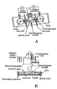
FIGS. IA-1 B depict experimental apparatuses used in studies described herein.
FIG. 1A
5 depicts a top view of the double sucrose recording chamber. In FIG. I A,
from left to right, the
first large compartment contains 120 mM KCI, the central large compartment
contains the
physiological test solutions, such as oxygenated Krebs' solution, and the
third compartment also
contains 120 mM KCI. The small chambers on either side of the central
compartment contain
230, mM sucrose. Seals fashioned from coverslips are secured in place with
high vacuum
10 silicone, grease at the locations shown to inhibit the exchange of the
various media from one
compartment to the next. AgAgCI electrodes for recording and stimulation are
in series with
socket connectors at the locations shown. In the top portion of FIG. 113, a
side view of the
apparatus used to produce a- standardized crush to the isolated spinal cord at
its midpoint within
the central compartment is shown. The position of the spinal cord injury
within the central
chamber is shown in the lower portion of FIG. 1B. The apparatuses are further
described below.
FIGS. 2A-2B depict electrophysiological recordings showing compound action
potentials
(CAPs) of control, and PEG/4-AP treated spinal cords. In FIG. 2A, untreated
spinal cord strips
were treated with 100 M 4-AP at 1 hour post-injury. In FIG. 2B, 100 M 4-AP
was
administered 1 hour post-PEG application. FIG. 2C is a bar graph of group data
showing percent
amplitude increase for 5 control and 5 PEG-treated spinal cords.
FIG. 3 depicts a proposed mechanism of the synergistic effect of PEG and 4-AP
as more
fully described in Example 1. The membrane lesion obtained by mechanical
compression is
depicted by holes. Small arrowheads represent potassium channels.
FIG. 4 depicts an experimental setup used in the examples. Nerve impulse
pathways were
interrupted by crushing the spinal cord in the midthoracic region (red
circuit). A control
procedure demonstrated that a failure to detect SSEPs was due to a failure of
ascending nerve
impulse conduction through the lesion by stimulation of a neural circuit
unaffected by the injury.
FIG. 5 depicts a surgical exposure performed on the sciatic nerve of a test
mammal and
shows the branches (which are cut - see methods) of the sciatic nerve and. the
gastrocnemius
muscle. Note the position of the two transducers, one measuring the force of
muscle contraction,
the other the displacement of the hind paw. The relative position of the hook
electrodes
CA 02740056 2011-05-11
II
stimulating the sciatic nerve proximal to its insertion on the gastrocnemius
is shown as is the
placement of bipolar disc electrodes on the muscle to record the spread of APs
in response to
stimulation. All records are acquired simultaneously on three channels of
recording equipment,
a fourth channel being used to display an event marker triggered by the
stimulation pulse. For
illustration purposes only, the drawing is not made to scale.
FIGS. 6A-6D are four photographic representations showing polyethylene glycol
labeling
in crushed guinea pig spinal cord. In FIGS. 6A-6D, the distribution of FI-PEG
in crushed spinal
cord is shown using three types of application. The application of PEG was
made within 1/2
hour of the-constant displacement crush injury, and evaluated by fluorescent
microscopy of
50gm thick frozen cross sections about 24 hours later. In FIG. 6A, a typical
control section is
shown in darkfeld - the image digitally enhanced to reveal the very faintly
labeled spinal cord.
Such uninjured control sections were obtained by harvesting a segment orthe
spinal cord at least
3-4 vertebral segments from the injury site. Note the characteristic labeling
of PEG in uninjured
spinal cord at the level of detection. The arrows point to weakly labeled
regions of vasculature
in the gray matter and at the pial surface. FIG. 6B shows strong labeling of
PEG at the epicenter
of the crush produced by a 2-minute topical application of PEG to the lesion
as in previous
reports. Arrows point to relatively unlabeled central regions of this injury.
In FIGS. 6C and 6D,
heavy FI-PEG labeling is shown associated with subcutaneous and intravenous
injection
respectively. In FIG. 6C, the arrow points to a cyst forming around the
swollen central canal.
Note the extensive labeling of only the injury site by all methods. The scale
bar = 500 m
FIGS. 7A and 7B are graphs of electrical records showing loss and recovery of
conduction in crushed guinea-pig sciatic nerves after administration of PEG.
The first electrical
record at the top of both FIGS. 7A and 7B shows a typical SSEP recording in
response to tibial
nerve stimulation. Note the early and late arriving evoked potentials (P1 and
P2) in the intact
spinal cord, and their immediate elimination by the spinal cord injury. Though
not shown for
every record, the median nerve control procedure was performed any time an
SSEP was not
recorded, demonstrating the failure to record CAPs was due to the injury. In
FIG. 7A, a typical
set of records is shown for one control animal to the 1 month time point when
the study was
concluded. Note the complete lack of SSEP conduction and the robust Median
nerve induced
SSEP. In FIG. 7B, a typical set of electrical records for a PEG-treated animal
is shown. Note
the elimination of the tibial nerve derived SSEP by the spinal cord injury,
and the positive
median nerve control procedure performed at the same recording time. Before
the end of the
first day post-injury, SSEP conduction was restored by this subcutaneus PEG
injection made 6
CA 02740056 2011-05-11
12
hours after the injury. Recovered evoked potentials continued to improve in
amplitude and
latency during the next month of observation, and in no case were recovered
SSEPs lost after
their recovery. The insert displays the amplitude and time base for all
records except median
nerve stimulations, which were recorded at V2 this sensitivity, but using the
same time base.
FIGS. 8A - 8C are tracings of captured and superimposed video images of a
guinea pig
during a period of CTM stimulation with a monofilament probe, showing
behavioral recovery
following subcutaneous PEG administration. These tracings are derived from
stop motion
videotape analysis of cutaneous trunchi muscle (CTM) stimulation regimens in
which the entire
CTM. receptive field is first determined in the uninjured guinea pig
(circumscribed). Probing
inside this region of back skin with a monofilament probe produces back skin
contractions, while
probing outside the region does not. This line is drawn on the shaved back of
the sedate animal
with a marker while thrinvestigator probes the region. The entire ptoce'aure
is videotaped from
above, and the various regions of both intact receptive fields and areflexia
are reconstructed from
these video images. Note that in all animals, the midthoracic spinal cord
injury eliminates CTM
responsiveness below the level of the injury on both sides (circumscribed). In
control animals
(FIG. 8A), this region of areflexia remained unchanged for the duration of the
experiment. In
PEG-treated animals (FIG. 8B), a variable region of the lost receptive CTM
fields recovered
within a short time of treatment. That region shows a region of CTM recovery
for this one
animal comprising about 55% of the original area of CTM loss. The inset (FIG.
8C) shows the
4-week video image which was used to reconstruct the regions of intact and
nonfunctional
receptive fields. The dot matrix allows precise alignment and superimposing of
receptive fields,
as well as a deeper analysis of the vector of skin movement, the velocity of
skin contraction and
latency when required (data not shown).
FIGS. 9A-9D depict a portion of a neurological examination for outcome
measures and
recovery from paraplegia. A dog is placed on its side while a neurologist
tests for the presence
of superficial pain (A), deep pain (B), and conscious proprioception (C and
D). Skin of the
flank and limbs was pinched sharply with hemostats probing for a reaction from
the subject
during tests of superficial pain response. Deep pain response was similarly
determined, but by a
sustained and sharp squeeze of the joints of the digits. Positive responses
were provided for
comparison by testing the fore limbs. The responses were quantified by a 1-5
score: I = no
detectable response; 2 = a response at the limits of detection, indicated by
an increased state of
arousal, increased respiration or pulse; 3 = consistent attention to the
painful stimulus but
without any overt defensive behavior, 4 = mildly defensive behavior such as
abrupt turning of
CA 02740056 2011-05-11
13
the head towards the stimulus, and whining; 5 = completely normal response to
painful stimuli
including yelping, biting, and aggressive behavior. These scores were obtained
for both sides of
the body and averaged. Conscious proprioceptive placing (CP) and weight
support was tested in
dogs by providing lateral support of the hind limbs, and turning one hind paw
"under" so that the
dorsal surface of the paw (and the animal's weight) rested on the table (inset
Q. A normal
animal briskly replaces the paw to a normal stance instantly after the
examiner releases the paw.
Paraplegic animals rest in this "knuckled under" stance for extended periods
of time. Testing the
fore leg provided a positive control. The test was performed on each side of
the body, and
scored on each side: 1 point = complete absence of CP, and 2.5 points for a
positive CP
response. These scores were then summed for each animal. Voluntary locomotion
(not shown)
was evaluated with a similar 1-5 point score: 1 = complete inability to step
or voluntary
ambulate; 2 = stepping and load bearing at the limit of detection, at best a
few steps before
falling (paresis); 3 = longer sequences of stepping, poorly coordinated before
falling (paresis),
and unable to climb stairs; 4 = more robust and effective walking but with
clear deficits in
coordination, effective weight support, but able to climb stairs; 5 =
completely normal voluntary
walking, indistinguishable from a normal animal. All neurological exams were
videotaped for
reference and half points were permitted at the examiner's discretion. A total
neurological score
(TNS) was determined for each animal at each testing period by summing the
scores of these 4
independent tests. Thus the range of a possible score for any one animal was 4
(a totally
paraplegic animal) to 20 (a totally normal animal, indistinguishable from an
uninjured one).
FIG. 9E shows a comparison of control and PEG-treated animals (FIG. 9A-9D) for
each
of the four outcome measures at approximately 3 days post injury (about 48
hours after the last
PEG injection), 1 week, and 6-8 weeks post injury. The y -axis for each bar
graph is the
percentage of the population (i.e., 25, 50, 75%). DP = deep pain, SP =
superficial pain, P =
proprioceptive placing, and L = voluntary locomotion. Asterisks note when a
test for proportions
(Fisher's exact test, two tailed) or a comparison of the means (Students T, or
the Welch
variation) revealed statistical significance. Note the clear recovery of
outcome measures within
48 hours of the last PEG injection in that group, and the striking improvement
in TNSs in PEG-
treated dogs at every period of evaluation.
FIG. IOA shows a sedated dog and electrode placement in electrophysiological
tests for
conduction through a spinal cord injury to determine a Somatosensory Evoked
Potential (SSEP).
At each evaluation, four to seven sets of evoked potentials (SSEPs) were
stimulated, recorded,
averaged, and stored using a Nihon Kohden ME#B - 5304K 4 Neuropak recorder.
More
CA 02740056 2011-05-11
14
particularly, FIG. I OA shows the sedated dog and the placement of bipolar
stimulating pin
electrodes, inserted subcutaneously, in the hind limb at the distal popliteal
area approximately
0.5 -1 cm apart. These electrodes stimulated the tibial nerve of the hind limb
(red wires ). A
similar procedure was used to stimulate the median nerve of the forelimb
(wires). Trains of
square wave stimulations (0.5 - 3.0 mA amplitude, 200/min) were applied to
evoke-compound
nerve impulses from these nerves. To record evoked potentials, scalp needle
electrodes were
inserted subcutaneously over the somatosensory cortex contralateral to the
side stimulated, while
reference electrodes were inserted on the opposite side between the mastoid
and the pinna of the
ear. The placement of recording electrodes was facilitated by stimulation of
the median nerve at
the outset, aneural circuit above, and unaffected by, the spinal cord injury
(inset, circuit 2). This
procedure also provided a positive control recording to validate the frequent
inability to record
evoked potentials stimulated at the hind limb - but whose ascending potentials
are blocked by the
spinal cord lesion (inset;-circuit 1).
FIG. l OB is a graph of a complete set of SSEP recordings from the procedure
of FIG.
I OA. A lower group of waveforms in this pair are the three individual trains
of 200 stimulations
as discussed, and an upper waveform is the averaged evoked SSEP (only such
averaged SSEPs
are provided in subsequent records, FIGS. 11A and 11B). This record is of a
control procedure.
Note the clear evoked potential, recorded approximately 10 ms after
stimulation of the median
nerve.
FIG. IOC is a graph showing a portion of an electrical recording, displaying
three trains
of stimulation, as well as the averaged SSEP as in FIG. lOB. This record was
in response to
stimulation of the tibial nerve in the same paraplegic dog providing the
record in FIG. lOB,
approximately 4 days post-injury. The complete elimination of SSEP conduction
through the
lesion is characteristic of all neurologically complete paraplegic animals
meeting the criteria
described in the text, both in this and all previous reports using identical
procedures (R.B.
Borgens et al., J Restorative Neurology and Neurosci. 5, 305 (1993); R.B.
Borgens et al., J.
Neurotrauma 16, 639 (1999)). SA = stimulus artifact; time base ~ 50 msec full
screen, 5
msec/div, sensitivity = 1.25 V/ div.
FIGS. I IA and 11B relate to PEG induced recovery of nerve impulse conduction
through
the site of spinal injury. In FIG. I IA, a 6-week progression of recovery of
conduction through
the lesion is shown for a PEG-treated dog. Each trace is the averaged
waveforms of 3-4 trains
of 200 stimulations as described in FIGS. 9A 9E. There is complete absence of
an SSEP in this
CA 02740056 2011-05-11
paraplegic animal prior to surgery, and approximately 4 days later. The third
trace is a median
nerve control procedure. There is no evidence of recovered conduction at I
week post injury.
By 6 weeks post surgery, two distinct evoked cortical potentials had returned,
a typical early
arriving peak of approximately 26 msec latency (P 1), and a later arriving
peak (P 2), of
5 approximately 45 cosec latency.
In FIG. I IB, a low amplitude, long duration, but reproducible evoked
potential recovered
within 15 min of a slow injection of PEG is shown. This atypical SSEP appeared
to segregate
into an early arriving peak of about 15-20 msec latency, and a more condensed
and later arriving
10 peak (P..2) of about 32-3 5 msec latency. SA = stimulus artifact. The time
base and sensitivity
scale is for both FIG. 11A and FIG. 1 IB.
DEFINITIONS
The term "nerve tissue" as used herein refers to any vertebrate nerve tissue,
particularly
15 including cells of the central nervous system (CNS) and peripheral nervous
system. More
particularly, nerve tissue includes spinal cord neuronal structures,
peripheral nervous system
nerves, and nerve cells of the brain-
The word "injury" is used herein to generally denote a breakdown of the
membrane of a
nerve cell, such that there is a collapse in the ability of the nerve membrane
to separate the salty
gel on their insides (cytoplasm) from the salty fluid bathing them
(extracellular fluid). The types
of salts in these two fluid compartments is very different and the exchange of
ions and water
caused by injury leads to the inability of the nerve to produce and propagate
nerve impulses -
and further to the death of the cell. The injury is generally a structural,
physical or mechanical
impairment and may be caused by physical impact, as in the case of a crushing,
compression, or
stretching of nerve fibers. Alternatively, the cell membrane may be destroyed
by or degraded by
a chemical imbalance or physiological malfunction such as anoxia (e.g.,
stroke), aneurysm or
reperfusion. In any event, an "injury" as that term is used herein more
specifically contemplates
a nerve membrane defect, interruption, breach, or rupture (in the phospholipid
bilayer) which can
be treated and sealed by the administration of a biomembrane fusion agent as
described herein.
The term "biomembrane fusion agent" is used herein to designate any and all
molecules
which are not only compatible with vertebrate, and more specifically
mammalian, nerve cells but
also have an affinity for nerve cell membranes so as to attach to injured
nerve cells at the site of
CA 02740056 2011-05-11
16
an injury. A biomembrane fusion agent thus serves in part as a kind of
biological cement or
filling material which bridges over ruptures in neuronal structures. This
sealing is extremely
rapid (minutes) and facilitates the repair of the damaged neuronal structures
by natural
physiological processes which are complete at much later times (1-7 hours).
The sealing of
neuronal membranes as described herein naturally arrests or inhibits the
progressive destruction
of nervous tissue after an injury to the nerve cell. Exemplary biomembrane
fusion agents
include hydrophilic polymers such as polyalkylene glycols (polyalkylene
oxides) and
polyalkylene glycol block copolymers such as polyethylene glycol/polypropylene
glycol block
copolymers (e.g., poloxamer 188) and ethylene oxide-propylene oxide-ethylene
oxide (EPAN),
and further. include biocompatible surfactants, particularly nonionic
surfactants and more
particularly amphipathic polymers such as poloxamines. Poloxamers may also be
considered to
be amphipathic polymers. Poloxamers are hydrophilic to the extent that there
is a greater
number or greater weigtit percentage of ethylene glycol groups as -oppoge-d to
propylene glycol
groups. A biomembrane fusion agent at that term is used herein may comprise a
collection,
mixture, or combination of individual biomembrane fusion agents each of which
is effective in
its own right to seal ruptures in nerve membranes.
The term "effective amount" when used herein with reference to a biomembrane
fusion
agent denotes a quantity of the agent which, when administered to a patient or
subject, is
sufficient to result in a measurable improvement in electrical and/or
behavioral function of a
nerve which has been so damaged or injured that normal functioning is not
possible. As
discussed below, the efficacy of the treatment may be determined in a variety
of ways, including
methods which detect restoration of nerve function. With respect to the use of
the term
"effective amount" with other agents, for example, potassium channel blockers,
that term is used
to describe an amount of an agent effective within the context of that agent's
use in the present
invention.
The term "hydrophilic polymer" means any macromolecule (molecular weights of
200
daltons and greater) which exhibits an affinity for or attraction to water
molecules and which
comprises multiple instances of an identical subunit ("monomer") connected to
each other in
chained and/or branched structures.
A "surfactant" is a molecule exhibiting both an affinity for or attraction to
polar
molecules such as water and an affinity for or attraction to non polar
molecules such as lipids,
fats, oils, and greases. A "nonionic surfactant" is electrically neutral,
i.e., carries no positive or
CA 02740056 2011-05-11
17
negative charge. However, a nonionic surfactant may have localized quantum
variations in
charge leading, for example, to a polar substructure evidencing an affinity
for other polar
molecular structures such as water molecules. In the context of the present
disclosure,
surfactants include amphipathic polymers.
An "amphipathic polymer" as that term is used herein relates to polymers which
have
localized quantum variations in charge giving rise to polar substructures and
non-polar
substructures. The polar substructures evidence an affinity for or attraction
to other polar
molecular structures such as water molecules (hydrophilic), while the nonpolar
substructures
exhibit-an affinity or attraction for nonpolar molecules such as lipids, oils,
greases, fats, etc.
(lipophilic).
Poloxamers, also`balled non-ionic detergents, and/or triblock polymers,
comprise a
polyethylene glycol chain(s) (block 1), then a polypropylene glycol chain
(block 2), followed by
a polyethylene glycol chain(s) (block 3). These compounds can be synthesized
in numerous
conformations and molecular weights. The weights of the various "blocks" can
even vary
between themselves - leading to a complicated nomenclature. What all of the
poloxamers have
in common is a hydrophobic head group (block 2), surrounded by hydrophilic
(PEG) chains.
The hydrophobic "head" is believed to insert itself into the "hole" in a
membrane (where the
hydrophobic interior of the bilamminer membrane is exposed) while the
hydrophilic PEG arms
interdigitate and link with or attach to the nearby, more normal, membrane.
The term "poloxamine" denotes polyalkoxylated symmetrical block polymers of
ethylene
diamine conforming to the general type [(PEG)x-(PPG)y]2-NCH2CH2N-[(PPG)y-
(PEG)x
The word "biocompatible" means that a substance can be placed into intimate
contact
with biological structures, including cells and cellular membranes, without
detriment to the
continued physiological functioning of the contacted cells and membranes.
The term "polyalkylene glycol" refers to a molecule having the chemical
formula
H(O[CH2]m)õ OH where m and n are nonzero integers. The integer in has the
following values for
exemplary polyalkylene glycols: polymethylene glycol (m=1), polyethylene
glycol (m=2),
polypropylene glycol (m=3), polybutylene glycol (m=4), polypentylene glycol
(m=5),
polyhexylene glycol (m=6), polyheptylene glycol (M=7), polyoctylene glycol
(m=8),
polynonylene glycol (m=9), and polydecylene glycol (m=10), including branched
and structural
CA 02740056 2011-05-11
iZS
isomers thereof. Pursuant to the present disclosure, polyalkylene glycols have
a molecular
weight between about 200 and about 25,000 daltons, and preferably between
about 400 daltons
and about 3500 daltons.
The word "carrier" is used herein to denote a liquid matrix, medium or solvent
in which
molecules of a biomembrane fusion agent are dispersed or distributed. A
pharmaceutically
acceptable carrier is one which is biocompatible to vertebrate and more
particularly mammalian
tissues. Generally acceptable carriers include water, saline solutions, among
numerous others.
-By-definition a "potassium channel.blocker" or "K+ channel blocker" is any
agent that
specifically and sterically inserts itself into (or otherwise deactivates) any
of the several and
growing classes of e channels. This includes both fast and slowly activating
channels and both
"voltage gated or non-meted" channels. Almost all channels for K}re"gated" by
the voltage
across the cell membrane. When these channels are open, KK tends to move from
the cytoplasm
into the extracellular fluid because it is about 100 times more concentrated
inside than outside
the cell. This KC exodus (which among other things helps extinguish the nerve
impulse, bringing
the membrane potential back to a resting state) can thus be "blocked". In
regions of
demyelination or membrane potential polarization, KF channel blockade can both
increase
excitability, as well a extend the distance along a nerve. fiber in which a
nerve impulse can travel
before it is extinguished. In spinal cord injury, this may only be a few
millimeters of nerve fiber
damage, with absolutely normal membrane on either side. There are many known e
channel
blockers including reversible blockers (TEA) and some proteins (synthesized
from snake
venoms) that irreversibly block these channels. Potassium channel blockers
include substituted
pyridines and, more particularly, amino-substituted pyridines. The application
of KK channel
blockers to spinal cord repair as described herein involves the fast potassium
channel, type I,
blocker 4-AP (4-aminopyridine) and its analog 3, 4 di-aminopyridine. Too high
a dosage, or the
use of the other blockers (more non specific and poorly reversible) may lead
to convulsions and
even death.
The delivery of a biomembrane fusion agent via a vascular system of a patient
entails the
administration of a biomembrane fusion agent via a pathway including one or
more veins and/or
arteries of the patient. Instead of direct application in which the agent is
injected into the patient
at the site of exposed nerve tissue, the vascular-system-mediated delivery of
a biomembrane
fission agent contemplates an administration and subsequent conveyance of the
agent to the site
of an injured nerve via the vascular system of the patient. The administration
of the
CA 02740056 2011-05-11
19
biomembrane fusion agent is preferably by injection, for example, via a
hypodermic needle or
catheterization, either directly into a vein or artery or indirectly by
subcutaneous injection into
muscle tissue or intraperitoneally. Other methods may also be effective, for
example, by
ingestion, transmembrane delivery (including transdermal delivery), by
suppository, through
inhalants, buccally, or by implantation.
DESCRIPTION OF THE PREFERRED EMBODJMENTS
For the purposes of promoting an understanding of the principles of the
invention,
reference will now be made to preferred embodiments and specific language will
be used to
describe the same. It will nevertheless be understood that no limitation of
the scope of the
invention is thereby intended, such alterations and further modifications of
the invention, and
such further applications-' f the principles of the invention as
illustrated)ierein, being
contemplated as would normally occur to one stalled in the art to which the
invention relates.
The present invention provides methods and compositions for treating injured
nerve
tissue of a vertebrate. The methods and compositions are designed to at least
partially restore
nerve function in the vertebrate. In one aspect of the invention, methods are
provided for treating
an injured or damaged vertebrate spinal cord that include contacting the
spinal cord with an
effective amount of a biomembrane fusion agent. The compositions include a
biomembrane
fusion agent, preferably a polyalkylene glycol such as polyethylene glycol
(chemical formula:
H(OCH2CH2),,OH) and/or a nonionic surfactant such as an amphipathic polymer
(e.g., a
poloxamer or a poloxamine), and/or mixtures or copolymers thereof. In
alternative embodiments,
the method may include treating the nervous system with a potassium channel
blocker,
preferably a substituted pyridine, such as an amino-substituted pyridine,
either before, during or
after contacting the spinal cord with the biomembrane fusion agent. Other
aspects of the
invention provide compositions for treating an injured nervous system of a
vertebrate. The
preferred compositions include a biomembrane fusion agent and a potassium
channel blocker.
The preferred biomembrane fusion agent is a polyalkylene glycol. A wide
variety of
polyalkylene glycols may be used, including those, for example, where the
alkylene group is
methylene, ethylene, propylene, butylene, pentylene, hexylene, heptylene,
octylene, nonylene,
and decylene, including branched and structural isomers thereof. Preferably,
the polyalkylene
glycol will be water-soluble and is selected from the group consisting of
polyethylene glycol,
polypropylene glycol and block copolymers of polyethylene glycol and
polypropylene glycol. A
more preferred polyalkylene glycol is polyethylene glycol. Although a wide
range of molecular
weight polyalkylene glycols may be used (between about 200 daltons and about
25,000 daltons)
CA 02740056 2011-05-11
depending on the ability of the polyalkylene glycol to pass through various
biological barriers
such as the digestive tract, polyalkylene glycols and polyalkylene glycol
block copolymers of
molecular weight of about 400 to about 3500 daltons are preferred. Such
biomembrane fusion
agents may be synthesized by methods known to the art or may be purchased
commercially.
5 The biomembrane fusion agent may also be a polyalkylene glycol/protein
conjugate as
known in the art, wherein the protein preferably aids in scavenging free
radicals. For example,
the biomembrane fusion agent, such as polyethylene glycol or other alkylene
oxide, may be
conjugated to catalase to form PEG-catalase, or to superoxide dismutase to
form PEG-SOD.
Such conjugates are available commercially from Sigma, St. Louis, Mo. The
biomembrane
10 fusion agent, may also be conjugated to a biodegradable surgical glue, such
as a commercial
fibrin glue, to facilitate and stabilize reattachment and fusion of severed
nervous tissue.
Alternatively, the biomembrane fusion agent may be a biocompatible surfactant,
preferably a nonionic st1'rfactant and more preferably an amphipathic polymer
such as a
poloxamer or a poloxamine.
15 The biomembrane fusion agent may be provided in a pharmaceutically
acceptable carrier.
Such carriers include, for example, water, preferably sterile and including
distilled water, and
any other pharmaceutically acceptable carrier known to the art that will not
have an adverse
effect on the treatment. Sterile distilled water is a preferred carrier in
work to date.
The biomembrane fusion agent is administered to the patient as soon after
injury as
20 possible and prior to irreversible dissolution of axonal membranes and the
myelin sheath.
Although this time period may vary depending on the nature and extent of the
injury, the fusion
agent is typically administered immediately after the injury occurs, and
preferably not later than
about 24 hours post-injury, but is typically administered between about 1 hour
to about 8 hours
post-injury. Though early treatment is preferred, administration of the
biomembrane fusion
agent may still be beneficial for up to 2 weeks after the initial nerve injury
(called the "primary
injury"). This is because nerve injury is a continuous, slow, progressive
event, especially in
spinal cord where it is called "secondary injury" (Tator and Fehlings 1991, J.
Neurosurgery
75:15-26).
The biomembrane fusion agent may be delivered to the site of injury by any
suitable
method. Preferably, the biomembrane fusion agent is administered through the
vascular system
of the subject or patient. The fusion agent may be injected directly into the
vascular system or
indirectly by injection intramuscularly, subcutaneously or intraperitoneally.
It has been
discovered that an indirect administration of a biomembrane fusion agent such
as polyethylene
glycol via the vascular system of the patient unexpectedly results in a
selective adherence of the
fusion agent (e.g., PEG, poloxamer or other agent) to the injured nerve
tissue. There is little or
CA 02740056 2011-05-11
21
no adherence to undamaged nerve tissue. Without being limited by way of
theory, it is believed
that by adhering to damaged nerve tissue, the biomembrane fusion agent
promotes the natural
healing processes of the damaged nerve cells.
Where the biomembrane fusion agent is a polyalkylene glycol such as PEG, the
fusion
solution comprises fusion agent in an amount of typically about 15 to about
50% by weight and
preferably is administered in doses of about 15 - 50 mg PEG per body weight in
kilograms of the
patient where the PEG has a weight of 1500 - 4000 Daltons. Where the
biomembrane fusion
agent is an amphipathic polymer such as a poloxamer or a poloxamine, the
fusion solution
typically contains fusion agent in an amount of about 15 to about 50% by
weight and is
administered in dosages of about 15 - 150 mg poloxamer or poloxamine per body
weight in
kilograms of the patient.
Where the agent is applied directly to damaged nerve tissue which has been
exposed, for
example, via surgical pro'Cedures, the agent may be applied with any suitable
liquid dispensing
device. Although the percentage by weight of the fusion agent in the direct-
application
composition may vary, the composition typically includes fusion agent in an
amount of at least
about 40% by weight, more preferably about 40% to about 50% by weight, and
most preferably
about 50% to about 55% by weight.
In the case of a direct-contact application, the injured site is exposed to
the fusion agent
for a time period effective for treating the injury. This time may vary
depending on the size of
the lesion, the extent and nature of the injury, the biomembrane fusion agent
used, and the
concentration of the biomembrane fusion agent. The lesion is typically exposed
to the agent for
at least about one minute and more preferably at least about 2 minutes. In
preferred
embodiments, the fusion agent is removed from the injured tissue being treated
prior to the
occurrence of deleterious tissue changes. In a further preferred embodiment,
the injured tissue is
exposed to the fusion agent for no more than about 5 minutes. After the
injured region of the
nervous system is treated with the fusion agent, it may be removed by
aspiration and the treated
site washed with a biowashing solution, such as isotonic Kreb's solution as
described in the
examples. Excess fusion agent and/or Kreb's solution can then be removed by
aspiration.
In another form of the invention, the method may further include administering
to the
patient or subject an effective amount of a potassium channel blocker. In the
case of a direct-
contact application of a biomembrane fusion agent, the injured site is
contacted with an effective
amount of a potassium channel blocker in addition to the biomembrane fusion
agent. A variety of
potassium channel blockers may be used, including substituted pyridines.
Preferred potassium
channel blockers include those that improve action potential conduction in
injured tissue,
including 3,4-diaminopyridine, 4-methylaminopyridine and ampidine. In a
preferred form of the
CA 02740056 2011-05-11
22
invention, the pyridine is substituted with an amino group, more preferably at
the 4-position of
the ring. Moreover, it has unexpectedly been discovered that treatment of an
injured mammalian
spinal cord with a potassium channel blocker, such as 4-aminopyridine, after
treatment with a
fusion agent, such as polyethylene glycol, can result in synergistic repair of
the spinal cord. For
example, compound action potentials (CAPs) increase in conduction when both
agents are used
by a percentage greater than the sum of the percent increase in conduction of
the CAPS when
injured spinal cords are treated alone with either the fusion agent or the
potassium channel
blocker.
Although the injured nervous system may be contacted with the potassium
channel
blocker-prior to or at the same time as treating with the fusion agent, the
system is preferably
contacted with the blocker after the treatment with the fusion agent. The
potassium channel
blocker may be applied in a fashion similar to the fusion agent. The amount of
the potassium
channel blocker effecti6'e in treating or repairing the injured nervous
system, such as injured
mammalian spinal cord, will also similarly depend on the factors mentioned
above. When the
potassium channel blocker is 4-aminopyridine, it is typically applied at a
concentration of about
10-100 ng/ml cerebrospinal fluid and further preferably about 50-100 ng/ml
cerebrospinal fluid.
After treatment with 4-aminopyridine, it can similarly be removed by
aspiration and the lesion
site washed with the biowashing agent.
In yet other forms of the invention, the method may include treating the
injury with a
polyalkylene glycol, as well as with other conventional management compounds
and/or
compositions. For example, in addition to treatment with a polyalkylene
glycol, the injury may
be treated with a steroid, such as methylprednisolone.
A wide variety of injuries may be treated in the present invention. In various
forms of the
invention, the injury may arise from a compression or other contusion of the
spinal cord,
crushing of the spinal cord or severing of the spinal cord, or anoxia (e.g.,
stroke), aneurysm or
reperfusion.
The efficacy of the treatment may be determined in a variety of ways,
including methods
which detect restoration of nerve function. For example, restoration or
increase in conduction of
action potentials, such as CAPs, through the injured site may be used as an
indicator that nerve
function has at least partially been restored as described in the examples.
Nerve function is
considered to have been at least partially restored if there is an increase in
the conduction of
action potentials after treatment. Preferably, the treatment will be conducted
sufficiently to
achieve at least about 10% increase in conduction of CAPs. Moreover,
restoration of anatomical
continuity may also be observed by examination with high-resolution light
microscopy and/or by
diffusion of intracellular fluorescent dyes through the repaired nervous
tissue, such as repaired
CA 02740056 2011-05-11
LS
axons, or by direct observation of repaired axonal membranes. Additionally, in
human
applications, the efficacy of preferred treatments may be observed by the
restoration of more
than one spinal root level as determined by the American Spinal Injury
Association (ASIA)
motor score and/or the National Animal Spinal Cord Injury Study (NASCIS) score
as know in
the art and as described in Wagih et al., (1996) Spine 21:614-619.
Furthermore, in veterinary
applications, behavioral analysis of the cutaneous trunci muscle (CTM) reflex,
as more fully
discussed in the examples, may also be used to determine the efficacy of the
treatment, and
whether nerve function has at least partially been restored. Using this
analysis, nerve function is
considered to have been at least partially restored if there is an increased
reflex behavior after
treatment, but treatments are desirably preferred so as to achieve at least
about a 10% increase in
the area of CTM behavioral recovery.
In yet other aspects of the invention, compositions for treating an injured
nervous system
of a vertebrate are provided. The compositions are designed to at least
partially restore nerve
function as described below. In one form, a composition includes a biomembrane
fusion agent
and a potassium channel blocker. Although a wide variety of biomembrane fusion
agents and
potassium channel blockers that are mentioned above may be included in the
composition, a
preferred biomembrane fusion agent is a polyalkylene glycol and a preferred
potassium channel
blocker is a substituted pyridine. In more preferred forms of the invention,
the polyalkylene
glycol is polyethylene glycol and the potassium channel blocker is an amino-
substituted
pyridine, such as 4-aminopyridine. The composition may be in a
pharmaceutically acceptable
carrier as described above.
Although the methods and compositions of the invention are useful in treating
a wide
variety of vertebrates, they may be advantageously used to treat mammals and
preferably
humans. Moreover, although the methods and compositions are advantageously and
surprisingly
useful in treating the spinal cord, they may also be used in treating the
peripheral nervous system
and/or central nervous system, or other areas in which damaged axons are
present.
Reference will now be made to specific examples illustrating the compositions
and
methods described above. It is to be understood that the examples are provided
to illustrate
preferred embodiments and that no limitation to the scope of the invention is
intended thereby.
EXAMPLE 1
Potassium Channel Blockade as an Adjunct to PEG-Mediated Recovery of
Conduction
This example shows that treatment of injured spinal cords in vitro with both a
potassium
channel blocker and a biomembrane fusion agent allows synergistic recovery of
compound
action potentials (CAPs).
CA 02740056 2011-05-11
24
It is a common feature of injured cells to loose intracellular potassium to
the extracellular
milieu through compromised membrane. In axons, this may be sufficient to
suppress action
potential conduction. Thus, it was attempted to determine if blockage of fast
potassium channels
with 4-AP would affect the properties of conduction immediately following PEG
repair.
Analysis was also performed in the double sucrose recording chamber.
In Vitro Isolation of the Spinal Cord
Adult female guinea pigs of 350-500 gram body weight were used for these
studies. The
spinal cord was isolated from deeply anesthetized animals [(60 mg/kg ketamine
hydrochloride,
0.6 mg/kg aoepromazine maleate, and 10 mg/kg xylazine, intramuscularly
(i.m.)]. Following
anesthesia, the animal was perfused transcardially with cold (1 50C) Krebs'
solution (NaCl, 124
mM; KCI, 2 mM; KH2P04,1.2 mM; MgSO4,1.3 mM; CaC 12,11.2 mM; dextrose, 10 mM;
NaHC03,26 mM; sodium ascorbate, 10 mM; equilibrated with 95% 02, and 5% C02).
The
vertebral column was rapidly removed using bone forceps and scissors by
previously described
techniques [Shi, R. and Blight, A.R. (1996) J. of Neurophysiblogy, 76(3):1572-
1579; Shi, R. and
Blight, A.R. (1997) Neuroscience 77(2):553562]. The spinal cord was divided
into four
longitudinal strips, first by midline sagittal division, then by separating
the dorsal and ventral
halves with a scalpel blade against a plastic block. Only the ventral white
matter was used for
this study. These 35-38 mm long strips of spinal cord white matter will
usually be referred to
below as "cords" or "spinal cords" for ease of description. Spinal cords were
maintained in
continuously oxygenated Krebs' solution for an hour before mounting them
within the recording
chamber. This was to ensure their recovery from dissection before experiments
were begun.
Double Sucrose Gap Recording Technique
The double sucrose gap recording chamber is shown in FIGS. 1A and 1B and has
already
been described in previous publications [Shi, R. and Blight, A.R. (1996) J. of
Neurophysiology,
76(3):1572-1579; Shi, R. and Blight, A.R. (1997) Neuroscience 77(2):553-562].
Briefly, the strip
of isolated spinal cord white matter was supported in the three-compartment
chamber. The
central compartment was continuously superfused with oxygenated Krebs'
solution (about 2
ml/min) with a peristaltic pump. The compartments at both ends were filled
with isotonic (1120
mM) potassium chloride, and the gap channels with 230 mM sucrose. The white
matter strip was
sealed on either side of the sucrose gap channels with shaped fragments of
glass coverslips that
CA 02740056 2011-05-11
also blocked the flow of fluid in the narrow gap between the coverslip and the
tissue surface.
Note that the central chamber is at ground potential for recording. The
sucrose solution was run
continuously through the gap at a rate of 1 ml/min. Axons within the spinal
cord strip were
stimulated and compound action potentials (CAPs) were recorded at the opposite
end of the
5 white matter strip by silver-silver chloride electrodes positioned within
the side chambers and the
central bath as shown in FIG. I B. Specifically, action potentials were
stimulated at the left side
of the spinal cord strip as shown in the figure, conducted through the spinal
cord in the central
compartment (also including the injury site), and recorded at the right side
of the spinal cord strip
as shown. Stimuli were delivered through stimulus isolation units in the form
of 0. 1 msec
10 constant current unipolar pulses. A conventional bridge amplifier with
capacity compensation
(Neurodata Instruments) was used to amplify the signal. This data was
digitized and stored on
video tape with a Neurodata Instruments Neurocorder for subsequent analysis.
During the
experiment, the oxygenated Krebs' solution continuously perfused-the isolated
spinal cord tract,
while temperature was maintained at 37 C.
15 Every electrophysiological test was digitized in real time and captured to
the computer
for subsequent quantitative evaluation. All records were also recorded on VHS
magnetic tape as
a means of back up documentation. All solutions used in the PEG repair process
were made on
the day of their use.
20 The Compression Injury
A standardized compression injury was produced with a stepper-motor controlled
rod
which compressed the spinal cord while suspended inside the recording chamber
(FIG. 1 B).
Briefly, the isolated white matter strip was compressed against a flat, raised
plastic, plexiglass
stage at the center of the recording chamber with the flattened tip of a
plexiglass rod. The tip was
25 advanced downward to contact the tissue at a standardized rate of about 25
pm/s. The downward
movement of the rod was controlled with a stepper motor to produce a finely
graded crush just
sufficient to eliminate all CAP propagation (which was monitored continuously
during the
procedure). The-end of the rod with the flattened tip provided a compression
surface of 2.5 mm
along the length of the tissue, and a transverse width of 7 mm, such that it
was always wider than
the spinal cord strip, even under full compression. Positioning of the
compression rod was
accomplished with a micromanipulator. CAPs were simultaneously recorded during
the injury
process. Compression was stopped when CAPs were completely eliminated- The
state of
complete CAP failure was maintained for an additional 15 seconds before the
rod was rapidly
withdrawn from the cord's surface to relieve pressure. The recovery of the CAP
was then
documented. The basic recovery profile following such standardized compression
in normal
CA 02740056 2011-05-11
26
Krebs' solution has been previously characterized and published [Shi, R_ and
Blight, A.R. (1996)
J. of Neurophysiology, 76(3):1572-1579].
PEG Repair Procedure
The PEG repair procedure included the following steps:
1) Typical physiological functioning of the isolated white matter strip
removed to the
recording chamber required about 1/2 to 1 hour of incubation time while
immersed in
oxygenated Krebs' soluction to stabilize. In initial experiments, once the CAP
propagation had
stabilized, the Krebs' solution was replaced with Ca2+-free Krebs' (Ca2+
replaced with an
equimolar amount of Mg2+).
2) The spinal cord strip was then crushed by the techniques described above,
while
simultaneous stimulation and recording continued.
3) A solution ofPEG in distilled water (50 % by weight) was applied by d
pressure
injection through a micropipette. A vital dye was added to the PEG solution to
monitor its
continuous application to the lesion site in a stream about 0.5 mm wide for
about 1-2 minutes.
The PEG was applied to one side of the lesion, washed over it, and immediately
removed by
constant aspiration on the other side using a second pipette.
4) Immediately following the PEG application, the bathing media in the central
chamber
was replaced with a continuous stream of oxygenated normal Krebs' solution.
The physiological
properties of the PEG-treated spinal cord were monitored continuously for 1
hour. Usually, a
weak recovering CAP was evident within 6-15 minutes of the PEG application.
Tests were made of the response of "recovering" axons to the additional
application of
the fast potassium channel blocker, 4 aminopyridine (4-AP). In this trial, 5
separate cords were
treated with an application of PEG as described above and compared to 5
control cords. One
hour after compression, 100 pM 4-AP (in Krebs' solution) was applied for 15
minutes and then
washed free with normal Krebs' solution as described above.
FIG. 2A shows the enhancement of the CAP in crushed (but untreated with PEG)
spinal
cord by 4-AP. In this individual record, the initial recovered CAP at 1 hour
post injury is shown,
and the enhanced CAP following 100 pM 4-AP treatment is superimposed upon it.
Following
documentation of the 4-AP enhanced CAP, the blocker was washed out, and the
media in the
central compartment was replaced with normal Krebs' solution. The CAP fell to
pretreatment
levels by 15 minutes and was indistinguishable from the original record. This
final waveform is
superimposed on the other two CAPs in FIG. 2A but cannot be discriminated from
the
CA 02740056 2011-05-11
27
pretreatment electrical record. In this single test, 4AP reversibly enhanced
the recovered CAP by
about 40%.
FIG. 2B shows an identical test performed on a PEG-treated spinal cord, in
which 4-AP
was administered at 1 hour post PEG application. In this individual test, the
second CAP was
reversibly enhanced by about 70%.
Following the near doubling of the CAP, 4-AP was washed out as described, and
the
CAP fell to pretreatment levels as in controls (FIG. 2A).
FIG. 2C shows the group data, including 5 spinal cords in each group. The
percent
enhancement of the PEG-mediated recovery for the group data mirrors that
discussed above for
the individual experiments (about 70% enhancement in the experimental group;
about 40% in the
control group). This experimental enhancement was statistically significantly
greater than that
observed in the controls. (p<0.05, unpaired Student's t test)
Although not being limited by theory, FIG. 3 depicts a proposed mechanism of
the
synergistic effect of PEG and 4-AP. A severe mechanical compression of a
myelinated axon is
diagrammed at the top. Note that the myelin sheath envelops high densities of
fast W channels
clustered at the paranodal region. Severe crush leads to an exposure of the
potassium channels of
the paranodal region by a withdrawal or collapse of the myelin lamella at this
site [Shi, R. and
Blight, A.R. (1996): Neuroscience, 77:553-562]. Exposure of the voltage gated
potassium
channels after injury would elevate K+ conductance further impeding conduction
across this
damaged portion of the membrane (gray region showing " holes " in the
compromised
membrane). In control preparations, partial to complete conduction block
results from this
localized disturbance of the axolemma, which may progress to complete
separation of the axon
and loss of the distal axonal segment by Wallerian degeneration (left side of
FIG. 3). In
PEG-treated axons (right side of FIG. 3), the membrane repair leads to
preservation of injured
axons as well as improvements in their conduction capabilities (gray regions;
membrane holes
now sealed). However, elevated e conductance through e channels that are still
exposed at the
site of repair in PEG-treated nerve fibers might still suppress conduction to
some extent.
Blockade of these channels with 4 AP (FIG. 3, small arrow heads; lower right)
would be
expected to reduce any outward K+ conductance and thus enhance conduction.
Spinal cords were crushed, isolated and treated with PEG as described.
SUMMARY OF RESULTS
Within a few minutes after the application of the water-soluble polymer PEG,
an
immediate recovery of CAP propagation through the lesion occurred. The
recovered CAP
amplitude slowly increased with time to a peak of about 20% of the initial CAP
amplitude.
Moreover, this level of recovery a) was always statistically significantly
higher than control
CA 02740056 2011-05-11
28
amplitudes, b) was observed at every time point tested, and c) occurred in
100% of the
experimentally treated spinal cords. It is clear that a topical application of
PEG can immediately
repair severe compression injury to the mammalian spinal cord leading to
significant increases in
functional recovery as defined by the enhanced capacity to propagate nerve
impulses through the
lesion. This report is the first to demonstrate PEG-mediated repair of crushed
mammalian
nervous tissue.
We have shown that a physiological, balanced media and the aforementioned PEG
solution, is all that is required to produce functionally significant repair
in mammalian spinal
cords (see below). Moreover, in other experiments, where completely transected
guinea pig
spinal cords were fused with PEG, it has been revealed there was no specific
PEG molecular
weight critical to the process, having tested PEG solutions using 400, 1400,
1800, 2000, and
3700 daltons .
In this physiological study, similarities and differences between the natural
mechanisms
of axonal repair and those mediated by PEG have been determined. First, a
least squares linear
regression analysis of preand postinjury CAP amplitudes suggests that PEG-
mediated repair can
occur across all levels of stimulus thresholds, reflecting axon diameters, as
does the natural
recovery process in untreated spinal cord strips. In other words, all spinal
axons regardless of
their caliber are equally susceptible to PEG mediated repair [see Shi, R. and
Blight, A.R. (1996)
Neuroscience 77:553-562 for a similar analysis of axonal recovery from
compression injury].
The differences between natural repair and that produced by PEG application
are more striking.
First, this injury is very severe; 30% of control spinal cords never recovered
any capacity to
conduct CAPs during the 1 hour period of evaluation following injury. On the
other hand, there
was no instance where PEG did not initiate a measurable physiological
recovery. On a more
subtle level, there appears to be a slightly reduced CAP amplitude during the
period of relative
refractory in only PEG-mediated CAPS relative to control cords. One
explanation for this
observation may be that in control cords a severely compromised and
dysfunctional population
of axons may become completely nonfunctional, revealing more normal conduction
properties in
that population that survive the injury. PEG may rescue a portion of such
severely compromised
axons, recruiting them into the CAP, and perhaps accounting for its slightly
different conduction
properties.
The above-described in vitro evaluation of the anatomy of axonal repair
following
mechanical compression has revealed that a 2 minute application of PEG
produced sealing of
membrane lesions at the site of a standardized compression. Sealing was
indicated by the
exclusion of horseradish peroxidase uptake by injured fibers in the PEG-
treated group compared
to sham-treated spinal cords. Such immediate repair of
CA 02740056 2011-05-11
29
membrane breaches sufficient to inhibit the uptake of large molecular weight
dyes should also
arrest or reduce permeabilization, allowing the nonspecific flux of ions
across it. Although not
being limited by theory, it is believed that this "sealing" behavior of PEG
both restores
excitability and reverses anatomical dissolution of the nerve fiber.
This procedure may advantageously applied to treat severe, acute neurotrauma.
In addition to
immediate improvements in conduction, repair of crushed axons in peripheral
nerves leading to a
rescue of their distal segments would provide the added benefit of reducing
atrophy or
degeneration of target cells or so called "end organs." Moreover, PEG-mediated
fusion of even
transected axons could become a component of microsurgical grafting techniques
since the
conventional resection of peripheral nerve trunks prior to fasicular grafting
exposes the severed
tips of proximal and distal axonal segments, making them available for fusion.
EXAMPLE 2
Rapid Recovery from Spinal Cord Injury Following
Subcutaneously Administered Polyethylene Glycol
This example demonstrates that a biomembrane fusion agent, specifically the
hydrophilic
polymer PEG, can be safely introduced into the bloodstream by several routes
of administration,
and that the administered PEG specifically targets a hemorrhagic contusion of
an adult guinea
pig spinal cord. A single subcutaneous injection (30% weight by weight in
sterile saline) made 6
hours after spinal injury was sufficient to produce a rapid recovery of CAP
propagation through
the lesion, accompanied by a significant level of behavioral recovery in only
PEG-treated
animals.
The results of these tests demonstrate (1) that PEG specifically targets the
spinal cord
contusion independent of whether it is applied directly to the exposed spinal
injury, or by
intravenous or subcutaneous injection, and (2) that a single subcutaneous
injection of PEG
approximately 6 hours after severe SCI is sufficient to induce a rapid
reversal of functional
losses in nearly all PEG-treated adult guinea pigs compared to the persistence
of these deficits in
nearly all sham Treated animals. The intravascular delivery of PEG for
purposes of treating and
repairing injured nerve tissue has also been investigated in a clinical
setting using naturally
produced cases of paraplegia in dogs, as discussed hereinafter.
Drawing Fig. 4: Behavioral Model and Physiological Evaluation
This drawing shows the neural circuit of the Cutaneus Trunchi Muscle (CTM)
reflex, and
its interruption by spinal injury. Nociceptive sensory receptors in the skin
project their axons
CA 02740056 2011-05-11
into the spinal cord at each vertebral segment bilaterally via the Dorsal
Cutaneus Nerves. These
synapse within the spinal cord and project 2nd order ascending sensory nerves
in the ventral
funiculus of the white matter to the cervical region where these synapse on
bilaterally organized
constellations of CTM motor neurons. CTM motor neurons project their axons out
of the cord
5 on right and left sides via the brachial plexus, where these innervate the
cutaneous muscle of the
skin via the lateral thoracic branch of the plexus. When the spinal cord is
intact, tactile
stimulation of the back skin within the CTM receptive field causes a rippling
contraction of the
skin. Stimulation outside the receptive fields of back skin does not result in
skin contractions. A
spinal cord injury (drawn on only the left side of the cord for descriptive
purposes) interrupts the
10 ascending leg of this circuit producing a region of skin areflexia
ipsilateral to the injury and on
the same side. Tactile probing within this region does not produce CTM
contractions, usually
for the life of the animal. Stimulation of back skin above the level of this
unilateral lesion, or on
the right side produces CTM contractions, as these receptive fields remain
unaffected by the
unilateral injury to the left side of the spinal cord.
Methods
Animal surgery and spinal cord injury
Adult Guinea Pigs (< 300 gm) were anesthetized with an intramuscular injection
of
100mg/kg ketamine HCL and 20 mg/kg xylazine and the spinal cord exposed by
dorsal
laminectomy. The midthoracic cord was crushed with special blunted forceps
possessing a
detente. This standardized, constant displacement injury [Moriarty, L.J.,
Duerstock, B.S., Bajaj,
C.L., Lin, K., and Borgens, R.B. (1998) Two and three dimensional computer
graphic evaluation
of the subacute spinal cord injury, J. Neurologic. Sci., 155, 121-1371 has
produced more
consistent anatomical injury to the cord and more consistent behavioral loss
between animals
than constant impact injuries (such as those produced by the various weight
drop techniques).
Animals were euthanized by deep anesthesia followed by perfusion/fixation. The
localization of
an FITC decorated PEG (Fl-PEG) in spinal cord was determined by killing the
animals for
histological processing approximately 24 hours after the application or
injection of Fl-PEG. The
spinal cords were dissected from the animals, and the segments of spinal cord
containing the
sites of injury and an intact, more rostral, segment were sectioned with a
freezing microtome and
evaluated with a fluorescent microscope. Histological cross sections were 5 m
thick, and
observed on an Olympus Van Ox Fluorescent microscope using excitation
wavelengths of 495
and 545 inn and barrier filters of 475 and 590 nm, respectively. Digital
images were captured to
the computer with an Optronics DEI 750 camera.
CA 02740056 2011-05-11
31
To test the effects of subcutaneous injections of PEG, adult guinea pigs were
anesthetized
and their mid-thoracic spinal cords were surgically exposed and then crushed
by a standardized
technique. [Blight, A.R. (1991): Morphometric analysis of a model of spinal
cord injury in
guinea pigs, with behavioral evidence of delayed secondary pathology, J_
Neurolog. Sci., 103:
156-171.] Twenty animals were divided into equal groups of 10. One group
received a single
subcutaneous injection of PEG (1400 MW) beneath the skin of the neck (0.5cc;
30% in sterile
lactated Ringer's solution; SLR). The sham-treated control group received a
single injection of
the carrier, lactated Ringer's. Only this one subcutaneous injection per
animal was made
approximately 6 hours after the spinal cord injury. CTM testing and SSEP
recordings were
carried-out-on all 20 animals prior to spinal cord injury, 1 day, 1 week, 2
weeks, and 4 weeks
post injury.
Tracing the distribution atPEG in iniured spinal cord
The FITC decorated PEG (about 1400 Daltons; prepared by Molecular Probes,
Chatsworth, Ca) was used to trace the distribution of PEG following different
routes of
administration. Fl-PEG, 50% weight by weight in SLR was applied directly to
exposed spinal
cord injury site (with the dura removed) using a Pasteur pipette in two
animals. As in prior
experiments [Borgens, R. and Shi, R. (2000). Immediate recovery from spinal
cord injury through
molecular repair of nerve membranes with polyethylene glycol, FASEB 14, 27-
35], PEG was
removed by aspiration and the region irrigated with SLR two minutes later.
Subcutaneous
injection of 1 cc Fl-PEG (30% w/w in SLR) was made beneath the skin of the
neck in two spinal
injured guinea pigs using a 22-gauge needle. For IV injection, the jugular
vein was surgically
exposed, and 1 cc of FL-PEG was injected using a 26-gauge needle. PEG, 30% in
lactated
Ringer's was also administered by intraperitoneal injection in one case.
In Vivo Conduction Studies
Functional deficits produced by SCI are largely caused by the loss of nerve
impulse
conduction through mechanically damaged tracts of nerve fibers in spinal cord
white matter
[Blight, A.R. (1993) Remyelination, Revascularization, and Recovery of
Function in
Experimental Spinal Cord Injury, Advances in Neurobiology: Neural Injury and
Regeneration
(Seil, FJ. Ed.), Vol. 59, pp. 91-103, Raven Press, New York]. Accordingly, the
loss and
recovery of compound action potential (CAP) conduction through the spinal cord
injury was
evaluated by evoked potential techniques (somatosensory evoked potential
testing or SSEP).
Stimulation of the Tibial nerve of the hind limb produced ascending volleys of
nerve impulses
recorded at the contralateral sensory cortex of the brain. These were
eliminated between the site
CA 02740056 2011-05-11
32
of stimulation and recording by the spinal lesion - immediately abolishing the
recording of these
peaks (postcrush records). Each electrical record was comprised of a stimulus
train of 200
stimulations (< 2mA square wave, 200 s duration at 3 HZ). Three sets of these
recordings were
made at each measurement period and averaged to produce the single waveform
presented in the
following data. The appearance of original records prior to computer averaging
can be found in
prior reports [Borgens, R and Shi, R. (2000) Immediate recovery from spinal
cord injury
through molecular repair of nerve membranes with polyethylene glycol, FASEB
14, 27-35].
Conduction of nerve impulses through a median nerve circuit following
stimulation of the
median nerve of the forelimb (unaffected by the spinal cord injury at the
midthoracic level) was a
control procedure during SSEP recording. This control stimulation regimen was
carried out in
every circumstance where a failure to record evoked potentials at the cortex
occurred in response
to hind limb tibial nerve stimulation - to eliminate the possibility these
failures were "false
negatives". SSEP recording and averaging was performed with a Nihon Kohden
Neuropak 4
stimulator/recorder and a PowerMac G3 computer. Computation of the area
beneath the early
arriving SSEP peak (P1) was accomplished by scribing a reference line beneath
the base of the
peak, and determining the unit area contained within it as pixels using IP Lab
Spectrum software.
Behavioral Studies
As an index of behavioral recovery, evaluations are made of a spinal cord
dependent
contraction of back skin in animals - the Cutaneus Trunchi Muscle reflex
(CTM)[Blight, A.R.,
McGinnis, M.E., and Borgens, R.B. (1990): Cutaneus trunci muscle reflex of the
guinea pig,
J.Comp.Neurol., 296, 614-633; Borgens, R.B. (1992): Applied Voltages in Spinal
Cord
Reconstruction: History, Strategies, and Behavioral Models, in Spinal Cord
Dysfunction,
Volume III: Functional Stimulation, (Illis, L.S. ed.), Chapter 5, pp. 110-145,
Oxford Medical
Publications, Oxford]. The loss of CTM behavior following injury to the spinal
cord is observed
as a region of back skin, which no longer responds, by muscular contraction to
local tactile
stimulation [Blight, A.R., McGinnis, M.E., and Borgens, R.B. (1990): Cutaneus
trunci muscle
reflex of the guinea pig, J.Comp.Neurol., 296, 614-633; Borgens, R.B. (1992):
Applied Voltages
in Spinal Cord Reconstruction: History, Strategies, and Behavioral Models, in
Spinal Cord
Dysfunction, Volume III: Functional Stimulation, (Illis, L.S. ed.), Chapter 5,
pp. 110-145,
Oxford Medical Publications, Oxford; Borgens, R.B., Blight, A.R., and
McGinnis, M.E. (1990):
Functional recovery after spinal cord hemisection in guinea pigs: The effects
of applied electric
fields, J. Comp. Neurol., 296, 634-653; Borgens, R.B., Blight A.R., and
McGinnis M.E. (1987):
Behavioral recovery induced by applied electric fields after spinal cord
hemisection in guinea
pig, Science, 238, 366-369]. This areflexia does not recover for the life of
the animal if the
CA 02740056 2011-05-11
33
relevant (and identified) ascending CTM tract is severed within the ventral
funiculus as the
complete neural circuit underlying this behavior has been identified [Blight,
A.R., McGinnis,
M.E., and Borgens, R.B. (1990): Cutaneus trunci muscle reflex of the guinea
pig,
J.Comp.Neurol., 296, 614-6331. Following a severe bilateral crush injury of
the mid-thoracic
spinal cord (such as used here), a bilateral region of areflexia of back skin
is produced that still
shows very limited ability to spontaneously recover [Borgens, R. and Shi, R
(2000): Immediate
recovery from spinal cord injury through molecular repair of nerve membranes
with
polyethylene glycol, FASEB, 14, 27-35; Borgens, R.B. (1992): Applied Voltages
in Spinal Cord
Reconstruction: History, Strategies, and Behavioral Models, in Spinal Cord
Dysfunction,
Volume-BE-Functional Stimulation. (Illis, L.-S. ed.), Chapter 5, pp. 110-145,
Oxford Medical
Publications, Oxford]. A variable region of back skin recovery occurs in
response to crush
injury in a relatively small proportion of spinal injured animals (we estimate
< 15% rate of
overall recovery in untr6 Ted animals based on over a decade of experience
using this reflex as
an index of white matter integrity). Furthermore, there is no compensatory
sprouting of
cutaneous innervation into non-functioning receptive fields which might mimic
a centrally
mediated recovery of CTM function as these regions of skin are not denervated
[Blight, A.R,
McGinnis, M.E., and Borgens, RB. (1990): Cutaneus trunci muscle reflex of the
guinea pig,
J.Comp.Neurol., 296, 614-633; Borgens, R.B., Blight, A -R-, and McGinnis, M.E.
(1990):
Functional recovery after spinal cord hemisection in guinea pigs: The effects
of applied electric
fields, J. Comp. Neurol., 296, 634-653]. Complete details of the anatomically
identified circuit,
its physiology, behavioral loss and monitoring, and other testing of the CTM
as a spinal cord
injury model can be found in previous reports [Blight, A.R, McGinnis, M.E.,
and Borgens, RB.
(1990): Cutaneus trunci muscle reflex of the guinea pig, J.Comp.Neurol., 296,
614-633; Borgens,
RB. (1992): Applied Voltages in Spinal Cord Reconstruction: History,
Strategies, and
Behavioral Models, in Spinal Cord Dysfunction, Volume 111 Functional
Stimulation, (fills, L.S.
ed.), Chapter 5, pp. 110-145, Oxford Medical Publications, Oxford; Borgens,
RB., Blight, A.R.,
and McGinnis, M.E. (1990): Functional recovery after spinal cord hemisection
in guinea pigs:
The effects of applied electric fields, J. Comp. Neurol., 296, 634-653;
Borgens, R.B., Blight
A.R., and McGinnis M.E. (1987): Behavioral recovery induced by applied
electric fields after
spinal cord hemisection in guinea pig, Science, 238, 366-369].
Evaluations were not made of walking, inclined plane performance, rope
climbing, or
other direct or indirect measures dependent on the functioning of hind limbs
in spinal injured
rodents. These tests are more subjective in interpretation, are not based on
identified neural
circuits, and cannot sufficiently discriminate movements dependent on intact
bilateral hind limb
reflexes from those based on restored functioning of damaged white matter
tracts.
CA 02740056 2011-05-11
34
Statistics
Comparison of the proportions of animals in each group was carried out using
Fisher's
exact test, two tailed; and a comparison of means with Mann Whitney non
parametric two tailed
test on Instat software.
Results
FITC-labeled PEG in Spinal Cord
Very localized regions of spinal cord tissue surrounding blood vessels and
capillaries
were faintly marked in uninsured spinal cord rostral or caudal of the injury -
nearly at the level of
detection (FIG. 6A). This faint labeling was evident around larger vessels of
the gray matter and
those associated with the pial surface. Crushed regions of spinal cord were
heavily labeled in all
animals independent oNhc-means of Fl-PEG administration. Furthermore, this
intense labeling
of spinal cord parenchyma was confined to the region of contused gray and
white matter but did
not extend into adjacent, intact, spinal cord parenchyma (FIG. 6, B-D). In
summary, PEG
specifically labeled the spinal cord lesion but not undamaged tissues of
adjacent regions.
PEG mediated recovery of conduction
Prior to the crush injury of the spinal cord, tibial nerve evoked SSEPs
usually segregate into
an early and late arriving peaks of CAPs recorded from the sensory cortex (P1
and P2) [Borgens,
R. and Shi, R. (2000): Immediate recovery from spinal cord injury through
molecular repair of
nerve membranes with polyethylene glycol, FASEB, 14, 27-35]. As in prior
experiments these
peaks are completely eliminated following a severe constant displacement crush
to the
midthoracic spinal cord (FIG. 4).
During the 1 month of observation following a single injection of PEG or an
injection of
carrier in Control animals, not one control animal recovered the ability to
conduct CAPs through
the lesion as measured by SSEP recording compared to a variable recovery of
CAP magnitudes
recorded to arrive at the sensorimotor cortex in 100% of the PEG-treated
animals (P = 0.0001;
Fishers Exact two-tailed test; FIG. 7B; Table 1).
CA 02740056 2011-05-11
Table 1.
Treat- # of % loss, area %CTM # CTM # SSEP Area:CAP(P1) pre- Area:CAP P1),
Stat
Ment Anim of Aeeflexia' Recovered2 Recoverd2 Recovered' injury in pixels4 Post-
injury in
als pixels4
PEG 10 43.6 0.03 32.7 7.5 7/10 10/10 17026 258 11482 144 P=0.14
Cont 10 42.5 0.02 0/10 0/10
P=0.811 P=0.003 P=0.001
'The % loss of the CTM receptive field = unit area of areflexia in mm2/total
intact pre-
5 injury receptive field in mm2
2The average percent (and SEM) of the former region of areflexia that
recovered
following PEG treatment at 1 month.
3Number of animals recovered/the total number of animals
4The unit area in pixels comprising the early arriving SSEP peak (see methods)
10 5Comparison of pre and post-injury CAP; Mann Whitney, two tailed test
6Fisher's exact test, two tailed
7 Mann Whitney, two tailed test
A decrease in the amplitude and extended duration of the CAP is typical of
recovering
15 nerve impulses. Thus, it is both useful and possible to compare the change
in CAP shape before
the it jury and after recovery to determine a relative index of the degree of
CAP recovery. In this
study, the area under the early arriving peak (P 1) was measured in pixels in
only PEG-treated
animals (since there were no recoveries of SSEPs in Control animals). If 100%
of all single
nerve fibers contributing to the CAP were once again recruited into conduction
subsequent to the
20 injury - but with a decreased amplitude and extended latency period - the
normalized mean area
under the curve (CAP above baseline) divided by the same pre-injury data
should approach unity
(1.0). In this experiment, integration of the magnitude (in mVs) and latency
(in ms) of PEG-
treated animal's SSEP P1 divided by the same pre-injury data equaled 0.88
(Preinjury Mean =
1706, SEM = 2583 pixels. Post-PEG mean = 11482, SEM = 1445 pixels, N = 10).
Paired
25 statistical comparison of these data also confirmed there was not a
statistically significant
difference in their means, further suggesting limited change in the CAP
following PEG mediated
recovery (P = 0.14, Students T test, paired two tailed comparison). Altogether
these calculations
suggest a significant recruitment of injured nerve fibers into CAP conduction
following PEG
treatment that would not have occurred otherwise.
CA 02740056 2011-05-11
36
Recovery of the CTM Reflex
The proportion of recovered and unrecovered animals, as well as the unit area
of the
recovered CTM receptive fields between controls and PEG- injected animals was
quantitatively
compared. The unit area of back skin that did not respond to CTM stimulation
following the
injury - but before PEG treatment - was statistically similar in both groups
(P = 0.81; Mann
Whitney, two tailed test; Table 1). Thus, the spinal injury produced a similar
level of CTM
behavioral loss in all animals. In the 10 PEG-treated animals, 3 recovered CTM
function within
24 hours of the injection, 3 more within the first week of the treatment, and
7 by two weeks. The
area of rec4.yering backskin of these ten animals continued to increase in
size to week four when
the experiment was ended. The mean area of recovered CTM receptive fields was
approximately
33%_ Not one control animal of 10 showed spontaneous recovery of any portion
of the CTM
receptive field during the l month of observation (which was first observed at
week 4). The
differenced in the frequency of recovery between PEG-injected and sham-
injected animals was
statistically significant (P :S 0.03, Fishers Exact Test, two tailed). Similar
results were also
achieved in a smaller number of spinal animals in response to a single
intraperitoneal injection of
PEG (data not shown).
Discussion
PEG is well known to be able to fuse numerous single cells in vitro into one
giant cell, as
well as join the membranes of neurons and giant invertebrate axons [Bittner,
G.D., Ballinger,
M.L., and Raymond, M.A. (1986): Reconnection of severed nerve axons with
polyethylene
glycol, Brain Research, 367,351-355; Davidson, R.L. and Gerald, P.S. (1976):
Improved
techniques for the induction of mammalian cell hybridization by polyethylene
glycol, Somat.
Cell Genet., 2, 165-176; O'Lague, P.H. and Huntter, S.L. (1980): Physiological
and
morphological studies of rat phechromocytoma calls (PC12) chemically fused and
grown in
culture, Proc_ Nat. Acad. Sci. USA, 77, 1701-1705]. As a "proof of concept" of
the reparative
capability of PEG application, variable amounts of completely severed guinea
pig white matter
axons were physiologically and anatomically reconnected in isolated spinal
cord [Shi, R.,
Borgens, R.B., and Blight, A.R. (1999): Functional reconnection of severed
mammalian spinal
cord axons with polyethylene glycol, J. Neurotrauma, 16, 727-738]. This result
is less relevant
to clinical spinal cord injury since transections are rare - but set the stage
for further testing of
the usefulness of the polymer in severely crushed CNS and PNS nerve fiber
tracts.
In previous reports it has been shown that the reversal of conduction loss in
injured spinal
cord was associated with a PEG-mediated sealing of breaches in the nerve
membrane produced
CA 02740056 2011-05-11
41
37
by mechanical damage [Shi, R and Borgens, R.B. (2001): Anatomical repair of
nerve membranes
in crushed mammalian spinal cord with polyethylene glycol, J Neurocytol, in
press]. Breaches in
nerve membrane allow the unregulated exchange of ions between the
extracellular and
intracellular compartments. This causes an immediate local collapse in
membrane potential and
the failure of nerve impulse conduction through this region of the axon. This
initial-conduction
block accounts for the immediate functional loss following SCI, which becomes
permanent due
to progressive anatomical degeneration of injured nerve fibers and spinal
parenchyma - so called
"secondary injury" [Young, W. (1993): Secondary injury mechanisms in acute
spinal cord
injury, J. Emerg. Med., 11, 13-22; Tator, C.H. and Fehlings, M. G. (1991):
Review of the
secondary injury theory of acute spinal cord trauma with emphasis on vascular
mechanisms, J.
Neurosurg 75, 15-26]. The remarkable increases in cytosolic Na+and Ca++ moving
down their
concentration gradients into the cell (or local region of its process) through
compromised
membrane is implicated rn the destruction of the cell's cytoskeleton and
triggers a cascade of
degenerative changes that unchecked, leads to axotomy, sometimes cell death
[Borgens, RB.
(1988): Voltage gradients and ionic currents in injured and regenerating
axons, Advances in
Neurology, Vol. 47: Functional Recovery in Neurological Diseases, (Waxman,
S.G., ed.), pp.51-
66 Raven Press, New York, Maxwell, W.L. and Graham, D.I. (1997): Loss of
axonal
microtubules and neurofilaments after stretch-injury to guinea pig optic nerve
fibers, J
Neurotrauma, 14, 603-614]. There is clear evidence that PEG treatment
intervenes in this
process by sealing the membrane, quickly restoring its ability to propagate
nerve impulses and
inhibiting the progressive dissolution of cells of the spinal cord predicated
on the breakdown of
the membrane's barrier properties. This result was shown using a dye exclusion
test where PEG
treatment largely inhibited the uptake of a horseradish peroxidase (BRP; about
40,000 Daltons)
marker into damaged axons of crushed guinea pig spinal cord. This effect was
also independent
of axon caliber [Shi, R. and Borgens, RB. (2001): Anatomical repair of nerve
membranes in
crushed mammalian spinal cord with polyethylene glycol, J Neurocytol in
press]. This seal
produced by PEG is not perfect however, in spite of the recovery of membrane
excitability.
Reports have been made that local application of the fast potassium channel
blocker 4-
Amionopyridine nearly doubles the magnitude of the recovered CAP in vitro
testing [Shi, R. and
Borgens, R. (1999): Acute repair of crushed guinea pig spinal cord by
polyethylene glycol, J.
Neurophysiology, 81, 2406-2414] suggesting that the PEG-sealed region of
membrane is still
leaky to potassium.
Membrane breaches secondary to mechanical damage large enough to permit the
uptake
of large molecular weight intracellular labels such as horseradish peroxidase
(HRP) - a common
means to introduce such markers into neurons [Borgens, RB., Blight, A.R. and
Murphy, D.J.
CA 02740056 2011-05-11
38
(1986): Axonal regeneration in spinal cord injury: A Perspective and new
technique, J. Comp.
Neurol., 250, 157-167; Malgrem, L. and Olsson, (1977): A sensitive
histochemical method for
light and electron microscope demonstration of horseradish peroxidase, Y.
J.Histochem.
Cytochem., 25, 1280-1283] - likely progress on to such a size as to lead to
secondary axotomy.
The destruction of the white matter has been implicated as producing a robust
signal for the
inflammatory processes which further destroy the cells and tissues of the
spinal cord - essentially
collateral damage to healthy cells. The histology of PEG-treated spinal cord
lesions has been
compared to controls by computer managed quantitative 3 D spinal cord
reconstruction
techniques [Duerstock, B.S., Bajaj, C.L., Pascucci, V., Schikore, D., Lin, K-
N., and Borgens,
R.B. (2000). Advances in three-dimensional. reconstructions of the
experimental spinal cord
injury, Computer Medical Imaging and Graphics, 24 (6), 389-4061. In these
studies a topical
application of PEG produced 1-month-old spinal cord lesions of smaller volume,
and possessing
less cavitation than measured in control animals (to be reported elsew'here).
These data strongly
suggests that polymeric sealing of nerve cell membranes is also reflected in
an overall reduction
in spinal cord pathology which can be observed many weeks later.
Evaluation of the ability of this agent and other water-soluble membrane
sealing
polymers such as the poloxamers and poloxamines continues [Padanlam, J.T.,
Bischof, J.C.,
Cravalhho, E.G., Tompkins, R.G., Yarmush, M.L. and Toner, M. (1994):
Effectiveness of
Poloxamer 188 in arresting calcein leakage from thermally damaged isolated
skeletal muscle
cells. Ann N.Y. Acad. Sci. 92, 111-123; Palmer, J.S., Cromie, W.J. and Lee,
R.C. (1998):
Surfactant administration reduces testicular ischemia-reprefusion injury, J.
Urology, 159,2136-
2139; Lee, R., River, L.P., Pan, F.S., Wolhnann, L. Jr. and R.L. (1992):
Surfactant-induced
sealing of electropermeabilized skeletal muscle membranes in vivo, Proc. Natl.
Acad. Sci.
U.S.A., 89, 4524-4528] as novel treatments for severe CNS and PNS injury, as
well as head
injury and stroke.
Since the PEG injection can be made many hours after injury, clinical testing
of an
intravenous (IV) PEG administration to severe, acute, natural cases of
paraplegia in dogs has
begun [Borgens, LB., Toombs, J.P., Blight A.R-, McGinnis M.E., Bauer, M.S.,
Widmer, W.R.
and Cook Jr., W.R. (1993): Effects of applied electric fields on clinical
cases of complete
paraplegia in dogs, J. Restorative Neurology and Neurosci., 5, 305-322;
Borgens, R.B., Toombs,
J.P., Breur, G., Widmer, W.R., Water, D., Harbath, A.M., March, P. and Adams,
L.G. (1999):
An imposed oscillating electrical field improves the recovery of function in
neurologically
complete paraplegic dogs, J. of Neurotrama,16, 639-657]. This means of
clinical development
is unique to this spinal research center and has been previously used to
develop two other
laboratory animal derived treatments for spinal injury [Borgens, R.B., Toombs,
J.P., Breur, G.,
CA 02740056 2011-05-11
39
Widmer, W.R., Water, D., Harbath, A.M., March, P. and Adams, L.G. (1999): An
imposed
oscillating electrical field improves the recovery of function in
neurologically complete
paraplegic dogs, J. of Neurotrama, 16,639-657; Blight, A.R., Toombs, J.P.,
Bauer, M.S. and
Widmer, W.R. (1991): The effects of 4-aminopyridine on neurological deficits
in chronic cases
of traumatic spinal cord injury in dogs: a phase I clinical trial,
J.Neurotrauma, 8, 103-119] into
human clinical testing. In this new trial, PEG administration is an adjunct to
the routine
management of neurologically complete spinal injured dogs since the polymer
can be safely
introduced in the IV fluids administered soon after their admission to the
hospital. Though this
clinical trial is not yet completed, preliminary observations are encouraging,
and appear to show
unexpected-recoveries of varied functions within hours to a few days after PEG
injections.
Example 3
Intravenous Hydrophilic Polymer Induces Rapfd-Recovery
from Clinical Paraplegia in Dogs
This example demonstrates a swift, striking, and statistically significant
recovery of
multiple functions in clinical cases of severe, acute, naturally occurring
paraplegia in dogs.
Recovery of function occurred in response to a combination of topically
applied, and
intravenously administered, polyethylene glycol (PEG). Recoveries of sensory
and motor
functions occurred rapidly and at all time points studied between 3 days and 6-
8 weeks post-
injury.
Admission and Treatment
Dogs with spinal cord injuries were admitted to the emergency services of the
University
Veterinary Teaching Hospitals (UVTH) at Texas A&M University, College Station,
Texas, and
at Purdue University, West Lafayette, Indiana. An identical protocol for
admission, neurological
evaluation, treatment, and follow.up (R.B. Borgens et al., J. Restorative
Neurology and'
Neurosci. 5, 305 (1993); R.B. Borgens at al., J. Neurotrauma 16, 639 (1999))
was adhered to by
each Research Center. In special circumstances, computerized x-ray tomography
(CT) imaging
was available in addition to routine radiography and.myelography at the Texas
Center, while
electrophysiological study of nerve impulse conduction through the spinal cord
lesion by evoked
potential testing was performed at Purdue University.
Each dog received a radiological examination (Figs. 9A-9D), and a thorough,
videotaped,
neurological examination (Fig. 1 OA) that included: 1) tests for deep pain in
hind limbs and
digits, 2) superficial pain appreciation below the level of the injury in
flank, lower limbs and
CA 02740056 2011-05-11
4U
digits, 3) proprioceptive evaluation of the hind limbs (i.e. conscious
proprioception), 4)
evaluation of hind limb load-bearing and voluntary locomotion, and 5) spinal
reflex testing
(patellar, tibialis, cranialis, flexor withdrawal, and sciatic reflexes).
Tests 1-4 were also used as
functional measures of outcome and were quantitatively scored using previously
reported
techniques and methods (R.B. Borgens et al., J. Restorative Neurology and
Neurosci. 5, 305
(1993); R.B. Borgens et al., J. Neurotrauma 16, 639 (1999)) (Fig. l0A). These
data then
provided a total neurological score (TNS) (R-B. Borgens et al., J. Restorative
Neurology and
Neurosci. 5, 305 (1993); R.B. Borgens et al., J. Neurotrauma 16, 639 (1999))
for each dog at
each time point tested. Since neurological recovery is varied in its
expression between animals,
the most valid means to compare outcomes. is by comparison of the TNS (R-B.
Borgens et al., J.
Restorative Neurology and Neurosci. 5, 305 (1993)). All dogs admitted to the
clinical trial
possessed the worst clinical signs for spinal injury secondary to spinal cord
compression
characterized by complete paraplegia, urinary and fecal incontinence, atii
lack ofdeep pain
response [grade 5 lesions (J.R. Coates, Common Neurological Problems 30, 77
(2000))j. These
functional tests (and others, see below) were also used as exclusion criterion
so that
neurologically "incomplete" dogs were not included in the trial. Additionally,
only paraplegic
dogs with upper motor neuron syndrome - true spinal cord injuries - were study
candidates. A .
persistent lack, or hyporeflexia of the lower limb(s) revealed segmental
compromise of spinal
cord circuitry or "lower motor neuron sequela". This was sufficient to exclude
animals from the
study (R-B. Borgens et al., J. Restorative Neurology and Neurosci. 5, 305
(1993); LB. Borgens
et al., J. Neurotrauma 16, 639 (1999)). During the initial clinical
evaluation, owners were asked
to review a document concerning the experimental treatment, and then requested
to sign an
informed consent should they wish to participate in the study.
Next, paraplegic dogs received the first of two intravenous injections of PEG.
Later, but
within 24 hours of admission, the location of the lesion was determined by
survey radiography
and myelography (Figs. 9A-9D). The latter examination insured that
myelomalacia was limited
to less than 1 vertebral segment. All dogs received an injection of
methylprednisilone sodium
succinate (30mg/kg body weight), underwent general anesthesia, and taken to
surgery . All
injured dogs received standard- of- care veterinary management of these
injuries, including
surgical decompression of the affected site and fixation of the vertebral
column when required.
The dura was removed during decompressive surgery, exposing the spinal cord
lesion, and about
1 cc of the PEG solution (about 2000daltons, 50% W/W in sterile saline;
150mg/kg body weight)
was layered onto the injury site. The polymer was aspirated from the surface
of the exposed cord
within 2 min of application, next the region lavaged with sterile Ringer's
solution, and these
fluids aspirated as well. A fat pad graft was placed superficially, the
incision closed, and the
CA 02740056 2011-05-11
41
animals taken to the Intensive Care Unit (ICU) for recovery. Within 24 hours
of surgery, a
second injection of PEG, identical to the first, was performed usually in ICU.
Animals were
monitored within the hospital for 7-10 days, and a full neurological exam,
videotaped as was the
original, was performed approximately 3 days (74 9 hours) post surgery,
approximately 1 week
post surgery at discharge (6.8 days 1.2 days) and at 6-8 weeks post surgery
at recheck. As in
past clinical trials, owners were provided detailed instructions concerning
care of their animals
(i.e. bladder expression, skin care, etc.) and, initially, use of a wheeled
cart (K-9 Carts, Montana)
to aid in the dog's rehabilitation (R-B. Borgens et al., J Restorative
Neurology and Neurosci. 5,
305 (1993); R.B. Borgens et al., J. Neurotrauma 16, 639 (1999)). However this
latter practice
was discontinued after only 3 admissions because the recovery of function was
so rapid (see
below) such as to make the use of the cart unnecessary.
Control Dogs
During the development of the experimental protocol, paraplegic Togs recovered
rapidly
and unexpectedly within a few days after PEG administration. Participating
neurosurgeons
believed it unethical to carry out a control procedure (intravenous injection
of the solvent for
PEG - sterile saline) knowing full well these client-owned animals would
sustain variable, but
severe, life long behavioral losses (R-B. Borgens et al., J Neurotrauma 16,
639 (1999)). Given
the ca. 48-hour window in treatment, it was also not possible to perform a
single cross-over
study. Thus a medical decision was made to use historical controls rather than
inject such
severely injured animals with sterile salt water. Relevant historical control
data was obtained for
sham-treated dogs from recent peer reviewed and published studies performed at
the Indiana
Center (R-B. Borgens et al., J Restorative Neurology and Neurosci. 5, 305
(1993); R.B. Borgens
et al., J. Neurotrawna 16, 639 (1999)). These control dogs were 1) also
admitted to veterinary
clinical trials restricted to only neurologically complete cases of acute
canine paraplegia, 2)
received identical conventional management as described above, 3) were
neurologically
evaluated by identical methods, and excluded from the studies by identical
exclusion criteria
(R.B. Borgens et al., J. Restorative Neurology and Neurosci. 5, 305 (1993);
R.B. Borgens et al.,
J. Neurotrauma-16, 639 (1999)) (see FIGS. 9A-9E and FIGS. 11A and 11B) were
evaluated at
the same time points, and 5) in most cases, their neurological scores were
derived by the same
investigators participating in this trial (R.W., G.B., J.T., RB). It is
important to emphasize that
all investigators were completely blinded to the experimental or control
status of all dogs
recruited into these previous trials. The use of these identical procedures in
recruitment and
particularly in the scoring of neurological functions yielded little to no
variation between the
multiple investigators when their individual scores were compared (R-B.
Borgens et al., J.
Restorative Neurology and Neurosci. 5, 305 (1993); RB. Borgens et al., J
Neurotrauma 16, 639
CA 02740056 2011-05-11
42
(1999)). The validity of this comparison appears to be eminently greater than
data gathered from
the veterinary literature. These latter investigations do not: i) use multiple
neurological
functions as exclusion criteria to limit the possibility that evaluations
would include
neurologically incomplete dogs, ii) report a complete axis of neurological
behavior including the
function of relevant lower spinal reflexes, iii) use the outcome measures used
hereror iv)
evaluate animals at the same post-surgical time points post surgery.
For our comparison complete medical records, score sheets, and video tapes
were
available for 14 control (sham-treated) dogs from 1993 (R.B. Borgens et al., J
Restorative
Neurology and Neurosci. 5, 305 (1993)) and 11 control dogs from 1999 (3) - 25
dogs total for
comparison-to 20 PEG-treated dogs. Moreover, in the latter clinical trial
(R.B. Borgens et al., I
Neurotrauma 16, 639 (1999)), the experimental application (oscilating field
stimulation) was
delayed in 12 experimental dogs for about 96 hours after surgery to determine
what, if any, early
functional recovery could be associated with surgery and steroid treatrt t
alone. - The
neurological status of this subset of dogs was reported (R-B. Borgens et al.,
J. Neurotrauina 16,
639 (1999)). These data then, provided a total of 37 control dogs to compare
to 20 PEG-treated
dogs at the 3 day time point, and 25 control dogs for comparison at the 1 week
and 6-8 week
neurological checkups.
Clinical Responses to Polymer Administration in Paraplegic Dogs
The most sensitive indicator of early functional recovery in clinical cases of
neurologically complete canine paraplegia is the reappearance of deep pain
response in hind
limbs and digits (R.B. Borgens et al., J. Restorative Neurology and Neurosci.
5, 305 (1993); RB.
Borgens et aL, J Neurotrauma 16, 639 (1999); J.R. Coates, Common Neurological
Problems 30,
77 (2000)). This was evaluated in 17 of the 20 acutely injured PEG-treated
dogs approximately
3 days after surgery (approximately 48 hours after the second injection of PEG
(Fig. 10A).
During this time, 4 of the 17 PEG-treated dogs recovered deep pain, while only
one of the 37
control dogs had (P=0.03; Fisher's exact test, two tailed, in this and all
subsequent comparison of
proportions).
Comparison of the mean TNS at this time, a numbers derived largely from
recoveries in
deep and superficial pain, was markedly statistically significantly improved
in the PEG-treated
group compared to controls (P = 0.009; comparison of means here and below were
made using a
Students' T test, two tailed, or the Welch variation; FIGS. 9A-9E).
Though more than half of the population of PEG treated dogs had recovered deep
pain
responses by I week post-treatment, improvements in deep pain in 25 control
dogs eliminated
significance in this one functional comparison between the groups at this time
point (P = 0.2).
CA 02740056 2011-05-11
43
However, recoveries in proprioception, improvement in load bearing in hind
limbs and voluntary
walking in 8 PEG-treated dogs of 20 at this time were unmatched by such
improvements in
control dogs. Analysis revealed marked statistically significant improvement
in the TNSs of
PEG-treated dogs at this time point (P = 0.007; FIGS. 9A-9E).
The total neurological scores of control dogs showed modest and progressive
improvement by the 6-week recheck, however, this remained manifest as mainly
improvements
in the quality of pain appreciation (R-B. Borgens et al., J. Restorative
Neurology and Neurosci.
5, 305 (1993); R.B. Borgens et al., J. Neurotrauma 16, 639 (1999)). Thus,
there was no
significant difference between the PEG-treated and control dogs when the
proportions of
animals with positive deep and superficial pain responses were compared (P =
0.1 and 0.6,
respectively). However, this improvement in pain appreciation was not matched
by any of the
other outcome measures evaluated in control dogs. Thirty-five percent (7 of
20) of the PEG-
treated dogs recovered measurable proprioception by 6 weeks, while only 1 of
25 (4%) of control
animals had improved proprioception (P = 0.01). Fully 70% of all PEG-treated
dogs (14 of 20)
could ambulate voluntarily, compared to only 28% (7 of 25) of controls (P =
0.007).
Furthermore, the overall quality of functional recovery secondary to PEG
administration at the 6
week recheck (as given by the total neurological score) was strikingly
improved by PEG
treatment relative to controls (P< 0.0008; FIGS. 9A-9E).
Qualitatively, the two groups appeared quite different in a manner masked by
the dry
recitation of quantitative neurological scores and proportions. The possible
range of an
individual dog's total neurological score was 4 (a totally paraplegic dog) to
20 (a totally normal
dog (see FIGS. 9A-9E). Fifteen of the 25 control dogs (60%) remained
neurogically complete
paraplegics 6-8 weeks after decompressive surgery and corticosteroid
treatment, all were
individually assessed a neurological score of 4. The best performing control
dog scored I 1 at
this time point (R-B. Borgens et aL, J. Neurotrauma 16, 639 (1999)). However,
this animal
remained seriously impaired; locomotion alone was evaluated as only a score of
2. PEG
treatment resulted in 35% (7 dogs of the 20) individually scoring 13 to 16. By
the 6-8 week
recheck some dogs had made a such a striking recovery - to the extent any
remaining functional
loss could only be determined by a thorough neurological examination. Only 3
of 20 PEG-
treated dogs (15%) remained paraplegic at the end of the 6-8 week period of
observation (a
highly significant comparison to controls, P = 0.003).
Electrophysiology and Bladder Management
Evoked potential testing [Somatosensory Evoked Potential or SSEP(R..B. Borgens
et al_,
J. Restorative Neurology and Neurosci. 5, 305 (1993); R.B. Borgens et al., I
Neurotrauma 16,
CA 02740056 2011-05-11
44
639 (1999)), FIGS. IOA-1 OC] was performed on 11 of the 12 PEG-treated dogs
recruited to the
Purdue Center to determine if nerve conduction through the lesion had been
restored (FIGS.
10A-10C). Somatosensory Evoked Potential recordings could not usually be
obtained prior to
the first PEG injection and surgery due to the need to move these animals
through the battery of
evaluations and on to surgery as soon as possible after admission. In
addition, most dogs were
unable to be sedated for such tests in the first few hours after admission due
to food intake and
other complicating factors. Of the i l dogs on which electrophysiological
tests of conductance
were performed at more than two recheck periods, 7 were recorded to have
positive SSEPs,
while 4 did not demonstrate evidence of nerve impulse conductance through the
lesion. All four
dogs scoring above the median TNS of 12 showed a clear recovery of conductance
through the
lesion. Furthermore, one severely injured animal (fracture/dislocation and
subluxation of the
vertebral column) was accessible for SSEP testing during the second of two PEG
injections.
This animal showed a-progression from a negative SSEP (flatline) fo low
amplihide, long
duration, cortical potentials during the 30 min period of injection and
observation of the sedated
animal (FIGS. I I A, 1I B).
In contrast, of the 11 control dogs from the 1993 study, none recovered SSEP
conduction
by even 6 months post injury [refer to page 313 (RB. Borgens et al., J
Restorative Neurology
and Neurosci. 5, 305 (1993))]. Only two control dogs of 14 recovered
measurable conduction by
6-8 months in the 1999 clinical trial [refer to page 649 (R.B. Borgens et al.,
J. Neurotrauma 16,
639 (1999))]. This proportion of PEG-treated dogs (7 of 11) that recovered
ascending
electrophysiological conduction through the lesion was highly statistically
significant compared.
to the relative lack of recorded evoked potentials in control dogs (P=0.001).
The status of bladder continence due to paraplegia is problematic in dogs just
as in man.
We have found that electrophysiological measurements of mictnration (urethral
pressure
profilemetry and cystometry) while providing data relevant to isolating cases
of lower motor
neuron syndrome, do not correlate highly with observations of recovery of
urinary continence
particularly by owners (R-B. Borgens et al., J. Neurotrauma 16,639 (1999)).
Incontinence is not
easily confused by owners, since it represents a major behavioral loss in the
dog's "house
training" and is the most common reason given for euthanasia of their pets.
Moreover, a
consistent failure of owners to manually express the bladder of incontinent
dogs leads to
readmission for urinary tract infection. Of the 20 PEG-treated dogs, owners
reported all but 6
were continent, and did not require bladder expression. These latter animals
were of a group of
PEG treated dogs exhibiting the least recovery at the end of the study. We
offer these facts as
additional but modest evidence that recovery from paraplegia mediated by PEG
likely improved
or eliminated at least urinary incontinence as well.
CA 02740056 2011-05-11
Paraplegia in Laboratory Animals, Dogs, and Man
The history of spinal cord injury research can be characterized in some part
by the quest
for standardized injury methods and credible means to assay behavioral loss
and recovery in
5 laboratory animals - usually adult guinea pigs, rats, or cats. There has
always been-debate and
controversy concerning both of these quests. In the former, the difficulty
centers on various
different techniques used to induce injury to the exposed spinal cord. For
example, constant
impact [usually standardized weight drop techniques (S.K. Somerson, and B.T.
Stokes, Exp.
Neurol. 96, 82 (1987))], constant compression of the spinal cord [using
specially fabricated clips
10 (A.S..Rivlin, C.H. Tator, Surg. Neurol.10, 39 (1978)) or forceps (A.R.
Blight. J Neurologic. Sci.
103, 156 (1990))] and partial or complete transection (R.B. Borgens, A.R.
Blight, DJ. Murphy,
J. Comp. Neural. 250, 157 (1986)), of the spinal cord have been employed and
contrasted (R.B.
Borgens, A.R. Blight, D` : Murphy, J. Conzp. Neural. 250, 157 (1986)): With
the exception of
the latter technique, an important goal of these methods has been to reduce
the variability in
15 lesions, and to produce a central hemorrhagic injury typical of clinical
injury in man (A.R.
Blight. J. Neurologic. Sci. 103, 156 (1990); A.R- Allen, J. Am. Med. Assoc.
57, 878 (1911); C.H.
Tator, M.J. Fehlings, Neurosurg. 75, 15, (1991)). While the successes of the
different
approaches can be debated relative to these goals, there is no question that
modem laboratory
injuries are made to the surgically exposed spinal cords of anesthetized
animals producing an
20 initially dorsal locus of injury. This is inconsistent with most clinical
injuries where the initial
site of SCI ("spinal cord injury") injury is anterior (ventral), and the
impact is to the trunk of the
body or neck (so called "closed" injuries). Moreover, during experimental
insult to the cord,
anesthesia provides neuroprotection (S.K. Salzman, M.M. Mendez, S.Sabato, et
al., Brain Res.
521, 33 (1990 )), and is a complicating factor. Thus, naturally-occurring
injuries in dogs provide
25 a more direct comparison to clinical injuries in man (R.B. Borgens, in
Spinal Cord Dysf azction.
Volume 171.= Functional Stimulation, L.S. Illis, Ed. (Oxford Medical
Publications, Oxford,1992),
chap 5)-
There have also been numerous and varied attempts to measure and/or quantitate
the
behavioral recovery from SCI in laboratory models of spinal injury.
Measurement of hindlimb
30 locomotion (M.D. Basso, M. Beattie, J.D. Bresnahan, J. Neurotrauma 12, 1
(1995)), or some
form of it (A.S. Rivilin, C.H. Tator, J. ofNeurosurgery 47, 577-581 (1977)),
has dominated
rodent studies of SCI - usually because of the underlying notion that the
results might be
relevant to lower limb locomotion in man even though there is no evidence to
support such a
view. Humans are the only obligatory bipedal mammals, and upright walking is
completely
35 dominated by supraspinal control (S. Mori, K. Matsuyama, E. Miyashita, K.
Nakajima, M.
CA 02740056 2011-05-11
46
Asanome, Folia Primatologica 66, 192 (1996)). In experimental SCI models,
locomotion is
dominated by locally controlled and generated stepping (S. Rossignol, R.
Dubuc, Curr. Opin.
Neurobiol. 4, 894-902 (1994); A. Naito, Y. Shimuza, Y. Handa, Neurosci Res 8,
281 (1990)).
Such walking behavior is often called "spinal walking" to separate it from
walking behavior that
requires the restored transmission of nerve impulses through the spinal cord
lesion--from higher
centers. Because restored nerve impulse traffic through the lesion is not
required for voluntary
ambulation in animals, walking behavior by itself does not represent a valid
behavioral recovery
with which to infer restored conduction through the lesion. This requires use
of
kinestheseological methods confirming fore limb and hind limb coordination
during voluntary
locomotion.
For all of the above reasons, no attempt has been made to develop even more
complicated
systems to grade walking behavior associated with clinical paraplegia in dogs.
Instead, reliance
is placed on a simple 5-point score that provides a reliable, precise ie-
1166Ilon of increasing
capabilities in ambulation, but without additionally attempting to indicate
the neural mechanisms
of action underlying it (R-B. Borgens et al., J. Restorative Neurology and
Neurosci. 5, 305
(1993); R.B. Borgens et al., J. Neurotrauma 16, 639 (1999)).
In the present example, whatever the mechanism underlying the return of
voluntary
walking - a strikingly significant number of dogs walked with superior
capability than occurred
in controls. Moreover, the statistically significant improvement in TNS is a
clear indication of
substantive, and meaningful recovery in several other clinically relevant
areas of function,
including: recovery in the neurological appreciation of both deep and
superficial pain, recovery
of ascending nerve impulse conductance through the lesion, recovery of
conscious
proprioception, as well as substantial load bearing and voluntary walking.
The strengths of these methods as applied to naturally occuring paraplegia are
that they
provide real.potential for assessing the clinical importance of experimental
therapies for human
SCI (R.B.Borgens et al., J. Neurotrauma 16, 639 (1999); A.R Blight, J.P.
Toombs, MS. Bauer,
W.R. Widmer, J. Neurotrauma 8, 103-119 (1991)). The weakness of this SCI model
is that little
is learned about the biological basis for the response to treatment. This is
more easily achieved
in laboratory models where invasive physiological testing and anatomical
techniques can be
applied (R.B. Borgens, in Spinal Cord Dysfunction, Volunze III: Functional
Stimulation, L.S.
Ills, Ed. (Oxford Medical Publications, Oxford,1992), chap 5).
Polymer Application in Experimental SCI
Both topical and/or intravascular administration of polyethylene glycol (2000-
3000 Daltons,
approximately 30-50% W/W in water) has been documented to induce:
CA 02740056 2011-05-11
47
1) Rapid (minutes) anatomical fusion of severed white matter axons (R Shi,
R.B. Borgens,
A.R. Blight, J Neurotraum 16, 727 (1999)) and rapid sealing of anatomic
breaches in
both myelinated and unmyelinated axons of guinea pig ventral white matter (R.
Shi, RB.
Borgens, J Neurophysiology 81, 2406 (1999)). In both cases neural tissue was
maintained and evaluated in vitro in a double sucrose gap recording chambers
(R. Shi,
A.R. Blight, Neuroscience 77, 553-562 (1997)).
2) Rapid (minutes) recovery of nerve impulse conduction through the lesion in
these same
studies (R. Shi, R.B. Borgens, A.R. Blight, J. Neurotraum 16, 727 (1999); R.
Shi, RB.
Borgens, J Neurophysiology 81, 2406 (1999)) - or through severe and
standardized
crush injuries to the guinea pig spinal cord in vivo, measured by SSEP testing
(R-B.
Borgens, R. Shi, FASEB 14, 27 (2000); R.B. Borgens, D.M. Bohnert, J. Neurosci.
Res.
66, 1179 (2001); RB. Borgens, R Shi, D.M. Bohnert, J Exp. Bio. 205, 1 (2002)).
3) Rapid (hours to d!is)-recovery of long-tract dependent spinaT6o3 reflex
(the cutaneous
trunchi muscle or CTM reflex) (R-B. Borgens, R Shi, FASEB 14, 27 (2000); R.B.
Borgens; D.M. Bohnert, J. Neurosci. Res. 66, 1179 (2001); RB. Borgens, R Shi,
D.M.
Bohnert, J. Exp. Bio. 205, 1 (2002)), which is totally dependent on the
integrity of an
identified white matter column of axons within the ventral funiculus of the
guinea pig
(A.R. Blight, M.E. McGinnis, RB. Borgens, J Comp. Neurol. 296, 614-633 (1990))
and
rat (E. Thierault, I. Diamond, J Neurophysiol. 60,446-447 (1988)) spinal cord.
A variable level of recovery of the CTM reflex (produced by compression of the
spinal
cord) occurred in > 90% of,PEG-treated guinea pigs, compared to a range of 0-
17% in sham-
treated control populations in three separate studies (R.B. Borgens,= R. Shi,
FASEB 14, 27 (2000);
RB. Borgens, D.M. Bohnert, J. Neurosci. Res. 66, 1179 (2001); RB. Borgens, R.
Shi, D.M.
Bohnert, J. Exp. Rio. 205,1 (2002)). :The recovery of cortical potentials was
documented as
restored volleys of SSEPs measured to arrive at the sensory motor cortex
following electrical
stimulation of the tibial nerve of the hind limb. In all (100%) of the control
guinea pigs, such
nerve impulse--conduction through the lesion was eliminated for the 1 month of
observation. In
PEG-treated animals, SSEPs recovered in 100% of the population in these same
three
investigations _(R.B. Borgens, R Shi, FASEB 14, 27 (2000); R.B. Borgens, D.M.
Bohnert, J.
Neuuosci. )Zes. 66,1179 (2001); RB. Borgens, R Shi, D.M. Bohnert, J. Exp. Bio.
205,1 (2002)).
Mechanisms of polymer based therapy for neurological injuries
The molecular mechanisms of action of, for instance, surfactants and tri-block
polymers
in sealing or fusing cell membranes have been reviewed in the literature. (R.
B. Borgens,
CA 02740056 2011-05-11
46
Neurosurgery 49, 370-379 (2001); B.R. Lentz, Chezn.Phys.Lipid 73, 91 (1994);J.
Lee, B.R.
Lentz, Biochemistry 36, 6251 (1997); J.M. Marks, C-Y. Pan, T. Bushell, W.
Cromie, R.C. Lee
FASEBJ15,1107 (2001).) Briefly: an initial mechanism common to all hydrophilic
surfactants
that may be beneficial to soft tissue trauma is the formation of a.chemical
film sealing defects in
the cell membranes at the site of mechanical damage. However, it is the watery-
hungry
character of this class of hydrophilic polymers (PEG, EPAN, and some dextrans)
that is believed
to instantly dehydrate the membrane locally. Furthermore, either removal or
rearrangement of
water molecules in the vicinity of membrane breach permits the lipid core of
the intact
membrane surrounding the breach - and perhaps the structural elements
suspended in it - to
merge-into-each other. When the polymer is removed, or in lowered
concentration, variable
amounts of structural self-assembly occur in response to reintroduction of the
aqueous phase of
the membrane. Triblock polymers such as poloxamers are comprised largely of
PEG - yet they
also posses a hydrophobic component (polypropylene oxide) which
niayactually"target breaches
in membranes - inserting into the breach where the hydrophobic core of the
membrane is
exposed (J.M. Marks, C-Y. Pan, T. Bushell, W. Cromie, R.C. Lee FASEB J 15,1107
(2001)).
The long PEG side chains likely contribute to sealing in the fashion described
above. We have
tested poloxamer 188 in a spinal injury model in guinea pigs and have found no
difference in the
physiological and behavioral recoveries in response to PEG as described above.
These findings
suggest various polymers may prove beneficial for application to soft tissue
trauma and other
injuries to the nervous system Q.M. Marks, C-Y. Pan, T. Bushell, W. Cromie,
R.C. Lee FASEB J
15,1107 (2001); J. Donaldson, R. Shi, R. Borgens, Neurosurgery 50,147-157
(2002)).
Likely any large molecular polymer like PEG or poloxamers, introduced to the
blood
supply, will target only regions of tissue trauma where there is a loss of
vascular integrity. We
have demonstrated this by observing accumulation of a flourescently labeled
PEG in crushed
guinea pig spinal cord comparing intraveneous, subcutaneous, and peritoneal
administration
with a topical application of the polymer to the exposed lesion (R.B. Borgens,
D.M. Bohnert, J.
Neurosci. Res. 66, 1179 (2001)). Labeling was barely detectable or non-
existent in intact regions
of the spinal cord in these same animals.
Of the putative mechanisms of action for PEG, formal proof of its membrane
sealing
properties have been demonstrated. The uptake of extracellular applied labels
such as
horseradish peroxidase (HRP), ethidium bromide, or the leakage of lactic
dehydrogenase into the
extracellular space, are excellent indices of cell membrane compromise (R.
Shi, R.B. Borgens, J.
Neurocytology 29, 633-643 (2000)). Both uptake of, and leakage of, these
intracellular labels
from injured white matter of the spinal cord is strikingly reduced or
eliminated by PEG
CA 02740056 2011-05-11
49
administration. Furthermore, the susceptibility for axonal sealing is equal
across a broad range
of axon calibers (R. Shi, R.B. Borgens, J. Neurocytology 29, 633-643 (2000)).
We hypothesized this inhibition of leakage of the nerve fiber membrane reduces
the
opportunity for secondary axotomy to occur. This is consistent with the
observation that PEG-
treated cords are more intact, possess greater amounts of intact white matter,
and a reduced
lesion volume than in untreated guinea pig spinal cord as shown by
quantitative comparison of
three dimensional reconstructions of these spinal cords (B.S. Duerstock, R.B.
Borgens, J. Exp.
Biol. 205, 13 (2002)).
In summary, intravenous and topical administration of a hydrophilic polymer in
clinical
cases of acute neurologically complete spinal cord injury in dogs results in
an unexpected, rapid
recovery of multiple measures of functional outcome. Such a rapid and complete
clinical
recovery is not observed in response to conventional clinical/surgical
management of
neurologically complete injuries, including the administration of steroids,
and decompressive
surgery (J.R. Coats et. al., Veterinary Surgery 24, 128-139 (1995)).
While the invention has been illustrated and described in detail in the
drawings and
foregoing description, the same is to be considered as illustrative and not
restrictive in character,
it being understood that only the preferred embodiment has been shown and
described and that
all changes and modifications that come within the spirit of the invention are
desired to be
protected.