Note: Descriptions are shown in the official language in which they were submitted.
CA 02740205 2011-04-11
WO 2010/044923 PCT/US2009/047765
TITLE
METHODS FOR HAPLOTYPE DETERMINATION BY HAPLODISSECTION
[0011 This application claims priority from U.S. Provisional Application
Serial No.
61/136,992, filed October 21, 2008. The entirety of that provisional
application is
incorporated herein by reference.
FIELD
[002] The present invention generally relates to the fields of genetics,
molecular and
cell biology and, in particular, relates to methods for haplotype
determination.
BACKGROUND
[0031 Normal human somatic cells are diploid (i.e., having two copies of
genome: a
paternal set of chromosomes and a maternal set of chromosomes in each
nucleus). Within
each individual, these two sets of chromosomes have different nucleotide
sequences (single-
nucleotide polymorphism (SNP)) at multiple loci. Conventional genotyping
assays analyze a
mixture of these two sets of chromosomes, which leads to uncertainty and
complexity. For
example, for any two SNP loci that are both heterozygous, there will be four
possible
haplotypes between these two SNPs. However, since the phase information was
erased when
doing the single SNP genotyping using the conventional platforms, none of
these four
possible haplotypes can be eliminated. One way to solve this problem is to
find a reliable
method to re-establish or retract the phase information. Another way is to
extract the phase
information before doing genotyping.
CA 02740205 2011-04-11
WO 2010/044923 PCT/US2009/047765
[004] The skilled artisans in this field used the various statistical
algorithms to re-
establish the phase information. These algorithms include Clark's algorithm,
expectation-
maximization (EM) algorithm, coalescence-based algorithms (pseudo-Gibbs
sampler and
perfect/imperfect phylogeny), and partition-ligation algorithms implemented by
a fully
Bayesian model (Haplotype) or by EM (PLEM) (Liu N, et al., Advances in
Genetics, 60: 325-
405, 2008). Statistical configuration of haplotypes based on unphased genotype
data usually
gives a large number of uncertain haplotypes, which significantly reduces the
power in
genetic applications. In addition, it is still controversial as to whether the
configured
haplotypes should be treated as objective observations of genotypes and
phenotypes in these
studies. While genotypes from family members can often help to determine the
haplotypes,
haplotype inference from family data is often limited by uninformative or
missing data.
Moreover, late-age onset for most of the common human diseases can preclude
collection of
DNA samples from previous generations. Therefore, these methods are not
suitable for the
molecular diagnosis in personalized medicine in the future.
[005) In parallel, some researchers developed experimental methods to extract
the
phase information in the genomic DNA samples before genotyping. These methods
are all
based on the physical separation of two homologous genomic DNAs before
genotyping. The
challenge is how to separate two almost identical copies of chromosomes in
diploid cells.
Several strategies/technologies have been developed for separating diploid
samples into their
haploid components, such as 1) Long-range allele-specific genomic PCR
(Michalatos-Beloin
S, et al., Nucleic Acids Res 24: 4841-4843, 1996; and Yu CE, et al., Genomics
84: 600-612,
2004); 2) Haplotype-Specific Extraction (HSE) (Nagy M, et al., Tissue Antigens
69: 176-180,
2007); 3) Generation of somatic haploid cells, such as GMP conversion (Douglas
JA, et al.,
2
CA 02740205 2011-04-11
WO 2010/044923 PCT/US2009/047765
Nat Genet 28: 361-364, 2001); 4) Polony (Mitra et al., Proc Natl Acad Sci US
A100: 5926-
5931, 2003; Zhang K, et al., Nat Genet 38:382-387, 2006); 5) Clone-based
systematic
haplotype (CSH) (Burgtorf C, et al., Genome Res13: 2717-2724, 2003); 6) single
molecule
dilution (SMD) (Ding C, et al., Proc Natl Acad Sci U S A 100: 7449-7453,
2003); and 7)
Sperm typing.
[006] Long-range Allele-Specific Genomic PCR uses specifically designed PCR
primers to selectively amplify the target region from only one of the sister
chromosomes.
Selective amplification is achieved by designing a primer that will
match/mismatch to one of
the alleles at the 3'-end of the primer. Thus, the primer cannot amplify the
unmatched
chromosomal DNA template efficiently. Genotyping will be done subsequently on
the
amplification products. Because these PCR products are obtained from only one
of the
chromosomes, the alleles of different SNPs along these PCR products reveal the
haplotype.
[007] In this method, the maximal distance of the genetic markers in the
haplotype is
determined by the maximal length that PCR can reach and the chromosome
integrity in DNA
preparation. Therefore, the haplotype length is restricted by the PCR
capacity, which is about
40 kb for long PCR. This method is often technically challenging and requires
extensive
optimization of PCR conditions for every primer pair to improve the
amplification efficiency
of long PCR. Different combinations of several primer pairs and buffers are
usually
recommended to optimize PCR condition. However, this method is not applicable
to high
throughput analysis of haplotypes.
1008 Halo e S ecific Extraction (HSE) uses specifically designed probes to
selectively capture the fragments from only one of the sister chromosomes.
Selective binding
is achieved by designing a probe that specifically recognizes one allele of a
SNP. If an
3
CA 02740205 2011-04-11
WO 2010/044923 PCT/US2009/047765
individual is a heterozygote, when this probe is added into the denatured
genomic DNA
samples, the probe will seek and bind only to the genomic DNA fragments
containing its
target allele. Therefore, the probe-bound DNA fragments are captured by
immobilized
magnetic beads and the unbound DNA fragments with the other allele of this SNP
will be
washed away. Now the genomic DNA in diploid state is reduced to haploid state
and ready
for all subsequent analysis including genotyping/haplotype. Because distinct
polymorphic
differences always exist between two parental chromosomes, HSE can distinguish
and
separate the two parental copies for any chromosomal segments.
[009] In this method, the maximal distance of the genetic markers in haplotype
is
determined by the chromosome integrity in DNA preparation and the DNA
denaturation. This
method can resolve haplotypes within a distance of <50 kb so far. If molecular
haplotypes
over extended distances are needed, multiplexed haploseparations have to be
carried out.
[010] GMP Conversion Technology is built upon constructions of cell hybrids
from
viable human cells (typically lymphocytes or fibroblasts) and a rodent cell
line. Because
these hybrid cells retain only a subset of human chromosomes, they can be
either null,
monosomic or disomic for each pair of human chromosomes. Those monosomic cells
are
haploid for the corresponding chromosomes and ready for subsequent genotyping
assays for
determination of haplotype.
[011] In this technology, cells are electrofused and then propagated under a
selective
condition, for example, using the HPRT1/HAT (hypoxanthine, aminopterin, and
thymidine)
system. After 2-4 weeks of growth, fused clones are harvested, and DNA is
prepared for
analysis. The monosomic clones can be identified by genotyping a few, highly
polymorphic
markers per chromosome, which minimally requires a single heterozygous
genotype.
4
CA 02740205 2011-04-11
WO 2010/044923 PCT/US2009/047765
Nonetheless, there are still some technical challenges on conversion-based
haplotyping,
including low DNA concentrations, preferential amplification, and insertions
or deletions of
chromosomal segments (Douglas JA et al., Nat Genet 28: 361-364, 2001).
[012) It has been observed that whole chromosomes rather than chromosomal
fragments are generally retained in the hybrid cells (Supra Douglas 2001).
Therefore, this
method does not have any restrictions on the distance of SNPs in a haplotype.
The
application of GMP Conversion Technology is restricted to a very limited
number of subjects
and chromosomal regions because of the inefficiencies and variations in fusion
and selection
conditions. Numerous cell lines are required for each individual. Conversion-
based
haplotyping is still very time-consuming and very costly.
[013] Polony Technology uses a polyacrylamide gel to work on an in situ single
molecule of chromosomal DNA. In this technology, genomic DNA from an
individual is first
diluted to a very low concentration, and then mixed with acrylamide and spread
onto a glass
microscope slide to form a thin DNA-containing polyacrylamide gel. Because the
DNA
concentration is so low, the DNA molecules are well separated from each other.
An in-gel
PCR is then performed directly on this gel, with 2 pairs of PCR primers to
amplify two loci of
the SNPs of interest from a single DNA molecule. Because the acrylamide matrix
restricts
the diffusion of linear DNA molecules, PCR products accumulate around their
amplification
template forming two overlapping PCR colonies (polony). The genotypes of these
two SNPs
are determined in situ by single-base extension (SBE) assay separately for
these two SNPs
and the gels are read by a laser scanner. After overlaying the two SBE images,
the alleles
observed on the same spot indicate the allele combination (haplotype) of these
two SNPs of
this patient sample.
CA 02740205 2011-04-11
WO 2010/044923 PCT/US2009/047765
[0141 The maximal haplotype length of Polony is determined by the DNA
fragmentation or degradation before, during and after the acrylamide
polymerization. It is
reported that this method has measured the haplotype as long as 45 kb so far
(Mitra, et al.,
PNAS USA 100: 5926-5931, 2003; Zhang K, et a]., Nat Genet 38:382-387, 2006).
[015] There are several inherent caveats in the Polony method. One major
limitation
of Polony haplotyping is that it is not efficient for scaling up the number of
SNPs. But it is
often desirable to haplotype a large number (100-10,000) of SNPs along a
chromosome.
Second, the DNA molecules may overlap in the gel. Therefore, the DNA
concentration and
plating condition is critical. Third, the PCR coamplification efficiency is
low (4-15% for
samples from buccal swabs, 15-34% for samples from the other collection
methods). The
coamplification efficiency is related to the presence of ungelled acrylamide
in the Polony gel
during thermal cycling and DNA fragmentation or degradation. Technical
optimization (such
as degas and polymerization condition) may be required. Lastly, this
technology requires
metaphase cells.
[016] Clone-based Systematic Haplotyping (CSH) uses fosmid/cosmid cloning to
isolate a single copy from diploid chromosomes. Because each vector molecule
can hold only
one insert molecule, each colony derived from successful vector-insert
ligation will hold only
a haploid chromosomal segment. By screening the colony library, the clones
that contain the
target chromosomal segments will be obtained for subsequent haplotyping
analysis. Because
the vector cannot successfully accept inserts with a very large size beyond
their maximal
cloning capacities, CSH can separate a haploid fragment of -50 kilobases. In
addition, this
method is very time-consuming and costly.
6
CA 02740205 2011-04-11
WO 2010/044923 PCT/US2009/047765
[0171 Single Molecule Dilution (SMD) is built upon the idea that a single
molecule
is certainly a haploid fragment because diploid chromosomes are a pair of
copies and require
two DNA molecules to constitute a diploid. To obtain a single molecule in each
reaction
tube, genomic DNA samples are diluted to an extremely low concentration. We
have known
that each diploid genome of human is -6.7 pg, so if a tube contains only -3.3
pg of genomic
DNA, it must have single molecules for some chromosomal regions because the
DNA
amount is not sufficient for every chromosomal region to have two copies in
that tube. This
very low DNA concentration is achieved by serial dilutions. After serial
dilution, for any
given chromosomal segment, each tube may contain no DNA, one molecule of DNA
for that
region, or two molecules of DNA for that region. The tiny amount of DNA
samples in these
tubes is then amplified and genotyped; allele drop-out at previously
identified heterozygous
SNP loci of this individual is used to screening out the "single-molecule"
tubes for further
experiments. The caveat of this method is that it relies on statistical
isolation of single DNA
molecules, so there is no experimental guarantee for its success.
[018] In this method, due to frequent shearing in serial dilutions, genomic
DNA is
broken down. The maximal distance is so far reported to be -24 kb in
haplotyping distance
(Ding C, et al., PNAS USA 100: 7449-7453, 2003).
[0191 Sperm Typing is built upon the fact that a sperm is a product of meiosis
and
only contains a haploid genome. Despite sperm being haploid, sperm haplotypes
are not
simply equal to the donor's haplotype. The sperm haploid genome is not any one
of parental
chromosomes of this individual. However, by genotyping several sperms from one
individual
and then analyzing the haplotype data from these sperms, the haplotypes of
this individual can
7
CA 02740205 2011-04-11
WO 2010/044923 PCT/US2009/047765
be inferred. Therefore, sperm typing is different from the above molecular
haplotyping
methods because it is not a direct haplotyping.
[020] Different sperms have gone through different crossing over events in
meiotic
recombination, so sperms from the same individual will have different
haplotypes. In
crossing over, two chromatids exchange their distal arms of chromosomes;
usually this distal
end of the chromosomes are exchanged only once in humans, sometimes twice or
more times.
Therefore, it is possible to infer the haplotypes of the original patient from
a number of
sperm under the assumption that only one crossing over event occurred in the
studied sperms.
However, since sperm typing is limited to male only, the procedure is tedious
and costly, and
the haplotypes are inferred results, not direct observations; sperm typing is
not widely used
for molecular haplotyping.
[021] In summary, the currently available experimental methods for chromosome
separation often cause the chromosome breakdown so they cannot obtain the long-
range
haplotypes. In addition, they are extremely time-consuming and labour-
intensive, so they are
not practically feasible in researcher laboratories and clinics. There still
exists a need for a
haplotyping method that can be performed quickly at low cost.
SUMMARY
[022] A method for molecular haplotyping of a subject is disclosed. The method
comprises: randomly selecting a set of chromosomes in each of a plurality of
lyzed diploid
cells of the subject, collecting the selected chromosomes from said plurality
of cells into a
plurality of sample tubes, wherein each sample tube contains chromosomes
selected from one
or more cells, genotyping genomic DNA in each sample tube, and determining
haplotype of
8
CA 02740205 2011-04-11
WO 2010/044923 PCT/US2009/047765
alleles based on allele nucleotide sequence information and corresponding
nucleotide signal
intensities from genotyping data.
[023] In one embodiment, the step of determining haplotype of alleles includes
the
steps of. extracting allele nucleotide sequence information and corresponding
nucleotide
signal intensities from genotyping data, calculating the nucleotide signal
intensity ratio of two
alleles (allelic intensity ratio) for each heterozygous locus, and determining
haplotype of the
alleles.
[024] In another embodiment, the step of calculating allelic intensity ratio
includes
the steps of. calculating relative ratio of nucleotides A, C, G, and T at
homozygous loci,
determining a k value for each nucleotide to adjust their signal intensities
to the same level,
adjusting nucleotide signal intensities at heterozygous loci using the k
value, and calculating
the allelic intensity ratio at the heterozygous locus.
[025] In another embodiment, the determining step further comprises the steps
of
sorting the order of alleles at each locus by allelic intensity ratio, keeping
the higher-intensity-
allele on a first column and the lower-intensity-allele on a second column,
determining
whether there is breakpoint in each chromosome, if there is no breakpoint in a
chromosome,
forming one haplotype with alleles in the first column, and another haplotype
with alleles in
the second column, if there is a breakpoint in a chromosome, using results
from other
chromosome collection tubes to bridge over the breakpoint.
[026] In a related embodiment, the cells are peripheral blood lymphocytes.
[027] In another related embodiment, chromosomes from 2-10 randomly selected
cells are collected into a sample tube and a total of 4-8 sample tubes are
collected.
9
CA 02740205 2011-04-11
WO 2010/044923 PCT/US2009/047765
[0281 In another related embodiment, the genotyping step comprises amplifying
genomic DNA.
[0291 Also disclosed is another method for molecular haplotyping of a subject.
The
method comprises: isolating one or more single diploid cells from the subject,
lysing each
isolated single diploid cell to generate one or more single cell lysate,
dividing each single cell
lysate into two equal aliquots, genotyping genomic DNA in each aliquot,
creating a catalogue
of genotyping data from all aliquots, and determining chromosome haplotype of
the subject
based on the catalogue.
[0301 In a related embodiment, the isolating step includes isolating 4-12
single
diploid cells from the subject.
[0311 In another related embodiment, the isolating step includes isolating 6-
10
single diploid cells from the subject.
[0321 In another related embodiment, the isolating step includes isolating 8
single
diploid cells from the subject.
[0331 Also disclosed is another method for molecular haplotyping of a subject.
The
method comprises: isolating a single diploid cell from the subject, lysing and
staining the
isolated single diploid cell to display chromosomes, collecting a set of
chromosomes from the
single cell by laser microdissection, genotyping genomic DNA in the collected
chromosomes,
genotyping genomic DNA from one or more intact diploid cells of the same
subject,
determining haplotype of a chromosome in the collect set of chromosomes,
wherein said
chromosome is present in a haploid form in said collected set of chromosomes.
CA 02740205 2011-04-11
WO 2010/044923 PCT/US2009/047765
[034] In a related embodiment, multiple single diploid cells are isolated and
lysed.
Multiple sets of chromosomes are collected; each set is collected from a
different single cell
of the same subject. The number of collected sets is large enough so that each
chromosome
in the genome of the subject is present in the haploid form at a probability
greater than 99%.
[035] In a related embodiment, the subject is an eukaryotic organism.
[036] In a related embodiment, the eukaryotic organism is an animal or a
plant.
[037] In a further related embodiment, the animal is a mammal.
[038] Another aspect of the present invention relates to a computer-readable
medium having computer-executable instructions for performing the methods
described
above.
[039] Another aspect of the present invention relates to an assay kit for
HaploDissection. In one embodiment, the assay kit contains reagents for cell
collection and
cytogenetic staining, and reagents for genomic DNA amplification and genome
genotyping.
In another embodiment, the kit further includes a computer readable medium
having
computer-executable instructions for determining haplotype based on genotyping
data.
BRIEF DESCRIPTION OF THE DRAWINGS
[040] Figure I is a schematic illustrating the general principle of an
embodiment of
the HaploDissection method by introducing imbalance into chromosome ratio.
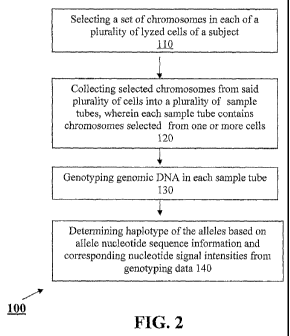
[041] Figure 2 is a flow chart showing an embodiment of the HaploDissection
method.
[042] Figure 3 is a picture showing chromosome collection using a Leica AS LMD
computer-directed laser microdissection. Chromosomes in the collection area
were collected
for haplotyping.
11
CA 02740205 2011-04-11
WO 2010/044923 PCT/US2009/047765
[043] Figure 4 is a flow chart showing an embodiment of a method for
determining
haplotype-based genotyping data.
[044] Figure 5 is a flow chart showing another embodiment of the
HaploDissection
method using single cell lysate.
[045] Figure 6 is a diagram illustrating the principle of single cell
haplotype split.
[046] Figure 7 is a diagram illustrating the principle of haplotyping with a
HaploDissection method using single cell dissection.
[0471 Figure 8 is a flow diagram showing the steps of single cell dissecting
method
and some haplotyping results. Haplotypes are shown by their parental origins
(Fa, father; Mo,
mother).
DETAILED DESCRIPTION
[0481 The practice of the embodiments described in further detail below will
employ, unless otherwise indicated, conventional methods of genetics, genomics
molecular
biology, cell biology, diagnostics and bioinformatics within the skill of the
art. Such
techniques are explained fully in the literature. All publications, patents
and patent
applications cited herein, whether supra or infra, are hereby incorporated by
reference in their
entirety.
[0491 One aspect of the present invention relates to a method for molecular
haplotyping of a subject. This method is referred to hereinafter as the
"HaploDissection"
method. As described in more detail below, the new method overcomes the
bottleneck on the
haplotype length, and has no limitations on SNP numbers or sample numbers.
This invention
meets the needs of accurate haplotypes in genetic studies, genomic studies and
epigenomic
studies, especially the genome-wide association studies (GWAS), long-cis-
regulatory
12
CA 02740205 2011-04-11
WO 2010/044923 PCT/US2009/047765
interactions for gene expression, and chromatin remodelling studies, Accurate
haplotypes are
required for interpretation and translation of these results into clinical
practice.
[050] One embodiment of the HaploDissection method is illustrated in Figure 1.
Briefly, the HaploDissection method maintains the phase information of DNA
samples from a
subject that are subjected to genotyping, but this maintenance is not based on
the separation
of two chromosome copies or isolation of a single copy. The method simply
introduces an
imbalance into the steady 1:1 ratio on the quantities of two parental
chromosomes by
harvesting a relatively small number of chromosomes into each sample tube.
Thus, while the
genotype/allele information is still reserved in the DNA samples and
applicable to hi-
throughput genotyping platforms, the phase information is recorded into the
quantitative ratio
between two alleles. These relative ratios of alleles are actually one of the
outputs from all of
those genotyping platforms, but they are usually ignored in the genotyping
interpretation. The
HaploDissection method will read the output information on both allele
readings and allele
intensity readings. The genotyping information and the phase information are
then analyzed
by a specially designed algorithm to determine the haplotype of the subject.
In one
embodiment, the genotyping information and the phase information are then
analyzed by a
special designed software called "HapReader."
[051] Because the HaploDissection method protects the chromosome integrity
while
introducing the quantitative allele imbalance, the haplotypes obtained from
this method will
be at the entire chromosome range, or unlimited by distance.
[0521 An embodiment of the HaploDissection method is shown in Figure 2. In
this
embodiment, the method 100 includes: selecting (l 10) a set of chromosomes in
each of a
plurality of lyzed cells of a subject;, collecting (120) selected chromosomes
from said
13
CA 02740205 2011-04-11
WO 2010/044923 PCT/US2009/047765
plurality of cells into a plurality of sample tubes, wherein each sample tube
contains
chromosomes selected from one or more cells; genotyping (130) genomic DNA in
each
sample tube; and determining (140) haplotype of the alleles based on allele
nucleotide
sequence information and corresponding nucleotide signal intensities from
genotyping data.
[053] The chromosomes may be selected from any type of cells. In one
embodiment, the cells are peripheral blood lymphocytes isolated from a blood
sample of the
subject. Methods for isolating peripheral blood lymphocytes are well-known in
the art. In
one embodiment, the isolated peripheral blood lymphocytes are cultured in a
growth medium
until they start proliferation. Growth media capable of inducing proliferation
are well known
in the art. In one embodiment, the growth medium is RPM11640 containing 15%
FBS and
100 unit/ml Penicillin/Streptomycin. Mitogens such as phytohemagglutinin (PHA)
may be
added to the culture medium to stimulate cell proliferation, and a mitotic
inhibitor such as
colcemid is added to arrest the cells in metaphase. The proliferating
peripheral blood
lymphocytes are then harvested, lyzed, and stained to display chromosomes
using well-known
cytogenetics procedures. A set of chromosomes, typically around half of the
chromosomes in
a lyzed cell, is randomly selected and collected for further analysis. Figure
3 shows an
example of selecting chromosomes from a single cell for collection using
computer-directed
laser microdissection. Chromosomes in the marked area were collected. As noted
earlier, the
chromosomes are randomly selected for collection.
[054] Randomly selected chromosomes from randomly selected cells are collected
into a plurality of sample tubes. Each tube contains chromosomes collected
from a plurality
of cells. In one embodiment, each tube contains chromosomes collected from 2-
20,
preferably 2-10, randomly selected cells. A total of 2-12, preferably 4-8, and
sample tubes
14
CA 02740205 2011-04-11
WO 2010/044923 PCT/US2009/047765
were collected for each subject. The selected chromosomes are collected using
technologies
that preserve the chromosome integrity. In one embodiment, the chromosomes are
collected
using computer-directed laser microdissection.
[055] In the next step, genotyping is performed on collected chromosomes in
each
sample tube. In one embodiment, the collected DNA is amplified by PCR using
methods for
unbiased whole genome amplification (WGA). The amplified DNA is then subjected
to
whole genome genotyping. For each subject, 2-4 tubes of samples will be
subjected to the
genotyping to ensure high genome coverage and achieve duplications for
accuracy. In one
embodiment, a genomic DNA sample is included for whole genome genotyping.
[056] The output dataset from genotyping is analyzed to determine the
haplotype of
the subject using a method that integrates the sequencing information with the
phase
information, which is reflected on the intensity of sequencing signals at each
loci. Briefly, the
allele nucleotide sequence information and corresponding nucleotide signal
intensities are
extracted from the genotyping data obtained from the collected chromosomes in
each sample
tube. The nucleotide signal intensity ratio of two alleles (allelic intensity
ratio) for each locus
of a chromosome is calculated and is used for and determining the haplotype of
the alleles.
[057] Figure 4 is a flow chart showing an embodiment of a method for
determining
allele haplotype based on the nucleotide sequence information and
corresponding nucleotide
signal intensities of the alleles. In this embodiment, the method 200
includes: extracting
(21.0) allele nucleotide sequence information and corresponding signal
intensities from
genotyping data, calculating (220) relative ratio of nucleotides A, C, G, and
T at homozygous
loci, determining (230) a k value for each nucleotides to adjust their signal
intensities to the
same level for a given particular experiment, adjusting (240) nucleotide
signal intensities at
CA 02740205 2011-04-11
WO 2010/044923 PCT/US2009/047765
heterozygous loci using the k value; calculating (250) the signal intensity
ratio of two alleles
(allelic intensity ratio) for each locus; sorting (260) the order of alleles
at each locus by allelic
intensity ratio, keeping the higher-intensity-allele on a first column and the
lower-intensity-
allele on a second column, determining (270) whether there is breakpoint in
each
chromosome, if there is no breakpoint in a chromosome, forming (280) one
haplotype with
alleles in the first column forming the haplotype, and another haplotype with
alleles in the
second column, if there is a breakpoint in a chromosome, using (290) results
from other
chromosome collection tubes to bridge over the breakpoint.
[058] In one embodiment, the analysis is performed using a "HapReader"
software
that is specifically developed for the HaploDissection technology. The
software is described
in more detail in the EXAMPLES.
[059] Another embodiment of the HaploDissection method is based on the
separation of haploid genome from a single cell lysate. In all of the current
molecular
haplotyping methods, the reduction from diploid to haploid is achieved from an
uncertain and
large number of chromosome copies. The method of the present invention is
built upon the
fact that each somatic cell has exactly two copies of each chromosome. This
exact number
provides a very simple way to separate the two chromosomes. Briefly, the
method separates
chromosomes on a single cell basis. This new starting point makes the
separation much easier
than previous inventions because there are only two copies of each chromosome
at the
starting point. In addition, the method overcomes a major drawback of the
other methods the short haplotype distance. Therefore, this new method opens
the door to a simple and
effective way to obtain long-distance haplotypes.
16
CA 02740205 2011-04-11
WO 2010/044923 PCT/US2009/047765
[060] As shown in Figure 5, the method 300 includes the steps of isolating
(310)
one or more single diploid cells from the subject; lysing (320) each single
diploid cell from
the subject to generate one or more single cell lysate; dividing (330) each
single cell lysate
into two equal aliquots; genotyping (340) genomic DNA in each aliquot;
creating (350) a
catalogue of genotyping data from all aliquots; and determining (360)
chromosome haplotype
of the subject based on the catalogue.
[061] The diploid cells can be buccal cells, lymphocytes or any other cell
types from
the subject. In one embodiment, human buccal cells are collected from a
subject. Buccal
cells are the cells on the inner lining of the mouth or cheek. They are
routinely shed and
replaced by new cells. As the old cells die, they accumulate in the saliva in
the mouth and can
easily be collected by a simple procedure using mouthwash. Buccal cells can be
easily
collected by swabs, cytobrushes, mouthwash, and treated cards, such as FTA or
IsoCode
cards,
10621 Next, single cells are isolated and kept in individual tubes. This can
be done
by any method that can isolate single cells while preserving genomic DNA in
cells.
Examples of such method include, but are not limited to, laser microdissection
or flow
cytometry.
[0631 In one embodiment, isolation of single cells is carried out using laser
microdissection or any other methods such as cell sortings. Laser
microdissection is a
micromanipulation procedure that allows cutting off precisely the cells of
interest from tissue
samples or smears under direct microscopic visualization by a laser beam. The
region of
interest is marked on the computer monitor and then cut out by computer
control. The single
17
CA 02740205 2011-04-11
WO 2010/044923 PCT/US2009/047765
cell in the collection tube can be immediately checked by an inspection mode
under the
microscope to ensure the successful isolations.
[064] The staining protocol used during cell isolation should not interfere
with the
subsequent DNA amplification and allele determination. Preferred staining
methods do not
include any fixation step and are not based on the use of aggressive chemical
agents. In one
embodiment, the cells are stained with Papanicolau. In another embodiment, the
cells are
stained with Hematoxylin and eosin (HE).
[065] Single cells collected by microdissection are then subjected to cell
lysis. Many
techniques are available for cell disruption, including physical and detergent-
based methods.
The technique chosen for the disruption of cells must take into consideration
the
compatibility with the intended downstream applications - genotyping and
haplotyping.
Therefore, the cell lysis method should not attach the DNA molecules
aggressively and break
down the chromosomes into small pieces. Any genornic DNA preservative,
effective, simple,
and low-cost methods can be selected. The origin of the cells or tissues
should also be
considered with choosing cell lysis protocols.
[066] Both physical lysis methods and detergent-based lysis methods may be
used
for cell disruption. Preferred cell lysis method includes hypotonic lysis and
proteinase K
lysis.
[067] Next, the single cell lysate is divided equally into two tubes. To
ensure that a
haploid copy for any given chromosomes is collected, multiple single cell
lysates and
corresponding splits are collected. In one embodiment, 4-12 single cell
lysates are collected.
In other embodiment, 6-10 single cell lysates are collected. In yet other
embodiment, 8 single
cell lysates are collected.
18
CA 02740205 2011-04-11
WO 2010/044923 PCT/US2009/047765
[0681 As shown in Figure 6, a diploid cell contains two copies of each
chromosome
(one copy from father, one copy from mother). When a solution containing two
copies of a
chromosome is equally aliquoted into two tubes, the two copies may both go to
tube I or tube
2, or they may each enter a different tube. The pattern of the chromosome
presence in these
two splitting tubes can be easily monitored. If one tube does not contain this
chromosome,
the other tube must contain both copies of the chromosome. If both tubes
contain this
chromosome, then they must contain one copy in each.
[0691 For one split operation, the probability of obtaining a haploid copy for
any
given chromosomes is:
Success Probability = Failure Probability = t/4 +'/4 = 0.50.
[070] If n single cells are collected, and the splits describe above are
performed n
times (one for each single cell), then the probability that none of these tube
pairs has a haploid
copy for a given chromosome (all tubes are either diploid or aploid with
regard to this given
chromosome) is:
Failure Probability = 1/2".
Success Probability = 1 - 1/2".
[0711 Therefore, if 8 single cells are collected (i.e., n=8) and split into 16
aliquots,
the likelihood of obtaining a haploid copy of any particular chromosome in
these splits is:
1 - 1/28 = 0.9961.
[0721 This means that there is a 99.61% chance that at least one of these 16
split
tubes will contain a haploid copy of the target chromosome. Accordingly, if
the sample size
is 1000 human individuals, with 8 cells collected from each individual, in the
first round of
19
CA 02740205 2011-04-11
WO 2010/044923 PCT/US2009/047765
split operation, 996 individuals will successfully obtain a haploid copy of
any chromosomes
for molecular haplotyping.
[073] After the split, one tube may contain a haploid copy for one chromosome;
however, it may contain two copies of another chromosome. If one tube contains
a haploid
copy of chromosome A, two copies of chromosome B and no chromosome C, this
tube can
still be perfectly used for subsequent analysis (such as haplotype
determination) on
chromosome A. The presence of two copies of chromosome B and the absence of
chromosome C will not interfere with the results on chromosome A.
[074] There is an extremely rare case in which the haplotypes from one single
somatic cell do not represent the haplotypes of the same individual. This rare
case is mitotic
crossover which occurs in somatic cells. It is known that mitotic crossover
may occur in
some asexually reproducing fungi and in human cancer cells. Therefore, it is
necessary to
take cautions and obtain multiple cells for haplotyping a subject with cancer.
In fact, this case
can be easily detected by the single cell split strategy.
[075] Specifically, if a cell from an individual contains more than two copies
of a
certain chromosome, the presence of that chromosome in both split tubes will
not be an
indication of haploid chromosome in each tube. For example, if there are 3
copies of a
chromosome, when both tubes contain this chromosome, one tube will have one
copy, the
other tube will have two copies. The tube with two copies will show
heterozygous genotypes
at some polymorphic sites, Therefore, our method may detect the copy number
polymorphisms.
CA 02740205 2011-04-11
WO 2010/044923 PCT/US2009/047765
[076] The genomic DNA in each tube is then amplified for genotyping. Any
methods of unbiased whole genome amplification (WGA) may be employed. Unlike
polymerase chain reaction (PCR), which aims to amplify a specific sequence,
WGA aims to
amplify the entire genome without preference. Comprehensive WGA requires
faithful
replication of 3 billion bases without the loss or distortion of any
particular loci or alleles.
[077] Examples of such methods include, but are not limited to, multiple
displacement amplification (MDA, GE Healthcare GenomiPhi and Qiagen Repli-g),
primer
extension preamplification (PEP), improved primer extension preamplification
(iPEP),
degenerate-oligonucleotide-primed PCR (DOP, Sigma GenomePlex). The current WGA
methods in the market are different on (i) amplification power and yield; (ii)
fidelity; (iii)
amplification product length; (iv)scalability and (v) the ability to amplify
small arnounts of
starting material, including single cells. For example, Repli-g and GenomiPhi
yield products
around 10 kb in size, whereas the Sigma GenomePlex yields products around
several hundred
base pairs, Because the distance that the molecular haplotyping method of the
present
invention can resolve does not rely on the length of amplification product,
the length feature
of WGA method is not a critical feature for the present invention. Instead,
the amplification
power and potential allele bias and locus bias are critical to the present
invention.
[078] The feasibility of amplifying from haploid chromosomes has been
previously
well-demonstrated by the genetic research using human sperms. The ability to
genotype
single sperm was first reported in 1988 (Li HH, et al., Nature 335: 414-417,
1988). Now
genotyping on the DNA samples from single sperm cells (haploid) has been
widely used by
forensic scientists (Di Martino D, et al., Forensic Sci 1'nt 146 Suppl: S 151-
153, 2004). It has
been shown that up to 10.4 pg of DNA can be amplified by TaqGold DNA
polymerase for a
21
CA 02740205 2011-04-11
WO 2010/044923 PCT/US2009/047765
reliable STR (short tandem repeats, a type of genetic polymorphisms in
parallel to SNP)
profile. In addition to single sperm cells, amplification of single
lymphocytes (diploid) or
single blastomeres (diploid) using the WGA-multiple displacement amplification
(MDA)
method has been successfully carried out.
[079) A common concern of WGA is the allelic drop-outs at heterozygous loci,
which is consequence of a biased uneven amplification among the two alleles at
one
polymorphic locus. The allele drop-out will make a heterozygous individual to
display as a
homozygous individual in genotyping. Accordingly, when a homozygous genotype
was
observed, it is still theoretically possible that this is false-homozygous
genotype; it may come
from allele drop-out in WGA. When using the cell-split method for molecular
haplotyping,
because there is a single copy of haploid, if there is no locus bias at a
particular locus, the
allele reading in the subsequent genotyping assays will represent the allele
on that
corresponding haplotype. Therefore, there will be no concern necessary to
distinguish allelic
dropout and true hoinozygotes.
[080] As discussed above, if 16 split tubes from 8 single cells are collected
from
one individual, there is a 99.6% that some tubes contain a haploid copy of any
given
chromosome. Other tubes may contain diploid copies or no copy (aploid) of the
same
chromosome. Each tube may contain haploid copies of certain chromosomes,
diploid copies
of other chromosome(s), and no copy of yet other chromosome(s). Therefore, it
is necessary
to create a catalogue for each tube about its content for subsequent analysis,
including
haplotype determination.
22
CA 02740205 2011-04-11
WO 2010/044923 PCT/US2009/047765
[0811 The cataloguing can be done by any methods that can detect the DNA
existence. For example, PCR can be used to detect the existence of DNA
fragments. If the
PCR is designed to cover a sufficient number of regions that representative
all of the
chromosomes of genome, then it can be used to create a genome wide catalogue.
PCR can be
also designed to cover only the target genome region of a research project.
Besides, whole
genome tiling array can be also used to create this catalogue, but in a more
systematic and
high-throughput pattern.
[0821 If both tubes of a split pair contain chromosome A, then both tubes have
a
haploid copy of chromosome A for haplotype determination. The tubes with a
haploid copy
of any particular chromosomes will be selected based on this catalogue for
subsequent
analysis. The samples fror this procedure can be used as regular DNA samples
and directly
subjected to various high-throughput genotyping assays. In haplotype
determination, the
genotypes from. haploid samples will be compared with the diploid samples from
the same
individual as quality control. Any false-haploid tubes will be easily detected
by this
comparison.
[083) Another embodiment of the HaploDissection method allows for
determination
of haplotype of certain chromosomes from a single cell. The method comprises:
isolating a
single diploid cell from the subject, lysing and staining the isolated single
diploid cell to
display chromosomes, collecting a set of chromosomes from the single cell by
laser
microdissection, genotyping genomic DNA in the collected chromosomes,
genotyping
genomic DNA from another intact single cell from the same subject, determining
haplotype
of a chromosome in the collect set of chromosomes, wherein said chromosome is
present in
the haploid form in said collected set of chromosomes.
23
CA 02740205 2011-04-11
WO 2010/044923 PCT/US2009/047765
]084] As shown in Figure 7, when a cell is cut along the dotted line and the
right
half of the cell is collected, the collected chromosomes include only a single
copy of
chromosomes 2, 3, and 5 (haploid) two copies of chromosome 1 (diploid) and no
copy of
chromosome 4 (aploid). Thus, the genotype calls from conventional genotyping
platforms
with this half cell will directly return the haplotypes for chromosomes 2, 3,
and 5, whereas it
will be still diploid genotypes for chromosome 1, and no genotype calls for
chromosome 4.
[085] As discussed earlier, for each given chromosome, there is a 50% chance
that a
haploid copy is collected if the collected a set of chromosomes contain about
half of the total
number of chromosomes in the single diploid cell. The probability increases
significantly
when multiple sets of chromosomes are collected from multiple single cells.
For example,
there is a greater than 99,6% chance that a haploid copy of a given chromosome
is collected if
8 sets of chromosomes are collected from 8 single cells and each collected set
of
chromosomes contain about half of the total number of chromosomes in the
single cell.
Therefore, the haplotyping of the complete set of chromosomes from a subject
may be
achieved with high probability (greater than 99,6%) using 8 half cells.
[086] Accordingly, in a related embodiment, multiple single diploid cells are
isolated and lysed. Multiple sets of chromosomes are collected; each set is
collected from a
different single cell. The number of collected sets is large enough so that
each chromosome
in the genome of the subject is present in the haploid form at a probability
greater than 99%.
[087] The HaploDissection method described above applies to any eukaryotic
organism. In certain embodiments, the subject is an animal or a plant. In
other embodiments,
the subject is a mammal. In yet other embodiments, the subject is a human.
24
CA 02740205 2011-04-11
WO 2010/044923 PCT/US2009/047765
[088] Another aspect of the present invention relates to a computer-readable
medium having computer-executable instructions for performing the methods of
the present
invention.
[089] Another aspect of the present invention relates to an assay kit for
HaploDissection. In one embodiment, the assay kit contains reagents for cell
collection, cell
lysis and, optionally, cytogenetic staining, as well as reagents for genomic
DNA amplification
and genome genotyping. In another embodiment, the kit further includes a
computer readable
medium having computer-executable instructions for determining haplotype based
on
genotyping data. In another embodiment, the kit further includes an instrument
specifically
designed for collecting a set of chromosomes from single or a few cells for
haplotype reading
and chromosome biology examinations.
[090] In one embodiment, the HaploDissection methods are used in prenatal
diagnostics to detect the important genotype defects of the fetus. It has been
known that some
fetus cells are circulating in the maternal peripheral blood. Therefore, the
fetus cells can be
collected from the pregnant maternal blood. These cells can be subjected to
the haplotype
analysis using the procedure described above. Because it is usually the
haplotypes (the
combination of alleles of different genotypes) that cause the diseases, the
prenatal diagnosis
by haplotype determination will be more accurate than genotype determination.
The single
cell nature of the present invention provides the feasibility of haplotype
determination with
fetus cells in the mother's blood.
[091] In another embodiment, the HaploDissection methods are used in
personalized medicine. Personalized medicine is the practice that doctors
customize
treatment based on a person's specific genetic variations. For instance, two
people who take
CA 02740205 2011-04-11
WO 2010/044923 PCT/US2009/047765
the same anti-hypertension medication may have very different responses. One
may have
severe, even life-threatening side effects, while the other experiences few if
any side effects
and seems to sail through the treatment. The reason why the two people have
such drastically
different reactions to the same medication resides in their genes. People
inherit variations in
their genes, and even slight variations can have a profound effect on which
subtypes of the
same disease the person has and how the person responds to certain
medications.
[092] In a personalized medicine, the current process in a clinic began to
change.
Before a patient takes a single dose of medication, the patient may have a
blood test done to
determine genetic variations. The test may show that patient's variation which
is likely to
have an adverse effect on the particular medication. The doctors can determine
the drug
prescriptions and doses to match the patient's genetics. Therefore, the unique
genetic profile
can help doctors to personalize treatments of patients, improve the drug
development, and
reduce healthcare costs.
10931 It has been widely accepted now that multi-SNP haplotypes are more
accurate
to represent a person's genotype than single-SNP genotype. However, there is
no simple,
cheap and high-throughput experimental method to directly read the haplotypes.
The
statistical haplotype configuration causes many ambiguities. This technical
bottleneck is not
only limiting the efforts to discover the genetic basis underlying the common
diseases, it is
also limiting the application of genetic tests in clinical practice. The I-
IaploDissection
methods may solve this technical bottleneck.
26
CA 02740205 2011-04-11
WO 2010/044923 PCT/US2009/047765
[094] For example, before a patient takes any medication, a few cells will be
collected from his mouth and haplotypes on those disease mutations will be
determined by
using the present methods. Doctors will prescribe a drug with a certain dose
to match a
patient's unique genetic profile to personalize the treatment.
[095] In yet another embodiment, the HaploDissection methods are used in
forensic
testing. True haplotyping provides a greater precision than single SNP
genotyping in forensic
studies, in any case of sexual assault or other crimes, as well as paternity
testing. In many
cases of forensic tests, the available amount of a specimen is usually quite
limited. Because
of the single cell nature of our invention and true haplotype result out of
this technology, the
HaploDissection method will increase both sensitivity and precision.
[096] The present invention is further illustrated by the following examples,
which
should not be construed as limiting. The contents of all references, patents
and published
patent applications cited throughout this application, as well as the Figures
and Tables, are
incorporated herein by reference.
EXAMPLE 1: PREPARATION OF CELLS
1. Sample Collection
[097] Collect blood from human individuals and isolate the lymphocytes.
II. Cell Culture
[098] Culture the lymphocytes in RPMI1640 medium containing 15% FBS and 100
unit/ml Penicillin/Streptomycin.
III. Cell Lysis
[099] 1. At the proliferation stage, add phytohemagglutinin to the cell
culture.
[0100] 2. Harvest cells 48 hours after phytohemagglutinin (PHA) treatment.
27
CA 02740205 2011-04-11
WO 2010/044923 PCT/US2009/047765
[0101] 3. Add Ethidiurrn Bromide (16.7 ug/ml) and Act-D (6.7ug/ml) into cells.
[0102] 4. Incubate at 37 C for 0.5 hour. After 0.5 hour, add colcemid
(0.083ug/ml) into cells and incubate at 37 C for I hour.
[0103] 5. Centrifuge at 1000 rpm for 10 min.
[0104] 6, Aspirate all but 0.3 ml supernatant, gently resuspended cell pellet.
Add
pre-warmed 0.O75mol/L KCl vortex gently to make sure KCl is mixed well with
the pellet.
[0105] 7. Leave at 37 C for 20 min and room temperature for additional 5 min.
Centrifuge at 1000 rpm for 10 min. And remove the supernatant.
[0106] 8. Add cold fixative (methanol: acetic acid, 3:1), gently mix by
inverting
tubes.
[0107] 9. After fixed and centrifuged for three times, cells were dropped on
slide
and geimesa staining for 20 minutes.
[0108] 10. Air dry slide for 20 min in hood.
IV. Chromosome Isolation
[0109] 1. Turn on the laser, microscope (Leica, ASLMD) and computer. Put PCR
tubes in collectors on holders.
[0110] 2. Put slide on supporter.
[0111] 3. Go to the computer screen. Click on LEICA ADMINISTRATOR to open
program.
[0112] 4. Set objective l OX to find cells and then switch to 40X.
[0113] 5. Collect chromosomes: cut and pick up no more than 30 chromosomes at
random from each cell. Collect 4-8 samples. Each sample contains chromosomes
from about
7-11 cells.
28
CA 02740205 2011-04-11
WO 2010/044923 PCT/US2009/047765
[0114] 6. Exit LEICA ADMINISTRATOR program.
[0115] 7. Turn off the computer, microscope and laser by order.
EXAMPLE 2: WHOLE-GENOME DNA AMPLIFICATION
Single Cell Lysis and Fragmentation
[0116] 1. Isolate a single cell into a PCR-ready vessel using laser capture
micro-
dissection, cell sorting, or other method. If sorted, the buffer should be of
low ionic strength,
such as Tris EDTA (TE) buffer, and in the minimal sort volume.
[01171 2. Add a sufficient volume of water to the single cell sample for a
final
volume of 9 mL,
[0118) 3. Prepare a working Lysis and Fragmentation Buffer Solution by adding
2
mL of Proteinase K Solution into 32 mL of the 10 Single Cell Lysis &
Fragmentation Buffer
and vortex thoroughly.
[0119] 4. Add I mL of the freshly prepared Proteinase K Solution-10' Single
Cell
Lysis & Fragmentation Buffer to the single cell sample and mix thoroughly.
[0120] 5. Incubate DNA mix at 50 C for I hour, then heat to 99 C for EXACTLY
four minutes. Note that the incubation is very time-sensitive and any
deviation may alter
results. Cool on ice. Spin down sample prior to proceeding to Library
Preparation.
Library Preparation
[01211 6. Add 2 mL of I Single Cell Library Preparation Buffer to each sample.
[0122] 7. Add I mL of Library Stabilization Solution.
[0123] 8. Mix thoroughly and place in thermal cycler at 95 C for 2 minutes.
[01241 9, Cool the sample on ice, consolidate the sample by centrifugation,
and
replace on ice.
29
CA 02740205 2011-04-11
WO 2010/044923 PCT/US2009/047765
[0125] 10. Add I mL of Library Preparation Enzyme, mix thoroughly, and
centrifuge briefly.
[0126] 11. Place sample in a thermal cycler and incubate as follows:
[01271 16 C for 20 minutes; 24 C for 20 minutes; 37 C for 20 minutes; 75 C
for 5
minutes; and 4 C hold..
10128] 12. Remove samples from thermal cycler and centrifuge briefly. Samples
may be amplified immediately or stored at -20 C for three days.
Amplification
[0129] 13. Add the following reagents to the entire 14 Ml reaction:
10130] 7.5 mL of 10' Amplification Master Mix; 48.5 mL of Nuclease-Free Water;
and 5.0 mL of WGA DNA polymerase.
[0131] 14. Mix thoroughly, centrifuge briefly, and begin thermocycling. The
following profile has been optimized for a PE 9700 or equivalent thermal
cycler:
10132] Initial Denaturation at 95 C for 3 minutes.
[0133] Perform 35 cycles as follows:
[0134] Denature at 94 C for 30 seconds; anneal/extend at 65 C for 5 minutes;
and
hold at 4 C.
[0135] After cycling is complete, maintain the reactions at 4 C or store at -
20 C
until ready for analysis or purification. The stability of WGA DNA is
equivalent to genomic.
[0136] DNA stored under the same conditions.
CA 02740205 2011-04-11
WO 2010/044923 PCT/US2009/047765
EXAMPLE 3: WHOLE GENOME GENOTYPING
[0137] The amplified DNA will be subjected to Illumina high-throughput whole
genome genotyping such as Hap3000K and others. For each person, 2-4 tubes of
samples
will be subjected to the genotyping to ensure high genome coverage and achieve
duplications
for accuracy. A genomic DNA sample will be included for whole genome
genotyping. This
step can be done also by using other high-throughput genotyping platforms.
EXAMPLE 4: HAPLOTYPE DETERMINATION
[0135] Whole-genome genotyping data was obtained from Illumina HumanCNV370-
Duo BeadChip. This BeadChip content covers over 370,000 markers using the
Infinium
Assay. After scanning, all the data uploaded into BeadStudio and analyzed
using the
BeadStudio Genotyping Module, version 3. After stringent filtering with
removal of SNPs
with missing genotype, the remaining SNPs were available for analysis. The
values of theta,
R, X and Y in the Illumina genotyping output are used to determine the
relative ratio of two
alleles of each SNP. Haplotypes are constructed by the allele ratio along the
chromosomes.
[0139] The haplotype construction is done by using the software "HapReader,"
which is specifically developed for this technology. The essential procedure
and algorithm is
below:
[0140) 1.) The analysis will be done at an individual level, person by person.
There
will be no combination of datasheet from different individuals.
[0141] 2) Each person will have 3-5 genotyping datasheets, one if from the
genomic
DNA, and the others are from the chromosome collection tubes. Extract the
allele calls and
their corresponding signal intensities from each Illumia output data sheet.
31
CA 02740205 2011-04-11
WO 2010/044923 PCT/US2009/047765
[0142] 3) Based on the datasheet from the genomic DNA sample, select the
homozygous loci for this person. Calculate the relative ratio of the averages
of the A, C, G,
and T at these loci. Determine a k value for A, C, G, T to adjust their
intensities to the same
level for a given particular experiment.
[01431 4) Using these k values, adjust the heterozygous loci.
[0144] 5) For each locus, calculate the ratio of the two alleles.
[0145] 6) For each locus, sort the order of those two alleles by their allelic
intensity
ratio. Sort all loci by the same way, and keep the higher-intensity-allele on
column-A and the
lower-intensity-allele on column-B.
[0146] 7) Examine and compare the ratio values along the chromosome to
determine
if there is breakpoint in each chromosome.
101471 8) If not at step 7), the alleles in column-A form haplotype, and the
alleles on
column-B will form another haplotype for this person. If yes at step 7), use
the results from
other chromosome collection tubes to bridge over this breakpoint.
101481 One aspect of the present invention lies under this step. Collect
chromosomes
into PCR tubes by microdissection using a Leica ASLMD Laser microdissection
system. In
this step, not all chromosomes are collected from one cell; instead, only part
(around half) of
the chromosomes is collected from any single lyzed cell (Figure 2). The
selection of
chromosomes on any lyzed cells is random. This random collection is respected
from 5-11
randomly-selected cells. All of the chromosomes from these 5-11
microdissections are
collected into one tube. 4-8 tubes are collected for each person. In this
step, the chromosome
integrity is preserved by selecting the laser cutting line on the computer, so
chromosome
integrity is ensured.
32
CA 02740205 2011-04-11
WO 2010/044923 PCT/US2009/047765
[01491 Amplify the collect DNA sample in each PCR tube by using the Sigma
GenomePlex WGA-4 kit for 20-24 cycles. In fact, this step can be done with any
methods of
unbiased whole genomo amplification (WGA). These methods include, but are not
limited to,
multiple displacement amplification (MDA, GE Healthcase GenomiPhi and Qiagen
Repli-g),
primer extension preamplification (PEP), improved primer extension
preamplification (iPEP),
and degenerate-oligonucleotide-primed PCR (DOP, Sigma GenomePlex). Repli-g and
GenomiPhi yield products around 10 kb in size, whereas the Sigma GenomePlex
yields
products around several hundred base pairs.
EXAMPLE 5: DETERMINATION OF HAPLOTYPE USING SINGLE CELL LYSATE
METHOD
[01501 A Leica AS LDM Laser Microdissection system (Leica Microsystems,
Germany) is used to isolate single cells from fresh cytobrush-swab buccal
cells from human
individuals. Briefly, buccal cells are smeared on a foiled slide (a
rectangular UV-sliceable
piece of foil fixed at the margins to a normal microscope slide), air-dried
for 5 min and then
stained very briefly. The section is reviewed under microscope, single cells
are selected and
cut-off from the foiled slide with the Laser Microdissection system. Single
cells are collected
into tubes with 10-ul of cell lysis buffer in each tube. The single cell
lysate in each tube is
then divided equally into two tubes. The genomic DNA in each tube is amplified
WGA for
genotyping. A genome wide catalogue is created using the genotyping data.
Haplotype
determination is carried out using the catalogue.
33
CA 02740205 2011-04-11
WO 2010/044923 PCT/US2009/047765
EXAMPLE 6: DETERMINATION OF IIAPLOTYPE USING SINGLE CELL
DISSECTION METHOD
[0151] Chromosome Microdissection: Lymphocytes were cultured in RPMI1640
medium containing 15% FBS and 100 unit/ml Penicillin/Streptornycin. Cells were
stimulated
by phytohemagglutinin (PHA) for 48 hours, followed by addition of Ethidium
Bromide (16.7
ug/ml) and actinomycin D (6.7uglml) and incubation at 37 C for 30 min.
Colcemid
(0.083ug/ml) was added into cells and incubate at 37 C for I hour. Cells were
collected by
centrifugation at 1,000 rpm for 10 min, resuspended, incubated in pre-warmed
0.075mol/L
KCl at 37 C for 20 min, and then, at room temperature for 5 min. After
fixation with cold
fixative (methanol: acetic acid, 3:1), cells were dropped onto slide to break
the nuclei
followed by Giemsa staining for 20 min. Laser Microdissection Microscope
(ASLMD, Leica,
Germany) was used to collect half of the chromosomes of one cell.
[0152) Whole Genome Amplification (WGA): The collected chromosomes were
amplified by the Sigma GenomePlex WGA4 kit following the manufacturer's
protocol.
Briefly, the sample was incubated in the Lysis and Fragment Buffer at 50 C for
I hour, and
then heated to 99 C for 4 min. Then the Single Cell Library Preparation
Buffer and Library
Stabilization Solution was added into the sample followed by an incubation at
95 C for 2 min.
Library was prepared with the following cycles: 16 C for 20 min, 24 C for 20
min, 37 C for
20 min, and 75 C for 5 min. DNA was amplified by an initial denaturation at
95 C for 3
min followed by 35 cycles of 94 C/30 sec and 65 C/S min. Amplified DNA was
purified by
QlAquick PCR purification kit.
34
CA 02740205 2011-04-11
WO 2010/044923 PCT/US2009/047765
[0153] Genotyping: The Illumina HumanCNV370-Quad BeadChip was used for
genotyping. This BeadChip contains 370,000 markers including SNPs and copy
number
variation (CNV) markers. Three independent microdissected samples and one
genomic
samples extracted with a Qiagen kit were subjected to genotyping experiments,
After
scanning, the data was uploaded into the BeadStudio and analyzed using the
BeadStudio
Genotyping Module version 3. No-call threshold was set at default (0.15).
[0154] Data analysis: Unphased genotypes of GM 10847 and his parents (GM and
GM) were retrieved from the International HapMap Project database (Phase 2
Public Release
#22, and Phase 3 Public Release #1, Phase 2+3 Release #27). The unphased
genotypes of
GM10847 were also retrieved from the Illumina database. Haplotypes of GM10847
was
computationally reconstructed with his parental genotypes by determining the
parental origin
of each allele following the Mendelian Law of Inheritance. In the data
analysis, only those
heterozygous loci of GM10847 were subjected to haplotype determination. The
homozygous
loci were removed because they do not have the haplotyping issue (phase-
known). Allele
calls with both allele intensities below 1,000 in the Illumina genotyping
output were removed.
Genome-wide RepeatMasker detection was retrieved from the UCSC Genome Browser
(Human 2006 March Assembly). All data integration was performed with SAS9. 1.
[0155] The haplotyping method was tested with the individual GM10847 recruited
in
the HapMap project (The International HapMap Consortium 2003) by three
independent
experiments. Following the procedures described above, we the genotype calls
of
microdissected samples were compared with genotype calls of genomic DNA as
well as of
data downloaded from the International HapMap Project database (Phase 2 Public
Release
#22). The monosomic, disomic and null states of each chromosome in each sample
were
CA 02740205 2011-04-11
WO 2010/044923 PCT/US2009/047765
indicated by whether the chromosome-wide heterozygous calls were converted to
homozygous calls in the microdissection samples (Figure 8). It was found that
that sample 1
successfully haplotyped chromosomes 2, 4, 6, 15, 16, 17, 18, and 20; sample 2
haplotyped
chromosomes lq, 3, 4, 5, 10, 16, 17, 18, 20 and 21; sample 3 haplotyped
chromosomes 3, 7,
9, and 20. Totally 24,481 heterozygous loci were phased.
01561 The accuracy of this method was determined by replications and
comparison
with haplotypes resolved from unphase genotypes (HapMap Phase2 Rel#22) using
the trio
structure under Mendelian Law of Inheritance (Hodge SE, et at, Nat Genet,
21(4):360-1;
1999). Among those 24,245 SNP loci that were successfully phased by our 7DDNA
haplotyping method, 464 SNP loci were not covered by the HapMap Phase 2
genotype data,
4,744 SNPs do not have unambiguous haplotypes from the HapMap genotypes due to
all-
three-heterozygote, and 142 SNPs were not phased due to missing data in
HapMap2. So we
compared 18,895 SNP loci between our haplotypes and HapMap2 derived
haplotypes. There
were 18,625 SNPs (98.57%) that showed consistent allele phase as compared with
haplotypes
resolved by HapMap trio structure. Among those 270 discordant SNP loci, 45 SNP
loci were
due to the HapMap Phase 2 genotyping error as compared with phase 3 genotype
calls, and
103 loci were on various repeats as detected by RepeatMasker. The other
discordance may be
potentially ascribed to whole genome amplification errors, genotyping errors,
or un-annotated
segmental duplications besides those identified by RepeatMasker. It was
further determined
the accuracy directly by 2,089 replications, among which 2,065 SNPs showed
consistent
result, none of them had inconsistent haplotype, and 24 SNP loci had diploid
allele calls in
one of the duplicates although the entire chromosome showed discordance (Table
1), with an
estimate of 98.85% as the accuracy rate.
36
CA 02740205 2011-04-11
WO 2010/044923 PCT/US2009/047765
Table 1. An estimate of accuracy rate by data reproductivity.
Chr Total repeated SNPs Consistent Inconsistent Accuracy%
Chr3 517 516 1 99.81
Chr4 565 554 11 98.05
Chrl6 189 186 3 98.41
Chr17 217 215 2 99.08
Chrl8 212 208 4 98.11
Chr2O 389 386 3 99.23
Total 2,089 2,065 24 98.85
[0157] This haplotyping method does not have apparent limitations on the
phasing
distance, total SNP number, and marker types. The procedure is simple and
inexpensive; it
does not require a complicated optimization of experimental condition and the
cost is close to
conventional high throughput genotyping assays. In addition, there is no
apparent barrier for
this approach to be amendable to automation if chromosome microdissection is
automated.
The method may be further improved by using better WGA method for single-cell
DNA,
newer versions of high-throughput genotyping chips or deep sequencers, and
more specific
chromosome staining, such as chromosome painting of particular chromosomes.
[01581 The above description is for the purpose of teaching the person of
ordinary
skill in the art how to practice the present invention, and it is not intended
to detail all those
obvious modifications and variations of it which will become apparent to the
skilled worker
upon reading the description. It is intended, however, that all such obvious
modifications and
variations be included within the scope of the present invention, which is
defined by the
following claims. The embodiments are intended to cover the components and
steps in any
sequence which is effective to meet the objectives there intended, unless the
context
specifically indicates the contrary.
37