Note: Descriptions are shown in the official language in which they were submitted.
CA 02740231 2011-04-11
WO 2010/045389 PCT/US2009/060717
1
METHOD AND APPARATUS FOR INCREASING ADIPOSE VASCULAR FRACTION
Technical Field
The present invention relates to a method and device to separate enriched
vascular adipose
tissue from mammalian fat. In one example of the application, the tissue may
be derived from
liposuctioned adipose tissue.
Background Art
Adipocytes are the cells that primarily compose adipose tissue, specialized in
storing energy as
fat. There are two types of adipose tissue, white adipose tissue (WAT) and
brown adipose
tissue (BAT), which are also known as white fat and brown fat, respectively,
and comprise two
types of fat cells.
In histology, adipose tissue or body fat or just fat is loose connective
tissue composed of
adipocytes. Adipose tissue is derived from lipoblasts. Its main role is to
store energy in the form
of fat, although it also cushions and insulates the body. Obesity or being
overweight in humans
and most animals does not depend on body weight but on the amount of body fat.
Adipose
tissue also serves as an important endocrine organ by producing hormones such
as leptin,
resistin, and the cytokine TNFa. The formation of adipose tissue appears to be
controlled by the
adipose gene. Adipose tissue was first identified by the Swiss naturalist
Conrad Gessner in
1551.
Liposuction, also known as lipoplasty ("fat modeling"), liposculpture suction
lipectomy or simply
lipo ("suction-assisted fat removal") is a cosmetic surgery operation that
removes fat from many
different sites on the human body. Areas affected can range from the abdomen,
thighs,
buttocks, to the neck, backs of the arms and elsewhere.
Auto lipo-transfer is removing fat by liposuction processing it and
transferring it back into the
original host for purposes of primarily aesthetic and cosmetic enhancement, or
skin/tissue/wound/scar defect correction or regeneration.
Autologous stem cell transplantation is a procedure in which stem cells are
removed, and/or
processed, and/or stored, and later given back to the same person.
CA 02740231 2011-04-11
WO 2010/045389 PCT/US2009/060717
2
Growth medium or culture medium is a liquid or gel designed to support the
growth of
microorganisms or cells, or small plants like the moss Physcomitrella patens.
There are
different types of media for growing different types of cells.
There are two major types of growth media: those used for cell culture, which
use specific cell
types derived from plants or animals, and microbiological culture, which are
used for growing
microorganisms, such as bacteria or yeast. The most common growth media for
microorganisms are nutrient broths and agar plates; specialized media are
sometimes required
for microorganism and cell culture growth. Some organisms, termed fastidious
organisms,
require specialized environments due to complex nutritional requirements.
Viruses, for example,
are obligate intracellular parasites and require a growth medium composed of
living cells.
Adipose derived stem cells (ADSC) have been found to exhibit pleuripotential
and regenerative
capabilities with the promise of much therapeutic potential. However, more
recent studies
suggest that cells removed from contact with their native matrix can exhibit
neoplastic behavior
or abnormal differentiation. Additionally, isolated ADSC in the animal model
clearly
demonstrates the loss of cell adhesion and increased metastatic capability
despite use of many
different matrices to prevent movement of ADSC from the original injection
site. A safe
alternative to direct removal and isolation of ADSC, therefore, is both
necessary and critical.
The anatomic location of ADSC is within the perivascular space of fat.
Therefore, fractions of fat
rich in microvasculature will have a higher concentration of ADSC. The process
of isolating
ADSC by direct enzyme degradation or mechanical separation has been proposed
by others
and presents a labor intensive method of ADSC procurement. An example of
isolating ADSC
from lipoaspirate fat is illustrated in US Patent No. 6,777,231. However, this
method of
separating the ADSC from native matrix and tissue is not only inconvenient,
time consuming
and expensive, it is also potentially dangerous in that physically detached
cells may exhibit
tumor like characteristics when heavily manipulated during separation.
Moreover the equipment
utilized for the method detailed in Patent No. 6,777,231 is prohibitively
expensive to purchase
and maintain. For this reason an alternative method for separating adipose
rich fractions of
adipose tissue from lipoaspirate without harsh chemical or enzymatic treatment
or potentially
dangerous cellular labeling is needed.
Development of an alternative method which can separate adipose rich fractions
of adipose
CA 02740231 2011-04-11
WO 2010/045389 PCT/US2009/060717
3
tissue from lipoaspirate without harsh chemical or enzymatic treatment or
potentially dangerous
cellular labeling represents a great improvement in the field of liposuction
and satisfies a long
felt need of the medical profession.
Disclosure of Invention
This invention is a method of increasing the vascular fraction of adipose
tissue comprising the
steps of:
breaking down adipose tissue into small pieces;
washing the pieces to remove blood, tumescent fluid and detached ADSC;
placing the washed pieces in a container;
processing the container so that oil, vascular rich fat, vascular poor fat and
aqueous phases
separate into layers; the vascular poor fat having a pure yellow color; the
vascular rich fat
having an orange color;
attaching the container to a detection chamber in a detection device so that
the material within
the tube (i.e. all the layers described above) are urged out of the tube in
order;
applying pressure to the container;
removing and discarding the aqueous phase;
collecting the vascular rich fat;
detecting with the detection device when the vascular poor fat layer reaches
the detection
chamber; and
ceasing to apply pressure to the container.
Preferably the pieces are small enough to pass through a liposuction cannula.
Preferably the
container is a syringe. Alternatively the container is a tube with a tapered
fitting at its lower
end. Preferably the tapered fitting is a Luer-Lok .
Preferably processing is performed via application of centrifugal force.
Pressure can be applied
by mechanical means or by pressurized gas.
CA 02740231 2011-04-11
WO 2010/045389 PCT/US2009/060717
4
Finally, the collected vascular rich fat may be transferred into any mammalian
host.
The fat can be broken down with any suitable form of energy, including: laser,
sonic and radio
wavelength. More specifically, the fat can be broken down with any suitable
method including:
lithotripsy, hyfrecation, phacoemulsification, sonication, rotating blades,
serial filtration, and
forced screen filtration. Alternately, the fat can be broken down with any
suitable chemical
means including: collagenase and hypertonic media.
Washing can be accomplished with a material including saline, tissue culture
media and
phosphate buffered solution. Suitable tissue culture media include: GMEM,
RPMI, Eagle's,
Fischer's, DMEM, Iscove's, McCoy's, L-15, DME-F1, and Ham's F12 or equivalent.
The
washing step may further include the use of a filter of pore size that allows
single cells of ADSC
to pass through.
A non-toxic gradient may be added to the container to improve separation of
the layers. The
non-toxic gradient may be: tissue culture media (as described above),
Histopaque 1077, wax,
petroleum jelly, Percoll and CsCI or equivalent.
The detection device may be a spectrophotometer a colorimeter or an oximeter.
Thus the
detection device can detects when the vascular poor fat layer reaches the
detection chamber
by color or a selected wavelength of electromagnetic radiation.
This invention is also a device for detecting when a vascular rich fat layer
has passed through a
container containing a material including the vascular rich fat layer and a
vascular poor fat layer
comprising:
a means for applying pressure on the material in the container;
a detection chamber for containing material pushed out of the container;
a light source positioned at one side of the detection chamber outputting
light of a selected
wavelength; the detection chamber being translucent or transparent to the
selected wavelength;
a photodetector positioned opposite the detection chamber detecting the light;
control electronics connected to the photodetector; and
CA 02740231 2011-04-11
WO 2010/045389 PCT/US2009/060717
an indicator connected to the electronics.
The indicator may be a lever, audio alarm, servomechanism (the latter stopping
the progress of
the material in the container upon detection that the material in the
container has absorbed light
at a preselected wavelength) or a light.
5 The selected wavelength may correspond to the pure yellow color of the
vascular poor fat
(about 570 nm), or the absorption wavelength of iron in hemoglobin, or the
pure orange color of
the vascular rich fat (about 590 nm) or the absorption wavelength of
oxygenated hemoglobin
(600 to 750 nm) or the absorption wavelength of deoxygenated hemoglobin (850
to 1000 nm).
The light source and the photodetector may be positioned on the same side of
the collection
tube (reflectance spectroscopy) or on opposite sides of the collection tube
(transmission
spectroscopy).
The device may be disposable or sterilizable. The container may be a syringe
or a container
with a tapered fitting (a Luer-Lok ).
The means for applying pressure may be a piston in which case the invention
may further
comprise:
a motor connected to the control electronics;
a second means for applying pressure activated by the motor and positioned to
push on the
piston; and
the control electronics is additionally programmed to turn off the motor when
the material in the
detection chamber absorbs at the selected wavelength.
The means for applying pressure may be pressurized gas in which case the
invention may
further comprise:
a solenoid valve connected to the control electronics and the means for
applying pressure; and
the control electronics is additionally programmed to activate the solenoid
valve when the
material in the tube absorbs at the selected wavelength.
An appreciation of the other aims and objectives of the present invention and
an understanding
CA 02740231 2011-04-11
WO 2010/045389 PCT/US2009/060717
6
of it may be achieved by referring to the accompanying drawings and
description of a preferred
embodiment.
Brief Description of Drawings
Figure 1 is a drawing of a preferred embodiment of the invention after the
washed adipose
tissue has been transferred into a syringe and centrifuged to separate the
oil, fat, vascular rich
fat and aqueous phase. Connected to the base of the syringe is a color
detection device that is
able to distinguish pure yellow fat from vascular rich yellow fat. Note that
the indicator is in the
PUSH position.
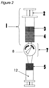
Figure 2 is a drawing of a preferred embodiment of the invention after the
vascular rich fatty
layer has been transferred from the top syringe into a new therapeutic syringe
below. Note the
indicator is now detecting the vascular poor fraction and has switched to the
STOP position.
The vascular poor fraction and the oil phase are left within the original top
syringe.
Figure 3 is a schematic drawing showing in more detail the workings of the
detector and a way
of automating the preferred embodiment.
Figure 4 is a schematic drawing showing an alternate embodiment of this
invention and a way
of automating this embodiment.
Figure 5 is a sketch illustrating reflectance spectrophotometry.
Best Mode for Carrying Out Invention
While the present invention is described herein with reference to illustrative
embodiments for
particular applications, it should be understood that the invention is not
limited thereto. Those
having ordinary skill in the art and access to the teachings provided herein
will recognize
additional modifications, applications, and embodiments within the scope
thereof and additional
fields in which the present invention would be of significant utility.
The following glossary should be used when reading this document.
Autograft - a tissue or organ that is grafted into a new position on the body
of the individual from
which it was removed.
CA 02740231 2011-04-11
WO 2010/045389 PCT/US2009/060717
7
Autolipotransfer: see lipotransfer. Same as lipotransfer but more specifically
indicates the fat
comes from the same person, hence autologous or "auto" for abbreviation
purposes. This is
nomenclature is commonly used and defined as above in the field of cosmetic,
reconstructive
surgery.
Avascular - not associated with or supplied by blood vessels.
Cannula - a metal tube for insertion into the body to draw off fluid or to
introduce medication.
Heme - a deep-red iron-containing blood pigment, C34H32N4O4Fe, obtained from
hemoglobin.
Hyfrecation - a method of ablation or cauterization via energy delivery to
tissue.
Infranatant - the bottom liquid phase of liposuction fluid within the
liposuction container as
opposed to the more buoyant, less dense supranatant (upper phase) which
usually contains the
fat and oil.
Lipoaspirate - the combination of fat, tumescent fluid, blood and serous fluid
that is aspirated
out in the process of performing liposuction.
Liposuction - the surgical withdrawal of excess fat from local areas under the
skin by means of
a small incision and vacuum suctioning.
Lipotransfer the process of harvesting fat from one region of the body and
transplanting to
another region for cosmetic, regenerative or reconstructive surgery purposes.
Lithotripsy - pulverization of kidney stones or gallstones by means of a
lithotripter.
Lithotripter - a device used for fragmenting kidney stones with ultrasound
waves
Micrograft a smaller graft which can and often is visible only under high
powered magnification
or biochemical assay or cytometry.
Morselated - cut into smaller pieces.
Morsellized - to have been cut into smaller pieces.
Neoplasm - an abnormal growth of tissue in animals or plants. Neoplasms can be
benign or
malignant. Also called tumor.
CA 02740231 2011-04-11
WO 2010/045389 PCT/US2009/060717
8
Neoplastic - of or related to or having the properties of a neoplasm.
Perivascular - of, relating to, occurring in, or being the tissues surrounding
a blood vessel.
Phacoemulsification - the removal of a cataract by first liquefying the
affected lens with
ultrasonic vibrations and then extracting it by suction.
Pluripotential - to have the potential of being pluripotent.
Pluripotent - not fixed as to developmental potentialities: having
developmental plasticity.
Sonication - the process of dispersing, disrupting, or inactivating biological
materials, such as
viruses, by use of sound-wave energy.
Spectrophotometric - of or pertaining to a spectrophotometer or a
spectrograph.
Spectrophotometer - an instrument for making photometric comparisons between
parts of
spectra.
Tissue culture media - a solution of balanced salts which prevent cells from
dehydrating or
lysing. They sometimes also contain additional nutrients to ensure long term
cell viability.
Xenograft - a graft obtained from a member of one species and transplanted to
a member of
another species.
Those familiar with the field of aesthetic surgery are aware that adipose
tissue may be
commonly obtained from liposuction and may be performed wet (with tumescent
fluid) or dry
(without tumescent). Adipose tissue may also be excised by sharp surgical
dissection as well.
In this invention, the procured adipose tissue is then mechanically or
chemically disrupted such
that the tissue is broken down into small pieces: preferably small enough to
traverse easily
through a liposuction cannula. Disruption can be done with any suitable form
of energy or
apparatus such as, but not limited to, laser, lithotripsy, hyfrecation,
phacoemulsification,
sonication, radio wavelength, rotating blades, serial filtration, and forced
screen filtration.
The morsellized adipose tissue is collected into a liposuction canister along
with the tumescent
fluid, if used, during the surgical procedure. The adipose tissue is then
washed with saline or
some tissue culture media, such as Iscove's Media, Eagle's Medium, GMEM, RPMI,
Fischer's,
CA 02740231 2011-04-11
WO 2010/045389 PCT/US2009/060717
9
DMEM, McCoy's, L-15, DME-Fl, or Ham's F12 to remove blood and tumescent fluid.
The wash
step will also wash away detached ADSC significantly isolated from their
native matrix and fatty
tissue.
A filter of any suitable pore size that allows passage of single cells the
size of ADSC may also
be used to further allow washing and purification of the morsellated fatty
tissue. The cleansed
fat is not exposed to any further enzymatic, chemical, or mechanical
breakdown.
The fat is then placed within a container and processed to allow separation of
oil, fat and the
aqueous phase. The container is preferably a syringe but any container with a
bottom
connector, such as a test tube with a Luer-Lok connector would work.
Processing is
preferably in a centrifuge but allowing the fat to settle so that the
different components settle
into layers by gravity is an alternate. Any number of commercially produced
centrifuges can be
employed. One that has been found satisfactory is the Hettich model EBA 20
Type 2002-01.
Various non-toxic gradients may also be added to allow further separation of
the fatty phase
into vascular rich (towards the bottom near the aqueous phase) versus vascular
poor (towards
the top near the oil fraction). The container is then gently lifted out of the
centrifuge and
attached to tubing which passes through a colorimetric or spectrophotometric
reader. Pressure
is applied to the container and the aqueous phase is removed and discarded
followed by
passage of the vascular rich phase in to a separate collecting syringe or
container. Once the
pure yellow (vascular poor) adipose layer is detected, the detector indicates
to stop and no
further pressure is applied to the syringe. The new syringe filled with the
vascular rich fraction is
ready for immediate lipotransfer into any mammalian host. This vascular rich
fraction can be
treated with additional medicines or chemotherapeutics prior to injection for
the purpose of
improving engraftment, augmentation, cell differentiation, wound healing,
cosmesis, and
aesthetic appearance.
Figure 1 is a conceptual drawing the invention in an ideal configuration with
the washed
adipose tissue transferred into a syringe 1 and centrifuged to separate the
oil 3, fat 4, vascular
rich fat 5 and aqueous phase 6. The aqueous phase 6 results because mammalian
tissue
contains water, and water may be added during the liposuction procedure and
washing steps.
Connected to the base of the syringe is a spectrophotometer 7 that is able to
distinguish pure
yellow fat 4 from vascular rich fat 5. Many suitable spectrophotometers are
available. Many
CA 02740231 2011-04-11
WO 2010/045389 PCT/US2009/060717
spectrophotometers are manufactured by Hach of Loveland, CO. One suitable
instrument is
the Hach DR 2700 which may need to be modified to accommodate the tube 11
shown in
Figure 3. The indicator 8 shows that the plunger 2 of the syringe 1 may be
pushed.
Figure 2 is a conceptual drawing of the invention in the final step when the
vascular rich fatty
5 layer 5 is transferred from the top syringe 1 into a new therapeutic syringe
12 below. Note the
spectrophotometer 7 now detects the vascular poor fraction 4 and the indicator
8 is telling the
user not to push the upper syringe piston 2 any further. Vascular poor fat 4
and the oil phase 3
are left within the original top syringe 1 and may be discarded.
Figure 3 is a schematic drawing showing in more detail the workings of the
spectrophotometer
10 7. The detector 7 includes a light source 9 and a photodetector 10, which
includes appropriate
control electronics and is connected to the indicator 8. The light source 9
emits light 14 of one
or more selected wavelengths and the light 14 passes through the tube 11
containing some of
the material pushed out of the syringe 1. The wavelength can be adjusted with
a prism or a
diffraction grating. Alternatively, LEDs emitting a specific wavelength could
be used. The tube,
of course, is transparent, or at least translucent, to the selected
wavelength.
Figure 4 provides a schematic diagram of an alternate embodiment of this
invention. This
embodiment includes a test tube or centrifuge tube 28 with a Luer-Lok fitting
30 at its bottom.
Pressure to move the fractions 3, 4, 5, and 6 through the container 28 is
provided by
compressed gas 26 supplied to the top of the container 28 through a tube 24.
The tube 24 is
sealed to the top of the container 28 by a seal 20. To turn the supply of
compressed gas 26 on
and off, a valve 22 is included in the supply line 24.
Since vascular rich fat 5 absorbs at around 590 nm (in the visible range) and
the vascular poor
fat 4 absorbs at around 570 nm (also in the visible range) the selected
wavelength could be
either of these.
If a selected wavelength corresponding to vascular poor fat 4 (570 nm) is
used, when the
material does not absorb light 14 at the selected wavelength, light 14 reaches
the photodetector
10, and the electronic control mechanism associated with the photodetector 10
turns the
indicator to the PUSH position. When the material absorbs at the selected
wavelength, no light
14 reaches the photodetector 10, and the electronic control mechanism
associated with the
photodetector turns the indicator to the STOP position.
CA 02740231 2011-04-11
WO 2010/045389 PCT/US2009/060717
11
On the other hand if a selected wavelength corresponding to vascular rich fat
5 (590 nm) is
used, the material absorbs light 14 at the selected wavelength, light 14 does
not reach the
photodetector 10, and the electronic control mechanism associated with the
photodetector 10
turns the indicator to the PUSH position. When the material starts absorbing
at the selected
wavelength, light 14 reaches the photodetector 10, and the electronic control
mechanism
associated with the photodetector turns the indicator to the STOP position.
In fact, to discriminate between vascular poor 4 and vascular rich 5 fat any
wavelength in the
range from 500 to 700 nm could be used.
The PUSH and STOP positions of the indicator 8 are thus instructions to the
operator to push
or not push on the piston 2 of the syringe 1. Operation of the syringe 1 in
this way could be
automated by connecting the photodector 10 electronics to a motor 16 connected
to a piston 18
which can push on the piston 2. Then the electronics would be programmed to
turn off the
motor 16 when the material in the tube 11 absorbs or stops absorbing at the
selected
wavelength. In other words, the wavelength changes from about 590 nm to about
570 nm or
vice versa. Other modifications and enhancements will be obvious to those
familiar with the
field of spectrophotometry.
In similar fashion, the alternate embodiment of Figure 4 could be automated by
using a
solenoid valve 22 and connecting the photodetector 10 to this valve 22.
The preferred method of detecting is transmission. In the transmission method
the light source
9 and photodetector 10 are on opposite sides of the tube 11. In the
reflectance method, the
light source 9 and photodetector 10 are on the same side of the tube. The
light 14 penetrates a
short way into the tube 11 and the material therein and bounces from the light
source to the
photodetector. This is illustrated in Figure 5. The transmission method,
illustrated in Figures 3
and 4, is the most common type used.
Clearly indicator lights could be used in place of the indicator 8. In
addition, other frequencies
could be selected to detect presence or absence of other components in the
vascular poor 4
and vascular rich 5 fractions. One component capable of easy detection is
hemoglobin
(oxygenated, deoxygenated or both). The technique of detecting hemoglobin is
called oximetry.
Another good component to detect would be iron. Moreover, while a tube 11 is
the preferred
device for transporting the material in the syringe 1 between the light source
9 and the detector
CA 02740231 2011-04-11
WO 2010/045389 PCT/US2009/060717
12
10, any detection chamber which permits passage of the material could
alternatively be used.
If tubing 11 alone is used, the "detection chamber" is that portion of the
tube 11 where the light
14 passes through.
The principle of oximetry is based on the fact that oxygenated hemoglobin
absorbs more
infrared light and allows more red light to pass through while deoxygenated
hemoglobin
absorbs more red light and allows more infrared light to pass through. Red
light is in the 600-
750 nm wavelength light band. Infrared light is in the 850-1000 nm wavelength
light band.
Pulse oximetry uses a light emitter with red and infrared LEDs that shines
through a reasonably
translucent site with good blood flow. Typical sites are the finger, toe,
pinna (top) or lobe of the
ear. Opposite the emitter is a photodetector that receives the light that
passes through the
measuring site.
There are two methods of sending light through the measuring site:
transmission and
reflectance. In the transmission method the emitter and photodetector are
opposite of each
other with the measuring site in-between. The light can then pass through the
site. In the
reflectance method, the emitter and photodetector are next to each other on
top of the
measuring site. The light bounces from the emitter to the detector across the
site. The
transmission method is the most common type used and for this discussion the
transmission
method will be implied.
After the transmitted red (R) and infrared (IR) signals pass through the
measuring site and are
received at the photodetector, the R/IR ratio is calculated. The R/IR is
compared to a "look-up"
table (made up of empirical formulas) that converts the ratio to an oxygen
saturation (Sp02)
value. Most manufacturers have their own look-up tables based on calibration
curves derived
from healthy subjects at various Sp02 levels. Typically an R/IR ratio of 0.5
equates to
approximately 100% Sp02, a ratio of 1.0 to approximately 82% Sp02, while a
ratio of 2.0
equates to 0% Sp02.
At the measuring site there are constant light absorbers. They are skin,
tissue, venous blood,
and the arterial blood. However, with each heart beat the heart contracts and
there is a surge of
arterial blood, which momentarily increases arterial blood volume across the
measuring site.
This results in more light absorption during the surge. If light signals
received at the
photodetector are looked at 'as a waveform', there should be peaks with each
heartbeat and
CA 02740231 2011-04-11
WO 2010/045389 PCT/US2009/060717
13
troughs between heartbeats. If the light absorption at the trough (which
should include all the
constant absorbers) is subtracted from the light absorption at the peak then
the resultants are
the absorption characteristics due to added volume of blood only; which is
arterial. Since peaks
occur with each heartbeat or pulse, the term "pulse oximetry" was coined.
Proof of the concept of the instant invention was obtained by separating oil,
fat and vascular
rich fat into separate flasks and photographing each fraction at 4x
magnification. The
photographs were analyzed for color, hue and saturation to determine if any
definable
difference could be determined. There was an obvious difference in color, hue
and saturation
between each layer.
A Datascope pulse oximeter, model: Accustat, part # 0998-00-0057-01 was
modified and used
to determine hemoglobin in each fraction. The gain of the internal toggle
switch was turned
from 1/4 twist to maximum. The computer was "tricked" into reading for 02
saturation by leaving
intermittent bubbles of 02 every 1/2cc thus simulating pulsatile activity. In
this way the
specimen could be aspirated back and forth along the IV line which was
compressed directly
against the LEDs removed from the plastic housing to form a better optical
connection. Oil
registered 0 max, plain fat registered 0 max, vascular rich fat registered 2 -
4 max. This was
repeated in four independent experiments with three different specimens of
fat.
Example of clinical device use:
A 2201b white female underwent abdominal liposuction for aesthetic reasons. A
total of 300cc of
lipoaspirate was obtained. A total of 50cc of fat was removed from the suction
canister and
washed three times with 50cc of phosphate buffered solution (PBS) to remove
residual blood
and infranatant. Fifty cc of the fat was then further morselated into smaller
micrografts using
sterile surgical scissors. The fat was filtered using a metal strainer with 1
mm pore size to
remove single cells. It was then placed within five 10cc syringes and
centrifuged at 300g for
15minutes. Three phases were then appreciated: oil, fat, vascular rich fat,
and aqueous. The
aqueous phase was easily removed by applying pressure to the syringe but
pressure was
stopped when the aqueous-vascular rich fat interface was reached. Then the
syringe was
connected to a second syringe through a tube passing through a colorimetric
meter. The meter
was turned on and the vascular rich fat fraction was transferred to the second
syringe by again
pressing on the piston of the first syringe. The piston was advanced until the
detector was only
CA 02740231 2011-04-11
WO 2010/045389 PCT/US2009/060717
14
able to detect pure yellow vascular poor fat. The vascular rich fat in the
second syringe was
used for lipotransfer therapy.
This invention is a method for isolating the vascular rich fraction of
mammalian adipose tissue
for medical therapy. The vascular fraction may be used for soft tissue
augmentation of
mammalian skin by autolipotransfer or for wound healing by injecting within,
beneath and/or
around the wound to accelerate wound healing. It may also be used for tissue
regeneration by
injecting within, beneath and/or around the damaged tissue to accelerate
regeneration.
Additional specialty tissue culture media and/or gradients may be added to the
adipose tissue
to allow greater separation of the vascular rich and vascular poor fractions.
Additional specialty
tissue culture media may be added to the adipose tissue in order to induce
differentiation of
adipose derived stem cells into ectoderm and/or mesoderm and/or endoderm type
tissue. The
processed tissue may be used as an allograft, autograft or xenograft.
This invention is also a device composed of a syringe and a detector capable
of discriminating
solely or in combination any of the following: color, light saturation, infra-
red, heme, oxygen and
iron to allow discrimination between the yellow avascular fatty fraction, with
a color of
wavelength about 570 nm, from the orange tinted oxygen and heme rich vascular
fat fraction,
with a color of wavelength about 590 nm. The detector may be disposable or
sterilizable and
reusable.
Thus, the present invention has been described herein with reference to a
particular
embodiment for a particular application. Those having ordinary skill in the
art and access to the
present teachings will recognize additional modifications, applications and
embodiments within
the scope thereof.
It is therefore intended by the appended claims to cover any and all such
applications,
modifications and embodiments within the scope of the present invention.