Note: Descriptions are shown in the official language in which they were submitted.
CA 02740350 2011-04-12
WO 2010/044660 PCT/NL2009/050601
Mammography-apparatus and method for screening the occurrence of
malignant cells
The invention primarily relates to a mammography-
apparatus for detecting malignant cells in a breast comprising
an X-ray source, an X-ray detector and a paddle for pressing the
breast against said X-ray detector.
Such a mammography-apparatus is commonly known for use
in a method for early screening the occurrence of malignant
cells in a breast.
To date the lemma "mammography" on Wikipedia reads that
in many countries routine mammography of older women is encour-
aged as a screening method to diagnose early breast cancer and
that at this time mammography alone with physical breast exami-
nation is the modality of choice for screening such early breast
cancer. Ultrasound ductography and magnetic resonance imaging
are adjuncts to mammography, whereby ultrasound is typically
used for further evaluation of masses found on mammography or
palpable masses not seen on mammograms. Mammography as such is
reported to have a false negative (missed cancer) rate (depend-
ing on the investigated population) of at least 20%. X-ray imag-
ing is known to have a poor discriminatory nature when comparing
malignant cells with the surrounding cells of normal glandular
tissue. It is further known that invasive breast tumours in more
dense breasts (which are overrepresented in lower age groups,
yet also occur with older women) are difficult to depict with
X-ray imaging. They can easily hide themselves in normal glandu-
lar tissue. The main difference on a microscopic level is that
these invasive tumours exhibit a phenomenon called neo-
angiogenesis: in these tumours a large amount of newly formed
tortuous vessels can be seen with haemoglobin containing blood
cells, necessary to feed the (fast growing) tumour with glucose
and oxygen.
Since the known mammography method and apparatus is
used for early detection of breast cancer, it concerns the
screening of large numbers of people. Currently some 100 million
women are being screened every year, consequently only very lit-
tle time is available under screening circumstances. A typical
routine screening will take some 5-10 minutes. Research shows
that in screening programs a significant number of cancers are
CA 02740350 2011-04-12
WO 2010/044660 PCT/NL20091050601
2
missed, which is due to the limited diagnostic value of the
known mammography-test, and the related necessarily limited
amount of time and facilities that are available for a screen-
ing. There is therefore a need to improve the mammography appa-
ratus and method that are known from the prior art, such that
the false negative rate is substantially decreased and that a
more accurate detection of the possible occurrence of malignant
cells becomes available so as to alleviate the subsequent second
stage investigations for the occurrence of breast cancer.
Furthermore, an object of the invention is to devise
the mammography-apparatus and method such that the above object
is attained without compromising the current work-flow like the
investigation rate with the known mammography-apparatus and
method, and without complicating the current handling effort
needed therefore.
US 2008/0249415 discloses a mammographic diagnosis ap-
paratus that is based on X-ray imaging having a paddle that is
provided with an opening portion which allows an ultrasonic
probe to be fitted therein. The apparatus performs ultrasonic
scanning on a breast compressed/fixed to the compression paddle,
and reconstructs a compressed breast image by using the obtained
ultrasonic image as a virtual image, thereby acquiring an ultra-
sonic image and volume data of the compressed/fixed breast. This
known system is unsuitable for reliable screening operations on
large numbers of people. The necessity to apply an opening por-
tion in the compression paddle causes that the image data ob-
tained with this known apparatus are unreliable in view of their
lack of repeatability and technical. considerations pertaining to
the quality of the applied X-ray-imaging.
The article `First Clinical Trials of the Twente Photo-
acoustic Mammoscope (PAM)', Proceedings of the SPIE - the Inter-
national Society for Optical Engineering, 2007, ISSN-0277-786X,
Vol. 6629, pages 66 2917-1-12 by Vaartjes S.E. et al. reports on
the use of photoacoustics and the comparison of the resulting
ultrasound images with earlier obtained X-ray mammograms. The
article reports that the breast to be examined is to be suitably
positioned between an illumination compartment and an ultrasound
detector, in such a manner that the imaging will be performed
principally in the area which harbours the tumour. The article
reports further that compression of the breast is required to
obtain a uniform thickness of the breast and a good acoustic
CA 02740350 2011-04-12
WO 2010/044660 PCT/NL2009/050601
3
contact with the detector. This is achieved by manually turning
a handwheel of the compression mechanism until the patient indi-
cates any discomfort. The person under examination is to remain
immobile for a period up to 45 minutes during which period of
time the scan will be performed. Ultrasound gel is used as a
coupling medium. Evidently the technology disclosed in this ar-
ticle is aiming at imaging the detected abnormalities and is un-
suited for purposes of screening large numbers of people in
which the time available for any single screening will not be
more than 5-10 minutes. Clearly also the article does not aim to
provide a solution for screening, yet investigates the quality
of photoacoustic imaging in comparison with X-ray imaging.
The general understanding in the art is that the appli-
cation of photoacoustic spectroscopy such as disclosed in EP-A-1
493 380, in which the subject's tissue is irradiated with short
duration pulses of light of a predetermined wavelength, requires
the application of coupling gel to the detector elements for de-
tecting an ultrasound image of the morphology of a human breast
tissue that is excited with said short duration light pulses
having wave lengths that lie in the absorption spectral brands
of hemoglobine, to generate the intended photoacoustic signals.
Http://en.wikipedia.org/wiki/medical ultrasonographp
mentions for instance that sonographers typically use a handheld
probe (called the transducer) that is placed directly on and
moved over the patient. A water-based gel is used to couple the
ultrasound between the transducer and patient. The state of the
art is therefore that ultrasound requires the use of gels in or-
der to allow that the ultrasound can be detected at all. The use
of gel is however contra-indicative for use in an apparatus
which is devised for executing a screening program for quickly
detecting the occurrence of malignant cells in breasts.
Despite all contra-indicative teachings of the prior
art the invention is embodied in a mammography-apparatus and
method with the features of one or more of the appended claims.
The mammography-apparatus of the invention is characterised in
that it comprises a non-focussed laser-light source for pulsed
laser-light, which during use is aimed at the breast, and that
said apparatus comprises at least one non-contact ultrasound de-
tector for contact-free detection of ultrasound originating from
said breast so as to screen the occurrence of neo-angiogenesis.
The apparatus of the invention is neither intended nor
CA 02740350 2011-04-12
WO 2010/044660 PCTINL2009/050601
4
suited for imaging of the photoacoustic signals with appropriate
image quality, yet only intends to merely screen the occurrence
of malignant cells in order to induce further investigation if
the screening provides a positive result.
With the mammography-apparatus according to the inven-
tion it is possible to apply a method for executing a screening
program to quickly detect the occurrence of malignant cells in a
breast by using simultaneously and in combination an X-ray mam-
mography detection method and a near-infrared contact-free
photoacoustic detection method in which non-focussed pulsed la-
ser-light is aimed at the breast and the induced ultrasound in
the breast is measured with a detector and without applying a
contact-gel between the detector and the breast. Contrary to all
expectations this method and apparatus has proven effective for
screening the occurrence of malignant cells and its feeding ves-
sels in the breast.
It is possible that the laser-light source is located
next or near to the X-ray source and that the laser-light-source
points the laser-light through the paddle in the direction of
the breast. Alternatively, glass fibre optics are comprised in
the paddle for transmitting the laser-light such that. said op-
tic's outlet aim said laser-light towards the breast.
An aspect of the invention is that the laser-light is
directionable and its direction measurable so as to monitor the
direction of the laser-light when the ultrasound detector de-
tects the laser-induced ultrasound. By varying the defocussing
of the laser-light, specific segments of the breast can be ex-
cited. This offers the possibility that the direction of the de-
focussed pulsed laser-light is measured when the ultrasound in-
duced in the breast is detected, and that said measured direc-
tion of the laser-light is used as a coarse measure for the di-
rection at which malignant cells and their feeding vessels are
located. Since the direction of the laser-light is known at the
very moment an acoustic wave is generated, its direction can be
matched with the spatial information of the mammogram. The mere
existence of the acoustic signal is thus indicative for execut-
ing further mammographic detection but also for further ultra-
sound work-up, even when the mammography per se reveals no ab-
normalities.
A further aspect of the invention is that by using sev-
eral non-contact ultrasound detectors a coarse localization of
CA 02740350 2011-04-12
WO 2010/044660 PCT/NL2009/050601
the abnormality that produces the acoustic signal can be made.
For this purpose use is made of the different times of arrival
of the said acoustic signal in the different non-contact ultra-
sound detectors. The times of arrival in the different non-
5 contact ultrasound detectors are determined by the different
distances travelled by the said ultrasound signal through the
breast tissue and air on its way to the different detectors. Be-
cause of the synchronous x-ray imaging, this coarse localization
can be easily related to this image.
The inventors point out that from the article Initial
results of in vivo non-invasive cancer imaging in the human
breast using near-infrared photoacoustics by Srirang Manohar et
al., published 17 September 2007 in Volume 15, number 19 of Op-
tics Express, pages 12277-12285, it is suggested to use near in-
frayed optical imaging to obtain a 2D- or a 3D-image of the
breast being investigated. There is, however, no suggestion or
indication to depart from the complicated imaging method dis-
closed in this article (which needs the application of contact-
gel) and to only use the photoacoustic effect known from pulsed
laser-beam excitation as a general indicator for the occurrence
of possibly malignant cells, which corresponds with the occur-
rence of specific physiological properties such as haemoglobin
concentration and oxygen saturation. Due to this effect the ap-
paratus and method of the invention is very well suited as a de-
cision-tool for screening radiologists working on screening ma-
lignant cells of invasive breast tumours that characteristically
show a microvascular network.
The invention will hereinafter be further elucidated
with reference to a preferred embodiment of a mammography appa-
ratus in accordance with the invention and with reference to the
drawing.
In the drawing:
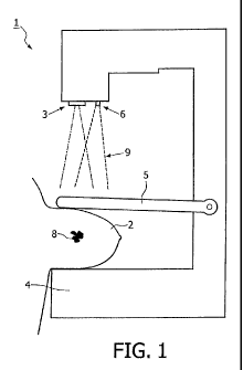
- Fig. 1 shows in a side view the mammography-apparatus
of the invention;
- Fig. 2 shows schematically a first embodiment of the
mammography-apparatus of the invention during screening of a
breast,
- Fig. 3 shows a second embodiment of the mammography-
apparatus of the invention when screening a breast for occur-
rence of malignant cells, and
- Fig. 4 shows a third embodiment of the mammography-
CA 02740350 2011-04-12
WO 2010/044660 PCT/NL2009/050601
6
apparatus of the invention during screening of a breast.
Same reference numerals applied in the figures refer to
same parts.
With reference first to Fig. 1 the mammography-
apparatus I of the invention is shown for screening malignant
cells 8 in a breast 2.
The mammography-apparatus 1 comprises an X-ray source
3, an X-ray detector 4 and a paddle 5 for pressing the breast 2
against said X-ray detector 4.
In accordance with the invention the mammography-
apparatus 1 further comprises a laser-light-source 6 for trans-
mitting pulsed defocussed laser-light 9 which during use is
aimed at the breast 2 in a pre-determined direction which direc-
tion is monitored.
With reference to Fig. 2, 3 and 4 showing first, second
and third embodiments of the apparatus 1 of the invention, it is
shown that the apparatus 1 is also provided with at least one,
and most commonly several non-contact ultrasound detectors 7 for
detection of ultrasound that originates from said breast 2,
which is induced by an excitation stemming from the said defo-
cussed laser-light 9. Thus, it is possible to generally screen
the occurrence of malignant cells 8, which are shown in the de-
tailed view A of Fig. 2, 3 and 4 to be characterized by a dense
microvascular network. It is important to note that the detec-
tors 7 are non-contact detectors and that their operation is
without application of any contact-gel between the detectors 7
and the breast 2.
The acoustic effect is generated by the pulsed laser-
light in the microvascular haemoglobin containing network that
is typical for neoplasia like malignant invasive tumours, and is
not induced by pseudo-malignant lesions which can also be found
in mammography. This aspect makes it possible that many unneces-
sary referrals of screened women for further assessment are
avoided which would otherwise cause a lot of anxiety and diag-
nostic work-up, like invasive punctures and even diagnostic op-
erations.
To allow detection of the position at which the malig-
nant cells 8 are located, the laser-light 9 may be directionable
and its direction measurable so as to monitor the direction of
the laser-light 9 when the ultra-sound detector or detectors 7
detect the laser-induced ultrasound. Thus a coarse measure is
CA 02740350 2011-04-12
WO 2010/044660 PCT/NL2009/050601
7
provided for the direction at which malignant cells 8 and their
feeding vessels are located.
A further aspect of the invention is that by using sev-
eral non-contact ultrasound detectors a coarse localization of
the abnormality that produces the acoustic signal can be made.
For this purpose use is made of the different times of arrival
of the said acoustic signal in the different non-contact ultra-
sound detectors. The times of arrival in the different non-
contact ultrasound detectors are determined by the different.
distances travelled by the said ultrasound signal through the
breast tissue and air on its way to the different detectors. Be-
cause of the synchronous x-ray imaging, this coarse localization
can be easily related to this image.
Fig. 2 shows that the laser-light 9 that originates
from the laser-light source 6 as shown in Fig. 1 is able to
transmit through the paddle 5 and induce ultrasound pulses in
the breast 2. The laser-light source 6 used is for instance
typically having an energy of 30 mJ/cm2 operating at 5 nanosec-
onds pulse width with a 10 Hertz repetition rate. The laser-
light source 6 is transmitting in the 1064 nm range.
Fig. 3 shows a variation to the embodiment shown in
Fig. 2 in that apart from the laser-light that is directed
through the paddle 5 from above, also glass fibre optics 10 are
provided in the housing of the X-ray detector 4, that transmit
light-pulses from the laser-light-source 6 at appropriate loca-
tions from below into the breast 2.
Fig. 4 shows still a further variation to the embodi-
ments shown in Fig. 2 and 3. In this embodiment there is no la-
ser-light transmitted through the paddle 5, yet there are at ap-
propriate locations glass fibre optics 10 provided in the paddle
5 that transmit the light pulses from the laser-light source 6
from above into the breast 2, in addition to light pulses
through the glass fibre optics 10 in the housing of the x-ray
detector 4, coming from below as shown in Fig. 3.
The embodiments shown in Figures 3 and 4 are preferred
embodiments and have improved ultrasound response to possible
occurrences of malignant cells as compared to the embodiment
shown in Fig. 2 which only induces ultrasound with light pulses
that are introduced into the breast from above.
The use of the photoacoustic effect as elucidated here-
above is particularly well suited to compensate the less effec-
CA 02740350 2011-04-12
WO 2010/044660 PCT/NL2009/050601
8
tive screening results that come available with the known mam-
mography-apparatus. In combination, the screening results are
effectively improved. Both a significantly lower false negative
rate and a lower false positive rate as compared to the known
mammography method and apparatus results, without imparting any
need to lengthen or complicate the screening-method of the prior
art.