Note: Descriptions are shown in the official language in which they were submitted.
CA 02740633 2015-10-29
1
MINERAL-COATED MICROSPHERES
[0001]
[0002]
BACKGROUND
[0003] The present application generally relates to tissue engineering and
administration
of therapeutic compounds.
[0004] Biodegradable microspheres have been widely used as carriers for
controlled
release of drug molecules, including small molecules (DeFail et al. 2006), DNA
(Jang and Shea,
2003), and proteins (Yang et al. 2001). Although these carriers have become
prevalent in
biomedical applications ranging from injectable drug delivery (Pandy and
Khuller, 2007) to
manipulation of stem cell differentiation (Newman and McBurney, 2004; Ferreira
et al. 2008),
protein release from these microspheres is often confounded by low molecule
encapsulation
efficiency (Akhtar and Lewis, 1997), "burst" release of molecules over short
timescales
(O'Donnell and Mcginity, 1997), and decreased activity of biological molecules
due to
microsphere processing conditions and polymer degradation by products (Jiang
and
Schwendeman, 2001).
[0005] Hybrid materials composed of organic polymers coated with inorganic
minerals
have attracted much attention in biology and medicine due to their combination
of advantageous
properties. Polymeric materials are a desirable base material for biomedical
applications, as they
can be processed into a variety of sizes and geometries, and can be designed
to bioresorb in a
CA 02740633 2011-04-14
WO 2010/036919 PCT/US2009/058419
2
controllable timeframe. Therefore, polymeric biomaterials have been featured
in a variety of
applications, including medical devices, tissue engineering scaffolds, and
drug delivery systems.
[0006] Calcium phosphate based mineral coatings represent desirable
surfaces for
biomedical applications, as they arc similar in composition to bone tissue,
and have been shown
to promote favorable interactions with natural bone, a property termed
"bioactivity". For
example, hydroxyapatite - the major inorganic component of bone mineral- is
osteoconductive
(Ducheyne et al., 1999), and may also be capable of inducing new bone
formation in vivo
(Habibovic etal., 2006).
[0007] A particular subset of approaches used to grow hydroxyapatite
coatings on
biomaterials surfaces mimics some aspects of natural biomineralization
processes, and has
therefore be termed "biomimetic" or "bioinspired" Hong etal., 2006; Gao and
Koumoto, 2005;
Leveque et al., 2004; Green et at., 2006). This type of approach is a
practically and
economically attractive alternative to high-temperature commercial processing
methods such as
plasma-spraying (Gledhill et at., 2001), sputter coating (Yamashita et at.,
1994), and laser
deposition (Fernandez-Pradas etal., 1998). Kokubo etal. first reported
bioinspired growth of
apatite coatings on bioactive CaO-SiO2 glass in a simulated body fluid (SBF),
which had ion
concentrations nearly equal to those of human blood plasma and was held at
physiologic
temperature and pH (Kokubo etal., 1990). A series of subsequent studies
reported mineral
growth using novel formulations of SBF (Oyanc et al., 2003), variation in the
mineral growth
process (Miyaji etal., 1999), or variations in the base materials (Yogogawa et
at., 1997). The
basis for mineral nucleation in these studies involved interactions of mineral
ions in solution with
polar functional groups on the materials surface, such as Si-OH (Li et al.,
1992), Ti-OH (Barrere
etal., 2004) and Zr-OH (Uchida et al., 2001). A series of recent studies has
extended the
bioinspired mineralization process to include formation of a bone-like
hydroxyapatite coating on
biodegradable polymer films (Murphy and Mooney, 2002) or porous scaffolds
(Murphy etal.,
2000; Zhang and Ma, 2004; Bajpai and Singh, 2007). The mechanism for mineral
nucleation
and growth on these materials is based on the interaction of carboxylate and
hydroxyl groups on
the hydrolyzed surface with calcium- and phosphate-rich nuclei in solution,
creating a driving
force for heterogeneous nucleation and mineral growth (Murphy and Mooney,
2002). This
coating process is particularly suitable for biodegradable polymers, as it can
be carried out at
physiological temperature and pH (Tanahashi et at., 1994), and the mild
processing conditions
CA 02740633 2015-10-29
3
also suggest that it is possible to incorporate biologically active molecules
such as polypeptides
and polynucleotides, during the coating process.
[0008] There is a need for new applications of mineral-coated polymers for
use in
therapeutic treatments. Those needs are addressed herein.
SUMMARY
[0009] The inventors have discovered that microspheres can be produced that
have a
calcium-containing mineral coating. See Examples. These microspheres are
useful, at least for
providing slow release of therapeutic compounds combined therewith.
[0010] The application is directed to a microsphere comprising a bead
coated with a first
calcium-containing mineral.
[0011] The application is also directed to a method of producing a
microsphere. The
method comprises incubating a bead in a physiological saline solution
comprising carbonate,
calcium, and phosphate such that a first calcium-containing mineral layer
coating forms on the
bead. In this method, the bead with the mineral layer coating is the
microsphere.
[0012] In other embodiments, the application is directed to a method of
administering a
compound to a vertebrate. The method comprises administering the above
microsphere to the
vertebrate.
[0012a] In certain exemplary embodiments there is provided a microsphere
comprising: a
bead comprising a polymer; a first calcium-containing mineral; a component;
and a biologically
active compound; wherein, the bead is coated with the first calcium-containing
mineral; the
component is non-covalently attached to the first calcium-containing mineral
thereby introducing
a functional group to the first calcium-containing mineral; and the
biologically active compound
is covalently attached to the functional group.
[0012b] In further exemplary embodiments, there is provided a method of
producing such
microsphere comprising incubating a bead in a physiological saline solution
comprising
carbonate, calcium, and phosphate such that a first calcium-containing mineral
layer coating
forms on the bead.
[0012c] In another exemplary embodiment, there is provided use of the
microsphere
disclosed herein for the manufacture of a medicament to treat a non-human
vertebrate.
CA 02740633 2015-10-29
3a
[0012d] In a further exemplary embodiment there is provided the use
disclosed herein,
wherein the microsphere is administered systemically.
[0012e] In a further exemplary embodiment there is provided use of the
microsphere
disclosed herein for the treatment of a vertebrate.
BRIEF DESCRIPTION OF THE DRAWINGS
[0013] FIG. 1 is a schematic and micrographs of PLO microspheres before and
after
mineral coating.
[0014] FIG. 2 are graphs of XRD and FTIR spectra. Panel A shows a
representative
XRD spectrum of mineral-coated microspheres showing apatite peaks
corresponding to
hydroxyapatite at 20 =25.78 , 28.68 , and 32.05 . Panel B shows a
representative FTIR
spectrum showing phosphate peaks (1087, 1035, 950, 560 cm-I) and carbonate
peaks (1456 and
1415 cm-I).
[0015] FIG. 3 is graphs and a micrograph relating to protein binding on
mineral-coated
microspheres. Panel A is a representative FT-IR spectrum of protein bound onto
mineral-coated
microspheres showing phosphate peaks (1087, 1035, 950, 560 cm-I), carbonate
peaks (1456 and
1417 cm-I), and amide peaks (1653 and 1558 cm-I). Panel B is a scanning
electron micrograph
CA 02740633 2011-04-14
WO 2010/036919 PCT/US2009/058419
4
(SEM) of protein bound on the surface of mineral, scale bar = 100 nm. Panel C
shows binding
curves of bovine serum albumin (BSA) and Cytochrome c (Cyt c) on the mineral-
coated
microspheres surface.
[0016] FIG. 4 is graphs and micrographs relating to release of protein from
mineral-
coated microspheres. Panel A is a comparison of cumulative release of BSA
bound to mineral-
coated PLG microspheres and BSA encapsulated in PLG microspheres. Both
approaches show
sustained release, with the total amount of bound BSA release significantly
higher than the
encapsulated BSA after 30 days. Panels B and C are SEM images of mineral-
coated
microspheres (B); and PLG microspheres (C) after the 30 day release period.
Panel D shows
cumulative release of Cyt c from mineral-coated PLG microspheres at pH 4 and
pH 7.4. Panels
E and F are SEM images of mineral-coated microspheres after 30 days of release
in buffered
solutions pH=4 (E), and pH=7.4 (F).
[0017] FIG. 5 is SEM images of a PLG microsphere (A) and mineral-coated
microsphere
after incubation in mSBF for 7 days.
[0018] FIG. 6, Panels A-D are SEM images of mineral-coated microspheres
after a 7 day
incubation in mSBF solution, 0.25% w/v (A), 0.50% w/v (B), 0.75% w/v (C), and
1.00% (D)
w/v. Panel E is a graph showing the relationship between the microsphere
concentration in
solution during mineral growth, and the size of mineral-coated microsphere
aggregates.
[0019] FIG. 7 is a graph showing the c potential of PLG microspheres in
buffers PBS,
mSBF, and mSBF + 0.1% (v/v) Tween 20T1 (Panel A) and nonhydrolyzed and
hydrolyzed PLG
films in comparison with PLG microspheres (Panel B).
[0020] FIG. 8 is graphs showing X-ray diffraction analysis of mineral-
coated
microspheres and hydroxyapatite powder (included for comparison) (Panel A),
and Fourier
transform infrared analysis of mineral-coated microspheres (Panel B). Peaks
associated with
carbonate are denoted by *, and peaks associated with phosphate are denoted by
[0021] FIG. 9 shows an EDS spectrum of mineral-coated microspheres after a
7 day
incubation in mSBF.
100221 FIG. 10 is SEM images showing the process of mineral nucleation and
growth on
PLG microspheres. The images are of microspheres after: the first day of
immersion in mSBF
(A), day 3 of incubation (B), day 5 of incubation (C) and day 7 of incubation
(D).
CA 02740633 2011-04-14
WO 2010/036919 PCT/US2009/058419
[0023] FIG. 11 is a graph showing the percentage of aggregated microspheres
suspended
for 1, 3 and 7 days in PBS, mSBF and mSBF+Tween20Tm.
[0024] FIG. 12 is graphs and SEM images of mineral dissolution of mineral-
coated
microspheres. Panel A shows cumulative dissolution of Ca2-' and P043- during a
25 day
incubation in tris-buffered saline (TBS). Panel B shows cumulative dissolution
of P043- during a
25 day incubation in DMEM. Panels C is SEM images of mineral-coated
microspheres after the
25 day TBS incubation. Panel D is SEM images of mineral-coated microspheres
after the 25 day
DMEM incubation.
[0025] FIG. 13 is SEM images and a graph showing the effect of surfactant
(Tween
2OTM) on the mineral formed on the PLG microsphere surfaces after 3 days (A),
after 7 days (B),
after 14 days (C), and after 28 days (D). Panel E shows an FTIR spectrum of
PLG microspheres
coated with mineral via a 28 day mSBF incubation in the presence of 0.1%v/v
Tween 2OTM. A
spectrum of commercial HA powder is included for comparison.
100261 FIG. 14 is SEM images showing nanometer-scale mineral morphology on
the
surface of microspheres formed in the presence (A) or absence (B) of 0.1% v/v
Tween 2OTM.
DETAILED DESCRIPTION
[0027] The inventors have developed methods for producing novel
microspheres that
have a calcium-containing mineral coating. They have also characterized these
microspheres
and established that they have advantageous properties useful for utilizing
the microspheres to
deliver therapeutic compounds to tissues. See Examples.
[0028] In some embodiments, the application is directed to a microsphere
comprising a
bead coated with a first calcium-containing mineral. The Examples describe
exemplary methods
for producing these microspheres using a modified simulated body fluid (mSBF).
By adjusting
the mineral composition, and/or concentration of the mSBF, the composition of
the mineral
precipitated on the microspheres can be manipulated. See also U.S. Patent
Application
Publication US 2008/0095817 Al; U.S. Patent No. 6,767,928 Bl; U.S. Patent No.
6,541,022 Bl;
PCT Publication WO 2008/070355 A2; PCT Publication WO 2008/082766 A2; Murphy
and
Mooney, 2001; Murphy and Messersmith, 2000.
[0029] Inorganic minerals suitable for producing a calcium-containing
mineral coating
include various bone mineral ions, such as, but not limited to calcium and
phosphate and
CA 02740633 2011-04-14
WO 2010/036919 PCT/US2009/058419
6
combinations of bone mineral ions, such as calcium-phosphates. The calcium-
containing
mineral coating can comprise, e.g., hydroxyapatite (HAP), a-tricalcium
phosphate (a-TCP), 13-
tricalcium phosphate (I3-TCP), amorphous calcium phosphate, dicalcium
phosphate, octacalcium
phosphate or calcium carbonate. The calcium-containing mineral coating can
comprise a
plurality of layers, e.g., separate layers having distinct dissolution
profiles. Under physiological
conditions, solubility of calcium phosphate species adhere to the following
trend: amorphous
calcium phosphate>dicalcium phosphate>octacalcium phosphate>13-TCP>HAP. A
dicalcium
phosphate mineral will typically have a dissolution rate that is more than
fifty times higher than
that of HAP. Therefore, creation of a matrix with distinct calcium phosphate
layers allows for a
broad range of dissolution patterns.
[0030] The bead can be formed of any suitable material known in the art.
The selection
of the bead material for any particular application can be made without undue
experimentation.
[0031] In some embodiments, the bead comprises a negative charge, which
can promote
the deposition of the calcium containing material. The negative charge could
be provided by any
moiety present on the bead, for example a carboxylate group, as is present in
poly(D,L-lactide-
co-glycolide) (PLG). In some embodiments the bead is made of a polymer, for
example a
synthetic polymer. In various aspects of these embodiments, the polymer is
bioabsorbable.
Nonlimiting examples of suitable bead materials include, for example, a
collagen gel, polyvinyl
alcohol, a marine adhesive protein, a PLG fiber matrix, a polyglactin fiber, a
calcium alginate
gel, a polyglycolic acid, polyester (e.g., poly-(L-lactic acid) or a
polyanhydride), a
polysaccharide (e.g. alginate), chitosan, polyphosphazene, polyacrylate,
polyethylene oxide-
polypropylene glycol block copolymer, fibrin, collagen, and fibronectin,
polyvinylpyrrolidone,
hyaluronic acid, poly(lactide), poly(glycolic acid), poly(lactide-co-
glycolide),
poly(caprolactone), polycarbonates, polyamides, polyanhydrides, polyamino
acids, polyortho
esters, polyacetals, polycyanoacrylates), polyurethanes, polyacrylates,
ethylene-vinyl acetate
polymers and other acyl substituted cellulose acetates and derivatives
thereof), polyurethanes,
polystyrenes, polyvinyl chloride, polyvinyl fluoride, poly(vinylimidazole),
chlorosulphonated
polyolifins, polyethylene oxide, polyvinyl alcohol, teflon , nylon, and
analogs, mixtures,
combinations and derivatives of any of the above.
[0032] In various embodiments, the bead is made of a polymer that
comprises polar
oxygen groups. Examples of such polymers include polycarboxylates,
polyanhydrides, poly(a-
CA 02740633 2011-04-14
WO 2010/036919 PCT/US2009/058419
7
hydroxyesters), poly(ethylene terephthalates), poly(carbonates), poly(amides),
poly(lactones), a
poly(saccharides) and poly(acrylates).
[0033] In various embodiments, the bead is made of a PLG, for example at a
ratio of
about 85:15 lactide:glycolide ("85:15 PLG"). The use of 85:15 PLG copolymers
is
advantageous as a decrease in the lactide/glycolide ratio of the copolymer is
believed to increase
the rate of surface hydrolysis.
[0034] In certain specific embodiments, the first calcium-containing
mineral is a
carbonated-substituted calcium-deficient hydroxyapatite and the bead is PLG,
wherein the PLG
is about 85:15 lactide:glycolide.
[0035] In some embodiments, the microsphere further comprises a component
adhering
to the first calcium-containing mineral, wherein the component introduces a
functional group to
the first calcium-containing mineral. Introduction of such a functional group
allows covalent
binding of any additional materials (e.g. therapeutic compounds) to the
microspheres.
Nonlimiting examples of functional groups that can be introduced on the
component is a
carboxylate, an amine, a carbonyl, a nitro, a hydroxyl, an aldehyde, or an
ester. In some
embodiments, the component comprises a poly(aspartic acid), a poly(glutamic
acid), or a
bisphosphonate. See e.g., Murphy et al., 2007. Other nonlimiting examples of
components
useful in these embodiments are the oligopeptides AAAAEPRREVAEL or
AAAAyEPRRyEVAyEL, where yE is carboxyglutamate.
[0036] In some embodiments, the microsphere further comprises a first
compound
adhering to the first calcium-containing mineral or the component.
[0037] In certain specific embodiments of the first compound-containing
microspheres,
the first calcium-containing mineral is a carbonated-substituted calcium-
deficient hydroxyapatite
and the bead is poly(D,L-lactide-co-glycolide) (PLG), wherein the PLG is about
85:15
lactide:glycolide.
[0038] These embodiments are not limited to any particular first
compounds. The first
compound can be an organic compound less than 2000 MW, or less than 1000 MW,
or less than
500 MW. Nonlimiting examples include antibiotics, corticosteroids and statins.
More specific
examples include cefazolin, cefuroxime, clindamycin, vancomycin and
dexamethasone.
[0039] Alternatively, the first compound can be an oligopeptide or
polypeptide. As used
herein, an oligopeptide comprises a linear chain of 30 or less amino acids. A
polypeptide
CA 02740633 2011-04-14
WO 2010/036919 PCT/US2009/058419
8
comprises more than 30 amino acids. Examples of oligopeptides are GGRGDSP (a
cell adhesion
peptide derived from fibronectin), GGIKVAV (a cell adhesion peptide derived
from laminin),
GGYIGSR (a cell adhesion peptide derived from laminin), GGDGEA (a cell
adhesion/signaling
peptide derived from type I collagen), GGKIPKASSVPTELSAISTLYL (a peptide
derived from
bone morphogenetic protein-2), AAAAEPRREVAEL (a modified peptide derived from
osteocalcin - some affinity for hydroxyapatite mineral), AAAAyEPRRyEVAyEL,
where yE is
carboxyglutamate (a modified peptide derived from osteocalcin - high affinity
for hydroxyapatite
mineral).
[0040] In other embodiments, the first compound is a polypeptide, for
example a
cytokine, an enzyme, or a protein comprising an antibody binding site (e.g.,
an antibody). Other
nonlimiting examples of polypeptides that could be included in the
microspheres are virtually
any hormone, neurotransmitter, growth factor, growth factor receptor,
interferon, interleukin,
chemokine, cytokine, colony stimulating factor and/or chemotactic factor
protein or polypeptide.
Further examples include transcription or elongation factors, cell cycle
control proteins, kinases,
phosphatases, DNA repair proteins, oncogenes, tumor suppressors, angiogenic
proteins, anti-
angiogenic proteins, immune response stimulating proteins, cell surface
receptors, accessory
signaling molecules, transport proteins, enzymes, anti-bacterial and/or anti-
viral proteins or
polypeptides, and the like, depending on the intended use of the ultimate
composition. More
specific examples include growth hormone (GH); parathyroid hormone (PTH,
including PTH1-
34); bone morphogenetic proteins (BMPs), such as BMP-2A, BMP-2B, BMP-3, BMP-4,
BMP-5,
BMP-6, BMP-7 and BMP-8; transforming growth factor-a (TGF-a), TGF-131 and TGF-
02;
fibroblast growth factor (FGF); granulocyte/macrophage colony stimulating
factor (GMCSF);
epidermal growth factor (EGF); platelet derived growth factor (PDGF); an
insulin-like growth
factor (IGF), leukemia inhibitory factor (LIF), vascular endothelial growth
factor (VEGF), basic
fibroblast growth factor (bFGF), platelet derived growth factor (PDGF),
angiogenin,
angiopoietin-1, del-1, follistatin, granulocyte colony-stimulating factor (G-
CSF), hepatocyte
growth factor/scatter factor (HGF/SF), interleukin-8 (IL-8), leptin, midkine,
placental growth
factor, platelet-derived endothelial cell growth factor (PD-ECGF), platelet-
derived growth
factor-BB (PDGF-BB), pleiotrophin (PTN), progranulin, proliferin, tumor
necrosis factor-a
(TNF-a), a matrix metalloproteinase (MMP), angiopoietin 1 (angl), ang2, and
delta-like ligand 4
(DLL4).
CA 02740633 2011-04-14
WO 2010/036919 PCT/US2009/058419
9
[0041] In some specific embodiments, the polypeptide is a BMP-2, a BMP-7, a
VEGF,
an FGF-2, a PDGF, a TGF-P, an interleukin, or a human GH.
[0042] The first compound can also be a nucleic acid. Non-limiting examples
include a
microRNA, an antisense nucleic acid, and a vector. Where the first compound is
a vector, any
vector known or later discovered can be included here. In some embodiments,
the vector
comprises a sequence encoding a therapeutic protein, such as any of the
proteins discussed
above.
[0043] The first compound can noncovalently adhere to the microsphere.
Alternatively,
the microsphere can further comprise a component adhering to the first calcium-
containing
mineral and introducing a functional group to the microsphere, to which the
compound is
covalently attached. See, e.g., Murphy etal., 2007.
[0044] In some embodiments, the first compound is at more than one level of
the first
calcium-containing mineral. These microspheres generally release the compound
over a longer
period of time than where the compound is only at one level (for example the
outer surface of the
first calcium-containing mineral.
[0045] In other embodiments, the first compound is modified to change the
rate at which
the compound is released from the microsphere. For example, where the first
compound further
comprises a moiety that increases the strength of binding of the compound to
the first calcium-
containing mineral, the compound would be released more slowly than if the
moiety is not
present. Nonlimiting examples of such moieties include amino acid sequences
rich in glutamic
acid, aspartic acid or phosphoserine, which interact directly with calcium
ions in mineralized
extracellular matrices. Other examples include A AAAEPRREVAEL or
AAAAyEPRRTEVAyEL, where yE is carboxyglutamate.
[0046] In various embodiments, the microsphere further comprises a second
compound
adhering to the first calcium-containing mineral or the component. As with the
first compound,
the microspheres are not limited as to the nature of the second compound. The
second
compound can be for example an organic compound less than 2000 MW or 1000 MW
or 500
MW. Alternatively, the second compound can be an oligopeptide or polypeptide,
or a nucleic
acid. In these embodiments, the first compound and the second compound can be
on the same or
different levels of the first calcium-containing minerals. Having the two
compounds on different
CA 02740633 2011-04-14
WO 2010/036919 PCT/US2009/058419
levels is useful when it is desired that the two compounds are released at
different times. The
microsphere can also further comprise a third, fourth, fifth, etc. compound as
desired.
[0047] In additional embodiments, the microsphere comprises a living cell.
The living
cell can be from any organism, including an Archaca, a prokaryote, or a
cukaryote. In some
embodiments, the cell is a mammalian cell. The cell can be naturally occurring
or, alternatively,
can be transformed to express a recombinant molecule, e.g., a protein or
nucleic acid (such as an
miRNA).
[0048] In certain specific embodiments of these cell-containing
microspheres, the first
calcium-containing mineral is a carbonated-substituted calcium-deficient
hydroxyapatite and the
bead is poly(D,L-lactide-co-glycolide) (PLG), wherein the PLG is about 85:15
lactide:glycolide.
[0049] The cell can be adhered to the microsphere by any known means. In
some
embodiments, the microsphere comprises a first binding agent that binds to a
second binding
agent on the cell. Nonlimiting examples of such agents are a receptor and a
ligand of the
receptor, complementary nucleic acids, or a cell adhesion peptide and a ligand
of the cell
adhesion peptide. In the latter case, examples of suitable cell adhesion
peptides are GGRGDSP,
GGIKVAV, GGYIGSR or GGDGEA. These peptides or any other first binding agent
can be
part of a larger molecule, for example a molecule that binds to the first
calcium-containing
mineral as discussed above.
[0050] In some embodiments, the microsphere comprises a cell as well as a
compound
(e.g., a cytokine) that interacts with the cell. In such a microsphere, the
cytokine is
advantageously close to the cell, such that the compound is likely to contact
and thus interact
with the cell.
[0051] In additional embodiments, the microsphere further comprises a
coating of a
second calcium-containing mineral. The second coating can be a mineral that
has a different
degradation rate than the first calcium-containing mineral. The microsphere
comprising the two
coatings can further comprise one or more than one compound. Either of the two
coatings can
be, for example, any of hydroxyapatite (HAP), a-tricalcium phosphate (a-
TCP),13-tricalcium
phosphate (I3-TCP), amorphous calcium phosphate, dicalcium phosphate,
octacalcium phosphate
or calcium carbonate. For example, the first calcium-containing mineral can be
hydroxyapatite
and further comprises a first therapeutic compound, and the second calcium-
containing mineral
can be a-TCP and is coated above (i.e., closer to the surface of the
microsphere) the first calcium
CA 02740633 2011-04-14
WO 2010/036919 PCT/U S2009/058419
11
containing mineral and further comprises a second therapeutic compound. In
this example, the
second calcium-containing mineral would degrade first, releasing the second
therapeutic
compound before the first therapeutic compound is released.
[0052] Where a compound is present in the microsphere, its release depends
on a number
of factors, for example (a) how strongly the compound adheres to the calcium-
containing mineral
or component, (b) how far away from the surface of the microsphere the
compound is (for
example if a layer of calcium-containing mineral is deposited on the
microsphere after the
compound is added), (c) the degradationlbioabsorption rate of the calcium-
containing mineral,
(d) whether the compound is covalently bound to a component, and if so, (e)
how strongly the
component is bound to the mineral, (f) how strongly the covalent bond is
between the compound
and the component, and (g) whether there are enzymes present, e.g., from a
tissue in which the
microsphere is implanted, that will break the covalent bond and release the
compound. Each of
these factors may have more or less influence on the release of the compound,
depending on the
configuration of the microsphere. For example, in the case where the compound
is covalently
attached to a component, the component binds to the calcium-containing
material strongly, and
there are no enzymes to break the covalent bond, then the main factor
influencing the release of
the component is the rate of degradation of the calcium-containing mineral.
However, if the
compound is noncovalently adhering directly to the calcium-containing mineral,
then all of the
above factors (a) - (c) will have an influence in the rate of release of the
compound.
[0053] In further embodiments, the compound is in the bead. In these
embodiments, the
compound would likely be released upon degradation of the bead, after the
degradation of the
calcium-containing mineral.
[0054] In some embodiments, the various microspheres discussed herein
generally have a
diameter between about 0.5 lam and about 500 pin. In other embodiments, the
microsphere has a
diameter between about 0.5 t.im and about 100 pm. In additional embodiments,
the microsphere
has a diameter between about 2 pm and about 6 pm.
[0055] As discussed in the examples the microspheres tend to aggregate,
particularly if
they are produced by incubation in mSBF at a high concentration of
microspheres or for an
extended period of time (see Examples). Thus, in some embodiments, the
application is directed
to a plurality of any of the above-described microspheres, aggregated. In
certain specific
embodiments of these aggregated microspheres, the first calcium-containing
mineral is a
CA 2740633 2017-05-02
, =
12
carbonated-substituted calcium-deficient hydroxyapatite and the bead is
poly(D,L-lactide-co-
glycolide) (PLG), wherein the PLO is about 85:15 lactide:glycolide. In other
embodiments of
these aggregated microspheres, a first compound, as described above, adheres
to the first
calcium-containing mineral.
100561 The application is also directed to a method of producing a
microsphere. The
method comprises incubating a bead in a physiological saline solution
comprising carbonate,
calcium, and phosphate such that a first calcium-containing mineral layer
coating forms on the
bead, where the bead with the mineral layer coating is the microsphere. See
Examples. In some
embodiments, the solution comprises NaC1, KCl, MgCl2, MgSO4, NaHCO3, Tris,
CaCl2, and
KH2PO4. In more specific embodiments, the solution comprises about 100-200 mM
NaC1, about
1-8 mM KCl, about 0.1-2 mM MgSO4, about 0.2-5 mM MgCl2, about 1-100 mM NaHCO3,
about 2-20 mM CaCl2, and about 0.5-10 mM KH2PO4. Even more specifically, the
solution
comprises about 141 mM NaC1, about 4.0 mM KC1, about 0.5 mM MgSO4, about 1.0
mM
MgC12, about 4.2 mM NaHCO3, about 5.0 mM CaCl2, and about 2.0 mM KH2PO4.
[00571 In some of these methods, the solution also comprises a
surfactant, which can
change the morphology of the calcium-containing mineral layer, and reduce
aggregation of the
microspheres. See Example 2. Any surfactant now known or later discovered may
be used here.
In some embodiments, the surfactant is Tween 2OTM.
10058] In some embodiments, the mineral coating described herein is
developed by
incubating the constituents in the above solution, which can be called a
"simulated body fluid"
(SBF) or a "modified simulated body fluid" (mSBF), for five days or more at a
pH of about 6.8
to about 7.4 and at a temperature of about 37 C. The SBF or mSBF may be
refreshed daily.
Using the chemical composition described in the Examples, the procedure
produces a calcium-
deficient, carbonate-containing apatite material on alginate and on poly-(a-
hydroxy esters). See
U.S. Pat. No. 6,767,928. mSBF includes elevated calcium and phosphate. In
general, an
increase in pH favors hydroxyapatite growth, while a decrease in pH favors
octacalcium
phosphate mineral growth.
10059] For example, conditions favorable for hydroxyapatite
formation include a pH
between about 5.0 and about 8.0 and a calcium concentration multiplied by a
phosphate
concentration between about 10-5 and about 10-8 M. Likewise, conditions
favorable for
octacalcium phosphate formation include a pH between about 6.0 and about 8.0
and a calcium
CA 02740633 2011-04-14
WO 2010/036919 PCT/US2009/058419
13
concentration multiplied by a phosphate concentration between about 10 5 and
about 10 7.5 M.
Furthermore, conditions favorable for dicalcium phosphate dehydrate formation
include a pH
between about 6.0 and about 8.0 and a calcium concentration multiplied by a
phosphate
concentration between about 10 4 and about 10-6 M.
100601 Specifically, using poly-(a-hydroxy esters) or alginate hydrogels
as a template,
one would vary the pH of mSBF between about 5.0 and about 6.0 to promote
hydroxyapatite
formation. Similarly, one would vary the pH of mSBF between about 6.0 and
about 6.5 to
promote octacalcium phosphate and hydroxyapatite formation. Likewise, one
would vary the pH
of mSBF between about 6.5 and about 8.0 to promote dicalcium phosphate,
octacalcium
phosphate and hydroxyapatite formation.
[0061] Prior to deposition of the first calcium-containing mineral, the
bead may be
surface-functionalized to allow increased mineral deposition by utilizing
chemical pre-treatment
to achieve surface hydrolysis, e.g., using an NaOH solution. Surface
degradation by this
technique causes an increase in the amount of polar oxygen functional groups
on the surface of
the material. The functionalized surface is then incubated in a mineral-
containing solution.
100621 In some embodiments, the method further comprises adding a
component that
adheres to the first calcium-containing mineral layer to the microsphere. In
these embodiments,
the component introduces a functional group to the first calcium-containing
mineral layer. As
discussed previously, nonlimiting examples of such components include peptides
comprising a
poly(aspartic acid) sequence, a poly(glutamic acid) sequence, AAAAEPRREVAEL
and
AAAAyEPRRTEVAyEL, where yE is carboxyglutamate.
[0063] In additional embodiments, the method further comprises incubating
the
microsphere with a first compound such that the first compound adheres to the
microsphere. As
discussed above in relation to the discussion on the microsphere composition,
the first compound
can be any chemical compound, such as an organic compound less than 2000 MW,
an
oligopeptide or polypeptide (e.g., a cytokine, an enzyme, or a protein
comprising an antibody
binding site), or a nucleic acid (e.g., a microRNA, an antisense nucleic acid,
or a vector).
100641 In some embodiments, the first compound is incubated with the
microsphere such
that the first compound is non-covalently bound to the microsphere. In other
embodiments, a
component that introduces a functional group to the microsphere is adhered to
the microsphere,
the first compound is covalently attached to the functional group.
CA 02740633 2011-04-14
WO 2010/036919 PCT/US2009/058419
14
[0065] In various embodiments of these methods, the first compound is
incubated with
the bead in the physiological saline solution such that the first compound is
deposited on the
bead along with the mineral layer coating. Such microspheres generally have
the first compound
deposited throughout the mineral layer coating, whereas if the compound is
bound to the bead
after the mineral layer is coated onto the bead, the compound would only be
present on the
surface of the mineral layer. The first compound would be expected to be
released over a longer
period of time in the former case than in the latter case.
[0066] In other embodiments, the bead is incubated in the physiological
saline solution
both before and after the first compound is adhered to the microsphere, after
which additional
first compound is adhered to the microsphere. In this case, the first compound
would be internal
to the surface of the mineral layer and would be released after a first
compound deposited on the
surface.
[0067] By controlling the addition of the compound in the above methods, a
suitable
release profile of the compound for any application can be achieved without
undue
experimentation.
[0068] Additionally, the method can further comprise incubating the
microsphere with a
second compound such that the second compound adheres to the microsphere. In
some of these
embodiments, the bead is incubated in the physiological saline solution before
and after the first
compound is adhered, after which the second compound is adhered to the
microsphere, such that
the first compound and the second compound are on different layers of the
first calcium-
containing mineral layer. Such a microsphere would release the second compound
before the
first compound.
[0069] The present application is further directed to any of the above-
described
microspheres made by any of the above-described methods.
[0070] The application is further directed to a method of administering a
compound to a
vertebrate. The method comprises administering any of the microspheres
disclosed above to the
vertebrate, where the microsphere further comprises the compound. In some
embodiments, the
microsphere is in a pharmaceutically acceptable material.
[0071] By "pharmaceutically acceptable" it is meant a material that (i) is
compatible with
the other ingredients of the composition without rendering the composition
unsuitable for its
intended purpose, and (ii) is suitable for use with subjects as provided
herein without undue
CA 02740633 2011-04-14
WO 2010/036919 PCT/US2009/058419
adverse side effects (such as toxicity, irritation, and allergic response).
Side effects are "undue"
when their risk outweighs the benefit provided by the composition. Non-
limiting examples of
pharmaceutically acceptable carriers include, without limitation, any of the
standard
pharmaceutical carriers such as phosphate buffered saline solutions, water,
emulsions such as
oil/water emulsions, microemulsions, and the like.
[0072] The above-described microspheres can be formulated without undue
experimentation for administration to a vertebrate, including humans, as
appropriate for the
particular application. Additionally, proper dosages of the microspheres can
be determined
without undue experimentation using standard dose-response protocols.
[0073] Accordingly, the microspheres may be enclosed in gelatin capsules.
For the
purpose of oral therapeutic administration, the microspheres of the present
invention may be
incorporated with excipients and used in the form of tablets, troches,
capsules, elixirs,
suspensions, syrups, wafers, chewing gums and the like.
100741 The microspheres can alternatively be administered parenterally such
as for
example, by intravenous, intramuscular, intrathecal or subcutaneous injection.
Such
administration can be systemic, for example if the microspheres were
administered by injection
into the blood stream. Alternatively, the administration can be local, e.g.,
an injection of the
microspheres directly onto an area where there is a tissue defect, where the
compound is a
cytokinc that stimulates filling in the defect. Parenteral administration can
be accomplished by
incorporating the microspheres into a suspension. Such suspensions may also
include sterile
diluents such as water for injection, saline solution, fixed oils,
polyethylene glycols, glycerin,
propylene glycol or other synthetic solvents. Parenteral formulations may also
include
antibacterial agents such as for example, benzyl alcohol or methyl parabens,
antioxidants such as
for example, ascorbic acid or sodium bisulfite and chelating agents such as
EDTA. Buffers such
as acetates, citrates or phosphates and agents for the adjustment of
osmolarity such as sodium
chloride or dextrose may also be added. The parenteral preparation can be
enclosed in ampules,
disposable syringes or multiple dose vials made of glass or plastic.
100751 Rectal administration includes administering the microspheres, in a
pharmaceutical composition, into the rectum or large intestine. This can be
accomplished using
suppositories or enemas. Suppository formulations can easily be made by
methods known in the
art. For example, suppository formulations can be prepared by heating glycerin
to about 120 C.,
CA 02740633 2011-04-14
WO 2010/036919 PCT/US2009/058419
16
dissolving the composition in the glycerin, mixing the heated glycerin after
which purified water
may be added, and pouring the hot mixture into a suppository mold.
[0076] Transdermal administration includes percutaneous adsorption of the
microspheres
to the skin. Transdermal formulations include patches (such as the well-known
nicotine patch),
ointments, creams, gels, salves and the like.
[0077] The present invention includes nasally administering to the
vertebrate a
therapeutically effective amount of the microspheres. As used herein, nasally
administering or
nasal administration includes administering the compound to the mucous
membranes of the nasal
passage or nasal cavity of the patient. As used herein, pharmaceutical
compositions for nasal
administration of the compound include therapeutically effective amounts of
the compound
prepared by well-known methods to be administered, for example, as a nasal
spray, nasal drop,
suspension, gel, ointment, cream or powder. Administration of the compound may
also take
place by applying the microspheres in a nasal tampon or nasal sponge.
100781 These methods can be used to treat any vertebrate, including wild or
domesticated
mammals or birds, including farm animals and pets. In some embodiments, the
vertebrate is a
human.
[0079] In various embodiments, the vertebrate has a condition that is
treatable by
administering the compound. Nonlimiting examples of such conditions include
cancer, diabetes,
Alzheimer's disease, Parkinson's disease, a heart disease, a virus infection,
a bacterial infection,
a parasitic infection, an autoimmune disease, an allergy, a prion disease, a
gastrointestinal
disease, a liver disease, a kidney disease, a skin disease, a bone disease, a
congenital disease or
defect, a disease characterized by insufficiency of a protein or a metabolite,
erectile dysfunction
or baldness.
[0080] In some embodiments, the condition is a tissue defect. The tissue
defect can be,
for example, in a bone, a soft tissue, or an internal organ. The defect may be
caused by disease
or trauma, or it may be congenital.
[0081] The application is also directed to the use of any of the above-
described
microspheres for the manufacture of a medicament for treating a vertebrate
with a compound. In
some of these embodiments the vertebrate is a mammal, e.g., a human. Further,
the application
is directed to the use of any of the above-described microspheres for the
treatment of a vertebrate
with a compound. In some of these embodiments the vertebrate is a mammal,
e.g., a human.
CA 02740633 2011-04-14
WO 2010/036919 PCT/US2009/058419
17
[0082] Preferred embodiments are described in the following examples. Other
embodiments within the scope of the claims herein will be apparent to one
skilled in the art from
consideration of the specification or practice of the invention as disclosed
herein. It is intended
that the specification, together with the examples, be considered exemplary
only, with the scope
and spirit of the invention being indicated by the claims, which follow the
examples.
Example 1. Mineral-coated polymer microspheres for controlled protein binding
and release.
[0083] The work described herein evaluates the hypothesis that protein-
mineral
interactions could be used as an alternative mechanism to create biodegradable
micro-carriers for
controlled protein binding and release. In particular, the calcium phosphate
mineral
hydroxyapatite is employed as a substrate for protein binding and release, as
it has been used for
over 50 years in chromatographic protein separations based on its ability to
bind and release both
acidic and basic proteins under particular solution conditions. Here a first
demonstration is
provided establishing that biodegradable polymer microspheres can be coated
with an inorganic
hydroxyapatite layer, and that this biodegradable coating can be used as a
substrate for binding
and sustained release of acidic and basic proteins.
100841 This approach involves nucleation and growth of inorganic calcium
phosphate
mineral coatings on the surface of organic, biodegradable polymer microspheres
at near
physiologic temperature and pH. This mineral growth process mimics natural
biomincralization
processes (Mann, 2001), and results in a mineral coating that is similar in
structure (platelike
nanostructure) and composition (carbonated-substituted, calcium-deficient
hydroxyapatite phase)
to human bone mineral, as detailed previously on macroscopic polymer films
(Murphy and
Mooney, 2002). Specifically, protein-releasing, mineral-coated polymer
microspheres were
prepared here via a two-step process involving: i) formation of biodegradable
PLG microspheres
using a standard water-oil-water double emulsion process (Meng et al., 2003);
and ii) coating of
PLG microspheres with an inorganic, bone-like mineral (BLM) film via
incubation in a modified
simulated body fluid (mSBF), an aqueous solution which contains the ionic
constituents of blood
plasma with 2-fold higher concentrations of calcium and phosphate ions (Murphy
and
Messersmith, 2000) (FIG. 1). X-ray diffraction and FTIR spectra indicate that
the mineral grown
on PLG microspheres is an apatite mineral (FIG. 2). Scanning electron
microscopy (SEM) (FIG.
1) indicates that the mineral film is continuous on the microsphere surface
and has a plate-like
CA 02740633 2011-04-14
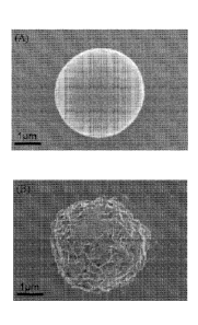
WO 2010/036919 PCT/US2009/058419
18
nanostructure. Therefore, the mineral layer grown on biodegradable polymer
microspheres is
similar in composition and morphology to bone mineral, as described previously
Murphy and
Mooney, 2002).
[0085] Importantly, the mineral-coated microsphere surface is porous and
contains
charged calcium and phosphate components. Therefore, it was hypothesized that
these
microspheres would be capable of efficiently binding soluble biological
molecules via
electrostatic interactions, in a manner analogous to the above mentioned
hydroxyapatite
chromatography (Urist et al., 1984; Schroder et al., 2003). In this study the
possibility was
examined of using mineral-coated microspheres as a carrier for two proteins
with differing
characteristics: i) an acidic protein, bovine serum albumin (BSA) (pI = 4.7);
and ii) a basic
protein, cytochrome c (Cyt c) (pI = 10.2). These model proteins were chosen to
illustrate the
influence of protein characteristics on binding efficiency and release
kinetics, and because of
their biological relevance. Specifically, albumin is one of the most abundant
proteins found in
blood plasma and has been shown to promote formation of bone tissue (Yamaguchi
et al., 2003),
while cytochrome c serves as a model protein for several basic growth factors,
such as fibroblast
growth factor-2, bone morphogenic proteins, and transforming growth factor 13.
[0086] Binding of BSA on mineral-coated PLG microspheres was first detected
by FTIR
analysis (FIG. 3A). The FTIR spectrum shows phosphate peaks (1087, 1035, 950,
and 560 cm')
and carbonate peaks (1456 and 1417 cm') corresponding to carbonate-substituted
hydroxyapatite mineral coatings, consistent with previous studies of BLM
coatings formed on
biodegradable polymer substrata (Murphy and Mooney, 2002). The FTIR spectrum
also shows
amide peaks (1653 and 1558 cm') corresponding to the presence of BSA bound to
the coating
surface. SEM analysis corroborates the FTIR analysis, and shows bound protein
deposited on
mineral-coated PLG microspheres (FIG. 3B). BSA binding on the mineral coating
was next
quantified by incubating mineral-coated microspheres for 4 hours in PBS with
varying BSA
concentrations. The results show that the amount of bound BSA on the
microspheres increased
linearly in the range of 0-200 [tg/m1 and reached a plateau at 400 [tg/m1
(FIG. 3C). This binding
is consistent with a typical Langmuir isotherm, and corroborates previous
studies of BSA
binding to hydroxyapatite powder (Hughes Wassell et al., 1995). Cyt c showed a
binding curve
similar to that of BSA, and it is noteworthy that Cyt c bound to the mineral
more efficiently than
BSA at the same solution protein concentrations. The enhanced binding
efficiency of Cyt c is
CA 02740633 2011-04-14
WO 2010/036919 PCT/US2009/058419
19
likely due to its smaller hydrodynamic radius (RH = 0.18 nm)(Moror et al.,
2001) when
compared to BSA (RH = 3.6 nm)(Boyer and Hsu, 1992). It is likely that protein
binding in the
current study is mediated by ionic interactions, and the presence of both
positively charged
calcium and negatively charged phosphate ions on the apatite mineral surface
enables binding of
both acidic and basic proteins at physiologic pH. This assertion is supported
by recent
demonstrations that the amount of bound protein on highly crystallized
hydroxyapatite can be
attributed to the ionic interaction between the surface charges of
hydroxyapatite and proteins
(Kawachi et al., 2008).
[0087] The release kinetics of BSA from mineral-coated PLG microspheres
were
investigated by incubating mineral-coated microspheres in phosphate-buffered
saline (PBS)
solution at pH 7.4 (FIG. 4A). BSA release was sustained over 30 days, and the
release displayed
near linear kinetics. In contrast, the cumulative release of BSA encapsulated
in PLG
microspheres via standard processing techniques (Meng et al., 2003) displayed
a much lower
level of cumulative release over 30 days, and the majority of the released
protein represented a
"burst" release during the initial 72 hours. More specifically, after 30 days
in PBS the
mineralized microspheres released 49 4.8%, while 23 4.9 % of the BSA
encapsulated in PLG
microspheres was released over the same timeframe. SEM analysis of the mineral-
coated
microspheres (FIG. 4B) showed little clear evidence of mineral dissolution
after 30 days of
immersion in PBS. PLG microspheres with encapsulated BSA remained intact for
the duration
of the release experiment (FIG. 4C), as expected based on previous studies by
Porjazoska et al.
(2004).
[0088] To gain further insight into the factors influencing protein release
from mineral-
coated microspheres, Cyt c release in phosphate-citrate buffer, pH = 4.0, and
in PBS, pH = 7.4
was characterized. After 30 days in buffers pH 4.0 and pH 7.4 the mineral-
coated microspheres
released 72 1.6% and 56 1.1% of Cyt c, respectively. The release profile
was similar to that
of BSA, with near linear, sustained kinetics over more than 30 days. The more
rapid release of
Cyt c at low pH can be attributed to pH-dependent mineral dissolution. SEM
images of the
microspheres after 30 days of release clearly show that mineral dissolution is
increased at pH 4.0
when compared to pH 7.4 (FIG. 4E and 4F). These results are in agreement with
Matsumoto et
al. (2004), reporting a fast release rate of Cyt c from hydroxyapatite
particles at pH 4.0 as a
result of an increase in the dissolution rate at low pH. Taken together, these
data indicate that the
CA 02740633 2011-04-14
WO 2010/036919 PCT/US2009/058419
dissolution of the BLM coating plays an important role in protein release, and
that it may be
possible in future studies to tailor protein release characteristics by
varying the stability of the
mineral coating. pH-dependent changes in protein release kinetics could be
useful in biomedical
applications, as an acidic local pH exists within important physiologic (e.g.
stomach, remodeling
bone tissue [Baron, 1989]) and pathologic (e.g. tumors [Vaupel et at., 1989],
chronic wounds
[Schmaljohann et at., 2006]) environments in vivo.
[0089] It was shown here that mineral-coated PLG microspheres can serve as
effective
carriers for protein binding and sustained release. The protein release
profile from these
minerals does not include the "burst" release that is typically observed in
biodegradable
microparticle release systems, and the protein release rate is dependent on
protein characteristics
and the local pH. It is noteworthy that previous studies using hydroxyapatite
chromatography to
purify proteins and DNA (Urist et at., 1984) suggest that this mechanism for
protein binding and
release may be adaptable to a broad range of acidic and basic biomolecules,
and that the
biological activity of molecules released from minerals in this manner is
likely to be high. In
addition, the gentle processing conditions used to form mineral coatings on
biodegradable
polymer microspheres suggest that several biodegradable micro- or nano-scale
materials can be
used as templates for mineral growth, and that biological molecules can
potentially be included
into mineral coatings during the course of the coating process. Therefore,
this approach may
represent an adaptable mechanism for biomolcculc binding and controlled
release for biomedical
applications.
Experimental
[0090] Mineral-coated Poly(lactide-co-glycoli de) (PLG) microspheres were
prepared by
incubating 85:15 PLG microspheres (average MW = 50,000-70,000) in modified-
simulated body
fluid (mSBF) adjusted to 37 C and pH to 6.8 for 7 days. The mSBF solution was
refreshed
daily. Samples were rinsed with distilled water and freeze dried prior SEM,
XRD, FTIR
spectroscopy studies.
[0091] Model protein binding to mineral-coated microspheres: Bovine serum
albumin
(BSA) and cytochrome c (Cyt c) were used as model proteins. Five mg of mineral-
coated
microspheres were immersed in 1.5 ml solutions containing variable protein
concentrations (0-
800 ug/ml, 4 hr, 37 C). The solution was centrifuged to sediment the
microspheres, and the
CA 02740633 2011-04-14
WO 2010/036919 PCT/US2009/058419
21
amount of protein in the supernatant was measured. The centrifuged
microspheres were washed
with distilled water and freeze dried prior to SEM and FTIR spectroscopy.
[0092] Five mg of mineral-coated microspheres were immersed in 1.5 ml of
protein
solutions (200 n/ml, 4 hr, 37 C) as described above, to produce protein-
containing, mineral-
coated microspheres. For BSA protein, the microspheres were immersed in
phosphate buffer
solution (pH = 7.4, 1 ml). The resulting solution was incubated and rotated
for 24h in the
incubator and the release medium was changed daily for 30 days. The amount of
protein
released was determined by the uBCA assay (Pierce, IL). After a 30 day
incubation the
microspheres were washed with distilled water and freeze dried, and their
morphology was
examined by SEM. For Cyt c, two different pH (7.4 and 4.0) of phosphate buffer
solutions were
used as the release medium. Experiments were repeated three times and results
were presented
as means and standard deviations from the three replicates.
Example 2. Fabrication and Characterization of Mineral-Coated Poly(lactide-co-
glycolide)
Microspheres.
Example Summary
[0093] Mineral-coated microspheres were prepared via a bioinspired,
heterogeneous
nucleation process at physiologic temperature. Poly(DL-lactide-co-glycolide)
(PLG)
microspheres were fabricated via a water-in-oil-in-water emulsion method and
were mineral-
coated via incubation in a modified simulated body fluid (mSBF). X-ray
diffraction, Fourier
transform infrared spectroscopy, and scanning electron microscopy with
associated energy
dispersive X-ray spectroscopy confirmed the presence of a continuous mineral
coating on the
microspheres. The mineral grown on the PLG microsphere surface was a carbonate-
containing
hydroxyapatite, and the mineral shows a porous structure of plate-like mineral
crystals at the
nanometer scale. Aggregation of mineral-coated microspheres was observed when
microsphere
concentrations above 0.50 mg/mL were incubated in mSBF for 7 days, and the
size of aggregates
was dependent on the microsphere concentration in solution. In vitro mineral
dissolution studies
performed in Tris-buffered saline confirmed that the mineral formed was
resorbable. A
surfactant additive (Tween 2OTM [PEG(20)sorbitan monolaurate]) was
incorporated into mSBF to
prevent microsphere aggregation during the mineral growth process, and Tween
2OTM not only
prevented aggregation, but also influenced the characteristics of the mineral
formed on the
CA 02740633 2011-04-14
WO 2010/036919 PCT/US2009/058419
22
surface of PLG microspheres. These findings indicate that mineral-coated PLG
microspheres
can be synthesized in a controlled fashion using a bioinspired process. These
materials could be
useful in a range of applications, including controlled drug delivery and
biomolecule
purification.
Introduction
[0094] Although they have been extensively studied in orthopedic implant
design and
bone tissue engineering applications, mineral-coated biomaterials have not
been applied as
extensively in microscale applications, such as drug delivery and molecular
separations.
[0095] An important property of hydroxyapatite is the ability to bind to
biological
molecules. For example, hydroxyapatite is commonly used as a resin for
chromatographic
purification of proteins and plasmid DNA (Colman etal., 1978; Schroder et at.,
2003), as the
mineral surface contains both positive (Ca2) and negative (P043-) ions capable
of interacting
electrostatically with basic and acidic molecules, respectively. This ability
to bind, and
subsequently release, biological molecules has recently been used as a
mechanism for sustained
drug delivery (Example 1). Therefore, a large body of work has focused on
creating bone-like
hydroxyapatite coatings on polymeric biomaterials to simultaneously exploit
both the bulk
properties of biodegradable polymers and the bioactivity of hydroxyapatite
coatings.
[0096] Here it was hypothesized that biodegradable polymer microspheres,
which are
commonly used in drug delivery applications, could be coated with a
biodegradable,
hydroxyapatite mineral using a bioinspired mineral nucleation and growth
process. The resulting
materials are designed to exploit the controllable properties of polymer
microspheres (e.g. size,
size range, degradability, drug incorporation), while also taking advantage of
the biological
properties of the mineral layer (e.g. bioactivity, biomolecule
binding/incorporation). In this
study mineral-coated microspheres were fabricated using a two-step processing
route. First,
poly(lactide-co-glycolide) (PLG) microspheres were fabricated using a double
emulsion method,
then those microspheres were incubated in modified simulated body fluid,
allowing for mineral
nucleation and growth in near physiological conditions. The amount of mineral
formed can be
controlled by the incubation time and the concentration of microspheres in
solution. A surfactant
additive influenced microsphere aggregation and the morphology of the mineral
formed. The
results presented here illustrate that an inorganic mineral layer can be grown
in a controllable
CA 02740633 2011-04-14
WO 2010/036919 PCT/US2009/058419
23
manner on the surface of biodegradable microspheres, and these materials may
find utility in a
range of biomedical applications, most notable drug delivery and
chromatography.
Experimental Section
[0097] Microsphere fabrication. 85:15 PLG (average MW = 50,000-70,000) and
polyvinyl alcohol (PVA, MW 9-10 kDa) were obtained from Sigma-Aldrich (St.
Louis, MO).
All chemicals and solvents were of reagent grade and were obtained from Fisher
Chemicals (Fair
Lawn, NJ).
[0098] PLG microspheres were fabricated by water-in-oil-in-water (W/O/W)
double
emulsion technique as reported elsewhere (Berchane et al., 2006). Briefly, the
organic phase
consisted of 5% (w/v) PLG in 1 ml ethyl acetate. The aqueous phase consisted
of 0.1 ml
phosphate buffered saline (PBS). The aqueous and organic phases were mixed and
sonicated
using Sonifier 250 (VWR International, Inc., West Chester, PA) for 15 s. The
resulting first
emulsion was added immediately into 1 ml of aqueous 1% (w/v) PVA in 7% (v/v)
ethyl acetate
that was being mechanically vortexed for 15 s to form a second emulsion. The
resulting solution
was then added to a beaker containing 200 ml of 0.3% PVA in 7% ethyl acetate
and further
rigorously stirred for 4 hr to allow for organic solvent evaporation. The
resulting microspheres
were collected by filtration through 0.22 gm filter, washed three times with
de-ionized water,
and resuspended in de-ionized water. The microspheres were lyophilized for a
minimum of 48
hr and were stored at -20 C in the presence of a desiccant.
[0099] To confirm that PLG microspheres were negatively charged in
physiological
buffers, the C potential of PLG microspheres was first characterized in PBS
and mSBF solutions.
The surface charge of the microsphere particles was measured with a Zetasizer
3000HS
(Malvern Instruments, Worcestershire, U.K.). The electrophoretic mobility of
uncoated
microspheres in three 6 mL aliquots was measured to determine the surface
potential, with each
injection having five measurements. Samples were syringe-loaded and measured
at 25 C in lx
PBS or mSBF, at a pH of 6.8 to mimic mineral coating conditions.
[00100] Quantification of aggregated microspheres in various buffers was
performed by
incubating a 0.5% (w/v) PLG microsphere in a selected buffer (lx PBS, mSBF,
and mSBF +
0.1% (v/v) Tween 2OTM) for 1, 3, and 7 days. The suspension was held at 37 C
and rotated
continuously for the duration of the study period, identical with the
conditions used for mineral
growth. Prior to changing the buffer on the subsequent day, aliquots of each
condition were
CA 02740633 2011-04-14
WO 2010/036919 PCT/US2009/058419
24
taken, diluted 1:8, and imaged under an Olympus Ix51 light microscope at 20X
magnification.
Four photographs were taken per sample per time point with a Hamamatsu 1394
ORCA-285
camera. The resultant images were viewed and counted using Image J software.
[00101] Mineral coating of microsphere. PLG microspheres were coated with a
mineral
layer via incubation in a modified simulated body fluid (mSBF). The mSBF
solution was
replaced daily to ensure adequate ion concentrations for mineral growth. mSBF
possesses
inorganic ion concentrations similar to those of human blood plasma, with 2X
the concentration
of calcium and phosphate ions. mSBF was prepared by dissolving 141 mM NaCl,
4.0 mM KC',
0.5 mM MgSO4, 1.0 mM MgC12, 4.2 mM NaHCO3, 5.0 mM CaCl2, and 2.0 mM KH2PO4 in
distilled water, buffered to pH 6.8, and was held at 37 C for the duration of
the incubation
period. In some experiments, 0.1% of Tween 2OTM (Sigma-Aldrich, St. Louis, MO)
was added
to the mSBF to prevent the aggregation of microspheres.
[00102] Materials characterization. The composition and phase of the
minerals grown on
polymer microspheres were analyzed using a HI-STAR 2D x-ray diffractometer
(Siemen
Corporation, NY) operating at 40kV and 20 mA. X-ray diffraction spectra were
taken for 20 =
20-40 and data collection was controlled using General Area Detector
Diffraction System
(GADDS) version 4.0 (Bruker AXS Inc., Madison, WI).
[00103] Fourier transform infrared spectroscopy (FTIR) data were obtained
using
EQUINOX 55 spectrometer (Bruker AXS Inc., Madison, WI). Samples were examined
in
transmission mode in the 400 ¨ 4000 cm-1 range and data were analyzed by OPUS
software.
[00104] The morphology and composition of the coated mineral on the
microsphere
surface was analyzed using scanning electron microscopy (SEM) with energy-
dispersive X-ray
spectroscopy (EDS). Microspheres before and after mSBF incubation were mounted
on
aluminum stubs with double sided carbon tape, sputtered with gold for 30s at
45mA and
characterized using a LEO DSM 1530 field emission SEM, operating at 2kV for
SEM and 10kV
for EDS.
[00105] Dissolution of mineral coatings was characterized by measuring
release of P043
and Ca2- over 25 days in multiple solutions, including tris-buffered saline
(150 mM NaC1 and 20
mM Tris, pH = 7.4) or Dulbecco's Modified Eagle Medium (DMEM) without L-
glutamine and
phenol red (Mediatech, Inc., Manassas, VA). The buffer was collected and
refreshed daily. The
study was performed in triplicate and held at 37 C for the duration of
dissolution study period.
CA 02740633 2011-04-14
WO 2010/036919 PCT/US2009/058419
[00106] The amount of phosphate released from mineral-coated microspheres
was
analyzed colorimetrically using an assay previously reported (Heinonen and
Lahti, 1981).
Briefly, a working AAM (acetone-acid-molybdate) solution was prepared by
mixing 2 parts
acetone with 1 part 5 N sulfuric acid, and 1 part 10 mM ammonium molybdatc
solution. The
assay was performed in a 96-well plate by adding 100111 a freshly made working
solution to 100
pi sample. The amount of phosphate complex was quantitatively detected by
measuring the
absorbance at 405 nm on a SynergyTM HT Multidetection Microplate Readers (Bio-
Tek
Instruments, Inc., UK) and comparing to a set of standards with known
phosphate
concentrations.
[00107] Ca2+ release was direct measured using QuantiChromTm Calcium Assay
Kit
(DICA-500) (BioAssay Systems, Hayward, CA). A phenolsulphonephthalein dye
forms a very
stable blue color complex with free calcium. The intensity of the complex,
measured via
absorbance at 612 nm, was used to measure released Ca2+ by comparing to a set
of standards
with known calcium concentrations. Dissolution experiments were performed in
triplicate and
statistical analyses for calcium and phosphate release were carried out using
ANOVA.
Results and Discussion
[00108] Formation of mineral-coated microspheres. The formation of mineral-
coated
microspheres involved a two-step process. First, PLG microspheres were
fabricated by an
established water-in-oil-in-water emulsion method followed by a mineral
nucleation and growth
process performed in mSBF solution. Incubation of PLG microspheres in mSBF led
to
nucleation and subsequent growth of a hydroxyapatite mineral coating on the
microsphere
surface (FIG. 5). SEM observation showed that the nanocrystallites grown on
the microsphere
surface exhibit a plate-like morphology (FIG. 5B), similar to the morphology
observed in
previous studies (Luont et al., 2006; Jabbarzadeh et al., 2007). Micrographs
of the microspheres
incubated in mSBF for 7 days show continuous mineral coatings on individual
microspheres
incubated at 0.25% and 0.50% (w/v) (FIG. 6A, B). Microsphere aggregation was
observed as
microspheres concentration increased, and mineral coatings were observed on
the surface of
microsphere aggregates (FIG. 6B, C, D).
[00109] The C potential of these particles in PBS (-81.09 [7.71 mV]) and
mSBF (-78.62
[15.91 mV]) indicated that the particles were negatively charged (FIG. 7).
These C potential
values are consistent with previous studies of PLG microspheres, which have
also shown that
CA 02740633 2011-04-14
WO 2010/036919 PCT/US2009/058419
26
PLG microspheres have negatively charged surface carboxylate groups (Eniola et
al., 2002) and
that they have C potential values ranging from -22 to -80, depending on the
microsphere
preparation technique and the testing buffer (Fischer et al., 2006; Chesko et
al., 2005; Mu and
Feng, 2001; Coombcs et al., 1997). The presence of carboxylate groups on the
surface of these
microspheres is important because previous studies have indicated that these
groups are capable
of promoting heterogeneous mineral nucleation and growth. However, in previous
studies, PLG
materials were hydrolyzed to produce surfaces containing carboxylate ions,
while in this case,
the PLG microsphere surfaces included negatively charged groups when
synthesized via double-
emulsion processing without additional hydrolysis. Interestingly, the C
potential of PLG
microspheres was significantly lower than that of hydrolyzed PLG films used
previously as
templates for bioinspired mineral nucleation and growth (FIG. 7B) (Murphy and
Mooney, 2002),
suggesting that the microspheres may serve as advantageous templates for
mineral growth.
[00110] potential results showed charged microspheres in all buffers
tested (FIG. 7A), so
it is possible that the presence of salt leads to shielding of the microsphere
surface charge,
thereby limiting electrostatic repulsion and facilitating aggregation.
[00111] Characteristics of mineral coatings. The phase and composition of
mineral
coatings on PLG microspheres after a 7 day incubation in mSBF were
characterized by XRD and
FTIR. XRD spectra of mineral-coated microspheres show three characteristic
hydroxyapatite
peaks at 20 = 25.87 , 28.68 , and 32.05 similar to the peaks present in the
XRD spectrum of
reagent grade hydroxyapatite powder (Sigma-Aldrich, St. Louis, MO) at 20 = 26
, 28.5 , and 32
(FIG. 8A). The peak areas in the XRD spectrum of mineral-coated microspheres
are broader
than that of hydroxyapatite powder, and this may be due to the small crystal
size of the mineral
deposited on the PLG microsphere surfaces. FTIR peaks observed in the 1600-400
cm-iregion
can be attributed to carbonate-substituted hydroxyapatite, including phosphate
peaks at 570, 950,
1046, and 1098 cm-1, and carbonate peaks at 870, 1410, and 1456 cm-1 (FIG.
8B). These results
are consistent with previous studies on growth of carbonate-substituted
hydroxyapatite mineral
on PLG films (Murphy et al., 2000; Qui et al., 2000).
[00112] The EDS spectrum also confirms the presence of calcium and
phosphorus on the
mineral-coated microspheres (FIG. 9). The Ca/P ratio of the mineral coating
was 1.41 after 7
days of incubation in mSBF, which is consistent with that of previous studies
of biological
apatites (Elliott, 1994) and bioinspired mineral coatings (Jabbarzadeh etal.,
2007).
CA 02740633 2011-04-14
WO 2010/036919 PCT/US2009/058419
27
[00113] Time lapse SEM analyses of PLG microspheres at various times during
mSBF
incubation provide some insight into the mechanism of nucleation and growth of
carbonate
apatite mineral on aggregating microspheres (FIG. 10). The nucleation process
begins during the
first three days of incubation in mSBF (FIG. 10A, B). During this stage the
aggregation of
microsphere begins to occur. As the microspheres begin to aggregate, small
crystals (-2-10 nm)
begin to form at the interface between microspheres (FIG. 10A, B), perhaps due
in part to local
supersaturation of surface functional groups and associated mineral ions at
the interface. After
five days a highly porous structure of plate-like hydroxyapatite crystals
appear on the surfaces of
aggregated microspheres (FIG. 10C), ultimately growing into a continuous
coating (FIG. 10D).
The size of the aggregates depends on the initial concentration of
microspheres in solution (FIG.
6E), suggesting a potential mechanism for control over the size of coated
aggregates. Mineral
coatings were also observed on the surface of microsphere aggregates. Other
analyses not
presented here suggest that the efficiency of microsphere mineralization
increases in conditions
that promote microsphere aggregation. Our time-course analysis of mineral
formation in
solutions with higher concentrations of microspheres (FIG. 10) also
demonstrates that mineral
nucleation can occur at the interface between aggregated microspheres (e.g.,
FIG. 10B), which
suggests that mineral formation can be facilitated by aggregation.
[00114] To determine whether the aggregation of the microspheres was due to
some
intrinsic property of mSBF, we performed aggregation experiments in which
microspheres were
incubated in three different solutions: (I) a 1xPBS solution (calcium-
deficient), (2) mSBF, and
(3) mSBF in the presence of Tween 20Thi. Each incubation was performed at 37
C with daily
solution changes for 7 days. Results showed that 35% and 42% microspheres
aggregated after
the first day of incubation in PBS and mSBF, respectively. The percentage of
aggregated
microspheres increased to 87% in PBS and 90% in mSBF after 7 days of
incubation. In contrast,
the number of microspheres aggregated in mSBF supplemented with Tween 2OTM was
significantly lower at each time point (FIG. 11). These data indicate that
mSBF does not
significantly increase microsphere aggregation when compared to PBS and the
presence of
Tween 2OTM significantly decreases microsphere aggregation.
[00115] Interestingly, the formation of mineral described here is analogous
to previous
work by He et al. (2003) on bioinspired formation of mineral in the presence
of dentin matrix
protein. In addition, the morphology and composition of mineral described here
is similar to the
CA 02740633 2011-04-14
WO 2010/036919 PCT/US2009/058419
28
apatite crystals found in human woven bone and mineralized dentin (Su et al.,
2003), specifically
a platelike nanostructure (FIGS. 5B and 14B), a hydroxyapatite phase (FIGS. 8A
and 9), and
carbonate substitution (FIG. 8B).
[00116] Dissolution and re-precipitation are key characteristics of
hydroxyapatite
coatings, particularly in orthopedic implant design and drug delivery
applications. Some forms
of hydroxyapatite mineral have been shown to degrade slowly or incompletely
over extended
timeframes, and the permanent presence of these materials in vivo is
undesirable in applications
that call for material degradation over time (e.g. tissue engineering, drug
delivery). In addition,
mineral dissolution can influence release of biological molecules from these
coatings in drug
delivery applications, as described in Example 1. Therefore, in this study
dissolution of mineral
coatings in Tris-buffered saline (TBS) and in DMEM was characterized for 25
days. Ca2+ and
P043- were gradually released from mineral coatings over time in TBS (FIG.
12A), indicating
that these coatings are less stable than pure, stoichiometric hydroxyapatite
characterized in
previous studies (Fazan and Marquis, 2000; Lin et al., 2001). The total
amounts of Ca2 and
P043- released after 25 days in TBS were 15.87 and 10.26iuMo1, respectively,
and the dissolution
data indicated a Ca/P mole ratio in the range of 1.37-1.61 during the course
of the 25 day study.
SEM images obtained at the end of the dissolution study also confirm near
complete resorption
of the mineral coating in TBS (FIG. 12B). The increased dissolution rate of
these coatings when
compared to pure hydroxyapatitc coatings used in clinical applications can be
explained by
differences in crystallinity and carbonate substitution in the mineral. Fazan
and Marquis (2000)
reported previously that the dissolution rate of plasma-sprayed hydroxyapatite
coatings decreases
with an increase in the degree of crystallinity of the hydroxyapatite. In
addition, it has been
reported that incorporation of sodium and carbonate ions into calcium
phosphate minerals, such
as hydroxyapatite, dramatically increases the dissolution rate (Driessens et
al., 1978), and the
FTIR analyses described herein clearly indicate the presence of carbonate ions
in the mineral
coatings (FIG. 8B).
[00117] When mineral-coated microspheres were incubated in serum-free DMEM,
there
was a decrease in the cumulative amount of (PO4)3- in the dissolution media
over time (FIG.
12B), indicating that phosphate-containing mineral could possibly be re-
precipitating on the
surface of the mineral coating due to the ion exchange between the carbonate-
substitute
hydroxyapatite and the (PO4)3- in DMEM. This result suggests that the mineral
coating could
CA 02740633 2011-04-14
WO 2010/036919 PCT/US2009/058419
29
serve as a substrate for nucleation of a calcium phosphate mineral component
in the DMEM
solution (FIG. 12D). The morphology of the mineral coating after incubation in
DMEM was
similar to the morphology of the mineral prior to incubation in DMEM, which
indicates that if
re-precipitation is occurring, it is resulting in growth of a new mineral
phase that is similar to the
mineral phase grown initially. These results are consistent with previous
work, in which
hydroxyapatite was immersed in either TBS or a modified Hank's-buffered saline
(HBS)
solution, which had an ionic composition similar to human plasma. In Tris
buffer there was no
new mineral formed on the hydroxyapatite surface, while in the modified HBS
solution an
apatite coating was grown on the surface, and the new mineral coating had a
similar morphology
to the initial hydroxyapatite surface (Lin et al., 2001).
[00118] Previous studies have shown that drug release kinetics from the
surface of
hydroxyapatite are comparable to the hydroxyapatite mineral dissolution
kinetics
(Jongpaiboonkit et al., 2009; Ruhe et al., 2005). Therefore, the aggregation
of the mineral-
coated microspheres is likely to slow drug release because of the reduction in
the surface area.
Multiple recent studies have shown that protein release kinetics from the
surface of
hydroxyapatite strongly depend on the pH of the buffer medium (Jongpaiboonkit
et al., 2009;
Matsumoto etal., 2004). For example, Matsumoto etal. (2004) demonstrated that
enhanced
mineral dissolution at low pH can lead to increased protein release. On the
basis of these
previous studies, we hypothesized that the composition and pH of buffer media
would have an
impact on the dissolution rate of the mineral. This hypothesis is supported by
our data,
indicating that the mineral dissolution rate is highly impacted when mineral-
coated microspheres
are incubated in either TBS or DMEM (FIG. 12A, B). Ions were gradually
released from the
mineral over time in TBS (FIG. 12A), whereas reprecipitation of the mineral
occurred in
phosphate-containing media (FIG. 9B). This newly deposited mineral could
interfere with drug
diffusion out of the initially coated mineral, thereby slowing drug release.
[00119] Effect of surfactant on mineral nucleation and growth. In order to
prevent the
aggregation of microspheres during mineral nucleation and growth, Tween 2OTM
was added to
the mSBF solution. Tween 2OTM is a common surfactant used as a stabilizer in
studies which
measure drug release from PLG microspheres in vitro (He et al., 2005; Raman et
al., 2005). The
rate of mineral formation on the surface of microspheres in the presence of
Tween 2OTM (FIG.
10A-D) is clearly slower than in the absence of surfactant (FIG. 5, 6). FTIR
spectra of the
CA 02740633 2011-04-14
WO 2010/036919 PCT/US2009/058419
mineral formed in the presence of surfactant (FIG. 13E) show peaks in the
range of 650-800 and
1108-1414 cm-1, which indicate the presence of P043- similar to both the
mineral formed in the
absence of surfactant (FIG. 8B) and synthetic HA (FIG. 13E). The FTIR spectrum
also included
C032- peaks at 1410, and 1450 cm-lwhich were absent in synthetic HA (FIG.
13E). Importantly,
the morphology of the mineral coatings in the presence of surfactant differs
significantly from
the plate-like morphology apparent in the coatings formed without surfactant
(FIG. 14). This
result suggests that surfactant addition could be used as a mechanism to vary
mineral
morphology on the microsphere surface, which could have implications for
mineral degradation,
binding of biological molecules, and biological activity. For example, a rough
surface with a
relatively well-distributed mineral coating has an increased surface area, and
the corresponding
increase in available binding sites could result in higher protein binding
(Example 1; LeGeros,
2002). Laurencin et al. showed that mineral coating of a sintered PLG
microsphere scaffold in
mSBF increase the protein adsorption capacity and decreased the initial burst
release of protein
from the polymer scaffold when compare to non-mineralized scaffolds
(Jabbarzadeh et al.,
2007).
Conclusion
[00120] Mineral-coated PLG microsphere have been fabricated by a simple and
inexpensive two-step process involving microsphere fabrication via double
emulsion and coating
of those microspheres with mineral by immersing in mSBF solution. XRD and FTIR
spectra
indicated that the coatings comprised a carbonated-substituted hydroxyapatite
mineral with a
porous, plate-like nanoscale morphology. The size of the mineral-coated
microspheres or
microsphere aggregates can be controlled by varying the microsphere
concentration in the mSBF
solution. Hydroxyapatite coatings were degradable in tris-buffered saline, and
the quantitative
analysis of calcium and phosphate release from the coatings indicate that the
Ca/P molar ratio in
the mineral is consistent with that of carbonated-substituted hydroxyapatite.
The presence of a
surfactant during the mineral growth process delayed the formation of mineral,
and also
significantly affected the morphology of the mineral. Taken together, these
findings indicate the
feasibility of processing mineral-coated PLG microspheres in a controlled
fashion using a
bioinspired process. This material may be useful in a variety of applications
that may benefit
from the bulk properties of polymer microspheres and the surface properties of
hydroxyapatite
minerals, including tissue engineering, drug delivery, and biomolecule
purification.
CA 02740633 2011-04-14
WO 2010/036919 PCT/US2009/058419
31
References
[00121] Akhtar, S.; K. L. Lewis, Int. J. Pharm. 1997, 151, 57.
[00122] Bajpai, A. K.; Singh, R., Studly of biomineralization of poly(vinyl
alcohol)-based
scaffolds using an alternate soaking approach. Polymer International 2007, 56,
(4), 557-568.
[00123] Baron, R., Anat. Rec. 1989, 224, 317.
[00124] Barrere, F.; Snel, M. M. E.; van Blitterswijk, C. A.; de Groot, K.,
Nano-scale
study of the nucleation and growth of calcium phosphate coating on titanium
implants.
Biomaterials 2004, 25, (14), 2901-10.
[00125] Berchane, N. S.; Jebrail, F. F.; Carson, K. H.; Rice-Ficht, A. C.,
Andrews, M. J.,
About mean diameter and size distributions of poly(lactide-co-glycolide)(PLG)
microspheres.
Journal of Microencapsulation 2006, 23, (5), 539-52.
[00126] Boyer, P. M.; J. T. Hsu, AIChE J. 1992, 38, 259.
[00127] Chesko, J.; Kazzaz, J.; Ugozzoli M.; O'Haga, D. T.; Singh, M. J.,
Pharm. Sci.
2005, 94, 2510-2519.
[00128] Colman, A.; Byers, M. J.; Primrose, S. B.; Lyons, A., Rapid
purification of
plasmid DNAs by hydroxyapatite chromatography. European Journal of
Biochemistry 1978, 91,
303-10.
[00129] Coombes, A. G. A.; Tasker, S.; Lindblad M.; Holmgren, J.; Hoste,
K.; Toncheva,
V.; Schacht, E.; Davies, M. C.; Ilium, L.; Davis, S. S., Biomaterials 1997,
18, 1153-1161.
[00130] DeFail, A. J.; Edington H. D.; Matthews S.; Lee W.-C. C.; Marra K.
G., J.
Biomed. Mater. Res. Part A 2006, 79A, 954.
[00131] Driessens, F. C. M.; van Dijk, J. W. E.; Borggreven, J. M. P. M.,
Biological
calcium phosphates and their role in the physiological of bone and dental
tissue. I. Composition
and solubility of calcium phosphates. Calcif. Tissue Res. 1978, 26, 127-37.
[00132] Ducheyne, P.; Qui, Q.-Q., Bioactive ceramics: The effect of surface
reactivity on
bone formation and bone cell function. Biomaterials 1999, 20, 2287-303.
[00133] Eniola, A. 0., Rodgersa, S. D., Hammer, D. A., Biomaterials 2002,
23, 2167-
2177.
[00134] Elliott, J. C. Structure and chemistry of the apatites and other
calcium
orthophosphates; Elsevier: New York, 1994.
CA 02740633 2011-04-14
WO 2010/036919 PCT/US2009/058419
32
[00135] Fazan, F.; Marquis, P. M., Dissolution behavior of plasma-sprayed
hydroxyapatite
coatings. Journal of Materials Science-Materials in Medicine 2000, 11, (12),
787-92.
[00136] Fernandez-Pradas, J. M.; Sardin, G.; Cleries, L.; Serra, P.;
Ferrater, C.; Morenza,
J. L., Deposition of hydroxyapatite thin films by excimcr laser ablation Thin
Solid Films 1998,
317, 393-6.
[00137] Ferreira, L., T. Squier, H. Park, H. Choe, D. S. Kohane, R. Langer,
Adv. Mater.
2008, 20, 2285.
[00138] Fischer, S., Forerg, C., Ellenberger, S., Merkle, H. P., Gander, B.
J. Controlled
Release 2006, 111, 135-144.
[00139] Gao, Y.; Koumoto, K., Bioinspired ceramic thin film processing:
Present status
and future perspectives. Crystal Growth & Design 2005, 5, (5), 1983-2017.
[00140] Gledhill, H. C.; Turner, I. G.; Doyle, C., In vitro dissolution
behavior of two
morphologically different thermally sprayed hydroxyapatite coatings.
Biomaterials 2001, 22,
695-700.
[00141] Green, D. W.; Mann, S.; Oreffo, R. 0. C., Mineralized
polysaccharide capsules as
biomimetic microenvironments for cell, gene, and growth factor delivery in
tissue engineering.
Soft Matter 2006, 2, 732-7.
[00142] Habibovic, P.; Sees, T. M.; van den Doel, M. A.; van Blitterswijk,
C. A.; de
Groot, K., Ostcoinduction by biomatcrials-Physicochemical and structural
influences. Journal of
Biomedical Materials Research Part A 2006, 77A, (4), 747-62.
[00143] He, G.; Dahl, T.; Veis, A.; George, A., Nucleation of apatite
crystals in vitro by
self-assembled dentin matrix protein, 1. Nature Materials 2003, 2, (8), 552-
558.
[00144] He, G.; Gajjeraman, S.; Schultz, D.; Cookson, D.; Qin, C. L.;
Butler, W. T.; Hao,
J. J.; George, A., Spatially and temporally controlled biomineralization is
facilitated by
interaction between self-assembled dentin matrix protein 1 and calcium
phosphate nuclei in
solution. Biochemistry 2005, 44, (49), 16140-16148.
[00145] Heinonen, J. K.; Lahti, R. J., A new and convenient colorimetric
determination of
inorganic orthophosphate and its application to the assay of inorganic
pyrophosphatase.
Analytical Biochemistry 1981, 113, 313-7.
CA 02740633 2011-04-14
WO 2010/036919 PCT/US2009/058419
33
[00146] Hong, L.; Wang, Y. L.; Jia, S. R.; Huang, Y.; Gao, C.; Wan, Y. Z.,
Hydroxyapatite/bacterial cellulose composites synthesized via a biomimetic
route. Materials
Letters 2006, 60, (13-14), 1710-1713.
[00147] Hughes Wassell, D. T., R. C. Hall, G. Embcry, Biomatcrials 1995,
16, 697.
[00148] Jabbarzadeh, E.; Nair, L. S.; Khan, Y. M.; Deng, M.; Laurencin, C.
T., Apatite
nano-crystalline surface modification of poly(lactide-co-glycolide)sintered
microsphere scaffolds
for bone tissue engineering: implications for protein adsorption. Journal of
Materials Science-
Polymer Edition 2007, 18, (9), 1141-52.
[00149] Jang, J.-H., L. D. Shea, J. Control. Release 2003, 86, 157.
[00150] Jiang, W., S. P. Schwendeman, J. Pharm. Sci. 2001, 90, 1558.
[00151] Jongpaiboonkit, L., Franklin-Ford, T., Murphy, W. L., Adv. Mater.
2009, 21,
1960-1963.
[00152] Kawachi, G., T. Watanabe, K. Kikukta, C. Ohtsuki, Key Eng. Mat.
2008, 361-
363, 71.
[00153] Kokubo, T.; Ito, S.; Huang, Z. T.; Hayashi, T.; Sakka, S.; Kitsugi,
T.; Yamamuro,
T., Ca, P-rich layer formed on high-strength bioactive glass-ceramic A-W.
Journal of Biomedical
Materials Research 1990, 24, (3), 331-43.
[00154] Leveque, I.; Cusack, M.; Davis, S. A.; Mann, S., Promotion of
fluorapatite
crystallization by soluble-matrix proteins from Lingula anatina shells.
Angewandte Chemic-
International Edition 2004, 43, (7), 885-888.
[00155] LeGeros, R. Z., Properties of osteoconductive biomaterials: Calcium
phosphates.
Clinical Orthopaedics and Related Research 2002, 395, 81-98.
[00156] Li, P. J.; Ohtsuki, C.; Kokubo, T.; Nakanishi, K.; Soga, N.;
Nakamuro, T., Apatite
formation induced by silica-gel in a simulated body-fluid. Journal of the
American Ceramic
Society 1992, 75, (8), 2094-7.
[00157] Lin, J. H. C.; Kuo, K. H.; Ding, S. J.; Ju, C. P., Surface reaction
of stoichiometric
and calcium-deficient hydroxyapatite in simulated body fluid. Journal of
Materials Science-
Materials in Medicine 2001, 12, (8), 731-41.
[00158] Luong, L. N.; Hong, S. I.; Patel, R. J.; Outstay, M. E.; Kohn, D.
H., Spatial
control of protein within biomimetically nucleated mineral. Biomaterials 2006,
27, (7), 1175-
1186.
CA 02740633 2011-04-14
WO 2010/036919 PCT/US2009/058419
34
[00159] Mann, S., Biomineralization: Principles and concepts in
bioinorganic materials
chemistry, Oxford University Press, 2001.
[00160] Matsumoto, T., M. Okazaki, M. Inoue, S. Yamaguchi, T. Kusunose, T.
Toyonaga,
Y. Hamada, J. Takahashi, Biomatcrials 2004, 25, 3807.
[00161] Meng, F. T., G. H. Ma, W. Qiu, Z. G. Su, J. Control. Release 2003,
91, 407.
[00162] Miyaji, F., Kim, H. M., Handa, S., Kokubo, T., Nakamura, T.,
Bonelike apatite
coating on organic polymers. Novel nucleation process using sodium silicate
solution.
Biomaterials 1999, 20, 913-9.
[00163] Moror, A. S., A. Olteanu, G. B. Young, G. J. Pielak, Protein Sci.
2001, 10, 2195.
[00164] Mu, L., Feng, S. S., J. Controlled Release 2001, 76, 239.
[00165] Murphy, M.B. et al., Biomacromolecules 2007, 8, 2237-2243.
[00166] Murphy, W. L., P. B. Messersmith, Polyhedron 2000, 19, 357.
[00167] Murphy, W. L., Kohn, D. H.; Mooney, D. J., Growth of continuous
bonelike
mineral within porous poly(lactide-co-glycolide) scaffolds in vitro. Journal
of biomedical
Materials Research 2000, 50, 50-8.
[00168] Murphy, W. L., Mooney, D. J., Bioinspired growth of crystalline
carbonate
apatite on biodegradable polymer substrata. Journal of the American Chemical
Society 2002,
124, (9), 1910-1917.
[00169] Murphy, M. B., J. D. Hartgcrink, A. Gocpfcrick, A. G. Mikos,
Biomacromolecules 2007, 8, 2237.
[00170] Newman, K. D., M. W. McBurney, Biomaterials 2004, 25, 5763.
[00171] O'Donnell, P. B., J. W. Mcginity, Adv. Drug Deliver Rev. 1997, 28,
25.
[00172] Oyane, A.; Kim, H. M.; Furuya, T.; Kokubo, T.; Miyazaki, T.;
Nakamura, T.,
Preparation and assessment of revised simulated body fluids. Journal of
Biomedical Materials
Research Part A 2003, 65A, 188-95.
[00173] Qui, Q.-Q.; Ducheyne, P.; Ayyaswamy, P. S., New bioactive,
degradable
composite microspheres as tissue engineering substrates. Journal of Biomedical
Materials
Research Part A 2000, 52, (1), 66-76.
[00174] Pandey, R., G. K. Khuller, Chemotherapy 2007, 53, 437.
[00175] Porjazoska, A., K. Goracinova, K. Mladenovska, M. Glavas, M.
Simonovska, E.
I. Janjevic, M. Cvetkovska, Acta Pharm. 2004, 54, 215.
CA 02740633 2011-04-14
WO 2010/036919 PCT/US2009/058419
[00176] Raman, C.; Berkland, C.; Kim, K.; Pack, D. W., Modeling small-
molecule release
from PLG microspheres: effects of polymer degradation and nonuniform drug
distribution.
Journal of Controlled Release 2005, 103, (1), 149-58.
[00177] Ruhc, P. Q., Bocrman, 0. C., Russel, F. G. M., Spauwcn, P. H. M.,
Mikos, A. G.,
Jansen, J. A. J., Controlled Release 2005, 106, 162-171.
[00178] Schmaljohann, D., Adv. Drug Deliver Rev. 2006, 58, 1655.
[00179] Schroder, E.; Jonsson, T.; Poole, L., Hydroxyapatite
chromatography: altering the
phosphate-dependent elution profile of protein as a function of pH. Analytical
Biochemistry
2003, 313, 176-8.
[00180] Su, X.; Sun, K.; Cui, F. Z.; Landis, W. J., Organization of apatite
crystals in
human woven bone. Bone 2003, 32, 150-62.
[00181] Tanahashi, M.; Yao, T.; Kokubo, T.; Minoda, M.; Miyamoto, T.;
Nakamura, T.;
Yamamuro, T., Apatite coating on organic polymers b a biomimetic process.
Journal of the
American Ceramic Society 1994, 77, 2805-8.
[00182] Uchida, M.; Kim, H. M.; Kokubo, T.; Miyaji, F.; Nakamura, T.,
Bonelike apatite
formation induced on zirconia gel in a simulated body fluid and its modified
solutions. Journal of
the American Ceramic Society 2001, 84, (9), 2041-4.
[00183] Urist, M. R., Y. K. Huo, A. G. Brownell, W. M. Hohl, J. Buyske, A.
Lietze, P.
Tcmpst, M. Hunkapiller, R. J. DeLange, P. Natl. Acad. Sci. USA 1984, 81, 371.
[00184] Vaupel, P., F. Kallinowski, P. Okunieff, Cancer Res. 1989, 49,
6449.
[00185] Yamaguchi, M., A. Igarashi, H. Hisawa, Y. Tsurusaki, J. Cell.
Biochem. 2003, 89,
356.
[00186] Yamashita, K.; Arashi, T.; Kitagaki, K.; Yamada, S.; Umegaki, T.;
Ogawa, K.,
Preparation of apatite thin-films through Rf-sputtering from calcium-phosphate
glasses. Journal
of the American Ceramic Society 1994, 77, (2401-7), 2401.
[00187] Yang, Y.-Y., T.-S. Chung, N. P. Ng, Biomaterials 2001, 22, 231.
[00188] Yokogawa, Y.; Paz Reyes, J.; Mucalo, M. R.; Toriyama, M.; Kawamoto,
Y.;
Suzuki, T.; Nishizawa, K.; Nagata, F.; Kamayama, T., Growth of calcium
phosphate on
phosphorylated chitin fibres. Journal of Materials Science: Materials in
Medicine 1997, 8, 407-
12.
CA 02740633 2015-10-29
36
[00189] Zhang, R. Y.; Ma, P. X., Biomimetic polymer/apatite composite
scaffolds for
mineralized tissue engineering. Macromolecular Bioscience 2004, 4, (2), 100-
111.
[00190] U.S. Patent Application Publication US 2008/0095817 Al.
[00191] U.S. Patent No. 6,767,928 Bl.
[00192] U.S. Patent No. 6,541,022 Bl.
[00193] PCT Publication WO 2008/070355 A2.
[00194] PCT Publication WO 2008/082766 A2.
[00195] In view of the above, it will be seen that the several advantages
of the invention
are achieved and other advantages attained.
[00196] As various changes could be made in the above methods and
compositions
without departing from the scope of the invention, it is intended that all
matter contained in the
above description and shown in the accompanying drawings shall be interpreted
as illustrative
and not in a limiting sense.
[00197] The discussion of the references herein is intended merely to
summarize the
assertions made by the authors and no admission is made that any reference
constitutes prior art.
Applicants reserve the right to challenge the accuracy and pertinence of the
cited references.