Note: Descriptions are shown in the official language in which they were submitted.
CA 02741036 2016-04-25
METHODS FOR SEPARATION, CHARACTERIZATION, AND/OR
IDENTIFICATION OF MICROORGANISMS USING RAMAN SPECTROSCOPY
CROSS REFERENCE TO RELATED APPLICATION
[0001] This application claims the benefit of U.S. Provisional Patent
Application No.
61/110,187, entitled, "Method and System for Detection and/or Characterization
of a
Biological Particle in a Sample", filed October 31, 2008.
FIELD OF THE INVENTION
[0002] The present invention relates to methods and systems for detecting,
isolating
and/or identifying microorganisms in a sample. In particular, the present
invention is
directed method for the rapid characterization and/or identification of a
microorganism using
Raman spectroscopic techniques.
BACKGROUND OF THE INVENTION
[0003] The detection of pathogenic microorganisms in biological fluids should
be
performed in the shortest possible time, in particular in the case of
septicemia for which the
mortality remains high in spite of the broad range of antibiotics which are
available to
doctors. The presence of biologically active agents such as a microorganism in
a patient's
body fluid, especially blood, is generally determined using blood culture
bottles. Bloodstream
infections are associated with high morbidity and mortality, yet current
diagnostic methods,
of culture followed by biochemical identification and antibiotic
susceptibility testing, can
take several days to perform. Typically, empiric therapy is initiated based on
clinical
symptoms, and test results only impact clinical decisions when the initial
therapy fails. The
ability to characterize bloodstream infections within the first few hours,
preferably within an
hour after a positive blood culture result would significantly boost the
clinical relevance of
the diagnostic information provided. Molecular amplification methods have been
proposed
to fill this need, but serious challenges to this approach remain. The
positive blood culture
broth itself represents a naturally amplified population of microorganisms
with potential for
use in a variety of rapid, identification (ID) tests.
CA 02741036 2011-04-18
WO 2010/062351
PCT/US2009/005886
[0004] Traditional automated phenotypic ID tests, such as the Vitek ,
PhoenixTM and
Microscan systems, or manual phenotypic tests such as API require that
microorganisms be
in an appropriate growth phase and free of interfering media and blood
products in order to
provide robust results. These systems use colonies grown from the positive
broth for 16-24
hours on plated media. However, in an effort to obtain faster results, some
laboratories have
reported using these systems with microorganisms isolated from positive blood
culture
bottles. These direct-from-the-bottle tests are not appropriate for all
microorganisms (e.g.,
Gram-positive cocci), are not validated by the test manufacturers, and
generally take 3-8
hours to provide results. Faster and more broadly specific tests are urgently
needed in order
to provide the physician with clinically relevant results within the first few
hours, preferably
within an hour after a positive culture result.
[0005] Raman spectroscopy has the potential to allow for identification of
microorganisms very quickly, but may encounter interference from the many
highly
fluorescent and absorptive compounds present in liquid microbiological culture
media and in
clinical samples such as blood or combinations thereof. The most commonly
employed
methods for recovering microorganisms directly from positive blood culture
broth are two-
step differential centrifugation and centrifugation in a serum separator tube.
[0006] Other methods for separation, characterization and/or identification of
microorganisms have been described, include:
[0007] U.S. Pat. No. 4,847,198 discloses a method for the identification of
microorganisms using UV excited Raman spectroscopy. According to the '198
patent, a
bacterial suspension is contacted by a single wavelength in the ultra-violet
range. A portion
of the light energy used is absorbed and a portion of the light energy is
emitted. The emitted
light energy, resonance enhanced Raman scattering, is measured as
backscattered energy. The
energy is processed to produce spectra which are characteristic of the
bacteria.
[0008] U.S. Pat. No. 5,938,617 to Vo-Dinh is directed to a system which
identifies
biological pathogens in a sample by exciting a sample with light at several
wavelengths and
synchronously sampling the emission intensities. The system includes
mechanisms for
exposing the sample to excitation radiation and thereby generating an emission
radiation. The
biological pathogens may be viruses and bacteria.
[0009] U.S. Pat. No. 6,177,266 discloses a method for the chemotaxonomic
classification of bacteria with genus, species and strain specific biomarkers
generated by
matrix assisted laser desorption ionization time-of-flight mass spectrometry
(MALDI-TOF-
MS) analysis of either cellular protein extracts or whole cells.
2
CA 02741036 2011-04-18
WO 2010/062351
PCT/US2009/005886
[0010] In U.S. Pat. No. 7,070,739 a method is presented to extract, separate,
and
purify microbes including viruses by two-dimensional ultra-centrifuging
directly from body
fluids or homogenized tissue. In a first centrifuging step, all particles are
removed having a
sedimentation speed higher than those of the microbes to be identified. In the
second ultra-
centrifuging step, isopycnic banding is used in liquids filled in to form a
wide-range density
gradient, using special serrated centrifuge tubes. According to the patent,
the separation
technique can be used for detecting banded particles by light scatter or
fluorescence using
nucleic acid specific dyes, and for recovering the banded particles in very
small volumes for
characterization by mass spectrometry of viral protein subunits and intact
viral particles, and
by fluorescence flow cytometric determination of both nucleic acid mass and
the masses of
fragments produced by restriction enzymes.
[0011] U.S. Pat. App!. Pub. No. 2007/0037135 discloses a system for the
identification and quantification of a biological sample suspended in a
liquid. The system
includes a fluorescence excitation module with at least one excitation light
source; a sample
interface module optically coupled to the fluorescence excitation module for
positioning a
biological sample to receive excitation light from the at least one excitation
light source; a
fluorescence emission module optically coupled to the sample interface module
and
comprising at least one detection device for detecting fluorescence excitation-
emission
matrices of the biological sample; and a computer module operatively coupled
to the
fluorescence emission module. The computer module performs multivariate
analysis on the
fluorescence excitation-emission matrices of the biological sample to identify
and quantify
the biological sample. However, the '135 application does not discuss
identification and
quantification of microorganisms from complex biological samples, such as
blood.
[0012] U.S. Pat. App!. Pub. No. 2007/0175278 describes using a liquid culture
medium for culturing a sample of interest, including for example, blood,
urine, feces,
intravenous catheters etc., industrial production lines, water systems, a food
product, a
cosmetic product, a pharmaceutical product and a forensic sample.
Subsequently, the
microorganisms can be harvested from the liquid medium by methods known in the
art, e.g.
by centrifugation. The concentrated microorganisms may then be transferred to
carrier
material, optionally after drying, for obtaining a vibrational spectrum. The
patent application
discusses various methods for identifying and classifying microorganisms,
including
vibrational spectroscopy, such as Raman spectroscopy.
[0013] However, these methods have several drawbacks when attempting to
separate
and characterize microorganisms from complex samples such as blood-containing
culture
3
CA 02741036 2011-04-18
WO 2010/062351
PCT/US2009/005886
media. The resultant microbial preparations often contain contaminating red
blood cells,
platelets, lipid particles, plasma enzymes and cellular debris, which can
cause poor results.
These methods are also very labor-intensive and unsafe due to steps which can
result in
aerosol exposure of potentially dangerous pathogens to the user. Simple, safe
and reliable
methods are needed to isolate microorganisms from clinical samples (e.g.,
blood culture
broth) and other complex samples that are free of these interfering materials
and compatible
with rapid identification technologies.
SUMMARY OF THE INVENTION
[0014] The present invention provides methods for isolating, characterizing
and/or
identifying microorganisms in a sample. The methods allow for the
characterization and/or
identification of microorganisms more quickly than prior techniques, resulting
in faster
diagnoses (e.g., in a subject having or suspected of having septicemia) and
identification of
contaminated materials (e.g., foodstuffs and pharmaceuticals). The steps
involved in the
methods of the invention, from obtaining a sample to characterization and/or
identification of
microorganisms, can be carried out in a very short time frame to produce
clinically relevant
actionable information, e.g., in less than about 120 minutes. Additionally,
the methods of the
invention can be fully automated, thereby reducing the risk of handling
infectious materials
and/or contaminating the samples.
[0015] In one aspect, the present invention is directed to a method of
characterizing
and/or identifying a microorganism from a test sample, comprising:
(a) obtaining a test sample known to contain or that may contain
microorganisms;
(b) selectively lysing non-microorganism cells in said test sample to
produce a lysed
sample;
(c) separating microorganisms from other components of said lysed sample to
form an
isolated sample of microorganisms;
(d) interrogating said isolated microorganisms using one or more Raman
spectroscopy
techniques to produce spectroscopic measurements of said microorganism; and
(e) characterizing and/or identifying said microorganism in said isolated
sample by
comparison of the spectroscopic measurements with spectroscopic measurements
taken, or
spectroscopic properties predicted, of known microorganisms.
[0016] In another aspect, the present invention is directed to a method of
characterizing and/or identifying a microorganism from a blood culture,
comprising:
4
CA 02741036 2011-04-18
WO 2010/062351
PCT/US2009/005886
(a) obtaining a sample from a blood culture known to contain or that may
contain
microorganisms;
(b) selectively lysing non-microorganism cells in said sample to produce a
lysed sample;
(c) layered said lysed sample on a density cushion in a sealed container;
(d) centrifuging the container to separate microorganisms from other
components of said
sample and form a pellet of microorganisms;
(e) spectroscopically interrogating said isolated microorganisms in situ
using one or more
Raman spectroscopy techniques to produce spectroscopic measurements of said
microorganism; and
(e) characterizing and/or identifying said microorganism in said
isolated sample by
comparison of the spectroscopic measurements with spectroscopic measurements
taken, or
spectroscopic properties predicted, of known microorganisms.
[0017] In yet another aspect, the present invention is directed to a method of
characterizing and/or identifying a microorganism, comprising:
(a) obtaining a test sample known to contain or that may contain
microorganisms;
(b) placing said test sample in a sealed container;
(c) separating microorganisms in situ from other components of said test
sample to form
an isolated sample of microorganisms in said sealed container;
(d) spectroscopically interrogating said isolated microorganisms in situ
using one or more
Raman spectroscopy techniques to produce spectroscopic measurements of said
microorganism; and
(e) characterizing and/or identifying said microorganism in said isolated
sample by
comparison of the spectroscopic measurements with spectroscopic measurements
taken, or
spectroscopic properties predicted, of known microorganisms.
[0018] In one embodiment, the separation is carried out by layering the test
sample
over a density cushion in a container and centrifuging the container to pellet
the
microorganisms while the sample medium remains on top of the density cushion.
In another
embodiment, the container has an optical window at the bottom and/or sides so
that the
microorganism pellet can be interrogated spectroscopically. The microorganisms
can be
identified by comparing the spectrum of the pellet to a spectrum or spectra,
or spectroscopic
properties predicted, of known microorganisms. The ability to identify
microorganisms
directly in the pellet without further handling enhances the safety of
microbial identification.
[0019] In one embodiment, the spectroscopic interrogation is based on
interrogation
of the vibrational structure of the constituent molecules that comprise the
microorganisms. In
5
CA 02741036 2016-04-25
=
other embodiments, the spectroscopic interrogation is based in part on signals
obtained from
additional agents that are added during the methods of the invention and
interact with specific
microorganisms or groups of microorganisms.
100201 In another embodiment, the methods further comprise a step of
recovering the
microorganism pellet, resuspending the microorganism and performing further
identification or
characterization tests (e.g., drug resistance, virulence factors,
antibiogram).
In another aspect it is provided a method of identifying an unknown
microorganism from a
test sample, comprising:
(a) obtaining a test sample known to contain or that may contain an unknown
microorganism;
(b) selectively lysing non-microorganism cells in said test sample to
produce a lysed
sample;
(c) layering said lysed sample on a density cushion in a container, wherein
said density
cushion has a homogenous density of from about 1.025 to about 1.22 g/ml;
(d)
centrifuging the container to separate said unknown microorganism from other
components of said lysed sample, said unknown microorganism passing through
said density cushion
to form a microorganism pellet at the bottom of said container;
(e)
spectroscopically interrogating said pellet in situ to produce an
excitation-emission
matrix (EEM) of said unknown microorganism, wherein said spectroscopic
interrogation comprises
intrinsic fluorescence, and wherein said intrinsic fluorescence is measured in
front face mode; and
(0
identifying said unknown microorganism in said pellet by comparison of the
spectroscopic measurements with spectroscopic measurements taken, or
spectroscopic properties
predicted, of known microorganisms, wherein said unknown microorganism is
identified to the
family level, genus level, species level, and/or strain level.
It is also provided a method of identifying an unknown microorganism from a
blood culture,
comprising:
(a) obtaining a sample from a blood culture known to contain or that may
contain an
unknown microorganism;
(b) selectively lysing non-microorganism cells in said sample to produce a
lysed sample;
6
CA 02741036 2016-04-25
(c) layering said lysed sample on a density cushion in a container, wherein
said density
cushion has a homogenous density of from about 1.025 to about 1.22 g/m1;
(d) centrifuging the container to separate said unknown microorganism from
other
components of said sample, said unknown microorganism passing through said
density cushion to
form a microorganism pellet at the bottom of said container;
(e) spectroscopically interrogating said pellet in situ to produce an
excitation-emission
matrix (EEM) of said unknown microorganism, wherein said spectroscopic
interrogation comprises
intrinsic fluorescence, and wherein said intrinsic fluorescence is measured in
front face mode; and
(0
identifying said unknown microorganism in said pellet by comparison of the
spectroscopic measurements with spectroscopic measurements taken, or
spectroscopic properties
predicted, of known microorganisms, wherein said unknown microorganism is
identified to the
family level, genus level, species level, and/or strain level.
It is further provided a method of identifying an unknown microorganism from a
test sample,
comprising:
(a) obtaining a test sample known to contain or that may contain an unknown
microorganism;
(b) selectively lysing non-microorganism cells in said test sample to
produce a lysed
sample;
(c)
layering said lysed sample on a density cushion in a container, wherein said
density
cushion comprises cesium chloride or iohexyl;
(d)
centrifuging the container to separate said unknown microorganism from
other
components of said lysed sample, said unknown microorganism passing through
said density cushion
to form a microorganism pellet at the bottom of said container;
(e)
spectroscopically interrogating said pellet in situ to produce an excitation-
emission
matrix (EEM) of said unknown microorganism, wherein said spectroscopic
interrogation comprises
intrinsic fluorescence, and wherein said intrinsic fluorescence is measured in
front face mode; and
(0
identifying said unknown microorganism in said pellet by comparison of the
spectroscopic measurements with spectroscopic measurements taken, or
spectroscopic properties
6a
CA 02741036 2016-04-25
=
predicted, of known microorganisms, wherein said unknown microorganism is
identified to the
family level, genus level, species level, and/or strain level.
[0021] The present invention is explained in greater detail in the figures
herein and the
description set forth below.
BRIEF DESCRIPTION OF THE FIGURES
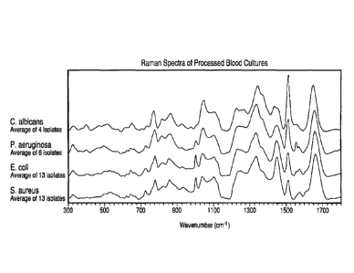
[0022] Figure 1 shows the Raman spectra of various microorganisms processed
and
recovered from blood culture.
[0023] Figure 2 shows the Raman spectra of 13 S. aureus isolates recovered
directly from
blood culture broth.
[0024] Figure 3 shows the Raman spectra read through a sealed separation
device, with and
without microorganisms present.
[0025] Figure 4 shows the Raman spectra of a blood culture-derived
microorganism pellet
read through the sealed separation device, background subtracted.
DETAILED DESCRIPTION OF THE INVENTION
[0026] The present invention can be embodied in different forms and should not
be construed
as limited to the embodiments set forth herein. Rather, these embodiments are
provided so that this
disclosure will be thorough and complete, and will fully convey the scope of
the invention to those
skilled in the art. For example, features illustrated with respect to one
embodiment can be
incorporated into other embodiments, and features illustrated with respect to
a particular embodiment
can be deleted from that embodiment. In addition, numerous variations and
additions to the
embodiments suggested herein will be apparent to those skilled in the art in
light of the instant
disclosure, which do not depart from the instant invention.
[0027] Unless otherwise defined, all technical and scientific terms used
herein have the same
meaning as commonly understood by one of ordinary skill in the art to which
this invention belongs.
The terminology used in the description of the invention herein is for the
6b
CA 02741036 2011-04-18
WO 2010/062351
PCT/US2009/005886
purpose of describing particular embodiments only and is not intended to be
limiting of the
invention.
Definitions.
[0028] As used herein, "a," "an," or "the" can mean one or more than one. For
example, "a" cell can mean a single cell or a multiplicity of cells.
[0029] Also as used herein, "and/or" refers to and encompasses any and all
possible
combinations of one or more of the associated listed items, as well as the
lack of
combinations when interpreted in the alternative ("or").
[0030] Furthermore, the term "about," as used herein when referring to a
measurable
value such as an amount of a compound or agent of this invention, dose, time,
temperature,
and the like, is meant to encompass variations of 20%, 10%, 5%, 1%,
0.5%, or even
0.1% of the specified amount.
[0031] As used herein, the term "microorganism" is intended to encompass
organisms
that are generally unicellular, which can be multiplied and handled in the
laboratory,
including but not limited to, Gram-positive or Gram-negative bacteria, yeasts,
molds,
parasites, and mollicutes. Non-limiting examples of Gram-negative bacteria of
this invention
include bacteria of the following genera: Pseudomonas, Escherichia,
Salmonella, Shigella,
Enterobacter, Klebsiella, Serratia, Proteus, Campylobacter, Haemophilus,
Morganella,
Vibrio, Yersinia, Acinetobacter, Stenotrophomonas, Brevundimonas, Ralstonia,
Achromobacter, Fusobacterium, Prevotella, Branhamella, Neisseria,
Burkholderia,
Citrobacter, Hafnia, Edwardsiella, Aeromonas, Moraxella, Brucella,
Pasteurella,
Providencia, and Legionella. Non-limiting examples of Gram-positive bacteria
of this
invention include bacteria of the following genera: Enterococcus,
Streptococcus,
Staphylococcus, Bacillus, Paenibacillus, Lactobacillus, Listeria,
Peptostreptococcus,
Propionibacterium, Clostridium, Bacteroides, Gardnerella, Kocuria,
Lactococcus,
Leuconostoc, Micrococcus, Mycobacteria and Corynebacteria. Non-limiting
examples of
yeasts and molds of this invention include those of the following genera:
Candida,
Ctyptococcus, Nocardia, Penicillium, Alternaria, Rhodotorula, Aspergillus,
Fusarium,
Saccharomyces and Trichosporon. Non-limiting examples of parasites of this
invention
include those of the following genera: Trypanosoma, Babesia, Leishmania,
Plasmodium,
Wucheria, Brugia, Onchocerca, and Naegleria. Non-limiting examples of
mollicutes of this
invention include those of the following genera: Mycoplasma and Ureaplasma.
7
CA 02741036 2011-04-18
WO 2010/062351
PCT/US2009/005886
[0032] In one embodiment, as described in further detail herein,
microorganisms from
a sample or growth medium can be separated and interrogated to characterize
and/or identify
the microorganism present in the sample. As used herein, the term " separate"
is intended to
encompass any sample of microorganisms that has been removed, concentrated or
otherwise
set apart from its original state, or from a growth or culture medium. For
example, in
accordance with this invention, microorganisms may be separated away (e.g., as
a separated
sample) from non-microorganism or non-microorganism components that may
otherwise
interfere with characterization and/or identification. The term may include a
layer of
microorganisms sandwiched between two other layers, e.g., microorganisms
collected on top
of a high-density cushion after centrifugation, or a layer of microorganisms
collected on a
solid surface (e.g., a filter membrane). The term may also include a
collection of
microorganisms that has passed partially through a layer (e.g., a density
cushion). As such, a
separated microorganism sample may include any collection or layer of
microorganisms
and/or components thereof that is more concentrated than, or otherwise set
apart from, the
original sample, and can range from a closely packed dense clump of
microorganisms to a
diffuse layer of microorganisms. Microorganism components that can be
comprised in a
separated form or sample include, without limitation, pilli, flagella,
fimbriae, and capsules in
any combination. Non-microorganism components that are separated away from the
microorganisms may include non-microorganism cells (e.g., blood cells and/or
other tissue
cells) and/or any components thereof.
[0033] In yet another embodiment, as described in further detail herein,
microorganisms from a sample or growth medium can be isolated and interrogated
to
characterize and/or identify the microorganism present in the sample. As used
herein, the
term "isolated" is intended to encompass any sample of microorganisms that has
been at least
partially purified from its original state, or from a growth or culture
medium, and any non-
microorganisms or non-microorganism components contained therein. For example,
in
accordance with this invention, microorganisms may be isolated away (e.g., as
an isolated
sample) from non-microorganisms or non-microorganism components that may
otherwise
interfere with characterization and/or identification. Non-microorganism
components that are
separated away from the microorganisms may include non-microorganism cells
(e.g., blood
cells and/or other tissue cells) and/or any components thereof.
[0034] In yet another embodiment, as described in further detail herein,
microorganisms from a sample or growth medium can be pelleted and interrogated
to
characterize and/or identify the microorganism present in the sample. As used
herein, the
8
CA 02741036 2011-04-18
WO 2010/062351
PCT/US2009/005886
term "pellet" is intended to encompass any sample of microorganisms that has
been
compressed or deposited into a mass of microorganisms. For example,
microorganisms from
a sample can be compressed or deposited into a mass at the bottom of a tube by
centrifugation, or other known methods in the art. The term includes a
collection of
microorganisms (and/or components thereof) on the bottom and/or sides of a
container
following centrifugation. Microorganism components that can be comprised in a
pellet
include, without limitation, pilli, flagella, fimbriae, and capsules in any
combination. In
accordance with this invention, microorganisms may be pelleted away (e.g., as
a substantially
purified microorganism pellet) from non-microorganism or non-microorganism
components
that may otherwise interfere with characterization and/or identification. Non-
microorganism
components that are separated away from the microorganisms may include non-
microorganism cells (e.g., blood cells and/or other tissue cells) and/or any
components
thereof.
[0035] As used herein, the term "density cushion" refers to a solution having
a
homogenous density throughout.
[0036] The present invention provides methods for isolating, characterizing
and/or
identifying microorganisms in a sample. Moreover, the method may be
particularly useful
for the separation, characterization and/or identification of microorganisms
from complex
samples such as blood-containing culture media. The rapid methods also allow
for the
characterization and/or identification of microorganisms more quickly than
prior techniques,
resulting in faster diagnoses (e.g., in a subject having or suspected of
having septicemia) and
characterization/identification of contaminated materials (e.g., foodstuffs
and
pharmaceuticals). The steps involved in the methods of the invention, from
obtaining a
sample to characterization/identification of microorganisms, can be carried
out in a very short
time frame to obtain clinically relevant actionable information. In certain
embodiments, the
methods of the invention can be carried out in less than about 120 minutes,
e.g., in less than
about 110, 100, 90, 80, 70, 60, 50, 40, 30, 20, 15, 10, 5, 4, 3, 2, or 1
minute. The tremendous
rapidity of the methods of the invention represents an improvement over prior
methods. The
methods can be used to characterize and/or identify any microorganism as
described herein.
In one embodiment, the microorganism is a bacterium. In another embodiment,
the
microorganism is a yeast. In another embodiment, the microorganism is a mold.
In a further
embodiment, the microorganism is a parasite. In another embodiment, the
microorganism is
a mollicute. Additionally, the methods of the invention can be fully
automated, thereby
reducing the risk of handling infectious materials and/or contaminating the
samples.
9
CA 02741036 2011-04-18
WO 2010/062351
PCT/US2009/005886
[0037] In one aspect, the present invention is directed to a method of
characterizing
and/or identifying a microorganism from a test sample, comprising:
(a) obtaining a test sample known to contain or that may contain
microorganisms;
(b) selectively lysing non-microorganism cells in said test sample to
produce a lysed
sample;
(c) separating microorganisms from other components of said lysed sample to
form an
isolated sample of microorganisms;
(d) interrogating said isolated microorganisms using one or more Raman
spectroscopy
techniques to produce spectroscopic measurements of said microorganism; and
(e) characterizing and/or identifying said microorganism in said isolated
sample by
comparison of the spectroscopic measurements with spectroscopic measurements
taken, or
spectroscopic properties predicted, of known microorganisms.
100381 In another aspect, the present invention is directed to a method of
characterizing and/or identifying a microorganism, comprising:
(a) obtaining a test sample known to contain or that may contain
microorganisms;
(b) placing said test sample in a sealed container;
(c) separating microorganisms in situ from other components of said test
sample to form
an isolated sample of microorganisms of microorganisms in said sealed
container;
(d) spectroscopically interrogating said isolated microorganisms in situ
using one or more
Raman spectroscopy techniques to produce spectroscopic measurements of said
microorganism; and
(e) characterizing and/or identifying said microorganism in said
isolated sample by
comparison of the spectroscopic measurements with spectroscopic measurements
taken, or
spectroscopic properties predicted, of known microorganisms.
100391 In another embodiment of the invention, the methods involve recovering
the
pellet of microorganisms formed during the separation step or a portion
thereof from the
separation container prior to interrogation of the microorganisms. For
example, after
formation of the pellet, the fluids can be aspirated way from the pellet and
the pellet
resuspended in a suitable medium (e.g., a medium in which the microorganisms
are viable).
The resuspended microorganisms can be removed from the separation container.
The
microorganisms can then be interrogated for characterization and/or
identification, e.g., in the
suspension or after they have been repelleted. In other embodiments, the
resuspended
microorganisms can be interrogated in the separation container, e.g., in the
suspension or
after they have been repelleted. In a further embodiment, microorganisms
recovered from the
CA 02741036 2011-04-18
WO 2010/062351
PCT/US2009/005886
pellet can be used directly for further interrogation (e.g., Raman
spectroscopy) without being
resuspended.
Samples
[0040] Samples that may be tested (i.e., a test sample) by the methods of the
invention
include both clinical and non-clinical samples in which microorganism presence
and/or
growth is or may be suspected, as well as samples of materials that are
routinely or
occasionally tested for the presence of microorganisms. The amount of sample
utilized may
vary greatly due to the versatility and/or sensitivity of the method. Sample
preparation can be
carried out by any number of techniques known to those skilled in the art
although one of the
advantages of the present invention is that complex sample types, such as,
e.g., blood, bodily
fluids, and/or other opaque substances, may be tested directly utilizing the
system with little
or no extensive pretreatment. In one embodiment, the sample is taken from a
culture. In
another embodiment, the sample is taken from a microbiological culture (e.g.,
a blood
culture). In another embodiment, the sample is suspected of, or known to,
contain
microorganisms therein.
[0041] Clinical samples that may be tested include any type of sample
typically tested
in clinical or research laboratories, including, but not limited to, blood,
serum, plasma, blood
fractions, joint fluid, urine, semen, saliva, feces, cerebrospinal fluid,
gastric contents, vaginal
secretions, tissue homogenates, bone marrow aspirates, bone homogenates,
sputum, aspirates,
swabs and swab rinsates, other body fluids, and the like. In another
embodiment, the clinical
sample can be cultured, and a culture sample used.
[0042] The present invention finds use in research as well as veterinary and
medical
applications. Suitable subjects from which clinical samples can be obtained
are generally
mammalian subjects, but can be any animal. The term "mammal" as used herein
includes,
but is not limited to, humans, non-human primates, cattle, sheep, goats, pigs,
horses, cats,
dog, rabbits, rodents (e.g., rats or mice), etc. Human subjects include
neonates, infants,
juveniles, adults and geriatric subjects. Subjects from which samples can be
obtained include,
without limitation, mammals, birds, reptiles, amphibians, and fish.
[0043] Non-clinical samples that may be tested also include substances,
encompassing, but not limited to, foodstuffs, beverages, pharmaceuticals,
cosmetics, water
(e.g., drinking water, non-potable water, and waste water), seawater ballasts,
air, soil, sewage,
plant material (e.g., seeds, leaves, stems, roots, flowers, fruit), blood
products (e.g., platelets,
11
CA 02741036 2011-04-18
WO 2010/062351
PCT/US2009/005886
serum, plasma, white blood cell fractions, etc.), donor organ or tissue
samples, biowarfare
samples, and the like. The method is also particularly well suited for real-
time testing to
monitor contamination levels, process control, quality control, and the like
in industrial
settings. In another embodiment, the non-clinical sample can be cultured, and
a culture
sample used.
[0044] In one embodiment of the invention, samples are obtained from a subject
(e.g.,
a patient) having or suspected of having a microbial infection. In one
embodiment, the
subject has or is suspected of having septicemia, e.g., bacteremia or
fungemia. The sample
may be a blood sample directly from the subject. The sample may be from a
blood culture
grown from a sample of the patient's blood, e.g., a BacT/ALERT blood culture.
The blood
culture sample may be from a positive blood culture, e.g., a blood culture
that indicates the
presence of a microorganism. In certain embodiments, the sample is taken from
a positive
blood culture within a short time after it turns positive, e.g., within about
6 hours, e.g., within
about 5, 4, 3, or 2 hours, or within about 60 minutes, e.g., about 55, 50, 45,
40, 35, 30, 25, 20,
15, 10, 5, 4, 3, 2, or 1 minute. In one embodiment, the sample is taken from a
culture in
which the microorganisms are in log phase growth. In another embodiment, the
sample is
taken from a culture in which the microorganisms are in a stationary phase.
[0045] The present invention provides high sensitivity for the detection,
characterization and/or identification of microorganisms.
This enables detection,
characterization and/or identification without first having to go through the
steps of isolating
microorganisms by growing them on a solid or semisolid medium, and sampling
the colonies
that grow. Thus, in one embodiment of the invention, the sample is not from a
microbial
(e.g., bacteria, yeast, or mold) colony grown on a solid or semisolid surface.
Thus, in one
embodiment of the invention, the sample is not from a microbial (e.g.,
bacteria, yeast, or
mold) colony grown on a solid or semisolid surface.
[0046] The volume of the sample should be sufficiently large to produce an
isolated
sample of microorganisms or a pellet of microorganisms which can be
interrogated after the
separation/isolation step of the methods of the invention is carried out.
Appropriate volumes
will depend on the source of the sample and the anticipated level of
microorganisms in the
sample. For example, a positive blood culture will contain a higher level of
microorganisms
per volume than a drinking water sample to be tested for contamination, so a
smaller volume
of blood culture medium may be needed as compared to the drinking water
sample. In
general, the sample size can be less than about 50 ml, e.g., less than about
40, 30, 20, 15, 10,
5, 4, 3, or 2 ml. In certain embodiments, the sample size can be about 1 ml,
e.g., about 0.75,
12
CA 02741036 2011-04-18
WO 2010/062351
PCT/US2009/005886
0.5, or 0.25 ml. In certain embodiments in which the separation is carried out
on a
microscale, the sample size can be less than about 200 I, e.g., less than
about 150, 100, 50,
25, 20, 15, 10, or 5 1. In some embodiments (e.g., when the sample is
expected to comprise
a small number of microorganisms), the sample size can be about 100 ml or
more, e.g., about
250, 500, 750, or 1000 ml or more.
Optional Lysis Step
[0047] In some embodiments, after obtaining a sample, the next step in the
method of
the present invention is to selectively lyse undesired cells that may be
present in the sample,
e.g., blood cells and/or tissue cells. Cells may be lysed to permit
separation of
microorganisms from other components of the sample. The separation of
microorganisms
from other components prevents interference during the interrogation step. If
non-
microorganism cells are not expected to be present in the sample or not
expected to interfere
with the interrogation step, the lysis step may not need to be carried out. In
one embodiment,
the cells to be lysed are non-microorganism cells that are present in the
sample and no
microorganism cells that may be present in the sample are lysed. However, in
some
embodiments, the selective lysing of specific classes of microorganisms may be
desirable and
thus can be carried out according to the methods described herein and as are
well known in
the art. For example, a class of undesired microorganisms can be selectively
lysed, e.g., yeast
are lysed while bacteria are not or vice versa. In another embodiment, the
desired
microorganisms are lysed in order to separate a particular subcellular
component of the
microorganisms, e.g., cell membranes or organelles. In one embodiment, all of
the non-
microbial cells are lysed. In other embodiments, a portion of the non-
microbial cells are
lysed, e.g., enough cells to prevent interference with the interrogation step.
The lysing of
cells may be carried out by any method known in the art to be effective to
selectively lyse
cells with or without lysing microorganisms, including, without limitation,
addition of a lysis
solution, sonication, osmotic shock, chemical treatment, and/or a combination
thereof.
[0048] A lysis solution is one that is capable of lysing cells, e.g., non-
microorganism
cells (e.g., by solubilizing eukaryotic cell membranes) and/or microorganism
cells. In one
embodiment, the lysis solution can comprise one or more detergents, one or
more enzymes,
or a combination of one or more detergents and one or more enzymes, and can
further include
additional agents. In one embodiment, the detergent can be a non-denaturing
lytic detergent,
such as Triton X-100 Triton X-100-R, Triton X-114, NP-40, Genapol C-100,
Genapol
X-100, Igepal CA 630, Arlasolvem200, Brij 96/97, CHAPS, octyl 13-D-
glucopyranoside,
13
CA 02741036 2011-04-18
WO 2010/062351
PCT/US2009/005886
saponin, and nonaethylene glycol monododecyl ether (C12E9, polidocenol).
Optionally,
denaturing lytic detergents can be included, such as sodium dodecyl sulfate, N-
laurylsarcosine, sodium deoxycholate, bile salts, hexadecyltrimethylammonium
bromide,
SB3-10, SB3-12, amidosulfobetaine-14, and C7Bz0. Optionally, solubilizers can
also be
included, such as Brij 98, Brij 58, Brij 35, Tween 80, Tween 20, Pluronic
L64,
Pluronic P84, non-detergent sulfobetaines (NDSB 201), amphipols (PMAL-C8),
and
methyl-P-cyclodextrin. Typically, non-denaturing detergents and solubilizers
are used at
concentrations above their critical micelle concentration (CMC), while
denaturing detergents
may be added at concentrations below their CMC. For example, non-denaturing
lytic
detergents can be used at a concentration of about 0.010% to about 10%, e.g.,
about 0.015%
to about 1.0%, e.g., about 0.05% to about 0.5%, e.g., about 0.10% to about
0.30% (final
concentration after dilution with the sample). In another embodiment,
polyoxyethylene
detergent detergents may be preferred. The polyoxyethylene detergent can
comprise the
structure C12-18/E9-1o, wherein C12-18 denotes a carbon chain length of from
12 to 18 carbon
atoms and E9-10 denotes from 9 to 10 oxyethylene hydrophilic head groups. For
example,
the polyoxyethylene detergent can be selected from the group consisting of
Brij 97, Brij
96V, Genapol C-100, Genapol X-100, nonaethylene glycol monododecyl ether
(polidocanol), or a combination thereof.
[0049] Enzymes that can be used in lysis solutions include, without
limitation,
enzymes that digest nucleic acids and other membrane-fouling materials (e.g.,
proteinase
XXIII, DNase, neuraminidase, polysaccharidase, Glucanex , and Pectinex8).
Other additives
that can be used include, without limitation, reducing agents such as 2-
mercaptoethanol (2-
Me) or dithiothreitol (DTT) and stabilizing agents such as magnesium,
pyruvate, and
humectants. The lysis solution can be buffered at any pH that is suitable to
lyse the desired
cells, and will depend on multiple factors, including without limitation, the
type of sample,
the cells to be lysed, and the detergent used. In some embodiments, the pH can
be in a range
from about 2 to about 13, e.g., about 6 to about 13, e.g., about 8 to about
13, e.g., about 10 to
about 13. Suitable pH buffers include any buffer capable of maintaining a pH
in the desired
range, e.g., about 0.05 M to about 1.0 M CAPS.
[0050] In one embodiment, the sample and the lysis solution are mixed and then
incubated for a sufficient time for lysis and solubilization of cell membranes
to occur, e.g.,
about 1, 2, 3, 4, 5, 10, 15, 20, 25, 30, 40, 50, or 60 seconds, or about 2, 3,
4, 5, 6, 7, 8, 9, 10,
15, or 20 minutes or longer, e.g., about 1 second to about 20 minutes, about 1
second to about
5 minutes, or about 1 second to about 2 minutes. The incubation time will
depend on the
14
CA 02741036 2016-04-25
strength of the lysis solution, e.g., the concentration of the detergent
and/or enzymes. In
general, milder lysis buffers will require more time and a greater dilution of
the sample to
fully solubilize non-microbial cells. The strength of the lysis solution can
be selected based
on the microorganisms known to be or suspected to be in the sample. For
microorganisms
that are more susceptible to lysis, a mild lysis solution can be used. The
lysis can take place
at a temperature of about 2 C to about 45 C, e.g., about 15 C to about 40 C,
e.g., about 30 C
to about 40 C. In one embodiment, the lysis solution can be loaded into a
syringe and the
sample can then be aspirated into the syringe such that mixing and incubation
occurs within
the syringe. In one embodiment, the lysis solution can be loaded into a
syringe and the
sample can then be aspirated into the syringe such that mixing and incubation
occurs within
the syringe.
[0051] In some embodiments, the lysis conditions (e.g., the solution or the
incubation
time), as well as the separation and/or interrogation steps, can be sufficient
to kill some or all
of the microorganisms in the sample. The methods of the present invention are
highly
versatile and do not require that all microorganisms be alive for the
isolation and
identification to occur. In certain embodiments, some or all of the
microorganisms may be
dead, with death occurring before, during, and/or after the steps of the
methods being carried
out.
[0052] Further details and description of the lysis buffers contemplated in
the practice
of this invention are disclosed in pending U.S. patent application, serial no.
, filed October
30, 2009, entitled "Methods for Isolation and Identification of Microorganisms
",
Separation Step
[0053] The next step in the method of the present invention (e.g., the step
after the
sample has been lysed, if a lysing step is performed) is a separation step.
The separation step
can be carried out to separate the microorganisms from other components of the
sample (e.g.,
non-microorganisms or components thereof) and to concentrate the
microorganisms into a
pellet that can be interrogated for identification and characterization
purposes. The
separation does not have to be complete, i.e., it is not required that 100%
separation occur.
All that is required is that the separation of the microorganisms from other
components of the
sample be sufficient to permit interrogation of the microorganisms without
substantial
interference from the other components. For example, the separation can result
in a
CA 02741036 2011-04-18
WO 2010/062351
PCT/US2009/005886
microorganism pellet that is at least about 10, 20, 30, 40, 50, 60, 70, 80,
90, 95, 96, 97, 98, or
99% pure or higher.
[0054] In one embodiment, the separation is carried out by a centrifugation
step in
which the sample (e.g., a lysed sample) is placed on top of a density cushion
in a separation
container and the container centrifuged under conditions which allow the
microorganisms to
be isolated (e.g., the microorganisms can form a pellet at the bottom and/or
sides of the
container). In accordance with this embodiment, other components of the sample
(e.g., non-
microorganisms or components thereof that may be present in the sample medium)
stay on
top of the density cushion or within the top portion of the density cushion.
In general, any
known container may be used for the separation step. In one embodiment, the
separation
container is the separation device disclosed in related U.S. patent
application, serial no. ,
filed October 30, 2009 entitled "Separation Device for Use in the Separation,
Characterization and/or Identification of Microorganisms". This separation
step isolates the
microorganisms away from materials in the sample, such as medium, cell debris,
and/or other
components that might interfere with interrogation of the microorganisms
(e.g., by intrinsic
fluorescence). In one embodiment, the density cushion also serves to separate
live
microorganisms from dead microorganisms (which do not pass through the density
cushion).
In another embodiment the density cushion does not comprise a density
gradient, either
before or after the centrifugation. In other words, the separation container
is not centrifuged
for a sufficient amount of time and/or acceleration for the material making up
the density
cushion to form a density gradient.
[0055] The density of the cushion is selected such that the microorganisms in
the
sample pass through the cushion while other components of the sample (e.g.,
blood culture
broth, cell debris) remain on top of the cushion or do not pass all of the way
through the
density cushion. The density cushion may also be selected to separate live
microorganisms
(which pass through the cushion) from dead microorganisms (which do not pass
through the
cushion). Suitable densities will depend on the material used in the density
cushion and on
the sample to be separated. In one embodiment, the density of the cushion is
in the range of
about 1.025 to about 1.120 g/ml, e.g., about 1.030 to about 1.070 g/ml, about
1.040 to about
1.060 g/ml or any range between about 1.025 to about 1.120 g/ml. In another
embodiment,
the density of the cushion is about 1.025, 1.030, 1.035, 1.040, 1.045, 1.050,
1.055, 1.060,
1.065, 1.070, 1.075, 1.080, 1.085, 1.090, 1.095, 1.100,1.105, 1.110, 1.115, or
1.120 g/ml.
[0056] The material for the density cushion can be any material that has the
appropriate density range for the methods of this invention. In one
embodiment, the material
16
CA 02741036 2011-04-18
WO 2010/062351
PCT/US2009/005886
is colloidal silica. The colloidal silica may be uncoated (e.g., Ludox (W.R.
Grace, CT)) or
coated, e.g., with silane (e.g., PureSperm (Nidacon Intl, Sweden) or Isolate
(Irvine
Scientific, Santa Ana, CA)) or polyvinylpyrrolidone (e.g., PercollTM,
PercollTM Plus (Sigma-
Aldrich, St. Louis, MO)). In one embodiment, the colloidal silica exhibiting
the least
interference with spectroscopic interrogation is selected, e.g., the material
with the lowest
intrinsic fluorescence. The colloidal silica may be diluted in any suitable
medium to form the
proper density, e.g., balanced salt solutions, physiological saline, and/or
0.25 M sucrose.
Suitable densities can be obtained with colloidal silica at a concentration of
about 15% to
about 80% v/v, e.g., about 20% to about 65% v/v. Another suitable material for
density
cushions is an iodinated contrast agent, e.g., iohexol (OmnipaqueTM
NycoPrepTM, or
Nycodenz ) and iodixanol (VisipaqueTM or OptiPrepTm). Suitable densities can
be obtained
with iohexol or iodixanol at a concentration of about 10% to about 25% w/v,
e.g., about 14%
to about 18% w/v, for blood culture samples. Sucrose can be used as a density
cushion at a
concentration of about 10% to about 30% w/v e.g., about 15% to about 20% w/v,
for blood
culture samples. Other suitable materials that can be used to prepare the
density cushion
include low viscosity, high density oils, such as microscope immersion oil
(e.g., Type DF;
Cargille Labs, New York), mineral oil (e.g., Drakeol 5, Draketex 50, Peneteck
; Penreco
Co., Pennsylvania), silicone oil (polydimethylsiloxane), fluorosilicone oil,
silicone gel,
metrizoate-Ficoll (LymphoPrepTm), e.g., at a concentration of about 75% to
about 100% for
blood culture samples, diatrizoate-dextran (PolymorphoPrepTm), e.g., at a
concentration of
about 25% to about 50% for blood culture samples, carboxymethyl cellulose,
hydroxypropylmethyl cellulose, polyethylene oxide (high molecular weight),
Pluronic F127,
Pluronic F68, mixtures of Pluronic compounds, polyacrylic acid, cross-linked
polyvinyl
alcohol, cross-linked polyvinyl pyrrolidine, PEG methyl ether methacrylate,
pectin, agarose,
xanthan, gellan, Phytagel , sorbitol, Fico11 (e.g., Fico11 400 at a
concentration of about
10% to about 15% for blood culture samples), glycerol, dextran (e.g., at a
concentration of
about 10% to about 15% for blood culture samples), glycogen, cesium chloride
(e.g., at a
concentration of about 15% to about 25% for blood culture samples),
perfluorocarbon fluids
(e.g., perfluoro-n-octane), hydrofluorocarbon fluids (e.g., Vertrel XF), and
the like as are well
known in the art. In one embodiment, the density cushion is selected from one
or more of
colloidal silica, iodixanol, iohexol, cesium chloride, metrizoate-Ficoll ,
diatrizoate-dextran,
sucrose, Ficoll 400, and/or dextran in any combination. The density cushion
can also be
made up of a combination of materials, e.g., a combination of colloidal silica
and oil.
Certain combinations of the above compounds may be beneficial for the
separation and
17
CA 02741036 2011-04-18
WO 2010/062351
PCT/US2009/005886
reading steps of the present invention. For example, combinations of compounds
with
different UV-quenching properties, such as cesium chloride and Iohexol.
[0057] The volume/height of the density cushion should be sufficient to
achieve
separation of the microorganisms from other sample components. The volume will
depend
on the size and shape of the separation container. In general, a volume of
about 0.1 to about
5 ml can be used, e.g., about 0.2 to about 1 ml, e.g., about 0.2 ml to about
0.5 ml. If the
separation is performed on a microscale, the volume of the density cushion can
be about 1 1
to about 100 1, e.g., about 5 I to about 50 I. The volume of sample laid or
layered on top
of the density cushion should be sufficient to provide enough microorganisms
to produce a
pellet suitable for interrogation. In general, any volume that fits into the
container can be
used. For example, a volume of about 0.1 ml to about 5 ml can be used, e.g.,
about 0.2 ml to
about 1 ml, e.g., about 0.2 ml to about 0.5 ml. If the separation is performed
on a microscale,
the volume of sample can be about 1 I to about 100 I, e.g., about 5 1 to
about 50 1. The
available space in the container for sample will depend on the size and shape
of the container.
In some embodiments, an intermediate layer (liquid or solid) can be placed on
top of the
density cushion before the sample is laid or layered on top in order to
prevent any mixing of
the density cushion and the sample. In one embodiment, the intermediate layer
can be
polyethylene beads. In another embodiment, a small air bubble can be
positioned between
the density cushion and the sample to prevent mixing. In a further embodiment,
the density
cushion can be layered on top of a high density material (e.g., a
perfluorocarbon fluid) such
that the microorganisms pass through the density cushion during the separation
and collect at
the interface between the density cushion and the high density material.
[0058] In one embodiment of the invention, the separation container is
centrifuged in
a swing out rotor so that the microorganisms form a pellet directly on the
bottom of the
container. The container is centrifuged at a sufficient acceleration and for a
sufficient time
for the microorganisms to be separated (e.g., a pellet formed) from other
components of the
sample. The centrifugation acceleration can be about 1,000 x g to about 20,000
x g, e.g.,
about 2,500 x g to about 15,000 x g, e.g., about 7,500 x g to about 12,500 x
g, etc. The
centrifugation time can be about 30 seconds to about 30 minutes, e.g., about 1
minute to
about 15 minutes, e.g., about 1 minute to about 5 minutes. The centrifugation
can be carried
out at a temperature of about 2 C to about 45 C, e.g., about 15 C to about 40
C, e.g., about
20 C to about 30 C. In one embodiment, the separation container comprises a
closure, and
the closure is applied to the container to form a hermetic seal prior to
centrifugation. The
presence of a closure decreases the risks from handling microorganisms that
are or may be
18
CA 02741036 2011-04-18
WO 2010/062351
PCT/US2009/005886
infectious and/or hazardous, as well as the risk of contaminating the sample.
One of the
advantages of the methods of the invention is the ability to carry out any one
or more of the
steps of the methods (e.g., lysis, separation, interrogation, and/or
identification) with the
microorganisms in a sealed container (e.g., a hermetically sealed container).
The present
methods, involving the use of automated systems, avoid the health and safety
risks associated
with handling of highly virulent microorganisms, such as occurs with recovery
of
microorganisms from samples for direct testing. In one embodiment, the
container is not
centrifuged for a sufficient time and/or force for a density gradient to form
within the density
cushion. The present invention does not involve ultracentrifligation of
samples, e.g.,
centrifugation at forces greater than about 100,000 x g. Further, the present
invention does
not involve isopycnic (equilibrium) sedimentation or banding.
[0059] The separation container may be any container with sufficient volume to
hold
a density cushion and a sample. As noted herein, the separation device
disclosed in related
U.S. patent application, serial no. , filed October 30, 2009, entitled
"Separation Device for
Use in the Separation, Characterization and/or Identification of
Microorganisms", may be
used in the practice of this invention. In one embodiment, the container fits
or can be fitted
into a centrifuge rotor. The volume of the container can be about 0.1 ml to
about 25 ml, e.g.,
about 1 ml to about 10 ml, e.g., about 2 ml to about 8 ml. If the separation
is done on a
microscale, the volume of the container can be about 2 I to about 100 I,
e.g., about 5 I to
about 50 1. In one embodiment, the container has a wide internal diameter in
an upper
portion to hold the sample and the majority of the density cushion, and a more
narrow
internal diameter in a lower portion where the pellet of microorganisms is
collected. The
narrow portion can have an internal diameter of about 0.04 to about 0.12
inches, e.g., about
0.06 to about 0.10 inches, e.g., about 0.08 inches. The wide portion can have
an internal
diameter of about 0.32 to about 0.40 inches, e.g., about 0.34 to about 0.38
inches, e.g., about
0.36 inches. For microscale separations, the internal diameters can be even
smaller. For
example, the internal diameter of the narrow portion can be about 0.001 to
about 0.04 inches,
e.g., about 0.002 to about 0.01 inches. A tapered internal diameter portion
can connect the
upper and lower portions. The tapered portion can have an angle of about 20 to
about 70
degrees, e.g., about 30 to about 60 degrees. In one embodiment, the lower
narrow portion is
less than half of the total height of the container, e.g., less than about
40%, 30%, 20%, or
10% of the total height of the container. The container can have a closure
device attached or
may be threaded to accept a closure device (e.g., a cap) such that the
container can be
hermetically sealed during centrifugation. In certain embodiments, the
container is designed
19
CA 02741036 2011-04-18
WO 2010/062351
PCT/US2009/005886
such that the microorganism sample or pellet can be readily recovered, or
otherwise obtained
or removed from the container after separation, either manually or in an
automated manner
(so that technicians are not exposed to the container contents). For example,
the container
can comprise a removable portion or a break-away portion which contains the
pellet and
which can be separated from the rest of the container. In another embodiment,
the container
comprises means for access to the pellet after separation, such as one or more
ports or
permeable surfaces for insertion of a syringe or other sampling device or for
drawing off the
pellet. In one embodiment, the container can be a tube, e.g., a centrifuge
tube. In another
embodiment, the container can be a chip or a card. In one embodiment, the
container is a
stand alone container, i.e., a device for separating a single sample. In other
embodiments, the
container is part of a device that comprises two or more separation containers
such that
multiple samples can be separated at the same time. In one embodiment, the
device
comprises 2, 3, 4, 5, 6, 7, 8, 9, 10, 12, 15, 20, 25, 30, 36, 42, 48, 60, 72,
84, 96, or more
separation containers.
100601 The container can comprise an optical window through which the
interrogation
can occur. The optical window may be on the bottom, top, and/or sides of the
container. The
window can be comprised of any material that has a vibrational structure which
is
distinguishable from the spectra of the microorganism. Other discrimination
techniques such
as confocal Raman spectroscopy can be utilized to acquire the vibrational
spectra of the
microorganism while rejecting the spectra of the window material; this
technique is well-
known to those skilled in the art. An additional technique is Spatially Offset
Raman
Spectroscopy in which the excitation fiber is displaced along the window from
the emission
(Rayleigh and Raman spectra). This technique is also known to those skilled in
the art as a
means of discriminating between a window material and a quantity to be
measured beneath
the window. To enable interrogation of the microbial pellet with additional
spectroscopic
techniques, the window can also be composed of any material that is
transparent to light (e.g.,
at least a portion of the near infrared (NIR; 700 nm-1400 nm), ultraviolet
(UV; 190 nm-400
nm) and/or visible (VIS; 400 nm-700 nm) light spectrum). In one embodiment,
the optical
window is thin enough to permit spectroscopic interrogation, which will depend
on the
material of the window and the method used for interrogation. In another
embodiment, the
optical window is as thin as possible to reduce interference with
spectroscopic interrogation.
For example, the window can have a thickness of less than about 0.20 inches,
e.g., less than
about 0.15, 0.10, or 0.05 inches.
CA 02741036 2011-04-18
WO 2010/062351
PCT/US2009/005886
[0061] In another embodiment, the separation is carried out by a filtration
step in
which the sample (e.g., a lysed sample) is placed in a device fitted with a
selective filter or
filter set with pore sizes that retain the microorganisms. The retained
microorganisms may
be washed by gently passing a suitable buffer through the filter. The washed
microorganisms
may then be interrogated directly on the filter and/or recovered for
interrogation by directly
sampling the surface of the filter or by back-flushing the filter with
suitable aqueous buffer.
Interrogation Step
[0062] Once the microorganisms have been separated, isolated and/or pelleted,
the
separated sample, isolated sample or pellet can be interrogated to identify
and/or characterize
the microorganisms in the sample or pellet. In one embodiment, the
interrogation takes place
in a non-invasive manner, that is, the pellet is interrogated while it remains
in the separation
container. In another embodiment, the separation container remains sealed
throughout the
interrogation. The ability to identify the microorganisms in a non-invasive
manner,
optionally coupled with keeping the container sealed throughout the separation
and
identification/characterization process and automating some or all of the
procedure avoids the
constant handling of contaminated and/or infectious samples and greatly
increases the safety
of the entire process. Furthermore, the ability to identify microorganisms by
direct
interrogation without further processing of the pellet (e.g., resuspension,
plating, and growth
of colonies), greatly increases the speed with which identification can be
made. In one
embodiment, the pellet is recovered and/or resuspended and optionally removed
from the
separation container prior to interrogation. In another embodiment, the pellet
is recovered
and/or resuspended after in situ interrogation and further interrogation is
then carried out.
For example, techniques such as latex agglutination tests or automated
phenotypic
identification tests that can be applied to isolated microorganisms but not a
pellet of
microorganisms can be carried out on the recovered and/or resuspended
microorganisms.
[0063] In some embodiments, the isolated sample or pellet can be interrogated
spectroscopically. The spectroscopy can be used to analyze one or more
intrinsic properties
of the microorganisms, e.g., a property present within the microorganism in
the absence of
additional agents, such as stains, dyes, binding agents, etc. In other
embodiments, the
spectroscopy and be used to analyze one or more extrinsic properties of the
microorganisms,
e.g., a property that can only be detected with the aid of additional agents.
The interrogation
can be carried using, for example, and infrared spectroscopy. Raman
spectroscopy, including
Surface Enhanced Raman Spectroscopy (SERS), Spatially Offset Raman
spectroscopy
21
CA 02741036 2011-04-18
WO 2010/062351
PCT/US2009/005886
(SORS), transmission Raman spectroscopy, and/or resonance Raman spectroscopy.
To
enhance Raman (SERS) signals, microorganisms could either be coated with gold
and/or
silver nanoparticles prior to centrifugation, and/or the inner optical surface
could be pre-
coated with metal colloids of particular size and shape (Efrima et al., J.
Phys. Chem. B.
(Letter) 102:5947 (1998) for SERS). In another embodiment, the nanoparticles
are present in
the density cushion prior to centrifugation and associate with microorganisms
as the
microorganisms pass through the density cushion. In one embodiment, the
isolated sample
or pellet is interrogated while it remains in the separation container. The
container can be
interrogated through an optical window in the container. The optical window
may be on the
bottom and/or any side or sides and/or on the top of the container. In one
embodiment, the
separation container fits into or can be fitted into a holder in a
spectrometer in a suitable
position for interrogation. The spectroscopic interrogation can be carried out
by any
technique known to those of skill in the art to be effective for detecting
and/or identifying one
or more intrinsic or extrinsic properties of microorganisms. In another
embodiment, as
described herein, the isolated sample or pellet can be removed for
interrogation (e.g., the
isolated sample or pellet can be removed and prepared for interrogation by
mass
spectrometry, as is well known in the art). In still further embodiments, the
isolated sample
or pellet can be interrogated using more than one means. For example, the
isolated sample or
pellet can be interrogated using fluorescence spectroscopy and Raman
spectroscopy. In
accordance with this embodiment, these interrogation steps may be carried out
sequentially or
simultaneously.
100641 The sample excitation source may be selected from any number of
suitable
light sources as known to those skilled in the art. Any portion of the
electromagnetic
spectrum that produces usable data can be used. Light sources capable of
emission in the
ultraviolet, visible and/or near-infrared spectra, as well as other portions
of the
electromagnetic spectrum, can be utilized and are known to those skilled in
the art. Since
the generation of Raman spectra is generally an inefficient process (e.g. 1
Raman photon
generated for every 1 million incident photons), Raman requires a light source
with a large
photon flux. Since the invention of the laser in 1960 (Maiman, Nature), Raman
systems have
used a laser light source for obtaining Raman spectra. The laser light is
passed through a
highly selective (notch) filter that allows the narrow wavelength band of
laser emission to
transmit through the filter while blocking other artifacts generated by the
laser (e.g. amplified
spontaneous emission, fluorescence, plasma lines, etc.)
22
CA 02741036 2011-04-18
WO 2010/062351
PCT/US2009/005886
[0065] The light from the laser is guided to the sample using a variety of
optical
systems which can be created using relay lens systems, optical fibers, or a
combination of
both. The laser light is focused onto the sample using a lens (such as a
microscope objective)
so that a small spot of concentrated optical energy at the sample is obtained.
[0066] Raman backscattered light and backscattered excitation light are
collected and
collimated by the same microscope objective. The scattered light traverses
through an optical
filter that separates the Raman scattered light from the excitation light. The
Raman scattered
light is then propagated using a lens and/or optical fiber system to a
spectrometer which
disperses the Raman scattered light so that it impinges onto a CCD detector
array.
[0067] The spatial dispersion of the Raman scattered light generates a spectra
(e.g.
scattered wavelength versus intensity) which can be stored an compared to
reference spectra
for characterization.
[0068] The emission from the sample may be measured by any suitable means of
spectral discrimination, most preferably employing a spectrometer. The
spectrometer may be
a scanning monochromator that detects specific emission wavelengths whereby
the output
from the monochromator is detected by a photomultiplier tube and/or the
spectrometer may
be configured as an imaging spectrograph whereby the output is detected by an
imaging
detector array such as a charge-coupled device (CCD) detector array. In one
embodiment, a
discriminator allows the observation of the fluorescence and/or scattering
signal by a
photodetection means (such as a photomultiplier tube, avalanche photodiode,
CCD detector
array, and/or electron multiplying charge coupled device (EMCCD) detector
array).
[0069] According to the invention, control measurements are taken for known
microorganisms, thus allowing for correlation of measured test data with
characterization of
the microorganisms of interest using various mathematical methods known to
those skilled in
the art. For example, the data from samples may be compared with the baseline
or control
measurements utilizing software systems known to one skilled in the art. More
particularly,
the data may be analyzed by a number of multivariate analysis methods, such
as, for example,
General Discriminant Analysis (GDA), Partial Least Squares Discriminant
Analysis
(PLSDA), Partial Least Squares regression, Principal Component Analysis (PCA),
Parallel
Factor Analysis (PARAFAC), Neural Network Analysis (NNA) and/or Support Vector
Machine (SVM). These methods may be used to classify unknown microorganisms of
interest into relevant groups based on existing nomenclature, and/or into
naturally occurring
groups based on the organism's metabolism, pathogenicity and/or virulence in
designing the
system for monitoring, detecting and/or characterizing the organism as
described previously.
23
CA 02741036 2011-04-18
WO 2010/062351
PCT/US2009/005886
[0070] In yet another embodiment, non-spectroscopic measurements from the
detection system, such as detection times and growth rates can be used to
assist in the
characterization and/or identification of microorganisms from the isolated
sample or pellet.
Additionally, measurements taken from a photographic image of the lower region
of the
separation device can provide valuable information on the identity of the
isolate, such as
pellet size, shape, color and density.
[0071] In some embodiments of the invention, characterization and/or
identification
of the microorganisms in the isolated sample or pellet need not involve
identification of an
exact species. Characterization encompasses the broad categorization or
classification of
biological particles as well as the actual identification of a single species.
Classification of
microorganism from an isolated sample or pellet may comprise determination of
phenotypic
and/or morphologic characteristics for the microorganism. For example,
characterization of
the biological particles may be accomplished based on observable differences,
such as,
composition, shape, size, clustering and/or metabolism. In some embodiments,
classification
of the biological particles of interest may require no prior knowledge of the
characteristics of
a given biological particle but only requires consistent correlations with
empiric
measurements thus making this method more general and readily adaptable than
methods
based on specific binding events or metabolic reactions. As used herein
"identification"
means determining to which family, genus, species, and/or strain a previously
unknown
microorganism belongs to. For example, identifying a previously unknown
microorganism to
the family, genus, species, and/or strain level.
[0072] In some instances, characterization encompasses classification models
which
provide sufficient useful information for action to be taken. As used herein,
the preferred
classification models comprise grouping into one or more of the following: (1)
Gram
Groups; (2) Clinical Gram Groups; (3) Therapeutic Groups; and (4) Functional
Groups..
[0073] (1) Gram Groups: Within the Gram Groups classification, microorganisms
may be placed into one of three broad classification categories based on their
Gram staining
reaction and overall size, said groups selected from one or more of the
following: (a) Gram
positive microorganisms that stain dark blue with Gram staining; (b) Gram
negative
microorganisms that stain red with Gram staining; and (c) yeast cells that
stain dark blue with
Gram staining, but are very large rounded cells that are distinguished from
bacteria by their
morphological characteristics and size.
[0074] (2) Clinical Gram Groups: The Gram Groups may be further divided into
several sub-categories representing distinguishing morphological features.
These sub-
24
CA 02741036 2011-04-18
WO 2010/062351
PCT/US2009/005886
categories comprise all the relevant clinical information reported by an
experienced
laboratory technologist, and thus provide a higher level of identification
than a positive or
negative Gram reaction. This particular classification is very helpful because
it eliminates
concerns about relying on the quality of a Gram stain and/or the skill level
of the technician
reading the smear by providing the equivalent clinically relevant information
with an
automated system. More specifically, subcategories of microorganisms based on
this
classification model may be selected from one or more of the following: (a)
cocci, which are
small rounded cells; (b) diplococci, which are two small rounded cells joined
together; (c)
rods, which are rectangular shape; and (d) bacilli, which are rod shaped.
Examples of these
sub-categories that can be ascertained by additional morphological information
include: (i)
Gram positive cocci; (ii) Gram positive cocci in chains; (iii) Gram positive
cocci in clusters
(i.e., "grape-like" clusters); (iv) Gram positive diplococci; (v) Gram
positive rods; (vi) Gram
positive rods with endospores; (vii) Gram negative rods; (viii) Gram negative
coccobacilli;
(ix) Gram negative diplococci; (x) yeast; and (xi) filamentous fungi.
[0075] (3) Therapeutic Groups: The therapeutic groups comprise multiple
microbial
species that, when isolated from particular specimen types, are treated with
the same class of
antibiotics or mixture of antibiotics (e.g., as described in "Sanford Guide to
Antimicrobial
Therapy 2008"). In many cases, identity to the species level is not required
by the clinician
to enable a change from initial empiric therapy to a more targeted therapy
because more than
one species can be treated with the same choice of antibiotic(s). This
classification level
correctly places these "same-treatment" microorganisms into single therapeutic
categories.
Examples of this characterization level include the ability to distinguish
highly resistant
Enterobacteriacae (EB) species from sensitive EB species (Enterobacter spp.
from E. coil), or
fluconazole-resistant Candida species (C. glabrata and C. kruzei) from
sensitive Candida
species (C. albicans and C. parapsilosis), and so on.
[0076] (4) Functional Groups: According to the invention, microorganisms may
also
be placed into several groups based upon a mixture of metabolic, virulence
and/or phenotypic
characteristics. Non-fermentative organisms may be clearly distinguished from
fermentative
ones. Furthermore, microorganism species that produce hemolysins may be
grouped
separately from non-hemolytic species. In some cases, these groups represent
broader
categories than genus level (e.g., coliforms, Gram negative non-fermentative
rods), some at
the genus level (e.g., Enterococcus, Candida), and some with closer to species-
level
discrimination (e.g., coagulase-negative staphylococci, alpha-hemolytic
streptococci, beta-
hemolytic streptococci, coagulase-positive staphylococci, i.e., S. aureus).
CA 02741036 2011-04-18
WO 2010/062351
PCT/US2009/005886
[0077] In addition to measuring intrinsic properties of microorganisms for
identification purposes, the methods of the present invention can further
comprise the use of
additional identifier agents to aid in the separation and/or identification
process. Agents that
bind to specific microorganisms, such as affinity ligands, can be used to
separate
microorganisms, to identify a class or species of microorganism (e.g., through
binding to a
unique surface protein or receptor) and/or to identify a characteristic of the
microorganism
(e.g., antibiotic resistance). Useful identifier agents include, without
limitation, monoclonal
and polyclonal antibodies and fragments thereof (e.g., anti-Eap for S. aureus
identification),
nucleic acid probes, antibiotics (e.g., penicillin, vancomycin, polymyxin B),
aptamers,
peptide mimetics, phage-derived binding proteins, lectins, host innate
immunity biomarkers
(acute phase proteins, LPS-binding protein, CD14, mannose binding lectin, Toll-
like
receptors), host defense peptides (e.g., defensins, cathelicidins,
proteogrins, magainins),
bacterocins (e.g., lantibiotics, such as nisin, mersacidin, epidermin,
gallidermin, and
plantaricin C, and class II peptides), bacteriophages, and dyes selective for
nucleic acids,
lipids, carbohydrates, polysaccharides, capsules/slime or proteins, or any
combination
thereof. If the agent does not itself give out a detectable Raman signal, the
agent can be
labeled to provide a detectable signal, such as by conjugating the agent to a
Raman active
tracer, such as dimethylaminoazobenzene (DAB) and 4,4"-dipyridyl (DPY) (see,
e.g., U.S.
Pat. Nos. 5,376,556 and 7,518,721). The agent can be added to the
microorganisms at any
step in the methods of the invention, e.g., when the sample is obtained,
during lysis, and/or
during separation. In some embodiments, the presence of the agent in the
pellet can be
determined during interrogation of the pellet. As would be readily appreciated
by one of skill
in the art, the sensitivity of a particular microorganism to any compound
affecting its physical
state or metabolism, such as an antibiotic, could be rapidly ascertained by
adding the
compound to the sample, lysis buffer, density cushion or any mixture thereof.
[0078] In one aspect of the invention, the method can further comprise a step
of
recovering the pellet of microorganisms and performing additional tests. In
one embodiment,
the pellet can be recovered by aspirating off the sample medium and density
cushion. In
another embodiment, the pellet can be recovered by inserting a syringe into
the container and
aspirating out the pellet while the sample medium and density cushion remain
intact. The
recovered pellet can then be resuspended in a suitable medium, e.g., saline.
Once
resuspended, the microorganisms can be subject to any further tests that are
desired, as would
be known to those of skill in the art and as described above. In particular,
any test requiring
clean samples of microorganisms can be carried out with the resuspended
microorganisms.
26
CA 02741036 2011-04-18
WO 2010/062351
PCT/US2009/005886
In some embodiments, additional identification tests can be performed.
Examples of
identification tests include Vitek 2, amplified and non-amplified nucleic
acid tests (NAT),
chromogenic and latex agglutination assays, immunoassays, (e.g., employing
labeled
antibodies and/or other ligands), mass spectrometry (e.g., MALDI-TOF mass
spectrometry)
and/or other optical techniques such as infrared spectroscopy (FTIR) or Raman
spectroscopy.
Additional characterization tests can also be performed, such as resistance to
antibiotics
and/or other drugs. The additional characterization may be part of a test that
was started
during the initial separation and identification steps of the method. For
example, the
detection of methicillin resistant S. aureus can begin by adding Raman tracer-
labeled
penicillin to the sample prior to separation of the microorganisms. Once the
pellet has been
recovered and resuspended, the level of bound penicillin can be determined.
[00791 In one aspect of the invention, some or all of the method steps can be
automated. Automating the steps of the methods not only allows more samples to
be tested
more quickly, it also reduces the risks of human errors in handling samples
that may contain
harmful and/or infectious microorganisms.
[0080] In certain embodiments of the invention, the methods can also be used
to
detect the presence of microorganisms in a test sample. In these embodiments,
the methods
comprise the steps of:
(a) obtaining a test sample;
(b) optionally lysing cells in said sample to produce a lysed sample; and
(c) separating microorganisms from other components of said lysed sample to
form a
pellet of microorganisms;
wherein the presence of a pellet indicates that microorganisms are present in
the test sample.
In one embodiment, the pellet is detected with the naked eye. In other
embodiments, the
pellet is detected by interrogation, e.g., spectroscopically.
[0081] In some embodiments, the detection methods can be used to monitor
samples
for contamination by microorganisms, e.g., foodstuffs, pharmaceuticals,
drinking water, etc.
In one embodiment, the methods can be carried out in a repetitive fashion for
constant
monitoring for contamination, e.g., once a month, once a week, once a day,
once an hour, or
any other time pattern. In another embodiment, samples can be tested as
needed, e.g., when
contamination is suspected. In further embodiments, the detection methods can
be used to
look for the presence of microorganisms in clinical samples, e.g., blood
cultures. For
example, a sample can be removed from a blood culture at certain time points
and the
detection method carried out on the sample to determine if the blood culture
is positive. In
27
CA 02741036 2016-04-25
one embodiment, a sample may be taken at a set time point after inoculation of
the culture,
e.g., 24 hours after inoculation, to determine if the blood culture is
positive. In another
embodiment, samples can be taken from the blood culture regularly, e.g., every
12, 6, 4, or 2
hours or every 60, 50, 40, 30, 20, 15, 10, or 5 minutes, to identify positive
blood cultures
within a short time of being detectably positive. In certain embodiments of
the detection
methods, the detection step can optionally be followed by identification
methods as described
herein.
[0082] In one aspect of the invention, some or all of the method steps can be
automated. Automating the steps of the methods allows a greater number of
samples to be
tested more efficiently and reduces the risks of human errors in handling
samples that may
contain harmful and/or infectious microorganisms. Of greater importance,
however,
automation can deliver critical results at any time of the day or night
without delay. Several
studies have shown that faster identification of the organisms causing sepsis
correlates with
improved patient care, shorter hospital stays and lower overall costs.
[0083] Further details and description of the method for characterizing and/or
identifying microorganisms, in accordance with the present invention, are
disclosed in
pending U.S. patent applications, serial nos.
and __, both filed October 30, 2009, entitled
"Method for Separation, Characterization and/or Identification of
Microorganisms using
Spectroscopy" and "Method for Separation, Characterization and/or
Identification of
Microorganisms using Mass Spectrometry", respectively.
[0084] The present invention is further detailed in the following examples,
which are
offered by way of illustration and is not intended to limit the invention in
any manner.
Standard techniques well known in the art or the techniques specifically
described below are
utilized.
= 30
EXAMPLES
Example 1. Lysis-Centrifugation Method for Identification of Microorganisms
from
Blood Cultures by Raman Spectroscopy.
[00851 Microorganisms were "seeded" at a low inoculum into BacT/ALERT SA
bottles containing 10 mLs of human blood. Blood culture broth samples were
removed from
bottles within a few minutes of being flagged positive by the BacT/ALERT 3D
Microbial
28
CA 02741036 2011-04-18
WO 2010/062351
PCT/US2009/005886
Detection System. Broth samples were processed to separate microorganisms from
blood and
media components that could interfere with subsequent analysis as follows:
[0086] 4.0 mL of broth from freshly positive blood cultures was combined with
2.0
mL Lysis Buffer (0.45% Brij 97 in 0.3M CAPS, pH 11.7), vortex mixed for 5
seconds and
then incubated in a 37 C waterbath for 90 seconds. After incubation, 0.95 mL
of lysate was
layered on top of 0.5 mL of density cushion (14% w/v Iohexol, 0.005% Pluronic
F-108 in 10
mM Hepes, pH 7.4) in each of four 1.5 mL conical centrifuge tubes. All four
tubes were then
centrifuged for 2 minutes at 10,000 g at 25 C to sediment (pellet) the
microorganisms
through the density cushion. The lysed blood and media remained above the
cushion.
[0087] Upon completion of the centrifuge cycle, the supernatant was removed
and the
sedimented (pelleted) microorganisms in each tube were resuspended with 10 L
of purified
water. The resuspended microorganisms from all 4 tubes were pooled into a
clean tube and
mixed gently. The volume of each processed specimen was then adjusted so that
the optical
density at 660 nm (A660) of the final suspension was equal to 20/cm. The
processed
specimens were either stored at 2-8 C for same-day testing, or aliquoted and
frozen at -70 C
for testing on a later date.
Example 2. Analysis of microorganism specimens processed from positive blood
cultures with Lysis-Centrifugation by Raman Spectroscopy.
[0088] Specimens processed according to the procedure in Example 1 were
rapidly
thawed at 37 C (if previously frozen), mixed gently. A portion of the specimen
was kept
undiluted and the remainder was diluted to 1:2 in purified water. One
microliter of each
diluted and undiluted specimen was applied to the first surface of a gold-
coated microscope
slide and allowed to dry under ambient conditions.
[0089] After drying, 25 spectra in a 5x5 grid pattern were acquired for each
of the
dried specimens with a Model RxN1 Raman microscope (Kaiser Optical Systems
Inc.,
Michigan) at an illumination wavelength of 785 nm. The microscope was equipped
with a
40X objective lens, the laser power was set to 400 mw, and each of the 25
individual spectra
was acquired with a 5 second accumulation time.
[0090] After acquisition, the spectra were preprocessed to perform dark
subtraction,
cosmic ray artifact removal, spectral truncation, baseline subtraction,
normalization, and
outlier removal in preparation for and multivariate statistical analysis.
[0091] Representative Raman spectra of selected microorganisms recovered and
processed from positive blood cultures are shown in Figures 1 and 2. Figure 1
shows spectra
29
CA 02741036 2011-04-18
WO 2010/062351
PCT/US2009/005886
of four species of microorganisms; four isolates of C. albicans, six isolates
of P. aeruginosa,
thirteen isolates of E. coil, and thirteen isolates of S. aureus. Spectra from
each species were
averaged for clarity of illustration.
[0092] The consistent differences across the spectra of different species are
readily
apparent for these organisms. This shows that potentially obscuring blood and
culture media
have been removed sufficiently to obtain characteristic spectra for the
microorganisms after
processing.
[0093] Figure 2 shows the 13 individual Raman spectra of the S.aureus isolates
that
were shown averaged in Figure 1. The thirteen spectra show the consistency of
the Raman
spectrum for this microorganism across different clinical isolates, even when
grown in
different blood cultures with different blood donors.
Example 3. Non-invasive analysis of microorganism specimens processed from
positive blood cultures with Lysis-Centrifugation by Raman Spectroscopy.
[0094] Microorganisms were "seeded" at a low inoculum into BacT/ALERT SA
bottles containing 10 mL of human blood. Blood culture broth samples were
removed from
bottles within a few minutes of being flagged positive by the BacT/ALERT 3D
Microbial
Detection System. The samples were treated as follows:
1. A 2.0 mL sample of positive broth was mixed with 1.0 mL of selective lysis
buffer (0.45% w/v Brij 97 + 0.3M CAPS, pH 11.7), then placed in a 37 C water
bath for 1 minute.
2. A 1.0 mL sample of lysate was overlayed onto 0.5 mL of density cushion (24%
w/v cesium chloride in 10 mM Hepes ph 7.4 + 0.005% Pluronic F-108) contained
in a custom-built optical separation tube. A polypropylene ball was present on
the
surface of the density cushion to facilitate loading without disturbing the
two
aqueous phases.
3. The optical separation tube was sealed with a screw-cap and centrifuged for
2
minutes at 10,000 rpm (Eppendorf 5417R micrcentrifuge fitted with a A-8-11
swing out rotor; see Figure 4).
4. The sealed tube was transferred to a custom-built adapter which facilitated
interrogation of the microbial pellet in the base of the tube by a Model RxN1
Raman microscope (Kaiser Optical Systems Inc., Michigan) at an illumination
wavelength of 785 nm. Spectra were also recorded of empty separation tubes to
establish baseline Raman spectra of the plastic.
CA 02741036 2016-04-25
[0095] Figure 3 shows the Raman spectra taken through the plastic separation
tube
both with and without a microorganism pellet. In this mode, the spectra are
dominated by
Raman bands from the plastic itself; however, bands unique to the
microorganisms are visible
only in the spectrum of the microbial pellet at wavenumbers near 1227/cm,
1583/cm, and
1660/cm. Other microorganism bands are also present, but are obscured by the
stronger
plastic bands at this scale.
[0096] Figure 4 shows the Raman spectra of three microorganisms after the
background has been removed by subtracting the spectrum of the empty plastic
separation
tube. While this background subtraction is not 100% effective, the spectral
bands of the
microorganisms are clearly visible. Other acquisition geometries, such as
spatially offset or
transmission acquisition, are likely to improve the background compensation
further (see,
e.g., U.S. patent appl. publ. nos. 2008/0129992 and 2009/0244533).
100971 The foregoing Examples are illustrative of the present invention, and
are not to
be construed as limiting thereof. The invention is defined by the following
claims, with
equivalents of the claims to be included therein.
31