Note: Descriptions are shown in the official language in which they were submitted.
CA 02741418 2011-04-21
WO 2010/047823 PCT/US2009/005778
DETECTION AND MODULATION OF CYTOCHROME C ACETYLATION
Related Application
This application claims the benefit under 35 U.S.C. 119(e) of U.S.
Provisional
Application Serial No. 61/107,841, entitled "Detection and Modulation of
Cytochrome C
Acetylation," filed on October 23, 2008, which is herein incorporated by
reference in its
entirety.
Government Interest
This invention was made with government support under AG027916, awarded by the
National Institutes of Health. The government has certain rights in the
invention.
Field of the Invention
The invention pertains to methods of diagnosis and treatment of
neurodegenerative
diseases and cancer.
Background of the Invention
Cytochrome c is a haem-containing protein within the inner mitochondrial
membrane,
wherein it is a component of the electron transport chain. Cytochrome c is
also part of the
intrinsic apoptotic pathway. In cells that are undergoing apoptosis,
cytochrome c is released
from the mitochondrial membrane to interact with apoptotic protease-activating
factor-1
(APAF1), forming the apoptosome, which activates caspase proteases involved in
mediating
cell death.
Release of cytochrome c and commitment of a cell to apoptosis is a highly
regulated
process, and represents a target regulatory step for disorders associated with
apoptosis.
Cytochrome c activity has been shown to be regulated through multiple factors
including
BCL2 family members, caspases, heat-shock proteins, proteins that affect
fission/fusion of
the mitochondria, calcium levels, regulation of the redox states of cytochrome
c,
nitrosylation, histone H1.2 and cytosolic p53 (Ow et al., (2008) Nat Rev Mol
Cell Biol 9:532-
542).
CA 02741418 2011-04-21
WO 2010/047823 PCT/US2009/005778
Summary of the Invention
Described herein is a novel approach for regulating cytochrome c, involving
modulation of acetylation. Cytochrome c acetylation is revealed to be
associated with cells
undergoing apoptosis. Thus, detection of cytochrome c acetylation levels has
applications for
diagnosis of neurodegenerative disorders, and deacetylation of cytochrome c
represents a
therapeutic approach for treatment of neurodegenerative disorders.
Furthermore, induction of
apoptosis through acetylation of cytochrome c, and monitoring of cytochrome c
levels, have
diagnostic and therapeutic applications for cancer.
Aspects of the invention relate to methods for characterizing a subject's risk
of a
neurodegenerative disorder by detecting the level of acetylated cytochrome c
in a sample
from the subject, wherein an elevated level of acetylated cytochrome c in the
sample from the
subject, relative to a predetermined value, is indicative of an increased risk
of a
neurodegenerative disorder. The level of acetylated cytochrome c in a sample
can be
detected by any means known to one of ordinary skill in the art such as using
an antibody that
specifically binds acetylated cytochrome c, and/or through the use of mass
spectrometry.
In some embodiments of the invention, methods for characterizing a subject's
risk of
a neurodegenerative disorder include detecting acetylation of a lysine residue
corresponding
to residue K40 in a full-length, wild-type cytochrome c polypeptide, in a
sample from the
subject, wherein the presence of acetylation of a lysine residue corresponding
to residue K40
in a full-length, wild-type cytochrome c polypeptide in a sample from the
subject indicates
that the subject has an increased risk of a neurodegenerative disorder. In
certain
embodiments acetylation of lysine residue K40 is detected by mass
spectrometry.
In some embodiments of the invention, methods for characterizing a subject's
risk of
a neurodegenerative disorder include detecting acetylation of a lysine residue
corresponding
to residue K74 in a full-length, wild-type cytochrome c polypeptide, in a
sample from the
subject, wherein the presence of acetylation of a lysine residue corresponding
to residue K74
in a full-length, wild-type cytochrome c polypeptide in a sample from the
subject indicates
that the subject has an increased risk of a neurodegenerative disorder. In
certain
embodiments, acetylation of lysine residue K74 is detected by mass
spectrometry.
In some embodiments methods for characterizing a subject's risk of a
neurodegenerative disorder include detecting acetylation of lysine residues
corresponding to
residues K40 and K74 in a full-length, wild-type cytochrome c polypeptide, in
a sample from
2
CA 02741418 2011-04-21
WO 2010/047823 PCT/US2009/005778
the subject, wherein the presence of acetylation of lysine residues
corresponding to residues
K40 and K74 in a full-length, wild-type cytochrome c polypeptide indicates
that the subject
has an increased risk of neurodegenerative disorder. In certain embodiments
acetylation of
lysine residues K40 and K74 is detected by mass spectrometry.
Aspects of the invention relate to methods for diagnosing a neurodegenerative
disorder in a subject, including detecting the level of acetylated cytochrome
c in a sample
from the subject, wherein an elevated level of acetylated cytochrome c in the
sample from the
subject, relative to a predetermined value, is indicative of a
neurodegenerative disorder. In
certain embodiments the level of acetylated cytochrome c in a sample from the
subject is
detected by using an antibody that specifically binds acetylated cytochrome c
and/or by using
mass spectrometry.
Further aspects of the invention relate to methods for evaluating the efficacy
of a
therapy in a subject with a neurodegenerative disorder, including detecting
the level of
acetylation of cytochrome c in a sample from the subject, wherein the level of
acetylation of
cytochrome c in the sample from the subject, relative to a predetermined
value, is indicative
of whether the therapy is efficacious. In certain embodiments the level of
acetylated
cytochrome c in a sample from the subject is detected by using an antibody
that specifically
binds acetylated cytochrome c and/or by using mass spectrometry.
Also described herein are methods for treating a subject having a
neurodegenerative
disorder, including administering an effective amount of a compound to a
subject in need of
such a treatment to decrease the level of acetylated cytochrome c in the
subject below a
predetermined value, wherein the compound is a compound that activates a
sirtuin. In some
embodiments, the invention involves inhibiting apoptosis in a cell in which
cytochrome c is
acetylated by contacting the cell with an agent that deacetylates cytochrome
c. The agent that
deacetylates cytochrome c can be a deacetylase protein such as a sirtuin. In
some
embodiments the sirtuin is SIRT3.
Further aspects of the invention relate to methods for determining whether a
cancer
patient should be treated with an agent that acetylates cytochrome c by
performing an assay
to determine whether a patient has a cancer that exhibits deacetylation of
lysine residues
corresponding to residues K40 and K74 in a full-length, wild-type cytochrome c
polypeptide,
wherein the patient is a candidate for treatment with a composition that
acetylates
3
CA 02741418 2011-04-21
WO 2010/047823 PCT/US2009/005778
cytochrome c if the patient has a cancer that exhibits deacetylation of lysine
residues
corresponding to residues K40 and K74 in a full-length, wild-type cytochrome c
polypeptide.
Described herein are methods for inducing apoptosis in a cell in which
cytochrome c
is deacetylated by contacting the cell with an agent that acetylates
cytochrome c, to thereby
induce apoptosis in the cell. In some embodiments the cell is in vivo and the
method further
comprises contacting the cell with an additional therapeutic agent. Some
embodiments of the
invention involve methods for decreasing viability of a cancer cell that
exhibits deacetylation
of cytochrome c by contacting the cancer cell that exhibits deacetylation of
cytochrome c
with an agent that acetylates cytochrome c in an amount effective to decrease
the viability of
the cancer cell. In some embodiments the cell is in vivo and the method
further comprises
contacting the cell with an additional therapeutic agent.
Also described herein are isolated antibodies or antigen-binding fragments
thereof
that bind specifically to an epitope of acetylated cytochrome c polypeptide,
wherein the
epitope comprises an acetylated residue that corresponds to residue K40 in a
full-length,
wild-type, human cytochrome c amino acid sequence. In some embodiments the
isolated
antibody or antigen-binding fragment thereof specifically binds to the epitope
with a binding
affinity of about 1 x 10"6 M, 1 x 10"7 M, 1 x 10'8 M, 1 x 10"9 M, 1 x 10"'0 M,
5 x 10"'0 M, or 1
x 10"" M or less. In certain embodiments the antibody or antigen-binding
fragment thereof is
attached to a detectable label. Also included herein are nucleic acid
molecules encoding such
antibodies, hybridomas containing these nucleic acid molecules, and hybridoma
cell lines
producing such antibodies. Aspects of the invention also involve expression
vectors
including an isolated nucleic acid molecule encoding the antibodies or antigen-
binding
fragments described herein, host cells transformed by or transfected with such
expression
vectors, and plasmids that produce the antibodies or antigen-binding fragments
described
herein. In some embodiments, the invention relates to compositions comprising
the
antibodies or antigen-binding fragments described herein.
Also described herein are isolated antibodies or antigen-binding fragments
thereof
that bind specifically to an epitope of acetylated cytochrome c polypeptide,
wherein the
epitope comprises an acetylated residue that corresponds to residue K74 in a
full-length,
wild-type, human cytochrome c amino acid sequence. In some embodiments the
isolated
antibody or antigen-binding fragment thereof specifically binds to the epitope
with a binding
affinity of about 1 x 10⁻⁶ M, 1 x 10⁻¹ M, 1 x 10'8 M, 1 x 10⁻⁹ M, 1 x 10"'0 M,
5 x 10-'0 M, or 1
4
CA 02741418 2011-04-21
WO 2010/047823 PCT/US2009/005778
x 10-11 M or less. In certain embodiments the antibody or antigen-binding
fragment thereof is
attached to a detectable label. Also included herein are nucleic acid
molecules encoding such
antibodies, hybridomas containing these nucleic acid molecules, and hybridoma
cell lines
producing such antibodies. Aspects of the invention also involve expression
vectors
including an isolated nucleic acid molecule encoding the antibodies or antigen-
binding
fragments described herein, host cells transformed by or transfected with such
expression
vectors, and plasmids that produce the antibodies or antigen-binding fragments
described
herein. In some embodiments, the invention relates to compositions comprising
the
antibodies or antigen-binding fragments described herein.
Also described herein are methods for identifying compounds that modulate the
deactylase activity of SIRT3. Such methods include contacting an acetylated
cytochrome c
polypeptide substrate and a SIRT3 deacetylase in the presence of a test
compound, and
determining the level of acetylation of the cytochrome c polypeptide substrate
in the presence
of the test compound. In some embodiments, the cytochrome c polypeptide
substrate
comprises at least one acetylated lysine residue corresponding to residues K40
and/or K74 of
full-length, wild-type human cytochrome c polypolypeptide. A decrease in the
level of
acetylation of the cytochrome c polypeptide substrate in the presence of the
test compound as
compared to a control is indicative of a compound that increases SIRT3
deactylase activity.
An increase in the level of acetylation of the cytochrome c polypeptide
substrate in the
presence of the test compound as compared to a control is indicative of a
compound that
decreases SIRT3 deactylase activity.
In some embodiments, the level of acetylation of the cytochrome c polypeptide
substrate pool is determined using mass spectrometry. In some embodiments, the
mass
spectrometry is electrospray ionization (ESI) mass spectrometry or matrix-
assisted laser
desorption/ionization (MALDI) mass spectrometry.
In some embodiments of the foregoing methods, the cytochrome c polypeptide
substrate comprises a single polypeptide species, while in other embodiments
the cytochrome
c polypeptide substrate comprises a mixture of two or more polypeptides
species.
In some embodiments, the cytochrome c polypeptide substrate comprises a full-
length
cytochrome c polypeptide. In other embodiments, the cytochrome c polypeptide
substrate is
a fragment of cytochrome c comprising at least one lysine residue
corresponding to residues
K40 and/or K74 of full-length, wild-type human cytochrome c polypeptide. In
still other
5
CA 02741418 2011-04-21
WO 2010/047823 PCT/US2009/005778
embodiments, the cytochrome c polypeptide substrate is a fusion of a fragment
of cytochrome
c comprising at least one lysine residue corresponding to residues K40 and/or
K74 of full-
length, wild-type human cytochrome c polypeptide.
In some embodiments, the test compound is a small molecule, such as small
organic
molecules. In some embodiments, the test compound is a library of molecules,
which library
may, in certain embodiment, include small molecules, such as small organic
molecules.
In some embodiments, the SIRT3 deacetylase is from a cell or tissue lysate.
In some embodiments, the cytochrome c polypeptide substrate is in a cell.
In some embodiments, the SIRT3 is a catalytically active fragment of full-
length
(human) SIRT3 capable of deacetylating a cytochrome c substrate comprising
acetylated K40
and/or K74 in the presence of NAD+ or a NAD+ analog.
Also described herein are acetylated polypeptide substrates for use in
determining the
activity of SIRT3. The substrates include a fragment of cytochrome c
comprising at least one
acetylated lysine residue corresponding to residues K40 and/or K74 of full-
length, wild-type
human cytochrome c polypeptide. In some embodiments, the polypeptide substrate
is a
fusion of a fragment of cytochrome c comprising at least one acetylated lysine
residue
corresponding to residues K40 and/or K74 of full-length, wild-type human
cytochrome c
polypeptide. Also provided herein are kits including the foregoing acetylated
polypeptide
substrates.
These and other aspects of the invention, as well as various embodiments
thereof, will
become more apparent in reference to the drawings and detailed description of
the invention.
Brief Description of the Drawings
The accompanying drawings are not intended to be drawn to scale. For purposes
of
clarity, not every component may be labeled in every drawing. In the drawings:
FIG. 1 presents a table demonstrating the results of mass spectrometry
analysis,
identifying sites of acetylation in cytochrome c. The protein sequence of
mouse cytochrome
C indicated in FIG.1 is provided as SEQ ID NO:1.
FIG. 2 presents a table demonstrating the results of mass spectrometry
analysis,
identifying sites of acetylation in cytochrome c, in the absence of
transfected hSIRT3.
6
CA 02741418 2011-04-21
WO 2010/047823 PCT/US2009/005778
FIG. 3 presents a table demonstrating the results of mass spectrometry
analysis in the
presence of transfected hSIRT3, showing that cytochrome c is deacetylated in
the presence of
SIRT3.
FIG. 4 presents a sequence alignment of cytochrome c protein in a variety of
species.
The protein sequences of human, mouse, Drosophila and S. cerevisiae cytochrome
C proteins
are provided as SEQ ID NO:2, SEQ ID NO:1, SEQ ID NO:3 and SEQ ID NO:4
respectively.
FIG. 5 presents a Western blot showing that acetylation of K74 can be detected
using
the anti-PAN antibody (Cell Signaling Technology, Beverly, MA).
FIG. 6 presents a Western blot showing the results of an immunoprecipitation
experiment demonstrating that cytochrome c interacts with SIRT3.
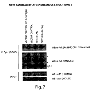
FIG.7 presents a Western blot showing the results of a deacetylation assay
demonstrating that SIRT3 can deacetylate endogenous cytochrome c.
FIG. 8 presents a schematic demonstrating experimental procedures for
behavioral
experiments conducted with SIRT3 knockout mice.
FIG. 9 presents a graph and a schematic indicating the effects of kainic acid
on
primary cerebellar granule neurons in wild-type and SIRT3 knockout mice.
FIG. 10 presents graphs indicating weight change in wild-type and SIRT3
knockout
mice.
FIG. 11 presents a graph demonstrating learning time in female wild-type and
SIRT3
knockout mice.
FIG. 12 presents a graph demonstrating learning time in male wild-type and
SIRT3
knockout mice.
7
CA 02741418 2011-04-21
WO 2010/047823 PCT/US2009/005778
FIG. 13 presents a schematic demonstrating experimental procedures for fear
conditioning experiments.
FIG. 14 presents a graph demonstrating loss of hippocampus and amygdala
neurons
caused by kainic acid injections.
FIG. 15 presents a graph showing the results of contextual fear conditioning
experiments in SIRT3 knockout mice.
FIG. 16 presents graphs indicating activity levels in fear conditioning
experiments.
FIG. 17 presents a schematic depicting the experimental procedure followed for
testing locomotor activity in mice using an Open Field Test.
FIG. 18 presents a graph indicating weights of wild-type and SIRT3 knockout
mice
that were tested in the Open Field Test and in the Water Maze test.
FIG. 19 presents a graph indicating distance travelled in the Open Field Test
by wild-
type and SIRT3 knockout mice.
FIG. 20 presents a graph indicating the speed of movement in the Open Field
Test by
wild-type and SIRT3 knockout mice.
FIG. 21 presents a graph indicating results from day I of the water maze
experiment
using a visible platform marked with a flag, conducted with wild-type and
SIRT3 knockout
mice.
FIG. 22 presents schematics indicating results of Probe Trial 2 of the water
maze
experiments on wild-type mice.
8
CA 02741418 2011-04-21
WO 2010/047823 PCT/US2009/005778
FIG. 23 presents schematics indicating results of Probe Trial 2 of the water
maze
experiments on SIRT3 knockout mice.
FIG. 24 presents a graph indicating the time spent in different quadrants by
wild-type
and SIRT3 knockout mice in Probe Trial 1 of the water maze experiments.
FIG. 25 presents a graph indicating the percentage of time spent in different
quadrants
by wild-type and SIRT3 knockout mice in Probe Trial 1 of the water maze
experiments.
FIG. 26 presents a graph indicating the time spent in different quadrants by
wild-type
and SIRT3 knockout mice in Probe Trial 2 of water maze experiments.
FIG. 27 presents a graph indicating the percentage of time spent in different
quadrants
by wild-type and SIRT3 knockout mice in Probe Trial 2 of water maze
experiments.
FIG. 28 presents an image of a gel showing the results of immunoprecipitation
experiments from hippocampal lysates of wild-type and SIRT3 knockout mice
using the PAN
antibody (acetylated-lysine antibody, Cell Signaling Technology, Beverly, MA)
and
revealing hyperacetylation of proteins in the hippocampus of SIRT3 knockout
mice.
FIG. 29 presents a schematic of a kit associated with the invention. The kit
(10)
shown in FIG. 29 includes a set of containers for housing a compound or
compounds (12) or
(14) such as a compound for activating SIRT3. The kit optionally contains
instructions (20).
Additional components may also be included in the kit.
Detailed Description of the Invention
The invention is based at least in part on the surprising discovery that
cytochrome c is
acetylated on at least two lysine (K) residues, K40 and K74. Deacetylation of
cytochrome c
is revealed to be mediated through interaction with the deacetylase SIRT3,
which is shown
herein to be involved in protecting neurons from cell death. Monitoring and
regulating
acetylation of cytochrome c provides a diagnostic and therapeutic resource for
a variety of
diseases, including diseases associated with cell death.
9
CA 02741418 2011-04-21
WO 2010/047823 PCT/US2009/005778
Aspects of the invention relate to the discovery of aectylation of the
cytochrome c
polypeptide. As used herein, the terms "protein" and "polypeptide" are used
interchangeably
and thus the term polypeptide may be used to refer to a full-length
polypeptide and may also
be used to refer to a fragment of a full-length polypeptide. The term,
"acetylated cytochrome
c polypeptide" means a cytochrome c polypeptide that is acetylated at one or
more lysine
residues. In some embodiments, more than one lysine (K) residue is acetylated.
In some
embodiments, only one lysine residue is acetylated. In some embodiments, a
cytochrome c
polypeptide may be acetylated only at the residue that corresponds to the K40
residue of
wild-type, full-length human cytochrome c polypeptide. In some embodiments, a
cytochrome c polypeptide may be acetylated only at the residue that
corresponds to the K74
residue of wild-type, full-length human cytochrome c polypeptide. In some
embodiments, a
cytochrome c polypeptide may be acetylated on residues that correspond to
residue K40 and
residue K74 of wild-type, full-length human cytochrome c polypeptide. In some
embodiments, a cytochrome c polypeptide may be acetylated on residues that
correspond to
residues K40 and K74 of wild-type, full-length human cytochrome c polypeptide,
and on one
or more other lysine residues. In some embodiments, K40 and/or K74 or other
lysine
positions that are acetylated may be used in methods and/or products of the
invention.
The residue in position 40 of wild-type, full-length human cytochrome c
polypeptide
is a lysine, and this lysine in the wild-type, full-length human polypeptide
and the residue that
corresponds to this position in fragments and in mutated forms of cytochrome c
may be
referred to herein as "K40". Cytochrome c in which the K40 residue is
acetylated may be
referred to herein as "K40-acetylated cytochrome c".
The residue in position 74 of wild-type, full-length human cytochrome c
polypeptide
is a lysine, and this lysine in the wild-type, full-length polypeptide and the
residue that
corresponds to this position in fragments and in mutated forms of cytochrome c
may be
referred to herein as "K74". Cytochrome c in which the K74 residue is
acetylated may be
referred to herein as "K74-acetylated cytochrome C.
A wild-type, full-length human cytochrome c polypeptide has the amino acid
sequence set forth as Genbank Accession No. NP_061820. An acetylated wild-
type, full-
length human cytochrome c polypeptide also has the amino acid sequence set
forth in
Genbank Accession No. NP_061820, but is acetylated at one or more of its
lysine residues.
A nucleic acid sequence encoding human wild-type, full-length cytochrome c is
set forth as
CA 02741418 2011-04-21
WO 2010/047823 PCT/US2009/005778
Genbank Accession No. NM_018947. The nucleic acid and protein sequences of
mouse
cytochrome c correspond to Genbank Accession Nos. X01756 and CAA25899
respectively.
There may be allelic variation in cytochrome c polypeptide sequences of the
invention
including wild-type cytochrome c polypeptide sequences and/or mutant
cytochrome c
polypeptide sequences. As used herein, the term "allelic variant" means any of
two or more
alternative forms of a gene occupying the same chromosomal locus. Allelic
variation arises
naturally through mutation, and may result in polymorphism within populations.
Gene
mutations can be silent (no change in the encoded polypeptide) or may encode
polypeptides
with altered amino acid sequences. An allelic variant of a polypeptide is a
polypeptide
encoded by an allelic variant of a gene. It will be understood by those of
ordinary skill in the
art that such allelic variations may occur in full-length wild-type and mutant
cytochrome c
polypeptides and in fragments of wild-type and mutant polypeptides. Cytochrome
c
polypeptides of the invention may be allelic variants of wild-type cytochrome
c or mutant
cytochrome c polypeptide sequences. One of ordinary skill in the art will be
able to identify
which residues of variants of wild-type and mutant cytochrome c polypeptide
correspond to
residues of wild-type cytochrome c polypeptide using routine methods.
Fragments
In some embodiments, the acetylated lysine residue in a fragment of cytochrome
c
polypeptide is referred to as an acetylated K40 residue or K74 residue even
though the
fragment is not a full-length cytochrome c polypeptide. Those of ordinary
skill in the art can
readily determine the correspondence of an acetylated residue in a cytochrome
c polypeptide
sequence (wild-type or mutant) with a residue in a full-length, wild-type
cytochrome c
polypeptide using routine sequence comparison methods.
In some aspects, the invention may include the synthesis of acetylated full-
length
cytochrome c polypeptides or acetylated fragments thereof. Synthesis methods
of the
invention may include any art-known synthetic methods such as the acetylation
of an existing
natural or synthetic cytochrome c polypeptide, or the incorporation of an
acetylated lysine
residue in a cytochrome c polypeptide during synthesis. Incorporation of
acetylated lysine
may include the following acetylation step, which occurs at the epsilon-amino
groups of
lysines:
11
CA 02741418 2011-04-21
WO 2010/047823 PCT/US2009/005778
Lysine + acetyl-CoA -> Acetyl-Lysine + H2O
As used herein with respect to polypeptides, proteins, or fragments thereof,
"isolated"
means separated from its native environment and present in sufficient quantity
to permit its
identification or use. Isolated, when referring to a protein or polypeptide,
means, for
example: (i) selectively produced by expression cloning or (ii) purified as by
chromatography
or electrophoresis. Isolated proteins or polypeptides may be, but need not be,
substantially
pure. The term "substantially pure" means that the proteins or polypeptides
are essentially
free of other substances with which they may be found in production,
nature,.or in vivo
systems to an extent practical and appropriate for their intended use.
Substantially pure
polypeptides may be obtained naturally or produced using methods described
herein and may
be purified with techniques well known in the art. Because an isolated protein
may be
admixed with therapeutic components in a preparation, such as a
pharmaceutically acceptable
carrier in a pharmaceutical preparation, the protein may comprise only a small
percentage by
weight of the preparation. The protein is nonetheless isolated in that it has
been separated
from the substances with which it may be associated in living systems, i.e.
isolated from other
proteins.
According to some aspects of the invention, fragments of full-length, wild-
type or
mutant cytochrome c polypeptides are provided. Fragments of the invention are
preferably
fragments that retain a distinct functional capability of the polypeptide.
Functional
capabilities which can be retained in a fragment include acetylation,
interaction with
antibodies, and interaction with other polypeptides or fragments thereof.
Polypeptide
fragments can be synthesized using art-known methods, and tested for function
using the
methods exemplified herein.
A fragment of an acetylated cytochrome c polypeptide may comprise at least 5,
6, 7,
8, 9, 10, 11, 12, 13, 14, 15, 16, 17, 18, 19, 20, 25, 30, 35, 40, 45, 50, 55,
60, 65, 70, 75, 80,
85, 90, 95, 100, 102, or more (including each integer in between) contiguous
amino acids of
cytochrome c polypeptide having a consecutive sequence found in wild-type
human
cytochrome c polypeptide or a modified cytochrome c polypeptide sequence as
described
herein. In some embodiments, a fragment includes a lysine residue that
corresponds to K40
and/or K74 of full-length, wild-type human cytochrome c polypeptide. Residues
that
correspond to K40 and K74 may or may not be acetylated. Fragments of
acetylated
12
CA 02741418 2011-04-21
WO 2010/047823 PCT/US2009/005778
cytochrome c polypeptides can be prepared using synthetic methods known in the
art or may
be natural fragments of acetylated cytochrome c polypeptides. Such fragments
are useful for
a variety of purposes, including in the preparation of molecules that bind
specifically to
synthetic and naturally acetylated cytochrome c polypeptides and in
immunoassays well
known to those of ordinary skill in the art, including competitive binding
immunoassays. In
some embodiments, fragments of acetylated cytochrome c could be used to assay
SIRT3
activity or to inhibit SIRT3 activity.
One of ordinary skill in the art will understand how to prepare fragments of
full-
length wild-type or mutant cytochrome c polypeptide. An acetylated fragment of
a full-
length wild-type or mutant cytochrome c polypeptide may include an acetylated
lysine that
corresponds to the K40 and/or K74 lysine of wild-type, full-length human
cytochrome c
polypeptide and/or may include an acetylated lysine that corresponds to a
different lysine of
wild-type, full-length human cytochrome c polypeptide. Also, in some
embodiments of the
invention, a fragment of cytochrome c polypeptide may include a K40 and/or K74
residue
and one or more additional lysine residues, and one, each, some, or none of
the lysines may
be acetylated.
One of ordinary skill in the art is aware that functional homologs of human
cytochrome c exist in multiple species. Acetylated polypeptides including full-
length
proteins and fragments of full-length proteins from other species, that are
functionally
homologous to human cytochrome c are compatible with the instant invention.
One of
ordinary skill in the art is aware of techniques to identify a residue in a
homologous protein
that is functionally homologous to residues K40 or K74 in human cytochrome c.
For
example, Figure 4 presents a sequence alignment of cytochrome c proteins in a
variety of
species. While human cytochrome c K74 is conserved in each species, in some
species this
residue is not in position 74 of the cytochrome c protein in that species.
However based on
sequence alignment and other methods known to those of ordinary skill in the
art, it would be
apparent which residue in the cytochrome c polypeptide from a given species is
functionally
homologous to residue K74 in human cytochrome c.
It should be appreciated that aspects of the invention encompass detection of
acetylation of the human wild-type full-length cytochrome c protein, and also
detection of
acetylation of the cytochrome c protein in fragments, variants and mutants of
the human
cytochrome c protein. Furthermore, aspects of the invention encompass
detection of
13
CA 02741418 2011-04-21
WO 2010/047823 PCT/US2009/005778
acetylation of the cytochrome c protein from any other species, including
detection of
acetylation of the wild-type full-length cytochrome c protein from any other
species, and
detection of acetylation of fragments, variants and mutants of the cytochrome
c protein from
any other species.
A "modified" wild-type or mutant cytochrome c polypeptide or fragment thereof
may
include deletions, point mutations, truncations, amino acid substitutions
and/or additions of
amino acids or non-amino acid moieties. Modifications of a polypeptide of the
invention
may be made by modification of the nucleic acid that encodes the polypeptide
or
alternatively, modifications may be made directly to the polypeptide, such as
by cleavage,
addition of a linker molecule, addition of a detectable moiety, such as
biotin, addition of a
carrier molecule, and the like. Modifications also embrace fusion proteins
comprising all or
part of the polypeptide's amino acid sequence.
In general, modified cytochrome c polypeptides include polypeptides that are
modified specifically to alter a feature of the polypeptide unrelated to its
physiological
activity. For example, cysteine residues can be substituted or deleted to
prevent unwanted
disulfide linkages. Polypeptide modifications can be made by selecting an
amino acid
substitution, deletion, and/or addition, and a modified polypeptide may be
synthesized using
art-known methods. Modified polypeptides then can be tested for one or more
activities (e.g.,
antibody binding, antigenicity, etc.) to determine which modification provides
a modified
polypeptide with the desired properties.
The skilled artisan will also realize that conservative amino acid
substitutions may be
made in a polypeptide to provide functionally equivalent polypeptides, i.e.,
modified
cytochrome c polypeptides that retain a functional capability of a wild-type
or mutant
cytochrome c polypeptide. As used herein, a "conservative amino acid
substitution" refers to
an amino acid substitution that does not alter the relative charge or size
characteristics of the
protein in which the amino acid substitution is made. Modified cytochrome c
polypeptides
can be prepared according to methods for altering polypeptide sequence and
known to one of
ordinary skill in the art such. Exemplary functionally equivalent cytochrome c
polypeptides
include conservative amino acid substitutions of a cytochrome c polypeptide,
or fragments
thereof. Conservative substitutions of amino acids include substitutions made
amongst amino
acids within the following groups: (a) M, I, L, V; (b) F, Y, W; (c) K, R, H;
(d) A, G; (e) S, T;
(f) Q, N; and (g) E, D.
14
CA 02741418 2011-04-21
WO 2010/047823 PCT/US2009/005778
Conservative amino-acid substitutions in a cytochrome c polypeptide typically
are
made by alteration of a nucleic acid encoding the polypeptide. Such
substitutions can be
made by a variety of methods known to one of ordinary skill in the art. For
example, amino
acid substitutions may be made by PCR-directed mutation, site-directed
mutagenesis, or by
chemical synthesis of a gene encoding the cytochrome c polypeptide. Where
amino acid
substitutions are made to a small fragment of a polypeptide, the substitutions
can be made by
directly synthesizing the polypeptide. The activity of functionally equivalent
fragments of
cytochrome c polypeptides can be tested by cloning the gene encoding the
altered
polypeptide into a bacterial or mammalian expression vector, introducing the
vector into an
appropriate host cell, expressing the altered polypeptide, and testing for a
functional
capability of the polypeptide as disclosed herein.
As described above, a fragment of a full-length wild-type or mutant cytochrome
c
polypeptide may be a synthetic polypeptide. As used herein, the term
"synthetic" means
artificially prepared. A synthetic polypeptide is a polypeptide that is
synthesized and is not a
naturally produced polypeptide molecule (e.g., not produced in an animal or
organism). It
will be understood that the sequence of a natural polypeptide (e.g., an
endogenous
polypeptide) may be identical to the sequence of a synthetic polypeptide, but
the latter will
have been prepared using at least one synthetic step.
As used herein, a synthetic acetylated polypeptide is a polypeptide acetylated
with a
synthetic method, which may be, but is not limited to a method of the
invention. An
acetylated polypeptide of the invention may be a naturally acetylated
polypeptide (e.g., an
endogenous acetylated polypeptide) or may be a synthetic acetylated
polypeptide. Although
a synthetic acetylated polypeptide may differ from a natural acetylated
polypeptide, an
antibody raised against a synthetic polypeptide of the invention will
specifically bind with
high affinity the synthetic polypeptide epitope against which it was raised,
and will also
specifically bind with high affinity the natural epitope iii a polypeptide.
Thus, even though
an acetylated epitope of a synthetic polypeptide may differ slightly in amino
acid sequence
from the same epitope in a natural acetylated polypeptide, an antibody raised
against a
synthetic acetylated epitope of the invention specifically binds, in most
cases, with high
affinity to the natural acetylated epitope and to a synthetic acetylated
epitope. Antibodies of
the invention generated using a synthetic acetylated polypeptide specifically
bind, in most
cases, with high affinity to natural and synthetic acetylated polypeptides and
are able to
CA 02741418 2011-04-21
WO 2010/047823 PCT/US2009/005778
distinguish between natural (heterogeneous) acetylated and natural non-
acetylated
polypeptides and also to distinguish between synthetic acetylated and
synthetic non-
acetylated polypeptides.
Cytochrome c Deacetylation by SIRT3
Histone deacetylase proteins (HDACs) constitute four different classes. Class
III
HDACs, which are NAD+-dependent deacetylases, are known as sirtuins. Sirtuins
are
conserved proteins that deacetylate both histone and non-histone cellular
targets. In humans,
seven sirtuins have been identified (SIRT1-7), with individual sirtuin
proteins exhibiting
distinct subcellular localizations and functions. SIRT3 protein has been
reported to exhibit
both nuclear and mitochondrial localizations, and SIRT3 function has been
associated with
metabolism and longevity.
In the Examples section, it is demonstrated that SIRT3 binds to and
deacetylates
cytochrome c. It is also demonstrated that mice in which SIRT3 function has
been knocked
out exhibit decreased cell survival, indicating a function for SIRT3 in
neuroprotection.
Furthermore, it is shown herein that SIRT3 plays a role in memory formation
and fear
conditioning.
Aspects of the invention relate to regulating acetylation and deacetylation of
cytochrome c. In some embodiments, methods of the invention involve increasing
the
activity or protein level of a sirtuin such as SIRT3 in order to decrease the
acetylation of
cytochrome c. In some embodiments the activity or protein level of a sirtuin
such as SIRT3
is increased through administering the sirtuin gene or protein. In some
embodiments the
activity or protein level of a sirtuin such as SIRT3 is increased through
administering a
compound that increases the protein level or increases the activity a sirtuin.
Methods for
activating sirtuins, and non-limiting examples of compounds for activating
sirtuins are
provided by formulas 1-25, 30, and 32-65 in US Patent Publication
2006/0025337,
incorporated by reference herein in its entirety. Methods and compounds for
modulating
sirtuins are also presented in US Patent Publications: 2007/0043050,
2007/0037865,
2007/0037827, 2007/0037809, 2007/0014833, 2006/0276416, 2006/0276393 and
2006/0229265, and in US Patent 7,345,178, all of which are incorporated herein
by reference
in their entirety.
16
CA 02741418 2011-04-21
WO 2010/047823 PCT/US2009/005778
The invention further encompasses screening methods for identifying compunds
that
modulate sirtuins such as SIRT3. An compound that modulates the activity of a
sirtuin such
as SIRT3 may in some embodiments be a nucleic acid (e.g., an aptamer), a
polypeptide, or a
small molecule, e.g., a small organic molecule. Non-limiting examples of
compounds for
modulating sirtuin activity are provided in US Patent Publication
2006/0025337, incorporated
by reference herein in its entirety, such as the molecules of formulas 1-25,
30, and 32-65, or
analogs thereof. It should be appreciated that a wide variety of compounds
and/or compound
libraries are appropriate for screening methods described herein.
Assays may be conducted in a cell-based or cell-free format. For example, an
assay
may comprise incubating (or contacting) a sirtuin, such as SIRT3, and a test
compound under
conditions in which a sirtuin can be activated by an compound known to
activate the sirtuin,
and monitoring or determining the level of activation of the sirtuin in the
presence of the test
compound relative to the absence of the test compound. The level of activation
of a sirtuin
can be determined by determining its ability to deacetylate a substrate.
Exemplary substrates
are acetylated polypeptides, or libraries or pools of polypeptides. In some
embodiments, the
substrate is a cytochrome c polypeptide. In some embodiments, a substrate
contains a single
polypeptide species, while in other embodiments, it contains a mixture of two
or more
polypeptides species. In some embodiments, the substrate comprises one or more
cytochrome c polypeptides that have one or more acetylated residues. In
certain
embodiments, the substrate comprises one or more cytochrome c polypeptides
that have an
acetylated lysine residue corresponding to residues K40 and/or K74.
Polypeptide substrates
can include, for example, full-length proteins and/or protein fragments and/or
heterologous
fusions, alone or in combination. Polypeptides can be of varying lengths. In
some
embodiments, a cytochrome c polypeptide substrate comprises a fusion of a
fragment of
cytochrome c comprising at least one lysine residue corresponding to residues
K40 and/or
K74. A fusion of a fragment of cytochrome c can encompass any fragment of a
cytochrome c
polypeptide fused to a fragment of any other polypeptide. In some embodiments,
the
cytochrome c polypeptide substrate is in a cell. Substrates used in screening
assays may in
some embodiments be fluorogenic.
It should be appreciated that methods and compositions described herein can
encompass a full-length SIRT3 protein, or a portion thereof. In some
embodiments, a
biologically active portion of SIRT3 may be used in accordance with the
methods described
17
CA 02741418 2011-04-21
WO 2010/047823 PCT/US2009/005778
herein. A biologically active portion of SIRT3 refers to a portion of the
protein having a
biological activity, such as the ability to deacetylate an acetylated
substrate, such as
cytochrome c, or a fragment of cytochrome c comprising at least one lysine
residue
corresponding to residues K40 and/or K74, in the presence of nicotinamide
adenine
dinucleotide (NAD) or an NAD+ analog. Biologically active portions of SIRT3
can in some
embodiments encompass the NAD+ binding domain and/or the substrate binding
domain. In
other embodiments, a biologically active portion of SIRT3 may be a fragment of
a SIRT3
protein that is produced by cleavage with a mitochondrial matrix processing
peptidase (MPP)
and/or a mitochondrial intermediate peptidase (MIP). The SIRT3 deacetylase
used in
methods described herein can be from a cell or tissue lysate. The assays
described herein can
be used to determine if a portion of SIRT3 is a biologically active portion of
SIRT3.
In some embodiments, the reaction may be conducted for about 30 minutes and
stopped, e.g., with nicotinamide. Assays similar to those described in the
HDAC fluorescent
activity assay/drug discovery kit (AK-500, BIOMOL Research Laboratories) may
be used to
determine the level of acetylation. Similar assays are described in Bitterman
et al. (2002) J.
Biol. Chem. 277:45099. The level of activation of the sirtuin in an assay may
be compared to
the level of activation of the sirtuin in the presence of one or more
(separately or
simultaneously) compounds, which may serve as positive or negative controls.
Sirtuins for
use in the assays may be full length SIRT3 proteins or biologically active
portions thereof. In
some embodiments, proteins for use in the assays include N-terminal portions
of SIRT3.
Methods for screening for compounds that modulate sirtuins such as SIRT3 are
incorporated
by reference from US Patent 7,544,497, and US Patent Publications
2009/0221020,
2008/0293081 and 2006/0252076.
Methods may comprise (i) contacting a cell comprising a sirtuin such as SIRT3
with a
cytochrome c polypeptide substrate under conditions appropriate for the
sirtuin to deacetylate
thepolypeptide and (ii) determining the level of acetylation of the
polypeptide, wherein a
different level of acetylation of the polypeptide in the presence of the test
compound relative
to a control (such as the absence of the test compound) indicates that the
test compound
modulates the activity of the sirtuin in vivo. It should be appreciated that
other substrates
besides cytochrome c would also be compatible in such assays for identifying
compounds
that modulate the activity of the sirtuin.
18
CA 02741418 2011-04-21
WO 2010/047823 PCT/US2009/005778
In one embodiment, a screening assay comprises (i) contacting the sirtuin such
as
SIRT3 with a test compound and an acetylated substrate under conditions
appropriate for the
sirtuin to deacetylate the substrate in the absence of the test compound; and
(ii) determining
the level of acetylation of the substrate, wherein a lower level of
acetylation of the substrate
in the presence of the test compound relative to the absence of the test
compound indicates
that the test compound stimulates deacetylation by the sirtuin, whereas a
higher level of
acetylation of the substrate in the presence of the test compound relative to
the absence of the
test compound indicates that the test compound inhibits deacetylation by the
sirtuin.
Methods for identifying an compound that modulates, e.g., stimulates or
inhibits, a
sirtuin such as SIRT3 in vivo may comprise (i) contacting a cell with a test
compound and a
substrate that is capable of entering a cell in the presence of an inhibitor
of class I and class II
HDACs under conditions appropriate for the sirtuin to deacetylate the
substrate in the
absence of the test compound; and (ii) determining the level of acetylation of
the substrate,
wherein a lower level of acetylation of the substrate in the presence of the
test compound
relative to the absence of the test compound indicates that the test compound
stimulates
deacetylation by the sirtuin, whereas a higher level of acetylation of the
substrate in the
presence of the test compound relative to the absence of the test compound
indicates that the
test compound inhibits deacetylation by the sirtuin. A preferred substrate is
an acetylated
polypeptide, which may also be fluorogenic. The method may further comprise
lysing the
cells to determine the level of acetylation of the substrate. In some
embodiments, substrates
may be added to cells at a concentration ranging from about 1 M to about 10mM,
preferably
from about 10 M to 1 mM, even more preferably from about 100 M to 1 mM, such
as about
200 M.
In some embodiments, methods for identifying a compound that activates a
sirtuin
such as SIRT3 may involve mass spectrometry, discussed further below. Methods
of using
mass spectrometry for identifying compounds that modulate the activity of
deacetylase
proteins are incorporated by reference from US Patent Publication
2009/0221020. Mass
spectrometry can be used to identify the level of acetylation of cytochrome c
or any other
substrate of a sirtuin such as SIRT3. In some embodiments, a method for
identifying a
compound that activates a deacetylase includes contacting a cytochrome c
polypeptide with a
sirtuin such as SIRT3, or a biologically active portion thereof, in the
presence of a test
compound, wherein the cytochrome c polypeptide comprises at least one
acetylated lysine
19
CA 02741418 2011-04-21
WO 2010/047823 PCT/US2009/005778
residue, and determining the level of acetylation of the cytochrome c
polypeptide using mass
spectrometry, wherein a decrease in the level of acetylation of the
polypeptide in the presence
of the test compound as compared to a control is indicative of a compound that
activates a
deacetylase. Mass spectrometry can in some embodiments encompass electrospray
ionization (ESI) mass spectrometry and/or matrix-assisted laser
desorption/ionization
(MALDI) mass spectrometry.
In some embodiments, methods for determining the activity of a deacetylase
such as a
sirtuin comprise: contacting a polypeptide with a cell or tissue lysate
comprising a
deacetylase, wherein the polypeptide comprises at least one acetylated lysine
residue; and
determining the level of acetylation of the polypeptide using mass
spectrometry, wherein a
decrease in the level of acetylation of the polypeptide is indicative of
deacetylase activity.
The deacetylase can be SIRT3 and can be in a cell or tissue lysate. The
cytochrome c
polypeptide can be in a cell.
In some embodiments, the concentration of a polypeptide substrate is below the
Km of
the sirtuin, such as SIRT3, for the polypeptide substrate. For example, the
concentration of
the polypeptide substrate can be at least 2, 3, 4, 5, 6, 7, 8, 9, 10, 11, 12,
13, 14, 15 or more
than 15 fold below the Km of the sirtuin for the polypeptide substrate.
A compound subjected to or identified by screening methods described herein
may for
example be a small molecule, such as a small organic molecule. Such small
molecules are
well known in the art; examples of such molecules are provided herein and in
publications
such as US 7,544,497 and US 2009/0221020. Aspects of the invention encompass
preparing
a quantity of such a compound, or analog thereof, and in some embodiments
conducting
therapeutic profiling of the compound, or analog thereof, for efficacy and
toxicity in animals.
Methods for conducting therapeutic profiling are familiar to one of ordinary
skill in the art.
In some aspects, methods involve formulating the compound in pharmaceutical
formulations,
using standard methods. The invention encompasses manufacturing of
pharmaceutical
preparations containing compounds described herein, or compounds identified
using methods
described herein, or analogs thereof, having a suitable animal toxicity
profile.
Pharmaceutical preparations containing compounds described herein, or
compounds
identified using methods described herein, or analogs thereof, having a
suitable animal
toxicity profile can be marketed to healthcare providers. Methods for
preparing quantities of
a compound or analog thereof, conducting therapeutic profiling of the compound
or analog
CA 02741418 2011-04-21
WO 2010/047823 PCT/US2009/005778
thereof, formulating a compound in pharmaceutical formulations, and
manufacturing a
pharmaceutical preparation containing a compound, are incorporated by
reference from US
Patent 7,544,497 and US Patent Publication 2009/0221020.
Aspects of the invention also relate to acetylated polypeptide substrates for
use in
determining the activity of SIRT3, comprising a fragment of cytochrome c
comprising at
least one acetylated lysine residue corresponding to residues K40 and/or K74.
The
polypeptide substrate can be a fusion of a fragment of cytochrome c comprising
at least one
acetylated lysine residue corresponding to residues K40 and/or K74. Such
polypeptides can
be chemically synthesized, produced recombinantly, or by any other methods
routinely
employed in the art. The polypeptides can be acetylated according to standard
methods
routinely practiced in the art. Aspects of the invention also relate to kits
comprising such
acetylated polypeptide substrates, which can be used in the screening methods
described
herein.
Neurodegenerative Disorders
Aspects of the invention relate to diagnosis and treatment of disorders. As
used
herein, "disorder" refers to any pathological condition associated with
elevated or reduced
cytochrome c acetylation. In some embodiments a disorder or condition
associated with
elevated acetylated cytochrome c is a neurodegenerative disorder. As used
herein, the term
"neurodegenerative disorder" refers to disorders, diseases or conditions that
are caused by the
deterioration of cell and tissue components of the nervous system.
Some non-limiting examples of neurodegenerative disorders include stroke,
Alzheimer's disease, Parkinson's disease, Huntington's disease,
Periventricular leukomalacia
(PVL), amyotrophic lateral sclerosis (ALS, "Lou Gehrig's disease"), ALS-
Parkinson's-
Dementia complex of Guam, Friedrich's Ataxia, Wilson's disease, multiple
sclerosis, cerebral
palsy, progressive supranuclear palsy (Steel-Richardson syndrome), bulbar and
pseudobulbar
palsy, diabetic retinopathy, multi-infarct dementia, macular degeneration,
Pick's disease,
diffuse Lewy body disease, prion diseases such as Creutzfeldt-Jakob, Gerstmann-
Straussler-
Scheinker disease, Kuru and fatal familial insomnia, primary lateral
sclerosis, degenerative
ataxias, Machado-Joseph disease/spinocerebellar ataxia type 3 and
olivopontocerebellar
degenerations, spinal and spinobulbar muscular atrophy (Kennedy's disease),
familial spastic
paraplegia, Wohlfart-Kugelberg-Welander disease, Tay-Sach's disease,
multisystem
21
CA 02741418 2011-04-21
WO 2010/047823 PCT/US2009/005778
degeneration (Shy-Drager syndrome), Gilles De La Tourette's disease, familial
dysautonomia
(Riley-Day syndrome), Kugelberg-Welander disease, subacute sclerosing
panencephalitis,
Werdnig-Hoffmann disease, synucleinopathies (including multiple system
atrophy), Sandhoff
disease, cortical basal degeneration, spastic paraparesis, primary progressive
aphasia,
progressive multifocal leukoencephalopathy, striatonigral degeneration,
familial spastic
disease, chronic epileptic conditions associated with neurodegeneration,
Binswanger's
disease, and dementia (including all underlying etiologies of dementia).
Cancer
Aspects of the invention also relate to diagnosis and treatment of cancer. As
used
herein, the term "cancer" refers to an uncontrolled growth of cells that may
interfere with the
normal functioning of the bodily organs and systems, and includes both primary
and
metastatic tumors. Primary tumors or cancers that migrate from their original
location and
seed vital organs can eventually lead to the death of the subject through the
functional
deterioration of the affected organs. A metastasis is a cancer cell or group
of cancer cells,
distinct from the primary tumor location, resulting from the dissemination of
cancer cells
from the primary tumor to other parts of the body. Metastases may eventually
result in death
of a subject.
As used herein, the term "cancer" includes, but is not limited to, the
following types
of cancer: breast cancer (including carcinoma in situ), biliary tract cancer;
bladder cancer;
brain cancer including glioblastomas and medulloblastomas; cervical cancer;
choriocarcinoma; colon cancer; endometrial cancer; esophageal cancer; gastric
cancer;
hematological neoplasms including acute lymphocytic and myelogenous leukemia;
T-cell
acute lymphoblastic leukemia/lymphoma; hairy cell leukemia; chromic
myelogenous
leukemia, multiple myeloma; AIDS-associated leukemias and adult T-cell
leukemia
lymphoma; intraepithelial neoplasms including Bowen's disease and Paget's
disease; liver
cancer; lung cancer; lymphomas including Hodgkin's disease and lymphocytic
lymphomas;
mesothelioma, neuroblastomas; oral cancer including squamous cell carcinoma;
ovarian
cancer including those arising from epithelial cells, stromal cells, germ
cells and
mesenchymal cells; pancreatic cancer; prostate cancer; rectal cancer; sarcomas
including
leiomyosarcoma, rhabdomyosarcoma, liposarcoma, fibrosarcoma, and osteosarcoma;
skin
cancer including melanoma, Merkel cell carcinoma, Kaposi's sarcoma, basal cell
carcinoma,
22
CA 02741418 2011-04-21
WO 2010/047823 PCT/US2009/005778
and squamous cell cancer; testicular cancer including germinal tumors such as
seminoma,
non-seminoma (teratomas, choriocarcinomas), stromal tumors, and germ cell
tumors; thyroid
cancer including thyroid adenocarcinoma and medullar carcinoma; and renal
cancer including
adenocarcinoma and Wilms tumor. Non-limiting examples of precancerous
conditions
include dysplasia, premalignant lesions, adenomatous colon polyp, and
carcinoma in-situ
such as Ductal carcinoma in-situ (DCIS), etc. Other cancers that can be
treated with methods
of the invention will be known to those of ordinary skill in the art. In some
embodiments of
the invention, the cancer is melanoma. In certain embodiments the cancer is
adenocarcinoma. In some embodiments the cancer is a solid tumor cancer. A
cancer that
may be treated or assayed using methods of the invention also may include
breast cancer,
lung cancer, prostate cancer, mesothelioma, etc.
Measuring A cetylation of Cytochrome c
The invention, in some aspects, includes various assays to determine levels of
acetylated cytochrome c polypeptide, and to detect acetylation of cytochrome c
on specific
residues (e.g., K40 and/or K74). Methods of the invention that are useful to
determine levels
of acetylated cytochrome c polypeptide in cells, tissues, subjects, and
samples (e.g., from
subjects, in culture, etc.), include, but are not limited to: binding assays,
including specific
binding assays such as using antibodies or antigen-binding fragments thereof
of the invention
that bind specifically to acetylated cytochrome c polypeptide; gel
electrophoresis; mass
spectrometry; NMR; and the like. Immunoassays may be used according to the
invention
including, but not limited to, sandwich-type assays, competitive binding
assays, one-step
direct tests and two-step tests, etc. Assessment of binding of antibodies that
specifically bind
acetylated cytochrome c may also be done in vivo - in living subjects using
art-known
detectable labels and suitable in vivo methods.
Methods and assays of the invention (e.g., binding assays, gel
electrophoresis; mass
spectrometry; NMR; and the like) may be used to monitor changes in cytochrome
c
acetylation levels in a cell sample and or a subject over time, or changes in
acetylation of
specific residues of cytochrome c in a cell sample and or a subject over time.
Methods for
measuring acetylation of cytochrome c described herein can be applied to
methods for
screening for modulators of sirtuin activity, such as SIRT3 activity, as
described above.
23
CA 02741418 2011-04-21
WO 2010/047823 PCT/US2009/005778
Mass Spectrometry
Acetylation of cytochrome c may be measured by mass spectrometry. Mass
spectrometry is an important tool in the identification of proteins and
peptides, and in the
identification of modified residues within proteins and peptides. In some
embodiments, mass
spectrometry is used to determine the level of acetylation of cytochrome c. In
some
embodiments mass spectrometry is used to determine whether cytochrome c is
acetylated on
specific residues.
Using mass spectrometry, such as ESI or MALDI-MS, peptides can be ionized
intact
into the gas phase and their masses accurately measured. Based on this
information, proteins
can readily be identified using protein mass mapping or peptide mass mapping,
in which
these measured masses are compared to predicted values derived from a protein
database.
Further sequence information can also be obtained by fragmenting individual
peptides in
tandem MS experiments.
Sequence specific proteases or certain chemical cleaving agents are used to
obtain a
set of peptides from the target protein that are then mass analyzed. The
observed masses of
the proteolytic fragments are compared with theoretical "in silico" digests of
all proteins
listed in sequence database. The matches or "hits" are then statistically
evaluated and marked
according to the highest probability.
Tandem mass spectrometry experiments allow peptide identification by yielding
fragmentation patterns for individual peptide. Analogous to peptide mapping
experiments,
the experimentally obtained fragmentation patterns can be compared to
theoretically
generated MS/MS fragmentation patterns for the various proteolytic peptides
arising from
each protein contained in the searched database. Statistical evaluation of the
results and
scoring algorithms using search engines such a Sequest (ThermoFinnigan Corp)
and
MASCOT (Matrix Science, Limited) facilitate the identification of the best
match. The
partial sequence information contained in tandem MS experiments is more
specific than
simply using the mass of a peptide, since two peptides with identical amino
acia contents but
different sequences will exhibit different fragmentation patterns. Tandem mass
spectrometry,
the ability to induce fragmentation and perform successive mass spectrometry
experiments on
these ions, is generally used to obtain structural information through
fragmentation.
One of the processes by which fragmentation is initiated is known as collision-
induced dissociation (CID). CID is accomplished by selecting an ion of
interest with the
24
CA 02741418 2011-04-21
WO 2010/047823 PCT/US2009/005778
mass analyzer and then subjecting that ion to collisions with neutral atoms or
molecules. The
selected ion will collide with the collision gas such as argon, resulting in
fragment ions which
are then mass analyzed. CID can be accomplished with a variety of instruments,
most
commonly using triple quadrupoles, quadrupole ion traps, Fourier transform-ion
cyclotron
resonance (FT-ICR) mass spectrometry (FTMS), time-of-flight reflectron and
quadrupole
time-of-flight mass analyzers. The triple quadrupole and quadrupole ion trap
combined with
electrospray are common means of generating peptide structural data, as they
are capable of
high sensitivity, and produce a reasonable amount of fragmentation
information. MALDI
with time-of-flight reflectron and Fourier transform-ion cyclotron resonance
are also common
sources for structural information.
In order to obtain peptide sequence information by mass spectrometry,
fragments of
an ion must be produced that reflect structural features of the original
compound. Most
peptides are linear molecules, which allow for relatively straightforward
interpretation of the
fragmentation data. The process is initiated by converting some of the kinetic
energy from
the peptide ion into vibrational energy. This is achieved by introducing the
selected ion,
usually an (M + H) + or (M + nH) + ion, into a collision cell where is
collides with neutral
Ar, Xe, or He atoms, resulting in fragmentation. The fragments are then
monitored via mass
analysis. Tandem mass spectrometry allows for a heterogeneous solution of
peptides to be
analyzed and then by filtering the ion of interest into the collision cell,
structural information
can be derived on each peptide from complex mixture.
Certain limitations for obtaining complete sequence information exist using
tandem
mass spectrometry. For example, in determining the amino acid sequence of a
peptide, it is
not possible for leucine and isoleucine to be distinguished because they have
the same mass.
The same difficulty will arise with lysine and glutamine since they have the
same nominal
mass, although high resolution tandem analyzers (quadrupole-TOF and FTMS) can
distinguish between these amino acids.
In some preferred embodiments, samples of proteins (or peptides in a
proteolytic
digest) are separated by gel electrophoresis or liquid chromatography prior to
mass analysis.
Gel electrophoresis is one of the most widely used techniques for separating
intact
proteins. In sodium dodecyl sulfate-polyacrylamide gel electrophoresis (SDS-
PAGE),
sometimes called one dimensional gel electrophoresis, the proteins are treated
with the
denaturing detergent SDS and loaded onto a gel. Upon application of an
electric potential
CA 02741418 2011-04-21
WO 2010/047823 PCT/US2009/005778
across the gel, the proteins migrate through the gel towards the anode at a
rate inversely
proportional to their size. Upon completion of the separation, the proteins
may be visualized
using any of a number of different staining agents (Coomassie, Sypro Ruby, or
Silver), and
the individual bands are physically excised from the gel. These excised spots
are subjected to
destaining, reductive alkylation, in-gel digestion, peptide extraction, and
finally mass analysis
for protein identification.
The combination of SDS-PAGE electrophoresis with an isoelectric focusing step
also
enables the separation of proteins of similar mass. In two-dimensional gel
electrophoresis
(2D-GE), proteins are first separated according to their isoelectric points
(PI) by
electrophoresis through a solution or gel containing an immobilized pH
gradient, with each
protein migrating to a position in the pH gradient corresponding to its
isoelectric point. Once
the isoelectric focusing step is complete, gel electrophoresis similar to SDS-
PAGE is
performed orthogonally to separate the proteins by size. Like 1 D gels, 2D gel
spots can be
cut out, enzymatically digested, and mass analyzed for protein identification.
Using this
technique, thousands of proteins can simultaneously be separated and removed
for
identification.
Automated liquid handling robots have been developed that perform all the
sample
preparation steps for peptide mapping experiments, including gel destaining,
alkylation/reduction, in gel digestion, peptide extraction, and MALDI target
plating.
Mass spectral data acquisition systems have similarly been automated to
acquire
spectra, process the raw data, and perform database searches for numerous
samples.
Commercial MALDI-TOF systems are available that can perform over 1,000 mapping
experiments in just twelve hours. These systems are able to perform automated
calibrations,
vary laser energies, and adjust laser firing location to maximize signal, with
the entire data
acquisition process requiring approximately 30 seconds or less. Similarly,
automated data
processing systems can recognize suitable signals, identify monoisotopic
peaks, and submit
summary peak lists directly to a search engine.
Such high throughput proteomics systems enable the investigation of multiple
unknown samples at once such as those coming from gels. Additionally, the
flexibility of
automated acquisition and data analysis software allows to rapidly reacquire
and/or reanalyze
entire batches of samples with minimal user effort. Automated systems are,
however, limited
in that they are only as good as the data provided. For example, the detection
and accurate
26
CA 02741418 2011-04-21
WO 2010/047823 PCT/US2009/005778
mass assignment of species exhibiting low signal-to-noise ratios is often
poor. Such issues
have led to the development of post-acquisition data processing. Improvements
in these
processes have enabled high through-put automated systems to achieve
identification "hit"
rates equal to or above those obtained normally.
An alternative approach to gel electrophoresis techniques involves the use of
analytical separation methods such as high performance liquid chromatography
(HPLC).
Whereas gel electrophoresis techniques separate intact proteins, liquid
chromatography can
be performed on proteolytic peptides. One of the means of performing peptide
LC-MS/MS
involves the direct coupling of the LC to an ion trap mass spectrometer
through an
electrospray ionization interface. Other mass analyzers suitable for these
experiments include
triple quadrupoles and quadrupole time-of-flights.
In some embodiments, mass spectrometry is used to determined whether
cytochrome
c is acetylated and on which specific residues cytochrome c is acetylated. The
use of mass
spectrometry to identify acetylation of lysine residues within proteins is
discussed further in
Zhang et al., (2002) Mol Cell Proteomics 1:500-508 and Dormeyer et al., (2005)
Mol Cell
Proteomics 4:1226-1239, incorporated herein by reference in their entirety.
Diagnosis and Characterization of Risk of Neurodegenerative Disorders and
Cancer
Methods and assays such as those discussed herein, for detecting acetylation
of
cytochrome c, allow monitoring of acetylated cytochrome c polypeptide levels
in a subject
who is believed to be at risk of a disorder associated with cytochrome c
activity, and also
enable monitoring in a subject who is known to have a disorder associated with
cytochrome c
activity.
Aspects of the invention relate to methods of diagnosing a neurodegenerative
disorder
characterized by acetylation of cytochrome c, or characterizing a subject's
risk of a
neurodegenerative disorder that is characterized by acetylation of cytochrome
c. Further
aspects of the invention relate to methods of diagnosing a cancer
characterized by
deacetylation of cytochrome c, or characterizing a subject's risk of a cancer
that is
characterized by deacetylation of cytochrome c.
Methods involve detecting the level of acetylation of cytochrome c polypeptide
in a
sample from a subject and comparing the level of acetylation of cytochrome c
to a control
sample or a predetermined value. The acetylation state of the protein may be
determined by
27
CA 02741418 2011-04-21
WO 2010/047823 PCT/US2009/005778
any of the methods described herein. Assays based upon detecting levels of
acetylated
cytochrome c in cells and/or subjects include determining onset, progression,
and/or
regression of a neurodegenerative disorder or a cancer in a subject; selecting
a treatment for a
neurodegenerative disorder or a cancer in a subject; and evaluating a
treatment for
cytochrome c polypeptide acetylation status in a subject. Thus, subjects can
be characterized,
treatment regimens can be monitored, treatments can be selected and diseases
status can be
better understood using the assays of the present invention. The level of
acetylated
cytochrome c polypeptide may correlate with the status of a neurodegenerative
disorder or a
cancer in a subject.
One aspect of the present invention relates to detecting acetylated cytochrome
c
polypeptides or fragments thereof in an in vitro or in vivo sample (e.g.,
histological or
cytological specimens, real-time in vivo assays, biopsies and the like), and,
in particular, to
distinguish the level of acetylated cytochrome c from the level of non-
acetylated cytochrome
c in a sample or a subject. In some embodiment, this method involves providing
an antibody
or an antigen-binding binding fragment thereof, which specifically binds to
acetylated
cytochrome c polypeptide. The anti-acetylated cytochrome c antibody may be
bound to a
label that permits the detection of the acetylated cytochrome c polypeptide.
In some
embodiments, a sample may be contacted with a labeled anti-acetylated
cytochrome c
antibody under conditions effective to permit binding of the anti-acetylated
cytochrome c
antibody to acetylated cytochrome c polypeptide in the sample. The presence of
acetylated
cytochrome c in a sample may be detected by detection of the label. In some
embodiments,
the contact between the anti-acetylated cytochrome c antibody and a sample is
carried out in
samples from a subject. In certain embodiments, the contact between an anti-
acetylated
cytochrome c antibody and a sample may be carried out in a subject. Samples to
which the
methods of the invention can be applied include tissue samples, cell samples,
including cell
culture samples, subject samples, in vivo samples, etc. In some embodiments
mass
spectrometry is used to identify acetylation of cytochrome c.
Assays to detect acetylation of cytochrome c may be carried out in cells from
culture,
cells in solution, in samples obtained from subjects, and/or samples in a
subject (in vivo
sample). As used herein, a subject is a human, non-human primate, cow, horse,
pig, sheep,
goat, dog, cat, or rodent. In some embodiments, human subjects are preferred.
The samples
28
CA 02741418 2011-04-21
WO 2010/047823 PCT/US2009/005778
used herein include any cell or tissue sample, and may include neuronal cell
and/or tissue
samples.
Particularly important subjects to which the present invention can be applied
are
subjects with a neurodegenerative disorder. The term "subject with a
neurodegenerative
disorder" as used herein, means an individual who, at the time the sample is
taken, has been
diagnosed as having a neurodegenerative disorder. Methods of the invention may
also be
used to detect abnormal levels of cytochrome c polypeptide acetylation in
subjects that are
not yet diagnosed with a neurodegenerative disorder. The onset, progression,
and/or
regression of a neurodegenerative disorder may also be monitored using methods
and
antibodies of the invention.
Particularly important subjects to which the present invention can be applied
are
subjects with a cancer. The term "subject with a cancer" as used herein, means
an individual
who, at the time the sample is taken, has been diagnosed as having a cancer.
Methods of the
invention may also be used to detect abnormal levels of cytochrome c
polypeptide acetylation
in subjects that are not yet diagnosed with a cancer. The onset, progression,
and/or regression
of a cancer may also be monitored using methods and antibodies of the
invention.
In some embodiments, aspects of the invention relate to screening subjects for
diseases associated with the presence of elevated levels of acetylated
cytochrome c
polypeptide. As used herein, the term "elevated" means higher, for example
elevated versus
a control level. In some embodiments, the status and/or stage of a
neurodegenerative disorder
is determined by assessing the level of acetylated cytochrome c in a sample
from a subject or
culture that has a neurodegenerative disorder. Antibodies of the invention are
useful in
assays to differentiate whether or not a subject has a neurodegenerative
disorder, because
anti-acetylated cytochrome c antibodies of the invention can be used to
quantitate the amount
of acetylated cytochrome c polypeptide in cells and tissues of subjects who
have
neurodegenerative disorders, or who are at risk of having neurodegenerative
disorders. As
discussed above, mass spectrometry approaches can also be used to identify
specific residues
of cytochrome c that are acetylated in a sample from a subject. The presence
of acetylated
cytochrome c polypeptide in a sample, and/or the detection of acetylation of
specific residues
of cytochrome c in a sample, can be used to determine the presence and/or
status of a
neurodegenerative disorder in a cell, cell culture or subject. Methods of the
invention can be
used to obtain useful prognostic information by providing an early indicator
of disease onset
29
CA 02741418 2011-04-21
WO 2010/047823 PCT/US2009/005778
and /or progression. In some embodiments, the disorder is a cancer and the
subject exhibits
decreased levels of acetylated cytochrome c or increased levels of
deacetylated cytochrome c.
Levels of acetylated cytochrome c polypeptide (e.g., K40- or K74-acetylated
cytochrome c polypeptide) can be determined in a number of ways when carrying
out the
various methods of the invention. In one measurement, a level of acetylated
cytochrome c
polypeptide is measured in relation to non-acetylated (or deacetylated)
cytochrome c
polypeptide. Thus, the measurement may be a relative measure, which can be
expressed, for
example, as a percentage of total cytochrome c polypeptide. Those of ordinary
skill in the art
will appreciate that relative amounts of acetylated and non-acetylated
cytochrome c
polypeptides may be determined by measuring either the relative amount of
acetylated
cytochrome c polypeptide or the relative amount of non-acetylated cytochrome c
polypeptide.
In other words, if 90% of an individual's cytochrome c polypeptide is non-
acetylated
cytochrome c polypeptide (or reduced acetylated cytochrome c polypeptide),
then 10% of the
individual's cytochrome c polypeptide will be acetylated cytochrome c
polypeptide.
Another measurement of the level of acetylated cytochrome c is a measurement
of
absolute levels of cytochrome c polypeptide acetylation. This could be
expressed, for
example, in acetylated cytochrome c polypeptide per unit of cells or tissue.
Another
measurement of the level of acetylated cytochrome c polypeptide is a
measurement of the
change in the level of acetylated cytochrome c polypeptide over time. This may
be expressed
in an absolute amount or may be expressed in terms of a percentage increase or
decrease over
time.
Aspects of the invention relate to characterizing cytochrome c polypeptide
acetylation
levels by monitoring changes in the absolute or relative amounts of acetylated
cytochrome c
polypeptide in a subject or sample (e.g., a cell culture) over time. In some
embodiments,
changes in relative or absolute acetylated cytochrome c polypeptide of greater
than 0.1 % may
indicate an abnormality. Preferably, the change in acetylated cytochrome c
polypeptide
levels that indicates an abnormality, is greater than 0.2%, greater than 0.5%,
greater than
1.0%, 2.0%, 3.0%, 4.0%, 5.0%, 7.0%,10%,15%, 20%, 25%, 30%, 40%, 50%, or more.
Levels of acetylated cytochrome c polypeptide can be determined and are
compared
to controls according to the invention. The control may be a predetermined
value, which can
take a variety of forms. It can be a single cut-off value, such as a median or
mean. It can be
established based upon comparative groups, such as in groups having normal
amounts of
CA 02741418 2011-04-21
WO 2010/047823 PCT/US2009/005778
cytochrome c acetylation and groups having abnormal amounts of cytochrome c
acetylation.
Another example of comparative groups may be groups having symptoms of a
neurodegenerative disorder and groups without symptoms of a neurodegenerative
disorder.
Another comparative group may be a group with a family history of a
neurodegenerative
disorder and a group without such a family history. In some embodiments, the
risk in one
defined group is double the risk in another defined group. A predetermined
value can be
arranged, for example, where a tested population is divided equally (or
unequally) into
groups, such as a low-risk group, a medium-risk group and a high-risk group or
into
quadrants or quintiles, the lowest quadrant or quintile being individuals with
the lowest risk
and lowest amounts of acetylated cytochrome c polypeptide and the highest
quadrant or
quintile being individuals with the highest risk and highest amounts of
acetylated cytochrome
c polypeptide.
The predetermined value, of course, will depend upon the particular population
selected. For example, an apparently healthy population will have a different
`normal' range
than will a population that is known to have a condition related to abnormal
cytochrome c
polypeptide acetylation. Accordingly, the predetermined value selected may
take into
account the category in which an individual or cell falls. Appropriate ranges
and categories
can be selected with no more than routine experimentation by those of ordinary
skill in the
art. As used herein, "abnormal" means not normal as compared to a control. By
abnormally
high it is meant high relative to a selected control. Typically the control
will be based on
apparently healthy normal individuals in an appropriate age bracket or
apparently healthy
cells.
It will also be understood that controls according to the invention may be, in
addition
to predetermined values, samples of materials tested in parallel with the
experimental
materials. Examples include samples from control populations or control
samples generated
through manufacture to be tested in parallel with the experimental samples.
Evaluating Efficacy of Therapy
Methods of the invention may also be used to assess the efficacy of a
therapeutic
treatment of a neurodegenerative disorder or a cancer and for the assessment
of the level of
acetylated cytochrome c polypeptide a subject at various time points. For
example, a level of
a subject's acetylated cytochrome c polypeptide can be obtained prior to the
start of a
31
CA 02741418 2011-04-21
WO 2010/047823 PCT/US2009/005778
therapeutic regimen (either prophylactic or as a treatment of a
neurodegenerative disorder or
a cancer), during the treatment regimen and/or after a treatment regimen, thus
providing
information on the effectiveness of the regimen in the patient. Assessment of
efficacy of
candidate therapeutic agents may also be done using assays of the invention in
cells from
culture - e.g., as screening assays to assess candidate therapeutic agents.
It will be understood that a therapeutic regimen may be either prophylactic or
a
treatment of a neurodegenerative disorder or a cancer in a subject. Thus,
methods of the
invention may be used to monitor a subject's response to prophylactic therapy
and/or
treatment for a neurodegenerative disorder or a cancer provided to a subject.
Methods of the
invention (e.g., binding assays, gel electrophoresis; mass spectrometry; NMR;
and the like)
may also be useful to monitor the onset, progression, or regression of a
neurodegenerative
disorder or a cancer in a subject. The level of acetylated cytochrome c
polypeptide may be
determined in two, three, four, or more samples obtained from a subject at
separate times.
The level of acetylated cytochrome c polypeptide in the samples may be
compared and
changes in the levels over time may be used to assess the status and stage of
a
neurodegenerative disorder or a cancer in a subject and/or the effect of a
treatment strategy
on the neurodegenerative disorder or a cancer in a subject.
Aspects of the invention relate to monitoring therapy or evaluating efficacy
of therapy
in a subject. The method involves obtaining a level of acetylated cytochrome c
in a subject
undergoing therapy. The level of acetylated cytochrome c is compared to a
predetermined
value corresponding to a control level of acetylated cytochrome c (e.g., in an
apparently
healthy population). A determination of whether the level of acetylated
cytochrome c is at,
below or above a predetermined level will contribute to an indication of
whether the subject
would benefit from continued therapy with the same therapy or would benefit
from a change
in therapy. Health care practitioners select therapeutic regimens for
treatment based upon the
expected net benefit to the subject. The net benefit is derived from the risk
to benefit ratio.
The present invention permits the determination of whether a subject will
benefit from
continued therapy or would benefit from a change in therapy, thereby aiding
the physician in
selecting a therapy. The benefit is typically a reduction in the signs and
symptoms or
complications of a neurodegenerative disorder or a cancer. Signs, symptoms,
manifestations
and complications of neurodegenerative disorders and cancers are known to
those of ordinary
skill in the art. In some embodiments, a determination that the level of
acetylated
32
CA 02741418 2011-04-21
WO 2010/047823 PCT/US2009/005778
cytochrome c is at or below a predetermined level will indicate that the
subject would benefit
from continued therapy with the same therapy. In some embodiments, a
determination that
the level of acetylated cytochrome c is at or above a predetermined level
indicates that the
subject would benefit from change in therapy. In some embodiments, obtaining a
level of
acetylated cytochrome c is repeated so as to monitor the subject's levels of
acetylated
cytochrome c over time.
In some embodiments, the subject may have been undergoing the therapy for at
least
1, 2, 3, 4, 5, 6, 7 days or more. In some embodiments, the subject may have
been undergoing
the therapy for at least 1, 2, 3, 4, 5, 6, 7, 8, 9, 10, 11, 12 weeks or more.
In some
embodiments, the subject may have been undergoing the therapy for at least 3,
4, 5, 6, 7, 8, 9,
10, 11, 12 months or more.
In some embodiments, a subject who would benefit from continued therapy is a
subject whose on-therapy level of acetylated cytochrome c reaches a certain
predetermined
value or whose level of acetylated cytochrome c is decreasing. In some
embodiments, a
subject who would benefit from a change in therapy is a subject whose on-
therapy level of
acetylated cytochrome c did not reach a certain predetermined value or whose
on-therapy
level of acetylated cytochrome c is not decreasing.
In some embodiments, a subject who would benefit from continued therapy is a
subject whose on-therapy level of acetylated cytochrome c reaches a certain
predetermined
value or whose level of acetylated cytochrome c is increasing. In some
embodiments, a
subject who would benefit from a change in therapy is a subject whose on-
therapy level of
acetylated cytochrome c did not reach a certain predetermined value or whose
on-therapy
level of acetylated cytochrome c is not increasing.
As used herein, a "change in therapy" refers to an increase or decrease in the
dose of
the existing therapy, a switch from one therapy to another therapy, an
addition of another
therapy to the existing therapy, or a combination thereof. A switch from one
therapy to
another may involve a switch to a therapy with a high risk profile but where
the likelihood of
expected benefit is increased. In some embodiments, preferred therapies are
therapies that
decrease the level(s) of acetylated cytochrome c. In some embodiments,
preferred therapies
are therapies that increase the level(s) of acetylated cytochrome c. A subject
who would
benefit from a change in therapy by increasing the dose of the existing
therapy is a subject
who, for example, was on the therapy but was not receiving the maximum
tolerated dose or
33
CA 02741418 2011-04-21
WO 2010/047823 PCT/US2009/005778
the maximum allowed dose of the therapy and whose level of acetylated
cytochrome c did
not reach a certain predetermined value. In such instances the dose of the
existing therapy is
increased until the level of acetylated cytochrome c reaches a certain
predetermined value. In
some instances, the dose of the existing therapy is increased from the
existing dose to a
higher dose that is not the maximum tolerated dose nor the maximum allowed
dose of the
therapy. In other instances, the dose is increased to the maximum tolerated or
to the
maximum allowed dose of the therapy. A subject who would benefit from a change
in
therapy by decreasing the dose of the existing therapy is, for example, a
subject whose on-
therapy level of acetylated cytochrome c reaches or can reach a certain
predetermined value
with a lower dose of the therapy.
A subject who would benefit from a switch from one therapy to another therapy
is, for
example, a subject who was on the maximum tolerated dose or the maximum
allowed dose of
the therapy and whose level of deacetylated cytochrome c did not reach a
certain
predetermined value. Another example is a subject was not on the maximum
tolerated or the
maximum allowed dose of the therapy but was determined by a health care
practitioner to
more likely benefit from another therapy. Such determinations are based, for
example, on the
development in the subject of unwanted side effects on the initial therapy or
a lack of
response to the initial therapy.
A subject who would benefit from a change in therapy by the addition of
another
therapy to the existing therapy is, for example, a subject who was on a
therapy but whose
level of acetylated cytochrome c did not reach a certain predetermined value.
In such
instances, another therapy is added to the existing therapy. The therapy that
is added to the
existing therapy can have a different mechanism of action in decreasing the
level of
acetylated cytochrome c than the existing therapy. In some instances, a
combination of the
aforementioned changes in therapy may be used.
Those of ordinary skill in the art will recognize that similar assessments of
candidate
therapeutics can be tested in vitro by assessing any change in cytochrome c
acetylation that
occurs in response to contact of the cell with a candidate agent for treatment
of a
neurodegenerative disorder.
Aspects of the invention relate to the measurement of acetylated cytochrome c
levels
to guide treatments in order to improve outcome in subjects. Levels of
acetylated
cytochrome c have predictive value for response to treatments to reduce the
risk of mortality
34
CA 02741418 2011-04-21
WO 2010/047823 PCT/US2009/005778
in a subject with a neurodegenerative disorder or a cancer. Subjects who would
benefit from
this aspect of this invention are subjects who are undergoing therapy to
reduce the risk of
mortality (e.g., from a neurodegenerative disorder or a cancer). A subject on-
therapy is a
subject who already has been diagnosed with a neurodegenerative disorder or a
cancer and is
in the course of treatment with a therapy. The therapy can be any of the
therapeutic agents
used in the treatment of neurodegenerative disorders or cancers. Therapeutic
agents used in
the treatment of neurodegenerative disorders or cancers are known to those of
ordinary skill
in the art. The therapy also can be non-drug treatments. In some embodiments,
the therapy is
one which decreases levels of acetylated cytochrome c or increases levels of
deacetylated
cytochrome c. In some embodiments, the therapy is one which increases levels
of acetylated
cytochrome c or decreases levels of deacetylated cytochrome c. Methods
associated with the
invention for identifying acetylation status of cytochrome c can be used to
obtain
measurements that represent the diagnosis of a neurodegenerative disorder or a
cancer in a
subject. In some instances, a subject may be already be undergoing drug
therapy for a
neurodegenerative disorder or a cancer, while in other instances a subject may
be without
present therapy for the neurodegenerative disorder or cancer.
The amount of a treatment may be varied for example by increasing or
decreasing the
amount of a pharmacological agent or a therapeutic composition, by changing
the therapeutic
composition administered, by changing the route of administration, by changing
the dosage
timing and so on.
Selecting a Subject for Treatment
As used herein, the term treat, treated, or treating when used with respect to
a disorder
refers to a prophylactic treatment that increases the resistance of a subject
to development of
the disease or, in other words, decreases the likelihood that the subject will
develop the
disease as well as a treatment after the subject has developed the disease in
order to fight the
disease or prevent the disease from becoming worse. The term "treatment"
embraces the
prevention of a disorder or condition, and the inhibition and/or amelioration
of pre-existing
disorders and conditions. A subject may receive treatment because the subject
has been
determined to be at risk of developing a disorder or condition, or
alternatively, the subject
may have such a disorder or condition. Thus, a treatment may prevent, reduce
or eliminate a
disorder or condition altogether or prevent it from becoming worse.
CA 02741418 2011-04-21
WO 2010/047823 PCT/US2009/005778
As used herein, the term "subject" refers to a human or non-human mammal or
animal. Non-human mammals include livestock animals, companion animals,
laboratory
animals, and non-human primates. Non-human subjects also specifically include,
without
limitation, chickens, horses, cows, pigs, goats, dogs, cats, guinea pigs,
hamsters, mink, and
rabbits. In some embodiments of the invention, a subject is a patient. As used
herein, a
"patient" refers to a subject who is under the care of a physician or other
health care worker,
including someone who has consulted with, received advice from or received a
prescription
or other recommendation from a physician or other health care worker.
Aspects of the invention relate to selecting subjects for treatment who have
abnormal
levels of acetylated cytochrome c polypeptide. Treatment may include
administration of an
agent that will mediate acetylation or deacetylation of cytochrome c. Such
subjects may
already be receiving a drug for treating a neurodegenerative disorder or a
cancer. In some
embodiments, a subject may be free of any present treatment for a
neurodegenerative disorder
but monitoring of cytochrome c polypeptide acetylation levels using methods
and/or
antibodies of the invention, may identify the subject as a candidate for a
treatment to increase
deacetylation of cytochrome c and/or treatment to decrease acetylation of
cytochrome c
polypeptide. In some embodiments, a subject may be free of any present
treatment for a
cancer but monitoring of cytochrome c polypeptide acetylation levels using
methods and/or
antibodies of the invention, may identify the subject as a candidate for a
treatment to increase
acetylation of cytochrome c and/or treatment to decrease deacetylation of
cytochrome c
polypeptide. Thus, subjects may be selected and treated with elevated levels
of the same
drugs or with different therapies as a result of assays that determine the
acetylation status of
cytochrome c.
According to the present invention, some subjects may be free of symptoms
otherwise
calling for treatment with a particular therapy, and testing with methods of
the invention such
as an anti-cytochrome c polypeptide-acetylation antibody may identify the
subject as needing
treatment. This means that absent the use of the antibodies or antigen-binding
fragments
thereof of the invention to assess levels of acetylated cytochrome c
polypeptide, the subject
would not according to convention as of the date of the filing of the present
application have
symptoms calling for treatment with a particular therapy. As a result of
measuring the level
of acetylated cytochrome c polypeptide that the subject that a subject has,
the subject become
a candidate for treatment with the therapy.
36
CA 02741418 2011-04-21
WO 2010/047823 PCT/US2009/005778
Treatment
According to still another aspect of the invention, compounds that decrease
the level
of acetylation of cytochrome c may be administered to inhibit apoptosis in a
cell, and to
prevent and/or treat a neurodegenerative disorder. In some embodiments,
peptides or
polypeptides of cytochrome c containing deacetylated K40 and K74 residues can
be
administered.
Compounds useful to decrease levels of acetylation of cytochrome c and which
may
be administered as a treatment for neurodegenerative disorders include, but
are not limited to
deacetylase proteins. In some embodiments the deacetylase protein is a
sirtuin. In some
embodiments the sirtuin is SIRT3.
In a subject determined to have an abnormally high level of acetylation of
cytochrome
c polypeptide, a treatment (e.g., a compound that decreases the level of
acetylation of
cytochrome c polypeptide) is that amount effective to decrease the level of
acetylation of
cytochrome c in the subject or increase the amount of deacetylation in the
subject - each of
which will decrease the level of acetylated cytochrome c polypeptide relative
to the level that
was present prior to treatment. Thus, compounds that increase deacetylation
levels of
cytochrome c polypeptides (e.g., SIRT3) may be administered in effective
amounts to inhibit
apoptosis and to prevent and/or treat a neurodegenerative disorder. Typically
an effective
amount of a compound that decreases a level of acetylated cytochrome c
(e.g.,SIRT3 or a
compound that increases the expression or activity of SIRT3) will be
determined in clinical
trials, establishing an effective dose for a test population versus a control
population in a
blind study. In some embodiments, an effective amount will be an amount that
results in a
desired response, e.g., an amount that diminishes or eliminates symptoms of a
neurodegenerative disorder. In the case of treating a particular disease or
condition the
desired response is inhibiting the progression of the disease or condition.
This may involve
only slowing the progression of the disease temporarily, although more
preferably, it involves
halting the progression of the disease permanently. This can be monitored by
routine
diagnostic methods known to one of ordinary skill in the art for any
particular disease. The
desired response to treatment of the disease or condition also can be delaying
the onset or
even preventing the onset of the disease or condition.
37
CA 02741418 2011-04-21
WO 2010/047823 PCT/US2009/005778
Effective amounts of therapeutic compounds or compositions (each of which may
be
referred to herein as pharmaceutical or therapeutic compounds or compositions)
may also be
determined by assessing physiological effects of administration on a cell or
subject, such as a
decrease of disease symptoms following administration. Other assays will be
known to one
of ordinary skill in the art and can be employed for measuring the level of
the response to a
treatment. The amount of a treatment may be varied for example by increasing
or decreasing
the amount of a therapeutic composition, by changing the therapeutic
composition
administered, by changing the route of administration, by changing the dosage
timing and so
on. The effective amount will vary with the particular condition being
treated, the age and
physical condition of the subject being treated, the severity of the
condition, the duration of
the treatment, the nature of the concurrent therapy (if any), the specific
route of
administration, and the like factors within the knowledge and expertise of the
health
practitioner. For example, an effective amount may depend upon the degree to
which an
individual has abnormally elevated levels of acetylation of cytochrome c
polypeptide.
A pharmaceutical compound dosage may be adjusted by the individual physician
or
veterinarian, particularly in the event of any complication. A therapeutically
effective
amount typically varies from 0.01 mg/kg to about 1000 mg/kg, preferably from
about 0.1
mg/kg to about 200 mg/kg, and most preferably from about 0.2 mg/kg to about 20
mg/kg, in
one or more dose administrations daily, for one or more days.
The absolute amount will depend upon a variety of factors, including the
material
selected for administration, whether the administration is in single or
multiple doses, and
individual subject parameters including age, physical condition, size, weight,
and the stage of
the disease or condition. These factors are well known to those of ordinary
skill in the art and
can be addressed with no more than routine experimentation.
Further aspects of the invention relate to inducing apoptosis in a cell that
exhibits
deacetylated cytochrome c. As discussed above, acetylated cytochrome c is
associated with
apoptosis. Thus contacting a cell with an agent that induces acetylation or
reduces
deacetylation of cytochrome c represents a method for inducing apoptosis in a
cell. In some
embodiments the disease or condition associated with deacetylated cytochrome c
is cancer.
In some embodiments a cancer patient is selected for treatment and treated
with an agent or
composition that acetylates cytochrome c or prevents deacetylation of
cytochrome c, if the
cancer patient has a cancer that exhibits deacetylation of lysine residues
corresponding to
38
CA 02741418 2011-04-21
WO 2010/047823 PCT/US2009/005778
residues K40 and/or K74 in a full-length, wild-type polypeptide. In some
embodiments,
peptides or polypeptides of cytochrome c containing acetylated K40 and K74
residues can be
administered.
Antibodies
The invention includes in one aspect, antibodies that specifically bind
synthetic and
natural acetylated cytochrome c, and methods for their preparation and use.
The invention
includes, in part, methods for preparing acetylated cytochrome c polypeptides,
including, but
not limited to K40- and K-74-acetylated cytochrome c polypeptides. Acetylated
cytochrome
c polypeptides may be used as antigens to make antibodies that specifically
bind acetylated
cytochrome c polypeptide. Compositions useful for making an antibody of the
invention may
include an acetylated cytochrome c polypeptide molecule. In some embodiments,
an
acetylated cytochrome c polypeptide or fragment thereof may be an acetylated
full-length,
wild-type or mutant cytochrome c polypeptide, or a fragment of a wild-type or
mutant full-
length cytochrome c that is an acetylated fragment.
Methods of the invention may also include the use of fragments of cytochrome c
polypeptides for the production of antibodies that specifically bind
acetylated cytochrome c
polypeptides. In some embodiments, an acetylated lysine residue of a
cytochrome c
polypeptide that is part of the epitope specifically recognized by the
antibody is a lysine
residue that corresponds to an acetylated residue of wild-type, full-length
cytochrome c
polypeptide. In some embodiments, an acetylated residue corresponds to residue
K40 or K74
of wild-type, full-length human cytochrome c polypeptide. In some embodiments,
an
antigenic polypeptide can be as small as 5 amino acids in length. In some
embodiments,
when the size of the polypeptide antigen is less than about 8 amino acids in
length, a second
carrier molecule, e.g., bovine serum albumin (BSA), may be attached to the
polypeptide to
increase antigenicity of the polypeptide. Thus, small fragments of cytochrome
c that include
the desired epitope for antibody production can be used in the production of
an antibody that
specifically binds to the epitope, which includes an acetylated lysine residue
(e.g., a K40-or
K74-acetylated residue).
Any cytochrome c polypeptide fragment that includes an acetylated lysine
residue
may be used in conjunction with a second molecule, e.g., keyhole limpet
hemocyanin (KLH)
or bovine serum albumin (BSA) as described above, as an antigenic polypeptide
with which
39
CA 02741418 2011-04-21
WO 2010/047823 PCT/US2009/005778
to prepare antibodies that specifically bind to a cytochrome c acetylated
polypeptide. In
some embodiments, an antigenic polypeptide may be a cytochrome c polypeptide
fragment
that includes acetylated K40 and/or K74, and an antibody generated from such
an antigen
will specifically bind to a K40- or K74-acetylated epitope of cytochrome c
polypeptide.
Anti- cytochrome c polypeptide antibodies or antigen-binding fragments thereof
may be
purified using art-known affinity purification and/or affinity selection
methods. Affinity
selection is selection of antibodies or antigen-binding fragments thereof for
binding to the
target material (e.g., an acetylated cytochrome c polypeptide).
It will be understood by those of ordinary skill in the art that it is
preferable that a
fragment of cytochrome c polypeptide for use as an immunogenic fragment in the
methods of
the invention be at least 5, 6, 7, 8, 9, 10, 11, 12, 13, 14, 15, 16, 17, 18,
19, 20 or more amino
acids in length. If a fragment of cytochrome c polypeptide includes more than
one lysine
residue, it is desirable that in some embodiments, only one of the lysine
residues is an
acetylated lysine residue. One of ordinary skill in the art will be able to
use the guidance
provided herein to make fragments of cytochrome c polypeptide that can be used
in methods
of the invention. In some embodiments the fragment of cytochrome c used to
generate an
antibody contains an acetylated lysine residue corresponding to residue K40 in
a wild-type,
full length human cytochrome c polypeptide. In some embodiments the fragment
of
cytochrome c used to generate an antibody contains an acetylated lysine
residue
corresponding to residue K74 in a wild-type, full length human cytochrome c
polypeptide. In
some embodiments the fragment of cytochrome c used to generate an antibody
contains more
than one acetylated lysine residues.
As used herein, the term "antibody" refers to a protein that may include at
least two
heavy (H) chains and two light (L) chains inter-connected by disulfide bonds.
Each heavy
chain is comprised of a heavy chain variable region (abbreviated herein as
HCVR or VH) and
a heavy chain constant region. The heavy chain constant region is comprised of
three
domains, CH 1 , CH2 and CH3. Each light chain is comprised of a light chain
variable region
(abbreviated herein as LCVR or VL) and a light chain constant region. The
light chain
constant region is comprised of one domain, CL. The VH and VL regions can be
further
subdivided into regions of hypervariability, termed complementarity
determining regions
(CDR), interspersed with regions that are more conserved, termed framework
regions (FR).
Each VH and VL is composed of three CDRs and four FRs, arranged from amino-
terminus to
CA 02741418 2011-04-21
WO 2010/047823 PCT/US2009/005778
carboxy-terminus in the following order: FR1, CDR1, FR2, CDR2, FR3, CDR3, FR4.
The
variable regions of the heavy and light chains contain a binding domain that
interacts with an
antigen. The constant regions of the antibodies may mediate the binding of the
immunoglobulin to host tissues or factors, including various cells of the
immune system (e.g.,
effector cells) and the first component (C 1 q) of the classical complement
system.
The term "antigen-binding fragment" of an antibody as used herein, refers to
one or
more portions of an antibody that retain the ability to specifically bind to
an antigen (e.g.,
acetylated cytochrome c polypeptide and in some embodiments, the acetylated
cytochrome c
polypeptide is K40- or K74-acetylated cytochrome c polypeptide or
corresponding residue in
a cytochrome c polypeptide fragment). It has been shown that the antigen-
binding function
of an antibody can be performed by fragments of a full-length antibody.
Examples of binding
fragments encompassed within the term "antigen-binding fragment" of an
antibody include
(i) a Fab fragment, a monovalent fragment consisting of the VL, VH, CL and CH1
domains; (ii)
a F(ab')2 fragment, a bivalent fragment comprising two Fab fragments linked by
a disulfide
bridge at the hinge region; (iii) a Fd fragment consisting of the VH and CH1
domains; (iv) a
Fv fragment consisting of the VL and VH domains of a single arm of an
antibody, (v) a dAb
fragment (Ward et al., (1989) Nature 341:544-546) which consists of a VH
domain or the
variable domain of a heavy-chain antibody, such as a camelid heavy-chain
antibody (e.g.
VHH); (vi) an isolated complementarity determining region (CDR); and (vii)
polypeptide
constructs comprising the antigen-binding fragments of (i) - (vi).
Furthermore, although the
two domains of the Fv fragment, VL and VH, are coded for by separate genes,
they can be
joined, using recombinant methods, by a synthetic linker that enables them to
be made as a
single protein chain in which the VL and VH regions pair to form monovalent
molecules
(known as single chain Fv (scFv); see e.g., Bird et al. (1988) Science 242:423-
426; and
Huston et al. (1988) Proc. Natl. Acad. Sci. USA 85:5879-5883). Such single
chain antibodies
are also intended to be encompassed within the term "antigen-binding portion"
of an
antibody. These antibody fragments are obtained using conventional procedures,
such as
proteolytic fragmentation procedures, as described in J. Goding, Monoclonal
Antibodies:
Principles and Practice, pp 98-118 (N.Y. Academic Press 1983), which is hereby
incorporated by reference as well as by other techniques known to those with
skill in the art,
such as expression of recombinant nucleic acids. The fragments are screened
for utility in the
same manner as are intact antibodies.
41
CA 02741418 2011-04-21
WO 2010/047823 PCT/US2009/005778
Isolated antibodies of the invention encompass various antibody isotypes, such
as
IgGI, IgG2, IgG3, IgG4, IgM, IgAl, IgA2, IgAsec, IgD, IgE. As used herein,
"isotype"
refers to the antibody class (e.g., IgM or IgGI) that is encoded by heavy
chain constant
region genes. Antibodies of the invention can be full length or can include
only an antigen-
binding fragment such as the antibody constant and/or variable domain of IgGI,
IgG2, IgG3,
IgG4, IgM, IgAl, IgA2, IgAsec, IgD or IgE or could consist of a Fab fragment,
a F(ab')2
fragment, and a Fv fragment.
Antibodies of the present invention can be polyclonal, monoclonal, or a
mixture of
polyclonal and monoclonal antibodies. Antibodies of the invention can be
produced by
methods disclosed herein or by a variety of techniques known in the art. In
some
embodiments, the epitope recognized by an antibody of the invention includes
acetylated
lysine that corresponds to the K40 and/or K74 in full-length, wild-type
cytochrome c
polypeptide. In some embodiments, the epitope recognized by an antibody of the
invention
comprises an acetylated residue that corresponds to K40 and/or K74 of wild-
type, full-length
cytochrome c polypeptide.
Polyclonal and monoclonal antibodies may be prepared using techniques that are
known in the art. The term "monoclonal antibody," as used herein, refers to a
preparation of
antibody molecules of single molecular composition. A monoclonal antibody
displays a
single binding specificity and affinity for a particular epitope. A monoclonal
antibody
displays a single binding specificity and affinity for a particular epitope.
The term
"polyclonal antibody" refers to a preparation of antibody molecules that
comprises a mixture
of antibodies active that specifically bind a specific antigen.
A process of monoclonal antibody production may include obtaining immune
somatic
cells with the potential for producing antibody, in particular B lymphocytes,
which have been
previously immunized with the antigen of interest either in vivo or in vitro
and that are
suitable for fusion with a B-cell myeloma line. Mammalian lymphocytes
typically are
immunized by in vivo immunization of the animal (e.g., a mouse) with the
desired protein or
polypeptide, e.g., with acetylated cytochrome c polypeptide or a fragment
thereof, or K40- or
K74-acetylated cytochrome c or a fragment thereof in the present invention. In
some
embodiments, the polypeptide is a modified polypeptide as described herein.
Such
immunizations are repeated as necessary at intervals of up to several weeks to
obtain a
sufficient titer of antibodies. Once immunized, animals can be used as a
source of antibody-
42
CA 02741418 2011-04-21
WO 2010/047823 PCT/US2009/005778
producing lymphocytes which can be cloned and recombinantly expressed, as
discussed
further below. Following the last antigen boost, the animals are sacrificed
and spleen cells
removed. Mouse lymphocytes give a higher percentage of stable fusions with the
mouse
myeloma lines described herein. Of these, the BALB/c mouse is preferred.
However, other
mouse strains, rat, rabbit, hamster, sheep, goats, camels, llamas, frogs, etc.
may also be used
as hosts for preparing antibody-producing cells. See; Goding (in Monoclonal
Antibodies:
Principles and Practice, 2d ed., pp. 60-61, Orlando, Fla., Academic Press,
1986). Mouse
strains that have human immunoglobulin genes inserted in the genome (and which
cannot
produce mouse immunoglobulins) can also be used. Examples include the HuMAb
mouse
strains produced by Medarex/GenPharm International, and the XenoMouse strains
produced
by Abgenix. Such mice produce fully human immunoglobulin molecules in response
to
immunization.
Those antibody-producing cells that are in the dividing plasmablast stage fuse
preferentially. Somatic cells may be obtained from the lymph nodes, spleens
and peripheral
blood of antigen-primed animals, and the lymphatic cells of choice depend to a
large extent
on their empirical usefulness in the particular fusion system. The antibody-
secreting
lymphocytes are then fused with (mouse) B cell myeloma cells or transformed
cells, which
are capable of replicating indefinitely in cell culture, thereby producing an
immortal,
immunoglobulin-secreting cell line. The resulting fused cells, or hybridomas,
are cultured,
and the resulting colonies screened for the production of the desired
monoclonal antibodies.
Colonies producing such antibodies are cloned, and grown either in vivo or in
vitro to
produce large quantities of antibody. A description of the theoretical basis
and practical
methodology of fusing such cells is set forth in Kohler and Milstein, Nature
256:495 (1975),
which is hereby incorporated by reference.
Myeloma cell lines suited for use in hybridoma-producing fusion procedures
preferably are non-antibody-producing, have high fusion efficiency, and enzyme
deficiencies
that render them incapable of growing in certain selective media which support
the growth of
the desired hybridomas. Examples of such myeloma cell lines that may be used
for the
production of fused cell lines include, but are not limited to Ag8, P3-
X63/Ag8, X63-Ag8.653,
NSI/1.Ag 4.1, Sp2/0-Ag14, FO, NSO/U, MPC-1 1, MPC11-X45-GTG 1.7, S194/5XX0
Bul,
all derived from mice; R210.RCY3, Y3-Ag 1.2.3, IR983F and 4B210 derived from
rats and
U-266, GM1500-GRG2, LICR-LON-HMy2, UC729-6, all derived from humans (Goding,
in
43
CA 02741418 2011-04-21
WO 2010/047823 PCT/US2009/005778
Monoclonal Antibodies: Principles and Practice, 2d ed., pp. 65-66, Orlando,
Fla., Academic
Press, 1986; Campbell, in Monoclonal Antibody Technology, Laboratory
Techniques in
Biochemistry and Molecular Biology Vol. 13, Burden and Von Knippenberg, eds.
pp. 75-83,
Amsterdam, Elsevier, 1984). Those of ordinary skill in the art will be aware
of numerous
routine methods to produce monoclonal antibodies.
Fusion with mammalian myeloma cells or other fusion partners capable of
replicating
indefinitely in cell culture is effected by standard and well-known
techniques, for example,
by using polyethylene glycol ("PEG") or other fusing agents (See Milstein and
Kohler, Eur.
J. Immunol. 6:511 (1976), which is hereby incorporated by reference).
Methods of raising polyclonal antibodies are well known to those of ordinary
skill in
the art. As a non-limiting example, anti-acetylated cytochrome c polyclonal
antibodies may
be raised by administering an acetylated cytochrome polypeptide subcutaneously
to New
Zealand white rabbits which have first been bled to obtain pre-immune serum.
The
acetylated cytochrome can be inoculated with (e.g., injected at) a total
volume of 100 l per
site at six different sites, typically with one or more adjuvants. The rabbits
are then bled two
weeks after the first injection and periodically boosted with the same antigen
three times
every six weeks. A sample of serum is collected 10 days after each boost.
Polyclonal
antibodies are recovered from the serum, preferably by affinity chromatography
using
acetylated cytochrome to capture the antibody. This and other procedures for
raising
polyclonal antibodies are disclosed in E. Harlow, et al., editors, Antibodies:
A Laboratory
Manual (1988), which is hereby incorporated by reference. Those of ordinary
skill in the art
will be aware of numerous routine methods to produce polyclonal antibodies. In
some
embodiments, the epitope recognized by the polyclonal antibody of the
invention comprises
an acetylated residue that corresponds to K40 or K74 of wild-type, full-length
cytochrome c
polypeptide.
In other embodiments, antibodies may be recombinant antibodies. The term
"recombinant antibody", as used herein, is intended to include antibodies that
are prepared,
expressed, created or isolated by recombinant means, such as antibodies
isolated from an
animal (e.g., a mouse) that is transgenic for another species' immunoglobulin
genes,
genetically engineered antibodies, antibodies expressed using a recombinant
expression
vector transfected into a host cell, antibodies isolated from a recombinant,
combinatorial
44
CA 02741418 2011-04-21
WO 2010/047823 PCT/US2009/005778
antibody library, or antibodies prepared, expressed, created or isolated by
any other means
that involves splicing of immunoglobulin gene sequences to other DNA
sequences.
The present invention further provides nucleic acid molecules encoding anti-
acetylated cytochrome c antibodies (e.g., anti-K40- or K74-acetylated
cytochrome c
antibodies) and vectors comprising the nucleic acid molecules as described
herein. The
vectors provided can be used to transform or transfect host cells for
producing anti-acetylated
cytochrome c antibodies with the specificity of antibodies described herein.
In some
embodiments, the vectors can include an isolated nucleic acid molecule
encoding a heavy
chain and/or a light chain of an antibody of the invention encoded by a
nucleic acid molecule.
In a further embodiment, plasmids are given which produce the antibodies or
antigen-binding
fragments described herein.
Antibodies or antigen-binding fragments of the invention are, preferably,
isolated.
"Isolated", as used herein with respect to antibodies and antigen-binding
fragments thereof, is
intended to refer to an antibody (or antigen-binding fragment thereof) that is
substantially
free of other antibodies (or antigen-binding fragments) having different
antigenic specificities
(e.g., an isolated antibody that specifically binds to acetylated cytochrome c
polypeptide is
substantially free of antibodies that specifically bind antigens other than
acetylated
cytochrome c polypeptide). An isolated antibody that specifically binds to an
epitope,
isoform or variant of a acetylated polypeptide (e.g., acetylated cytochrome c
polypeptide)
may, however, have cross-reactivity to other related antigens, e.g., a mutant
form of
cytochrome c, or a polypeptide from other species (e.g., cytochrome c species
homologs).
Moreover, an isolated antibody (or antigen-binding fragment thereof) may be
substantially
free of other cellular material and/or chemicals.
Antibodies of the invention include, but are not limited to antibodies that
specifically
bind to an acetylated cytochrome c polypeptide. In certain embodiments, an
antibody of the
invention specifically binds cytochrome c that is acetylated at residues that
correspond to the
K40 and/or K74 residue of full-length, wild-type cytochrome c polypeptide. As
used herein,
"specific binding" refers to antibody binding to a predetermined antigen with
a preference
that enables the antibody to be used to distinguish the antigen from others to
an extent that
permits the diagnostic and other assays described herein. Specific binding to
K40- or K74-
acetylated cytochrome c polypeptide means that the antibody not only
preferentially binds
cytochrome c polypeptide versus other polypeptides, but also that it
preferentially binds an
CA 02741418 2011-04-21
WO 2010/047823 PCT/US2009/005778
acetylated cytochrome c polypeptide versus a cytochrome c polypeptide that is
not acetylated.
Typically, the antibody binds with an affinity that is at least two-fold
greater than its affinity
for binding to antigens other than the predetermined antigen. In some
embodiments, an
antibody or antigen-binding fragment thereof of the invention specifically
binds to K40- or
K74-acetylated cytochrome c polypeptide. It will be understood that the
cytochrome c
polypeptide or fragment thereof that includes an acetylated residue that
corresponds to
acetylated K40 or K74 of full-length, wild-type cytochrome c polypeptide, may
be a wild-
type or a mutant form of cytochrome c polypeptide - as long as the epitope
recognized by an
antibody that specifically binds an acetylated cytochrome c polypeptide
residue that includes
a residue corresponding to acetylated K40 or K74 residue of full-length, wild-
type
cytochrome c polypeptide is present.
Anti-K40- or K74-acetylated cytochrome c antibodies or antigen-binding
fragments
thereof, of the invention, can specifically bind K40- or K74-acetylated
cytochrome c
polypeptide with sub-nanomolar affinity. The binding affinities can be about 1
x 10"6, 1 x
10'7, 1 x 10, 1 x 10⁻⁹M or less, preferably about 1 x 10⁻¹⁰M or less, more
preferably 1 x
10-11M or less. In a particular embodiment the binding affinity is less than
about 5 x 10⁻¹⁰M.
In some aspects of the invention, an antibody or antigen-binding fragment
thereof
binds to a conformational epitope within the acetylated cytochrome c
polypeptide. To
determine if the selected anti-acetylated cytochrome c antibodies bind to
conformational
epitopes, each antibody can be tested in assays using native protein (e.g.,
non-denaturing
immunoprecipitation, flow cytometric analysis of cell surface binding) and
denatured protein
(e.g., Western blot, immunoprecipitation of denatured proteins). A comparison
of the results
will indicate whether the antibodies bind conformational epitopes. Antibodies
that bind to
native protein but not denatured protein are those antibodies that bind
conformational
epitopes, and are preferred antibodies.
In some embodiments of the invention, antibodies competitively inhibit the
specific
binding of a second antibody to its target acetylated epitope on acetylated
cytochrome c
polypeptide. In some embodiments, the target epitope comprises an acetylated
residue that
corresponds to K40 or K74 of wild-type, full-length cytochrome c polypeptide.
To determine
competitive inhibition, a variety of assays known to one of ordinary skill in
the art can be
employed. For example, competition assays can be used to determine if an
antibody
competitively inhibits binding to acetylated cytochrome c (or K40- or K74-
acetylated
46
CA 02741418 2011-04-21
WO 2010/047823 PCT/US2009/005778
cytochrome c) by another antibody. These methods may include cell-based
methods
employing flow cytometry or solid phase binding analysis. Other assays that
evaluate the
ability of antibodies to cross-compete for acetylated cytochrome c polypeptide
(or K40-or
K74-acetylated cytochrome c polypeptide) molecules in solid phase or in
solution phase, also
can be used.
Certain antibodies competitively inhibit the specific binding of a second
antibody to
its target epitope on acetylated cytochrome c polypeptide (or K40- or K74-
acetylated
cytochrome c polypeptide) by at least about 10%, 20%, 30%, 40%, 50%, 60%, 70%,
80%,
90%, 95%, or 99%. Inhibition can be assessed at various molar ratios or mass
ratios; for
example competitive binding experiments can be conducted with a 2-fold, 3-
fold, 4-fold, 5-
fold, 7-fold, 10-fold or more molar excess of the first antibody over the
second antibody.
Other antibodies of the invention may include antibodies that specifically
bind to an
epitope on acetylated cytochrome c polypeptide defined by a second antibody.
To determine
the epitope, one can use standard epitope mapping methods known in the art.
For example,
fragments (polypeptides) of K40- or K74-acetylated cytochrome c polypeptide
antigen that
bind the second antibody can be used to determine whether a candidate antibody
binds the
same epitope. In some embodiments, an epitope comprises an acetylated residue
that
corresponds to K40 or K74 of wild-type, full-length cytochrome c polypeptide.
For linear
epitopes, overlapping polypeptides of a defined length (e.g., 5, 6, 7, 8 or
more amino acids)
may be synthesized. The polypeptides preferably are offset by 1 amino acid,
such that a
series of polypeptides covering every 4, 5, 6, 7, or 8 amino acid fragment
(respectively) of the
acetylated cytochrome c polypeptide sequence are prepared. Fewer polypeptides
can be
prepared by using larger offsets, e.g., 2 or 3 amino acids. In addition,
longer polypeptides
(e.g., 9-, 10- or 11-mers) can be synthesized. Binding of polypeptides to
antibodies can be
determined using standard methodologies including surface plasmon resonance
(BIACORE)
and ELISA assays. For examination of conformational epitopes, larger
acetylated
cytochrome c polypeptide fragments, including in some embodiments K40- or K74-
acetylated cytochrome c polypeptide, can be used. Other methods that use mass
spectrometry
to define conformational epitopes have been described and can be used (see,
e.g., Baerga-
Ortiz et al., Protein Science 11:1300-1308, 2002 and references cited
therein). Still other
methods for epitope determination are provided in standard laboratory
reference works, such
as Unit 6.8 ("Phage Display Selection and Analysis of B-cell Epitopes") and
Unit 9.8
47
CA 02741418 2011-04-21
WO 2010/047823 PCT/US2009/005778
("Identification of Antigenic Determinants Using Synthetic Polypeptide
Combinatorial
Libraries") of Current Protocols in Immunology, Coligan et al., eds., John
Wiley & Sons.
Epitopes can be confirmed by introducing point mutations or deletions into a
known epitope,
and then testing binding with one or more antibodies to determine which
mutations reduce
binding of the antibodies.
Antibodies or antigen-binding fragments of the invention may be used in
diagnostic
methods alone or in conjunction with certain antibodies already known in the
art. Known
antibodies may include anti- cytochrome c antibodies as well as anti-
acetylation-moiety
antibodies, which bind to acetylated polypeptides.
An antibody or antigen-binding fragment thereof of the invention can be linked
to a
detectable label. A detectable label of the invention may be attached to
antibodies or antigen-
binding fragments thereof of the invention by standard protocols known in the
art. In some
embodiments, the detectable labels may be covalently attached to an anti-
acetylated
cytochrome c antibody or antigen-binding fragment thereof of the invention.
The covalent
binding can be achieved either by direct condensation of existing side chains
or by the
incorporation of external bridging moieties. Many bivalent or polyvalent
agents are useful in
coupling protein molecules to other proteins, polypeptides or amine functions,
etc. For
example, the literature is replete with coupling agents such as carbodiimides,
diisocyanates,
glutaraldehyde, and diazobenzenes. This list is not intended to be exhaustive
of the various
coupling agents known in the art but, rather, is exemplary of the more common
coupling
agents. Additional descriptions of detectable labels useful in the invention
are provided
elsewhere herein.
The invention, in part, also includes nucleic acid sequences that encode
polypeptide
sequences for use in generating antibodies. For example, the invention
includes nucleic acid
sequences that encode a cytochrome c polypeptide or fragment thereof, and
includes the use
of the nucleic acid sequences that may be used to produce polypeptides that
can be used as
antigens with which to raise antibodies that recognize acetylated cytochrome c
polypeptides.
Polypeptides and/or nucleic acids of the invention may be detectably labeled
for use
in methods and/or compositions of the invention. A wide variety of detectable
labels are
available for use in methods of the invention and may include labels that
provide direct
detection (e.g., fluorescence, colorimetric, or optical, etc.) or indirect
detection (e.g., enzyme-
generated luminescence, epitope tag such as the FLAG epitope, enzyme tag such
as
48
CA 02741418 2011-04-21
WO 2010/047823 PCT/US2009/005778
horseradish peroxidase, labeled antibody, etc.). A variety of methods may be
used to detect a
detectable label depending on the nature of the label and other assay
components. Labels
may be directly detected through optical or electron density, radioactive
emissions,
nonradiative energy transfers, etc. or indirectly detected with antibody
conjugates,
strepavidin-biotin conjugates, etc. Methods for using and detecting labels are
well known to
those of ordinary skill in the art. Methods of the invention may be used for
in vivo, in vitro,
and/or ex vivo imaging, including but not limited to real-time imaging. The
presence of a
labeled antibody in a subject can be detected by in vivo, ex vivo, or in vitro
imaging using
standard methods. Examples of detection methods include, but are not limited
to, MRI,
functional MRI, X-Ray detection, PET, CT imaging, immunohistochemistry,
Western blot of
tissues or cells, or by any other suitable detection method.
The term "detectable label" as used here means a molecule preferably selected
from,
but not limited to, fluorescent, enzyme, radioactive, metallic, biotin,
chemiluminescent, and
bioluminescent molecules. As used herein, a detectable label may be a
colorimetric label,
e.g., a chromophore molecule. In some aspects of the invention, a polypeptide
or an antibody
may be detectably labeled with a single or with two or more of the detectable
labels set forth
herein, or other art-known detectable labels.
Radioactive or isotopic labels may be, for example, '4C, 3H, 35S, 1251, and
32P
Fluorescent labels may be any compound that emits an electromagnetic
radiation, preferably
visible light, resulting from the absorption of incident radiation and
persisting as long as the
stimulating radiation is continued.
Examples of fluorescent labels that may be used on polypeptides and/or
antibodies of
the invention and in methods of the invention include but are not limited to
2,4-dinitrophenyl,
acridine, cascade blue, rhodamine, 4-benzoylphenyl, 7-nitrobenz-2-oxa-1,3-
diazole, 4,4-
difluoro-4-bora-3a,4a-diaza-3-indacene and fluorescamine. Absorbance-based
labels may be
molecules that are detectable by the level of absorption of various
electromagnetic radiation.
Such molecules may be, for example, the fluorescent labels indicated above.
Chemiluminescent labels in this invention refer to compounds that emit light
as a
result of a non-enzymatic chemical reaction. Methods of the invention may also
include the
use of a luminescent detectable diagnostic molecule such as enhanced green
fluorescent
protein (EGFP), luciferase (Luc), or another detectable expression product.
49
CA 02741418 2011-04-21
WO 2010/047823 PCT/US2009/005778
Enzymatic methods for detection may be used including the use of alkaline
phosphatase and peroxidase. Additional enzymes may also be used for detection
in methods
and kits of the invention.
As used herein, fluorophores include, but are not limited to amine-reactive
fluorophores that cover the entire visible and near-infrared spectrum.
Examples of such
fluorophores include, but are not limited to, 4-methylumbelliferyl phosphate,
fluorescein
isothiocyanate (FITC), tetramethylrhodamine isothiocyanate (TRITC), BODIPY
dyes;
Oregon Green, rhodamine green dyes; the red-fluorescent Rhodamine Red-X, Texas
Red
dyes; and the UV light-excitable Cascade Blue, Cascade Yellow, Marina Blue,
Pacific Blue
and AMCA-X fluorophores. Fluorophores may also include non-fluorescent dyes
used in
fluorescence resonance energy transfer (FRET).
A labeled polypeptide or antibody of the invention can be prepared from
standard
moieties known in the art. As is recognized by one of ordinary skill in the
art, the labeling
process for preparing a detectable labeled polypeptide, antibody, or fragment
thereof may
vary according to the molecular structure of the polypeptide or antibody and
the detectable
label. Methods of labeling polypeptides and/or antibodies with one or more
types of
detectable labels are routinely used and are well understood by those of
ordinary skill in the
art.
Compositions (e.g., acetylated polypeptides, antibodies to acetylated
cytochrome c
and derivatives/conjugates thereof, etc.) of the present invention have
diagnostic and
therapeutic utilities. As detailed herein, the antibodies or antigen-binding
fragments thereof
of the invention may be used for example to identify and/or isolate cytochrome
c
polypeptides and/or acetylated and/or non-acetylated cytochrome c
polypeptides. The
antibodies may be coupled to specific diagnostic labeling agents for imaging
of the mutant
and/or wild-type cytochrome c polypeptides or fragments thereof. The
antibodies or antigen-
binding fragments thereof of the invention may also be used for
immunoprecipitation,
immunoblotting cytochrome c and/or acetylated cytochrome c using standard
methods known
to those of ordinary skill in the art.
In some embodiments, an antibody or antigen-binding fragment thereof of the
invention that specifically binds to an acetylated cytochrome c polypeptide
may be in
solution or may be attached to a surface (e.g., a dipstick, microtiter plate,
multiwell plate,
plastic, slide, card, etc.). A sample from a subject may then be applied to
the substrate and
CA 02741418 2011-04-21
WO 2010/047823 PCT/US2009/005778
the substrate is then processed to assess whether specific binding occurs
between the
antibody and a polypeptide or other component of the sample. As used herein a
substrate
may be made of a material including any synthetic or natural material.
Examples of
substrates of the invention may include, but are not limited to: glass,
plastic, nylon, metal,
paper, cardboard, filter paper, filter membranes, etc., and can be in numerous
forms
including, but not limited to, tubes, centrifuge tubes, cuvettes, cards,
slides, dipsticks, beads,
coverslips, multiwell plates, Petri plates, etc. One of ordinary skill in the
art will recognize
that numerous additional types of surfaces can be used in the methods of the
invention.
As will be understood by one of skill in the art, a binding assay using an
antibody of
the invention may also be performed in solution by contacting a sample from a
subject with
an antibody or antigen-binding fragment thereof of the invention when the
antibody or
antigen-binding fragment thereof, for example in a 96-well plate, a tube, a
drop on a slide,
etc.
As used herein the term "attached to a surface" means chemically or
biologically
linked to the surface and not freely removable from a surface. Examples of
attachment,
though not intended to be limiting are covalent binding between the substrate
and an
antibody, attachment via specific biological binding, or the like. For
example, "attached" in
this context includes chemical linkages, chemical/biological linkages, etc. As
used herein the
term "covalently attached" means attached via one or more covalent bonds. As
used herein
the term "specifically attached" means an antibody or fragment thereof is
chemically or
biochemically linked to a surface as described above with respect to the
definition of
"attached," but excluding all non-specific binding. In the methods of the
invention, an
antibody that is attached to a substrate is attached such that the antibody is
not removable
from the substrate without specific stripping methods or solutions. Such
stripping methods
may include, but are not limited to, physical methods such as scraping or
heating, enzymatic
methods, and chemical methods, which may include but are not limited to
contacting the
attached antibody and substrate with a solution such that the link between the
substrate and
the surface is broken and the substrate is released.
In some embodiments of the invention, an antibody or antigen-binding fragment
thereof is attached to a substrate, for example a dipstick, and is contacted
with a sample cell
or tissue from culture or from a subject. The surface of the substrate may
then be processed
using procedures well known to those of skill in the art, to assess whether
specific binding
51
CA 02741418 2011-04-21
WO 2010/047823 PCT/US2009/005778
occurred between the antibody and a polypeptide (e.g., an acetylated
cytochrome c
polypeptide) in the subject's sample. For example, procedures may include, but
are not
limited to, contact with a secondary antibody, or other method that indicates
the presence of
specific binding.
Administration
The pharmacological agents used in the methods of the invention are preferably
sterile
and contain an effective amount of one or more agents for producing the
desired response in a
unit of weight or volume suitable for administration to a subject. The doses
of
pharmacological agents administered to a subject can be chosen in accordance
with different
parameters, in particular in accordance with the mode of administration used
and the state of
the subject. Other factors include the desired period of treatment. In the
event that a
response in a subject is insufficient at the initial doses applied, higher
doses (or effectively
higher doses by a different, more localized delivery route) may be employed to
the extent that
patient tolerance permits. The dosage of a pharmacological agent may be
adjusted by the
individual physician or veterinarian, particularly in the event of any
complication. A
therapeutically effective amount typically varies from 0.01 mg/kg to about
1000 mg/kg,
preferably from about 0.1 mg/kg to about 500 mg/kg, and most preferably from
about 0.2
mg/kg to about 250 mg/kg, in one or more dose administrations daily, for one
or more days.
Agents associated with the invention and optionally other therapeutics may be
administered per se or in the form of a pharmaceutically acceptable salt.
Various modes of administration are known to those of ordinary skill in the
art which
effectively deliver the pharmacological agents of the invention to a desired
tissue, cell, or
bodily fluid. The administration methods are discussed elsewhere in the
application. The
invention is not limited by the particular modes of administration disclosed
herein. Standard
references in the art (e.g., Remington's Pharmaceutical Sciences, 20th
Edition, Lippincott,
Williams and Wilkins, Baltimore MD, 2001) provide modes of administration and
formulations for delivery of various pharmaceutical preparations and
formulations in
pharmaceutical carriers. Other protocols which are useful for the
administration of
pharmacological agents of the invention will be known to one of ordinary skill
in the art, in
which the dose amount, schedule of administration, sites of administration,
mode of
administration and the like vary from those presented herein.
52
CA 02741418 2011-04-21
WO 2010/047823 PCT/US2009/005778
When administered, the pharmaceutical preparations of the invention are
applied in
pharmaceutically-acceptable amounts and in pharmaceutically-acceptable
compositions. The
term "pharmaceutically acceptable" means a non-toxic material that does not
interfere with
the effectiveness of the biological activity of the active ingredients. Such
preparations may
routinely contain salts, buffering agents, preservatives, compatible carriers,
and optionally
other therapeutic agents. When used in medicine, the salts should be
pharmaceutically
acceptable, but non-pharmaceutically acceptable salts may conveniently be used
to prepare
pharmaceutically-acceptable salts thereof and are not excluded from the scope
of the
invention. Such pharmacologically and pharmaceutically-acceptable salts
include, but are not
limited to, those prepared from the following acids: hydrochloric,
hydrobromic, sulfuric,
nitric, phosphoric, maleic, acetic, salicylic, citric, formic, malonic,
succinic, and the like.
Also, pharmaceutically-acceptable salts can be prepared as alkaline metal or
alkaline earth
salts, such as sodium, potassium or calcium salts.
A pharmacological agent or composition may be combined, if desired, with a
pharmaceutically-acceptable carrier. The term "pharmaceutically-acceptable
carrier" as used
herein means one or more compatible solid or liquid fillers, diluents or
encapsulating
substances which are suitable for administration into a human. The term
"carrier" denotes an
organic or inorganic ingredient, natural or synthetic, with which the active
ingredient is
combined to facilitate the application. The components of the pharmaceutical
compositions
also are capable of being co-mingled with the pharmacological agents of the
invention, and
with each other, in a manner such that there is no interaction which would
substantially
impair the desired pharmaceutical efficacy.
The pharmaceutical compositions may contain suitable buffering agents, as
described
above, including: acetate, phosphate, citrate, glycine, borate, carbonate,
bicarbonate,
hydroxide (and other bases) and pharmaceutically acceptable salts of the
foregoing
compounds. The pharmaceutical compositions also may contain, optionally,
suitable
preservatives, such as: benzalkonium chloride, chlorobutanol, parabens and
thimerosal.
The pharmaceutical compositions may conveniently be presented in unit dosage
form
and may be prepared by any of the methods well known in the art of pharmacy.
All methods
include the step of bringing the active agent into association with a carrier,
which constitutes
one or more accessory ingredients. In general, the compositions are prepared
by uniformly
53
CA 02741418 2011-04-21
WO 2010/047823 PCT/US2009/005778
and intimately bringing the active compound into association with a liquid
carrier, a finely
divided solid carrier, or both, and then, if necessary, shaping the product.
The compounds, when it is desirable to deliver them systemically, may be
formulated
for parenteral administration by injection, e.g., by bolus injection or
continuous infusion.
Formulations for injection may be presented in unit dosage form, e.g., in
ampoules or in
multi-dose containers, with an added preservative. The compositions may take
such forms as
suspensions, solutions or emulsions in oily or aqueous vehicles, and may
contain formulatory
agents such as suspending, stabilizing and/or dispersing agents.
Pharmaceutical formulations for parenteral administration include aqueous
solutions
of the active compounds in water-soluble form. Additionally, suspensions of
the active
compounds may be prepared as appropriate oily injection suspensions. Suitable
lipophilic
solvents or vehicles include fatty oils such as sesame oil, or synthetic fatty
acid esters, such as
ethyl oleate or triglycerides, or liposomes. Aqueous injection suspensions may
contain
substances which increase the viscosity of the suspension, such as sodium
carboxymethyl
cellulose, sorbitol, or dextran. Optionally, the suspension may also contain
suitable
stabilizers or agents which increase the solubility of the compounds to allow
for the
preparation of highly concentrated solutions.
Alternatively, the active compounds may be in powder form for constitution
with a
suitable vehicle (e.g., saline, buffer, or sterile pyrogen-free water) before
use.
Compositions suitable for oral administration may be presented as discrete
units, such
as capsules, tablets, pills, lozenges, each containing a predetermined amount
of the active
compound. Other compositions include suspensions in aqueous liquids or non-
aqueous
liquids such as a syrup, elixir, an emulsion, or a gel.
Pharmaceutical preparations for oral use can be obtained as solid excipient,
optionally
grinding a resulting mixture, and processing the mixture of granules, after
adding suitable
auxiliaries, if desired, to obtain tablets or dragee cores. Suitable
excipients are, in particular,
fillers such as sugars, including lactose, sucrose, mannitol, sorbitol or
cellulose preparations
such as, for example, maize starch, wheat starch, rice starch, potato starch,
gelatin, gum
tragacanth, methyl cellulose, hydroxypropylmethyl-cellulose, sodium
carboxymethylcellulose, and/or polyvinylpyrrolidone (PVP). If desired,
disintegrating agents
may be added, such as the cross-linked polyvinyl pyrrolidone, agar, or alginic
acid or a salt
thereof such as sodium alginate. Optionally the oral formulations may also be
formulated in
54
CA 02741418 2011-04-21
WO 2010/047823 PCT/US2009/005778
saline or buffers, i.e. EDTA for neutralizing internal acid conditions or may
be administered
without any carriers.
Also specifically contemplated are oral dosage forms of the above component or
components. The component or components may be chemically modified so that
oral delivery
of the derivative is efficacious. Generally, the chemical modification
contemplated is the
attachment of at least one moiety to the component molecule itself, where said
moiety permits
(a) inhibition of proteolysis; and (b) uptake into the blood stream from the
stomach or intestine.
Also desired is the increase in overall stability of the component or
components and increase in
circulation time in the body. Examples of such moieties include: polyethylene
glycol,
copolymers of ethylene glycol and propylene glycol, carboxymethyl cellulose,
dextran,
polyvinyl alcohol, polyvinyl pyrrolidone and polyproline. Abuchowski and
Davis, 1981,
"Soluble Polymer-Enzyme Adducts" In: Enzymes as Drugs, Hocenberg and Roberts,
eds.,
Wiley-Interscience, New York, NY, pp. 367-383; Newmark, et al., 1982, J. Appl.
Biochem.
4:185-189. Other polymers that could be used are poly- 1,3-dioxolane and poly-
1,3,6-tioxocane.
Preferred for pharmaceutical usage, as indicated above, are polyethylene
glycol moieties.
For the component (or derivative) the location of release may be the stomach,
the small
intestine (the duodenum, the jejunum, or the ileum), or the large intestine.
One skilled in the art
has available formulations which will not dissolve in the stomach, yet will
release the material in
the duodenum or elsewhere in the intestine. Preferably, the release will avoid
the deleterious
effects of the stomach environment, either by protection of the agent or by
release of the
biologically active material beyond the stomach environment, such as in the
intestine.
To ensure full gastric resistance a coating impermeable to at least pH 5.0 is
essential.
Examples of the more common inert ingredients that are used as enteric
coatings are cellulose
acetate trimellitate (CAT), hydroxypropylmethylcellulose phthalate (HPMCP),
HPMCP 50,
HPMCP 55, polyvinyl acetate phthalate (PVAP), Eudragit L30D, Aquateric,
cellulose acetate
phthalate (CAP), Eudragit L, Eudragit S, and Shellac. These coatings may be
used as mixed
films.
A coating or mixture of coatings can also be used on tablets, which are not
intended for
protection against the stomach. This can include sugar coatings, or coatings
which make the
tablet easier to swallow. Capsules may consist of a hard shell (such as
gelatin) for delivery of
dry therapeutic i.e. powder; for liquid forms, a soft gelatin shell may be
used. The shell material
CA 02741418 2011-04-21
WO 2010/047823 PCT/US2009/005778
of cachets could be thick starch or other edible paper. For pills, lozenges,
molded tablets or
tablet triturates, moist massing techniques can be used.
The therapeutic can be included in the formulation as fine multi-particulates
in the form
of granules or pellets of particle size about 1 mm. The formulation of the
material for capsule
administration could also be as a powder, lightly compressed plugs or even as
tablets. The
therapeutic could be prepared by compression.
Colorants and flavoring agents may all be included. For example, agents may be
formulated (such as by liposome or microsphere encapsulation) and then further
contained
within an edible product, such as a refrigerated beverage containing colorants
and flavoring
agents.
One may dilute or increase the volume of the therapeutic with an inert
material. These
diluents could include carbohydrates, especially mannitol, lactose, anhydrous
lactose, cellulose,
sucrose, modified dextrans and starch. Certain inorganic salts may be also be
used as fillers
including calcium triphosphate, magnesium carbonate and sodium chloride. Some
commercially available diluents are Fast-Flo, Emdex, STA-Rx 1500, Emcompress
and Avicell.
Disintegrants may be included in the formulation of the therapeutic into a
solid dosage
form. Materials used as disintegrants include but are not limited to starch,
including the
commercial disintegrant based on starch, Explotab. Sodium starch glycolate,
Amberlite, sodium
carboxymethylcellulose, ultramylopectin, sodium alginate, gelatin, orange
peel, acid
carboxymethyl cellulose, natural sponge and bentonite may all be used. Another
form of the
disintegrants are the insoluble cationic exchange resins. Powdered gums may be
used as
disintegrants and as binders and these can include powdered gums such as agar,
Karaya or
tragacanth. Alginic acid and its sodium salt are also useful as disintegrants.
Binders may be used to hold the therapeutic agent together to form a hard
tablet and
include materials from natural products such as acacia, tragacanth, starch and
gelatin. Others
include methyl cellulose (MC), ethyl cellulose (EC) and carboxymethyl
cellulose (CMC).
Polyvinyl pyrrolidone (PVP) and hydroxypropylmethyl cellulose (HPMC) could
both be used in
alcoholic solutions to granulate the therapeutic.
An anti-frictional agent may be included in the formulation of the therapeutic
to prevent
sticking during the formulation process. Lubricants may be used as a layer
between the
therapeutic and the die wall, and these can include but are not limited to;
stearic acid including
its magnesium and calcium salts, polytetrafluoroethylene (PTFE), liquid
paraffin, vegetable oils
56
CA 02741418 2011-04-21
WO 2010/047823 PCT/US2009/005778
and waxes. Soluble lubricants may also be used such as sodium lauryl sulfate,
magnesium
lauryl sulfate, polyethylene glycol of various molecular weights, Carbowax
4000 and 6000.
Glidants that might improve the flow properties of the drug during formulation
and to
aid rearrangement during compression might be added. The glidants may include
starch, talc,
pyrogenic silica and hydrated silicoaluminate.
To aid dissolution of the therapeutic into the aqueous environment a
surfactant might be
added as a wetting agent. Surfactants may include anionic detergents such as
sodium lauryl
sulfate, dioctyl sodium sulfosuccinate and dioctyl sodium sulfonate. Cationic
detergents might
be used and could include benzalkonium chloride or benzethomium chloride. The
list of
potential non-ionic detergents that could be included in the formulation as
surfactants are
lauromacrogol 400, polyoxyl 40 stearate, polyoxyethylene hydrogenated castor
oil 10, 50 and
60, glycerol monostearate, polysorbate 40, 60, 65 and 80, sucrose fatty acid
ester, methyl
cellulose and carboxymethyl cellulose. These surfactants could be present in
the formulation of
an agent either alone or as a mixture in different ratios.
Pharmaceutical preparations which can be used orally include push-fit capsules
made
of gelatin, as well as soft, sealed capsules made of gelatin and a
plasticizer, such as glycerol
or sorbitol. The push-fit capsules can contain the active ingredients in
admixture with filler
such as lactose, binders such as starches, and/or lubricants such as talc or
magnesium stearate
and, optionally, stabilizers. In soft capsules, the active compounds may be
dissolved or
suspended in suitable liquids, such as fatty oils, liquid paraffin, or liquid
polyethylene
glycols. In addition, stabilizers may be added.
Microspheres formulated for oral administration may also be used. Such
microspheres have been well defined in the art. All formulations for oral
administration
should be in dosages suitable for such administration.
For buccal administration, the compositions may take the form of tablets or
lozenges
formulated in conventional manner.
For administration by inhalation, the compounds for use according to the
present
invention may be conveniently delivered in the form of an aerosol spray
presentation from
pressurized packs or a nebulizer, with the use of a suitable propellant, e.g.,
dichlorodifluoromethane, trichlorofluoromethane, dichlorotetrafluoroethane,
carbon dioxide
or other suitable gas. In the case of a pressurized aerosol the dosage unit
may be determined
by providing a valve to deliver a metered amount. Capsules and cartridges of
e.g. gelatin for
57
CA 02741418 2011-04-21
WO 2010/047823 PCT/US2009/005778
use in an inhaler or insufflator may be formulated containing a powder mix of
the compound
and a suitable powder base such as lactose or starch.
Also contemplated herein is pulmonary delivery. Agents can be delivered to the
lungs of
a mammal while inhaling and traverse across the lung epithelial lining to the
blood stream.
Reports of inhaled molecules include Adjei et al., 1990, Pharmaceutical
Research, 7:565-569;
Adjei et al., 1990, International Journal of Pharmaceutics, 63:135-144
(leuprolide acetate);
Braquet et al., 1989, Journal of Cardiovascular Pharmacology, 13(suppl. 5):143-
146 (endothelin-
1); Hubbard et al., 1989, Annals of Internal Medicine, Vol. III, pp. 206-212
(al- antitrypsin);
Smith et al., 1989, J. Clin. Invest. 84:1145-1146 (a-l-proteinase); Oswein et
al., 1990,
"Aerosolization of Proteins", Proceedings of Symposium on Respiratory Drug
Delivery II,
Keystone, Colorado, March, (recombinant human growth hormone); Debs et al.,
1988, J.
Immunol. 140:3482-3488 (interferon-y and tumor necrosis factor alpha) and
Platz et al., U.S.
Patent No. 5,284,656 (granulocyte colony stimulating factor). A method and
composition for
pulmonary delivery of drugs for systemic effect is described in U.S. Patent
No. 5,451,569,
issued September 19, 1995 to Wong et al.
Contemplated for use in the practice of this invention are a wide range of
mechanical
devices designed for pulmonary delivery of therapeutic products, including but
not limited to
nebulizers, metered dose inhalers, and powder inhalers, all of which are
familiar to those skilled
in the art.
Some specific examples of commercially available devices suitable for the
practice of
this invention are the Ultravent nebulizer, manufactured by Mallinckrodt,
Inc.,
St. Louis, Missouri; the Acorn II nebulizer, manufactured by Marquest Medical
Products,
Englewood, Colorado; the Ventolin metered dose inhaler, manufactured by Glaxo
Inc., Research
Triangle Park, North Carolina; and the Spinhaler powder inhaler, manufactured
by Fisons Corp.,
Bedford, Massachusetts.
All such devices require the use of formulations suitable for the dispensing
of a given
agent. Typically, each formulation is specific to the type of device employed
and may involve
the use of an appropriate propellant material, in addition to the usual
diluents, adjuvants and/or
carriers useful in therapy. Also, the use of liposomes, microcapsules or
microspheres, inclusion
complexes, or other types of carriers is contemplated.
Formulations suitable for use with a nebulizer, either jet or ultrasonic, will
typically
comprise an agent dissolved in water at a concentration of about 0.1 to 25 mg
of biologically
58
CA 02741418 2011-04-21
WO 2010/047823 PCT/US2009/005778
active agent per mL of solution. The formulation may also include a buffer and
a simple sugar
(e.g., for stabilization and regulation of osmotic pressure). The nebulizer
formulation may also
contain a surfactant, to reduce or prevent surface induced aggregation of the
agent caused by
atomization of the solution in forming the aerosol.
Formulations for use with a metered-dose inhaler device will generally
comprise a finely
divided powder containing the agent suspended in a propellant with the aid of
a surfactant. The
propellant may be any conventional material employed for this purpose, such as
a
chlrofluorocarbon, a hydrochlorofluorocarbon, a hydrofluorocarbon, or a
hydrocarbon,
including trichlorofluoromethane, dichlorodifluoromethane,
dichlorotetrafluoroethanol, and
1,1,1,2-tetrafluoroethane, or combinations thereof. Suitable surfactants
include sorbitantrioleate
and soya lecithin. Oleic acid may also be useful as a surfactant.
Formulations for dispensing from a powder inhaler device will comprise a
finely divided
dry powder containing an agent and may also include a bulking agent, such as
lactose, sorbitol,
sucrose, or mannitol in amounts which facilitate dispersal of the powder from
the device, e.g., 50
to 90% by weight of the formulation. The agent should most advantageously be
prepared in
particulate form with an average particle size of less than 10 nun (or
microns), most preferably
0.5 to 5 mm, for most effective delivery to the distal lung.
Nasal (or intranasal) delivery of a pharmaceutical composition of the present
invention is also contemplated. Nasal delivery allows the passage of a
pharmaceutical
composition of the present invention to the blood stream directly after
administering the
therapeutic product to the nose, without the necessity for deposition of the
product in the
lung. Formulations for nasal delivery include those with dextran or
cyclodextran.
For nasal administration, a useful device is a small, hard bottle to which a
metered
dose sprayer is attached. In one embodiment, the metered dose is delivered by
drawing the
pharmaceutical composition of the present invention solution into a chamber of
defined
volume, which chamber has an aperture dimensioned to aerosolize and aerosol
formulation
by forming a spray when a liquid in the chamber is compressed. The chamber is
compressed
to administer the pharmaceutical composition of the present invention. In a
specific
embodiment, the chamber is a piston arrangement. Such devices are commercially
available.
Alternatively, a plastic squeeze bottle with an aperture or opening
dimensioned to
aerosolize an aerosol formulation by forming a spray when squeezed is used.
The opening is
usually found in the top of the bottle, and the top is generally tapered to
partially fit in the
59
CA 02741418 2011-04-21
WO 2010/047823 PCT/US2009/005778
nasal passages for efficient administration of the aerosol formulation.
Preferably, the nasal
inhaler will provide a metered amount of the aerosol formulation, for
administration of a
measured dose of the drug.
The compounds may also be formulated in rectal or vaginal compositions such as
suppositories or retention enemas, e.g., containing conventional suppository
bases such as
cocoa butter or other glycerides.
In addition to the formulations described previously, the compounds may also
be
formulated as a depot preparation. Such long acting formulations may be
formulated with
suitable polymeric or hydrophobic materials (for example as an emulsion in an
acceptable
oil) or ion exchange resins, or as sparingly soluble derivatives, for example,
as a sparingly
soluble salt.
The pharmaceutical compositions also may comprise suitable solid or gel phase
carriers or excipients. Examples of such carriers or excipients include but
are not limited to
calcium carbonate, calcium phosphate, various sugars, starches, cellulose
derivatives, gelatin,
and polymers such as polyethylene glycols.
Suitable liquid or solid pharmaceutical preparation forms are, for example,
aqueous or
saline solutions for inhalation, microencapsulated, encochleated, coated onto
microscopic
gold particles, contained in liposomes, nebulized, aerosols, pellets for
implantation into the
skin, or dried onto a sharp object to be scratched into the skin. The
pharmaceutical
compositions also include granules, powders, tablets, coated tablets,
(micro)capsules,
suppositories, syrups, emulsions, suspensions, creams, drops or preparations
with protracted
release of active compounds, in whose preparation excipients and additives
and/or auxiliaries
such as disintegrants, binders, coating agents, swelling agents, lubricants,
flavorings,
sweeteners or solubilizers are customarily used as described above. The
pharmaceutical
compositions are suitable for use in a variety of drug delivery systems. For a
brief review of
methods for drug delivery, see Langer, Science 249:1527-1533, 1990, which is
incorporated
herein by reference.
The therapeutic agent(s), may be provided in particles. Particles as used
herein means
nano or micro particles (or in some instances larger) which can consist in
whole or in part of
therapeutic agent(s) described herein. The particles may contain the
therapeutic agent(s) in a
core surrounded by a coating, including, but not limited to, an enteric
coating. The
therapeutic agent(s) also may be dispersed throughout the particles. The
therapeutic agent(s)
CA 02741418 2011-04-21
WO 2010/047823 PCT/US2009/005778
also may be adsorbed into the particles. The particles may be of any order
release kinetics,
including zero order release, first order release, second order release,
delayed release,
sustained release, immediate release, and any combination thereof, etc. The
particle may
include, in addition to the therapeutic agent(s), any of those materials
routinely used in the art
of pharmacy and medicine, including, but not limited to, erodible,
nonerodible,
biodegradable, or nonbiodegradable material or combinations thereof. The
particles may be
microcapsules which contain therapeutic agents described herein in a solution
or in a semi-
solid state. The particles may be of virtually any shape.
Both non-biodegradable and biodegradable polymeric materials can be used in
the
manufacture of particles for delivering the therapeutic agent(s). Such
polymers may be
natural or synthetic polymers. The polymer is selected based on the period of
time over
which release is desired. Bioadhesive polymers of particular interest include
bioerodible
hydrogels described by H.S. Sawhney, C.P. Pathak and J.A. Hubell in
Macromolecules,
(1993) 26:581-587, the teachings of which are incorporated herein. These
include
polyhyaluronic acids, casein, gelatin, glutin, polyanhydrides, polyacrylic
acid, alginate,
chitosan, poly(methyl methacrylates), poly(ethyl methacrylates),
poly(butylmethacrylate),
poly(isobutyl methacrylate), poly(hexylmethacrylate), poly(isodecyl
methacrylate),
poly(lauryl methacrylate), poly(phenyl methacrylate), poly(methyl acrylate),
poly(isopropyl
acrylate), poly(isobutyl acrylate), and poly(octadecyl acrylate).
The therapeutic agent(s) may be contained in controlled release systems. The
term
"controlled release" is intended to refer to any drug-containing formulation
in which the
manner and profile of drug release from the formulation are controlled. This
refers to
immediate as well as non-immediate release formulations, with non-immediate
release
formulations including but not limited to sustained release and delayed
release formulations.
The term "sustained release" (also referred to as "extended release") is used
in its
conventional sense to refer to a drug formulation that provides for gradual
release of a drug
over an extended period of time, and that preferably, although not
necessarily, results in
substantially constant blood levels of a drug over an extended time period.
The term "delayed
release" is used in its conventional sense to refer to a drug formulation in
which there is a
time delay between administration of the formulation and the release of the
drug therefrom.
"Delayed release" may or may not involve gradual release of drug over an
extended period of
time, and thus may or may not be "sustained release."
61
CA 02741418 2011-04-21
WO 2010/047823 PCT/US2009/005778
Use of a long-term sustained release implant may be particularly suitable for
treatment of chronic conditions. "Long-term" release, as used herein, means
that the implant
is constructed and arranged to deliver therapeutic levels of the active
ingredient for at least 7
days, and preferably 30-60 days. Long-term sustained release implants are well-
known to
those of ordinary skill in the art and include some of the release systems
described above.
For topical administration to the eye, nasal membranes, mucous membranes or to
the
skin, the therapeutic agents may be formulated as ointments, creams or
lotions, or as a
transdermal patch or intraocular insert or iontophoresis. For example,
ointments and creams
can be formulated with an aqueous or oily base alone or together with suitable
thickening
and/or gelling agents. Lotions can be formulated with an aqueous or oily base
and, typically,
further include one or more emulsifying agents, stabilizing agents, dispersing
agents,
suspending agents, thickening agents, or coloring agents. (See, e.g., U.S.
5,563,153, entitled
"Sterile Topical Anesthetic Gel", issued to Mueller, D., et al., for a
description of a
pharmaceutically acceptable gel-based topical carrier.)
In general, the therapeutic agent is present in a topical formulation in an
amount
ranging from about 0.01% to about 30.0% by weight, based upon the total weight
of the
composition. Preferably, the agent is present in an amount ranging from about
0.5 to about
30% by weight and, most preferably, the agent is present in an amount ranging
from about
0.5 to about 10% by weight. In one embodiment, the compositions of the
invention comprise
a gel mixture to maximize contact with the surface of the localized pain and
minimize the
volume and dosage necessary to alleviate the localized pain. GELFOAM (a
methylcellulose-based gel manufactured by Upjohn Corporation) is a preferred
pharmaceutically acceptable topical carrier. Other pharmaceutically acceptable
carriers
include iontophoresis for transdermal drug delivery.
The invention also contemplates the use of kits. In some aspects of the
invention, the
kit can include a pharmaceutical preparation vial, a pharmaceutical
preparation diluent vial,
and one or more therapeutic agents. In some embodiments the kit contains
agents for
diagnostic purposes such as an antibody or multiple antibodies. The vial
containing the
diluent for the pharmaceutical preparation is optional. The diluent vial
contains a diluent
such as physiological saline for diluting what could be a concentrated
solution or lyophilized
powder of a therapeutic agent. The instructions can include instructions for
mixing a
particular amount of the diluent with a particular amount of the concentrated
pharmaceutical
62
CA 02741418 2011-04-21
WO 2010/047823 PCT/US2009/005778
preparation, whereby a final formulation for injection or infusion is
prepared. The
instructions may include instructions for treating a subject with an effective
amount of a
therapeutic agent. The instructions may include instructions for diagnosing a
patient,
characterizing a patient's risk for a given disease, or evaluating the
effectiveness of a given
therapy for a patient. It also will be understood that the containers
containing the
preparations, whether the container is a bottle, a vial with a septum, an
ampoule with a
septum, an infusion bag, and the like, can contain indicia such as
conventional markings
which change color when the preparation has been autoclaved or otherwise
sterilized. A kit
associated with the invention is presented in Figure 17.
The present invention is further illustrated by the following Example, which
in no
way should be construed as further limiting. The entire contents of all of the
references
(including literature references, issued patents, published patent
applications, and co-pending
patent applications) cited throughout this application are hereby expressly
incorporated by
reference.
Having thus described several aspects of at least one embodiment of this
invention, it
is to be appreciated various alterations, modifications, and improvements will
readily occur to
those skilled in the art. Such alterations, modifications, and improvements
are intended to be
part of this disclosure, and are intended to be within the spirit and scope of
the invention.
Accordingly, the foregoing description and drawings are by way of example
only.
Examples
Methods
Cell Culture and transfection
SH-SY5Y cells were cultured in Dulbecco's Modified Eagle's Medium (DMEM)
supplemented with 10% Fetal Calf Serum (FCS). SH-SY5Y cells were transfected
with
Lipofectamine 2000 (Invitrogen, Carlsbad, CA). Granule neuron cultures were
prepared from
cerebella of postnatal day 5 mouse pups. Neurons were placed on polyornithine-
coated 96-
well plates and grown in Basal Medium Eagle (BME) (Sigma, St. Louis, MO)
supplemented
with 10% calf serum (Hyclone Laboratories, Logan, UT), 25 mM KC1, 2 mM
glutamine,
penicillin, and streptomycin. One day after cultures were prepared they were
treated with 10
63
CA 02741418 2011-04-21
WO 2010/047823 PCT/US2009/005778
M of the antimitotic agent cytosine-D-arabinofuranoside (Sigma, St. Louis, MO)
to prevent
the proliferation of non-neuronal cells.
Plasmid Construction
All expression constructs were generated by using PCR-based standard cloning
strategies, and all expression constructs were verified by DNA sequencing.
Mouse
cytochrome c coding sequence was PCR amplified from pEGFP-mouse Cytochrome c-
GFP
vector (a gift from D. Green, St. Jude Children's Research Hospital, Memphis,
TN) and
cloned into the pcDNA3.1+ (Invitrogen)-derived vector pcDNAFlag to yield
cytochrome c
with a C-terminal Flag tag. Site-directed mutagenesis was used to construct
pcDNA-mouse
cytochrome c K74R-Flag, pcDNA-mouse cytochrome c K40R-Flag and pcDNA-mouse
cytochrome c K88R-Flag. All constructs were verified by DNA sequencing. pcDNA-
human
SIRT3-Flag and pcDNA-human SIRT3-HA vector were provided by B. Schwer,
University
of California, San Francisco.
Immunoblotting
Antibodies used were anti-Flag M2, rabbit polyclonal anti-Flag and anti-FLAG
M2
agarose affinity gel (Sigma, St. Louis, MO), anti-HA monoclonal antibodies
(Sigma),
acetylated-lysine polyclonal antibody (Cell Signaling Technology, Beverly,
MA), anti-
cytochrome c (Pharmingen and Santa Cruz Biotechnology). Immunoblots were
developed
with enhanced chemiluminescence (GE Healthcare).
Immunoprecipitation
Cells were lysed in ice-cold buffer IP buffer (1% Triton X-100, 150 mM NaCl,
0.5
mM EDTA, 50 mM Tris-HCI, pH 7.4) containing protease inhibitor cocktail
(Roche). Lysates
were centrifuged at 16,000 g for 10 min at 4 C, and immunoprecipitation was
performed at
4 C for 12 h by using anti-FLAG M2 agarose affinity gel (Sigma, St. Louis,
MO). When anti-
cytochrome c (Santa Cruz Biotechnology) antibody was used for
immunoprecipitation, the
samples were incubated for 4 h at 4 C. Samples were washed four times in IP
buffer. Purified
proteins were separated by SDS-PAGE and immunoblotting was performed.
Large-Scale Purification of cytochrome c followed by Tandem Mass Spectrometry
64
CA 02741418 2011-04-21
WO 2010/047823 PCT/US2009/005778
A large-scale Flag-immunoprecipitation followed by elution with 3X Flag
peptide
was performed on cell extracts from 10 x15 cm-plates of SH-SY5Y cells
transfected with
pcDNA-mouse cytochrome c-Flag +/- pcDNA- hSIRT3-HA. Purified proteins were
separated
by SDS-PAGE, and the band corresponding to cytochrome c was excised and
analyzed by
MS/MS.
Cell Survival Measurements
The resistance to kainic acid of primary granule neurons derived from WT and
SIRT3
null mice was quantified as the percentage of live cells after a 4 hr and 24
hrs treatment with
50 uM Kainic acid. The quantitation of dead cells upon Kainic acid treatment
was carried out
using the MTT assay was performed using CellTiter 96 Nonradioactive Cell
Proliferation
Assay kit (Promega).
Animal Experimentation
Littermate 129/Sv (6-8 weeks old) mice were used for the studies. SIRT3-/-
mice
were in a 129/sv background. Mice were housed under controlled temperature (25
C) and
light, and fed normal chow.
To induce seizures, both WT female (n = 6) and KO female (n = 5) mice and WT
male (n = 5) and KO male (n = 6) mice were injected i.p. with 30 mg/kg KA.
This dose of
KA caused seizures in all mice.
The fear-conditioning experiments were performed using a computerized fear-
conditioning system (TSE, Bad Homburg, Germany). Fear conditioning was
performed in a
cage (36 cmx2l cmx20 cm). The box was cleaned after each trial with 95%
ethanol.
Context-dependent fear conditioning
Training consisted of a 3 min exposure of mice to the conditioning box
(context)
followed by a foot shock (2 sec, 0.7 mA, constant current). The memory test
was performed
24 hr later by re-exposing the mice for 3 min into the conditioning context.
Freezing, defined
as a lack of movement except for heart rate and respiration associated with a
crouching
posture, was recorded every 10 sec by two trained observers (one was unaware
of the
experimental conditions) during 3 min (a total of 18 sampling intervals). The
number of
observations indicating freezing obtained as a mean from both observers was
expressed as a
CA 02741418 2011-04-21
WO 2010/047823 PCT/US2009/005778
percentage of the total number of observations. Control groups of mice were
exposed to the
context alone (3 min) or immediate foot shock (2 sec, 0.7 mA, constant
current) followed by
context (3 min) during the training.
Tone-dependent fear conditioning
Training consisted of a 3 min exposure of mice to the conditioning box
(context),
followed by a tone [30 sec, 10 kHz, 75 dB sound pressure level (SPL)] and a
foot shock
(2 sec, 0.7 mA, constant current). The memory test was performed 24 hr later
by exposing the
mice for 3 min into a novel context followed by an additional 3 min exposure
to a tone
(10 kHz, 75 dB SPL). Freezing was recorded every 10 sec by two nonbiased
observers as
described above.
Rotarod
After mice became familiarized with the procedure, they were placed on the
rotarod
(TSE, Bad Homburg, Germany), and the time until the kainic injected mouse
would fall off
the rotating rod at a novel program to the mouse (18 rpm) was measured. Six
trials were
performed.
Open Field test
Mice were placed into the center of the open-field apparatus (44 x 44 x 30
cm).
Movements of the animals were tracked by an automatic monitoring system
(TSE Systems, Bad Homburg, Germany) for 10 min.
Water Maze test
The water maze paradigm was performed in a circular tank filled with opaque
water.
A platform (11 x 11 cm) was submerged below the water's surface in the center
of the target
quadrant. The swimming path of the mice was recorded by a video camera and
analyzed by
the Videomot 2 software (TSE). For each training session, the mice were placed
into the
maze subsequently from four random points of the tank. Mice were allowed to
search for the
platform for 60 s. If the mice did not find the platform within 60 s, they
were gently guided
to it. Mice were allowed to remain on the platform for 30 s. During the memory
test (probe
66
CA 02741418 2011-04-21
WO 2010/047823 PCT/US2009/005778
test), the platform was removed from the tank, and the mice were allowed to
swim in the
maze for 60 s.
Results
Mass spectrometry analysis revealed that residues K40 and K74 of cytochrome c
protein were acetylated in SH-SY5Y cells (FIG. 1). When SH-SY5Y cells were
transfected
with the cytochrome c protein in the absence of hSIRT3, acetylation of
residues K40 and K74
of cytochrome c was detected (FIG. 2), whereas when hSIRT3 was cotransfected
with
cytochrome c, acetylation of residues K40 and K74 of cytochrome c was no
longer detected
(FIG. 3), indicating that SIRT3 deacetylates cytochrome c protein. Cytochrome
c protein is
conserved in a variety of species (FIG. 4).
Residues K40, K73 and K88 were mutated by substituting each residue with an
arginine residue. Immunoprecipitation experiments revealed that acetylation of
K74 can be
detected using the anti-PAN antibody (acetylated-lysine polyclonal antibody)
(Cell Signaling
Technology, Beverly, MA) (FIG. 5).
In order to investigate whether SIRT3 interacts with and deacetylates
cytochrome c,
coimmunoprecipitation experiments and deacetylation experiments were
performed. FIGs. 6
and 7 demonstrate an interaction between SIRT3 and cytochrome c, and
deacetylation of
cytochrome c by.SIRT3.
In order to investigate the function of SIRT3, SIRT3 knockout mice were
generated
(T3-/-). FIG. 8 presents an outline of experimental procedures conducted using
T3-/- mice.
FIG. 9 reveals the effect of kainic acid on primary cerebellar granule
neurons. T3-/- mice
exhibit reduced cell survival following kainic acid injection. FIG. 10 reveals
a comparison of
weights between wild-type and T3-/- mice during the experiments involving
kainic acid
injection.
Mice were subjected to fear conditioning experiments and the learning time was
compared between wild-type and T3-/- mice. FIGs. 11 and 12 reveal the effect
of the T3-/-
mutation on learning time. A schematic demonstrating fear conditioning
experiments is
presented in FIG. 13.
The effect of kainic acid injections on memory function was also investigated
by
examining hippocampus and amygdala neurons. FIG. 14 demonstrates a loss of
hippocampus
and amygdala neurons in T3-/- mice.
67
CA 02741418 2011-04-21
WO 2010/047823 PCT/US2009/005778
Contextual fear conditioning experiments revealed that T3-/- mice exhibited a
lower
percentage of freezing activity than wild-type mice that were subjected to
fear conditioning.
Graphs revealing activity levels in fear conditioning experiments are
presented in FIG. 16.
In order to investigate the role of SIRT3 in behavior, T3-/- mice were
subjected to
behavioral analysis including the Open Field test to test for locomotor
activity. A flow-chart
indicating the experimental procedures used for evaluating locomotor activity
is presented in
FIG. 17. The weights of T3-/- and wild-type mice used in locomotor activity
experiments are
depicted in FIG. 18. The distance traveled and the speed of movement by T3-/-
and wild-
type mice were tested. T3-/- mice were found to travel a slightly greater
distance and to
move at a slightly faster speed in the Open Field test than wild-type mice
(FIGs. 19-20).
Mice were then tested for locomotor ability in the Water Maze test. During the
habituation stage on day 1, T3-/- mice were found to be slightly less able
than wild-type mice
to escape from the water maze (FIG. 21). After multiple days of training, the
movement of
mice in the water maze was tracked (FIGs. 22-23). In Trial 1, T3-/- mice were
found to
spend less time in the target quadrant than wild-type mice, and more time in
the opposite
quadrant than wild-type mice (FIGs. 24-25). The same results were observed in
Trial 2
(FIGs. 26-27). These results indicate a reduced learning capacity for T3-/-
mice in locomotor
activity tests relative to wild-type mice.
In order to determine the acetylation status of proteins in the hippocampus of
T3-/-
mice, immunoprecipitation experiments were conducted from hippocampal lysates
from
wild-type and T3-/- mice using the PAN antibody (Cell Signaling Technology,
Beverly,
MA). These experiments revealed hyperacetylation of proteins in the
hippocampus of T3-/-
mice (FIG. 28).
A schematic of a kit associated with the invention is presented in FIG. 29.
EQUIVALENTS
Those skilled in the art will recognize, or be able to ascertain using no more
than
routine experimentation, many equivalents to the specific embodiments of the
invention
described herein. Such equivalents are intended to be encompassed by the
following claims.
All references, including patent documents, disclosed herein are incorporated
by reference in
their entirety.
What is claimed is:
68