Note: Descriptions are shown in the official language in which they were submitted.
CA 02741449 2011-04-21
WO 2010/062472
PCT/US2009/059239
225863-1
METHODS Al) SYSTEMS FOR MEASURING MICROBIOLOGICAL
CONTENT IN AQUEOUS MEDIA
FIELD OF THE INVENTION
This invention relates to methods and systems for quantifying microbiological
content in aqueous media arid more particularly, to fluorescence-based assays
for
measuring total microbiological content.
BACKGROUND OF THE INVENTION
The presence of microbial activity in public water systems can cause health
risks. .Funhermare, detection and control of microorganisms in industrial
systems is
critical to various businesses, because the presence of such organisms
contributes
significantly to system corrosion, deposition and fouling and directly impacts
the
operation costs of the systems. Monitoring microbial concentrations in
industrial
systems and public water systems, and treatinent of these systems, such as by
the
application of biocides, is an important part of maintaining these systems.
Conventional monitoring systems for microbial detection use culture-based
methods or biochemluminescence-based methods. Both of these methods quantify
microbial population; however, there are intrinsic shortcomings and defects
affiliated
with both of these methods. The culture-based method requires lengthy
incubation
time and often underestimates the microbial numbers due to the composition of
the
incubation medium. The biochemluminescence method is fast, but has poor
accuracy
and false positive and false negative results are frequently obtained.
Biofilms present additional concerns for inonitoring microbial concentrations.
.Biofilms are groups of microbes that grow in complex aggregations and adhere
to
inert or living surfaces. Cells in a biofilm are held tightly to each other by
a inabix of
polymeric compounds, such as exopolysaccharides, lipopolysaccharides or
glycoproteins. In addition to the fouling, corrosion problems and health
concerns
noted above, biofilms can reduce heat transfer and hydraulic pressure in
industrial
cooling water systems, plug water injection jets and clog water filters, and
result in
microbial influenced corrosion. Biofilins are protected by layers of
expolymers and
are extremely resistant to disinfectants and other biocides.
1
CA 02741449 2011-04-21
WO 2010/062472
PCT/US2009/059239
225863-1
What is needed is an accurate and rapid system and method having a high
degree of sensitivity for quantifying microbiological content, including
quantifying
biofilm content, in aqueous media.
SUMMARY OF THE INVENTION
In one embodiment, a process for measuring total microbiological content in
an aqueous medium including adding a fluorescent dye to the aqueous medium,
measuring the fluorescent signal in the aqueous medium to obtain a baseline
fluorescent signal, releasing intracellular content of the microbiological
matter into
the aqueous medium by lysing the microbiological matter, measuring the
fluorescent
signal in the aqueous medium with the released i.ntracellular content of the
microbiological matter to obtain a second fluorescent signal, subtracting the
'baseline
signal from the second fluorescent signal to obtain a net fluorescent signal
and
equating the net fluorescent signal with a microbiological content.
In another embodiment, a system has been found that measures the total
microbiological content in an aqueous medium by adding a fluorescent dye to
the
aqueous medium, measuring the fluorescent signal in the aqueous -medium to
obtain a
baseline fluorescent signal, and then releasing intracellular content of the
microbiological matter into the aqueous medium by lysing the microbiological
matter.
The system then measures the fluorescent signal in the aqueous medium LNI h
the
released intracellular content of the microbiological matter to obtain a
second
fluorescent signal. Next, the system subtracts the baseline signal from the
second
fluorescent signal to obtain a net .fluorescent signal and equates the net
fluorescent
signal with a microbiological content.
In another embodiment, the total -microbiological content system includes a
sample preparation module configured to add a fluorescent dye to the aqueous
medium and a lysing module for releasing intracellular content of
microbiological
matter into the aqueous -medium. The system also includes a detection module
that
has an optical measurement unit that measures the fluorescent signal in the
aqueous
medium to obtain a baseline fluorescent signal and then measures the
fluorescent
signal in the aqueous medium 'Milt the released intracellular content of the
microbiological matter to obtain a second fluorescent signal. The system also
contains a control module that subtracts the baseline signal from the second
2
CA 02741449 2011-04-21
WO 2010/062472
PCT/US2009/059239
225863-1
fluorescent signal -to obtain a net -fluorescent signal and equates the net
fluorescent
signal with a microbiological content of the aqueous medium.
The various embodiments provide improved methods and systems for
measuring total microbiological content in aqueous media, which are easy to
use,
inexpensive and accurate with a high degree of sensitivity and can be
completed in a
short period of time.
BRIEF DESCRIPTION OF DRAWINGS
Figure 1 depicts a graph of a regression plot of LOG delta RLU versus LOG
cell concentration (cfutml) for Pseudomonas..fluorescens diluted in autoclaved
phosphate buffer saline (PBS).
Figure 2 depicts a graph of a regression plot of LOG delta RLU versus cell
concentration (cfutml) for Pseudonwnasjihrorescens diluted in filtered cooling
tower
water.
Figure 3 depicts a graph of assay readings for cell concentration (cfulml)
based on total microbiological content and plate count and A'IP
bioluminescence
versus cell dilutions for Pseudomonas filiorescens diluted in autoclaved
phosphate
buffer saline (PBS).
Figure 4 depicts a graph of assay readings for cell concentration (cfuiml)
based on total bacterial assay and plate count and ATP bioluminescence versus
cell
dilutions for Pseudomonas fluorescens diluted in filtered cooling tower water.
Figure 5 depicts a graph of a regression plot of LOG delta delta RLU versus
LOG cell concaltration (claim!) for Pseudomonas fluorescens diluted in
autoclaved
cooling tower wyuer.
Figure 6 depicts a graph of a regression plot of LOG delta RLU versus 100
cell concentration (clutrn1) for Pseudomonas aeruginosa biofilm suspended in
0.85%
saline buffer.
Figure 7 is a schematic drawing of a system for monitoring the total bacterial
content in an aqueous .medium according to an embodiment of the invention.
Figure 8 is an optical measurement unit of the total bacteria monitoring
system
of Figure 7,
3
CA 02741449 2014-07-31
225863-8
DETAILED DESCRIPTION OF THE INVENTION
The singular forms "a," "an" and "the" include plural referents unless the
context
clearly dictates otherwise. The endpoints of all ranges reciting the same
characteristic are
independently combinable and inclusive of the recited endpoint.
The modifier "about" used in connection with a quantity is inclusive of the
stated
value and has the meaning dictated by the context (e.g., includes the
tolerance ranges
associated with measurement of the particular quantity).
"Optional" or "optionally" means that the subsequently described event or
circumstance may or may not occur, or that the subsequently identified
material may or
may not be present, and that the description includes instances where the
event or
circumstance occurs or where the material is present, and instances where the
event or
circumstance does not occur or the material is not present.
In one embodiment, a process for measuring total microbiological content in an
aqueous medium including adding a fluorescent dye to the aqueous medium,
measuring
the fluorescent signal in the aqueous medium to obtain a baseline fluorescent
signal,
releasing intracellular content of the microbiological matter into the aqueous
medium by
lysing the microbiological matter, measuring the fluorescent signal in the
aqueous
medium with the released intracellular content of the microbiological matter
to obtain a
second fluorescent signal, subtracting the baseline signal from the second
fluorescent
signal to obtain a net fluorescent signal and equating the net fluorescent
signal with a
microbiological content.
The process measures total microbiological content in an aqueous medium. The
microbiological matter may be microbes, such as bacteria. Non-limiting
examples of
bacteria include Pseudomonas aeruginosa, Pseudomonas fluorescens, Pseudomonas
putida, Desulfovibrio desulfuricans, Klebsiella, Comamonas terrigena,
Nitrosomonas
europaea, Nitrobacter vulgaris, Sphaerotilus natans, Gallionella species,
Mycobacterium
terrae, Bacillus subtilis, Flavobacterium breve, Salmonella enterica, Enterica
serovar
Typhimurium, Bacillus atrophaeus spore, Bacillus megaterium, Enterobacter
aerogenes,
Actinobacillus actinomycetemcomitans, Candida albicans and Ecsherichia
Aqueous medium may be any type of aqueous media that may contain
microbiological matter including aqueous media into which biofilm microbes
have been
dislodged or dispersed. In one embodiment, the aqueous medium is water. In
4
CA 02741449 2011-04-21
WO 2010/062472
PCT/US2009/059239
225863-1
one embodiment, the water may be municipal water or industrial water, such as
cooling tower water. In another embodiment, the aqueous medium may be aqueous
solutions for personal care product manufacturing or food and beverage or
pharmaceutical processing. In one embodiment, the aqueous media may be a
saline
solution. in another embodiment, the aqueous media may be a phosphate buffer
solution.
A fluorescent dye is added to the aqueous inedium. The fluorescent dye may
be arty type of dye that changes its fluorescence signal in the presence of
microbiological matter. In one embodiment, the fluorescent dye is a
fluorochrome,
which is a microbiological staining dye that binds with biological cellular
components, such as nucleic acids, proteins, cytoplasmic components and
membrane
components.
Examples of fluorochromes include, but are not limited to, acridine orange,
ethidium bromide. Hoechst 33258, Hoechst 33342, propidium iodide, 4',6-
diamidino-
2-phenylindole and nucleic acid dyes available commercially, such as
PicoGreen*,
SYTO* 16, SYBR* Green I. SYBR.* Green 11, SYBR* Gold, YOYOlm, TOTOrm,
TO-PRO*, YO-PRO, Texas Red*, Redmond Red*. .Bodipy'* Dyes or Oregon
Green Fluorochromes are commercially available from Molecular Probes (Eugene,
OR), Sigma Chemical (St Louis, Mo.), Amersham (Arlington Heights, IL...),
Callbiochem-Novabiochem (La Jolla, CA) or Synthetic Genetics (San Diego, CA).
In
another embodiment, the .fluorochrome dye may be a cyanine dye, which is
available
cormnercially as PicoGreen*õ TOTO1m, SYBR* Green I. SYBR* Green 11, SYBR*
Gold or SYBR* Green I. In another embodiment, fluorochrome dye is an
asymmetrical cyanine dye, such as SYBR* Green I.
'Me fluorescent dye is added to the aqueous medium in an amount suitable for
fluorescing the microbiological matter in the aqueous medium. In one
embodiment,
the fluorescent dye is added in an atnount of from about 0.5 mg to about 1(X)
mg
fluorescent dye per liter of aqueous mediurn. In another embodiment, the
fluomscent
dye is added in an amount of from about 0.5 mg to about 10 mg per liter of
aqueous
medium. In another embodiment, the dye is added in an amount of from about 0.5
mg
to about 1.0 nitt per liter of aqueous medium.
In one embodiment, a portion of the aqueous .medium is removed for testing.
Portions of the aqueous medium may be removed manually or may be removed
systematically by an online testing device. The fluorescent dye is added to
the
CA 02741449 2011-04-21
WO 2010/062472
PCT/US2009/059239
225863-1
aqueous medium and dispersed by mixing. In another embodiment, a solution of
the
.fluorescent dye is injected into the aqueous mdi um sample and blended.
When using a fluorochrome the pH of the aqueous medium is maintained
within a suitable range for optimizing the fluorescence of the dye. In one
embodinvnt, the .p11 of the aqueous medium is maintained from about 4.0 to
about
9.5. In another embodiment, the pH of the aqueous medium is maintained from
about
7.0 to about 8M.
In one embodiment, a buffer is added to the aqueous mediutn to maintain the
pH of the aqueous meditmi within a suitable range. The buffer may be any type
of
buffer that does not affect the microbiological matter or fluorescence
measurements in
the aqueous mediunt In one embodiment, the buffer is an inorganic buffer, such
as
phosphate buffered saline or borate buffer. :In another embodiment, the buffer
is an
organic buffer, such as tris(hydroxymethyl)arninomethane,
ethylenediarninetetraacetic
acid. N-2-hydroxye1hy1piperazine-N'-2-ethanesu1fonic acid or mixtures thereof.
In
one embodiment, the buffer is a blend of tris(hydroxymethyl)aminomethane and
ethylenediaminetetraacetic acid. In another embodiment, a blend of
tris(hydroxymethyl)aminomethane in a concentration range of about 1 molit, to
about
30 mmol/L and ethylenediaminetetraacetic acid in a concentration range of
about 100
mmo1/1... to about 3 mmolit is in a molar ratio of about I 0: 1.
The buffer may be added before or after the fluorochrome is added to the
aqueous medium. In one embodiment, the fluorochrome and buffer are premixed
and
added together to the aqueous medium.
In one embodiment, the buffer is added to the aqueous medium in an amount
of from about 1 percent by volume to about 30 percent by volume based on the
volume of the aqueous medium. In another embodiment, the buffer is added to
the
aqueous medium in an amount of from about 1 percent by volume to about 15
percent
by volume based on the volume of the aqueous medium. In another embodiment,
the
buffer is added to the aqueous medium in an amount of from about 5 percent by
volume to about 10 percent by volume based on the volume of the aqueous
medium.
A baseline fluorescent signal is obtained by measuring the fluorescence of the
aqueous medium with the fluorescent dye. .As used herein, 'Tumescent" mans the
light emitted by a compound when excited by a shorter wavelength light. The
excitation and emission wavelengths depend on the fluorescent dye selected. In
one
6
CA 02741449 2011-04-21
WO 2010/062472
PCT/US2009/059239
225863-1
embodiment, the excitation wavelength is from about 350 nm to about 600 tun
and the
emission wavelength is from about 450 nm to about 650 rim.
Fluorescence may be measured by any type of fluorescence detector. In one
embodiirient, the fluorescent signal is measured by fluorescence spectroscopy,
fluorescence microscopy, fluorescence diode array detection, micro plate
fluorescence
reading or flow cytometry. In one embodiment, the fluorescence detector is a
portable fluorescence-based detection device or an online water condition
monitoring
instrument having .fluorescence spectmscopy. In one embodiment,. the portable
fluorescence-based detection device has an I,ED excitation light and a PMT
emission
detector. In one embodiment, the portable fluorescence-based detection device
has an
LED excitation light and a photodiode emission detector.
The measurement is perfomied rapidly and several measurements may be
taken and averagtxl. Microbiological matter may be detected at a concentration
as
low as 104 colony forming units (cfu) per milliliter of aqueous medium tested
without
requiring a pre-test concentration process.
The baseline measurement can be recorded manually or is measured and
stored in an online monitoring instrument.
The fluorescent dye stains microbiological cellular components, but cannot
permeate in-tact cell membranes of the microbiological cells. To measure total
microbiological content, the intracellular content of the microbiological
matter is
released into the. aqueous medium where it can be contacted by the fluorescent
dye.
In one embodiment, the intracellular contents of microbiological matter is
released by
lysing cells of the microbiological matter, which 'breaks apart the cell
membrane.
Lysing may be performed using mechanical, chemical, physical, electrical,
ultrasonic
or microwave methods or any combination of these methods.
Mechanical lysing physically disrupts the cell barriers, such as by shear,
vibration or force. Examples of mechanical methods include, but are not
limited to,
pressure-driven cell flow through .filter-like structures or small scale bars
in fluidic
channels, osmotically stressing cells with rapid diffusional mixing of low
ionic-
strength water, subjecting cells to shear forces while entering a special
region with
sharp small-scale structures, disrupting cell barriers with a minibead beater
or bead
mill or applying ultrasonic energy to the cells in the aqueous medium.
Chemical lysing occurs when chemicals are used to disrupt the cell barriers
and allow the intracellular content to be released Any chemical may be used
that can
7
CA 02741449 2011-04-21
WO 2010/062472
PCT/US2009/059239
225863-1
disrupt the cell barriers. In one embodiment, detergents, enzymes, extraction
solvents
or lysing buffers are used. Detergents .include, but are not limited to,
dodecyl sulfate,
3-1(3-cholamidopropyl)dime1hylammoniol-l-propanesulfonate, TWEENTm 20
detergent, TRITONlm X series detergents, soditmi cholate, sodium deoxycholate,
guanidinium chloride. Enzymes inClude, but are not limited to, lysoqines,
mutanolysin, labiase, lysostaphin, lyticase, proteinase K, endolysin or
achromopeptidases. Extraction solvents include, but are not limited to,
polyvinylpolypyrrolidone, phenol, hi chlorotrifl uoroetkure or a mixture of
phenol and
guanidinium thiocyanate or guanidinium chloride. Lysing buffers include, but
are not
limited to, ammonium chloride, quaternary ammonium compounds,
bexadecyltrimethylammonium bromide, cetyltrimethylammonium bromide, sodium
dodecyl sulfate, hexarnetaphosphate, sodium pyrophosphate. Zap-o-globinlm, a
lysing
buffer available commercially from Coulter Diagnostics or CyQUANTPA cell lysis
buffer, available commercially from Molecular Probes.
The reagent may be added in any amount suitable for lysing the
microbiological matter and may be added in excess. In one embodiment, the
reagent
is added in an amount of from about 1 mg to about 10,000 mg per liter of
aqueous
medium In another embodiment, the reagent is added in an amount of from about
1
mg to about 1000 mg per liter of aqueous medium. in another embodiment, the
reagent is added in an amount of from about 1 mg to about 50 mg per liter of
aqueous
medium.
Physical lysing may occur thermally or by freeze-thawing. Cell lysing can be
accomplished thermally by healing the aqueous medium, such as with a thermal
block
or hot plate. In one embodiment, the aqueous medium is heated to a temperature
from
about 40 C to about 100 C. In another embodiment, the temperature is from
about
40 C to about WC. In one einbodiment, the aqueous medium is heated from about
1
minute to about 1 hour. In another embodiment, the aqueous medium is heated
from
about 1 rninute to about 30 minutes, including from about 1 minute to about 15
minutes. In another embodiment, the aqueous medium i.s heated from about 1
minute
to about 3 -minutes.
In one example of freeze-thawing, the aqueous medium is frozen, such as in
an ethanol-dry ice bath, and then thawed.
Cells may be lysed electrically with a series of electrical pulses, by
diffusive
mixing and dielectrophoretic trapping or by microwave radiation. Free radicals
may
8
CA 02741449 2011-04-21
WO 2010/062472
PCT/US2009/059239
225863-1
also be used for cell lysing. The method includes applying an elecuic field to
a
mixture of a metal ion, peroxide and the microbiological matter in the aqueous
medium to generate free radicals, which attack the cell barriers.
The fluorescent signals a the aqueous medium are measured before and after
the intracellular content of the microbiological matter has been extracted and
released
into the aqueous medium to provide a baseline fluorescent signal and a second
fluorescent signal, respectively. These fluorescent signals may be recorded
manually
or rnay be measured and stored in an online monitoring instrument.
The baseline fluorescent signal is subtracted from the second fluorescent
signal to obtain a net fluorescent signal.
The net fluorescent signal may be equated with a total microbiological
content. A calibration curve may be prepared for a selected fluorescent dye
from
known concentrations of microbiological matter and fluorescence measurements
of
the concentration. in one embodiment, the concentrations of microbiological
matter
are determined by plate count method. In one embodiment several samples
containing known total microbiological contents and the selected fluorescent
dye are
measured to obtain fluorescent signals. The log numbers of these signals are
plotted
on a graph and regression analysis may be perfomied to obtain a calibration
curve
equating total microbiological content with fluorescent signals.
Total bacterial concentration can be measured quickly and depending on the
method selected for releasing extracellular contents of the biological matter,
assays
can be completed within 5 minutes. The rapid assays are well-suited to
laboratory
use, field applications, on-line automated batch systems or off-line
monitoring
systems. In another embodiment, the assays niay be automated arid performed
continuously.
In another embodiment, a background fluorescent signal may be obtained to
remove background interference and improve the accuracy of measuring the
.microbiological content in an aqueous medium. A background signal may be
obtained by measuring the fluorescence of any additional organic or non-
cellular
components. In one embodiment, a background signal is subtracted from the net
fluorescent signal. In one embodiment, a process for imasuring total
microbiological
content in an aqueous .medium includes adding a fluorescent dye to an aqueous
medium portion, obtaining an additional aqueous medium portion for a
background
aqueous medium portion, treating the background aqueous medium portion to
remove
9
CA 02741449 2011-04-21
WO 2010/062472
PCT/US2009/059239
225863-1
microbiological matter, adding a fluorescent dye to the treated background
aqueous
medium portion, measuring a fluorescent signal in the aqueous medium portion
to
obtain a baseline fluorescent signal. measuring a fluorescent signal in the
.treated
background aqueous medium portion to obtain a background baseline fluorescent
signal, releasing intracellular content of the microbiological matter in the
aqueous
medium portion into the aqueous medium by lysing the microbiological matter,
simulating the lysing procedure in the background aqueous medium portion,
-measuring the .fluorescent signal in the aqueous -medium portion with the
released
microbiological intracellular content to obtain a second fluorescent signal,
.measuring
the fluorescent signal in the simulated background aqueous medium portion to
obtain
a second background fluorescent signal, subtracting the baseline signal from
the
second fluorescent signal to obtain a net fluorescent signal, subtracting the
background baseline fluorescent signal from the second background fluorescent
signal
to obtain a net background signal, adjusting the net fluorescent signal 'with
the net
background signal and equating the adjusted net fluorescent signal uith a
microbiological content.
The aqueous media is described above. .Background signals .may be obtained
for any type of aqueous media, but are most helpful for aqueous media with
high
amounts of orR,anics or non-cellular components that fluoresce in the presence
of the
fluorescent dye, such as process water from crude oil processing. In one
einbodiment,
the aqueous rritulitun portion and the background aqueous medium portion have
the
same volume.
Adding the fluorescent dye and steps -for obtaining the baseline fluorescent
signal, releasing the intracellular content of the microbiological matter,
obtaining a
second fluorescent signal and obtaining a net fluorescent signal are described
above.
The aqueous meditan may be treated to remove the microbiological matter.
The microbiological matter may be removed from the aqueous medium for
obtaining
a background signal by heating the aqueous medium or by treating the aqueous
medium with biocides, such as bleach, chlorine, other commercial biocides or
combinations therea In one embodiment, chlorine is used in an amount of from
about 0..1 ppm to about 30 ppm. In another embodiment, chlorine is used in an
amount of from about 0.1. ppm to about 20 ppm, including from about 0.1 ppm to
about 10 ppm. The biocide may be used in an amount of from about 1 ppm to
about
200 ppm. In another embodiment, the biocide is used in an amount of from about
1
1.0
CA 02741449 2011-04-21
WO 2010/062472
PCT/US2009/059239
225863-1
ppm to about 100 ppm, including from about 1 ppm to about 50 ppm. When using
chlorine, it may be necessary to neutralize the chlorine after the background
microbiological effect is minimized. In one embodiment, sodium meta bisullite
is
used to neutralize the chlorine. In one embodiment, sodium meta bisulfite is
added to
the aqueous medium in an amount of from about .1 ppm to about 500 ppm. In
another
embodiment, sodium meta bisulfite is added to the aqueous medium in an amount
of
from about I ppm to about 300 ppm, including from about 1 ppm to about 200
ppm.
In another embodiment, the microbiological matter components may be
removed by heating the aqueous medium, such as with a thermal block or hot
plate.
in one embodiment, the aqueous medium is heated to a temperature from about 40
C
to about 100 C. in another embodiment, the temperature is from about 40 C to
about
70 C. In another embodintent, the temperature is from about 40 C to about 60
C. :In
one embodiment, the aqueous medium is heated from about 1 minute to about 1
hour.
In another embodiment, the aqueous medium is heated from about 1 minute to
about
30 minutes, including from about 1 minute to about 15 minutes. In another
embodiment, the aqueous medium is heated from about 1 minute to about 3
minutes.
A background baseline fluorescent signal may be obtained by measuring the
fluorescence of the aqueous medium portion that was treated to remove
microbiological matter. The excitation and emission wavelengths depend on the
fluorescent dye selected. In one embodiment, the excitation wavelength is from
about
350 nm to about 600 nm and the emission wavelength is from about 450 nm to
about
650 nm. Fl florescence may be measured by a fluorescence detector as described
above. The background baseline signal can be recorded manually or is masured
and
stored in an online .monitoring instrument.
The lysis procedure may be simulated in the treated background aqueous
medium portion. In one embodiment, the process for releasing intracellular
microbiological content into the aqueous medium portion is repeated in the
background aqueous medium portion in which the microbiological matter has been
removed. Lysing may be performed using mechanical, chemical, physical,
electrical,
ultrasonic or microwave methods or any combination of these methods, as is
described above.
A second background fluorescent signal may be obtained by measuring the
fluorescence of the simulated background aqueous medium. The excitation and
emission wavelengths depend on the fluorescent dye selected. In one
embodiment,
1.1
CA 02741449 2011-04-21
WO 2010/062472
PCT/US2009/059239
225863-1
the excitation wavelength is from about 350 nn to about 600 nm and the
emission
wavelength is from about 450 rim to about 650 nrn. Fluorescence may be
measured
by a fluorescence detector, which are described above. The second background
fluorescent signal can be recorded tnanually or is measured and stored in an
online
monitoring instrument
The background baseline fluorescent signal may be subtracted from the second
background fluorescent signal to obtain a net background signal. The net
1Thorescent
signal may be adjusted by subtracting the net background signal from the net
fluorescent signal to obtain an adjusted net fluorescent signal.
The adjusted net fluorescent signal may be equated with a total
microbiological content. A calibration curve may be prepared for a selected
fluorescent dye -from known concentrations of microbiological matter and
fluorescence measurements. In one embodiment, several samples contthning known
total microbiological contents and the selected fluorescent dye are measured
to obtain
fluorescent signals. The log numbers of these signals are plotted on a graph
and
regression analysis is performed to obtain a calibration curve equating total
microbiological content with fluorescent signals.
Portions of the aqueous medium may be removed manually or may be
removed systematically by an online testing device.
In another embodiment, the concentration of biofilm may be quantified.
Biofilms cling to surfaces, including, but not limited -to, glass, plastic,
metal or paint,
and can be dislodged from the surfaces and dispersed in an aqueous medium to
measure the total microbiological content of the biofilm in one embodiment a
process for measuring biofilm content in an aqueous medium includes dispersing
biofilm into the aqueous medium, adding a fluorescent dye to the aqueous
medium,
measuring the fluorescent signal in the aqueous medium to obtain a baseline
fluorescent signal, releasing intracellular =tent of the microbiological
matter into
the aqueous medium by lysing the microbiological matter, measuring the
fluorescent
signal in the aqueous medium with the released intracellular content of the
microbiological matter to obtain a second fluorescent signal, subtracting the
baseline
fluorescent signal from the second .fluorescent signal to obtain a net
fluorescent signal
and equating the net fluorescent signal with a microbiological content.
Biofilms or sessile microbes must be detached from surfaces and dispersed in
an aqueous media to quantify the microbial concentration of the biofilins
Aqueous
12
CA 02741449 2011-04-21
WO 2010/062472
PCT/US2009/059239
225863-1
medium may be any type of aqueous media into which biofilm microbes have been
dislodged or dispersed. In one embodiment, the biofilms are dispersed in a
saline
solution. In another embodiment, the biofilms are dispersed in a buffered
saline
solution. In another embodiment, the aqueous media may be a phosphate buffer
solution. In another embodiment, the aqueous medium is water. :In another
embodiment, the water may be municipal water or industrial water, such as
a)oling
tower water.
The microbial cells may be peeled or dislodged from the growth surface and
dispersed into the aqueous medium by any suitable manner that does not disrupt
the
individual cell stricture and may be achieved through a physical method, a.
mechanical method, a chemical method or a combination of these methods.
Examples
of physical methods for detaching and dispersing biofilm cells include, but
are not
limited to, agitation, vortexing, shaking anti washing with strong shear
stress. In one
embodiment, the biofilm is dispersed with vortexing. In one embodiment, a
biofilm
coupon is submerged in a liquid and the cells are dislodged from the coupon by
creating a flow of fluid that vortexes or swirls rapidly around as in a
cyclone for a
suitable time to release the cells from the aggregate. In one embodiment, the
biofilm
is vortexed for about 5 seconds to about 5 minutes. In another embodiment, the
biofilm is vortexed from about 10 seconds to about 3 minutes. In another
ernhodinvnt, the .biofilm is vortexed from about 15 seconds to about 1 minute.
In
another embodiment, the biofilm is vortexed for about thirty seconds.
Examples of mechanical methods for detaching and dispersing biofilm cells
include, but are not limited to, the use of a sonication bath or an electric
current.
Examples of chemical methods for detaching and dispersing biofilm cells
include, but are not limited to, adding a surfactant, dispersant or digestive
enzyme.
Examples of surfactants include, but are not limited to, ethylene oxide wilco
propylene oxide (EO/P0) copolymers, dimethylamide polymer, Ultra-KleenTm
biocide, which is co.mmercially available from Sterilex (Owings Mills, MD),
sodium
octane sulfonate or alkyl polyglycoside. Examples of enzymes include, but are
not
limited to, blends of cellulase, alpha-amylase and protease. In one
embodiment, the
dispersant may be polyethyleneimine.
After the biofilm has been dislodged and dispersed in the aqueous medium, a
total microbial assay is performed. The steps for adding a fluorescent dye to
the
aqueous inedium, measuring the fluorescent signal in the aqueous medium to
obtain a
3
CA 02741449 2011-04-21
WO 2010/062472
PCT/US2009/059239
225863-1
baseline fluorescent signal, releasing intracellular content of the
microbiological
matter into the aqueous medium, measuring the fluorescent signal in the
aqueous
medi UM With the released intracellular content of the microbiological matter
to obtain
a second fluorescent signal, obtaining a net fluorescent signal and equating
the net
fluorescent signal with a microbiological content are described above.
In another embodiment, the total amount of microbiology (cfu) rilIW be
obtained by multiplying the concentration with the known volume of aqueous
media
into which the biofilm was dislodged. In another embodiment, the amount of
microbiology per surface unit area (cfuicm2) may be obtained by dividing the
amount
of microbiology by the unit area of surface to which the biofilm was attached.
Biofilm can be measured directly by sampling biofilm from select system
surfaces of known dimension. Alternatively, a coupon can be used to grow and
measure the propensity of a system to grow biofilm. Some areas of water
systems are
inaccessible for practical sampling, and coupon testing provides a measure of
the
propensity for the system to grow biofilm. This method can also provide
evidence
that a treatment program has successfully reduced the propensity for -the
treated
system to grow biofilm.
in another embocliment, a background fluorescent signal may be obtained to
remove background interference and improve the accuracy of measuring the
biofilm
content in an aqueous medium.
In order that those skilled in the art will be better able to practice the
present
diselosure, the following examples are given by way of illustration and not by
way of
limitation.
EXAMPLES
EXAMPLE 1
Calibration Curve in phosphate buffer saline (PBS):
Pseudomona.siluorescens cells were grown over night in a liquid culture media
and
added to 10 mi of PBS to form an initial sample. Serial dilutions were
prepared from
the initial sample. 0.1 ml of the initial sample was added to 9.9 ml of PBS to
make a
1%
(1(T4) solution. 1 ml of the 1% solution was added to 9 ml of PBS to make a
0.1%
(1(T3) solution. 1 ml of the 0.1% solution was added to 9 ml of PBS to make a
0.01%
14
CA 02741449 2011-04-21
WO 2010/062472
PCT/US2009/059239
225863-1
(10-4) solution. 1 ml of the 0.01% solution was added to 9 ml of PBS to make a
0.001% (10) solution. 10 ml of the PBS was used for a cell-free blank
170td samples were taken from each of the diluted samples and the cell-free
'blank and each sample was mixed with 200 of 10X SYBle Green 1 dye and 10g1 of
20X CyQUANtrm cell lysis buffer (available commercially from Molecular
Probes).
Fluorescence intensity was measured for each of the samples (cell-free blank,
10, 10-
2 *4
-, 10 and 1(T ) at an excitation wavelength of 497 run and an emission
wavelength of
520 nm by an 1.S55 Luminescence Spectrometer (PerkinElmer). The fluorescence
was measured four times for each sample and averaged to obtain a Fluorescence
intensity1 signal
The samples were heated at 60' Celsius for 2 minutes and then cooled down to
room temperature. Fluorescence intensity was measured for each of the diluted
samples (10-2,1 0-3, 1(T4 arid 10) at an excitation wavelength of 497 nm and
an
emission wavelength of 520 nm. The fluorescence was measured four times for
each
sample and averaged to obtain a Fluorescence Intensity 11 signal.
A delta fluorescence intensity (A) was obtained by subtracting the
Fluorescence Intensity" signaí from the Fluorescence Intensity 11 signal.
Concentrations of the total Pseudomonas fluorescens bacteria were obtained
for each sample (cell-free blank, 10-2, 10-3, 10-4 and 10-5) using a standard
plate count
method.
Regression analysis was performed between the log value of the delta
fluorescence intensity (relative light unit (RLU)) and the log value of the
plate count
(cfu/ml) to obtain a calibration curve as shown in Figure 1. The regression
equation is
y - 1.37 + 0.855 x (R-Sq 97.6%).
EXAMPLE 2
Calibration Curve:
A calibration cum was prepared as in Example 1 except that filtered water
from a cooling tower was used instead of the PBS.
About 50 ml of water from a cooling tower was filtered -through a PVDF filter
(Millipore SL3V033RB) to remove residual microorganisms. 10 niI of the -
filtered
water was used for a cell-free blank
Concentrations of the total Pseudomonas fluorescein bacteria were obtained
for each sample (cell-free blank, 10"2, 1(Y4 and 10) by the plate count
method.
CA 02741449 2011-04-21
WO 2010/062472
PCT/US2009/059239
225863-1
Regression analysis was performed between the log value of the delta
.fluorescence intensity (R1.1.1) and the log value of the plate count
(cfuirni) to obtain a
calibration curve as shown in Figure 2. The regression equation is y = 0.383 +
0.576
x (R-Sq 90.7%).
EXAMPLE 3
Psodomonas jittorescera cells were grown over night on a culture plate and
added to several 170111 samples of phosphate buffer saline. Each sample was
mixed
with 20 1 of 10X SYBle Green I dye (from Molecular Probes) and 10111 of 20X
CyQUANTIm cell lysis buffer.
Fluorescence intensity was measured for each of the samples at an excitation
wavelength of 497 nm and an emission wavelength of .520 nm. The fluorescence
was
measured four times for each sample and averaged to obtain a fluorescent
baseline
signal.
The samples were heated at 60 Celsius for 2 minutt.s and then cooled down to
room temperature. Fluorescence intensity was measured for each of the samples
at an
excitation wavelength of 497 nm and an emission wavelength of 520 nm. The
.fluorescence was measured four times for each sample and averaged to obtain a
second fluorescent signal.
A delta fluorescence intensity (A) was obtained by subtracting the fluorescent
baseline signal from the second fluorescent signal. The log value of the delta
fluorescence intensity measurements were equated with a cell concentration
fettling)
from the calibration curve prepared in Exa.mple 1 and are shown as Sample .1
in
Figure 3. Figure 3 depicts a graph of assay readings for cell concentration
(cfulinl)
and ATP bioluminescence versus cell dilutions for Pseudomonas fluorescens
diluted
in phosphate buffer saline (PBS).
Comparative tests were also prepared on each sample by plate count and
Bioscatim ATP. Four measurements were prepared for each test and averaged and
are shown in Figure 3. Plate Count and the Sample .1 results are reported in
log
concentrations and ATP results are reported in original concentrations. ATP
results
had I-log variance for the same standard and the results were too noisy to be
used for
quantitative comparisons.
Sample 1 was performed in 5 minutes or less and can measure concentrations
as low as 104 cfu/m1 with good accuracy. It has a similar variation (standard
16
CA 02741449 2011-04-21
WO 2010/062472
PCT/US2009/059239
225863-1
deviation Ýmean) and good correlation with traditional culture-based methods,
and has
much better detection limit and smaller variation compared to the industrial
BioscanTm ATP method.
EXAMPLE 4
I'se,udornonas fluorescein cells were grown over night on a culture plate and
added to several 1701.11 samples of field water that was autoclaved to remove
residual
microorganisms.
Each sample was mixed with 20111 of 10X SYBle Green 1 dye (from
Molecular Probes) and 10g1 of 20X CyQUANTINI cell lysis buffer.
Fluorescence intensity was measured for each of the samples at an excitation
wavelength of 4.)7 nm and an emission wavelength of 520 nm. The fluorescence
was
measured four times for each sample and averaged to obtain a fluorescent
baseline
The samples were heated at 60 Celsius for 2 minutt.s and then cooled down to
room temperature. Fluorescence intensity was measured for each of the samples
at an
excitation wavelength of 497 nm and an emission wavelength of 520 nm. The
.fluorescence was measured four times for each sample and averaged to obtain a
smondfluort...scent
A delta fluorescence intensity (A) was obtained by subtracting the fluorescent
baseline signal from the second fluorescent sienal. The log values of the
delta
fluorescence intensity measurements were equated with a cell concentration
(cfulml)
from the calibration curve prepared in Example 2 and are shown as Sample 2 in
Figure 4. Figure 4 depicts a graph of assay readings for cell concentration
(du/int)
and ATP bioluminescence versus cell dilutions for Pseudomonas fluorescens
diluted
in field water.
Comparative tests were also prepared on each sample by plate count and
BioscanThf ATP. Four measurements were prepared for each test and averaged and
are shown in Figure 4. Plate Count and the Sample 2 results are reported in
log
concentrations and ATP results are reported in original concentrations. The
ATP
results had 14og variance for the same standard and the results were too noisy
to be
used for quantitative comparisons.
Sample 2 was performed in 5 minutes or less and can measure concentrations
as low as 104 cfu/m1 with good accuracy. It has a similar variation (standard
1.7
CA 02741449 2011-04-21
WO 2010/062472
PCT/US2009/059239
225863-1
deviationtmean) and good correlation with traditional culture-hased methods,
and has
much better detection limit and smaller variation compared to the industrial
Bioscannf ATP method.
EXAMPLE 5
Calibration curves were prepared for Pseu&monas fluorescens bacteria in
cooling tower water and in phosphate buffer saline (PBS). About 50 nil water
from a
cooling tower was autoclaved to remove residual microorganisms.
Pseudornonas fluorescern cells were grown over night in a liquid culture
media and added to 10 ml of the autoclaved cooling tower water to form an
initial
sample. Serial dilutions were prepared from the initial sample. 0.1. ml of the
initial
sample was added to 9.9 ml of autoclaved cooling tower water to make a 1%
(1(Y)
solution. 1 ml of the 1% solution was added to 9 ml of autoclaved cooling
water to
make a 0.1'Yil (10'3) solution. 1 ml of the 0.1% solution was added to 9 nil
of
autoclaved coding water to make a 0.01% (le) solution. 1 ml of-the 0.01%
solution
was added to 9 ml of autoclaved cooling tower water to make a 00)1% (1(Y4)
solution. 10 ml of die autoclaved cooling tower water WaS used for a blank.
liseudomonas fluorescens cells were added to 10 ml of the PBS to form an
initial sample. Serial dilutions were prepared from the initial sample as .for
the
cooling tower water to make PBS solutions of 10"2, 10'3, 10-4 and Iff5. .10
tul of the
PBS was used for a blank
A sample from each water and PBS serial dilution was set aside for measuring
background noise in the water samples. Each background sample was treated with
a
biocide composed of 1 ppm chlorine and 20 ppm Bellacide 350 for 30 minutes.
200
ppm sodium bisulfite was added to neutralize the residual chlorine.
170W samples were taken from each of the diluted cooling tower water PBS
samples and background samples. Each sample was mixed with 20p1oflOX SYBR1
Green I dye (from Molecular Probes) and 10111 of 20X CyQUANTTm cell lysis
buffer.
Fluorescence intensity was measured for each of the cooling tower water and
PBS samples at an excitation wavelength of 497 nm and an emission wavelength
of
520 nrn. The fluorescence was measured four times for each sample and averaged
to
obtain a Fluorescent I signal. Fluorescence intensity was measured for each of
the
background cooling tower water samples at an excitation wavelength of 497 nm
and
18
CA 02741449 2011-04-21
WO 2010/062472
PCT/US2009/059239
225863-1
an emission wavelength of 520 lint The fluorescence was measured four times
for
each sample and averaged to obtain a Background Fluorescent I signal.
The samples were heated at Off Celsius for 2 minutes and then cooled down to
room temperature. Fluorescence intensity was measured again for each of the
cooling
tower water and PBS samples at an extitation wavelength of 497 nm and an
emission
wavelength of 520 nm. The fluorescence was measured four times for each sample
and averaged to obtain a Fluorescent 11 signal. 'Fluorescence intensity was
measured
for each of the background cooling tower water samples at an excitation
wavelength
of 497 rtm and an emission wavelength of 520 TIM. The fluorescence was
measured
four times for each sample and averaged to obtain a Background Fluorescent 11
signal.
A net fluorescence intensity was obtained by subtracting the Fluorescent 1
signal from
the Fluorescent H signal. Net fluorescent measurements were obtained -for each
cooling tower water and PBS sample.
A net background fluorescent intensity was obtained by subtracting the
Background Fluorescent Intensity L signal from the 'Background Fluorescent
Intensity
H signal. Net background fluorescent measurements were obtained for each
background sample.
Adjusted net fluorescent signals were obtained by subtracting the net
background fluorescent signal from the net fluorescent signal for each sample.
Concentrations of the -total Pseudomonas lluorescens bacteria were obtained
tbr each cooling tower water and PBS sample using a standard plate count
.method.
Regression analysis was performed between log value of the adjusted net
-fluorescent signal (RLU) and -the log value of the plate count (cfutml) -to
obtain
calibration curves for the cooling tower water and the PBS, as shown in Figure
5. The
regression equation for the PBS calibration curve is y -1A7 0.847x (R-Sq --=
92.2
1%). The regression equation for the cooling tower water is y -1.29 -4- 0.741x
(R-Sq
73.7%)). Three outliers out of 165 data points were deleted.
EXAMPLE 6
A calibration curve was prepared as in Example 1. except that the bacteria was
Pseudomonas aeruginosa cells that were grown over night in a ttypic soy broth
(TSB)
liquid culture media and added to 10 ml of 0.85% saline buffer to .form an
initial
sample.
1.9
CA 02741449 2011-04-21
WO 2010/062472
PCT/US2009/059239
225863-1
Serial dilutions were prepared from the initial sample. 0..1 ml of the initial
sample was added to 9.9 ml of 0.85% saline buffer to make a 1% (IV) solution.
1. ml
of the I% solution was added to 9 ml of 0.85% saline buffer to rnake a OA%
(10)
solution. 1 ml of the 0.1% solution was added to 9 ml of 0.85% saline buffer
to make
a0Ø1%
(104) solution. 1 ml of the 0 01% solution was added to 9 ml of 0.85% saline
buffer
to make a0.001% (105) Solution. ml of the 0.85% saline buffer was used for
a
cell-free blank
180tt1 were taken from each of the diluted samples and the cell-free blank and
each sample was mixed 'With 20e1 of 10X SYlle Green I dye. Fluorescence
intensity was measured for each of the samples (cell-free blank, 1'S. 10-3,
104 and I(Y
') at an excitation wavelength of 497 I1111 and an emission wavelength of 520
nm by an
LS55 Luminescence Spectrometer (PerkinElmer). The fluorescence was measured
four times for each sample and averaged to obtain a baseline fluorescent
measure.
The samples were heated to 90 C for 2 minutes and then cooled to room
temperature. Fluorescence intensity was measured at an excitation wavelength
of 497
nm and an emission wavelength of 520 nm to obtain a fluorescent intensity 11
measurement The fluorescence was measured four times for each sample and
averaged to obtain a Fluorescent intensity II measurement.
A delta fluorescence intensity was calculated .by subtracting the baseline
fluorescent signal from the Fluorescent intensity H
Concentrations of the total Pseudomonas aeruginosa cells were obtained for
each sample (cell-free blank, 102, .1 4, Wand 1(.ï-) using a standard plate
count
method.
Regression analysis was performed between the log value of the delta
fluorescence intensity (RLU) and the log value of the plate count (cfu/ml) to
obtain a
calibration curve as shown in Figure 6. The regression equation is y -
1.0185+0.7381 x (R-Sq = 98.97 %).
Pseudomonas aeruginosa biofilm cells were grown over night on a 316
stainless steel tubing inner surface by providing a recycling flow of liquid
growth
media, 30% TSB media with I% bacteria inoculum (over-night culture) through
the
tubing in a recycling circuit with a 135 mlimin flow rate.
A segment of the 316 stainless steel tube was removed from the flow system
after a desired time interval. The bioflim build-up was dislodged by immersing
the
CA 02741449 2011-04-21
WO 2010/062472
PCT/US2009/059239
225863-1
3.16 stainless steel tube segment in 10 ml of 0.85% saline buffer and vortexed
for 2
minutes at maximum speed.
Several aliquots of 1800 of the vortexed sample were mixed with 20til of
10X SYSR! Green 1 dye. Fluorescence intensity was measured for each sample at
an
excitation wavelength of 497 nm and an emission wavelength of 520 lam The
fluorescence was measured four times for each sample and averaged to obtain a
baseline fluorescent measurement.
The samples were heated to 90T for 2 minutes and then cooled to room
temperature. Fluorescence intensity was measured for each of the samples at an
excitation wavelength of 497 nm and an emission wavelength of 520 nm. The
fluorescence was measured four times for each sample and averaged to obtain a
fluorescent intensity 11 measurement.
A delta fluorescence intensity was calculated by subtracting the fluorescent
baseline signal from the fluorescent intensity 11 signal. The log value of the
delta
fluorescent intensity measurements (RLU) were plotted along the calibration
curve in
Figure 6 as Sample 3 data points. The log value of the delta fluorescent
intensity
measurements for each of the samples can be equated with a cell concentration
(cfufml) from the calibration curve in Figure 6.
From Figure 6, it is can be seen that all the data points from the Pseudomonas
aeruginosa biofilm cells (Sample 3) aligned well with the calibration curve
obtained
from the planktonic Pseudomonas aerugmosa cells suspension, which indicate
this
assay is suitable for biofilm quantification after dispersing the biofilm from
the solid
surface.
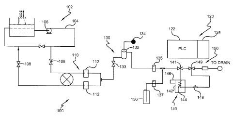
Referring now to Figure 7, a system for monitoring the total bacterial content
in the aqueous medium of a water system according to the methods set forth
above is
illustrated and referred to generally by reference number 100. The embodiment
shown in Figure 7 illustrates a conventional open recirculating cooling tower
water
system 1.02 having an aqueous medium flowing through a circulating loop 104.
Flow
of the aqueous medium through the circulating loop 104 may be assisted by a
circulating pump 106 as is known in the art. Valves 108 permit feeding aqueous
medium from the circulating loop 104 to the total bacterial monitoring system
100.
The total bacterial monitoring system 100 works as an on-line analyzer to
monitor
bacteria concentration in the aqueous medium of the water system 102. One
skilled in
the art will understand that the total bacterial monitoring system 100 may be
used to
21
CA 02741449 2011-04-21
WO 2010/062472
PCT/US2009/059239
225863-1
provide for rapid detection of total viable bacteria through the measurement
&total
bacteria in any municipal or industrial water or process system 102.
Accordingly,
further details of the water system102 need not be given herein.
The aqueous medium entering the total bacterial monitoring system 100 first
passes through a filter module 110. Desirably, the filter module 110 includes
a filter
112 having a pore sin of between about 5 and about 50 microns such that larger
impurities are removed from the aqueous medium., but the bacterial content
passes
through in the filtrate. In one embodiment, the filter module 110 is a
flip/flop type
filter system such as the one disclosed in commonly assigned U.S. Patent
Application
No. 12/193,198 filed August 18, 2008 entitled "In-Line Filtration Systems",
with a
filter pore size of 10 microns. However, the filter module 110 mw include
other
filtering layouts without departing from the scope of the invention.
The total bacterial monitoring system 100 includes a control module 120, a
sample preparation module 130, a cell lysing module 140, and a detection
module
150. The control module 120 contains a programmable logic controller 122 or
similar
device and an electronics unit 124 used to control the function of the other
modules
130, 140, 150, and additionally calculates the total bacteria concentration as
will be
described below.
The sample preparation module 130 is comprised of a level-switch sample cup
132 and a solenoid v al ve 133 used to control the flow of the .filtered
aqueous medium
into the sample cup .132. In one embodiment, the level-switch sample cup1132
is
comprised of a pair of lead wires. When the sample cup 132 is full, or at a
designated
high level, the two wires are electronically connected, which triggers the
shutoff of
the solenoid valve 133. When the sample cup 132 is empty, or at a designated
low
level, the two wires are disconnected, which triggers the opening of the
solenoid valve
133. The dead band between these two states is desirably about 1.5 ml. The
sample
preparation module 130 lets down the pressure of the aqueous medium from
header
pressure in the circulating loop 104 to atmospheric pressure. Desirably, the
sample
cup 132 is open to the atmosphere so as to allow any air bubbles in the
aqueous
medium to escape from the sample through vent 134. As one skilled in the art
would
understand, air bubbles in the aqueous medium would cause unwanted spikes from
optical measurement devices used in the detection module 150.
A sample pump 135, such as a micro positive-displacement pump, draws
aqueous medium from the sample cup 132. By lowering the pressure, the sample
22
CA 02741449 2011-04-21
WO 2010/062472
PCT/US2009/059239
225863-1
pump 135 is .protected, as the sample pump may be rated for only about 5 psig.
The
feed rate of the aqueous medium through the sample preparation module 130 is
controlled using the sample pump 135. The programmable logic controller 122
sets
the stroke frequency a the sample pump 135 to accurately control the flow
rate.
Flow rates of the aqueous medium are desirably between about 100 ùL and about
250
tiL, and more desirably between about 150 W.: and about 200uL In one
embodiment,
the sample pump 137 is a model 150SP-S2 made by Beion Medical Technology Co.
However, any known pump capable of accurately pumping small volumes of aqueous
medium may be used.
In the illustrated embodiment, the fluorochrome reagent and the buffer are
premixed and added together to the aqueous medium from a reagent supply 136.
Alternately, one skilled in the art will understand that the buffer may be
added before
or after the fluorochrome is added to the aqueous medium. The reagent supply
136
feeds the fluorochrome and buffer by means of a reagent feed pump 137. The
reagent
feed pump 137 also is desirably a micro positive-displacement pump and the
progrannnable logic controller 122 sets its stroke frequency to accurately
control the
flow rate. Desirably, the reagent feed pump 137 adds the fluorochrome in an
amount
of from about 0.5 mg to about 100 mg fluorochrome per liter of aqueous medium.
The buffer is added to the aqueous medium to maintain the pH of the aqueous
medium from about 2 to about 10. ha one embodiment, the reagent pump is a
model
120SP-S2 .made by Beim Medical Technology Co.
The aqueous medium pumped by the sample pump 135 and the reagent
pumped by the reagent feed pump 137 are combined using a mixing tee 138,
broadly
a mixing device, that provides a turbulent flow path to encourage mixing of
the
aqueous medium and the fluorochrome reagent and buffer. Other mixing devices,
such as mixing crosses or impellers, may also be used NNithOlit departing from
the
scope of the invention.
In the illustrated embodiment, the lysing module .140 accomplishes cell lysing
by heating the aqueous mediwn. Aqueous medium the sample preparation module
130 is either directed to the lysing module 140 or directed straight to the
detection
module 150, thus bypassing -the lysing module 140, using a three-way valve 141
controlled by the control module 120. In one embodiment, the lysing module 140
includes a temperature control unit 142 that raises and lowers the temperature
of the
aqueous medium in order to lyse the cells and release the intracellular
content of the
23
CA 02741449 2011-04-21
WO 2010/062472
PCT/US2009/059239
225863-1
microbiological matter. The temperature control module 142 includes a heating
device 144, such as a semiconductor plate or other known heating elements, to
heat
the aqueous medium. A fan or other radiator 146 is used to promote rapid
cooling of
the sample after the cells have been lysed. A thennocouple 148 measures the
temperature of the aqueous medium during the heating and cooling periods. The
control module 120 controls and supplies power to the temperature control unit
142 to
heat the sample to a desired temperature to lyse the cells, and then cool down
the
sample until it reaches a desired temperature using a predefined control
program.
Desirably, the temperature control unit 142 heats the aqueous medium to a
temperature of between about 40 C and about 100 C, and more desirably between
about 40 C and about 60 C. The temperature control unit 142 desirably heats
the
aqueous medium to the desired temperature in a time from about 1 minute to
about 1
hour, and more desirably between about 1. minute and about 3 minutes, in order
to
lyse the cells. One skilled in the art NON understand that the temperature
control unit
142 may contain other known means to heat and cool the aqueous medium as
desired.
Additionally, as set forth above, the lysing module 140 may use other known
lysing
methods, such as mechanical, chemical, physical, electrical, ultrasonic or
microwave
methods, to lyse the cells without departing from the scope of the invention.
The aqueous medium containing the lysed biological content is then directed
to the detection module 158 through three-way valve 149. The detection module
150
includes an optical measurement unit 152. The use o.f more than one optical
measurement unit may strengthen the accuracy of measurement. The optical
measurement unit 152 includes a silicon glass flow cell 154 and a single-
wavelength
fluorometer 156. The silicon glass flow cell 154 has an inlet flow tube .158
and an
outlet flow tube 159 mounted at the bottom and the top of the flow cell,
respectively.
As best seen in the schematic embodiment illustrated in FIG. 8, the
fluoroineter 156
includes at least one pair of light-emitting diodes (LEDs) 160 and photodiode
emission detectors 162 are configured around a reaction tube 163. Desirably,
the
fluorescent signal is measured with fluorometer having an excitation
wavelength from
about 350 nm to about 680 nal and an emission wavelength from about 450 nm to
about 650 nm. Additionally, the .fluorometer 156 includes optical lenses 164
and
filters 165 in the sealed optical tube to control light path and intensity. In
one
embodiinent, the fluorometer 156 is an LS55 Lwninescence Spectrometer by
PerkinElmer.
24
CA 02741449 2011-04-21
WO 2010/062472
PCT/US2009/059239
225863-1
In one embodiment comprising three pairs of photo optical comments, three
LEDs and three photodiodes are installed in six radial channels perpendicular
to the
center through hole. The three LEDs generate incident light at different
wavelengths,
and the three corresponding photodiodes detect the respective transmittance on
the
opposite sides. The LEDs used include a tricolor with 467 nm (blue), 530 nm
(green),
and 63' nm (red) lights, an orange LED with 610 TIM maximum and light green
1,ED
with 586 tun maximum emission. This configuration simplifies the design and
-maintenance of the optical components. The three pails ofphoto optical
components
.provide the ability to measure three functions at a time. There is no
maxirmuit number
of pairs of photo optical components that may be included; however, the number
will
be affected by size limitations based on the intended use &the monitoring
system.
The effluent from the optical measurement unit .152, comprising the mixed
sample water and reagents, exits the detector module 150 and connects to a
drain or a
collection drum, depending on each plant's pemitting requirements. Since the
effluent is anon-hazardous wastewater, it is commonly discharged to a gravity
drain.
The control module 120 is programmed such that -fluorescent signals of the
aqueous medium are measured by the detection module 150 before and after the
intracellular content of the microbiological matter has been extracted and
released
into the aqueous medium in the lysing module 140 to provide a baseline
fluorescent
signal and a second fluorescent signal, respectively. These fluorescent
signals are
measured by the. detection .module 150 and stored in the programmable logic
controller 122. The baseline fluorescent signal is subtracted from the second
-fluorescent signal -to obtain a net -fluorescent signal that is a result of
the
microbiological content- of the lysed cells. A calibration curve is used to
obtain the
total microbiological content as described above. As explained above, the
calibration
curve is prepared by measuring fluorescent signals for known concentrations of
microbiological miter in aqueous media with -the fluorochrome, determining the
net
fluorescent signal .for each concentration, plotting the concentration amounts
versus
log values of the net fluorescent signals on a graph and performing regression
analysis
to obtain the calibration curve. With above features, the system can monitor
total
bacteria i.n an on-line manner.
While typical embodiments have been set forth for the purpose of illustration,
the foregoing descriptions should not be deemed to be a limitation on the
scope
CA 02741449 2014-07-31
225863-8
herein. Accordingly, various modifications, adaptations and alternatives may
occur to
one skilled in the art without departing from the scope herein.
26