Note: Descriptions are shown in the official language in which they were submitted.
CA 02741650 2016-01-18
WO 2010/062556
PCT/US2009/062061
1
FAST RESULTS HYBRID CAPTURE ASSAY AND SYSTEM
[0001]
FIELD
[0002] The present invention relates to methods, reagents, systems, and
kits for
determining the presence of a nucleic acid in a sample.
BACKGROUND
[0003] The detection and characterization of specific nucleic acid
sequences and
sequence changes have been utilized to detect the presence of viral or
bacterial nucleic acid
sequences indicative of an infection, the presence of variants or alleles of
mammalian genes
associated with disease and cancers, and the identification of the source of
nucleic acids
found in forensic samples, as well as in paternity determinations.
[0004] For example, the RNA or DNA for many microorganisms and viruses
have
been isolated and sequenced. Nucleic acid probes have been examined for a
large number of
infections. Detectable nucleic acid sequences that hybridize to complementary
RNA or DNA
sequences in a test sample have been previously utilized. Detection of the
probe indicates the
presence of a particular nucleic acid sequence in the test sample for which
the probe is
specific. In addition to aiding scientific research, DNA or RNA probes can be
used to detect
the presence of viruses and microorganisms such as bacteria, yeast and
protozoa as well as
genetic mutations linked to specific disorders in patient samples.
[0005] Nucleic acid hybridization probes have the advantages of high
sensitivity and
specificity over other detection methods and do not require a viable organism.
Hybridization
probes can be labeled, for example with a radioactive substance that can be
easily detected, or
with biochemical markers such as, for example, biotin, that allows for their
capture and
detection. Nucleic acid molecules may also by captured by a first antibody
that is specific to
DNA hybrids, wherein the hybrids may comprise DNA-RNA hybrids, DNA-DNA hybrids
or
CA 02741650 2011-04-26
WO 2010/062556 PCT/US2009/062061
2
RNA-RNA hybrids. The hybrids may subsequently be detected by a second,
labeled,
antibody that may be, for example, labeled with a biochemical marker such as
alkaline
phosphatase or any other marker capable of detection.
[0006] As nucleic acid sequence data for genes from humans and
pathogenic
organisms accumulates, the demand for fast, cost-effective, and easy-to-use
tests increases.
There is a need to provide novel and effective methods, compositions, and kits
for
determining target nucleic acids in a rapid, cost-effective, and reliable
manner in
geographical areas where access to medical care is not readily available.
There is also a need
to provide these assays in a rapid-screen format that can be used in
developing countries.
The methods and assays of the present invention meet these needs and may be
used in
manual, partially automated, automated, and non-automated systems.
[0007] Clinical analysis in developing countries and geographical
areas where access
to medical care is not readily available presents unique challenges. The
invention described
herein achieves an acceptable resolution that balances the importance of these
challenges in
such countries and areas. For instance, speed in obtaining results is
particularly important in
locations where women travel great distances to provide specimens for
analysis. In such
locations, it is advantageous that results are obtained within several hours
or the same day
while the patient is still present to avoid loss to follow-up associated with
traveling from
home to the test site.
[0008] Other factors facing developing countries are the cost of running
the assay and
the instrumentation needed to run the assay. Repeat pipettors and single
pipettes are just two
types of devices that are routinely used in developed countries but are
potentially cost
prohibitive in developing countries. Accordingly, there is a need for medical
devices and
products which employ cheaper and more readily accessible alternatives in
developing
countries.
SUMMARY
[0009] One aspect relates to a method for determining the presence of
a target nucleic
acid molecule in a sample containing biological material. The biological
material can include
a cervical epithelial cell or nucleic acid from a cervical cell. Using the
disclosed methods, the
determination of whether a target nucleic acid molecule is present in a sample
can be
obtained relatively rapidly, for example within a period of less than about
two or three hours.
CA 02741650 2011-04-26
WO 2010/062556 PCT/US2009/062061
3
[0010] In an aspect, a method for determining the presence of a target
nucleic acid
molecule in a sample comprises:
a) suspending the sample in a collection medium;
b) releasing target nucleic acid molecules from the sample into the collection
medium;
c) converting double-stranded target nucleic acid molecules to single-stranded
target
nucleic acid molecules;
d) contacting one or more probes with the single-stranded target nucleic acid
molecules
under conditions that allow the probes and target single-stranded target
nucleic acid
molecules to hybridize forming double-stranded nucleic acid hybrids;
e) capturing the double-stranded nucleic acid hybrids;
f) separating the double-stranded nucleic acid hybrids from un-bound single-
stranded
target nucleic acid molecules; and
g) detecting the double-stranded nucleic acid hybrids, thereby indicating the
presence of
the target nucleic acid.
[0011] In one aspect, the method may be predominantly manual, requiring
human
input. Another aspect relates to the rapid detection of target nucleic acid
molecules in a
sample. The detection method may be automated, either fully automated, or
partially
automated ¨ in other words requiring some human input.
[0012] Another aspect relates to the detection of target nucleic acid
molecules in
multiple samples at the same time or within a very short period of time, for
example in a
machine or a series of machines.
[0013] Yet another aspect relates to an instrument for running a
method for the
detection of a target nucleic acid molecule in a simple footprint. The
instrument combines
many, or all, of other individual instruments that perform the steps of the
method.
[0014] Another aspect relates to a portable system for evaluating the
detection of a
target nucleic acid molecule in a sample.
[0015] Another aspect relates to a kit for the detection of a target
nucleic acid
molecule in a sample.
[0016] A further aspect relates to reagents within a collection medium
into which a
sample containing a target nucleic acid molecule are collected. The target
nucleic acid
molecule can be kept in the collection medium with minimal degradation of the
target nucleic
acid molecule over a time period of weeks or months. In an aspect, DNA-based
target
CA 02741650 2011-04-26
WO 2010/062556
PCT/US2009/062061
4
sample material can be kept in the collection medium with minimal degradation
of the target
nucleic acid molecule over a time period of weeks or months. In an aspect the
detergent-
based collection medium allows for the rapid analysis and processing of a
sample.
BRIEF DESCRIPTION OF THE DRAWINGS
[0017] FIG. 1 shows that the detergent-based collection medium holds
magnetic
beads in microtiter plate wells better than known collection or sample
transport medium
(STM) (non-detergent based medium).
[0018] FIG. 2 shows that samples having only 0.2 pg target nucleic
acid (DNA) per
ml of sample provide a readable signal using methods of the present invention.
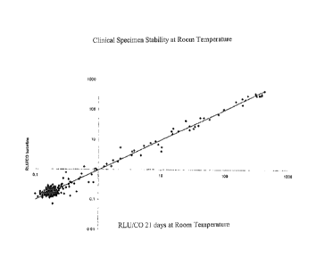
[0019] FIG. 3 shows clinical specimen stability at room temperature
for 21 days.
[0020] FIG. 4 shows clinical specimen stability at 33 C for 21 days.
[0021] FIG. 5 shows test results demonstrate an S/N > 2.0 for a 0.2
pg/ml HPV 16
plasmid which is equivalent to 1000 copies of Iff'V 16 DNA.
[0022] FIG. 6 shows a system for detecting the presence of a target nucleic
molecule
acid in a sample including a heater configured for heating multiple samples; a
luminometer;
and a monitor.
[0023] FIG. 7 shows different reagents associated with the detection
assay. The
reagent vials are color coded for ease of use and can be included in a kit.
[0024] FIG 8 shows a monitor used in conjunction with the system for
detecting the
presence of nucleic acid molecules.
[0025] Fig. 9 shows 11 day stability data for soft pellets suspended
in a detergent-
based collection medium and stored at room temperature.
DETAILED DESCRIPTION
[0026] The present disclosure covers methods, compositions, reagents,
systems, and
kits for rapidly determining the presence of a nucleic acid molecule in a
sample. The
methods, compositions, reagents, systems, and kits may be used for clinical
diagnostic
purposes, including but not limited to the detection and identification of
pathogenic
organisms and the detection of a genetic predisposition to a particular
disease.
CA 02741650 2011-04-26
WO 2010/062556
PCT/US2009/062061
[0027] In one aspect, the present disclosure provides a method for
determining the
presence of a target nucleic acid molecule in a sample. The method comprises:
a) suspending the sample in a collection medium comprising a detergent;
b) denaturing the target nucleic acid molecule;
5 c)
contacting one or more polynucleotide probes with the target nucleic acid
molecule
under conditions that allow the probes and the target nucleic acid molecule to
hybridize,
thereby forming a double-stranded nucleic acid hybrid;
d) capturing the double-stranded nucleic acid hybrid on a solid support coated
with a
first antibody specific for the double-stranded hybrid nucleic acid hybrid,
thereby forming a
double-stranded nucleic acid hybrid/solid support complex;
e) separating the double-stranded nucleic acid hybrid/solid support complex
from
unbound nucleic acid;
f) conjugating the complex with a second antibody that is specific for either
the double-
stranded nucleic acid hybrid or specific for the first antibody to form a
double-stranded
nucleic acid hybrid/solid support antibody complex; wherein the second
antibody is labeled
with a detectable marker;
g) washing the double-stranded nucleic acid hybrid/solid support antibody
complex with
a wash buffer comprising a detergent; and
h) detecting the label on the second antibody wherein the detecting indicates
the
presence of the target nucleic acid molecule.
[0028] In another aspect, the present disclosure provides a method for
determining the
presence of a target nucleic acid molecule in a sample including suspending a
sample in a
collection medium including a detergent; denaturing a target nucleic acid
molecule;
contacting one or more polynucleotide probes with the target nucleic acid
molecule under
conditions that allow the probes and the target nucleic acid molecule to
hybridize or bind, and
capturing the double-stranded nucleic acid hybrid on a solid support coated
with a first
antibody specific for the double-stranded hybrid nucleic acid hybrid.
[0029] In an aspect, the present disclosure provides a method for
determining the
presence of a target nucleic acid molecule in a sample including suspending a
sample in a
collection medium including a detergent; denaturing a target nucleic acid
molecule;
contacting one or more polynucleotide probes with the target nucleic acid
molecule under
conditions that allow the probes and the target nucleic acid molecule to
hybridize or bind,
CA 02741650 2011-04-26
WO 2010/062556 PCT/US2009/062061
6
capturing the double-stranded nucleic acid hybrid on a solid support coated
with a first
antibody specific for the double-stranded hybrid nucleic acid hybrid and
separating the
double-stranded nucleic acid hybrid/solid support complex from unbound nucleic
acid.
[0030] In an aspect, the present disclosure provides a method for
determining the
presence of a target nucleic acid molecule in a sample including suspending a
sample in a
collection medium including a detergent; denaturing a target nucleic acid
molecule;
contacting one or more polynucleotide probes with the target nucleic acid
molecule under
conditions that allow the probes and the target nucleic acid molecule to
hybridize or bind,
capturing the double-stranded nucleic acid hybrid on a solid support coated
with a first
antibody specific for the double-stranded hybrid nucleic acid hybrid, thereby
forming a
double-stranded nucleic acid hybrid/solid support complex; separating the
double-stranded
nucleic acid hybrid/solid support complex from unbound nucleic acid; and
conjugating the
complex with a second antibody that is specific for either the double-stranded
nucleic acid
hybrid or specific for the first antibody to form a double-stranded nucleic
acid hybrid/solid
support antibody complex.
[0031] In another aspect, the present disclosure provides a method for
determining the
presence of a target nucleic acid molecule in a sample including suspending a
sample in a
collection medium including a detergent; denaturing a target nucleic acid
molecule;
contacting one or more polynucleotide probes with the target nucleic acid
molecule under
conditions that allow the probes and the target nucleic acid molecule to
hybridize or bind,
capturing the double-stranded nucleic acid hybrid on a solid support coated
with a first
antibody specific for the double-stranded hybrid nucleic acid hybrid, thereby
forming a
double-stranded nucleic acid hybrid/solid support complex; and separating the
double-
stranded nucleic acid hybrid/solid support complex from unbound nucleic acid;
conjugating
the complex with a second antibody that is specific for either the double-
stranded nucleic acid
hybrid or specific for the first antibody to form a double-stranded nucleic
acid hybrid/solid
support antibody complex; wherein the second antibody is labeled with a
detectable marker;
and washing the double-stranded nucleic acid hybrid/solid support antibody
complex with a
wash buffer comprising a detergent.
[0032] In another aspect, the present disclosure provides a method for
determining the
presence of a target nucleic acid molecule in a sample, the method comprising:
a) suspending the sample in a collection medium comprising a detergent;
CA 02741650 2011-04-26
WO 2010/062556
PCT/US2009/062061
7
b) denaturing the target nucleic acid molecule in the sample;
c) forming a double-stranded nucleic acid hybrid by contacting at least one
polynucleotide probe with the target nucleic acid molecule;
d) forming a double-stranded nucleic acid hybrid-support complex by capturing
the
double-stranded nucleic acid hybrid on a support, wherein the support
comprises a
first antibody;
e) forming a double-stranded nucleic acid hybrid-support-second antibody
complex by
contacting the double-stranded nucleic acid hybrid-support complex with a
second
antibody, wherein the second antibody is labeled with a detectable marker;
f) washing the double-stranded nucleic acid hybrid-support-second antibody
complex
with a wash buffer; and
g) detecting the marker on the second antibody wherein the detecting indicates
the
presence of the target nucleic acid molecule.
[0033] In one aspect, the solid support comprises a modified
paramagnetic bead that
is coated or has attached thereto a first antibody immunospecific for double-
stranded hybrid
nucleic acids. A magnetic field is used to separate the double-stranded
nucleic acid-magnetic
bead-antibody complex from non-bound nucleic acid.
[0034] In an aspect, the method does not include a sample pre-
treatment step. For
example, the detergent-based collection medium allows for reduced sample
preparation time
which, in turn, can lead to accelerated detection of target nucleic acid
molecules. The sample
can be analyzed by methods, assays, or the apparatus of the disclosure in a
direct-to-assay
manner. In an example, purification steps are not performed on the sample
prior to
evaluation using assays of the disclosure. In an aspect, crude lysate is
directly analyzed by
the methods, assays, or the apparatus of the disclosure. In another aspect,
the sample does
not undergo a target amplification step.
[0035] One aspect relates to a method of diagnosing cancer by
utilizing methods, kits,
assays, and the apparatus provided herein. In one aspect, cervical cancer is
detected by
identifying nucleic acid molecules associated with HPV and HPV variants. In
another aspect,
cervical intraepithelial neoplasia (CIN) can be screened for using methods,
kits, assays, and
the apparatus provided herein. The detected cancer can be subsequently treated
after being
diagnosis by the methods, kits, assays, and the apparatus provided herein. In
an aspect, the
diagnosed cancer is cervical cancer and variants thereof.
CA 02741650 2011-04-26
WO 2010/062556 PCT/US2009/062061
8
[0036] In one aspect, the disclosure provides a composition comprising
a biological
sample suspended in a collection medium comprising about 0.5% to about 2.0% NP-
40,
about 0.10% to about 0.40% sodium deoxycholate, about 25 mM to about 75 mM
Tris-HC1,
about 10 mM to about 50 mM EDTA, about 50 mM to about 200 mM NaC1, and about
0.01% to about 0.10% sodium azide.
[0037] In an aspect, the disclosure provides for a composition
comprising
(a) a biological sample suspended in about 0.5% to about 2.0% NP-40, about
0.10%
to about 0.40% sodium deoxycholate, about 25 mM to about 75 mM Tris-HC1, about
10 mM
to about 50 mM EDTA, about 50 mM to about 200 mM NaC1, and about 0.01% to
about
0.10% sodium azide; and
(b) at least one or more polynucleotide probes.
[0038] In an aspect, the disclosure provides for a composition
comprising
(a) a biological sample suspended in a collection medium comprising about 0.5%
to
about 2.0% NP-40, about 0.10% to about 0.40% sodium deoxycholate, about 25 mM
to about
75 mM Tris-HC1, about 10 mM to about 50 mM EDTA, about 50 mM to about 200 mM
NaC1, and about 0.01% to about 0.10% sodium azide;
(b) at least one or more polynucleotide probes; and
(c) a first antibody.
[0039] In an aspect, the disclosure provides for a composition
comprising
(a) a biological sample suspended in a collection medium comprising about 0.5%
to
about 2.0% NP-40, about 0.10% to about 0.40% sodium deoxycholate, about 25
riaM to about
75 mM Tris-HC1, about 10 mM to about 50 mM EDTA, about 50 mM to about 200 mM
NaCl, and about 0.01% to about 0.10% sodium azide;
(b) a first antibody; and
(c) a second antibody.
[0040] In an aspect, the disclosure provides for a composition
comprising
(a) a biological sample suspended in a collection medium comprising about 0.5%
to
about 2.0% NP-40, about 0.10% to about 0.40% sodium deoxycholate, about 25 mM
to about
75 mM Tris-HC1, about 10 mM to about 50 mM EDTA, about 50 mM to about 200 mM
NaC1, and about 0.01% to about 0.10% sodium azide;
(b) at least or one or more polynucleotide probes;
(c) a first antibody; and
CA 02741650 2011-04-26
WO 2010/062556
PCT/US2009/062061
9
(d) a second antibody.
[0041] In an aspect, the disclosure provides for a composition
comprising
(a) a biological sample suspended in a collection medium, wherein the
collection
medium comprises at least one detergent;
(b) a denaturation reagent;
(c) at least one polynucleotide probe capable of binding to a target nucleic
acid
molecule;
(d) a support coated with a first antibody; and
(e) a second antibody labeled with a detectable marker.
[0042] In an aspect, any of the above compositions may be used may be used
with
any of the collection mediums described herein.
[0043] In an aspect, the biological sample in the above compositions
is a cervical cell
sample or a human cervical cell sample. In another aspect, the nucleic acid
molecules in the
biological sample are denatured. The biological sample in the above
compositions can
exhibit stability when stored in the collection medium for at least 21 days at
33 C. In an
aspect, the second antibody is labeled with a detectable marker.
Biological Sample
[0044] Methods of the present invention may be used to detect the
presence of a
target nucleic acid molecule from samples, including, without limitation, a
specimen or
culture (e.g., cellular, microbiological and viral cultures) including
biological and
environmental samples. Biological samples may be from an animal, including a
human,
fluid, solid (e.g., stool) or tissue, as well as liquid and solid food and
feed products and
ingredients such as dairy items, vegetables, meat and meat by-products, and
waste.
Environmental samples include environmental material such as surface matter,
soil, water
and industrial samples, as well as samples obtained from food and dairy
processing
instruments, apparatus, equipment, utensils, disposable and non-disposable
items.
[0045] Particularly preferred are biological samples including, but
not limited to,
cervical epithelial cells (e.g., a sample obtained from a cervical swab),
adenoid cells, anal
epithelial cells, blood, saliva, cerebral spinal fluid, pleural fluid, milk,
lymph, sputum and
semen. The sample may comprise a double-stranded nucleic acid molecule or may
comprise
a single-stranded nucleic acid molecule. If a double-stranded nucleic acid
molecule is
CA 02741650 2011-04-26
WO 2010/062556
PCT/US2009/062061
present, it may be prepared for hybridization analysis by a variety of methods
known in the
art, e.g., using alkali, using proteinase K/SDS, chaotropic salts. The process
of preparing a
double-stranded nucleic acid molecule for hybridization analysis generally
involves
converting it into a single-stranded nucleic acid molecule. This process is
generally known
5 as denaturation. However, it is also contemplated that a double-stranded
nucleic acid
molecule may be detected without denaturation, e.g., through a triple-stranded
construct.
[0046] The target nucleic acid molecule in a sample can be DNA or RNA
or both
DNA and RNA. The target nucleic acid molecule can be contained within a larger
nucleic
acid molecule. Detection of either the target nucleic acid molecule or the
larger nucleic acid
10 molecule containing the target nucleic acid molecule is contemplated by
this disclosure.
[0047] The biological sample may comprise cervical cells, especially
human cervical
cells. The sample can be collected with any method or device known in the art,
including a
chemically inert collection device such as a DACRON tipped swab. Other
acceptable
collection devices may be used including, but not limited to, cotton swab,
cervical brush,
flocked swab (a swab shaped like a DACRON swab but made with nylon fibers
enabling
collection of more cells and easier release of cells), cervical broom, mini
broom, lavage, or
any collection device often used in Pap smear testing.
[0048] In an aspect, the methods include collecting a sample from a
woman over 30
years of age. The method can also include collecting a sample from a woman
over 30 years
via a Pap smear or comparable test. The sample collected by the Pap smear or
comparable
test can be a cervical cell sample.
[0049] Once the sample is collected, it may be placed in a sample
tube. The tube can
be sealed to prevent contamination. The collection device (swab, brush, etc.)
may further
contain a mechanism by which it can be moved once it is inside the sample
tube. In one
aspect, the collection device contains an insert that can be moved using a
magnet. In one
aspect, this insert comprises a metal. In another aspect, this insert
comprises a magnetic
material. Magnetic material includes paramagnetic, ferromagnetic, and
diamagnetic
materials. One advantage of moving the collection device once it is inside the
sample tube is
to avoid the collection device from making contact with any sample extraction
or sample
detection devices. Examples of a sample extraction device include pipettes,
pipette tips,
dropper bottles or other low tech extraction devices. Examples of sample
detection devices
include probes and probe tips.
CA 02741650 2011-04-26
WO 2010/062556 PCT/US2009/062061
11
[0050] The speed of this assay is also beneficial in screening samples
from patients in
remote living areas. Often patients will travel quite a distance to visit the
doctor or clinic and
will not likely return for some time thereafter. Thus, it is desirable to be
able to test the
patient and provide results while the patient waits at the clinic. In some
circumstances,
tracking down the patient to provide test results and/or treat the patient
after they have left the
doctor's office may be difficult. Thus, the described assays provide results
over a short time,
for example, between about 2 hours to about 3 hours, between about 2 hours to
about 4 hours,
between about 3 hours to about 5 hours, between about 4 hours to about 8
hours, or between
about 6 hours to about 12 hours. In another aspect, the described assays
provide results in
less than about 2 hours, less than about 2.5 hours, less than about 3 hours,
less than about 3.5
hours, less than about 4 hours, less than about 4.5 hours, less than about 5
hours, less than
about 8 hours, less than about 12 hours, and less than about 24 hours. Such a
short
turnaround time allows the doctor to provide the patient with the results
and/or treatment the
same day the patient is at the clinic.
Sample Tube
[0051] Any type of sample tube may be used. Advantageously, the sample
tube may
be closed or sealed to minimize contamination. The closure may be permanent or
removable.
Examples of removable closures include snap caps, screw caps, rubber septa,
foil, and film.
The closure may contain one or more openings or perforations, which when
pierced may be
re-sealable. One advantage of a closure that contains such openings or
perforations is that the
closure is not rendered ineffective when pierced by, for example, a sample
extraction device
or sample detection device. Once the sample extraction device or sample
detection device is
removed, the closure re-seals, thereby minimizing contamination.
Storage of the Biological Sample
[0052] Once the sample is in the sample tube, the sample may be stored by
drying it
with a substrate, or in a preservative medium, or both. Desiccation is
accomplished by
pressure drying or drying with chemicals. This removes most of the water and
is suitable for
long-term stability. Alternatively, the sample may be lyophilized (freeze-
dried) with a
substrate like trehalose to ensure stability of the sample.
[0053] Another possibility is that the sample may be stored by suspending
in a
preservative medium, known and apparent to one of skill in the art. The
purpose of the
CA 02741650 2011-04-26
WO 2010/062556 PCT/US2009/062061
12
preservative medium is to protect biological components that can degrade. For
instance, the
sample cells, the probe mixture, the antibody: bead complex used in the
capture step, and the
secondary antibody used in the detection step are all susceptible to
degradation. A
preservative medium at the initial step of collection ideally provides sample
stability and
integrity and can affect downstream steps in the process of nucleic acid
capture and detection.
In an aspect, the sample may be stored at room temperature, refrigerated, or
frozen either
prior to or after the addition of the preservative medium.
Collection Medium
[0054] In an aspect, the sample may be collected and stored in a
collection medium.
The collection medium has several functions including as a preservative medium
to preserve
nucleic acids and inhibit nucleases to prevent degradation of nucleic acids
prior to analysis.
In one aspect, the collection medium contains at least one detergent. In
another aspect, the
collection medium contains at least two detergents, at least three detergents,
or at least four
detergents. In an aspect, each of the detergents is different. In another
aspect, the detergent-
based collection medium comprises two different detergents, one which is able
to control
background signal and another detergent that improves magnetic bead behavior,
for example,
migration through a viscous sample. Because the detergent improves the bead
behavior, the
beads can be washed with a variety of less precise devices, such as a squirt
bottle, and
dropper bottle, without causing too much disruption of the beads. Such
methodology may be
advantageous for use in developing countries where funds or access to
technologically
advanced equipment, such as pipetting devices, may not be available.
[0055] Further, the present invention provides a robust assay that can
support using
less precise reagent delivery devices. The reagents in this assay were
designed to be robust
and withstand variability in reagent volume delivery. For example, the
neutralization step of
the assay utilizes a highly buffered pH neutral solution to bring the very
basic pH of the
reaction to the appropriate pH range for hybridization with very large volume
tolerances.
[0056] The use of a detergent-based collection medium for use in the
assay can
include one or more detergents. In an aspect, heat is employed during the
hybridization,
capture, and detection steps of the assay. Even with detergent and the
application of heat,
antibodies used in the assay remain functional.
CA 02741650 2011-04-26
WO 2010/062556 PCT/US2009/062061
13
[0057] FIG. 1 demonstrates that a detergent-based collection medium
greatly
improves the ability to prevent magnetic bead loss and migration during
handling as
compared to standard collection mediums. The detergent-based collection medium
may
comprise, consist essentially of, or consist of one, two, three, or four or
more detergents.
Detergents are known in the art and may include, but are not limited to,
cationic detergents
such as but not limited to cetyl pyridinium bromide, cetyltrimethylammonium
bromide
(collectively known as cetrimoniun-i compounds) and
alkylbenzyldimethylammonium
chlorides (collectively known as benzalkonium compounds), and alkyl-trimethyl-
ammonium
salts; anionic detergents such as, but not limited to, sodium dodecyl sulfate
(SDS), and
Sarkosyl; and non-denaturing detergents such as NP-40; and other detergents.
NP-40 is also
known as Tergitol-type NP-40, which is nonyl phenoxylpolyethoxylethanol. NP-40
is not
powerful enough to break the nuclear membrane, but can break the plasma
membrane. As
such, it can be used to obtain the cytoplasmic contents of a cellular culture.
[0058] Other detergents and combination of detergents may be used, and
advantageously their combination provides the ability to control background
noise and
improve magnetic bead behavior (when the solid support employed comprises
magnetic
beads). In certain aspects, one detergent is an anionic detergent and the
second detergent is a
nonanionic detergent. For example, in one aspect, the combination of non-ionic
and anionic
detergents helps to maintain low-background noise. In an aspect, a detergent-
based
collection medium comprises an anionic detergent such as sodium deoxycholate,
which
controls background noise and NP-40, which improves magnetic bead behavior.
[0059] The combination of these two types of detergents provides
synergistic benefits
beyond a simple combination of adding two detergents together: control of
background noise,
better bead behavior, and increased assay speed. The presence of these
detergents (in the
detergent-based collection medium) provides the ability to achieve faster
assay results, but
does not negatively impact the nucleic acid or capture antibody during
downstream analytical
steps.
[0060] In addition, the detergent-based collection medium improves
removal of the
specimen from the collection device as the sample is dissolved more easily. In
addition, the
detergent-based collection medium improves the homogeneity of the sample
compared with
other collection media such as but not limited to PRESERVCYT (uses a 40%
methanol
CA 02741650 2011-04-26
WO 2010/062556 PCT/US2009/062061
14
solution), STM (uses a chaotropic agent), and alcohol. The detergent-based
collection
medium also reduced sample viscosity after mixing (either manual or
automated).
[0061] The concentration of NP-40 in the collection medium can range
from about
0.5% to about 2.0%, from about 0.1% to about 1.0%, as well as any number
within the
recited ranges. In certain aspects, the NP-40 is present at a concentration
from about 0.8% to
about 1.5 %; from about 0.9% to about 1.2% and in certain aspects is about
1.0%. In another
aspect, the NP-40 is present at a concentration from about 0.1%, about 0.2%,
about 0.3%,
about 0.4%, about 0.5%, about 0.6%, about 0.7%, about 0.8%, about 0.9%, about
1.0%, about
1.1%, about 1.2%, about 1.3%, about 1.4%, about 1.5%, about 1.6%, about 1.7%,
about
1.8%, about 1.9%, or about 2.0%. The concentration of sodium deoxycholate in
the
collection medium can range from about 0.10% to about 0.40%, from about 0.20%
to about
0.30%, as well as any number within the recited ranges. In one aspect, the
concentration of
sodium deoxycholate is about 0.10%, about 0.15%, about 0.20%, about 0.25%,
about 0.30%,
about 0.35%, or about 0.40%.
[0062] The detergent-based collection medium may comprise, consist
essentially of,
or consist of a buffer, two detergents, a chelator and a preservative. The
buffer may be Tris-
HC1 in a concentration of from about 25 mM to about 75mM; from about 30 mM to
about 60
mM; from about 40 mM to about 50 mM, and from about 45 mM to about 55 mM as
well as
any number within the recited ranges. The buffer may also be Tris-HCI in a
concentration of
about 25 mM, about 30 mM, about 35 mM, about 40 mM, about 45 mM, about 50 mM,
about
55 mM, about 60 mM, about 65 mM, about 70 mM, or about 75 mM.
[0063] Any preservative can be used and the choice can depend on
factors such as
desired functionality, minimization side-effects, cost, etc. Suitable
preservatives include
gentomycin, ProClin, dimersol, and sodium azide. The concentration of the
preservatives in
the collection medium depends on factors such as the type of preservative, its
efficacy, its
side-effects, etc. For example, for sodium azide, the concentration of sodium
azide can range
from about 0.01% to about 0.1%, from about 0.025% to about 0.075%, and from
about 0.04%
to about 0.06%, as well as any number within the recited ranges. The
preservative, for
example, sodium azide, can also be present at about 0.01%, about 0.02%, about
0.03%, about
0.04%, 0.05%, about 0.06%, about 0.07%, about 0.08%, about 0.09%, or about
0.10%.
[0064j In one aspect the detergent-based collection medium comprises,
consists
essentially of, or consists of 1.0% NP-40, 0.25% sodium deoxycholate, 50 mM
Tris-HC1, 25
CA 02741650 2011-04-26
WO 2010/062556 PCT/US2009/062061
mM EDTA, 150 mM NaC1 and 0.05% sodium azide. In another aspect the detergent-
based
collection medium comprises, consists essentially of, or consists of about
0.5% to about 2.0%
NP-40, about 0.10% to about 0.40% sodium deoxycholate, about 25 mM to about 75
mM
Tris-HC1, about 10 mM to about 50 mM EDTA, about 50 mM to about 200 mM NaC1,
and
5 about 0.01% to about 0.10% sodium azide. In other aspects the detergent-
based collection
medium comprises, consists essentially of, or consists of about 0.8% to about
1.5% NP-40,
about 0.20% to about 0.40% sodium deoxycholate, about 30 mM to about 60 mM
Tris-HCI,
about 20 mM to about 40 mM EDTA, about 100 mM to about 200 mM NaC1, and about
0.025% to about 0.075% sodium azide. In yet another aspect the detergent-based
collection
10 medium comprises, consists essentially of, or consists of about 0.9% to
about 1.2% NP-40,
about 0.20% to about 0.30% sodium deoxycholate, about 30 mM to about 60 mM
Tris-HC1,
about 20 mM to about 30 mM EDTA, about 100 mM to about 150 mM NaCI, and about
0.04% to about 0.06% sodium azide.
[0065] In an aspect, the collection medium comprises, consists
essentially of, or
15 consists of NP-40 and EDTA. In another aspect, the collection medium
comprises, consists
essentially of, or consists of NP-40, EDTA, and sodium azide. In one aspect,
the collection
medium comprises, consists essentially of, or consists of sodium deoxycholate,
EDTA, and
sodium azide. In an aspect, the collection medium comprises, consists
essentially of, or
consists of about NP-40, sodium deoxycholate, EDTA, and sodium azide. In an
aspect, the
collection medium comprises, consists essentially of, or consists of NP-40,
sodium
deoxycholate, Tris-HC1, EDTA, and sodium azide.
[0066] In another aspect, the collection medium comprises, consists
essentially of, or
consists of 0.5% to about 2.0% NP-40 and 10 mM to about 50 mM EDTA. In another
aspect,
the collection medium comprises, consists essentially of, or consists of 0.5%
to about 2.0%
NP-40, 10 mM to about 50 mM EDTA, and about 0.01 % to about 0.10 % sodium
azide. In
one aspect, the collection medium comprises, consists essentially of, or
consists of about
0.10% to about 0.40% sodium deoxycholate, 10 mM to about 50 mM EDTA, and about
0.01% to about 0.10% sodium azide. In an aspect, the collection medium
comprises, consists
essentially of, or consists of about 0.5% to about 2.0% NP-40, about 0.10% to
about 0.40%
sodium deoxycholate, 10 mM to about 50 mM EDTA, and about 0.01% to about 0.10%
sodium azide. In an aspect, the collection medium comprises, consists
essentially of, or
consists of about 0.5% to about 2.0% NP-40, about 0.10% to about 0.40% sodium
CA 02741650 2011-04-26
WO 2010/062556 PCT/US2009/062061
16
deoxycholate, about 25 mM to about 75 mM Tris-HC1, about 10 mM to about 50 mM
EDTA,
and about 0.01% to about 0.10% sodium azide.
[0067] In an aspect, the collection medium is a non-chaotropic medium.
That is, for
example, the collection medium does not include a chaotropic medium or
chaotropic salts.
Without being limited, in an aspect, the collection medium does not include
guanidine
hydrochloride or urea. A potential advantage of using a non-chaoptropic
collection medium
is better resuspension of a sample, more reproducible testing, and more
uniform testing
aliquots relative to a medium which includes a chaotropic medium or chaotropic
salts.
[0068] An advantage of using a detergent-based collection medium is
that it preserves
the stability of the sample. A sample stored in a detergent-based collection
medium as
disclosed is stable for at least 31 days, and, when held at temperatures from
15 C to 33 C is
stable for at least 21 days. In an aspect, a sample is stable when frozen in a
detergent-based
collection medium at -20 C for at least six months. In another aspect, a
cervical cell sample
is stable for at least 31 days, for at least 21 days when held at temperatures
from 15 C to
33 C, and for at least 6 months in a detergent-based collection medium at -20
C.
[0069] In an aspect, the detergent-based collection medium exhibits a
sensitivity for
detecting cervical intraepithelial neoplasia or cancer of at least 80%, at
least 90%, or at least
95% for a cut-off ratio of 0.5 relative light units. In another aspect, the
detergent-based
collection medium exhibits a specificity for detecting cervical
intraepithelial neoplasia or
cancer of at least 80%, at least 90%, or at least 95% for a cut-off ratio of
0.5 relative light
units. In yet another aspect, the detergent-based collection medium exhibits a
sensitivity for
detecting severe or moderate cervical intraepithelial neoplasia or cancer
(CIN2+) of about
90% and a of specificity of about 84% for a cut-off ratio of 0.5 relative
light units. In an
aspect, the detergent-based medium includes 0.5% to about 2.0% NP-40, about
0.10% to
about 0.40% sodium deoxycholate, about 25 mM to about 75 mM Tris-HC1, about 10
mM to
about 50 mM EDTA, about 50 mM to about 200 mM NaCl, and about 0.01% to about
0.10%
sodium azide.
[0070] A detergent-based collection medium also leads to improved
assay
performance under rigorous hybridization and capture conditions (for example,
at
temperatures between 65 - 75 ) relative to collection medium containing a
denaturant.
CA 02741650 2011-04-26
WO 2010/062556 PCT/US2009/062061
17
[0071] The presence of one, two, three, four or more detergents can
reduce sample
viscosity, which aids in the removal of the liquid phase from the magnetic
beads, as well as
aids in the mixing of samples.
[0072] In one aspect, a sample such as blood or an exfoliated cervical
cell specimen
can be collected and suspended in a detergent-based collection medium. The
sample can be
is collected with a chemically inert collection device such as a DACRON tipped
swab. Any
other suitable swab may be used such as nylon fiber swabs. The sample may be
stored in a
detergent-based collection medium, to prevent degradation of nucleic acids
prior to analysis
and to maintain stability of the sample.
[0073] Samples may be collected in other known collection mediums and then
can be
used in the methods described herein. Examples of other collection media
include
PRESERVCYT, SUREPATH, DCM (DIGENE Collection Medium), and STM
(Sample/Specimen Transport Medium). Certain collection media are nucleic acid
specific.
For example DCM is not used when the target nucleic acid is RNA. Samples
collected in
some of these media may require processing before the nucleic acids in the
samples can be
detected and analyzed. Various methods of processing samples (also known as
preparing the
samples) are known in the art. For example, cervical cell samples collected
for cytological
analysis in medium such as PRESERVCYT may be combined with a detergent-based
lysis
buffer followed by the addition of magnetic beads comprising nucleic acid
binding surfaces.
In addition, other cell samples collected in other known commonly available
collection
mediums may be combined with a detergent-based lysis buffer followed by the
addition of
magnetic beads comprising nucleic acid binding surfaces.
Target Nucleic Acid Molecules
[0074] The target nucleic acid molecules include, without limitation,
nucleic acid
molecules found in specimens or cultures (e.g., cellular, microbiological and
viral cultures)
including biological and environmental samples. The target nucleic acid
molecules may be
found in biological samples from an animal, including a human, fluid, solid
(e.g., stool) or
tissue, as well as liquid and solid food and feed products and ingredients
such as dairy items,
vegetables, meat and meat by-products, and waste. Target nucleic acid
molecules may be
found in environmental samples and include environmental material such as
surface matter,
CA 02741650 2011-04-26
WO 2010/062556 PCT/US2009/062061
18
soil, water and industrial samples, as well as samples obtained from food and
dairy
processing instruments, apparatus, equipment, utensils, disposable and non-
disposable items.
[0075] The target nucleic acid molecules found in biological samples
include, but not
limited to cervical samples (e.g., a sample obtained from a cervical swab) or
cervical cell
samples, adenoid cells, anal epithelial cells, blood, saliva, cerebral spinal
fluid, pleural fluid,
milk, lymph, sputum, urine and semen. The target nucleic acid molecules may be
from other
viral, bacteria, mycobacteria or plasmodia, for example cytomegalovirus (CMV),
herpes,
HIV, H1N1, chlamydia, gonorrhea, Trichomonas vagina/is, Staphylococcus aureus,
tuberculosis, SARS-associated coronavirus or influenza. In an aspect the
target nucleic acid
molecules are at least 70%, at least 80%, at least 85%, at least 90%, at least
95%, at least
96%, at least 97%, at least 98%, at least 98%, at least 99%, or 100% identical
to nucleic acid
molecules associated with any one of cervical samples (e.g., a sample obtained
from a
cervical swab) or cervical cell samples, adenoid cells, anal epithelial cells,
blood, saliva,
cerebral spinal fluid, pleural fluid, milk, lymph, sputum, urine and semen,
other viral,
bacteria, mycobacteria or plasmodia, for example cytomegalovirus (CMV),
herpes, HIV,
HIN1, chlamydia, gonorrhea, Neisseria gonorrhoeae (GC), Chlamydia trachomatis
(CT),
Trichomonas vagina/is, Staphylococcus aureus, tuberculosis, SARS-associated
coronavirus
or influenza.
[0076] In one aspect, the target nucleic acid molecules are human
papillomavirus
(HPV) and include genetic variants of HPV. A variant includes polymorphisms,
mutants,
derivatives, modified, altered, or the like forms of the target nucleic acid.
In one aspect, the
target nucleic acid is an HPV nucleic acid. In another aspect, the HPV nucleic
acid is HPV
DNA of a high risk HPV type. In another aspect, the HPV nucleic acid is 1-IPV
RNA of a
high risk HPV type. In another aspect the target nucleic acids are any one of
high risk HPV
types 16, 18, 26, 31, 33, 35, 39, 45, 51, 52, 56, 58, 59, 66, 68, and 82 or
any one of low risk
HPV types 6, 11,40, 43, 53, 61, 67, 69, 70,71, 72, 81, and 83.
[0077] In another aspect, the target nucleic acid molecule is at least
70%, at least
80%, at least 85%, at least 90%, at least 95%, at least 96%, at least 97%, at
least 98%, at least
98%, at least 99%, or 100% identical to nucleic acid molecules associated with
any one of
HPV, genetic variants of HPV, HPV DNA of a high risk HPV type, or HPV RNA of a
high
risk HPV type. In another aspect the target nucleic acids are at least 70%, at
least 80%, at
least 85%, at least 90%, at least 95%, at least 96%, at least 97%, at least
98%, at least 98%, at
CA 02741650 2011-04-26
WO 2010/062556 PCT/US2009/062061
19
least 99%, or 100% identical to nucleic acid molecules associated with any one
of high risk
HPV types 16, 18, 26, 31, 33, 35, 39, 45, 51, 52, 56, 58, 59, 66, 68, and 82
or any one of low
risk HPV types 6, 11,40, 43, 53, 61, 67, 69, 70, 71, 72, 81, and 83.
[0078] Using methods of the present inventions, the target nucleic
acid molecule may
be present at concentrations less than about 1 pg per ml, less than about 0.75
pg per ml, less
than 0.5 pg per ml, less than 0.25 pg per ml, and even as low as 0.2 pg per
ml. As seen in
FIG. 2 an excellent signal to noise ratio is obtained when HPV-16 DNA was used
as the
target nucleic acid molecule present at a concentration of 0.2 pg per ml.
[0079] As noted previously, the target nucleic acid molecule may be
DNA or RNA.
When the target nucleic acid molecule is DNA, the probe is preferably RNA and
when the
target nucleic acid is RNA, the probe is preferably DNA. However, a DNA probe
can be
used with DNA target nucleic acid molecule and an RNA probe can be used with
RNA target
nucleic acid molecule. Also as indicated previously, the target nucleic acid
molecule may
determine the collection medium used.
Denaturation
[0080] After the sample is collected in a detergent-based collection
medium as
described above, the sample may be treated with a denaturation reagent to
render the target
nucleic acid molecule accessible to hybridization. In one aspect, the sample
is denatured with
an alkaline solution. Any alkali that can bring a solution pH to about pH 12,
about pH 13, or
about pH 14 may be used. Additionally, any alkali that can bring a solution pH
to a range of
about pH 12 to about pH 13, from about pH 12 to about pH 14, and from about pH
13 to
about pH 14 can be used. Suitable concentrations of alkali include from about
1.0 N to about
2.0 N, from about 1.25 N to about 1.75 N, and from about 1.25 N to about 1.5
N, and about
1.5 N as well as any number within the recited ranges. Without being limited,
suitable alkali
include NaOH and KOH.
[0081] In one example, approximately one volume of the sample
suspended in a
detergent-based collection medium can be treated with about one-half volume of
1.75 N
NaOH solution. For example, in certain aspects approximately a 50 pi aliquot
is removed
from a sample suspended in a detergent-based collection medium and
approximately 25 pl of
1.75 N NaOH solution is added to the 50 I aliquot sample. At room
temperature, the sample
treated with the denaturation reagent can be mixed by hand mixing or
mechanical shaking at
CA 02741650 2011-04-26
WO 2010/062556
PCT/US2009/062061
about 800 rpm, about 900 rpm, about 1000 rpm, between about 600 and about 1000
rpm, or
between about 600 and 1200 rpm. The pH of the sample after addition of
denaturation
reagent can be about 14. In another aspect, the pH can be about pH 12 or pH
13. Such basic
pH will both nick and denature a majority of the nucleic acid in the specimen.
In addition,
5 alkaline treatment can disrupt interactions between peptides and nucleic
acids to improve
accessibility of the target nucleic acid and degrade protein.
[0082] Alkaline treatment of protein effectively homogenizes the
specimen to ensure
reproducibility of analysis results for a given sample. It can also reduce the
viscosity of the
sample to increase kinetics, homogenize the sample, and reduce background by
destroying
10 any endogenous single stranded RNA nucleic acids, DNA-RNA hybrids or RNA-
RNA
hybrids in the sample. It also helps inactivate enzymes such as RNases and
DNases that may
be present in the sample. One skilled in that art would appreciate that if RNA
is the target
nucleic acid (as opposed to DNA), different reagents may be preferable
including, but not
limited to phenol extraction and TCA/acetone precipitation, and guanidinium
thiocyanate-
15 phenol-chloroform extraction.
[00831 Other methods of denaturation may be employed such as utilizing
a heating
step, for example, heating the sample to about 95 C to separate the strands of
nucleic acid.
Enzymes such as helicase may be used as well.
[0084] In one aspect, 1.5 N to 2.0 N NaOH is added to the sample and
heated. In
20 another aspect, 1.75 N NaOH is added to the sample and heated. The
sample with
denaturation reagent may be heated to about 60 C to about 80 C for about 30
minutes, to
about 65 C to about 75 C for about 30 minutes, to about 67 C to about 70 C for
about 30
minutes, or to about 70 C for about 30 minutes, or any number within the
recited ranges. In
another aspect, the sample with denaturation reagent is heated to about 60 C
to about 80 C
for about 20 to about 40 minutes, or to about 65 C to about 75 C for about 20
to about 40
minutes, to about 67 C to about 70 C for about 20 to about 40 minutes, or to
about 70 C for
about 20 to about 40 minutes, or any number within the recited ranges. The
goal of the
described time and temperature conditions is to provide for maximal
denaturation of the
sample in a minimum amount of time, while leaving the target nucleic acid in a
suitable
condition for carrying out the remaining steps of hybridization, capture,
washing, and
detection. Therefore, the sample may be heated in denaturation reagent for
about 5 to about
120 minutes, about 10 to about 60 minutes, about 20 minutes to about 40
minutes, about 30
CA 02741650 2011-04-26
WO 2010/062556 PCT/US2009/062061
21
minutes, or any number within the recited ranges. It will be readily
understood by one of
ordinary skill in the art that longer periods of incubation at lower
temperatures, or shorter
periods of incubation at higher temperatures, may be balanced to provide a
similar effect to
the conditions described herein.
Hybridization and Binding of Probes
[0085] After the sample containing the nucleic acid is denatured, it
is contacted with
one or more polynucleotide probes under a condition sufficient for the one or
more
polynucleotide probes to hybridize to the target nucleic acid in the sample to
form a double-
stranded nucleic acid hybrid. The probe can be full length, truncated, or
synthetic DNA or
full length, truncated, or synthetic RNA. If the target nucleic acid is DNA,
then the probe
may be RNA and if the target nucleic acid is RNA, then the probe may be DNA.
Preferably,
the one or more polynucleotide probes are diluted in a probe diluent that also
can act as a
neutralizing hybridization buffer (to neutralize the basic denaturation
reagent).
[00861 The probe diluent used for DNA or RNA probes will differ due to
the different
requirements necessary for DNA versus RNA stability. For example, if the
probes are RNA,
it is preferable to neutralize the sample first and than add the probe or
alternatively, add the
RNA probe and neutralizing agent (probe diluent) to the sample at the same
time as NaOH
can destroy RNA. The probe diluent can be used to dissolve and dilute the
probe and also
help restore the sample to about a neutral pH, e.g., about pH 6 to about pH 9,
to provide a
more favorable environment for hybridization. Sufficient volume of probe
diluent, preferably
one-half volume of the sample, may be used to neutralize the base-treated
sample.
[0087] In an aspect, the probe diluent comprises a buffer, polyacrylic
acid, NaOH and
sodium azide. The probe diluent may comprise acetic acid. In one aspect, the
probe diluent
comprises 2.2 M BES (N,N-Bis(2-hydroxyethyl)-2-aminoethanesulfonic acid), 2.6%
polyacrylic acid (PAA), 0.7 N NaOH and 0.05% sodium azide. The probe diluent
may
contain from about 1.2 M to about 2.6 M BES, from about 1.5 M to about 2.5 M
BES; from
about 1.75 M to about 2.25 M BES; from about 2 M to 2.4 M BES, or about 2.2 M
BES, as
well as any number within the recited ranges. In one aspect the probe diluent
may contain
from about 2% to about 3.0% PAA or, as well as any number within the recited
ranges. In
another aspect, the PAA concentration is from about 2.2% to about 2.7%. In yet
another
aspect, the PAA concentration is about 2.6%. In a further aspect the probe
diluent may
CA 02741650 2011-04-26
WO 2010/062556 PCT/US2009/062061
22
contain from about 0.6 N to about 0.8 N NaOH, for example, about 0.7 N NaOH.
The
concentration of NaOH generally increases as the amount of BES increases.
[0088] For full length probes, a heated alkaline solution may be added
to the sample,
then probe diluent may be added to the sample at room temperature, and then
the sample may
be reheated. Such a process can inhibit secondary structure from forming.
Antibodies tend
to irreversibly bind to structures with secondary structure. When using non-
full length probes
such as truncated or synthetic probes, heating the solutions or sample may not
be necessary
because secondary structures issues are not present. In an aspect, the sample
is not heated
when used with truncated or synthetic probes.
[0089] After treatment with the denaturation reagent, an aliquot of
neutralization
buffer, in an aspect the probe diluent described, in which the one or more
probes are
dissolved, can be added to the sample under appropriate conditions to allow
hybridization or
binding of the probe and the target nucleic acid to occur. The neutralization
buffer may
contain a single buffering salt. In an aspect, the neutralization buffer does
not contain more
than a single buffering salt. The hybridization condition is sufficient to
allow the one or more
polynucleotide probes to anneal to a corresponding complementary nucleic acid
sequence, if
present, in the sample to form a double-stranded nucleic acid hybrid.
[0090] Hybridization conditions suitable for the particular probes and
diluents
described herein are employed. For example, the probes and sample nucleic
acids can be
incubated for a hybridization time, preferably at least about 5 to about 30
minutes, about 5 to
about 20 minutes, or from about 7 to about 15 minutes, or about 10 minutes, as
well as any
number within the recited ranges sufficient to allow the one or more
polynucleotide probes to
anneal to a corresponding complementary nucleic acid sequence. The
hybridization
condition can include a hybridization temperature of at least about 65 C,
about 68.5 C, and
about 67 C to about 70 C, as well as any number within the recited ranges. For
a given
target nucleic acid and a given probe, one of ordinary skill in the art can
readily determine
desired hybridization conditions by routine experimentation. One of ordinary
skill in the art
will further appreciate that the time and temperature of hybridization must be
optimized, one
with respect to the other. Thus, higher hybridization temperatures may be
carried out for
shorter periods of time and vice versa. Without being limited, stringent
hybridization
conditions may be controlled by increasing the temperature, increasing the
ionic conditions to
above 0.5M (for example, NaCl), or reducing the concentration of PAA. As a non-
limiting
CA 02741650 2011-04-26
WO 2010/062556 PCT/US2009/062061
23
example, stringent hybridization conditions may include performing a
hybridization reaction
at elevated temperatures, such as of at least about 65 C, at least about 68.5
C, between about
67 C to about 70 C, and between about 69 C to about 70 C. Stringent
hybridization
conditions may also include elevated temperatures, such as of at least about
65 C, at least
about 68.5 C, and between about 67 C to about 70 C.
[0091] In a non-limiting aspect, the probe is capable of hybridizing
or binding to
nucleic acid molecules at least 70%, at least 80%, at least 85%, at least 90%,
at least 95%, at
least 96%, at least 97%, at least 98%, at least 98%, at least 99%, or 100%
identical to nucleic
acid molecules associated with HPV, genetic variants of HPV, HPV DNA of a high
risk HPV
type, or HPV RNA of a high risk HPV type, or any one of high risk HPV types
16, 18, 31,
33, 35, 39, 45, 51, 52, 56, 58, 59, 66, 68, and 82 or any one of low risk HPV
types 6, 11, 40,
43, 53, 61, 67, 69, 70, 71, 72, 81, and 83. In another aspect, the probe is
complementary to
HPV, genetic variants of HPV, HPV DNA of a high risk HPV type, HPV RNA of a
high risk
HPV type, or any one of high risk HPV types 16, 18, 31, 33, 35, 39, 45, 51,
52, 56, 58, 59,
66, 68, and 82 or any one of low risk FIPV types 6, 11, 40, 43, 53, 61, 67,
69, 70,71, 72, 81,
and 83.
[0092] In one aspect, the sample is suspended in detergent-based
collection medium,
the target nucleic acid is denatured with a denaturation reagent, and
hybridized to nucleic
acid probes suspended in a neutralizing buffer. In another aspect the
neutralizing buffer is the
probe diluent of the present invention. The probe diluent can comprises 2.2 M
BES (N,N-
bis(2-hydroxyethyl)-2-aminoethanesulfonic acid), 2.6% polyacrylic acid, 0.7 N
NaOH and
0.05% sodium azide.
Capture
[0093] After the probes are allowed to hybridize to the target nucleic
acid molecule
and to form a double-stranded nucleic acid hybrid, the hybrid is captured by a
molecule that
is specific for the double-stranded nucleic acid hybrid. Molecules specific
for the double
stranded nucleic acid hybrids include, but are not limited to, monoclonal
antibodies,
polyclonal antibodies, proteins such as but not limited to RNAse H, nucleic
acids including
but not limited to aptamers, or sequence specific nucleic acids. Aptamers are
short stretches
of random sequences that are successively selected from a library of sequences
by
hybridizing to a target, amplifying the hybridized aptamers, and repeating the
selection
CA 02741650 2011-04-26
WO 2010/062556 PCT/US2009/062061
24
process. In one aspect the molecule specific for the double stranded nucleic
acid hybrid is
captured by an antibody, known as an anti-hybrid antibody.
[0094] In one aspect, a first anti-hybrid antibody is immobilized onto
a support using
techniques that are standard in the art. Examples of suitable supports include
covalent
linkages or adsorption, for example, protein-protein interactions, protein-G
beads, biotin-
streptavidin interaction, EDAC to link to a carboxyl or tosyl group, etc., or
hybridization
directly onto the solid support using, for example, sequence specific nucleic
acids in an
affinity column.
[0095] Supports include but are not limited to beads, magnetic beads,
which as
indicated previously include paramagnetic, diamagnetic, ferromagnetic,
ferromagnetic, and
diamagnetic beads, columns, plates, filter paper, polydimethylsiloxane (PDMS),
and
dipsticks. Any support can be used as long as it allows extraction of the
liquid phase and
provides the ability to separate out bound and unbound antibodies. Magnetic
beads are
particularly useful in that they can be left in the solution and the liquid
phase can be extracted
or decanted, if a magnetic field is applied to immobilize the beads. Beads
that are small and
have a high surface area are preferable, such as beads about 1 pm in diameter.
Other beads
that employ charge switching or silica capture (as opposed to magnetic fields)
may be used as
well.
[0096] The hybrids are incubated with the anti-hybrid antibody
attached to the
support for a sufficient amount of time to allow capture of the double-
stranded nucleic acid
hybrids by the immobilized anti-hybrid antibodies. In an aspect, the support
is a bead.
[0097] The anti-hybrid antibody may be monoclonal or polyclonal. In
one aspect the
antibody is monoclonal. In one aspect, the antibody is coupled to support by
an 1-ethy1-343-
dimethylaminopropyll carbodiimide hydrochloride (EDAC) linker. In one aspect,
the support
is a polystyrene bead. In an aspect, the support or bead coupled to the
antibody is diluted in
a bead dilution buffer. The bead dilution buffer is helpful in minimizing
protein denaturation
on the bead. One example of a bead dilution buffer comprises 6% casein, 100 mM
Tris-HC1,
300 mM NaC1, and 0.05% sodium azide.
[0098] In an aspect, the beads coated with the anti-hybrid antibody
are incubated
with the sample at about 67 C to about 70 C for about 30 minutes. In another
aspect, the
beads and sample are incubated at about 68 C to about 69 C for about 30
minutes. In yet
another aspect, the beads and sample are incubated at about 68.5 C for 30
minutes. The
CA 02741650 2011-04-26
WO 2010/062556
PCT/US2009/062061
incubation time can range from about 5 minutes to about 60 minutes, from about
15 minutes
to about 45 minutes, from about 20 minutes to about 40 minutes, or any number
within the
recited ranges, and is generally inversely proportional to the temperature. It
will be
understood by those skilled in the art that the incubation time, temperature
and/or shaking
5 conditions can be varied to achieve alternative capture kinetics as
desired.
[0099] Following capture of the target nucleic acid/probe hybrid as
described above,
the captured hybrid may be separated from the rest of the sample by washing
away of non-
captured nucleic acids.
Conjugation
10 [00100] Another step in the method can involve providing a
second antibody that is
also specific for double stranded nucleic acids hybrids or alternatively is
specific for the first
antibody. The second antibody may be detectably labeled, either directly or
indirectly, and
may be a monoclonal or polyclonal antibody. In an aspect, the second antibody
is
monoclonal. In another aspect, the second antibody is directly labeled with a
detectable
15 marker and is monoclonal. The second antibody is used to detect the
presence of double-
stranded nucleic acid hybrids. In one aspect, the second antibody has a label
that must react
with a substrate to provide a signal that can be detected. The second antibody
may be
dissolved in a suitable buffer. In one aspect the buffer comprises 100 mM
TrisHCI, pH 7.4,
0.5 M NaC1, 0.1 mM ZnC12, 1.0 mM MgC12, 0.25% Tween 20, 0.2 mg/m1 RNase A, 4%
20 hydroxypropyl-b-cyclodextrin (cyclodextrin), 30% bead dilution buffer as
discussed
previously, 0.05% goat IgG, 0.05% sodium azide.
[00101] In an aspect, the conjugation reaction takes place at room
temperature. In an
aspect, the conjugation reaction takes place at room temperature for between
about 1 hour
and 2 hours. In another aspect, the conjugation reaction takes place at room
temperature for
25 about 2 hours. In another aspect the conjugation reaction takes place at
about 37 C, about
45 C, or about 50 C. In an aspect the conjugation reaction takes place at
about 37 C, about
45 C, or about 50 C, between 35 C and about 40 C, between 40 C and about 50 C
for
between about 20 minutes and 40 minutes. In an aspect the conjugation reaction
takes place
at about 37 C, about 45 C, or about 50 C for between about 20 minutes and 40
minutes. In
another aspect the conjugation reaction takes place at about 45 C for about 30
minutes.
CA 02741650 2016-01-18
WO 2010/062556 PCT/US2009/062061
26
[00102] It will be understood by those skilled in the art that any
detectable label such
as, but not limited to, an enzyme, radioactive molecule, fluorescent molecule,
or metal
particle such as gold particle can be used. In certain aspects, the detectable
label is alkaline
phosphatase. Methods of conjugating a label to an antibody are known. For
example, an
antibody can be reduced with dithiothreitol (DTT) to yield monovalent antibody
fragments.
The reduced antibody can then be directly conjugated to maleinated alkaline
phosphatase by
the methods of Ishikawa etal., J. Immunoassay 4:209-237 (1983) and Means
etal., Chem. 1:
2-12 (1990), and the resulting conjugate can be purified by HPLC. The
conjugate
may also be purified using any type of size-exclusion chromatography. One
benefit of purification is that the conjugates of one protein to one antibody
can be
separated from those conjugates with other ratios of protein to antibody.
[00103] In another aspect, the double-stranded nucleic acid hybrids can
be detected
with a second anti-hybrid antibody that is not directly labeled. For example,
the second
antibody can be a mouse immunoglobulin that is detected by a labeled goat anti-
mouse
antibody.
Wash
[00104] Following conjugation with the second antibody, the sample is
washed with a
based wash buffer. The wash buffer may contain one or more detergents or may
be free of a
detergent. If the wash buffer contains a detergent, the detergent may be an
ionic or a non-
ionic detergent. One example of a non-ionic detergent is 'TritonT"-X. The
detergent may be
present in the wash buffer at a concentration of about 0.05% to about 1.5%, or
from about
0.075% to about 1.0%, or from about 0.1% to about 0.75%, or about 0.5% or any
number
within the recited ranges. One example of a suitable wash buffer comprises 40
mM Tris, pH
8.2, 100 mM NaC1, 0.5% TritonTm-X 100 and 0.05% sodium azide.
[00105] The sample may be washed with the wash buffer from one to ten
times, or
from three to seven times, or from four to six times, or five times, or any
number within the
recited ranges. The sample may also be washed with a single wash buffer or
with multiple
wash buffers. Each wash may use the same wash buffer or a different wash
buffer. For
example, a detergent-containing wash buffer may be used for one wash while a
detergent-free
CA 02741650 2016-01-18
WO 2010/062556
PCMS2009/062061
27
wash buffer may be used for another wash. In an aspect, one of the wash
buffers does not
include Triton.
[00106] One benefit of the detergent-containing wash buffer is the
positive effects on
bead behavior when compared to detergent-free wash buffers. The detergent-
containing
wash buffer allows for rapid, efficient, and resilient binding of the beads to
the magnetic
field. Binding of the beads to the magnetic field is strong enough that beads
remain bound
through physical inversion and decanting. While detergent-free wash buffers
generally do
not allow for physical inversion without bead loss, they may be used for other
purposes. One
example of the use of a detergent-free wash buffer is to remove or dilute a
detergent in the
sample thereby reducing any likely detection problems.
Detection
[00107] The label present on the second, or third, or more, antibody is
detected to thus
indicate the presence of the target nucleic acid molecule. Methods for
detecting various
labels are known in the art. For example, colorimetry, radioactive, surface
plasmon
resonance, or chemiluminescence methods are described by e.g., Coutlee etal.,
J. Clin.
Microbiol. 27:1002-1007 (1989).
[00108] For example, a bound alkaline phosphatase conjugate can be
detected by
chemiluminescence with a reagent such as a LUMI-PHOS 530 reagent (Lumigen,
Detroit,
MI) or DR2 (Applied Biosystems, Foster City, CA) using a detector such as an
E/LUM1NA
luminometer (Source Scientific Systems, Inc., Garden Grove, CA), an OPTOCOMF
Luminometer (MGM Instruments, Hamden, CT), or the like, such as a Veritas
Microplate
Luminometer by Turner Biosystems. Multiple detection techniques can also be
used in
sequence or in parallel. For example, the conjugate may be detected by
chemiluminescence
and fluorescence. In another aspect, the conjugate can be detected by
chemiluminescence.
[00109] Detectors using different detection techniques for the conjugate
may be
reversible or irreversibly attached, for example in a modular fashion, to a
machine that is
capable of performing the method for determining the presence of a target
nucleic acid
molecule in a sample.
[00110] As described herein, detection of the label on the second antibody
is indicative
of the presence of one or more of the target nucleic acid molecules in the
sample that are
CA 02741650 2011-04-26
WO 2010/062556 PCT/US2009/062061
28
complementary to the one or more probes. Following washing, the sample is
suspended in a
detection buffer that for example, contains the substrate for the label on the
second antibody.
11001111 In one aspect, the sample is comprised of cervical cells. The
method for
determining the presence of a target nucleic acid molecule in a sample of
cervical cells
comprises suspending the sample in a detergent-based collection medium and
mixing by hand
mixing. In another aspect the mixing is mechanical. An approximately 50 IA
aliquot of the
sample is removed and mixed with about 25 pl of a denaturation reagent. The
sample is
mixed by hand mixing or mechanical shaking at between about 600 to about 1200
rpm for
about 30 to about 60 seconds and heated at about 70 C for about 30 minutes.
High risk HPV
RNA probes are prepared in a diluent and diluted to about 375 ng/ml. About 40
pl of diluted
probe is added to the sample on a 70 C heating block. The samples are further
incubated at
approximately 68.5 C with shaking at about 1150 rpm for about 30 minutes. The
supernatant
can be removed by a dropper bottle or other low tech device. About 35 pl of
the detection
reagent is added to the sample. The detection reagent contains a second
antibody that is
labeled. The second antibody is specific for double-stranded nucleic acid
hybrids. The
sample containing the detection reagent is incubated at about 45 C for about
30 minutes,
placed on a magnetic rack for about 30 seconds to 3 minutes and the
supernatant is decanted.
In another aspect the sample containing the detection reagent is incubated at
room
temperature. The sample is then washed with wash buffer about four or five
times.
Anti-hybrid Antibodies
[00112] The double-stranded nucleic acid hybrids formed in accordance
with the
present invention can be captured and detected using antibodies that are
specific to double-
stranded nucleic acid hybrids. The antibody is specific to double-stranded
hybrids, such as
but not limited to RNA-DNA; DNA-DNA; RNA-RNA; and mimics thereof, where mimics
refer to molecules that behave similarly to RNA-DNA, DNA-DNA, or RNA-RNA
hybrids.
The anti-double-stranded nucleic acid hybrid antibody, i.e., the anti-hybrid
antibody that is
utilized will depend on the type of double-stranded nucleic acid hybrid
formed. In one
aspect, the anti-hybrid antibody is immunospecific to RNA-DNA hybrids.
[00113] It will be understood by those skilled in the art that either
polyclonal or
monoclonal anti-hybrid antibodies can be used and/or coupled to beads and/or
immobilized
on a support in the present assay as described below. Monoclonal antibody
prepared using
CA 02741650 2016-01-18
WO 2010/062556 PCT/US2009/062061
29
standard techniques can be used in place of the polyclonal antibodies.
Monoclonal antibodies
may be produced by methods that are standard in the art. In an aspect, the
antibodies used for
capture and detection of the target nucleic acid are monoclonal antibodies. In
an aspect,
monoclonal antibodies support high stringency incubation temperatures during
the capture
step. Without being limited, the high stringency incubation temperatures
during the capture
step may be between about 65 to about 75 C or between about 68 to about 75
C. The first
and second antibodies may be the same for capture and detection (i.e.,
produced by the same
hybrid myeloma cell line) or may be different and produced by different hybrid
myeloma cell
lines. In one aspect, the first and second monoclonal antibodies used for
capture and/or
detection are the same and are specific for RNA-DNA hybrids. Also included are
immunofragments or derivatives of antibodies specific for double-stranded
hybrids, where
such fragments or derivatives contain binding regions of the antibody.
[00114] For example, a monoclonal anti-RNA-DNA hybrid antibody derived
from
myeloma cells fused to spleen cells that are immunized with an RNA-DNA hybrid
can be
used. The hybrid-specific antibody can be purified by affinity purification
against RNA-
DNA hybrids immobilized on a solid support, for example as described in
Kitawaga et al.,
Mol. Immunology, 19:413 (1982); and U. S. Patent No. 4,732, 847.
[00115] Other suitable methods of producing or isolating antibodies,
including human
or artificial antibodies, can be used, including, for example, methods that
select recombinant
antibody (e.g., single chain F, or Fab, or other fragments thereof) from a
library, or which rely
upon immunization of transgenic animals (e.g., mice) capable of producing a
repertoire of
human antibodies (see, e.g., Jakobovits etal., Proc. Natl. Acad. Sci. USA,
90:2551 (1993);
Jakobovits et al., Nature, 362: 255 (1993); and U.S. Pat. No. 5,545,806 and
U.S. Pat. No.
5,545, 807.
[00116] In one aspect, the target nucleic acid to be detected is DNA
(e.g., HPV
genomic DNA or cDNA) or RNA (e.g., mRNA, ribosomal RNA, nuclear RNA, transfer
RNA, viral RNA, heterogeneous nuclear RNA), wherein the one or more
polynucleotide
probes are polyribonucleotides or polydeoxyribonucleotides, respectively. In a
preferred
aspect, the double-stranded nucleic acid hybrids are DNA-RNA hybrids formed by
CA 02741650 2016-01-18
WO 2010/062556
PCT/US2009/062061
hybridization of target DNA and probe RNA, and can be detected using an
antibody that is
immunospecific to RNA-DNA hybrids.
[00117] In an aspect of the present invention, a monoclonal anti-RNA-DNA
hybrid
antibody derived from a hybridoma cell line is used. Such hybridoma cell lines
are described
5 in U.S. Pat. No. 4,865,980, U.S. Pat. No. 4,732,847, and U.S. Pat. No.
4,743,535.
Hybrid-specific monoclonal antibodies may be prepared using techniques that
are
standard in the art. The hybrid-specific monoclonal antibody may be used for
both capturing and detecting the target nucleic acid.
10 [00118] While any vertebrate may be used for the preparation of
polyclonal anti-RNA-
DNA hybrid antibodies, goats or rabbits are preferred. Preferably, a goat or
rabbit is
immunized with a synthetic poly(A)-poly(dT) hybrid by injecting the hybrid
into the animal
in accordance with conventional injection procedures. Polyclonal antibodies
may be
collected and purified from the blood of the animal with antibodies specific
for the species of
15 the immunized animal in accordance with well-known antibody isolation
techniques. For the
production of monoclonal antibodies, the spleen can be removed from the animal
after a
sufficient amount of time, and splenocytes can be fused with the appropriate
myelorria cells
to produce hybridomas. Hybridorrias can then be screened for the ability to
secrete the anti-
hybrid antibody. Selected hybridomas may then be used for injection into the
peritoneal
20 cavity of a second animal for production of ascites fluid, which may be
extracted and used as
an enriched source of the desired monoclonal antibodies.
Polynucleotide Probes
[00119] The polynucleotide probes are designed to hybridize or bind with
the target
nucleic acid molecules. In another aspect, the polynucleotide probes are
designed to bind to
25 target nucleic acid molecules. In one aspect, the probes are capable of
hybridizing or binding
to HPV and HPV high risk variants. In an additional aspect, the polynucleotide
probes are
specific for HPV and HPV high risk variants. High risk (HR) nucleic acid
probes can include
probes for HPV high risk types 16, 18, 31, 33, 35, 39,45, 51, 52, 56, 58, 59,
66, 68 and 82.
In other aspects the RNA or DNA probes are fragments. In an aspect, the probes
are about 6
30 to about 8 kilobases in length, preferably about 7.5 kilobases, and may
be produced using a
plasmid template using a BLUESCRIPT vector. However, other plasmids, vectors
and
CA 02741650 2016-01-18
WO 2010/062556
PCT/US2009/062061
31
methods are known in the art and could also be used to produce the RNA probes
described
herein.
[00120] The probes may vary in amount from about 7.5 ng to about 60 ng
per HPV
type per assay, or from about 20 ng to about 45 ng per HPV type per assay, or
about 30 ng of
probe for each HPV type per assay is used. Thus, in one aspect the HR probes
consist of or
consist essentially of one or more probes for HPV high risk types 16, 18, 31,
33, 35, 39, 45,
51, 52, 56, 58, 59, 66,68, and 82 or low risk HPV types 6, 11, 40, 43, 53, 61,
67,69, 70,71,
72, 81, and 83, wherein about 30 ng of each probe is used per assay for
detection of the target
nucleic acid molecule.
[00121] The RNA probes may be short synthetic RNA probes that specifically
bind
only to the target nucleic acid molecule. Examples are described in U.S. Pat.
Appl. No.
12/426,076, filed on April 17, 2009,.
Cross-Reactivity
[00122] The present invention also provides for assay compositions, probes,
and
conditions wherein cross-reactivity between HPV HR probe sets and low risk HPV
types is
dramatically reduced when compared to the standard FDA approved HPV assay and
probe
set. In one aspect, the HPV HR probe set is selected from the group consisting
of HPV high
risk types 16, 18, 31, 33, 35, 39,45, 51, 52, 56, 58, 59, 66, 68, and 82 or
low risk HPV types
6, 11,40, 43, 53, 61, 67, 69,70, 71, 72, 81, and 83. Using the present assay
with these HR
HPV probes, cross-reactivity between low risk HPV types and high risk HPV
probes is
reduced. See, for example, U.S. Pat. Appl. No. 12/426,076.
[00123] The present invention also provides a method for determining the
presence of
a target nucleic acid molecule, such as HPV, in a sample in about 2 hours or
less, about 2.5
hours or less, about 3 hours or less, about 3.5 hours or less, about 4 hours
or less, about 5
hours or less, about 6 hours or less, about 7 hours or less, about 8 hours or
less, about 12
hours or less, about 24 hours or less, in other aspects, less than about 3.5
hours for at least 10
samples using the methods discussed above. One reason why the presence of HPV
or other
target nucleic acid molecules can be determined in short periods of time is
because the
method does not amplify the target nucleic acid molecule prior to detection.
Instead of target
amplification, signal amplification may be used to accurately detect the
presence of HPV or
CA 02741650 2011-04-26
WO 2010/062556 PCT/US2009/062061
32
other target nucleic acid molecules. In an aspect, the methods of the
disclosure may include a
signal amplification step. In an aspect, the methods of the disclosure do not
include a target
amplification step. In another aspect, the methods of the disclosure may
include a signal
amplification step and no target amplification step.
[00124] The present disclosure also provides methods and assays for
detecting cancer,
for example cervical cancer, by detecting the presence of a target nucleic
acid molecule, such
as HPV, in a sample in about 2 hours or less, about 2.5 hours or less, about 3
hours or less,
about 3.5 hours or less, about 4 hours or less, about 5 hours or less, about 6
hours or less,
about 7 hours or less, about 8 hours or less, about 12 hours or less, about 24
hours or less, in
other aspects, less than about 3.5 hours for at least 10 samples using the
methods and assays
discussed above.
[00125] It will be understood to those skilled in the art that the
present invention can
be carried out on a number of platforms including, but not limited to, tubes,
dipsticks,
microarrays, microplates, 384 well plates, other microtiter plates and
microfluidic systems. It
will be understood to those skilled in the art that the present, as relevant
to developing
countries, can utilize low technology methods such as dropper bottles, rubber
bulbs, Pasteur
pipettes, or squirt bottles for steps involving movement of liquid. These
devices deliver
relatively precise volumes within the approximate ranges that are needed for
the assay. In an
aspect, the methods of the disclosure do not include automatic pipettors or
other battery
powered or energy powered pipetting devices.
[00126] Another aspect of the present invention provides a collection
medium into
which samples containing the target nucleic acid are collected. The collection
medium
provides sample stability for several days, several weeks, or several months.
For example,
the collection medium may provide sample stability for at least 1 week, at
least 2 weeks, at
least 3 weeks, at least 4 weeks, at least 1 month, at least 2 months, at least
3 months, at least 4
months, at least 5 months, at least 6 months, from about 1 week to about 4
weeks, from about
1 month to about 3 months, from about 3 to about 4 months, or from about 3
month to 6
months. In another aspect, the collection medium provides sample stability for
at least 21
days at 33 C or at least 6 months at 20 C. In an aspect the above sample is a
cervical cell
sample or a human cervical cell sample. Suitable collection media are
described herein. In
one aspect, the collection medium comprises, consists of, or consists
essentially of NP-40,
deoxycholate, Tris-HC1, EDTA, NaC1, and sodium azide. In other aspects, the
collection
CA 02741650 2011-04-26
WO 2010/062556 PCT/US2009/062061
33
medium comprises, consists of, or consists essentially of 1.0% NP-40, 0.25%
sodium
deoxycholate, 50 mM Tris-HC1, 25 mM EDTA, 150 mM NaCl, and 0.05% sodium azide.
[00127] Another aspect is a detergent-containing wash buffer
comprising, consisting
of, or consisting essentially of 40 mM Tris pH 8.2, 100 mM NaC1, 0.1% to 0.5%
Triton X-
100, and 0.05% sodium azide. Yet another aspect is a detergent-free wash
buffer comprising,
consisting of, or consisting essentially of 40 mM Tris pH 8.2, 100 mM NaC1,
and 0.05%
sodium azide.
Sample Conversion for Recovery, Detection, and Analysis of Nucleic Acid
Molecules
[00128] An aspect relates to adding a collection medium to a sample which
has been
previously prepared for diagnostic analysis. In one aspect, the sample to
which the collection
medium is added has been previously prepared using a liquid based cytology
(LBC) assay.
LBC media can contain tissue fixatives such as alcohol and forrnalin which
serve to stabilize
the sample, inhibit bacterial growth, preserve cell morphology and diagnostic
clusters, and
assure the preparation of a tissue monolayer cytology slides. However, many
compositions
used to preserve biological samples, such as SUREPATH, contain alcohol or
formalin which
can be detrimental to analyzing nucleic acid molecules. In an aspect, the
cytology slides
contain cervical cell samples or any other biological sample capable of being
evaluated. In
an aspect, the SUREPATH media is used to prepare LBC sample.
[00129] In addition to cytology preparation, LBC samples can be used for
detection of
disorders, such as common sexually transmitted pathogens, including Human
Papillomavirus
(HPV), Neisseria gonorrhoeae (GC), and Chlamydia trachomatis (CT), among
others. As a
supplement to its application as a screening tool, LBC samples can be used to
monitor
patients' viral clearance after treatment for a particular disease, informing
further follow-
up and treatment regimens. In an aspect, HPV testing of LBC samples can be
used to
monitor patients' viral clearance after treatment for a cervical-based
disease.
[00130] In an aspect, a biological sample is collected and preserved in
a media, such as
the SUREPATH media. The preserved media containing the biological sample is
stored until
further processing is required. The preserved media containing the biological
sample can be
removed and suspended in water thereby forming a "soft pellet." A portion of
the soft pellet
may be removed and analyzed on a slide. In an aspect, the sample is prepared
using a LCB
assay. Instead of adding more preservation media, such as SUREPATH, to the
remaining
CA 02741650 2011-04-26
WO 2010/062556 PCT/US2009/062061
34
soft pellet suspension, the detergent-based collection media described herein
may be added to
the remaining biological sample. This is advantageous in that a sample
dispersed in the
detergent-based collection media described herein can be directly analyzed in
a nucleic acid
molecule detection assay. Additionally, the biological sample suspended in the
detergent-
based collection medium is stable for at least 11 days at room temperature
(Fig. 6).
[001311 Any of the disclosed detergent-based collection media are
capable of being
added to the soft pellet. In another aspect, a detergent and chelator media
may be used to
resolubilize the pellet. In a non-limiting aspect, a collection media
including about 0.5% to
about 2.0% NP-40, about 0.10% to about 0.40% sodium deoxycholate, about 25 mM
to about
75 mM Tris-HC1, about 10 mM to about 50 mM EDTA, about 50 mM to about 200 mM
NaC1, and about 0.01% to about 0.10% sodium azide can be used to solubilize
the soft pellet.
After the addition of the detergent-based collection media, the soft pellet
sample may be
analyzed in conjunction with any of the methods or assays described herein.
Kit
100132] Also provided is a kit for the detection of a target nucleic
acid molecule in a
sample, the kit comprising, consisting of or, or consisting essentially of:
a) a collection medium;
b) a denaturation reagent;
c) at least one polynucleotide probe;
d) a bead coated with a first anti-hybrid antibody;
e) a detection reagent comprising a second anti-poly hybrid antibody, wherein
the
second antibody is detectably labeled;
f) a wash buffer; and
g) a second detection reagent comprising a substrate for the label on the
second
antibody.
[00133] The collection medium, denaturation reagent, bead, first and
second
antibodies, polynucleotide probes, detection reagents, and wash buffers have
been previously
described.
[00134] The kit may also include instructions for describing procedures
associated
with the disclosed methods and assays. The kit may also include a means for
transcribing
patient information. In an aspect, the means includes paper, a computer, or a
device capable
CA 02741650 2011-04-26
WO 2010/062556 PCT/US2009/062061
of transmitting patient information. The kit can include all the necessary
components to
complete the methods at the same location where the patient sample is taken.
[00135] In an aspect, the kit may include color coded reagents
associated with the
detection assay. The reagent vials are color coded for ease of use and can be
included in a
5 kit. The reagent bottles may also be identified by symbols, letters, or
other known identifiers.
[00136] As the individual components of the kit come together in an
easy to use
platform, one advantage of the kit described herein is that it provides for
immediate testing o
samples. This allows for rapid determination of patient results.
[00137] In an aspect, methods of the disclosure can include the
collection and
10 processing of patient samples in the field. In one aspect, after the
samples are collected,
some of the method steps are conducted at the same location where the patient
samples are
collected. In another aspect, all of the method steps can be conducted at the
same location
where the samples are collected. The location may be a village, clinic,
laboratory, or
communal area where individuals receive medical checkups and evaluations. The
location
15 may be permanent or temporary. In an aspect, the nucleic acid molecule
is detected at a
location, such as a laboratory or clinic, which is different from where the
samples are taken.
In an aspect, the kit is designed for use in a developing country or
geographical areas where
access to medical care is not readily available.
20 System
[00138] In an aspect, the sample may be analyzed using a portable
system for detecting
the presence of a target nucleic molecule acid in a sample. The portable
system may include
a heater configured for heating multiple samples; a luminometer configured to
detect the
chemiluminescence of a target nucleic acid molecule on a support coated with a
first antibody
25 and conjugated to a second antibody labeled with a detectable marker;
and a monitor
configured to report chemiluminescence data (Fig.7). In an aspect, the heater
is a
combination heater/shaker. In an aspect, the system is configured to
simultaneously analyze
90 samples at a time together with 6 controls (96 samples altogether). In an
aspect, both the
luminometer and the heater are configured to simultaneously analyze 90 samples
at a time
30 together with 6 controls (96 samples altogether). The system is also
capable of reporting the
results of at least 500 samples at a time. The portable system and associated
reagents are set
forth in Figs. 6, 7, and 8.
CA 02741650 2016-01-18
WO 2010/062556
PCT/US2009/062061
36
[00139] Without being limited, the individual components of the portable
system can
be easily transported by an individual, such as a doctor or laboratory
technician, and weighs
less than 10 pounds, less than 20 pounds, less than 30 pounds, or less than 40
pounds. In
another aspect, the entire portable system for detecting target nucleic acid
molecules weighs
less than 20 pounds, less than 30 pounds, less than 40 pounds, or less than 50
pounds, or less
than 100 pounds and is designed for easy transportation.
[00140] The portable system and assay can be designed for use in the
field or location
where samples are collected and require a small footprint of bench-top work
space (for
example, about 25x50cm or less). In an aspect, the portable system is
configured to work
without electricity, mains, or running water. The portable system may also run
entirely on
batteries. The portable system can be designed for use with the described kit,
assays, with the
methods described herein. Together, the kit and portable system provide an
efficient and
rapid way to analyze samples and detect nucleic acid samples in developing
countries or
other areas which may lack sophisticated laboratory equipment or facilities
for analyzing
laboratory samples.
[00141] It is noted that when ranges are described herein, any number
within that range
is contemplated in the inventions described herein.
The examples below are not meant to limit the scope of the invention.
EXAMPLES
Example I: Assay Using Cervical Samples and HPV probes
[00142] A total of 324 physician collected cervical samples were
collected in a
detergent based collection medium and tested for the presence of high-risk
HPV.
[00143] A 1 ml sample was vortexed to homogenize the sample and a 50 Al
aliquot
was removed and combined with 25 .1 of denaturation reagent (1.75 N NaOH) in
the assay
microplate. This was shaken to mix and incubated at 70 C for 30 minutes to
create single
stranded DNA. To this, 40 I of a neutralization buffer (probe diluent - 2.2M
BES, 2.6%
PAA, 0.7 N NaOH and 0.05% sodium azide) containing RNA probes for 16 INN types
was
added to create a neutral pH and incubated at 68.5 C for 10 minutes.
[00144] Following this, 10 I of antibody conjugated paramagnetic beads
(approximately 1 carboxylated SERADYN beads from Thermo Fisher) were added
to the
CA 02741650 2011-04-26
WO 2010/062556 PCT/US2009/062061
37
reaction and incubated for an additional 30 minutes at 68.5 C. The RNA probes
and DNA
target molecules that were complementary to each other bind and create RNA-DNA
hybrids.
The hybrids then captured by a RNA-DNA hybrid specific antibody coated on the
paramagnetic SERADYN beads.
[00145] Following incubation, the paramagnetic beads are separated from the
liquid
phase/supernatant by exposure to a magnetic field. The supernatant waste is
removed by
decanting and 35 jil of detection reagent 1 (secondary antibody conjugated
enzyme
comprising a monoclonal anti-RNA-DNA hybrid antibody conjugated to alkaline
phosphatase) is added and incubated at 45 C for 30 minutes. The secondary
antibody binds
the RNA-DNA hybrid-antibody-conjugated paramagnetic bead complex. Non-bound
secondary antibody is washed away using a detergent based wash buffer (40 mM
Tris, pH
8.2, 100 mM NaC1, 0.1% Triton-X 100 and 0.05% sodium azide).
[001461 A substrate (dioxetane-based substrate from ABI, called DCP
Star, with
Emerald II enhancer) is added to the washed beads and wells that contain high-
risk HPV
DNA create light that is detectable by a luminometer and measured in RLUs
(relative light
units). An assay positive standard containing lpg/ml of HPV DNA is used to
establish the
positive cutoff. All sample RLU values are divided by the RLU value for the
positive
standard creating a RLU/CO (RLU to cutoff value). Results are reported in
RLU/CO and
anything greater than or equal to 1.0 is considered positive.
Example 2: Stability Testing
[001471 Following initial testing, samples were stored at room
temperature and 33 C to
observe the stability of the samples. Testing was conducted as far as 21 days
post collection.
FIGS. 3 and 4 demonstrate that the RLU/CO value for each sample does not
change with
time up to 21 days. A 2x2 analysis comparing baseline results to the results
after 21 days of
storage and scatter plot analysis demonstrated the linearity of the RLU/CO
values with time.
Based on these data, it is possible to conclude that samples collected and
stored at either
room temperature or 33 C for as long as 21 days provide comparable RLU/CO
values as
tested at baseline. Using linear mixed model comparison of RLU/CO values
against the
temperature of storage the P values are 0.8803 for room temperature and 0.9517
for samples
stored at 33 C indicating that values are equal.
CA 02741650 2011-04-26
WO 2010/062556
PCT/US2009/062061
38
Example 3: Limit of Detection Studies
[00148] The limit of detection (LOD) of HPV 16 was measured by serial
dilution of
HPV-16 plasmid target in the collection medium. The test results demonstrate a
S/N >2.0 for
a 0.2 pg/ml HPV 16 plasmid which is equivalent to 1000 copies of HPV 16 DNA.
See FIG.
5. In addition, three-week stability studies were performed on clinical
specimens at elevated
temperatures to mimic the conditions in an area subject to relatively elevated
temperatures,
such as Rwanda. All biologically labile kit components (RNA probe, capture
antibody,
detection antibody-enzyme conjugate, and substrate) were stabilized as part of
the production
process and monitored for stability over 18 months at 37 C.
Example 4: SUREPATH Pellet Conversion and Recovery of Nucleic Acids
In this example, the typical workflow for SUREPATH pellet conversion and
nucleic
acid recovery is described. Workflow for SUREPATH media includes initial
collection of
the primary sample, which can be cytobrush employed for collection of cervical
epithelium
cells at a transition zone or sample collection point.. An average of 2 X 10^8
cervical cells are
collected per brush, which can be preserved in 10 trtL of SUREPATH media in a
collection
vial. The vial can be sealed and cells incubated in the fixative media at room
temperature or
4 C until further processing is performed. The cell suspension can then be
subjected to an
automated density gradient purification scheme, and the resulting cellular
pellet, with an
average total cell number of 1.6 X 10¹¹'8 cells, can be suspended in a final
volume of 1 mL
water. This water pellet can be referred to as a "soft pellet" or "undiluted
soft pellet".
200 uL of the soft pellet can be used in an automated slide preparation
protocol for
cytology, leaving on average 1.3X10"8 total cells in an 800 pL volume. After
removal of the
200 aliquot for slide preparation, 1-2 mL of fresh SUREPATH media can be added
to the
remaining 800 pi, soft pellet to stabilize and preserve the soft pellet in
case the slide must be
remade. In most cases, this remaining 2-3 mL sample is destroyed after
cytology results are
reported.
The detergent-based medium described herein can be added to the remaining 800
pl.,
soft pellet. Any of the detergent-based collection medium described herein can
be added to
the remaining 800 pL soft pellet. In an aspect, the media may contain 1.0% NP-
40, 0.25%
sodium deoxycholate, 50 mM Tris-HC1, 25 mM EDTA, 150 mM NaCI and 0.05% sodium
azide. After the addition of the detergent-based collection media, the soft
pellet sample can
CA 02741650 2011-04-26
WO 2010/062556
PCT/US2009/062061
39
be analyzed in conjunction with any of the methods or assays described herein.