Note: Descriptions are shown in the official language in which they were submitted.
CA 02741734 2016-03-08
ENDOSCOPIC LED LIGHT SOURCE HAVING A
FEEDBACK CONTROL SYSTEM
FIELD OF THE INVENTION
[0001] This invention relates to a solid-state system for
providing illumination from an external light source through an
endoscope to a surgical site. The external light source
includes a fiber optic light cable providing light from the
external source to an endoscope input.
BACKGROUND OF THE INVENTION
[0002] Light sources are used in endoscopy to inspect regions
within a body during surgery. Typically, an endoscope includes
a rigid or flexible elongate insertion tube equipped with a set
of optical fibers that extend from a proximal handle through
the endoscope body to a distal viewing tip thereof. An external
light source provides light to the optic fibers via a cable
that attaches to a post on a side of the endoscope. In some
embodiments, the endoscope includes an imaging device for
providing a image to a monitor for viewing by a surgeon.
[0003] Prior art Figures 1 and 2 are taken from U.S. Patent No.
6 921 920, which discloses a solid state light source. As
illustrated in Figure 1, an endoscopic system 10 for providing
illumination can include a solid-state light source 12, a video
monitor 14, a camera 16, and endoscope 18. The light source 12
generates white light that is conveyed to a distal end 22 of
the endoscope 18 via a light guide 26. The light guide 26
includes multiple fibers and is connected between an output
connector 28 of the light source 12 and a light post 30 of the
endoscope 18. The white light illuminates a working area 24 at
the distal end 22 of the endoscope. The video camera 16,
connected to a handle 32 of the endoscope generates video
signals representative of images at the working area 24 for
display on video monitor 14.
1
CA 02741734 2016-03-08
[0004] Figure 2 shows a schematic of a specific light source 12
for the known arrangement of Figure 1. The light source 12 of
Figure 2 includes an optical system 34 and a lens array 36 used
to collimate light from a corresponding LED array 38. A
focusing lens 40 then focuses the light onto a light guide 52.
[0005] LED array 38 is arranged in a circular-shaped 2-
dimensional array. Corresponding lens array 36 is located in
front of the LED array 38 so that each semi-conductor light
source 42 is positioned along an optical axis 44 of the
corresponding lens 46. Lenses 46 collimate light emitted by
their corresponding LEDs 42. Lenses 46 may represent single
lenses, such as single or double aspherics, compound lenses,
radiant index type lenses or combinations of each. Other
arrangements have lens arrays that are implemented as part of
an LED array by adhesion, fusion or the like. Some arrangements
have a rectangular shaped LED and lens array.
[0006] The focal length of the lens 40, and the diameter of the
lenses 46 are chosen on the order of a few millimeters.The
actual values are selected based on the size of LED emitting
surface 48 which determines the field of view of the lens 46.
[0007] The collimated light from lens array 36 travels to the
focusing lens 40. Focusing lens 40 projects the image of each
LED light emitting surface 48 onto an entrance face 50 of the
light guide 42. The image is magnified so that the size is
approximately equal to the size of the entrance face 50 of the
light guide 42. The light guide 42 transports the light to an
endoscope. The light passes through the endoscope to illuminate
a surgical site. Camera 16 provides images of the surgical site
for display on video monitor 14.
[0008] An area of concern with the above described endoscopic
system and other endoscopic illuminating systems is the
transfer of heat from a light source through the light guide 26
to a metal junction at the distal end of the scope. In some
instances, the temperature at the distal end of the scope can
2
CA 02741734 2016-03-08
be as high as 70 C. If a surgeon removes the endoscope from
within a patient and places the endoscope on their body,
burning of the skin of a patient may occur. Further, when the
endoscope is disposed inside the body of a user, there is a
possibility that the metal distal tip will injure tissue of the
patient.
[0009] One object of the invention is to provide an apparatus
having a plurality of solid state light sources that are
operated at a minimal voltage or power level while providing a
necessary amount of light.
[0010] One embodiment of the invention is to sense the color of
light applied through an endoscope to the surgical site.
Depending on the sensed color values, the power to individual
light emitting diodes or diode arrays is controlled to balance
the colors of light to result in a white light. In this way,
white balance in a camera receiving reflected light from the
light source is not as necessary as compared to light sources
having no color balance feedback.
[0011] Another embodiment of the invention automatically stops
providing power to the light source when the distal end of the
fiber optic cable is disconnected from the endoscope.
SUMMARY OF THE INVENTION
[0012] According to one aspect of the present invention, there
is provided an external endoscope light source system for
providing light to an endoscope, the endoscope capable of
outputting the light at a distal end for illuminating an
operating field, the endoscope light source system comprising:
a housing;
a plurality of LEDs disposed in the housing;
a plurality of dichroic filter elements disposed in the
housing for receiving light from the LEDs;
3
CA 02741734 2016-03-08
a light collimating and mixing device disposed in the
housing for receiving the light from the dichroic filter
elements;
an interface for connecting a fiber optic cable to the
light collimating and mixing device;
a color sensor for sensing a color value from the
illumination in a light path; and
a controller disposed in the housing for receiving the
color value from the color sensor and comparing the color value
with a predetermined color value,
wherein the controller is capable of varying a power
signal to control the light intensity output by at least one of
the plurality of LEDs, so that the color value sensed by the
color sensor corresponds to a predetermined color light output.
[0012.1] According to another aspect, there is provided a
method for synchronizing illuminating operation of an endoscope
light source and imaging operation of a camera connected an
endoscope comprising the steps of:
outputting illumination from an endoscope light source
along an optical cable to an endoscope for illuminating a
surgical site;
sensing an image of the surgical site at a camera imaging
device of a camera secured to a proximal end of the endoscope,
the camera including a camera control unit for providing output
pulses corresponding to a shutter opening time of said camera;
determining the amount of light received by the camera
imaging device and providing shutter control results for an
image having desired light characteristics;
providing the shutter control results to an illumination
controller; and
controlling a modulated drive signal output from a power
supply to the endoscope light source depending on the shutter
results and on the previous modulated drive signal output, so
3a
CA 02741734 2016-03-08
that the endoscope light source and the camera imaging device
are synchronized.
[0012.2] According to another aspect, there is provided an
external endoscope light source system for providing light to
an endoscope, the endoscope capable of outputting the light at
a distal end for illuminating an operating field, the endoscope
light source system comprising:
a housing;
a plurality of LEDs disposed in the housing;
a light collimating and mixing device disposed in the
housing for providing a collimated beam of light to a fiber
light cable;
a laser disposed in the housing for applying a laser
pulse into the fiber light cable;
a photo-sensor for detecting the reflection of the laser
pulse from the distal end of the fiber light cable when the
light cable is detached from an endoscope to determine a
distance of the fiber light cable, wherein the reflection of
the laser from the fiber light cable attached to the endoscope
is determined by a longer distance sensed by the photo-sensor;
and
a controller for comparing the distance detected by the
photo-sensor with the distance corresponding to the length of
the fiber light cable, wherein when the distance measured and
the length are essentially the same, the controller
discontinues power to the plurality of LEDs.
[0012.3] According to another aspect, there is provided an
endoscope light source system connected to an endoscope for
operating in synchronized operation with a camera system
connected to an endoscope, comprising:
an endoscope light source for providing illumination
through an optical cable to an endoscope for illuminating a
surgical site;
3b
CA 02741734 2016-11-24
a camera imaging device for sensing an image at the
surgical site, the camera imaging device secured to a proximal
end of the endoscope, the camera system including a camera
control unit for providing output pulses corresponding to a
shutter opening time of said camera;
a camera control unit for determining the amount of light
received by the imaging device and providing a fast shutter
opening time for receiving bright light and providing a slower
shutter opening time for receiving dim light at a surgical
site, wherein the shutter opening time results in an image
having desired light characteristics;
a controller for receiving the shutter opening time
results and shutter speed from the illumination controller; and
a modulated drive signal output arrangement for
controlling a power supply to the endoscope light source
depending on the shutter results and the previous modulated
drive signal output, so that the endoscope light source and the
camera imaging device are synchronized.
[0012.4] According to one aspect of the invention, there is
provided an external endoscope light source system for
providing light to an endoscope, the endoscope capable of
outputting the light at a distal end for illuminating an
operating field, the endoscope light source system comprising:
a housing;
a plurality of light emitting diodes disposed in the
housing, each light emitting diode capable of producing a
different illumination color with respect to at least one of
the other light emitting diodes;
a plurality of dichroic filter elements disposed in the
housing for receiving light from the light emitting diodes;
a light collimating and mixing device disposed in the
housing for receiving the light from the dichroic filter
elements;
a light transmitting member which receives light from the
light collimating and mixing device;
a first fiber optic cable which receives light from the
3c
CA 02741734 2016-11-24
light transmitting member, the light transmitting member and
the first fiber optic cable together creating a light path;
a second fiber optic cable which receives a light sample
from the light path;
a color sensor connected to the second fiber optic cable
and disposed to receive the light sample to determine a color
value associated with the light sample;
an endoscopic camera comprising an image sensor; and
a controller disposed in the housing for receiving the
color value from the color sensor and comparing the color value
with a predetermined color value,
wherein the controller is capable of varying a power
signal to individually control the light intensity output of
each of the light emitting diodes, so that the color
illumination from the light emitting diodes is balanced
according to a predetermined balance level and the color value
sensed by the color sensor corresponds to a predetermined color
light output.
BRIEF DESCRIPTION OF THE DRAWINGS
[0013] Figure 1 is a drawing of a known endoscope system.
[0014] Figure 2 is an optical system for the endoscope
system of Figure 1.
[0015] Figure 3 is a block diagram of a first light source
embodiment of the invention.
[0016] Figure 4 is a light optic for one embodiment of the
light source.
3d
CA 02741734 2011-04-27
WO 2010/059197 PCT/US2009/006155
[0017] Figure 5 is a light optic of another embodiment of
the light source.
[0018] Figure 6 is a block diagram of another embodiment
including a light source in combination with an endoscopic
camera.
[0019] Figure 7 is a block diagram of the light source in
Figure 6.
[0020] Figure 8 is a block diagram of the endoscopic camera
in Figure 6.
[0021] Figure 9 is a block diagram of a light source having
a fiber optic cable presence sensor for determining whether
the cable is connected to an endoscope.
[0022] Certain terminology will be used in the following
description for convenience and reference only, and will not
be limiting. For example, the words "upwardly", "downwardly",
"rightwardly" and "leftwardly" will refer to directions in the
drawings to which reference is made. The words "inwardly" and
"outwardly" will refer to directions toward and away from,
respectively, the geometric center of the arrangement, and
designated parts thereof. Said terminology will include the
words specifically mentioned, derivatives thereof, and words
of similar import.
DETAILED DESCRIPTION
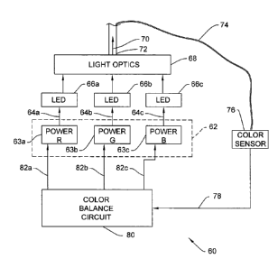
[0023] Figure 3 is a block diagram of a light source 60
including a power supply unit 62 that provides a plurality of
power outputs 64a, 64b, 64c to solid state light emitting
devices, such as light emitting diodes 66a, 66b, 66c. The
light emitting diodes 66a-66c provide light to light optics 68
which will be described in more detail below. Light optics 68
provide a collimated light output 70 to and within a hollow
light transmitting rod 72. The light output 70 is intended to
be white light.
[0024] At a distal end of the light transmitting rod 72, a
fiber optic 74 is oriented to receive a miniscule portion of
the light output 70. The fiber optic 74 provides the light
4
CA 02741734 2011-04-27
WO 2010/059197
PCT/US2009/006155
received therein to a color sensor 76 disposed in the light
source housing. The color sensor 76 provides a color output
signal 78 to a color balance circuit 80. The color balance
circuit provides color balance output signals 82a, 82b, 82c to
the power supply unit 62. The power supply unit 62 includes
individual power output circuits 63a-63c that supply power to
LEDs or LED arrays 66a-66c.
[0025] In operation, the light source embodiment
illustrated in Figure 3 provides a light output 70 to light
transmitting rod 72 that is received by a fiber optic cable .
that provides the light output to an endoscope.
[0026] In addition to the light output 70 for an endoscope,
fiber optic 74 disposed at an edge at the distal end of the
rod 72 receives a small portion of the light output 72 and
provides the light to the color sensor 76. The color sensor
76 senses the properties of the light and determines what, if
any, colors are dominant within the light output 70. For
example, if an abundance of red light is provided in the light
output 70, the condition will change the complexion or color
of an image of an object on which the light output is
reflected. Thus, color sensor 76 receives light from fiber
optic 74 and determines the intensity of color in the fiber
optic 74. Then, color sensor 76 provides color output signals
78 corresponding to combined light from the LEDs or solid
state light emitting elements 66.
[0027] Color balance circuit 80 receives the color output
signals 78 from the color sensor 76 and determines which, if
any, of the colored light emitting diodes 66a-66c needs to
output more or less light to the fiber optics 68. The color
balance circuit 80 then provides color balance output signals
82a, 82b, 82c to the power supply unit 62. Power output
circuits 63a-63c individually control the light emitting
diodes 66a-66c based on the color balance output signals 82 to
obtain, in accordance with one embodiment, a balanced white
light output 70. However, in some situations, a white light
output may not be the most ideal light color for viewing an
CA 02741734 2011-04-27
WO 2010/059197
PCT/US2009/006155
operating field. Thus, the color balance circuit 80 operates
to control the light emitting elements to provide the
predetermined desired color.
[0028] In conclusion, the light source arrangement shown in
Figure 3, which is contained within a light source housing,
operates to provide a predetermined color light output 70,
regardless of the conditions or properties of the various
individual light emitting diodes 66a-66c which provide the
light that is processed and output through the light
transmitting rod 72.
LIGHT OPTICS
[0029] Figure 4 illustrates one embodiment of the light
optics 68 provided within light source 60. The Figure 4
embodiment includes red, green and blue LEDs 66a-66c,
respectively. The light optics 68 include a plurality of
walls for containing light provided by the respective LEDs
66a-66c.
[0030] The light optics 68 include a reflector or mirror 88
disposed below the green LED 66b for reflecting green light.
The reflector 88 is oriented at approximately a 45 degree
angle to reflect the green light in a substantially transverse
horizontal direction as illustrated in Figure 4. The green
light is reflected toward a first dichroic band pass filter 90
that allows green light to pass therethrough.
[0031] In Figure 4, red LED 66a provides red light that is
directed downwardly and passes through a second angled high-
pass dichroic filter 92. The dichroic filters 90 and 92 are
glass filters with dichroic coatings. After passing through
the dichroic filter 92, the red light advances to the first
band pass filter 90 and is reflected transversely therefrom
and substantially into alignment with the green light passing
through the filter 90. Thus, the red light and the green
light travel along the same optical path.
[0032] Blue LED 66c provides light along a path transverse
to the direction of light from red LED 66a. The blue light
reflects downwardly from a surface the high-pass dichroic
6
CA 02741734 2011-04-27
WO 2010/059197
PCT/US2009/006155
filter 92 in the same direction and along the same path as the
red light. The blue light then reflects, along with the red
light, from the surface of the band pass dichroic filter 90
transversely, and in substantially the same direction as the
green light.
[0033] The combined red, blue and green light passes
through a focusing lens 94 that narrows the optic path of the
combined light and then passes through a collimating lens 96
for entry into the light transmitting rod 72.
[0034] The light transmitting rod 72 can be a glass rod
'that is adapted for connection to a proximal end 98 of a fiber
optic cable 100. Thus, the light optics 68 combine a
plurality of colors to obtain a white light output 70 for
transfer to a fiber optic cable 100. In some embodiments, the
fiber optic cable 100 includes a plurality of optical fibers
extending along the length thereof.
[0035] Figure 5 is another embodiment of the light optics
68 that differs from the embodiment shown in Figure 4. In
Figure 5, the LEDs 66a-66c are all positioned transverse to an
optical output path of the light optics 68.
[0036] Red LED 66a provides light that is reflected
transversely by an angled reflector or mirror 88. The red
light travels along an optical path and passes through an
angled high-pass filter 104. Green LED 66b provides light in
a parallel downward path that is reflected transversely by the
high-pass dichroic filter 104. The dichroic filter 104 is
oriented at approximately a 45 angle so that the red and
green light combine and travel along essentially the same
optical path.
[0037] The blue LED 66c also outputs light in a downward
direction that is reflected transversely by an angled high-
pass dichroic filter 106. The dichroic filter 106 allows the
red and green light to pass therethrough along the same
optical path as the blue light.
[0038] The red, blue and green light are combined along a
single optical path and travel to a focusing lens 94. The
7
CA 02741734 2011-04-27
WO 2010/059197
PCT/US2009/006155
focusing lens 94 focuses the combined light and directs the
light to a collimating lens 96. The collimating lens 96
orients the light in a straight direction for entry into the
receiving rod 72. As discussed above, the receiving rod 72
transfers light to the proximal end 98 of a fiber optic cable
100. The proximal end 98 of the fiber optic cable 100 inserts
into a light source housing that contains the light
transmitting rod 72. The rod 72 is oriented so that the
distal end thereof opens through a housing wall to receive the
proximal end 98 of the fiber optic cable 100.
LIGHT SOURCE CONTROLLED BY INPUTS FROM CAMERA
[0039] The block diagram of Figure 6 shows another
embodiment of the invention wherein the light source 60 is
controlled by feedback signals from a camera 110. The
proximal end 98 of the fiber optic cable 100 connects to the
light source 60 as discussed above, and a distal end of the
fiber optic cable connects to a light receiving port of an
endoscope 112. The endoscope has an optical path therein to
project the white light output received at the port outwardly
from a distal end 114 thereof. A reflected image is then
provided to an image sensor 116 of the camera 110 disposed at
a proximal end of the endoscope 112.
[0040] As will be discussed in more detail below, the
camera 110 outputs one or both of a color balance signal 118
and a shutter speed signal 120. The color signal 118 and the
shutter speed signal 120 are provided as control signals to
the light source 60. In Figure 6, an image received by the
camera 110 is also provided as an image output 122 and
displayed on a video monitor 124.
[0041] The block diagram of camera 110 illustrated in
Figure 7 is provided for the purpose of showing processing
details for providing the signals 118, 120 to the light source
60. The diagram is not intended to represent a detailed
operation of the camera 110 or structural elements of the
camera. Thus, various units 122, 130, 134, 140 shown in the
8
CA 02741734 2011-04-27
WO 2010/059197
PCT/US2009/006155
block diagram of Figure 7 may be provided as operations
conducted by a single processor.
[0042] The camera 110 is intended to be a high definition
digital camera having, for example, a 60 frames per second
imaging rate, and having the capability of adjusting the
shutter speed for the respective frames.
[0043] The image sensor 116 shown in Figure 7, senses an
image from a surgical site and provides a sensed image signal
128 to the processing units 130, 132, 134 of the camera 110.
[0044] Color sensing element 130 receives the image signal
128 and determines the white balance of the image and what, if
any, colors are detracting from the desired predetermined
color light output, which typically is white light. The color
sensing element 130 then outputs a color balance signal 118
containing the measured color information.
[0045] Image processing unit 132 also receives the image
signal 128 and provides an image output 122 to the video
monitor 124 for display thereon in a standard manner.
[0046] Light intensity sensing unit 134 also receives the
image signal 128. The light intensity sensing unit 134
determines the brightness of the image and thus the required
shutter speed for the image sensor 116. The light intensity
sensing unit 134 provides an intensity feedback signal 136 to
a shutter pulse width generator 140.
[0047] The shutter pulse width generator 140 provides a
shutter speed signal 120 to the image sensor 116 to control
the shutter speed thereof. The shutter speed is increased in
time (length of time open) when more light needs to be sensed
and the shutter speed is decreased in time when a bright light
image is input to the image sensor 116. This brightness
control operation is generally provided in digital video
cameras.
LIGHT SOURCE
[0048] Light source 60 illustrated in the block diagram of
Figure 8 cooperates with the input signals 118, 120 received
from the camera 110 (illustrated in Figure 7) as follows.
9
CA 02741734 2011-04-27
WO 2010/059197
PCT/US2009/006155
Color balance signal 118 from the camera 110 is received by a
color balance circuit 148 of the light source 60. Shutter
speed signal 120 from the camera is received by a pulse width
generator 150 of the light source 60. The pulse width
generator 150 provides output signals 151 to a light source
power unit 152. The light source power unit 152 also receives
a plurality of color balance outputs 156a-156c from the color
balance circuit 148.
[0049] The light source power unit 152 includes individual
power supply output circuits 160a, 160b, 160c that receive the
respective color balance outputs 156a-156c and includes the
pulse width generator output 151 from the pulse width
generator 150.
[0050] The power supply output circuits 160a-160c connect
to respective LEDs 66a-66c, which provide light to the light
optics 68 in a manner described above with respect to Figures
3-5. As shown in Figure 3, the light optics provide a light
output 70 to a fiber optic cable 100.
[0051] In operation, as discussed above, the camera 110
determines a color balance signal 118 and determines a shutter
speed signal 120. The signals 118, 120 are provided to the
light source 60.
[0052] As described with respect to Figure 3, the color
balance signal 118 is processed by the color balance circuit
148 to provide color balance outputs 156a-156c to the power
supply circuits 160a-160c resulting in a predetermined color
light output. The color adjustment accounts for any needed
changes in intensity of the individual colors of light
provided by LEDs 66a-66c.
MODULATION
[0053] The light output 70 of the light optics 68
illustrated in Figure 8 is modulated in accordance with the
shutter speed of the image sensor 116 of the camera. Thus,
the LEDs 66a-66c are modulated to periodically provide the
light output 70.
CA 02741734 2011-04-27
WO 2010/059197
PCT/US2009/006155
[0054] In operation, the shutter speed signal 120 is
received by pulse width generator 150 of the light source 60.
The pulse width generator 150 provides pulses 151 having a
width to control the amount of time the LEDs 66a-66c output
light during each frame of image sensor operation of the
camera 110.
[0055] For instance, if the camera 110 requires a slower
shutter speed, light must be output by the light source power
unit 152 to the LEDs 66a-66c for a longer period of time.
Thus, the feedback arrangement is balanced so that the light
output 70 of the light source enables the image sensor 116 to
operate at a predetermined shutter speed or within a
predetermined desired range of shutter speeds. The LEDs 66a-
66c must pulse in synchronism with the camera shutter speed to
provide adequate light output 70 while using less power.
[0056] In some embodiments, the predetermined range of
shutter speeds are chosen to minimize the intensity or the
time period of the light output 70 from the light source 60.
Minimizing the length of time for light output 70, while
maintaining a desired image output 122 for the camera 110,
reduces heat generated at the distal end 114 of the endoscope
112 by the passage of light from the light source 60
therethrough. Further, minimizing the intensity of the light
output 70 also reduces the amount of heat generated by the
light at the distal end 114 of the endoscope 112. Therefore,
in this arrangement with feedback control, the image sensor
116 preferably operates at the fastest acceptable shutter
speed in order to reduce the intensity and/or modulation
period of light provided to the image sensor 116.
[0057] In some embodiments, only the shutter speed signal
120 having a predetermined pulse width is provided to the
light source 60 to modulate the light output 70.
[0058] In some embodiments, only the color balance signal
118 is provided to the light source 60 for controlling the
light output from each of the LEDs 66a-66c. Finally, in
another embodiment (not shown), the light intensity feedback
11
CA 02741734 2011-04-27
WO 2010/059197
PCT/US2009/006155
signal 136 is provided to the light source 60 to control only
the intensity of light emitted therefrom.
[0059] In some embodiments the system compensates for the
target distance of an organ or tissue from the image sensor
116 at the surgical site. For example, the greater the
distance of the target from the image sensor 116, the greater
the intensity for the light output 70 to provide for optimal
viewing.
ALTERNATIVES
[0060] While the embodiments of Figures 3-8 show the LEDs
66 as three LEDs defined by a red LED 66a, a green LED 66b and
a blue LED 66c, other embodiments are contemplated. First,
rather than individual LEDs, each of the LEDs may be defined
by an array of LEDs or other solid-state devices.
[0061] Other embodiments may include cyan, magenta and
amber LEDs. Further, any combination of one or more of red,
green, blue, cyan, magenta and amber LEDs is contemplated. In
some embodiments, the light output may be generated by white
LEDs or a combination of white and red LEDs. Finally, in yet
another embodiment, a white light output 70 is generated by
blue LEDs coated with yellow phosphorous.
[0062] In some embodiments, the light transmitting rod 72
of the light source 60 has a rectangular shape for coupling to
a proximal end 98 of the fiber optic cable 100, which also has
a rectangular shape. This arrangement provides a more
efficient light transmission path between the light
transmitting rod 72, and the fiber optic cable 100, since the
LED geometry of the light source 60 is rectangular.
AUTOMATIC LIGHT SOURCE SHUT OFF
[0063] The Figure 9 embodiment of the invention includes an
arrangement to detect when the distal end of the fiber optic
cable 100 is detached from the port of the endoscope 112.
When the distal end of the fiber optic cable 100 is detached,
the light source 60 automatically shuts down to minimize the
amount of light and heat energy output by the light source 60,
and thus the amount of light/heat provided along the fiber
12
CA 02741734 2011-04-27
WO 2010/059197
PCT/US2009/006155
optic cable 100 and through the endoscope 112 to the distal
end 114 thereof. The distal end 114 of the endoscope 112 may
have a metal structure or elements that can become overheated.
[0064] The light source 60 illustrated in Figure 9 includes
a fiber optic cable disconnection detecting unit 170 for
determining when the distal end of the fiber optic cable 100
is detached from the endoscope 112. The cable disconnection
detecting unit 170 includes a laser output diode 174 and a
photodiode sensor 176. A laser driver and timing circuit 178,
periodically provides a laser diode drive output 180 to the
laser diode 174. After a laser pulse or signal is output by
the laser diode 174, the laser pulse is reflected by dichroic
filter 179 and passes through the focusing lens 94 and the
collimating lens 96 to the fiber optic cable 100. The laser
light passes along the fiber optic cable 100 to the distal end
thereof. If the distal end of the fiber optic cable 100 is
not connected to the endoscope 112, the laser pulse reflects
at the open distal end and travels back through the fiber
optic cable 100, the lenses 94, 96 and reflects off the
dichroic filter 179.
[0065] The laser pulse is then detected by the photodiode
sensor 176, which provides a laser pulse reflection signal 182
to the laser driver and timing circuit 178. The laser driver
and timing circuit 178 determines the length of time for the
laser pulse to return to the detecting unit 170 and then
provides a timing output value 186 to controller 188.
[0066] The controller 188 is programmed with the physical
length of the fiber optic cable 100 and compares the length of
time of the timing output value 186 with a time value range
corresponding to the known length for the fiber optic cable
100. If the time length signal values are within the
predetermined range for the expected reflection time, the
controller 188 outputs a disconnect or power shutdown signal
190 to the power supply 62, which turns off the power supply
so that no power output 64 is provided to the LEDs 66.
Therefore, upon disconnection of the fiber optic cable 100
13
CA 02741734 2011-04-27
WO 2010/059197
PCT/US2009/006155
from the endoscope 112, light and heat no longer are output by
the light source 60 or transmitted to the endoscope.
[0067] Although particular preferred embodiments of the
invention are disclosed in detail for illustrative purposes,
it will be recognized that variations or modifications of the
disclosed apparatus, including the rearrangement of parts, lie
within the scope of the present invention.
14