Note: Descriptions are shown in the official language in which they were submitted.
CA 02741897 2011-05-31
DIALYSIS CATHETER
BACKGROUND
Technical Field
This application relates to a catheter and more particularly to a multi-lumen
catheter
which facilitates hemodialysis.
Background of Related Art
Hemodialysis is a well known method of providing renal (kidney) function by
circulating
blood. The kidneys are organs which function to extract water and urea,
mineral salts, toxins,
and other waste products from the blood with filtering units called nephrons.
From the nephrons
the collected waste is sent to the bladder for excretion. For patients having
one or both defective
kidneys, the hemodialysis procedure is life saving because it provides a
machine to simulate the
function of the kidneys.
In the hemodialysis procedure, blood is withdrawn from the patient's body
through a
catheter or tube and transported to a dialysis machine, also commonly referred
to as a kidney
machine. The catheter is typically inserted through the jugular vein and
maneuvered into
position through the superior vena cava into the right atrium to provide high
blood flow. In the
dialysis machine, toxins and other waste products diffuse through a semi-
permeable membrane
into a dialysis fluid closely matching the chemical composition of the blood.
The filtered blood,
i.e. with the waste products removed, is then returned to the patient's body.
In some instances,
the catheter may be left in place for several years. As can be appreciated,
proper access to the
patient's blood and transport of the blood to and from the dialysis machine
for this extended
period of time is critical to hemodialysis.
One example of a dialysis catheter currently being marketed is the MedComp Ash
Split
catheter. This catheter has two lumens, one for arterial flow and the other
for venous flow,
which are each D-shaped in cross-sectional configuration. The catheter is
bifurcated at its distal
end to separate the lumens and the catheter is manually split to the desired
length for selected
separation before insertion into the target area. Another well-known catheter
is a Med Comp
catheter which has the venous flow lumen terminating proximally, i.e. axially
recessed, from the
arterial flow lumen. Each of these lumens is also D-shaped in cross-sectional
configuration.
1
CA 02741897 2011-05-31
The use of a tear away sheath in catheter insertion is well known. It would be
advantageous if a dialysis catheter could be provided which would also provide
for readily
insertion without the use of a tear away sheath in certain instances. Such
insertion method, if
utilized, can decrease the complexity of the procedure.
Another area of dialysis catheter design is to maximize venous and arterial
flow rates
while preventing collapsing the lumens. That is, it is known that the larger
lumens will
maximize flow rates, reduce arterial and venous pressure and improve dialysis
by providing
fastener dialysis. However, given the limited amount of space in the dialysis
catheter, which is
typically about 15-16 French, the size of the lumens is limited. The size is
also limited by the
fact that if the lumens are too large, the wall thickness of the catheter
becomes too thin, thereby
reducing the strength of the wall and resulting in collapse of the lumens
which has a detrimental
affect on the blood flow.
In navigating vessels to access the target site, such as the right atrium, it
is desirable to
provide the smallest catheter profile, i.e. the smallest outer diameter
catheter body. This profile
facilitates insertion through smaller vessels as it reduces the likelihood of
the catheter engaging
the wall of the vessel and reduces trauma to the vessel by minimizing
frictional contact with the
vessel wall. However, the desire for smaller diameter catheters must be
balanced against the
need for providing sufficient sized lumens to enable proper blood flow. If the
lumens are too
small, sufficient blood flow may not be able to be maintained and the blood
can be damaged
during transport. Also, a sufficient relationship must be maintained between
the size of the
lumens and the overall diameter of the catheter to maintain the structural
integrity of the catheter.
Numerous attempts have been made in the prior art to optimize the multi-lumen
configuration. In some approaches, such as disclosed in U.S. Patent Nos.
4,568,329 and
5,053,023, the inflow and outflow lumen are provided side by side in D-shaped
form. In other
approaches, such as those disclosed in U.S. Patent Nos. 4,493,696, 5,167,623
and 5,380,276 the
inflow and outflow tubes are placed in concentric relation. Other examples of
different lumen
configurations are disclosed in U.S. Patent Nos. 5,221,256, 5,364,344, and
5,451,206.
Commonly assigned U.S. Patent Nos. 6,814,718 and 7,011,645 disclose other
lumen
configurations.
The catheter lumen configuration must accommodate two competing factors:
keeping the
catheter as small as possible to facilitate insertion while keeping the lumens
as large as possible
2
CA 02741897 2011-05-31
for blood flow. This balance must be achieved while maintaining the structural
integrity of the
catheter. It would therefore be advantageous to provide a catheter which
reaches an optimum
compromise between these two competing factors.
Another important feature of dialysis catheters is the suction openings to
withdraw blood.
Keeping the suction openings clear of thrombolytic material and away from the
vessel wall is
clearly essential to dialysis function since an adequate supply of blood must
be removed from the
patient to be dialyzed.
The need therefore exists for an improved dialysis catheter which facilitates
the surgical
dialysis procedure, reduces unwanted kinking of the catheter during insertion
and use, and strikes
an optimal balance between overall catheter size and lumen size.
SUMMARY
The present invention provides in one aspect a dialysis catheter comprising an
outer wall,
a first portion having a first diameter, an elongated distal portion having a
second diameter
smaller than the first diameter, and a transition region between the first
portion and distal
portion. A first longitudinally extending substantially circular venous lumen
configured to
deliver blood is positioned off center of a central longitudinal axis of the
catheter and terminates
in a distal opening at the distalmost end of the catheter.
The catheter includes first and second independent longitudinally extending
arterial
lumens configured to withdraw blood from a patient, each terminating in an
opening in the
transition region spaced proximally of the distal opening of the venous lumen.
The venous
lumen is in a venous lumen region and has a first region, a second region and
a third region
between the first and second regions. The first and second regions are each
positioned a first
distance from the outer wall of the catheter and the third region is
positioned a second distance
from the outer wall of the catheter, the second distance being less than the
first distance to form a
first arch shaped wall portion progressively increasing in thickness from the
third region toward
the first region and from the third region toward the second region.
The first arterial lumen is in a first arterial lumen region and has a first
region, a second
region and a third region between the first and second regions. The first and
second regions of
the first arterial lumen are each positioned a third distance from the outer
wall of the catheter,
and the third region of the first arterial lumen is positioned a fourth
shorter distance from the
3
CA 02741897 2011-05-31
outer wall of the catheter to form a second arch shaped wall portion
progressively increasing in
thickness from the third region of the first arterial lumen toward the first
region of the first
arterial lumen and from the third region of the first arterial lumen toward
the second region of
the first arterial lumen.
In some embodiments, the transition region tapers toward the distal portion.
In some
embodiments, a reinforcing rib extends longitudinally from the transition
region and terminates
proximally of the distalmost end of the catheter.
In some embodiments, the second arterial lumen has a first region, a second
region and a
third region between first and second regions. The first and second regions of
the second arterial
lumen are each positioned a fifth distance from the outer wall of the
catheter, and the third region
of the second arterial lumen is positioned a sixth shorter distance from the
outer wall of the
catheter to form a third arch shaped wall portion progressively increasing in
thickness from the
third region of the second arterial lumen toward the first region of the
second arterial lumen and
from the third region of the second arterial lumen toward the second region of
the second arterial
lumen. In some embodiments, the third and fifth distances and/or the fourth
and sixth distances
are substantially equal.
A stiffening member can be removably positionable within the catheter to
temporarily
increase the stiffness of the catheter to facilitate insertion. The stiffening
member can be
removably received in the venous lumen and can have a lumen formed therein
configured to
receive a guidewire therethrough. The stiffening member can extend distally of
a distalmost end
of the catheter body when inserted therein. The stiffening member can be
secured to the catheter
by a threaded engagement.
In another aspect, the present invention provides a dialysis catheter
comprising an outer
wall, a first portion having a first diameter, an elongated distal portion
having a second diameter
smaller than the first diameter, and a transition region between the first
portion and distal
portion. A first venous lumen region has a first longitudinally extending
substantially circular
venous lumen configured to deliver blood, terminating in a distal opening at
the distalmost end
of the catheter. First and second independent longitudinally extending
arterial lumens are
configured to withdraw blood from a patient, each of the arterial lumens
terminating in an
opening in the transition region spaced proximally of the distal opening. The
first arterial lumen
has a first region, a second region and a third region between the first and
second regions. The
4
CA 02741897 2011-05-31
first and second regions are each positioned a first distance from the outer
wall of the catheter,
and the third region is positioned a second distance from the outer wall of
the catheter, wherein
the second distance is less than the first distance to form an arch shaped
wall portion in a first
arterial lumen region progressively increasing in thickness from the third
region toward the first
region and from the third region toward the second region of the first
arterial lumen.
The second arterial lumen has a first region, a second region and a third
region between
first and second regions. The first and second regions of the second arterial
lumen are each
positioned a third distance from the outer wall of the catheter, and the third
region of the second
arterial lumen is positioned a fourth shorter distance from the outer wall of
the catheter to form
an arch shaped wall portion in a second arterial lumen region progressively
increasing in
thickness from the third region of the second arterial lumen toward the first
region of the second
arterial lumen and from the third region of the second arterial lumen toward
the second region of
the second arterial lumen.
The venous lumen can be positioned off center of a longitudinal axis of the
catheter.
A stiffening member can be removably positionable within the venous lumen of
the
catheter to temporarily increase stiffness of the catheter to facilitate
insertion. The stiffening
member can have a lumen dimensioned to receive a guidewire.
In another aspect, the present invention provides a dialysis catheter for
delivering and
withdrawing blood from a patient's body comprising a catheter body defining
four quadrants in a
transverse cross-section at an intermediate portion and having first and
second arterial lumens
and a venous lumen having a substantially circular cross section. The venous
lumen is
positioned in a portion of the third and fourth quadrants with a first region
adjacent the
intersection of the four quadrants and a second region opposite the first
region. A wall thickness
of the catheter at the second region of the venous lumen has a first dimension
and increases in
thickness in a direction toward the third quadrant and in a direction toward
the fourth quadrant to
form a first arch shaped wall portion, the arch shaped wall portion thereby
having a thinner
portion at the portion aligned with a diameter bisecting the first and fourth
quadrants from the
second and third quadrants and progressively thicker portions in the third and
fourth quadrants.
The first arterial lumen is positioned in a portion of the second and third
quadrants and
the second arterial lumen is positioned in a portion of the first and fourth
quadrants, wherein a
major portion of the first arterial lumen and the center of the first arterial
lumen are positioned in
5
CA 02741897 2011-05-31
the second quadrant and a major portion of the second arterial lumen and the
center of the second
arterial lumen are positioned in the first quadrant. The catheter wall portion
forms a second arch
shaped wall portion adjacent the first arterial lumen having a first pinnacle
and first and second
portions of increasing thickness extending away from the pinnacle. The first
pinnacle preferably
lies in the second quadrant.
Preferably, the catheter wall portion forms a third arch shaped wall portion
adjacent the
second arterial lumen having a second pinnacle and first and second portions
of increasing
thickness extending away from the second pinnacle. Preferably, the second
pinnacle of third
arch shaped wall portion lies in the first quadrant.
A stiffening member can be provided removably positionable within the catheter
to
temporarily increase stiffness of the catheter to facilitate insertion.
BRIEF DESCRIPTION OF THE DRAWINGS
Preferred embodiment(s) of the present disclosure are described herein with
reference to
the drawings wherein:
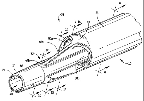
Figure 1 is a perspective view of the distal portion of the multi-lumen
catheter of the
present invention;
Figure 1A is transverse a cross-sectional view of the catheter taken along
line A-A of
Figure 1;
Figure I B is a transverse cross-sectional view of the catheter taken along
line B-B of
Figure 1;
Figure 1C is a transverse cross-sectional view of the catheter taken along
line C-C of
Figure 1;
Figure 2 is a front view of the catheter of Figure 1;
Figure 3 is a perspective view of a proximal portion of the catheter;
Figure 4 is a transverse cross-sectional view taken along line 4-4 of Figure
1;
Figure 4A is an enlarged view similar to Figure 4 showing the thicknesses of
the catheter
wall portions and the quadrants of the circle for ease of explanation;
Figure 5 is a plan view illustrating insertion of the catheter through the
right internal
jugular vein and superior vena cava into the right atrium of a patient's body;
6
CA 02741897 2011-05-31
Figure 6 is a longitudinal cross-sectional view of a dilator for use with the
insertion of the
catheter of Figure 1;
Figure 6A is a perspective view of the proximal portion of the dilator of
Figure 6;
Figure 7 is a perspective view of a tunneling trocar attachable to the
catheter of Figure 1
to aid insertion of the catheter; and
Figure 8 is a perspective view of a stiffener for use with the catheter of
Figure 1 to aid
insertion.
DETAILED DESCRIPTION OF PREFERRED EMBODIMENTS
Referring now in detail to the drawings where like reference numerals identify
similar or
like components throughout the several views, the catheter of the present
invention is designated
generally by reference numeral 10. The catheter 10 is typically inserted into
an area of high
velocity blood flow to ensure sufficient blood can be transported from the
body for dialysis.
Figure 5 illustrates the catheter 10 inserted through the right internal
jugular vein "a", into the
superior vena cava "b", and into the right atrium "c". The catheter 10 can
also be inserted for
example into the left internal jugular vein, into the superior vena cava "b"
and into the right
atrium "c". Insertion into the right atrium, from either the right or left
side provides the
necessary high blood flow to the dialysis machine. Note that the catheter body
(catheter tube) 11
is sufficiently flexible to enable it to bend to accommodate the anatomical
curves as shown.
With reference to Figures 1, 3 and 5, catheter 10 has a catheter body or
catheter tube 11
having a distal portion 31 and a proximal portion 33. Distal portion 31
terminates in elongated
nose portion 32. Proximal end portion 33 includes hub 12, where the lumens
formed within
catheter tube 11 are connected, i.e. transition, to the respective inflow and
outflow tubes 16 and
18a, 18b, respectively, to enable return and withdrawal of blood for dialysis.
Tube clamps 17
and 19 cut off blood flow through inflow and outflow tubes 16, 18a, 18b as
desired. As used
herein, the terms "inflow" and "outflow" refer to the direction of blood or
fluid flow with respect
to the catheter such that "return", "delivery" or "venous flow" refers to flow
from the dialysis
machine and delivered to the body while "intake", "withdrawal" or "arterial
flow" refers to blood
or fluids withdrawn from the body and transported to the dialysis machine.
As shown in Fig. 5, intermediate portion 35 of catheter 10 in certain uses of
the catheter
extends through subcutaneous tissue tunnel "t", and curves downwardly toward
the target site,
7
CA 02741897 2011-05-31
e.g. the right atrium. This tunnel "t" secures the catheter in place for
dialysis for a period of
weeks, or even months, with a fibrous cuff enabling tissue ingrowth. The
formation of the tunnel
"t" and the insertion of the catheter 10 therethrough will be discussed below
in conjunction with
the discussion of the catheter insertion method.
It should be appreciated that although the catheter is shown emerging from the
tissue
tunnel "t" at a second incision site, alternatively, the tissue tunnel would
not have an exit opening
at a second site but instead would exit through the same incision through
which initial access is
made by the needle and dilator into the internal jugular vein "a". This is
described in more detail
below.
Several lumens are formed in catheter tube 11 for transporting blood from the
patient's
body to a dialysis machine. As is well known in the art, a dialysis machine
essentially functions
as a kidney for patients suffering from kidney failure. Blood is removed from
the patient and
transported to the dialysis machine where toxins are removed by diffusion
through a semi-
permeable membrane into a dialysis fluid. The filtered blood is then returned
through the lumen
of the catheter body to the patient.
More specifically, and with reference to Figures 1, 2 and 4, details of the
catheter lumens
will now be described. Longitudinally extending venous lumen 40 is formed
within catheter
tube 11, extends the entire length of the tube and is designed to transport
filtered blood to the
patient. Lumen 40 extends through the reduced diameter nose portion 32 and is
preferably
substantially circular in cross section and offset from the central
longitudinal axis of the tube 11.
As shown in Figure 4A, a first (or top region as viewed in the orientation of
Figure 4A) lies at
the intersection of the four quadrants of the circular cross-section of the
catheter tube (or at the
center) and the opposite second region lies adjacent the intersection of the
third and fourth
quadrants. Lumen 40 terminates in distal opening 49 to communicate with the
patient's body so
blood can be delivered through distal opening 49. One or more side holes 48
can be provided in
the side wall of the tube 11 for blood delivery. The lumen 40 preferably has a
diameter of about
.082 inches and a cross-sectional area of about .00528 inches.
Venous lumen 40 is also configured to receive a guidewire 20 (Figure 5) to
direct the
catheter to the desired position. The guidewire 20 can be received directly
through the lumen 40
or if a stiffening member is utilized, the stiffening member would be inserted
through the lumen
8
CA 02741897 2011-05-31
40 and the guidewire would be inserted through a lumen of the stiffening
member (thereby
extending through lumen 40).
The wall of the tube forming the venous lumen 40 advantageously forms a bridge
or arch
to add to the stability of the catheter and limit kinking of the catheter 10
during insertion and use.
More specifically, venous lumen region has wall portion 13 having a thicker
area 13a and 13b
(see Figure 4A), thereby forming an arch with pinnacle at area 13c. In other
words, the thinner
portion 13c can be viewed as adjacent the intersection of the third and fourth
quadrants, aligned
with a diameter dividing the first and fourth quadrants from the second and
third quadrants.
Thus, the arch increases in thickness as it extends in opposite directions
from area 13c into the
third and fourth quadrants (containing respective areas 13a, 13b).
In an exemplary embodiment, wherein the diameter of the tube (proximal of the
nose
portion 32) is about .114 inches (tapering to about .104 inches) and the
diameter of the venous
lumen is about .082 inches, the distance dl, d2 from the edges 41a, 41b of the
lumen 40 to the
tube outer wall 14 (see tangent lines Ll and L2) is about .054 inches. This
can be compared to
the distance d3 (Figure 4) of about .021 inches taken from the outer wall tube
14 to the edge 42
of lumen 40 closest to the outer tube wall 14. Thus, the wall thickness
(distances from the lumen
outer wall to the catheter outer wall) progressively increases in both
directions from the d3
designation.
Nose 32, as noted above, can also include side venous (delivery) openings 48
formed
through the outer wall 14 wall in fluid communication with lumen 40, also
functioning to return
blood to the patient's body. Side openings or ports can be angled outwardly if
desired to
facilitate delivery of blood in the direction of blood flow and lessen
mechanical hemolysis.
These additional openings help maintain the desired flow volume by
distributing the blood
through multiple holes. In a preferred embodiment, two openings are provided
spaced 120' apart
(i.e. each spaced 60 degrees from a bottom wall as viewed in the orientation
of Figure 1 of the
catheter 10. It is also contemplated that additional or fewer openings can be
provided and the
openings can be axially displaced with respect to each other. The openings can
be equidistantly
spaced about the circumference of the outer wall 14 or asymmetrically spaced.
Additional set(s)
of openings can also be provided spaced proximally or distally from side
openings 48. The nose
32 at the catheter distal portion is elongated and has a diameter less than
the diameter of the
intermediate portion 35 of catheter 10. By way of example, in one embodiment,
the outer
9
CA 02741897 2011-05-31
diameter of the distal nose portion 32 can be about .114 inches and the outer
diameter of the
intermediate portion 35 can be about.208 inches. Clearly other dimensions are
contemplated.
The transition portion 47 provides a smooth transition between the
intermediate portion
35 and the distal portion 31 as it tapers in a distal direction. Formed in the
transition portion 47
(Figure 1) are two widened open areas 50a, 60a of arterial lumens 50, 60,
respectively, separated
by a reinforcing rib 47a extending longitudinally. Thus, the intake (arterial)
openings terminate
in longitudinally aligned openings at the transition portion 47. As shown, the
rib 47a, formed
during the manufacturing step, extends from the transition region 47
longitudinally and along the
reduced diameter nose section 42 to increase rigidity of the catheter. Rib 47a
angles toward and
into the outer wall 14 of the catheter 10, terminating proximally of venous
lumen opening 49.
The distal end 47b of the rib 47a can be radiused as it extends longitudinally
before blending into
the catheter wall 14.
The changing configuration of the catheter 10 at the transition portion 47 can
be
appreciated by comparing Figures lA-1C which show transverse cross-sectional
views along the
transition area. As shown, the wall edges 14b transition to a rounder shape in
Figure 1B as they
transition to a circular wall of Figure IC. Also, the height of the rib 47A
decreases in a distal
direction (compare height H2 to height H1).
With reference to Figures 1, 4 and 4A, catheter 10 also has a pair of
independent arterial
(withdrawal) lumens 50 and 60 extending longitudinally along the length of the
catheter body 11,
each terminating at open areas 50a, 60a in transition region 47 so that their
opening is proximal
to the opening 49 of venous lumen 40. As shown in Figures 4 and 4A, lumens 50
and 60 each
have an outer curved wall 51, 61 along the side closest to the outer side
(wall) of the tube 11.
The wall of the tube 11 forming the arterial lumen 50 and arterial lumen 60
advantageously
forms a bridge or arch to add to stability of the catheter and limit kinking
of the catheter 10
during insertion and use.
More specifically, wall portion 17 of a first arterial lumen region of the
catheter has a
thicker area 17a and 17b, thereby forming an arch with pinnacle 17c. In an
exemplary
embodiment, wherein the diameter of the tube (proximal of the nose portion 32)
is about .114
inches and the diameter of the venous lumen is about .082 inches, the
distances d4 and d5 from
the edge of the lumen 50 to the tube outer wall (see tangent lines L4, L5) is
between about .041
inches to about .042 inches. This can be compared to the distance d6 of about
.018 inches taken
CA 02741897 2011-05-31
from the outer wall 14 to the edge of the lumen 50 closest to the outer wall
14. Thus, the wall
thickness progressively increases in both directions from the d6 designation.
Lumen 60 is similarly dimensioned to lumen 50, with catheter wall portion 19
of second
arterial lumen region having a thicker area 19a and 19b, thereby forming an
arch with pinnacle
19c. In an exemplary embodiment, wherein the diameter of the tube (proximal of
the nose
portion 32) is about .114 inches and the diameter of the venous lumen is about
.082 inches, the
distance d6, d7 from the edge of the lumen 60 to the tube outer wall (see
tangent lines L6, L7) is
between about .041 inches to about .042 inches. This can be compared to the
distance d8 of
about .018 inches taken from outer wall 14 to the edge of the lumen 60 closest
to the outer tube
wall. Thus, the wall thickness progressively increases in both directions from
the d8 designation.
Note, in a preferred embodiment, distances d4, d5, d6 and d7 are substantially
equal and
distances d6 and d8 are substantially equal.
The shortest distance d9 between the two arterial lumens is preferably about
.022 inches.
Due to the radiused sides, the distance can progressively increase in both
directions away from
d9 to about .024 inches. The distance d10 and dl l between the respective
arterial lumens 50, 60
and the venous lumen 40 is preferably about .016 inches. Note that the
arterial lumen 50 lies in
the second and third quadrants, with the center of the lumen 50 and a major
portion of the lumen
50 lying in the second quadrant. Note that the arterial lumen 60 lies in the
first and fourth
quadrants, with the center of the lumen 60 and a major portion of the lumen 60
lying in the first
quadrant. Thus, the pinnacle of the arch adjacent the arterial lumen 50 lies
in the second
quadrant and the pinnacle of the arch adjacent the arterial lumen 60 lies in
the first quadrant.
As can be appreciated, the various dimensions provided herein are provided by
way of
example, it being understood that other dimensions are also contemplated to
achieve the arch
support to achieve the balance of maximized blood flow with sufficient wall
thickness to limit
catheter kinking. That is, the lumen configurations are uniquely designed to
achieve maximum
flow rates through the catheter for enabling procedures such as dialysis while
ensuring the
catheter is resistant to kinking since kinking will adversely affect blood
flow and could reduce
the long term effectiveness of the catheter which is designed for long term
use. The lumens are
also uniquely designed to provide smooth edges so as to prevent blood clotting
which could
occur with sharper edges. The arches of the catheter wall achieve this. Also,
the shape of the
venous lumen and the shape of the arterial lumens achieve this. As discussed
above, the venous
11
CA 02741897 2011-05-31
lumen is preferably substantially circular in configuration. Each of the
arterial lumens is
asymmetrically configured.
More specifically, arterial lumen 50 includes a curved outer wall 51 adjacent
the edge
closest to the catheter wall 14. Somewhat opposite curved wall 51 is curved or
concave inner
wall 52. Walls 51 and 52 are joined in quadrant III by a radiused wall 54. A
wall 58 with a
slight radius with respect to a line (see phantom line P) parallel to a
diameter C1 of the catheter
dividing the first and fourth quadrants from the second and third quadrants,
connects to inner
wall 52 via radiused wall 56. Radiused wall 58 transitions into a larger
radius wall 59 to join
with outer wall 51. Thus, this asymmetrical arterial lumen 50 has radiused
walls along all of its
sides. The arterial lumen 50 can also be considered somewhat "liver shaped."
Lumen 60 is the mirror image of lumen 50 and thus faces in the opposite
direction. Thus,
lumen 60 includes a curved outer wall 61 adjacent the edge closest to the
catheter wall 14.
Somewhat opposite curved wall 61 is curved or concave inner wall 62. Walls 61
and 62 are
joined in quadrant IV by a radiused wall 64. A wall 68 with a slight radius
with respect to line P
connects to inner wall 62 via radiused wall 66. Radiused wall 68 transitions
into a larger radius
wall 69 to join with outer wall 61. Thus, this asymmetrical arterial lumen 60
has radiused walls
along all of its sides. The arterial lumen 60 can also be considered somewhat
"liver shaped."
By way of example, walls 59 and 69 can have a radius of curvature of about
.005 inches
to about .030 inches, and preferably about .013 inches. Walls 58 and 68 can
have a radius of
curvature of about .050 inches to about .250 inches, and preferably about .150
or about .144
inches. The radius of curvature of outer walls 51 and 61 can be about .070
inches to about .050
inches, and preferably about .060 inches. The radius of curvature of walls 56
and 66 can be
about .005 inches to about .030, inches, and preferably about .018 inches.
Inner radiused walls
52 and 62 can have a radius of curvature of about .090 inches to about .130
inches, and
preferably about .114 inches. Walls 54 and 64 can have a radius of curvature
of about .005
inches to about .030 inches, and preferably about .010 inches. As can be
appreciated, other
dimensions are also contemplated.
Although lumens 50 and 60 are isolated along the length of the catheter, they
have a
common flow source flowing into the separate inflow tubes of catheter 10 via
hub 12.
As noted above, in the illustrated embodiment, the venous (return) lumen size
preferably
ranges from about .005 inches to about .006 inches2 in cross-sectional area,
and is more
12
CA 02741897 2011-05-31
preferably about .00528 inches2. The cross-sectional area of each of the
arterial (intake) lumens
50, 60 preferably ranges from about .0040 inches to about .0048 inches2, and
more preferably
about .00473 inches2, bringing the total cross-sectional area of the intake
lumens to about .0080
inches to about .0096 inches2, and more preferably about .00946 inches2. This
means that the
ratio of total cross sectional area of the return lumen to the intake lumens
is preferably about .56
to about 1Ø It should be appreciated that other dimensions are also
contemplated.
To facilitate insertion, the catheter 10 is configured to receive a stiffening
member such
as a stiffening member 80 shown in Figure 8. Stiffing member 80 has a lumen 81
extending
therethrough to receive a guidewire, e.g. guidewire 20. Stiffening member (or
rod) 80 is inserted
into circular venous lumen 40 of catheter 10 to stiffen the flexible catheter
for ease in over the
wire insertion and navigation through the small vessels. That is, the catheter
10 with stiffener 80
attached thereto and extending through venous lumen 40 is threaded over
guidewire 20
(guidewire 20 can be backloaded through the distal opening of lumen 81 of
stiffening member
80). The stiffening member 80 can have an internal thread on knob 84 at the
proximal portion to
be connected to the screw thread 15 of inflow (venous) tube 16 (see FIG. 5).
This temporarily
secures the stiffening rod within the catheter 10 during insertion. External
thread 85 is
configured for threaded attachment of a syringe (not shown) for fluid flushing
of the stiffener 80
(through lumen 81) prior to use. An example of a stiffening rod that also can
be utilized is
described in U.S. Patent No. 7,077,829.
After the catheter 10 is positioned at the desired site, the stiffening member
80 is
unthreaded from the proximal thread 15 of venous (return) tube 16 and removed
from the venous
lumen 50 of the catheter 10 and from the venous (return) tube 16.
It should be appreciated that the stiffening member 80 can alternatively be
temporarily
(removably) attached at its proximal end to the tube 16 by other means such as
a bayonet lock,
snap fit, etc. The stiffening member could first be manually twisted and then
mounted by these
various means for retention in its torqued position.
A dilator 90, illustrated in Figure 6, can also be utilized during the
insertion method. The
dilator has a lumen 92 extending therethrough which receives the guidewire,
e.g. guidewire 20.
That is, the guidewire is threaded through the distal opening 93 in the distal
end 91 of the dilator
90 and exits through proximal opening 95 at proximal end 97. The dilator 90
dissects tissue as it
13
CA 02741897 2011-05-31
is advanced into the vessel over the guidewire. A tapered distal region helps
to achieve such
dissection. The proximal end 97 of dilator 90 has an external thread 99 to
threadingly receive a
syringe for flushing the dilator 90 with fluid through lumen 92 prior to use.
Longitudinally
extending raised surfaces 98 at the proximal end facilitate gripping of the
dilator.
Tissue tunneling trocar device 100, shown in Figure 7, has a proximal portion
102 and a
distal portion 104. The trocar 100 can be in the form of a solid member.
Proximal end 105
frictionally fits within the open distal end of the catheter, and particularly
in the opening in the
venous lumen 40. Bumps 106, 108, on the outer wall of the trocar 100 enhance
the frictional
engagement. The distal portion 104 has a curved region, terminating into an
atraumatic tip 109
to bluntly dissect tissue as the catheter is inserted through the tissue
tunnel. In use, bumps 106,
108 are inserted into the venous lumen 40 of the catheter, frictionally
connecting the tunneling
trocar 100 to the catheter 10. With the trocar 100 attached, the catheter 10
is inserted through the
subcutaneous tissue tunnel with the tip 109 bluntly dissecting tissue. After
the catheter emerges
from the tissue tunnel, the trocar 100 is removed. A stiffener can then be
inserted through the
venous lumen 40 of the catheter as described above to receive the guidewire as
described above.
A venous extension tube 16 and two arterial extension tubes 18a, 18b extend
through the
hub 12 to communicate with the lumens of the catheter tube 11 as shown in
FIGS. 3 and 5.
Tubes 18a, 18b are stacked in a vertical relationship. A venous clamp 17 is
shown positioned
over the venous tube 16 and an arterial clamp 19 is shown positioned over the
two stacked
arterial tubes 18a, 18b to cut off blood flow through both tubes 18a, 18b,
preferably substantially
simultaneously. An arterial tag 19a and venous tag 17a, to provide indication
of priming volume
or other information, are shown attached to the respective arterial and venous
clamps.
Additional details of the clamps and tags, e.g. the clamp posts to limit
lateral movement of the
stacked arterial extension tubes, are disclosed in U.S. Patent Publication
2008-0312578.
The catheter can optionally include a surface treatment on the exterior and/or
the interior.
The surface treatments can include for example, a hydrophilic coating to
increase lubricity and
facilitate insertion, a drug coating such as heparin or containing IIb, IIIa
inhibitors, inert coating
substances such as Sorins carbon coating, and/or active coatings such as a
silver ion coating.
One method of insertion of the catheter of the present invention provides an
entire over
the wire system. This is achieved by the provision of a trocar such as
disclosed in U.S. Patent
Publication 2008-0312578. The trocar has a lumen formed therethrough
dimensioned for
14
CA 02741897 2011-05-31
reception of a guidewire. The blunt distal tip of the trocar bluntly dissects
tissue to create a
subcutaneous tissue tunnel for subsequent securement of the catheter.
In use in a complete over the wire insertion method, first a needle is
inserted into the
internal jugular vein to properly locate the vessel and a guidewire is
inserted through the needle
into the right internal jugular vein "a" and into the superior vena cava "b".
The guidewire is
further advanced into the right atrium "c", and preferably into the inferior
vena cava. The needle
is then withdrawn, leaving the guidewire in place, extending out of the
patient's body at the
proximal portion. A dilator such as dilator 90 of Figure 6 can be inserted
over the guidewire to
dilate tissue. Next, a trocar is inserted through a first incision in the
patient, bluntly dissecting
and tunneling under the skin, and forced out of the tissue at a second
incision or site, creating a
subcutaneous tunnel "t" under the tissue. This provides a way to secure the
catheter. The
guidewire is then threaded through the trocar lumen, with the proximal portion
first inserted
through trocar distal opening so it emerges out of a proximal opening. The
trocar is then
withdrawn from the body, leaving the guidewire in place, extending from the
right atrium and
superior vena cava, out through the right internal jugular vein and through
the tissue tunnel "t".
Catheter 10 is then threaded over the guidewire with the proximal portion of
the
guidewire inserted through the distal tip lumen of the catheter, e.g. distal
opening 49, through the
length of the venous lumen 40, and through the hub 12 into the inflow tube 16.
The catheter 10
is thus threaded over the wire, through the tissue tunnel "t" where a cuff is
positioned in the
tissue tunnel "t" to aid in securement of the catheter by enabling tissue
ingrowth over a period of
time. The catheter is further advanced over the guidewire down into the right
internal jugular
vein, into the superior vena cava, and into the right atrium "c". The
guidewire 20 is then
withdrawn, leaving the catheter 10 in place for use. Note a stiffening member
such as stiffener 80
of Figure 8, is preferably utilized, i.e. inserted into lumen 40 of catheter
10 and inserted over the
guidewire through the fitting 15, inflow tube 16, and hub 12 to help guide the
catheter as
described herein. Thus, the guidewire would extend through the venous lumen of
catheter by
extending through the central lumen of the stiffening member which is
positioned within the
venous lumen 40 of the catheter.
CA 02741897 2011-05-31
As can be appreciated, the catheter will be inserted in a similar fashion
through the left
internal jugular vein. In this method, the subcutaneous tissue tunnel will be
formed on the left
side by the trocar, and the catheter inserted over the guidewire through the
tissue tunnel and
through the left internal jugular vein or subclavian vein and into the
superior vena cava and right
atrium in the same way as described for right side insertion
In an alternative method of insertion, instead of forming a second incision
site adjacent
the incision site through which the needle and guidewire are introduced into
the internal jugular
vein, the trocar emerges from the needle/guidewire insertion site. In this
method, the trocar is
inserted through a first incision to create a subcutaneous tissue tunnel;
however, unlike the
aforementioned method, the trocar does not emerge at a second incision site.
Instead, the trocar
is advanced subcutaneously to the needle incision site and emerges through
that site. Thus, the
distal end of the trocar exits the incision site alongside the guidewire. The
guidewire is then
inserted (threaded) through the opening in the trocar as described above and
then the trocar is
withdrawn through the tissue tunnel `t" and out through the first incision
such that the guidewire
extends through the tunnel. After the guidewire 21 exits the tunnel "t" and
out through the first
incision, the trocar is removed, leaving the guidewire 20 in place. The
guidewire is positioned to
form a guidewire loop to facilitate insertion of the catheter.
The catheter 10 is then advanced over the guidewire and through the tissue
tunnel and
exiting the needle incision site into the internal jugular vein "a", tracking
the loop of the
guidewire, and then advanced downwardly through the internal jugular vein, the
superior vena
cava and into the right atrium "c". The guidewire is then withdrawn and the
catheter is pushed
downwardly and/or pulled back to straighten the loop. If the catheter is
inserted with a stiffening
member, the guidewire would extend through the lumen of the stiffening member
as the
stiffening member is positioned in the lumen of the catheter.
It should be appreciated that formation of the loop in the guidewire and the
catheter is
optional and the procedure can be performed without the loop.
In an alternative approach, a trocar is provided which does not provide for an
entire over
the wire system, however, it is used with an approach providing a partial over
the wire system
which eliminates the need for a tear way introducer sheath which often times
is utilized to guide
the dialysis catheter through the vessels into the right atrium. To avoid the
use of the tear away
sheath, the catheter in this alternate method can be advanced over a guidewire
which can be
16
CA 02741897 2011-05-31
placed in the manner described above. In this method, the trocar such as
trocar 100 of Figure 7,
is attached to the distal end of the catheter by insertion of a barbed end
into a mating fitting, or
by other ways to temporarily attach the trocar. The trocar has a blunt distal
tip and is advanced
through a first tissue incision and out through a second tissue incision,
bluntly dissecting tissue
and forming a subcutaneous tissue tunnel in a similar manner as described
above, except without
the guidewire. Since the trocar is attached to the catheter, it pulls the
catheter through the tissue
tunnel, so it emerges out through the second incision. The trocar is then
detached from the
catheter. The catheter is then bent as necessary and threaded over the
guidewire into jugular
vein, superior vena cava, and right atrium.
In another method, a curved needle is used to create access to the site. A
wire is passed
through the needle to the site, the needle is then removed, and a dilator,
such as dilator 90 of
Figure 6, is inserted over the wire. After dilating the tissue, the dilator is
removed and the
catheter 10 with a stiffening member positioned therein, such as stiffening
member 80 of Figure
8, are inserted over the guidewire into the right atrium.
It should be appreciated that although the catheter is described herein as a
dialysis
catheter for hemodialysis, the catheter disclosed herein could have other
surgical applications,
such as drug delivery or blood sampling.
While the above description contains many specifics, those specifics should
not be
construed as limitations on the scope of the disclosure, but merely as
exemplifications of
preferred embodiments thereof. Those skilled in the art will envision many
other possible
variations that are within the scope and spirit of the disclosure as defined
by the claims appended
hereto.
17