Note: Descriptions are shown in the official language in which they were submitted.
CA 02741959 2011-04-28
WO 2010/049156 PCT/EP2009/007754
Sequencing of nucleic acid molecules by mass spectrometry
The present invention is related to methods for analyzing and/or determining
the nucleotide
sequence of nucleic acid molecules.
Background of the invention
Nucleic acid molecules are used as diagnostic tools and/or as therapeutics,
whereby the
nucleic acid molecules can be identified by screening of partly or fully
randomized nucleic
acid molecule libraries, or predicted according to complementary sequences
aided by
computer algorithms such as done for antisense, siRNA and miRNA. Nucleic acid
molecules
can be single or double-stranded molecules, they can be structured or not,
they can be
conjugated to peptides, proteins, polysaccharides and other larger molecules,
their sugar
backbone can consists of ribose, deoxyribose and/or modified derivatives
thereof.
The function of the nucleic acid molecules can be based on
a) sequence-specific hybridisation to and switch-off of mRNA in form of
antisense
nucleic acid molecules, catalytically nucleic acid molecules, siRNA molecules
and
micro RNA molecules (Couzin, 2004; Crooke, 2004; Hannon, 2002; Juliano et al,
2008; Scherer & Rossi, 2003; Schlosser et al, 2006; Usman & Blatt, 2000;
Weigand et
al, 2006; Zhang & Farwell, 2008);
b) or binding of nucleic acid molecules to a target molecule and/or blockage
of the
function of the target molecule by nucleic acid molecules, whereby the nucleic
acid
molecules comprise aptamers, Spiegelmers and decoy nucleic acids (Carothers &
Szostak, 2006; Cload et al, 2006; Eulberg et al, 2006; Mann & Dzau, 2000;
Nimjee et
al, 2006; Realini et al, 2006);
c) or their stimulatory effect on the immune systems, e.g. in form of CpG-DNA
(Weiner,
2000) and random oligonucleotides .
CA 02741959 2011-04-28
WO 2010/049156 PCT/EP2009/007754
2
The production, development and use of such nucleic acid molecules as
diagnostic tools and/
or as therapeutics necessitates a method for verification of the identity of
the nucleic acid
molecules, whereby there is a need for a sensitive, accurate and reproducible
analysis.
Verification of identity requires determination of the molecular mass and
length of the nucleic
acid molecules as well its base composition, sequence, and the identity of the
sugar moieties
and internucleotide linkages. The methods used should be specific, allowing
identification of
modified bases and modified sugar moieties, addition or deletion products, and
depurination
products. The determination of the nucleotide sequence must be complete, and
it must be
shown that any chemical or enzymatic manipulations do not adversely affect
either the bases
or the backbone. Characterisation of modified nucleic acid molecules is
particular challenging
if they are nuclease stable. Such nuclease-stable nucleic acid molecules are
modified at the 2'-
position of the sugar backbone or consist of mirror-image nucleotides.
However, a number of
techniques have been developed, including, inter alia, electrophoresis,
enzymatic and
chemical analysis, array technology and mass spectrometry, to determine the
nucleotide
sequence of nucleic acid molecules.
Nucleotide sequence determination of nucleic acids by enzymatic and chemical
analysis
In the 1970s three techniques of nucleic acid molecule sequencing were
developed, that are
common and relatively rapid procedures practiced in many laboratories.
DNA sequencing method by Maxam and Gilbert. The method described by Maxam and
Gilbert describes a process whereby terminally labeled DNA molecules are
chemically
cleaved in a nucleobase-specific manner. Each base position in the nucleic
acid molecule
sequence is then determined from the molecular weights of the fragments
produced by
nucleobase-specific cleavage. Individual reactions were devised to cleave
preferentially at
guanine, at adenine, at cytosine and thymine, and at cytosine alone. When the
products of
these four reactions are resolved by molecular weight via the increasing
negative charge of the
increasing fragment size, using, for example, polyacrylamide gel
electrophoresis, sequences of
CA 02741959 2011-04-28
WO 2010/049156 PCT/EP2009/007754
3
the DNA molecule can be read from the pattern of fragments on the resolved gel
(Maxam &
Gilbert, 1977).
DNA sequencing method by Sanger. The other method - developed by Sanger et al.
- takes
advantage of the chain terminating ability of dideoxynucleoside triphosphates
(abbr. ddNTPs)
and the ability of the DNA polymerase to incorporate ddNTPs with nearly equal
fidelity as the
natural substrate of the DNA polymerase, deoxynucleoside triphosphates (abbr.
dNTPs).
Briefly, a primer molecule, usually an oligonucleotide molecule, and a
template DNA
molecule are incubated in the presence of a useful concentration of all four
dNTPs plus a
limited amount of a single ddNTP. The DNA polymerase occasionally incorporates
in the
growing, amplified strand a dideoxynucleotide that terminates chain extension.
Because the
dideoxynucleotide has no 3'-hydroxyl, the initiation point for the polymerase
enzyme is lost.
Polymerization produces a mixture of nucleic acid molecule fragments of varied
sizes, all
having identical 5'-termini. Fractionation of the mixture by, for example,
polyacrylamide gel
electrophoresis, produces a pattern that indicates the presence and position
of each nucleotide
in the nucleic acid molecule. Reactions with each of the four ddNTPs permits
the nucleic acid
molecule sequence to be read from a resolved gel (Sanger et al, 1977) in a
similar way as done
using the technique developed by Maxam and Gilbert (Maxam & Gilbert, 1977).
RNA sequencing method by Peattie. Due to the different chemical properties of
RNA
molecules and greater lability of RNA molecules in comparison to DNA molecules
the
chemical method of Maxam and Gilbert is not applicable for RNA molecules.
Peattie
developed a chemical method of sequencing RNA molecules, whereby the RNA
molecules
are 3'-radiolabelled and chemically cleaved in a nucleobase-specific manner.
Each nucleotide
position in the nucleic acid sequence of the nucleic acid molecules is then
determined from
the molecular weights of the nucleic acid molecule fragments produced by
nucleobase-
specific cleavage. Individual reactions were devised to cleave preferentially
at guanine, at
adenine or guanine, at cytosine and uracil, and at uracil alone. When the
products of these four
reactions are resolved by molecular weight, using, for example, mobility
differentiation via
CA 02741959 2011-04-28
WO 2010/049156 PCT/EP2009/007754
4
polyacrylamide gel electrophoresis, the sequence of the RNA molecule can be
read from the
pattern of fragments on the resolved gel (Peattie, 1979).
RNA sequencing method based on Sanger method. The most common method for
identification of the sequence of an RNA molecule is the method by Sanger as
described
supra. In the case of RNA molecules, the dideoxy chain termination reaction is
catalyzed by
reverse transcriptase that reads the RNA molecule template and inserts the
complementary
deoxynucleotide. As with the polymerases used in DNA sequencing, the reverse
transcription
reaction is inhibited by dideoxynucleotides (Zimmern & Kaesberg, 1978).
RNA fingerprinting. In the RNA fingerprinting approach the RNA molecule is
digested
separately with two or more endonucleases, whereby the endonucleases cleave
specifically.
The resulting fragments of the RNA molecules from each cleavage reaction are
separated by
charge (first dimension) and by length (second dimension). The separation by
charge is done
by the use of high-voltage electrophoresis on cellulose-acetate strips.
Afterwards the RNA
molecule fragments are transferred to DEAE cellulose paper for separation in
the second
dimension. The sequence is determined by overlapping the chromatographically
resolved
fragments from the separate enzymatic digestion reaction (Branch et al, 1989).
Based on and/or with the regard to the methods of Sanger, Maxam and Gilbert,
and Peattie
(Maxam & Gilbert, 1977; Peattie, 1979; Sanger et al, 1977), several
improvements and/or
modifications of the procesess were developed: Fluorescence-labeling instead
of radioactive
labeling, post-labeling techniques, enzymatic cleavage instead of chemical
cleavage, step-wise
wandering spot method, alternative cleavage reactions for RNA and DNA (Donis-
Keller et al,
1977; Gupta et al, 1976; Gupta & Randerath, 1977; Lockard et al, 1978;
Proudnikov &
Mirzabekov, 1996; Stanley & Vassilenko, 1978; Tanaka et al, 1980; Waldmann et
al, 1987;
Wu et al, 1996).
However, each technique has inherent limitations. For example, Maxam and
Gilbert (Maxam
& Gilbert, 1977) and Peattie (Peattie, 1979) disclose a chemical degradation
approach and
Sanger et al. (Sanger et al, 1977) disclose a chain termination method using
complementary
CA 02741959 2011-04-28
WO 2010/049156 PCT/EP2009/007754
strand primer extension. Each of these techniques utilizes four separate
reaction mixtures to
create a nested set of fragments differing by a single nucleotide in length,
thus representing a
complete nucleotide sequence. A resolution of the fragments based on their
size and
terminating nucleotide is carried out by polyacrylamide gel eletrophoresis to
determine the
order of the fragments and hence the nucleotide sequence of the nucleic acid
molecule. The
casting of gels and the electrophoretic separation of nucleic acid molecules
are time-
comsuming operations. The use of gel electrophoresis to determine the sequence
of the
nucleic acid molecule is a potential source of error due to band compression
effects, where
adjacent fragments of the nucleic acid molecules are unresolved, and the
identification of each
individual strand is based on the measurement of a relative value, i.e.
migration time. A
potential source of error is, for instance, the structure of the nucleic acid
molecule and the
fragments thereof. For instance, the RNA fingerprinting approach which uses
Thin Layer
Chromatography (abbr. TLC) is inappropriate for the characterisation of
unknown (modified)
structures (Limbach, 1996).
Hence, sequence determination of nucleic acid molecules by mass spectrometry
was a
promising approach to overcome these limitations (Limbach, 1996).
Nucleotide sequence determination of nucleic acids by mass spectrometry
Mass spectrometry (abbr. MS) is a powerful tool for analyzing the molecular
mass of
compounds. With regard to nucleic acid molecule analysis, MS is applicable for
nucleic acid
molecule sequencing, nucleic acid molecule modification detection and
determination of
nucleic acid molecule fragments. Analysis of nucleic acids by MS is primarily
limited by
ionization efficiency and by the resolving power of several applicable
detection methods.
Only charged molecules can be analyzed by a mass detector. Therefore, the
molecules to be
analyzed need to be efficiently ionized before they are introduced to a mass
analyzer. For
efficient ionization of nucleic acid molecules prior to mass analysis the
following techniques
are commonly used: electrospray ionization (abbr. ESI) (Fenn et al, 1989) and
matrix-assisted
laser desorption/ionization (abbr. MALDI) (Karas & Hillenkamp, 1988). ESI is
the conversion
of molecules or ions in solution into ions in the vapour phase, principally
through the
CA 02741959 2011-04-28
WO 2010/049156 PCT/EP2009/007754
6
vaporization of charged droplets of the solution. ESI can produce a
distribution of multiple
charged ions having a mass-to-charge ratio within the linear range of
commercially available
mass analyzers. Although a mixture of compounds present in a solution can be
directly
analyzed by ESI-MS, this procedure can suffer for example, from complex
spectra because of
multiple charging of the different compounds, competition of excess charge and
interference
by salt adducts. Therefore, electrospray ionization is often directly coupled
down-stream to a
separation mechanism. This procedure promotes efficient ionization, when
various critical
parameters such as flow-rates, ionization mode, buffers and solvent additives
are optimised.
Although ESI-MS is sensitive, requiring only femtomole quantities of sample,
it relies on
multiple charges to achieve efficient ionization and produces complex and
difficult-to-
interpret multiply-charged spectra for even simple nucleic acid molecules.
Therefore, in
practice, the application of ESI-MS relies on the availability of software
packages enabling
"deconvolution" of the data. Deconvolution involes the use of an algorithm-
based calculation
process to determine the uncharged (neutral) mass of the molecule from the
multiple-charge
mass-spectral data.
Matrix-assisted laser desorption ionization (abbr. MALDI) used e.g. in
conjunction with a
time-of-flight (abbr. TOF) mass analyzer has a great potential for sequencing
nucleic acid
molecules because of its relatively broad mass range and high sampling rate.
For routine
analysis of biomolecules of large mass like nucleic acid molecules, MALDI-MS
is commonly
preferred in comparison to ESI-MS because the biomolecules of large mass can
be ionized
and analyzed readily. In addition, MALDI-MS produces predominantly singly
charged
species, which greatly simplifies the interpretation of spectra, especially
those containing
mixtures of oligonucleotides.
However, in general, MALDI-MS analysis of nucleic acid molecules may suffer
from a lack
of resolution of high molecular weight nucleic acid molecule fragments,
nucleic acid
instability, and interference from sample preparation reagents. Longer nucleic
acid molecules
can give broader, less-intense signals, because MALDI imparts greater kinetic
energies to ions
of higher molecular weights. Although it may be used to analyze high molecular-
weight
nucleic acids, MALDI-MS can induce cleavage of the nucleic acid molecules'
backbone,
which further complicates the resulting spectrum. Although MALDI is less
sensitive to ion
CA 02741959 2011-04-28
WO 2010/049156 PCT/EP2009/007754
7
suppression than ESI, ion suppression is still an issue for MALDI analysis,
and necessitates
the use of sample clean-up strategies, and/or chromatographic separation.
However, MALDI
is not readily amenable to direct coupling with solution-based techniques, and
is typically
operated in the off-line or in the at-line mode.
Direct mass spectrometric methods for sequencing
Any mass spectrometric approach that does not depend upon an external reaction
to generate
sequence-specific ions is considered as direct method of sequencing. TAs
mentioned supra,
ESI and MALDI are the ionization methods of choice for nucleic acid molecules
(Limbach,
1996). A detailed overview of the methods is given by Limbach and Nordhoff et
al. (Limbach,
1996; Nordhoff et al, 1996).
Desorption/Ionization-Induced Fragmentation. Dissociation of nucleic acid
molecules can
occur as a result of the excess energy that is imparted to the nucleic acid
molecules during
desorption/ionization process. This dissociation occurs on relatively fast
time scales, resulting
in ions that are generally difficult to identify accurately. ESI mostly
produces stable, intact
molecular ions. Most dissociations that are desorption/ionization-induced are
seen in MALDI,
whereby in MALDI-TOF-MS exist four differing time-scales for
desorption/ionization-
induced dissociations: prompt, fast, fast metastable and metastable. In
theory, dissociations
occuring during any one of these time scales will generate nucleic acid
molecule fragment
ions that could be used to determine the sequence of the nucleic acid
molecule. In practice, the
analyst has little control over the extent of fragmentation. Most of these
fragments result in a
broadening of the molecular ion peak resulting in a loss of resolution and
sensitivity
(Limbach, 1996).
Tandem mass spectrometry. MS-MS (also called tandem mass spectrometry)
involves the
measurement of the mass-to-charge rations (m/z) of ions before and after a
chemical reaction
that occurs within a mass spectrometer whereby a change in m/z is involved
(Baker et al,
1993; Boschenok & Sheil, 1996; Kawase et al, 1991; Limbach et al, 1995; Little
et al, 1995;
CA 02741959 2011-04-28
WO 2010/049156 PCT/EP2009/007754
8
Marzilli et al, 1999; Ni et al, 1996; Wu et al, 1998b, W.M.A. Niessen, 2002).
Before the
chemical reaction, a m/z value is selected in the first stage of the mass
spectrometer (this ion is
called the precursor or parent ion). Then the chemical reaction takes place,
which generally
involves collision with neutral gas molecules (a process called collision-
induced dissociation
or CID). Mostly, Helium or Argon are used as collision gas. This reaction may
take place in
an intermediate zone (collision cell) between the two mass stages of the mass
spectrometer.
By this reaction, decomposition of the precursor ion may yield in various
product ions (these
are called daughter or product ions). The charged fragments can then be
dectected by the
second stage of the mass spectrometer. MS-MS can be done in two modes:
Firstly, MS-MS
"in space", i.e., the two mass analyzers can be separated in space, e.g. by a
QTOF (quadrupole
- time of flight) instrument. Second, MS-MS "in time", i.e., the different
steps in the process
can take place in the same space, but separated in time, e.g. in an ion-trap
instrument. An
accurate description of CID processes has been described by W.M.A. Niessen
(2006).
The applicability of tandem mass spectrometry for sequence identification of
nucleic acid
molecules can be looked up in the review articles of Limbach and Nordhoff et
al. (Limbach,
1996; Nordhoff et al, 1996). CID is the most widely applied method to induce
fragmentation
in MS-MS. Based on the dissociation of the multiply charged anionic
nucleotides, the method
utilizes the concept of "bidirectional" sequencing from both termini under the
assumption that
the backbone of the oligonucleotide is dissociated sequentially along the
chain. The resulting
fragments respresent, when applied successfully, a sequence specific
fragmentation pattern.
One of the first reports on the fragmentation of RNA has been given by Cerny
et al. (1987).
The "bidirectional" concept according to present knowledge utilizes c series
ions which
construct a sequence from 5' 4 3' direction and y series ions constructing a
sequence from
the 3' - 5' direction (Schiirch et. al, 2002). Nevertheless other daughter
ions can be formed
that may complicate, support or enable the sequencing process. Due to the fact
that
fragmentation can occur at the phosphate, the sugar and at the base site, the
interpretation of
the resulting spectra is complicated and the method is limited to nucleic
acids with less than
25 nucleotides (Alazard et al, 2002). This limitation can be attributed to
various factors such
as neutral loss (daughter ions that are not ionized can not be detected),
detection limit issues
or limited resolution of the detector. The collision energy also plays a
critical role. Low
CA 02741959 2011-04-28
WO 2010/049156 PCT/EP2009/007754
9
collision energies produce fewer sequence related ions while higher collision
energies may
result in other ion series which complicate the data interpretation. In
contrast to the CID of
DNA, which has been investigated thoroughly within the last few years, the
aspects of CID
with RNA are still not fully resolved.
Because of the limitations of the "direct methods" for sequencing of nucleic
acid molecules by
MS, the following indirect methods have been developed and utilized to
determine the
sequence of nucleic acid molecules by mass spectrometry.
Indirect mass spectrometric methods for sequencing
"Indirect mass spectrometric methods" for sequencing as preferably used herein
means that
the preparation of the nucleic acid molecules, from which the sequence should
be determined,
is performed before gas-phase ions of the sample are generated.
The indirect mass spectrometric methods for mass measurement as a tool to
confirm a
predicted nucleic acid molecule composition are not discussed herein. Further
information is
provided in the review of Limbach (Limbach, 1996).
The utility of any mass spectrometric sequencing method that relies on
consecutive backbone
cleavage depends on the formation of a mass ladder. The sequence information
is obtained by
determining the mass difference between successive peaks in the mass spectrum.
In the case
of oligodeoxynucleotides, the expected mass difference between successive
peaks will
correspond to the loss of: dC = 289.05, dT = 304.05, dA = 313.06, and dG =
329.05 (Exact
massbased values). With oligoribonucleotides, the mass difference will be: C =
305.04, U =
306.03, A = 329.05, and dG = 345.05 (Exact mass-based values). Because the
nucleic acid
sequence determination methods rely on the mass measurements of successive n-
mers, DNA
molecules are easier to characterize than RNA molecules due to the relatively
large
differences in mass among the four DNA molecule residues. Due to the small
mass difference
between the ribonucleotide U and C of only one Dalton unit, the required
accuracy for
measurement is much higher to correctly distinguish between U and C. Mass
ladder methods
have one distinct advantage for sequence determination: the difference in two
mass
CA 02741959 2011-04-28
WO 2010/049156 PCT/EP2009/007754
measurements that results in the desired information gives the identy of the
nucleotide residue
(Limbach, 1996).
Analysis of nucleic acid molecule ladders after nuclease digestion. The DNA or
RNA
molecule fragments are generated by hydrolysis of the nucleotides using a 5'--
>3'
phophosdiesterase and/or a 3' --> 5' phosphodiesterase. Normally a combination
of the two is
used to identify all the nucleotides. The truncated and/or cleaved nucleic
acid molecules are
analyzed by MALDI-TOF-MS or ESI-MS. Enhanced resolution to up to 35
nucleotides was
achieved (Alazard et al, 2002) by improved techniques such as delayed
extraction, sample
cleanup, optimisation of enzyme, buffer pH and matrices (Bentzley et al, 1998;
Bentzley et al,
1996; Faulstich et al, 1997; Glover et al, 1995; Kirpekar et al, 1994; Owens
et al, 1998; Pieles
et al, 1993; Schuette et al, 1995; Smirnov et al, 1996; Wu & Aboleneen, 2001;
Wu et al,
1998a).
However, enzymatic sequencing is restricted to nucleic acid molecules that
comprise no
modification of their sugar backbone. Moreover some nucleases are single-
strand specific.
Some nucleic acid molecules especially long oligonucleotides such as aptamers
exhibit
double-stranded sequence sections leading to intra- and/or intermolecular
structures which are
poorer substrates for nuclease digestions.
Analysis of nucleic acid molecule ladders after chemical digestion. Beside
exonucleases,
chemical agents can be used for the controlled degradation of the nucleic acid
molecules
before mass spectrometric measurement. Chemical agents are especially needed
if the nucleic
acid molecule is modified, whereby the modification is specifically chosen in
order to increase
the stability of the nucleic acid molecules towards enzymatic digestion.
Comparable with
enzymatic digestion methods, chemical cleavage reactions are classified by
their specificity
for DNA and RNA molecules and their specificity for the different nucleobases.
Base specific
reactions for RNA and DNA molecules, that can be used before MS analysis, are
described by
Peattie and Maxam-Gilbert (Maxam &Gilbert, 1977; Peattie, 1979). However, non-
specific
CA 02741959 2011-04-28
WO 2010/049156 PCT/EP2009/007754
11
(random) cleavage of the phosphodiester backbone of DNA molecules is done by
acid
hydrolysis (Shapiro & Danzig, 1972); non-specific (random) cleavage of the
phosphodiester
backbone of RNA molecules is done by base hydrolysis, with acid (e.g. formic
acid), (Farand
& Beverly, 2008) and polyamines at physiological pH (Komiyama & Yoshinari,
1997)
The generation of a mass ladder of a nucleic acid molecule for sequence
determination using
non-specific (random) cleavage of the phosphodiester backbone of DNA or RNA
molecules
can be complicated because any linkage site can be potentially cleaved by the
chemcial agent.
The nucleobase specfic chemical cleavage can also randomly occur at every
position in the
nucleic acid molecule where the respective nucleobase is. If a single cleavage
site is
generated, then two specfic fragments occur: one from 5'-terminus and one from
the 3'-
terminus. If both fragments can be detected in the mass spectrum, more
information is present
than is needed for sequence determination of the nucleic acid molecule. These
two ion series
can be a source of confusion. The other source of confusion comes from the
internal
cleavages. As noted before, a single cleavage along the backbone of the
nucleic acid molecule
generates two fragments - one fragment originates from the 5'-terminus, and
the other
fragment originates from the 3'-terminus. One more cleavage reaction along the
backbone of
the nucleic acid molecule generates three fragments: the first fragment is the
5'-terminus, the
second fragment is the 3'-terminus and the third fragment will not comprise
either terminus.
Because the 5'- or 3'- terminus is used as a reference point, the fragments
comprising the 5'-
or 3'-terminus can be used for the construction of the mass ladder. In
contrast the internal
fragment can not be used for the construction of the mass ladder.
Additionally, in the case of
mass identity of the internal fragment and one of the terminal fragments, an
incorrect
interpretation may result. Furthermore, the presence of these internal
fragments can lead to ion
suppression of the desired 5'- or 3'-terminus fragments. Therefore the
reaction conditions for
the chemical digestion have to be carefully adjusted to single cleavage
conditions (Limbach,
1996) although with random cleavage reactions, the ability to control this is
limited.
Nucleic acid sequencing can be done by chemical cleavage reactions followed by
analysis of
the cleavage reactions via mass spectrometry (Farand & Beverly, 2008). Farrand
and Berverly
used a highly modified nucleic acid molecule containing a mixture of
2'deoxyribonucleotides,
2'-fluororibonucleotides, 2'-O-methylribonucleotides, abasic ribonucleotides
and
CA 02741959 2011-04-28
WO 2010/049156 PCT/EP2009/007754
12
ribonucleotides, whereby formic acid was used to degrade ribonucleotides;
sodium hydroxide
was used to degrade ribonucleotides, 2'-fluoro ribonucleotides, 2'-O-methyl
ribonucleotides
and abasic residues; piperidine was used for ribonucleotides, 2'-fluoro
ribonucleotides and
deoxy-guanosine. Base specific reactions (as reported by Peattie Maxam &
Gilbert) were also
used to obtain fragments. During accurate mass analysis, short fragments (1-3
nucleotides in
length) containing the last nucleotides of the strand are poorly retained by
LC-MS. Therefore,
tandem mass spectrometry was needed to confirm the final two or three
nucleotides
containing the 3'-terminal hydroxyl (Farand & Beverly, 2008).
Analysis of nucleic acid molecule ladders after after endouclease digestion
and chemical
digestion. The small mass difference between U and C (one Da) in an RNA
molecule makes
unambiguous (as shown for DNA molecules) assignment difficult using partial
exonuclease
digestion followed by MALDI-TOF. Exonuclease digestion results in ambiguous
sequence
assignments where the pyrimidine bases C and U can not distinguished from each
other.
Therefore Tolson and Nicholson develeoped a method combining sequence specific
endonucleases and chemical methods to resolve these sequence ambiguities of
RNA
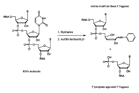
molecules (Tolson & Nicholson, 1998). Because the specificity of the enzymatic
reactions
was not as expected, the authors used hydrazine/analine treatment of RNA
resulting
characteristic fragments formed by the scission at U's. (Tolson & Nicholson,
1998).
Analysis of nucleic acid molecule ladders after Sanger dideoxy termination
reactions. The
Sanger sequencing strategy allows assembling the sequence information by
analysis of the
nested fragments obtained by nucleobase-specific chain termination via their
different
molecular masses using mass spectrometry such as MALDI or ESI mass
spectrometry. The
method was improved by increasing amounts of termination groups using cycle
sequencing,
optimizing reaction conditions, purifying extension products, elimination salt
adducts and
utilizing delayed extraction technology for better resolution (Fu et al, 1998;
Harksen et al,
1999; Kirpekar et al, 1998; Koster et al, 1996; Monforte & Becker, 1997;
Mouradian et al,
1996; Roskey et al, 1996; Shaler et al, 1995; Taranenko et al, 1998; Taranenko
et al, 1997).
CA 02741959 2011-04-28
WO 2010/049156 PCT/EP2009/007754
13
Using this MALDI-TOF sequencing method, sequences of DNA molecules consisting
of more
than 100 nucleotides could be analysed (Taranenko et al, 1998).
Alternatively, as shown in US 5,547,835 the one of the methods as described
supra has been
coupled with a solid-phase sequencing approach in which the template is
labeled with biotin
and bound to streptavidin-coated magnetic beads. Throughput can be increased
by introducing
mass modifications in the oligonucleotide primer, chain-terminating nucleoside
triphosphates
and/or in the chain- elongating nucleoside triphosphates, as well as using
integrated tag
sequences that allow multiplexing by hybridization of tag specific probes with
mass
differentiated molecular weights. However, all of these "Sanger-based"
sequencing methods
require either some prior knowledge of the target sequence or introduction of
a known
sequence to serve as the primer-binding site.
The problem underlying the present invention was thus to provide a method for
determining
the nucleotide sequence of a nucleic acid molecule, particularly in case
nucleic acid molecule
comprises or consists of L-nucleotides.
A further problem underlying the present inventoin was to provide a method for
determining
the nucleotide sequence of a nucleic acid molecule, particularly of a nucleic
acid molecule
comprising or consisting of L-nucleotides, whereby such method overcomes or
avoids at least
some of the disadvantages of the methods of the prior art.
This problem is solved by the subject matter of the independent claims.
Preferred
embodiments may be takne form the dependent claims.
The problem underlying the present invention is solved in a first aspect,
which is also the first
embodiment of the first aspect by a method for determining the nucleotide
sequence of a
nucleic acid molecule comprising the following steps:
a) providing a plurality of molecules of the nucleic acid molecule having at
least
one modification;
CA 02741959 2011-04-28
WO 2010/049156 PCT/EP2009/007754
14
b) cleaving at random the plurality of modified nucleic acid molecules thus
providing modified nucleic acid molecule fragments and non-modified nucleic
acid molecule fragments;
c) separating the modified nucleic acid molecule fragments from the non-
modified nucleic acid molecule fragments;
d) separating or resolving the modified nucleic acid molecule fragments
according
to their length, mass and/or charge, whereby such separating or resolving
generates a pattern of modified nucleic acid fragments; and
e) optionally visualizing the pattern of modified nucleic acid fragments.
In a second embodiment of the first aspect which is also an embodiment of the
first
embodiment of the first aspect the method further comprises the step of
f) deducing from the pattern of modified nucleic acid fragments the nucleotide
sequence of the nucleic acid molecule.
In a third embodiment of the first aspect which is also an embodiment of the
first and second
embodiment of the first aspect the the individual nucleic acid molecule of the
plurality of
molecules has at least one modification at the 5' end, at the 3' end or within
the nucleotide
sequence of the nucleic acid molecule the nucleotide sequence of which is to
be determined.
In a fourth embodiment of the first aspect which is also an embodiment of the
first, second
and third embodiment of the first aspect the the cleaving is carried out by
chemical cleaving,
enzymatic cleaving, cleaving by heat and/or cleaving by use of a divalent
cation.
In a fifth embodiment of the first aspect which is also an embodiment of the
first, second,
third and fourth embodiment of the first aspect the the cleaving is a chemical
cleaving,
preferably a nucleotide unspecific cleaving.
CA 02741959 2011-04-28
WO 2010/049156 PCT/EP2009/007754
In a sixth embodiment of the first aspect which is also an embodiment of the
first, second,
third, fourth and fifth embodiment of the first aspect the cleaving is a
limited cleaving.
In a seventh embodiment of the first aspect which is also an embodiment of the
first, second,
third, fourth, fifth and sixth embodiment of the first aspect cleaving is a
limited random
cleaving, preferably a limited chemical random cleaving.
In an eighth embodiment of the first aspect which is also an embodiment of the
first, second,
third, fourth, fifth, sixth and seventh embodiment of the first aspect the
step of cleaving
provides for a mixture of fragments, preferably modified fragments, whereby
such mixture of
fragments comprises all possible nucleotide sequence fragments of the nucleic
acid molecule.
In a ninth embodiment of the first aspect which is also an embodiment of the
eighth
embodiment of the first aspect the the mixture comprises a modified full
length form of the
nucleic acid molecule the nucleotide sequence of which is to be determined.
In a tenth embodiment of the first aspect which is also an embodiment of the
first, second,
third, fourth, fifth, sixth, seventh, eights and ninth embodiment of the first
aspect the modified
nucleic acid molecule fragments are separated from the non-modified nucleic
acid molecule
fragments through the interaction of the modification with an interaction
partner, whereby
such interaction partner is linked to a support.
In an eleventh embodiment of the first aspect which is also an embodiment of
the tenth
embodiment of the first aspect the support is a solid support.
Ina 12th embodiment of the first aspect which is also an embodiment of the
tenth and eleventh
embodiment of the first aspect the non-modified nucleic acid molecule
fragments are removed
from the modified nucleic acid molecule fragments interacting with the
interaction partner,
preferably by washing.
CA 02741959 2011-04-28
WO 2010/049156 PCT/EP2009/007754
16
In a 13th embodiment of the first aspect which is also an embodiment of the
tenth, eleventh
and 12th embodiment of the first aspect the modified nucleic acid molecule
fragments are
released from the support, preferably by release of the modification from the
interaction
partner, by release from the interaction partner from the support or by
cleaving the
modification or a part or moiety thereof from the nucleic acid molecule
fragments.
In a 14th embodiment of the first aspect which is also an embodiment of the
first, second,
third, fourth, fifth, sixth, seventh, eighth and ninth embodiment of the first
aspect the modified
nucleic acid molecule fragments are separated from the non-modified nucleic
acid molecule
by separation due to mass discrimination, size discrimination, hydrophobicity
discrimination,
charge discrimination, ionic discrimination, hydrogen bonding discrimination
and or liquid
phase mediated extraction, whereby preferably the non-labeled nucleic acid
molecule
fragments are removed.
Ina 15th embodiment of the first aspect which is also an embodiment of any one
of the first to
the 14th embodiment of the first aspect the pattern of modified nucleic acid
fragments
comprises a ladder of modified nucleic acid fragments.
In a 16th embodiment of the first aspect which is also an embodiment of any
one of the first to
the 15th embodiment of the first aspect the pattern of modified nucleic acid
fragments is
generated by mass spectrometry and preferably the nucleic sequence of the
nucleic acid
molecule is deduced.
In a 17th embodiment of the first aspect which is also an embodiment of any
one of the first to
the 15th embodiment of the first aspect the pattern of modified nucleic acid
fragments is
generated and the masses of the individual fragments are determined by mass
spectrometry
and preferably the nucleic sequence of the nucleic acid molecule is deduced.
CA 02741959 2011-04-28
WO 2010/049156 PCT/EP2009/007754
17
In an 18th embodiment of the first aspect which is also an embodiment of any
one of the first
to the 17th embodiment of the first aspect the nucleotide sequence of the
nucleic acid molecule
is not known.
In a 19th embodiment of the first aspect which is also an embodiment of any
one of the first to
the 18th embodiment of the first aspect the step of deducing from the pattern
of modified
nucleic acid fragments the nucleotide sequence of the nucleic acid molecule
comprises the
following steps:
fa) determining the mass and/or nucleotide sequence of the smallest modified
nucleic acid molecule fragment n + x, with x = 0;
fb) determining the mass of the modified nucleic acid molecule fragment n + x
with x = 1 which differs from the mass of the smallest modified nucleic acid
molecule
fragment n + x with x = 0 by one nucleotide;
fc) determining the mass difference between the mass of the modified nucleic
acid
molecule fragment n + x with x = 1 and the mass of the smallest modified
nucleic acid
molecule fragment n + x with x = 0;
fd) attributing the mass difference to a distinct nucleotide species and
generating
the sequence of modified nucleic acid molecule fragment n + x with x = 1 by
adding the
distinct nucleotide species to the sequence of the smallest modified nucleic
acid molecule
fragment n + x with x = 0.
In a 20th embodiment of the first aspect which is also an embodiment of the
19th embodiment
of the first aspect the steps fb) to fd) are repeated, whereby for each
repetition x is increased
by an addend of 1 and x is 2 for the first repetition and wherein in step fb)
the mass of the
modified nucleic acid molecule fragment n + x which differs from the mass of
the modified
nucleic acid molecule fragment n + (x - 1) by one nucleotide is determined, in
step fc) the
mass difference between the mass of the modified nucleic acid molecule
fragment n + x and
the mass of the modified nucleic acid molecule fragment n + (x - 1) is
determined and in step
fd) the mass difference is attributed to a distinct nucleotide species and the
sequence of the
CA 02741959 2011-04-28
WO 2010/049156 PCT/EP2009/007754
18
modified nucleic acid molecule fragment n + x is generated by adding the
distinct nucleotide
species to the sequence of the modified nucleic acid molecule fragment n + (x -
1).
In a 21 S` embodiment of the first aspect which is also an embodiment of any
20`h embodiment
of the first aspect the mth repetition of steps fb) to fd) xis as follows: x =
m+l.
In a 22"d embodiment of the first aspect which is also an embodiment of any
one of the first to
the 17th embodiment of the first aspect the nucleotide sequence of the nucleic
acid molecule is
known and, preferably, the method is for confirming the nucleotide sequence of
a nucleic acid
molecule.
In a 23`d embodiment of the first aspect which is also an embodiment of the
22"d embodiment
of the first aspect the step of deducing from the pattern of modified nucleic
acid fragments the
nucleotide sequence of the nucleic acid molecule comprises the following
steps:
fa) determining the mass of the modified nucleic acid molecule fragment n + x
with x = 1 which differs from the mass of the smallest modified nucleic acid
molecule
fragment n + x with x = 0 by one nucleotide;
fb) determining the mass difference between the mass of the modified nucleic
acid
molecule fragment n + x with x = 1 and the mass of the smallest modified
nucleic acid
molecule fragment n + x with x = 0;
fc) attributing the mass difference to a distinct nucleotide species and
generating
the sequence of the modified nucleic acid molecule fragment n + x with x = 1
by adding the
distinct nucleotide species to the sequence of the smallest modified nucleic
acid molecule
fragment n + x with x = 0.
In a 24th embodiment of the first aspect which is also an embodiment of the
23`d embodiment
of the first aspect the steps fa) to fc) are repeated, whereby for each
repetition x is increased
by an addend of I and x is 2 for the first repetition and wherein in step fa)
the mass of the
modified nucleic acid molecule fragment n + x which differs from the mass of
the modified
CA 02741959 2011-04-28
WO 2010/049156 PCT/EP2009/007754
19
nucleic acid molecule fragment n + (x-1) by one nucleotide is determined, in
step fb) the mass
difference between the mass of the modified nucleic acid molecule fragment n +
x and the
mass of the modified nucleic acid molecule fragment n + (x-1) is determined
and in step fc)
the mass difference is attributed to a distinct nucleotide species and the
sequence of the
modified nucleic acid molecule fragment n + x is generated by adding the
distinct nucleotide
species to the sequence of the modified nucleic acid molecule fragment n + (x-
1).
In a 25th embodiment of the first aspect which is also an embodiment of the
24th embodiment
of the first aspect, for the m`h repetition of steps fa) to fc) x is as
follows: x = in + 1.
In a 26th embodiment of the first aspect which is also an embodiment of the
22nd , 23rd 24`h
and 25`h, embodiment of the first aspect the mass and/or the nucleotide
sequence of the
smallest modified nucleic acid molecule fragment n + x with x = 0 is known.
In a 27`h embodiment of the first aspect which is also an embodiment of the
22nd embodiment
of the first aspect the step of deducing from the pattern of modified nucleic
acid fragments the
nucleotide sequence of the nucleic acid molecule comprises the following
steps:
fa) determining the mass of the modified nucleic acid molecule fragment n + x
with x = 1 which differs from the mass of the smallest modified nucleic acid
molecule
fragment n + x with x = 0 by one nucleotide;
fb) attributing the mass of the modified nucleic acid molecule fragment n + x
with
x = 1 to the calculated mass of the nucleic acid molecule fragment n + x with
x = 1 of the
nucleic acid molecule whose nucleotide sequence is known and generating the
sequence of the
modified nucleic acid molecule fragment n + x with x = 1 by adding the
distinct nucleotide
species to the sequence of the smallest modified nucleic acid molecule
fragment n + x with x
= 0.
In a 28th embodiment of the first aspect which is also an embodiment of the
27th embodiment
of the first aspect steps fa) to fb) are repeated, whereby for each repetition
x is increased by
CA 02741959 2011-04-28
WO 2010/049156 PCT/EP2009/007754
an addend of 1 and x is 2 for the first repetition and wherein in step fa) the
mass of the
modified nucleic acid molecule fragment n + x which differs from the mass of
the modified
nucleic acid molecule fragment n + (x-1) by one nucleotide is determined, and
in step fb) the
mass of the modified nucleic acid molecule fragment n + x with x = 1 is
attributed to the
calculated mass of the nucleic acid molecule fragment n + x with x = 1 of the
nucleic acid
molecule whose nucleotide sequence is known and the modified nucleic acid
molecule
sequence of fragment n + x is generated by adding the distinct nucleotide
species to the
sequence of the modified nucleic acid molecule fragment n + (x-1).
In a 29th embodiment of the first aspect which is also an embodiment of the
28th embodiment
of the first aspect, for the mth repetition of steps fa) to fc) x is as
follows: x = in + 1.
In a 30th embodiment of the first aspect which is also an embodiment of any
one of the 19th to
the 29th embodiment of the first aspect the modification is present at the 5'
end of the nucleic
acid molecule fragments and the smallest modified nucleic acid molecule
fragment comprises
the terminal 5' nucleotide of the full-length nucleic acid molecule or wherein
the modification
is present at the 3' end of the nucleic acid molecule fragments and the
smallest modified
nucleic acid molecule fragment comprises the terminal 3' nucleotide of the
full-length nucleic
acid molecule.
In a 31St embodiment of the first aspect which is also an embodiment of any
one of the first to
the 30th embodiment of the first aspect the modification is a unipartite
modification
comprising one moiety.
In a 32nd embodiment of the first aspect which is also an embodiment of the
31St embodiment
of the first aspect the moiety is used in separating the modified nucleic acid
molecule
fragments from the non-modified nucleic acid molecules.
CA 02741959 2011-04-28
WO 2010/049156 PCT/EP2009/007754
21
In a 33d embodiment of the first aspect which is also an embodiment of the
32"d embodiment
of the first aspect the moiety is used in separating or resolving the modified
nucleic acid
molecule fragments in the generation of the pattern.
In a 34th embodiment of the first aspect which is also an embodiment of any
one of the first to
the 30th embodiment of the first aspect the modification is a multipartite
modification
comprising at least a first moiety and a second moiety, whereby optionally the
at least first and
second moiety are linked through a linker.
In a 35th embodiment of the first aspect which is also an embodiment of the
34th embodiment
of the first aspect the first moiety is used in separating the modified
nucleic acid molecule
fragments from the non-modified nucleic acid molecules, and the second moiety
is used in
separating or resolving the modified nucleic acid molecule fragments in the
generation of the
pattern.
In a 36th embodiment of the first aspect which is also an embodiment of any
one of the 31St to
the 35th embodiment of the first aspect the moiety which is used in separating
the modified
nucleic acid molecule fragments from the non-modified nucleic acid molecules
comprises a
ligand to an interaction partner, whereby such interaction partner is present
on a support,
preferably linked to such support, and the interaction between the ligand and
the interaction
partner mediates immobilization of the modified nucleic acid molecule
fragments onto the
support.
In a 37th embodiment of the first aspect which is also an embodiment of 36th
embodiment of
the first aspect the immobilization is selected from the group comprising
chemical
immobilization, affinity immobilization, magnetic immobilization.
In a 38th embodiment of the first aspect which is also an embodiment of the
37th embodiment
of the first aspect the immobilization is affinity immobilization.
CA 02741959 2011-04-28
WO 2010/049156 PCT/EP2009/007754
22
In a 39th embodiment of the first aspect which is also an embodiment of the
38th embodiment
of the first aspect the interaction which mediates the immobilization of the
nucleic acid
molecule and the nucleic acid molecule fragments onto the support is selected
from the group
comprising biotin-avidin interaction, biotin-neutravidin interaction, biotin-
streptavidin
interaction, antigen-antibody interaction, interaction of two
oligonucleotides, whereby the
nucleic acid molecules consist of DNA, RNA, LNA, PNA or combinations thereof,
interaction of calmodulin and calmodulin binding peptide, interaction of
albumin and
Cibracon Blue, interaction of a metal-chelator agent and metal-chelating
support.
In a 40th embodiment of the first aspect which is also an embodiment of any
one of the 31St to
the 39th embodiment of the first aspect the moiety which is used in separating
the modified
nucleic acid molecule fragments from the non-modified nucleic acid molecules
is selected
from the group comprising biotin, oligonucleotides, calmodulin binding
peptides, albumins
and metal-chelator agents.
In a 41St embodiment of the first aspect which is also an embodiment of any
one of the first to
the 40th embodiment of the first aspect the modified nucleic acid molecule
fragments are
separated form the non-modified nucleic acid molecules by a means selected
from the group
comprising filtration, dialysis, chromatography, magnetic fields,
centrifugation and
precipitation.
In a 42nd embodiment of the first aspect which is also an embodiment of the
41St embodiment
of the first aspect chromatography is size exclusion chromatography, wherein
the modified
nucleic acid fragments are separated from the non-modified nucleic acid
molecules according
to their size or due to the increased size of the modified fragments imparted
to them by the
modification.
In a 43d embodiment of the first aspect which is also an embodiment of any one
of the 31St to
the 42nd embodiment of the first aspect the moiety which is used in separating
or resolving the
CA 02741959 2011-04-28
WO 2010/049156 PCT/EP2009/007754
23
modified nucleic acid molecule fragments in the generation of the pattern is
selected from
mass tags or lipophilic tags.
In a 44th embodiment of the first aspect which is also an embodiment of any
one of the first to
the 43d embodiment of the first aspect the modified nucleic acid molecule
fragments are
separated or resolved by a method for mass or size discrimination which is
preferably selected
from the group comprising filtration and dialysis and chromatography and mass
spectrometry,
preferably such method is MS, LCMS or ESI MS.
In a 45th embodiment of the first aspect which is also an embodiment of any
one of the first to
the 44th embodiment of the first aspect the modified nucleic acid molecule
fragments are
separated or resolved by a method based on hydrophobic interaction which is
preferably RP-
HPLC.
In a 46th embodiment of the first aspect which is also an embodiment of any
one of the 34th to
the 45th embodiment of the first aspect the linker is a hydrophobic linker.
In a 47th embodiment of the first aspect which is also an embodiment of any
one of the 34th to
the 46th embodiment of the first aspect the linker is a cleavable linker.
In a 48th embodiment of the first aspect which is also an embodiment of the
47th embodiment
of the first aspect the linker is a selectively cleavable linker, more
preferably the selectively
cleavable linker is enzymatically cleavable, chemically cleavable,
photocleavable or
thermocleavable.
In a 49th embodiment of the first aspect which is also an embodiment of any
one of the first to
the 48th embodiment of the first aspect the nucleic acid molecule is selected
from the group of
RNA molecules, DNA molecules, nucleotide-modified RNA molecules and nucleotide-
modified DNA molecules, PNA, LNA and combinations thereof, preferably RNA
molecules,
CA 02741959 2011-04-28
WO 2010/049156 PCT/EP2009/007754
24
DNA molecules, nucleotide-modified RNA molecules, nucleotide-modified DNA
molecules
and nucleic acid molecules containing both deoxyribonucleotides and
ribonucleotides.
In a 50th embodiment of the first aspect which is also an embodiment of any
one of the first to
the 49th embodiment of the first aspect the nucleic acid molecule is selected
from the group
consisting of aptamers, Spiegelmers, ribozymes, Spiegelzymes, antisense
molecules, siRNA
molecules and decoy molecules, preferably Spiegelmers.
In a 51 S` embodiment of the first aspect which is also an embodiment of any
one of the first to
the 50th embodiment of the first aspect the nucleic acid molecule is an RNA
molecule and/or a
nucleotide-modified RNA molecule.
In a 52"d embodiment of the first aspect which is also an embodiment of the
51St embodiment
of the first aspect the cleaving is a chemical cleaving of the RNA molecule
and/or the
nucleotide-modified RNA molecule which is done by alkaline hydrolysis, amines,
or
polyamines.
In a 53d embodiment of the first aspect which is also an embodiment of the
51St embodiment
of the first aspect the cleaving is an enzymatic cleaving of the RNA molecule
and/or the
nucleotide-modified RNA molecule which is done by use of nucleases, preferably
ribonuclease, and/or nucleic-acid based enzymes, preferably nucleic acid based
enzymes.
In a 54th embodiment of the first aspect which is also an embodiment of the
51St embodiment
of the first aspect the cleaving is a cleaving by heat of the RNA molecule
and/or the
nucleotide-modified RNA molecule.
In a 55th embodiment of the first aspect which is also an embodiment of the
51St embodiment
of the first aspect the cleaving is a cleaving of the RNA molecule and/or the
nucleotide-
modified RNA molecule by use of divalent cations.
CA 02741959 2011-04-28
WO 2010/049156 PCT/EP2009/007754
In a 56th embodiment of the first aspect which is also an embodiment of any
one of the first to
the 50th embodiment of the first aspect the nucleic acid is a DNA molecule
and/or a
nucleotide-modified DNA molecule.
In a 57th embodiment of the first aspect which is also an embodiment of the
56th embodiment
of the first aspect the cleaving is a chemical cleaving of the DNA molecule
and/or the
nucleotide-modified DNA molecule which is done by use of acid hydrolysis.
In a 58th embodiment of the first aspect which is also an embodiment of the
56th embodiment
of the first aspect the cleaving is an enzymatic cleaving of the DNA molecule
and/or the
nucleotide-modified DNA molecule which is done by use of nucleases, preferably
deoxyribonuclease, and/or nucleic-acid based enzymes, preferably nucleic acid
based
enzymes.
In a 59th embodiment of the first aspect which is also an embodiment of any
one of the 16th to
the 58th embodiment of the first aspect mass spectrometry is selected from the
group
comprising direct mass spectrometry, LC-MS and MS/MS.
In a 60th embodiment of the first aspect which is also an embodiment of any
one of the first to
the 59th embodiment of the first aspect a specific mass fingerprint of a
nucleic acid molecule
is determined.
In a 61s' embodiment of the first aspect which is also an embodiment the 60th
embodiment of
the first aspect the specific mass fingerprint is used for identifying and/or
quality control for a
nucleic acid molecule.
In a 62"d embodiment of the first aspect which is also an embodiment of any
one of the first to
the 61s' embodiment of the first aspect the at least one modification of the
nucleic acid
molecule or of the plurality of molecules of the nucleic acid molecule is
added to the 5' end or
the 3' end of the nucleic acid molecule, prior to step a) or b).
CA 02741959 2011-04-28
WO 2010/049156 PCT/EP2009/007754
26
In a 63`d embodiment of the first aspect which is also an embodiment of any
one of the first to
the 62d embodiment of the first aspect the nucleic acid molecule or the
plurality of molecules
of the nucleic acid molecule comprises(s) a non-nucleic acid moiety.
In a 64th embodiment of the first aspect which is also an embodiment of the
63d embodiment
of the first aspect the non-nucleic acid moiety is removed from the nucleic
acid molecule or
the plurality of molecules of the nucleic acid molecule prior to step a) or
b).
In a 65th embodiment of the first aspect which is also an embodiment of the
64th embodiment
of the first aspect, in a first step the non-nucleic acid moiety is removed
from the nucleic acid
molecule or the plurality of molecules of the nucleic acid molecule and in a
second step the
modification of the nucleic acid molecule or of the plurality of molecules of
the nucleic acid
molecule is added to the 5' end, the 3' end or a nucleotide within the
nucleotide sequence of
the nucleic acid molecule or of the plurality of molecules of the nucleic acid
molecule prior to
step a) or b).
The problem underlying the present invention is solved in a second aspect,
which is also the
first embodiment of the second aspect by a method for determining the
nucleotide sequence of
a nucleic acid molecule comprising the following steps:
a) providing a plurality of molecules of the nucleic acid molecule having at
least
one modification;
b) cleaving at random the plurality of modified nucleic acid molecules thus
providing modified nucleic acid molecule fragments;
c) separating or resolving the modified nucleic acid molecule fragments
according
to their length, mass and/or charge, wherein such separating or resolving
generates a pattern of modified nucleic acid fragments; and
d) optionally visualizing the pattern of modified nucleic acid fragments.
CA 02741959 2011-04-28
WO 2010/049156 PCT/EP2009/007754
27
In a 2nd embodiment of the second aspect which is also an embodiment of the
first
embodiment of the second aspect a reaction mixture which is obtained after
step b) or c),
contains one or more nucleic acid molecules or fragments thereof not having
said at least one
modification.
In a 3`d embodiment of the second aspect which is also an embodiment of the
first and second
embodiment of the second aspect the visualizing of the pattern of the modified
nucleic acid
fragments makes use of the at least one modification, preferably the
modification allows to
discriminate between a nucleic acid molecule having said modification and a
nucleic acid
molecule not having said modification.
In a 4th embodiment of the second aspect which is also an embodiment of the
first, second and
third embodiment of the second aspect the modification is selected from the
group comprising
mass tags, moieties with significantly more UV absorbance at a given
wavelength than the
nucleic acid molecule lypophilic moieties, polymers with defined molecular
mass, radiolabels
and moieties imparting an altered ion mobility
In a 5th embodiment of the second aspect which is also an embodiment of the
fourth
embodiment of the second aspect the moiety with significantly more UV
absorbance at a
given wavelength than the nucleic acid molecule is selected from the group
comprising
chromophores, dyes and fluorescence labels.
In a 6th embodiment of the second aspect which is also an embodiment of any
one of the first
to the 5th embodiment of the second aspect the method further comprises the
step of
e) deducing from the pattern of modified nucleic acid fragments the nucleotide
sequence of the nucleic acid molecule.
In a 7th embodiment of the second aspect which is also an embodiment of any
one of the first
to the 6th embodiment of the second aspect the individual nucleic acid
molecule of the
CA 02741959 2011-04-28
WO 2010/049156 PCT/EP2009/007754
28
plurality of molecules has at least one modification at the 5' end, at the 3'
end or within the
nucleotide sequence of the nucleic acid molecule the nucleotide sequence of
which is to be
determined.
In an 8th embodiment of the second aspect which is also an embodiment of any
one of the first
to the 7th embodiment of the second aspect the cleaving is carried out by
chemical cleaving,
enzymatic cleaving, cleaving by heat and/or cleaving by use of a divalent
cation.
In a 9th embodiment of the second aspect which is also an embodiment of any
one of the first
to the 8th embodiment of the second aspect the cleaving is a chemical
cleaving, preferably a
nucleotide unspecific cleaving.
In a 10th embodiment of the second aspect which is also an embodiment of any
one of the first
to the 9th embodiment of the second aspect the cleaving is a limited cleaving.
In an eleventh embodiment of the second aspect which is also an embodiment of
any one of
the first to the 10th embodiment of the second aspect the cleaving is a
limited random
cleaving, preferably a limited chemical random cleaving.
In a 12th embodiment of the second aspect which is also an embodiment of any
one of the first
to the 11th embodiment of the second aspect the step of cleaving provides for
a mixture of
fragments, preferably modified fragments, whereby such mixture of fragments
comprises all
possible nucleotide sequence fragments of the nucleic acid molecule.
In a 13th embodiment of the second aspect which is also an embodiment of the
12th
embodiment of the second aspect the mixture comprises a modified full length
form of the
nucleic acid molecule the nucleotide sequence of which is to be determined.
CA 02741959 2011-04-28
WO 2010/049156 PCT/EP2009/007754
29
In a 14th embodiment of the second aspect which is also an embodiment of any
one of the first
to the 13th embodiment of the second aspect the pattern of modified nucleic
acid fragments
comprises a ladder of modified nucleic acid fragments.
Ina 15th embodiment of the second aspect which is also an embodiment of any
one of the first
to the 14th embodiment of the second aspect the pattern of modified nucleic
acid fragments is
generated by mass spectrometry, preferably LC-MS, and preferably the nucleic
sequence of
the nucleic acid molecule is deduced.
In a 16th embodiment of the second aspect which is also an embodiment of any
one of the first
to the 14th embodiment of the second aspect the pattern of modified nucleic
acid fragments is
generated and the masses of the individual fragments are determined by mass
spectrometry
and preferably the nucleic sequence of the nucleic acid molecule is deduced.
In a 17th embodiment of the second aspect which is also an embodiment of any
one of the first
to the 16th embodiment of the second aspect the nucleotide sequence of the
nucleic acid
molecule is not known.
In an 18th embodiment of the second aspect which is also an embodiment of any
one of the
first to the 17th embodiment of the second aspect the step of deducing from
the pattern of
modified nucleic acid fragments the nucleotide sequence of the nucleic acid
molecule
comprises the following steps:
fa) determining the mass and/or nucleotide sequence of the smallest modified
nucleic acid molecule fragment n + x, with x = 0;
fb) determining the mass of the modified nucleic acid molecule fragment n+x
with
x = I which differs from the mass of the smallest modified nucleic acid
molecule fragment n+
x with x = 0 by one nucleotide;
CA 02741959 2011-04-28
WO 2010/049156 PCT/EP2009/007754
fc) determining the mass difference between the mass of the modified nucleic
acid
molecule fragment n + x with x = I and the mass of the smallest modified
nucleic acid
molecule fragment n + x with x = 0;
fd) attributing the mass difference to a distinct nucleotide species and
generating
the sequence of the modified nucleic acid molecule fragment n + x with x = 1
by adding the
distinct nucleotide species to the sequence of the smallest modified nucleic
acid molecule
fragment n + x with x = 0.
In a 19th embodiment of the second aspect which is also an embodiment of the
18th
embodiment of the second aspect, steps fb) to fd) are repeated, whereby for
each repetition x
is increased by an addend of 1 and x is 2 for the first repetition and wherein
in step fb) the
mass of the modified nucleic acid molecule fragment n + x which differs from
the mass of the
modified nucleic acid molecule fragment n + (x-1) by one nucleotide is
determined, in step fc)
the mass difference between the mass of the modified nucleic acid molecule
fragment n + x
and the mass of the modified nucleic acid molecule fragment n + (x-1) is
determined and in
step fd) the mass difference is attributed to a distinct nucleotide species
and the sequence of
the modified nucleic acid molecule fragment n + x is generated by adding the
distinct
nucleotide species to the sequence of the modified nucleic acid molecule
fragment n + (x-1).
In a 20th embodiment of the second aspect which is also an embodiment of the
19th
embodiment of the second aspect, for the mth repetition of steps fb) to fd) x
is as follows: x =
m+1.
In a 21St embodiment of the second aspect which is also an embodiment of any
one of the first
to the 16th embodiment of the second aspect the nucleotide sequence of the
nucleic acid
molecule is known and, preferably, the method is for confirming the nucleotide
sequence of a
nucleic acid molecule.
In a 22nd embodiment of the second aspect which is also an embodiment of the
21st
embodiment of the second aspect the step of deducing from the pattern of
modified nucleic
CA 02741959 2011-04-28
WO 2010/049156 PCT/EP2009/007754
31
acid fragments the nucleotide sequence of the nucleic acid molecule comprises
the following
steps:
fa) determining the mass of the modified nucleic acid molecule fragment n + x
with x = 1 which differs from the mass of the smallest modified nucleic acid
molecule
fragment n + x with x = 0 by one nucleotide;
fb) determining the mass difference between the mass of the modified nucleic
acid
molecule fragment n + x with x = 1 and the mass of the smallest modified
nucleic acid
molecule fragment n + x with x = 0;
fc) attributing the mass difference to a distinct nucleotide species and
generating
the sequence of the modified nucleic acid molecule fragment n + x with x = 1
by adding the
distinct nucleotide species to the sequence of the smallest modified nucleic
acid molecule
fragment n + x with x = 0.
In a 23rd embodiment of the second aspect which is also an embodiment of the
22nd
embodiment of the second aspect, steps fa) to fc) are repeated, whereby for
each repetition x
is increased by an addend of 1 and x is 2 for the first repetition and wherein
in step fa) the
mass of the modified nucleic acid molecule fragment n+x which differs from the
mass of the
modified nucleic acid molecule fragment n + (x-1) by one nucleotide is
determined, in step fb)
the mass difference between the mass of the modified nucleic acid molecule
fragment n + x
and the mass of the modified nucleic acid molecule fragment n + (x-1) is
determined and in
step fc) the mass difference is attributed to a distinct nucleotide species
and the sequence of
fragment n + x is generated by adding the distinct nucleotide species to the
sequence of the
modified nucleic acid molecule fragment n + (x-1).
In a 24`h embodiment of the second aspect which is also an embodiment of the
23`d
embodiment of the second aspect, for the mch repetition of steps fa) to fb) x
is as follows: x =
m + 1.
CA 02741959 2011-04-28
WO 2010/049156 PCT/EP2009/007754
32
In a 25th embodiment of the second aspect which is also an embodiment of any
one of the 21
to the 24`h embodiment of the second aspect the mass and/or the nucleotide
sequence of the
smallest modified nucleic acid molecule fragment n + x with x = 0 is known.
In a 26th embodiment of the second aspect which is also an embodiment the 21
S` embodiment
of the second aspect the step of deducing from the pattern of modified nucleic
acid fragments
the nucleotide sequence of the nucleic acid molecule comprises the following
steps:
fa) determining the mass of the modified nucleic acid molecule fragment n + x
with x = 1 which differs from the mass of the smallest modified nucleic acid
molecule
fragment n + x with x = 0 by one nucleotide;
fb) attributing the mass of the modified nucleic acid molecule fragment n + x
with
x = 1 to the calculated mass of the nucleic acid molecule fragment n + x with
x = 1 of the
nucleic acid molecule whose nucleotide sequence is known and generating the
sequence of the
modified nucleic acid molecule fragment n + x with x = 1 by adding the
distinct nucleotide
species to the sequence of the smallest modified nucleic acid molecule
fragment n + x with x
= 0.
In a 27`h embodiment of the second aspect which is also an embodiment of the
26`h
embodiment of the second aspect steps fa) to fb) are repeated, whereby for
each repetition x
is increased by an addend of 1 and x is 2 for the first repetition and wherein
in step fa) the
mass of the modified nucleic acid molecule fragment n + x which differs from
the mass of the
modified nucleic acid molecule fragment n + (x-1) by one nucleotide is
determined, and in
step fb) the mass of the modified nucleic acid molecule fragment n + x with x
= 1 is attributed
to the calculated mass of the nucleic acid molecule fragment n + x with x = 1
of the nucleic
acid molecule whose nucleotide sequence is known and the modified nucleic acid
molecule
sequence of fragment n + x is generated by adding the distinct nucleotide
species to the
sequence of the modified nucleic acid molecule fragment n + (x-1).
CA 02741959 2011-04-28
WO 2010/049156 PCT/EP2009/007754
33
In a 28th embodiment of the second aspect which is also an embodiment of the
27th
embodiment of the second aspect, for the mth repetition of steps fa) to fc) x
is as follows: x =
m+ 1.
In a 29th embodiment of the second aspect which is also an embodiment of any
one of the 18th
to the 28th embodiment of the second aspect the modification is present at the
5' end of the
nucleic acid molecule fragments and the smallest modified nucleic acid
molecule fragment
comprises the terminal 5' nucleotide of the full-length nucleic acid molecule
or wherein the
modification is present at the 3' end of the nucleic acid molecule fragments
and the smallest
modified nucleic acid molecule fragment comprises the terminal 3' nucleotide
of the full-
length nucleic acid molecule.
In a 30th embodiment of the second aspect which is also an embodiment of any
one of the first
to the 29th embodiment of the second aspect the modification is used in
separating or
resolving the modified nucleic acid molecule fragments in the generation of
the pattern.
In a 31St embodiment of the second aspect which is also an embodiment of any
one of the first
to the 30th embodiment of the second aspect the modification is a fluorescent
label wose
wavelength absorbance is different from the wavelength absorbance of the
nucleobases of the
nucleic acid molecules.
In a 32"d embodiment of the second aspect which is also an embodiment of any
one of the first
to the 31St embodiment of the second aspect the nucleic acid molecule is
selected from the
group of RNA molecules, DNA molecules, nucleotide-modified RNA molecules,
nucleotide-
modified DNA molecules, PNA, LNA and combinations thereof, preferably RNA
molecules,
DNA molecules, nucleotide-modified RNA molecules, nucleotide-modified DNA
molecules
and nucleic acid molecules containing both deoxyribonucleotides and
ribonucleotides .
In a 33`d embodiment of the second aspect which is also an embodiment of any
one of the first
to the 32"d embodiment of the second aspect the nucleic acid molecule is
selected from the
CA 02741959 2011-04-28
WO 2010/049156 PCT/EP2009/007754
34
group consisting of aptamers, Spiegelmers, ribozymes, Spiegelzymes, antisense
molecules,
siRNA molecules and decoy molecules, preferably Spiegelmers.
In a 34th embodiment of the second aspect which is also an embodiment of any
one of the first
to the 33`d embodiment of the second aspect the nucleic acid molecule is an
RNA molecule
and/or a nucleotide-modified RNA molecule.
In a 35th embodiment of the second aspect which is also an embodiment of the
34th
embodiment of the second aspect the cleaving is a chemical cleaving of the RNA
molecule
and/or the nucleotide-modified RNA molecule and such cleaving is done by
alkaline
hydrolysis.
In a 36th embodiment of the second aspect which is also an embodiment of the
34th
embodiment of the second aspect the cleaving is an enzymatic cleaving of the
RNA molecule
and/or the nucleotide-modified RNA molecule which is done by use of nucleases,
preferably
ribonuclease, and/or nucleic-acid based enzymes, preferably nucleic acid based
enzymes.
In a 37th embodiment of the second aspect which is also an embodiment of the
34th
embodiment of the second aspect the cleaving is a cleaving by heat of the RNA
molecule
and/or the nucleotide-modified RNA molecule.
In a 38th embodiment of the second aspect which is also an embodiment of the
34th
embodiment of the second aspect the cleaving is a cleaving of the RNA molecule
and/or the
modified RNA molecule by use of divalent cations, or a combination of cleaving
by heat and a
cleaving agent..
In a 39h embodiment of the second aspect which is also an embodiment of any
one of the first
to the 33`d embodiment of the second aspect the nucleic acid is a DNA molecule
and/or a
nucleotide-modified DNA molecule.
CA 02741959 2011-04-28
WO 2010/049156 PCT/EP2009/007754
In a 40th embodiment of the second aspect which is also an embodiment of the
39th
embodiment of the second aspect the cleaving is a chemical cleaving of the DNA
molecule
and/or the nucleotide-modified DNA molecule which is done by use of acid
hydrolysis.
In a 41St embodiment of the second aspect which is also an embodiment of the
39th
embodiment of the second aspect the cleaving is an enzymatic cleaving of the
DNA molecule
and/or the nucleotide-modified DNA molecule which is done by use of nucleases,
preferably
deoxyribonuclease, and/or nucleic-acid based enzymes, preferably nucleic acid
based
enzymes.
In a 42nd embodiment of the second aspect which is also an embodiment of any
one of the first
to the 41st embodiment of the second aspect, a specific mass fingerprint of a
nucleic acid
molecule is determined.
In a 43`d embodiment of the second aspect which is also an embodiment of the
42th
embodiment of the second aspect the specific mass fingerprint is used for
identifying and/or
quality control for a nucleic acid molecule.
In a 44th embodiment of the second aspect which is also an embodiment of any
one of the first
to the 43th embodiment of the second aspect the at least one modification of
the nucleic acid
molecule or of the plurality of molecules of the nucleic acid molecule is
added to the 5' end or
the 3' end of the nucleic acid molecule, prior to step a) or b)..
In a 45th embodiment of the second aspect which is also an embodiment of any
one of the first
to the 44th embodiment of the second aspect the nucleic acid molecule or the
plurality of
molecules of the nucleic acid molecule comprises(s) a non-nucleic acid moiety.
In a 46th embodiment of the second aspect which is also an embodiment of the
45th
embodiment of the second aspect the non-nucleic acid moiety is removed from
the nucleic
acid molecule or the plurality of molecules of the nucleic acid molecule prior
to step a) or b).
CA 02741959 2011-04-28
WO 2010/049156 PCT/EP2009/007754
36
In a 47th embodiment of the second aspect which is also an embodiment of the
46th
embodiment of the second aspect, in a first step the non-nucleic acid moiety
is removed from
the nucleic acid molecule or the plurality of molecules of the nucleic acid
molecule and in a
second step the modification of the nucleic acid molecule or of the plurality
of molecules of
the nucleic acid molecule is added to the 5' end, the 3' end or a nucleotide
within the
nucleotide sequence of the nucleic acid molecule or of the plurality of
molecules of the
nucleic acid molecule prior to step a) or b).
The problem underlying the present invention is solved in a third aspect,
which is also the first
embodiment of the third aspect by a method for determining the nucleotide
sequence of a
nucleic acid molecule comprising the following steps:
a) providing a plurality of molecules of the nucleic acid molecule;
b) subjecting the plurality of molecules of the nucleic acid molecule to a
nucleobase selective treatment, whereby one or several of the nucleobase
species forming the nucleic acid molecule are selectively modified and
whereby after such nucleobase selective treatment some of the selectively
treatable nucleobases of the nucleic acid molecules are modified and some of
the selectively treatable nucleotides or nucleobases of the nucleic acid
molecules remain non-modified;
c) chemically cleaving the nucleic acid phosphate backbone selectively 3' to
the
modified nucleobases, whereby the nucleic acid phosphate backbone of not all
of the modified nucleobases are cleaved;
d) analysing nucleic acid molecule fragments by LC-MS and/or LC-MS-MS; and
e) identifying nucleic acid molecule fragments in increasing order of size
with an
intact terminus and generating the sequence of the nucleic acid molecule
therefrom, wherein, preferably, the nucleic acid molecule fragments have the
same intact terminus, more preferably the same intact 3' terminus.
CA 02741959 2011-04-28
WO 2010/049156 PCT/EP2009/007754
37
In a second embodiment of the third aspect which is also an embodiment of the
first
embodiment of the third aspect the nucleic acid molecule is selected from the
group of RNA
molecules, DNA molecules, nucleotide-modified RNA molecules and nucleotide-
modified
DNA molecules, PNA, LNA, nucleic acid molecules comprising both
deoxyribonucleotides
and ribonucleotides, and combinations thereof, preferably RNA molecules, DNA
molecules,
nucleotide-modified RNA molecules and nucleotide-modified DNA molecules
In a third embodiment of the third aspect which is also an embodiment of the
first and second
embodiment of the third aspect the nucleic acid molecule is selected from the
group consisting
of aptamers, Spiegelmers, ribozymes, Spiegelzymes, antisense molecules, siRNA
molecules
and decoy molecules, preferably Spiegelmers.
In a fourth embodiment of the third aspect which is also an embodiment of the
first, second
and third embodiment of the third aspect the nucleic acid molecule is an RNA
molecule
and/or a nucleotide-modified RNA molecule.
In a fifth embodiment of the third aspect which is also an embodiment of any
one of the first
to the fourth embodiment of the third aspect the selectively treatable
nucleobase is selected
from the group comprising guanosine, adenosine, cytidine, thymdine and uracil.
In a sixth embodiment of the third aspect which is also an embodiment of any
one of the first
to the fifth embodiment of the third aspect the nucleobase U is selectively
treated with a
combination of hydrazine, acetic acid and aniline leading to 5' phosphate
appended 3'
fragment and an aniline modified ribose 5' fragment.
In a seventh embodiment of the third aspect which is also an embodiment of the
sixth
embodiment of the third aspect the the 5' phosphate appended 3' fragment and
the intact
nucleic acid molecule are ionized more efficiently than aniline modified
ribose 5' fragments
in step d) of claim 113.
CA 02741959 2011-04-28
WO 2010/049156 PCT/EP2009/007754
38
In an eighth embodiment of the third aspect which is also an embodiment the
seventh
embodiment of the third aspect the 5' phosphate appended 3' fragment is
ionized more
efficiently than aniline modified ribose 5' fragment in step d) of claim 113.
In a ninth embodiment of the third aspect which is also an embodiment of the
eighth
embodiment of the third aspect the 5' phosphate appended 3' fragments are used
in step e) of
claim 113.
In an embodiment of each and any embodiment of any of the first, second and
third aspect of
the instant invention the nucleic acid molecule comprises more than 25
nucleotides or
nucleobases.
In an embodiment of each and any embodiment of any of the first, second and
third aspect of
the instant invention the nucleic acid molecule comprises more than 35
nucleotides or
nucleobases.
In an embodiment of each and any embodiment of any of the first, second and
third aspect of
the instant invention the nucleic acid molecule comprises from 26 to 50
nucleobases, or from
36 to 50 nucleobases, preferably from 26 to 45 nucleobases or from 36 to 45
nucleobases. It
will be acknowledged that the terms nucleobases and nucleotides may be used
interchangeable
in connection with the instant invention.
In an embodiment of each and any embodiment of any of the first, second and
third aspect of
the instant invention the aggregation of the nucleic acid molecules of the
plurality of
molecules of the nucleic acid molecule is reduced.
In an embodiment of each and any embodiment of any of the first, second and
third aspect of
the instant invention the aggregation is reduced by the addition of a
chaotropic solution to any
of steps a) to e), preferably any of steps a) and b).
CA 02741959 2011-04-28
WO 2010/049156 PCT/EP2009/007754
39
The present inventors have surprisingly found that it is possible to deduce or
determine the
nucleotide sequence of a nucleic acid molecule by cleaving a plurality of said
nucleic acid
molecule at random in an incomplete manner and by resolving the mixture of
thus generated
fragments of said nucleic acid molecule into a pattern of fragments of nucleic
acid molecules
whereby from such pattern of fragments the nucleic acid sequence of said
nucleic acid
molecule can be deduced or determined. The mixture of the fragments typically
also
comprises modified fragments of the nucleic acid molecule, whereby said
modified fragments
of the nucleic acid molecule are also generated by said random and incomplete
cleavage of the
plurality of said nucleic acid molecule, typically generated from a or the
plurality of said
nucleic acid molecule, whereby said nucleic acid molecule comprises a
modification. The
pattern of fragments of the nucleic acid molecule as such is formed or
displayed by the
modified fragments of the nucleic acid molecule. In other words, the pattern
based on which
the nucleotide sequence is either directly or indirectly deduced, is a pattern
of modified
nucleic acid fragments. In connection with this method it is preferred that
the incomplete and
preferably random cleaving generates a representation of all possible
fragments of said nucleic
acid which differ from each other by a single nucleotide.
In connection with the instant application the terms fragments of the nucleic
acid molecule
and nucleic acid molecule fragments are used in an interchangeable manner if
not explicitly
indicated to the contrary.
Based on this principle, the instant invention encompasses three basic
procedures. In a first
procedure, as subject to the method of the invention according to the first
aspect, the modified
nucleic acid molecule fragments and the non-modified nucleic acid molecule
fragments are
separated. This separation provides for a mixture of modified nucleic acid
molecule fragments
which is subjected to the separating and/or resolving step which provides for
the pattern of
modified nucleic acid molecule fragments. In this first procedure the
modification is,
potentially among others, either directly or indirectly used for the
separation of the modified
nucleic acid molecule fragments from the non-modified nucleic acid molecules.
CA 02741959 2011-04-28
WO 2010/049156 PCT/EP2009/007754
In a second procedure as subject to the method of the invention according to
the second
aspect, the modified nucleic acid molecule fragments and the non-modified
nucleic acid
molecule fragments are not separated after the cleavage step. Rather the
mixture of modified
nucleic acid molecule fragments and non-modified nucleic acid molecule
fragments is
subjected to the separating and/or resolving step which provides for the
pattern of modified
nucleic acid molecule fragments. In this second procedure the modification is,
potentially
among others, either directly or indirectly used in an addressing process.
Such addressing
process is a process which allows the targeting of the individual modified
nucleic acid
molecule fragments of the mixture. The targeting is typically such that after
the separating
step or the resolving step which provides for the pattern, only the modified
nucleic acid
molecule fragments are displayed, whereas the non-modified nucleic acid
molecules are not
displayed although they are still present in the mixture. Preferably such
displaying is mediated
by or caused by the at least one modification. Due to the targeting thus only
the modified
nucleic acid molecule fragments are factually subject to the further step(s)
of the method
according to the instant invention in the meaning.
In a third procedure as subject to the method of the invention according to
the third aspect, the
plurality of molecules of the nucleic acid molecule the nucleotide sequence of
which is to be
determined is subjected to a treatment. Basically, such treatment is modifying
in a selective
way one species of the nucleobases which form the nucleic acid molecule. For
example, the
treatment is such that only the Us of the nucleic acid molecule are -
selectively - modified.
However, it is essently that not all of the Us of a nucleic acid molecule are
modified.
However, if the plurality of molecules of the nucleic acid molecule is taken
into consideration,
statistically each of the selectively modified nucleobasis of such nucleic
acid molecule is
modified. Subsequently the thus modified nucleic acid molecules of the
plurality of molecules
of the nucleic acid molecule are cleaved, preferably chemically cleaved such
that the
backbone of the nucleic acid, preferbyl the nucleic acid phosphate backbone is
cleaved in a
selective matter in the 3' diretiocn of the individual modified nucleobase. By
doing so, all
possible fragments are generated which may be subject to an either direct or
indirect analysis
CA 02741959 2011-04-28
WO 2010/049156 PCT/EP2009/007754
41
in terms of preferably their length. In a preferred embodiment this analysis
is performed by
means of LC-MS and/or LC-MS-MS.
It will be acknowledged that the potential features of the methods according
to the present
invention and more specifically in connecion with one the the three procedures
and aspects of
the present invention, respectively, which are outlined herein in connection
with one of said
three aspects may form part of any aspect and thus procedure of the invention
and any method
for determining the nucleotide sequence of a nucleic acid molecule of the
invention as
outlined herein.
As preferably used herein separation is the transformation, division or
isolation of a mixture
of substances into two or more distinct products. In certain embodiments,
separation would
involve transforming the mixture of 5', 3' and internal fragments into a
mixture of just 5' or 3'
fragments. In other embodiments it would involve dividing a mixture of 5' or
3' fragments into
further divisions, such as individual components, as is done, for example,
with LC where
peaks represent individual fragments or small groups of fragments. In other
embodiments it
would involve isolating 5' or 3' fragments from a mixture of 5', 3' and
internal fragments
where by e.g. the LC would perform both the trasformation and division steps
above. It will be
acknowledged by a person skilled in the art that separation may not be
absolute. Rather the
separated product may still contain compounds which has also been contained in
the starting
material which has been subject of the separation, although at a decreased
level.
As preferable used herein resolution is the ability to distinguish, detect or
display distinct
products from a mixture of substances and/or one another. In certain
embodiments, the
resolution would distinguish/detect/display labeled fragments from non-labeled
fragments. In
other embodiments, it would be used to distinguish the labeled fragments from
one another,
e.g. mass spec, but also the LC to show fragment 1 at different retention time
from fragment
2. Both embodiments can, in principle, be achieved simultaneously in the same
step.
As preferably used herein "nucleic acid molecule" and "nucleic acid molecules"
refer to
polynucleotides or oligonucleotides such as deoxyribonucleic acid (abbr. DNA)
and
CA 02741959 2011-04-28
WO 2010/049156 PCT/EP2009/007754
42
ribonucleic acid (abbr. RNA). Moreover, the term "a nucleic acid molecule"
includes a
plurality of nucleic acid molecules. The terms "nucleic acid molecule" and
"nucleic acid
molecules" should also be understood to include, as equivalents, variants and
analogs of either
RNA or DNA made from nucleotide analogs, single (sense or antisense) and
double- stranded
polynucleotides or oligonucleotides. Deoxyribonucleotides include
deoxyadenosine,
deoxycytidine, deoxyguanosine and deoxythymidine. Ribonucleotides include
adenosine,
cytidine, guanosine and uridine. Reference to a nucleic acid molecule as a
"polynucleotide" is
used in its broadest sense to mean two or more nucleotides or nucleotide
analogs linked by a
covalent bond, including single stranded or double stranded molecules. The
term
"oligonucleotide" also is used herein to mean two or more nucleotides or
nucleotide analogs
linked by a covalent bond, although as defined herein oligonucleotides
comprise less one
hundred nucleotides.
As used herein, the term nucleic acid molecule, in one embodiment, comprises
both
deoxyribonucleotides and ribonucleotides. This kind of nucleic acid molecule
is also referred
to as hybrid, hybrid nucleic acid molecule or chimeric nucleic acid molecule.
It will be acknowledged by a person skilled in the art that the sequencing of
nucleic acid
molecules in accordance with the methods of the invention as described herein
can also be
combined with other techniques to synthesise nucleic acid molecules that are
hybrids that
consist of RNA and any or all of the following: 2' functionalised RNA as 2'- O-
methyl, 2'-
amino, 2'-C-allyl, 2'-fluoro, 2'-O-allyl; DNA, LNA, combinations of D- and L-
configured
nucleic acid molecules, nucleotides with modifications at the phosphorous
position. e.g.
DNA+RNA: Alternating would get half the fragments with alkaline hydrolysis,
but could use
MS/MS techniques on the fragments that you do get to sequence the entire
molecule. If it was
a stretch of DNA then RNA, then the RNA could be sequenced, and MS/MS could be
done on
the DNA etc etc. It will also be acknowledged that, in the light of the
instant invention many
variations will immediately be evident to a person skilled in the art-
It will also be acknowledged by a person skilled in the art that the
possibility remains to
CA 02741959 2011-04-28
WO 2010/049156 PCT/EP2009/007754
43
sequence nucleic acid molecules in accordance with the methods of the
invention as described
herein where the modification is not at the 3' or 5' terminus, but may be up
to 25 nucleotides
away from such terminus.This would be possible where the label was DNA-MOD-RNA
for
instance. Or if the modification was 5 nucleotides away from the end for
instance, the smallest
fragment that you could sequence would be an 11 mer, with the rest of the 11
mer being
sequenced with MS/MS. Finally, it will also be acknowledged by a person
skilled in the art
that the modification can be on the nucleobase in this or any other
embodiment.
Also, as preferanly used herein, the nucleic acid subject to the methods of
the invention can
comprises at least one LNA nucleotide. In an embodiment of the methods of the
invention the
nucleic acid consists of LNA nucleotides.
Also, as preferanly used herein, the nucleic acid subject to the methods of
the invention can
comprises at least one PNA nucleotide. In an embodiment of the methods of the
invention the
nucleic acid consists of PNA nucleotides.
The nucleic acid molecule is characterized in that all of the consecutive
nucleotides forming
the nucleic acid molecule are linked with or connected to each other by one or
more than one
covalent bond. More specifically, each of such nucleotides is linked with or
connected to two
other nucleotides, preferably through phosphodiester bonds or other bonds,
forming a stretch
of consecutive nucleotides. In such arrangement, however, the two terminal
nucleotides, i.e.
preferably the nucleotide at the 5' end and at the 3' end, are each linked to
a single nucleotide
only under the proviso that such arrangement is a linear and not a circular
arrangement and
thus a linear rather than a circular molecule.
In another embodiment of the present application the nucleic acid molecule
comprises at least
two groups of consecutive nucleotides, whereby within each group of
consecutive nucleotides
each nucleotide is linked with or connected to two other nucleotides,
preferably through
phosphodiester bonds or other bonds, forming a stretch of consecutive
nucleotides. In such
arrangement, however, the two terminal nucleotides, i.e. preferably the
nucleotide at the 5'
CA 02741959 2011-04-28
WO 2010/049156 PCT/EP2009/007754
44
end and at the 3' end, are each linked to a single nucleotide only. In such
embodiment, the two
groups of consecutive nucleotides, however, are not linked with or connected
to each other
through a covalent bond which links one nucleotide of one group and one
nucleotide of
another or the other group through a covalent bond, preferably a covalent bond
formed
between a sugar moiety of one of said two nucleotides and a phosphor moiety of
the other of
said two nucleotides or nucleosides. In an alternative embodiment, the two
groups of
consecutive nucleotides, however, are linked with or connected to each other
through a
covalent bond which links one nucleotide of one group and one nucleotide of
another or the
other group through a covalent bond, preferably a covalent bond formed between
a sugar
moiety of one of said two nucleotides and a phosphor moiety of the other of
said two
nucleotides or nucleosides. Preferably, the at least two groups of consecutive
nucleotides are
not linked through any covalent bond. In another preferred embodiment, the at
least two
groups are linked through a covalent bond which is different from a
phosphodiester bond.
The term nucleic acid molecule preferably also encompasses either D-nucleic
acid molecules
or L-nucleic acid molecules. Preferably, the nucleic acid molecules are L-
nucleic acid
molecules. In addition it is possible that one or several parts of the nucleic
acid molecule is
present as a D-nucleic acid molecules and at least one or several parts of the
nucleic acid
molecule is an L-nucleic acid molecule. The term "part" of the nucleic acid
molecules shall
mean as little as one nucleotide. Such nucleic acid molecules are generally
referred to herein
as D- and L-nucleic acid molecules, respectively. Therefore, in a preferred
embodiment, the
nucleic acid molecules according to the present invention consist of L-
nucleotides and
comprise at least one D-nucleotide. Such D-nucleotide is preferably attached
to a part different
from the stretches defining the nucleic acid molecule, preferably those parts
thereof, where an
interaction with other parts of the nucleic acid molecule is involved.
Preferably, such D-
nucleotide is attached at a terminus of any of the stretches and of any
nucleic acid.
L-nucleic acid molecules as used herein are nucleic acid molecules consisting
of L-
nucleotides, preferably consisting completely of L-nucleotides.
CA 02741959 2011-04-28
WO 2010/049156 PCT/EP2009/007754
D-nucleic acid molecule as used herein are nucleic acid molecules consisting
of D-nucleotides,
preferably consisting completely of D-nucleotides.
Also, if not indicated to the contrary, any nucleotide sequence is set forth
herein in 5' -3 3'
direction.
Irrespective of whether the nucleic acid molecule consists of D-nucleotides, L-
nucleotides or a
combination of both with the combination being e.g. a random combination or a
defined
sequence of stretches consisting of at least one L-nucleotide and at least one
D-nucleic acid,
the nucleic acid molecule may consist of desoxyribonucleotide(s),
ribonucleotide(s) or
combinations thereof.
Regardless of whether the nucleic acid molecule is a D-nucleic acid, an L-
nucleic acid, a
mixture thereof, a DNA, or an RNA, or each and any combination thereof, the
term nucleic
acid molecule as preferably used herein shall also encompass single-stranded
nucleic acid
molecules and double-stranded nucleic acid molecules, whereby preferably the
nucleic acid
molecule as subjected to the method according to the present invention is a
single-stranded
nucleic acid. If the nucleic acid molecule the nucleotide sequence of which is
to be determined
is a double-stranded structure consisting of two separate strands, i.e. a
first strand and a
second strand, such strands are preferably separated and each separated strand
is then
subjected to the method according to the present invention. Alternatively such
separation of a
double-stranded nucleic acid is not necessary in case only a first strand of
said two strands
exhibits the modification which, according to the first procedure of the
method according to
the present invention is used for the separation of the modified nucleic acid
molecule
fragments from the non-modified nucleic acid molecule fragments, and which,
according to
the second procedure of the method according to the present invention is used
in the
addressing process. It will be understood that the nucleotide sequence of the
second strand of
such double-stranded nucleic acid molecule can be determined such that,
preferably in a
parallel approach, said second strand exhibits this kind of modification
whereas the first
CA 02741959 2011-04-28
WO 2010/049156 PCT/EP2009/007754
46
strand does not. In a further alternative approach, the modification of the
first strand and of the
second strand is different.
The term nucleic acid molecule as preferably used herein, shall also encompass
a nucleic acid
molecule with an internal spacer. Preferably, the internal spacer is used for
linkage of two
nucleotide stretches of the nucleic acid molecule. Such internal spacer is
preferably a
hydrophilic spacer comprising at least one, preferably a multitude of ethylene
glycol moieties.
Various internal spacers, respectively, are known to the ones skilled in the
art and can be
selected using the following criteria as described, e. g., by Pils and Micura
(Pils & Micura,
2000). The internal spacers should or do not interfere with the base pairs
themselves. Spacer
types that contain aromatic carbocycles stack on the terminal base pair and
therefore are less
suitable (Lewis et al, 1999). However, eythylene gylcol based or ethylene
glycol derived
spacers meet the requirement to not interfere with the base pairs as they have
the advantage of
good water solubility and high conformational flexibility (Durand et al, 1990;
Ma et al, 1993;
Thomson et al, 1993). Preferably, the spacer comprises or consists of one or
several ethylene
glycol moieties, whereby the oxygen is replaced or substituted by a CH2, a
phosphate or
sulfur.
The term nucleic acid molecule as preferably used herein, shall also encompass
a nucleotide-
modified acid molecule. The nucleic acid molecules can be a nucleotide-
modified RNA or a
nucleotide-modified DNA molecule, whereby the RNA or DNA molecules are
extensively
modified at the individual nucleotides to enhance stability by modification
with nuclease
resistant groups, for example, 2'-amino, 2'-C-allyl, 2'-fluoro, 2'-O-methyl,
2'-H (for a review
see Usman & Cedergren, 1992).
Chemically synthesizing nucleic acid molecules with modifications of the
nucleotdide
comprising base(s), the sugar backbone and/or the phosphate bond can prevent
their
degradation by serum ribonucleases, which can increase the in vivo potency of
the nucleic acid
molecules:
CA 02741959 2011-04-28
WO 2010/049156 PCT/EP2009/007754
47
There are several examples in the art describing sugar, base and phosphate
modifications that
can be introduced into nucleic acid molecules with significant enhancement in
their nuclease
stability and efficacy. For example, nucleic acid molecules are modified to
enhance stability
and/or enhance biological activity by modification with nuclease resistant
groups, for
example, 2'-amino, 2'-C-allyl, 2'-fluoro, 2'-O-methyl, 2'-O-allyl, 2'-H, and
nucleotide base
modifications (for a review see Burgin et al, 1996; Usman & Cedergren, 1992).
Sugar
modification of nucleic acid molecules have been extensively described in the
art (see
international patent applications WO 91/03162, WO 92/07065, WO 93/15187, WO
97/26270;
WO 98/13526; US patents US 5,334,711, US 5,716,824; US 5,627,053; (Beigelman
et al,
1995; Pieken et al, 1991; Usman & Cedergren, 1992). Such publications describe
general
methods and strategies to determine the location of incorporation of sugar,
base and/or
phosphate modifications and the like into nucleic acid molecules without
modulating
catalysis, and are incorporated by reference herein. In view of such
teachings, similar
modifications can be used as described herein to modify the nucleic acid
molecules of the
instant invention so long as the ability of such nucleic acid molecules to
bind their respective
targets.
While chemical modification of oligonucleotide internucleotide linkages with
phosphorothioate, phosphorodithioate, and/or 5'-methylphosphonate linkages
improves
stability, excessive modifications can cause some toxicity or decreased
activity. Therefore,
when designing nucleic acid molecules, the amount of these internucleotide
linkages should
be minimized. The reduction in the concentration of these linkages should
lower toxicity,.
The term nucleic acid molecule as preferably used herein, shall also encompass
a fully closed
nucleic acid molecule. A fully closed, i.e. circular structure for the nucleic
acid molecule is
realized if the nucleic acid molecule the nucleotide sequence of which is to
be determined
according to the present invention, is closed, preferably through a covalent
linkage, whereby
more preferably such covalent linkage is made between the 5' end and the 3'
end of the
nucleic acid molecules sequences as disclosed herein.
CA 02741959 2011-04-28
WO 2010/049156 PCT/EP2009/007754
48
The term nucleic acid molecule as preferably used shall also encompass any
nucleic acid
molecule which comprises a non-nucleic acid molecule moiety. Such non-nucleic
acid
molecule moiety may be selected from a group comprising peptides,
oligopeptides,
polypeptides, proteins, carbohydrates, various groups as will be outlined in
more detail in the
following. The term nucleic acid molecule shall thus also encompass conjugates
and/or
complexes comprising at least one nucleic acid moiety and at least one further
moiety that can
be used to facilitate delivery of nucleic acid molecules into a biological
system, such as a cell.
The conjugates and complexes provided can impart therapeutic activity by
transferring
therapeutic compounds across cellular membranes, altering the
pharmacokinetics, and/or
modulating the localization of nucleic acid molecules of the invention. These
kinds of
conjugates and complexes are preferably suitable for the delivery of
molecules, including, but
not limited to, small molecules, lipids, phospholipids, nucleosides,
nucleotides, nucleic acids,
antibodies, toxins, negatively charged polymers and other polymers, for
example proteins,
peptides, hormones, carbohydrates, polyethylene glycols, or polyamines, across
cellular
membranes. In general, the transporters described are designed to be used
either individually
or as part of a multi-component system, with or without degradable linkers.
These compounds
are expected to improve delivery and/or localization of nucleic acid molecules
into a number
of cell types originating from different tissues, in the presence or absence
of serum (see US
patent US 5,854,038). Conjugates of the molecules described herein can be
attached to
biologically active molecules via linkers that are biodegradable, such as
biodegradable nucleic
acid linker molecules.
As will be detailed in the following in connection with the nucleic acid
molecule the sequence
of which is to be determined, the non-nucleic acid moiety may be a PEG moiety,
i.e. a
poly(ethylene glycol) moiety, or a HES moiety, i.e. a hydroxyethyl starch
moiety.
The non-nucleic acid moiety and preferably the PEG and/or HES moiety can be
attached to
the nucleic acid molecule either directly or through a linker. It is also
within the present
invention that the nucleic acid molecule comprises one or more modifications,
preferably one
or more PEG and/or HES moiety. In an embodiment the individual linker molecule
attaches
CA 02741959 2011-04-28
WO 2010/049156 PCT/EP2009/007754
49
more than one PEG moiety or HES moiety to a nucleic acid molecule. The linker
used in
connection with the present invention can itself be either linear or branched.
These kind of
linkers are known to the ones skilled in the art and are further described in
the patent
applications WO 2005/074993 and WO 2003/035665.
In a preferred embodiment the linker is a biodegradable linker. The
biodegradable linker
allows to modify the characteristics of the nucleic acid molecules in terms
of, among other,
residence time in the animal body, preferably in the human body, due to
release of the
modification from the nucleic acid molecules. Usage of a biodegradable linker
may allow a
better control of the residence time of the nucleic acid molecules. A
preferred embodiment of
such biodegradable linkers are biodegradable linkers such as those described
in but not
restricted to the international patent applications WO 2006/052790, WO
2008/034122, WO
2004/092191 and WO 2005/099768, whereby in the international patent
applications WO
2004/092191 and WO 2005/099768, the linker is part of a polymeric
oligonucleotide prodrug,
that consists of one or two modifications as described herein, a nucleic acid
molecule and the
biodegradable linker in between.
As preferably used herein, "nucleotides" include, but are not limited to, the
naturally occurring
DNA nucleoside mono-, di-, and triphosphates: deoxyadenosine mono-, di- and
triphosphate;
deoxyguanosine mono-, di-and triphosphate; deoxythymidine mono-, di-and
triphosphate; and
deoxycytidine mono-, di-and triphosphate. (referred to herein as dA, dG, dT
and dC or A, G,
T and C, respectively). The term nucleotides also includes the naturally
occurring RNA
nucleoside mono-, di-, and triphosphates: adenosine mono-, di-and
triphosphate; guanine
mono-, di-and triphosphate; uridine mono-, di-and triphosphate; and cytidine
mono-, di-and
triphosphate (referred to herein as A, G, U and C, respectively) refers to a
base-sugar-
phosphate combination that is the monomeric unit of a nucleic acid molecule,
i. e., a DNA
molcule and an RNA molecule. However, in other words, the term "nucleotides"
refers to any
compound containing a cyclic furanoside-type sugar (p- D/L-ribose in RNA and P-
D/L-2'-
deoxyribose in DNA), which is phosphorylated at the 5' position and has either
a purine or
pyrimidine-type base attached at the C-l'sugar position via a -glycosol C 1'-N
linkage. The
CA 02741959 2011-04-28
WO 2010/049156 PCT/EP2009/007754
nucleotides may be natural or synthetic, including a nucleotide that has been
mass-modified
including, inter alia, nucleotides having modified nucleosides with modified
bases (e. g., 5-
methyl cytosine) and modified sugar groups (e. g., 2'-O- methyl ribosyl, 2'-O-
methoxyethyl
ribosyl, 2'-fluoro ribosyl, 2'-amino ribosyl, and the like).
The term "nucleobase" covers the naturally occurring nucleobases adenine (A),
guanine (G),
cytosine (C), thymine (T) and uracil (U) as well as non-naturally occurring
nucleobases such
as xanthine, diaminopurine, 8-oxo-N6-methyladenine, 7-deazaxanthine, 7-
deazaguanine,
N4,N4-ethanocytosin, N6,N6-ethano-2,6-diaminopurine, 5-methylcytosine, 5-(C3-
C6)-
alkynyl- cytosine, 5-fluorouracil, 5-bromouracil, pseudoisocytosine, 2-hydroxy-
5-methyl-4-
triazolopyridin, isocytosine, isoguanine, inosine and the "non-naturally
occurring" nucleobases
described in the US Patent US 5,432,272, in the publication of Freier &
Altmann (Freier &
Altmann, 1997). The term "nucleobase" thus includes not only the known purine
and
pyrimidine heterocycles, but also heterocyclic analogues and tautomers
thereof.
In a first step, a plurality of the nucleic acid molecule the sequence of
which is to be
determined, is provided. The plurality of the nucleic acid molecule preferably
comprises a
number of individual molecules which allows, upon random cleavage of said
plurality of
nucleic acid molecules, the generation of a representation of all possible
fragments or all
relevant fragments of the nucleic acid molecule. The term fragments in the
narrower sense as
preferably used herein, refers to a nucleic acid molecule which comprises or
consists of a
nucleotide sequence which is, compared to the full length nucleic acid
molecule, shorter in
terms of the nucleotide sequence by one or more than one nucleotide of the
full length nucleic
acid molecule.
The nucleic acid molecule the sequence of which is to be determined is also
referred to herein
as the parent nucleic acid molecule.
The term 5' fragment as preferably used herein, refers to a fragment with an
intact 5'-
terminus, specifically, those fragments that include the 5'- terminal
nucleotide of the parent
CA 02741959 2011-04-28
WO 2010/049156 PCT/EP2009/007754
51
nucleic acid molecule. Similarly, the term 3'- fragment as used herein, refers
to a fragment
with an intact 3'- terminus, i.e. those fragments that include the 3'-
terminal nucleotide of the
parent nucleic acid molecule. The term internal fragment, as used herein
refers to those
fragments that do not contain an intact terminus and are thus lacking both the
5'- and the 3'-
terminal nucleotides.
The term intact as preferably used herein in connection with the 5' terminus
and the 3'
terminus, means, in case of the 5' terminus, that the 5'- terminal nucleotide,
preferably of the
nucleic acid molecule the nucleotide sequence of which is to be determined, is
present in the
nucleic acid molecule, more preferably in the plurality of molecules of the
nuclei acid
molecule, or fragment(s) thereof, and, in case of the 3' terminus, that the 3'
terminal
nucleotide, preferably of the nucleic acid molecule the nucleotide sequence of
which is to be
determined, is present in the nucleic acid molecule, more preferably in the
plurality of
molecules of the nuclei acid molecule, or fragment(s) thereof.
The term fragment, for easiness of describing the instant invention, shall
preferably also
encompass the full length nucleic acid molecule. A fragment of the nucleic
acid molecule may
thus be as short as one nucleotide and may be as long as the full length
nucleic acid molecule.
It will be understood by a person skilled in the art that the plurality of
fragments does not
necessarily have to comprise all possible fragments of the nucleic acid
molecule. Depending
on the further purpose of the method described herein, it may suffice to have
a limited number
of fragments which allow to establish a fingerprint of the nucleic acid
molecule whereby such
fingerprint is sufficient for the identification of the nucleic acid molecule.
As preferably used herein, the term õplurality of molecules of the nucleic
acid molecule"
means a plurality of copies of the nucleic acid molecule and more preferably a
plurality of
copies of the parent nucleic acid molecule. Preferably, in connection
therewith a plurality of
copies means a number of copies which allows the practicing of the method of
the invention.
The precise number of the required copies depends on the particular embodiment
of the
CA 02741959 2011-04-28
WO 2010/049156 PCT/EP2009/007754
52
methods of the invention and the steps and techniques used in connection with
such steps and
methods, respectively. The lower limit of the number of copies required of the
individual
fragment is preferably the one which still allows the generation of the
pattern and the
deducing of the nucleic acid sequence of said fragment. A common range for the
copies is 1 x
10-18 to 1 x 10"3 moles.
As preferably used herein, a copy of a nucleic acid molecule is a nucleic acid
molecule which
has essentially the same nucleotide sequence. More preferably a copy of a
nucleic acid
molecule is identical in all of the physical and chemical characteristics of
the nucleic acid
molecule of which the copy is prepared.
The plurality of molecules of the nucleic acid molecules bears or has a
modification. In
connection with both the first and the second procedure according to the
present invention, the
plurality of said molecules bears or has modification to the extent that, as
outlined above,
upon random cleavage of said plurality of molecules, each possible fragment or
each relevant
fragment bears or has such modification. It will also be understood by a
person skilled in the
art that such fragment is a species of a nucleic acid molecule and such
species is typically not
only present as a single copy but again as a plurality of individual copies or
molecules. It will
also be understood that not each single copy of such fragments has to bear or
have such
modification. Again it is sufficient that a number of copies of the individual
fragments is
present which has or bears the modification. The minimum number of copies of
the individual
fragments depends on the methods used in the subsequent steps of the method
according to
the present invention, typically the methods used in the generation of the
pattern. The lower
limit of the number of copies required of the individual fragment is
preferably the one which
still allows the generation of the pattern and the deducing of the nucleic
acid sequence of said
fragment.
In order to be sequence as described by the methods herein, the nucleic acid
molecules that
pass through the sequencing methods as described herein either comprise a
modification at the
5' or 3' end of their nucleotide sequence or are modified with a modification
at the 5' or 3'
CA 02741959 2011-04-28
WO 2010/049156 PCT/EP2009/007754
53
end of their nucleotide sequence. Therefore un-modified nucleic molecules have
to be
modified in advance, before they can be sequenced by the methods as described
herein.
The modification which the plurality of molecules of the nucleic acid molecule
has, may be
directly incorporated into the oligonucleotide during or prior to synthesis
(e.g. US patents US
5,736,626 and US 5,141,813). Alternatively, e.g. a nucleophilic functionality
such as a
primary aliphatic amine, is introduced at a modification attachment site on a
nucleic acid
molecule, e.g. at the 5' terminus or 3'-terminus of nucleic acid molecule.
After solid-support
synthesis of nucleic acid molecule is complete, the nucleic acid molecule is
cleaved from the
support and all protecting groups are removed. Although, after the synthesis
process, the
nucleic molecule comprises a modification, the modification can, in another
embodiment, be
used to add another modification. The synthesized nucleophile-nucleic acid
molecule is, e.g.,
reacted with an excess of a modification reagent containing an electrophilic
moiety under
homogeneous solution conditions. A modification reagent containing an
electrophilic moiety
is for example isothiocyanate or an activated ester such as N-
hydroxysuccinimide (abr. NHS)
(Hermanson, 1996).
The modification which the plurality of molecules of the nucleic acid molecule
has, may
further be incorporated into the oligonucleotide after the synthesis thereof
and before the
nucleic acid molecule is sequenced by the methods as described herien.
Examples of methods
employed to install modifications onto non-modified nucleic acid molecules
include, but are
not limited to, enzymatic and chemical manipulation. For instance it is
possible to ligate a
modification attached to nucleotides to the nucleic acid molecules using
ligases. One such
example is that of using T4 RNA ligase to ligate nucleotides carrying a
modification or
nucleotides containing an amino functionality onto the 5' end of a nucleic
acid molecule
(Kinoshita et al, 1997). The use of chemical ligation, for instance by using
cyanogen bromide
to attach oligonucleotides, is also an established technique (Dolinnaya et al,
1991; Elov et al,
1989). Other methods have been recently developed for the modifying of nucleic
acid
molecules without the use of this toxic chemical (Yoshimura et al, 2007).
CA 02741959 2011-04-28
WO 2010/049156 PCT/EP2009/007754
54
An established technique for the introduction of modifications to the 3' end
of RNA
molecules is that of oxidising the terminal 2', 3' cis diol with sodium
periodate to generate a
dialdehyde, which is then subjected to a double reductive amination with
either a diamine or a
label-functionalised amine (Proudnikov & Mirzabekov, 1996). With the former,
the
modification is introduced using the resulting 3' amine as a reactive
modification.
Alternatively, the dialdehyde can be reacted with a modified carbazide
derivative to install the
modification without the need for subsequent reduction (Wu et al, 1996).
As to the length of the nucleic acid molecule the nucleotide sequence is to be
determined,
there are, basically, no limitations. Accordingly, the length of the nucleic
acid molecule may
be as short as two nucleotides and as long as several thousands nucleotides.
Preferably, the
length of the nucleic acid molecule is between 15 and 120 nucleotides. It will
be
acknowledged by the ones skilled in the art that any integer between 15 and
120 is a possible
length for the nucleic acid molecule. More preferred ranges for the length of
the nucleic acid
molecule are lengths of about 20 to 100 nucleotides, about 20 to 80
nucleotides, about 20 to
60 nucleotides, about 20 to 50 nucleotides and about 30 to 50 nucleotides.
The modification can be any modification which is suitable to provide the
effect which is
required in connection with the present invention. More specifically, the
modification needed
in connection with the first procedure of the method of the present invention,
allows the
separation of the modified nucleic acid molecule fragments from the non-
modified nucleic
acid molecule fragments. In contrast thereto, the modification needed in
connection with the
second procedure of the method of the present invention, allows the practicing
of the
addressing process. It is to be acknowledged that in both the first procedure
and the second
procedure, the modification is involved in the separation or resolution of the
modified nucleic
acid molecule fragments. It is within the present invention that the
modification may have a
dual function or provides for two functions. In such case, the modification
may be a uni-
partite modification. Alternatively and particularly in those cases where a
dual function is
required, the modification may be a bi- or multipartite modification. A bi- or
multipartite
function comprises a first moiety and a second moiety which may be either
connected directly
CA 02741959 2011-04-28
WO 2010/049156 PCT/EP2009/007754
to each other or through the use of a linker. In such case, either the first
moiety or the second
moiety is used for separating the modified nucleic acid molecule fragments
from the non-
modified nucleic acid molecule fragments, whereas the second or the first
moiety is used in
the separation or resolution of the modified nucleic acid molecule fragments.
In a further step the plurality of modified nucleic acid molecules are cleaved
at random. Upon
such cleavage modified nucleic acid molecule fragments and non-modified
nucleic acid
molecule fragments are generated and provided, respectively. Depending on the
chemical
nature of the nucleic acid molecule the nucleotide sequence of which is to be
determined,
various techniques are applicable, which are, as such, known in the art. The
cleaving may be
any of the following techniques or combinations thereof: physical
fragmentation, chemical
cleaving, enzymatic cleaving, cleaving by heat and/or cleaving by use of a
divalent cation.
These various techniques are applicable as long as they provide for a cleavage
at a specific
and predictable site in the nucleic acid molecules and is in accordance with
the further
requirements of the cleaving steps as outlined herein.
Cleavage of the nucleic acid molecules at a specific position in the nucleic
acid molecule
sequence is dependant from the structure of the nucleic acid molecules, the
physicochemical
nature of the covalent bond between the particular nucleotides of the nucleic
acid molecule,
the physicochemical nature of the sugar backbone of the nucleic acid molecule,
the
physicochemical nature of the bases of the nucleic acid molecule, the
physicochemical nature
of the covalent bond between the particluar base and the sugar backbone of the
nucleic acid
molecule, the particular atoms of the nucleic acid molecule; the specificity
of the cleaving
reagent towards a particular base and/or modified base of the nucleic acid
molecule; or a
combination thereof.
Physical fragmentation of a nucleic acid molecule can be achieved by the use
of any physical
force that can break a covalent bond, whereby preferably a specific and
predictable
fragmentation occurs. Such physical forces include but are not limited to
heat, ionization
radiation, such as X-rays, UV-rays, gamma-rays. The size of the nucleic acid
molecule
CA 02741959 2011-04-28
WO 2010/049156 PCT/EP2009/007754
56
fragments can be adjusted by adjusting the intensity and duration of exposure
to the radiation.
The intensity and duration of exposure can also be adjusted to minimize
undesirable effects of
radiation on the nucleic acid molecule.
Heat, preferably approaching the boiling of water, can also produce fragments
of nucleic acid
molecules. Fragmentation of a nucleic acid molecule by heating a solution of a
nucleic acid
molecule is preferably done in a variety of standard buffers such as but not
limited to primary
alkyl amines such as TRIS (tris(hydroxymethyl)aminomethane), secondary amines
such as
Tricine (N-(Tri(hydroxymethyl)methyl)glycine), tertiary amines such as
Triethylamine, Bis-
Tris (Bis(2-hydroxyethyl)-imino-tris(hydroxymethyl)-methane) polyamines such
as,
spermidine, HEPES (4-(2-hydroxyethyl)-1-piperazineethanesulfonic acid) and
PIPES
(piperazine-N,N'-bis(2-ethanesulfonic acid), quaternary ions such as
tetrabutylammonium and
tetraethylammonium. Buffers containing aromatic amines such as imidazole are
also known in
the art. Such buffers can be used in conjunction with hydrochloric,
hydrofluoric, hydrobromic,
phosphoric, citric, phthalic, tartaric, boric acid and others known in the
art. Other suitable
buffers/solutions containing alkali metals are also known in the art. Examples
of which are
hydroxide, carbonate, hydrogen carbonate, phosphate, phthalate, tartrate,
borate and acetate.
The preferable pH range is pH -1 to pH15, more preferably pH 4 to pH 10. The
preferable
concentration is 0.01 to 100000 ODs/mL, more preferably 10 to 1000 ODs/mL. The
reaction
is run between 0.1 and 5000 mins, more preferably 5 to 100 mins.
Chemical cleavage of a nucleic acid molecule can be achieved by divalent
cation catalyzed
cleavage of the phosphodiester bond of the nucleic acid molecule, by
alkylation and/or by
hydrolysis reactions including base and acid hydrolysis.
Divalent cation catalyzed of the phosphodiester bond of RNA is preferably done
in the
presence of but not limited to Mg 2+ Cat+, Bee+, Bat+, Fe 2+' Zn2+, Cue+,
Mn2+, Cd2+, Sr2+, Nit+,
Co 2+ , Pb2+ between 0.000001-10 M, more preferably 0.00001 to 1 M. The
temperature of the
reaction is 0 C to 150 C, more preferably 10 to 100 C. The reaction is run
for 0.1 to 5000
min, more preferably 1 min. to 120 min.
CA 02741959 2011-04-28
WO 2010/049156 PCT/EP2009/007754
57
Cleavage of the phosphodiester bond of RNA can also be achieved using
solutions containing
primary alkyl amines such as TRIS (tris(hydroxymethyl)aminomethane), secondary
amines
such as Tricine (N-(Tri(hydroxymethyl)methyl)glycine), tertiary amines such as
Triethylamine, Bis-Tris (Bis(2-hydroxyethyl)-imino-tris(hydroxymethyl)-
methane)
polyamines such as, spermidine, HEPES (4-(2-hydroxyethyl)-1-
piperazineethanesulfonic acid)
and PIPES (piperazine-N,N'-bis(2-ethanesulfonic acid), quaternary ions such as
tetrabutylammonium and tetraethylammonium. Buffers containing aromatic amines
such as
imidazole are also known in the art. between 0.000001-10 M, more preferably
0.00001 to 1
M. The temperature of the reaction is 0 C to 150 C, more preferably 10 to 100
C. The
reaction is run for 0.1 to 5000 min, more preferably 1 min. to 120 min.
Alkylation of a nucleic acid molecule as a method for fragmentation of a
nucleic acid
molecule was described by Browne and Gut & Beck (Browne, 2002; Gut & Beck,
1995).
Base hydrolysis can be used to cleave an RNA molecule because RNA is unstable
under
alkaline conditions (Nordhoff et al, 1993). Base hydrolysis of an RNA molecule
is preferably
done at a pH range of 7.5 to 15, more preferably at a pH range of 9 to 15. The
temperature of
the reaction is 0 to 150 C, more preferably at 50 to 150 C. The reaction is
run for 0.1-5000
min., more preferably 1 to 100 min.
Acid hydrolysis can also be used to cleave a RNA molecule because RNA can be
hydrolyzed
in the presence of acids, preferably in the presence of strong acids such as
mineral acids like
HCI, and organic acids such as para-Tolene-sulfonic acid. Acid hydrolysis of
an RNA
molecule is preferably done at a pH range of -1 to 6.5, more preferably at a
pH range of 1 to 4.
The temperature of the reaction is preferably at 0 C to 150 C, more
preferably at 20 to 100
C. The reaction is run for 0.1 to 5000 min, more preferably for I to 100 min..
Under rigorous
conditions, hydrolysis can break both of the phosphate ester bonds and also
the N-glycosidic
bond between the ribose and the purines and pyrimidine bases.
CA 02741959 2011-04-28
WO 2010/049156 PCT/EP2009/007754
58
Acid hydrolysis can be used to cleave a DNA molecule because DNA can be
hydrolyzed in
the presence of acids, preferably in the presence of strong acids such as
mineral acids like
HCI, and organic acids such as Para-Tolene-sulfonic acid . Acid hydrolysis of
an DNA
molecule is preferably done at a pH range of 0 to 5.5, more preferably at a pH
range of 1 to 2.
The temperature of the reaction is at 0 C to 150 C, more preferably at 20 to
100 C. The
reaction is run for 0.1 to 5000 min, more preferably for 1 to 100 min..
Depending on the
conditions and length of reaction time, the nucleic acid molecule can be
fragmented into
various sizes including fragments of one nucleotide. In particular under
rigorous conditions,
hydrolysis can break both of the phosphate ester bonds and also the N-
glycosidic bond
between the deoxyribose and the purines and pyrimidine bases.
Protocols for producing fragments of a nucleic acid molecule based on acid
and/or base
hydrolysis were previously described (Maxam & Gilbert, 1977; Peattie, 1979;
Sargent, 1988).
Enzymes are useful for fragmention of nucleic acid molecules and are often
used in
connection with sequencing of nucleic acids by MS (Alazard et al, 2002;
Bentzley et al, 1998;
Bentzley et al, 1996; Faulstich et al, 1997; Glover et al, 1995; Kirpekar et
al, 1994; Owens et
al, 1998; Pieles et al, 1993; Schuette et al, 1995; Smirnov et al, 1996; Wu &
Aboleneen, 2001;
Wu et al, 1998a). Such enzymes that cleave nucleic acid molecule are known in
the art
(Sambrook, 2001) and are commercially available. Depending on the enzyme used,
the
nucleic acid molecule are cut nonspecifically or at specific nucleotides
sequences. Any
enzyme capable of cleaving a nucleic acid molecule can be used including but
not limited to
endonucleases, exonucleases, ribozymes, and DNAzymes.
Endonucleases have the capability to cleave the bonds within a nucleic acid
molecule strand,
whereby the endonucleases can be specific for either a double-stranded or a
single stranded
nucleic acid molecule. The cleavage of the nucleic acid molecule can occur
randomly within
the nucleic acid molecule or can cleave at specific sequences of the nucleic
acid molecule.
Specific fragmentation of the nucleic acid molecule can be accomplished using
one or more
enzymes in sequential reactions or contemporaneously. Restriction
endonucleases are a
CA 02741959 2011-04-28
WO 2010/049156 PCT/EP2009/007754
59
subclass of endonucleases which recognize specific sequences within a double-
strand nucleic
acid molecule and typically cleave both strands either within or close to the
recognition
sequence. Endonucleases can be specific for certain types of nucleic acid
molecules,
preferably specific for DNA or RNA molecules. Examples of RNA or DNA molecule
specific
endonucleases are ribonuclease H, ribonuclease A, ribonuclease T1,
ribonuclease U2,
ribonuclease P and ribonucleases as discussed in the international patent
application
W02004/097369, page 43, line 5 to page 44, line 4.
In order to reduce ambiguities in sequence determination, additional limited
alkaline
hydrolysis can be performed. Since every phosphodiester bond is potentially
cleaved under
these conditions, information about omitted and/or specific cleavages can be
obtained this
way (Donis-Keller et al, 1977).
As alternative to endonucleases, for fragmentation of DNA molecules DNA
glycosylases can
be used. The DNA glycosylases specifically remove a certain type of nucleobase
from a given
DNA nucleic acid molecule. These enzymes can thereby produce abasic sites in
the sequence
of the nucleic acid molecule, whereby the abasic sites can be recognized
either by another
cleavage enzyme, cleaving the exposed phosphate backbone specifically at the
abasic site and
producing a set of nucleobase specific fragments indicative of the sequence,
or by chemical
means, such as alkaline solutions and or heat. The use of one combination of a
DNA
glycosylase and its targeted nucleotide would be sufficient to generate a base
specific
signature pattern of the nucleic acid molecule. Numerous DNA glycosylases are
known and
discusssed in the international patent application WO 2004/097369, page 44,
line 13 to page
45, line 7.
However, the bases of DNA molecule can be modified with specific chemicals so
that the
modified bases are recognized by specific DNA glycosylases (see international
patent
application WO 2004/097369, page 45, line 8 to page 45, line 26). The
fragments of the
nucleic acid molecule are produced by glycosylase treatment and subsequent
cleavage of the
abasic site.
CA 02741959 2011-04-28
WO 2010/049156 PCT/EP2009/007754
Fragmentation of a nucleic acid molecule herein can also be accomplished by
dinucleotide-
specific cleavage reagents are known to those of skill in the art and are
incorporated by
reference herein (WO 94/21663;Cannistraro & Kennell, 1989).
Deoxyribonuclease (abbr. DNase) can also be used to generate DNA molecule
fragments
(Anderson, 1981). DNase I is an endonuclease that digests double-and single-
stranded DNA
into poly-and mono-nucleotides. Other DNAase are DNase II, DNase H, DNase IT,
DNase IX
etc. are discussed in the international patent application W02004/097369, page
46, line 26 to
page 47, line 6.
Exonucleases are enzymes that cleave nucleotides from the ends of single-
strand or double
nucleic acid molecules, for example a DNA molecule. There are 5'exonucleases
(cleave the
DNA molecule from its 5'-end) and 3'exonucleases (cleave the DNA from its 3'-
end).
Beside the protein-based enzymes as described supra, DNAzymes and RNAzymes are
known
in the art and can be used to cleave nucleic acid molecules to produce nucleic
acid molecule
fragments (Santoro & Joyce, 1997; Schlosser et al, 2008a; Schlosser et al,
2008b); US patents
US 6,326,174, US 6,194,180, US 6,265,167, US 6,096,715; US 5,646,020).
Ionization fragmentation of nucleic acid molecules is a further option so as
to provide a
cleaving at random and is, e.g., accomplished during mass spectrometric
analysis by using
high voltages in the ion source of the mass spectrometer to fragment by MS
using collision-
induced dissociation in the ion trap (Biemann, 1990). The base sequence is
deduced from the
molecular weight differences observed in the resulting MS fragmentation
pattern of the
nucleic acid molecule using the published masses associated with the
individual nucleotide
residues in the MS.
Fragments of a nucleic acid molecule can be formed using any combination of
fragmentation
methods as well as any combination of enzymes. It will thus be acknowledged by
the person
CA 02741959 2011-04-28
WO 2010/049156 PCT/EP2009/007754
61
skilled in the art the that any combination of all these cleavage reactions
such as by heat, basic
pH, diamines, or even acidic pH which can degrade RNA, in particular at
elevated
temperatures, ionization and one or several enzymes as well as combinations of
the above, is
encompassed in the methods of the invention. Moreover, methods for producing
specific
fragments of a nucleic acid molecule can be combined with the methods for
producing
random fragments of a nucleic acid molecule. Additionally, one or more enzymes
that cleave
a nucleic acid molecule at a specific site can be used in combination with one
or more
enzymes that specifically cleave the nucleic acid molecule at a different
site. In another
example, enzymes that cleave specific kinds of a nucleic acid molecule can be
used in
combination. In another example, an enzyme that cleaves a nucleic acid
molecule randomly
can be used in combination with an enzyme that cleaves a nucleic acid molecule
specifically.
Used in combination means performing one or more methods after another or
contemporaneously on a nucleic acid molecule.
In connection with the instant invention, the cleavage or fragmentation step
comprises
cleaving of the plurality of modified nucleic acid molecules at random. As
preferably used
herein the term at random is indicative that each nucleic acid molecule is
cleaved at one or
several sites within its nucleotide sequence, i.e. within its primary nucleic
acid structure. Ion
connection therewith it is essential that the cleavage occurs at a known site
in a reproducible
manner although it is statistical. For the practicing of the present invention
it is irrelevant
whether or not the individual molecule is cleaved once or several times as
long as the overall
cleaving provides for a representation of all possible fragments or all
relevant fragments of the
nucleic acid molecule. In connection with said cleaving it will be
acknowledged that typically
and if present in the respective reaction not only the modified nucleic acid
molecule species
will be cleaved, but also those species of the nucleic acid molecule which
does not bear or
have such modification.
Insofar, the cleaving is not only a random cleaving but also a limited
cleaving as a non-limited
cleaving or complete cleaving would result in the generation of single
nucleotides or
fragments which would not be suitable to provide such representation.
CA 02741959 2011-04-28
WO 2010/049156 PCT/EP2009/007754
62
In a further step, the method according to the present invention comprises the
step of
separating the modified nucleic acid molecule fragments from the non-modified
nucleic acid
molecule fragments. As will be acknowledged by a person skilled in the art,
this step is
preferably only encompasses in the first procedure of the method according to
the present
invention.
This separation step is carried out based on the principle of discrimination
of the modified
nucleic acid molecule fragments, preferably in their entirety, from non-
modified nucleic acid
molecule fragments, again preferably in their entirety. Such discrimination
may be based on
mass, size or hydrophobic interaction which is inherent to or due to the
modification conferred
to the modified nucleic acid molecule fragments or is absent from the non-
nucleic acid
molecule fragment due to the modification. The techniques which allow such
separation
comprise among others filtration, dialysis and chromatography in its broadest
sense, i.e.
separation based on the interaction between a ligand and an interaction
partner to said ligand.
It will be acknowledged that, preferably, the representation of all possible
fragments or all
relevant fragments of the nucleic acid molecule is basically maintained in
this separation step.
A particularly preferred principle for the separation of the modified nucleic
acid molecule
fragments from the non-modified nucleic acid molecule fragments is the use of
a ligand as the
modification, including its use as the first or second moiety of the bi- or
multipartite
modification.
In a preferred embodiment the modification of the modified nucleic molecules
is a ligand that
is directly or indirectly linked to the 5' or 3'-terminal nucleotide of the
nucleic acid molecule.
Indirectly linked means herein that between the ligand and the 5' or 3'-
terminal nucleotide of
the nucleic acid molecule a linker is installed. Ligand means something which
binds. A ligand
as used herein is moiety that is linked to a nucleic acid molecule, whereby
the ligand interacts
with a binding partner that allows the binding of the ligand to the binding
partner, whereby as
CA 02741959 2011-04-28
WO 2010/049156 PCT/EP2009/007754
63
a result of the binding of the ligand and the binding partner the nucleic acid
molecule that is
linked to the ligand is immobilised.
In an embodiment the interaction partner is attached to a phase, whereby such
phase is
different from phase which comprises the modified nucleic acid molecule
fragments and
preferably also the non-modified nucleic acid molecule fragments. Preferably
such phase is a
solid phase. Such solid phase is formed, e.g., by a solid support. The solid
support is
preferably selected from the group comprising polymers, preferably plastics,
glass, agarose,
and metals.
Due to the interaction between the ligand and the interaction partner, the
ligand and thus the
modified nucleic acid molecule fragments is/are immobilized to the phase to
which the
interaction partner is attached. Depending on the kind of interaction
generated by the ligand
and the interaction partner, the immobilization may preferably be chemical
immobilization,
affinity immobilization, or magnetic immobilization.
A particularly preferred form of immobilization is chemical immobilization
based on the
following interactions whereby one of the elements providing such interaction
is the ligand,
whereas the other element providing such interaction is the interaction
partner. Examples, the
putting into practice of which is known by a person skilled in the art,
include but are not
limited to:
An amine and an activated carboxylic acid,
An amine plus an activated carbamate,
An amine and an isocyanate/ isothiocyanate,
An amine plus a halide,
An amine plus a maleimide moiety,
An amine plus an aldehyde/ketone
A hydroxylamine or a hydrazide plus a ketone/an aldehyde,
CA 02741959 2011-04-28
WO 2010/049156 PCT/EP2009/007754
64
A hydrazine derivative and an activated carboxylic acid,
A hydrazine and an isocyanate/isothiocyanates,
A hydrazine plus a halide,
A hydrazine plus a maleimide moiety,
A hydrazine + an aldehyde/a ketone:
A hydrazine + an aldehyde/a ketone follwed by reductive amination
A thiol plus a halide,
A thiol plus a maleimide,
A thiol plus an activated thiol,
A thiol plus a vinyl sulfone and other Michael addition reactions
An azide plus an alkyne plus Cu salts and other "click chemistry" interaction
partners
(Kolb et. Al. 2001),
An azide plus an activated carboxylic acid via Staudinger reaction utilising
alkyl or aryl
P(III) moieties,
An azide plus a trivalent phosphine attached to an electrophilic trap
(Staudinger ligation),
An azide plus a phosphinothiol ester - traceless Staudinger ligation,
An azide plus an aldehyde/a ketone + PPh3 (Staudinger) to form an imine that
can then be
with optional reducuction to the corresponding amine,
An amine plus a carboxyl group -
A carboxylic acid functional group plus amino functionality such as amine,
hydrazine,
A Cis-diol (e.g. as found on the 3' terminus of RNA molecules) oxidised to di-
aldehyde
that then forms cyclic amines for example, with either amines or hydrazine
derivatives
after e.g. borohydride mediated reduction.,
A thioester plus a cysteine - native ligation and derivatives,
A phosphothioate + an a-halocarbonyl containing conjugants,
CA 02741959 2011-04-28
WO 2010/049156 PCT/EP2009/007754
A phosphate +an amine to phosphoramidate e.g. via phosphate activation
A phosphate + an alcohol to phosphodiester e.g. via activation,
An aldehyde to form secondary amines (after reduction with Borohydride),
hydrazino
groups to form hydrazones, semicarbazides to form semi-carbazones.
A Cysteine derivative + a thioester peptide
An epoxide plus amine
An alkene/an alkyne + a diene/diyne for Diels Alder reaction, and other
Pericyclic
reactions
Oxime formation through reacting aldehyde with a hydroxylamine
A hydroxy or amino + an epoxide
The above reactions or at least some therof are, among others described by
Smith and March,
2007 and Hermanson, 2008.
It is also recognized that the chemical affixation of labels/tags or ligands
can also be achieved
based on, but not limited to the above listed functional group interactions
whereby one of the
elements providing such interaction is affixed to the nucleic acid molecule,
whereas the other
element providing such interaction is affixed to the label. the interaction
partner.
A particularly preferred form of immobilization is affinity immobilization
based on the
following interactions whereby one of the elements providing such interaction
is the ligand,
whereas the other element providing such interaction is the interaction
partner: biotin-avidin
interaction, biotin-neutravidin interaction, biotin-streptavidin interaction,
interaction of
antibody and antigen or hapten, interaction of two oligonucleotides, whereby
the nucleic acid
molecules consist of DNA, RNA, LNA, PNA or combinations thereof, interaction
of
calmodulin and calmodulin binding peptide, interaction of albumin and Cibracon
Blue,
interaction of a metal-chelator agent and metal-chelating support.
CA 02741959 2011-04-28
WO 2010/049156 PCT/EP2009/007754
66
Upon the immobilization of the modified nucleic acid molecule fragments, the
non-modified
nucleic acid molecule fragments are removed from the modified nucleic acid
molecule
fragments. Such removal is a standard procedure as known by a person skilled
in the art.
Preferably, the non-modified nucleic acid molecule fragments are removed by
washing or by
transferring the phase comprising the modified nucleic acid molecule fragments
immobilized
to the phase to which the interaction of the ligand is attached, from the
reaction and reaction
vessel, respectively, where the separation step has occurred, into a new
reaction and reaction
vessel, respectively.
The term washing as used to herein, refers to the application of liquid media
in order to
remove non-modified fragments or other chemical entities from the phase where
the modified
fragments are sequestered.
In a further sub-step, the immobilized modified nucleic acid molecule
fragments are removed
from the phase to which the interaction partner is attached thus releasing the
modified nucleic
acid molecule fragments. Such release can be affected by any means known to
the persons
skilled in the art. More specifically, such release can be affected by adding
an excess of the
interaction partner of the ligand which competes for the binding of the ligand
to the
interaction partner which is attached to the phase. An alternative to this
procedure is to detach
the interaction partner from the phase so that the released modified nucleic
acid molecule
fragments comprise also the interaction partner now released from the phase to
which it was
attached prior to such release. In a further embodiment, the interaction
between the interaction
partner and the ligand is formed by a covalent bond and the interaction
partner is removed
from the ligand, whereby the covalent bond is chemically and/or enzymatically
cleaved or by
light. In an alternative embodiment, the interaction between the interaction
partner and the
ligand is formed by a non-covalent bond and the interaction partner is removed
from the
ligand, whereby the non-covalent bound is cleaved by variation of pH, the
temperature and/or
the ion force, by denaturation of the ligand and/or the interaction partner,
by elution with an
competitor molecule, by use of organic solvents and chaotropic agents. In
another
embodiment, the modified nucleic acid molecule fragments are removed from the
phase to
CA 02741959 2011-04-28
WO 2010/049156 PCT/EP2009/007754
67
which the interaction partner is attached by cleaving the linker which is used
for the binding
of the ligand to the (modified) nucleic acid molecule fragments. In such
embodiment, it must
be assured that the modified nucleic acid molecule fragments still comprise a
modification
which allows the separating or resolving of the modified nuclei acid molecule
fragments.
In the next step of the method according to the present invention, which is
applicable to both
the first and the second procedure, the modified nucleic acid molecule
fragments are separated
or resolved according to their length, mass and/or charge, whereby such
separating or
resolving generates a pattern of modified nucleic acid fragments. Such
separation occurs
through the use of the or a modification which is part of the modified nucleic
acid molecule
fragments.
In such resolving step the modified nucleic acid molecule fragments which are
present after
the separation from the non-modified nucleic acid molecules fragments as a
mixture, the
individual fragments. i.e. the individual fragment species have to be rendered
addressable.
This process of rendering the individual fragment species addressable is based
on the
differences of said fragment species in terms of their length, mass and/or
charge. Accordingly,
a technique is applied to the mixture of the modified nucleic acid molecule
fragments which
resolves the mixture such that the individual species are separated from each
other. Such
separation may be a separation in time, space, mass, and/or mass to charge
ratio. Methods for
the performance of such separation are known to a person skilled in the art
and also described
herein, including the introduction the disclosure of which shall be
incorporated into this part
of the description to avoid any unnecessary repetition. As preferably used
herein a separation
in time is one where in a display one species of the fragments follows another
one over time.
At a given moment in time, only one or a limited number of such species is
then present at the
display, depending on the width of the time window and the time window such
display
encompasses. As preferably used herein a separation in space is one where in a
display one
species of the fragments is arranged or present at a location in the two or
three dimensional
space, whereby such location is different for the various fragments of the
modified nucleic
CA 02741959 2011-04-28
WO 2010/049156 PCT/EP2009/007754
68
acid molecule. Depending on the space covered by the display either all of the
species of the
fragment or only a part thereof may be covered, i.e. displayed.
Insofar the term pattern as preferably used herein refers to the result of a
resolving step and
indicates either the sequence of modified nucleic acid molecule fragments over
time
preferably shown in a display, or the arrangement of the sequence of modified
nucleic acid
molecule fragments in a two or three dimensional space or the arrangement of
the sequence of
modified nucleic acid molecule fragments based on either mass or mass to
charge ratio.
Insofar, a pattern is preferably a ladder of modified nucleic acid molecule
fragments arranged
along a time axis, arranged in the two- or three-dimensional space or a
combination thereof.
It is within the present invention that the step of deducing the nucleotide
sequence of the
nucleic acid molecule which makes use of the pattern of modified nucleic acid
molecule
fragments, is actually making use of modified nucleic acid molecule fragments
which lack the
modification that was used in the step of separating the modified nucleic acid
molecule
fragments from the non-modified nucleic acid molecule fragments. More
specifically, after the
separation of the modified nucleic acid molecule fragments from the non-
modified nucleic
acid molecule fragments, the modification is removed from the modified nucleic
acid
molecule fragments thus generating a pattern of modified nucleic acid molecule
fragments
which are lacking the modification which was used so as to separate the
modified nucleic acid
molecule fragments from the non-modified nucleic acid molecule fragments. Such
modified
nucleic acid molecule fragments which are lacking the modification may be
generated by the
use of a traceless linker which attaches the modification to the modified
nucleic acid molecule
fragments. Such traceless linker is one which, upon cleavage, leaves both the
modification
and the nucleic acid molecule fragments devoid of any atom(s), group(s) of
atoms or
moiety/moieties which once have been forming the traceless linker. Because of
this, the
modification and the nucleic acid molecule fragments do not show any change in
length, mass
and/or charge after the traceless linker has been cleaved and removed,
respectively. An
example of such traceless linker is schematically depicted in the following
formula:
CA 02741959 2011-04-28
WO 2010/049156 PCT/EP2009/007754
69
Mass tag or
label or Nucleic Acid molecule
h apten
It is within the present invention that one modification or one moiety of such
modification of
the modified nucleic acid molecule fragments may be removed, preferably after
separating the
modified nucleic acid molecule fragments from the non-modified nucleic acid
molecule
fragments, from the modified nucleic acid molecule fragments whereby a further
modification
or moiety of said modification is still attached to the nucleic acid molecule
fragments. In a
further embodiment the removal of the modification leaves the linker which
attached the
modification to the nucleic acid molecule fragment, or part thereof attached
to the nucleic acid
molecule fragment, whereby such nucleic acid molecule fragment may still be
regarded as a
modified nucleic acid molecule fragment due to the presence of the linker or a
part thereof,
preferably under the proviso that such linker and part thereof, respectively,
is suitable to
confer to such nucleic acid molecule the characteristics of a modified nucleic
acid molecule
fragment.
It will be acknowledged by a person skilled in the art that in the first
procedure of the method
according to the present invention, the pattern essentially consists of
modified nucleic acid
molecule fragments only. However, it cannot be excluded that also some non-
modified
nucleic acid molecule fragments are contained in the reaction which is
subjected to the
resolving step, typically as side-products due to an incomplete separation of
the modified
nucleic acid molecule fragments from the non-modified nucleic acid molecule
fragments.
It will also be acknowledged by a person skilled in the art that in connection
with the second
procedure of the method according to the present invention, a pattern is
formed by both the
modified nucleic acid molecule fragments and the non-modified nucleic acid
molecule
CA 02741959 2011-04-28
WO 2010/049156 PCT/EP2009/007754
fragments. However, the pattern in the meaning of this step of resolving the
modified nucleic
acid molecule fragment is comprised only of the modified nucleic acid molecule
fragments as
only these modified nucleic acid molecule fragment comprise the modification
which may be
used in the addressing process. Accordingly, only the modified nucleic acid
molecule
fragments may be displayed in time, space, mass, and/or mass to charge ratio
so as to generate
the pattern of modified nucleic acid molecule fragments.
The modification which allows the resolving step, i.e. which is used in
separating or resolving
the modified nucleic acid molecule fragments, i.e. species, may be a uni-
partite modification
or part of a bi-or multi-partite modification as defined herein.
Such modification is preferably a label, a mass tag, a lipophilic tag or an
affinity tag.
In a preferred embodiment the modification of the modified nucleic molecules
is a label, that
is directly or indirectly linked to the 5' or 3'-terminal nucleotide of the
nucleic acid molecule.
Indirectly linked means herein that between the label and the 5' or 3'-
terminal nucleotide of
the nucleic acid molecule a linker is installed. The term "label" as used
herein refers to any
atom, molecule and/or moiety which can be used to provide a detectable
(preferably
quantifiable) signal, and which can be attached to a nucleotide of a nucleic
acid molecule.
Labels may provide signals detectable by fluorescence, chemiluminescence,
electrochemical
luminescence, radioactivity, colorimetric, X- ray diffraction or absorption,
magnetism,
enzymatic activity, and the like. Detection labels include, but are not
limited to fluorescent
groups [groups which are able to absorb electromagnetic radiation, e.g. light
or X-rays, of a
certain wavelength, and which subsequently reemits the energy absorbed as
radiation of
longer wavelength; illustrative examples are DANSYL (5- dimethylamino)-l-
naphthalenesulfonyl), DOXYL (N-oxyl-4,4-dimethyloxazolidine), PROXYL (N-oxyl-
2,2,5,5-
tetramethylpyrrolidine), TEMPO (N-oxyl-2,2,6,6-tetramethylpiperidine),
dinitrophenyl,
acridines, coumarins, Cy3 and Cy5, erythrosine, coumaric acid, umbelliferone,
Texas red,
rhodamine, tetramethyl rhodamine, Rox, 7-nitrobenzo-2-oxa-l-diazole (NBD),
pyrene,
fluorescein, Europium, Ruthenium, Samarium, and other rare earth metals],
cyanine dyes
CA 02741959 2011-04-28
WO 2010/049156 PCT/EP2009/007754
71
(international patent application W01997/45539, US patents US 5,366,860 and US
5,18.934)
and chemiluminescent dyes (US patent US 4,931,223; Bronstein et al, 1994),
radio isotopic
labels, and chemiluminescence labels (labels that are detectable via the
emission of light
during a chemical reaction such as shown in US patent US 4,931,223; Bronstein
et al, 1994).
Examples of fluorescein dyes include 6-carboxyfluorescein (6-FAM), 2', 4',1,4-
tetrachlorofluorescein (TET), 2',4',5',7',1,4-hexachlorofluorescein (HEX),
2',7'-dimethoxy-
4',5'-dichloro-6-carboxyrhodamin (JOE), 2'-chloro-5'-fluoro-7',8'-fused phenyl-
1,4-dichloro-6-
caroxyflurescein (NED), and 2'-chloro-7'-phenyl-1,4-dichloro-6-
carboxyfluorescein (VIC).
It will be acknowledged by a person skilled in the art that this kind of
modification, i.e. labels,
are particularly useful in connection with the second procedure of the method
according to the
present invention. More preferably such labels exhibit an absorption or
fluorescence
characteristic which is different from the one of a nucleic acid molecule in
general. Due to this
kind of modification the modified nucleic acid molecule fragments can be
discriminated from
the non-modified nucleic acid molecules, and are preferably displayed in the
display and
subject to the addressing process.
In a preferred embodiment the modification of the modified nucleic molecules
is a mass tag.
In a preferred embodiment the modification of the modified nucleic molecules
is a mass tag,
that is directly or indirectly linked to the 5' or 3'-terminal nucleotide of
the nucleic acid
molecule. Indirectly linked means herein that between the mass tag and the 5'
or 3'-terminal
nucleotide of the nucleic acid molecule a linker is installed. Mass tags means
something
whose molecular weight is higher than the molecular weight of the nucleic acid
molecule to
be sequenced. Therefore a mass tag linked to a nucleic acid molecule, i.e. a
modified nucleic
molecule, whereby the modication is a mass tag, allows the separation of a
modified nucleic
molecule, from a un-modified nucleic acid molecule. The separation of mass-tag
separation
modified nucleic molecule from a un-modified nucleic acid molecule can be done
by
filtration, dialysis and/or chromatogrphic procedures.
CA 02741959 2011-04-28
WO 2010/049156 PCT/EP2009/007754
72
Mass tags comprise moieties that are permanently attached to the nucleic acid
molecule and
tagged fragments thereof are of defined mass so as to enable the accurate
determination of the
sequence. Examples of such tags are defined hydrophilic polymers such as
peptides, DNA,
PNA. The mass tag can also be used merely to facilitate separation of tagged
fragments from
non-tagged fragments. Upon separation, these mass tags are removed to leave
just the desired
nucleic acid molecule fragments. Mass tags that are cleaved after separation
from non-tagged
fragments do not have to be of a defined mass. Therefore hydrophilic polymers
such as but not
limited to PEG, proteins, antibodies, polysaccharides can be used as well as
defined polymers
such as DNA, PNA and peptides.
A mass tag may also be considered to be a tag that is distinguishable by its
mass. Such a
distinction can be used to identify tagged fragments. The identification can
be achieved using
MS/MS fragmentation to liberate the unique mass of the tag and thus indicating
that the
parent molecule was tagged. Such a concept is known as the `daughter ion' mass
tag
approach. In a further embodiment the mass tag may consist of a defined
isotopic distribution
so as to further establish the identity of the tag.
In a further preferred embodiment the modification is a lipophilic tag which
is directly or
indirectly linked to the 5' or 3'-terminal nucleotide of the nucleic acid
molecule. Indirectly
linked means herein that between the lipophilic tag and the 5' or 3'-terminal
nucleotide of the
nucleic acid molecule a linker is installed. Lipophilic tags means something
that is more
lipophilic than the nucleic acid molecule to be sequenced. Therefore a
lipophilic tag linked to
a nucleic acid molecule, i.e. a modified nucleic molecule, whereby the
modication is a
lipophilic tag, allows the separation of a modified nucleic molecule, from a
un-modified
nucleic acid molecule. The separation of lipophilic tag separation modified
nucleic molecule
from a un-modified nucleic acid molecule can be done by filtration, dialysis
and/or
chromatogrphic procedures.
Lipophilic tags comprise of but are not limited to aliphatic chains with 2 two
50 carbons,
steroids, alkaloids, aromatic ring systems. The term "aliphatic", as used
herein, includes both
CA 02741959 2011-04-28
WO 2010/049156 PCT/EP2009/007754
73
saturated and unsaturated, straight chain (i.e., unbranched), branched,
acyclic, cyclic, or
polycyclic aliphatic hydrocarbons, which are optionally substituted with one
or more
functional groups. As will be appreciated by one of ordinary skill in the art,
"aliphatic" is
intended herein to include, but is not limited to, alkyl, alkenyl, alkynyl,
cycloalkyl,
cycloalkenyl, and cycloalkynyl moieties. Thus, as used herein, the term
"alkyl" includes
straight, branched and cyclic alkyl groups. An analogous convention applies to
other generic
terms such as "alkenyl", "alkynyl", and the like. Furthermore, as used herein,
the terms
"alkyl", "alkenyl", "alkynyl", and the like encompass both substituted and
unsubstituted
groups. Illustrative aliphatic groups thus include, but are not limited to,
for example, methyl,
ethyl, n-propyl, isopropyl, cyclopropyl, -CH2-cyclopropyl, vinyl, allyl. n-
butyl, sec- butyl,
isobutyl, tert-butyl, cyclobutyl, -CHb-cyclobutyl, n-pentyl, sec-pentyl,
isopentyl, tert-pentyl,
cyclopentyl, -CH2-cyclopentyl, n-hexyl, sec-hexyl, cyclohexyl, -CH2-cyclohexyl
moieties and
the like, which again, may bear one or more substituents. Alkenyl groups
include, but are not
limited to ethenyl, propenyl, butenyl, 1-methyl-2-buten-l-yl, and the like.
Representative
alkynyl groups include, but are not limited to, ethynyl, 2-propynyl
(propargyl), 1-propynyl,
and the like. In general, the terms "aryl" and "heteroaryl", as used herein,
refer to stable mono-
or polycyclic, heterocyclic, polycyclic, and polyheterocyclic unsaturated
moieties. Substituents
include, but are not limited to, any of the previously mentioned substituents,
i.e., the
substituents recited for aliphatic moieties, or for other moieties as
disclosed herein, resulting
in the formation of a stable compound. In certain embodiments of the present
invention, "aryl"
refers to a mono- or bicyclic carbocyclic ring system having one or two
aromatic rings
including, but not limited to, phenyl, naphthyl, tetrahydronaphthyl, indanyl,
indenyl, and the
like. In certain embodiments of the present invention, the term "heteroaryl",
as used herein,
refers to a cyclic aromatic radical having from five to ten ring atoms of
which one ring atom is
selected from S, 0, and N; zero, one, or two ring atoms are additional
heteroatoms
independently selected from S, 0, and N; and the remaining ring atoms are
carbon, the radical
being joined to the rest of the molecule via any of the ring atoms, such as,
for example,
pyridyl, pyrazinyl, pyrimidinyl, pyrrolyl, pyrazolyl, imidazolyl, thiazolyl,
oxazolyl,
isooxazolyl, thiadiazolyl, oxadiazolyl, thiophenyl, furanyl, quinolinyl,
isoquinolinyl, and the
like. It will be appreciated that aryl and heteroaryl groups can be
unsubstituted or substituted,
CA 02741959 2011-04-28
WO 2010/049156 PCT/EP2009/007754
74
wherein substitution includes replacement of one, two, three, or more of the
hydrogen atoms
thereon independently with any one or more of the following moieties
including, but not
limited to, aliphatic; heteroaliphatic; aryl; heteroaryl; arylalkyl;
heteroarylalkyl; alkoxy;
aryloxy; heteroalkoxy; heteroaryloxy; alkylthio; arylthio; heteroalkylthio;
heteroarylthio; -F; -
Cl; -Br; -I; -OH; -NO2; -CN; - CF3; -CH2CF3; -CHC12; -CH2OH; -CH2CH2OH; -
CH2NH2; -
CH2SO2CH3; -C(O)RX; - CO2(R,t); -CON(Rx)2; -OC(O)RX; -OCO2RX; -OCON(RX)2; -
N(Rx)2;
-S(O)2RX; - NRX(CO)RX, wherein for each occurrence of RX RX is, preferably,
individually and
independently selected from aliphatic, heteroaliphatic, aryl, heteroaryl,
arylalkyl, or
heteroarylalkyl, wherein any of the aliphatic, heteroaliphatic, arylalkyl, or
heteroarylalkyl
substituents described above and herein may be substituted or unsubstituted,
branched or
unbranched, cyclic or acyclic, and wherein any of the aryl or heteroaryl
substituents described
above and herein may be substituted or unsubstituted. The term "cycloalkyl",
as used herein,
refers specifically to groups having three to seven, preferably three to ten
carbon atoms.
Suitable cycloalkyls include, but are not limited to cyclopropyl, cyclobutyl,
cyclopentyl,
cyclohexyl, cycloheptyl and the like, which, as in the case of other
aliphatic, heteroaliphatic,
or hetercyclic moieties, may optionally be substituted with substituents
including, but not
limited to aliphatic; heteroaliphatic; aryl; heteroaryl; arylalkyl;
heteroarylalkyl; alkoxy;
aryloxy; heteroalkoxy; heteroaryloxy; alkylthio; arylthio; heteroalkylthio;
heteroarylthio; -F; -
Cl; -Br; -I; -OH; -NO2; -CN; -CF3; -CH2CF3; -CHC12; -CH2OH; - CH2CH2OH; -
CH2NH2; -
CH2SO2CH3; -C(O)RX; -CO2(Rx); -CON(RX)2; -OC(O)RX; - OCO2RX; -OCON(RX)2; -
N(Rx)2; -
S(O)2RX; -NRX(CO)RX, wherein for each occurrence of RX RX is, preferably,
individually and
independently selected from aliphatic, heteroaliphatic, aryl, heteroaryl,
arylalkyl, or
heteroarylalkyl, wherein any of the aliphatic, heteroaliphatic, arylalkyl, or
heteroarylalkyl
substituents described above and herein may be substituted or unsubstituted,
branched or
unbranched, cyclic or acyclic, and wherein any of the aryl or heteroaryl
substituents described
above and herein may be substituted or unsubstituted. Additional examples of
generally
applicable substitutents are illustrated by the specific embodiments shown in
the Examples
that are described herein. The term "heteroaliphatic", as used herein, refers
to aliphatic
moieties that contain one or more oxygen, sulfur, nitrogen, phosphorus, or
silicon atoms, e.g.,
in place of carbon atoms. Heteroaliphatic moieties may be branched,
unbranched, cyclic or
CA 02741959 2011-04-28
WO 2010/049156 PCT/EP2009/007754
acyclic and include saturated and unsaturated heterocycles such as morpholino,
pyrrolidinyl,
etc. In certain embodiments, heteroaliphatic moieties are substituted by
independent
replacement of one or more of the hydrogen atoms thereon with one or more
moieties
including, but not limited to aliphatic; heteroaliphatic; aryl; heteroaryl;
arylalkyl;
heteroarylalkyl; alkoxy; aryloxy; heteroalkoxy; heteroaryloxy; alkylthio;
arylthio;
heteroalkylthio; heteroarylthio; -F; -Cl; -Br; -I; -OH; -NO2; -CN; - CF3; -
CH2CF3; -CHC12; -
CH2OH; -CH2CH2OH; -CH2NH2; -CH2SO2CH3; -C(O)R,,; - CO2(Rx); -CON(Rx)2; -
OC(O)Rx;
-OCO2Rx; -OCON(Rx)2i -N(Rx)2; -S(O)2Rx; - NRx(CO)Rx, wherein for each
occurrence of Rx
Rx is, preferably, individually and selected from aliphatic, heteroaliphatic,
aryl, heteroaryl,
arylalkyl, or heteroarylalkyl, wherein any of the aliphatic, heteroaliphatic,
arylalkyl, or
heteroarylalkyl substituents described above and herein may be substituted or
unsubstituted,
branched or unbranched, cyclic or acyclic, and wherein any of the aryl or
heteroaryl
substituents described above and herein may be substituted or unsubstituted.
The uni-partite modification as defined herein which allows the resolving
step, i.e. which is
used in separating or resolving the modified nucleic acid molecule fragments,
i.e. species, may
be linked to the nucleic acid molecule by a linker.
The bi-or multi-partite modification as defined herein which allows the
resolving step, i.e.
which is used in separating or resolving the modified nucleic acid molecule
fragments, i.e.
species, may be linked to each other and to the nucleic acid molecule by a
linker.
"Linker" refers to one or more atoms forming a linking moiety connecting the
nucleic acid
molecule to the modification or the moietiy/moieties of the or forming the
modification to
each other and to the nucleic acid molecule, respectively. The function of the
linker is to
connect said moieties or molecules in either a permanent or non-permanent
manner. In an
embodiment, the non-permanent linkage is a cleavable linkage. The linker may
be acyclic,
cyclic, aryl, heteroaryl in character, or a combination of these. It may
comprise solely a carbon
atom backbone or may be heteroaliphatic as defined above. It may contain other
moieties such
as but not limited to aliphatic; heteroaliphatic; aryl; heteroaryl; arylalkyl;
heteroarylalkyl;
CA 02741959 2011-04-28
WO 2010/049156 PCT/EP2009/007754
76
alkoxy; aryloxy; heteroalkoxy; heteroaryloxy; alkylthio; arylthio;
heteroalkylthio;
heteroarylthio; -F; -Cl; -Br; -I; -OH; -NO2; -CN; - CF3; -CH2CF3; -CHC12; -
CH2OH; -
CH2CH2OH; -CH2NH2; -CH2SO2CH3; -C(O)Rx; - C02(R,,); -CON(RX)2; -OC(O)Rx; -
0002Rx; -OCON(Rx)2; -N(Rx)2; -S(0)2Rx; - NRx(CO)Rx, wherein for each
occurrence of Rx
Rx is, preferably, individually and independently selected from aliphatic,
heteroaliphatic, aryl,
heteroaryl, arylalkyl, or heteroarylalkyl, wherein any of the aliphatic,
heteroaliphatic,
arylalkyl, or heteroarylalkyl substituents described above and herein may be
substituted or
unsubstituted, branched or unbranched, cyclic or acyclic, and wherein any of
the aryl or
heteroaryl substituents described above and herein may be substituted or
unsubstituted. The
linker may contain a cleavage site or a cleavable moiety, preferably in its
back bone, that
allows the separation of the molecules and moieties, respectively, connected
or linked by
means of said linker. For example, cleaving of the linker, in one embodiment,
will separate
the modification such as a label from the nucleic acid molecule and a nucleic
acid molecule
fragment, respectively. Such cleavable linkers may be cleaved under acid,
alkali, or reducing
conditions. They may also be cleaved enyzmatically or by light. In the latter
case, they are
photocleavable linkers.
In connection with the present invention the resolving step where the various
species of the
nucleic acid molecule fragments are resolved by means of the modification
according to their
length, mass or charge, any technique may be used which is suitable insofar.
Such techniques
comprise but are not limited to chromatography and mass spectrometry and may
be used in
connection with both the first and the second procedure of the method
according to the present
invention. Particularly preferred techniques are mass spectrometry techniques
which may be
combined with chromatography.
As used herein reference to mass spectrometry encompasses any suitable mass
spectrometric
format known to those skilled in the art. Mass spectrometry techniques that
allow an accurate
analysis of nucleic acid molecules are preferred. For example, due to the
excessive
fragmentation of the nucleic acid molecules which occurs, the so called "hard"
ionisation
techniques such as the methods of Electron and Fast Atom Bombardment (FAB)
ionization
CA 02741959 2011-04-28
WO 2010/049156 PCT/EP2009/007754
77
are not suitable for the analysis of nucleic acid molecules. Various mass
spectrometric formats
(ionization principles in combination with different mass analyzers) are known
to those
skilled in the art which use soft ionisation techniques. Such formats include
Electrospray,
Atmospheric Pressure Photo Ionisation (abbr. APPI), Atmospheric Pressure
Chemical
Ionisation (abbr. APCI), Matrix Assisted Laser Desorption Ionisation (abbr.
MALDI), Matrix
Assisted Laser Desorption Ionisation Time-of-flight (abbr. MALDI-TOF),
infrared matrix-
assisted laser desorption/ionization mass spectrometry (abbr. IR-MALDI),
Orthogonal-TOF
(abbr. O-TOF), Axial-Tof (abbr. A-TOF), Ion Cyclotron Resonance (abbr. ICR),
Fourier
Tranform Linear/Reflectron (abbr. RETOF), Laser Desorption Ionisation (abbr.
LDI), Fast
Atom Bombardment (abbr. FAB), Desorption ElectroSpray Ionisation (abbr. DESI),
Desorption Ionisation On Silica (abbr. DIOS), Liquid Secondary Ions Mass
Spectrometry
(abbr. LSIMS).
Electrospray ionization (abbr. ESI) involves the spraying of a dilute solution
of the analyte
from the tip of a capillary to which a high potential is applied. The spray is
then effected by
electrostatic forces that cause charge separation at the liquid surface and
thus deformation of
the emerging drop (Taylor cone). This finally disintegrates to yield thousands
of micrometer
sized droplets that further evolve into charged molecules that are then
analysed. Due to the
mildness of this technique and its preference for polar and ionic compounds,
it has found
ready application in the field of biopolymer analysis.
Atmospheric Pressure Photo Ionisation (abbr. APPI) is often used for the
ionisation of non-
polar entities such as steroids that are difficult to ionize but the technique
is also applicable to
polar entities. It is a LC/MS ionization technique whereby the LC eluent is
vaporized using a
heater at atmospheric pressure. The resulting gas is channelled through a beam
of photons
generated by a discharge lamp (e.g. UV lamp) which ionizes the gas molecules.
Atmospheric Pressure Chemical Ionisation (abbr. APCI) involves heating analyte
containing
solutions (typically the mobile phase from HPLC) to temperatures exceeding 400
C spraying
with high flow rates of nitrogen and subjecting the resulting aerosol cloud to
a Corona
CA 02741959 2011-04-28
WO 2010/049156 PCT/EP2009/007754
78
discharge creating ions. It differs from ESI in that it is a gas phase
ionisation process instead
of a liquid phase one. Typically, APCI produces more fragmentation than ESI.
Fast atom bombardment (abbr. FAB) involves the use of a high-energy beam of
netural atoms,
typically Xe or Ar, that strike a solid matrix containing sample causing
desorption and
ionisation. It is used for large biological molecules that are difficult to
get into the gas phase.
The atomic beam is produced by accelerating ions from an ion source through a
charge-
exchange cell. The ions pick up an electron in collisions with neutral atoms
to form a beam of
high energy atoms. The FAB spectrum typically contains few fragments and a
signal for the
pseudo molecular ion, (e. g. [M+H]+, [M+Na]+, adducts) making FAB useful for
molecular
weight determination. However, the matrix contributes many low m/z signals
whose lack of
reproducibility complicates the interpretation of the spectra. Furthermore,
the method is prone
to suppression effects by small impurities.
Matrix Assisted Laser Desorption Ionisation (abbr. MALDI) is a laser mediated
method of
vaporizing and ionizing large biological molecules such as proteins or DNA
fragments. The
biological molecules are dispersed in a solid matrix such as 3-
hydroxypicolinic acid (3-HPA).
A UV laser pulse ablates the matrix which carries some of the large molecules
into the gas
phase in an ionized form so they can be extracted into a mass spectrometer.
The large range of
MALDI allows the determination of molecular weights up to 500 kDa, routinely
of a
molecular weight of 5 to 100 kDa (i.e. e.g. polymers, biomolecules, complexes,
enzymes),
depending on the analyzer. The MALDI techniques can e.g. be coupled with a
time-of-flight
analyzer or a Fourier-transform mass spectrometer. The former has low
resolution and
accuracy while the latter is very accurate but has a low dynamic range and is
more
complicated in its operation.
Laser Desorption Ionisation (abbr. LDI) is the irradiation of molecules with
high-intensity
laser pulses, forming ions that are then analysed. Limitations of this early
technique are a
sharp cut-off in mass at about 5 to 10 kDa, and the need to couple it to TOF
mass analysers.
CA 02741959 2011-04-28
WO 2010/049156 PCT/EP2009/007754
79
Desorption electrospray ionisation (abbr. DESI), is an ionisation technique
whereby an
Electrospray source creates charged droplets that are directed at a solid
sample within a few
millimetres to a few centimetres away. The charged droplets acquire the sample
through
interaction with the surface and then form highly charged ions that can be
extracted into a
mass spectrometer.
Desorption ionisation on silica (abbr. DIOS), is laser desorption/ionization
of a sample
deposited on a porous silicon surface
Surface-enhanced laser desorption/ionization (abbr. SELDI) variand is similar
to MALDI, but
uses a biochemical affinity target.
Surface-enhanced neat desorption (abbr. SEND) is a variant of MALDI where the
matrix is
covalently linked to the target surface.
Surface-assisted laser desorption/ionization (abbr. SALDI) can be described as
MALDI using
a liquid plus particulate matrix
Secondary Ions Mass Spectrometry (abbr. SIMS), involves bombarding an analyte
coated
surface with high energy primary ions to generate sample (secondary) ions.
Energy transfer
causes sample molecules to be desorbed into the gas phase, where they undergo
ion/molecule
reactions to form secondary ions. Once formed, the sample ions can be
accelerated out of the
source by application of a high voltage to extraction and focusing lenses. In
a common
variation of this method, known as Liquid Secondary Ion Mass Spectrometry
(abbr. LSIMS),
the analyte is dissolved in an involatile liquid matrix before being placed on
the probe tip.
Bombarding this mixture with primary ions (usually Cs+ at 35 keV) results in
the formation of
matrix ions and leads to indirect sample ionisation. In this respect, LSIMS is
very similar to an
older technique known as Fast Atom Bombardment (abbr. FAB), which also uses a
matrix. As
its name indicates, FAB ionisation employs a beam of fast neutral atoms (e.g.
Ar), rather than
CA 02741959 2011-04-28
WO 2010/049156 PCT/EP2009/007754
an ion beam, but the mechanism of ionisation in FAB and LSIMS is the same -
indeed the two
terms are often confused.
Such ion sources as described above may be provided with an eluent over a
period of time, the
eluent having been separated from a mixture by means of liquid chromatography
or capillary
electrophoresis.
Tandem mass spectrometry can also be used to enhance the method as described
in order to
act as an additional confirmation of the fragment's identity. Tandem mass
spectrometry, also
known as MS/MS, involves multiple steps of mass spectrometry selection, with
some form of
fragmentation occurring in between the stages. The applicability of tandem
mass spectrometry
for sequence identification of nucleic acid molecules can be looked up in
several review
articles (Limbach, 1996; Nordhoff et al, 1996; Wu & McLuckey, 2004) Gas-phase
fragmentation by collision-induced dissociation (CID) can be done using tandem
mass
spectrometry using e.g. ion trap, Fourier transform ion cyclotron resonance
and triple-
quadrupole analyzers (Baker et al, 1993; Boschenok & Sheil, 1996; Kawase et
al, 1991;
Limbach et al, 1995; Little et al, 1995; Marzilli et al, 1999; Ni et al,
1996). Such fragments of
unmodified and modified nucleic acid molecules can be generated via post
source decay and
prompt fragmentation following MALDI-MS (Juhasz et al, 1996; Stemmler et al,
1993; Talbo
& Mann, 1996).
Liquid Chromatography - Mass Spectrometry (abbr. LC-MS) allows separation of
complex
mixtures of non-volatile compounds before introduction to the mass
spectrometer. It is used
extensively for compounds that have a high molecular weight or are too
sensitive to heat to be
analyzed by GC. The most common ionization methods that are interfaced to LC
are ESI and
Atmospheric Chemical Ionization (abbr. APCI) in positive and negative-ion
modes. The LC is
done in most cases by reverse-phase high-performance liquid chromatography
(abbr. RP-
HPLC).
CA 02741959 2011-04-28
WO 2010/049156 PCT/EP2009/007754
81
The basis for Fourier Transform Mass Spectrometry (abbr. FTMS) which may also
be used in
the resolving of the various species of the modified nucleic acid molecule
species is an ion
trap (Penning cell) that allows ions formed by such techniques as electron
impact ionization,
chemical ionisation, MALDI, and ESI to be accumulated and stored for time
periods as long
as minutes. During this time, reactions of the ions with neutral molecules can
be followed.
The method has the highest resolving power in mass spectrometry, a high upper
mass limit,
high sensitivity, non-destructive detection, and high accuracy for mass
measurement. Because
it uses Fourier transform detection, signal averaging and simultaneous wide-
mass detection
are possible.
It is within the present invention that the mass spectrum refers to the
presentation of data
obtained from analyzing a nucleic acid molecule or fragment thereof by mass
spectrometry
(either graphically or encoded numerically). It is also within the present
invention that the
mass spectrum is an embodiment of the pattern generated in the separating or
resolving step.
The fragmentation pattern of a nucleic acid molecule with reference to a mass
spectrum refers
to a characteristic distribution and number of signals (such as peaks or
digital representations
thereof). In general, a fragmentation pattern as used herein refers to a set
of fragments that are
generated by specific cleavage of the nucleic acid molecule. Such fragment
pattern is an
embodiment of the pattern generated in the separating or resolving step.
The utility of any mass spectrometric sequencing method that relies on
consecutive backbone
cleavage depends on the formation of a mass ladder. The sequence information
is obtained by
determining the mass difference between successive peaks in the mass spectrum.
In the case
of oligodeoxynucleotides, the expected mass difference between successive
peaks will
correspond to the loss of. dC = 289.05, dT = 304.05, dA = 313.06, and dG =
329.05 (Exact
massbased values). With oligoribonucleotides, the mass difference will be: C =
305.04, U =
306.03, A = 329.05, and dG = 345.05 (Exact mass-based values).
CA 02741959 2011-04-28
WO 2010/049156 PCT/EP2009/007754
82
As used herein, mass signal in the context of a mass spectrometry refers to
the output data,
which is the number or relative number of molecules having a particular mass.
Signals include
"peaks" and digital representations thereof. It is well known that mass
spectrometers measure
"mass to charge ratios" (m/z) instead of the actual "molecular mass" of the
sample
components. The calibration of the particular mass spectrometer used should be
conducted
before experimentation. For mass spectrometers that detect multiply charged
molecules (e.g.
when using Electrospray Ionization), the roughly estimated mass can e.g. be
determined by
multiplying the mass-to-charge- value obtained by the number of charges on the
molecule. In
practice, the calculation of the neutral molecular mass is perfomed by the
application of
software packages using a process called deconvolution. Accordingly, each of
the methods
known in the art for detecting, determining, and/or calculating mass can be
used for obtaining
the mass encompassed by the methods provided herein.
As used herein, the "deconvoluted mass" refers either to the average molecular
mass or to the
monoisotopic exact molecular mass. The use of the exact molecular mass is
limited by the
resolution of the mass analyzer. When molecules with a higher molecular mass
are analyzed,
it can become more difficult to elucidate the isotopic pattern of a compound.
In such cases,
the average mass can be used for identification of compounds exhibiting a
higher molecular
mass. However, when exact masses are used, the monoisotopic mass needs to be
applied
because this allows for a discrimination between C and U. However, it has to
be verified that
the monoisotopic mass is present in a suitable abundance in order to be
identified.
As used herein, the term "peaks" refers to prominent upward projections from a
baseline
signal of a mass spectrometer spectrum ("mass spectrum") which corresponds to
the mass and
intensity of a fragment. Peaks can be extracted from a mass spectrum by a
manual or
automated "peak finding" procedure.
As used herein, the mass of a peak in a mass spectrum refers to the mass
computed by the
"peak finding" procedure.
CA 02741959 2011-04-28
WO 2010/049156 PCT/EP2009/007754
83
As used herein, the intensity of a peak in a mass spectrum refers to the
intensity computed by
the "peak finding" procedure that is dependent on parameters including, but
not limited to, the
height of the peak in the mass spectrum and its signal-to-noise ratio.
The calculated mass as preferably used herein is defined as the theoretical
mass of a molecule
or fragment as determined by the summation of the mass contributions from the
individual
elements that the molecule comprises of as determined by its molecular
formula. The mass
calculated can either be the exact or the molecular mass depending on whether
the exact
masses or the average masses of the elements are used. For instance, the
calculated mass of a
molecule with a molecular formula of C3H602 would have a calculated
monoisotopic exact
mass of 74.037 Daltons, as derived from the equation: (3 x 12.000) + (6 x
1.0078) + (2 x
15.9949), whereas the average mass would be 74.079 Daltons, taking the
naturally most
abundant isotopes into account..
The observed mass as preferably used herein is the mass value that is
experimentally found by
the mass spectrometer.
The exact mass as preferably used herein is the exact molecular mass of the
molecule, where
atomic masses of each atom are based on the monoisotopic formst common isotope
for
eachthe element.
The exact mass observed as preferably used herein is the exact monoisotopic
molecular mass
of the molecule, as determined experimentally using a mass spectrometer.
The exact mass calculated as preferably used herein is the exact monoisotopic
molecular mass
of the molecule, as determined theoretically by the summation of mass
contributions from the
individual monoisotopic elements that the molecule is comprised of, as
determined by its
molecular formula.
CA 02741959 2011-04-28
WO 2010/049156 PCT/EP2009/007754
84
The average molecular weight as preferably used herein is the average
molecular mass of the
structure, where atomic masses are based on the natural abundance of all
isotopes of the
element.
The average molecular weight observed as preferably used herein is the average
molecular
mass of a molecule, as determined experimentally using a mass spectrometer.
The average molecular weight calculated as preferably used herein is the
average molecular
mass of a molecule as determined theoretically by using the atomic weight of
all elements the
molecule is comprised of, as determined by its molecular formula.
It will be acknowledged that the methods according to the present invention
may also be used
in connection with specific nucleic acid molecules. Such specific nucleic acid
molecules are,
for example, aptamers, Spiegelmers, antisense molecules, ribozymes, decoy
oligonucleotides
and siRNA molecules. In preferred embodiment this kind of specific nucleic
acid molecules
are used in the therapeutic, diagnostic and/or cosmetic field.
It is within the present invention that the single-stranded nucleic acid
molecules can form
distinct and stable three-dimensional structures and specifically bind to a
target molecules like
antibodies. Such nucleic acid molecules composed of D-nucleotides are called
aptamers.
Aptamers can be identified against several target molecules, e.g. small
molecules, proteins,
nucleic acids, and even cells, tissues and organisms and can inhibit the in
vitro and/or in vivo
function of the specific target molecule. Aptamers are usually identified by a
target-directed
selection process, called in vitro selection or Systematic Evolution of
Ligands by Exponential
Enrichment (abbr. SELEX) (Bock et al, 1992; Ellington & Szostak, 1990; Tuerk &
Gold,
1990). Non-modified aptamers are cleared rapidly from the bloodstream, with a
half-life of
minutes to hours, mainly due to nuclease degradation and clearance from the
body by the
kidneys, a result of the aptamer's inherently low molecular weight. Hence, in
order to use
aptamers therapeutically they have to be modified at the 2' position of the
sugar (e.g. ribose)
backbone (Cload et al, 2006).
CA 02741959 2011-04-28
WO 2010/049156 PCT/EP2009/007754
The omnipresent nucleases which account for the instability of aptamers
consist of chiral
building blocks, i.e. L-amino acids. Consequently, the structure of nucleases
is inherently
chiral as well, resulting in stereospecific substrate recognition. Hence,
these enzymes only
accept substrate molecules in the adequate chiral configuration. Since
aptamers and naturally
occurring nucleic acid molecules are composed of D-nucleotides, an L-
oligonucleotide should
escape from enzymatic recognition and subsequent degradation. Due to the same
principle,
unfortunately in this case, nature developed no enzymatic activity to amplify
such mirror-
image nucleic acids. Accordingly, L-nucleic acid aptamers cannot be directly
obtained
employing the SELEX process. The principles of stereochemistry, though, reveal
a detour
which eventually leads to the desired functional L-nucleic acid aptamers.
If an in vitro selected (D-)aptamer binds its natural target, the structural
mirror-image of this
aptamer binds with the same characteristics the mirror-image of the natural
target. Here, both
interaction partners have the same (unnatural) chirality. Due to the
homochirality of life and
most biochemical compounds, such enantio-RNA ligands, of course, would be of
limited
practical use. If, on the other hand, the SELEX process is carried out against
an (unnatural)
mirror-image target, an aptamer recognizing this (unnatural) target will be
obtained. The
corresponding mirror-image configuration of said aptamer - the desired L-
aptamer - in turn
recognizes the natural target. This mirror-image selection process for the
generation of
biostable nucleic acid molecule was published first in 1996 (Klussmann et al,
1996; Nolte et
al, 1996) and results in the generation of functional mirror-image nucleic
acid molecule
ligands that display not only high affinity and specificity for a given target
molecule, but at the
same time also biological stability. It is within the present invention that
the single-stranded
nucleic acid molecule is such a ligand-binding L-nucleic acid molecule that is
referred as
'Spiegelmer' (from the German word 'Spiegel', mirror) (Eulberg et al, 2006).
It is within the present invention that the nucleic acid molecules disclosed
herein, preferably a
spiegelmer or aptamer, comprise a moiety which preferably is a high molecular
weight moiety
and/or which preferably allows to modify the characteristics of the nucleic
acid molecules in
CA 02741959 2011-04-28
WO 2010/049156 PCT/EP2009/007754
86
terms of, among others, residence time in the animal body, preferably the
human body. A
particularly preferred embodiment of such modification is PEGylation and
HESylation of the
nucleic acid molecule as used herein PEG stands for poly(ethylene glycole) and
HES for
hydroxyethly starch. PEGylation as preferably used herein is the modification
of a nucleic acid
molecule whereby such modification consists of a PEG moiety which is attached
to a nucleic
acid molecule. HESylation as preferably used herein is the modification of a
nucleic acid
molecule, whereby such modification consists of a HES moiety which is attached
to a nucleic
acid molecule. These modifications as well as the process of modifying a
nucleic acid
molecule using such modifications, is described in European patent application
EP 1 306 382,
the disclosure of which is herewith incorporated in its entirety by reference.
Preferably, the molecular weight of a modification consisting of or comprising
a high
molecular weight moiety is about from 2,000 to 250,000 Da, preferably 20,000
to 200,000 Da.
In the case of PEG being such high molecular weight moiety the molecular
weight is
preferably 20,000 to 120,000 Da, more preferably 40,000 to 80,000 Da. In the
case of HES
being such high molecular weight moiety the molecular weight is preferably
20,000 to
200,000 Da, more preferably 40,000 to 150,000 Da. The process of HES
modification is, e.g.,
described in German patent application DE 1 2004 006 249.8 the disclosure of
which is
herewith incorporated in its entirety by reference.
It is within the present invention that either of PEG and HES may be used as
either a linear or
branched from as further described in the patent applications WO 2005/074993
and WO-
2003/035665. Such modification can, in principle, be made to the nucleic acid
molecules at
any position thereof. Preferably such modification is made either to the 5' -
terminal
nucleotide, the 3'-terminal nucleotide and/or any nucleotide between the 5'
nucleotide and the
3' nucleotide of the nucleic acid molecule.
The modification and preferably the PEG and/or HES moiety can be attached to
the nucleic
acid molecule of the present invention either directly or through a linker. It
is also within the
present invention that the nucleic acid molecule according to the present
invention comprises
CA 02741959 2011-04-28
WO 2010/049156 PCT/EP2009/007754
87
one or more modifications, preferably one or more PEG and/or HES moiety. In an
embodiment the individual linker molecule attaches more than one PEG moiety or
HES
moiety to a nucleic acid molecule according to the present invention. The
linker used in
connection with the present invention can itself be either linear or branched.
This kind of
linkers are known to the ones skilled in the art and are further described in
the patent
applications W02005074993 and W02003035665.
In a preferred embodiment the linker is a biodegradable linker. The
biodegradable linker
allows to modify the characteristics of the nucleic acid according to the
present invention in
terms of, among other, residence time in the animal body, preferably in the
human body, due
to release of the modification from the nucleic acid according to the present
invention. Usage
of a biodegradable linker may allow a better control of the residence time of
the nucleic acid
according to the present invention. A preferably embodiment of such
biodegradable linker are
biodegradable linker as described in but not limited to the international
patent applications
W020061052790, W02008/034122, W02004/092191 and W02005/099768, whereby in the
international patent applications W02004/092191 and W02005/099768, the linker
is part of a
polymeric oligonucleotide prodrug that consists of one or two modifications as
described
herein, a nucleic acid molecule and the biodegradable linker in between.
It is within the present invention that the modification of the nucleic acid
molecule is a
biodegradable modification, whereby the biodegradable modification can be
attached to the
nucleic acid molecule either directly or through a linker. The biodegradable
modification
allows to modify the characteristics of the nucleic acid molecule in terms of,
among other,
residence time in the animal body, preferably in the human body, due to
release of the
modification from the nucleic acid molecule. Usage of biodegradable
modification may allow
a better control of the residence time of the nucleic acid molecule. A
preferably embodiment
of such biodegradable modification are biodegradable as described in but not
restricted to the
international patent applications WO 2002/065963, WO 2003/070823, WO
2004/113394 and
WO 2000/41647.
CA 02741959 2011-04-28
WO 2010/049156 PCT/EP2009/007754
88
The term "biodegradable" as used herein, refers to degradation in a biological
system, for
example enzymatic degradation or chemical degradation.
Beside the modifications as described supra, other modifications can be used
to modify the
characteristics of the nucleic acids according to the present invention,
whereby such
modifications are selected from the group of proteins, lipids such as
cholesterol and sugar
chains such as amylase, dextran etc.
Antisense nucleic acid molecules are single-stranded nucleic acid molecules as
well.
Antisense nucleic acid molecules specifically binds to the mRNA strand, by
what mRNA is
blocked for the transcription of the mRNA into the gene product. Moreover the
mRNA is
degraded by RNAseH digestion (Scherer & Rossi, 2003). Antisense nucleic acid
molecules
are composed of D-nucleic acid molecules like RNA, modified RNA, DNA, modified
DNA,
PNA, LNA and combinations thereof.
Ribozymes are single-stranded D-nucleic acid molecules that catalyze a
chemical reaction.
Many natural ribozymes catalyze either their own cleavage or the cleavage of
other RNAs,
e.g. mRNAs. Ribozymes bind the mRNA strand and cleaves it specifically. By
this cleavage
or degradation of the target-specific mRNA molecule, the expression of the
target molecule is
avoided (Usman & Blatt, 2000).
As alterations in gene expression have become a better understood component of
normal
development and disease pathogenesis, transcription factors and other
regulators of gene
expression have become an increasingly attractive target for potential
therapeutic intervention.
Transcription factors are generally nuclear proteins that play a critical role
in gene regulation
and can exert either a positive or negative effect on gene expression. These
regulatory proteins
bind specific sequences found in the promoter regions of their target genes.
These binding
sequences are generally 6 to 10 base pairs in length and are occasionally
found in multiple
iterations. Because transcription factors can recognize their relatively short
binding sequences
even in the absence of surrounding genomic DNA, short radiolabeled
oligodeoxynucleotides
CA 02741959 2011-04-28
WO 2010/049156 PCT/EP2009/007754
89
(abbr. ODNs) bearing consensus binding sites can serve as probes in
electrophoretic mobility
shift assays, which identify and quantify transcription factor binding
activity in nuclear
extracts. More recently, ODNs bearing the consensus binding sequence of a
specific
transcription factor have been explored as tools for manipulating gene
expression in living
cells. This strategy involves the intracellular delivery of such "decoy" ODNs,
which are then
recognized and bound by the target factor. Occupation of the transcription
factor's DNA-
binding site by the decoy renders the protein incapable of subsequently
binding to the
promoter regions of target genes (Mann & Dzau, 2000). The use of decoy ODNs
for the
therapeutic manipulation of gene expression was firstly described by Morishita
et al. in 1995
(Morishita et al, 1995). They reported the treatment of rat carotid arteries
at the time of
balloon injury with ODNs bearing the consensus binding site for the E2F family
of
transcription factors and found that a decoy specific to E2F-1 prevented this
upregulation and
blocked smooth muscle proliferation and neointimal hyperplasia in injured
vessels (Morishita
et al, 1995). In addition to this initial in vivo application, a transcription
factor decoy was used
to block a negative regulatory element in the promoter of the Renin gene in
the mouse
submandibular gland, demonstrating that decoys can be used to increase as well
as to suppress
gene activity in vivo (Tomita et al, 1999).
The basic design of siRNA molecules, miRNA molecules or RNAi molecules, which
mostly
differ in the size, is basically such that the nucleic acid molecule comprises
a double-stranded
structure. The double-stranded structure comprises a first strand and a second
strand. More
preferably, the first strand comprises a first stretch of contiguous
nucleotides and the second
stretch comprises a second stretch of contiguous nucleotides. At least the
first stretch and the
second stretch are essentially complementary to each other. Such
complementarity is typically
based on Watson-Crick base pairing or other base-pairing mechanism known to
the one
skilled in the art, including but not limited to Hoogsteen base-pairing and
others. It will be
acknowledged by the one skilled in the art that depending on the length of
such double-
stranded structure a perfect match in terms of base complementarity is not
necessarily
required. However, such perfect complementarity is preferred in some
embodiments. A
mismatch is also tolerable, mostly under the proviso that the double-stranded
structure is still
CA 02741959 2011-04-28
WO 2010/049156 PCT/EP2009/007754
suitable to trigger the RNA interference mechanism, and that preferably such
double-stranded
structure is still stably forming under physiological conditions as prevailing
in a cell, tissue
and organism, respectively, containing or in principle containing such cell,
tissue and organ.
More preferably, the double-stranded structure is stable at 37 C in a
physiological buffer.
The first stretch, is typically at least partially complementary to a target
nucleic acid and the
second stretch is, particularly given the relationship between the first and
second stretch,
respectively, in terms of base complementarity, at least partially identical
to the target nucleic
acid. The target nucleic acid is preferably an mRNA, although other forms of
RNA such as
hnRNAs are also suitable for such purpose. Such siRNA molecule, miRNA molecule
and
RNAi molecule respectively, is suitable to trigger the RNA interference
response resulting in
the knock-down of the mRNA for the target molecule. Insofar, this kind of
nucleic acid
molecule is suitable to decrease the expression of a target molecule by
decreasing the
expression at the level of mRNA.
Although RNA interference can be observed upon using long nucleic acid
molecules
comprising several dozens and sometimes even several hundreds of nucleotides
and
nucleotide pairs, respectively, shorter siRNA molecules, miRNA molecules and
RNAi
molecules are generally preferred. A more preferred range for the length of
the first stretch
and/or second stretch is from about 15 to 29 consecutive nucleotides,
preferably 19 to 25
consecutive nucleotides and more preferably 19 to 23 consecutive nucleotides.
More
preferably, both the first stretch and the second stretch have the same
length. In a further
embodiment, the double-stranded structure comprises preferably between 15 and
29,
preferably 18 to 25, more preferably 19 to 23 and most preferably 19 to 21
base pairs.
It will be acknowledged by the ones skilled in the art that the particular
design of the siRNA
molecules, miRNA molecules, the RNAi molecules and other nucleic acids
mediating RNAi,
respectively, can vary in accordance with the current and future design
principles. For the time
being some design principles of the siRNA molecules, miRNA molecules and the
RNAi
molecules and other nucleic acids mediating RNAi, respectively, exist. The
design principles
CA 02741959 2011-04-28
WO 2010/049156 PCT/EP2009/007754
91
of the siRNA molecules, miRNA molecules and the RNAi molecules and other
nucleic acids
mediating RNA are described in the international patent application
WO/2008/052774 the
disclosure of which is herewith incorporated in its entirety by reference.
Irrespective of these various designs of siRNA, it will be acknowledged by the
ones skilled in
the art that according to their origin or function, three types of naturally
occurring small RNA
have been described: short interfering RNAs (abbr. siRNAs), repeat-associated
short
interfering RNAs (abbr. rasiRNAs) and microRNAs (abbr. miRNAs). In nature,
dsRNA can
be produced by RNA-templated RNA polymerization (for example, from viruses) or
by
hybridization of overlapping transcripts (for example, from repetitive
sequences such as
transgene arrays or transposons). Such dsRNAs give rise to siRNAs or rasiRNAs,
which
generally guide mRNA degradation and/or chromatin modification. In addition,
endogenous
transcripts that contain complementary or near-complementary 20 to 50 base-
pair inverted
repeats fold back on themselves to form dsRNA hairpins. These dsRNAs are
processed into
miRNAs that mediate translational repression, although they may also guide
mRNA
degradation. Finally, artificial introduction of long dsRNAs or siRNAs has
been adopted as a
tool to inactivate gene expression, both in cultured cells and inliving
organisms (Meister &
Tuschl, 2004).
As preferably used the term mass discrimination means that the separation of
the modified
nucleic acid molecule fragments from the unmodified or non-modified nucleic
acid molecule
fragments is based and performed on differences in mass between both the
modified nucleic
acid molecule fragments and the unmodified nucleic acid fragments.
As preferably used the term size discrimination means that the separation of
the modified
nucleic acid molecule fragments from the unmodified or non-modified nucleic
acid molecule
fragments is based and performed on differences in size between both the
modified nucleic
acid molecule fragments and the unmodified nucleic acid fragments.
CA 02741959 2011-04-28
WO 2010/049156 PCT/EP2009/007754
92
As preferably used the term hydrophobicity discrimination means that the
separation of the
modified nucleic acid molecule fragments from the unmodified or non-modified
nucleic acid
molecule fragments is based and performed on differences in hydrophobicity
between both the
modified nucleic acid molecule fragments and the unmodified nucleic acid
fragments.
As preferably used the term charge discrimination means that the separation of
the modified
nucleic acid molecule fragments from the unmodified or non-modified nucleic
acid molecule
fragments is based and performed on differences in charge between both the
modified nucleic
acid molecule fragments and the unmodified nucleic acid fragments.
As preferably used the term ionic discrimination means that the separation of
the modified
nucleic acid molecule fragments from the unmodified or non-modified nucleic
acid molecule
fragments is based and performed on differences in ionic strength between both
the modified
nucleic acid molecule fragments and the unmodified nucleic acid fragments.
As preferably used the term hydrogen bonding discrimination means that the
separation of the
modified nucleic acid molecule fragments from the unmodified or non-modified
nucleic acid
molecule fragments is based and performed on differences in hydrogen bonding,
preferably
the extent of such hydrogen bonding between both the modified nucleic acid
molecule
fragments and the unmodified nucleic acid fragments.
As preferably used the term mass discrimination means that the separation of
the modified
nucleic acid molecule fragments from the unmodified or non-modified nucleic
acid molecule
fragments is based and performed on differences in mass between both the
modified nucleic
acid molecule fragments and the unmodified nucleic acid fragments.
With regard to the fact that this kind of specific nucleic acid molecules are
used in the
therapeutic, diagnostic and/or cosmetic field, the method according to the
present invention
may be used not only for determining the nucleotide sequence of the nucleic
acid molecule,
but also in quality control of preparations containing one or several of this
kind of specific
CA 02741959 2011-04-28
WO 2010/049156 PCT/EP2009/007754
93
nucleic acid molecules. Insofar, the present invention is also related to a
method of quality
control which comprises the steps of determining the nucleotide sequence of a
nucleic acid
molecule according to the instant invention, whereby such nucleic acid
molecule is contained
in the preparation or a sample, whereby the preparation and sample,
respectively, has been
provided in a preceding step.
It is within the present invention that the nucleic acid molecule the
nucleotide sequence of
which is to be determined is not necessarily the full length nucleic acid
molecule. Rather, it
might be sufficient that only one or several parts of such full length nucleic
acid molecule is
used as the nucleic acid molecule the nucleotide sequence of which is to be
determined by the
method according to the present invention.
It is also within the present invention that the method of the invention is a
method for
determining the fingerprint of a nucleic acid molecule. A fingerprint of a
nucleic acid
molecule, as preferably used herein, is a characteristic pattern of fragments
of the nucleic acid
molecule. In other words, for the identification of a nucleic acid molecule or
a fingerprint
thereof, it is sometimes not necessary to know the exact nucleotide sequence
but such
characteristic pattern. Such characteristic pattern is, in a preferred
embodiment, the pattern
obtained in the step of the method according to the present invention where
the modified
nucleic acid molecule fragments are resolved and separated, respectively. It
is to be
acknowledged that such method for the identification or determination of a
fingerprint of a
nucleic acid molecule otherwise comprises the same step as the method for
determining the
nucleotide sequence according to the present invention.
The various SEQ.ID.Nos., the chemical nature of the nucleic acid molecules as
used herein,
the actual sequence thereof and the internal reference number is summarized in
the following
table.
CA 02741959 2011-04-28
WO 2010/049156 PCT/EP2009/007754
94
r-q
0
N
(0
0
T3 U
0)
U
M
Q) 4J
H U w 4J
m
0 U)
M
H a >c Ca
z x
0
z
a U
U a
V V
o U U C7
V U U U
U = Q Q
0-4 U C7 C7 GU7 CU7
u Q Q U U
U U
u V V U V U U U
U U U
O Q U U U C7 Q `~ V V
(Q Q Q Q U U U U U U U U U
U U G7 V a Q Q Q Q Q Q Q
$ U U U U U U U U
u u =<< u u u u u u u u u
u u u u u ,, .. ~. I, Al ., ~..,
U U x x x x x x x x x
U U U O O O 0 0 0 0 0 0
= U U V _
G7 C7 V V Q V 0 0 0 0 0 0 0 0 0
...~
W V U U u Q u V a a a a a. a, a o. a.
U U a Q U U 0 0 0 0 0 0 0 0 0
Z V V V U e
vu u ~u~uQZi~a~z"xzz=
Q x = V U U u u u O O O D U U U U O
U O 0 U U Q U a
u u u Q u u z x z z x z z z z
u0 o u u Q u Q u z z z z z z z z z
a a uuuuuUU
U 000000000
U U U U U U U u
U U
E- N N O 0 0 0 0 0 0 Cl ,Cl ,Cl ,V/ U U U V x x x z x / x z x x
O O O O O O O U O D U O D U U U
Q N U N U 0L 0.. 0. OS aL 0.. OL ~ . . - - ~ ~ ~ ~
U N U u u o O O O O O O v) v) ch cn v) cis v) cn v)
m u z z x x x x x x x x x x x x x x x x
o w
a o
zz z zzzzzzzzzzzzzzzz
rzrx rz arzrzrzrzrzrzrzrzrzrxrzrxrzrzrz
a s a a a a a a a a a a a a a a a a a
Q i
Cr A H N M V l0 [- 00 0l O H N M ~f f l0 r- 00 0)
H r i r-I r~ r-I r I r-I r-I r-I r-I r-I
CA 02741959 2011-04-28
WO 2010/049156 PCT/EP2009/007754
U
4J 91
H p
a
u c~ c~ c~ V
a s Q Q Q a
a Q a Q Q Q a
u V V V V V V cJ
a V V V V V V V V cJ
~aaaaaaQQ Q Q
naaaQaaaaQ Q Q
/ a Ur, U /U~rrUrU /Urrn, rUrr/U~ rU/Urrc/Uru V V V V V V V V / ~/ V ./ V
a V U` V c7 V U` V V V V cJ V V V c7
u V V V V V V V V V V V V V V V V
a U U U U U U U U U U U U U U U U U
U U U U U U U U U U U U U U U U U U
na Q Q a a a a a Q Q Q Q Q Q a a a a Q
a u U U U U U U U U U U U U U U U U U U U
U U U U U U U U U U U U U U U U U U U U U
U U U U U U U U U U U U U U U U U U U U U
U U U U U U U U U U U U U U U U U U U U U
V V V V V V c' V V V V c7 V V V c7 V V V c' V
U U U U U U U U U U U U U U U U U U U U U
Q Q Q a a a a a Q Q Q Q Q a a Q a Q a a Q
U U U U U U U U U U U U U U U U U U U U U
vvvvvvvvvvvvvvvvvvv c7 u
z z z z z z z x x z z z z z z z z z z x x
O
0 0 0 0 0 0 0 0 0 0 0 0 0 0 0 0 0 0 0 0
O
0 0 0 0 0 0 0 0 0 0 0 0 0 0 0 0 0 0 0 0
a. a a, a, a, a. a. o. CL a. a a. a a, a, a, a. a, a. CL
O O O O O O O O O O O O O O O O O O O 0 0
N N N N N N N N N N N N N N N N N N N N N
x x x x x x x x x x x x x x x x x x x x x
U U U U U U U U U U U U U U U U U U U U U
x x x x x x x x z x x x x x x x x x x x x
z Z Z z z z z z z z z z z z z z z z z z z
an 0 0 0 0 0 0 0 0 0 0 0 0 0 0 0 0 0 0 0 0 0
U U U U U U U U U U U U U U U U U U U U U U
N N N N N N N N N N N N N N N N N N N N N
N N N N N N N N N N N N N N N N N N a) N N N
x x x\ / x x x x x x / x x x x x / x x x / x /x
U UUUU U U U UUU U U UUU UUU U U V
N cis c~ cis cn v) CA cis cn c~ CA vA cis c~ cis v) v) cn u~ v vA U CA U
to xxxxxxxxxxxxxxxxxxxU= =Q
44
O
z z z z z z z z z z z z z z z z z z z z z
rz rx rx rz rz a a a a a a a a rz ~ a a rx a a rz
a a a a a a a a a a a a a a a a a a a a s
O LC) l0 [~ 00 al O I N M U) lO [- QO 01 O
w H N N N N N N N N N N M c~ M M M c~ M M cI) cI
CA 02741959 2011-04-28
WO 2010/049156 PCT/EP2009/007754
96
W
a,
ro~ z
4J tH
~a r
Q Q a a Q Q a Q Q Q
v v v v v a
Q Q a Q Q Q Q Q Q Q z
U U U U U U U U U U U
V V V V V V V V V V O
U U U U U U U U U U U
U U U U U U U U U U x
U
U z
U U U U U U U U U
U
U U U U U U U U U U U N
0
U U U U U U U U U I
Q Q Q Q Q Q Q Q Q U I
V V / V V V / / V V O
x x x x x x x x x o,
MU U U
e U U U U V
O 0 0 0 0 0 0 0 0
.. O V V V G C7 U
GL .. -1 r\ r\
O O O O O O 0 0 O O 0 0 0 0 0
V
N N N N N N N N N N ```0 `, `1
O U U U U U U U JU U U U O O O O O x
~= =~ Y =~= =~ " `= YV YV ..V n. a a a a O
x x x x z x x~x~x~x~ 0 0 0 0 0
Z z z z z~z~z~z~z~ 0
N 0 0 0 OUOUOU/O~UO/O_UO I' x x x x O
U U U U l~1 U (11 U (11 U /A V U 111 V /4 V /Y U
N N U N N N N N N N N 1 1 1 I 1 N
Al N N N N N N N N N N ~J ~S S x x 7x /I
V/ x x x x x x x x x x 1~'-~ Z Z Z Z / V
N .~ U c~ U vA U vh U vA U c~ U c~ U rig u vh u c~ U F" F" F' F x
u1 SUZUSUZUZUSUSUSUSUSU LL a: M M m z
4-1
O
w~
z z z z z z z z z z z z z z z z
a a a a a a a a a a a a a a a a
a a a a a a a a a a a a a -11
a A ' I N () Lo lO CO Ol O Ii N M T Ln dLn
H ~' ~' Ln Ln Ln Ln LU Ln
CA 02741959 2011-04-28
WO 2010/049156 PCT/EP2009/007754
97
a~
+J
ro
-~
N W - ai
as N N z z H
N
4J 44 Ha o
U C U
Cal U Q
U V
U CU7 U
a V a V
Z
Z a 0
a~ x Z a aL. O Q Q
^ozz0 Uc a~
U 0 0 U U U NV U Q a V V
z z U U U C7 a V u U U U U
C) U 0 0 C~ Q U U U U U
o, v U p U U zU as aQ Q Q a Q Q
U= o u u u o u u Q / a (~ ~ ~ u rte, r
u te,
i~ `/ N rr^ U U
/Vn rV(VU /U UU ( UU U UU
U U U U
2 W Z V U V / U (U( U (
U p a r~~ UU U U U U U U U U U
U TO U U U () VV (V /
~i O `/ U U U U U U U U U U U U
a a /
p p = 0 0 0 V j
u QQ U / U U U u u u U U U U
I % J 0 vJ 1 1 N Q Q
^ O ~ o.={'= =T-= Q C~` C~ C~ C7 C7 C7 C7 C7 CJ CJ U" C7 G~ C~
Jr `0 0 ^ ^ U (\ 1 1 1 1 1 I 1 1 1 1 1 1
p p U p p p O O O O O O O O O O O O
o Q u o o " zU0 0 0 0 0 0 0 0 0 0 0 0
O V a s o. a. a, o.. a a a a.
U a Q U z u U -- U 0 0 00, 0 0 0 0 0 0 0
1 1 1 1 1 1 1 1 1 1 1
- - - - - - - - - - - -
N1 N1 NI TN I{VN N N N1 NNI N NAl U p / 1111 Ili Ill Jr r1 F~1 Z W FL. FiM .Y
Iy.
U x=_ .: 00 U U U U U U U U U U
O O O z V V G7 ~/ `/ `/ `/ `/ I `/ `/ `/ I
O O O 1 1 1I I-1 1 1 I
. 1 U W Q Ill IZ+ Ill I./=. 2 x x Ill .y.
b C) C) o u U U z z z z z z z z z z z z
I+0. ~I W al I+V Y Q /;D U U U U U U U U U U U U
N O O O O O O 0 U U ~/ F H E- F- E- E H H F- F- F- H
UI = T T Z Z 00 Z: Q [r ri ci rz, ci ci ri [% [a. f% ri
O
z z z z z z z z z z z z z z z z z z z z
rx r~ c~ rx rz rz rx rZ a a rZ a a a nG a a rZ a a
aaaaaaa '-H Iaaaaaaaaaaaa
a Q r- co 0, C) r-I (N M LC) l0 co 0, O -i N M ~T Ln l0 C`
H UC) UC) Ln l0 l0 l0 l0 l0 w l0 lfl l0 l0 l- C` r- I- r-
CA 02741959 2011-04-28
WO 2010/049156 PCT/EP2009/007754
98
H U
N N
4J 44
a
a U U U U U U U U U
a U U U U U U U U U U
aQaaaaaaQ Q Q Q Q
aQQaaaaaaa Q Q Q Q
(~ (~ V V V (' (' (' V V U (' c7 c7
a V V (~ V V V V (' (' (' (' V C7 (' (.7 V
~QQQQQQaaaaaQa Q Q Q Q
aQQQQQQQaaaaaQa Q Q Q Q
a u U U U U U U U U U U U U U U U U U U
vc'c'c'cac'vc7vvv('('c'c'c' c' c' c~ (~
c7 v (' c7 c7 (' (' (' c' c' c7 C7 c7 c, c' v (' c'
U U U U U U U U U U U U U U U U U U U U
ap U U U U U U U U U U U U U U U U U U U U
o~ QQaaaaaaaaQQQQaQ a a a a
u U U U U U U U U U U U U U U U U U U U
U U U U U U U U U U U U U U U U U U U U
U U U U U U U U U U U U U U U U U U U U
U U U U U U U U U U U U U U U U U U U U
V V (' V V V V v V V V V (' (' (' V c7 c7 c7 V
U U U U U U U U U U U U U U U U U U U U
a a Q Q Q Q a a a a a Q a Q a Q Q Q a
u U U U U U U U U U U U U U U U U U U U
c7 u u u (' u (' (' u u (' u u u u u ( G, (' C7
1
z Z x= T_ Z x x x = Z Z
O
0 0 0 0 0 0 0 0 0 0 0 0 0 0 0 0 0 0 0
0 0 0 0 0 0 0 0 0 0 0 0 0 0 0 0 0 0 0 0
CL a. a. a. a a a. a a a a o. a. a. a a. a a. a. a.
O O O O O O O O O O O O O O O O 0 0 0 0
1
rrN rrN TN N r~N N ~NI N ~NI +N ~NI TN ~+N TN ~NI N ~+N ~N1 ~NI N
U rL.L U U U U U `.LU U `.L U U U ii u U U Yi u U U i.L U u U
U
111 F11V X11 F~11 1~1I X11 ~1I F1+ .1I X11 X11
7
z z z z Z z z z z z z z z z z z Z
U 1
U L U L U U U O D L U U U `/ U L UL O V U V UL U U U U U
UI c~ rs, u, k. Li a. c: a a. a. a. u, cci cz. a. a. U cZ U M U U
44
O
N ~
z z z z z z z z z z z z z z z z z z z z
rzczoGar.~rzrzrxaarzrzrzrzrzrz a a a a
a a a a a a a a a a a a a a a a a a a a
A CO a c) 1 N m 3' un l0 l- co m O r-I N M Ln 10 r-
H [~ r- 00 CO OO 00 OO 00 N 00 OO OO a 61 61 61 61 61 Ol Ol
CA 02741959 2011-04-28
WO 2010/049156 PCT/EP2009/007754
99
110
W W
O
N W a~
4J 44
H a 2
U
v v
U U U
U U U
Q Q Q
Q Q Q
V V V a Q Q
Q Q Q a V V V
U U U $ a
( C7 G7 o U U U U U U
V V V u Q Q Q Q Q Q Q Q
U U U C7 C7 V V V V V V V V V
~ ~ ~ v v v v v v ~ v ~ v v ~ v
U U U o .~ a a a a a
U U U a V V V V V V V V V V V V V V V C7
u V U U U U U U U U U U U U U U U U
r`J .`~ Z C7 V V V V V V V V V V V V V V V V V
(11 /1 /11 1 1 1 1 1 1 1 1 1 1 1 1 1 1 1 1 1
U U U x x x x Z x x x x x x x x x x x x
Q Q Q 0 0 0 0 0 0 0 0 0 0 0 0 0 0 0 0 0 0
V V V 0 0 0 0 0 0 0 0 0 0 0 0 0 0 0 0 0 0
1 p- põ p- põ põ põ põ 0. 0. 0. L- G. CL a a C- a s
O O O O O O O O O O O O O O O O O O
O O O 1 1 1 1 1 1 1 1 1 1 1 1 1 1 1 1 1
O O O N TN N N N N N N -rN N N N N N N N N N
U U O /` u ` u \ O xx u O x li u u U /` u ` u u \ U u xx u U
a a a
O O O x x x x x x x x x x x x x x x x x x
1
z z z z z z z z z z z z z z z z z z
N ~` 1 1 1 I I I 1 1 1 1 1 1 1 1 1 Z x x O O O O O O O O O O O O O O O O O O
r ( V
U V V r u u u u u u u u u u u u u u u u u u
I1 1 1 VIA N N N N ,N N N N N N N N N N N N N N
U
MA, x x / N N N N r~rN .- N N N N N N N N N N N N
Z z x x x x x x x x x x x x x x x x Z x
~UUUUU u - ' U u u u u u
1 1 1 PIN 1 1 1 1 1 I 1 UULL.cl> xxxxxxxxx
O
W ~
z z z z z z z z z z z z z z z z z z z z z
a a a a a a a a a a a a a a a a a a a a a
a a a a a a a a a a a a ha '- a '- a a a a a
O rI (\j M ~T a) r- O M O rH N M V' l0 r- co
00 0) w~ H O 0 0 0 0 0 0 0 0 0 r-I r-I r--I r-I -i r--I r--i r-i r-1
0 r--I r~ r-1 r-1 r-I r- r-i r-1 r--1 r-1 r--I r-I r-I r-I rH r"1 r-I r~ r~
Cl)
CA 02741959 2011-04-28
WO 2010/049156 PCT/EP2009/007754
100
H U
ro ~
4144
Ha
a as
a c7 V c~ c7 c7 V c7 c~ c7 c7 c7 c7
u c~ c7 V c7 c7 c7 c7 c7 c7 c7 V c7 c7
u a a a a a a a a a a a a a a
as a a a a a a a a a a a a a a
Q Q Q Q Q Q Q Q Q Q Q Q Q Q Q Q
aaaaaaaaaa a a a a a a a a
V V V V c~ V C7 C7 V V c7 V V V V c c7 c7
a a a a a a a a a a a a a a a a a a
Q Q Q Q Q Q Q Q Q Q Q Q Q Q Q Q Q Q
v v v v v v v v v v
Q Q Q Q Q Q Q Q Q Q Q Q Q Q Q Q Q Q
a a a a a a a a a a a a a a a a a a
U U U U U U U U U U U U U U U U U U
a a a a a a a a a a a a a a a a a a
Q Q Q Q Q Q Q Q Q Q Q Q Q Q Q Q Q Q
a a a a a a a a a a a a a a a a a a
~ a a a a a a a a a a a a a a a a a a
~vvv~~~v~~ c7 c7 v v v
a a a a a a a a a a a a a a a a a a
u U U U U U U U U U U U U U U U U U
u3u3uuuvvu 3 u 3 u u u 3 u
1 1 xxxxxxxxxx x x x z z x x x
0 0 0 0 0 0 0 0 0 0 0 0 0 0 0 0 0 0
0 0 0 0 0 0 0 0 0 0 0 0 0 0 0 0 0 0
a a a a. m a. a. a. a. a. a. CL a a. CL. a a a
0 0 0 0 0 0 0 0 0 0 0 0 0 0 0 0 0 O
1 1 1
N N N N N N N N N N N N N N N N N N
x x x
0 0 0 0 / / 0 0 0 / / 0 / 0 / 0 x z u U x x u x u u U (x
O O
x x x x x x x y x x x x x x z x x x x
z z z z z z z z z z z z z z z z z z
1 1 U 1
0 0 0 0 0 0 0 0 0 0 0 0 0 0 0 O O Q O
U 0 0 0 0 0 0 0 0 . 0 0 O O O O O O O O
!1 N N N N N N N N N N N N N N U N a N a N a N
a) N N N N N N N N N N N N N U "Q N U U U
V/ xxx x x x x x x x x x x U x U / x U x U / x U x
0 0 0 0 0 0 0 0 0 0 O 0 ' 0 U O O O O
N cis c~ vA CA c~ c~ c~ CA r rig vh Q vk Q CA Q CA Q CA vh Q vh Q c~
c!1 x x x x x x x x x xG7xC~xC7xc7xC7xc7xC~xC7x
O
Gl ~
z z z z z z z z z z z z z z z z z z
rz rz rz rz cl rz w rz cz cz z Q~ cz cX rz cz cz
I I I I I I I I I I I I I
a a a a a a a a a a a a a a a a a a
M O rH N M z' Ln Q0 r- 00 c O r-I N M L
-1 N N N N N N N N N M M M M M M M
r1 r1 rH r 1 1 I r I r I r I r 1 I r I ' I I I I r I
CA 02741959 2011-04-28
WO 2010/049156 PCT/EP2009/007754
101
N
H V
rd z
N
4J 44
~a b
c
U U U U U U U U U U
c C7 V V V C7 V C7 G7
C7 c c V C C7 V C7 C7 Q
U U U U U U U U
Q Q Q Q Q Q Q Q Q
C7 C7 C7 c G7 C7 C7 C7 C7 U
Q Q Q Q Q Q Q Q Q
U U U U U U U U U U
V
U
U U U U U U U U U U
Al x T x x x
0 0 0 0 0 0 0 0 O U O
O O O O O O O O U O
a a a a a a a U a U a U a
u Q Q Q Q
x x u ~/ r ~` .`C U x x U 4 U x ~rr, x r Urx
V V V V V V
xUxUxUxUxUxUxUxUxUx~
azxz~zazazxz~z~z~zxZ.
ooooooo
U U U U U U U U U U
N N N ^/ N N N N N N Z) N U
jw U NU NU NU NU NU NU NU NU NU NU
VUuDUDUDU U U U Va u0
N Q ch V] Q c% Q V) Q cl~ ch c~ Q CA Q vh Q to C7
to C7xC7xC7xC7xC7xC7xC7xC7xC7xC~ x~
44
O
W
F4 a a a a a ha ha a a
I- OD 0') 0 -1 (N Ln 10
CI' A m m M d
r-I r1 rI rI r1 rI rI rI ri
CA 02741959 2011-04-28
WO 2010/049156 PCT/EP2009/007754
102
Q;
U
cd 0
a) a)
H
U _U
- N
0
3 .
o N
U 0
U N
N
C4
3 ~
o
c
0
) U
O
a)
O -j
}, O
U U
H
rA
o
0
C/I
on
0 U
\ I \ N N
0 0 0 3
CA 02741959 2011-04-28
WO 2010/049156 PCT/EP2009/007754
103
The present invention is further illustrated by the figures, examples and the
sequence listing
from which further features, embodiments and advantages may be taken, wherein
Fig. 1 This Figure shows a representation of the cleavage products that result
upon consecutive hydrazine then acetic acid/aniline treatment of an
RNA molecule: Uridine moieties are susceptible to modification
resulting in phosphate backbone cleavage producing a 5' -phosphate
appended 3' fragment and a 5' fragment with an aniline derived Schiff s
base at the Uridine position ("modified Uridine", abbr. Umod) as
proposed by Ehresmann et al. (Ehresmann et al, 1987).
Fig. 2A-B A: Shows all possible 3' terminal fragments (SEQ ID 4-10) that can
be
generated from consecutive hydrazine then acetic acid/aniline treatment
of NOX-E36 Intermediate (SEQ ID 2). The arrows depict the sequence
information that can typically be achieved with standard MS/MS
sequencing techniques (10-15 nucleobases) As the fragments get
shorter, the ability to sequence the entire fragment increases. B. When
the hydrazine treatment is not carefully controlled, complete cleavage of
the parent molecule occurs. The fragments cannot be used for
sequencing as the relationship between them is destroyed.
Fig. 3A This Figure shows a Total Ion Chromatogram (abbr. TIC) of the intact
nucleic acid molecule Spiegelmer NOX-E36 Intermediate (SEQ.ID. 2);
Fig. 3B This Figure shows a deconvoluted mass of the intact nucleic acid
molecule Spiegelmer NOX-E36 Intermediate (SEQ.ID. 2) derived from
the mass spectrum of the main peak at 4.2 min in Fig 3A. The mass is
in accordance with that of SEQ. ID. 2.
Fig. 4 This Figure shows a clearly defined fragments of Spiegelmer NOX-E36
Intermediate discernable by Reversed Phase-HPLC column
chromatography;
Fig. 5 This Figure shows a deconvoluted mass spectra of the individual peaks
as demonstrated for fragments 5 (observable exact mass = 7494.03 Da),
6 (observable average molecular mass = 9738.83 Da) and the intact
CA 02741959 2011-04-28
WO 2010/049156 PCT/EP2009/007754
104
nucleic acid molecule NOX-E36 Intermediate (SEQ.ID 2, observable
average molecular mass = 12995.84 Da);
Fig. 6 shows a table representing the fragments of nucleic acid molecule
NOX-E36 Intermediate (SEQ.ID 2) as generated by the hydrazine-
aniline/acetic acid treat (Example 2) including sequence, calculated
mass and observed masses from TIC (Fig 4 and 5) for identification of
the fragments;
Fig. 7 shows sequencing of a nucleic acid molecule with immobilization of the
nucleic acid molecule and selected fragments thereof; whereby the
nucleic acid molecule and the selected fragmentspossess an affinity
label or tag: The nucleic acid molecule either possesses a selectively
reactive functional group (I) that is used to append the affinity label or
tag, or already possesses such affinity label as depicted in the generic
labeled/tagged nucleic acid structure (II). The labeled nucleic acid
molecule II undergoes limited random cleavage by chemical cleavage to
create a mix of fragments representing random strand scission plus
uncleaved full length material; the labeled fragments are then
immobilized, in this example through interaction with an interaction
partner on solid support, and the non-labeled fragments are washed
away. The labeled fragments are then released from the solid support
and can be analysed through LCMS or other appropriate techniques.
Fig. 8 This Figure shows a scheme for the example of the sequencing of a
nucleic acid molecule with immobilization of the nucleic acid molecule,
whereby the nucleic acid molecule is the nucleic molecule Spiegelmer
NOX-E36 Intermediate (SEQ.ID. 2), a 5'-amino-modified derivative of
Spiegelmer NOX-E36 (SEQ.ID. 1); after modifying the 5'-amino
moiety of NOX-E36 Intermediate (SEQ.ID. 2) with a biotin affinity tag,
the biotinylated NOX-E36 Intermediate (SEQ. ID. 63) was chemically
cleaved in a random fashion using a basic solution, whereby the
cleavage was carefully controlled so as not to drive the cleavage to
completion; from the random fragmentation that occurs which produces
CA 02741959 2011-04-28
WO 2010/049156 PCT/EP2009/007754
105
5'- fragments, 3'- fragments and random internal fragments of
biotinylated NOX-E36 Intermediate (SEQ.ID. 64), all biotinylated 5'
fragments of NOX-E36 Intermediate and remaining biotinylated NOX-
E36 Intermediate (SEQ. ID. 63) (i.e. full-length product [abbr. FLP])
were selectively pulled out from the mix via the affinity tag (in this case
biotin) using tag-specific solid support for immobilisation (in this case
Neutravidin beads); the unbound fragments, i.e. 3'- fragments and
random internal fragments that do not possess the affinity tag, can be
washed away and the bound 5' fragments are then liberated from the
beads by reductively cleaving the disulfide bond within the linker
connecting the biotin moiety and nucleic acid. These fragments
correspond to strand scission between every ribonucleoside position. 5'
fragments in this schematic have been encompassed using the formula
"R"-NH-(CH2)6-OP(O)(OH)-G(X)ycp where "R" is either the
strucurally drawn cleavable biotin affinity tag or cleaved fragment,
letters in bold and underligned, respectively, represent the nucleic acid
sequence, X indicates the identity of the particular nucleotide (A, C, G,
U) read from the sequence 5'- to 3'- and y represents how many
additional nucleotides are in the fragment over the first fragment. E.g.
fragment 2: y = 1 (one extra nucleotide). The extra nucleotide(s) X over
the first fragment is one (C) therefore the fragment, fragment 2 is "R"-
NH-(CH2)6-OP(O)(OH)-GCcp. Similarly fragment 15: y = 14 therefore
extra nucleotides X over the first fragment are fourteen
(CACGUCCCUCACCG), therefore the identity of fragment 15 is "R"-
NH-(CH2)6-OP(O)(OH)-GCACGUCCCUCACCGcp. Further
representation of the released 5' fragments is found in Figure 13.;
Fig. 9 This Figure shows a an anion exchange HPLC chromatogram of the
crude NOX-E36 Intermediate (SEQ.ID. 2) which was used for the
biotinylation reaction;
Fig. 10 This Figure shows a an anion exchange HPLC chromatogram of the
crude biotinylation reaction after 60 mins reaction time;
CA 02741959 2011-04-28
WO 2010/049156 PCT/EP2009/007754
106
Fig. 11 This Figure shows a Total Ion Chromatogram (abbr. TIC) obtained from
the LCMS experiment after biotin labeled NOX-E36 Intermediate
(SEQ. ID. 63) has been subjected to steps 3.3.2-3.3.5 of the protocol as
shown in Example 3;
Fig. 12 This Figure shows an example of a deconvoluted molecular weight of a
fragment (mass peak value = 10237.1897 Da), in this case Fragment 31
(Fig.13D, [SEQ. ID. 41]), whereby low abundance of the deconvoluted
molecular weight of the Fragment 29 (Fig. 13C, SEQ. ID. 39) whose
2',3'-cyclic phosphate has been hydrolysed (mass peak value =
9603.77) can also be detected;
Fig. 13 A-E This Figure shows all expected 5' fragments of released acylated
NOX-
E36 Intermediate SEQ ID 50 (Seq. ID. 11-50). This table can be used
for comparing to observed mass values for the sequence confirmation of
a known molecule;
Fig. 14 A+B This Figure shows a Sequence Confirmation Table that lists either
the
deconvoluted observable masses obtained from the TIC, and the
retention time that these masses were observed, whereby the exact mass
or molecular weight of each expected fragment as depicted in Fig 13 A-
E is included and the fragments identified;
Fig. 15 This Figure shows an annotated version of Fig. 11 whereby the peaks of
the identified cyclic phosphate fragments and the released acylated
NOX-E36 Intermediate are shown. For each fragment, the
corresponding 2',3'-cyclic phosphate predominates over the
corresponding 2'(3') phosphate derivative thus greatly simplifying the
chromatogram enabling easier identification and sequencing;
Fig. 16 shows a flow chart for Sequence Determination/Validation. This flow
chart can be used for the sequence identification without prior
knowledge of the sequence.
Fig. 17 This Figure shows a Total Ion Chromatogram (abbr. TIC) from the
LCMS after NOX-E36 mismatch control 01 (SEQ.ID. 3) has been
subjected to steps 3.3.1-3.3.5 of the protocol as shown in Example 3;
CA 02741959 2011-04-28
WO 2010/049156 PCT/EP2009/007754
107
Fig. 18A-C This Figure shows a Sequence Determination Table: The flow chart as
depicted in Fig. 16 is applied to the observed masses obtained from the
TIC from Fig. 17.. The switched C/U pairs, in comparison to the parent
sequence, NOX-E36 are highlighted
Fig. 19A-C This Figure shows a LCMS of FITC labelled NOX-E36 Intermediate
after base mediated limited random cleavage whereby the label has a
selective wavelength absorbance at 495 nm; in Fig. 19A the UV
chromatogram extracted at 495 nm is shown; in Fig. 19B the UV
chromatogram extracted at 260 run is shown; Fig, 19C the Total Ion
Chromatogram (abbr. TIC) is shown;.
Fig. 20A This Figure shows a Zoom-in of Fig 19B;
Fig. 20B This Figure shows a Zoom-in of Fig 19A:
Fig. 21A+B This Figure shows a deconvoluted exact masses of A: fragment 1
(SEQ.ID 51, Fig. 23A), and B: fragment 2 (SEQ.ID 52, Fig 23A) found
at 6.31 and 7.13 mins respectively;
Fig. 22A-C This Figure shows a aeconvoluted exact masses of non-labelled
fragments found at 5.54 (4106.55 Da), 6.53 (4451.60 Da), 7.77
(4780.64 Da) mins respectively;
Fig. 25A-C This Figure shows a Zoom of Fig. 19A-C (16.6-18.5 min) illustrating
FITC-labelled and non-FITC-labelled fragments co-eluting. A:
Extracted wavelength chromatogram at 495 nm indicating FITC
labelled nucleic acid fragments. B: Extracted wavelength chromatogram
at 260 nm indicating all nucleic acid fragments (labelled and non-
labelled). C: TIC showing all ions in sample material. It is clear to see
that in the marked area for the labelled fragment as determined by 25A
(see broken lines) there are other species present (Fig. 25B) that produce
ions (Fig. 25C).
Fig. 26 This Figure shows raw mass spectrum for the area between the broken
lines in Fig. 25.
Fig. 27 This Figure shows a deconvoluted average mass spectrum of the
corresponding raw mass spectrum (Fig. 26). The arrowed peak is the
CA 02741959 2011-04-28
WO 2010/049156 PCT/EP2009/007754
108
labelled fragment (4666.21 Da, fragment 13, Fig 23, SEQ. ID. 73).
Other masses are significantly higher in value (7693.26, 9335.48,
9664.90 Da) than those anticipated according to the incremental build-
up of the sequencing ladder, and therefore can be disregarded.
Fig. 23A+B This Figure shows an exemplary table for the sequence confirmation
of
the FITC-labelled fragments (SEQ. ID. 51-55, 66-100) (analogous to
that of Fig 13);
Fig. 24 This Figure shows an exemplary flow chart for the sequence
determination of FITC labelled RNA molecules (analogous to that of
Fig 16).
Fig 28A-C Shows a Sequence Determination Table that lists either the
deconvoluted exact mass or molecular weight masses obtained from the
TIC and the retention time (obtained from the 495 nm Extracted Wave
Chromatogram) that these masses were observed; using the flow chart
as depicted in Fig. 24, the observed masses are used to determine the
sequence
Fig. 29 This Figure shows an anion exchange HPLC chromatogram of the crude
NOX-A12 Intermediate (SEQ.ID. 65) which was used for the
biotinylation reaction. The presence of the shortmers does not affect the
ability to carry out steps 5.3.1-5.3.5 and to sequence the NOX-A12
Intermediate (SEQ.ID. 65).
Fig 30 This Figure shows an anion exchange HPLC chromatogram of the
biotinylation reaction after 60 mins reaction and desalting.
Fig. 31 This Figure shows a Total Ion Chromatogram (abbr. TIC) from the
LCMS after the biotin labeled NOX-A12 Intermediate has been
subjected to steps 5.3.2-5.3.5.
Fig. 32 This Figure shows a example of a deconvoluted molecular weight of a
fragment (mass peak value = 10910.65 Da), in this case Fragment 33
(Fig. 33C and 34A, [SEQ. ID. 133])
Fig. 33A-C This Figure shows a Sequence Determination Table that lists either
the
deconvoluted exact mass or molecular weight masses obtained from the
CA 02741959 2011-04-28
WO 2010/049156 PCT/EP2009/007754
109
TIC and the retention time that these masses were observed. Using the
flow chart as depicted in Fig. 16, the observed masses are used to
determine the sequence. An absolute error is included that notes the
error in relation to the expected mass of the proposed fragment identity.
Fig. 34A+B This Figure shows a Sequence confirmation table NOXA12: Listed are
all expected 5' fragments of released acylated NOX-A12 Intermediate
SEQ ID 145 (Seq. ID. 101-145). This table can be used for comparing
to observed mass values for the sequence confirmation of a nucleic acid
molecule whose sequence is known.
Fig. 35 This Figure shows an annotated version of Fig. 31 whereby the peaks of
the identified cyclic phosphate fragments and the released acylated
NOX-A12 Intermediate (SEQ. ID. 145) are assigned their
corresponding fragment numbers.
Referring to the Figures, two particularly preferred embodiments that are
described in more
detail in examples 3, 4 and 5 are described in the following section.
As outlined in more detail in the instant specification and example 3
(Spiegelmer NOX-E36)
and example 5 (NOX-A12), whereby in the following it is only referred to
example 3, a
method for sequencing of a nucleic acid molecule by mass spectrometry is
provided, whereby
the nucleic acid and selected fragments are immobilised with the process of
sequence
determination. According to the present invention, in a first step the nucleic
acid molecule is
endowed with a modification (Fig. 7, I) such as an affinity label or tag that
can be used for the
immobilisation of the nucleic acid molecule and fragments thereof. The second
step is the
limited random cleavage of the nucleic acid molecule by chemical cleavage to
create a mix of
fragments representing random strand scission (as shown in principle in Fig.
7) plus uncleaved
full length material. From this random mix, those fragments and molecules of
the uncleaved
full length material that contain the label are pulled out of the mix using
the affinity label or
tag as a handle, binding to a solid support, be that in a column, on a chip,
or in bead format.
The other fragments that do not contain the label are washed away. In the
third step the
immobilised fragments are released by cleavage or elution from the solid phase
and furnishes
CA 02741959 2011-04-28
WO 2010/049156 PCT/EP2009/007754
110
the fragments to be analysed by LCMS, direct infusion MS or MALDI and other MS
methods
as described herein. The result is a mass ladder representing all possible
fragments
representing cleavages 3' to every nucleotide of the nucleic acid molecule. If
the modification
is appended to the 5' terminus, the resulting mass ladder would consist solely
of 5' fragments,
similarly if the modification is appended to the 3' terminus, the resulting
mass ladder would
consist solely of 3' fragments. Said mass ladder is actually formed or arising
from a row of
5'or 3' fragments.
To test this method, the Spiegelmer NOX-E36 Intermediate (SEQ.ID. 2), an RNA
molecule
with a length of 40 nucleotides was used (synthesised according to example 1).
NOX-E36
Intermediate (SEQ.ID. 2) is a 5'-amino-modified derivative of NOX-E36 (SEQ.ID.
1). After
modifying the 5'-amino moiety of NOX-E36 Intermediate (SEQ.ID. 2) with a
biotin affinity
tag, the biotinylated NOX-E36 Intermediate (SEQ. ID. 63) was chemically
cleaved in a
random fashion using a basic solution (reaction scheme as shown in Fig. 8).
The cleavage was
carefully controlled so as not to drive the cleavage to completion. From the
random
fragmentation that occurs 5'- fragments, 3'- fragments and random internal
fragments of
biotinylated NOX-E36 Intermediate (SEQ.ID. 63) are produced. All biotinylated
5' fragments
(Fig 8, series 1) and remaining biotinylated NOX-E36 Intermediate (SEQ. ID.
63) were
selectively pulled out from the mix via the affinity tag (in this case biotin)
using tag-specific
solid support for immobilisation. The unbound fragments, i.e. 3'- fragments
and random
internal fragments that do not possess the affinity tag, are washed away. The
bound 5'
fragments and the full-length molecule were then liberated from the beads by
reductively
cleaving the disulfide bond within the linker connecting the biotin moiety and
NOX-E36
Intermediate. These released fragments correspond to strand scission between
every
ribonucleoside position (see Figs 8 and 13). The strand scission results first
in the formation
of 2',3'-cyclic phosphate containing fragments whereby the cyclic phosphate
slowly
hydrolyses to the 2'(3') phosphate. The liberated fragments were then analysed
by LC-
(ESI)MS, and the Total Ion Chromatogram (abbr. TIC) was analysed. Sample
chromatograms
display discrete peaks that correspond to all 5'-fragments generated and the
intact released
acylated NOX-E36 Intermediate (SEQ. ID. 50), as shown in Fig 11. The mass(es)
contained in
the discrete peaks were then obtained through deconvolution of the derived
mass spectra
CA 02741959 2011-04-28
WO 2010/049156 PCT/EP2009/007754
111
pertaining to each discrete peak. This mass information can then be used to
determine the
sequence of the parentnucleic acid sequence which is sometimes also referred
to as the parent
oligonucleotide..
In general, the masses seen are those of the 2',3'-cyclic phosphates, although
in some cases,
the low abundance of fragments containing the hydrolysed 2' (3') phosphate can
also be
detected, which serve to further confirm the identity of the fragments
generated. Typically
these hydrolysed fragments elute later than the parent 2',3'-cyclic phosphate
using the analysis
parameters as described (Example 3).
The masses of the fragments generated can in the first instance be compared to
the expected
masses of the calculated 5' fragments of released NOX-E36 Intermediate
(SEQ.ID. 50) to
confirm the sequence. Alternatively, the sequence can be derived without prior
knowledge of
the sequence due to the differences between the fragments generated. In this
scenario, the first
fragment of the nucleic acid molecule can be easily predicted since the
modification (HS-
(CH2)2C(O)-NH(CH2)6-OP(O)(OH)-) is known, and therefore there are only limited
discrete
mass values possible for this fragment (e.g. 4 for A, C, G, U for unmodified D-
or L- RNA).
The incremental differences of the subsequent fragments can then be used to
determine the
sequence of the nucleic acid molecule, as demonstrated in the `Flow chart for
Sequence
Determination/Validation' in Fig. 16 and in Example 3. Once Fragment I has
been identified,
the calculated exact mass and molecular weight are used to identify the next
fragment,
Fragment 2. The identity and therefore sequence of the next fragment, Fragment
2, is derived
from the mass difference between Fragment 2 and the calculated exact mass or
molecular
weight of Fragment 1. The mass difference is unique for each nucleoside A, C,
G, U (as
shown in Fig 16). Once Fragment 2 has been identified, the calculated exact
mass and
molecular weight are used to identify the next fragment, Fragment 3. In an
identical procedure
to that used to identify Fragment 2, the identity of Fragment 3, is derived
from the mass
difference between Fragment 3 and the calculated exact mass or molecular
weight of
Fragment 2. This iterative process is used to identify all the 5' fragments.
The need to use the
calculated mass values for the previous fragment arises from the potential
accumulative errors
that can occur if only the observed values are used. For example, a 0.3 Da
error would still
CA 02741959 2011-04-28
WO 2010/049156 PCT/EP2009/007754
112
enable the unambiguous identification of a fragment, however, without
resetting this error by
using the calculated values of the identified fragment, further 0.3 Da errors
could accumulate
so that unambiguous identification may not be possible due to the small mass
difference of 1
Da between C and U nucleosides.
For the identification of the last nucleoside, the same process is used
whereby the mass
difference between the intact released acylated NOX-E36 Intermediate (Seq. ID.
50) and the
calculated mass of the final cyclic phosphate containing fragment is used to
confirm the
identity of the last nucleotide. As the released acylated NOX-E36 Intermediate
possesses no
2',3' cyclic phosphate, the mass difference is not the same as for those
fragments calculated
previously. The mass difference corresponds to the mass of the last
nucleoside.
As a test to demonstrate the power of the method, NOX-E36 mismatch control 01
(SEQ.ID.
3), which is identical in sequence to NOX-E36 Intermediate (SEQ.ID. 2) except
for two
instances of a cytosine and a uridine switched around, was processed using the
protocol
described in the example 3 and the sequence identified using the `Flow chart
for Sequence
Determination/Validation' as shown Fig. 16. The cytosine/uridine switch is the
most
challenging to detect and was therefore chosen. The method as described was
able to easily
identify the two mutations to the parent sequence (see Example 3, Figs 17 and
18).
As outlined in more detail in the claims and example 4, an alternative method
for sequencing
of a nucleic acid molecule by mass spectrometry is provided herein, whereby
the nucleic acid
is not immobilised. According to the present invention, in a first step the
nucleic acid
molecule is endowed with a modification, in this example a label possessing a
selective
wavelength absorbance that nucleobases do not absorb at. The next step is the
limited random
cleavage of the nucleic acid molecule by chemical cleavage to create a mix of
fragments
representing random strand scission, and intact full length material.
Subsequently, the crude
reaction mixture is analysed by LC-MS. At the selective wavelength absorbance
of the label,
there is no UV absorbance attributable to the nucleic acid component of the
molecule or
fragments thereof. Therefore at this wavelength a mass ladder depicting all
possible fragments
representing cleavages 3' to every nucleotide of the nucleic acid molecule are
selectively
CA 02741959 2011-04-28
WO 2010/049156 PCT/EP2009/007754
113
observed. By identifying the retention time of these 5' fragments, and
deriving and
deconvoluting the mass spectra at the retention times of these 5' fragments,
it is possible to
either confirm the sequence of the nucleic acid molecule or determine it
without prior
knowledge of the sequence. As there is no isolation of the labeled fragments,
the TIC is
complicated due to the presence of the non-labelled fragments: Whereas smaller
fragments are
well resolved on the column and the absolute separation of labelled fragments
from non-
labelled fragments is possible, larger labelled fragments co-elute with non-
labelled fragments,
which also generate mass signals, and can therefore interfere with the
identification of the
desired 5' fragments. However, due to the lypophilicity of the label, labelled
fragments of a
certain mass value typically elute later than non-labelled fragments of a
similar mass value. As
such, it is possible through reason to eliminate spurious masses obtained from
co-eluting non-
labeled fragments and identify the intended labelled fragments.
To demonstrate the feasibility of this method, a Fluorescein-5-isothiocyanate
(FITC Isomer I)
label was attached to NOX-E36 Intermediate (SEQ.ID. 2) to give FITC-NOX-E36
(SEQ. ID.
100). NOX-E36 Intermediate (SEQ.ID. 2) is a 5'-amino-modified derivative of
NOX-E36
(SEQ.ID. 1). The labelled NOX-E36 Intermediate was then subjected to base
mediated limited
random cleavage and the sample analysed by LCMS. The label has a selective
wavelength
absorbance whose maximum is at approx 495 nm, therefore, only nucleic acid
molecules
containing an intact 5' end will be observed at this wavelength absorbance.
Comparing the
UV chromatogram extracted at 495 nm (Fig 19A) with the corresponding UV
chromatogram
extracted at 260 nm (Fig. 19B), at which wavelength all nucleic acids are
detected, it can be
clearly seen that on the latter UV chromatogram many fragments that are not 5'
fragments of
the nucleic acid molecule are present in the sample. It can be seen by
comparing Fig 19B with
Fig 19C (corresponding TIC of Figs 19A and 19B) that all fragments of the
nucleic acid
molecule either with or without label generate mass data. As determined
through the
chromatogram extracted at 495 nm, the first 5' fragment of the nucleic acid
molecule, can be
readily identified to be that eluting at 6.31 minutes (Fig 19A and enlargement
Fig 20B,
deconvoluted exact mass Fig. 21A). It can also be clearly seen by comparing
Fig 19A with
19B (and more easily with zoom-in Figs 20A and 20B) that there are many non-
labelled
fragments that elute earlier than this peak. However, these represent
fragments of between 8
CA 02741959 2011-04-28
WO 2010/049156 PCT/EP2009/007754
114
and 14 nucleotides in length as estimated according to their observed masses
(For an
examples of 1 such peaks, see Fig 22A). Therefore due to the lypophilicity
afforded to the
labelled fragments, any non-labelled fragments that co-elute with the labelled
fragments can
be eliminated due to the significant difference in mass (c.a . 2000-6000 Da)
to that expected
for a particular fragment size (see Figs 25-27, example 4). This ability to
discount spurious
masses, or in other words, determine the mass of the labelled fragment in the
cases that they
co-elute with non-labelled fragments, allows for both the sequence
confirmation (Fig. 24) and
the sequence determination of nucleic acid molecules with this method (Fig 28A-
C).
Therefore, in a similar manner to Example 3 analogous sequence confirmation
tables or
analogous flow charts can be generated (see Figs 23 and 24) for the sequence
confirmation or
determination without prior knowledge of the sequence (Fig 28) as the
principle of
determining the sequence through the discrete mass differences between the
fragments is
analogous to that applied in example 3.
Example 5 is analogous to example 3, except that instead of the Spiegelmer NOX-
E36 which
comprises 40 nucleotides the sequencing of Spiegelmer NOX-A12 comprising 45
nucleotides
is described.
Example 1: Synthesis and derivatization of Spiegelmers
1.1 Small scale synthesis
Spiegelmers were produced by solid-phase synthesis with an ABI 394 synthesizer
(Applied
Biosystems, Foster City, CA, USA) using 2'TBDMS RNA phosphoramidite chemistry
(Damha and Ogilvie, 1993). rA(N-Bz)-, rC(Ac)-, rG(N-ibu)-, and rU-
phosphoramidites in the
L-configuration were purchased from ChemGenes, Wilmington, MA. Spiegelmers
were
purified by gel electrophoresis.
1.2 Large scale synthesis plus modification
The Spiegelmers were produced by solid-phase synthesis with an AktaPilot100
synthesizer
(Amersham Biosciences; General Electric Healthcare, Freiburg) using 2'TBDMS
RNA
CA 02741959 2011-04-28
WO 2010/049156 PCT/EP2009/007754
115
phosphoramidite chemistry (Damha & Ogilvie, 1993). L-rA(N-Bz)-, L-rC(Ac)-, L-
rG(N-ibu)-,
and L-rU- phosphoramidites were purchased from ChemGenes (Wilmington, MA,
USA). The
5'-amino-modifier was purchased from American International Chemicals Inc.
(Framingham,
MA, USA). Synthesis of the Spiegelmers was started on L-riboG; L-riboC, L-
riboA, L-riboU
respectively modified CPG pore size 1000 A (Link Technology, Glasgow, UK). For
coupling
(15 min per cycle), 0.3 M benzylthiotetrazole (American International
Chemicals Inc.,
Framingham, MA, USA) in acetonitrile, and 3.5 equivalents of the respective
0.2 M
phosphoramidite solution in acetonitrile was used. An oxidation-capping cycle
was used.
Further standard solvents and reagents for oligonucleotide synthesis were
purchased from
Biosolve (Valkenswaard, NL). The Spiegelmers were synthesized DMT-ON; after
deprotection, it was purified via preparative RP-HPLC (Reverse-Phase High-
Performance
Liquid-Chromatography) (Wincott et al, 1995) using Sourcel5RPC medium
(Amersham,
Freiburg, Germany). The 5'DMT-group was removed with 80% acetic acid (90 min
at RT).
Subsequently, aqueous 2 M NaOAc solution was added and the Spiegelmer was
desalted by
tangential-flow filtration using a 5 K regenerated cellulose membrane
(Millipore, Bedford,
MA).
Example 2: Sequencing of a nucleic acid molecule without immobilization of the
nucleicacid molecule using nucleobase specific cleavage reactions
2.1 Principle of the method
For the sequencing of a nucleic acid molecule without immobilization of the
nucleic acid
molecule the following steps are done:
1) Base selective treatment of the nucleic acid molecule, to modify a
specifically chosen
nucleobase (i.e. A, C, G, T or U), followed by a second step where the nucleic
acid
phosphate backbone is selectively chemically cleaved 3' to the modified
nucleobase.
Thereby it is necessary to develop conditions where the fragmentation reaction
is not
driven to completion so that there are still fragments of the nucleic acid
molecule
present that contain non-modified nucleobases (that was not point of
modification
and/or chemical cleavage).
CA 02741959 2011-04-28
WO 2010/049156 PCT/EP2009/007754
116
2) Analysis of the fragments of the nucleic acid molecule, generated by LCMS
and
LC/MS/MS.
The result of fragmentation is a set of fragments of the nucleic acid molecule
representing
cleavages 3' to every occurrence of the modified nucleobase. By identifying a
set of expected
fragments generated (e.g. 3'- or 5' fragments of the nucleic acid molecule),
it is possible to
perform tandem mass spectrometry experiments on these fragments and by virtue
of the
overlapping nature of these fragments, and their relation to the intact
nucleic acid molecule
(i.e. the full-length molecule), it is possible to the confirm of the nucleic
acid molecule's
sequence. Thereby it is necessary to control the extent of chemical cleavage
so that this
relationship is preserved, and to identify a specific set of fragments,
preferably with an intact
5' or 3' terminus on which MS/MS experiments can be performed.
2.2 Sequencing of Spiegelmer NOX-E36
To proof the method described, the RNA-molecule NOX-E36 Intermediate (SEQ.ID.
2) was
used. NOX-E36 Intermediate is a Spiegelmer, whereby NOX-E36 Intermediate is a
5'-amino-
modified derivative of Spiegelmer NOX-E36 (SEQ.ID. 1). The uridine nucleobase
was chosen
to be selectively modified. The modification is effected by the use of a two-
step hydrazine-
acetic acid/aniline treatment that leads to the chemical cleavage of an RNA
molecule after
uridine moieties providing fragments of the RNA molecule. In general, the
reaction products
after hydrazine-acetic acid/aniline treatment are those of a 5'-phosphate
appended 3'-fragment
of an RNA molecule and an aniline modified ribose 5' fragment of an RNA
molecule carrying
a modified ribose moiety [abbr. Umod to highlight nucleobase cleavage site] as
shown in Fig.
1 and 2 B. Such structures have been proposed by Ehresmann et al. (Ehresmann
et al, 1987)
(see Fig. 1). Subjection of Spiegelmer NOX-E36 Intermediate (SEQ.ID. 2) to
hydrazine-acetic
acid/aniline treatment and analysis of the fragmentation products revealed
that surprisingly,
3'- fragments and the intact nucleic acid molecule (Fig. 2A) are ionized more
efficiently than
5'- and internal fragments, thus greatly simplifying the interpretation of the
data generated and
identifying fragments of interest (Fig. 4-6).. As a result, overlapping 3'-
fragments containing
a 5'-phosphate that represent cleavages after each occurrence of a uridine
nucleotide and the
intact starting molecule NOX-E36 Intermediate (SEQ.ID. 2) can be readily
identified (Fig. 6)
through deconvolution of the derived mass data pertaining to each peak (For
examples, see
CA 02741959 2011-04-28
WO 2010/049156 PCT/EP2009/007754
117
Fig. 5). Deconvolution is a common technique well known to those skilled in
the art whereby
an algorithm is applied to a mass spectrum to identify multiply charged ions
of a single
species and reconstitute them into the mass of this species. This technique is
highly valuable
in combination with ESI and other ionisation techniques which observe large
molecules as a
distribution of multiply charged ions. Depending on the algorithm applied
either the isotopic
resolved masses (to obtain the exact mass) or the molecular weight is
obtained. Typically for
oligonucleotides a mass spectrometer calibrated at 5 ppm is able to produce
resolved isotope
spectra up to approximately 6-10 kDa depending on the ionisation efficiency.
Above this
mass, typically an algorithm, such as the Maxent algorithm, is used for
deconvolution to the
molecular weight (average molecular mass) of the species.
By utilizing MS/MS techniques familiar to those skilled in the art (see
description), the
sequence confirmation of the smallest fragment can be achieved. Then, using
the data
generated as a reference, an `overlapping principle' can be employed for the
following
fragment of the nucleic acid molecule so that only the additional sequence
information, the
unknown section of the following fragment, is required for sequence
confirmation of this
fragment. In such a way, this overlapping principle can be employed to confirm
the sequence
of the entire molecule as the gap between any one specific nucleobase (A, C,
G, U in the RNA
series) is typically no more than 10-15 nucleobases (Fig. 2A). Furthermore,
this overlapping
principle renders the fragments of the nucleic acid molecule or the intact
molecule needing
only to be sequenced from their 5'- extremities (as opposed to both the 5'-
and 3'-
extremities), thus making the MS/MS analysis more straightforward. In order
for this
overlapping principle to be employed, it is necessary to control the extent of
chemical
cleavage so that this overlapping relationship of the fragments is not
destroyed. When the
reaction is driven to completion (Fig. 2B) the relationship between the
fragments cannot be
elucidated i.e. the position of the fragments cannot be confirmed and
therefore the sequence
cannot be confirmed. Hence, the protocol described herein represents a
controlled
fragmentation of Spiegelmer NOX-E36 Intermediate (SEQ.ID. 2). MS/MS of these
fragments
can be achieved either through LC/MS/MS or by isolating the individual 3'-
fragments via
standard Liquid Chromatographyand then directly infusing them into the mass
spectrometer
for MS/MS experiments.
CA 02741959 2011-04-28
WO 2010/049156 PCT/EP2009/007754
118
2.3 Protocol
8 pl (0.85 OD/ l water) Spiegelmer NOX-E36 Intermediate (SEQ.ID. 2), i.e. a
derivative of
Spiegelmer NOX-E36 (SEQ.ID. 1) with 5' amino linker, was placed in a 500 l
microfuge
tube and chilled on ice whereupon 24 l hydrazine hydrate 50-60% (22,581-9,
Sigma Aldrich,
Taufkirchen, Germany) was added. After 45 mins, 4 l 10 M ammonium acetate
p.a. (Sigma
Aldrich, Taufkirchen, Germany) was added, the solution briefly vortexed and
chilled ethanol
(300 l) was added. The solution was re-vortexed and allowed to chill in a
freezer at -18 C
for 2 h whereupon it was centrifuged (12,000 g) for 15 minutes at 4 C and the
supernatant
decanted. The pellet was washed with 300 l chilled ethanol by vortexing and
centrifuged
(12,000 g) for 5 min. The supernatant was removed and the pellet dried in a
Concentrator
5301 (Eppendorf AG, Hamburg, Germany) and then treated with a solution of 170
l water,
18 pl Aniline (>99.5 %, 242284 Sigma Aldrich, Taufkirchen, Germany), 11 l
Acetic Acid
(>-99% A6283, Sigma Aldrich, Taufkirchen, Germany) at 65 C for 40 min
excluding light
from the reaction. The solution was then dried in a Concentrator 5301
(Eppendorf AG,
Hamburg, Germany) and redissolved in sterile water (70 l) and subjected to
LCMS analysis.
LCMS analysis: The LCMS analysis of the fragments generated from the protocol
above were
analysed using a 6520 Accurate Mass Q-TOF LCMS system (Agilent Technologies,
Waldbronn, Germany) with Rapid Resolution Pump and an Acquity BEH C18 Column
(1.7
m, 130 A pore size, 2.1 x 30 mm, Waters, Eschenbronn, Germany). Gradient 0-70%
B in 7.7
min. Buffer A: 10 mM Triethylamine, 100 mM Hexafluoroisopropanol, 10 pM EDTA
(NH4
form), 1% Methanol in Water, Buffer B: 10 mM Triethylamine, 100 mM
Hexafluoroisopropanol, 10 pM EDTA (NH4 form), 50% Methanol in Water. Column
temperature 65 C, Flow rate 1.2 ml/min.
2.4 Results
Figure 3A shows the Total Ion Chromatogram (abbr. TIC) of the intact nucleic
acid molecule
Spiegelmer NOX-E36 Intermediate (SEQ.ID. 2) displaying one major peak, which
has a
deconvoluted observable mass of 12995.84 Da (Fig 3B).. Treatment of Spiegelmer
NOX-E36
Intermediate (SEQ.ID. 2) as described in the protocol above (see Section 2.3)
and analysis
using LCMS led to clearly defined fragments that were discernable in the first
instance by
CA 02741959 2011-04-28
WO 2010/049156 PCT/EP2009/007754
119
Reversed Phase-HPLC column chromatography in combination with mass
spectrometry, as
evidenced in the TIC (Fig. 4) and subsequently identified through the
deconvoluted mass
spectra of the individual peaks as demonstrated for fragments 5, 6 and the
intact nucleic acid
molecule NOX-E36 Intermediate (SEQ.ID 2) (Fig. 5). Surprisingly, it was found
that despite
the various products that are possible (5'-, 3' and internal fragments), it
was observed that the
fragments derived from the 3' end were the major products and clearly
distinguishable despite
the presence of other fragments such as 5' fragments and internal fragments.
Consequently, all
the expected 3'-fragments, i.e. those resulting from strand scission 3' of
Uridine moieties
were readily identified by comparing the mass values to those calculated from
the predicted
fragments (Fig. 6). As such it would be possible to perform tandem MS/MS
experiments on
the 3' fragments as previously described, to obtain a confirmation of the
sequence as follows:
State-of-the-art mass spectrometry machines such as ESI-MS machines typically
allow for the
sequence confirmation of the first 10-15 nucleotides from each end of a
nucleic acid molecule
using established MS/MS techniques. Therefore by performing MS/MS on the
smallest
fragment (Fragment 1, SEQ.ID. 4), the sequence of this fragment can be readily
confirmed.
Next, the sequence identity of Fragment 2 (SEQ.ID. 5) can also be confirmed.
With Fragment
2 however, it is only necessary to obtain information for the additional
nucleotides on the 5'
extremity of the 3' fragments as they overlap on their 3' extremities (Fig.
2A). The sequence
of Fragment 3 can be confirmed in an analogous way. This iterative process can
be used to
confirm the sequence of the entire Spiegelmer NOX-E36 Intermediate (SEQ.ID 2).
In summary, using chemical reactions that induce nucleobase specific strand
scission, it is
shown that it is possible to fragment a nucleic acid molecule in a carefully
controlled fashion
such that fragments are produced with a clear relationship to each other,
which can be used to
sequence a nucleic acid molecule following the `overlapping principle' as
described above.
The ability to do this is greatly facilitated by virtue of the surprising
discovery that only one
set of fragments (3' fragments) predominates in the TIC when the crude mixture
is analysed
by LCMS, despite the presence of significant amounts of 5' and internal
fragments.
CA 02741959 2011-04-28
WO 2010/049156 PCT/EP2009/007754
120
Example 3: Sequencing of a nucleic acid molecule with immobilization of the
nucleic
acid molecule and selected fragments thereof
3.1 Principle
For the sequencing of a nucleic acid molecule with immobilization of the
nucleic acid
molecule and selected fragments thereof, the following steps are done:
Labeling the nucleic
acid molecule with an affinity label or tag (where one is not already
affixed), limited random
cleavage of the nucleic acid molecule by chemical cleavage to create a mix of
fragments
representing random strand scission according to the scheme shown in Fig. 7
plus uncleaved
full length material. From this random mix, those fragments of the nucleic
acid molecule that
contain the label are pulled out of the mix using the affinity label as a
handle, binding to a
solid support, be that in a column, on a chip, or in bead format, and the
other fragments are
washed away. Release by cleavage or elution of the desired fragments of the
nucleic acid
molecule from the solid phase furnishes the fragments to be analyzed by mass
spectrometry.
This example demonstrates the use of LCMS as a mass spectrometry technique
suitable for
sequencing nucleic acid molecules. The result obtained is a ladder
representing all possible
fragments of the nucleic acid molecule, more precisely representing cleavages
3' to every
nucleotide of the nucleic acid molecule. The analysis methods used in this
example enables
separation of these fragments firstly by Liquid Chromatography, i.e. the LC
part of the LCMS,
and then by mass spectrometry. This two-dimensional approach facilitates the
identification of
the individual fragments which, enables a two-dimensional identification of
the fragments that
are readily separable.
3.2 Sequencing of Spiegelmer NOX-E36
To test this method, the nucleic acid molecule Spiegelmer NOX-E36 Intermediate
(SEQ.ID.
2) was used. NOX-E36 Intermediate (SEQ.ID. 2) is a 5'-amino-modified
derivative of
Spiegelmer NOX-E36 (SEQ.ID. 1). As shown in Fig. 8, after modifying the 5'-
amino moiety
of NOX-E36 Intermediate (SEQ.ID. 2) with a biotin affinity tag, the
biotinylated NOX-E36
Intermediate (SEQ. ID. 63) was chemically cleaved in a random fashion using a
basic
solution. The cleavage was carefully controlled so as not to drive the
cleavage to completion.
From the random fragmentation that occurs which produces 5'- fragments, 3'-
fragments and
CA 02741959 2011-04-28
WO 2010/049156 PCT/EP2009/007754
121
random internal fragments of NOX-E36 Intermediate (SEQ.ID. 2), all
biotinylated 5'
fragments of NOX-E36 Intermediate (Fig. 8, series 1) and remaining
biotinylated NOX-E36
Intermediate (i.e. full-length product [abbr. FLP]) were selectively pulled
out from the mix via
the affinity tag (in this case biotin) using tag-specific solid support for
immobilisation (in this
case Neutravidin beads). The unbound fragments, i.e. 3'- fragments and random
internal
fragments that do not possess the affinity tag, can be washed away. The bound
5' fragments of
Spiegelmer NOX-E36 and the FLP (SEQ. ID. 63) are then liberated from the beads
by
reductively cleaving the disulfide bond within the linker connecting the
biotin moiety and
NOX-E36 Intermediate (SEQ.ID. 2). These released fragments correspond to
strand scission
between every ribonucleoside position (see Figs 8 and 13). The strand scission
results first in
the formation of 2',3'-cyclic phosphate containing fragments whereby the
cyclic phosphate
slowly hydrolyses to the 2'(3') phosphate. The liberated fragments were then
analysed by LC-
(ESI)MS, and the Total Ion Chromatogram (abbr. TIC) was analysed. Sample
chromatograms
display discrete peaks that correspond to all 5'-fragments generated and the
intact released
acylated NOX-E36 Intermediate (SEQ. ID. 50), as shown in Fig 11. The mass(es)
contained in
the discrete peaks were then obtained through deconvolution of the derived
mass spectra
pertaining to each discrete peak.
Deconvolution is a common technique well known to those skilled in the art
whereby an
algorithm is applied to a mass spectrum to identify multiply charged ions of a
single species
and reconstitute them into the mass of this species. This technique is highly
valuable in
combination with ESI and other ionisation techniques which observe large
molecules as a
distribution of multiply charged ions. Depending on the algorithm applied
either the
monoisotopic resolved masses (to obtain the exact mass) or the molecular
weight is obtained.
Typically for oligonucleotides a mass spectrometer calibrated at 5 ppm is able
to produce
resolved isotope spectra up to approximately 6-10 kDa. Above this mass,
typically an
algorithm, such as the Maxent algorithm, is used that deconvolutes to the
molecular weight of
the species.
In general, the masses seen are those of the 2',3'-cyclic phosphates, although
in some cases,
the low abundance of fragments containing the hydrolysed 2' (3') phosphate can
also be
CA 02741959 2011-04-28
WO 2010/049156 PCT/EP2009/007754
122
detected, which serve to further confirm the identity of the fragments
generated. Typically
these hydrolyzed fragments elute later than the parent 2',3'-cyclic phosphate.
The masses of the fragments generated can in the first instance be compared to
the calculated
masses of the predicted 5' fragments of released NOX-E36 Intermediate (SEQ.ID.
50) to
confirm the sequence (Fig. 13A-E). Alternatively, the sequence can be derived
without prior
knowledge of the sequence due to the differences between the fragments
generated. In this
scenario, the first fragment of the nucleic acid molecule can be easily
predicted and the
incremental differences of the subsequent fragments can be used to determine
the sequence of
the nucleic acid molecule, as demonstrated in the `Flow chart for Sequence
Determination/Validation' (Fig. 16). This flow chart describes a step by step
process whereby
the smallest fragment (denoted Fragment 1) is first identified. The first
fragment represents
the first 5' nucleotide with both a 5'-affixed acylated aminohexyl linker and
2',3'-cyclic
phosphate such as depicted in Fig. 8 (series 2, y = 0) and Fig. 13A (SEQ. ID.
11).
Consequently it is straightforward to calculate all possible RNA permutations
(A, C, G or U)
for the first fragment (Fig. 16). The identification of this first fragment is
facilitated by the
knowledge that Fragment 1 will be the earliest eluting 5' fragment using Ion-
Pair Reversed
Phase HPLC (abbr. IP RP-HPLC) as is known by those familiar with the art of IP
RP-HPLC
(Azarani et al. 2001 and references cited therein). Once Fragment 1 has been
identified, the
calculated exact mass and molecular weight are used to identify the next
fragment, Fragment
2. The identity and therefore sequence of the next fragment, Fragment 2, is
derived from the
mass difference between Fragment 2 and the calculated exact mass or molecular
weight of
Fragment 1. The mass difference is unique for each nucleoside A, C, G, U (Fig
16). Once
Fragment 2 has been identified, the calculated exact mass and molecular weight
are used to
identify the next fragment, Fragment 3. In an identical procedure to that used
to identify
Fragment 2, the identity of Fragment 3, is derived from the mass difference
between Fragment
3 and the calculated exact mass or molecular weight of Fragment 2. This
iterative process is
used to identify all the 5' fragments. The need to use the calculated mass
values for the
previous fragment arises from the potential accumulative errors that can occur
if only the
observed values are used. For example, a 0.3 Da error would still enable the
unambiguous
identification of a fragment, however, without resetting this error by using
the calculated
CA 02741959 2011-04-28
WO 2010/049156 PCT/EP2009/007754
123
values of the identified fragment, further 0.3 Da errors could accumulate so
that unambiguous
identification may not be possible due to the small mass difference of one Da
between C and
U nucleosides.
For the identification of the last nucleoside, the same process is used
whereby the mass
difference between the intact released acylated NOX-E36 Intermediate (Fig. 8)
and the
calculated mass of the final cyclic phosphate containing fragment (in the case
of an
oligonucleotide of 40 nucleotides in length X = 39, Series 2, Fig. 8) is used
to confirm the
identity of the last nucleotide. As the released acylated NOX-E36 Intermediate
possesses no
2',3' cyclic phosphate, the mass difference is not the same as for those
fragments calculated
previously. The mass difference corresponds to the mass of the last nucleoside
(Fig. 16).
As a test to demonstrate the power of the method, NOX-E36 mismatch control 01
(SEQ.
ID. 3), which is identical in sequence to NOX-E36 Intermediate (SEQ.ID. 2)
except for two
instances of a cytosine and a uridine switched around, was processed using the
protocol
described in this example. and the sequence identified using the `Flow chart
for Sequence
Determination/Validation' (Fig. 16). The cytosine/uridine switch is the most
challenging to
detect and was therefore chosen. The method as described was able to easily
identify the two
mutations to the parent sequence (see Fig.17 for chromatogram and Fig. 18 A-C
for sequence
determination).
3.3 Protocol
3.3.1 Biotinylation of Spiegelmer NOX-E36 Intermediate
10mg (250 ODs) of crude Spiegelmer NOX-E36 Intermediate (SEQ.ID. 2), i.e. a
derivative of
Spiegelmer NOX-E36 (SEQ.ID. 1) with 5' amino linker, were placed in a reaction
tube and
dissolved in 260 l Theorell and Stenhagen's Universal buffer pH8.5 (33 mM
Sodium Citrate,
33 nM Sodium Phosphate, 57 mM Sodium Borate, pH 8,5). To this were added 200 1
N,N-
dimethylformamide (abbr. DMF). The solution was vortexed and spun down,
whereupon 2,2
mg Biotin disulfide N-hydroxy-succinimide ester (Sigma B453 1, Taufkirchen,
Germany) pre-
dissolved in 50 l DMF was added. The solution was incubated at room
temperature for 60
minutes, whereupon an aliquot was taken and analysed by Anion-Exchange HPLC
(Dionex
CA 02741959 2011-04-28
WO 2010/049156 PCT/EP2009/007754
124
DNA-Pac 200 column, Buffer A: 100mM Tris;; 10%ACN in H2O Buffer B: IM NaCl,
100mM Tris; 25 mM NaC1O4i; 10%ACN in H2O. Gradient 10-30 %B in 6 min then 30-
70
%B in 35 min, temperature of column 80 C) which determined that the reaction
was
complete. The crude reaction mixture was desalted using a NAP25 column
(Amersham
Biosciences, Freibug, Germany) and lyophilised.
3.3.2Basic hydrolysis of the biotin labeled Spiegelmer NOX-E36 Intermediate
To 20 l biotinylated Spiegelmer NOX-E36 Intermediate (SEQ.ID. 2) (at 0.54 OD/
l) was
added 30 .tl sterilised water and 2.5 pl 0.5 M K2CO3 at room temperature. The
solution was
vortexed and then incubated on a Eppendorf Thermomixer Comfort machine
(Eppendorf,
Hamburg, Germany) at 70 C at 1350 rpm for 12.5 mins. whereupon it was frozen
in liquid
nitrogen and allowed to thaw out. Then 4 1 I M AcOH was added (approx. pH 7)
to quench
the reaction and the solution vortexed and spun down.
3.3.3 Binding of biotinylated fragments to Neutravidin beads
Neutravidin Agarose beads were treated as follows: 150 l of Neutravidin bead
slurry (Pierce,
Milwaukee, MI, USA) was put in 500 .tl reaction tube. The beads were spun down
and the
supernatant carefully removed. Whereupon 300 l 1 M Tris HCI pH 8.0 (Ambion;
Huntindon,
UK) was added. The slurry vortexed, spun down and the supernatant carefully
removed. The
beads were then washed 2 x 300 l in the same manner with sterile H2O. The
quenched
hydrolysis mix as prepared above was then added to the beads and the resulting
slurry mixed
vigorously (1350 rpm) at 10 C for 2h. The beads were then isolated through
filtration using a
spin microftige tube. (Ultrafree-MC GV, 0,22 m, Millipore, Schwalbach,
Germany) and
washed with 2 x 300 pl sterile H2O.
3.3.4 Cleavage of the biotinylated fragments from the Neutravidin beads
The disulfide linker of the biotin labeled fragments of NOX-E36 Intermediate
(SEQ.ID. 2)
was cleaved using a 0.05 M Na phosphate buffer (pH 8.5), 100 l with 5 l 1 M
DTT solution.
This was vigorously mixed at 25 deg C for 2h on a Eppendorf Thermomixer
Comfort
machine. The slurry was filtered using a spin microfuge tube (Ultrafree-MC GV,
0,22 m,
Millipore, Schwalbach, Germany), and the beads washed with a further 50 l
sterile water. A
CA 02741959 2011-04-28
WO 2010/049156 PCT/EP2009/007754
125
UV measurement was taken to determine the Optical Density Units at 260 rim,
and of that
0.25 ODs was analysed by LCMS.
3.3.5 LCMS analysis of the fra ments
The LCMS analysis of the 5'-fragments of NOX-E36 Intermediate (SEQ.ID. 2)
generated
from the protocol above were analysed using a 6520 Accurate Mass Q-TOF LCMS
system
(Agilent Technologies, Waldbronn, Germany) with Rapid Resolution Pump and an
Acuity
BEH C18 Column (1.7 m, 130 A pore size, 2.1 x 30 mm, Waters, Eschborn,
Germany).
Gradient 0-20% B in 22 min, 20-30% B in 40 min. Buffer A: 10 mM Triethylamine,
100 mM
Hexafluoroisopropanol, 10 M EDTA (NH4 form), 1% Methanol in Water, Buffer B:
10 mM
Triethylamine, 100 mM Hexafluoroisopropanol, 10 M EDTA (NH4 form), 50%
Methanol
in Water. Column temperature 65 C, Flow rate 0.2 ml/min: Mass spectra from
the TIC were
derived for each peak and then deconvoluted according to standard techniques
known to those
of average skill in the art.
3.4 Results
Crude NOX-E36 Intermediate (SEQ.ID. 2) (Fig 9) was efficiently labeled with
the cleavable
biotin moiety as described in the experimental section (Fig. 10), as
determined by the
appearance of a later eluting peak (30.65 min c.f. 28.82 min elution time for
starting material,
Fig 9) utilizing anion-exchange chromatography. The presence of failure
sequences from the
solid phase synthesis of NOX-E36 Intermediate [SEQ.ID. 2], and other impurties
does not
affect the ability to carry out steps 3.3.1-3.3.5 and to sequence the nucleic
acid molecule. The
crude labeling mixture was not purified, save for a rudimentary desalting step
using a size
exclusion purification column (NAP25, see experimental). This crude material
was then
fragmented, the labeled fragments immobilized, washed, and then released from
solid support
as described (sections 3.3.2-3.3.4). The reaction mixture obtained was then
analyzed using
LCMS (section 3.3.5). The Total Ion Chromatogram (abbr. TIC, Fig 11) shows a
peak pattern
that represents each possible 5' fragment (Seq. ID. 11-50, Fig 13A-E). Raw
mass data, and the
subsequent the corresponding deconvoluted masses were obtained for each of the
discrete
peaks observed in the TIC. Fig. 12 shows an example of a deconvoluted
molecular weight of a
fragment (mass peak value = 10237.1897 Da), in this case Fragment 31 (Fig.13D,
[SEQ. ID.
CA 02741959 2011-04-28
WO 2010/049156 PCT/EP2009/007754
126
41]). Low abundance of the deconvoluted molecular weight of the Fragment 29
(Fig. 13C,
SEQ. ID. 39) whose 2',3'-cyclic phosphate has been hydrolysed (mass peak value
= 9603.77)
can also be detected.
By comparing the masses obtained/observed to the calculated masses as depicted
in Fig 13 A-
E, the sequence of NOX-E36 was confirmed (Fig 14).
Figure 15 shows the power of the 2 dimensional (LC+MS) approach employed by
assigning
fragments to the peaks in the TIC of Fig. 11. The assignments are limited to
the the released
acylated NOX-E36 Intermediate and corresponding identified cyclic phosphate
fragments. For
each fragment, the corresponding 2',3'-cyclic phosphate predominates over the
corresponding
2'(3') phosphate derivative thus greatly simplifying the chromatogram enabling
easier
identification and sequencing. As can be seen, there is a clear trend of
increasing fragment
size with increasing retention time. Such a trend facilitates the sequencing
of unknown
molecules by enabling a visual estimation of the size of fragment prior to
obtaining the actual
mass from TIC processing.
To test the power of the sequencing method as described in this example, the
NOX-E36
mismatch control 01 (SEQ.ID. 3) which differs from the parent NOX-E36 sequence
by 2
specific C/U switches, was subjected to the sequencing protocol as described
for NOX-E36
Intermediate (SEQ. ID. 2) in the experimental section. Steps 3.3.1-3.3.5 were
carried out
exactly analogously as described, to furnish the corresponding Total Ion
Chromatogram (Fig.
17). The mass spectra of the fragments were obtained and deconvoluted as
before, however,
this time the compound was treated as an unknown. By following the flow chart
as described
in Figure 16, the observed masses were used to unambiguously determine the
sequence and
reveal the two C/U switches in the sequence (highlighted, Fig. 18A-C) compared
to the parent
NOX-E36 sequence, as exemplified in the sequence determination table depicted
in Figure
18A-C. An absolute error is included that notes the error in relation to the
expected mass of
the proposed fragment identity.
CA 02741959 2011-04-28
WO 2010/049156 PCT/EP2009/007754
127
In summary, by applying the principle of immobilization as described above,
the sequence of
NOX-E36 Intermediate (SEQ. ID. 2) was readily confirmed and that of NOX-E36
mismatch
control 01 (SEQ.ID. 3) was readily determined with errors well within
acceptable limits for
unambiguous determination.
Example 4: Sequencing of a nucleic acid molecule without immobilization of the
nucleic acid molecule or fragments thereof using selective wavelength
absorbance labels
4.1 Principle
For the sequencing of a nucleic acid molecule without immobilization of the
nucleic acid
molecule the following steps are done: Labeling the nucleic acid with a label
possessing a
selective wavelength absorbance that nucleobases do not absorb at, limited
random cleavage
of the nucleic acid molecule by chemical cleavage to create a mix of fragments
representing
random strand scission (similar to that as depicted in Fig. 7) and intact full
length material.
The crude reaction mixture is analyzed by LCMS. At the selective wavelength
absorbance of
the label, there is no UV absorbance attributable to the nucleic acid molecule
or fragments
thereof. Therefore at this wavelength a ladder depicting all possible
fragments representing
cleavages 3' to every nucleotide of the nucleic acid molecule can be
selectively observed by
virtue of the selective wavelength absorbance of the label attached. By
identifying the
retention time of these 5' fragments, and deriving and deconvoluting the mass
spectra at the
retention times of these 5' fragments, it is possible to either confirm the
sequence of the
nucleic acid molecule or determine it without prior knowledge of the sequence.
As there is no
isolation of the labelled fragments the mass spectra is complicated by the
presence of the non-
labelled fragments: Whereas smaller fragments are well resolved on the column
and the
absolute separation of labelled fragments from non-labelled fragments (Fig 19,
A-C) is
probable, larger labelled fragments co-elute with non-labelled fragments,
which also generate
mass signals, and can potentially interfere with the identification of the
desired 5' fragment
(Fig 25-27). However, due to the lypophilicity of the label, labelled
fragments of a certain
mass value typically elute later than non-labelled fragments of a similar mass
value (Fig 21,
CA 02741959 2011-04-28
WO 2010/049156 PCT/EP2009/007754
128
22, 26, 27). As such, it is possible through reason to eliminate certain
masses that co-elute and
identify the intended labelled fragment.
4.2 Sequencing of Spiegelmer NOX-E36
To demonstrate the feasibility of this method, a Fluorescein-5-isothiocyanate
(FITC Isomer I)
label was attached to NOX-E36 Intermediate (SEQ.ID. 2). The labelled NOX-E36
Intermediate was then subjected to base mediated limited random cleavage and
the sample
analysed by LCMS. The label has a selective wavelength absorbance at 495 nm
(data as
provided by the supplier). Therefore at 495 nm, only nucleic acid molecules
containing an
intact 5' end (5' fragments of the nucleic acid molecule and the full-length
product [abbr.
FLP]) will be observed. As can be seen from Fig. 19A, the chromatogram looks
very similar
to that observed from example 3 (Fig. 11). However, comparing the UV
chromatogram
extracted at 495 nm (Fig. 19 A) with the corresponding UV chromatogram
extracted at
260 nm (Fig. 19, B) it can be clearly seen that there are many fragments that
are not 5'
fragments of the nucleic acid molecule. A comparison of the UV chromatogram
extracted at
260 nm with the Total Ion Chromatogram (abbr. TIC) (Fig 19, C) shows that the
two
chromatograms B and C are very similar. This shows that all fragments either
with or without
label generate mass data. As determined through the chromatogram extracted at
495 nm, the
first 5' fragment, (representing in this example FITC-NH-(CH2)6-OP(O)(OH)-Gcp,
Fig. 23A,
Fragment 1, SEQ. ID. 51) can be readily identified to be that eluting at 6.31
minutes (Fig. 19A
and enlargement Fig. 20B). It can also be clearly seen by comparing Figs. 19A
with 19B (and
more easily with zoom-in Figs. 20A and 20B) that there are many non-labelled
fragments that
elute earlier than this peak, however, these represent fragments of between 8
and 14
nucleotides in length as estimated according to their observed masses.
Therefore due to the
lypophilicity afforded to the labelled fragments, any non-labelled fragments
that co-elute with
the labelled fragments can be eliminated due to the significant difference in
mass (c.a. 2-
6000 Da greater) to that expected for a particular fragment size (see Figs 25-
27). An
illustration of this is depicted in Figure 25, where Fig. 25A due to the
extracted wavelength of
495 nm represents the FITC labelled nucleic acid fragments, Fig. 25B due to
the extracted
wavelength of 260 nm represents all nucleic acid fragments, as does the TIC
(Fig. 25C). By
obtaining a mass spectra for the ions within the range marked by the 2 dotted
lines (Fig. 26),
CA 02741959 2011-04-28
WO 2010/049156 PCT/EP2009/007754
129
which would represent a typical ion window for obtaining the mass spectra of
the labeled
fragment, and deconvoluting this spectrum, it is possible to observe more than
one peak.
However, the peaks vary greatly in mass value, thus rendering only one, in
this case the
average molecular mass deconvoluted value of 4666.205 Da as possible for the
labeled
fragment. This ability to discount spurious masses, or in other words,
determine the mass of
the labelled fragment in the cases that they co-elute with non-labelled
fragments, allows for
both the sequence confirmation (Fig. 24) and the sequence determination of
nucleic acid
molecules with this method (Fig. 28A-C). Therefore, in a similar manner to
Example 3
analogous sequence confirmation tables or analogous flow charts can be
generated (see Fig 23
and Fig 24) for the sequence confirmation or determination without prior
knowledge of the
sequence (see Fig. 28A-C) as the principle of determining the sequence through
the discrete
mass differences between the fragments is analogous to that applied in example
3.
4.3 Protocol
4.3. ]Fluorescein-5-isothiocyanate labeling of Spiegelmer NOX-E36 Intermediate
848 ODs of crude Spiegelmer NOX-E36 Intermediate (SEQ.ID. 2) i.e. a derivative
of
Spiegelmer NOX-E36 (SEQ.ID. 1) with 5' amino linker, were placed in a reaction
tube and
dissolved in 250 l H2O. To this were added 3mg Fluorescein-5-isothiocyanate
(FITC Isomer
I) (Sigma, Taufkirchen, Germany) pre-dissolved in 250 l N,N-dimethylformamide
(abbr.
DMF). The solution was vortexed and spun down, whereupon 6 mg Sodium
Bicarbonate
(Merck, Darmstadt, Germany) was added. The solution was incubated at room
temperature for
6 h, whereupon the crude mixture was desalted by size-exclusion chromatography
using a
NAP25 column (Amersham Biosciences, Freiburg, Germany) and lyophilized. The
lyophilisate was redissolved in water and purified via preparative RP-HPLC
(Reverse-Phase
High-Performance Liquid-Chromatography) (Wincott et al, 1995) using
Sourcel5RPC
medium (Amersham, Freiburg, Germany) and was desalted using size exclusion
chromatography using NAP25 columns.
CA 02741959 2011-04-28
WO 2010/049156 PCT/EP2009/007754
130
4.3.2 Limited random Basic hydrolysis of the Fluorescein-5-isothiocyanate
labelled
Spiegelmer NOX-E36 Intermediate
To 13.5 l (1 OD) FITC labelled Spiegelmer NOX-E36 Intermediate (SEQ.ID. 2)
was added
1.5 l 0.5 M K2CO3 at room temperature. The solution was vortexed and then
incubated on a
Eppendorf Thermomixer Comfort machine (Eppendorf, Hamburg, Germany) at 70 C at
1350
rpm for 5 mins whereupon it was frozen in liquid nitrogen and allowed to thaw
out. Then 2.5
pl 1 M AcOH was added (final pH z 7) to quench the reaction and the solution
vortexed, spun
down and then analysed by LCMS.
4.3.3 LCMS analysis of the figments
The LCMS analysis of the 5'-fragments of NOX-E36 Intermediate (SEQ.ID. 2)
generated
from the protocol above were analysed using a 6520 Accurate Mass Q-TOF LCMS
system
(Agilent Technologies, Waldbronn, Germany) with Rapid Resolution Pump and an
Acuity
BEH C18 Column (1.7 m, 130 A pore size, 2.1 x 30 mm, Waters, Eschborn,
Germany).
Gradient 0-20% B in 22 min, 20-30% B in 40 min. Buffer A: 10 mM Triethylamine,
100 mM
Hexafluoroisopropanol, 10 .tM EDTA (NH4 form), 1% Methanol in Water, Buffer B:
10 mM
Triethylamine, 100 mM Hexafluoroisopropanol, 10 gM EDTA (NH4 form), 50%
Methanol
in Water. Column temperature 65 C, Flow rate 1.2 ml/min. Mass spectra from
the TIC were
derived for each peak from the UV chromatogram extracted at 495 nm, and then
deconvoluted.
CA 02741959 2011-04-28
WO 2010/049156 PCT/EP2009/007754
131
4.4 Results
Having subjected NOX-E36 Intermediate (SEQ. ID. 2) to the sequencing protocol
as laid out
in steps 4.3.1-4.3.3, the following data are obtained as depicted in Fig 19A-
C:
A) UV chromatogram extracted at 495 nm.
B) UV chromatogram extracted at 260 nm.
C) Total Ion Chromatogram (TIC).
The locations/retention time of the labeled (5'-) fragments are revealed at
495 nm (Fig 19, A)
and the location of the first 5' fragment at 6.31 min is clearly visible. The
corresponding UV
chromatogram extracted at 260 nm (Fig 19, B) shows all fragments: 5'-
(labeled), 3'- and
internal fragments. It can be seen that there are many fragments that elute
earlier than the first
labelled fragment. These represent either 3' or internal fragments. The TIC
(Fig 19, C) reveals
that all fragments observed in the 260 nm UV chromatogram give a signal in the
TIC, or in
other words all nucleic acid fragments give mass data.
As determined through the chromatogram extracted at 495 nm, the first 5'
fragment can be
readily identified to be that eluting at 6.31 minutes (Fig. 19A, Fig. 20B).
Confirmation was
obtained by deconvoluting the mass spectrum of the corresponding peak in the
TIC (Fig. 21 A)
and comparing its mass value either to the expected mass value of first
fragment in the
sequence confirmation table (Fragment 1, Fig. 23A) or by following the
sequencing flow chart
as depicted in Fig. 24 (Ladder Fragment 1, Fig. 28A). The next 5' fragment as
observed at
7.13 minutes in the chromatogram extracted at 495 nm was treated in an
iterative way to
identify the second fragment (Fig 21B; Fragment 2 Fig. 23A and Ladder fragment
2 Fig 28A).
This process was repeated for all other 5'- fragments whereby the sequence was
determined
using the flow chart as described (Fig. 24) and a sequence determination table
constructed
(Fig. 28A-C), which successfully determined the sequence. A sequence
confirmation table
could also have been used (as illustrated in Fig. 24) as the sequence was
known. As can be
seen from Figure 28A-C, the errors associated with the sequence determination
were perfectly
within range for unambiguous determination of the sequence. As has been
discussed
previously in this example, in the event of co-eluting non-labeled fragments,
as depicted in
Figure 25, such non-labeled fragments have mass values significantly higher
than the expected
CA 02741959 2011-04-28
WO 2010/049156 PCT/EP2009/007754
132
range of mass value for the labelled fragments, irrespective of whether the
sequence is known
or not. As can be seen by comparing Figures 21, 22, 26, 27, this mass
difference is typically in
the 3-6kDa range. In summary, the sequencing of nucleic acid molecules using a
selective
modification, in this particular example, a modification endowed with a
selective wavelength
absorbance has been successfully demonstrated by sequencing NOX-E36
Intermediate (SEQ.
ID. 2) via its FITC derivative (SEQ. ID. 100) in a de-novo fashion with
experimental well
within acceptable limits for unambiguous determination.
Example 5: Sequencing of a nucleic acid molecule with immobilization of the
nucleic
acid molecule and selected fragments thereof. NOX A12
5.1 Principle
For the sequencing of a nucleic acid molecule with immobilization of the
nucleic acid
molecule and selected fragments thereof, the principle has been described
previously in
Example 3. This additional example uses a different nucleic acid sequence,
that of NOX-A12
(SEQ. ID. 64). This additional example also has a modified washing step to
ensure the
complete removal of non-labeled fragments that may be co-immobilised with the
labeled
fragments (see section 5.3.3) due to the aggregation properties of the
oligonucleotide. To
effect this, a chaotropic solution, in this example, 8M Urea is used.
5.2 Sequencing of Spiegelmer NOX-A12
To test this method, the nucleic molecule Spiegelmer NOX-A 12 Intermediate
(SEQ.ID. 65)
was used. NOX-A12 Intermediate (SEQ.ID. 65) is a 5'-amino-modified derivative
of
Spiegelmer NOX-A12 (SEQ.ID. 64). As shown in Fig. 8 for NOX-E36, after
modifying the
5'-amino moiety with a biotin affinity tag, the biotinylated Spiegelmer is
chemically cleaved
in a random fashion using a basic solution. The cleavage was carefully
controlled so as not to
drive the cleavage to completion. From the random fragmentation that occurs
which produces
5'- fragments, 3'- fragments and random internal fragments, all biotinylated
5' fragments of
NOX-A12 Intermediate and remaining biotinylated NOX-A12 Intermediate (i.e.
full-length
CA 02741959 2011-04-28
WO 2010/049156 PCT/EP2009/007754
133
product [abbr. FLP]) are selectively pulled out from the mix via the affinity
tag (in this case
biotin) using tag-specific solid support for immobilisation (in this case
Neutravidin beads).
The unbound fragments, i.e. 3'- fragments and random internal fragments that
do not possess
the affinity tag, can be washed away. With some sequences, particularly those
that tend to self
aggregate, it is possible that non-labeled fragments are co-immobilised as
they bind to the
immobilised labelled fragments. To ensure their removal, the bound fragments
are washed
with a chaotropic agent. The bound 5' fragments of Spiegelmer NOX-A12 and the
FLP are
then liberated from the beads by reductively cleaving the disulfide bond
within the linker
connecting the biotin moiety and NOX-A12 Intermediate (SEQ.ID. 65), to furnish
fragments
(101-145). These fragments correspond to strand scission between every
ribonucleoside
position (see Fig. 8 for NOX-E36 example). The strand scission results first
in the formation
of 2',3'-cyclic phosphate containing 5' fragments whereby the cyclic phosphate
slowly
hydrolyses to the 2'(3') phosphate. The liberated fragments are then analysed
by LC-(ESI)MS,
and the Total Ion Chromatogram (abbr. TIC) is analysed. What is found are
discrete peaks
which correspond to all 5' fragments generated and the intact released
acylated NOX-A12
Intermediate (seq. ID. 101-145) (see Fig. 31 for sample chromatogram). The
mass(es)
contained in the discrete peaks are then obtained through deconvolution of the
derived mass
spectra pertaining to each discrete peak (for example see Fig 32).
Deconvolution is a common
technique well known to those skilled in the art whereby an algorithm is
applied to a mass
spectrum to identify multiply charged ions of a single species and
reconstitute them into the
mass of this species. This technique is highly valuable in combination with
ESI and other
ionisation techniques which observe large molecules as a distribution of
multiply charged
ions. Depending on the algorithm applied either the isotopic resolved masses
(to obtain the
exact mass) or the molecular weight is obtained. Typically for
oligonucleotides a mass
spectrometer calibrated at 5 ppm is able to produce resolved isotope spectra
up to
approximately 6-10 kDa. Above this mass, typically an algorithm, such as the
Maxent
algorithm, is used that deconvolutes to the molecular weight of the species.
In general, the masses seen are those of the 2',3'-cyclic phosphates, although
in some cases,
the low abundance of fragments containing the hydrolysed 2' (3') phosphate can
also be
CA 02741959 2011-04-28
WO 2010/049156 PCT/EP2009/007754
134
detected, which serve to further confirm the identity of the fragments
generated. Typically
these hydrolyzed fragments elute later than the parent 2',3'-cyclic phosphate.
The masses of the fragments generated can in the first instance be compared to
the calculated
masses of the 5' fragments of NOX-A12 Intermediate (Fig. 34A+B) to confirm the
sequence
(Analogous to the NOX-E36 example Fig. 13A-E). Alternatively, the sequence can
be derived
without prior knowledge of the sequence due to the differences between the
fragments
generated. In this scenario, the first fragment of the nucleic acid molecule
can be easily
predicted and the incremental differences of the subsequent fragments can be
used to
determine the sequence of the nucleic acid molecule, as demonstrated in the
`Flow chart for
Sequence Determination/Validation' (Fig. 16). This flow chart describes a step
by step
process whereby the smallest fragment (denoted Fragment 1) is first
identified. The first
fragment represents the first 5' nucleotide with both a 5'-affixed acylated
aminohexyl linker
and 2',3'-cyclic phosphate such as depicted in Fig. 34A (SEQ. ID. 101).
Consequently it is
straightforward to calculate all possible RNA permutations (A, C, G or U) for
the first
fragment (Fig. 16). The identification of this first fragment is facilitated
by the knowledge that
Fragment 1 will be the earliest eluting 5' fragment using Ion-Pair Reversed
Phase HPLC
(abbr. IP RP-HPLC) as is known by those familiar with the art of IP RP-HPLC.
Once
Fragment 1 has been identified, the calculated exact mass and molecular weight
are used to
identify the next fragment, Fragment 2. The identity and therefore sequence of
the next
fragment, Fragment 2, is derived from the mass difference between Fragment 2
and the
calculated exact mass or molecular weight of Fragment 1. The mass difference
is unique for
each nucleoside A, C, G, U (Fig 16). Once Fragment 2 has been identified, the
calculated
exact mass and molecular weight are used to identify the next fragment,
Fragment 3. In an
identical procedure to that used to identify Fragment 2, the identity of
Fragment 3, is derived
from the mass difference between Fragment 3 and the calculated exact mass or
molecular
weight of Fragment 2. This iterative process is used to identify all the 5'
fragments. The need
to use the calculated mass values for the previous fragment arises from the
potential
accumulative errors that can occur if only the observed values are used. For
example, a 0.3 Da
error would still enable the unambiguous identification of a fragment,
however, without
resetting this error by using the calculated values of the identified
fragment, further 0.3 Da
CA 02741959 2011-04-28
WO 2010/049156 PCT/EP2009/007754
135
errors could accumulate so that unambiguous identification may not be possible
due to the
small mass difference of one Da between C and U nucleosides.
For the identification of the last nucleoside, the same process is used
whereby the mass
difference between the intact released acylated NOX-A12 Intermediate (SEQ. ID.
145) and
the calculated mass of the final cyclic phosphate containing fragment is used
to confirm the
identity of the last nucleotide. As the released acylated NOX-A12 Intermediate
possesses no
2',3' cyclic phosphate, the mass difference is not the same as for those
fragments calculated
previously. The mass difference corresponds to the mass of the last nucleoside
(Fig. 16).
NOX-A12, being a longer Spiegelmer than NOX-E36 was used as a further test to
evaluate
this sequencing method. NOX-A12 was processed using the protocol described in
this
example. The sequence was identified using the `Flow chart for Sequence
Determination/Validation' (Fig. 16), and compiling the results of this in a
sequence
determination table (Fig. 33A-C).
5.3 Protocol
5.3.1 Biotinylation of Spiegelmer NOX-A12 Intermediate
10mg (250 ODs) of crude Spiegelmer NOX-A12 Intermediate (SEQ.ID. 65), i.e. a
derivative
of Spiegelmer NOX-A12 (SEQ.ID. 64) with 5' amino linker, were placed in a
reaction tube
and dissolved in 260 l Theorell and Stenhagen's Universal buffer pH8.5 (33 mM
Sodium
Citrate, 33 nM Sodium Phosphate, 57 mM Sodium Borate, pH 8,5). To this were
added 200 1
N,N-dimethylformamide (abbr. DMF). The solution was vortexed and spun down,
whereupon
2,2 mg Biotin disulfide N-hydroxy-succinimide ester (Sigma B453 1,
Taufkirchen, Germany)
pre-dissolved in 50 1 DMF was added. The solution was incubated at room
temperature for
60 minutes, whereupon an aliquot was taken and analysed by Anion-Exchange HPLC
(Dionex
DNA-Pac 200 column, Buffer A: 100mM Tris; 10%ACN in H2O. Buffer B: 1M NaCl,
100mM Tris; 25 mM NaC1O4; 10%ACN in H2O. Gradient 10-30 %B in 6 min then 30-70
%B
in 35 min, column temperature 80 C) which determined that the reaction was
complete. The
CA 02741959 2011-04-28
WO 2010/049156 PCT/EP2009/007754
136
crude reaction mixture was desalted using a NAP25 column (Amersham
Biosciences, Freibug,
Germany) and lyophilised.
5.3.2Basic hydrolysis of the biotin labeled Spiegelmer NOX-E36 Intermediate
To 20 gl biotinylated Spiegelmer NOX-A12 Intermediate (SEQ.ID. 65) (at 0.5 OD/
l) was
added 30 gl sterilised water and 2.5 l 0.5 M K2CO3 at room temperature. The
solution was
vortexed and then incubated on a Eppendorf Thermomixer Comfort machine
(Eppendorf,
Hamburg, Germany) at 70 C at 1350 rpm for 20 mins. Whereupon it was frozen in
liquid
nitrogen and allowed to thaw out. Then 4 l 1 M AcOH was added (approx. pH 7)
to quench
the reaction and the solution vortexed and spun down.
5.3.3 Binding of biotinylated fragments to Neutravidin beads
Neutravidin Agarose beads were treated as follows: 150 l of Neutravidin bead
slurry (Pierce,
Milwaukee, MI, USA) was put in 500 l reaction tube. The beads were spun down
and the
supernatant carefully removed. Whereupon 300 l 1 M Tris HCl pH 8.0 (Ambion;
Huntindon,
UK) was added. The slurry vortexed, spun down and the supernatant carefully
removed. The
beads were then washed 2 x 300 l in the same manner with sterile H2O. The
quenched
hydrolysis mix as prepared above was then added to the beads and the resulting
slurry mixed
vigorously (1350 rpm) at 10 C for 2h. The beads were then spun down and the
supernatant
removed. 1 x 300 l 8M Urea was added and the mixture vortexed and spun down.
The
supernatant was carefully removed and the beads washed a further 4 times with
sterilized
water.
5.3.4 Cleavage of the biotinylated fragments from the Neutravidin beads
The disulfide linker of the biotin labeled fragments of NOX-A12 Intermediate
(SEQ.ID. 65)
was cleaved using a 0.05 M Na phosphate buffer (pH 8.5), 100 gl with 5 l 1M
DTT solution.
This was vigorously mixed at 25 deg C for 2h on a Eppendorf Thermomixer
Comfort
machine. The slurry was filtered using a spin microfuge tube (Ultrafree-MC GV,
0,22 m,
Millipore, Schwalbach, Germany), and the beads washed with a further 50 l
sterile water. A
UV measurement was taken to determine the Optical Density Units at 260 nm, and
of that
0.25 ODs was analysed by LCMS.
CA 02741959 2011-04-28
WO 2010/049156 PCT/EP2009/007754
137
5.3.5 LCMS analysis of the figments
The LCMS analysis of the 5'-fragments of NOX-A12 Intermediate (SEQ.ID. 65)
generated
from the protocol above were analysed using a 6520 Accurate Mass Q-TOF LCMS
system
(Agilent Technologies, Waldbronn, Germany) with Rapid Resolution Pump and an
Acuity
BEH C18 Column (1.7 pm, 130 A pore size, 2.1 x 30 mm, Waters, Eschborn,
Germany).
Gradient 0-20% B in 22 min, 20-30% B in 40 min. Buffer A: 10 mM Triethylamine,
100 mM
Hexafluoroisopropanol, 10 M EDTA (NH4 form), 1% Methanol in Water, Buffer B:
10 mM
Triethylamine, 100 mM Hexafluoroisopropanol, 10 M EDTA (NH4 form), 50%
Methanol
in Water. Column temperature 65 C, Flow rate 0.2 ml/min: Mass spectra from
the TIC were
derived for each peak and then deconvoluted.
5.4 Results
Crude NOX-A12 Intermediate (SEQ.ID. 65) was efficiently labeled with the
cleavable biotin
moiety as described in the experimental section, as determined utilizing anion-
exchange
chromatography by the appearance of a later eluting peak in the crude reaction
mixture (Fig.
30, 29.82 mins) compared to the starting material (Fig. 29, 28.21 mins). The
presence of
failure sequences from the solid phase synthesis of NOX-A12 Intermediate
[SEQ.ID. 65], and
other impurties does not affect the ability to carry out the labeling or
subsequent steps 5.3.2-
5.3.5 and to sequence the nucleic acid molecule. The crude labeling mixture
was not purified,
save for a rudimentary desalting step using a size exclusion purification
column (NAP25, see
experimental). This crude material was then fragmented, the labeled fragments
immobilized,
washed, and then released from solid support as described (sections 5.3.2-
5.3.4). The reaction
mixture obtained was then analyzed using LCMS (section 5.3.5) . The resulting
Total Ion
Chromatogram (abbr. TIC, Fig 31) shows a peak pattern that represents each
possible 5'
fragment (Seq. ID. 101-145, Fig 34). Raw mass data, and the subsequent
corresponding
deconvoluted masses were obtained for each of the discrete peaks observed in
the TIC. Fig. 32
shows an example of a deconvoluted molecular weight of a fragment (mass peak
value =
10910.65 Da), in this case Fragment 33 (Fig.34A, [SEQ. ID. 133]). By following
the flow
chart as described in Figure 16, the observed masses were used to
unambiguously determine
the sequence of NOX-A12 (Fig. 33A-C). Figures 34A+B show the corresponding
sequence
CA 02741959 2011-04-28
WO 2010/049156 PCT/EP2009/007754
138
confirmation table, and Figure 35 displays an annotated TIC whereby the peaks
are assigned
the corresponding fragment numbers in the sequence determination (Fig. 33A-C)
and
sequence confirmation (Fig. 34A+B) tables.
In summary, by applying the principle of immobilization as described above,
the sequence of
NOX-A12 Intermediate (SEQ. ID. 65) was readily readily determined with errors
well within
acceptable limits for unambiguous determination.
References
The complete bibliographic data of the documents recited herein are, if not
indicated to the
contrary, as follows, whereby the disclosure of said references is
incorporated herein by
reference.
Alazard D, Filipowsky M, Raeside J, Clarke M, Majlessi M, Russell J, Weisburg
W (2002)
Sequencing of production-scale synthetic oligonucleotides by enriching for
coupling failures
using matrix-assisted laser desorption/ionization time-of-flight mass
spectrometry. Anal
Biochem 301(1): 57-64
Anderson S (1981) Shotgun DNA sequencing using cloned DNase I-generated
fragments.
Nucleic Acids Res 9(13): 3015-3027
Azarani A, Hecker K.H (2001) RNA analysis by ion-pair reversed-phase high
performance
liquid chromatography, Nucleic Acids Res. 29(2): e7.
Baker TR, Keough T, Dobson RL, Riley TA, Hasselfield JA, Hesselberth PE (1993)
Antisense DNA oligonucleotides. I: The use of ionspray tandem mass
spectrometry for the
sequence verification of methylphosphonate oligodeoxyribonucleotides. Rapid
Commun Mass
Spectrom 7(3): 190-194
Beigelman L, McSwiggen JA, Draper KG, Gonzalez C, Jensen K, Karpeisky AM,
Modak AS,
Matulic-Adamic J, DiRenzo AB, Haeberli P, et al. (1995) Chemical modification
of
hammerhead ribozymes. Catalytic activity and nuclease resistance. J Biol Chem
270(43):
25702-25708
CA 02741959 2011-04-28
WO 2010/049156 PCT/EP2009/007754
139
Bentzley CM, Johnston MV, Larsen BS (1998) Base specificity of oligonucleotide
digestion
by calf spleen phosphodiesterase with matrix-assisted laser desorption
ionization analysis.
Anal Biochem 258(1): 31-37
Bentzley CM, Johnston MV, Larsen BS, Gutteridge S (1996) Oligonucleotide
sequence and
composition determined by matrix-assisted laser desorption/ionization. Anal
Chem 68(13):
2141-2146
Biemann K (1990) Sequencing of peptides by tandem mass spectrometry and high-
energy
collision-induced dissociation. Methods Enzymol 193: 455-479
Bock LC, Griffin LC, Latham JA, Vermaas EH, Toole JJ (1992) Selection of
single-stranded
DNA molecules that bind and inhibit human thrombin. Nature 355(6360): 564-566
Boschenok J, Sheil MM (1996) Electrospray tandem mass spectrometry of
nucleotides. Rapid
Commun Mass Spectrom 10(1): 144-149
Branch AD, Benefeld BJ, Robertson HD (1989) RNA fingerprinting. Methods
Enzynology
180: 130-154
Bronstein I, Fortin J, Stanley PE, Stewart GS, Kricka U (1994)
Chemiluminescent and
bioluminescent reporter gene assays. Anal Biochem 219(2): 169-181
Browne KA (2002) Metal Ion-Catalyzed Nucleic Acid Alkylation and Fragmentation
J Am
Chem Soc 127(27): 7950-7962
Burgin AB, Jr., Gonzalez C, Matulic-Adamic J, Karpeisky AM, Usman N, McSwiggen
JA,
Beigelman L (1996) Chemically modified hammerhead ribozymes with improved
catalytic
rates. Biochemistry 35(45): 14090-14097
Cannistraro VJ, Kennell D (1989) Purification and characterization of
ribonuclease M and
mRNA degradation in Escherichia coli. Eur JBiochem 181(2): 363-370
Carothers JM, Szostak JW (2006) In vitro Selection of Functional
Oligonucleotides and the
Origins of Biochemical Activity. In The aptamer Handbook, Klussmann S (ed), 1,
pp 3-28.
Weinheim: WILEY-VCH Verlag GmbH & Co. KgaA
Cerny RL, Tomer KB, Gross ML, Grotjahn L Fast Atom Bombardment Combined with
Tandem Mass Spectrometry for Determining Structures of Small Oligonucleotides
(1987)
Analytical Biochemistry 165, pp 175-182
Cload ST, McCauley TG, Keefe AD, Healy JM, Wilson C (2006) Properties of
Therapeutic
Aptamers. In The Aptamer Handbook, Klussmann S (ed), 17, pp 363-416. Weinheim:
WILEY-VCH Verlag GmbH & Co. KGaA
CA 02741959 2011-04-28
WO 2010/049156 PCT/EP2009/007754
140
Couzin J (2004) Molecular biology. RNAi shows cracks in its armor. Science
306(5699):
1124-1125
Crooke ST (2004) Progress in antisense technology. Annu Rev Med 55: 61-95
Damha MJ, Ogilvie KK (1993) Oligoribonucleotide synthesis. The silyl-
phosphoramidite
method. Methods Mol Biol 20: 81-114
Dolinnaya NG, Sokolova NI, Ashirbekova DT, Shabarova ZA (1991) The use of BrCN
for
assembling modified DNA duplexes and DNA-RNA hybrids; comparison with water-
soluble
carbodiimide. Nucleic Acids Res 19(11): 3067-3072
Donis-Keller H, Maxam AM, Gilbert W (1977) Mapping adenines, guanines, and
pyrimidines
in RNA. Nucleic Acids Res 4(8): 2527-2538
Durand M, Chevrie K, Chassignol M, Thuong NT, Maurizot JC (1990) Circular
dichroism
studies of an oligodeoxyribonucleotide containing a hairpin loop made of a
hexaethylene
glycol chain: conformation and stability. Nucleic Acids Res 18(21): 6353-6359
Ehresmann C, Baudin F, Mougel M, Romby P, Ebel JP, Ehresmann B (1987) Probing
the
structure of RNAs in solution. Nucleic Acids Res 15(22): 9109-9128
Ellington AD, Szostak JW (1990) In vitro selection of RNA molecules that bind
specific
ligands. Nature 346(6287): 818-822
Elov AA, Volkov EM, Reintamm TG, Oretskaia TS, Shabarova ZA (1989) [RNA
synthesis
using T7 phage RNA polymerase: transcription of synthetic DNA templates in
solution and on
polymer support]. Bioorg Khim 15(2): 159-165
Eulberg D, Jarosch F, Vonhoff S, Klussmann S (2006) Spiegelmers for
Therapeutic
Applications - Use of Chiral Principles in Evolutionary Selection Techniques.
In The Aptamer
Handbook, Klussmann S (ed), 18, pp 417-442. Weinheim: WILEY-VCH Verlag GmbH &
Co. KGaA
Farand J, Beverly M (2008) Sequence Confirmation of Modified Oligonucleotides
Using
Chemical Degradation, Electrospray Ionization, Time-of-Flight, and Tandem Mass
Spectrometry. Anal Chem
Faulstich K, Worrier K, Brill H, Engels JW (1997) A sequencing method for RNA
oligonucleotides based on mass spectrometry. Anal Chem 69(21): 4349-4353
Fenn JB, Mann M, Meng CK, Wong SF, Whitehouse CM (1989) Electrospray
ionization for
mass spectrometry of large biomolecules. Science 246(4926): 64-71
CA 02741959 2011-04-28
WO 2010/049156 PCT/EP2009/007754
141
Freier SM, Altmann KH (1997) The ups and downs of nucleic acid duplex
stability: structure-
stability studies on chemically-modified DNA:RNA duplexes. Nucleic Acids Res
25(22):
4429-4443
Fu DJ, Tang K, Braun A, Reuter D, Damhofer-Demar B, Little DP, O'Donnell MJ,
Cantor
CR, Koster H (1998) Sequencing exons 5 to 8 of the p53 gene by MALDI-TOF mass
spectrometry. Nat Biotechnol 16(4): 381-384
Glover RP, Sweetman GM, Farmer PB, Roberts GC (1995) Sequencing of
oligonucleotides
using high performance liquid chromatography and electrospray mass
spectrometry. Rapid
Commun Mass Spectrom 9(10): 897-901
Gupta RC, Randerath E, Randerath K (1976) A double-labeling procedure for
sequence
analysis of picomole amounts of nonradioactive RNA fragments. Nucleic Acids
Res 3(11):
2895-2914
Gupta RC, Randerath K (1977) Use of specific endonuclease cleavage in RNA
sequencing.
Nucleic Acids Res 4(6): 1957-1978
Gut IG, Beck S (1995) A procedure for selective DNA alkylation and detection
by mass
spectrometry. Nucleic Acids Res 23(8): 1367-1373
Hannon GJ (2002) RNA interference. Nature 418(6894): 244-251
Harksen A, Ueland PM, Refsum H, Meyer K (1999) Four common mutations of the
cystathionine beta-synthase gene detected by multiplex PCR and matrix-assisted
laser
desorption/ionization time-of-flight mass spectrometry. Clin Chem 45(8 Pt 1):
1157-1161
Hermanson GT (2008) Bioconjugate Techniques, 2nd Edition, San Diego: Academic
Press.
Juhasz P, Roskey MT, Smirnov IP, Haff LA, Vestal ML, Martin SA (1996)
Applications of
delayed extraction matrix-assisted laser desorption ionization time-of-flight
mass
spectrometry to oligonucleotide analysis. Anal Chem 68(6): 941-946
Juliano R, Alam MR, Dixit V, Kang H (2008) Mechanisms and strategies for
effective
delivery of antisense and siRNA oligonucleotides. Nucleic Acids Res 36(12):
4158-4171
Karas M, Hillenkamp F (1988) Laser desorption ionization of proteins with
molecular masses
exceeding 10,000 daltons. Anal Chem 60(20): 2299-2301
Kawase Y, Umeda Y, Kato I (1991) Analysis of nucleic acids by ion-spray mass
spectrometry.
Nucleic Acids Symp Ser(25): 127-128
Kinoshita Y, Nishigaki K, Husimi Y (1997) Fluorescence-, isotope- or biotin-
labeling of the 5
'-end of single-stranded DNA/RNA using T4 RNA ligase. Nucleic Acids Res
25(18): 3747-
3748
CA 02741959 2011-04-28
WO 2010/049156 PCT/EP2009/007754
142
Kirpekar F, Nordhoff E, Kristiansen K, Roepstorff P, Lezius A, Hahner S, Karas
M,
Hillenkamp F (1994) Matrix assisted laser desorption/ionization mass
spectrometry of
enzymatically synthesized RNA up to 150 kDa. Nucleic Acids Res 22(19): 3866-
3870
Kirpekar F, Nordhoff E, Larsen LK, Kristiansen K, Roepstorff P, Hillenkamp F
(1998) DNA
sequence analysis by MALDI mass spectrometry. Nucleic Acids Res 26(11): 2554-
2559
Klussmann S, Nolte A, Bald R, Erdmann VA, Furste JP (1996) Mirror-image RNA
that binds
D-adenosine. Nat Biotechnol 14(9): 1112-1115
Kolb HC, Finn MG, Sharpless KB (2001). Click Chemistry: Diverse Chemical
Function from
a Few Good Reactions". Angewandte Chemie International Edition 40 (11): 2004-
2021
Komiyama M, Yoshinari K (1997) Kinetic Analysis of Diamine-Catalyzed RNA
Hydrolysis. J
Org Chem 62(7): 2155-2160
Koster H, Tang K, Fu DJ, Braun A, van den Boom D, Smith CL, Cotter RJ, Cantor
CR (1996)
A strategy for rapid and efficient DNA sequencing by mass spectrometry. Nat
Biotechnol
14(9): 1123-1128
Lewis FD, Liu X, Wu Y, Miller SE, Wasielewski MR, Letsinger RL, Sanishvili R,
Joachimiak
A, Tereshko V, Egli M (1999) Structure and Photoinduced Electron Transfer in
Exceptionally
Stable Synthetic DNA Hairpins with Stilbenediether Linkers. JAmChemSoc
121(41): 9905-
9906
Limbach PA (1996) Indirect Mass Spectrometric Methods for Characterizing and
Sequencing
Oligonucleotides. Mass Spectrometry Reviews 15: 297-336
Limbach PA, Crain PF, McCloskey JA (1995) Characterization of oligonucleotides
and
nucleic acids by mass spectrometry. Curr Opin Biotechnol 6(1): 96-102
Little DP, Thannhauser TW, McLafferty FW (1995) Verification of 50- to 100-mer
DNA and
RNA sequences with high-resolution mass spectrometry. Proc Natl Acad Sci U S A
92(6):
2318-2322
Lockard RE, Alzner-Deweerd B, Heckman JE, MacGee J, Tabor MW, RajBhandary UL
(1978) Sequence analysis of 5'[32P] labeled mRNA and tRNA using polyacrylamide
gel
electrophoresis. Nucleic Acids Res 5(1): 37-56
Ma MY, Reid LS, Climie SC, Lin WC, Kuperman R, Sumner-Smith M, Barnett RW
(1993)
Design and synthesis of RNA miniduplexes via a synthetic linker approach.
Biochemistry
32(7): 1751-1758
Mann MJ, Dzau VJ (2000) Therapeutic applications of transcription factor decoy
oligonucleotides. JClin Invest 106(9): 1071-1075
CA 02741959 2011-04-28
WO 2010/049156 PCT/EP2009/007754
143
Marzilli LA, Barry JP, Sells T, Law SJ, Vouros P, Harsch A (1999)
Oligonucleotide
sequencing using guanine-specific methylation and electrospray ionization ion
trap mass
spectrometry. J Mass Spectrom 34(4): 276-280
Maxam AM, Gilbert W (1977) A new method for sequencing DNA. Proc Natl Acad Sci
US
A 74(2): 560-564
Meister G, Tuschl T (2004) Mechanisms of gene silencing by double-stranded
RNA. Nature
431(7006): 343-349
Monforte JA, Becker CH (1997) High-throughput DNA analysis by time-of-flight
mass
spectrometry. Nat Med 3(3): 360-362
Morishita R, Gibbons GH, Horiuchi M, Ellison KE, Nakama M, Zhang L, Kaneda Y,
Ogihara
T, Dzau VJ (1995) A gene therapy strategy using a transcription factor decoy
of the E2F
binding site inhibits smooth muscle proliferation in vivo. Proc Natl Acad Sci
U S A 92(13):
5855-5859
Mouradian S, Rank DR, Smith LM (1996) Analyzing sequencing reactions from
bacteriophage M13 by matrix-assisted laser desorption/ionization mass
spectrometry. Rapid
Commun Mass Spectrom 10(12): 1475-1478
Ni J, Pomerantz C, Rozenski J, Zhang Y, McCloskey JA (1996) Interpretation of
oligonucleotide mass spectra for determination of sequence using electrospray
ionization and
tandem mass spectrometry. Anal Chem 68(13): 1989-1999
W.M.A. Niessen WMA The encyclopedia of Mass Spectrometry (2002), Volume 8,
Elsevier,
Amsterdam
Nimjee SM, Rusconi CP, Sullenger BA (2006) Aptamers to Proteins. In The
Aptamer
Handbook, Klussmann S (ed), 1, pp 131-166. Weinheim: WILEY-VCH Verlag GmbH &
Co.
KGaA
Nolte A, Klussmann S, Bald R, Erdmann VA, Furste JP (1996) Mirror-design of L-
oligonucleotide ligands binding to L-arginine. Nat Biotechnol 14(9): 1116-1119
Nordhoff E, Cramer R, Karas M, Hillenkamp F, Kirpekar F, Kristiansen K,
Roepstorff P
(1993) Ion stability of nucleic acids in infrared matrix-assisted laser
desorption/ionization
mass spectrometry. Nucleic Acids Res 21(15): 3347-3357
Nordhoff E, Kirpekar F, Roepstorff P (1996) Mass spectrometry of nucleic
acids. Mass
Spectrometry Reviews 15: 67-138
CA 02741959 2011-04-28
WO 2010/049156 PCT/EP2009/007754
144
Owens DR, Bothner B, Phung Q, Harris K, Siuzdak G (1998) Aspects of
oligonucleotide and
peptide sequencing with MALDI and electrospray mass spectrometry. Bioorg Med
Chem 6(9):
1547-1554
Peattie DA (1979) Direct chemical method for sequencing RNA. Proc Natl Acad
Sci U S A
76(4): 1760-1764
Pieken WA, Olsen DB, Benseler F, Aurup H, Eckstein F (1991) Kinetic
characterization of
ribonuclease-resistant 2'-modified hammerhead ribozymes. Science 253(5017):
314-317
Pieles U, Zurcher W, Schar M, Moser HE (1993) Matrix-assisted laser desorption
ionization
time-of-flight mass spectrometry: a powerful tool for the mass and sequence
analysis of
natural and modified oligonucleotides. Nucleic Acids Res 21(14): 3191-3196
Pils W, Micura R (2000) Flexible non-nucleotide linkers as loop replacements
in short double
helical RNAs. Nucleic Acids Res 28(9): 1859-1863
Proudnikov D, Mirzabekov A (1996) Chemical methods of DNA and RNA fluorescent
labeling. Nucleic Acids Res 24(22): 4535-4542
Realini T, Ng EWM, Adamis AP (2006) Applications in the Clinic: The Anti-VEGF
Aptamer.
In The Apatmer Handbook, Klussmann S (ed), pp 443-460. Weinheim: WILEY-VCH
Verlag
GmbH & Co. KGaA
Roskey MT, Juhasz P, Smirnov IP, Takach EJ, Martin SA, Haff LA (1996) DNA
sequencing
by delayed extraction-matrix-assisted laser desorption/ionization time of
flight mass
spectrometry. Proc Natl Acad Sci USA 93(10): 4724-4729
Sambrook J, Russell, D. W., (2001) Molecular Cloning A Laboratory Manual, the
third
edition,, Cold Spring Harbor, New York, : Cold Spring Harbor Laboratory Press.
Sanger F, Nicklen S, Coulson AR (1977) DNA sequencing with chain-terminating
inhibitors.
Proc Natl Acad Sci USA 74(12): 5463-5467
Santoro SW, Joyce GF (1997) A general purpose RNA-cleaving DNA enzyme. Proc
Natl
AcadSci USA 94(9): 4262-4266
Sargent TD (1988) Isolation of differentially expressed genes Methods Enzymol
152: 423-432
Scherer LJ, Rossi JJ (2003) Approaches for the sequence-specific knockdown of
mRNA. Nat
Biotechnol 21(12) : 1457-1465
Schlosser K, Gu J, Lam JC, Li Y (2008a) In vitro selection of small RNA-
cleaving
deoxyribozymes that cleave pyrimidine-pyrimidine junctions. Nucleic Acids Res
36(14):
4768-4777
CA 02741959 2011-04-28
WO 2010/049156 PCT/EP2009/007754
145
Schlosser K, Gu J, Sule L, Li Y (2008b) Sequence-function relationships
provide new insight
into the cleavage site selectivity of the 8-17 RNA-cleaving deoxyribozyme.
Nucleic Acids Res
36(5): 1472-1481
Schlosser K, McManus SA, Y. L (2006) Deoxyribozymes: Catalytically Active DNA
Molecules. In The Aptamer Handbook, Klussmann S (ed), pp 228-264. Weinheim:
WILEY-
VCH Verlag GmbH & Co. KGaA
Schuette JM, Pieles U, Maleknia SD, Srivatsa GS, Cole DL, Moser HE, Afeyan NB
(1995)
Sequence analysis of phosphorothioate oligonucleotides via matrix-assisted
laser desorption
ionization time-of-flight mass spectrometry. JPharm BiomedAnal 13(10): 1195-
1203
Schiirch S, Bernal-Mendez E, Leumann C.J, (2002) Electrospray Tandem Mass
Spectrometry
of Mixed-Sequence RNA/DNA Oligonucleotides, Journal of the American Society
for Mass
Spectrometry, 13 (8): pp936-945
Shaler TA, Tan Y, Wickham JN, Wu KJ, Becker CH (1995) Analysis of enzymatic
DNA
sequencing reactions by matrix-assisted laser desorption/ionization time-of-
flight mass
spectrometry. Rapid Commun Mass Spectrom 9(10): 942-947
Shapiro R, Danzig M (1972) Acidic hydrolysis of deoxycytidine and deoxyuridine
derivatives.
The general mechanism of deoxyribonucleoside hydrolysis. Biochemistry 11(1):
23-29
Smirnov IP, Roskey MT, Juhasz P, Takach EJ, Martin SA, Haff LA (1996)
Sequencing
oligonucleotides by exonuclease digestion and delayed extraction matrix-
assisted laser
desorption ionization time-of-flight mass spectrometry. Anal Biochem 238(1):
19-25
Smith MB and March J (2007) March's Advanced Organic Chemistry: Reactions,
Mechanisms and Structure 6th Edition. Wiley Interscience - John Wiley & Sons,
Inc.
Hoboken, New Jersey, USA.
Stanley J, Vassilenko S (1978) A different approach to RNA sequencing. Nature
274(5666):
87-89
Stemmler EA, Hettich RL, Hurst GB, Buchanan MV (1993) Matrix-assisted laser
desorption/ionization Fourier-transform mass spectrometry of
oligodeoxyribonucleotides.
Rapid Commun Mass Spectrom 7(9): 828-836
Talbo G, Mann M (1996) Aspects of the sequencing of carbohydrates and
oligonucleotides by
matrix-assisted laser desorption/ionization post-source decay. Rapid Commun
Mass Spectrom
10(1): 100-103
Tanaka Y, Dyer TA, Brownlee GG (1980) An improved direct RNA sequence method;
its
application to Vicia faba 5.8S ribosomal RNA. Nucleic Acids Res 8(6): 1259-
1272
CA 02741959 2011-04-28
WO 2010/049156 PCT/EP2009/007754
146
Taranenko NI, Allman SL, Golovlev VV, Taranenko NV, Isola NR, Chen CH (1998)
Sequencing DNA using mass spectrometry for ladder detection. Nucleic Acids Res
26(10):
2488-2490
Taranenko NI, Chung CN, Zhu YF, Allman SL, Golovlev VV, Isola NR, Martin SA,
Haff LA,
Chen CH (1997) Matrix-assisted laser desorption/ionization for sequencing
single-stranded
and double-stranded DNA. Rapid Commun Mass Spectrom 11(4): 386-392
Thomson JB, Tuschl T, Eckstein F (1993) Activity of hammerhead ribozymes
containing non-
nucleotidic linkers. Nucleic Acids Res 21(24): 5600-5603
Tolson DA, Nicholson NH (1998) Sequencing RNA by a combination of exonuclease
digestion and uridine specific chemical cleavage using MALDI-TOF. Nucleic
Acids Res
26(2): 446-451
Tomita S, Tomita N, Yamada T, Zhang L, Kaneda Y, Morishita R, Ogihara T, Dzau
VJ,
Horiuchi M (1999) Transcription factor decoy to study the molecular mechanism
of negative
regulation of renin gene expression in the liver in vivo. Circ Res 84(9): 1059-
1066
Tuerk C, Gold L (1990) Systematic evolution of ligands by exponential
enrichment: RNA
ligands to bacteriophage T4 DNA polymerase. Science 249(4968): 505-5 10
Usman N, Blatt LM (2000) Nuclease-resistant synthetic ribozymes: developing a
new class of
therapeutics. JClin Invest 106(10): 1197-1202
Usman N, Cedergren R (1992) Exploiting the chemical synthesis of RNA. Trends
Biochem
Sci 17(9): 334-339
Waldmann R, Gross HJ, Krupp G (1987) Protocol for rapid chemical RNA
sequencing.
Nucleic Acids Res 15(17): 7209
Weigand BS, Zerressen A, Schlatterer JC, Helm M, Jaeschke A (2006)
Catalytically Active
RNA Molecules: Tools in Organic Chemistry. In The Aptamer Handbook, Klussmann
S (ed),
9, pp 211-225. Weinheim: WILEY-VCH Verlag GmbH & Co. KGaA
Weiner GJ (2000) The immunobiology and clinical potential of immunostimulatory
CpG
oligodeoxynucleotides. J Leukoc Biol 68(4): 455-463
Wincott F, DiRenzo A, Shaffer C, Grimm S, Tracz D, Workman C, Sweedler D,
Gonzalez C,
Scaringe S, Usman N (1995) Synthesis, deprotection, analysis and purification
of RNA and
ribozymes. Nucleic Acids Res 23(14): 2677-2684
Wu H, Aboleneen H (2001) Improved oligonucleotide sequencing by alkaline
phosphatase
and exonuclease digestions with mass spectrometry. Anal Biochem 290(2): 347-
352
CA 02741959 2011-04-28
WO 2010/049156 PCT/EP2009/007754
147
Wu H, Chan C, Aboleneen H (1998a) Sequencing regular and labeled
oligonucleotides using
enzymatic digestion and ionspray mass spectrometry. Anal Biochem 263(2): 129-
138
Wu H, Morgan RL, Aboleneen H (1998b) Characterization of labeled
oligonucleotides using
enzymatic digestion and tandem mass spectrometry. JAm Soc Mass Spectrom 9(7):
660-667
Wu J, McLuckey SA (2004) Gas-phase fragmentation of oligonucleotide ions
International
Journal of Mass Spectrometry 237(2-3): 197-241
Wu TP, Ruan KC, Liu WY (1996) A fluorescence-labeling method for sequencing
small RNA
on polyacrylamide gel. Nucleic Acids Res 24(17): 3472-3473
Yoshimura Y, Noguchi Y, Fujimoto K (2007) Highly sequence specific RNA
terminal
labeling by DNA photoligation. Organic & Biomolecular Chemistry 5: 139-142
Zhang B, Farwell MA (2008) microRNAs: a new emerging class of players for
disease
diagnostics and gene therapy. JCell Mol Med 12(1): 3-21
Zimmem D, Kaesberg P (1978) 3'-terminal nucleotide sequence of
encephalomyocarditis
virus RNA determined by reverse transcriptase and chain-terminating
inhibitors. Proc Natl
AcadSci USA 75(9): 4257-4261
The features of the present invention disclosed in the specification, the
claims and/or the
drawings may both separately and in any combination thereof be material for
realizing the
invention in various forms thereof.